Modulation of tolerance to Cr(VI) and Cr(VI) reduction by sulfate ion in a Candida yeast strain...
11
ORIGINAL PAPER Modulation of tolerance to Cr(VI) and Cr(VI) reduction by sulfate ion in a Candida yeast strain isolated from tannery wastewater Flor de Marı ´a Guille ´n-Jime ´nez Liliana Morales-Barrera Jesu ´ s Morales-Jime ´nez Ce ´sar Hugo Herna ´ndez-Rodrı ´guez Eliseo Cristiani-Urbina Received: 16 March 2008 / Accepted: 29 July 2008 / Published online: 20 August 2008 Ó Society for Industrial Microbiology 2008 Abstract The main aim of this study was to investigate the influence of the sulfate ion on the tolerance to Cr(VI) and the Cr(VI) reduction in a yeast strain isolated from tannery wastewater and identified as Candida sp. FGSFEP by the D1/D2 domain sequence of the 26S rRNA gene. The Candida sp. FGSFEP strain was grown in culture media with sulfate concentrations ranging from 0 to 23.92 mM, in absence and presence of Cr(VI) [1.7 and 3.3 mM]. In absence of Cr(VI), the yeast specific growth rate was practically the same in every sulfate concentration tested, which suggests that sulfate had no stimulating or inhibiting effect on the yeast cell growth. In contrast, at the two initial Cr(VI) concentrations assayed, the specific growth rate of Candida sp. FGSFEP rose when sulfate concentration increased. Likewise, the greater efficiencies and volumetric rates of Cr(VI) reduction exhibited by Candida sp. FGSFEP were obtained at high sulfate concentrations. Yeast was capable of reducing 100% of 1.7 mM Cr(VI) and 84% of 3.3 mM Cr(VI), with rates of 0.98 and 0.44 mg Cr(VI)/L h, with 10 and 23.92 mM sulfate concentrations, respectively. These results indicate that sulfate plays an important role in the tolerance to Cr(VI) and Cr(VI) reduction in Candida sp. FGSFEP. These findings may have significant implications in the biological treatment of Cr(VI)-laden wastewaters. Keywords Candida Cr(VI) reduction Hexavalent chromium [Cr(VI)] Modulation Sulfate Introduction Chromium compounds are environmental pollutants occurring in soil, water and industrial effluents because they are widely used in electroplating, metal finishing, magnetic tapes, pigments, dyes, photographic film, auto- motive parts, leather tanning, wood protection, chemical manufacturing, electrical and electronic equipment, catal- ysis, among many other industrial activities [25, 32]. Chromium exists in nine oxidation states, from -2 to +6[23]; however, the most stable states in the environ- ment are the trivalent [Cr(III)] and hexavalent [Cr(VI)] forms [34]. The hexavalent form of chromium is highly soluble in water, mobile in the environment, toxic, muta- genic, carcinogenic [18, 54], and it is the most frequently used in industrial processes [55]. The trivalent form is less soluble and mobile [54], 100 times less toxic [10] and 1,000 times less mutagenic [31] than the hexavalent form. In addition, Cr(III) is an essential trace element required for glucose and lipid metabolism, as well as for amino acid utilization [59]. Considering its potential for hazardous toxicity and exposure, Cr(VI) has been designated as a priority pollutant in many countries [7, 30, 59]. Conventional methods of Cr(VI) removal from industrial wastewaters are chemical reduction to Cr(III) followed by precipitation under alkaline conditions F. d. M. Guille ´n-Jime ´nez L. Morales-Barrera E. Cristiani-Urbina (&) Departamento de Ingenierı ´a Bioquı ´mica, Escuela Nacional de Ciencias Biolo ´gicas, Instituto Polite ´cnico Nacional, Prolongacio ´n de Carpio y Plan de Ayala s/n, Colonia Santo Toma ´s, CP 11340 Mexico DF, Mexico e-mail: [email protected] J. Morales-Jime ´nez C. H. Herna ´ndez-Rodrı ´guez Departamento de Microbiologı ´a, Escuela Nacional de Ciencias Biolo ´gicas, Instituto Polite ´cnico Nacional, Prolongacio ´n de Carpio y Plan de Ayala s/n, Colonia Santo Toma ´s, CP 11340 Mexico DF, Mexico 123 J Ind Microbiol Biotechnol (2008) 35:1277–1287 DOI 10.1007/s10295-008-0425-7
Transcript of Modulation of tolerance to Cr(VI) and Cr(VI) reduction by sulfate ion in a Candida yeast strain...
ORIGINAL PAPER
Modulation of tolerance to Cr(VI) and Cr(VI) reduction by sulfateion in a Candida yeast strain isolated from tannery wastewater
Flor de Marıa Guillen-Jimenez Æ Liliana Morales-Barrera ÆJesus Morales-Jimenez Æ Cesar Hugo Hernandez-Rodrıguez ÆEliseo Cristiani-Urbina
Received: 16 March 2008 / Accepted: 29 July 2008 / Published online: 20 August 2008
� Society for Industrial Microbiology 2008
Abstract The main aim of this study was to investigate
the influence of the sulfate ion on the tolerance to Cr(VI)
and the Cr(VI) reduction in a yeast strain isolated from
tannery wastewater and identified as Candida sp. FGSFEP
by the D1/D2 domain sequence of the 26S rRNA gene. The
Candida sp. FGSFEP strain was grown in culture media
with sulfate concentrations ranging from 0 to 23.92 mM, in
absence and presence of Cr(VI) [1.7 and 3.3 mM]. In
absence of Cr(VI), the yeast specific growth rate was
practically the same in every sulfate concentration tested,
which suggests that sulfate had no stimulating or inhibiting
effect on the yeast cell growth. In contrast, at the two initial
Cr(VI) concentrations assayed, the specific growth rate of
Candida sp. FGSFEP rose when sulfate concentration
increased. Likewise, the greater efficiencies and volumetric
rates of Cr(VI) reduction exhibited by Candida sp.
FGSFEP were obtained at high sulfate concentrations.
Yeast was capable of reducing 100% of 1.7 mM Cr(VI)
and 84% of 3.3 mM Cr(VI), with rates of 0.98 and 0.44 mg
Cr(VI)/L h, with 10 and 23.92 mM sulfate concentrations,
respectively. These results indicate that sulfate plays an
important role in the tolerance to Cr(VI) and Cr(VI)
reduction in Candida sp. FGSFEP. These findings may
have significant implications in the biological treatment of
Cr(VI)-laden wastewaters.
Keywords Candida � Cr(VI) reduction �Hexavalent chromium [Cr(VI)] � Modulation � Sulfate
Introduction
Chromium compounds are environmental pollutants
occurring in soil, water and industrial effluents because
they are widely used in electroplating, metal finishing,
magnetic tapes, pigments, dyes, photographic film, auto-
motive parts, leather tanning, wood protection, chemical
manufacturing, electrical and electronic equipment, catal-
ysis, among many other industrial activities [25, 32].
Chromium exists in nine oxidation states, from -2 to
+6 [23]; however, the most stable states in the environ-
ment are the trivalent [Cr(III)] and hexavalent [Cr(VI)]
forms [34]. The hexavalent form of chromium is highly
soluble in water, mobile in the environment, toxic, muta-
genic, carcinogenic [18, 54], and it is the most frequently
used in industrial processes [55]. The trivalent form is less
soluble and mobile [54], 100 times less toxic [10] and
1,000 times less mutagenic [31] than the hexavalent form.
In addition, Cr(III) is an essential trace element required
for glucose and lipid metabolism, as well as for amino acid
utilization [59]. Considering its potential for hazardous
toxicity and exposure, Cr(VI) has been designated as
a priority pollutant in many countries [7, 30, 59].
Conventional methods of Cr(VI) removal from
industrial wastewaters are chemical reduction to Cr(III)
followed by precipitation under alkaline conditions
F. d. M. Guillen-Jimenez � L. Morales-Barrera �E. Cristiani-Urbina (&)
Departamento de Ingenierıa Bioquımica,
Escuela Nacional de Ciencias Biologicas,
Instituto Politecnico Nacional,
Prolongacion de Carpio y Plan de Ayala s/n,
Colonia Santo Tomas, CP 11340 Mexico DF, Mexico
e-mail: [email protected]
J. Morales-Jimenez � C. H. Hernandez-Rodrıguez
Departamento de Microbiologıa,
Escuela Nacional de Ciencias Biologicas,
Instituto Politecnico Nacional,
Prolongacion de Carpio y Plan de Ayala s/n,
Colonia Santo Tomas, CP 11340 Mexico DF, Mexico
123
J Ind Microbiol Biotechnol (2008) 35:1277–1287
DOI 10.1007/s10295-008-0425-7
(mainly as chromium hydroxide), as well as ion exchange,
reverse osmosis and adsorption [20, 37], which demand
large amounts of chemicals or energy, and generate toxic
sludge or other residues that are difficult to manage and
treat [9, 37]. It is, therefore, important to develop more
economic, safe and environmental friendly methods to
remove Cr(VI) ions from industrial wastewaters.
A potential method is microbial biotransformation (i.e.,
bioreduction) of the highly toxic, water-soluble and
mobile Cr(VI), to the less toxic, insoluble and immobile
Cr(III). This process has been considered as an economi-
cally feasible alternative for the treatment of wastewaters
contaminated with Cr(VI) [6].
Many bacterial species capable of reducing Cr(VI) to
Cr(III) under aerobic and/or anaerobic conditions have
been reported [5, 6, 11–16, 21, 46, 47, 51, 53]; in contrast,
there are only meager reports on fungi with this ability.
Among the reported fungi are the yeasts of the genus
Candida [24, 41, 52] and filamentous fungi of the genera
Aspergillus [1, 17], Penicillium [1, 48], Phanerochaete
[43], Trichoderma [38, 39], and Hypocrea [40].
Microbial reduction of Cr(VI) is affected by several
environmental conditions such as: type of electron donor,
type of final electron acceptor, Cr(VI) concentration, pH,
temperature, dissolved oxygen, oxidation-reduction poten-
tial, as well as the presence of other metals and/or toxic
organic compounds in the microorganism growth medium
[28, 29, 35, 56, 59]. Therefore, any study regarding the
influence of significant factors of the culture medium on
Cr(VI) reduction by microbial cells is highly desirable,
since Cr(VI) reduction processes can be improved, and
times and costs of treatment of industrial effluents con-
taminated with Cr(VI) can be reduced.
In this work, the effect of the sulfate ion on the tolerance
to Cr(VI) and on its reduction is reported in a Candida
yeast strain isolated from leather tannery wastewater.
Material and methods
Microorganism
A yeast strain capable of reducing Cr(VI) was isolated
from leather tannery wastewater by batch enrichment cul-
ture techniques. By morphological and biochemical
techniques, it was determined that the yeast belongs to the
genus Candida.
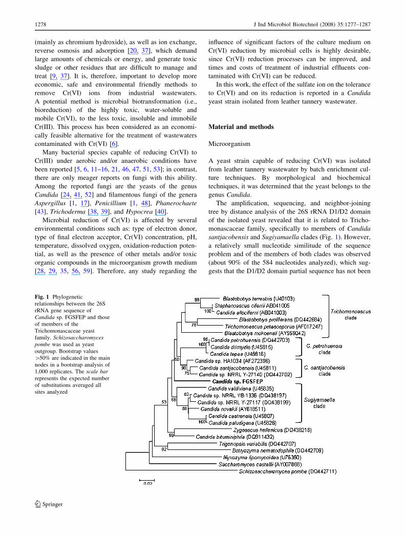
The amplification, sequencing, and neighbor-joining
tree by distance analysis of the 26S rRNA D1/D2 domain
of the isolated yeast revealed that it is related to Tricho-
monascaceae family, specifically to members of Candida
santjacobensis and Sugiyamaella clades (Fig. 1). However,
a relatively small nucleotide similitude of the sequence
problem and of the members of both clades was observed
(about 90% of the 584 nucleotides analyzed), which sug-
gests that the D1/D2 domain partial sequence has not been
Fig. 1 Phylogenetic
relationships between the 26S
rRNA gene sequence of
Candida sp. FGSFEP and those
of members of the
Trichomonascaceae yeast
family. Schizosaccharomycespombe was used as yeast
outgroup. Bootstrap values
[50% are indicated in the main
nodes in a bootstrap analysis of
1,000 replicates. The scale barrepresents the expected number
of substitutions averaged all
sites analyzed
1278 J Ind Microbiol Biotechnol (2008) 35:1277–1287
123
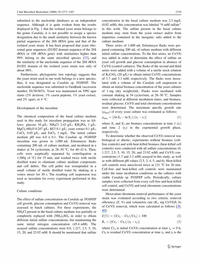
submitted to the nucleotide databases as an independent
sequence. Although it is quite evident from the results
displayed in Fig. 1 that the isolated yeast strain belongs to
the genus Candida, it is not possible to assign a species
designation due to the small similarity between the known
partial sequences of the 26S rRNA gene and that of the
isolated yeast strain. It has been proposed that yeast ribo-
somal gene sequences (D1/D2 domain sequence of the 26S
rRNA or 18S rRNA gene) with similarities higher than
99% belong to the same microbial species [27], and
the similarity of the nucleotide sequence of the 26S rRNA
D1/D2 domain of the isolated yeast strain was only of
about 90%.
Furthermore, phylogenetic tree topology suggests that
the yeast strain used in our work belongs to a new species;
thus, it was designated as Candida sp. FGSFEP. The
nucleotide sequence was submitted to GenBank (accession
number: EU563853). Yeast was maintained on YPG agar
slants (2% dextrose, 1% casein peptone, 1% yeast extract,
and 2% agar), at 4 �C.
Development of the inoculum
The chemical composition of the basal culture medium
used in this study for inoculum propagation was as fol-
lows: glucose 10 g/L; NH4Cl 2.43 g/L; KH2PO4 1 g/L;
MgCl2�6H2O 0.247 g/L; KCl 0.1 g/L; yeast extract 0.1 g/L;
CaCl2 0.05 g/L, and FeCl3 1 mg/L. The initial culture
medium pH was 6.0 ± 0.1. The Candida sp. FGSFEP
inoculum was grown in 1,000 mL Erlenmeyer flasks
containing 200 mL of culture medium, and incubated in a
shaker at 54 cycles/min, at 28–30 �C, for 40–42 h. Then,
cells were aseptically separated by centrifugation at
1,500g (4 �C) for 15 min, and washed twice with sterile
distilled water to eliminate culture medium components
and cell debris. The cell pellet was resuspended in a
small volume of sterile distilled water by shaking in a
vortex mixer for 30 s. The resulting cell suspension was
used as inoculum for every experiment performed in this
study.
Culture conditions
The effect of sulfate concentration on Candida sp. FGSFEP
cell growth, glucose consumption and Cr(VI) removal was
assessed in batch cultures. For these experiments, the
NH4Cl present in the basal culture medium was partially or
completely replaced with (NH4)2SO4 in order to obtain
different initial sulfate concentrations, but maintaining the
same initial nitrogen concentration (45.4 mM). The
assayed sulfate concentrations were 0.0, 1.217, 2.5, 5, 10,
15, 20, and 23.92 mM. It should be mentioned that sulfate
concentration in the basal culture medium was 2.3 mg/L
(0.02 mM); this concentration was labeled ‘‘0 mM sulfate’’
in this work. The sulfate present in the basal culture
medium may stem from the yeast extract and/or from
impurities contained in the inorganic salts added to the
culture medium.
Three series of 1,000 mL Erlenmeyer flasks were pre-
pared containing 200 mL of culture medium with different
initial sulfate concentrations. To the first series, no Cr(VI)
was added in order to determine the effect of sulfate on
yeast cell growth and glucose consumption in absence of
Cr(VI) (control cultures). The flasks of the second and third
series were added with a volume of a sterile stock solution
of K2CrO4 (20 g/L) to obtain initial Cr(VI) concentrations
of 1.7 and 3.3 mM, respectively. The flasks were inocu-
lated with a volume of the Candida cell suspension to
obtain an initial biomass concentration of the yeast cultures
of 1 mg (dry weight)/mL. Flasks were incubated with
constant shaking at 54 cycles/min, at 28–30 �C. Samples
were collected at different incubation times, and biomass,
residual glucose, Cr(VI) and total chromium concentrations
were determined. The maximum specific growth rate
(lmax) of every yeast culture was estimated as follows:
lmax ¼ ln X2 � ln X1ð Þ= t2 � t1ð Þ½ � ð1Þ
where X1 and X2 are biomass concentrations at time 1 (t1)
and time 2 (t2) in the exponential growth phase,
respectively.
To determine whether the observed Cr(VI) removal was
biological or abiotic, experiments without biomass (cell-
free controls) and with heat-killed biomass (heat-killed cell
controls) were conducted with all sulfate concentrations (0,
1.217, 2.5, 5, 10, 15, 20, and 23.92 mM) and Cr(VI) con-
centrations (1.7 and 3.3 mM) assayed in this study, as well
as with different pH values (2.5, 3, 4, 5, and 6). Heat-killed
cell controls were autoclaved twice at 121 �C for 20 min.
Cell-free and heat-killed cell controls were maintained
under the same incubation conditions as the cultures with
viable Candida sp. FGSFEP cells. Periodically, culture
samples were collected from every cell-free and heat-killed
cell control, and Cr(VI) and total chromium concentrations
were determined.
Hexavalent chromium removal performance of the yeast
strain was evaluated according to two criteria: removal
efficiency (E, %) and volumetric rate (Rv, mg Cr(VI)/L h)
of Cr(VI) removal, which were calculated as follows [39,
40]:
Eð%Þ ¼ Cro � Crfð Þ=Cro½ � � 100 ð2ÞRv ¼ Cro � Crfð Þ= tf � toð Þ½ � ð3Þ
where Cro is initial Cr(VI) concentration at time to = 0 h,
Crf is residual Cr(VI) concentration at time tf, and tf is the
J Ind Microbiol Biotechnol (2008) 35:1277–1287 1279
123
cultivation time at which Cr(VI) was completely removed
or the total incubation time for those experiments in which
Cr(VI) was not completely removed.
All experiments in this study were performed in tripli-
cate and average values are reported herein. The maximum
variation coefficient of the three replicates was 4.2%.
Analytical techniques
Biomass concentration
Biomass concentration was determined by measuring dry
cell weight. Culture samples were filtered through pre-
weighed 1.6 lm filters (Whatman GF/A), which were
washed twice with sterile distilled water and subsequently
dried at 90 �C until constant weight was attained. The
obtained filtrates were used to determine residual glucose,
Cr(VI) and total chromium concentrations.
Glucose concentration
Glucose concentration of culture samples was enzymati-
cally determined (glucose oxidase and peroxidase) using a
Sigma glucose assay kit.
Cr(VI) and total chromium concentration in aqueous
solution
Cr(VI) concentration was determined using the 1,5-diphe-
nylcarbohydrazide method according to the procedures
described in the Hach Water Analysis Handbook [19].
Total chromium concentrations in solution were measured
by atomic absorption spectroscopy (SpectrAA220 FS,
Varian, Inc.), following the procedures of method 3111B of
the Standard Methods for the Examination of Water and
Wastewater [8].
Total chromium contents in yeast biomass
To determine total chromium uptake by the yeast bio-
mass, aliquots of the washed cells were pretreated
according to the procedures described in method 3030E of
the Standard Methods for the Examination of Water and
Wastewater [8]. Briefly, aliquots of the washed cells were
mineralized by burning in concentrated nitric acid, then
cooled and made up with deionized water to a final vol-
ume of 50 mL. The resulting solution was analyzed for
total chromium by electrothermal atomic absorption
spectroscopy (SpectrAA220 FS, Varian, Inc.) with a
graphite furnace and a chromium hollow cathode lamp,
according to the procedures described in method 3113B
of the Standard Methods for the Examination of Water
and Wastewater [8].
Results and discussion
Influence of sulfate on tolerance of Candida
sp. FGSFEP to Cr(VI)
The effect of eight different sulfate concentrations (0,
1.217, 2.5, 5, 10, 15, 20, and 23.92 mM) on Candida sp.
FGSFEP cell growth was investigated in batch cultures
without Cr(VI) and with initial Cr(VI) concentrations of
1.7 and 3.3 mM.
Figure 2 depicts the Candida sp. FGSFEP cell growth
curves obtained at different sulfate and Cr(VI) concen-
trations. In this figure, a horizontal straight line was drawn
at a biomass concentration of 1 g/L in order to show if the
yeast was capable or not of growing at the assayed con-
centration of sulfate and/or of Cr(VI). Points lying on the
straight line indicate that there was not a net yeast growth;
points above the line show that the yeast was able to
grow, and points below the line indicate that cell lysis did
occur.
In culture media without Cr(VI), yeast cell growth was
very similar at all assayed sulfate concentrations (Fig. 2a),
which suggests that the sulfate ion had no stimulating or
inhibiting effect on Candida sp. FGSFEP cell growth. In
addition, the amount of sulfate (0.02 mM) present in the
basal culture medium proved to be enough to allow ade-
quate growth of the yeast.
In culture media with 1.7 mM Cr(VI), Candida sp.
FGSFEP cell density decreased as culture time increased
when a sulfate concentration of 0 mM was used, which
indicated the presence of cell lysis (Fig. 2b). With
1.217 mM sulfate concentration, biomass concentration
was almost constant along the incubation period indicating
that, under these conditions, yeast net growth was practi-
cally negligible. In contrast, starting at a sulfate
concentration of 2.5 mM, Candida sp. FGSFEP cell con-
centration gradually increased as sulfate concentration
increased; the maximum cell density values were obtained
within the sulfate concentration interval of 10–23.92 mM
(Fig. 2b). Biomass concentration profiles obtained within
this sulfate concentration interval were similar to those in
cultures without Cr(VI) (Fig. 2a), which suggests that toxic
effects of Cr(VI) on yeast cell growth are practically null at
these sulfate concentrations.
Initial 3.3 mM concentration of Cr(VI) produced lysis of
Candida sp. FGSFEP cells when sulfate concentrations in
the culture medium were lower than 10 mM (Fig. 2c); in
contrast, as sulfate concentration increased from 10 to
23.92 mM, cell concentration also increased. Within this
sulfate concentration interval, cell lysis was observed at
incubation times longer than 150 h. Cell density in culture
media with 3.3 mM Cr(VI) was lower than in cultures
without Cr(VI) (Fig. 2a) or with 1.7 mM Cr(VI) (Fig. 2b)
1280 J Ind Microbiol Biotechnol (2008) 35:1277–1287
123
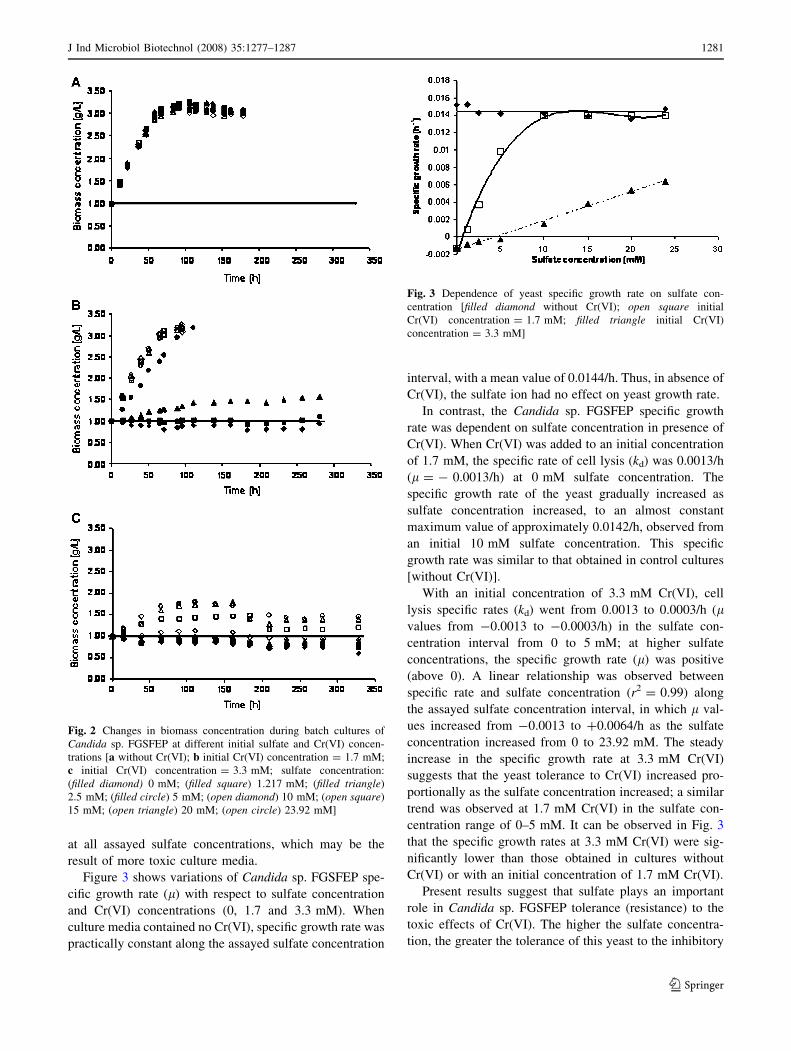
at all assayed sulfate concentrations, which may be the
result of more toxic culture media.
Figure 3 shows variations of Candida sp. FGSFEP spe-
cific growth rate (l) with respect to sulfate concentration
and Cr(VI) concentrations (0, 1.7 and 3.3 mM). When
culture media contained no Cr(VI), specific growth rate was
practically constant along the assayed sulfate concentration
interval, with a mean value of 0.0144/h. Thus, in absence of
Cr(VI), the sulfate ion had no effect on yeast growth rate.
In contrast, the Candida sp. FGSFEP specific growth
rate was dependent on sulfate concentration in presence of
Cr(VI). When Cr(VI) was added to an initial concentration
of 1.7 mM, the specific rate of cell lysis (kd) was 0.0013/h
(l = - 0.0013/h) at 0 mM sulfate concentration. The
specific growth rate of the yeast gradually increased as
sulfate concentration increased, to an almost constant
maximum value of approximately 0.0142/h, observed from
an initial 10 mM sulfate concentration. This specific
growth rate was similar to that obtained in control cultures
[without Cr(VI)].
With an initial concentration of 3.3 mM Cr(VI), cell
lysis specific rates (kd) went from 0.0013 to 0.0003/h (lvalues from -0.0013 to -0.0003/h) in the sulfate con-
centration interval from 0 to 5 mM; at higher sulfate
concentrations, the specific growth rate (l) was positive
(above 0). A linear relationship was observed between
specific rate and sulfate concentration (r2 = 0.99) along
the assayed sulfate concentration interval, in which l val-
ues increased from -0.0013 to +0.0064/h as the sulfate
concentration increased from 0 to 23.92 mM. The steady
increase in the specific growth rate at 3.3 mM Cr(VI)
suggests that the yeast tolerance to Cr(VI) increased pro-
portionally as the sulfate concentration increased; a similar
trend was observed at 1.7 mM Cr(VI) in the sulfate con-
centration range of 0–5 mM. It can be observed in Fig. 3
that the specific growth rates at 3.3 mM Cr(VI) were sig-
nificantly lower than those obtained in cultures without
Cr(VI) or with an initial concentration of 1.7 mM Cr(VI).
Present results suggest that sulfate plays an important
role in Candida sp. FGSFEP tolerance (resistance) to the
toxic effects of Cr(VI). The higher the sulfate concentra-
tion, the greater the tolerance of this yeast to the inhibitory
Fig. 2 Changes in biomass concentration during batch cultures of
Candida sp. FGSFEP at different initial sulfate and Cr(VI) concen-
trations [a without Cr(VI); b initial Cr(VI) concentration = 1.7 mM;
c initial Cr(VI) concentration = 3.3 mM; sulfate concentration:
(filled diamond) 0 mM; (filled square) 1.217 mM; (filled triangle)
2.5 mM; (filled circle) 5 mM; (open diamond) 10 mM; (open square)
15 mM; (open triangle) 20 mM; (open circle) 23.92 mM]
Fig. 3 Dependence of yeast specific growth rate on sulfate con-
centration [filled diamond without Cr(VI); open square initial
Cr(VI) concentration = 1.7 mM; filled triangle initial Cr(VI)
concentration = 3.3 mM]
J Ind Microbiol Biotechnol (2008) 35:1277–1287 1281
123
effects of Cr(VI), and, thus, the less effect on cell growth.
Likewise, results reveal that the ‘‘protective effect’’ of
sulfate on cell growth of the yeast kept in Cr(VI)-con-
taining media depended on initial Cr(VI) concentration.
Ohtake et al. [42] reported increased cell growth in two
Pseudomonas fluorescens strains (LB300 and LB303) in
the presence of CrO42- and high sulfate concentrations.
Also, the P. fluorescens tolerance level to CrO42- depen-
ded on the sulfur source, since bacterial cells growing in
culture medium with added cysteine were more resistant to
CrO42- than cells growing on sulfate. Since chromate is
carried into the cells by the sulfate transport system, the
ability of sulfate to protect P. fluorescens LB300 cells from
the inhibitory effects of Cr(VI) was attributed to the fact
that sulfate competitively inhibits chromate uptake by
bacterial cells [42].
Likewise, a study carried out by Pepi and Baldi [49] with
a Candida sp. strain, using varying sulfate (4–160 mM) and
Cr(VI) (0.04–0.2 mM) concentrations, revealed that sulfate
ion increases yeast tolerance to Cr(VI). Maximum growth of
this Candida sp. strain was observed in Yeast Nitrogen Base
medium without amino acids, at initial Cr(VI) concentra-
tions of 0.04 and 0.08 mM, and 50 mM sulfate. It was also
found that if Candida sp. is grown in the presence of S-
amino acids, especially methionine, it is more resistant to
Cr(VI) than if the sulfur source is sulfate [49]. The general
mechanism of resistance to chromate in Candida sp. was
also attributed to a reduced chromium uptake [49]. In con-
trast, resistance of Ochrobactrum tritici strain 5bvI1 to
Cr(VI) was not dependent on sulfate concentration [4].
Effect of sulfate on glucose consumption
in Candida sp. FGSFEP
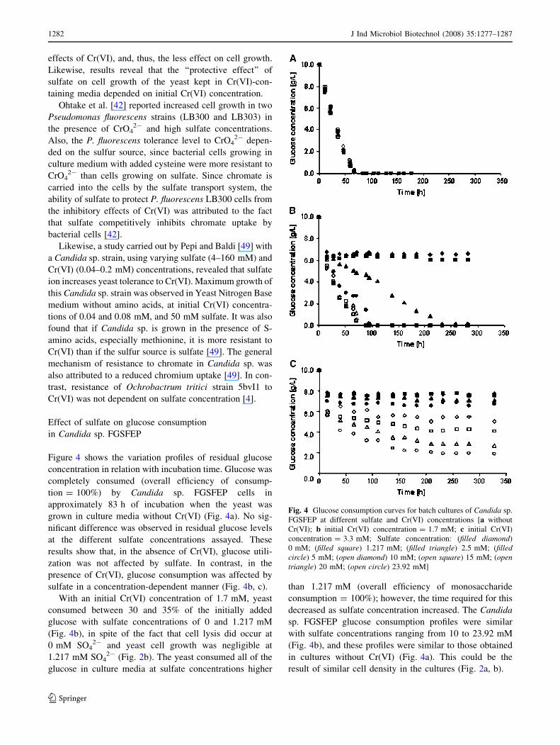
Figure 4 shows the variation profiles of residual glucose
concentration in relation with incubation time. Glucose was
completely consumed (overall efficiency of consump-
tion = 100%) by Candida sp. FGSFEP cells in
approximately 83 h of incubation when the yeast was
grown in culture media without Cr(VI) (Fig. 4a). No sig-
nificant difference was observed in residual glucose levels
at the different sulfate concentrations assayed. These
results show that, in the absence of Cr(VI), glucose utili-
zation was not affected by sulfate. In contrast, in the
presence of Cr(VI), glucose consumption was affected by
sulfate in a concentration-dependent manner (Fig. 4b, c).
With an initial Cr(VI) concentration of 1.7 mM, yeast
consumed between 30 and 35% of the initially added
glucose with sulfate concentrations of 0 and 1.217 mM
(Fig. 4b), in spite of the fact that cell lysis did occur at
0 mM SO42- and yeast cell growth was negligible at
1.217 mM SO42- (Fig. 2b). The yeast consumed all of the
glucose in culture media at sulfate concentrations higher
than 1.217 mM (overall efficiency of monosaccharide
consumption = 100%); however, the time required for this
decreased as sulfate concentration increased. The Candida
sp. FGSFEP glucose consumption profiles were similar
with sulfate concentrations ranging from 10 to 23.92 mM
(Fig. 4b), and these profiles were similar to those obtained
in cultures without Cr(VI) (Fig. 4a). This could be the
result of similar cell density in the cultures (Fig. 2a, b).
Fig. 4 Glucose consumption curves for batch cultures of Candida sp.
FGSFEP at different sulfate and Cr(VI) concentrations [a without
Cr(VI); b initial Cr(VI) concentration = 1.7 mM; c initial Cr(VI)
concentration = 3.3 mM; Sulfate concentration: (filled diamond)
0 mM; (filled square) 1.217 mM; (filled triangle) 2.5 mM; (filledcircle) 5 mM; (open diamond) 10 mM; (open square) 15 mM; (opentriangle) 20 mM; (open circle) 23.92 mM]
1282 J Ind Microbiol Biotechnol (2008) 35:1277–1287
123
At initial concentration of 3.3 mM Cr(VI), residual
glucose levels decreased as sulfate concentration
increased (Fig. 4c); however, residual glucose levels were
higher than those obtained with 1.7 mM Cr(VI) (Fig. 4b),
at all assayed sulfate concentrations. The highest glucose
consumption efficiency was about 80% and was obtained
at sulfate concentration of 23.92 mM. With the two
assayed Cr(VI) concentrations (1.7 and 3.3 mM), some
glucose was consumed during the first hours of incubation
by yeast cultures which also showed cell lysis [0 mM
SO42-, 1.7 mM Cr(VI); and 0–5 mM SO4
2-, 3.3 mM
Cr(VI)]. The glucose was probably used to maintain the
living cells.
As expected, Candida sp. FGSFEP cultures with the
highest cell density at 1.7 and 3.3 mM Cr(VI) consumed
more glucose, and this happened when high concentrations
of sulfate were used. Taken together, the results obtained in
the presence of Cr(VI) indicate that glucose utilization was
dependent on the sulfate concentration, which in turn
affected the intensity of Candida sp. FGSFEP growth.
Influence of sulfate on Cr(VI) removal by Candida
sp. FGSFEP
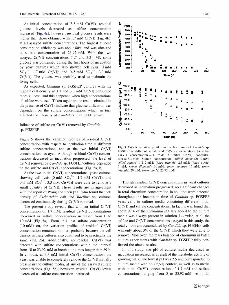
Figure 5 shows the variation profiles of residual Cr(VI)
concentration with respect to incubation time at different
sulfate concentrations, and at the two initial Cr(VI)
concentrations assayed. Although residual Cr(VI) concen-
trations decreased as incubation progressed, the level of
Cr(VI) removal by Candida sp. FGSFEP cultures depended
on the sulfate and Cr(VI) concentrations (Fig. 5a, b).
At the two initial Cr(VI) concentrations, yeast cultures
showing cell lysis [0 mM SO42-, 1.7 mM Cr(VI); and
0–5 mM SO42-, 3.3 mM Cr(VI)] were able to remove a
small quantity of Cr(VI). These results are in agreement
with the report of Wang and Shen [57], who found that cell
density of Escherichia coli and Bacillus sp. cultures
decreased continuously during Cr(VI) removal.
The present study reveals that with an initial Cr(VI)
concentration of 1.7 mM, residual Cr(VI) concentrations
decreased as sulfate concentration increased from 0 to
10 mM (Fig. 5a). From this last sulfate concentration
(10 mM) on, the variation profiles of residual Cr(VI)
concentration remained similar, probably because the cell
density in these cultures also continued to be practically the
same (Fig. 2b). Additionally, no residual Cr(VI) was
detected with sulfate concentrations within the interval
from 10 to 23.92 mM at incubation times longer than 89 h.
In contrast, at 3.3 mM initial Cr(VI) concentration, the
yeast was unable to completely remove the Cr(VI) initially
present in the culture media, at any of the assayed sulfate
concentrations (Fig. 5b); however, residual Cr(VI) levels
decreased as sulfate concentration increased.
Though residual Cr(VI) concentrations in yeast cultures
decreased as incubation progressed, no significant changes
in total chromium concentration in solution were detected
throughout the incubation time of Candida sp. FGSFEP
yeast cells in culture media containing different initial
Cr(VI) and sulfate concentrations. In fact, it was found that
about 97% of the chromium initially added to the culture
media was always present in solution. Likewise, at all the
sulfate and Cr(VI) concentrations assayed in this study, the
total chromium accumulated by Candida sp. FGSFEP cells
was only about 3% of the Cr(VI) which they were able to
remove. Moreover, the mass balance of chromium in batch
culture experiments with Candida sp. FGSFEP fully con-
firmed the above results.
In this study, the pH of culture media decreased as
incubation increased, as a result of the metabolic activity of
growing cells. The lowest pH was 2.5 and corresponded to
culture media with no Cr(VI) content, as well as to media
with initial Cr(VI) concentration of 1.7 mM and sulfate
concentrations ranging from 5 to 23.92 mM. At initial
Fig. 5 Cr(VI) variation profiles in batch cultures of Candida sp.
FGSFEP at different sulfate and Cr(VI) concentrations [a initial
Cr(VI) concentration = 1.7 mM; b initial Cr(VI) concentra-
tion = 3.3 mM; Sulfate concentration: (filled diamond) 0 mM;
(filled square) 1.217 mM; (filled triangle) 2.5 mM; (filled circle)
5 mM; (open diamond) 10 mM; (open square) 15 mM; (opentriangle) 20 mM; (open circle) 23.92 mM]
J Ind Microbiol Biotechnol (2008) 35:1277–1287 1283
123
3.3 mM Cr(VI) concentration, the lowest pH was 3.6 in
culture media with sulfate concentrations of 20 and
23.92 mM. As expected, the lowest pH values were
observed in yeast cultures with the highest cell concen-
trations and highest glucose consumption.
As it is known that low pH’s favor Cr(VI) reduction by
organic matter [54], experiments with two different abiotic
controls (cell-free and heat-killed cell controls) were car-
ried out in this work at different pH values (2.5, 3, 4, 5, and
6 ± 0.1) and initial Cr(VI) (1.7 and 3.3 mM) and sulfate
concentrations (0, 1.217, 2.5, 5, 10, 15, 20 and 23.92 mM),
in order to determine if the pH influences chromium
removal in the absence of living biomass. It was found that
the Cr(VI) and total chromium concentrations remained
constant along 340 h, in both the cell-free and heat-killed
cell controls at pH values ranging from 3.0 to 6.0. At pH
2.5, no measurable change in total chromium concentration
was observed in the cell-free and heat-killed cell controls;
in contrast, the Cr(VI) concentration decreased by 2.7–3%
and 8–10% in the cell-free and heat-killed cell controls,
respectively. The loss of Cr(VI) content in the cell-free and
heat-killed cell controls at pH 2.5 are attributable to Cr(VI)
reduction by medium components and by medium com-
ponents and dead biomass, respectively. The results
obtained in the heat-killed cell controls at pH 2.5 are in
agreement with those reported by Park et al. [44, 45], who
showed that dead biomass of Rhizopus oryzae, Aspergillus
niger, Penicillium chrysogenum, Saccharomyces cerevisiae
[44] and Ecklonia sp. [45] is capable of reducing Cr(VI)
when the biomass is brought into contact with a chromate
solution at pH 2.0.
Taken together, the above observations indicate that the
primary mechanism of Cr(VI) removal by viable cells of
Candida sp. FGSFEP was the transformation (reduction) of
Cr(VI) to forms of lower valence. As the stable forms of
chromium are the trivalent and the hexavalent [34], it
seems most likely that the yeast was capable of trans-
forming the highly toxic and soluble hexavalent chromium
to the much less toxic and less mobile trivalent form.
Furthermore, the incubation of viable Candida sp. FGSFEP
cells in culture media without carbon and energy source
(media without glucose and yeast extract, but with sulfate
and chromate at the different concentrations assayed in this
study) produced no changes in Cr(VI) and total chromium
concentrations, which indicates that a carbon source is
required in the yeast growth medium in order to provide the
necessary reducing power for Cr(VI) reduction and,
therefore, that the observed Cr(VI) reduction was mainly
due to the metabolic activity of Candida sp. FGSFEP cells.
At initial 1.7 mM Cr(VI) concentration, overall effi-
ciency of Cr(VI) reduction exhibited by Candida sp.
FGSFEP increased from 17.9% to 98.6% as sulfate con-
centration increased from 0 to 2.5 mM, and at higher
sulfate concentrations, Cr(VI) reduction efficiency was
100%. Likewise, at 3.3 mM Cr(VI), reduction efficiency
increased from 8.3 up to 84% as sulfate concentration
increased from 0 to 23.92 mM.
These results clearly show that Candida sp. FGSFEP has
a remarkable ability to reduce very high Cr(VI) concen-
trations in the presence of sulfate. The Cr(VI)
concentrations that were reduced by Candida sp. FGSFEP
were much higher than concentrations commonly found to
be reduced by bacteria [2, 5, 6, 16, 28, 36, 60], yeasts [41,
52] and filamentous fungi [1, 38] in the presence of sulfate.
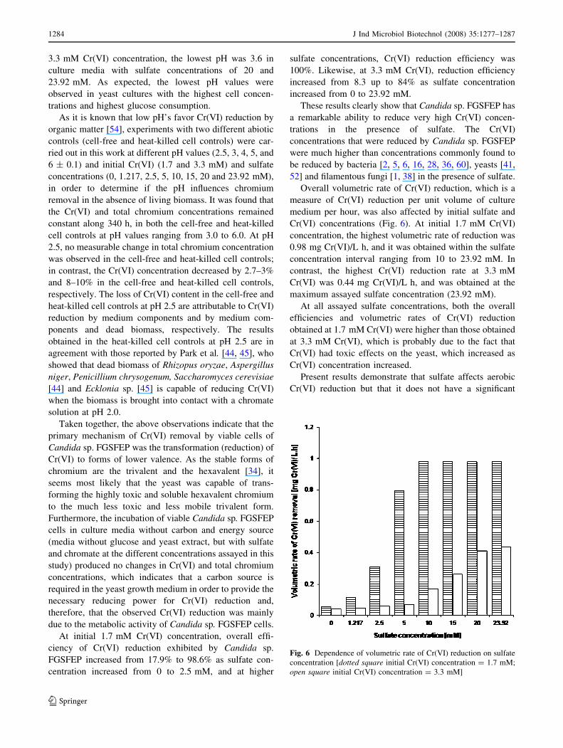
Overall volumetric rate of Cr(VI) reduction, which is a
measure of Cr(VI) reduction per unit volume of culture
medium per hour, was also affected by initial sulfate and
Cr(VI) concentrations (Fig. 6). At initial 1.7 mM Cr(VI)
concentration, the highest volumetric rate of reduction was
0.98 mg Cr(VI)/L h, and it was obtained within the sulfate
concentration interval ranging from 10 to 23.92 mM. In
contrast, the highest Cr(VI) reduction rate at 3.3 mM
Cr(VI) was 0.44 mg Cr(VI)/L h, and was obtained at the
maximum assayed sulfate concentration (23.92 mM).
At all assayed sulfate concentrations, both the overall
efficiencies and volumetric rates of Cr(VI) reduction
obtained at 1.7 mM Cr(VI) were higher than those obtained
at 3.3 mM Cr(VI), which is probably due to the fact that
Cr(VI) had toxic effects on the yeast, which increased as
Cr(VI) concentration increased.
Present results demonstrate that sulfate affects aerobic
Cr(VI) reduction but that it does not have a significant
Fig. 6 Dependence of volumetric rate of Cr(VI) reduction on sulfate
concentration [dotted square initial Cr(VI) concentration = 1.7 mM;
open square initial Cr(VI) concentration = 3.3 mM]
1284 J Ind Microbiol Biotechnol (2008) 35:1277–1287
123
effect on total chromium removal by Candida sp. FGSFEP.
These results differ from other reports which find no
inhibitory or stimulating effect of sulfate on Cr(VI)
reduction in aerobic microbial cultures. Sulfate concen-
tration of up to 1 mM was reported to have no effect on
chromate reduction by Pseudomonas putida cells [22].
Sulfate at 10 mM concentration did not affect Cr(VI)
reduction by Bacillus sp. [58]. Liu et al. [28] reported that
sulfate concentrations of 40 and 80 mg/L (0.416 and
0.83 mM) did not affect aerobic Cr(VI) reduction by
Bacillus sp. XW-4. Likewise, Cr(VI) reduction by Acine-
tobacter haemolyticus was not altered in the presence of
10 mM sulfate [60]. In contrast, under anaerobic condi-
tions, sulfate does usually have effects on chromate
bacterial reduction; this may be due to the fact that sulfate
competes with chromate as final electron acceptor [16, 28].
In anaerobic cultures of Escherichia coli ATCC 33456,
Cr(VI) reduction rate was not affected by up to 41.6 mM
sulfate; however, the rate decreased at 83 mM sulfate [53].
Sulfate concentrations as high as 50 mM did not inhibit
Cr(VI) reduction by Desulfovibrio vulgaris [33]. Philip
et al. [50] reported that the presence of up to 1,000 mg/L
(10.4 mM) of sulfate did not affect Cr(VI) reduction by
Bacillus coagulans. However, Cr(VI) reduction by Enter-
obacter cloacae HO1 under anaerobic conditions showed
32% inhibition in the presence of only 25 lM of sulfate
[26].
As mentioned above, sulfate increases tolerance to
Cr(VI) in the Candida sp. strain isolated by Pepi and Baldi
[49]; however, this yeast strain was not capable to reduce
Cr(VI). Likewise, even if Pseudomonas fluorescens LB300
is capable of reducing Cr(VI) to Cr(III) [3] and sulfate
increases its cell growth in the presence of chromate [42],
to the best of our knowledge no effect of sulfate on Cr(VI)
reduction by this microorganism has been reported.
Results obtained in the present study clearly demon-
strate that sulfate affected tolerance of Candida sp.
FGSFEP to Cr(VI) and its ability to reduce it. It is evident
that, as sulfate concentration increased, yeast growth as
well as the overall Cr(VI) reduction efficiency and rate
increased. These results also reveal that Cr(VI) reduction
by Candida sp. FGSFEP depends on culture cell density, in
such a way that the larger the quantity of biomass pro-
duced, the higher the Cr(VI) reduction. Even if the
Candida sp. FGSFEP strain is sensitive to the toxic effects
of Cr(VI) in the absence of sulfate, this yeast has a
remarkable ability to reduce very high concentrations of
Cr(VI) in presence of sulfate.
The present work proves the important role of sulfate for
both, tolerance to Cr(VI) and aerobic Cr(VI) reduction by
Candida sp. FGSFEP. These findings may have important
implications in the biological treatment of Cr(VI) con-
taminated wastewaters.
Conclusions
This work shows that the tolerance of Candida sp. FGSFEP
to the toxic effects of Cr(VI) rose as sulfate concentration
increased; thus, yeast cell growth, glucose consumption,
overall efficiency and volumetric rate of Cr(VI) reduction
remarkably increased at high sulfate concentrations. The
results obtained in this work could be useful to improve the
biological treatment of industrial effluents contaminated
with Cr(VI).
Acknowledgments C. H. H.-R. and E. C.-U. are fellow holders of a
grant from the Comision de Operacion y Fomento de Actividades
Academicas, Instituto Politecnico Nacional, Mexico City, Mexico.
The authors gratefully acknowledge the financial support provided by
the Secretarıa de Investigacion y Posgrado, IPN. The CONACyT
awarded a graduate scholarship to one of the co-authors (F. M. G.-J.).
References
1. Acevedo-Aguilar FJ, Espino-Saldana AE, Leon-Rodriguez IL,
Rivera-Cano ME, Avila-Rodriguez M, Wrobel K, Wrobel K,
Lappe P, Ulloa M, Gutierrez-Corona JF (2006) Hexavalent
chromium removal in vitro and from industrial wastes, using
chromate-resistant strains of filamentous fungi indigenous to
contaminated wastes. Can J Microbiol 52:809–815
2. Bae WC, Kang TG, Kang IK, Won YJ, Jeong BC (2000)
Reduction of hexavalent chromium by Escherichia coli ATCC
33456 in batch and continuous cultures. J Microbiol 38:36–39
3. Bopp LH, Ehrlich HL (1988) Chromate resistance and reduction
in Pseudomonas fluorescens strain LB300. Arch Microbiol
150:426–431
4. Branco R, Alpoim MC, Morais PV (2004) Ochrobactrum triticistrain 5bvI1—characterization of a Cr(VI)-resistant and Cr(VI)-
reducing strain. Can J Microbiol 50:697–703
5. Chardin B, Dolla A, Chaspoul F, Fardeau ML, Gallice P, Bruschi
M (2002) Bioremediation of chromate: thermodynamic analysis
of the effects of Cr(VI) on sulfate-reducing bacteria. Appl
Microbiol Biotechnol 60:352–360
6. Cheung KH, Gu JD (2003) Reduction of chromate (CrO42-) by an
enrichment consortium and an isolate of marine sulfate-reducing
bacteria. Chemosphere 52:1523–1529
7. Cheung KH, Gu JD (2007) Mechanism of hexavalent chromium
detoxification by microorganisms and bioremediation application
potential: a review. Int Biodeter Biodegr 59:8–15
8. Clesceri LS, Greenberg AE, Eaton AD (eds) (1998) Standard
methods for the examination of water and wastewater, 20th edn.
American Public Health Association, Washington, DC
9. Crini G (2005) Recent developments in polysaccharide-based
materials used as adsorbents in wastewater treatment. Prog Polym
Sci 30:38–70
10. DeFlora S, Bagnasco M, Serra D, Zanacchi P (1990) Genotoxi-
city of chromium compounds: a review. Mutat Res 238:99–172
11. DeLeo PC, Ehrlich HL (1994) Reduction of hexavalent chro-
mium by Pseudomonas fluorescens LB300 in batch and
continuous cultures. Appl Microbiol Biotechnol 40:756–759
12. Dmitrenko GN, Konovalova VV, Shum OA (2003) The reduction
of Cr(VI) by bacteria of the genus Pseudomonas. Microbiology
72:327–330
13. Francisco R, Alpoim MC, Morais PV (2002) Diversity of chro-
mium-resistant and -reducing bacteria in a chromium-
contaminated activated sludge. J Appl Microbiol 92:837–843
J Ind Microbiol Biotechnol (2008) 35:1277–1287 1285
123
14. Fujie K, Hu HY, Huang X, Tanaka Y, Urano K, Ohtake H (1996)
Optimal operation of bioreactor system developed for the treat-
ment of chromate wastewater using Enterobacter cloacae HO-1.
Water Sci Technol 33:173–182
15. Ganguli A, Tripathi AK (2002) Bioremediation of toxic chro-
mium from electroplating effluent by chromate-reducing
Pseudomonas aeruginosa A2Chr in two bioreactors. Appl
Microbiol Biotechnol 58:416–420
16. Garbisu C, Alkorta I, Llama MJ, Serra JL (1998) Aerobic chro-
mate reduction by Bacillus subtilis. Biodegradation 9:133–141
17. Gouda MK (2000) Studies on chromate reduction by three
Aspergillus species. Fresen Environ Bull 9:799–808
18. Guertin J (2005) Toxicity and health effects of chromium (all
oxidation states). In: Guertin J, Jacobs JA, Avakian CP (eds)
Chromium(VI) handbook. CRC Press, Boca Raton, pp 215–234
19. Hach Company (ed) (2002) Hach water analysis handbook, 4th
edn. Hach Company, Loveland
20. Hawley EL, Deeb RA, Kavanaugh MC, Jacobs JA (2005)
Treatment technologies for chromium(VI). In: Guertin J, Jacobs
JA, Avakian CP (eds) Chromium(VI) handbook. CRC Press,
Boca Raton, pp 275–309
21. Humphries AC, Macaskie LE (2002) Reduction of Cr(VI) by
Desulfovibrio vulgaris and Microbacterium sp. Biotechnol Lett
24:1261–1267
22. Ishibashi Y, Cervantes C, Silver S (1990) Chromium reduction in
Pseudomonas putida. Appl Environ Microbiol 56:2268–2270
23. Jacobs JA, Testa SM (2005) Overview of chromium(VI) in the
environment: background and history. In: Guertin J, Jacobs JA,
Avakian CP (eds) Chromium(VI) handbook. CRC Press, Boca
Raton, pp 1–21
24. Juvera-Espinosa J, Morales-Barrera L, Cristiani-Urbina E (2006)
Isolation and characterization of a yeast strain capable of
removing Cr(VI). Enzyme Microbial Technol 40:114–121
25. Kimbrough DE, Cohen Y, Winer AM, Creelman L, Mabuni CA
(1999) Critical assessment of chromium in the environment. Crit
Rev Environ Sci Technol 29:1–46
26. Komori K, Rivas A, Toda K, Ohtake H (1989) Biological
removal of toxic chromium using an Enterobacter cloacae strain
that reduces chromate under anaerobic conditions. Biotechnol
Bioeng 35:951–954
27. Kurtzman CP, Fell JW (1998) The yeast, a taxonomic study.
Elsevier Science B.V., Amsterdam, p 367
28. Liu YG, Xu WH, Zeng GM, Li X, Gao H (2006) Cr(VI) reduction
by Bacillus sp. isolated from chromium landfill. Process Biochem
41:1981–1986
29. Llovera S, Bonet R, Simon-Pujol MD, Congregado F (1993)
Effect of culture medium ions on chromate reduction by resting
cells of Agrobacterium radiobacter. Appl Microbiol Biotechnol
39:424–426
30. Lloyd JR (2003) Microbial reduction of metals and radionuclides.
FEMS Microbiol Rev 27:411–425
31. Lofroth G, Ames BN (1978) Mutagenicity of inorganic com-
pounds in Salmonella typhimurium: arsenic, chromium and
selenium. Mutat Res 53:65–66
32. Losi ME, Amrhein C, Frankenberger WT (1994) Environmental
biochemistry of chromium. Rev Environ Contam Toxicol
136:91–121
33. Lovley DR, Phillips EJP (1994) Reduction of chromate by Des-ulfovibrio vulgaris and its c3 cytochrome. Appl Environ
Microbiol 60:726–728
34. McGrath SP, Smith S (1990) Chromium and nickel. In: Alloway
BJ (ed) Heavy metals in soils. Wiley, New York, pp 125–150
35. Mergeay M (1995) Heavy metal resistances in microbial eco-
systems. In: Akkermans ADL, van Elsas JD, De Bruijn FJ (eds)
Molecular microbial ecology manual. Kluwer Academic Press,
Dordrecht, pp 1–17
36. Middleton SS, Latmani RB, Mackey MR, Ellisman MH, Tebo
BM, Criddle CS (2003) Cometabolism of Cr(VI) by Shewanellaoneidensis MR-1 produces cell-associated reduced chromium and
inhibits growth. Biotechnol Bioeng 83:627–637
37. Mohan D, Pittman CU (2006) Activated carbons and low cost
adsorbents for remediation of tri- and hexavalent chromium from
water. J Hazard Mater 137:762–811
38. Morales-Barrera L, Cristiani-Urbina E (2006) Removal of hexa-
valent chromium by Trichoderma viride in an airlift bioreactor.
Enzyme Microbial Technol 40:107–113
39. Morales-Barrera L, Cristiani-Urbina E (2008) Hexavalent chro-
mium removal by a Trichoderma inhamatum fungal strain
isolated from tannery effluent. Water Air Soil Pollut 187:327–336
40. Morales-Barrera L, Guillen-Jimenez FM, Ortiz-Moreno A,
Villegas-Garrido TL, Sandoval-Cabrera A, Hernandez-Rodrıguez
CH, Cristiani-Urbina E (2008) Isolation, identification and
characterization of a Hypocrea tawa strain with high Cr(VI)
reduction potential. Biochem Eng J 40:284–292
41. Muter O, Patmalnieks A, Rapoport A (2001) Interrelations of
the yeast Candida utilis and Cr(VI): metal reduction and its
distribution in the cell and medium. Process Biochem
36:963–970
42. Ohtake H, Cervantes C, Silver S (1987) Decreased chromate
uptake in Pseudomonas fluorescens carrying a chromate resis-
tance plasmid. J Bacteriol 169:3853–3856
43. Pal N (1997) Reduction of hexavalent chromium to trivalent
chromium by Phanerochaete chrysosporium. In: Alleman BC,
Leeson A (eds) In situ and on-site bioremediation, vol 2. Batelle
Press, Columbus, pp 511–517
44. Park D, Yun YS, Park JM (2005) Use of dead fungal biomass for
the detoxification of hexavalent chromium: screening and kinet-
ics. Process Biochem 40:2559–2565
45. Park D, Yun YS, Ahn CK, Park JM (2007) Kinetics of the
reduction of hexavalent chromium with the brown seaweed
Ecklonia biomass. Chemosphere 66:939–946
46. Pattanapipitpaisal P, Brown NL, Macaskie LE (2001) Chromate
reduction and 16S rRNA identification of bacteria isolated from
Cr(VI)-contaminated site. Appl Microbiol Biotechnol 57:257–
261
47. Pattanapipitpaisal P, Brown NL, Macaskie LE (2001) Chromate
reduction by Microbacterium liquefaciens immobilised in poly-
vinyl alcohol. Biotechnol Lett 23:61–65
48. Pazouki M, Keyanpour-Rad M, Shafie SH, Shahhoseini SH
(2007) Efficiency of Penicillium chrysogenum PTCC 5037 in
reducing low concentration of chromium hexavalent in a chro-
mium electroplating plant wastewater. Bioresour Technol
98:2116–2122
49. Pepi M, Baldi F (1992) Modulation of chromium(VI) toxicity by
organic and inorganic sulfur species in yeasts from industrial
wastes. BioMetals 5:179–185
50. Philip L, Iyengar L, Venkobachar C (1998) Cr(VI) reduction by
Bacillus coagulans isolated from contaminated soils. J Environ
Eng ASCE 124:1165–1170
51. QuiIntana M, Curutchet G, Donati E (2001) Factors affecting
chromium(VI) reduction by Thiobacillus ferrooxidans. Biochem
Eng J 9:11–15
52. Ramırez-Ramırez R, Calvo-Mendez C, Avila-Rodrıguez M,
Lappe P, Ulloa M, Vazquez-Juarez R, Gutierrez-Corona JF
(2004) Cr(VI) reduction in a chromate-resistant strain of Candidamaltosa isolated from the leather industry. Antonie van Leeu-
wenhoek 85:63–68
53. Shen H, Wang YT (1994) Biological reduction of chromium by
E. coli. J Environ Eng 120:560–572
54. Stanin FT (2005) The transport and fate of chromium(VI) in the
environment. In: Guertin J, Jacobs JA, Avakian CP (eds) Chro-
mium(VI) handbook. CRC Press, Boca Raton, pp 165–214
1286 J Ind Microbiol Biotechnol (2008) 35:1277–1287
123
55. Testa SM (2005) Sources of chromium contamination in soil and
groundwater. In: Guertin J, Jacobs JA, Avakian CP (eds) Chro-
mium(VI) handbook. CRC Press, Boca Raton, pp 143–163
56. Wang YT, Shen H (1995) Bacterial reduction of hexavalent
chromium. J Ind Microbiol 14:159–163
57. Wang YT, Shen H (1997) Modelling Cr(VI) reduction by pure
bacterial cultures. Water Res 31:727–732
58. Wang YT, Xiao C (1995) Factors affecting hexavalent chromium
reduction in pure cultures of bacteria. Water Res 29:2467–2474
59. Wang YT (2000) Microbial reduction of chromate. In: Lovley
DR (ed) Environmental microbe–metal interactions. ASM Press,
Washington, DC, pp 225–235
60. Zakaria ZA, Zakaria Z, Surif S, Ahmad WA (2007) Hexavalent
chromium reduction by Acinetobacter haemolyticus isolated from
heavy-metal contaminated wastewater. J Hazard Mater 146:30–
38
J Ind Microbiol Biotechnol (2008) 35:1277–1287 1287
123