Modulation of Retroviral Restriction and Proteasome Inhibitor-Resistant Turnover by Changes in the...
17
JOURNAL OF VIROLOGY, Oct. 2007, p. 10362–10378 Vol. 81, No. 19 0022-538X/07/$08.000 doi:10.1128/JVI.00703-07 Copyright © 2007, American Society for Microbiology. All Rights Reserved. Modulation of Retroviral Restriction and Proteasome Inhibitor-Resistant Turnover by Changes in the TRIM5 B-Box 2 Domain Felipe Diaz-Griffero, 1 Alak Kar, 1 Michel Perron, 1 Shi-Hua Xiang, 1 Hassan Javanbakht, 1 Xing Li, 1 and Joseph Sodroski 1,2 * Department of Cancer Immunology and AIDS, Dana-Farber Cancer Institute, Department of Pathology, Division of AIDS, Harvard Medical School, Boston, Massachusetts 02115, 1 and Department of Immunology and Infectious Diseases, Harvard School of Public Health, Boston, Massachusetts 02115 2 Received 3 April 2007/Accepted 2 July 2007 An intact B-box 2 domain is essential for the antiretroviral activity of TRIM5. We modeled the structure of the B-box 2 domain of TRIM5 based on the existing three-dimensional structure of the B-box 2 domain of human TRIM29. Using this model, we altered the residues predicted to be exposed on the surface of this globular structure. Most of the alanine substitutions in these residues exerted little effect on the antiretroviral activity of human TRIM5 hu or rhesus monkey TRIM5 rh . However, alteration of arginine 119 of TRIM5 hu or the corresponding arginine 121 of TRIM5 rh diminished the abilities of the proteins to restrict retroviral infection without affecting trimerization or recognition of the viral capsid. The abilities of these functionally defective TRIM5 proteins to accelerate the uncoating of the targeted retroviral capsid were abolished. Removal of the positively charged side chain from B-box 2 arginines 119/120/121 resulted in diminished proteasome-independent turnover of TRIM5 and the related restriction factor TRIMCyp. However, testing of an array of mutants revealed that the rapid turnover and retroviral restriction functions of this B-box 2 region are separable. Proteins of the tripartite motif (TRIM) family contain RING, B-box, and coiled-coil domains and thus have also been referred to as RBCC proteins (25). TRIM proteins often self- associate and form aggregates called nuclear or cytoplasmic bodies (3, 25, 27). Although TRIM proteins have been impli- cated in transcriptional regulation, cell division, retroviral re- striction, determination of cell polarity, and differentiation, the precise functions of most TRIM proteins remain to be deter- mined (15, 19, 34). TRIM5 was identified in a random genetic screen as the factor responsible for the early postentry block to human im- munodeficiency virus type 1 (HIV-1) in Old World monkeys (30). TRIM5 is a cytoplasmic protein that is capable of re- stricting infection by different retroviruses in a species-depen- dent manner (13, 29). Variation among TRIM5 proteins in different primates accounts for the early postentry blocks to infection by particular retroviruses (5, 9, 12, 13, 37). For ex- ample, the TRIM5 proteins of several Old World monkey species block HIV-1 infection (8, 13, 18), whereas the TRIM5 proteins of New World monkeys block infection by simian immunodeficiency virus (29). TRIM5 from humans (TRIM5 hu ) is not as potent in restricting HIV-1 infection as Old World monkey TRIM5 proteins, but TRIM5 hu potently restricts other retroviruses, e.g., N-tropic murine leukemia virus (N- MLV), equine infectious anemia virus (EIAV), and feline im- munodeficiency virus (FIV) (8, 13, 21, 26, 37). Variation in splicing of the TRIM5 primary transcript leads to the expression of TRIM5 isoforms, designated , , and (25). The TRIM5 isoform contains, in addition to the RING, B-box 2, and coiled-coil domains, a carboxy-terminal B30.2 (SPRY) domain. The B30.2(SPRY) domain is essential for the antiretroviral activity of TRIM5 (32). In some cases, the dif- ferences in the abilities of TRIM5 proteins from various primate species to restrict particular retroviruses are deter- mined by sequences in the B30.2(SPRY) domain (20, 22, 28, 35). The B30.2 domain of rhesus monkey TRIM5 (TRIM5 rh ) is required for specific recognition of the HIV-1 capsid (31). HIV-1 capsid recognition is also dependent on TRIM5 rh trimerization, which is mediated by the coiled-coil and adjacent linker 2 regions (4, 12, 16). Disruption of the TRIM5 rh or TRIM5 hu B-box 2 domain by mutagenesis resulted in loss of retroviral restriction (11, 22, 24). Thus, it has been suggested that the B-box 2 TRIM5 mutants may lack an “effector” function important for restric- tion. Studies following the fate of the HIV-1 and N-MLV capsids in the cytoplasm of cells expressing TRIM5 rh or TRIM5 hu , respectively, indicate that the conversion of par- ticulate capsids to soluble capsid proteins is accelerated by a restricting TRIM5 protein (24, 31). Thus, the B-box 2 domain may contribute to this process. In this work, we modeled the structure of the B-box 2 do- mains of TRIM5 rh and TRIM5 hu based on the available B-box 2 domain structure from the TRIM29 protein (9). The modeled TRIM5 structure revealed that the B-box 2 domain is globular and highly charged on its surface. We tested the * Corresponding author. Mailing address: Dana-Farber Cancer In- stitute, 44 Binney Street-JFB 824, Boston, MA 02115. Phone: (617) 632-3371. Fax: (671) 632-4338. E-mail: [email protected] .edu. Published ahead of print on 11 July 2007. 10362 on January 17, 2015 by guest http://jvi.asm.org/ Downloaded from
-
Upload
independent -
Category
Documents
-
view
0 -
download
0
Transcript of Modulation of Retroviral Restriction and Proteasome Inhibitor-Resistant Turnover by Changes in the...

JOURNAL OF VIROLOGY, Oct. 2007, p. 10362–10378 Vol. 81, No. 190022-538X/07/$08.00�0 doi:10.1128/JVI.00703-07Copyright © 2007, American Society for Microbiology. All Rights Reserved.
Modulation of Retroviral Restriction and ProteasomeInhibitor-Resistant Turnover by Changes in the
TRIM5� B-Box 2 Domain�
Felipe Diaz-Griffero,1 Alak Kar,1 Michel Perron,1 Shi-Hua Xiang,1 Hassan Javanbakht,1Xing Li,1 and Joseph Sodroski1,2*
Department of Cancer Immunology and AIDS, Dana-Farber Cancer Institute, Department of Pathology, Division of AIDS,Harvard Medical School, Boston, Massachusetts 02115,1 and Department of Immunology and Infectious Diseases,
Harvard School of Public Health, Boston, Massachusetts 021152
Received 3 April 2007/Accepted 2 July 2007
An intact B-box 2 domain is essential for the antiretroviral activity of TRIM5�. We modeled the structureof the B-box 2 domain of TRIM5� based on the existing three-dimensional structure of the B-box 2 domain ofhuman TRIM29. Using this model, we altered the residues predicted to be exposed on the surface of thisglobular structure. Most of the alanine substitutions in these residues exerted little effect on the antiretroviralactivity of human TRIM5�hu or rhesus monkey TRIM5�rh. However, alteration of arginine 119 of TRIM5�huor the corresponding arginine 121 of TRIM5�rh diminished the abilities of the proteins to restrict retroviralinfection without affecting trimerization or recognition of the viral capsid. The abilities of these functionallydefective TRIM5� proteins to accelerate the uncoating of the targeted retroviral capsid were abolished.Removal of the positively charged side chain from B-box 2 arginines 119/120/121 resulted in diminishedproteasome-independent turnover of TRIM5� and the related restriction factor TRIMCyp. However, testing ofan array of mutants revealed that the rapid turnover and retroviral restriction functions of this B-box 2 regionare separable.
Proteins of the tripartite motif (TRIM) family containRING, B-box, and coiled-coil domains and thus have also beenreferred to as RBCC proteins (25). TRIM proteins often self-associate and form aggregates called nuclear or cytoplasmicbodies (3, 25, 27). Although TRIM proteins have been impli-cated in transcriptional regulation, cell division, retroviral re-striction, determination of cell polarity, and differentiation, theprecise functions of most TRIM proteins remain to be deter-mined (15, 19, 34).
TRIM5� was identified in a random genetic screen as thefactor responsible for the early postentry block to human im-munodeficiency virus type 1 (HIV-1) in Old World monkeys(30). TRIM5� is a cytoplasmic protein that is capable of re-stricting infection by different retroviruses in a species-depen-dent manner (13, 29). Variation among TRIM5� proteins indifferent primates accounts for the early postentry blocks toinfection by particular retroviruses (5, 9, 12, 13, 37). For ex-ample, the TRIM5� proteins of several Old World monkeyspecies block HIV-1 infection (8, 13, 18), whereas the TRIM5�proteins of New World monkeys block infection by simianimmunodeficiency virus (29). TRIM5� from humans (TRIM5�hu) isnot as potent in restricting HIV-1 infection as Old Worldmonkey TRIM5� proteins, but TRIM5�hu potently restrictsother retroviruses, e.g., N-tropic murine leukemia virus (N-
MLV), equine infectious anemia virus (EIAV), and feline im-munodeficiency virus (FIV) (8, 13, 21, 26, 37).
Variation in splicing of the TRIM5 primary transcript leadsto the expression of TRIM5 isoforms, designated �, �, and �(25). The TRIM5� isoform contains, in addition to the RING,B-box 2, and coiled-coil domains, a carboxy-terminal B30.2(SPRY) domain. The B30.2(SPRY) domain is essential for theantiretroviral activity of TRIM5� (32). In some cases, the dif-ferences in the abilities of TRIM5� proteins from variousprimate species to restrict particular retroviruses are deter-mined by sequences in the B30.2(SPRY) domain (20, 22,28, 35). The B30.2 domain of rhesus monkey TRIM5�(TRIM5�rh) is required for specific recognition of the HIV-1capsid (31). HIV-1 capsid recognition is also dependent onTRIM5�rh trimerization, which is mediated by the coiled-coiland adjacent linker 2 regions (4, 12, 16).
Disruption of the TRIM5�rh or TRIM5�hu B-box 2 domainby mutagenesis resulted in loss of retroviral restriction (11, 22,24). Thus, it has been suggested that the B-box 2 TRIM5mutants may lack an “effector” function important for restric-tion. Studies following the fate of the HIV-1 and N-MLVcapsids in the cytoplasm of cells expressing TRIM5�rh orTRIM5�hu, respectively, indicate that the conversion of par-ticulate capsids to soluble capsid proteins is accelerated by arestricting TRIM5� protein (24, 31). Thus, the B-box 2 domainmay contribute to this process.
In this work, we modeled the structure of the B-box 2 do-mains of TRIM5�rh and TRIM5�hu based on the availableB-box 2 domain structure from the TRIM29 protein (9). Themodeled TRIM5 structure revealed that the B-box 2 domain isglobular and highly charged on its surface. We tested the
* Corresponding author. Mailing address: Dana-Farber Cancer In-stitute, 44 Binney Street-JFB 824, Boston, MA 02115. Phone: (617)632-3371. Fax: (671) 632-4338. E-mail: [email protected].
� Published ahead of print on 11 July 2007.
10362
on January 17, 2015 by guesthttp://jvi.asm
.org/D
ownloaded from

hypothesis that residues predicted to be on the surface of theB-box 2 domain contribute to the “effector” function ofTRIM5�. These residues were individually altered, and thephenotypes of the mutants were characterized. We found thatthe positively charged arginine 121 residue on the surface ofthe B-box 2 domain of TRIM5�rh was important for the re-striction of several retroviruses, including HIV, EIAV, andFIV. Moreover, the homologous residue, arginine 119, ofTRIM5�hu was found to be essential for the restriction ofN-MLV, EIAV, and FIV. Studies of the oligomerization, cap-sid-binding abilities, and effects of these mutant proteins onthe infecting retroviral capsid suggested that this B-box 2 res-idue contributes to the ability of TRIM5� to accelerate theconversion of the particulate cytosolic capsid to soluble capsidproteins. The analysis of a larger panel of mutants involvingresidue 121 of TRIM5�rh or residue 119 of TRIM5�hu re-vealed that changing the positively charged side chain of thisB-box 2 residue dramatically reduced TRIM5� turnover. TheB-box 2-dependent turnover of TRIM5� was not inhibited byproteasome inhibitors. Some B-box 2 mutants of TRIM5�rh
and TRIM5�hu were long-lived but exhibited potent antiret-roviral activity. Thus, the rapid turnover and effector functionsof this TRIM5� B-box 2 region are separable.
MATERIALS AND METHODS
Modeling the TRIM5� B-box 2 domain. The structure of the human TRIM5�B-box 2 domain and the immediate flanking regions was modeled using theMODELLER (version 9.0) program (5) in Discovery Studio 1.7 (Accelrys Soft-
ware, Inc., San Diego, CA). The structure of the TRIM29 B-Box 2 domain(Protein Data Bank file 2CSV) was used as the template for homology-basedprotein modeling (9). The B-box protein sequences from human TRIM5� andTRIM29 exhibit 50% sequence identity, and the TRIM5� model was built basedon the sequence alignment. The model was further refined through loop and sidechain refinement and energy minimization. The coordinates of zinc atoms werederived from the template.
Creation of cells stably expressing TRIM5� variants. Retroviral vectors en-coding wild-type or mutant TRIM5�hu, TRIM5�rh, or TRIMCyp proteins werecreated using the pLPCX vector. Recombinant viruses were produced in 293Tcells by cotransfecting the pLPCX plasmids with the pVPack-GP and pVPack-VSV-G packaging plasmids (Stratagene). The pVPack-VSV-G plasmid encodesthe vesicular stomatitis virus (VSV) G envelope glycoprotein, which allows effi-cient entry into a wide range of vertebrate cells. Cf2Th canine thymocytes weretransduced and selected in 5 �g/ml puromycin (Sigma).
Infection with viruses expressing GFP. Recombinant HIV-1, N-MLV, andB-tropic murine leukemia virus (B-MLV) expressing green fluorescent protein(GFP) were prepared as described previously (19). For infections, 3 � 104 Cf2Thcells seeded in 24-well plates were incubated with virus for 24 h. The cells werewashed and returned to culture for 48 h and then subjected to fluorescence-activated cell sorter analysis with a FACScan (Becton Dickinson). The FIVvector expressing GFP was obtained from System Biosciences (Mountain View,CA) and was prepared following the manufacturer’s instructions. The EIAVvector expressing GFP was a gift from John Olsen (21). HIV-1, N-MLV, B-MLV,FIV, and EIAV viral stocks were titrated by serial dilution on Cf2Th cells todetermine the concentrations of infectious viruses.
Protein analysis. Cellular proteins were extracted with radioimmunoprecipi-tation assay buffer (10 mM Tris, pH 7.4, 100 mM NaCl, 1% sodium deoxycholate,0.1% sodium dodecyl sulfate [SDS], 1% NP-40, 2 mg/ml aprotinin, 2 mg/mlleupeptin, 1 mg/ml pepstatin A, 100 mg/ml phenylmethylsulfonyl fluoride). Thecell lysates were analyzed by SDS-polyacrylamide gel electrophoresis (PAGE)(10% acrylamide), followed by blotting them onto nitrocellulose membranes(Amersham Pharmacia Biotech). The detection of protein by Western blottingutilized monoclonal antibodies directed against the hemagglutinin (HA) epitope
FIG. 1. Modeling the B-box 2 domain of TRIM5�hu based on the TRIM29 B-box 2 structure. Modeling of the TRIM5�hu B-box 2 domain andflanking sequences was performed using the Modeler program (Accelrys Software, Inc.). Modeling was based on the TRIM29 B-box 2 structure(9). The C� trace of the modeled TRIM5�hu B-box 2 domain is shown, with the side chains of lysine 111 and arginine 119. The beginnings of theL1 linker and the coiled coil are depicted in cyan and purple, respectively. The molecule is seen from the perspective of the putative trimer axis.The globular B-box 2 domain is colored green, with basic and acidic residues shown in blue and red, respectively. The alignment of the B-box 2domains of TRIM5�rh and TRIM5�hu with human TRIM29 (TRIM29hu) is shown below. Identical residues are highlighted in yellow. Residuesinvolved in coordinating zinc ions are colored red. Lysines 111/113 and arginines 119/121 of TRIM5�hu and TRIM5�rh, respectively, are coloredblue.
VOL. 81, 2007 B-BOX 2 DOMAIN MUTANTS OF TRIM5� AND TRIMCyp 10363
on January 17, 2015 by guesthttp://jvi.asm
.org/D
ownloaded from
10364 DIAZ-GRIFFERO ET AL. J. VIROL.
on January 17, 2015 by guesthttp://jvi.asm
.org/D
ownloaded from

tags (Roche) and monoclonal antibodies to �-actin (Sigma). Detection of pro-teins was performed by enhanced chemiluminescence (NEN Life Sciences Prod-ucts), using anti-mouse immunoglobulin (for �-actin) and anti-rat immunoglob-ulin (for HA) secondary antibodies (Amersham Pharmacia Biotech).
TRIM5� turnover. The turnover rates of wild-type and mutant TRIM5�proteins were estimated as previously described (3). Briefly, cells expressing theTRIM5 proteins were incubated with medium containing 100 �g/ml cyclohexi-mide, and the steady-state levels of TRIM5 protein were determined by Westernblotting cell lysates for the HA epitope tags or, as a control, for �-actin, asdescribed above. In some cases, TRIM5� proteins with HA epitope tags at boththe N and C termini were studied. In some of the experiments, cycloheximide-treated cells were also incubated with the proteasome inhibitor MG115 (50 �M),MG132 (50 �M), or clasto-lactacystin (10 �M) before lysis and Western blottingas described above.
Localization of wild-type and mutant TRIM5� proteins. The localization ofTRIM5�rh, TRIM5�hu, or TRIMCyp variants in expressing cells was investi-gated as previously described (2). Briefly, cells were grown overnight on 12-mm-diameter coverslips and fixed in 3.9% paraformaldehyde (Sigma) in phosphate-buffered saline (PBS) (Cellgro) for 30 min. The cells were washed in PBS,incubated in 0.1 M glycine (Sigma) for 10 min, washed in PBS, and permeabilizedwith 0.05% saponin (Sigma) for 30 min. Samples were blocked with 10% donkeyserum (Dako, Carpinteria, CA) for 30 min and incubated for 1 h with antibodies.The anti-HA fluorescein isothiocyanate-conjugated 3F10 antibody (Roche) wasused to stain HA-tagged proteins. Subsequently, samples were mounted forfluorescence microscopy by using the ProLong Antifade Kit (Molecular Probes,Eugene, OR). Images were obtained with a Bio-Rad Radiance 2000 laser scan-ning confocal microscope with Nikon 60� 1.4 numerical aperture optics.
TRIM5� oligomerization. Approximately 1 � 107 293T cells transfected with5 �g of plasmids expressing TRIM5�rh or TRIM5�hu variants were lysed inimmunoprecipitation buffer and cross-linked with different concentrations (0 to10 mM) of glycolbis-succinimidylsuccinate for 30 min as previously described(11). The cross-linking reaction was stopped by adding 10 �l of 1 M Tris-HCl.After the cross-linking, samples were resuspended in 2� sample buffer andincubated at 37°C for 30 min. The samples were analyzed by SDS-PAGE andWestern blotting with an anti-HA antibody (Roche).
HIV CA-NC expression and purification. HIV-1 CA-NC protein was ex-pressed, purified, and assembled as previously described (6, 7). The pET11aexpression vector (Novagen) expressing the CA-NC protein of HIV-1 was usedto transform Escherichia coli BL-21(DE3). CA-NC expression was induced with1 mM IPTG (isopropyl-�-D-thiogalactopyranoside) when the culture reached anoptical density of 0.6 at 600 nm. After 4 h of induction, the cells were harvestedand resuspended in 20 mM Tris-HCl (pH 7.5), 1 �M ZnCl2, 10 mM 2-mercap-toethanol, and protease inhibitors (Roche). Lysis was performed through soni-cation, and the debris was pelleted for 30 min at 35,000 � g. Nucleic acids werestripped from the solution by using 0.11 equivalent of 2 M (NH4)2SO4 and thesame volume of 10% polyethylenimine. Nucleic acids were removed by stirringand centrifugation at 29,500 � g for 15 min. The protein was recovered by theaddition of 0.35 equivalent of saturated (NH4)2SO4. The protein was centrifugedat 9,820 � g for 15 min and resuspended in 100 mM NaCl, 20 mM Tris-HCl (pH7.5), 1 �M ZnCl2, and 10 mM 2-mercaptoethanol. The protein was dialyzedagainst 50 mM NaCl, 20 mM Tris-HCl (pH 7.5), 1 �M ZnCl2, and 10 mM2-mercaptoethanol.
In vitro assembly of CA-NC complexes. HIV-1 CA-NC particles were assem-bled in vitro by diluting the CA-NC protein to a concentration of 0.3 mM in 50mM Tris-HCl (pH 8.0), 0.5 M NaCl, and 2 mg/ml DNA oligo-(TG)50. Themixture was incubated at 4°C overnight and centrifuged at 8,600 � g for 5 min.The pellet was resuspended in assembly buffer (50 mM Tris-HCl [pH 8.0], 0.5 MNaCl) at a final protein concentration of 0.15 mM (6, 7).
Binding of TRIM5�rh variants to HIV-1 capsid complexes. 293T cells weretransfected with plasmids expressing wild-type or mutant TRIM5�rh proteins.Forty-eight hours after transfection, cell lysates were prepared as follows. Pre-viously washed cells were resuspended in hypotonic lysis buffer (10 mM Tris, pH
7.4, 1.5 mM MgCl2, 10 mM KCl, 0.5 mM dithiothreitol). The cell suspension wasfrozen and thawed and incubated on ice for 10 min. Afterwards, the lysate wascentrifuged at full speed in a refrigerated Eppendorf microcentrifuge(�14,000 � g) for 5 min. The supernatant was supplemented with 1/10 volumeof 10� PBS and then used in the binding assay. To test binding, 5 �l of CA-NCparticles assembled in vitro was incubated with 200 �l of cell lysate at roomtemperature for 1 h. A fraction of this mixture was stored. The mixture was spunthrough a 70% sucrose cushion (70% sucrose, 1� PBS, and 0.5 mM dithiothre-itol) at 100,000 � g in an SW55 rotor (Beckman) for 1 h at 4°C. After centrif-ugation, the supernatant was carefully removed and the pellet was resuspendedin 1� SDS-PAGE loading buffer. The level of TRIM5�rh proteins was deter-mined by Western blotting, as described above.
RESULTS
Modeling the B-box 2 domain of human and rhesusTRIM5� proteins. The B-box domains of TRIM proteins con-tain conserved cysteine and histidine residues that have beenshown in some instances to bind zinc atoms (1). Modificationof particular cysteine residues predicted to bind zinc atoms inthe B-box 2 domain of TRIM5�rh abrogates its anti-HIV-1activity (11). To gain additional insights into the structure-function relationships of TRIM5�, we used the available struc-ture of the B-box 2 domain of human TRIM29 to model thisdomain of TRIM5 (Fig. 1). Both human and rhesus monkeyTRIM5� B-box 2 domains exhibit approximately 50% identityto the B-box 2 domain of human TRIM29, implying a highlikelihood that the TRIM5 and TRIM29 B-box 2 domains arefolded similarly (Fig. 1). The modeled B-box 2 domain ofTRIM5� exhibits a globular structure, with the N-terminallinker (L1) that connects the B-box 2 domain to the RINGdomain and the C-terminal helical coiled coil projecting or-thogonally from this globular structure (Fig. 1). The amphi-pathic character of the nascent coiled-coil helix suggests aprobable orientation of the modeled B-box 2 domain withrespect to the trimer axis. The globular B-box 2 domain pre-sents a polar surface with prominent clusters of positively andnegatively charged amino acids.
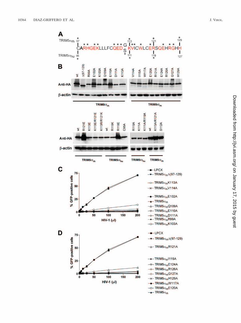
The role in antiretroviral activity of TRIM5� residues pre-dicted to be on the B-box 2 surface. To understand the contri-bution of the predicted surface-exposed residues of the B-box2 domain to the anti-HIV-1 activities of TRIM5 proteins, wechanged these residues in the TRIM5�rh B-box 2 domain in-dividually to alanine (Fig. 2A). Dog cells (Cf2Th) stably ex-pressing the different mutants (Fig. 2B) were challenged withdifferent amounts of recombinant HIV-1 expressing (HIV-1–GFP) (Fig. 2C and D). Deletion of the B-box 2 domain ofTRIM5�rh [(97-129)] completely eliminated its ability to re-strict HIV-1 (Fig. 2C and D). By contrast, all of the TRIM5�rh
proteins with single amino acid changes restricted HIV-1 in-fection. However, the K113A and R121A TRIM5�rh mutantsinhibited HIV-1 infection less efficiently than the wild-typeTRIM5�rh protein (Fig. 2C and D). Thus, replacement of
FIG. 2. TRIM5�rh and TRIM5�hu mutants and retroviral restriction. (A) The primary sequences of the B-box 2 domains from TRIM5�rh andTRIM5�hu are aligned. Residues depicted in red are predicted to be exposed on the surface of the B-box 2 domain. The asterisks indicate residuesthat were changed by site-directed mutagenesis. wt, wild type. (B) Cf2Th cells were transduced with the LPCX vector expressing HA-taggedwild-type and mutant TRIM5�rh proteins. Stable cell lines were selected with 5 �g/ml of puromycin. Cell lysates were analyzed by Western blottingusing antibodies against HA (Anti-HA) and �-actin. (C and D) Cf2Th cells expressing the indicated wild-type and mutant TRIM5�rh proteins wereincubated with HIV-1–GFP, and GFP-positive cells were measured by fluorescence-activated cell sorting. The results of two independentexperiments were similar, and the results of one experiment are shown.
VOL. 81, 2007 B-BOX 2 DOMAIN MUTANTS OF TRIM5� AND TRIMCyp 10365
on January 17, 2015 by guesthttp://jvi.asm
.org/D
ownloaded from

10366 DIAZ-GRIFFERO ET AL. J. VIROL.
on January 17, 2015 by guesthttp://jvi.asm
.org/D
ownloaded from

individual TRIM5�rh residues predicted to be exposed on thesurface of the B-box 2 domain with alanine exerted at most amodest effect on anti-HIV-1 activity.
Lysine 113 and arginine 121 are predicted to be located onopposite sides of the upper surface of the TRIM5�rh B-box 2domain (Fig. 1). To examine further the potential functionalroles of these basic residues, we altered lysine 113 and arginine121 simultaneously to alanine residues. The K113A/R121ATRIM5�rh protein was expressed at a level equivalent to thatof the wild-type TRIM5�rh protein (Fig. 3A). The susceptibil-ity of Cf2Th cells expressing the K113A/R121A mutant toinfection by HIV-1, EIAV, and FIV was examined (Fig. 3B, C,and D). For comparison, the abilities of these three retrovi-ruses to infect cells expressing wild-type TRIM5�rh orTRIM5�rh mutants with other alterations affecting the B-box 2domain were examined in parallel. Cells expressing wild-typeTRIM5�rh and the K103A mutant strongly resisted infectionby HIV-1, EIAV, and FIV. As expected, TRIM5�rh variantswith deletions [(1-132) and (97-129)] or disruption (C97A/H100A) of the B-box 2 domain did not inhibit infection by thethree retroviruses. The antiretroviral activity of the K113A/R121A mutant was significantly attenuated, similar to that ofthe TRIM5�rh (1-93) mutant, which lacks the RING and L1regions. Thus, lysine 113 and arginine 121 contribute to theantiretroviral activity of TRIM5�rh.
Residues 113 and 121 of TRIM5�rh were altered individu-ally or in combination to other basic residues or to acidicresidues. The substitution of negatively charged residues forarginine 121 completely eliminated the ability of TRIM5�rh torestrict HIV-1, EIAV, and FIV infections (Fig. 4A to D),despite efficient expression of these mutants (see below). Sub-stitution of acidic residues at position 113 exerted only modesteffects on the antiretroviral activity of TRIM5�rh. The simul-taneous replacement of lysine 113 with arginine and arginine121 with lysine only minimally affected the ability of TRIM5�rh
to inhibit HIV-1, EIAV, and FIV infection (see K113R/R121Kin Fig. 4A to C). Thus, the presence of a negatively chargedresidue at position 121 is particularly disruptive of the antiret-roviral activity of TRIM5�rh.
Roles of charged B-box 2 surface residues in the antiretro-viral activity of human TRIM5�. Arginine 121 is conserved inall primate TRIM5� proteins, whereas lysine 113 is conservedin Old World primate TRIM5� proteins (see below). To ex-amine the contributions of these basic residues to the antiret-roviral function of another TRIM5� protein, the equivalentresidues, lysine 111 and arginine 119, of TRIM5�hu were al-tered. The abilities of the wild-type and mutant TRIM5�hu
proteins to restrict infection of Cf2Th cells by N-MLV, EIAV,and FIV were examined. Alteration of arginine 119 ofTRIM5�hu to alanine resulted in complete loss of the ability torestrict N-MLV and EIAV (Fig. 5A and C) and significant
reduction in activity against FIV (Fig. 5D). Alanine substitu-tion for lysine 111 in TRIM5�hu exerted less of an effect onN-MLV and EIAV inhibition (Fig. 5A and C). The TRIM5�hu
K111A mutant exhibited partial restricting activity against FIV(Fig. 5D). As expected, none of the TRIM5�hu variants wereactive against B-MLV infection (Fig. 5B). Thus, the alterationof arginine 119 significantly affects the retrovirus-restrictingability of TRIM5�hu.
To examine the role of charge in modulating the function ofarginine 119 of TRIM5�hu, mutants were created with a basicsubstitution (R119K) or acidic substitutions (R119E andR119D) at this residue. The R119K mutant exhibited wild-typelevels of activity against N-MLV, EIAV, and FIV infections(Fig. 6A to D). By contrast, the R119E and R119D mutantswere devoid of detectable antiretroviral activity. The antiret-roviral activities of the R119E and R119D mutants were nobetter than that of the C95A mutant, which has a disruption ofthe B-box 2 domain. Apparently, as was seen for TRIM5�rh,introduction of an acidic residue at this position in TRIM5�hu
abolishes antiretroviral activity.Trimerization and capsid binding of TRIM5� B-box 2 mu-
tants. To investigate the mechanistic basis of the attenuation ofantiretroviral activity associated with changes in the B-box 2residues, the mutants were compared with wild-type TRIM5�proteins for oligomerization and capsid-binding abilities. BothTRIM5�rh and TRIM5�hu form trimers (16). Cross-linking oflysates from cells expressing wild-type or mutant TRIM5� pro-teins suggested that all of the human and rhesus monkeyTRIM5� mutants efficiently formed trimers (Fig. 7A and Band data not shown). These data are consistent with previousresults demonstrating that the TRIM5� B-box 2 domain is notnecessary for trimerization (12).
The abilities of the TRIM5�rh mutants to bind HIV-1CA-NC complexes assembled in vitro were examined. Asshown in Fig. 8, deletion or amino acid alteration of the B-box2 domain did not significantly compromise the ability ofTRIM5�rh to bind HIV-1 capsid complexes. As expected (12,31), deletion of the TRIM5�rh coiled coil [(132-233)] dis-rupted its capsid-binding ability. Also as expected (14c, 31),TRIM5�hu associated less efficiently with HIV-1 capsid com-plexes than TRIM5�rh.
These results suggest that the changes introduced into theTRIM5�rh B-box 2 domain allow trimerization and HIV-1capsid binding.
Effects of B-box 2 changes in TRIM5� on the fate of theretroviral capsid in infected cells. Previous studies (24, 31)suggested that restriction of HIV-1 and N-MLV infection isaccompanied by an accelerated conversion of the cytosolicretroviral capsid from particulate to soluble forms. We wishedto examine the effects of restriction-attenuating changes in theTRIM5� B-box 2 domain on the fate of the retroviral capsid.
FIG. 3. Alteration of two basic residues in the TRIM5�rh B-box 2 domain. Cells were transduced with either the empty LPCX vector or a vectorexpressing wild-type (wt) TRIM5�rh or the following TRIM5�rh mutants: K113A/R121A, C97A/H100A, K103A, the mutant with RING deleted[(1-93)], the mutant with the RING-B box deleted [(1-132)], or the mutant with the B-box deleted [(97-129)]. (A) Cell lysates were analyzedby Western blotting using antibodies against HA (Anti-HA) and �-actin. (B to D) Cf2Th cells expressing the different TRIM5�rh variants werechallenged with HIV-1–GFP (B), EIAV-GFP (C), or FIV-GFP (D). GFP-positive cells were counted. Similar results were obtained in threeindependent experiments.
VOL. 81, 2007 B-BOX 2 DOMAIN MUTANTS OF TRIM5� AND TRIMCyp 10367
on January 17, 2015 by guesthttp://jvi.asm
.org/D
ownloaded from

FIG. 4. Effects of altering the charge on residues 113 and 121 of the TRIM5�rh B-box 2 domain. Cf2Th cells expressing the indicated wild-typeand mutant TRIM5�rh proteins were challenged with HIV-1–GFP (A and D), EIAV-GFP (B), or FIV-GFP (C). GFP-positive cells were counted.Similar results were obtained in three independent experiments.
10368 DIAZ-GRIFFERO ET AL. J. VIROL.
on January 17, 2015 by guesthttp://jvi.asm
.org/D
ownloaded from

FIG. 5. Effects of altering residues 111 and 119 of the TRIM5�hu B-box 2 domain. Cf2Th cells expressing the indicated wild-type and mutantTRIM5�hu proteins were challenged with N-MLV–GFP (A), B-MLV–GFP (B), EIAV-GFP (C), or FIV-GFP (D). GFP-positive cells werecounted. Similar results were obtained in three independent experiments.
VOL. 81, 2007 B-BOX 2 DOMAIN MUTANTS OF TRIM5� AND TRIMCyp 10369
on January 17, 2015 by guesthttp://jvi.asm
.org/D
ownloaded from
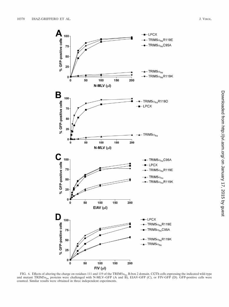
FIG. 6. Effects of altering the charge on residues 111 and 119 of the TRIM5�hu B-box 2 domain. Cf2Th cells expressing the indicated wild-typeand mutant TRIM5�hu proteins were challenged with N-MLV–GFP (A and B), EIAV-GFP (C), or FIV-GFP (D). GFP-positive cells werecounted. Similar results were obtained in three independent experiments.
10370 DIAZ-GRIFFERO ET AL. J. VIROL.
on January 17, 2015 by guesthttp://jvi.asm
.org/D
ownloaded from

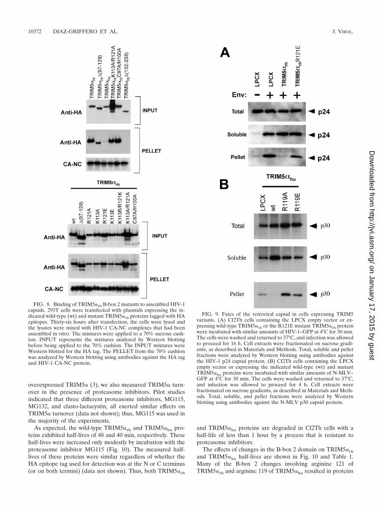
First, we examined the fate of the HIV-1 capsid in cells ex-pressing the wild-type or R121E TRIM5�rh protein or in con-trol cells transduced with the empty LPCX vector. No cytosolicHIV-1 capsid protein, in either soluble or particulate form, wasdetected in the control LPCX-transduced cells incubated withHIV-1 lacking envelope glycoproteins (Fig. 9A). By contrast,at 1 hour after infection of these cells with a VSV G glyco-protein-pseudotyped HIV-1, particulate and soluble capsid pro-teins were detected in the assay. This result demonstrates that theassay can distinguish retroviral capsid proteins that have enteredthe cytosol through the action of the VSV G glycoprotein fromvirions that are nonspecifically attached to or endocytosed intocells (24, 31). Compared with the results in the control LPCXcells, a significant decrease in the level of the particulate HIV-1capsid was observed in the cytosol of TRIM5�rh-expressing cells.By contrast, particulate capsid was detected at this time point inthe cytosol of cells expressing the TRIM5�rh R121E mutant.
The fate of the N-MLV capsid in the cytosol of cells express-ing wild-type TRIM5�hu or the R119A and R119E TRIM5�hu
mutants was compared with that seen in control cells trans-duced with the empty LPCX vector. Particulate capsid wasdetected in the LPCX-transduced cells and in the cells express-ing the R119A and R119E TRIM5�hu mutants (Fig. 9B). Bycontrast, no detectable particulate capsid was observed in cellsexpressing wild-type TRIM5�hu. Consistent with previous ob-
servations (24, 31), an increase in soluble forms of the N-MLVcapsid was observed in cells expressing the wild-typeTRIM5�hu protein compared with the levels of soluble capsidproteins in the other cells. Thus, although the B-box 2 mutantsare trimeric and retain the ability to bind retroviral capsids,they are deficient in promoting a decrease in the level ofparticulate cytosolic capsid and in restricting infection. Appar-ently, an “effector” function necessary for the last two pro-cesses has been compromised by the changes introduced intothe TRIM5� B-box 2 domain.
Effects of B-box 2 changes on TRIM5� degradation. Duringthe course of these studies, we noticed that the steady-statelevels of some of the B-box 2 mutants of TRIM5�rh andTRIM5�hu were increased. Wild-type TRIM5� turns over witha half-life of approximately 50 to 70 min, and B-box 2 changeshave been shown to affect TRIM5� turnover (3). As steady-state levels reflect rates of both synthesis and degradation, weexamined whether the B-box 2 mutants exhibited changes inhalf-life compared with the wild-type TRIM5� protein. Cf2Thcells stably expressing the mutant proteins were incubated withcycloheximide for several hours, during which time the steady-state levels of the TRIM5� variants were determined by West-ern blotting. Levels of a control protein, �-actin, were mea-sured in parallel and used as a normalization standard.Because the proteasome can contribute to the degradation of
FIG. 7. Oligomerization of TRIM5�rh and TRIM5�hu mutants. 293T cells were transiently transfected with plasmids expressing the indicatedwild-type (wt) or mutant TRIM5� proteins with HA epitope tags. Cells expressing wild-type and mutant TRIM5�rh (A) or TRIM5�hu (B) proteinswere lysed 48 h after transfection and cross-linked with the indicated concentrations of glycolbis-succinimidylsuccinate. The cell lysates weresubsequently Western blotted with an anti-HA antibody. t, trimer; d, dimer.
VOL. 81, 2007 B-BOX 2 DOMAIN MUTANTS OF TRIM5� AND TRIMCyp 10371
on January 17, 2015 by guesthttp://jvi.asm
.org/D
ownloaded from
overexpressed TRIM5� (3), we also measured TRIM5� turn-over in the presence of proteasome inhibitors. Pilot studiesindicated that three different proteasome inhibitors, MG115,MG132, and clasto-lactacystin, all exerted similar effects onTRIM5� turnover (data not shown); thus, MG115 was used inthe majority of the experiments.
As expected, the wild-type TRIM5�rh and TRIM5�hu pro-teins exhibited half-lives of 48 and 40 min, respectively. Thesehalf-lives were increased only modestly by incubation with theproteasome inhibitor MG115 (Fig. 10). The measured half-lives of these proteins were similar regardless of whether theHA epitope tag used for detection was at the N or C terminus(or on both termini) (data not shown). Thus, both TRIM5�rh
and TRIM5�hu proteins are degraded in Cf2Th cells with ahalf-life of less than 1 hour by a process that is resistant toproteasome inhibitors.
The effects of changes in the B-box 2 domain on TRIM5�rh
and TRIM5�hu half-lives are shown in Fig. 10 and Table 1.Many of the B-box 2 changes involving arginine 121 ofTRIM5�rh and arginine 119 of TRIM5�hu resulted in proteins
FIG. 8. Binding of TRIM5�rh B-box 2 mutants to assembled HIV-1capsids. 293T cells were transfected with plasmids expressing the in-dicated wild-type (wt) and mutant TRIM5�rh proteins tagged with HAepitopes. Thirty-six hours after transfection, the cells were lysed andthe lysates were mixed with HIV-1 CA-NC complexes that had beenassembled in vitro. The mixtures were applied to a 70% sucrose cush-ion. INPUT represents the mixtures analyzed by Western blottingbefore being applied to the 70% cushion. The INPUT mixtures wereWestern blotted for the HA tag. The PELLET from the 70% cushionwas analyzed by Western blotting using antibodies against the HA tagand HIV-1 CA-NC protein.
FIG. 9. Fates of the retroviral capsid in cells expressing TRIM5variants. (A) Cf2Th cells containing the LPCX empty vector or ex-pressing wild-type TRIM5�rh or the R121E mutant TRIM5�rh proteinwere incubated with similar amounts of HIV-1–GFP at 4°C for 30 min.The cells were washed and returned to 37°C, and infection was allowedto proceed for 16 h. Cell extracts were fractionated on sucrose gradi-ents, as described in Materials and Methods. Total, soluble and pelletfractions were analyzed by Western blotting using antibodies againstthe HIV-1 p24 capsid protein. (B) Cf2Th cells containing the LPCXempty vector or expressing the indicated wild-type (wt) and mutantTRIM5�hu proteins were incubated with similar amounts of N-MLV–GFP at 4°C for 30 min. The cells were washed and returned to 37°C,and infection was allowed to proceed for 4 h. Cell extracts werefractionated on sucrose gradients, as described in Materials and Meth-ods. Total, soluble, and pellet fractions were analyzed by Westernblotting using antibodies against the N-MLV p30 capsid protein.
10372 DIAZ-GRIFFERO ET AL. J. VIROL.
on January 17, 2015 by guesthttp://jvi.asm
.org/D
ownloaded from

with dramatic increases in half-life. Notably, replacement ofthese arginine residues with lysine did not increase the stabilityof TRIM5�rh or TRIM5�hu. We conclude that the presence ofa basic residue at position 121 of TRIM5�rh or position 119 ofTRIM5�hu is important for the rapid turnover of these pro-teins via a pathway resistant to proteasome inhibitors.
With respect to HIV-1 restriction, the TRIM5�rh RING, L1,
and B-box 2 domains can be functionally replaced by those ofhuman TRIM6 or TRIM34 (14). Because TRIM6 andTRIM34 exhibit long half-lives compared with TRIM5� (14),we measured the half-lives of the chimeric proteins [Table 1,TRIM6-5(CC-B30.2) and TRIM34-5(CC-B30.2)]. Both chi-meric proteins exhibited very long half-lives compared withthat of wild-type TRIM5�rh.
FIG. 10. Turnover of wild-type and mutant TRIM5� proteins. Cf2Th cells stably expressing the indicated TRIM5� proteins were treated withcycloheximide (CHX) or with cycloheximide and MG115 (CHX � MG115). At the indicated times after the initiation of treatment, the cells were lysed.The cell lysates were Western blotted using an antibody directed against the HA tags on the TRIM5 proteins. The intensities of the bands were estimatedby densitometry and plotted versus time. The results of a typical experiment are shown, with similar results obtained in three separate experiments.
VOL. 81, 2007 B-BOX 2 DOMAIN MUTANTS OF TRIM5� AND TRIMCyp 10373
on January 17, 2015 by guesthttp://jvi.asm
.org/D
ownloaded from
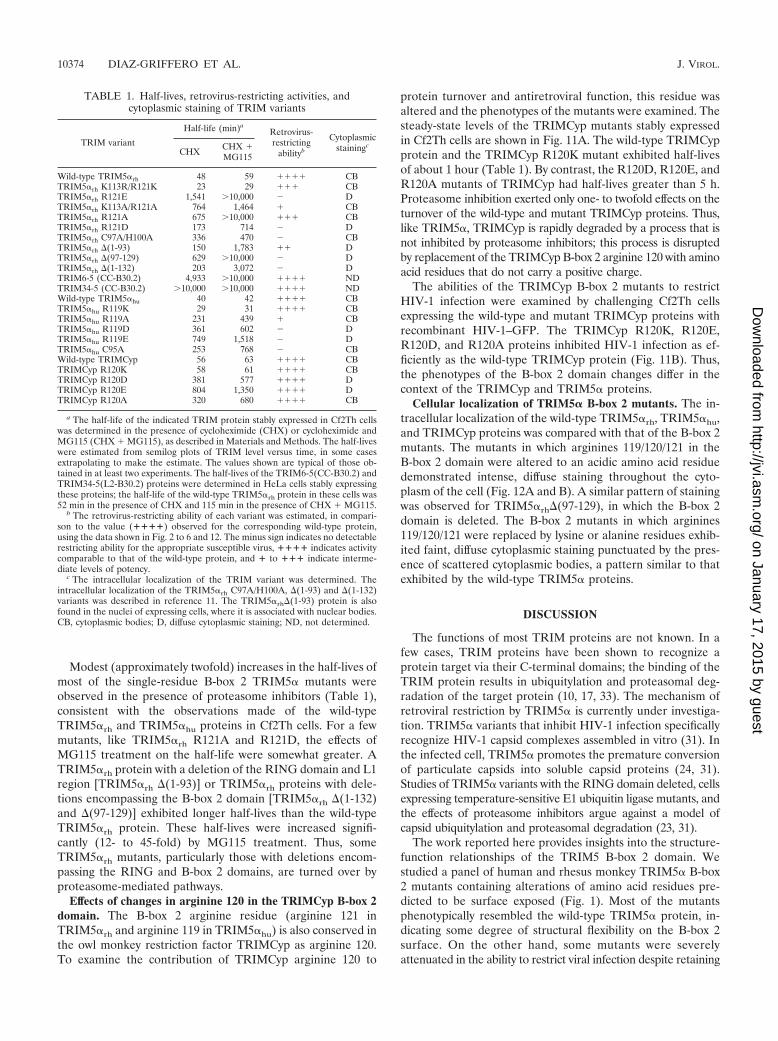
Modest (approximately twofold) increases in the half-lives ofmost of the single-residue B-box 2 TRIM5� mutants wereobserved in the presence of proteasome inhibitors (Table 1),consistent with the observations made of the wild-typeTRIM5�rh and TRIM5�hu proteins in Cf2Th cells. For a fewmutants, like TRIM5�rh R121A and R121D, the effects ofMG115 treatment on the half-life were somewhat greater. ATRIM5�rh protein with a deletion of the RING domain and L1region [TRIM5�rh (1-93)] or TRIM5�rh proteins with dele-tions encompassing the B-box 2 domain [TRIM5�rh (1-132)and (97-129)] exhibited longer half-lives than the wild-typeTRIM5�rh protein. These half-lives were increased signifi-cantly (12- to 45-fold) by MG115 treatment. Thus, someTRIM5�rh mutants, particularly those with deletions encom-passing the RING and B-box 2 domains, are turned over byproteasome-mediated pathways.
Effects of changes in arginine 120 in the TRIMCyp B-box 2domain. The B-box 2 arginine residue (arginine 121 inTRIM5�rh and arginine 119 in TRIM5�hu) is also conserved inthe owl monkey restriction factor TRIMCyp as arginine 120.To examine the contribution of TRIMCyp arginine 120 to
protein turnover and antiretroviral function, this residue wasaltered and the phenotypes of the mutants were examined. Thesteady-state levels of the TRIMCyp mutants stably expressedin Cf2Th cells are shown in Fig. 11A. The wild-type TRIMCypprotein and the TRIMCyp R120K mutant exhibited half-livesof about 1 hour (Table 1). By contrast, the R120D, R120E, andR120A mutants of TRIMCyp had half-lives greater than 5 h.Proteasome inhibition exerted only one- to twofold effects on theturnover of the wild-type and mutant TRIMCyp proteins. Thus,like TRIM5�, TRIMCyp is rapidly degraded by a process that isnot inhibited by proteasome inhibitors; this process is disruptedby replacement of the TRIMCyp B-box 2 arginine 120 with aminoacid residues that do not carry a positive charge.
The abilities of the TRIMCyp B-box 2 mutants to restrictHIV-1 infection were examined by challenging Cf2Th cellsexpressing the wild-type and mutant TRIMCyp proteins withrecombinant HIV-1–GFP. The TRIMCyp R120K, R120E,R120D, and R120A proteins inhibited HIV-1 infection as ef-ficiently as the wild-type TRIMCyp protein (Fig. 11B). Thus,the phenotypes of the B-box 2 domain changes differ in thecontext of the TRIMCyp and TRIM5� proteins.
Cellular localization of TRIM5� B-box 2 mutants. The in-tracellular localization of the wild-type TRIM5�rh, TRIM5�hu,and TRIMCyp proteins was compared with that of the B-box 2mutants. The mutants in which arginines 119/120/121 in theB-box 2 domain were altered to an acidic amino acid residuedemonstrated intense, diffuse staining throughout the cyto-plasm of the cell (Fig. 12A and B). A similar pattern of stainingwas observed for TRIM5�rh(97-129), in which the B-box 2domain is deleted. The B-box 2 mutants in which arginines119/120/121 were replaced by lysine or alanine residues exhib-ited faint, diffuse cytoplasmic staining punctuated by the pres-ence of scattered cytoplasmic bodies, a pattern similar to thatexhibited by the wild-type TRIM5� proteins.
DISCUSSION
The functions of most TRIM proteins are not known. In afew cases, TRIM proteins have been shown to recognize aprotein target via their C-terminal domains; the binding of theTRIM protein results in ubiquitylation and proteasomal deg-radation of the target protein (10, 17, 33). The mechanism ofretroviral restriction by TRIM5� is currently under investiga-tion. TRIM5� variants that inhibit HIV-1 infection specificallyrecognize HIV-1 capsid complexes assembled in vitro (31). Inthe infected cell, TRIM5� promotes the premature conversionof particulate capsids into soluble capsid proteins (24, 31).Studies of TRIM5� variants with the RING domain deleted, cellsexpressing temperature-sensitive E1 ubiquitin ligase mutants, andthe effects of proteasome inhibitors argue against a model ofcapsid ubiquitylation and proteasomal degradation (23, 31).
The work reported here provides insights into the structure-function relationships of the TRIM5 B-box 2 domain. Westudied a panel of human and rhesus monkey TRIM5� B-box2 mutants containing alterations of amino acid residues pre-dicted to be surface exposed (Fig. 1). Most of the mutantsphenotypically resembled the wild-type TRIM5� protein, in-dicating some degree of structural flexibility on the B-box 2surface. On the other hand, some mutants were severelyattenuated in the ability to restrict viral infection despite retaining
TABLE 1. Half-lives, retrovirus-restricting activities, andcytoplasmic staining of TRIM variants
TRIM variant
Half-life (min)aRetrovirus-restricting
abilityb
Cytoplasmicstainingc
CHX CHX �MG115
Wild-type TRIM5�rh 48 59 ���� CBTRIM5�rh K113R/R121K 23 29 ��� CBTRIM5�rh R121E 1,541 10,000 � DTRIM5�rh K113A/R121A 764 1,464 � CBTRIM5�rh R121A 675 10,000 ��� CBTRIM5�rh R121D 173 714 � DTRIM5�rh C97A/H100A 336 470 � CBTRIM5�rh (1-93) 150 1,783 �� DTRIM5�rh (97-129) 629 10,000 � DTRIM5�rh (1-132) 203 3,072 � DTRIM6-5 (CC-B30.2) 4,933 10,000 ���� NDTRIM34-5 (CC-B30.2) 10,000 10,000 ���� NDWild-type TRIM5�hu 40 42 ���� CBTRIM5�hu R119K 29 31 ���� CBTRIM5�hu R119A 231 439 � CBTRIM5�hu R119D 361 602 � DTRIM5�hu R119E 749 1,518 � DTRIM5�hu C95A 253 768 � CBWild-type TRIMCyp 56 63 ���� CBTRIMCyp R120K 58 61 ���� CBTRIMCyp R120D 381 577 ���� DTRIMCyp R120E 804 1,350 ���� DTRIMCyp R120A 320 680 ���� CB
a The half-life of the indicated TRIM protein stably expressed in Cf2Th cellswas determined in the presence of cycloheximide (CHX) or cycloheximide andMG115 (CHX � MG115), as described in Materials and Methods. The half-liveswere estimated from semilog plots of TRIM level versus time, in some casesextrapolating to make the estimate. The values shown are typical of those ob-tained in at least two experiments. The half-lives of the TRIM6-5(CC-B30.2) andTRIM34-5(L2-B30.2) proteins were determined in HeLa cells stably expressingthese proteins; the half-life of the wild-type TRIM5�rh protein in these cells was52 min in the presence of CHX and 115 min in the presence of CHX � MG115.
b The retrovirus-restricting ability of each variant was estimated, in compari-son to the value (����) observed for the corresponding wild-type protein,using the data shown in Fig. 2 to 6 and 12. The minus sign indicates no detectablerestricting ability for the appropriate susceptible virus, ���� indicates activitycomparable to that of the wild-type protein, and � to ��� indicate interme-diate levels of potency.
c The intracellular localization of the TRIM variant was determined. Theintracellular localization of the TRIM5�rh C97A/H100A, (1-93) and (1-132)variants was described in reference 11. The TRIM5�rh(1-93) protein is alsofound in the nuclei of expressing cells, where it is associated with nuclear bodies.CB, cytoplasmic bodies; D, diffuse cytoplasmic staining; ND, not determined.
10374 DIAZ-GRIFFERO ET AL. J. VIROL.
on January 17, 2015 by guesthttp://jvi.asm
.org/D
ownloaded from

the ability to trimerize and to recognize the relevant retroviralcapsid. Thus, these mutants appear to lack an effector functionnecessary for retroviral restriction. An overlapping subset ofB-box 2 mutants lost the ability to be rapidly degraded by apathway insensitive to proteasome inhibitors. Some changes inthe B-box 2 domain also influenced the subcellular localizationof TRIM5� and TRIMCyp proteins.
A discrete TRIM5 B-box 2 region predicted to reside nearthe trimer axis was found to be important for retroviral restric-tion, formation of cytoplasmic bodies, and protein turnover.The structural requirements for these three processes differsubtly but involve arginines 119/120/121 in all three instances.All primate TRIM5 proteins, as well as owl monkey TRIMCyp,have arginine residues at these positions (Fig. 13). The rapidturnover of TRIM5� and TRIMCyp apparently requires abasic residue at these positions. The structural requirementsfor cytoplasmic-body formation and retroviral restriction ap-pear to be less stringent. Substitution of acidic residues forarginines 119/120/121 or deletion of the B-box 2 domain resultsin a diffuse cytoplasmic staining pattern for TRIM5� andTRIMCyp, with lack of cytoplasmic-body formation. Theseacidic substitutions also attenuate retroviral restriction byrhesus monkey and human TRIM5� proteins, but not byTRIMCyp. Replacement of arginine 119 by alanine significantlyimpaired the retrovirus-restricting ability of TRIM5�hu, al-
though cytoplasmic-body formation and rapid turnover werenot affected. Retroviral restriction by TRIM5�rh, on the otherhand, can tolerate the substitution of an alanine residue forarginine 121. Thus, TRIM5�rh R121A forms cytoplasmic bod-ies and retains potent restricting activity against HIV-1 but,unlike the wild-type protein, exhibits a long half-life. Likewise,replacement of the RING and/or B-box 2 domains ofTRIM5�rh with those of the long-lived TRIM6, TRIM34, orTRIM21 proteins also resulted in stable proteins that exhibitedpotent antiretroviral activity (Table 1) (3, 14a). Thus, the func-tions of the TRIM5� B-box 2 domain related to rapid turnoverand retroviral restriction are separable. These results seemincompatible with recently proposed (1a) models of restrictionin which the TRIM5�rh protein and the capsid subunits asso-ciated with it are degraded through a proteasome-independenthost cell pathway.
The pathway by which TRIM5� and TRIMCyp are rapidlyturned over is not understood. Despite testing multiple pro-teasome inhibitors, we were unable to eliminate the rapidturnover of the wild-type TRIM5� and most of the TRIM5�single-amino-acid mutants. Apparently, a nonproteasome pro-tein degradation pathway interacts with the amino-terminalTRIM5� domains and results in rapid turnover. The role ofthis rapid turnover in the natural function of TRIM5� requiresfurther investigation.
FIG. 11. Effects of B-box 2 arginine changes on TRIMCyp. (A) The steady-state expression levels of the indicated TRIMCyp variants in Cf2Th cellswere determined by Western blotting with an anti-HA antibody. wt, wild type. (B) Cf2Th cells expressing the indicated TRIMCyp variants were incubatedwith HIV-1–GFP. GFP-positive cells were counted. The results of a typical experiment are shown. The experiment was repeated with similar results.
VOL. 81, 2007 B-BOX 2 DOMAIN MUTANTS OF TRIM5� AND TRIMCyp 10375
on January 17, 2015 by guesthttp://jvi.asm
.org/D
ownloaded from

Our results suggest that the TRIM5 B-box 2 domain con-tributes to cytoplasmic-body formation but cannot do so whenan acidic residue is present at positions 119/120/121. The for-mation of cytoplasmic bodies is not sufficient for potent retro-viral restriction, as evidenced by the weak retroviral restrictionmediated by the TRIM5�hu R119A and TRIM5�rh K113A/R121A mutants, both of which are found in cytoplasmic bod-ies. The necessity of cytoplasmic-body formation for retroviralrestriction by TRIM5� cannot be ruled out by our data, asacidic substitutions for residue 119 of TRIM5�hu or residue121 of TRIM5�rh eliminated both the appearance of cytoplas-mic bodies and restricting activity. Other studies of TRIM5�-expressing cells in which cytoplasmic bodies have been dis-rupted or minimized suggest that detectable bodies are notnecessary for efficient inhibition of retrovirus infection (23, 27).Acidic substitutions involving arginines 119/121 of theTRIM5� B-box 2 domain, or deletion of this domain, maycoincidentally affect cytoplasmic-body formation and anotherproperty (e.g., association with self or another factor) requiredfor retroviral restriction.
Disruption of the B-box 2 domain can result in loss of ret-
rovirus-restricting ability for at least two reasons. First, asshown here, some changes in the TRIM5�rh B-box 2 domaineliminate the ability of the protein to restrict HIV-1 infectionwithout significantly affecting expression, oligomerization, orcapsid binding. These mutagenic studies suggest that theTRIM5� B-box 2 domain plays a key role in retroviral restric-tion separate from the requirements for capsid recognition/binding. In this study and in previous studies (24, 31), weobserved the accelerated conversion of particulate retroviralcapsids to soluble forms of capsid protein in the cytosol ofinfected cells expressing a functional TRIM5� protein com-pared with that in control cells not expressing TRIM5�. No-tably, this accelerated uncoating of the viral capsid was notobserved in cells expressing the B-box 2 TRIM5� mutantsdefective in the “effector function” of retroviral restriction.Our studies indicate that the majority of the capsid proteinsdisassembled during TRIM5�-mediated restriction do not un-dergo ubiquitylation (or other large modification) and are notdegraded. Second, some combinations of changes in severalB-box 2 residues have been shown to decrease the binding ofTRIM5� proteins to the HIV-1 capsid, apparently by affecting
FIG. 12. Subcellular localization of TRIM5�rh and TRIM5�hu mutants. (A and B) Cf2Th cells stably expressing the indicated wild-type andmutant TRIM5� or TRIMCyp proteins were fixed and stained using a fluorescein isothiocyanate-conjugated anti-HA antibody as described inMaterials and Methods. Representative confocal microscope images are shown.
10376 DIAZ-GRIFFERO ET AL. J. VIROL.
on January 17, 2015 by guesthttp://jvi.asm
.org/D
ownloaded from

the relationship of the B-box 2 and B30.2(SPRY) domains(14b). Understanding these distinct consequences of B-box 2domain modification will require additional studies.
The B-box 2 arginine residue identified in this work as im-portant for TRIM5�rh and TRIM5�hu effector function ap-
pears to be less critical for TRIMCyp-mediated retroviral re-striction. Deletion or disruption of the B-box 2 domain ofTRIMCyp can lead to a reduction in antiretroviral activity (4).In general, however, it appears that TRIMCyp anti-HIV-1activity is less dependent on the integrity of the B-box 2 do-
FIG. 12—Continued.
FIG. 13. Sequence alignment of the B-box 2 domain sequences from different TRIM5 proteins. The B-box 2 domain sequences of the TRIM5relatives TRIM6, TRIM34, and TRIM22 are also shown for comparison. Residues predicted to coordinate the two zinc ions in the B-box 2 domainare depicted in red. Residues depicted in black correspond to the consensus sequence for this set of proteins, whereas residues in blue differ fromthe consensus sequence. The arginine (119/120/121) studied in this work is conserved in all TRIM5 proteins (black box). In contrast, lysine(111/113) is conserved only in the TRIM5 proteins of hominoids and Old World monkeys.
VOL. 81, 2007 B-BOX 2 DOMAIN MUTANTS OF TRIM5� AND TRIMCyp 10377
on January 17, 2015 by guesthttp://jvi.asm
.org/D
ownloaded from

main than is the virus-restricting activity of TRIM5� proteins.This is consistent with recent observations that moderate levelsof antiretroviral activity can be achieved simply by oligomer-izing the cyclophilin A domain, a function that does notrequire the TRIM5 B-box 2 domain (12a, 36).
The arginine 119/121 residue identified here as being impor-tant for TRIM5� antiretroviral function, cytoplasmic-body for-mation, and TRIM5� turnover is predicted to be exposed onthe modeled TRIM5� trimer. This raises the possibility thatthe region surrounding arginines 119/121 may be a docking sitefor association of TRIM5� with itself or with TRIM5� cofac-tors that support retroviral restriction or rapid turnover. Thepanel of mutants generated in this study should assist theinvestigation of these possibilities.
ACKNOWLEDGMENTS
We thank Yvette McLaughlin and Elizabeth Carpelan for manu-script preparation.
This study was supported by the National Institutes of Health(AI063987 and a Center for AIDS Research Award AI60354), theInternational AIDS Vaccine Initiative, the Bristol-Myers Squibb Foun-dation, and the late William F. McCarty-Cooper.
REFERENCES
1. Borden, K. L. 1998. RING fingers and B-boxes: zinc-binding protein-proteininteraction domains. Biochem. Cell Biol. 76:351–358.
1a.Chatterji, U., M. D. Bobardt, P. Gaskill, D. Sheeter, H. Fox, and P. A. Gallay.2006. Trim5� accelerates degradation of cytosolic capsid associated withproductive HIV-1 entry. J. Biol. Chem. 281:37025–37033.
2. Diaz-Griffero, F., S. A. Hoschander, and J. Brojatsch. 2002. Endocytosis is acritical step in entry of subgroup B avian leukosis viruses. J. Virol. 76:12866–12876.
3. Diaz-Griffero, F., X. Li, H. Javanbakht, B. Song, S. Welikala, M. Stremlau,and J. Sodroski. 2006. Rapid turnover and polyubiquitylation of the retro-viral restriction factor TRIM5. Virology 349:300–315.
4. Diaz-Griffero, F., N. Vandegraaff, Y. Li, K. McGee-Estrada, M. Stremlau, S.Welikala, Z. Si, A. Engelman, and J. Sodroski. 2006. Requirements forcapsid-binding and an effector function in TRIMCyp-mediated restriction ofHIV-1. Virology 351:404–419.
5. Fiser, S. 2003. Modeller: generation and refinement of homology-basedprotein structure models. Methods Enzymol. 374:461–491.
6. Ganser, B. K., S. Li, V. Y. Klishko, J. T. Finch, and W. I. Sundquist. 1999.Assembly and analysis of conical models for the HIV-1 core. Science 283:80–83.
7. Ganser-Pornillos, B. K., U. K. von Schwedler, K. M. Stray, C. Aiken, andW. I. Sundquist. 2004. Assembly properties of the human immunodeficiencyvirus type 1 CA protein. J. Virol. 78:2545–2552.
8. Hatziioannou, T., D. Perez-Caballero, A. Yang, S. Cowan, and P. D. Bien-iasz. 2004. Retrovirus resistance factors Ref1 and Lv1 are species-specificvariants of TRIM5�. Proc. Natl. Acad. Sci. USA 101:10774–10779.
9. Inoue, K., F. Hayashi, and S. Yokoyama. 2005. Solution structure of thezf-B-box type 2 domain of human tripartite motif protein TRIM29 isoformalpha, pdb 2CSV. RCSB Protein Data Bank. http://www.rcsb.org/pdb.
10. Ishikawa, H., H. Tachikawa, Y. Miura, and N. Takahashi. 2006. TRIM11binds to and destabilizes a key component of the activator-mediated cofactorcomplex (ARC105) through the ubiquitin-proteasome system. FEBS Lett.580:4784–4792.
11. Javanbakht, H., F. Diaz-Griffero, M. Stremlau, Z. Si, and J. Sodroski. 2005.The contribution of RING and B-box 2 domains to retroviral restrictionmediated by monkey TRIM5�. J. Biol. Chem. 280:26933–26940.
12. Javanbakht, H., W. Yuan, D. F. Yeung, B. Song, F. Diaz-Griffero, Y. Li, X. Li,M. Stremlau, and J. Sodroski. 2006. Characterization of TRIM5� trimer-ization and its contribution to human immunodeficiency virus capsid binding.Virology 353:234–246.
12a.Javanbakht, H., F. Diaz-Griffero, W. Yuan, D. F. Yeung, X. Li, B. Song, andJ. Sodroski. 13 June 2007. The ability of multimerized cyclophilin A torestrict retrovirus infection. Virology. doi:10.1016/j.virol.2007.04.034.
13. Keckesova, Z., L. M. Ylinen, and G. J. Towers. 2004. The human and Africangreen monkey TRIM5� genes encode Ref1 and Lv1 retroviral restrictionfactor activities. Proc. Natl. Acad. Sci. USA 101:10780–10785.
14. Li, X., B. Gold, C. O’Huigin, F. Diaz-Griffero, B. Song, Z. Si, Y. Li, W. Yuan,M. Stremlau, C. Mische, H. Javanbakht, M. Scally, C. Winkler, M. Dean,and J. Sodroski. 2007. Unique features of TRIM5� among closely relatedhuman TRIM family members. Virology 360:419–433.
14a.Li, X., Y. Li, M. Stremlau, W. Yuan, B. Song, M. Perron, and J. Sodroski.2006. Functional replacement of the RBCC domains of TRIM5� by heter-ologous TRIM domains. J. Virol. 80:6198–6206.
14b.Li, X., B. Song, S.-H. Xiang, and J. Sodroski. 2007. Functional interplaybetween the B-box 2 and the B30.2(SPRY) domains of TRIM5�. Virology.doi:10.1016/j.virol.2007.04.022.
14c.Li, Y., X. Li, M. Stremlau, M. Lee, and J. Sodroski. 2006. Removal ofarginine 332 allows human TRIM5� to bind human immunodeficiency viruscapsids and to restrict infection. J. Virol. 80:6738–6744.
15. Meroni, G., and G. Diez-Roux. 2005. TRIM/RBCC, a novel class of ‘singleprotein RING finger’ E3 ubiquitin ligases. Bioessays 27:1147–1157.
16. Mische, C. C., H. Javanbakht, B. Song, F. Diaz-Griffero, M. Stremlau, B.Strack, Z. Si, and J. Sodroski. 2005. Retroviral restriction factor TRIM5� isa trimer. J. Virol. 79:14446–14450.
17. Niikura, T., Y. Hashimoto, H. Tajima, M. Ishizaka, Y. Yamagishi, M.Kawasumi, M. Nawa, K. Terashita, S. Aiso, and I. Nishimoto. 2003. Atripartite motif protein TRIM11 binds and destabilizes Humanin, a neuro-protective peptide against Alzheimer’s disease-relevant insults. Eur. J. Neu-rosci. 17:1150–1158.
18. Nisole, S., C. Lynch, J. P. Stoye, and M. W. Yap. 2004. A Trim5-cyclophilinA fusion protein found in owl monkey kidney cells can restrict HIV-1. Proc.Natl. Acad. Sci. USA 101:13324–13328.
19. Nisole, S., J. P. Stoye, and A. Saib. 2005. TRIM family proteins: retroviralrestriction and antiviral defence. Nat Rev. Microbiol. 3:799–808.
20. Ohkura, S., M. W. Yap, T. Sheldon, and J. P. Stoye. 2006. All three variableregions of the TRIM5� B30.2 domain can contribute to the specificity ofretrovirus restriction. J. Virol. 80:8554–8565.
21. Olsen, J. C. 1998. Gene transfer vectors derived from equine infectiousanemia virus. Gene Ther. 5:1481–1487.
22. Perez-Caballero, D., T. Hatziioannou, A. Yang, S. Cowan, and P. D. Bieniasz.2005. Human tripartite motif 5� domains responsible for retrovirus restrictionactivity and specificity. J. Virol. 79:8969–8978.
23. Perez-Caballero, D., T. Hatziioannou, F. Zhang, S. Cowan, and P. D. Bieniasz.2005. Restriction of human immunodeficiency virus type 1 by TRIM-CypAoccurs with rapid kinetics and independently of cytoplasmic bodies, ubiquitin,and proteasome activity. J. Virol. 79:15567–15572.
24. Perron, M. J., M. Stremlau, M. Lee, H. Javanbakht, B. Song, and J.Sodroski. 2007. The human TRIM5� restriction factor mediates accelerateduncoating of the N-tropic murine leukemia virus capsid. J. Virol. 81:2138–2148.
25. Reymond, A., G. Meroni, A. Fantozzi, G. Merla, S. Cairo, L. Luzi, D.Riganelli, E. Zanaria, S. Messali, S. Cainarca, A. Guffanti, S. Minucci, P. G.Pelicci, and A. Ballabio. 2001. The tripartite motif family identifies cellcompartments. EMBO J. 20:2140–2151.
26. Saenz, D. T., W. Teo, J. C. Olsen, and E. M. Poeschla. 2005. Restriction offeline immunodeficiency virus by Ref1, Lv1, and primate TRIM5� proteins.J. Virol. 79:15175–15188.
27. Song, B., F. Diaz-Griffero, D. H. Park, T. Rogers, M. Stremlau, and J.Sodroski. 2005. TRIM5� association with cytoplasmic bodies is not requiredfor antiretroviral activity. Virology 343:201–211.
28. Song, B., B. Gold, C. O’Huigin, H. Javanbakht, X. Li, M. Stremlau, C.Winkler, M. Dean, and J. Sodroski. 2005. The B30.2(SPRY) domain of theretroviral restriction factor TRIM5� exhibits lineage-specific length and se-quence variation in primates. J. Virol. 79:6111–6121.
29. Song, B., H. Javanbakht, M. Perron, D. H. Park, M. Stremlau, and J.Sodroski. 2005. Retrovirus restriction by TRIM5� variants from Old Worldand New World primates. J. Virol. 79:3930–3937.
30. Stremlau, M., C. M. Owens, M. J. Perron, M. Kiessling, P. Autissier, and J.Sodroski. 2004. The cytoplasmic body component TRIM5� restricts HIV-1infection in Old World monkeys. Nature 427:848–853.
31. Stremlau, M., M. Perron, M. Lee, Y. Li, B. Song, H. Javanbakht, F. Diaz-Griffero, D. J. Anderson, W. I. Sundquist, and J. Sodroski. 2006. Specificrecognition and accelerated uncoating of retroviral capsids by the TRIM5�restriction factor. Proc. Natl. Acad. Sci. USA 103:5514–5519.
32. Stremlau, M., M. Perron, S. Welikala, and J. Sodroski. 2005. Species-specificvariation in the B30.2(SPRY) domain of TRIM5� determines the potency ofhuman immunodeficiency virus restriction. J. Virol. 79:3139–3145.
33. Toniato, E., X. P. Chen, J. Losman, V. Flati, L. Donahue, and P. Rothman.2002. TRIM8/GERP RING finger protein interacts with SOCS-1. J. Biol.Chem. 277:37315–37322.
34. Towers, G. J. 2005. Control of viral infectivity by tripartite motif proteins.Hum. Gene Ther. 16:1125–1132.
35. Yap, M. W., S. Nisole, and J. P. Stoye. 2005. A single amino acid change inthe SPRY domain of human Trim5� leads to HIV-1 restriction. Curr. Biol.15:73–78.
36. Yap, M. W., G. B. Mortuza, I. A. Taylor, and J. P. Stoye. 2007. The designof artificial retroviral restriction factors. Virology. doi:10.1016/j.virol.2007.04.005.
37. Yap, M. W., S. Nisole, C. Lynch, and J. P. Stoye. 2004. Trim5� proteinrestricts both HIV-1 and murine leukemia virus. Proc. Natl. Acad. Sci. USA101:10786–10791.
10378 DIAZ-GRIFFERO ET AL. J. VIROL.
on January 17, 2015 by guesthttp://jvi.asm
.org/D
ownloaded from