Modulation of Long-Term Synaptic Depression in Visual Cortex by Acetylcholine and Norepinephrine
11
Modulation of Long-Term Synaptic Depression in Visual Cortex by Acetylcholine and Norepinephrine Alfredo Kirkwood, 1 Carlos Rozas, 1 John Kirkwood, 2 Fernanda Perez, 2 and Mark F. Bear 2 1 Mind Brain Institute, Johns Hopkins University, Baltimore, Maryland 21218, and 2 Department of Neuroscience, Howard Hughes Medical Institute, Brown University, Providence, Rhode Island 02912 In a slice preparation of rat visual cortex, we discovered that paired-pulse stimulation (PPS) elicits a form of homosynaptic long-term depression (LTD) in the superficial layers when car- bachol (CCh) or norepinephrine (NE) is applied concurrently. PPS by itself, or CCh and NE in the absence of synaptic stimulation, produced no lasting change. The LTD induced by PPS in the presence of NE or CCh is of comparable magnitude with that obtained with prolonged low-frequency stimulation (LFS) but requires far fewer stimulation pulses (40 vs 900). The cholinergic facilitation of LTD was blocked by atropine and pirenzepine, suggesting involvement of M 1 receptors. The nor- adrenergic facilitation of LTD was blocked by urapidil and was mimicked by methoxamine, suggesting involvement of a1 re- ceptors. b receptor agonists and antagonists were without effect. Induction of LTD by PPS was inhibited by NMDA recep- tor blockers (completely in the case of NE; partially in the case of CCh), suggesting that one action of the modulators is to control the gain of NMDA receptor-dependent homosynaptic LTD in visual cortex. We propose that this is a mechanism by which cholinergic and noradrenergic inputs to the neocortex modulate naturally occurring receptive field plasticity. Key words: visual cortex; development; synaptic plasticity; long-term potentiation; long-term depression; acetylcholine; norepinephrine It is well established that synapses in sensory neocortex can be modified by experience. For example, brief deprivation of normal vision during early postnatal development can lead to a depres- sion of synaptic transmission that renders visual cortical neurons unresponsive to retinal stimulation. This type of synaptic plastic- ity obviously depends on information of retinal origin. However, there is evidence that experience-dependent plasticity also re- quires that animals be awake, alert, and paying attention to sensory stimuli (for review, see Singer, 1995). Thus, issues of great interest are the mechanisms of experience-dependent syn- aptic plasticity and their modulation by behavioral state. Extrathalamic inputs convey information to the cortex about behavioral state. Attention has focused mainly on the inputs arising from the locus coeruleus and the basal telencephalon that use norepinephrine (NE) and acetylcholine (ACh), respectively, as neurotransmitters. In one early study, it was shown that partial destruction of the noradrenergic or the cholinergic inputs alone did not disrupt deprivation-induced synaptic depression in visual cortex. However, their combined destruction produced a large deficit in this form of experience-dependent plasticity (Bear and Singer, 1986). The results of this experiment suggested that both of these inputs to visual cortex facilitate synaptic plasticity. Fur- thermore, because the simultaneous loss of both noradrenergic and cholinergic inputs was required to produce the defect in plasticity, the suggestion was made that these two modulators may substitute for one another and act via a common molecular mechanism. The notion that ACh and NE modulate naturally occurring cortical plasticity has received ample support (Kasamatsu and Pettigrew, 1979; Gordon et al., 1990; Juliano et al., 1991; Gu and Singer, 1993; Osterheld-Haas et al., 1994; Bakin and Weinberger, 1996; Baskerville et al., 1997; Kilgard and Merzenich, 1998; Sachdev et al., 1998; Zhu and Waite, 1998). The important question that remains, of course, is precisely how these neuro- transmitters affect the cortical synapses that carry detailed infor- mation about sensory experience. Over the past several years, slice preparations of visual neocor- tex have been used in an effort to clarify the elementary mecha- nisms of activity-dependent synaptic plasticity. One hypothesis that derived from this work is that modulation of inhibition in the cortex might be a way in which ACh and NE control plasticity (Kirkwood and Bear, 1994a). Because the state of functional inhibition in the cortex can be assayed by recording layer III responses to paired-pulse stimulation (PPS) of the white matter (Luhmann and Prince, 1991; Metherate and Ashe, 1994), we set out to examine the effects of ACh and NE on responses to PPS. In the course of this investigation we made the unexpected discovery that PPS in the presence of ACh or NE triggers a form of long-term synaptic depression (LTD). Here we report that ACh, acting via M 1 receptors, and NE, acting via a1 receptors, dramatically facilitate NMDA receptor-dependent homosynaptic LTD in visual cortex. We suggest that this reflects a mechanism whereby these modulators facilitate experience-dependent synap- tic plasticity in sensory neocortex. MATERIALS AND METHODS The experiments described in this paper were performed on transverse slices prepared from the visual cortex of 3- to 5-week-old Long–Evans rats. Each animal was deeply anesthetized by exposure to methoxyflu- rane vapors and was decapitated soon after the disappearance of any corneal reflexes. The brain was rapidly removed and immersed in ice- cold dissection buffer containing (in mM): sucrose, 212.7; KCl, 5; NaH 2 PO 4 , 1.25; MgSO 4 , 3; CaCl 2 , 1; NaHCO 3 , 26; dextrose, 10; and kynurenate, 10. A block of visual cortex was removed and sectioned in Received July 30, 1998; revised Dec. 10, 1998; accepted Dec. 14, 1998. This work was partly supported by grants from the National Eye Institute, the National Science Foundation, and the Charles A. Dana Foundation. We thank Dr. Kim Huber for helpful comments. Correspondence should be addressed to Dr. Mark Bear, Howard Hughes Medical Institute and Department of Neuroscience, Brown University, Providence, RI 02912. Copyright © 1999 Society for Neuroscience 0270-6474/99/191599-11$05.00/0 The Journal of Neuroscience, March 1, 1999, 19(5):1599–1609
Transcript of Modulation of Long-Term Synaptic Depression in Visual Cortex by Acetylcholine and Norepinephrine

Modulation of Long-Term Synaptic Depression in Visual Cortex byAcetylcholine and Norepinephrine
Alfredo Kirkwood,1 Carlos Rozas,1 John Kirkwood,2 Fernanda Perez,2 and Mark F. Bear2
1Mind Brain Institute, Johns Hopkins University, Baltimore, Maryland 21218, and 2Department of Neuroscience, HowardHughes Medical Institute, Brown University, Providence, Rhode Island 02912
In a slice preparation of rat visual cortex, we discovered thatpaired-pulse stimulation (PPS) elicits a form of homosynapticlong-term depression (LTD) in the superficial layers when car-bachol (CCh) or norepinephrine (NE) is applied concurrently.PPS by itself, or CCh and NE in the absence of synapticstimulation, produced no lasting change. The LTD induced byPPS in the presence of NE or CCh is of comparable magnitudewith that obtained with prolonged low-frequency stimulation(LFS) but requires far fewer stimulation pulses (40 vs 900). Thecholinergic facilitation of LTD was blocked by atropine andpirenzepine, suggesting involvement of M1 receptors. The nor-adrenergic facilitation of LTD was blocked by urapidil and was
mimicked by methoxamine, suggesting involvement of a1 re-ceptors. b receptor agonists and antagonists were withouteffect. Induction of LTD by PPS was inhibited by NMDA recep-tor blockers (completely in the case of NE; partially in the caseof CCh), suggesting that one action of the modulators is tocontrol the gain of NMDA receptor-dependent homosynapticLTD in visual cortex. We propose that this is a mechanism bywhich cholinergic and noradrenergic inputs to the neocortexmodulate naturally occurring receptive field plasticity.
Key words: visual cortex; development; synaptic plasticity;long-term potentiation; long-term depression; acetylcholine;norepinephrine
It is well established that synapses in sensory neocortex can bemodified by experience. For example, brief deprivation of normalvision during early postnatal development can lead to a depres-sion of synaptic transmission that renders visual cortical neuronsunresponsive to retinal stimulation. This type of synaptic plastic-ity obviously depends on information of retinal origin. However,there is evidence that experience-dependent plasticity also re-quires that animals be awake, alert, and paying attention tosensory stimuli (for review, see Singer, 1995). Thus, issues ofgreat interest are the mechanisms of experience-dependent syn-aptic plasticity and their modulation by behavioral state.
Extrathalamic inputs convey information to the cortex aboutbehavioral state. Attention has focused mainly on the inputsarising from the locus coeruleus and the basal telencephalon thatuse norepinephrine (NE) and acetylcholine (ACh), respectively,as neurotransmitters. In one early study, it was shown that partialdestruction of the noradrenergic or the cholinergic inputs alonedid not disrupt deprivation-induced synaptic depression in visualcortex. However, their combined destruction produced a largedeficit in this form of experience-dependent plasticity (Bear andSinger, 1986). The results of this experiment suggested that bothof these inputs to visual cortex facilitate synaptic plasticity. Fur-thermore, because the simultaneous loss of both noradrenergicand cholinergic inputs was required to produce the defect inplasticity, the suggestion was made that these two modulators maysubstitute for one another and act via a common molecularmechanism.
The notion that ACh and NE modulate naturally occurring
cortical plasticity has received ample support (Kasamatsu andPettigrew, 1979; Gordon et al., 1990; Juliano et al., 1991; Gu andSinger, 1993; Osterheld-Haas et al., 1994; Bakin and Weinberger,1996; Baskerville et al., 1997; Kilgard and Merzenich, 1998;Sachdev et al., 1998; Zhu and Waite, 1998). The importantquestion that remains, of course, is precisely how these neuro-transmitters affect the cortical synapses that carry detailed infor-mation about sensory experience.
Over the past several years, slice preparations of visual neocor-tex have been used in an effort to clarify the elementary mecha-nisms of activity-dependent synaptic plasticity. One hypothesisthat derived from this work is that modulation of inhibition in thecortex might be a way in which ACh and NE control plasticity(Kirkwood and Bear, 1994a). Because the state of functionalinhibition in the cortex can be assayed by recording layer IIIresponses to paired-pulse stimulation (PPS) of the white matter(Luhmann and Prince, 1991; Metherate and Ashe, 1994), we setout to examine the effects of ACh and NE on responses to PPS.In the course of this investigation we made the unexpecteddiscovery that PPS in the presence of ACh or NE triggers a formof long-term synaptic depression (LTD). Here we report thatACh, acting via M1 receptors, and NE, acting via a1 receptors,dramatically facilitate NMDA receptor-dependent homosynapticLTD in visual cortex. We suggest that this reflects a mechanismwhereby these modulators facilitate experience-dependent synap-tic plasticity in sensory neocortex.
MATERIALS AND METHODSThe experiments described in this paper were performed on transverseslices prepared from the visual cortex of 3- to 5-week-old Long–Evansrats. Each animal was deeply anesthetized by exposure to methoxyflu-rane vapors and was decapitated soon after the disappearance of anycorneal reflexes. The brain was rapidly removed and immersed in ice-cold dissection buffer containing (in mM): sucrose, 212.7; KCl, 5;NaH2PO4 , 1.25; MgSO4 , 3; CaCl2 , 1; NaHCO3 , 26; dextrose, 10; andkynurenate, 10. A block of visual cortex was removed and sectioned in
Received July 30, 1998; revised Dec. 10, 1998; accepted Dec. 14, 1998.This work was partly supported by grants from the National Eye Institute, the
National Science Foundation, and the Charles A. Dana Foundation. We thank Dr.Kim Huber for helpful comments.
Correspondence should be addressed to Dr. Mark Bear, Howard Hughes MedicalInstitute and Department of Neuroscience, Brown University, Providence, RI 02912.Copyright © 1999 Society for Neuroscience 0270-6474/99/191599-11$05.00/0
The Journal of Neuroscience, March 1, 1999, 19(5):1599–1609

the coronal plane into 0.4-mm-thick slices using a microslicer (DTK1000; Ted Pella, Redding, CA). The slices were gently transferred to aninterface storage chamber containing artificial CSF (ACSF) and main-tained at room temperature for at least an hour before recording. TheACSF was saturated with 95% O2 /5% CO2 and contained (in mM):NaCl, 124; KCl, 5; NaH2PO4 , 1.25; MgCl2 , 1; CaCl2 , 2; NaHCO3 , 26;and dextrose, 10. The experiments were performed on submerged slicescontinuously perfused at a rate of 2 ml/min with 30°C ACSF saturatedwith 95% O2 /5% CO2. Microelectrodes were filled with ACSF (1–2 MV)for extracellular recording or 3 M potassium acetate (80–120 MV) forintracellular recording. Only cells with resting membrane potentialsmore negative than 270 mV and input resistances .20 MV were studied.A site in the middle of the cortical thickness, confirmed histologically tocorrespond to layer IV and upper layer V, was stimulated to evoke fieldpotentials (FPs) in layer III, as described previously (Kirkwood andBear, 1994a,b). The amplitude of the maximum negative FP in layer IIIwas used as a measure of the evoked population excitatory synapticcurrent. Changes in the amplitude of the maximum negative FP reflectchanges in the magnitude of a synaptic current sink (Mitzdorf, 1985;Aizenman et al., 1996) and correlate with changes in the initial slope ofEPSPs recorded intracellularly in layer III neurons (Kirkwood and Bear,1994a,b). Baseline responses were obtained every 15 sec with a stimula-tion intensity that yielded a half-maximal response. PPS was usedthroughout the experiment unless stated otherwise. At least 10 min ofstable baseline recordings were made before drugs were applied. WhenNE was applied, 40 mM sodium ascorbate was included in the ACSF toprevent oxidation of the drug. Sodium ascorbate was also included withthe application of noradrenergic agonists and antagonists. Carbachol andatropine were purchased from Sigma (St. Louis, MO); all other drugswere purchased from Research Biochemicals (Natick, MA).
Only data from slices with stable recordings (,3% change over thebaseline period) were included in the analysis. The data were analyzed asfollows: (1) the maximum negative FP amplitude data for each experi-ment were expressed as percentages of the preconditioning baselineaverage; (2) the time scale in each experiment was converted to timefrom the onset of conditioning, and the four responses recorded in eachminute were averaged; and (3) then, the time-matched, normalized datawere averaged across experiments and expressed as the means (6 SEM).Within each group, the statistical significance of a change produced byconditioning stimulation was assessed with a paired t test, comparingvalues immediately before the application of neuromodulators with those30 min after the end of the application. Statistical significance acrossgroups was assessed with an unpaired t test.
RESULTSVisual cortical slices were prepared from 3- to 5-week-old rats.Initially, layer III synaptic responses were evoked with stimula-tion applied to the underlying white matter. Our original goal wasto study the effects of cholinergic and noradrenergic stimulationon paired-pulse suppression, i.e., the attenuation of the synapticresponses when two pulses are given in rapid succession. In theseexperiments, the stimulation consisted of two pulses (40 msecapart) delivered repetitively every 15 sec throughout the experi-ment. To activate cholinergic receptors, we bath applied 50 mM
carbachol (CCh), and to activate noradrenergic receptors, webath applied 40 mM NE in ascorbate (40 mM). When we realizedthat the PPS in the presence of these modulators caused LTD, weshifted the stimulating electrode to layer IV to activate a lesscomplex circuit. All the data presented below were obtained withthis stimulation-recording configuration.
Paired-pulse stimulation in carbachol induces a long-lasting depression of the synaptic responsesFigure 1 illustrates the effects of a 10 min application of 50 mM
CCh on the FP responses to paired pulses. Under control condi-tions, the response amplitude to the second pulse is usuallysomewhat smaller than the response amplitude to the first pulse.In the experiments shown in Figure 1, the ratio of the secondresponse to the first response is 0.59 6 0.08 (n 5 11). Exposureto CCh strongly reduced the response to the first pulse (64 6 4%
of control measured at 10 min of CCh), but it had a smaller effecton the response to the second pulse. Thus, paired-pulse suppres-sion was virtually eliminated in the presence of CCh; the ratio ofthe second to the first response was 0.99 6 0.14 at the end of the10 min of CCh perfusion.
These acute effects of CCh—that is, the transient and revers-ible reduction of the synaptic responses and the reduction inpaired-pulse suppression—have been reported previously (Vak-nin and Teyler, 1991; Murakoshi, 1995). These effects are likely toreflect a reduced probability of glutamate release (Markram andTsodyks, 1996; Gil et al., 1997), probably because of the action ofCCh on cholinergic receptors located on glutamatergic presynap-tic terminals (Valentino and Dingledine, 1981; Segal, 1982; Dodtet al., 1991; Vaknin and Teyler, 1991). An unexpected result,however, was that after removal of CCh the responses to the firststimulation pulse never returned to the original level. After aninitial partial recovery, the response magnitude reached a stablelevel 20 min after washing out the CCh and remained depressed(80 6 6% of control) even after an additional 30 min of washout.The LTD was a specific consequence of the CCh, because pro-longed PPS by itself had no effect on the FPs (Fig. 1C).
To confirm that the changes in the FP reflect changes inexcitatory synaptic transmission, we repeated the experimentusing intracellular recording. Simultaneous recordings were madeof the evoked FP in layer III and the EPSP in a nearby layer IIIneuron. PPS during CCh resulted in a reduction of FP amplitude(70 6 6%) that was paralleled by a comparable reduction in theinitial slope of the EPSPs (73 6 8%; n 5 7), indicating that areduction of the synaptic responses of layer II /III cells is reflectedin the reduction of the field responses. The application of CChalso produced a modest depolarization that was rapidly reversedafter removal of the drug. Thus the LTD of FP responses is notcaused by a tonic depolarization of postsynaptic neurons. Todetermine whether the slight depolarization of the neuron byCCh during PPS was the cause of LTD, we injected currentintracellularly that yielded similar levels of depolarization (10mV for 12 min; n 5 3). However, in no case was LTD observed,suggesting that the depolarization alone (at least in the cell soma)is not sufficient to cause expression of the LTD.
The LTD induced by PPS in 50 mM CCh was very reliable, sothis procedure was used to characterize the phenomenon. How-ever, we did examine the CCh dose–response relationship. By theuse of a 20 min application during PPS, LTD of increasingmagnitude (measured 30 min after washout) was observed with0.1 mM (94 6 2% of baseline; n 5 7), 1 mM (81 6 3%; n 5 7), 10mM (84 6 3%; n 5 7), 50 mM (72 6 3%; n 5 8), and 100 mM CCh(68 6 4%; n 5 7).
Synaptic depression induced in the presence ofcarbachol requires synaptic stimulation and isinput specificAn important issue to be resolved concerned the role of synapticactivation in the depression induced with CCh. Thus we askedwhether CCh applied alone, without concurrent synaptic activa-tion, would be sufficient to produce LTD. Figure 2 shows that inthe absence of stimulation, CCh application has minor effects onthe synaptic responses (97 6 4% of baseline; paired t test, p . 0.2;n 5 5). However, subsequent administration of CCh in the sameslices during PPS did result in substantial depression (78 6 4%;p , 0.02). These results indicate that CCh permits or facilitatesan activity-dependent form of synaptic depression. These find-ings also eliminate the possibility that the lasting depression is
1600 J. Neurosci., March 1, 1999, 19(5):1599–1609 Kirkwood et al. • Modulation of Visual Cortical Plasticity by ACh and NE

caused by tonic activation of ACh receptors (i.e., incompletewashout of CCh).
Next we wished to determine whether this form of depressionis input specific, that is, whether the reduction in synaptic efficacywas confined to the stimulated inputs only. To address this ques-tion, separate inputs to layer III were isolated by making a radialcut in the slice that extended from the white matter to layer IV,and baseline stimulation was applied in layer IV on each side ofthe cut. During CCh, PPS was applied to only one input, the otherserving as an unstimulated control. The results, summarized inFigure 2B, showed that only inputs receiving stimulation duringCCh show the LTD (stimulated path, 82 6 6% of baseline;control path, 98 6 1%; n 5 5). Thus, cholinergic stimulationpromotes LTD only in active inputs.
The results described above indicate that CCh in conjunctionwith paired-pulse stimulation induces an activity-dependent andhomosynaptic form of LTD. We were interested to know whetherthe CCh effect is specific to inputs receiving PPS or whether CChexerts a more general facilitation of homosynaptic LTD mecha-nisms. In hippocampal and visual cortical slices, a homosynapticform of LTD can also be reliably induced with low-frequencystimulation (LFS; typically 1 Hz), but only if this stimulation is
prolonged [usually 10–15 min (Dudek and Bear, 1992; Kirkwoodet al., 1993)]. To see whether CCh can facilitate LFS-inducedLTD, we combined application of the drug with brief epochs ofLFS (1 Hz for 5 min; Fig. 3). When the brief LFS was appliedalone, little or no LTD was induced (94 6 3%; n 5 11). However,when brief LFS was applied in conjunction with CCh, substantialLTD was induced (81 6 3; n 5 17; p , 0.005; Fig. 3C). Theseresults indicate that CCh application facilitates the induction ofhomosynaptic LTD with LFS, as well as with PPS.
Involvement of NMDA receptors and M1 receptors inthe induction of synaptic depression in the presenceof carbacholThe induction of one prominent form of homosynaptic LTD invisual cortex requires the activation of NMDA receptors (Kirk-wood and Bear, 1994b). To test whether this is also the case forthe synaptic depression promoted by CCh, we attempted to blockits induction with the NMDA receptor antagonist 2-amino-5-phosphonovaleric acid (AP5). Figure 4, A–C, illustrates the effectsof bath-applied 100 mM AP5 on the induction of depression inCCh. In three cases, one of them shown in Figure 4A, AP5completely blocked LTD induced with the aid of CCh, and this
Figure 1. The cholinergic agonist CCh induces a lasting depression of the layer III synaptic responses to layer IV stimulation. A, B, Effects of a brief(10 min) bath application of CCh (50 mM; open horizontal bar in B) on the field responses evoked with paired-pulse stimulation [interstimulus interval(ISI) 5 40 msec] are shown. A, FPs are from an experiment in which the CCh application resulted in a clear depression of the response to the first pulsebut had virtually no effect on the response to the second pulse. The traces were recorded immediately before (control) and during the application of CChand 30 min after washout of the drug. B, Time course of the average of 11 experiments is shown. Solid circles are responses to the first pulse; open circlesare responses to the second pulse. C, Prolonged paired-pulse stimulation alone has no effect on the FPs (n 5 9). D, E, The effects of CCh on FPs correlatewith changes in simultaneously recorded intracellular EPSPs. D, Traces are averages of four consecutive intracellular (top) and extracellular (bottom)responses recorded before (control) and 30 min after washout of CCh. E, Average time course of seven similar experiments is shown. Top, Changes inFP amplitude (open circles) and initial slope of the EPSP ( filled triangles) are presented. Bottom, Changes in the membrane potential are shown.
Kirkwood et al. • Modulation of Visual Cortical Plasticity by ACh and NE J. Neurosci., March 1, 1999, 19(5):1599–1609 1601

blockade was relieved after removal of AP5. On average, LTDinduced by PPS in the presence of CCh was significantly reducedbut not eliminated by AP5 (AP5, 91 6 9%; n 5 12; control, 77 612; n 5 10; p , 0.01; Fig. 4C). The data indicate that cholinergicactivation promotes the induction of an NMDA receptor-dependent form of LTD. However, the fact that the inhibition ofLTD with AP5 is incomplete suggests that an NMDA receptor-independent form of LTD may also be promoted by CCh. In thisregard, it is worth mentioning that in the CA1 region it has been
demonstrated that two forms of LTD can be induced at the samesynapses (Oliet et al., 1997).
In adults, cholinergic terminals and muscarinic receptors aredistributed throughout the depth of the visual cortex. Nicotinicreceptors, in contrast, are confined to the middle layers. It seemedreasonable to assume, therefore, that the effects of CCh on layerIII synaptic responses depend on the activation of muscarinicreceptors. To test this assumption, we studied the effects of themuscarinic antagonist atropine on the synaptic depression in-
Figure 2. Synaptic depression induced with CCh re-quires synaptic stimulation. A, Left, In the absence ofstimulation, CCh failed to produce any significant changein the FP responses. Right, Subsequently, depression wasreliably produced when CCh was applied with PPS. Inboth panels the responses were normalized with respectto the average response during the initial baseline periodbefore CCh application. The time that elapsed betweenthe last data point on the lef t and the first data point onthe right varied but was not .30 min. B, The depressioninduced by CCh is input specific. Right, The stimulation-recording configuration used to assess input specificity isshown. Layer IV was stimulated on either side of a radialcut that extended from the white matter (WM ) throughlayer IV. Left, The graph shows that CCh applicationinduces depression only on the side that was stimulated.In all experiments designed to test for input specificity,responses from the two inputs showed summation andwere of similar amplitudes.
Figure 3. CCh promotes the induction of LTD with a LFS. A, Results from an experiment in which a brief epoch of LFS (300 pulses at 1 Hz; solidhorizontal bar) failed to induce LTD under control conditions but did produce LTD when it was subsequently applied in the presence of CCh. Thebaseline was collected with single-pulse stimulation delivered every 15 sec. B, Changes in the FP amplitude induced by LFS alone (open circles; n 5 11)and by LFS in the presence of 50 mM CCh ( filled circles; n 5 17). C, Cumulative probability distribution of the changes induced by LFS in the absence(dashed line) and in the presence (solid line) of CCh. The percentage change over the baseline was measured at 30 min after conditioning. Data are fromthe same experiments shown in B.
1602 J. Neurosci., March 1, 1999, 19(5):1599–1609 Kirkwood et al. • Modulation of Visual Cortical Plasticity by ACh and NE

duced with CCh. In the presence of 5 mM atropine, the applicationof CCh failed to produce either the transient or the lastingdepression of the field responses (98 6 3.4% of baseline; n 5 6),whereas substantial depression was induced in the interleavedcontrols (87 6 2; n 5 7; Fig. 5A). A major postsynaptic muscarinicreceptor type is the M1 receptor (Mrzljak et al., 1993; Wang andMcCormick, 1993), and this receptor has been implicated in theregulation of visual cortical plasticity (Gu and Singer, 1993). Asshown in Figure 5B, in the presence of the selective M1 antagonistpirenzepine (50 mM), application of CCh failed to induce LTD[98 6 3% of baseline (n 5 10) as compared with 80 6 4% (n 510) in the interleaved controls]. In contrast, the M2 receptorantagonist gallamine (50 mM) had no effect (79 6 3% of baseline30 min after CCh; n 5 7; data not shown). Thus, facilitation ofLTD by CCh depends, at least in part, on M1 receptor activationcoincident with synaptic activation of NMDA receptors.
Norepinephrine induces a long-lasting depression ofthe synaptic responsesThe suggestion has been made from lesion studies in vivo thatACh and NE might have similar modulatory effects on synapticplasticity in visual cortex (Bear and Singer, 1986). Therefore, weinvestigated whether NE can also promote synaptic plasticity oflayer III synaptic responses in visual cortical slices. In theseexperiments we used the same stimulation paradigms used in theCCh experiments. Figure 6, A and B, shows that application of 40mM NE for 10 min results in an acute reduction of the responsesto PPS. The ratio of the second to the first response, which was0.90 6 0.05 (n 5 8) at the beginning of the experiments, grew to0.98 6 0.06 during NE application and remained at that value(1.00 6 0.05) after the washout of NE. As was the case for CCh,when NE was removed the response to the first pulse remaineddepressed for as long as the response was recorded (82 6 2% ofcontrol at 60 min after NE). Simultaneous intracellular and
extracellular recordings revealed that NE reduced the EPSP(90 6 2%; n 5 11) and the field potential (88 6 3%) to a similarextent and caused a slight and transient depolarization (Fig.6C,D) that disappeared after washout of the drug. Thus, the
Figure 4. Synaptic depression induced by PPS in CCh is partially dependent on NMDA receptor activation. A, Results are from one of the threeexperiments in which application of 100 mM AP5 reversibly blocked the depression induced with CCh. B, On average, application of 100 mM AP5 reducedthe depression induced with CCh. The graph shows the effects of CCh on the first response in the presence ( filled circles; n 5 10) and in the absence(open circles; n 5 12) of 100 mM AP5. C, Cumulative probability distribution of the changes in the field potential amplitude measured 30 min afterwashout of CCh is shown. Experiments were done in the presence of AP5 (solid line) and in the absence of AP5 (dashed line). Data are from the sameexperiments shown in B.
Figure 5. Synaptic depression induced by CCh is dependent on theactivation of M1 muscarinic receptors. A, Time course of the effects ofCCh applied in the absence (open circles; n 5 6) and presence ( filledcircles; n 5 7) of the muscarinic antagonist atropine (5 mM). B, Timecourse of the effects of CCh applied in the absence (open circles; n 5 10)and presence ( filled circles; n 5 10) of the specific M1 muscarinic antag-onist pirenzepine (50 mM).
Kirkwood et al. • Modulation of Visual Cortical Plasticity by ACh and NE J. Neurosci., March 1, 1999, 19(5):1599–1609 1603

lasting depression of the field responses induced with the aid ofNE reflects, at least in part, changes in synaptic efficacy.
Norepinephrine promotes an activity- and NMDAreceptor-dependent form of LTDThe striking similarity of the long-term effects of CCh and NE ledus to examine further whether LTD induced with the aid of NEalso requires synaptic activity. In these experiments we stimu-lated and recorded simultaneously from two independent sites inthe same slice (see Fig. 7). Stimulation at one site was suspendedduring the application of NE and resumed 5 min after washoutof the drug; the other site was stimulated throughout the exper-iment and served as a control. As shown in Figure 7, very littledepression developed in the nonstimulated site (99 6 3%;n 5 5), whereas robust depression was observed in the control site(83 6 6%).
Next we investigated the involvement of NMDA receptors inthe induction of LTD by PPS in the presence of NE, using theNMDA receptor antagonist AP5. As shown in Figure 8, in thepresence of 100 mM AP5, PPS in NE failed to induce LTD (103 63%; n 5 6), but it did induce substantial LTD after washout of theantagonist (84 6 3%; p , 0.005). Taken together, these resultssupport the conclusion that noradrenergic activation, like cholin-ergic activation, facilitates the induction of homosynaptic LTD invisual cortex.
a1 receptors are involved in the noradrenergicfacilitation of LTDNE can activate a variety of adrenergic receptors in the superfi-cial layers of visual cortex, including a1-, a2-, and b-adrenergic
receptors. To determine the receptor subtype involved in thefacilitation of LTD, we investigated the effect of a battery ofreceptor-specific antagonists and agonists.
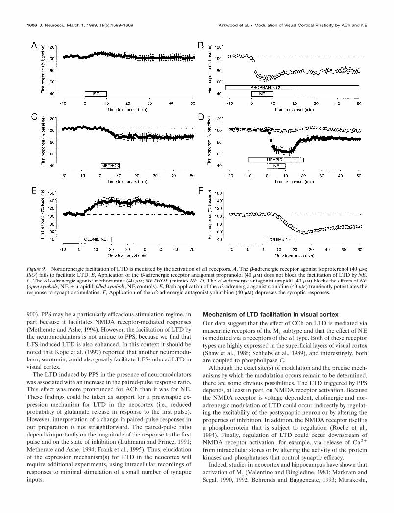
Figure 9, A and B, shows that b-adrenergic receptors areunlikely to be involved in the facilitation of LTD. PPS in thepresence of the b-adrenergic receptor agonist isoproterenol (40mM) produced no LTD (97 6 4%; n 5 6), and application of theb receptor antagonist propranolol (40 mM) had no effect on theinduction of LTD in the presence of NE (74 6 4%; n 5 5). Theseresults contrast with the effects of a1 receptor agonists andantagonists (Fig. 9C,D). PPS during application of the a1 recep-tor agonist methoxamine (40 mM) caused a sustained depressionof the synaptic responses (84 6 3; n 5 8) that mimicked the LTDinduced with NE (Fig. 9C). Moreover, the a1 antagonist urapidil(40–80 mM) effectively blocked the effects of NE (98 6 3%; n 512) as compared with interleaved controls (85 6 2%; n 5 12; Fig.9D). Together, these data indicate that a1, but not b, receptorsare involved in the facilitation of LTD by NE.
The pharmacological manipulation of a2 receptors producedsome perplexing results. The a2 agonist clonidine had the oppo-site effect of NE; PPS in clonidine produced a large, but transient,potentiation of the synaptic responses (130 6 8% at 20 min ofclonidine application). After clonidine was removed, the re-sponses slowly came back to control levels (103 6 3% at 50 minof washout; n 5 4; Fig. 9E). The a2 antagonist yohimbine (40 mM)mimicked the effects of NE and profoundly reduced the fieldresponses. When the drug was removed, the responses recoveredonly partially, reaching a stably depressed level (70 6 4%; n 5 7;
Figure 6. PPS in the presence of NE induces a lasting depression of the layer III synaptic responses to layer IV stimulation. A, B, Effects of a brief (10min) bath application of NE (40 mM) on the field responses evoked with paired-pulse stimulation (ISI 5 40 msec) are shown. A, FPs were recordedimmediately before (control) and during the application of NE and 30 min after washout of the drug. B, Time course of the average of nine experimentsis shown. Solid circles are responses to the first pulse; open circles are responses to the second pulse. C, D, The effects of NE on FPs correlate with changesin simultaneously recorded intracellular EPSPs. C, Traces are averages of four consecutive intracellular (top) and extracellular (bottom) responsesrecorded before and 30 min after washout of NE. D, Average time course of 11 similar experiments is shown. Top, Changes in FP amplitude (open circles)and initial slope of the EPSP ( filled triangles) are presented. Bottom, Changes in the membrane potential are shown.
1604 J. Neurosci., March 1, 1999, 19(5):1599–1609 Kirkwood et al. • Modulation of Visual Cortical Plasticity by ACh and NE

Fig. 9F). The paradoxical results obtained with yohimbine andclonidine might be attributable, at least in part, to their effects onpresynaptic a2 inhibitory autoreceptors. By blocking the autore-ceptors, yohimbine might enhance the release of endogenous NEand facilitate the induction of LTD.
Differential effects of neuromodulators in visual cortexand hippocampusPrevious studies indicate that synaptic plasticity evoked in visualcortical layer III and synaptic plasticity evoked in the CA1 regionof the hippocampus share crucial similarities in their mechanismof induction (Kirkwood et al., 1993). Therefore, it was of interestto investigate the effect of the neuromodulators on the CA1synaptic responses. In these experiments, the responses to PPSwere recorded simultaneously in visual cortex and hippocampususing slices that contained both structures (Fig. 10A). During theCCh application, the CA1 responses were the most affected (Fig.10B). After removal of the drug, however, the responses in bothvisual cortex and hippocampus stabilized at a similar, depressedlevel (82 6 7% in visual cortex; 89 6 4% in CA1; n 5 8). On theother hand, whereas during the application of NE both CA1 andvisual cortex were depressed to the same level (Fig. 10C), only thevisual cortical responses remained depressed after the removal ofthe drug (82 6 3% in visual cortex; 103 6 6% in CA1; n 5 10).
DISCUSSIONThe principal finding of our study is that cholinergic or norad-renergic receptor activation dramatically facilitates the inductionof homosynaptic LTD in layer III of visual cortex. In fact, whenPPS is used, the neuromodulators enable induction and expres-sion of synaptic plasticity that otherwise would not be observed.Thus, our results provide strong support for the view that AChand NE can serve as “enabling factors” for activity-dependentcortical plasticity (Singer, 1979, 1995; Dykes et al., 1990; Dykes,1997). Because this robust modulation of plasticity can be ob-served under controlled conditions in vitro, the paradigm we
describe here may be useful for the further dissection of themolecular mechanism of experience-dependent cortical plasticityand its modulation by behavioral state.
Properties of LTD induced in the presence of AChand NEIn recent years, it has become clear that many synapses in theCNS can undergo activity-dependent LTD. In the hippocampusand neocortex, LTD can be induced reliably with prolonged (e.g.,15 min), low-frequency (e.g., 1 Hz) synaptic stimulation. LTDinduced in this way occurs only at the stimulated synapses andthus is said to be homosynaptic. In the CA1 region of hippocam-pus and in layer III of visual cortex, homosynaptic LTD evokedwith LFS requires NMDA receptor activation under most exper-imental conditions (e.g., Dudek and Bear, 1992; Kirkwood et al.,1993).
The properties of LTD in visual cortex induced by PPS in thepresence of NE suggest that it uses a similar induction mecha-nism. In particular, the LTD is completely blocked by applicationof AP5, an NMDA receptor antagonist. Thus, NE may act as asort of gain-control mechanism for NMDA receptor-dependenthomosynaptic LTD. The situation for ACh, however, may bemore complicated. Although the LTD induced by PPS in thepresence of CCh was input specific, it was only partially blockedby AP5. Thus, in addition to facilitating NMDA receptor-dependent homosynaptic LTD, ACh may also promote othermechanisms of LTD induction. In this context, it should be notedthat NMDA receptor-independent forms of homosynaptic LTDhave been described, both in visual cortex (Artola et al., 1990)and in hippocampus, including area CA1 (Stanton and Sejnowski,1989; Bolshakov and Siegelbaum, 1994; Oliet et al., 1997).
With PPS, a brief application of NE or CCh resulted in LTD ofmagnitude comparable with that obtained with prolonged LFS,the standard method to induce LTD in many laboratories. How-ever, PPS induced LTD with far fewer stimulation pulses (40 vs
Figure 7. Synaptic depression induced with NErequires synaptic activity. Left, Time course ofthe changes induced by NE on the amplitude ofthe first response in two sites recorded simulta-neously in the same slices (n 5 5). One site wasstimulated throughout the experiment (solid cir-cles), whereas in the other site stimulation wassuspended during NE application and resumed 5min after washout (open circles). Right, Stimulus-recording arrangement.
Figure 8. Synaptic depression induced with NE is reversibly blocked by the NMDA receptor antagonist AP5. Left, In the presence of AP5 (100 mM),NE failed to produce any change in the FP response to test stimulation. Right, After washout of the drug, however, depression was reliably produced.In both panels the responses were normalized with respect to the average response during the initial baseline period before NE application. The timethat elapsed between the last data point on the lef t and the first data point on the right varied but was not .30 min.
Kirkwood et al. • Modulation of Visual Cortical Plasticity by ACh and NE J. Neurosci., March 1, 1999, 19(5):1599–1609 1605
900). PPS may be a particularly efficacious stimulation regime, inpart because it facilitates NMDA receptor-mediated responses(Metherate and Ashe, 1994). However, the facilitation of LTD bythe neuromodulators is not unique to PPS, because we find thatLFS-induced LTD is also enhanced. In this context it should benoted that Kojic et al. (1997) reported that another neuromodu-lator, serotonin, could also greatly facilitate LFS-induced LTD invisual cortex.
The LTD induced by PPS in the presence of neuromodulatorswas associated with an increase in the paired-pulse response ratio.This effect was more pronounced for ACh than it was for NE.These findings could be taken as support for a presynaptic ex-pression mechanism for LTD in the neocortex (i.e., reducedprobability of glutamate release in response to the first pulse).However, interpretation of a change in paired-pulse responses inour preparation is not straightforward. The paired-pulse ratiodepends importantly on the magnitude of the response to the firstpulse and on the state of inhibition (Luhmann and Prince, 1991;Metherate and Ashe, 1994; Frank et al., 1995). Thus, elucidationof the expression mechanism(s) for LTD in the neocortex willrequire additional experiments, using intracellular recordings ofresponses to minimal stimulation of a small number of synapticinputs.
Mechanism of LTD facilitation in visual cortexOur data suggest that the effect of CCh on LTD is mediated viamuscarinic receptors of the M1 subtype and that the effect of NEis mediated via a receptors of the a1 type. Both of these receptortypes are highly expressed in the superficial layers of visual cortex(Shaw et al., 1986; Schliebs et al., 1989), and interestingly, bothare coupled to phospholipase C.
Although the exact site(s) of modulation and the precise mech-anisms by which the modulation occurs remain to be determined,there are some obvious possibilities. The LTD triggered by PPSdepends, at least in part, on NMDA receptor activation. Becausethe NMDA receptor is voltage dependent, cholinergic and nor-adrenergic modulation of LTD could occur indirectly by regulat-ing the excitability of the postsynaptic neuron or by altering theproperties of inhibition. In addition, the NMDA receptor itself isa phosphoprotein that is subject to regulation (Roche et al.,1994). Finally, regulation of LTD could occur downstream ofNMDA receptor activation, for example, via release of Ca 21
from intracellular stores or by altering the activity of the proteinkinases and phosphatases that control synaptic efficacy.
Indeed, studies in neocortex and hippocampus have shown thatactivation of M1 (Valentino and Dingledine, 1981; Markram andSegal, 1990, 1992; Behrends and Buggencate, 1993; Murakoshi,
Figure 9. Noradrenergic facilitation of LTD is mediated by the activation of a1 receptors. A, The b-adrenergic receptor agonist isoproterenol (40 mM;ISO) fails to facilitate LTD. B, Application of the b-adrenergic receptor antagonist propranolol (40 mM) does not block the facilitation of LTD by NE.C, The a1-adrenergic agonist methoxamine (40 mM; METHOX ) mimics NE. D, The a1-adrenergic antagonist urapidil (40 mM) blocks the effects of NE(open symbols, NE 1 urapidil; filled symbols, NE controls). E, Bath application of the a2-adrenergic agonist clonidine (40 mM) transiently potentiates theresponse to synaptic stimulation. F, Application of the a2-adrenergic antagonist yohimbine (40 mM) depresses the synaptic responses.
1606 J. Neurosci., March 1, 1999, 19(5):1599–1609 Kirkwood et al. • Modulation of Visual Cortical Plasticity by ACh and NE

1995; Kimura and Baughman, 1997) and a1 receptors (Madisonand Nicoll, 1988) reduces evoked GABA release by inhibitoryinterneurons. In addition, M1 receptor activation increases theexcitability of pyramidal neurons (McCormick and Prince, 1986;Dutar and Nicoll, 1988) and directly enhances NMDA receptorfunction (Segal, 1992; Aramakis et al., 1997). Any or all of these
effects could contribute to the modulation of LTD in visualcortex, and it will be of interest to investigate these possiblemechanisms in future studies. It should be noted, however, thatthere is already evidence that decreasing inhibition can facilitatehomosynaptic LTD in CA1 (Wagner and Alger, 1995).
Neuromodulators and bidirectional synaptic plasticityTo our knowledge, our study is the first to show facilitation ofLTD by ACh and NE. However, there have been many reports onthe effects of ACh and NE on long-term potentiation (LTP). Asis the case for LTD, there are multiple forms of LTP withdifferent induction and expression mechanisms. One form, whichis highly expressed in CA1 and in the superficial layers of neo-cortex (e.g., Kirkwood et al., 1993), depends on NMDA receptoractivation for its induction. This type of LTP is of particularinterest because it is the functional inverse of NMDA receptor-dependent LTD (for review, see Bear and Kirkwood, 1996).
As we have shown for LTD, NMDA receptor-dependent LTPis enhanced by muscarinic receptor activation (Tanaka et al.,1989; Blitzer et al., 1990; Burgard and Sarvey, 1990; Brocher etal., 1992; Huerta and Lisman, 1993; Sokolov and Kleschevnikov,1995). In fact, Auerbach and Segal (1994, 1996) report that lowconcentrations of CCh (0.25–0.75 mM) can directly trigger LTP inCA1. We did not observe a similar effect in visual cortex,however.
Similarly, NMDA receptor-dependent LTP is enhanced byNE. However, this facilitatory effect is mediated by b receptorsand not a receptors as is the case for LTD (Stanton and Sarvey,1987; Brocher et al., 1992; Kato, 1993; Thomas et al., 1996;Katsuki et al., 1997). Moreover, in CA1 10 mM NE not onlyfacilitates LTP but also inhibits induction of LTD with LFS(Blitzer et al., 1995; Thomas et al., 1996). The apparent discrep-ancy between the effects of NE on LTD in visual cortex andhippocampus could be explained by differences in the balance ofa and b receptor activation by NE in the two preparations.Indeed, our direct comparison of hippocampus and visual cortexrevealed that synaptic transmission in the two structures respondsdifferently to bath-applied NE. Unlike in the visual cortex, therewas no LTD induced in CA1 by PPS in the presence of NE.
Relevance to experience-dependent cortical plasticityThe stimulus selectivity of cortical neurons is subject toexperience-dependent modification, and such changes are likelyto reflect the storage of information by cortical synapses. Theo-retical studies have shown that synaptic modifications with theproperties of LTP and LTD are well suited to account forexperience-dependent shifts in selectivity (Bienenstock et al.,1982; Bear et al., 1987; Bear, 1996). The striking facilitation byNE and ACh of both LTP and LTD suggests that these modu-lators could function generally as a gain-control mechanism forthe synaptic plasticity that underlies receptive field plasticity andlearning. Indeed, the notion that cortical synaptic plasticity issomehow “gated” by ACh and NE has received abundant supportfrom studies performed in vivo on visual, auditory, and somato-sensory cortex (Kasamatsu and Pettigrew, 1979; Bear and Singer,1986; Gordon et al., 1990; Juliano et al., 1991; Kasamatsu, 1991;Gu and Singer, 1993; Edeline et al., 1994; Baskerville et al., 1997;Dykes, 1997; Kilgard and Merzenich, 1998; Zhu and Waite,1998).
Although plasticity is an important feature of adult corticalorganization, it is clearly most robust during early postnataldevelopment. A classic example of developmental plasticity in the
Figure 10. Differential effects of CCh and NE in hippocampus and visualcortex. A, The drawing depicts the electrode configuration used forsimultaneous recording in CA1 and visual cortex. B, Application of CChyields comparable LTD in CA1 and visual cortex. Top, The traces areaverages of four consecutive recordings taken before (thick line), during(dotted line), and 30 min after the application of CCh (thin line). Bottom,The graph shows the time course of the changes induced in the firstresponse recorded simultaneously in the CA1 region of the hippocampus(initial slope of the field potential; open circles) and in the visual cortex(field potential amplitude; filled circles). C, The time course of the changesinduced by bath application of NE is shown. PPS in the presence of NEfailed to produce LTD in the hippocampus.
Kirkwood et al. • Modulation of Visual Cortical Plasticity by ACh and NE J. Neurosci., March 1, 1999, 19(5):1599–1609 1607

visual cortex is the loss of responsiveness to stimulation of an eyethat has been briefly deprived of vision. Like other forms ofcortical plasticity, the deprivation-induced synaptic depression isdisrupted by lesions that interrupt cholinergic and noradrenergicinputs to cortex (Bear and Singer, 1986; Gordon et al., 1990).Interestingly, although partial destruction of each input alone wasinsufficient to produce a detectable loss of deprivation-inducedsynaptic depression, their combined destruction did disrupt thisform of plasticity. This finding led to the suggestion that the twoinputs might be able to substitute for one another in the controlof cortical plasticity. Our discovery that ACh and NE produce aqualitatively similar facilitation of LTD is entirely consistent withthis idea. Thus, the study of LTD and its modulation may providenew insights into the mechanisms of experience-dependent syn-aptic plasticity in the neocortex.
REFERENCESAizenman C, Kirkwood A, Bear M (1996) Current source density anal-
ysis of evoked responses in visual cortex in vitro: implications for theregulation of long-term potentiation. Cereb Cortex 6:751–758.
Aramakis VB, Bandrowski AE, Ashe JH (1997) Activation of musca-rinic receptors modulates NMDA receptor-mediated responses in au-ditory cortex. Exp Brain Res 113:484–496.
Artola A, Brocher S, Singer W (1990) Different voltage-dependentthresholds for inducing long-term depression and long-term potentia-tion in slices of rat visual cortex. Nature 347:69–72.
Auerbach JM, Segal M (1994) A novel cholinergic induction of long-term potentiation in rat hippocampus. J Neurophysiol 72:2034–2040.
Auerbach JM, Segal M (1996) Muscarinic receptors mediating depres-sion and long-term potentiation in rat hippocampus. J Physiol (Lond)492:479–493.
Bakin JS, Weinberger NM (1996) Induction of a physiological memoryin the cerebral cortex by stimulation of the nucleus basalis. Proc NatlAcad Sci USA 93:11219–11224.
Baskerville KA, Schweitzer JB, Herron P (1997) Effects of cholinergicdepletion on experience-dependent plasticity in the cortex of the rat.Neuroscience 80:1159–1169.
Bear MF (1996) A synaptic basis for memory storage in the cerebralcortex. Proc Natl Acad Sci USA 93:13453–13459.
Bear MF, Kirkwood A (1996) Bidirectional plasticity of cortical syn-apses. In: Cortical plasticity: LTP and LTD (Fazeli MS, CollingridgeGL, eds), pp 191–205. Oxford: Bios Scientific.
Bear MF, Singer W (1986) Modulation of visual cortical plasticity byacetylcholine and noradrenaline. Nature 320:172–176.
Bear MF, Cooper LN, Ebner FF (1987) A physiological basis for atheory of synaptic modification. Science 237:42–48.
Behrends JC, Buggencate GT (1993) Cholinergic modulation of synapticinhibition in the guinea pig hippocampus in vitro: excitation ofGABAergic interneurons and inhibition of GABA release. J Neuro-physiol 69:626–629.
Bienenstock EL, Cooper LN, Munro PW (1982) Theory for the devel-opment of neuron selectivity: orientation specificity and binocularinteraction in visual cortex. J Neurosci 2:32–48.
Blitzer RD, Gil O, Landau EM (1990) Cholinergic stimulation enhanceslong-term potentiation in the CA1 region of the hippocampus. NeurosciLett 119:207–210.
Blitzer RD, Wong T, Nouranifar R, Iyengar R, Landau EM (1995)Postsynaptic cAMP pathway gates early LTP in hippocampal CA1region. Neuron 15:1403–1414.
Bolshakov VY, Siegelbaum SA (1994) Postsynaptic induction and pre-synaptic expression of hippocampal long-term depression. Science264:1148–1152.
Brocher S, Artola A, Singer W (1992) Agonists of cholinergic and nor-adrenergic receptors facilitate synergistically the induction of long-termpotentiation in slices of rat visual cortex. Brain Res 573:27–36.
Burgard EC, Sarvey JM (1990) Muscarinic receptor activation facilitatesthe induction of long-term potentiation (LTP) in the rat dentate gyrus.Neurosci Lett 116:34–39.
Dodt H-U, Pawelzik P, Zieglgansberger W (1991) Actions of noradren-aline on neocortical neurons in vitro. Brain Res 545:307–331.
Dudek SM, Bear MF (1992) Homosynaptic long-term depression in area
CA1 of hippocampus and effects of N-methyl-D-aspartate receptorblockade. Proc Natl Acad Sci USA 89:4363–4367.
Dutar P, Nicoll RA (1988) Classification of muscarinic responses inhippocampus in terms of receptor subtypes and second-messengersystems: electrophysiological studies in vitro. J Neurosci 8:4214–4224.
Dykes RW (1997) Mechanisms controlling neuronal plasticity in somato-sensory cortex. Can J Physiol Pharmacol 75:535–545.
Dykes RW, Tremblay N, Warren RA, Bear MF (1990) Cholinergicmodulation of synaptic plasticity in sensory neocortex. In: Activation toacquisition: functional aspects of the basal forebrain cholinergic system(Richardson RT, ed), pp 325–346. Boston: Birkhauser.
Edeline JM, Hars B, Maho C, Hennevin E (1994) Transient and pro-longed facilitation of tone-evoked responses induced by basal forebrainstimulations in the rat auditory cortex. Exp Brain Res 97:373–386.
Frank H, Kirkwood A, Paradiso MA, Bear MF (1995) Probing the“plasticity gate” in visual cortex using paired-pulse stimulation. SocNeurosci Abstr 21:2024.
Gil Z, Connors BW, Amitai Y (1997) Differential regulation of neocor-tical synapses by neuromodulators and activity. Neuron 19:679–686.
Gordon B, Mitchell B, Mohatadi K, Roth E, Tseng Y, Turk F (1990)Lesions of non-visual inputs affect plasticity, norepinephrine contentand acetylcholine content of visual cortex. J Neurophysiol64:1851–1860.
Gu Q, Singer W (1993) Effects of intracortical infusion of anticholinergicdrugs on neuronal plasticity in kitten striate cortex. Eur J Neurosci5:475–485.
Huerta PT, Lisman JE (1993) Heightened synaptic plasticity of hip-pocampal CA1 neurons during a cholinergically induced rhythmic state.Nature 364:723–725.
Juliano SL, Ma W, Eslin D (1991) Cholinergic depletion prevents expan-sion of topographic maps in somatosensory cortex. Proc Natl Acad SciUSA 88:780–784.
Kasamatsu T (1991) Adrenergic regulation of visuocortical plasticity: arole of the locus coeruleus system. Prog Brain Res 88:599–616.
Kasamatsu T, Pettigrew JD (1979) Restoration of visual cortical plastic-ity by local microperfusion of norepinephrine. J Comp Neurol185:163–182.
Kato N (1993) Mechanisms of beta-adrenergic facilitation in rat visualcortex. NeuroReport 4:1087–1090.
Katsuki H, Izumu Y, Zorumski CF (1997) Noradrenergic regulation ofsynaptic plasticity in the hippocampal CA1 region. J Neurophysiol77:3013–3020.
Kilgard MP, Merzenich MM (1998) Cortical map reorganization en-abled by nucleus basalis activity. Science 279:1714–1718.
Kimura F, Baughman RW (1997) Distinct muscarinic receptor subtypessuppress excitatory and inhibitory synaptic responses in cortical neu-rons. J Neurophysiol 77:709–716.
Kirkwood A, Bear MF (1994a) Hebbian synapses in visual cortex.J Neurosci 14:1634–1645.
Kirkwood A, Bear MF (1994b) Homosynaptic long-term depression inthe visual cortex. J Neurosci 14:3404–3412.
Kirkwood A, Dudek SM, Gold JT, Aizenman CD, Bear MF (1993)Common forms of synaptic plasticity in the hippocampus and neocortexin vitro. Science 260:1518–1521.
Kojic L, Gu Q, Douglas RM, Cynader MS (1997) Serotonin facilitatessynaptic plasticity in kitten visual cortex: an in vitro study. Dev BrainRes 101:299–304.
Luhmann HJ, Prince DA (1991) Postnatal maturation of the GABAer-gic system in rat neocortex. J Neurophysiol 65:247–263.
Madison DV, Nicoll RA (1988) Norepinephrine decreases synaptic in-hibition in the rat hippocampus. Brain Res 442:131–138.
Markram H, Segal M (1990) Acetylcholine potentiates responses toN-methyl-D-aspartate in the rat hippocampus. Neurosci Lett 113:62–65.
Markram H, Segal M (1992) The inositol 1,4,5-triphosphate pathwaymediates cholinergic potentiation of rat hippocampal neuronal re-sponses to NMDA. J Physiol (Lond) 447:513–533.
Markram H, Tsodyks M (1996) Redistribution of synaptic efficacy be-tween neocortical pyramidal neurons. Nature 382:807–810.
McCormick DA, Prince DA (1986) Mechanisms of action of acetylcho-line in the guinea-pig cerebral cortex in vitro. J Physiol (Lond)375:169–194.
Metherate R, Ashe JH (1994) Facilitation of an NMDA receptor-mediated EPSP by paired-pulse stimulation in rat neocortex via depres-sion of GABAergic IPSPs. J Physiol (Lond) 481:331–348.
Mitzdorf U (1985) Current source-density method and application in cat
1608 J. Neurosci., March 1, 1999, 19(5):1599–1609 Kirkwood et al. • Modulation of Visual Cortical Plasticity by ACh and NE

cerebral cortex: investigation of evoked potentials and EEG phenom-ena. Physiol Rev 65:37–100.
Mrzljak L, Levey A, Goldman-Rakic PS (1993) Association of m1 andm2 muscarinic receptor proteins with asymmetric synapses in theprimate cerebral cortex: morphological evidence for cholinergic mod-ulation of excitatory neurotransmission. Proc Natl Acad Sci USA90:5194–5198.
Murakoshi T (1995) Cholinergic modulation of synaptic transmission inthe rat visual cortex in vitro. Vision Res 35:25–35.
Oliet SH, Malenka RC, Nicoll RA (1997) Two distinct forms of long-term depression coexist in CA1 hippocampal pyramidal cells. Neuron18:969–982.
Osterheld-Haas MC, Van der Loos H, Hornung JP (1994) Monoamin-ergic afferents to cortex modulate structural plasticity in the barrelfieldof the mouse. Dev Brain Res 77:189–202.
Roche K, Tingley W, Huganir R (1994) Glutamate receptor phosphor-ylation and synaptic plasticity. Curr Opin Neurobiol 4:383–388.
Sachdev R, Lu S, Wiley R, Ebner F (1998) Role of the basal forebraincholinergic projection in somatosensory cortical plasticity. J Neuro-physiol 79:3216–3228.
Schliebs R, Walch C, Stewart MG (1989) Laminar pattern of cholinergicand adrenergic receptors in rat visual cortex using quantitative receptorautoradiography. J Hirnforsch 30:303–311.
Segal M (1982) Multiple actions of acetylcholine at a muscarinic recep-tor studied in the hippocampal slice. Brain Res 246:77–87.
Segal M (1992) Acetylcholine enhances NMDA-evoked calcium rise inhippocampal neurons. Brain Res 587:83–87.
Shaw C, Wilkinson M, Cynader M, Needler MC, Aoki C, Hall SE (1986)The laminar distributions and postnatal development of neurotransmit-ter and neuromodulator receptors in cat visual cortex. Brain Res Bull16:661–671.
Singer W (1979) Central-core control of visual cortex functions. In: Theneurosciences fourth study program (Schmitt FO, Worden FG, eds), pp1093–1109. Cambridge, MA: MIT.
Singer W (1995) Development and plasticity of cortical processing archi-tectures. Science 270:758–759.
Sokolov M, Kleschevnikov A (1995) Atropine suppresses associativeLTP in CA1 region of rat hippocampal slices. Brain Res 672:281–284.
Stanton PK, Sarvey JM (1987) Norepinephrine regulates long-term po-tentiation of both the population spike and dendritic EPSP in hip-pocampal dentate gyrus. Brain Res Bull 18:115–119.
Stanton PK, Sejnowski TJ (1989) Associative long-term depression inthe hippocampus induced by Hebbian covariance. Nature 229:215–218.
Tanaka Y, Sakurai M, Hayashi S (1989) Effect of scopolamine and HP029, a cholinesterase inhibitor, on long-term potentiation in hippocam-pal slices of the guinea pig. Neurosci Lett 98:179–193.
Thomas MJ, Moody TD, Makhinson M, O’Dell TJ (1996) Activity-dependent b-adrenergic modulation of low frequency stimulation in-duced LTP in the hippocampal CA1 region. Neuron 17:475–482.
Vaknin G, Teyler TJ (1991) Ontogenesis of the depressant activity ofcarbachol on synaptic activity in rat visual cortex. Brain Res Bull26:211–214.
Valentino RJ, Dingledine R (1981) Presynaptic inhibitory effect of ace-tylcholine in the hippocampus. J Neurosci 1:784–792.
Wagner JJ, Alger BE (1995) GABAergic and developmental influenceson homosynaptic LTD and depotentiation in rat hippocampus. J Neu-rosci 15:1577–1586.
Wang Z, McCormick DA (1993) Control of firing mode of corticotectaland corticopontine layer V burst-generating neurons by norepineph-rine, acetylcholine, and 1S,3R-ACPD. J Neurosci 13:2199–2216.
Zhu XO, Waite PME (1998) Cholinergic depletion reduces plasticity ofbarrel field cortex. Cereb Cortex 8:63–72.
Kirkwood et al. • Modulation of Visual Cortical Plasticity by ACh and NE J. Neurosci., March 1, 1999, 19(5):1599–1609 1609