Medico-Legal Considerations in the Sphere of Intravenous ...
Upload
khangminh22Category
view
0download
0

ASSOCIATE EDITOR
1. S.K. Dhattarwal (Professor)Forensic Medicine, PGIMS, Rohtak, Haryana
2. Dr. Adarsh Kumar (Additional Professor)Forensic Medicine, AIIMS, New Delhi
3. Dr. Vijaynath V (Associate Professor)Forensic Medicine, Vinayaka Mission Medical college, Tamil Nadu
4. Ms. Roma Khan, Forensic Sciences, INSAAF Mumbai
5. Dr. Imran Sabri (Assistant Professor)Department of Bio-Medical Sciences.College of Medicine, KingFaisal University,Saudi Arabia
INTERNATIONAL EDITORIAL ADVISORY BOARD
1. B. N. Yadav (Professor)Forensic Medicine, BP Koirala Institute of Medical Sciences, Nepal
2. Dr. Vasudeva Murthy Challakere Ramaswam (Senor Lecturer)Department of Pathology, International Medical University, BukitJalil, Kuala Lumpur. Malaysia
3. Babak Mostafazadeh (Associate Professor)Department of Forensic Medicine & Toxicology, Shahid BeheshtiUniversity of Medical Sciences, Tehran-Iran
4. Dr. Sarathchandra Kodikara (Lecturer)Forensic Medicine Department of Forensic Medicine, Faculty ofMedicine, University of Peradeniya, Sri Lanka
NATIONAL EDITORIAL ADVISORY BOARD
1. Prof. N.K. Agarwal (Professor) Forensic Medicine, UCMS, Delhi
2. P.K. Chattopadhyay, (Professor)Forensic Sciences, Amity University, Noida
3. Dalbir Singh (Professor) Forensic Medicine, PGIMER, Chandigarh
4. Dr. Harish Pathak, Mumbai
5. J. Gargi (Professor) GGS Medical College, Faridkot
6. P.C. Dikshit (Professor)Forensic Medicine, Jamia Hamdard Medical College, New Delhi
7. Anil Mittal (Professor)Forensic Medicine, Vardhman Mahavir Medical college, New Delhi
8. Balbir Kaur (Professor)Forensic Medicine, MM institute of Medical Sciences, Ambala
9. Mukesh Yadav (Professor) Forensic Medicine, School of Medical Sciences and research,Greater Noida
10. T.K.K. Naidu (Professor) Forensic Medicine, Prathima Instituteof Medical Sciences Andhra Pradesh
11. S. Das (Professor) Forensic Medicine, Himalayan Institute ofMedical Sciences Dehradun
12. Col Ravi Rautji, Forensic Medicine, Armed Forces Medical College, Pune
13. Dr. Manish Nigam (Professor and Head)Department of Forensic Medicine & Toxicology Sri AurobindoInstitute of Medical Sciences, INDORE (M.P.)
14. Dr. Shailesh Kudva (Principal)Rajasthan Dental College and Hospital Jaipur-302026
15. Usmanganishah Makandar (Associate Professor)Anatomy, AIMS, Bhatinda
16. Dr. Pratik Patel (Professor and Head) Forensic Medicine, SmtNHL Municipal Medical College Ahmedabad
17. Basappa S. Hugar (Associate Professor)Forensic Medicine, Ramaiah Medical College, Bangalore
NATIONAL EDITORIAL ADVISORY BOARD
18. Dr. Vandana Mudda (Awati) (Associate Prof)Dept of FMT, M.R. Medical College, Gulbarga, Karnataka, India
19. Dr. HarishKumar. N. (AssociateProfessor)Dept.of ForensicMedicine, Sri Siddhartha MedicalCollege, Tumkur
20. Dr. Gowri Shankar (Associate Professor)Forensic Medicine, SNMC, Bagalkot
21. Dr. Manjunath Badni (Reader) Dept of Oral pathology MaharanaPratap college of Dentistry and Research Centre, Gwalior
22. Dr. L.Ananda Kumar (Associate Professor) Forensic Medicine,Rajiv Gandhi Institute of Medical Sciences, (RIMS), Kadapa
23. Dr. Ramesh Nanaji Wasnik (Associate Professor and Head)Forensic Medicine Late B.R.K.M. Govt. Medical college, Jagdalpur
24. Dr. Sachin Sinha (Reader), Dept. of Oral Pathology & Microbiology Daswani Dental College & Research Centre, Rajasthan
25. Dr.Sasi Kanth, Asst. Professor, A.C.S.R Government Medical College,
Nellore, Andhra Pradesh.
Medico-Legal UpdateEditor-in Chief
Prof. (Dr) R K SharmaFormer Head, Department of Forensic Medicine & Toxicology
All-India Institute of Medical Sciences, New Delhi-110029E-mail: [email protected]
EditorDr. R.K. Sharma
Institute of Medico-legal Publications4th Floor, Statesman House Building, Barakhamba Road,
Connaught Place, New Delhi-110 001Printed, published and owned by
Dr. R.K. SharmaInstitute of Medico-legal Publications
4th Floor, Statesman House Building, Barakhamba Road,Connaught Place, New Delhi-110 001
Published atInstitute of Medico-legal Publications
4th Floor, Statesman House Building, Barakhamba Road,Connaught Place, New Delhi-110 001
Medico Legal Update is a scientifi c journal which brings latest knowledge
regarding changing medico legal scenario to its readers. The journal caters
to specialties of Forensic Medicine, Forensic Science, DNA fi ngerprinting,
Toxicology, Environmental hazards, Sexual Medicine etc. The journal has
been assigned international standard serial number (ISSN) 0971-720X. The
journal is registered with Registrar of Newspaper for India vide registration
numbers 63757/96 under Press and Registration of Books act, 1867. The
journal is also covered by EMBASE (Excerpta Medica Database) from
1997 and by INDEX COPERNICUS, POLAND. Medico legal update is
a half yearly peer reviewed journal. The journal has also been assigned
E-ISSN 0973-1283 (Electronic version). The fi rst issue of the journal was
published in 1996.
Website: www.medicolegalupdate.org© All Rights reserved The views and opinions expressed are of the authors
and not of the Medico Legal Update. The Medico Legal Update does not
guarantee directly or indirectly the quality or effi cacy of any products or
service featured in the advertisement in the journal, which are purely
commercial.
Volume 17, Number 1 January-June 2017
I
CONTENTS
Medico-Legal Update
1. Stature Estimation Using Anthropometric Measurements on X-rays of Long Bones in ................................... 01Living Individuals of Indian PopulationReddy Ravikanth, Partha Sarathi Sarkar, Babu Philip
2. Reliability of Cranial Measurements in Identification of Sex of Skull .............................................................. 06 Manjunath T H, Narsimhamurthy S, Nagesh Kuppast, Raghavendra R U, Umesh S R,Muralidhar Shepur
3. One should be Ectopic Minded – A Case Report ............................................................................................... 12H S Tatiya, V T Jadhao, A A Taware, S B Punpale
4. A Study on Grading and Severity of Aortic Athermatous Plaques- Autopsy based Study .............................. 16 M Babu, B Venkata Sai Prasad, I Mohan Prasad, B Venkateswarlu
5. Fatal Compression of Neck by Ligature: Study of Profile of Cases and External Findings .............................. 22over the Body Sumit Tellewar, Abhishek Yadav, Mohit Gupta, Lohith Kumar R, Sarala M
6. Suicide by Hanging in Jharkhand: An Autopsy based Cross Sectional Study ................................................... 26Kaushal Kishore, Bhoopendra Singh, Ajit Kumar Chaudhary
7. Study of Trends of Death due to Burns Cases at Hubballi Region of Karnataka .............................................. 34Gajanan H Nayak, Madhu Sudhan S, Sunilkumar S Biradar, Ravindra Kumar C N, Hemanth Raj M N
8. Management of Patient Criticality at the Hospital: A Study in Reference to Second Opinion ......................... 38Amit Kumar Pandey, Garima Malik, Vinamra Jain, Ashok Sharma
9. Demographic Profiles of Suicidal Deaths in Coastal Odisha ............................................................................. 44Soumya Ranjan Nayak, Braja Kishore Dash
10. Can Lip Print Patterns Determine Sex of an Individual? ................................................................................... 48Umesh Babu R, Ameena Ahamed, Imaad M I
11. Study of Patterns of Deaths in Unknown Dead Bodies - A Two Year Study .................................................... 54N T Satish, MG Shivaramu, Kumar U, Vijay Kumar
12. A Study of Prediction of Stature from Percutaneous Ulna Length .................................................................... 57Madhu Sudhan S, H C Govindaraju, Hemanth Raj M N

13. Estimation of Stature from Percutaneous Tibial Length – A Study Conducted in Natives of Gujarat ............. 60Reekee Patel, Niraj Bharadva
14. Prospective Study of Postmortem Cases of Hanging as a Method of Suicide in North Karnataka ................... 65Mohsenul haq, Ayesha Farheen, S K Goli
15. Observational Study of Victims of Alleged Sexual Assault in Central Mumbai Region .................................. 70Shashank Tyagi, Vikas P Meshram, Rajesh B Sukhadeve, Harish M Pathak, Hemlata Pandey
16. Estimation of Age from Eruption of 3rd Molar and its Comparison with the Chronological Age .................... 75Mahanta Putul, Talukdar KL, Baishya Manoj Kr., Mahanta HK
17. Pattern of Skull Fractures due to Blunt Force in and around Khammam .......................................................... 82Bharath Kumar Guntheti, Uday Pal Singh
18. Cardiac Troponin T as a Marker in the Diagnosis of Acute Myocardial Infarction at Autopsy ........................ 88B Venkata Naga Mohan Rao, Bandi Sree Ramulu, B Raju
19. Profile of Homicidal Deaths in Medicolegal Autopsies at Jorhat Medical College & ...................................... 91Hospital, Jorhat, AssamNitu Kr. Gogoi, Kanak Ch. Das
20. Study of Patterns of Cranio-cerebral Injuries in Road Traffic Accidents Involving ......................................... 94Two Wheelers in Gulbarga AreaMohsenul haq, Ayesha Farheen2, S K Goli
21. Study of Correlation of Index Finger Lengths and Stature ................................................................................ 98Pramod Kumar GN, Roopa Urs AN
22. Study of Pattern of Ligature Mark in Cases of Hanging in Kamrup Region, Assam State, India ................... 102Arunava Borah, Rituraj Chaliha
23. Study of Post Mortem Interval Using Entomological Evidence ...................................................................... 105Shashikanth Naik C R, S Venkata Raghava
24. Profile of Medicolegal Cases at a Tertiary Care Hospital in North Karnataka ................................................ 111Hemanth Kumar R G, Pratibha R Kulkarni
25. Age Assessment from Vault Sutures Closure in Elderly Persons: An Autopsy Study .................................... 115 in Haroti RegionAshok Moondra, Deepak Sharma, B S Shekhawat
26. Anatomical Variation of Left Anterior Descending Artery and Atherosclerotic Coronary .......................... 120Artery Disease in Sudden Cardiac DeathSujith Sreenivas C
27. Effectiveness of Home Safety Supervisory Program (HSSP) on Childhood Injury, Caregivers’ ................... 124Knowledge and Behavioural Outcomes- A Randomized Controlled Trial ProtocolRenu G, Mamatha S Pai, Baby S Nayak, Suneel C Mundkur, Dinesh M Nayak, Shashidhara YN, Anice George
II

28. Aneurysmal Bone Cyst Involving Frontal and Parietal Bones with Involvement of ...................................... 130 Temporoparietal Joint of Skull Vault – A Unique Finding Surabhi Tyagi, Rohin Bahtia, Jai Choudhary
29. A Faith or Fatal: A Case Report ....................................................................................................................... 133Taware AA, Jadhao VT, Khade RV, Tatiya HS, Punpale SB
30. Cardiac Changes Seen in Hanging Victims Who Survived for Varying Periods before ................................. 137Death: An Autopsy based StudyS Harish, K Sasikala, K S Meena, A Sarath
31. Premortem Clinical Diagnoses and Postmortem Autopsy Findings in Traumatic Cases: .............................. 141A Comparative Study Chepyala Lakshman Rao, Kurasam Venkata Ramana Murthy
32. A Study of Ossification Centres of Bones of Shoulder Joint in Adolescents in Hyderabad Area ................... 146Kankan Janardhan, Bandi Sree Ramulu, Harikrishna
33. A Study of Common Type of Poisoning in the Cases Referred for Medicolegal Autopsy at ......................... 151Sri Venkateswara Medical College, TirupatiVishnu Vardhan Poluru, Karnam Mamatha
34. Study of Patterns of Sexual Offence Cases in Tripura Medical College ......................................................... 157S C Sarkar, P N Chakraborty
35. Demographic Profile of Fatal Road Traffic Accidents among Under 30 Years in .......................................... 163Union Territory of Pondicherry Ananda Reddy, Pramod Kumar GN, Hemanth Kumar R G, Balaraman R
36. Analysis of Pattern of Medico Legal Cases Referred to Medical Board Department of a .............................. 168Tertiary Care and Teaching Institute of Northern IndiaBrijender Singh, Sukhbir Singh, Ashok Chauhan
37. Role of Alar Index & Auricular Index in Determining Sex of Sacrum ........................................................... 173M Mohan Reddy, Raghavendra K M, Prakash I Babladi
38. Analysis of Fatalities in Two Wheeler Road Traffic Injury and the Beneficial Effect of .............................. 178Implementation of Helmet Law- A Study from, Madurai South IndiaT Selvaraj, K Rajavelu
39. Drawing New Formulaes for Dental Age Estimation: An Attempt at Extending Kvaal’s Technique ............ 183Shruti Sinha, Akanksha Srivastava, Sunira Chandra
40. Analysis of Mandibular Fractures in Road Traffic Accident Cases- A Retrospective Autopsy Study ........... 189Vijay Kumar AG, Hemanth Raj M N
41. Hanging Deaths in Imphal: An Autopsy Analysis ........................................................................................... 192Anant Prakash Rank, Th. Meera, Th. Bijoy Singh, Memchoubi Ph.
42. Age Determination from Sternum .................................................................................................................... 196RenjuRaveendran, Sasikala K, S Sivasuthan
III
43. Giant Malignant Peripheral Nerve Sheath Tumor with Intraspinal & Extraspinal ......................................... 199Extension – A Rare Entity Surabhi Tyagi, Pankaj Gupta, K K Dangayach, Jitendra Verma
44. Cranio-cerebral Trauma ................................................................................................................................... 203B Karunaker, Abhijit Subhedar, Jakkam Surendar, Vamshi Madav
45. Proposed Legislative: Human DNA Profiling Bill: Indian Scenario ............................................................... 209Singh Sukhdeep, Chaudhary B L , Kumar Arvind Sharma G K
46. Morphological Study of Lip Print Pattern among Medical Students: An Anthropological Study ................... 213 Sidramappa Gouda, M Surender Rao
47. Correlation between Fingerprint and Lip Print Pattern in Gujarati Population ............................................... 217 Senthil Kumaran M, Binaya Kumar Bastia, Lavlesh Kumar, Sweta H Patel
48. What Can affect Clinical Training Programs? A Cross Sectional Study on Surgical Technology Students .... 222in Zahedan 2016Zahra Pournamdar, Ali Ghanbari Bonjar, Zahra Mahdavinia, Mahshid Nazemzadeh Shoaei, Mahnaz Shahrakipour
49. Analysis of Burns Cases – in the Forensic Department of Government Madurai Medical ............................. 227College, Madurai during the Period from 1st January to 31 December 2015st January to 31 December 2015st
T Selvaraj, K Rajavelu
50. Why Students don’t do Research in Zahedan University of Medical Sciences? A Cross-sectional ................ 230 Study in Zahedan 2016Zahra Pournamdar, Asef Jahani, Mahnaz Shahrakipour
51. Profile of Poisoning Cases in a Tertiary Care Hospital .................................................................................... 234Sidramappa Gouda, M Surender Rao
52. How Can I have a better Communication with my Professors? a Study form Student’s attitude .................... 237in Zahedan 2016Zahra Pournamdar, Alireza Shameli, Mahnaz Shahrakipour
53. Are the Patients Satisfied with Healthcare Services Provided by Nurses in Hospital? A Descriptive ............ 241Analytic Study in Ali-ebne Abi Taleb Hospital in Zahedan 2016Zahra Pournamdar, Ali Alemifar, Mahnaz Shahrakipour
54. Determination of Body Weight from Footprint Length Measurements among Melanau ................................ 244Population in MalaysiaT Nataraja Moorthy, Hairunnisa Bt Mohd Anas Khan
55. Bodyweight Estimation from Foot Impresson Breadth Measurements among Iban Population .................... 250 of East Malaysia T Nataraja Moorthy, Hairunnisa Bt Mohd Anas Khan
IV
Stature Estimation Using Anthropometric Measurements on X-rays of Long Bones in Living Individuals of
Indian Population
Reddy Ravikanth1, Partha Sarathi Sarkar2, Babu Philip3
1Post-graduate Student, Radiology, 2Senior Resident, Radiology, 3Professor, Radiology, St. John’s Medical College, Bangalore
ABSTRACT
Determining stature of an individual from measurement of different parts of body is used in Anthropology and Forensic Medicine for personal identification and race. Stature has been one of the most important factor for description of individual characteristics for a long time. (1) The length of right ulna and tibia of 50 male and 50 female adults, who were anatomically healthy were measured on the antero-posterior radiographs in the current study. In Croatia, cadavers of 21 males and 19 females have been studied extensively by Petrovečki et al. (2007). They have determined the relationship between the length of the long bones and the height with the help of radiographic images. The results showed that there was a significant difference in the stature and maximum length of long bones between female and male cadavers. The correlation between the stature and long bone length was best for the humerus in females and the tibia in male. (2)
Measurements of the right ulna and right tibia from radiographs were taken with the stature, and the data tabulated and statistically analysed, to formulate regression equation for stature estimation using SPSS. Regression analysis was used to generate predictive equations of stature from ulna and tibia variables.
Keywords: Stature, radiological evaluation, ulna, tibia.
INTRODUCTION
Stature prediction has a central role in forensic identification. Many studies have been carried out to estimate stature by taking measurements from radiographic materials.(3,4) With increasing frequency of mass disasters, it is very essential to find correlations between stature, age, sex of an individual with other information collected from different body parts. Bhavna and Nath(5) used lower limb measurements in reconstructing stature among Shia Muslims. Studies are also available from a population of West India (6,7)
as well as the population of South India(8). Telkka (9,10)
showed that lengths of humerus, radius, ulna, femur,
tibia and fibula could be used in calculating the heights of Finnish children with acceptable accuracy.
Methodology to measure the length of long bones by use of x-ray imaging was applied by Sarajlić (11), who developed regression equations for stature calculation based on the lengths of femur, tibia, and fibula of 50 Bosnian male cadavers. Determination of height by anthropometric measurements of long bones and radiological evaluation becomes important in natural disasters, mass deaths and in disintegrated bodies where long bones cannot be found. Choi et al.(12) observed no discrepancy in the length of bones between the left and right side and performed regression analysis with right bones length only. Some other authors adopted a similar practice, measuring only long bones of the right limbs (13,14).
MATERIALS AND METHOD
A total of 100 male and 100 female anatomically
Corresponding author:Partha Sarathi Sarkar MBBS, DMRD, DNBSenior Resident in Radiology, St. John’s Medical College, Bangalore – 560034.Email: [email protected]: +91-8861522272
DOI Number: 10.5958/0974-1283.2017.00001.9

Medico-legal Update, January-June 2017, Vol.17, No. 1 2 3 -legal Update, January-June 2017, Vol.17, No. 1
healthy adults between the age groups 20-40 years who were referred to St. John’s Medical College Hospital, Bangalore for problems except for orthopaedic and bony structural disorders were randomly selected and included in this study. Patients with nutritional, musculoskeletal, congenital or acquired deformities, gonadal dysgenesis or amputated right forearm / leg were not included in the study. After taking written consent and giving a brief explanation of the study, antero-posterio radiographs of each patients right forearm and right leg were taken and studied.
Length of the tibia was measured from lateral condyle to the medial malleolus. Length of the ulna was measured from the olecranon to the styloid process. For increasing the accuracy of measurements, each measurement was repeated three times and the mean value of all the three readings was used. The maximum lengths of right ulna and right tibia were measured according to the standard anthropological techniques directly from the anteroposterior radiographs by use of a ruler with a measurement accuracy of 1 mm. The length of long bones measured from anteroposterior
radiographs equals the length of "fresh" long bones with joint cartilage. Demographic characteristics of the subjects including their age and sex were also included. Stature was measured barefoot and in standing position. Height was measured between the vertex and the end point of calcaneus with the back of the shoulders and buttocks touching the wall. With respect to the sex variable, the relationship between the stature of the cases and length of their ulna and tibia was tabulated, analysed and mathematical formulae were obtained. Stature formulae are population and sex specific. All morphometric measurements analyzed by SPSS.
RESULTS
Regression equation that predicts stature from various variables is Y = a + Bx, where Y = dependent variable (stature), a = intercept (constant), b = slope (Beta coefficient) and x = independent variable (ulnar length / tibial length).The uni- or bi-variable obtained formulae showing the relationship between the stature and length of the bones have been exhibited in the Table 1.
Table 1. Uni-variable and Bi-variable formulae showing relationship between the stature and length of long bones (ulna and tibia) in centimetres
Sex Regression Equation Coefficient of determination Probability
Male Stature = Ulna length (5.595) + 18.27 R2 = 0.95 P<0.001
Male Stature = Tibia length (16.98) – 574.1 R2 = 0.99 p<0.001
Female Stature = Ulna length (4.656) + 43.71 R2 = 0.90 p<0.001
Female Stature = Tibia length (1.798) + 87.36 R2 = 0.95 p<0.001
Male Stature (+/-2 SD) = Ulna length (2.79) + Tibia length (8.49) – 277.91
R2 = 0.95 p<0.001
FemaleStature (+/-2 SD) = Ulna length (2.32) + Tibia length (0.90) + 65.5
R2 = 0.95 p<0.001
Mean stature of males and females were 169.5 +/- 6.0 cm and 157 +/- 5.0 cm respectively. Mean length of the ulna and tibia were 26.5 +/- 0.6 cm and 35.9 +/- 2.7 in men. Mean length of the ulna and tibia were 24 +/- 0.5 cm and 33.8 +/- 2.1 cm in women. Determination coefficients of ulnar and tibial length measurements were significantly high for stature determination in males
(R2 = 0.95) and females (R2 = 0.95) with statistically significant probability values. (p < 0.05)
DISCUSSION
Estimation of stature from dead bodies using skeletal remains is commonly used in the practice of forensic medicine which can be performed by either

-legal Update, January-June 2017, Vol.17, No. 1 2 3 Medico-legal Update, January-June 2017, Vol.17, No. 1
anatomical or numerical methods. (15,16) Commonly used bones that present high accuracy in stature determination are the long bones found in upper and lower extremities. Using the anatomical method, the accurate stature can be determined by direct measurement of bones with few centimetres of difference. This method accompanies two major difficulties. Firstly, the total skeletal remains including the skull, bones of the lower extremities and spinal column should be available while estimating the stature which is generally not possible. Secondly, the appropriate thickness of the soft tissues of the head, calcaneus region, joint cartilages and intervertebral discs should be added to the length of the bones measured which is not accurate. In such a situation, a minimum of 6-8 cm of difference between the estimated and true stature should generally be expected. (17,18,19) In the numerical method, regarding the length of the upper or lower extremities and the stature, a mathematical formula is obtained which is possible to be generalized to the skeletal of the same population. (20,21)
Different studies have been performed in this regard in the western countries and United States, one of these is the Trotter’s and Gleser’s method (22). By application of their tables, stature is measured using the length of femur, tibia, and humerus bones and with respect to the sex and race (21,22,23). Forensic anthropologists while dealing with skeletal remains have very little choice to use anatomical method for stature reconstruction due to non-availability of complete skeleton from a scene of crime in most of the cases. Thus, they have no choice to use a relatively less precise method of stature reconstruction, i.e. the mathematical method. It is the method for calculating the height by considering the mathematical regression coefficients obtained from the measurements of many bones of the body. (16)
A formula for one population does not necessarily yield reliable results for another due to inherent population variations that may be attributed to genetic and environmental factors as climate, nutrition and lifestyle. Thus, separate regression formulae should be developed in order to determine stature for each population group.(24) Stature (S) was measured in standing posture with the subject barefooted and without a hat on his head. Subjects were instructed to stand with both feet in close contact to each other; head was oriented such that the Frankfurt plane (the lateral palpebral commissure and the upper border of the external auditory meatus) was in a horizontal plane
parallel to the feet according to Krishan and Sharma. (25) Stature was obtained in centimetres as the distance between the standing surface and the highest point on the head (vertex) in the midsagittal plane. Linear regression models are derived to reconstruct stature when a single dimension from the extremities is available. (26)
Nowadays, determining the body stature in Indian population is being performed using the Trotter and Gleser tables which have been being used since the early 1900s in the black and white Americans (21,22). With respect to the affecting factors on the stature including the race, nutrition, and genetics as well as passing of about 115 years from the date of the generation of these formulae, it seems that their use is not only not helpful in determining the stature in the Indian population, but also may cause incorrect identification in some situations. Duyar and Pelin (27) and Auerbach and Ruff (28) have estimated stature previously in indigenous North American population based on tibial height and concluded that individual’s general stature category should be taken into consideration and have yielded promising results in stature estimation.
These regression formulae can be used for adults between 20-40 years and the current data will be helpful for standardizing data for epidemiological purpose or where stature determination objectively is needful.
CONCLUSION
Identification is the most important issue in forensic. Stature is necessary for medical and nutritional assessment in the living individual. Length of ulna and tibia measured on radiographs may be useful for estimation of stature in cases of forensic personal identification. According to the results of the present study, there is a relationship between stature and length of the long bones of extremities. Length of tibia and ulna can be used a good predictor of stature estimation in forensic anthropology. We conclude that measurement of the length of ulna and tibia on radiographs may be simple, reliable and practical method for stature estimation among indian population in forensic practice. This may be helpful to obtain approximate stature when there is difficulty in obtaining a direct measurement as in amputated extremity, mutilated bodies, accidents, mass disasters and severely decomposed bodies. We recommend further research to be done among indian population using larger number of subjects.
Medico-legal Update, January-June 2017, Vol.17, No. 1 4 5 -legal Update, January-June 2017, Vol.17, No. 1
Conflict of Interest: None
Source of Funding: None
Informed Consent: Obtained
Ethical Clearance: Obtained from Institutional Ethical Review Board, St. John’s Medical College, Bangalore – 560034.
REFERENCES
1. Athawale M. Estimation of height from lengths of forearm bones. A study of one hundred Maharashtrian male adults of ages between twenty five and thirty years. American Journal of physical anthropology. 1963; 21(2):105-12.
2. Petrovečki V, Mayer D, Šlaus M, Strinović D, Škavić J. Prediction of stature based on radiographic measurements of cadaver long bones: a study of the Croatian population. Journal of Forensic Sciences. 2007;52(3):547-52.
3. Munoz JI, Iglesias M, Penaranda JM, Mayo M, Mignuens M. Stature estimation from radiographically determined long bone length in a Spanish population sample. J Forensic Sci 2001;46(2):363–6.
4. Warren MW, Smith KR, Stulblefield PR, Martin SS, Walsh-Haney HM. Use of radiographic atlases in a mass fatality. J Forensic Sci 2000;45(2):467–70.
5. Bhavna and Nath S. Use of lower limb measurements in reconstructing stature among Shia muslims. Int J of Biological Anthropology. 2009; 2(2): 86-97.
6. Patel SM, Shah GV, Patel SV. Estimation of height from measurements of foot length in Gujarat region. J Anatomy Soc India. 2007; 56(1): 25-27.
7. Chavan SK, Chavan KD, Mumbre SS et al. Stature and percutaneous tibial length: A correlational study in maharashtrian population. J of Indian Academy of Forensic Medicine and Pathology. 2009; 2(3): 334-37.
8. Verghese AJ, Balaraj BM, Kumar PGN. A study of estimation of stature from length of fingers in Mysore. Indian J of Forensic Medicine Toxicology. 2010; (4)2: 26-30.
9. Telkka A, Palkama A, Virtama P. Estimation of stature from radiographs of long bones in children.
Ann Med Exp Biol Fenn 1962;40:91-6.
10. Telkka A. On the prediction of human stature from the long bones. Acta Anat (Basel) 1950;9(1-2):103-17.
11. Sarajlić N, Cihlarž Z, Klonowski EE, Selak I. Stature estimation for Bosnian male population. Bosn J Basic Med Sci 2006;6:62-7.
12. Choi BY, Chae YM, Chung IH, Kang HS. Correlation between the postmortem stature and the dried limb-bone lengths of Korean adult males. Yonsei Medical Journal 1997;38:79-85.
13. De Mendonça MC. Estimation of height from the length of long bones in Portuguese adult population. Am J Phys Anthropol 2000;112:39-48.
14. Muñoz JI, Liñares-Iglesias M, Suárez-Peñaranda JM, Mayo M, Miguéns X, Rodríguez-Calvo MS, et al. Stature estimation from radiographically determined long bone length in a Spanish population sample. J Forensic Sci 2001;46:363-6.
15. Lundy JK. The mathematical versus anatomical methods of stature estimate from long bones. Am J Forensic Med Pathol 1985; 6: 73-76.
16. Karaman AG, Teke HY, Gunay I, Dog˘an B, Bilge Y. Height estimation using anthropometric measurements on X-rays of wrist and metacarpal bones. Internet J Biol Anthropol 2008;2(1), ISSN:1939-4594.
17. Vercellotti G, Agnew AM, Justus HM, Sciulli PW. Stature estimation in an early medieval (XI-XII c.) Polish population: testing the accuracy of regression equations in a bioarcheological sample. Am J Phys Anthropol 2009; 140: 135-142.
18. Camps FE. Identification by the Skeletal Structures, Gradwohl’s Legal Medicine (3rd edn). Bristol:John Wright and Sons LTD; 1976: 109-35.
19. Yayim Yili. Estimation of stature from tibial length.Journal of Forensic Medicine 1996; 12: 87-93.
20. Jantz RL. Modification of the Trotter and Gleser female stature estimation formulae. J Forensic Sci 1992; 37: 1230-1235.
21. Iscan MY. The Wisdom of Wilton Marion Krogman Fonder of Forensic Anthropology. Adli Týp Dergisi 1990; 6: 107-117.
-legal Update, January-June 2017, Vol.17, No. 1 4 5 Medico-legal Update, January-June 2017, Vol.17, No. 1
22. Trotter M, Gleser GC. Estimation of stature from long bones of American Whites and Negroes. Am J Phys Anthropol 1952; 10: 463-514.
23. Krogman WM. Iscan MY. The Human Skeleton in Forensic Medicine (2nd edn). Illinois: Thomas Publisher and Springfield; 1986: 58.
24. De-Mendonc, a MC. Estimation of height from the length of long bones in a Portuguese adult population. Am J Phys Anthropol 2000;112:39–48.
25. Krishan K, Sharma A. Estimation of stature from dimensions of hands and feet in a North Indian population. J Forensic Leg Med 2007;14(6):
327–32.
26. Kanchan J, Krishan K, Sharma A, Menezes RG. A study of correlation of hand and foot dimensions for personal identification in mass disasters. Forensic Sci Int 2010;199(1–3), 112.e1–6.
27. Duyar I, Pelin C. Body height estimation based on tibia length in different stature groups. Am J Phys Anthropol 2003; 122: 23-27.
28. Auerbach BM, Ruff CB. Stature estimation formulae for indigenous North American populations.Am J Phys Anthropol 2010; 141: 190-207.
7 -legal Update, January-June 2017, Vol.17, No. 1
Reliability of Cranial Measurements in Identification of Sex of Skull
Manjunath T H1, Narsimhamurthy S2, Nagesh Kuppast3, Raghavendra R U4, Umesh S R5,Muralidhar Shepur6
1Associate Professor, Department of Forensic Medicine and Toxicology, Subbaiah Institute of Medical 1Associate Professor, Department of Forensic Medicine and Toxicology, Subbaiah Institute of Medical 1
Science, Shimoga, Karnataka, 2Associate Professor, Department of Forensic Medicine and Toxicology, 2Associate Professor, Department of Forensic Medicine and Toxicology, 2
Basaveshwara Medical College and Hospital, Chitradurga, Karnataka, 3Assistant Professor, Department 3Assistant Professor, Department 3
of Forensic Medicine and Toxicology, S. S. Institute of Medical Sciences and Research Center, Davangere, Karnataka, 4Assistant Professor, Department of Forensic Medicine & Toxicology, Subbaiah Institute of 4Assistant Professor, Department of Forensic Medicine & Toxicology, Subbaiah Institute of 4
Medical Sciences, Shimoga, Karnataka, 5Professor and HOD, Department of Forensic Medicine and 5Professor and HOD, Department of Forensic Medicine and 5
Toxicology, 6Assistant Professor, Dept of Anatomy, GIMS, Gulbarga, Karnataka6Assistant Professor, Dept of Anatomy, GIMS, Gulbarga, Karnataka6
ABSTRACT
Determination of sex from the skeletal remains is of medico legal importance for establishing the identity of an individual. The determination of deceased sex is first step in skeletal analysis since estimation of age at death, race, and stature depends on sex of deceased. Total 100 adult human skulls (50 male and 50 female) of known sex available in department of anatomy and Forensic Medicine of M. R. Medical College and K. B. N. Medical College, Gulbarga are studied. Various measurements like cranial length, cranial breadth, cranial height and cranial weight are measured. The demarking point (D.P.) and identification point (I.P.) of above four measurements is calculated and then percentage of bones identified by D.P. and I.P. is recorded. The results are compared with the available literature. Among all the 4 independent variables statistically analysed, considering the Demarking Point, the most reliable parameter is weight of the cranium, 14% of male skull can be sorted out by this parameter alone. Considering the Identification point, the most reliable parameter is maximum cranial length 36% of female skulls and 02% of male skulls can be sorted out by this single parameter alone. Though, the Demarking Point of a single parameter may not identify sex in all the bones but the accuracy is nearly 100% in the bones, which are identified.
Keywords: Cranial length, Cranial height, Cranial breadth, Cranial weight, Demarking Point, Identification Point, Sexing of Cranium.
Corresponding author:Dr. Manjunath T. H.Associate Professor, Department of Forensic Medicine and Toxicology, Subbaiah Institute of Medical Sciences, Shimoga, Karnataka.E-mail I. D. [email protected]
INTRODUCTION
Several forensic anthropologists have described qualitative sex differentiation using many bones, but sexing from single bone is difficult task. Almost all the elements of human skeleton show some degree of sexual dimorphism, but reliable indicators can be obtained from specific bones like hip bone, skull and sacrum. The determination of deceased sex is first step in skeletal
analysis since estimation of age at death, race, and stature depends on sex of deceased.
The identity of the sex of the deceased is the first question to be answered. Various studies have been done earlier by different workers like Pearson1 (1950), Fisher (1936), Washburn, Krogman2 (1949) and Armitage3
(1971) to name few.
Several forensic anthropologist have described qualitative sex differentiation using many bones, but sexing from single bone is a difficult task. Almost all elements of human skeleton show some degree of sexual dimorphism, but reliable indicators can be obtained from specific bones like hip bone, sacrum, skull, etc.
Various methods of sex determination of human
DOI Number: 10.5958/0974-1283.2017.00002.0

-legal Update, January-June 2017, Vol.17, No. 1 6 7 Medico-legal Update, January-June 2017, Vol.17, No. 1
skeletal are:
1. Traditional non metrical method (morphological)
2. Metrical methods
a. Pearson’s univariate analysis
b. Demarking point (Jit and Singh4, 1966)
c. Identification point (Washburn, 1968)
d. Use of various indices on the basis of significant measurements.
e. The multivariate discriminant function analysis technique of Armitage3 (1971).
Traditional method is non metrical and morphological. Morphological features of bones depend upon nutrition, occupation, race and geography of the region, so the traditional method is not reliable in the study of bones.
In this study 4 parameters are studied. They are analysed statistically by applying routine statistical data like identification point and demarking point.
Similar works were conducted earlier by Harihara5
(1959) worked on Japanese skull, Giles and Elliot6
(1963) worked on American cranium and Hong Wei Song7 (1992) studied on Chinese skull.
The available literature shows that the Indian skull has not been studied widely except by Deshmukh AG and Devarshi DB8 (2006). Hence, the present study is undertaken with a view to study the sex differences in skull of Hyderabad-Karnataka region of Karnataka.
MATERIALS AND METHOD
The materials for the present study consisted of 100 adult human skulls of known sex (50 male and 50 female) available in the Department of Forensic Medicine and Toxicology and Department of Anatomy of M. R. Medical College and K. B. N. Medical College
Gulbarga, Karnataka. Following parameters were studied:
1. Weight (W) in grams: Weight of skull is recorded with the help of scientific balance.
2. Maximum Cranial Length (L): Distance between glabella and opisthocranion (Farthest point on occiput in midline) is measured with spreading caliper.
3. Maximum Cranial Breadth (B): Measured above the level of supramastoid crest at right angle to median sagittal plane with the help of spreading caliper.
4. Maximum Cranial Height (H): Distance between basion (most anterior point on the anterior margin of foramen magnum) and bregma (junction of sagittal and coronal suture) is measured with spreading caliper.
As the first part of the study, all the values are tabulated and analyzed statistically by routine statistical methods.
The value of Range, Mean, Standard Deviation (SD), Calculated Range (mean ± 3SD), Demarking Point and Identification Point are obtained. Maximum value of female range is considered as identification point for male. Minimum value of male range is considered as identification point for female. Maximum value of female calculated range is considered as demarcation point for male. Minimum value of male calculated range is considered as demarcation point for female. Subsequently ‘t’ is applied to all four parameters.
OBSERVATIONS
The Range, Mean, Calculated Range (mean + 3 S.D.), Demarking Points (DP) of various parameters, Identification Point (IP) of various parameters, and the percentage of bones in which sex could be identified by them, are given in table no 1.

Medico-legal Update, January-June 2017, Vol.17, No. 1 8 9 -legal Update, January-June 2017, Vol.17, No. 1
Table 1. Showing various parameters of Skull and their statistical analysis.
Sr.No Parameters Sex Range Mean S.D. ‘p’ value
Calculated rangeMean + 3 S.D.
I.P. D.P.
% of bone identified by I.P.
% of boneidentified by D.P.
1.Weight of Crainium (in gms)
MF
370-845300-665
591.04459.76
104.2378.60 <0.001<0.001
278.35-903.73223.95-695.57
>665 <370
>695.57<278.35
20 4
1400
2.Maximum Cranial Length (in mm)
MF
164-200112-190
178.20166.42
7.4511.46 <0.001<0.001
155.82-200.6132.03-200.81
>190 <164
>200.81<155.82
2 36
0006
3.Maximum Cranial Breadth (in mm)
MF
123-150120-141
130.22125.02
5.344.65 <0.001<0.001
114.12-146.3111.07-138.97
>141<123
>138.97<114.12
4 32
800
4.Maximum Cranial Height (in mm)
MF
120-150120-145
136128.9
6.105.40 <0.001<0.001 118-154
112.7-145.1>145<120
>145.11<118
40
400
Table 2: Comparison of weight of skull of present study with previous workers.
Sl.No Name of the worker
Male Female
N M R S.D. N M R S.D.
1 Keen11 (1950) 50 618 390-840 106.5 50 572 340-840 111.8
2 Deshmukh8 (2003) 40 526 363-664 74.94 34 494 297-631 71.09
3 Present study (2009) 50 591.04 370-845 104.23 50 459.76 300.665 78.60
Where, N= number of skull, M-mean, R-Range, SD-standard deviation, the scale in grams.
DISCUSSION
The female skull retains the gracile attributes seen in prepubescent skull. Male cranium becomes markedly rougher in adulthood, the differentiating features of sex become more prominent after puberty, again towards old age there occurs blurring of sexually dimorphic traits. So the determination of sex from bones should ideally be limited to 15-55 years of age9. Krogman WM10 (1978) analyzed 750 skeletons and came to a conclusion that the determination of sex is possible with accuracy of about 100% if whole skeleton is available, 92% when skull alone and 98% when both pelvis and skull are available. A great number of measurements of the skull have been proposed and used by different investigators during the past. Martin and Saller used eighty one measures; Howell described seventy; Hrdlicka lists thirty two; Bass gives twenty three.
In the present study all four parameters like weight, maximum cranial height, maximum cranial breadth and maximum cranial length are statistically highly
significant. The results are compared with those of previous workers.
Weight of Skull
The mean weight of male skull was 591.04 grams ranging between 370-845 grams. The mean weight of female skull was 459.76 grams with the values ranging between 300-665 grams. The identification point of male skull was > 665 grams and of female skull was < 370 grams and percentage of skull identified by I.P alone was 20% of males and 04% of females. The SD of male and female were 104.23 and 78.60 respectively. The calculated range of mean ± 3SD in males and females was 278.35-903.73 grams and 223.95-695.57 grams respectively. The demarking point for males was >695.57 grams and for females it was <278.35 grams and the percentage of skull identified by DP alone was 14% of males and 0 % of female. ‘t’ test was highly significant with p<0.001.
Findings of present studies were similar with previous workers as shown in table no 2.

-legal Update, January-June 2017, Vol.17, No. 1 8 9 Medico-legal Update, January-June 2017, Vol.17, No. 1
Maximum cranial length
The mean length of male cranium was 178.20 mm ranging between 164-200 mm. The mean length of female skull was 166.42 mm with the values ranging between 112-190mm. The identification point of male skull was > 190 mm and of female skull was < 164mm and percentage of skull identified by I.P alone was 02% of males and 36% of females. The SD for male and female were 7.45 and 11.46 respectively. The calculated range
of mean ± 3SD in males and females was 155.82-200.60 mm and 132.03-200.81 mm respectively. The demarking point for males was > 200.81 mm and for female it was < 155.82 mm, and the percentage of skull identified by DP alone was 0% of male and 6% of females. ‘t’ test was highly significant with p<0.001.
Findings of present studies were similar with previous workers as shown in table no 3.
Table 3 : Comparison of Maximum Cranial Length of present study with previous studies.
SL NO Name of worker
Male Female SS
N M R SD DP N M R SD DP P
1 Keen11 50 185.6 168-198 6.2 --- 50 178.6 165-192 6.9 --- ---
2 Stewart12 (1954) 79 --- 158-210 -- --- 182 --- 157-193 --- --- ----
3 Harihara5 (1959) 64 180.1 -- -- --- 41 170.6 --- ---- --- ----
4Giles& Elliot6
(1963)
White 75 181.3 --- 6.84 --- 75 171.4 ---- 6.63 --------
Negro 75 185.8 ---- 6.43 --- 75 177.8 ---- 6.27 ---
5 Bagde13 (1981) 70 164.39 150-182 7.13 >172 30 158.1 145-172 6.8 <150 <0.001
6 Hong wei song7 (1992) 30 171 - 6.9 -- 30 165.7 --- 6.3 --- <0.001
7 Maryna steyn14 (1998) 43 187.7 - 5.45 -- 46 179.0 ---- 5.85 --- <0.001
8 Deshmukh8 (2003) 40 173 158-185 6.04 >190 34 166 145--175 8.03 <154 <0.001
9 Present study (2009) 50 178.20 164-200 7.45 >200.81 50 166.42 112-190 11.46 <164 <0.001
Where N-number of skull, M-mean, SD-standard deviation, DP-demarking point, SS-statistical significance, Scale in mm.
Maximum Cranial Breadth.
The mean maximum cranial breadth of male was 130.22mm ranging between 123-150 mm. The mean maximum cranial breadth of female skull was 125.02 mm with the values ranging between 120-141mm. The identification point of male skull was > 141 mm and of female skull was <123 mm and percentage of skull
identified by I.P alone was 04% of males and 32% of females. The SD for male and female were 5.34 and 4.65 respectively. The calculated range of mean ± 3SD in males and females was 114.12-146.3 mm and 111.07-138.97 mm respectively. The demarking point for males was >138.97 mm and for females it was < 114.12 mm and the percentage of skull identified by DP alone was 8% of males and 0% of female. ‘t’ test was highly significant with p<0.001.
Findings of present studies were similar with previous workers as shown in table no 4.
Medico-legal Update, January-June 2017, Vol.17, No. 1 10 11 -legal Update, January-June 2017, Vol.17, No. 1
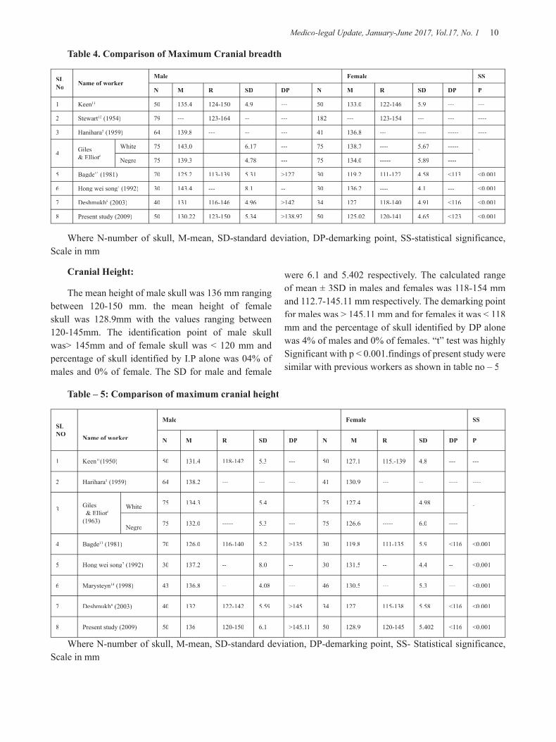
Table 4. Comparison of Maximum Cranial breadth
SL No Name of worker
Male Female SS
N M R SD DP N M R SD DP P
1 Keen11 50 135.4 124-150 4.9 --- 50 133.0 122-146 5.9 --- ---
2 Stewart12 (1954) 79 --- 123-164 -- --- 182 --- 123-154 --- --- ----
3 Hanihara5 (1959) 64 139.8 --- -- --- 41 136.8 --- ---- ----- ----
4 Giles& Elliot6
White 75 143.0 6.17 --- 75 138.7 ---- 5.67 ----- ----Negro 75 139.3 4.78 --- 75 134.0 ----- 5.89 ----
5 Bagde13 (1981) 70 125.2 113-139 5.31 >127 30 119.2 111-127 4.58 <113 <0.001
6 Hong wei song7 (1992) 30 143.4 --- 8.1 -- 30 136.2 ---- 4.1 --- <0.001
7 Deshmukh8 (2003) 40 131 116-146 4.96 >142 34 127 118-140 4.91 <116 <0.001
8 Present study (2009) 50 130.22 123-150 5.34 >138.97 50 125.02 120-141 4.65 <123 <0.001
Where N-number of skull, M-mean, SD-standard deviation, DP-demarking point, SS-statistical significance, Scale in mm
Cranial Height:
The mean height of male skull was 136 mm ranging between 120-150 mm. the mean height of female skull was 128.9mm with the values ranging between 120-145mm. The identification point of male skull was> 145mm and of female skull was < 120 mm and percentage of skull identified by I.P alone was 04% of males and 0% of female. The SD for male and female
were 6.1 and 5.402 respectively. The calculated range of mean ± 3SD in males and females was 118-154 mm and 112.7-145.11 mm respectively. The demarking point for males was > 145.11 mm and for females it was < 118 mm and the percentage of skull identified by DP alone was 4% of males and 0% of females. “t” test was highly Significant with p < 0.001.findings of present study were similar with previous workers as shown in table no – 5
Table – 5: Comparison of maximum cranial height
SL NO Name of worker
Male Female SS
N M R SD DP N M R SD DP P
1 Keen11(1950) 50 131.4 118-142 5.3 --- 50 127.1 115.-139 4.8 --- ---
2 Harihara5 (1959) 64 138.2 --- --- --- 41 130.9 --- -- ---- ----
3 Giles & Elliot6
(1963)
White 75 134.3 ----- 5.4 --- 75 127.4 --- 4.98 ---- -
--- Negro 75 132.0 ----- 5.3 --- 75 126.6 ----- 6.0 ----
4 Bagde13 (1981) 70 126.0 116-140 5.2 >135 30 119.8 111-135 5.9 <116 <0.001
5 Hong wei song7 (1992) 30 137.2 -- 8.0 -- 30 131.5 -- 4.4 -- <0.001
6 Marysteyn14 (1998) 43 136.8 -- 4.08 --- 46 130.5 --- 5.3 --- <0.001
7 Deshmukh8 (2003) 40 132 122-142 5.59 >145 34 127 115-138 5.58 <116 <0.001
8 Present study (2009) 50 136 120-150 6.1 >145.11 50 128.9 120-145 5.402 <116 <0.001
Where N-number of skull, M-mean, SD-standard deviation, DP-demarking point, SS- Statistical significance, Scale in mm
-legal Update, January-June 2017, Vol.17, No. 1 10 11 Medico-legal Update, January-June 2017, Vol.17, No. 1
CONCLUSION
Among all the 4 independent variables statistically analysed, considering the Demarking Point, the most reliable parameter is weight of the cranium, 14% of male skull can be sorted out by this parameter alone. Next is maximum cranial breadth (8% of male skulls identified), followed by maximum cranial length (6% of female skull identified) and maximum cranial height (4% of male skull identified). Considering the Identification point, the most reliable parameter is maximum cranial length 36% of female skulls and 02% of male skulls can be sorted out by this single parameter alone. Next is maximum cranial breadth (32% female skull and 4% male skull identified), weight of cranium (20% male skull and 4% female skull identified) and maximum cranial height (4% of male skull identified).
This study concludes that single parameter is of not much help in identification of sex of skull. So it is better to consider maximum number of parameters for identification of sex of skull.
Ethical Clearance: The ethical clearance was taken prior to the study from the ethical committee of M.R medical college, Gulbarga.
Source of Funding: Self
Conflict of Interest: Nil
REFERENCES
1. Parsons FG. Sexual differences in the skull. J Anat. 1920; 54: 58-65, cited by Krogman WM (1962), vide Supra.
2. Krogman WM. The human skeleton in legal medicine, medical aspects. In symposium on Medical problems – series 11, Levingson, SA editor.
3. Knight Bernard – Knight’s Forensic Pathology, 3rd
edition, Arnold Publication, Page No.108 (2004).
4. Jit I and Singh S. The sexing of adult clavicles. Ind J Med Res. 1966; 54: 556-57.
5. Hanihara K. Sex diagnosis of Japanese skulls and scapulae by means of discriminant functions. J Anth Soc Nippan, 1959; 67(722): 21-27. Cited by Krogman Wm (1962) vide Infra.
6. Giles E. and Elliot G. Sex determination by discriminant function analysis of crania. Am J Phys anthrop. 1963; 21: 53-66.
7. Hong Wei Song, Zi, Qing Lin and Jing Tao Jia. Sex diagnosis of Chinese skulls using multiple stepwise discriminant functional analysis. Forensic Science International, 1992; 54: 135-140.
8. Deshmukh AG & Deverthi DB comparison of Cranial Sex determination by univariate and multivariate analysis, Joint Anatomy Society of India 55 (2), Page No.48-51 ,(2006).
9. Pillay V.V – Text book of Forensic Medicine & Toxicology, 14th edition,Paras Publication, Page No.56 (2004).
10. Krogman WM. The human skeleton in forensic medicine. Charles C, Thomas, Springfield,Illinois, 1962; USA.
11. Keen JA(1950) “Sex differences in skull”, APJA, 8(1), Page No.65-79.
12. Stewart T.D (1954) Evaluation of Evidence from the Skeleton in Legal Medicine, Gredwohl R.E.H Editor musby St. Louis Page No.407-450 Quoted in Gray’s anatomy 36th edition Page No.1520.
13. Bagade K.G. (1981) Determination of Sex from Axial Skeleton. Dissertation for M.S. Anatomy Examination, Marathwada University Aurangabad.
14. Maayna Steyn, M. Yasar Iscan. Sexual dimorphism in the crania and mandibles of South African Whites. Forensic Sci Int. 1998; 98: 9-28.

13 -legal Update, January-June 2017, Vol.17, No. 1
One should be Ectopic Minded – A Case Report
H S Tatiya1, V T Jadhao2, A A Taware3, S B Punpale4
1Assistant Professor, Department of Forensic Medicine and Toxicology, B.J. Government Medical College, 1Assistant Professor, Department of Forensic Medicine and Toxicology, B.J. Government Medical College, 1
Pune, 2Associate Professor, Department of Forensic Medicine and Toxicology, SBH Government Medical 2Associate Professor, Department of Forensic Medicine and Toxicology, SBH Government Medical 2
College, Dhule, 3Associate Professor, 3Associate Professor, 3 4Professor and Head, Department of Forensic Medicine and 4Professor and Head, Department of Forensic Medicine and 4
Toxicology, B.J. Government Medical College, Pune
ABSTRACT
Rupture of tubal ectopic pregnancy is the most common life threatening emergency in early pregnancy. The multitude of presentations to clinician on first contact can be misleading in the absence of high suspicion. The current case report is to explain the importance of keeping ruptured ectopic pregnancy as a differential diagnosis in women of child bearing age and importance of obtaining history in such cases.
Keywords- Ruptured ectopic, unusual presentation, amenorrhea, pain in abdomen.
INTRODUCTION
Ectopic pregnancy is defined as a pregnancy that develops after implantation of the blastocyst anywhere other than the endometrial lining of uterine cavity1.One out of every 100 pregnancies is ectopic with the most common site being within fallopian tube2. Ectopic pregnancy can have varied outcome. Mostly it remains dormant; however it can also miscarry, rupture or extend into broad ligament. There are no reliable clinical, sonographic or biological markers that can predict rupture of tubal pregnancy3. It is the leading cause of pregnancy related death in the first trimester, accounting for 90% of all pregnancy related deaths4. Among every ten women admitted with ectopic pregnancy in developing world there is one maternal death5. Only 50% of patients present with the classic triad of rupture of ectopic pregnancy i.e. pain in abdomen, amenorrhea and vaginal bleeding6. However clinicians should consider the diagnosis of ectopic pregnancy in any woman of child bearing age with history of secondary amenorrhea and who has abdominal or pelvic pain, vaginal bleeding, or both7. The current case is reported to explain the problem in diagnosis of ruptured ectopic pregnancy and a simple measure to avoid such a catastrophe in future.
Corresponding author: Dr. H. S. Tatiya, MDAssistant Professor, Department of Forensic Medicine and Toxicology, B.J. Government Medical College, Pune. Pin-411001, Email: [email protected]
CASE
A dead body of 23 year old woman, married since one year, housewife belonging to a middle class family was brought for postmortem examination. On revealing history from her husband and other relative and on perusal of referral sheet from concerned clinician, following chain of history was noted –
• Five days prior the deceased complained of mild pain in abdomen.
• Menstrual history was mentioned as regular, 4 / 30.
• Last menstrual period was noted as 5 days prior to complaint of pain in abdomen.
• Vitals were normal.
• Pain killers and bed rest were advised by clinician.
• Till next three days even after continuous pain in abdomen, decreased appetite the same treatment was continued.
• On fourth day there was intolerable increase in abdominal pain. The condition of patient worsened.
• The patient was referred to tertiary care centre where she was declared brought dead and the body was sent for medicolegal post-mortem examination.
DOI Number: 10.5958/0974-1283.2017.00003.2

13 Medico-legal Update, January-June 2017, Vol.17, No. 1
Postmortem examination was conducted on the same day. Following positive observations were noted –
On external examination –
• Rigor mortis was well marked in whole body.
• Faint post-mortem staining was present over back and was fixed.
• Generalised features of pallor were present over body.
• No any external injury noted.
On internal examination –
• 1.5 litres of blood and blood clots were present within abdominal and pelvic cavity.
• All visceral organs were pale.
• A rent pregnancy was noted at isthmus of left fallopian tube of size 1.5 cm x 1 cm x cavity deep, margins irregular, red, with evidence of uterine artery erosion, oozing blood, red. (Figure 1)
• Histopathology of fallopian tube and associated mass confirmed the conception and corpus luteum was present in left ovary.
• Cause of death was given as – Death due to hemorrhagic shock due to ruptured tubal ectopic pregnancy.
DISCUSSION
Ectopic pregnancy occurs when a fertilized egg implants outside of the uterine cavity2. It is a condition of immense gynecological importance, particularly in developing world, because of high mortality and morbidity issues associated with it, and enormous threat to life8. The highest rate of ectopic pregnancy occurs in the age group of 35 -44 years. A three to four fold increase in the risk for developing an ectopic exist in this age group compared to woman aged 15- 24 years. This is said to be due to progressive loss of myoelectrical activity along fallopian tube with aging9. However in current case the deceased was 23 years old.
One out of every 100 pregnancies is ectopic with most common site being within fallopian tube2. Commonest site being the ampullary region of fallopian tube 8. In current case the isthmus of fallopian tube was involved. Tubal rupture is predominantly common in
isthmic and interstitial implantation. As the ishtmic portion is narrow and the wall is less distensible, the wall may be easily eroded by the chorionic villi. Commonly the ovum is partly expelled out through the rent so that the bleeding is heavy and continuous10, as seen in this case. When ruptured, ectopic pregnancy is a true medical emergency. It is a leading cause of maternal mortality in first trimester and accounts for 10% to 15% of all maternal deaths. Doctors should be ectopic minded to avoid delay in prompt intervention11. The classical clinical triad of ectopic pregnancy is pain, amenorrhea and vaginal bleeding; unfortunately only about 50% of patients present with all 3 symptoms. About 40 to 50% of patients present with vaginal bleeding, 50% have palpable adnexal mass and 75% may have abdominal tenderness9. In current case the deceased presented with the complaint of pain in abdomen. Only 20% of patients of ectopic pregnancies are hemodynamically compromised at initial stages, which is highly suggestive of rupture10. In the current case patient’s condition worsened gradually till death.
The first part in the diagnosis of ectopic pregnancy is demonstration of pregnancy by means of a rapidly performed and sensitive qualitative urine test for beta subunit of human chorionic gonadotropin. A negative urine pregnancy test result will generally be used to exclude ectopic pregnancy from further consideration12. However Kalinski et al reported a case of ruptured ectopic pregnancy with negative urine pregnancy test13. Routine ultrasound can miss the diagnosis of an ectopic pregnancy in almost 50% cases14. In India more than 80% of ectopic pregnancies are diagnosed after rupture, but with high resolution transvaginal sonography, serum β – hcg assay, and increased vigilance of the clinicians, more and more cases are being diagnosed before rupture15.
According to a study by Lewis, ectopic pregnancy was one of the most important causes of death in early pregnancy. A significant number of these early pregnancy deaths were in women who were discharged from primary health care setting (either general practice or emergency department) having never had a pregnancy test or misdiagnosed as gastroenteritis16. A similar case was reported where 25 year old woman succumbed to death after careless diagnosis of gastroenteritis and wrong prescription of drugs17. In an another case reported by Basak Bayram et al, 43 year patient with bilateral tubal ligation was diagnosed as ruptured ectopic

Medico-legal Update, January-June 2017, Vol.17, No. 1 14 15 -legal Update, January-June 2017, Vol.17, No. 1
pregnancy18. Ambade et al reported a case of ruptured ectopic following tubal sterilization procedure12. All these cases elaborate the varied presentation of ruptured ectopic pregnancies in unexpected circumstances.
What went wrong in current case?
In the current case the clinician who obtained the history advised neither the urine pregnancy test nor the ultrasonography. The clinician did not think of ruptured ectopic pregnancy. The reason being
1. No clear history of amenorrhoea,
2. The last menstrual period was just 5 days prior to complaint of abdominal pain.
3. Female being primiparous.
4. Hemodynamically stable at initial presentation.
As discussed earlier, per vaginal discharge of blood is one of the signs of ruptured ectopic pregnancy, hence we can say that in current case the patient and clinician misinterpreted it as last menstrual period. Hence none of the tests were advised by the clinician. From the patient’s perspective, the deceased and her relatives brought her again for re-examination on fourth day when her condition worsened.
What could have been done?
Looking into the details of this case, we think that in females of child bearing age, even though she is young and primiparous, the clinicians should suspect possibility of ectopic pregnancy even in absence of clear history of amenorrhoea. According to Dutta a short period of amenorrhoea of 6-8 wks or delayed period or spotting on the expected date of period is usually present. Amenorrhoea may be absent in good number of cases10. As Indian women are quite shy and ignorant in discussing the menstrual periods, the clinicians should be smart enough to elicit menstrual history properly. Considering the current case we suggest that the clinicians should ask for last two episode of last menstrual period, and should note the gap between them (Diagram 1). Early or delayed vaginal discharge of blood than the stipulated time of menstruation in females with regular menses should be considered as suspicious. In all such cases a simple and cheaper urine pregnancy test can avoid many catastrophic situations. Secondly from patient’s point of view, it is suggested that she and her family members
should not wait till a point of intolerable pain. A second opinion in such cases can be of great help.
Diagram 1: Diagrammatic representation of current case
scenario.
Image 1 – Uterus with adnexal structures. Rent present at isthmic portion of left fallopian tube.
CONCLUSION
A smart elicitation of history and simple urine pregnancy test can save many mothers form dying, if the clinician is ectopic minded.
Conflict of Interest: Nil
Source of Funding: Self
Ethical Clearance: Not required.
REFERENCES
1. Mishell DR. Ectopic pregnancy etiology, pathology, diagnosis, management, fertility prognosis. In: Stenchever MA, Droegmuller W, Herbst AL, editors. Comprehensive gynaecology. 4th ed. St. Louis (MO): Mosby;2001.
-legal Update, January-June 2017, Vol.17, No. 1 14 15 Medico-legal Update, January-June 2017, Vol.17, No. 1
2. Walker JJ. Ectopic Pregnancy. Clinical obstetrics and gynaecology 2007;50(1):89-99.
3. Latchaw G, Takacs P, Gaitan L, Geren S, Burzawa J. Risk factors associated with the rupture of tubal ectopic pregnancy. Gynaecology Obstet Invest. 2005;60:177-80.
4. Centre for Disease control and prevention. Ectopic pregnancy survelliance, United States, 1970-1987. MMWR CDC Surveill Summ 1990;39 (SS-4):9-17.
5. Leke RJ, Goyaux N, Matsuda T, Thounneau P F. Ectopic pregnancy in Africa; a population based study. Obstet Gynecol 2004;103:692-697.
6. Rakhi, Mital Prem Lata, Hooja Nupur et al. Ectopic Pregnancy : A devastating Catastrophe. SJAMS.2014;2 (3A):903-7.
7. American College of Obstetricians and Gynaecologist and American Academy of Paediatrics. Guidelines for perinatal care. 5th
ed. Washington DC: American College of Obstetricians and Gynaecologist and American Academy of Paediatrics;2002.
8. Pradhan Bandana, Singh Ranjit, Singh Nabhkishore N. A clinical study on ectopic pregnancy in RIMS from 2013 June to 2015 July. Jour of Dent and Med Sciences 2016;15(Issue 1 version III):08-12.
9. Shalev E, Peleg D, Tasabari A, Romano S, Bustan M. Spontaneous resolution of ectopic pregnancy: Natural history. Fertil Steril. 1995;63:15-19.
10. Textbook of Obstetrics. 6th ed. Datta D C,2004;New Central Book Agency(P) Ltd.:Calcutta,Chapter 15;2004:179-187.
11. Berek and Novak’s gynaecology, 15th ed. Jonathen S. Berek,2012; Wolters Kluwers;Philadelphia,Chapter 38;2012:1428-1444.
12. Ambade VN, Chimne HD, Shanbhag SD, Ropmay AD. Ectopic tubular pregnancy in pos tubectomy death. Rev Esp Med Legal.2013;37(2):67-71.
13. Kalinski MA, Guss DA. Hemorrhagic shock from a ruptured ectopic pregnancy in a patient with a negative urine test result. Ann Emerg Med.2002;40:102-5.
14. Agrwal SS, Chavali KH, Lavlesh K, Satish K, Iliyas S. Secondary abdominal ectopic pregnancy: diagnosis at autopsy. J Ind Acad Forensic Med.2006;28:5-7.
15. Duggal BS, Tarneja P, Sharma RK, Rath Sk, Wadhawa RD, Laparoscopic management of ectopic pregnancies.MJAFI.2004;60:220-3.
16. Kaplan BC, Dart RG, Moskos M et al. Ectopic pregnancy: prospective study with improved diagnostic accuracy. Ann Emerg Med, Jul 1996;28(1):10-7.
17. Kumar A, Chavali KH, Singh A et al. Death due to ruptured ectopic pregnancy: Natural death or Negligene. JIAFM. 2006;32(3):264-6.
18. Bayram B, Dedeoglu E, Karatag G. Ruptured ectopic pregnancy in a patient with bilateral tube ligation. Tr J Emerg Med 2012;12(3):134-6.
17 -legal Update, January-June 2017, Vol.17, No. 1
A Study on Grading and Severity of Aortic Athermatous Plaques- Autopsy based Study
M Babu1, B Venkata Sai Prasad2, I Mohan Prasad3, B Venkateswarlu4
1Associate Professor, Dept of Forensic Medicine, 1Associate Professor, Dept of Forensic Medicine, 1 2Associate Professor, Dept of Pathology, S.V. Medical 2Associate Professor, Dept of Pathology, S.V. Medical 2
College, Tirupati, Andhra Pradesh, 3Associate Professor, Dept of Forensic Medicine, Narayana Medical 3Associate Professor, Dept of Forensic Medicine, Narayana Medical 3
College, Nellore, Andhra Pradesh, 4Professor & HOD, Dept of Forensic Medicine, S.V. Medical College, 4Professor & HOD, Dept of Forensic Medicine, S.V. Medical College, 4
Tirupati, Andhra Pradesh
ABSTRACT
Atherosclerosis is a complex (disease of the large and medium sized arteries) process that involves lipoprotein influx, lipoprotein modification, increased prooxidant stress and inflammatory, angiogenic and fibroproliferative responses intermingled with extra cellular matrix and lipid accumulation resulting in the formation of an atherosclerotic plaque. Endothelial dysfunction is common in atherosclerosis and often manifests as a reduced vasodilator or enhanced vasoconstrictor phenotype which contributes to luminal compromise. Thrombosis resulting from plaque rupture or superficial erosion complicates atherosclerosis often resulting in abrupt luminal occlusion with resultant acute ischemic syndromes. Infectious agents may contribute to the inflammatory response and thus to destabilization of the lesions. An improved understanding of the patho physiology of atherosclerosis providing novel direction for its prevention and treatment. (7)
Acceleration of Atherosclerotic process leading to macrovascular disease. The Study is prospective and one Hundred Aortas are examined from the different age groups of males and females, during the medico legal Autopsies and the results are statistically analysed.
Keywords: - Atheromatous plaques, Endothelial dysfunction, Thrombosis.
Corresponding author: Dr. M BabuAssociate Professor, Dept of Forensic Medicine, S.V. Medical College, Tirupati, Andhra Pradesh. Email : [email protected], Mobile : +91 9440556788
INTRODUCTION
Atherosclerosis is a multifocal, smoldering, immunoinflamatory disease of medium sized and large arteries fuelled by lipids, endothelial cells, leucocytes and intimal muscle cells are the major players in the development of this disease.(10) Vascular endothelium is the cell layer and has anti atherogenic property and inhibits platelet aggregation, adhesion of smooth muscle cell proliferation and adhesion of leucocytes acts as barrier between flowing of blood and vascular wall and regulates vascular growth, platelet function and synthesizes and releases vasoactive substances e.g. -Nitric oxide. Nitric Oxide has low molecular weight, diffuses rapidly and able to induce instant changes in
arterial diameter to the prevailing blood flow and shear stress and inhibits platelet aggregation and smooth muscle proliferation. Endothelium also produces vasoconstrictor substances such as Endothelin-1,Thromboxane A2(TXA2) and prostaglandin I 2 (PGI2), ) maintains balance between vasodilatation and vasoconstriction to preserve sufficient vascular diameter for the satisfactory perfusion of the vascular system and causes physiological vasodilatation and relaxation of smooth muscle cells and increases the intracellular concentration of cyclic guanosine monophosphate(CGMP) which decreases the intracellular Ca 2 + concentration causes vaso relaxation.(17) Physical injury to the blood vessel as the result of risk factors leads to endothelial injury with increased infiltration of atherogenic lipo proteins at the site of low or oscillating shear stress. Sub endothelial retention of atherogenic lipo proteins (LDL/VLDL). LDL oxidation, glycation, aggregation, that leads to endothelial activation with increased mononuclear leukocytes (Inflammatory cells) adhesion, chemotaxis and sub endothelial recruitment. Further sub endothelial
DOI Number: 10.5958/0974-1283.2017.00004.4

17 Medico-legal Update, January-June 2017, Vol.17, No. 1
inflammatory cell activation with lipid ingestion through monocytes scavenger receptor expression resulting in foam cell formation. Inflammatory cell proliferation leads to migration to intima and proliferation of medial/ advential smooth muscle cells / myofibroblasts in response to growth factors released by activated monocytes with matrix production and formation of plaque and fibrous positive (outward) arterial adventitial stages. Later plaque growth or negative remodelling results in luminal narrowing. Neoangio genesis due to angiogenic stimuli produced by macrophages and other arterial wall cells (VEGF,IL-8) plaque haemorrhage and expansion of lipid core. Death of foam cells by necrosis / apoptosis leading to necrotic lipid-core formation. Finaly rupture of fibrous cap or endothelial erosion, exposure to thrombogenic substance and leads to arterial thrombosis.
High density Lipo proteins are the smallest and densest of lipoproteins which are negatively associated with cardiovascular diseases. HDL Cholesterol which account for 20-30% of total cholesterol consist of 3 sub components HDL1,HDL2 and HDL3 of these HDL1 is closely related to Atherosclerosis.
Endothelial dysfunction results in reduction of relaxing factors and increase in constricting factors are the main features in development of vascular disease. The combination factors like Hyperglycemia, impaired insulin secretion and proatherogenic dyslipidemia, hypertension , obesity, vaso active hormones , cytokines, growth factors-angio tension II ( AT –II ) Tumour necrosis factor (TNF) and vascular endothelial growth factor (VEGF) play important role in causing Atherogenesis. These metabolic and hormonal factors induce endothelial dysfunction, vascular inflammation, smooth cell proliferation, intimal accumulation of lipids, fibrosis and hypercoagulability state in causing atherosclerosis and thrombosis. Elevated Fibrionogen level contribute to a procogagulative state and stimulate athero thrombosis. The endothelial cells produce the von-willebrand factor (vwf) which complexes with factor VIII to initiate thrombosis as well as platelet aggregation.(17) In conditions such as hypertension, diabetes, dyslipidemia and smoking cause the physiologic and structural changes in the vessel that leads to vascular disease. In order of frequency the commonest sites for Atherosclerosis are Abdominal Aorta, Coronary arteries, popliteal arteries and the cerebral circulus arteriosus.(18) Five important risk factors influences the Atherosclerosis namely
Hyperlipidemia, Hypertension, Smoking, Diabetes mellitus and Hyperhomocysteinemia(18) and subordinate factors are overweight and a sedentary or stress full lifestyle(18). Triglycerides, cholesterol and lipo proteins are implicated in the pathogenis of atherosclerosis. Reduced concentration of high density lipo protein (HDL) and increased Triglycerides have been shown to be responsible for the genesis of Atherosclerotic lesions.(10)
Histopathology of atherosclerotic lesions - New concept (21)
1. Stary I Lesion: The endothelium surface adhesion molecules E selectin and P selectin, attracting more polymorphonuclear cells and monocytes in the sub endothelial space.
2. Stary II Lesion: Macrophages begin to make up large amounts of LDL (fatty streak).
3. Stary III Lesion: As the process continues, macrophages eventually become foam cells.
4. Stary IV Lesion: Lipid exudes into the extra cellular space and begins to coalesce to form the lipid core.
5. Stary V Lesion: SMCs and fibroblasts migrate in, forming fibro atheromas with soft inner lipid cores and outer fibrous caps.
6. Stary VI Lesion: Rupture of the fibrous cap with resultant thrombosis causes ACS.
7. Stary VII and VIII Lesions: As lesions stabilize may become fibro calcific (Stary VII lesion) and ultimately, fibrotic with extensive collagen content (Stary VIII lesion).
MATERIAL & METHOD
To study the Atherosclerosis in autopsy cases, Specimen 100 Aortas were collected from the post mortem cases from the level of just above the aortic valves to the Bifurcation of right and left Iliac arteries, Descending Thoracic aorta: The aorta extending from a horizontal line drawn through the first two intercostals arteries to a horizontal line drawn through the upper edge of the coeliac artery. Abdominal aorta: it is the area extending from a horizontal line drawn through the upper edge of the orifice of the coeliac artery to a horizontal line drawn through the inner surface of the bifurcation
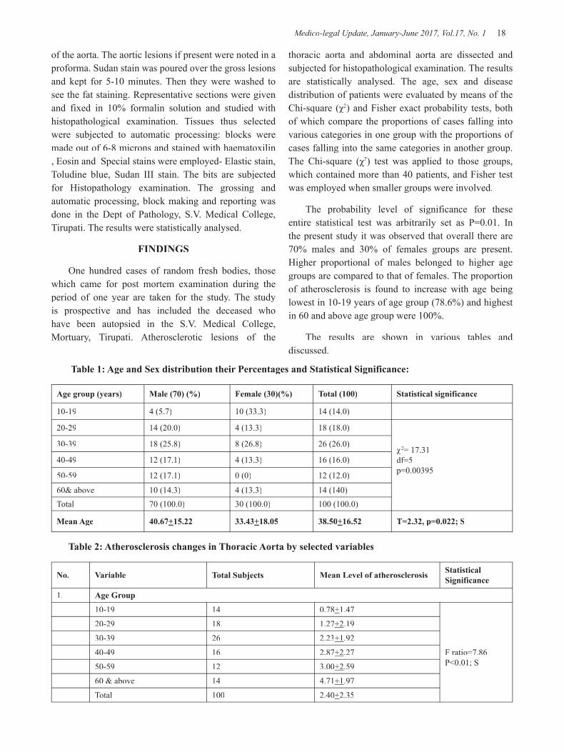
Medico-legal Update, January-June 2017, Vol.17, No. 1 18 19 -legal Update, January-June 2017, Vol.17, No. 1
of the aorta. The aortic lesions if present were noted in a proforma. Sudan stain was poured over the gross lesions and kept for 5-10 minutes. Then they were washed to see the fat staining. Representative sections were given and fixed in 10% formalin solution and studied with histopathological examination. Tissues thus selected were subjected to automatic processing: blocks were made out of 6-8 microns and stained with haematoxilin , Eosin and Special stains were employed- Elastic stain, Toludine blue, Sudan III stain. The bits are subjected for Histopathology examination. The grossing and automatic processing, block making and reporting was done in the Dept of Pathology, S.V. Medical College, Tirupati. The results were statistically analysed.
FINDINGS
One hundred cases of random fresh bodies, those which came for post mortem examination during the period of one year are taken for the study. The study is prospective and has included the deceased who have been autopsied in the S.V. Medical College, Mortuary, Tirupati. Atherosclerotic lesions of the
thoracic aorta and abdominal aorta are dissected and subjected for histopathological examination. The results are statistically analysed. The age, sex and disease distribution of patients were evaluated by means of the Chi-square (χ2Chi-square (χ2Chi-square (χ ) and Fisher exact probability tests, both of which compare the proportions of cases falling into various categories in one group with the proportions of cases falling into the same categories in another group. The Chi-square (χ2The Chi-square (χ2The Chi-square (χ ) test was applied to those groups, which contained more than 40 patients, and Fisher test was employed when smaller groups were involved.
The probability level of significance for these entire statistical test was arbitrarily set as P=0.01. In the present study it was observed that overall there are 70% males and 30% of females groups are present. Higher proportional of males belonged to higher age groups are compared to that of females. The proportion of atherosclerosis is found to increase with age being lowest in 10-19 years of age group (78.6%) and highest in 60 and above age group were 100%.
The results are shown in various tables and discussed.
Table 1: Age and Sex distribution their Percentages and Statistical Significance:
Age group (years) Male (70) (%) Female (30)(%) Total (100) Statistical significance
10-19 4 (5.7) 10 (33.3) 14 (14.0)
20-29 14 (20.0) 4 (13.3) 18 (18.0)
χ2= 17.31df=5p=0.00395
30-39 18 (25.8) 8 (26.8) 26 (26.0)
40-49 12 (17.1) 4 (13.3) 16 (16.0)
50-59 12 (17.1) 0 (0) 12 (12.0)
60& above 10 (14.3) 4 (13.3) 14 (140)Total 70 (100.0) 30 (100.0) 100 (100.0)
Mean Age 40.67+15.22 33.43+18.05 38.50+16.52 T=2.32, p=0.022; S
Table 2: Atherosclerosis changes in Thoracic Aorta by selected variables
No. Variable Total Subjects Mean Level of atherosclerosis Statistical Significance
1. Age Group
10-19 14 0.78+1.47
F ratio=7.86P<0.01; S
20-29 18 1.27+2.19
30-39 26 2.23+1.92
40-49 16 2.87+2.27
50-59 12 3.00+2.59
60 & above 14 4.71+1.97
Total 100 2.40+2.35

-legal Update, January-June 2017, Vol.17, No. 1 18 19 Medico-legal Update, January-June 2017, Vol.17, No. 1
2. Sex
Male 70 2.74+2.32 t=2.26
Female 30 1.60+2.26 P=0.02,S
3. Diet
Vegetarian 23 1.47+2.08 t=2.18
Non-Vegetarian 77 2.67+2.37 P=0.03,S
4. Smoking
Yes 24 4.01+1.66 t=7.52
No 76 1.60+1.94
5. Alcoholism
Yes 21 5.00+1.54 t=6.95
No 79 1.70+2.02 P<0.001,S
Table 3: Atherosclerosis changes in Abdominal Aorta by selected variables
S.No. Variable Total Subjects Mean Level of atherosclerosis Statistical Significance
1. Age Group
10-19 14 1.35+1.44
F ratio=8.26P<0.001; S
20-29 18 2.66+2.2730-39 26 2.23+1.9240-49 16 3.37+1.7450-59 12 3.08+2.6760 & above 14 5.42+2.24Total 100 2.97+2.35
2. Sex
Male 70 3.40+2.28 t=2.91Female 30 1.96+2.23 P=0.004,S
3. Diet
Vegetarian 23 2.04+2.24 t=2.19Non-Vegetarian 77 3.24+2.32 P=0.03,S
4. Smoking
Yes 24 5.16+1.37 t=6.13No 76 2.27+1.37 P<0.001, S
5. AlcoholismYes 21 5.09+1.41 t=5.24No 79 2.40+2.23 P<0.001,S
Cont... Table 2: Atherosclerosis changes in Thoracic Aorta by selected variables
Fig 1: Calcified Atherosclerotic plaques - AortaFig 1: Calcified Atherosclerotic plaques - Aorta
Fig 2: Atherosclerotic Plaque with calcification Fig 2: Atherosclerotic Plaque with calcification (H&E Stain 10X)

Medico-legal Update, January-June 2017, Vol.17, No. 1 20 21 -legal Update, January-June 2017, Vol.17, No. 1
Fig 3: Atherosclerosis with multiple focal calcifications (H&E Fig 3: Atherosclerosis with multiple focal calcifications (H&E Stain 10X)
CONCLUSION
In the present study total one hundred samples were collected among 70 males and 30 females. In addition to age factor, diet and behavioural characters, diseases like hypertension, diabetes are taken into consideration to know the grades of Atherosclerosis. Behavioural characters like smoking and alcoholism which can contribute to Atherogenesis were also included in the present study.
In the present study Dietary History gave the significant results, non-vegetarians study group were having more advanced lesions than the vegetarian group. Proportion of atherosclerosis is found to increase with age being lowest in 10-19 years of age group (78.6%) and highest in 60 and above age group were 100%.There is a significant raise of Atherosclerosis in Non vegetarians and smokers and alcoholics. In the present study positive lesions were seen at the age 14 in the females and at the age 18 in the males. As age advanced there was gradual raise of Atherosclerotic lesions. 60 and above age group had advanced complicated lesions in the present study and correlates with Dr.Sahoo’s. study (10) in 60 and above age group the lesions were almost equal in both sex. As the age advanced the severity of Atherosclerosis increased from Grade II to Grade VII and VIII correlating with the study of Autopsy studies in Atherosclerosis. (13). Dr. James and C.Robert suggested that Atherosclerosis increases with age and involves primarily in the larger arteries (13) and study correlates. Atheromas and complicated lesions are present in the middle and older age groups. Significant lesions are seen in those had history of Diabetes mellitus and Hypertension. Both sex were affected by Atherosclerosis from 2nd decade nd decade nd
onwards. Age group of 60 and above groups had 100% lesions in both sex and males and females were equally affected. In thoracic aorta 100% lesions were seen in both sexes, correlating with Brit, Heart study (7). The study has
given the positive results that as the age advances. In the study group Grade II lesions were gradually progressed to Grade V, VI, VII and VIII lesions. As age advances, the severity of Atherosclerosis is also increased and this may lead to sudden death if the total artery is involved when the lumen is blocked.
All the reports are statistically calculated and reported in the tables and reports are as follows.
Table 1: Higher proportion of males belonged to higher age groups compared to that in females. The mean age of male subjects is comparatively higher than females. The age differences by sex are also statistically significant.
As the age advances the complicated lesions were seen in the arteries 60 and above age groups in both sexes correlates with the study by Dr. M. K. S.Reddy (14).
Table 2: The mean level of atherosclerotic changes has shown a significantly rising trend with rise in the age of the study participants, being highest in 60 & above group (4.71) and least in 10-19 years group (0.78) and the differences are also statistically significant. Higher level of atherosclerosis is found in males (2.74), Non-vegetarian diet (2.67), smokers (4.91) and alcoholics (5.00) and the differences were also statistical significant.
Table 3: The degree of atherosclerosis has shown a rising trend with age being highest in 60 & above group (5.42) and least in 10.19 age group (1.35) and the differences are also statistically significant. Significantly higher trend of atherosclerosis is found in males (3.40), Non vegetarian diet (3.24), smokers (5.16) and Alcoholics (5.09).
Before the conclusion there is still a need for autopsy studies in the Investigation of risk factors and atherosclerotic lesions. The study comprises only one hundred cases, it reveals that much of the prevalence rate of atherosclerosis in this region. It may be taken as a sample study for further research work. Recommendations are fish liver oil containing omega fatty acids and the eicosanoids helps in lower the atherogenic lipids, higher in complex carbohydrates, lower fat adequate proteins. Complex carbohydrates obtained from cereals, pulses, which are rich in fibre contents and reduces risk of atherosclerosis and hyperlipemia. Lifestyle change modification, adequate
-legal Update, January-June 2017, Vol.17, No. 1 20 21 Medico-legal Update, January-June 2017, Vol.17, No. 1
exercise, eating a diet rich in fruits and vegetables and low in saturated fat, weight loss if obese. Diabetes, high blood pressure, abnormal cholesterol, obesity, elevated homocysteine and elevated risk of blood clots should be under control and periodical health checkups are recommended.
Conflict of Interest: Nil
Source of Funding: Self funding with Govt Assistance
Ethical Committee Clearance: Taken from S.V.Medical College Ethical Committee, Tirupati, Andhra Pradesh.
REFERENCES
1) Albrect Von Heller (1755)- Quoten from Harrison’s principles of internal medicine VI edition, Chap.275, P.1241.
2) Aldersberg, D. and Schaefer, L.E Am. J. Med., 1959, 26, 1 (Interplay of heredity and environment in Atherosclerosis).
3) Atherosclerosis Forum- Internet Edition, File://G:\New Folder\Ahterosclerosis.
4) Aschoff, A.Uber Entwicklung- Wach stums and Attersvorgange an den Getassen von Muskuleram and Klastic\schem Typ. Jena. Guston. Fischer, 1909.
5) Auerbach, O., Hammond, E.C. Garfinkel, L: Smoking in relation to Atherosclerosis of the coronary arteries:L New Eng.J. Med. 273: 775-779, 1965.
6) Avtandilov, G.G. Dynamics of Atherosclerotic process in man. Moscow, Medicine, 1970.
7) Brit, Heart, Incidence of atherosclerotic lesions at Madras, South India, 1967-29, 33-Internet.
8) C.Rosendorff, Essential cardiology, Princilples and practice,2 nd edition, 2005,409.
9) Davidson, Principles and practice of Medicine- 17th Edition, 1995, 245-247.
10) Dr. P.C. Sahoo, Age factor in Casuation of Atherosclerosis in the population of Southern Orissa. Journal of Indian Academy of Forensic Medicine, Volume XXII, No. 1 to 2, 2000, 6-8.
11) Erling Falk,Pathogenisis of Atheroscerosis,Journal of American College of Cardiology, Vol.47 No 8 suppl C.
12) J.B.Mukherjee’ Forensic Medicine and Toxicology, edited by R.N.Karmakar, 4th Edition 2011,227.
13) James C. Roberts, Robert H Willking and Campbell Moses- Learn and Live, Autopsy studies in Atherosclerosis II –Distribution and severity of Atherosclerosis catastrope- Circulation- Journal of American Heart Association, 1959: 20, 520-526.
14) Kumara Swamy Reddy.M , A study on incidence of Atherosclerosis in Tirupati Area, 1979.
15) Kasper,Braunwald Fauci,Harrison’s Principles of Internal Medicine,16th edition,1425.
16) Padmavathi S.et. al, Incidence of coronary artery disease in Delhi from Medico-legal autopsies. Ind. Jour. Me. Res. 57, 3, PP 465, March 1969.
17) Seshaiah, Hand book on Diabetes mellitus, IV Edition, 2015,55,57, 234,246,248,452.
18) Stefan silbernag/lang, color Atlas of Pathophysiology, 2000,236.
19) Strong, J.P. et al: On the association of cigarette smoking with coronary aortic atherosclerosis: J.Atheroscler, Res.10: 303, 1969.
20) Subramaniyam.R, Kulangara and Abraham, Incidents of Atherosclerotic lesions at Madras, South India. Brit, Heatz, 1967-29, 33. Internet Edition.
21) Vibhuti N.Singh, Coronary Artery Atherosclerosis, e-medicine from webmed, August 17th, 2005, 1to 48 //G:\newfolder\emedicine.
22) Vihert A.M.: Bull. World Health Organ, Atherosclerosis of the aorta and coronary arteries in coronary heart disease 1976, Vol.53, 585-596.
23 -legal Update, January-June 2017, Vol.17, No. 1
Fatal Compression of Neck by Ligature: Study of Profile of Cases and External Findings over the Body
Sumit Tellewar1, Abhishek Yadav2, Mohit Gupta3, Lohith Kumar R4, Sarala M5
1Assistant Professor, Department of Forensic Medicine and Toxicology, Army College of Medical Sciences, 1Assistant Professor, Department of Forensic Medicine and Toxicology, Army College of Medical Sciences, 1
Delhi Cantonment, Near Base Hospital, New Delhi, India, 2Assistant Professor, Department of Forensic 2Assistant Professor, Department of Forensic 2
Medicine and Toxicology, All India Institute of Medical Sciences (AIIMS), Ansari Nagar, New Delhi, India, 3Associate Professor, Department of Forensic Medicine, VMMC & Safdarjung Hospital, New Delhi, 3Associate Professor, Department of Forensic Medicine, VMMC & Safdarjung Hospital, New Delhi, 3
4Assistant Professor, Department of Forensic Medicine, Shivamogga Institute of Medical Sciences, District 4Assistant Professor, Department of Forensic Medicine, Shivamogga Institute of Medical Sciences, District 4
Hospital, Shivamogga, Karnataka, 5Assistant Professor, Department of Forensic Medicine and Toxicology, 5Assistant Professor, Department of Forensic Medicine and Toxicology, 5
Army College of Medical Sciences, Delhi Cantonment, Near Base Hospital, New Delhi, India
ABSTRACT
Determining the manner of death in equivocal cases of fatal compression of neck by ligature had been a challenge for the autopsy surgeons. Keeping the same in view, the profile of fatal compression of neck by ligature along with the appearance of ligature mark and other external findings was studied so as to note down any specific pattern which could help to differentiate between a case of hanging and ligature strangulation. The study was conducted in a prospective manner in UCMS and GTB Hospital, New Delhi. The hanging cases were all suicidal in nature while no suicide case was reported in strangulation cases. The frequency of hanging was found to be more in the younger age groups of 21-30 years followed by 11-20 years. The pattern of ligature mark was found to be a reliable factor to differentiate between the cases of hanging and strangulation.
Keywords: Hanging, Strangulation, Ligature Mark, Sub-conjuctival Hemorrhage, Facial Congestion.
INTRODUCTION
Asphyxia can occur in various situations like Traumatic asphyxia, Hanging, Choking, Smothering, Strangulation, throttling, etc1-5. Many research studies have been done in the past for better understanding of asphyxial deaths6-8. Each type of violent asphyxia has peculiar diagnostic features. Similarly in cases of neck compression by ligature, the external and internal neck findings help a lot on arriving at a proper cause and manner of death1-5. Still in some cases a forensic expert may face challenge to differentiate between hanging & strangulation. This problem can be solved by defining the minimally adequate well defined diagnostic criteria for compressive neck injury. Keeping the fact in view,
Corresponding athor:Dr. Abhishek Yadav Assistant Professor, Dept. of Forensic MedicineAIIMS, New Delhi-110029E-Mail:[email protected]: 09818052523, 09868020684
profile of fatal compression of neck by ligature was studied. The appearance of ligature mark and other external findings were included in the study so as to note down any specific pattern which could help to differentiate between a case of hanging and ligature.
MATERIAL AND METHOD
The cases of fatal neck compression by ligature were included in this study, which had come to the mortuary of the department of Forensic Medicine at University College of Medical Sciences & Guru Teg Bahadur hospital for the medico legal postmortem examination. All the cases with alleged history of fatal neck compression were included in the study, except where the dead body was severely decomposed, that would effect the observation of findings. A thorough external examination of the body was carried out with special emphasis on the examination of face & neck. A master chart was prepared and the data analysis was done in relation to different parameter and has been described under results and observations.
DOI Number: 10.5958/0974-1283.2017.00005.6
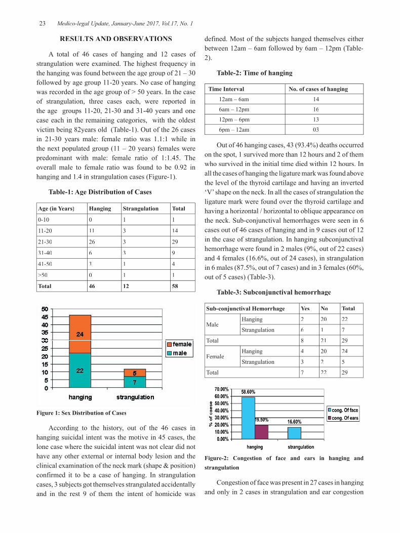
23 Medico-legal Update, January-June 2017, Vol.17, No. 1
RESULTS AND OBSERVATIONS
A total of 46 cases of hanging and 12 cases of strangulation were examined. The highest frequency in the hanging was found between the age group of 21 – 30 followed by age group 11-20 years. No case of hanging was recorded in the age group of > 50 years. In the case of strangulation, three cases each, were reported in the age groups 11-20, 21-30 and 31-40 years and one case each in the remaining categories, with the oldest victim being 82years old (Table-1). Out of the 26 cases in 21-30 years male: female ratio was 1.1:1 while in the next populated group (11 – 20 years) females were predominant with male: female ratio of 1:1.45. The overall male to female ratio was found to be 0.92 in hanging and 1.4 in strangulation cases (Figure-1).
Table-1: Age Distribution of Cases
Age (in Years) Hanging Strangulation Total
0-10 0 1 1
11-20 11 3 14
21-30 26 3 29
31-40 6 3 9
41-50 3 1 4
>50 0 1 1
Total 46 12 58
Figure 1: Sex Distribution of Cases
According to the history, out of the 46 cases in hanging suicidal intent was the motive in 45 cases, the lone case where the suicidal intent was not clear did not have any other external or internal body lesion and the clinical examination of the neck mark (shape & position) confirmed it to be a case of hanging. In strangulation cases, 3 subjects got themselves strangulated accidentally and in the rest 9 of them the intent of homicide was
defined. Most of the subjects hanged themselves either between 12am – 6am followed by 6am – 12pm (Table-2).
Table-2: Time of hanging
Time Interval No. of cases of hanging
12am – 6am 14
6am – 12pm 16
12pm – 6pm 13
6pm – 12am 03
Out of 46 hanging cases, 43 (93.4%) deaths occurred on the spot, 1 survived more than 12 hours and 2 of them who survived in the initial time died within 12 hours. In all the cases of hanging the ligature mark was found above the level of the thyroid cartilage and having an inverted ‘V’ shape on the neck. In all the cases of strangulation the ligature mark were found over the thyroid cartilage and having a horizontal / horizontal to oblique appearance on the neck. Sub-conjunctival hemorrhages were seen in 6 cases out of 46 cases of hanging and in 9 cases out of 12 in the case of strangulation. In hanging subconjunctival hemorrhage were found in 2 males (9%, out of 22 cases) and 4 females (16.6%, out of 24 cases), in strangulation in 6 males (87.5%, out of 7 cases) and in 3 females (60%, out of 5 cases) (Table-3).
Table-3: Subconjunctival hemorrhage
Sub-conjunctival Hemorrhage Yes No Total
MaleHanging 2 20 22
Strangulation 6 1 7
Total 8 21 29
FemaleHanging 4 20 24
Strangulation 3 2 5
Total 7 22 29
Figure-2: Congestion of face and ears in hanging and strangulation
Congestion of face was present in 27 cases in hanging and only in 2 cases in strangulation and ear congestion
Medico-legal Update, January-June 2017, Vol.17, No. 1 24 25 -legal Update, January-June 2017, Vol.17, No. 1
was found in 9 cases of hanging and none in the case of strangulation (Figure-2). External injuries other than ligature mark were found in 2 cases of strangulation and in none of hanging cases.
DISCUSSION
The present study comprised cases of fatal compression of neck by ligature i.e. hanging (where the weight of the body acts as constricting force) and ligature strangulation (where constricting force is other than the body weight). In hanging cases, the highest number of cases was found between 21-30 years, followed by 11-20 years and 31-40 years respectively. No incidence was reported in the age group >50 years. Our findings are consistent with the previous studies like Yadav9 (56.5%), Singh10 (48.13%), Sharma11 (46.15%), Naik12 (52.92%), Ahmad13 (45.51%), Meera14 (35.71%) and Patel15 (40%). The individuals in this age group are more susceptible to frustration as there is a transformation from a student’s life to an adult life where they are exposed to harsh realities of the world like career problems, higher studies, marriage, financial issues, failure of love affairs etc. The previous studies reported male predominance 10,11,14-16 while in our study female cases were higher with male to female ratio being 0.92. There was no specific pattern found in the age or sex distribution of the strangulation cases. The least number of hanging cases were reported between 6 PM to 12 AM when most of the family members are present at home.
The manner of death in almost all of the hanging cases is suicidal; accidental is uncommon and homicidal is rare17,18. In our study, all 46 cases of hanging were suicidal in nature. There was no associated external injury other than the ligature mark in any of the hanging cases. The reason behind this is that it is very difficult to hang a conscious person from a high point of suspension. It will have to be done by more than one person and will cause too much disturbance of the crime scene. Further there will be marks of struggle and associated injuries on the body of the deceased. This will defeat the purpose of the perpetrator who would like to disguise his act of homicide as suicide. On the contrary most of the ligature strangulation are homicidal, few being accidental and suicidal possibility being rare1-5,17,18. In our study no case of suicidal ligature strangulation was reported. 75% cases of ligature strangulation were homicidal and the rest 25% were accidental.
Subconjuctival hemorrhage was seen more in cases of strangulation than hanging1-5,17,18. We also found subconjuctival hemorrhage in 75% of strangulation cases as compared to 13% of hanging cases. One of the contrary findings noted in our study was the congestion of face and ears to be more in hanging cases, whereas facial congestion was reported to be more marked in ligature strangulations1-5,18.
Ligature mark in hanging is inverted V-shape above the level of thyroid cartilage while in strangulation it was mostly horizontal over the thyroid cartilage or below its level1-5,18. The similar observations were noted in the current study. So we can deduce that ligature mark is a very reliable factor in deciding the manner of deaths in equivocal cases.
CONCLUSION
Hanging is more prevalent in 2nd and 3nd and 3nd rd decades of rd decades of rd
life and more probable at time when the victim is alone. Ligature mark position and orientation is a very reliable factor in differentiating between cases of hanging and ligature strangulation. Subconjuvtival hemorrhages are found more commonly in ligature strangulation than hanging. In suicidal hanging, no external injury other than then ligature mark is present over the body. If any such injuries are noted by the autopsy surgeon then homicidal angle must be ruled out by assessment of witnesses account and circumstantial evidence.
Conflict of Interests: None
Source of Funding: Nil Required.
Ethical Clearance: UCMS Thesis Protocol Committee
REFERENCES
1. Pillay VV. Textbook of Forensic Medicine and toxicology. 17th Ed. Hyderabad: Paras Medical Publisher; 2016. Chapter 14; Mechanical Asphyxia: p 296-325
2. Reddy KSN. The essentials of Forensic Medicine and Toxicology. 25th Ed. Hyderabad: K Suguna Devi; 2006. Chapter 14; Mechanical Asphyxia: p 297-331.
3. Mahanta P. Modern Textbook of Forensic medicine & Toxicology. New delhi: Jaypee Brothesr: 2014. Chapter 17; Asphyxial deaths: p 340-360.
-legal Update, January-June 2017, Vol.17, No. 1 24 25 Medico-legal Update, January-June 2017, Vol.17, No. 1
4. Aggarwal A. Textbook of Forensic Medicine and Toxicology New Delhi: Avichal Publishing Company; 2014. Chapter-19; Asphyxia: p 366-392.
5. Vij K. Textbook of Forensic Medicine and Toxicology: Principles and practice. 5thed. New Delhi: Elsevier India Pvt Ltd; 2011. Chapter-6; Asphyxial Deaths: p110-145.
6. Paparo GP, Siegel H. Neck markings and fractures in suicidal hangings. FSI 1984. 24; 1: 27-35.
7. Simonsen J. Patho-anatomic findings in neck structures in asphyxiation due to hanging: a survey of 80 cases. FSI 1988. 38; 1-2: 83-91.
8. Verma SK, Lal S. Strangulation deaths during 1993-2002 in East Delhi (India). Leg Med (Tokyo) 2006. 8;1:1-4. Epub 2005 Jul 28.
9. Yadav A, Kumath M, Tellewar S, L Kumar R. Study of Fracture of Hyoid Bone In Hanging Cases. JIAFM. 2013; 35(3):239-241.
10. Singh A, Gorea RK, Dalal JS, Thind AS, Walia D. A study of demographic variables of violent asphyxial death. Journal of Punjab Academy of Forensic Medicine and Toxicology. 2003; 3:32–34.
11. Sharma BR, Harish D, Singh VP and Singh P. Ligature mark on neck: How informative? J Indian Acad Forensic Med. 2005; 27(1):10-15.
12. Naik SK, Patil DY. Fracture of hyoid bone in cases of asphyxia deaths resulting from constricting force round the neck. J Indian Acad Forensic Med. 2005; 27 (3):149-153.
13. Ahmad M, Hossain MZ. Hanging as a method of suicide- Retrospective analysis of postmortem cases. JAFMC Bangladesh 2010; 6(2):37-39.
14. MeeraTh, Singh MBK. Pattern of Neck Findings in Suicidal Hanging- A Study in Manipur. J Indian Acad Forensic Med. 2011; 33(4):352-354.
15. Patel AP, Bansal A, Shah JV, Shah KA. Study of hanging cases at Ahemdabad Region. J Indian Acad Forensic Med. 2012; 34(4):342-345.
16. Reddy S, Kumar R, Rudramurthy. Asphyxial Deaths at District Hospital, Tumkur- A Retrospective Study. J Indian Acad Forensic Med. 2012; 34(2): 146-147.
17. DiMaio VJ, Dimaio D. Forensic Pathology, 2nd
Ed. Boca Raton: CRC Press; 2001. Chapter 8, Asphyxia: p245-262.
18. Knight B, Saukko P. Knight’s Forensic Pathology, 3rd ed. London: Arnold; 2004. Chapter 15; Fatal rd ed. London: Arnold; 2004. Chapter 15; Fatal rd
Pressure on Neck: p368- 394.

27 -legal Update, January-June 2017, Vol.17, No. 1
Suicide by Hanging in Jharkhand: An Autopsy based Cross Sectional Study
Kaushal Kishore1, Bhoopendra Singh2, Ajit Kumar Chaudhary3
1Postgraduate Student, 2Toxicologist (Associate Professor), 3Professor, Department of Forensic Medicine & Toxicology, Rajendra Institute of Medical Sciences, Ranchi, Jharkhand, India
ABSTRACT
Background: Intentional violent asphyxial deaths are one of the important causes of death. Nowadays hanging is one of the preferred modes of committing suicide, as it supposedly produces less pain and sure instantaneous death as compared to other modes of suicide. As per data available with WHO and National Crime Report Bureau (NCRB) Government of India that there are few studies conducted in India. Such study has not been conducted in this part of India i.e. Ranchi, Jharkhand.
Objective: The aim of this study is to find out the demographic profile and attributing factors for suicide hanging in the state of Jharkhand of India.
Materials & Method: This prospective study was carried out on 351 cases of death due to hanging in the department of forensic medicine & toxicology of Rajendra Institute of Medical sciences, Ranchi during April 2013 to October 2014. Information regarding the socio-demographic, mode of suicides, time of incidence, place of incidence, occupation, etc. were gathered from the police papers like inquest report, dead body Challan and through detailed interviews with the relatives, neighbours, friends, and police officials accompanying the dead bodies.
Results: Amongst total cases of suicidal hanging, males were predominant to female. The majority (61.0%) of victims who had committed suicides were youths in the age-group 15-29 years. Maximum victims (57%) were married. The majority of the victims were from middle economic class (53 %) and maximum incidences were occurred during summer (43%). The majority (69%) were chosen parental home for hanging. The maximum suicidal incident occurred at evening (between 4 PM to 8 PM), which accounts 33%. The lowest occurrence of suicidal hanging was reported at afternoon (between 12 Pm -4 pm) 6%.
Conclusion: In this area of the country the majority (61.0%) of victims who had committed suicides were youths in the age-group 15-29 years.
Keyword: Suicidal Hanging, Seasonal Variations, Place of Incidence
Corresponding author: Dr. Bhoopendra Singh, Toxicologist Associate Professor, Department of Forensic Medicine & Toxicology, Rajendra Institute of Medical Sciences, Ranchi, 834009, Jharkhand, India. Email: [email protected]
INTRODUCTION
Suicide is one of the leading causes of death in World. Approximately one people commit suicide each year, or about one lost every 40 seconds.1 Around 800,000
to million people die by suicide every year, making it the 10th leading cause of death worldwide.2,3 Suicide rate in the world, mainly in developing countries, in rate past fifty years have increased about 60%. A significant amount of suicide occurs in Asia, which includes about 60% of suicides. Based on WHO reports, China, India and Japan are included in approximately 40% of all suicides in the world.4
Effective suicide prevention requires good studies on the use of suicide methods and attributing various other factors in different countries. Suicide is
DOI Number: 10.5958/0974-1283.2017.00006.8

27 Medico-legal Update, January-June 2017, Vol.17, No. 1
often committed out of despair, or attributed to some underlying mental disorder, which includes depression, bipolar disorder, schizophrenia, alcoholism and drug abuse.1 Financial difficulties, troubles with interpersonal relationships and other undesirable situations also play significant role.2 According to Kevin Chen-Chang et al (2012)5 hanging was the most common method of suicide in nine out of the seventeen countries/regions reviewed (i.e., Bahrain, Iran, Japan, South Korea, Philippines, Saudi Arabia, Taiwan, Thailand, and Turkey).6-15
According to International Association for Suicide Prevention, Suicide is nevertheless a private and personal act and a wide disparity exists in the rates of suicide across different countries. A greater understanding of region-specific factors related to suicide would enable prevention strategies to be more culturally sensitive. 16
As per data available with the National Crime Report Bureau (NCRB) Government of India, 17 there are few studies conducted in India. Such study had not been conducted in this part of India i.e. Ranchi, Jharkhand. Therefore, we have planned this study to know the demographic profile and attributing factors for suicide hanging in the state of Jharkhand in India. The result of the study may help to plan strategies to prevent such mortality and morbidity due to suicidal hanging in this part of the country.
MATERIALS AND METHOD
Study type and study design: Prospective study
Place and duration: This study was carried out in the Department of Forensic Medicine & Toxicology, Rajendra Institute of Medical Sciences, Ranchi for a period of one and a half year i.e. from April, 2012 to October, 2013.
Sample size and Sampling method: Total 351 cases of suicidal hanging were autopsied during the period of study and were included in the present study. These cases were screened on the basis of information provided by the relatives, neighbours, friends, and police officials accompanying the dead bodies and findings present on the bodies. Further information regarding the Socio-demographic, mode of suicides, time of incidence, place of incidence, occupation, etc. were gathered from the police papers like inquest report, dead body Challan etc., and through detailed interviews of the relatives, neighbours, friends, and police officials accompanying
the dead bodies. In case of hospital deaths, hospital papers were also examined. To calculate the seasonal variation, the seasons were classified according to the Indian Meteorological Department (IMD) which designated four climatological seasons in India, which are: Winter, occurring from December to March, Summer or pre-monsoon season from April to June, Monsoon or rainy season from July to September and Post-monsoon or autumn season from October to November.
Ethical Approval and Patient’s consent: Approval was obtained from the Institutional Ethics Committee before commencement of the study. Since this study was conducted in post-mortem cases, the patient consent was not applicable.
Collected data were entered into SPSS version 10 and were analysed. The results are presented in the form of table and figures.
RESULTS
This study is an attempt to analyse the demographic profile of suicidal deaths due to hanging in this part of the country. To achieve the goal of the study of 351 cases of suicidal deaths by hanging were screened and analysed. This comprised 10.05% of the total post-mortem conducted in the department during the study period.
Amongst total 351 cases of suicidal hanging, 66.67% (234/351) were males and 33.33 % (117/351) were females. It was found that the male victims were outnumbering the females.
It was found that the majority of victims were Hindus (82 %) followed by Christians (15%) and Muslims (3 %).
Age-wise profile of victims showed that the majority (61.0%) of victims who were committed suicides were in the age-group 15-29 years. Among them, 142 (66%) were males and 74 (34%) were females. This was followed by age group of 30-44 years with 21.0% and then 45-59 years (13%). Amongst the age group of 30-44 years, 52 (72%) were males and 20 (28%) were females. While in the age group of less than 15 years the females were outnumbering the males (Table-1).

Medico-legal Update, January-June 2017, Vol.17, No. 1 28 29 -legal Update, January-June 2017, Vol.17, No. 1
The majority of the victims were from middle economic class (53%) followed by lower economic class (36 %) and high economic class (11%) (Figure-2).
The seasonal variations in cases are depicted in Table-3. Most of the hanging suicides occurred during summer season (43%) among the summer the maximum in the month of April (39%). This is followed by winter season (Dec-March) (26%), and monsoon (July-Sept) (19%). The least frequency was reported in the post-monsoon or autumn (Oct- Nov.) (12%) (Table-3).
The majority victims of suicidal hanging were from joint family (61%) followed by Nuclear Family (22%) and Living alone victims (17%).
In terms of occupational status, the majority were unemployed (52%), followed by employed (28%), and then farmers (20%).
The education-wise break-up of suicide victims is presented in Figure-1. The maximum numbers of suicide victims were educated (72%) (Fig-1).
Table-1- Distribution of victims of suicidal hanging in relation to age and Gender
Age Group(in years)
GenderTotal
Male Female
Frequency % Frequency % Frequency %
0-14 05 01.4 13 3.7 18 05.1
15-29 142 40.4 74 21.1 216 61.5
30-44 52 14.8 20 5.7 72 20.5
45-59 35 10.0 10 2.8 45 12.8
>60 00 00.00 00 00 00 00.0
Total 234 66.6 117 33.3 351 100
The information on the marital status of victims has been presented in Table–2. It was observed that the majority victims, both males and females, were married (57%). Out of these 57% victims, 62.81% (125/199) were males and 37.19% (73/199) were females. The male and female ratio among married victims was 1.71:1 while among unmarried it was 3:1.
Table-2- Distribution of victims of suicidal hanging in relation to Nuptial and Gender
Marital Status Male Female
Frequency % Frequency % Frequency %
Married 199 57 125 53 74 63
Unmarried 125 35 94 40 31 27
Widow 27 8 15 07 12 10
Total 351 100 234 117
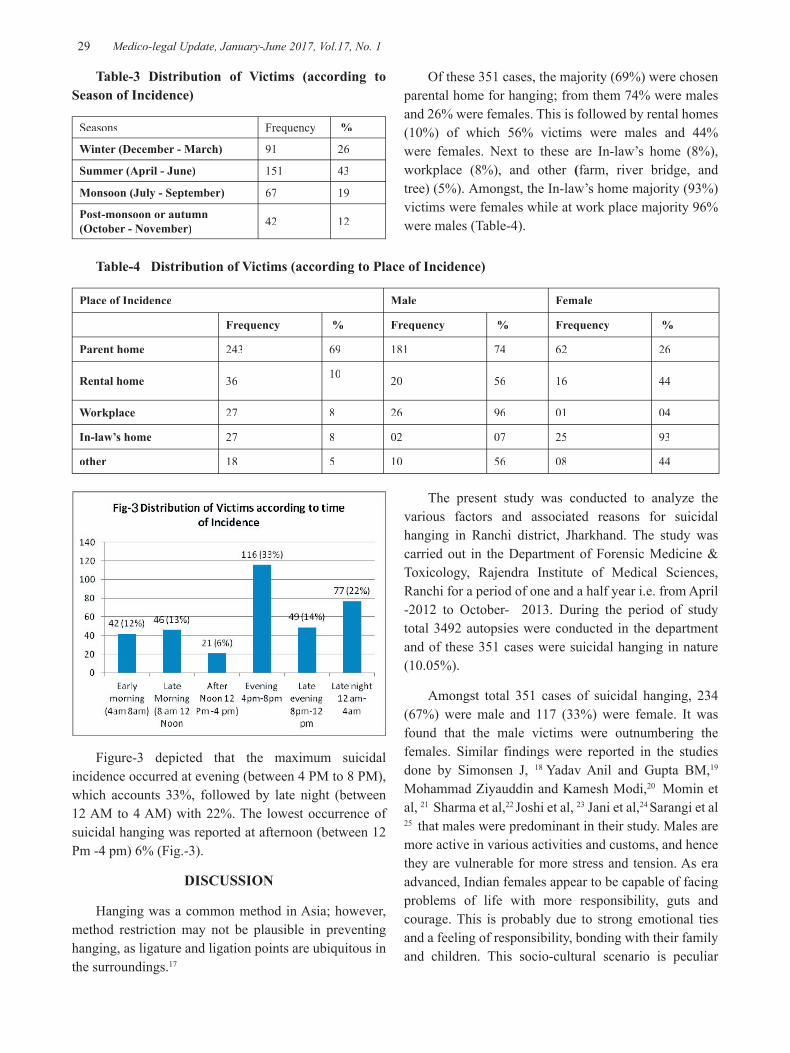
-legal Update, January-June 2017, Vol.17, No. 1 28 29 Medico-legal Update, January-June 2017, Vol.17, No. 1
Figure-3 depicted that the maximum suicidal incidence occurred at evening (between 4 PM to 8 PM), which accounts 33%, followed by late night (between 12 AM to 4 AM) with 22%. The lowest occurrence of suicidal hanging was reported at afternoon (between 12 Pm -4 pm) 6% (Fig.-3).
DISCUSSION
Hanging was a common method in Asia; however, method restriction may not be plausible in preventing hanging, as ligature and ligation points are ubiquitous in the surroundings.17
Table-3 Distribution of Victims (according to Season of Incidence)
Seasons Frequency %
Winter (December - March) 91 26
Summer (April - June) 151 43
Monsoon (July - September) 67 19
Post-monsoon or autumn (October - November) 42 12
Of these 351 cases, the majority (69%) were chosen parental home for hanging; from them 74% were males and 26% were females. This is followed by rental homes (10%) of which 56% victims were males and 44% were females. Next to these are In-law’s home (8%), workplace (8%), and other (farm, river bridge, and tree) (5%). Amongst, the In-law’s home majority (93%) victims were females while at work place majority 96% were males (Table-4).
Table-4 Distribution of Victims (according to Place of Incidence)
Place of Incidence Male Female
Frequency % Frequency % Frequency %
Parent home 243 69 181 74 62 26
Rental home 36 10 20 56 16 44
Workplace 27 8 26 96 01 04
In-law’s home 27 8 02 07 25 93
other 18 5 10 56 08 44
The present study was conducted to analyze the various factors and associated reasons for suicidal hanging in Ranchi district, Jharkhand. The study was carried out in the Department of Forensic Medicine & Toxicology, Rajendra Institute of Medical Sciences, Ranchi for a period of one and a half year i.e. from April -2012 to October- 2013. During the period of study total 3492 autopsies were conducted in the department and of these 351 cases were suicidal hanging in nature (10.05%).
Amongst total 351 cases of suicidal hanging, 234 (67%) were male and 117 (33%) were female. It was found that the male victims were outnumbering the females. Similar findings were reported in the studies done by Simonsen J, 18 Yadav Anil and Gupta BM,19 Mohammad Ziyauddin and Kamesh Modi,20 Momin et al, 21 Sharma et al,22 Joshi et al, 23 Jani et al,24 Sarangi et al 25 that males were predominant in their study. Males are more active in various activities and customs, and hence they are vulnerable for more stress and tension. As era advanced, Indian females appear to be capable of facing problems of life with more responsibility, guts and courage. This is probably due to strong emotional ties and a feeling of responsibility, bonding with their family and children. This socio-cultural scenario is peculiar

Medico-legal Update, January-June 2017, Vol.17, No. 1 30 31 -legal Update, January-June 2017, Vol.17, No. 1
to India. The youngest victims, those were between 10 years to 14 years old, committed suicide hanging due to problems at school or related to education being cited as a reason. They were not from the same school, but were from different areas of the same state. This substantiates the Modi’s statement- “Age is no bar to suicide by hanging” 26. Polson and Gee also reported suicide of a 10 year old child by hanging.27
It is observed that the majority of victims were Hindus (82%) followed by Christians (15%) and Muslim (3%). Muslim countries such as Iran, Pakistan, Saudi Arabia, and Turkey had relatively lower suicide rates (<6.5/100,000), with Iran having the lowest suicide rate (male: 0.3/100,000; female: 0.1/100,000). It could be due to stigmatization and legal sanctions against suicide; under-reporting of suicide rates in these Muslim countries may partly contribute to the extremely low rate. 28, 29
Age-wise profile of victims shows that the majority (61.0%) of victims who had committed suicides were in the age-group 15-29 years. Among them, 142 (66%) were males and 74 (34%) were females. This is followed by age group of 30-44 years with 21.0% and then 45-59 years (13%). Amongst the age 30-44 years, 52 (72%) were males and 20 (28%) were females. While in the age group of less than 15 years the females were outnumbering the males (Table-1). Similar findings were also reported by various authors of other studies which was conducted in Kurnool (Andhra Pradesh) by T. Saisudheer and T.V. Nagaraja, 30 Ashok Kumar Samanta, Soumya Ranjan Nayak, 31 Momin et al, 21 Sharma et al, 22 Joshi et al,23 and Jani CB 24 that the majority of the victims were belonging to the age group 21 to 30 years (39%). There were few studies’ findings, which contrasted to present findings of which they had found 74.83% cases of hanging in the age group of 31-40 years.25 The justification for majority victims in the age group of 21-30 years could be because this is the most active period of one’s life. People become violent and arrogant in this age group, and are more vulnerable to the fast changing social trends and cultures, as they are mentally a bit immature with little experience of life, and there is a great fluctuation of emotion in this age group. The influencing factors in this distribution are unemployment, failure in love, marital disharmony, financial problems, dowry harassment etc.
RELATED TO MARITAL STATUS
The information on the marital status of victims has been presented in Table–2. It was observed that the majority victims were married (57%) of which 63% were males and 37% were females. This is followed by unmarried (35%) of which (75%) were males and 25% were females. This is further followed by Widowed & Widower (8%) of which 56% were males and 44% were females. The majority of married males were from age group 31 to 40, while majority of female were from age group 21 to 30. It suggests that marital disharmony was a major factor in female hanging as compared to males. It is also found that hanging for female was frequent in early married life, which was supported in a study done by K P Singh, Aelifeter R Marak, Th Meera.32 These observations are consistent with the study of Momin et al 21 and Jani CB 24 . This can be explained by early marriages in the Indian community, familial responsibilities, social customs, limited source of income, frustrations, dowry, family quarrels, maladjustment in marriage life, low level of education, infidelity, unemployment etc.
Most studies report that the majority of suicide decedents was married at the time of death (57 to 73%), 33 – 39 but two studies reported that a higher proportion of suicide decedents were unmarried (52 to 58%).40- 41
In one prospective study of suicide autopsies, it was found that male suicide decedents were more likely to be single (60.5%) while female suicide decedents were more likely to be married (73.8%); or in other words, marriage was protective in males (OR = 0.1, 95% CI = 0.1–0.4), but not in females.38 In a case series of suicidal deaths, being married put women – particularly women under 30 years of age – at a higher risk of suicide, but this was not in the case of men.42 One case control study found a much higher risk of suicide in persons who were widowed, separated or divorced, compared to persons who were currently married (13% v. 1%; p=0.003) 43 ; this finding was supported by another study.
In terms of occupational status, the majority (52%) were unemployed, followed by employed (28%), and farmers (20%). The association of occupation to suicide is also inconsistent. In a psychological autopsy of suicides, 55% were unemployed.41 In a community-based study, most of the women who committed suicide were housewives (54%).44

-legal Update, January-June 2017, Vol.17, No. 1 30 31 Medico-legal Update, January-June 2017, Vol.17, No. 1
RELATED TO PLACE OF HANGING
In the present study, parental home was the place of hanging in the majority cases (69%), whereas in rest (31%) cases the place of hanging was rented home (10%), In-law’s home (8%), workplace (8%), and other (farm, river bridge, and tree) (5%). Similar findings were observed by Pareshkumar Chandegara et al that among 87 cases of hanging, houses were the place of choice by 78 persons (90%). Similar findings were found in a study done by Ashok Kumar Samanta and Soumya Ranjan Nayak.31 They observed that 66% males and 80% female chose home for hanging. This is also supported by findings of others.32, 45
RELATED TO INCOME
The majority victims of suicidal hanging were from joint family (61 %) followed by Nuclear Family (22%) and Living alone (17%) victims. The majority of the victims were from middle economic class (53%) followed by lower economic class (36 %) and higher economic class (11%). The present findings are supported by the study done by T. Saisudheer and T.V. Nagaraja 30: the incidence of hanging was recorded more common in the middle income group (78%), and next common was a lower income group (17%) and least common were a higher income group (2%). Similar findings were obtained in the study done by Samanta AK and Nayak SR.31 This supports the low income factor as one of the reasons for dissatisfaction and hanging.
RELATED TO EDUCATION
In the present study, the maximum numbers of suicide victims were educated (72%) whereas the illiterates accounted for 18%. In a study conducted by Mohanty S et al46, the majority had gained an education up to secondary level (52%), followed by graduates (26%), and primary level education (18%). None of the persons who have committed suicide were illiterate. Evidence for the association of education and suicide is inconsistent.46
CONCLUSION
In this area of the country the majority (61.0%) of victims who had committed suicides were youths in the age-group 15-29 years.
Conflict of Interest – There is no conflict of interest
Source of Funding - Self source
Ethical Clearance – Cleared by Institutional Ethics Committee
REFERENCES
1. Hawton K, Van Heeringen K. Suicide. Lancet. 2009; 373 (9672): 1372–81.
2. Värnik, P. Suicide in the world. Int J Environ Res Public Health. 2012; 9(3): 760-71.
3. Vijayakumar L, Kumar MS, Vijayakumar V. Substance use and suicide. Current opinion in psychiatry. 2011; 24(3): 197-202.
4. Hawton K; Saunders KE, O’Connor RC. Self-harm and suicide in adolescents. Lancet. 2012; 379 (9834): 2373–82.
5. Kevin Chien-Chang Wu, Ying-Yeh Chen, and Paul S. F. Yip. Suicide Methods in Asia: Implications in Suicide Prevention, Int J Environ Res Public Health. 2012 Apr; 9 (4): 1135–1158.
6. Al Ansari A., Hamadeh R.R., Ali M.K., El Offi A. Suicide in Bahrain in the last decade. Crisis. 2007; 28: 11–15.
7. Ahmadi A., Mohammadi R., Stavrinos D., Almasi A., Schwebel D. Self-immolation in Iran. J. Burn Care Res. 2008; 29 : 451–460.
8. Takahashi Y. Suicide in Japan: What Are the Problems? In: Kosky R.J., Eshkevari H.S., Goldney R.D., editors. Suicide in Japan: What Are the Problems? In Suicide Prevention: The Global Context. Kluwer Academic Publishers; Hingham, MA, USA: 1998. Pp. 121–130.
9. Kim S.Y., Kim M.-H., Kawachi I., Cho Y. Comparative epidemiology of suicide in South Korea and Japan: Effects of age, gender and suicide methods. Crisis. 2010; 32: 5–14.
10. Ojima T., Nakamura Y., Detels R. Comparative study about methods of suicide between Japan and the United States. J. Epidemiol. 2004; 14: 187–192.
11. Kamizato E., Yoshitome K., Yamamoto Y., Iwase T., Tsuda T., Miyaishi S., Doi H. Factors affecting the choice of suicide method in Okayama: A database analysis from a forensic perspective. Acta Med. Okayama. 2009; 63: 177–186.
12. Redaniel M.T., Lebanan-Dalida M.A., Gunnel D.
Medico-legal Update, January-June 2017, Vol.17, No. 1 32 33 -legal Update, January-June 2017, Vol.17, No. 1
Suicide in the Philippines: Time trend analysis (1974–2005) and literature review. BMC Public Health. 2011; 11: 536
13. Elfawal M.A. Cultural influence on the incidence and choice of method of suicide in Saudi Arabia. Am. J. Forensic Med. Pathol. 1999; 20: 163–168.
14. Chung K.H., Lee HC., Chen SF., Lin HC. The ten-year trend in suicide methods: Evidence from an Asian population. Can. J. Public Health. 2008; 99: 406–410.
15. Lin JJ, Chang SS, Lu TH. The leading methods of suicide in Taiwan, 2002-2008. BMC Public Health. 2010 Aug 13; 10: 480.
16. Accidental Deaths and Suicides in India 2008. New Delhi: Ministry of Home Affairs, Government of India; 2010. National Crime Records Bureau (NCRB).
17. Chen Y.-Y., Wu K.C.-C., Yip P.S.F. Suicide Prevention through Restricting Access to Suicide Means and Hotspots. In: O’Connor R.C., Platt S., Gordon J., editors. International Handbook of Sucide Prevention: Research, Policy and Practice. John Wiley & Sons; Chichester, UK: 2011. pp. 545–560.
18. Simonsen J. Patho-anatomic findings in neck structures in asphyxiation due to hanging: a survey of 80 cases. Forensic Sci Int 1988; 38: 83-91.
19. Yadav A, Gupta BM. Histopathologicla changes in skin and subcutaneous tissues at ligature site in cases of hanging and strangulation. J Indian Acad Forensic Med 2010; 31: 200-4.
20. Saiyed MZG, Modi KA. Retrospective Study of Postmortem Cases of ‘Hanging– A Method of Suicide’. NHL Journal of Medical Sciences 2013; 2: 48-50.
21. Momin SG, Mangal HM, Kyada HC, Vijapura MT, Bhuva SD. Pattern of ligature mark in cases of compressed neck in Rajkot region. J Indian Acad Forensic Med 2012; 34: 40-3.
22. Sharma BR, Harish D, Singh VP, Singh P. Ligature mark on neck: How informative?. J Indian Acad Forensic Med 2005; 27: 10-5.
23. Joshi R, Chanana A, Rai H. Incidence and medicolegal importance of autopsy study of fracture of neck structures in hanging and strangulation. Medico-Legal Update 2007; 7:
105-10.
24. Jani CB. An autopsy study of neck compression cases with emphasis on hanging. Thesis submitted to Saurashtra University: Rajkot; 2001.
25. Sarangi MP. Ligature marks in forensic pathologists perspective. J Forensic Med and Toxicology 1998;15: 99-102.
26. Mathiharan K, Amrith Patnaik. Modi‘s Medical Jurisprudence and Toxicology. 23rd ed. Lexis Nexis Butterworths. New Delhi. 2006: 565 – 90.
27. Polson CJ, Gee DJ, Knight B. The Essentials of Forensic Medicine. 4thed. Pergamon Press. Oxford. 1985: 351- 420.
28. Khan M.M., Syed E.U. Suicide in Asia: Epidemiology, Risk Factors, and Prevention. In: O’Connor R.C., Platt S., Gordon J, editors. International Handbook of Suicide Prevention: Research, Policy and Practice. Wiley-Blackwell; Oxford, UK: 2011. pp. 487–506.
29. Khan M.M., Naqvi H., Thaver D., Prince M. Epidemiology of suicide in Pakistan: Determining rates in six cities. Arch. Suicide Res. 2008; 12:155–160.
30. Saisudheer T, Nagaraja TV. A study of ligature mark in hanging deaths. Int J Pharm Biomed Sci 2012; 3: 80-4.
31. Samanta AK, Nayak SR. Newer trends in Hanging Deaths. J Indian Acad Forensic Med 2012; 34: 37-9.
32. Singh KP, Marak AR, Meera TH. Multifaactorial analysis of hanging deaths. J Med Soc 2013; 27:49-51.
33. Sharma SD, Gopalakrishna R. Suicide--a retrospective study in a culturally distinct community in India. Int J Soc Psychiatry. 1978 Spring; 24(1): 13-8.
34. Shukla G, Verma B, Mishra DN. Suicide in Jhansi City. Indian J Psychiatry.1990; 32: 44–51.
35. Bhatia MS, Aggarwal NK, Aggarwal BBL. Psychosocial profile of suicide ideators, attempters and completers in India. Int J Soc Psychiatry. 2000;46: 155–163.
36. Sharma BR, Sharma V, Harish D, Vij K. Suicides in northern India: causes, methods used and prevention. Medicine, Science and the Law. 2003;

-legal Update, January-June 2017, Vol.17, No. 1 32 33 Medico-legal Update, January-June 2017, Vol.17, No. 1
43: 221–229.
37. Mohanty S, Sahu G, Mohanty MK, Patnaik M. Suicide in India: a four year retrospective study. J Forensic Leg Med. 2007; 14: 185–189.
38. Bastia BK, Kar N. A psychological autopsy study of suicidal hanging from Cuttack, India: focus on stressful life situations. Arch Suicide Res. 2009; 13: 100–104.
39. Manoranjitham SD, Rajkumar AP, Thangadurai P, Prasad J, Jayakaran R, Jacob KS. Risk factors for suicide in rural south India. Br J Psychiatry. 2010; 196: 26–30.
40. Khan FA, Anand B, Devi MG, Murthy KK. Psychological autopsy of suicide-a cross-sectional study. Indian J Psychiatry. 2005 Apr; 47(2):73-8.
41. Chavan BS, Singh GP, Kaur J, Kochar R. Psychological autopsy of 101 suicide cases from
northwest region of India. Indian J Psychiatry. 2008 Jan; 50(1): 34-8.
42. Ponnudurai R, Jeyakar J. Suicide in madras. Indian J Psychiatry. 1980 Apr; 22(2): 203-5.
43. Vijayakumar L, Rajkumar S. Are risk factors for suicide universal? A case-control study in India. Acta Psychiatr Scand. 1999 Jun; 99(6): 407-11.
44. Parkar SR, Dawani V, Weiss MG. Clinical diagnostic and sociocultural dimensions of deliberate self-harm in Mumbai, India. Suicide Life Threat Behav. 2006; 36: 223–228.
45. Meera TH, Singh MB. Pattern of neck findings in suicidal hanging- a study in Manipur. J Indian Acad Forensic Med 2011; 33: 350-2.
46. Mohanty S, Sahu G, Mohanty MK, Patnaik M. Suicide in India: a four year retrospective study. J Forensic Leg Med. 2007; 14: 185–189.
35 -legal Update, January-June 2017, Vol.17, No. 1
Study of Trends of Death due to Burns Cases at Hubballi Region of Karnataka
Gajanan H Nayak1, Madhu Sudhan S2, Sunilkumar S Biradar3, Ravindra Kumar C N4, Hemanth Raj M N5
1Professor & Head, Department of Forensic Medicine & Toxicology, KIMS, Hubballi. 2Assistant Professor, 2Assistant Professor, 2
Department of Forensic Medicine, Shridevi Institute of Medical Sciences & Research Hospital, Tumkur, 3Associate Professor, 3Associate Professor, 3 4Postgraduate, Department of Forensic Medicine & Toxicology, KIMS, Hubballi, 4Postgraduate, Department of Forensic Medicine & Toxicology, KIMS, Hubballi, 4
5Associate Professor, Department of Forensic Medicine, Shridevi Institute of Medical Sciences 5Associate Professor, Department of Forensic Medicine, Shridevi Institute of Medical Sciences 5
& Research Hospital, Tumkur
ABSTRACT
Background: The magnitude of deaths due to burns is so large. The use of fire in various aspects has not only added to his comforts, but it also added to his miseries by increasing the risk of burns.
Material and Method: This was an observational, retrospective, descriptive study that included all cases of death due to burns during the period of January 2009 to December 2009, for which a postmortem was performed in the mortuary premises of the department.
Results: The study of death due to burns showed a female predominance. The maximum number of cases was in the 21-30 years age group. Overall the most common causative material used for burns was kerosene oil and stove. Majority of incidence due burn cases occurred at home and between 12pm- <6pm and 6pm-<12am and deaths occurred after 4-7 days and >7days of incident after admission to hospital. In most of the cases cause of death was due to septicaemia.
Conclusion: The present study of death due to burns revealed a young female predominance. Kerosene oil use was dependent on its ready availability and commonly used material for domestic use in kitchen. Septicaemia was the cause of death in many cases due to high degree of susceptibility to infection due to damage and destruction of the skin.
Keywords: Burns; Septicaemia; BSA.
Corresponding author:Dr. Madhu Sudhan S,Assistant Professor, Department of Forensic Medicine, Shridevi Institute of Medical Sciences & Research Hospital, Tumkur.Karnataka. Mob: 09902269303, E-mail: [email protected]
INTRODUCTION
Man has invented fire since times immortal. The use of fire in various aspects has not only added to his comforts, but it also added to his miseries by increasing the risk of burns. Fire was perhaps man’s first double-edged sword, evidenced throughout history; it has served as well as destroyed mankind1. Burns are injuries caused due to application of heat, which may be dry heat (free
flame, hot metals or similar hot materials), Moist heat (hot liquid or vapour) causes scalding 2. Burns is the 4th most common type of trauma in the world, subsequent traffic accidents, falls, and interpersonal violence. Microbial infection after burns, where a large portion of the skin is damaged, is a very serious complication that often results in the death of the patients. About 45% of the mortality in burns patients is caused by septicaemia3. The present study attempts to study various factors related burn deaths in hubli region.
MATERIALS AND METHOD
This is an observational, retrospective, descriptive study that included all cases of deaths due to burns during the period of January 2009 to December 2009, for which a postmortem was performed in the mortuary premises
DOI Number: 10.5958/0974-1283.2017.00007.X
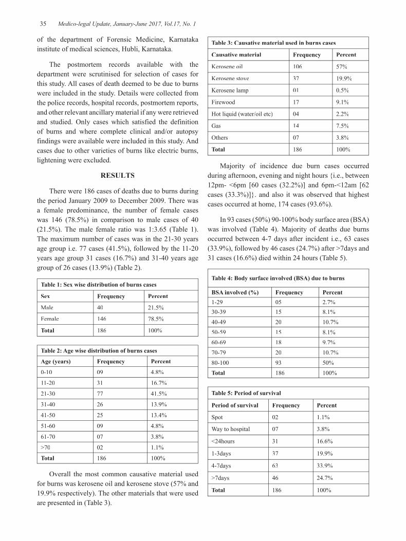
35 Medico-legal Update, January-June 2017, Vol.17, No. 1
of the department of Forensic Medicine, Karnataka institute of medical sciences, Hubli, Karnataka.
The postmortem records available with the department were scrutinised for selection of cases for this study. All cases of death deemed to be due to burns were included in the study. Details were collected from the police records, hospital records, postmortem reports, and other relevant ancillary material if any were retrieved and studied. Only cases which satisfied the definition of burns and where complete clinical and/or autopsy findings were available were included in this study. And cases due to other varieties of burns like electric burns, lightening were excluded.
RESULTS
There were 186 cases of deaths due to burns during the period January 2009 to December 2009. There was a female predominance, the number of female cases was 146 (78.5%) in comparison to male cases of 40 (21.5%). The male female ratio was 1:3.65 (Table 1). The maximum number of cases was in the 21-30 years age group i.e. 77 cases (41.5%), followed by the 11-20 years age group 31 cases (16.7%) and 31-40 years age group of 26 cases (13.9%) (Table 2).
Table 1: Sex wise distribution of burns cases
Sex Frequency Percent
Male 40 21.5%
Female 146 78.5%
Total 186 100%
Table 2: Age wise distribution of burns cases
Age (years) Frequency Percent
0-10 09 4.8%
11-20 31 16.7%
21-30 77 41.5%
31-40 26 13.9%
41-50 25 13.4%
51-60 09 4.8%
61-70 07 3.8%
>70 02 1.1%
Total 186 100%
Overall the most common causative material used for burns was kerosene oil and kerosene stove (57% and 19.9% respectively). The other materials that were used are presented in (Table 3).
Table 3: Causative material used in burns cases
Causative material Frequency Percent
Kerosene oil 106 57%
Kerosene stove 37 19.9%
Kerosene lamp 01 0.5%
Firewood 17 9.1%
Hot liquid (water/oil etc) 04 2.2%
Gas 14 7.5%
Others 07 3.8%
Total 186 100%
Majority of incidence due burn cases occurred during afternoon, evening and night hours {i.e., between 12pm- <6pm [60 cases (32.2%)] and 6pm-<12am [62 cases (33.3%)]}. and also it was observed that highest cases occurred at home, 174 cases (93.6%).
In 93 cases (50%) 90-100% body surface area (BSA) was involved (Table 4). Majority of deaths due burns occurred between 4-7 days after incident i.e., 63 cases (33.9%), followed by 46 cases (24.7%) after >7days and 31 cases (16.6%) died within 24 hours (Table 5).
Table 4: Body surface involved (BSA) due to burns
BSA involved (%) Frequency Percent1-29 05 2.7%30-39 15 8.1%40-49 20 10.7%50-59 15 8.1%60-69 18 9.7%70-79 20 10.7%80-100 93 50%Total 186 100%
Table 5: Period of survival
Period of survival Frequency Percent
Spot 02 1.1%
Way to hospital 07 3.8%
<24hours 31 16.6%
1-3days 37 19.9%
4-7days 63 33.9%
>7days 46 24.7%
Total 186 100%
Medico-legal Update, January-June 2017, Vol.17, No. 1 36 37 -legal Update, January-June 2017, Vol.17, No. 1
In majority of cases cause of death was due to septicaemia in 120 cases (64.6%), followed by 50 cases (26.8% due to burn shock and 16 cases (8.6%) due to toxaemia (Table 6).
Table 6: Cause of death
Cause of death Frequency Percent
Burn Shock 50 26.8%
Toxemia 16 8.6%
Septicaemia 120 64.6%
Total 186 100%
DISCUSSION
Burn deaths are a major public health problem in our country. In India about 60,000 people suffer from burns annually, more than 50,000 are treated in hospitals and about 10,000 succumb to thermal injuries 4.
In the present study, there was higher incidence of 146 cases (78.5%) of females deaths compared to 40 cases (21.5%) of male burn deaths and male to female ratio was 1:3.65 and the maximum number of cases was in the age group of 21-30 years i.e. 77 cases (41.5%), followed by 31 cases (16.7%) in 11-20 years age group and 26 cases (13.9%) in 31-40 years age group, such similar findings with female predominance were found in studies conducted by Tasgaonkar GV et al 5 and Shinde AB, Keoliya 6. Study conducted by Tasgaonkar GV et al 5 showed that out of 325 cases, 93 cases were male and 232 were female with male to female ratio 1:2.5 and maximum number of cases belonged to 21-30 years of age group- 146 (44.92%), followed by those of 31-40 years- 73 cases (22.46%). Study by Shinde AB, Keoliya AN 6 reported that higher incidence in female category with 94 cases (85.45%) compared to 16 cases (14.55%) of male burn deaths and also observed maximum cases in the age group of 21-30 years- 50 cases (45.45%), followed by 31-40 years- 26 cases (23.64%), most likely reasons for higher number of burns deaths in young females are probably due to main working hands for the domestic activities in kitchen and thus exposed on open, unguarded flame such as stove, cooking gas and other combustible materials which are used.
Overall the most common causative material used for burns was of kerosene oil and kerosene stove (57% and 19.9% of cases respectively). Similar findings was also reported by Gupta R, Kumar V, Tripathi S K 4 that
most common causative material was of kerosene oil in 43.5% of cases followed by kerosene lamp/stove in 29% of cases.
In the current study it was also observed that majority of incidence due to burn occurred during afternoon, evening and night hours i.e., between 12pm- <6pm- 60 cases (32.2%) and 6pm-<12am - 62 cases (33.3%). This finding was slightly contradicting with the study conducted by Shinde AB, Keoliya AN 6 showed that maximum incidence was between 10pm to 6am- 49 cases (44.5%), followed by 34 cases (30.9%) between 2pm to 10pm. And in our study it was observed that home was the place of occurrence in majority of cases- 174 cases (93.6%). Similar findings were also observed by study conducted by M M Afify et al 7 showed highest cases occurred at home -87 cases (82.07%).
The present study shows in 93 cases (50%) 80-100% body surface area was involved and majority of deaths due burns occurred between 4-7 days after incident i.e., 63 cases (33.9%), followed by 46 cases (24.7%) after >7days. Similar findings were also observed by Sheikh et al 8 in their study that in 81 cases (48.24%) 81-100% BSA was involved and majority of cases died between 3-7 days and >7days i.e., 40 cases (23.8%) 23 cases (13.6%) respectively. These observations are because of damage and destruction to the skin, which makes the person highly susceptible to infection.
In our study in majority of cases, cause of death was due to septicaemia in 120 cases (64.6%), followed by 50 cases (26.8%) due to burn shock and 16 cases (8.6%) due to toxemia. These findings are consistent with studies conducted by Tasgaonkar GV et al 5 (198 cases - 60.9%) died due to septicaemia) and Gupta R, Kumar V, Tripathi S K 4 (62 cases -50% due to septicaemia and 41 cases - 33% due to burn shock).
CONCLUSION
The trends of burn deaths revealed from this study showed a female predominance. The maximum numbers of cases were in the 21-30 years group. Overall the most common causative material used for burns was kerosene oil and stove. Majority of incidence due burn cases occurred at home and between 12pm- <6pm and 6pm-<12am and deaths occurred after 4-7 days and >7days of incident after admission to hospital. In most of the cases cause of death was due to septicaemia.

-legal Update, January-June 2017, Vol.17, No. 1 36 37 Medico-legal Update, January-June 2017, Vol.17, No. 1
Conflict of Interest: Nil.
Source of Funding: Self.
Consent: Consent was not taken, as it was autopsy based study.
REFERENCES
1. Mathiharan K and Patnaik AK. Modi’s. A textbook of Medical Jurisprudence and Toxicology. 23rd edition. LexisNexis: 2007; pp:629.
2. Nandy A. Principles of Forensic Medicine including Toxicology. 3rd edition. New central book agency (P) Ltd: 2012; pp:432.
3. Dokter J. Boxma H, Oen IM. The mortality and the causes of death at a burn centre. Burns 2008; 34: 1103–07.
4. Gupta R, Kumar V, Tripathi S K. Profile of the fatal burn deaths from the Varanasi region, India. Journal of clinical and diagnostic research. 2012 May (suppl-2);6(4):608-611.
5. Tasgaonkar GV, Zine KU, Meshram VP, Parchake MB, Sukhdeve RB, Hosmani AH. Study of Trends of Burn deaths at Aurangabad region in India. Scholars journal of applied medical sciences. 2015;3(5C):2013-2018.
6. Shinde AB, Keoliya AN. Socio-demographic characteristics of burn deaths in rural India. International Journal of healthcare & biomedical research. 2013;1(3):227-233.
7. M M Afify, Naglaa F, Mahamoud, Ghada M, M. Abd El Azzim, El Desouky NA. Fatal burn injuries: A five year retrospective autopsy study in cairo city, Egypt. Egyptian Journal of forensic sciences. 2012;2,117-122.
8. Sheikh MI. Study of burn cases with special attention to survival period. British journal of medical and health research.2016;3(4).

39 -legal Update, January-June 2017, Vol.17, No. 1
Management of Patient Criticality at the Hospital: A Study in Reference to Second Opinion
Amit Kumar Pandey1, Garima Malik1, Vinamra Jain1, Ashok Sharma2
1Assistant Professor, 2Associate Professor, Amity Business School, Amity University Noida
ABSTRACT
Although second opinion is a not a new terminology and medical fraternity mostly uses because it provide better services to their patients. Million dollar question, what is the acceptability of this term in Indian context. Is our hospital really serious for second opinion? No doubt these are a good practice and save patients life who suffering with severe ailments. Getting a second opinion from a different doctor might give a fresh perspective and new information. It may also provide you with new options for treating your condition. Today patients understand the importance of managing their health and an organization would like to promote good health through holistic approach. Medical second opinion was created with a vision to providing outstanding patient care based upon the principles of compassion, care and innovation.
This paper will cover the role of second opinion and here it is a pure conclusive study where primary data will use. Apart from that there will be qualitative as well as quantitative method of data analysis will use for this research.
Keywords: Second opinion, Care, Cost, Hospitals and Medical fraternity
Corresponding author:Amit Kumar PandeyAssistant Professor, Amity Business SchoolAmity University NoidaMob: 09811471775, [email protected]
INTRODUCTION
The search for a second opinion in medical decision-making can be initiated by payers, physicians or patients. Payers (e.g., public or private insurers) may decide to introduce mandatory second opinion programs as a cost containment measure and/or reduce the risk stemming from providing treatments that might prove ineffective or sub-optimal (Rosenberg). Second opinion has become a routine part for many patients. According to a study 20 percent of the patients seek second medical opinion especially in oncology around 50% of the patents opt second opinion. However second opinion is prevalent after the reports of the different diagnoses or treatment. Most frequently recommended area for second opinion is cancer, cardiology and neuro surgery1. In life- threatening illness, second opinion may provide the opportunity for different treatment facilities to the
patients. Second opinion is required in that situation where severe ailment has been reported and they need a different point of views. Second opinion sought by patient under such circumstances when physician recommend any kind of surgery. Patients reported to the physician and he is having the doubt of the development of cancers cells, where patient does not believe they have. In most of the cases physician himself recommended the second opinion.
Getting a second opinion from a different doctor might give a fresh perspective and new information. It may also provide new options for treating your condition. There are some professional organizations working in the same direction to help the patients to get the second opinion. They are having the panel of the expert in which Doctors from the different specialty are there to provide expert opinion timely for the treatment of the patients2. However, on the request they have the specialized panels for the better caring. Moreover experts may also guide to the best place for treating the condition.
Today patients understand the importance of managing health and every organization like to promote
DOI Number: 10.5958/0974-1283.2017.00008.1

39 Medico-legal Update, January-June 2017, Vol.17, No. 1
good health through holistic approach. Medical second opinion was created with a vision to provide outstanding patient care based upon the principles of empathy, care and origination. Second opinions involve a review of pathology reports, laboratory tests, medical records, and notes from the doctor who diagnosed your illness. Sometimes, second opinions can include a physical examination by the doctor providing the second opinion. Asking about the insurance information is also the reserve rights of the patients. Be sure to ask the second-opinion physician to specify exactly what information must be provided in order to properly evaluate the medical cases. Some medical centers offer the services of a specialized. A specialized board conducts regular meeting (monthly, bimonthly, or, in some cases, weekly) at which specialists of different domain from the treatment team come together to discuss patients and their conditions. Specialized boards are often multidisciplinary and neurosurgeons, neurologists, radiation oncologists, pathologists, and other medical professionals may participate. This speciallized board meets and reviews the patient’s MRI films and clinical and pathological information, then discusses treatment options and makes a group recommendation regarding treatment.
Cost associated with second opinion:
Cost is the major factors in any procedure. In the case of the second opinion, cost differs significantly. Recently, most of the health insurance is planning to offer to pay for the second opinion. Before going for the second opinion, patients need to understand that it should not be out of the pocket expenses. But insurance company have the right to ask either second opinion is required are not? Country like India where medical treatment cost is constantly high for the people belong to the have not category. Who is the competent authority decide to take the second opinion? Patients may take second opinion or not. Sometime decision taken by the patients itself. If you are travelling to another city, out of state or country for the second opinion, he must be aware that travelling and other miscellaneous expenses would be the part of the second opinion3.
Patient Rights and second opinion:
Second opinions are a way to learn about your diagnosis and choices for treatment options. Some doctors are more conservative while others tend to be
more aggressive. A patient has rights and one of your most important rights is the ability to get a second opinion about your diagnosis. Being informed is critical in deciding your choice of treatment. Statistics show that over one third of adults in the United States will never seek a second opinion and almost one tenth of newly diagnosed patients rarely, or never understand their diagnosis. A second opinion means patients are consulting with another doctor to confirm a diagnosis and/or find possible different treatment choices available to you. It is recommended to get a second opinion immediately to avoid delays in your treatment and recovery. Seven states currently have health laws pertaining to second opinions.
LITERATURE REVIEW
Study conducted in reference to second opinion at the hospitals (Sutherland & Verhoef, 1994). The objectives of the study were
(a) To determine psychosocial determinants of two measures of health care use: seeking second opinion and alternative medicine use, and
(b) To assess whether changes in these two measures of health care use had taken
Place during the past 4 to 5 years.
A self-administered questionnaire has been put among the patients who were there to attend the university-based gastroenterology clinic. The response rate was 83% (N=341). Health locus of control, skepticism toward medicine, satisfaction with health care, and perceived health status were included as potential determinants. Sixteen percent (21) of the new patients (95% confidence interval, 10.0-22.8) sought a second opinion compared to 7.5% (95% confidence interval, 4.3-10.7) 5 years ago. Eight percent (28) of all patients (95% confidence interval, 5.3- 11.1) saw an alternative practitioner for the same problem for which they saw the gastroenterologist compared to 9% (95% confidence interval, 6.2-12.8) 4 years ago. Using alternative medical care was positively related to skepticism toward conventional medicine and negatively related to perceived health status and satisfaction with clinic physicians. Knowledge about the incidence and determinants of these behaviors provides valuable information for clinicians in communicating with their patients and may eventually result in cost containment.
Medico-legal Update, January-June 2017, Vol.17, No. 1 40 41 -legal Update, January-June 2017, Vol.17, No. 1
In the case of radiology and pathology the impact of the second opinion is well documented4.
The impact of second opinions on diagnosis in radiology and pathology is well documented; however, the value of patient-initiated second opinions for diagnosis and treatment in general medical practice is unknown. We conducted a systematic review of patient-initiated second opinions to assess their impact on clinical outcomes and patient satisfaction and to determine characteristics and motivating factors of patients who seek a second opinion.
Objective of the study
1. To find out the relevance of second opinion for medical fraternity.
2. To understand the psychology of patients as well doctors for an expert second opinion.
3. To analyze the empowerment of the patients to avail state –of-art care in second opinion.
Data Collection and sampling:
As data collection is an important part of the research and its give a clear picture of the problem. It also supports to analyze the objectives in their own way. In this research, researcher used primary data collection method in which used structured questionnaire for the further analysis. In questionnaire, researchers used the
Thought the subject matter is pure scientific and researcher apply primary data method for the further analysis; conclusive research design has been used in this study. Researcher used simple random sampling
method for the further analysis where size of sample is 75.
Hypothesis formulation:
Hypothesis 1. Second opinion is an undisputed tool for the patients and medical fraternity to overcome with the severe ailments.
Hypothesis 2..Patients is not interested for the second opinion because of cost factor and Un-awareness.
Hypothesis 3. Second opinion makes patients more enable to overcome with the severe ailments and facilitate doctors to share their expertise.
Hypothesis testing for the study
Hypotyhesis 1. Second opinion is an undisputed tool for the patients and medical fraternity to overcome with the severe ailments.
Ho: There is no significant relationship between the severe ailment and second opinion for medical fraternity.
H1: There is a significant relationship between the severe ailment and second opinion for medical fraternity.
1.1 Paired Samples Correlations
N Correlation Sig.
Pair 1 Second Opinion Ailments 75 .218 0.200
1.2 Paired Samples Test
Paired Differences
T Df Sig. (2-tailed)
Mean Std. Deviation Std. Error Mean
94% Confidence Interval of the Difference
Lower Upper
Pair 1 Second Opinion Ailments .1300 12.3241 3.821 -29.325 22.123 .011 74 0.007

-legal Update, January-June 2017, Vol.17, No. 1 40 41 Medico-legal Update, January-June 2017, Vol.17, No. 1
Proposed hypothesis, Second opinion is an undisputed tool for the patients and medical fraternity to overcome with the severe ailments. Against this hypothesis after applying t-test over the data collected and testing hypothesis the calculated value is 0.007 and standard value of t is 0.0500 so calculated values is less than the standard values which indicate the rejection of null hypothesis.
Hypothesis. 2 Patients are still not more interested for the second opinion just because of cost factor and unawareness.
H0: There is no significant relationship between cost and second opinion.
H1: There is a significant relationship between cost and second opinion.
2.1 Paired Samples Correlation
N Correlation Sig.
Pair 2 Second Opinion–Cost 75 .032 .325
2.2. Paired Samples Statistics
Mean N Std. Deviation Std. Error Mean
Second Opinion–Cost 0.1467 75 0.79471 .071243
0.1033 100 0.03871 .012987
2.3 Paired Differences
Paired Differences
t Df Sig. (2-tailed)Mean Std.
DeviationStd. Error Mean
94% Confidence Interval of the Difference
Lower Upper
Pair 2 Second Opinion–Cost .0434 9.4211 7.132 -19.201 20.401 .084 74 .0023
Proposed hypothesis Patients are still not more interested for the second opinion just because of cost factor and unawareness. Against this hypothesis after applying t-test over the data collected and testing hypothesis the calculated value is 0.023 and standard value of t is 0.0500 so calculated values is less than the standard values which indicate the rejection of null hypothesis.
Hypothesis 3. Second opinion makes patients more enable to overcome with the severe ailments and facilitate doctors to share their expertise.
Ho: There is no significant relationship between patients state of art care and second opinion for patients.
H1: There is significant relationship between the state of art care and second opinion for patients.
3.1 Paired Samples CorrelationN Correlation Sig.
Pair 3 Second opinion- State of art care 75 .032 .412
3.2 Paired Samples Statistics
Mean N Std. Deviation Std. Error Mean
Second opinion- State of art care 0.4521 75 1.41235 .021532
0.3412 75 1.21343 .031234

Medico-legal Update, January-June 2017, Vol.17, No. 1 42 43 -legal Update, January-June 2017, Vol.17, No. 1
3.3 Paired Differences
t Df Sig. (2-tailed)Mean Std. Deviation Std. Error
Mean
94% Confidence Interval of the Difference
Lower Upper
Pair-3 Second opinion- State of art care
0.1109 5.6231 1.432 -10.222 10.2312 .074 74 .007
The very next proposed hypothesis Second opinion makes patients more enable to overcome with the severe ailments and facilitate doctors to share their expertise. Against this hypothesis after applying t-test over the data collected and testing hypothesis the calculated value is 0.007 and standard value of p is 0.0500 so calculated values is less than the standard values which indicate the rejection of null hypothesis.
RESULTS
Data has been analyzed with the help of SPSS software where t-test applied and following results extracted.
1. Against this hypothesis after applying t-test over the data collected and testing hypothesis the calculated value is 0.007 and standard value of t is 0.0500 so calculated values is less than the standard values which indicate the rejection of null hypothesis. So in this case alternate hypothesis will accepted. Thus researcher fined that there is a significant relationship between the severe ailment and second opinion for medical fraternity.
2. Proposed hypothesis Patients are still not more interested for the second opinion just because of cost factor and unawareness. Against this hypothesis after applying t-test over the data collected and testing hypothesis the calculated value is 0.023 and standard value of t is 0.0500 so calculated values is less than the standard values which indicate the rejection of null hypothesis and acceptance of alternate hypothesis.
Thus researcher fined that there is a significant relationship between cost and second opinion.
3. Second opinion makes patients more enable to overcome with the severe ailments and facilitate doctors to share their expertise. Against this hypothesis after applying t-test over the data collected and testing
hypothesis the calculated value is 0.007 and standard value of p is 0.0500 so calculated values is less than the standard values which indicate the rejection of null hypothesis and acceptance of alternate hypothesis. Thus researcher fined that there is significant relationship between the state of art care and second opinion for patients.
CONCLUSION
Severe ailment and medical second opinion are relating to each other. Due to the dynamics of the society patients and their akin are very attentive for the well-being. Second opinion also depends on the paying capacity of the patients. In developing countries the patients spending power is not as good as the developed nations. Two decades ago, patients didn’t have many options for the remedy of his ailments because trust level with their doctors was very high and this still exist in major part of the world. But due the severity of the ailment and especially the rampant of the diseases, doctors itself has to put various options available for the treatment and this is up the patients, either they are opting it or not. Patients can ask their family physician for a referral to a specialist who can give a second opinion. Getting a second opinion from a different doctor might give you a fresh perspective and new information to get treatment accordingly. It may also provide a better treatment option for speedy recovery or complete cure. If requested our panel our experts may also guide you to the best place for treating the condition. An increasing number of the online services are proposing second opinions and some are farsighted for the growing demand for the second opinion in near future. There are several reason for the second opinion, especially general physician and patients both seek the specialist opinion and advice with benefits.

-legal Update, January-June 2017, Vol.17, No. 1 42 43 Medico-legal Update, January-June 2017, Vol.17, No. 1
SCOPE OF THE STUDY
The number of cases submitted for second opinion was from the urban locality as compared to the rural origin. Education matter in the case of second opinion, because awareness and level of education have strong impact on the cases of the second opinion (Dr.S.Janani). Second opinion helps to avoid the medical litigation due to the negligence. In daily medical procedure, there are ample chances of medical error not by the mistake but only by the chance. However, a well maintained patient medical history records would be helpful for the second opinion. Patient can ask for their records for on line second opinion services, where another medical specialist can provide different opinion for the same disease. Even some expert suggest for the third and fourth opinion also. It may be more helpful for the patients to diagnosis the disease.
Conflict of Interest – This is to declare that all of us are authors of the following manuscript titled “Management of patient Criticality at the hospital: A study in reference to second opinion” and we hereby agree to the following:
1. All of us the authors have read the manuscript and take responsibility for its contents.
2. We have taken special care towards the language and grammar of the article and certify it is correct to the best of our knowledge.
3. We have no conflict of interest
Source of Funding- Self
Ethical Clearance – Nil
REFERENCES
1. D.C.Bedroussian, A., & De (June 2001) America’s Most Ignored Health Problem: Caring for the Chronically Ill. Alliance for Health Reform. Washington, Vol 2 pp 67-70.
2. Belar, C.D., & Geisser, M.E; Nicassio, P.M., & Smith, T.W. (Eds) (1995).. Managing chronic illness: A biopsychosocial perspective, Washington, DC, US: American Psychological Association, pp. 33-57.
3. Rosenberg SN, Gorman SA, Snitzer S, Herbst EV, Lynne D (1989) Patients’ reactions and physician–patient communication in a mandatory surgical second opinion program. Medical Care; 27-77
4. Dr.S.Janani, D. P. (2007), A Review of Second Medical Opinion Cases and Its Attribute to. J Indian Acad Forensic Med, 216-219.
5. Sutherland, L. R., & Verhoef, M. J. (1994). Why Do Patients Seek a Second Opinion or Alternative Medicine? Journal of Clinical Gastrointestinology, 183-270.
45 -legal Update, January-June 2017, Vol.17, No. 1
Demographic Profiles of Suicidal Deaths in Coastal Odisha
Soumya Ranjan Nayak1, Braja Kishore Dash2
1Assistant Professor,1Assistant Professor,1 2Associate Professor, Deptt. of FMT, S.C.B Medical College, Cuttack, Odisha 2Associate Professor, Deptt. of FMT, S.C.B Medical College, Cuttack, Odisha 2
ABSTRACT
Suicide is the ten leading causes of death for all ages in most of the countries. In some countries, it is among the top three causes of death for people of 15 to 34 years age group. According to WHO, on an average 440,000 people commit suicide every year around the world (Prevention of Suicide by WHO-1996). This present study was conducted on 320 cases of suicide, the bodies of whom were brought to the mortuary of S.C.B Medical College, Cuttack for autopsy in the period June 2012 to June 2014 with aim and objective to identify the risk factors leading to suicidal behaviour among people of Coastal Odisha. In this study, out of the total 320 victims, 230(71.9%) were male and 90(28.1%) were female. Majority of the victims i.e, 212 (66.3%) were in the age group of 20-39 years. 43.5% male and 64.4% female victims were found married at the time of committing suicide. About 240 (75%) suicide victims were unemployed at the time of death. Family conflict was the precipitating factor in most 210 (65.6%) of the cases.
Keywords : Suicide, Victim, Marriage, Unemployment, Family conflict.
INTRODUCTION
Suicide is self killing or self destruction, the act of intentionally & voluntarily taking one’s own life or in other words it is a self chosen behaviour that is intended to bring about one’s own death. This refers to any death that is the direct or indirect result of a positive or negative act accomplished by the victim himself or herself which he or she knows or believes will produce this result. More than one lakh lives are lost every year in India due to suicide1. In India, according to National Crime Record Bureau, common method of committing suicide are poisoning (36.6%), hanging (32.1%), self immolation (7.9%) and drowning (6.8%)2.The southern states of Kerala, Karnataka, Andhra Pradesh and Tamil Nadu have a suicide rate of > 15 while in the Northern States of Punjab, Uttar Pradesh, Bihar and Jammu Kashmir, the suicide rate is < 3.1
Suicide is a world wide phenomenon, though its rate varies from place to place. According to Kuruvilla and Venkoba Rao, the rate is very high in Australia and Jermany3. In Nigeria and Gulf countries it is low3. It is 6-7 cases per one lakh population in India4. In India, around 90000 people committed suicide in 19945. Majority of the suicides (37.8%) in India are by those below the age of 30 years1. The fact that 71% suicides in India are by persons below the age of 44 years imposes a huge social,
emotional and economic burden on society1.
AIMS AND OBJECTIVES
This study was undertaken with a view to identify the risk factors leading to suicidal behaviour among people of Coastal Odisha, the study has the following objectives :
1. Prevalence of suicides in the community along with their epidemiological parameters.
2. To find out the method adopted by the victims.
3. To bring out the underlying factors motivating the individual to commit suicide.
4. To suggest remedial measures.
MATERIALS AND METHOD
The study subjects for the present study were from the dead bodies sent for autopsy at S.C.B Medical College, Cuttack mortuary with a police inquest report of suicide. This study was under taken with a view to identify the risk factors leading to suicidal behaviour among people in coastal part of Odisha. Out of the cases brought for autopsy during the period June 2012 to June 2014, only those cases which were suicidal in nature were taken in to consideration. During the study period 320 cases of
DOI Number: 10.5958/0974-1283.2017.00009.3
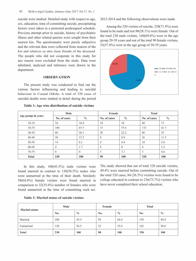
45 Medico-legal Update, January-June 2017, Vol.17, No. 1
2012-2014 and the following observations were made.
Among the 320 victims of suicide, 230(71.9%) were found to be male and rest 90(28.1%) were female. Out of the total 230 male victims, 160(69.6%) were in the age group 20-39 years and out of the total 90 female victims, 52(57.8%) were in the age group of 20-39 years.
suicide were studied. Detailed study with respect to age, sex, education, time of committing suicide, precipitating factors were taken in a pretested predesigned schedule. Previous attempt prior to suicide, history of psychiatric illness and other related queries were sought from their nearest kin. The questionnaire were purely subjective and the relevant data were collected from nearest of the kin and relatives so also close friends of the deceased. The people who did not cooperate in this study for any reason were excluded from the study. Data were tabulated, analysed and inference were drawn in the department.
OBSERVATION
The present study was conducted to find out the various factors influencing and leading to suicidal behaviour in Coastal Odisha. A total of 320 cases of suicidal deaths were studied in detail during the period
Table 1: Age wise distribution of suicide victims
Age group in years Male Female Total No. of cases % No. of cases % No. of cases %
10-19 24 10.4 24 24 48 15 20-29 100 43.5 32 35.6 132 41.3 30-39 60 26.1 20 22.2 80 25 40-49 28 12.2 8 8.9 36 11.3 50-59 14 6.1 4 4.4 18 5.6 60-69 4 1.7 0 0 4 1.2 70-79 0 0 2 2.2 2 0.6 Total 230 100 90 100 320 100
In this study, 100(43.5%) male victims were found married in contrast to 130(56.5%) males who were unmarried at the time of their death. Similarly 58(64.4%) female victims were found married in comparison to 32(35.6%) number of females who were found unmarried at the time of committing such act.
The study showed that out of total 320 suicide victims, 49.4% were married before committing suicide. Out of the total 320 cases, 84 (26.3%) victims were found to be college educated in contrast to 236(73.7%) victims who have never completed their school education.
Table 2: Marital status of suicide victims
Marital status Male Female Total
No. % No. % No. %
Married 100 43.5 58 64.4 158 49.4
Unmarried 130 56.5 32 35.6 162 50.6
Total 230 100 90 100 320 100
Medico-legal Update, January-June 2017, Vol.17, No. 1 46 47 -legal Update, January-June 2017, Vol.17, No. 1
Among male victims, 158(68.7%) were unemployed and 82(91.1%) females were unemployed at the time of committing suicide. This showed that, total 240(75%) out of 320 cases were unemployed at the time of their death. Out of the total 230 male suicide cases, 130(56.5%) were found to be having some sort of addiction. Male preferred alcohol in 100(76.9%) cases followed by tobacco 30(23.1%) in their life. None of the female showed any kind of addiction prior to committing suicide.
The precipitating factor for committing suicide ascertained from the next of kin or close relatives of the victims showed that the leading cause of death was family conflict, 210 (65.6%) cases and frustration in love affair in 24 (7.5%) of cases. Both male and female commit suicide by consuming poison i.e, in 248(77.5%) of cases out of 320 followed by hanging in 66(20.6%) cases.
Table 3: Precipitating factors that led to suicide
Precipitating factor for suicide No. of victims Percentage (%)Family conflict 210 65.6Frustration in love affair 24 7.5Unemployment 18 5.6Mental illness 18 5.6Chronic painful Disease 18 5.6Infertility 10 3.1Failure in examination 6 2Not known 16 5Total 320 100
Table 4: Methods adopted to commit suicide
Method adopted for suicide
Male Female Total
No. Percentage(%) No. Percentage(%) No. Percentage(%)
Consuming poison 174 75.6 74 82.2 248 77.6
Hanging 52 22.6 14 15.6 66 20.6
Burning 0 0 2 2.2 2 0.6
Railway track 2 0.9 0 0 2 0.6
Drowning 2 0.9 0 0 2 0.6
Total 230 100 90 100 320 100
Out of the total 320 victims, 22 (6.9%) cases were found to have attempted suicide previously. 260(81.3%) victims committed suicide during night and 54 (16.9%) cases committed during day hour and in 6(1.8%) cases, time of suicide was not known to the relatives.
DISCUSSION
The present study showed that 230 (71.9%) males and 90 (28.1%) females committed suicide and the male to female ratio is 2.5 : 1 which is almost similar to the study of Williams JMG and Pallock LR(1993)6. This suggests that males commit suicide more often than females in this geographical region.
It is observed that 160 (69.6%) number of male victims and 52 (57.8%) females are in the age group of 20-39 year. It is the most active period in life and fluctuation of emotions are commonly noticed in this age group. People in 20-39 years age group are more susceptible for frustration and break down because of increased incidences of unemployment, failure in love affair, dowry torture and domestic violence in our society.
In our study, 43.5% male victims were married in comparison to 64.4% female victims who committed suicide after getting married. This is suggesting married
-legal Update, January-June 2017, Vol.17, No. 1 46 47 Medico-legal Update, January-June 2017, Vol.17, No. 1
female were more tend to such extreme decision (64.4%) than their male counterpart. This could be due to lack of adjustment with their marital life and in-laws and dowry torture in our society. Our study shows that out of the total 320 cases, 240(75%) victims were unemployed at the time of committing suicide which suggests that majority of victims were unemployed or economically dependent on others at the time of suicide and unemployment and poverty are a major cause of suicide.
It is observed that 236 (73.7%) victims have never completed their school education in this study group. This suggests that suicide rate can be reduced in our society by increasing the literacy rate. Out of the total 230 male victims, 130(56.5%) males were found to have some sort of addiction at the time of suicide and alcohol was the most preferred substance. This suggests that addiction to alcohol is associated with majority of the male suicide in our community.
Our study shows that, a majority 210(65.6%) victims commit suicide due to some family conflict. This clearly suggests that disharmony existing between family members trigger to take the fatal decision. It is observed that 248 (77.5%) victims in this study commit suicide by consuming some poisonous substance which is followed by hanging in 66 (20.6%) cases. This correlates with the study of Shukla G.D. et al (1990)7. This could be due to the fact that people of Coastal Odisha depend mostly on agriculture and poisonous substance like insecticides and herbicides are easily available in this part of the world.
In diurnal variation, out of the study population 81.3% victims committed suicide in night. This denotes lack of day light correlates with depression and then to suicide, which is similar to the study of Soreff S.M.(2004)8. Out of the total 320 victims, 22(6.9%) were found to have attempted suicide previously. This shows someone who has already tried to take his own life once is more likely to do it again.
CONCLUSION
With over millions of persons committing suicide in the world and millions more attempting it every year, in spite of all legal, moral, social and religious barriers, suicide today has come to constitute a major public health issue confronting civilized societies throughout the world. In the present study, common causes of committing suicide were found to be family conflict, frustration in love affair, unemployment, discordant
relationship among spouses etc. The social scientists should think of seriously to develop some sort of primordial prevention. Some sessions of counselling to couples with history of broken family may be helpful to some extent. The follow up of survivors should be routinely done to avoid further steps.
Ethical Clearance – This is not applicable in this study as because this study was conducted on dead bodies brought for medico legal autopsy and no organs of the body was taken out for the study purpose. This study was completely based upon demographic profiles of the suicidal victims which were ascertained from the verbal information provided by the close relatives and friends of the deceased and from the data provided in the Inquest report and Dead body challan.
Source of Funding – Self
Conflict of Interest - Nil
REFERENCES
1. Vijayakumar L. Indian research on Suicide. Indian Journal of Psychiatry. 2010 Jan; 52 (suppl 1): s291-s296.
2. Sharija S., Sreekumar K., Geetha O., Epidemiological Profile of Suicide by Hanging in Southern Parts of Kerala: An autopsy based Study. Journal of Indian Academy of Forensic Medicine.2011 July-September; 33(3): 237-240.
3. Rao A.V., Kuruvilla K., Suicidal Behaviour, “Psychiatry”, B-1 Churcill Livingstone, 1st Ed., st Ed., st
1997; 220-220.
4. Stengel E., Suicide and attempted suicide, “Prediction of Suicide”, Penguin Books, New York, Reprint 1983.
5. Vyas J.N., Ahuja N., Text Book of Post Graduate Psychiatry, 2nd ed., 1999; 526-556.nd ed., 1999; 526-556.nd
6. Williams J.M.G., & Pollock L.R., Factors mediating suicidal behaviour : Their utility in primary & secondary prevention, Journal of Mental Health. 1993(2); 3-26.
7. Shukla G.D., & Mishra D., Suicide in Jansi City, Indian Journal of Psychiatry, 1990; 32: 44-51.
8. Soreff S.M., The Psychiatrist role and responses and responsibilities when a patient commit suicide. Annual Journal of Psychiatry, 1991 June; 148(6): 739-43.

49 -legal Update, January-June 2017, Vol.17, No. 1
Can Lip Print Patterns Determine Sex of an Individual?
Umesh Babu R1, Ameena Ahamed2, Imaad M I3
1Professor and Head, 2Assistant Professor, Department of Forensic Medicine and Toxicology, 3Assistant Professor, Community Medicine, Kannur Medical College, Anjarakandy, Kerala
ABSTRACT
The wrinkles and grooves on labial mucosa, called as sulci labiorum forms a characteristic pattern called as “LIP PRINTS” and the study of which is referred to as CHEILOSCOPY. This is unique to an individual just like the fingerprints. The print patterns show some sexual dimorphic differences in distribution patterns. The study reveals that there is a statistically significant sex distinguishing features in the lip prints. The study reveals among other findings that
• All quadrants different -variety of lip prints was seen most commonly in males
• All quadrants same -variety of lip prints were common in females.
• Type IV was seen most commonly in females whereas Type I, I1 and III occurred more in male lip prints.
However the methodology used for recording and studying the prints is easy, economical and convenient. No specialized equipment or training is required to use this method.
Keywords; Lip print, cheiloscopy, sexual dimorphism.
Author for Correspondence: Dr Umesh Babu R,Professor and Head, Department of Forensic Medicine and Toxicology, Kannur Medical College, Anjarakandy, Kerala.670612, PH; 9986116341Email; [email protected]
INTRODUCTION
Finger prints, postmortem reports, anthropometry and of late DNA finger printing have been successful in personal identification in many of the cases. Just like these methods lip prints can be instrumental in identifying a person positively and can be used to verify the presence or absence of a person at the scene of crime.
1 The wrinkles and grooves on labial mucosa, called as sulci labiorum forms a characteristic pattern called as “LIP PRINTS” and the study of which is referred to as CHEILOSCOPY. 2 This is unique to an individual just like the fingerprints.2,3,4 The biological phenomenon of systems of furrows on the red part of the human lips was first noted and described by anthropologist R.S. Fischer.4,5, However, until 1930, anthropology merely mentioned the existence of furrows without suggesting a
practical use of the phenomenon3. Japanese have carried out extensive research in this matter. In the period 1968 to 1971 two Japanese scientists, Y. Tsuchihashi and T. Suzuki examined 1364 persons at Department of Forensic Odontology at Tokyo University and established that the arrangement of lines on the red part of the human lips is individual and unique for each human being.5 Cheiloscopic research was also carried out by specialists in anthropology, odontology, forensic medicine and forensic science in Brazil, Iran, Hungary, France, Germany, Italy, United Kingdom, Soviet Union and Czechoslovakia5. Lip prints are useful not only for identification purpose but also can be used in detection work, being the source of tactical and criminalistic information. A lip print can give clue about the character of an event, number of people involved, sexes, cosmetics used, habits, occupational traits and also pathological changes, if at all any, which by itself may form an identification feature of an individual. Hence lip prints have a potential evidentiary value which plays a prominent role in linking the criminal with the crime and also establishing identity of an individual. Hence, the study of lip prints need to be developed so as to prove it is one of the useful tool for investigation. Hence in this
DOI Number: 10.5958/0974-1283.2017.00010.X
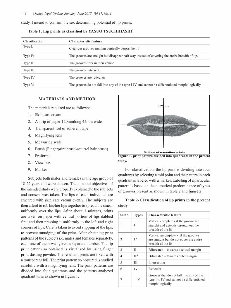
49 Medico-legal Update, January-June 2017, Vol.17, No. 1
study, I intend to confirm the sex determining potential of lip prints.
Table 1: Lip prints as classified by YASUO TSUCHIHASHI7
Classification Characteristic featureType I: Clear-cut grooves running vertically across the lip
Type I1: The grooves are straight but disappear half way instead of covering the entire breadth of lip.
Type II: The grooves fork in their course
Type III: The grooves intersect
Type IV: The grooves are reticulate
Type V: The grooves do not fall into any of the type I-IV and cannot be differentiated morphologically
MATERIALS AND METHOD
The materials required are as follows:
1. Skin care cream
2. A strip of paper 120mmlong 45mm wide
3. Transparent foil of adherent tape
4. Magnifying lens
5. Measuring scale
6. Brush (Fingerprint brush\squirrel hair brush)
7. Proforma
8. View box
9. Marker
Subjects both males and females in the age group of 18-22 years old were chosen. The aim and objectives of the intended study were properly explained to the subjects and consent was taken. The lips of each individual are smeared with skin care cream evenly. The subjects are then asked to rub his/her lips together to spread the smear uniformly over the lips. After about 3 minutes, prints are taken on paper with central portion of lips dabbed first and then pressing it uniformly to the left and right corners of lips. Care is taken to avoid slipping of the lips, to prevent smudging of the print. After obtaining print patterns of the subjects i.e. males and females separately, each one of them was given a separate number. The lip print pattern so obtained is visualized by using finger print dusting powder. The resultant prints are fixed with a transparent foil. The print pattern so acquired is studied carefully with a magnifying lens. The print patterns are divided into four quadrants and the patterns analyzed quadrant wise as shown in figure 1.
Figure 1- print pattern divided into quadrants in the present study.
For classification, the lip print is dividing into four quadrants by selecting a mid point and the pattern in each quadrant is labeled with a marker. Labeling of a particular pattern is based on the numerical predominance of types of grooves present as shown in table 2 and figure 2.
Table 2- Classification of lip prints in the present study
Sl.No. Types Characteristic feature
1 IVertical complete - if the groove are straight and extends through out the breadth of the lip
2 I IVertical incomplete - If the grooves are straight but do not cover the entire breadth of the lip
3 II Bifurcated – towards occlusal margin
4 II I Bifurcated – towards outer margin
5 III Intersecting
6 IV Reticular
7 VGrooves that do not fall into any of the type I to IV and cannot be differentiated morphologically

Medico-legal Update, January-June 2017, Vol.17, No. 1 50 51 -legal Update, January-June 2017, Vol.17, No. 1
Figure 2 - Classification of lip prints in the present study
RESULTS
Table 3: Pattern predominance
Quadrants Male Female Male % Female %
Type I
First quadrant 37 21
17.5 15.5Second quadrant 34 20Third quadrant 17 22Fourth quadrant 25 15
Type I1
First quadrant 05 04
4.6 2Second quadrant 12 05Third quadrant 06 04Fourth quadrant 06 01
Type II
First quadrant 11 08
7 6Second quadrant 18 07Third quadrant 12 08Fourth quadrant 03 08
Type II1
First quadrant 30 28
28 28Second quadrant 29 42Third quadrant 64 46Fourth quadrant 55 13
Type III
First quadrant 29 22
21 15.5Second quadrant 34 23Third quadrant 35 20Fourth quadrant 36 43
Type IV
First quadrant 37 32
14.5 22Second quadrant 22 15Third quadrant 12 21Fourth quadrant 22 07
Type V
First quadrant 08 11
6 6.3Second quadrant 13 07Third quadrant 09 07Fourth quadrant 08 07

-legal Update, January-June 2017, Vol.17, No. 1 50 51 Medico-legal Update, January-June 2017, Vol.17, No. 1
Table 3 shows the descriptive statistics of lip print patterns in the four quadrants
Type I is more common in the upper lip especially
in males where as Type II1 is seen more commonly in lower lip especially in males. Types I, I1 and III are more commonly seen on males whereas type IV is more commonly seen in females.
Table 4: Frequency and repetition of different patterns among subjects
All Q* same
2 Q* same 3 Q* same All Q*
different
No. % No. % No. % No. %
Male 21 12.35 90 52.9 40 23.5 09 5.3
Female 21 16.7 67 53.2 32 25.4 07 4.1
Average 14.53% 53.05% 24.45% 47%
* Q = Quadrants
Table 4 shows the frequency and repetition of the print patterns in the four quadrants. It is found that more than 90% of the subjects have same patterns in at least two quadrants. Most common variety found is that of two quadrants having same types which is seen in 53% of subjects. Least common variety being all the quadrants having different lip print patterns followed by all quadrants having same patterns (14.5%) and 3 quadrants having same patterns (24.5%).
Table 5; Pearsons chi square test
Quadrants Value df Asymp. Sig (2-sided)
Quadrant 1 16.228a 7 0.023
Quadrant 2 16.080a 7 0.024
Quadrant 3 14.317a 7 0.046
Quadrant 4 15.305 7 0.032
Table 5 reveals that all the quadrants show statistically significant (P<0.05 )difference between genders. First and second quadrants show greater significance of sex differentiation than third and fourth quadrants.
DISCUSSION
In the past, some researchers have worked on lip prints with the idea of proving that a gender difference do exist in lip prints..1-6 In the present study a statistically significant differences were observed between the prints
of two genders as shown in tale 5. The study also reveals that type IV was more predominant in females in upper lip and type I in males in upper lip. Previous studies have not considered the ridge count as an identification feature. This present study has attempted to trace the ridge count differences between sexes in a particular pre selected segment of the prints. Two Indian scientists S.P. Vahanwala and B.K. Parekh have shown that lip prints can prove vital in identifications. In their study on 50 males and 50 females lip patterns following conclusions are drawn6.
• Type II is common in males in 2nd quadrant i.e. left part of upper lip. This could help in sex determination.
• Type II was not seen in lower lip and only if it did it was in male subjects on the upper lip.
• Individuals with all quadrants having different patterns were common in males, whereas having same patterns in all quadrants were seen in females.
The print patterns in the present study show some sexual dimorphic differences in distribution patterns.
• All quadrants different -variety of lip prints was seen most commonly in males
• All quadrants same -variety of lip prints were common in females.
• Type IV was seen most commonly in females whereas Type I, I1 and III occurred more in male lip prints.

Medico-legal Update, January-June 2017, Vol.17, No. 1 52 53 -legal Update, January-June 2017, Vol.17, No. 1
CONCLUSION
The lip prints of 300 subjects of the south Indian population were studied by obtaining a thin smear of the prints and visualizing by the finger print powder (black), to know sexual dimorphic features if any. The need to study the lip prints for identification has been stressed by many workers for the past 50 years, resulting in the importance of this technique being recognized and accepted in many countries. Due to its application, many medico-legal investigations have been reported to be successful in identifying the culprits. In this study, sex determining potential of the lip prints has been analyzed. Newly determined percentages of predominance of various types of lip print patterns (as classified by previous authors) in various quadrants in males and females are presented and evaluated. The findings obtained are consistent with other studies published previously. The study reveals that there is a statistically significant sex distinguishing features in the lip prints. However the methodology used for recording and studying the prints is easy, economical and convenient. No specialized equipment or training is required to use this method. Thus, this study shows that lip print patterns are a reliable means of determining sex of an individual.
Conlict of Interest: None.
Source of Funding: Self.
Ethical Clearance: Obtained from institutional ethical committee.
REFERENCES
1. Utsuno H, Kanoh T, Tadokoro O, Inoue K. Preliminary study of post mortem identi cation using lip prints. Forensic Sci Int 2005;149:129-32.
2. Qudusia Sultana, M.H Shariff, Muhammed Asif, Ramakrishna Avadhani. CHEILOSCOPY: A SCIENTIFIC APPROACH FOR PERSONAL IDENTIFICATION. Int J Anat Res 2014;2(4):668-672.
3. Sivapathasundaram B, Ajay Prakash, Sivakumar. Lip prints (Cheiloscopy) Indian Journal of Dental Research Dec. 2001; 12 (4): 234-237.
4. Preeti S., Sushmita S., Vanita R., Cheiloscopy; The study of lip prints in sex identification, J of Forensic Dental Science, Jan-jun 2009;01;01;24-
27.
5. Kazuo Suzuki and Yasuo Tsuchihashi. New attempt of personal identification by means of lip prints. Journal of Indian Dental Association 1970: 8-9.
6. Vahanwala SP, Parekh BK. Study of lip prints as on Aid to Forensic methodology. JDA, 71; Oct. 2000: 268-271.
7. Yasuo Tsuchihoshi. Studies on personal identification by means of lip print. Journal of Forensic Sciences, Tokyo, Japan 1974: 233-248.
8. Anil Aggarwal. http://www.geradts.com, II, issue 2, 2004, Lifeloom.com.
9. Strandling S, Face and Scalp. Gray’s Anatomy, 39th edition, Churchill Livingstone, London, 2005: 506-507pp.
10. Apurba Nandy. Identification of an individual. Principles of Forensic Medicine and Toxicology, 1st edition, New Central Book Agency (P) Ltd, Calcutta, 1995: 99-100pp.
11. Whittaker D.K. and Moc Donald D.G. Forensic Dentistry. Soft Tissues, Wolfe Publishing Ltd., England, 1989: 55-56pp.
12. Pillay VV. Identification. Handbook of forensic medicine and toxicology. 12th edition, Paras Publications, Bangalore, 2001: 51pp
13. Tedeshi. Forensic Odontology. Forensic Medicine, Vol. II, Physical Trauma, W.B. Saunders Company, Philadelphia, 1997: 1117-1119pp.
14. Polson CJ, Gee DJ, Knight B. Identification. Essentials of forensic medicine, 4th edition, Pergamon Press, Oxford, 1985: 71pp.
15. Taylor. Forensic odontology. Principles and Practice of Medical Jurisprudence. 13th edition, Churchill Livingstone, 2000: 196-197pp.
16. Dr. Pramod K, Daya. Special methods in dental identification, Textbook of Forensic Odontology. 12th edition, Paras Medical Publisher, Hyderabad, 1998: 51pp.
17. Mukherjee JB. Identification. Forensic Medicine of Forensic and Toxicology, second edition, Vol. I, Arnold Associates, New Delhi, 1996: 151pp.
18. LeMoyne Snyder. Identification of Dead Bodies. Homicide Investigation, 3rd edition, Charles C Thomas Publishers, Illinois, USA, 1997: 65pp.

-legal Update, January-June 2017, Vol.17, No. 1 52 53 Medico-legal Update, January-June 2017, Vol.17, No. 1
19. Kasprzak J Cheiloscopy, Encyclopedia of Forensic Sciences, Vol. I, Academic Press, New York, 358-363pp.
20. Ball J. The current status of lip prints and their use for identification. J Forensic odeontostomatology 20 (2); 2002: 43-46pp.
21. Curtis A Mertiz. Dental identification. The Dental Clinics of North America, W.B. Saunders Company, Philadelphia, London, Vol. 21 (1):: 64-5pp.
22. Utsuno H, Kanoh T, Tadokovo O, Inoue K. Preliminary study of post mortem identification using lip prints. Forensic science International 149 (2-3); 2005: 129-32pp.
23. Hirth L, Gottsche H, Goedde HW. Lip prints variability and genetics, Humangenetik 30 (1): 1975: 47-62pp.
24. Dr. Parikh CK. Identification in mass disasters. Textbook of medical jurisprudence and toxicology. 5th edition, CBS Publishers, 1990; 75-76pp.
25. Muhammad Y, Choudhry. Comparison of minute smears of lipstick by microspectrophotometry
and scanning electron microscopy/Energy – Disperssive spectroscopy. Journal of Forensic Sciences 86; 1991: 366-375pp.
26. Segui MA, Feucht MM, Pones AC, Femando A, Verdu Pascual. Persistent lipsticks and their lip prints, new hidden evidence at the crime scene. Forensic Science International, 1121; 200: 41-47.
27. Ehara Y, Masumo Y. Identification of lipstick smears by fluorescence observation and Purge and trap gas chromatography. Forensic Science International 96; 1998: 1-10.
28. Victor P Eroschenko. Digestive system. Atlas of Histology, 8th edition, difiore’s Publications, Mosco, Idahe, 1996; 150-251pp.
29. Kerkovitz BKB, Holland GR, Moxham BJ. Oral mucosa. Textbook of oral anatomy, Histology and Embryology, 3rd edition, Mosby Publications, Edinburgh, 2002: 230-232pp.
30. Lloyd Dubrul. Ora viscera. Oral Anatomy, 8th
edition, Ishiyaker Euro America Inc., St Louis, Tokyo, 1990: 161-162pp.
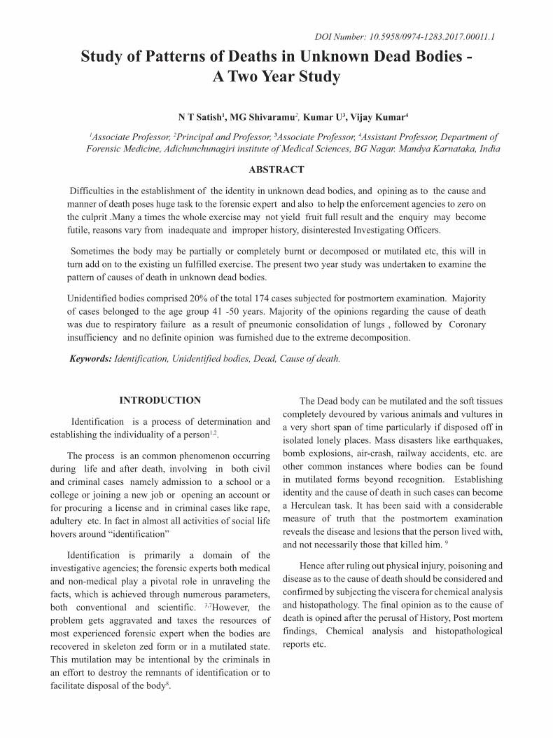
55 -legal Update, January-June 2017, Vol.17, No. 1
Study of Patterns of Deaths in Unknown Dead Bodies - A Two Year Study
N T Satish1, MG Shivaramu2, Kumar U3, Vijay Kumar4
1Associate Professor, 2Principal and Professor, 3Associate Professor, 4Assistant Professor, Department of Forensic Medicine, Adichunchunagiri institute of Medical Sciences, BG Nagar. Mandya Karnataka, India
ABSTRACT
Difficulties in the establishment of the identity in unknown dead bodies, and opining as to the cause and manner of death poses huge task to the forensic expert and also to help the enforcement agencies to zero on the culprit .Many a times the whole exercise may not yield fruit full result and the enquiry may become futile, reasons vary from inadequate and improper history, disinterested Investigating Officers.
Sometimes the body may be partially or completely burnt or decomposed or mutilated etc, this will in turn add on to the existing un fulfilled exercise. The present two year study was undertaken to examine the pattern of causes of death in unknown dead bodies.
Unidentified bodies comprised 20% of the total 174 cases subjected for postmortem examination. Majority of cases belonged to the age group 41 -50 years. Majority of the opinions regarding the cause of death was due to respiratory failure as a result of pneumonic consolidation of lungs , followed by Coronary insufficiency and no definite opinion was furnished due to the extreme decomposition.
Keywords: Identification, Unidentified bodies, Dead, Cause of death.
INTRODUCTION
Identification is a process of determination and establishing the individuality of a person1,2.
The process is an common phenomenon occurring during life and after death, involving in both civil and criminal cases namely admission to a school or a college or joining a new job or opening an account or for procuring a license and in criminal cases like rape, adultery etc. In fact in almost all activities of social life hovers around “identification”
Identification is primarily a domain of the investigative agencies; the forensic experts both medical and non-medical play a pivotal role in unraveling the facts, which is achieved through numerous parameters, both conventional and scientific. 3,7However, the problem gets aggravated and taxes the resources of most experienced forensic expert when the bodies are recovered in skeleton zed form or in a mutilated state. This mutilation may be intentional by the criminals in an effort to destroy the remnants of identification or to facilitate disposal of the body8.
The Dead body can be mutilated and the soft tissues completely devoured by various animals and vultures in a very short span of time particularly if disposed off in isolated lonely places. Mass disasters like earthquakes, bomb explosions, air-crash, railway accidents, etc. are other common instances where bodies can be found in mutilated forms beyond recognition. Establishing identity and the cause of death in such cases can become a Herculean task. It has been said with a considerable measure of truth that the postmortem examination reveals the disease and lesions that the person lived with, and not necessarily those that killed him. 9
Hence after ruling out physical injury, poisoning and disease as to the cause of death should be considered and confirmed by subjecting the viscera for chemical analysis and histopathology. The final opinion as to the cause of death is opined after the perusal of History, Post mortem findings, Chemical analysis and histopathological reports etc.
DOI Number: 10.5958/0974-1283.2017.00011.1

55 Medico-legal Update, January-June 2017, Vol.17, No. 1
AIM AND OBJECTIVES
1. To study the cause of death, and the pattern of cause of death in unknown dead bodies.
2. To analyze the various efforts made on part of the investigating officer and the autopsy surgeon to establish the identity of the unidentified bodies.
MATERIAL AND METHOD
Unidentified bodies brought for postmortem examination to the mortuary, Department of Forensic Medicine & Toxicology, Adichunchungiri institute of medical sciences & Hospital
BG Nagar, during the two year period April 2012 – March2014 comprised the material for the study. Data regarding these cases was compiled from the postmortem reports, inquest papers; detailed history elicited from the concerned police officials at the time of autopsy, etc
A thorough search of the literature did not yield much information regarding the identification of the unidentified dead in the Indian context. Mostly, they were devoted to individual body identification or identification of victims of mass disasters. 10-11
RESULTS AND DISCUSSION
Table 1: Age-wise distribution of unidentified cases
Age Group Total
Serial Age Total cases
1 0 -10 0
2 11-20 0
3 21-30 8
4 31-40 4
5 41-50 9
6 51-60 8
7 60 -70 6
Table 2 Data/material retrieved by the autopsy surgeon
Material cases
Sl no material cases
1 Clothes 18
2 Tattoos 5
3 Deformities 2
4 Marks of identification 55
Table 3 Cases sent for analysis
Sl no Examination cases
1 Chemical analysis 5
2 Histopathoplogy 10
3 Both 5
4 Not sent 20
Table 4 Opinion as to the Cause of Death
Sl no Cause of Death No. cases
1 Respiratory failure 18
2 OP Compound poisoning 2
3 Coronary insufficiency 12
4 No definite opinion 3
DISCUSSION
Unidentified bodies comprised 10% of the total 180 cases subjected for postmortem examination. Majority of cases belonged to the age group 41 -50 years with male preponderance. The maximum number of cases were encountered in the month of June followed by May. Majority of the opinions regarding the cause of death was due to respiratory failure as a result of pneumonic consolidation of lungs , followed by no definite opinion was furnished due to the extreme decomposition.
The police complete their formalities viz photography and publication of details of un known in local and regional dailies. The clothes of all the cases was handed over (to facilitate the process of identification) assisting to lift fingerprints was done. finger pulps of all the ten fingers in separate sealed packets was collected , noted the marks of identification/ deformities/tattoos in all cases and blood for DNA analysis was collected and handed to police personnel.
Homicidal manner of death with fire arm injuries or sharp weapons injuries were not detected in the present study. One of the probable reasons could be that the police make sincere efforts to trace the victim in order to solve the crime on priority basis and thus the body no longer remains unknown. Moreover, homicide of unknown persons is a rarity unless it is done with the motive of robbery, or when the body is so mutilated by the criminals that identification may become very difficult or sometimes impossible.
Medico-legal Update, January-June 2017, Vol.17, No. 1 56
CONCLUSION
The efforts for identification of an individual is done by the. Bodies that are unknown/unclaimed should be presented for autopsy forthwith without any delay so that decomposition and other artifacts do not set in and obscure the findings of the postmortem examination. The rule for preservation of an unknown body for 72 hours applies for its disposal and not for its postmortem examination.
Thus valuable data regarding the cause of death can be established. Active investigation and modern investigative techniques are to be used, workload of the police officers needs to be redistributed, and accountability of the police has to be fixed to get the body identified. Simple color photography of the body, especially of the clothes, the tattoo marks, scars, deformities, etc will help the police in identifying such bodies.
Dental records of the deceased should be maintained, which was not done in cases under study, if a probable relative comes with a dental record. In all cases of unknown bodies, whole body radiographs may be useful for establishing the identity, which may reveal an old fracture or an implant (in the presence of a surgical orthopedic scar).
DNA analysis and fingerprinting should be done in each and every case so that proper records will be there for identification of the deceased years after the death or postmortem.
With a little effort on the part of the doctor and the investigation officer, in many cases, surprisingly, a large amount of information can be obtained even in the presence of advanced state of decomposition. Such information can be of use to the doctor and the investigation agencies both in establishing identity and in forming an opinion about the cause and manner of death.
Ethical Clearance- Taken from ethical committee.
Source of Funding- Self
Conflict of Interest - Nil
REFERENCES
1. Kumar A, Tyagi A, Aggarwal NK. Sex determination by morphology of talus bone. J For Med Toxicol 2008;25(1):50-3
2. Vij K. Textbook of Forensic Medicine and Toxicology Principles and practice 5thed Elsevier 2011.p.35-7
3. Jayaprakash PT, Singh B, Yusop RAAM, Asmuni HS. Skull-photo superimposition: a remedy to the problem of unidentified in Malaysia. Malaysian J For Sci 2010;1(1):34-41
4. Balloch J. Identifying the unknown dead: new system cross-checks missing person cases with unidentified bodies. http://m.knoxnews.com/news/2012/jan/15/identifying-the-unknown-dead-new-system-that-to/ published Jan 15 2012. Accessed March 20, 2012
5. Riepert T, Ulmcke D, Schweden F, Nafe B. Identification of unknown dead bodies by X-ray image comparison of the skull using the X-ray simulation program . For Sci Int 2001;117(1-2):89-98
6. Positive identification of the dead. Crime Blog. http://www.crimemuseum.org/blog/positive-identification-of-the-dead/ Accessed 21st March 2012
7. Smith EL. Scientific identification of deceased prevents misidentification. Forensic Science @ suite 101http://erikalynsmith.suite101.com/scientific-identification-of-deceased-prevents-misidentification-a360528. Published May 20, 2011 Accessed 21st March 2012
8. Kumar A, Kumar SGA, Tyagi A, Aggarwal NK. Sex determination using discriminant function analysis in adult talus and calcaneum bones. Int J Med Toxicol legal med: 2009; 12(1):4-12.
9. Reddy KSN. The essentials of Forensic Medicine and Toxicology 28th ed; K Sugna Devi 2009.p.112-3.
10. Job C. Determination of cause of death in decomposed bodies – a regional study. JIAFM 2009;31(1):11-17
11. Kahana T, Hiss J. Personal identification based on radiographic vertebral features. Am J For Med Pathol 2002;28(1):36-41.

-legal Update, January-June 2017, Vol.17, No. 1 56
A Study of Prediction of Stature from Percutaneous Ulna Length
Madhu Sudhan S1, H C Govindaraju2, Hemanth Raj M N3
1Assistant Professor, 1Assistant Professor, 1 2Professor, 2Professor, 2 3Associate Professor, Department of Forensic Medicine & Toxicology, 3Associate Professor, Department of Forensic Medicine & Toxicology, 3
Shridevi Institute of Medical Sciences & Research Hospital, NH-4, Sira Road, Tumkur District, Karnataka State, India
ABSTRACT
Introduction: Height is one the important parameter in the anthropometric study of humans. Establishment of alternative methodologies for estimation of stature of the person is important for a number of reasons like in condition, where stature estimates needed to be made from fragments of bones brought for forensic examination. Material and Method: The present study was conducted on 120 medical students (60 males and 60 females) of Shridevi institute of medical sciences and research hospital, tumkur. The right and left and left ulna length and height was measured by the same observer, same instrument and between 2.00pm-4.00pm. Results: Observations were analysed and tabulated. Regression equation for estimation of stature are derived for both right and left ulna in both sex and coefficient correlation of height with respect to ulna was also calculated. P value was found to be significant in all cases. Conclusion: The present study would be useful for prediction or estimation of stature from ulna length in medicolegal investigations and in anthropometry.
Keywords: Stature; Height; Percutaneous ulna length; Regression equation; Coefficient correlation.
INTRODUCTION
By stature we mean body height or length of a person. It is an important criterion for identification of a person1. Growth the vital process is considered by measuring the height of the person, which itself is a sum of length of certain bones and appendages of the body represent certain relationship with form of proportion to the total stature2. Assessment of height from different parts of the body by anthropometric study of skeleton is an area of interest to anatomist, anthropologist and forensic experts3. Establishment of alternative methodologies for stature estimation is important for a number of reasons like in condition where stature estimates needed to be made from fragments of bones brought for forensic examination. And also for evaluation of nutritional status
relies on accurate measurement of not only body weight but also height. Most studies have stressed that regression formula for stature estimation should be population specific. So the present study attempts to estimate stature from ulna length, as olecranon and styloid process of the ulna are easily felt through the skin.
MATERIALS AND METHOD
The present study was conducted on 120 medical students, who are randomly selected (60 males and 60 females) of Shridevi institute of medical sciences and research hospital, tumkur after obtained informed consent from the students. The age of the students ranged from 20-23 years who belonged to South Indian region. The ulna length was measured as the direct distance between the tip of the olecranon process to the tip of the styloid process while the elbow flexed and palm spread over opposite shoulder using sliding callipers (percutaneous ulna length). Measurements of length of right and left ulna were taken separately for calculation. Height was measured by measuring crown to heel standing erect posture with and head in Frankfurt plane. All the measurements were taken between 2:00pm
Corresponding authorDr Hemanth Raj M NAssociate Professor, Dept of Forensic MedicineShridevi Institute of Medical Sciences & Research Hospital, Sira Road, NH-4, Bypass, Tumkur 572106Karnataka, Mobile: 9886301287
DOI Number: 10.5958/0974-1283.2017.00012.3
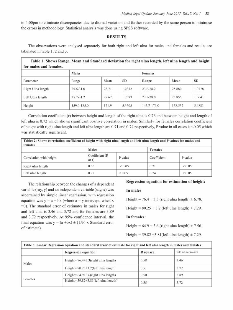
Medico-legal Update, January-June 2017, Vol.17, No. 1 58 59 -legal Update, January-June 2017, Vol.17, No. 1
to 4:00pm to eliminate discrepancies due to diurnal variation and further recorded by the same person to minimise the errors in methodology. Statistical analysis was done using SPSS software.
RESULTS
The observations were analysed separately for both right and left ulna for males and females and results are tabulated in table 1, 2 and 3.
Table 1: Shows Range, Mean and Standard deviation for right ulna length, left ulna length and height for males and females.
Males Females
Parameter Range Mean SD Range Mean SD
Right Ulna length 25.6-31.0 28.71 1.2332 23.6-28.2 25.880 1.0778
Left Ulna length 25.7-31.2 28.62 1.2093 23.5-28.0 25.855 1.0643
Height 159.0-185.0 171.9 5.3505 145.7-176.0 158.532 5.4883
Correlation coefficient (r) between height and length of the right ulna is 0.76 and between height and length of left ulna is 0.72 which shows significant positive correlation in males. Similarly for females correlation coefficient of height with right ulna length and left ulna length are 0.71 and 0.74 respectively, P value in all cases is <0.05 which was statistically significant.
Table: 2: Shows correlation coefficient of height with right ulna length and left ulna length and P values for males and females
Males Females
Correlation with height Coefficient (R or r) P value Coefficient P value
Right ulna length 0.76 < 0.05 0.71 < 0.05
Left ulna length 0.72 < 0.05 0.74 < 0.05
The relationship between the changes of a dependent variable (say, y) and an independent variable (say, x) was ascertained by simple linear regression, with regression equation was y = a + bx (where a = y intercept, when x =0). The standard error of estimates in males for right and left ulna is 3.46 and 3.72 and for females are 3.89 and 3.72 respectively. At 95% confidence interval, the final equation was y = (a +bx) ± (1.96 x Standard error of estimate).
Regression equation for estimation of height:
In males
Height = 76.4 + 3.3 (right ulna length) ± 6.78.
Height = 80.25 + 3.2 (left ulna length) ± 7.29.
In females:
Height = 64.9 + 3.6 (right ulna length) ± 7.56.
Height = 59.82 +3.81(left ulna length) ± 7.29.
Table 3: Linear Regression equation and standard error of estimate for right and left ulna length in males and females
Regression equation R square SE of estimate
MalesHeight= 76.4+3.3(right ulna length) 0.58 3.46
Height= 80.25+3.2(left ulna length) 0.51 3.72
FemalesHeight= 64.9+3.6(right ulna length) 0.50 3.89Height= 59.82+3.81(left ulna length) 0.55 3.72

-legal Update, January-June 2017, Vol.17, No. 1 58 59 Medico-legal Update, January-June 2017, Vol.17, No. 1
DISCUSSION
Anthropometric characteristics have direct relationship with sex, shape and form of an individual and these factors are intimately linked with each other and manifestation of internal structure and tissue components which in turn are influenced by environmental and genetic factors. A number of common disabilities and disease processes make it difficult to accurately measure standing height in many patients 4. The ulna length was proven to be superior to arm span measurement 5 and length of hand 6 in predicting height.
From the present study, the results obtained are in par with the studies conducted by Dileep KR et al 7(where in correlation coefficient (r) of height with right ulna length and left ulna length for males are 0.85 and 0.84 and in females 0.92 and 0.93 respectively) and Issa SY 8 (correlation coefficient (r) = 0.65 in males and for females (r) = 0.832).
Dileep KR et al7 in their study conducted in davengere has derived separate linear regression equation for stature estimation from right and left ulna length in both males and females. And also study conducted by Bansal AK4 reported different equations for males and females. Similarly in our study also we have derived different linear regression equation for stature prediction from right and left ulna length separately for both sex.
CONCLUSION
The present study would be useful for prediction or estimation of stature in medicolegal investigations and in anthropometry. So in this study we have derived a separate regression equation to estimate stature from percutaneous ulna length which represents the South Indian population.
Conflict of Interest: Nil
Source of Funding: Self
Ethical Clearance: Obtained from the Institutional Ethical Committee.
REFERENCES
1. Nandy A. Principles of Forensic Medicine including Toxicology. 3rd edition. New central book agency rd edition. New central book agency rd
(P) Ltd: 2012; pp:148.
2. Patel SM, Shah GV and Patel SV. Estimation of height from measurements of foot length in Gujarat region. J.Anat.soc.India 2007;56(1):25-27.
3. Mondal MK, Jana TK, Das J, Susmita G, Hironmoy R. Height prediction from ulnar length in females: A study in burdwan district of west Bengal (regression analysis). Journal of clinical and diagnostic research 2012;6(8): 1401-04.
4. Bansal AK, Vaghela PC, Lad KS, Mittal P, Kapadiya KI. A study of relation between and ulnar length. Int J Res Med. 2014; 3(4): 137-139.
5. Gauld, L. M, Kappers, J, Carlin, J. B. & Robertson, C.F. Height prediction from ulna length. Dev. Med. Child Neurol 2004;46(7):475-80.
6. Illayperuma I, Nanayakkara G, Palahepitiya N. A model for the estimation of personal stature from length of forearm. Int. J.Morphol.2010;28(4);1081-1086.
7. Dileep KR, Kuppast N, Iddalgave S, Raju GM, Kadam SS, Hemanthraj MN. Prediction of stature from percutaneous ulna length. Journal of south Indian medicolegal association Sep 2013; 5(2): 67-72.
8. Issa SY, Khanfour AA, Kharoshah Magdy. A model for stature estimation and sex prediction using percutaneous ulnar and radial lengths in autopsied adult egyptians. Egyptian journal of forensic sciences 2016; 6: 84-89.
61 -legal Update, January-June 2017, Vol.17, No. 1
Estimation of Stature from Percutaneous Tibial Length – A Study Conducted in Natives of Gujarat
Reekee Patel1, Niraj Bharadva2
1Assistant Professor, Dept of 1Assistant Professor, Dept of 1 Forensic Medicine, 2Assistant Professor, Dept of PSM, 2Assistant Professor, Dept of PSM, 2
Gujarat Adani Institute of Medical Sciences, Bhuj
ABSTRACT
Crimes are increasing day by day and criminals intend to destroy the identity of an individual. So stature estimation from skeletal remains bears utmost significance in establishment of individuality of an individual which has been achieved from measurement of long bones. An attempt was made to formulate linear regression equations for estimation of stature in natives of Gujarat for which 148 young and healthy medical students (67 males and 81 females) in age group of 17 to 21 years were examined and measurements were taken, tabulated and statistically analyzed. It can be concluded that there exist a positive correlation between stature & percutaneous length of tibia and linear regression equations so derived can be used for estimation of stature of an individual which will be a useful tool for experts of the anthropology & forensic medicine field.
Keywords: Gujarat, Percutaneous tibial length, Regression equation, Stature
Corresponding author: Dr. Reekee Patel, Email id: [email protected]. Contact no: +919724326289.
INTRODUCTION
Crimes are increasing day by day and criminals always intend to destroy the identity of an individual so at this material point estimation of stature from skeletal remains forms core part of identification of individual and thereby helping investigation process where role of forensic expert or anthropologist or anatomist come into play. On the other hand natural disasters, rail/aircraft accidents, war and terrorist explosions also lead to situation where only body parts like only limbs are available for identification.
Stature is defined as height of body in standing position1. When the skeleton is incomplete or severely disintegrated, the stature can be calculated by applying mathematical formulae to the length of long bones. A single formula cannot suit all parts of India due to different morphological features and racial characters2. While calculating the stature, it is seen that estimates from long bones of lower extremities are more accurate than from the bones of upper limbs. Femur and tibia give
better results than humerus and radius3.
Due to variations in length of long bones relative to stature, race, sex, climate, heredity and nutritional status, each race requires its own formula for stature estimation4. A number of authors developed regression equations since the original work of Rollet for stature estimation from long bones5-9.So this study was intended to know whether stature can be estimated from percutaneous tibial length and to derive linear regression equations and multiplication factor in natives of Gujarat state which can be helpful tool for investigators.
MATERIAL AND METHOD
This study was conducted on 148 young and healthy medical students (67 males and 81 females) in age group of 17 to 21 years in Department of forensic medicine and Toxicology, Gujarat Adani Institute of Medical Sciences, Bhuj, Gujarat. Written informed consent in vernacular of an each individual was taken. In case of minors, consent was taken from their legally acceptable representative and in case of female subjects, measurements were taken in presence of female attendant. Certain conditions or diseases that may affect the general or bony growth like bony deformity, old fractures and metabolic or developmental diseases were excluded from study.
DOI Number: 10.5958/0974-1283.2017.00013.5

61 Medico-legal Update, January-June 2017, Vol.17, No. 1
Height of an individual was measured with the use of standard height measuring instrument, in standing erect Anatomical position with bare foot & head in Frankfurt’s plane from crown to heel. Percutaneous tibial length was measured with help of Spreading Caliper as the distance between medial most point on the medial condyle of tibia to the tip of the medial malleolus of tibia with knee in semi flexed position and foot partly inverted. Measurement of length of Right and Left tibia
were taken separately for calculation. Both the Stature and the percutaneous tibial length were measured in centimeter.
All the measurements were taken at a fixed time between 14:00–16:30 hrs to eliminate discrepancies due to diurnal variation and the measurements were recorded by the same person to minimize the errors in methodology. Results were analyzed using Microsoft excel and SPSS software version 17.0.
RESULTS
Table I. Distribution of subjects in relation to Gender.
Gender No of subjectsMale 67Female 81Total 148
Study includes total 148 subjects out of which 81 were female and 67 were male with male to female ratio being 1:1.2.
Table II: Measurement of Height and Percutaneous Tibial length in Males.
Measurement Min (cm) Max (cm) Mean (cm) SD
Height 155 185 170.45 6.39
Percutaneous Tibial Length 34 44 38.54 1.66
Maximum height recorded in males was 185 cm and minimum was 155 cm. Mean height of the males was 170.45 cm with a standard deviation of 6.39 cm. Mean percutaneous tibial length of the males of the right side was 38.54 cm with a standard deviation of 1.66 cm. There was no statistically significant difference observed in percutaneous length of right and left tibia.
Table III: Measurement of Height and Percutaneous Tibial length in Females.
Measurements Min (cm) Max (cm) Mean (cm) Standard Deviation
Height 148 178 161.63 6.66
Percutaneous Tibial Length 31 43 37.42 2.29
Maximum height recorded in females was 178 cm and minimum was 148 cm. Mean height of the females was 161.63 cm with a standard deviation of 6.66 cm. Mean percutaneous tibial length of the females of the right side was 37.42 cm with a standard deviation of 2.29 cm. There was no statistically significant difference observed in percutaneous length of right and left tibia.
Graph I: Scattered graph of height vs percutaneous tibial length of males

Medico-legal Update, January-June 2017, Vol.17, No. 1 62 63 -legal Update, January-June 2017, Vol.17, No. 1
correlation coefficient 0.78 & regression coefficient 3.01 [p value < 0.001] and female with correlation coefficient 0.80 & regression coefficient 2.33 [p value < 0.001].
So the Regression equations for stature estimation from percutaneous tibial length derived are as follows:
For Male:
Stature (in cm) = 54.41 + 3.01 (percutaneous length of tibia) ± 4.02
For Female:
Stature (in cm) = 74.51 + 2.33 (percutaneous length of tibia) ± 3.99
For Combined for male and female:
Stature (in cm) = 55.15 + 2.91 (percutaneous length of tibia) ± 4.96
DISCUSSION
The study deals with total 148 living subjects (67 males and 81 females) with male to female ratio being 1:1.2 and males outnumbered females in terms of average height with mean height of 170.45 cm with a standard deviation of 6.39 cm and mean percutaneous tibial length of 38.54 with a standard deviation of 1.66 cm. There was no statistically significant difference observed in percutaneous length of right and left tibia which was also observed by various other researchers10, 11, 12, 13.
Study was aimed to know whether stature can be estimated from percutaneous tibial length or not. As tibial length can be a good predictor of stature if there is positive correlation with height and it will be a very helpful tool for anthropologists, anatomists and forensic experts when identification will be a question in fragmented or mutilated body remains. Stature cab be estimated by anatomical method or by mathematical method but mathematical method is commonly used by experts because complete skeletons are not always available14.
Present study shows positive correlation between stature and percutaneous tibial length so stature can be estimated from percutaneous tibial length with correlation coefficient for males being 0.78 and for females 0.80.
A similar study was conducted by Bhavna and Surinder Nath involving shia muslims of Delhi,
Graph II: Scattered graph of height vs percutaneous tibial length of females
Graph III: Scattered graph of height vs percutaneous tibial length of males and females combined.
In all the above scattered graphs, percutaneous length of tibia is depicted on X - axis and height on Y – axis. All the graphs show that there exists a linear relationship between the two parameters. T test was applied and p value was obtained that suggest positive correlation with r2 = 0.61 in males and 0.64 in females.
Table IV: Linear regression values of Stature versus percutaneous tibial length in Males and Females.
Statistical parameters Tibial length
Male Female Combined
Correlation Coefficient (r) 0.78 0.80 0.78
Regression coefficient (b) 3.01 2.33 2.91
Coefficient of determination (r2) 0.61 0.64 0.60
Standard error of estimate 4.02 3.99 4.96
t value 4.73 10.20 7.46
p value < 0.001 < 0.001 < 0.001
There exist a significant positive correlation between height and percutaneous tibial length of male with
Graph II: Scattered graph of height vs

-legal Update, January-June 2017, Vol.17, No. 1 62 63 Medico-legal Update, January-June 2017, Vol.17, No. 1
concluded that tibial length gives reliable value of correlation with statute as far as long bones of lower limb are concern. Average stature calculated was 167.69 cm and r value was 0.76513.
A study was conducted by Mukta Rani et al involving male subjects from delhi between age group of 18 years to 22 years which also suggested that there exist at positive and reliable correlation between percutaneous tibial length and stature. An average statue measured was 169.5 cm15.
Similar study was also conducted by Chavan at al who also finds values statistically significant for estimation of stature from percutaneous tibial length with Multiplicaiton factor for tibial being 4.7 in male and 4.8 in female and average stature calculated was 170.69 for male and 157.06 for female with average error of 0.61 cm in male and 0.86 cm in female with estimated r value 0.82 and 0.68 for males and females respectively16.
In India, there exist various castes and tribes which reside in various states. It also observed by some researchers that stature differs in different states as well as it differs in various castes and tribes17, 18, 19. Furthermore the difference in stature exists not only in various states but also in various regions of same state and thereby requires new formula for stature estimation. In earlier times, Stevenson suggested that results are unsatisfactory when regression formula of one race is used for other6.
Present study provides different formulae for males and females as well as combined formula for estimation of stature form percutaneous tibial length. These formulae were checked for accuracy by comparing with actual stature for the given multiplication factor and the results were reliable for estimation of stature.
CONCLUSION
From the present study, it can be concluded that the mean height & percutaneous length of tibia is more in males in comparison to females & these gender differences were found highly significant. There exist a positive correlation between stature & percutaneous length of tibia, so if one of the measurements is available the other measurement cab be said. Linear regression equations so derived can be used for estimation of stature of an individual which will be a useful tool for experts of the anthropology & forensic medicine field.
LIMITATIONS OF THE STUDY
Age limit considered was 17 to 21 years & that also includes healthy individuals so data may not be useful to malnourished &/or malformed subjects. This study is preliminary one & would be followed by other studies from different regions to overcome limitations.
Conflict of Interest: None declared.
Source of Support: None needed.
Ethical Clearance: Study is ethically approved.
REFERENCES
1. Krogman WM, Isean MY;The human skeleton in forensic Medicine. Thomas CC; 2nd edition, Sprinlfield, 1986: 58.
2. Reddy KSN. The Essential of Forensic Medicine & Toxicology. 33rd ed. Hyderabad: K. Suguna rd ed. Hyderabad: K. Suguna rd
Devi; 2014. P. 129.
3. Mukherjee JB. Textbook of forensic medicine and toxicology. 4th edition. Kolkata: Academic;2011.148.
4. Mildred T, Gleser GC; Estimation of stature from long bones of American White and Negroes. Am J Phys Anthrop., 1952; 10(4): 436 – 514.
5. Rollet E. De la mensuration des os longs des membres. Thesis pour le doc. en med., 1st series, 1888; 43: 1-128.
6. Stevenson PH. On racial differences in stature long bone regression formulae, with special reference to stature reconstruction formulae for the Chinese. Biometrika, 1929; 21: 303-321.
7. Dupertuis CW, Hadden JA Jr. On the reconstruction of stature from long bones. Am J Phys Anthropol. 1951; 9: 15-53.
8. Telkka a. On the prediction of human stature from the long bones. Acta Anat (Basel). 1950; 9: 103-117.
9. Trotter M, Gleser GC. A re-evaluation of estimation of stature based on measurements of stature taken during life and of long bones after death. Am J Phys Anthropol. 1958; 16: 79-123.
10. Agnihotri AK, Kachhwaha S, Jowaheer V, Singh AP. Estimating stature form percutaneous length of tibia and ulna in indo-Mauritian population. Forensic Science International. 2009;187 (1) :
Medico-legal Update, January-June 2017, Vol.17, No. 1 64
109.el-109.e3.
11. Chavan SK, Chavan KD, Mumbre SS, Makhani CS. Stature and Percutaneus Tibial Length: A Correlational Study in Maharashtrian Population. Indian Journal of Forensic Medicine and Pathology. 2009; 2(3):109-12
12. Bhavna S, Surinder Nath. Estimation of Stature on the Basis of Measurements of the Lower Limb. Anthropologist Special Volume. 2007; 3: 219-222.
13. Bhavna S, Surinder Nath. Use of Lower Limb Measurements in Reconstructing Stature among Shia Muslims. The internet Journal of Biological Anthropology. 2009; 2(2):86-97.
14. Vercellotti G, Agnew AM, Justus HM, Sciulli PW. Stature estimation in an early medieval (XI-XII c.) Polish population: testing the accuracy of regression equations in a bioarcheological sample. Am J Phys Anthropol. 2009; 140: 135-142.
15. Rani M, Tyagi AK, Verma SK., Kohli A;Estimation of stature from percutaneous measurements of legs (1999-2000). Journal of Forensic Medicine and Toxicology. 2004; 21(1):12 -14.
16. Chavan SK, Chavan KD, Mumbre SS, Makhani CS. Stature and Percutaneus Tibial Length: A Correlational Study in Maharashtrian Population. Indian Journal of Forensic Medicine and Pathology. 2009; 2(3):109-12
17. Bhargava L, Kher GA; A comparative Anthropometric study of Bhils and Barelas of Central India. J Anat Soc India, 1961; 10: 31-33.
18. Bose A; Anthropometry of the Kayasthas of Bengal with special reference to Vangaj Kayasthas. J Anat Soc India, 1963; 12: 20 – 25.
19. . Patel MP, Joshi NB, Dongre AV; Regression equation of height on tibial length. Indian J Med Res., 1964; 52: 541 – 534.
-legal Update, January-June 2017, Vol.17, No. 1 64
Prospective Study of Postmortem Cases of Hanging as a Method of Suicide in North Karnataka
Mohsenul haq1, Ayesha Farheen2, S K Goli3
1Associate Professor, Department of Forensic Medicine, K.B.N.I.M.S, Gulbarga, 1Associate Professor, Department of Forensic Medicine, K.B.N.I.M.S, Gulbarga, 1 2Assistant Professor, 2Assistant Professor, 2
Department of Pathology, ESIC Medical College, Gulbarga, 3Professor & Head, Department of Forensic Medicine, K.B.N.I.M.S, Gulbarga
ABSTRACT
Hanging is one of the common methods of committing suicide and it is considered suicidal unless contrary is proved.It is one of the 10-leading causes of death in the world, accounting for more than a million deaths annually1. Over the past 30 years the incidence of suicide by hanging has increased,especially among young adults2.
This prospective study was conducted in all cases of death due to hanging(86 cases).This study was conducted at Khaja Banda Nawaz institute of medical science Gulbarga(North-Karnataka) from January 2014 to June 2015.
Among 86 cases autopsied, males 54(62.79%) predominated females 32(37.20%) in the age group 21-30 years was the most vulnerable for commiting suicide by hanging.
Nearly half of the hanging victims 46(53.48%) used nylon rope for hanging 9(10.46%) rest of the victims 9(10.46%) used dupatta, 7(8.13%)used bedsheet and belt was used as ligature by 2(2.32%) of the victims.It was observed that in more than half of the cases 57(66.27%) hanging was partial(i.e. some body parts were touching the ground).29(33.72%) cases were complete hanging(i.e.body was not touching the ground).quarrel among husband and wife was the commonest 36(41.86%) cause of suicidal hanging.
Keywords: Hanging, Suicide, complete hanging, partial hanging, nylon rope.
INTRODUCTION
Hanging is a form of violent asphyxia death in which the body is suspended by a ligature from above that constricts the neck & prevents entry of air into lungs.The constricting force is the weight of the body3. It produces painless death for the victims so that it is a widely accepted method of suicide.When the body is completely suspends from above and is called complete hanging.When some part of body touches the ground,the prodedure is called incomplete or partial hanging4. Hanging is a particularly lethal method of suicide with an estimated fatality rate of over 70%5-7.
The ligature usually consist of rope,electrical cord belt or other material fashioned into a slip knot.Avariety of ligature materials may be used for hanging ranging from rope or chain to a cord,belt,towel,bed sheet and
so forth8. Pathophysiology of hanging involves venous obstruction, arterial obstruction, vagal inhibition and spinal cord injury.
The objective of this study is to find out pattern of hanging, its distribution according to age group, sex, most common ligature material used by victim and observed post-mortem findings & to identify the causative factors and developing the preventive measures that are essential to reduce death due to hanging.
MATERIALS AND METHOD
This study was conducted in all cases of death due to hanging during the year January 2014 to June 2015 coming for medico legal post mortem examination at Khaja Banda Nawaz institute of Medical sciences, Gulbarga.
DOI Number: 10.5958/0974-1283.2017.00014.7

Medico-legal Update, January-June 2017, Vol.17, No. 1 66 67 -legal Update, January-June 2017, Vol.17, No. 1
Total 86 cases of asphyxia deaths due to hanging were autopsied.Various identification data of the victims regarding the age, sex, circumstances of death, type of ligature material, manner and supposed cause of death were obtained from inquest reports and relatives of deceased’s family members.
The ligature material was brought along with the dead body with the noose and knot intact.The meticulous examination of ligature material, whenever available, was carried out in this study.A thorough and complete autopsy was carried out in each case with special reference to injuries to the neck structures.The data of 86 cases of death due to hanging were recorded compiled and analyzed statistically.
RESULTS
The present study revealed that, the total number of suicidal hanging deaths was 86, which were autopsied, and it was found that 38(44.18%) of the victims was in the age group 21-30 years followed by 31-40 years. The lowest incidence was seen in age group of 61-70 years(3.48%).(Table No-2).
It was observed among choices of method of committing suicide as much as 56% victims hanged themselves to accomplice suicide amongst them males were more in number 54(62.79%) as compared to females 32(37.20%).(Table N0-1).
It was observed that in more than half of the cases 57(66.27%) hanging was partial(i.e. some body parts was touching the ground).while rest 29(33.72%) cases were complete hanging (i.e. body was not touching the ground). (Table.No-3).
Nylon rope was the commonest type of ligature material used for hanging in 46(53.48%) of the cases, followed by dupatta 9(10.46%), jute rope 8(9.30%), bed sheet 7(8.13%). Other types of ligature material used for hanging were sari,curtain, cloth, belt and electric wire.Nylon rope as a ligature material for hanging was preferred by both sexes, but sari and cloth were more preffered by female victims.
Domestic issues like quarrel among husband and wife was the commonest 36(41.86%) cause of suicidal hanging.This was followed by financial problems 17(19.76%) and education/career 12(13.95%). Other factors like love affairs 10(11.62%), incurable disease 8(9.30%) and Mental depression 3(3.48%) contributed
to self suspension.(Table No- 6).
DISCUSSION
In the present study, a total of 86 cases of suicidal hanging of all age group and both sex were studied for duration of 18 months, In both developed and developing countries the suicide rare among young people appears to be rising9.
Highest incidence of violent asphyxia deaths occurred in the age group 21-30 years 38(44.18%) followed by 31-40 years18 (20.93%). Since the age group 20-40 years is the most active phase of life and there are great fluctuations of emotions in this age group.Besides the age group 21-30 yrs,young people get frustration due to various reasons such as unemployment,family acceptance,poverty,anxiety,stress,alcohol addiction and love failure etc so most of the cases were below 40 years of age.All these causes form the motivational factors to terminate their lives. Similar findings were observed by other researchers10-13.
In the present study , male comprised a majority and constituted 62.79% of 86 cases compared to females 37.20% . Men prefer to commit suicide by hanging than women who prefer to commit suicide by other means like burning,drowning and poisoning14.
A male preponderance almost in consistence with the study reported15-16.
Nylon rope was the commonest ligature material 46(53.48%) used for hanging in our study.Similar findings was also reported by Sheikh et al17, Dixit et al18 & cooke et al19.Nylon rope is easily available due to common use for various purposes for domestic uses.other ligature material used were Dupatta, sari bed sheet, curtain,cloth , belt & electric wire.
In our present study it was found (Table no-3), that in more than half of the cases 57(66.27%) hanging was partial.while rest 29(33.72%) cases were complete hanging. Thus it was observed that majority of the suicidal hanging victims body was touching the ground. This finding is in conformity with the well accepted fact that partial hanging is mostly suicidal in nature. Naik reported on 232 cases of hanging, 15 victims died due to partial hanging where some parts of victims were touching the ground where as 217 victims died due to complete hanging20.

-legal Update, January-June 2017, Vol.17, No. 1 66 67 Medico-legal Update, January-June 2017, Vol.17, No. 1
Most of the victims had injury to the sternoclidomastoid muscles 36(41.86%).similar were the views of Sharma et al21. 18 cases of cervical vertebra fracture. Hyoid bone fractures were seen in 14(16.27%) of cases. These findings coincide with studies done before22. Jayprakash and sreekumar23 had indicated that 1.1% of the internal carotid artery showed intimal tears which is contrary to the observations made in the present study where in 13.95% of the victims showed intimal tears in the carotids.
The major motivating factors observed in the study were Domestic/family related issues comprising 36(41.86%) cases. Similar were the observations made by Saisudheer and Nagaraja24. The other major factors were the financial conditions 17(19.76%) contributing to self suspension.other factors like love affairs,education/career, incurable disease & mental depression also contributed to the cause. Hence the psychological state, economic state of the individual and his health issues are the major driving force behind suicidal hanging.Since these issues are closely associated in married individuals, suicidal hanging is common among them.
Generally it is presumed that hanging is a process in which the body is suspended with a ligature around the neck, which causes constriction of air passage preventing exchange of air between atmosphere & alveoli of lungs, leading to asphyxia and death. Constriction force is either the weight of the whole body or the weight of the head alone. A weight of 2kg is sufficient for death in hanging.In present study we have seen that more than half of the cases were partial hanging and the fact that complete suspension is not required to succeffully hang onself, needs to be understood and communicated to those reviewing potential ligature points in institutional settings.
Thus we should always focus that along with the fact that hanging can be attempted with any obtainable ligature material and that one should be aware that although there may be pattern which may resemble strangulation but it should be distinguished from partial hanging through neck examination in depth.
Table.No.(1) Sex wise Distribution
SEX No of cases Percentage
Male 54 62.79%
Female 32 37.20%
Total 86 100
Table .No.(2) Age wise distribution
Age No of cases Percentage
1-10 00 0.00%
11-20 16 18.60%
21-30 38 44.18%
31-40 18 20.93%
41-50 06 6.97%
51-60 05 5.81%
61 -70 03 3.48%
>70 00 0.00%
Total 86 100%
Table.No.(3) Point of suspension
Type of Hanging No of cases Percentage
Complete Hanging 29 33.72%
Partial Hanging 57 66.27%
Total 86 100
Table .No.(4) Nature of ligature material used
Ligature Material used No of cases Percentage
Nylon rope 46 053.48%
Jute rope 08 9.30%
Dupatta 09 10.46%
sari 04 4.65%
Bedsheet 07 8.13%
Curtain 03 3.48%
Cloth 06 6.97%
Belt 02 2.32%
Electric wire 01 1.16%
Total 86 100%
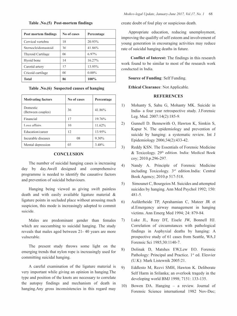
Medico-legal Update, January-June 2017, Vol.17, No. 1 68 69 -legal Update, January-June 2017, Vol.17, No. 1
Table .No.(5) Post-mortem findings
Post mortem findings No of cases Percentage
Cervical vertebra 18 20.93%
Sternocleidomastoid 36 41.86%
Thyroid Cartilage 06 6.97%
Hyoid bone 14 16.27%
Carotid artery 12 13.95%
Cricoid cartilage 00 0.00%
Total 86 100%
Table .No.(6) Suspected causes of hanging
Motivating factors No of cases Percentage
Domestic(Between couples) 36 41.86%
Financial 17 19.76%
Love affairs 10 11.62%
Education/career 12 13.95%
Incurable diseases 08 9.30%
Mental depression 03 3.48%
CONCLUSION
The number of suicidal hanging cases is increasing day by day.Awell designed and comprehensive programme is needed to identify the causative factors and prevention of suicidal behaviours.
Hanging being viewed as giving swift painless death and with easily available ligature material & ligature points in secluded place without arousing much suspicion, this mode is increasingly adopted to commit suicide.
Males are predominant gender than females which are succumbing to suicidal hanging. The study reveals that males aged between 21- 40 years are more vulnerable.
The present study throws some light on the emerging trends that nylon rope is increasingly used for committing suicidal hanging.
A careful examination of the ligature material is very important while giving an opinion in hanging.The type and position of the knots are necessary to correlate the autopsy findings and mechanism of death in hanging.Any gross inconsistencies in this regard may
create doubt of foul play or suspicious death.
Appropriate education, reducing unemployment, improving the qualifty of self esteem and involvement of young generation in encouraging activities may reduce rate of suicidal hanging deaths in future.
Conflict of Interest: The findings in this research work found to be similar to most of the research work conducted in India.
Source of Funding: Self Funding.
Ethical Clearance: Not Applicable.
REFERENCES
1) Mohanty S, Sahu G, Mohanty MK. Suicide in India- a four year retrospective study. J.Forensic Leg. Med. 2007:14(2):185-9.
2) Gunnell D. Bennewith O, Hawton K, Simkin S, Kapur N. The epidemiology and prevention of suicide by hanging: a systematic review. Int J Epidemiology 2006;34(2):433-42.
3) Reddy KSN. The Essentials of Forensic Medicine & Toxicology. 29th edition. India: Medical Book coy; 2010.p.296-297.
4) Nandy A. Principle of Forensic Medicine including Toxicology. 3rd edition.India: Central rd edition.India: Central rd
Book Agency; 2010.p 517-518.
5) Simounet C, Bourgeios M. Suicides and attempted suicides by hanging. Ann Med Psychol 1992; 150:481-5.
6) Aufderheide TP, Aprahamian C, Mateer JR et al.Emergency airway management in hanging victims. Ann Emerg Med 1994; 24: 879-84.
7) Luke JL, Reay DT, Eisele JW, Bonnell HJ. Correlation of circumstances with pathological findings in Asphyxial deaths by hanging: A prospective study of 61 cases from Seattle, WA.J Forensic Sci 1985;30:1140-7.
8) Dolinak D, Matshes EW,Lew EO. Forensic Pathology: Principal and Practice. 1st ed. Elesvier st ed. Elesvier st
(U.K): Mark Listewnik 2005:21.
9) Eddlesto M, Rezvi SMH, Hawton K. Deliberate Self Harm in Srilanka; an overlook tragedy in the developing world BMJ 1998; 7151: 133-135.
10) Bowen DA. Hanging – a review. Journal of Forensic Science international 1982 Nov-Dec;
-legal Update, January-June 2017, Vol.17, No. 1 68 69 Medico-legal Update, January-June 2017, Vol.17, No. 1
20(3): 247-9.
11) Elfawal MA, Awad OA. Death from hanging in the eastern province of Saudi Arabia. Med Sci Law 1994 Oct; 34(4): 307-12.
12) Nikolic S, Misic J, Atanasijevic T, Djokic V, Djonic D. Analysis of neck injuries in hanging.Am J Forensic Med Pathol 2003 June; 24(2); 179-82.
13) Paparo PG, Siegel H. Neck Markings and fractures in Suicidal hanging. Forensic Science international 1984 Jan; 24(1): 27-35.
14) Batra AK, Dongre AP. A Preliminary Analysis of Medico-legal Autopsies performed Over five years in a rural Health District of Maharashtra stat of India. Journal of Forensic Medicine and Toxicology 2003 Jan-June;20(1):41-46.
15) Morild I. Fractures of neck structures in suicidal hanging. Med Sci Law 1996 Jan;36(1):80-4.
16) Singh P, Marak F. Longkumer K, Momonchand A. Suicides in Imphal. Journal of Indian Association of Forensic Medicine 2005;27(2):85-86.
17) Sheikh MI, Agarwal SS. Medicolegal implications of hyoid bone fracture – A study. Journal of Indian Academy of Forensic Medicine 2001 Apr-Jun;23(4): 61-63.
18) Dixit PG, Mohite PM, Ambade VN. Study of histological changes in thyroid,salivary gland and lymph nodes in hanging, Journal of forensic medicine and toxicology. 2001 July-Dec; 18(2): 1-4.
19) Cooke CT, Cadden GA, Margolius KA. Death by hanging in Western Australia. Pathology 1995 July;27(3):268-72.
20) Dolinak D, Matshes EW, Lew EO. Forensic Pathology: Principal and Practice. 1st ed, st ed, st
Elesvier(U.K.): Mark Listewnik 2005:21.
21) Sharma BR, Harish D, Sharma A, Sharma S, Singh H. Injuries to neck structures in Deaths due to constriction of neck with a special reference to hanging. J Forensic Legal Med 2008 July: 15(5):298-305.
22) Slobodan N, Jelena M, Tatjana A, Vesna D, Danijela D. Analysis of neck injuries in hanging. Am J For Med & Path 2003;24(2):179-182.
23) Jayaprakash S. Sreekumari K. Pattern of injuries to neck structures in hanging an autopsy study. Am J Forensic Med Pathol 2012 Dec;33(4):395-9.
24) Saisudheer T. Nagaraja TV. A study of ligature mark in cases of hanging deaths. Int J Pharm Biomed Sci 2012;3(3):80-4.
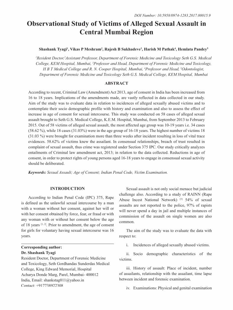
71 -legal Update, January-June 2017, Vol.17, No. 1
Observational Study of Victims of Alleged Sexual Assault in Central Mumbai Region
Shashank Tyagi1, Vikas P Meshram2, Rajesh B Sukhadeve3, Harish M Pathak4, Hemlata Pandey5
1Resident Doctor,2Assistant Professor, Department of Forensic Medicine and Toxicology Seth G.S. Medical College, KEM Hospital, Mumbai, 3Professor and Head, Department of Forensic Medicine and Toxicology,
H B T Medical College and R. N. Cooper Hospital, Mumbai, 4Professor and Head, 5Odontologist, Department of Forensic Medicine and Toxicology Seth G.S. Medical College, KEM Hospital, Mumbai
ABSTRACT
According to recent, Criminal Law (Amendment) Act 2013, age of consent in India has been increased from 16 to 18 years. Implications of the amendments made, are vastly reflected in data collected in our study. Aim of the study was to evaluate data in relation to incidences of alleged sexually abused victims and to contemplate their socio demographic profile with history and examination and also to assess the effect of increase in age of consent for sexual intercourse. This study was conducted on 58 cases of alleged sexual assault brought to Seth G.S. Medical College, K.E.M. Hospital, Mumbai, from September 2013 to February 2015. Out of 58 victims of alleged sexual assault, the most affected age group was 10- 19 years i.e. 34 cases (58.62 %), while 18 cases (31.03%) were in the age group of 16-18 years. The highest number of victims 18 (31.03 %) were brought for examination more than three weeks after incident resulting in loss of vital trace evidences. 58.62% of victims knew the assailant. In consensual relationships, breach of trust resulted in complaint of sexual assault, thus crime was registered under Section 375 IPC. Our study critically analyzes entailments of Criminal law amendment act, 2013; in relation to the data collected. Reductions in age of consent, in order to protect rights of young persons aged 16- 18 years to engage in consensual sexual activity should be deliberated.
Keywords: Sexual Assault; Age of Consent; Indian Penal Code, Victim Examination.
Corresponding author:Dr. Shashank TyagiResident Doctor, Department of Forensic Medicine and Toxicology, Seth Gordhandas Sunderdas Medical College, King Edward Memorial, HospitalAcharya Donde Marg, Parel, Mumbai- 400012India, Email: [email protected]: +917738922308
INTRODUCTION
According to Indian Penal Code (IPC) 375, Rape is defined as the unlawful sexual intercourse by a man with a woman without her consent, against her will or with her consent obtained by force, fear, or fraud or with any woman with or without her consent below the age of 18 years [1, 2]. Prior to amendment, the age of consent for girls for voluntary having sexual intercourse was 16 years.
Sexual assault is not only social menace but judicial challenge also. According to a study of RAINN (Rape Abuse Incest National Network) [3] 54% of sexual assaults are not reported to the police, 97% of rapists will never spend a day in jail and multiple instances of commission of the assault on single women are also common.
The aim of the study was to evaluate the data with respect to:
i. Incidences of alleged sexually abused victims.
ii. Socio demographic characteristics of the victims.
iii. History of assault: Place of incident, number of assailants, relationship with the assailant, time lapse between incident and forensic examination.
iv. Examinations: Physical and genital examination
DOI Number: 10.5958/0974-1283.2017.00015.9

71 Medico-legal Update, January-June 2017, Vol.17, No. 1
including evidence of general and local violence, type and site of injures.
v. Effect of recent criminal amendment act 2013, with regards to increase in age of consent for sexual intercourse.
MATERIAL AND METHOD
This retrospective study was carried out in the department of forensic medicine and toxicology of Seth G.S Medical College, KEM Hospital, Mumbai from September 2013 to February 2015 involving all cases of victims of alleged sexual assault. Examination of females was carried out by doctors of the department of forensic medicine along with the department of obstetrics and gynecology and department of pediatric surgery for child victims. Complete detailed history was taken from the alleged sexual assault survivors and from police investigating reports, these parameters were used in determining the cases. The data of all the alleged sexual assault victims who had been brought for examination in the Department was reviewed. Total 58 cases were studied; data entered on a proforma prepared for the purpose, was then critically analyzed and discussed.
RESULTS
Out of 58 victims of alleged sexual assault, 56 (96.55%) were females and 2 (3.4%) were males. Of all the cases, predominantly were of offence of rape i.e. 52 (89.65%) while cases of sodomy were 6 (10.3%) (Table 1). The most affected age group among victims was of
10 19 years i.e. in 34 cases (58.62 %) as shown in Figure no. 1.
Majority of the victims were belonging to Hindu religion i.e. 43 cases (74.13 %) and were of lower socio economic status i.e. 46 cases (79.31 %). Unmarried victims were predominant sufferers i.e. 52 (89.65 %) whereas married and divorced victims were equal in number i.e. 3 each (5.1 %). Maximum numbers of the victims were students i.e. 51 (87.93 %) while 6 cases i.e. (10.34 %) were employed and 1 i.e. (1.72 %) was housewife (Table 2).
The highest numbers of victims were examined after more than three weeks of alleged incidence which were 18 (31.03 %). Examination was conducted on same day in 13 (22.41 %) cases whereas least number of victims were examined on third day and after second to third
weeks of the incident registering same frequency of 1 each (1.72 %) as shown in Table 3.
In vast majority of the cases i.e. 34 (58.62 %) cases, assailant was known to victim. The offence was committed by a neighbour, stranger and teacher in 11 (18.96 %), 09 (15.51 %) and 03 (5.17 %) cases respectively (Figure no. 2). Offences were mostly committed in the house of accused i.e. in 18 (31.03 %) cases then followed by in victim’s house in 17 (29.31 %) cases while in the office least commonly in 1 (1.72 %) case as shown in Table 4.
On local examination, external injuries were seen in 9 cases (15.51 %) comprising of abrasions and bruises. Bite marks were present over face, breast and arm in 6 cases (10.34 %) while foreign body was retrieved during genital examination in 1(1.72 %) case. Hymen was found intact in 14 victims (24.13%). The hymenal tears were old in 31 victims (53.44 %) while they were fresh in 10 victims (17.24 %) out of 41 cases of ruptured hymen. Tear of anal mucosa was seen in 3 victims of alleged sodomy (5.17 %) shown in Table 5.
Hymenal tears were most commonly found in 5 “O” clock position (24.39 %) then followed by 7 “O” clock position (19.51 %) and 9 (15.51 %) victims were intoxicated at the time of assault as illustrated.
DISCUSSION
The latest statistics of National Crime Records Bureau (NCRB) states that every day 93 women are reported as having been raped in India. According to figures released by the National Crime Records Bureau (NCRB), the total number of rape cases reported in India has gone up to 33,707 in 2013 from 24,923 in 2012. In 15,556 cases, the rape victims were aged between 18 and 30 years in 2013.
In our study, majority of victims were females 56 cases (96.55 %). This is in agreement with study by Tamuli RP et al [4], S. Bandyopadhay et al [5], K. Bhowmik et al [6] and Sukul B et al [7]. In our study alleged sodomy cases were 6 (10.3 %), all males were victims of sodomy which is regarded as unnatural sexual act. 10 (17.24 %) victims were under the age of 10 years, which is consistent with S. Bandyopadhay et al [5] 8 (15%). The most vulnerable age group to sexual assault was 10 -19 years consisting 34 (58.62 %) cases in our study which is similar to Tamuli RP et al [4] 213 cases
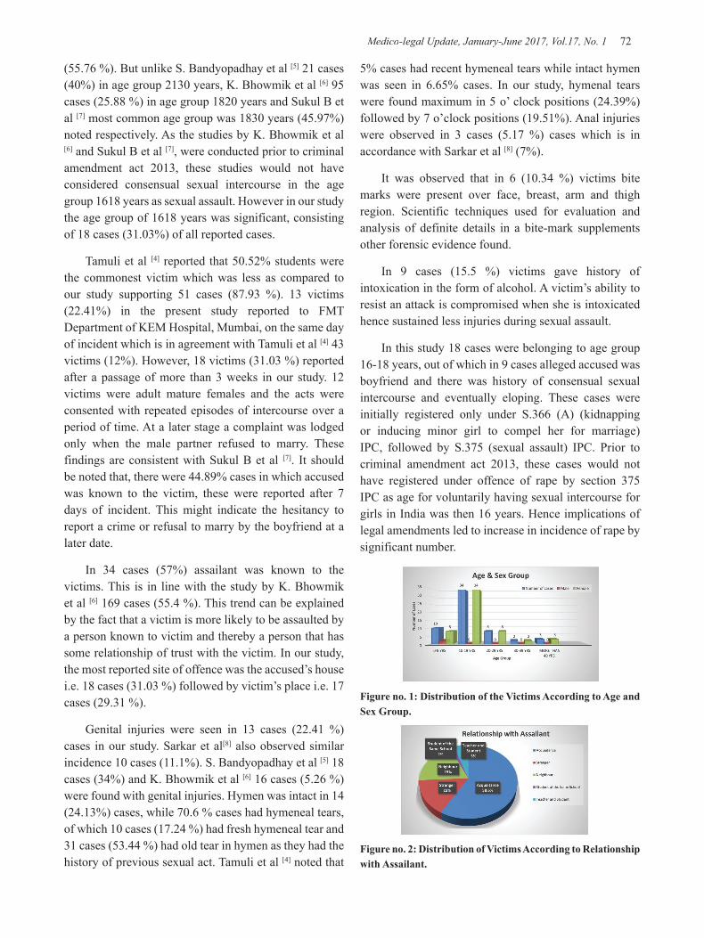
Medico-legal Update, January-June 2017, Vol.17, No. 1 72 73 -legal Update, January-June 2017, Vol.17, No. 1
(55.76 %). But unlike S. Bandyopadhay et al [5] 21 cases (40%) in age group 21 30 years, K. Bhowmik et al [6] 95 cases (25.88 %) in age group 18 20 years and Sukul B et al [7] most common age group was 18 30 years (45.97%) noted respectively. As the studies by K. Bhowmik et al [6] and Sukul B et al [7], were conducted prior to criminal amendment act 2013, these studies would not have considered consensual sexual intercourse in the age group 16 18 years as sexual assault. However in our study the age group of 16 18 years was significant, consisting of 18 cases (31.03%) of all reported cases.
Tamuli et al [4] reported that 50.52% students were the commonest victim which was less as compared to our study supporting 51 cases (87.93 %). 13 victims (22.41%) in the present study reported to FMT Department of KEM Hospital, Mumbai, on the same day of incident which is in agreement with Tamuli et al [4] 43 victims (12%). However, 18 victims (31.03 %) reported after a passage of more than 3 weeks in our study. 12 victims were adult mature females and the acts were consented with repeated episodes of intercourse over a period of time. At a later stage a complaint was lodged only when the male partner refused to marry. These findings are consistent with Sukul B et al [7]. It should be noted that, there were 44.89% cases in which accused was known to the victim, these were reported after 7 days of incident. This might indicate the hesitancy to report a crime or refusal to marry by the boyfriend at a later date.
In 34 cases (57%) assailant was known to the victims. This is in line with the study by K. Bhowmik et al [6] 169 cases (55.4 %). This trend can be explained by the fact that a victim is more likely to be assaulted by a person known to victim and thereby a person that has some relationship of trust with the victim. In our study, the most reported site of offence was the accused’s house i.e. 18 cases (31.03 %) followed by victim’s place i.e. 17 cases (29.31 %).
Genital injuries were seen in 13 cases (22.41 %) cases in our study. Sarkar et al[8] also observed similar incidence 10 cases (11.1%). S. Bandyopadhay et al [5] 18 cases (34%) and K. Bhowmik et al [6] 16 cases (5.26 %) were found with genital injuries. Hymen was intact in 14 (24.13%) cases, while 70.6 % cases had hymeneal tears, of which 10 cases (17.24 %) had fresh hymeneal tear and 31 cases (53.44 %) had old tear in hymen as they had the history of previous sexual act. Tamuli et al [4] noted that
5% cases had recent hymeneal tears while intact hymen was seen in 6.65% cases. In our study, hymenal tears were found maximum in 5 o’ clock positions (24.39%) followed by 7 o’clock positions (19.51%). Anal injuries were observed in 3 cases (5.17 %) cases which is in accordance with Sarkar et al [8] (7%).
It was observed that in 6 (10.34 %) victims bite marks were present over face, breast, arm and thigh region. Scientific techniques used for evaluation and analysis of definite details in a bite-mark supplements other forensic evidence found.
In 9 cases (15.5 %) victims gave history of intoxication in the form of alcohol. A victim’s ability to resist an attack is compromised when she is intoxicated hence sustained less injuries during sexual assault.
In this study 18 cases were belonging to age group 16-18 years, out of which in 9 cases alleged accused was boyfriend and there was history of consensual sexual intercourse and eventually eloping. These cases were initially registered only under S.366 (A) (kidnapping or inducing minor girl to compel her for marriage) IPC, followed by S.375 (sexual assault) IPC. Prior to criminal amendment act 2013, these cases would not have registered under offence of rape by section 375 IPC as age for voluntarily having sexual intercourse for girls in India was then 16 years. Hence implications of legal amendments led to increase in incidence of rape by significant number.
Figure no. 1: Distribution of the Victims According to Age and Sex Group.
Figure no. 2: Distribution of Victims According to Relationship with Assailant.

-legal Update, January-June 2017, Vol.17, No. 1 72 73 Medico-legal Update, January-June 2017, Vol.17, No. 1
Table 1: Type of Sexual Offence According to Gender
Sexual Offence Females Males Number Percentage
Rape 52 0 52 89.65
Sodomy 4 2 6 10.34
Total 56 2 58 100
Table 2: Occupation of the Victims:
Occupation Number PercentageEmployed 6 10.34Student 51 87.93Housewife 1 01.72Total 58 100
Table 3: Distribution of the Victims according To Time of Examination since assault:
Days/ Weeks Number Percentage
Same day 13 22.41
Second day 6 10.34
Third day 1 01.72
Fourth day 4 06.89
Fifth – seventh day 8 13.79
First week – second week 7 12.06
Second to third week 1 01.72
More than three weeks 18 31.03
Total 58 100
Table 4: Distribution of Victims According To Place of Incident
Place of incident Number PercentageVictim’s house 17 29.31Accused house 18 31.03Isolated place 4 06.89Field-near victims home 11 18.96
Guest house/ hotel 5 08.62School 2 03.44Office 1 01.72Total 58 100
Table 5: Distribution of Genital Injuries in Sexual Assault Victims
Injuries Number Percentage
External findings
Abrasion Bruises 9 15.51
Foreign body 1 01.72
Bite Marks Over face, breast and arm.
6 10.34
Injuries Number Percentage
Internal findings
Intact hymen 14 24.13
Ruptured hymen (fresh) 10 17.24
Ruptured hymen(old) 31 53.44
Tear of anal mucous membrane 3 05.17
CONCLUSION
The most vulnerable age group of sexual assault is 10 -19 years; Majority being unmarried and students with assailants being an acquaintance and mostly single person. Also in most of the cases, there was delayed reporting leading to loss of evidence as well as absence of any injury. Hence reporting of such offence at earliest must be encouraged with sensitization as well as sex education.
In this study significant number of cases belonging to age group 16-18 years, were registered under offence of rape by section 375 IPC inspite of being consensual sexual act and hence implicating impact of increase in age of consent of sexual act from 16 to 18 years as per criminal amendment act 2013. To deal with this, lawmakers could hold discussion and debates, considering modifications in the Criminal Law Amendment act 2013 regarding consensual sexual activity among teenagers in the age group of 16 -18 years. Revision of Sexual Assault Laws like reduction in the age of consent that is recommended to protect the rights of young person between the age of 16 and 18 years to engage in consensual sexual activity. Sexual assaults should not be simply treated as problems faced by a woman, rather it should be taken as a crime against entire society.
Funding Received: None.

Medico-legal Update, January-June 2017, Vol.17, No. 1 74
Conflicts of Interest: None declared.
Ethical Clearance- Taken from Institutional ethical committee.
REFERENCES
1. Nandy A. Principles of Forensic Medicine including Toxicology. 5th ed. Calcutta: New Current Book Agency (P) Ltd; Reprinted 2010.p. 610.
2. Al Azad MAS, Rahman Z, Ahmad M, Wahab MA, Ali M , Khalil MI. Socio Demographic Characteristics of Alleged Sexual Assault (Rape) Cases in Dhaka City. JAFMC Bangladesh 2011; l7 (2):21 24.
3. Rape Abuse and Incest National Network [Online] Available from http://www.rainn.org/statistics; accessed on 18/04/2013.
4. Tamuli RP, Paul B, Mahanta P. A Statistical Analysis of Alleged Victims of Sexual Assault – A Retrospective Study. J Punjab Accad Forensic Med Toxicology 2013; 13(1):12.
5. Bandyopadhay Soumyajyoti, Ghosh Somanjana, Adhya Shouvanik, Pal Kuhu, Dalai Chanchal Kumar. A Study on Sexual Assault Victims attending a Tertiary Care Hospital of Eastern India. IOSR. Journal of Dental and Medical Sciences. 2013; 6(6):16–19.
6. Kalpasree Bhowmik, Rituraj Chaliha, A Descriptive One Year Study on the Alleged Male and Female Victims and Accused of Sex Crimes, Journal of Indian Academy of Forensic Med, July September 2011, Vol. 33, No. 3 214,216.
7. Sukul B, Chattopadhyay S and Bose T K. A Study of the Victims of Natural Sexual Offence in the Bankura District in West Bengal. J Indian Acad Forensic Med .2009; 31(1); 25 29.
8. Sarkar SC, Lalwani S, Rautji R, Bhardwaj DN, Dogra TD. A Study on Victims of Sexual Offences in South Delhi, J Fam welf 2005; 51(!): 60 6.

-legal Update, January-June 2017, Vol.17, No. 1 74
Estimation of Age from Eruption of 3rd Molar and its Comparison with the Chronological Age
Mahanta Putul1, Talukdar KL2, Baishya Manoj Kr.3, Mahanta HK4
1Associate Professor, Forensic Medicine, Tezpur Medical College, Tezpur, Assam, 1Associate Professor, Forensic Medicine, Tezpur Medical College, Tezpur, Assam, 1 2Prof and Head 2Prof and Head 2
Anatomy, 3Demonstrator Forensic Medicine, Gauhati Medical College, Guwahati, 3Demonstrator Forensic Medicine, Gauhati Medical College, Guwahati, 3 4Professor and Head 4Professor and Head 4
Forensic Medicine, Tezpur Medical College, Tezpur, Assam
ABSTRACT
Introduction: Age estimation is an important activity that is frequently required to be carried out by a medico-legal expert. An individual is often referred to a medical man by the court or investigating authority in criminal as well as in civil cases to give the opinion regarding his or her age. Aims: The aim of this study was to reconstruct the relation between dental age and chronological age. Material and Method: Age is based on the dental developmental stages mainly the eruption of third molar between 14 to 28 years of age among the people of Assam in relation to some socio-demographic data. Result: From the above finding it is clear that the third molar erupts between 15-25 years and that too in female earlier than male. Conclusion: The result has highlighted the needs of thorough analysis for population-specific dental development for accurate dental age assessment in Assam.
Keywords: Age estimation, third molar eruption, chronological age, people of Assam
Address for Correspondence:Mahanta PutulAssociate Professor, Forensic Medicine, Tezpur Medical College, Tezpur, Assam, Email: [email protected] No: +919435017802
BACKGROUND
Estimation of age from dental examinations especially from the eruption of third molar are implemented in Forensic Age Assessments to discriminate between juveniles and adults considering the judgment of young unaccompanied asylum seekers. An accurate and unbiased age estimation combined with appropriate quantified uncertainties is the required properties for accurate forensic reporting. As because third molar erupt between 17 to 25 years, the subjects being older than 15 years will be more appropriate and produces more meaningful prediction.
Assam has come a long way since India’s independence and given its size and complexity to have ongoing modern challenges in the field of Forensic Medicine. There is a need of population-based study amongst the various ethnic groups of Assam.
Till date no such study has been carried out in Assam where a number of ethnic groups are living. In view of a different food habit, geographical location and life style this study will be undertaken amongst the people of Assam. The results thus obtained on the age of permanent 3rd molar eruption with due comparison rd molar eruption with due comparison rd
with the chronological age of the person can be utilized for the forensic estimation of the minimum and most probable ages of individuals under investigation.
REVIEW OF LITERATURE
Forensic age estimation in living subjects has gained increasing significance in recent years. In dental age estimation, tooth eruption is a parameter of developmental morphology that can be analyzed by either clinical examination or by evaluation of dental X-rays1.
It is known that chronology of dental development is less variable than the bone development and the method applied for this particular period of life is a reliable indicator of age. Though eruption of teeth may be effected by dietary, climatic, racial and geographical variation, the eruption time for deciduous and permanent teeth are fairly constant. Eruption of teeth is one of the
DOI Number: 10.5958/0974-1283.2017.00016.0

Medico-legal Update, January-June 2017, Vol.17, No. 1 76 77 -legal Update, January-June 2017, Vol.17, No. 1
changes observed easily among the various dynamic changes that occur from formation of teeth to the final shedding of the teeth. There is a significant time lag between cutting of a tooth into the mouth and completion of eruption in both deciduous and permanent teeth2.
Skeleton maturity and dental maturity to some extent are related to morphological advancement during the whole growth period. It is observed, that unlike deciduous teeth there existed marked variation not only in the eruption time but also in the sequence of eruption of permanent teeth among different populations of the world3.
With the increase of criminals and crimes in a country, improved and rapid method of age estimation is required. The case in which the opinion regarding the age is required includes criminal responsibility, consent, marriage contract, kidnapping, rape and attainment of majority, eligibility for the employment and for the judicial punishment4.
The third molar erupted earliest in 16-17 years and that too in female and one year later in male and completed by 24 years in both the sexes. This findings of third molar eruption between 16-25 years are well tallied with the findings of Gorden et al5, Scot6, Polson7, Smith8, Kerr9, Grewal10, Savera and Szteen11, Tedechi et al12, Hassanali13, Hagg and Taranger14, Pathak et al15, Rao16, Vij17, Korhonen et al18, and Chaurasia19. Hence, the study group from 14 to 29 years will be preferred here.
One main criterion for dental age estimation in the relevant age group is the evaluation of third molar eruption. The importance of ethnic variation in dental development requires further population studies in order to reach an adjustment of each method according to the specific population, with an increase in precision and accuracy.20
One main criterion for dental age estimation in the relevant age group is the evaluation of third molar eruption. The importance of ethnic variation in dental development requires population specific data for dental age evaluation. In a study in Canada A Olze et al. determined the stages of third molar eruption in 347 female and 258 male First Nations people of Canada aged 11 to 29 years based on radiological evidence from 605 conventional orthopantomograms.21
Teeth are the most durable structures in the human
body. The pattern of their development has been used as a credible technique of age determination of unidentified bodies. Dental age estimation is by comparison of the dental status of an individual with published dental surveys. The third molars are the last teeth to erupt and are regarded as the most variable in the dentition.22
There is no significant difference between the average age and the calcification stage taking gender and localization of the third molar into consideration to evaluate the correlation between chronological age and the degree of third molar mineralization from patients aged between 6 and 22 years. It is possible to estimate chronological age based on stage of a third molar, regardless of gender and location.23
A strong agreement exist between eruptions time of third molar and chronological age. The statistical analysis revealed that earliest third molar eruption was in the female at 16 year which has completed at 24 years in both the sexes. It also erupted earlier in lower jaw in both the sexes. The spaces for third molar also appear earliest in female at 15 year of age with early preponderance in lower jaw.24
Age estimation for medico-legal and clinical purposes represents a fundamental problem, and various methods have been established for age determination. It has been shown that dental development relates more closely to chronological age than skeletal, somatic or sexual maturity indicators.25
AIMS AND OBJECTIVES
The aim of this study is to reconstruct the relation between dental age and chronological age based on the dental developmental stages mainly the eruption of third molar between 14 to 28 years of age among the people of Assam in a 50 randomly picked up individuals..
MATERIAL AND METHOD
Sample Selection: 50 individuals has randomly picked up for this study from the age group of 14 to 29 years irrespective of eruption of the 3rd molar.
Criteria of Inclusion
a. People born in Assam and living since birth
b. Healthy sample free from any genetically disorders, growth retardation, endocrinal, nutritional or musculo-skeletal diseases and diseases of tooth or

-legal Update, January-June 2017, Vol.17, No. 1 76 77 Medico-legal Update, January-June 2017, Vol.17, No. 1
chronic illness which retard the process of growth.
Criteria of Exclusion
a. Born and grown up outside the Assam
b. One who does not full fill the above health criteria
c. Family history of genetically disorder
d. Congenital absence of teeth.
Chronological age (CA): CA is the number of years a person has lived, which is measured by the time (years, month and days) and recorded as per the record of date of birth (DOB). DOB will be established from the witness elements of other participants (viz., parents, doctors, school teachers) and from documented elements like birth certificate, passport, driving licenses, etc.
Biological age (BA): BA also called physiological age, is a measure of how well or poorly our body is functioning relative to our actual calendar age. BA of the subjects are ascertained through the degree of maturation of third molar only, i.e., the eruption stages of third molar under the influence of diverse genetic and environmental factors which excludes skeleton maturity, etc.
Physical examination: A physical examination will be carried out to know the following points:
• To record the anthropometric data
• To know any age-relevant developmental disorders
• To know about his health status.
Dental examination: A systematic dental examination is carried out. The dental findings were plotted in the respective chart as mentioned in the proforma.
Resources Required
• The teeth will be examined either in good day light or by using a torch having a very fine focusing of light
• For space of permanent 3rd molar, a mirror and the finger for to know hardening of space behind the second molar will be used
• The normal dental instruments that are widely used in dental check-up to examine the teeth and surrounding oral structures will be used
• Sterilized gloves, gauze, etc.
• Instrument Sterilizer
• Height and weight machine
• Computer set and printer.
RESULT AND OBSERVATION
The analysis of data of 50 cases are shown in relation to various variables.
Table 1 Distribution of the study participants according to age and sex
Age (in Years)Male Female TotalNo. % No. % No. %
14 1 0 1 2.015 1 0 1 2.016 1 1 2 4.017 1 1 2 4.018 1 1 2 4.019 2 2 4 8.020 2 2 4 8.021 5 5 10 20.022 3 2 5 10.023 4 3 7 14.024 2 3 5 10.025 3 1 4 8.026 1 2 3 6.027 0 0 0 0.028 0 0 0 0.029 0 0 0 0.0Total 27 54.0 23 46.0 50 100
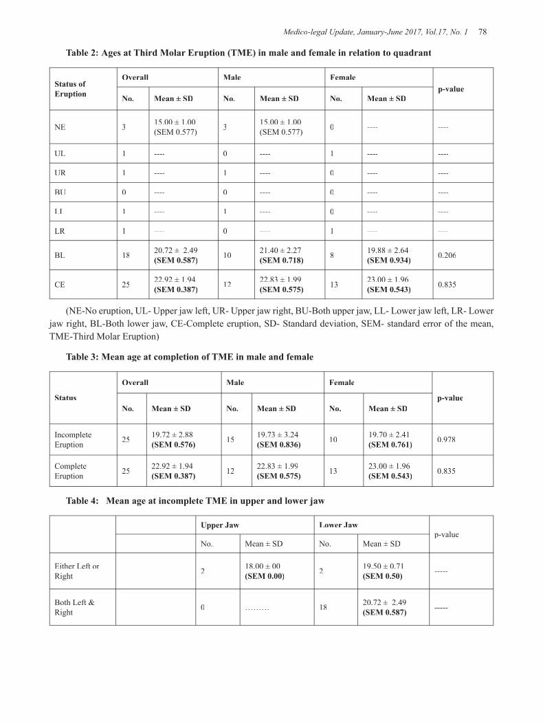
Medico-legal Update, January-June 2017, Vol.17, No. 1 78 79 -legal Update, January-June 2017, Vol.17, No. 1
Table 2: Ages at Third Molar Eruption (TME) in male and female in relation to quadrant
Status of Eruption
Overall Male Femalep-value
No. Mean ± SD No. Mean ± SD No. Mean ± SD
NE 3 15.00 ± 1.00 (SEM 0.577) 3 15.00 ± 1.00
(SEM 0.577) 0 ---- ----
UL 1 ---- 0 ---- 1 ---- ----
UR 1 ---- 1 ---- 0 ---- ----
BU 0 ---- 0 ---- 0 ---- ----
LL 1 ---- 1 ---- 0 ---- ----
LR 1 ---- 0 ---- 1 ---- ----
BL 18 20.72 ± 2.49 (SEM 0.587) 10 21.40 ± 2.27
(SEM 0.718) 8 19.88 ± 2.64 (SEM 0.934) 0.206
CE 25 22.92 ± 1.94 (SEM 0.387) 12 22.83 ± 1.99
(SEM 0.575) 13 23.00 ± 1.96 (SEM 0.543) 0.835
(NE-No eruption, UL- Upper jaw left, UR- Upper jaw right, BU-Both upper jaw, LL- Lower jaw left, LR- Lower jaw right, BL-Both lower jaw, CE-Complete eruption, SD- Standard deviation, SEM- standard error of the mean, TME-Third Molar Eruption)
Table 3: Mean age at completion of TME in male and female
Status
Overall Male Female
p-valueNo. Mean ± SD No. Mean ± SD No. Mean ± SD
IncompleteEruption 25 19.72 ± 2.88
(SEM 0.576) 15 19.73 ± 3.24(SEM 0.836) 10 19.70 ± 2.41
(SEM 0.761) 0.978
Complete Eruption 25 22.92 ± 1.94
(SEM 0.387) 12 22.83 ± 1.99(SEM 0.575) 13 23.00 ± 1.96
(SEM 0.543) 0.835
Table 4: Mean age at incomplete TME in upper and lower jaw
Upper Jaw Lower Jawp-value
No. Mean ± SD No. Mean ± SD
Either Left or Right 2 18.00 ± 00
(SEM 0.00) 2 19.50 ± 0.71(SEM 0.50) -----
Both Left & Right 0 ……… 18 20.72 ± 2.49
(SEM 0.587) -----
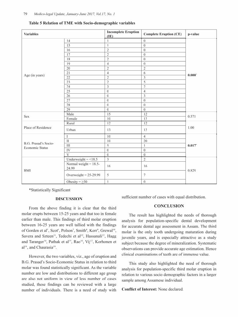
-legal Update, January-June 2017, Vol.17, No. 1 78 79 Medico-legal Update, January-June 2017, Vol.17, No. 1
Table 5 Relation of TME with Socio-demographic variables
Variables Incomplete Eruption (IE) Complete Eruption (CE) p-value
Age (in years)
14 1 0
0.000*
15 1 016 2 017 2 018 2 019 4 020 2 221 4 622 2 323 2 524 3 225 0 426 0 327 0 028 0 029 0 0
Sex Male 15 12 0.571Female 10 13
Place of ResidenceRural 12 12
1.00Urban 13 13
B.G. Prasad’s Socio-Economic Status
I 10 4
0.017*II 10 20III 5 1IV 0 0V 0 0
BMI
Underweight = <18.5 3 2
0.829
Normal weight = 18.5-24.99 16 16
Overweight = 25-29.99 5 7
Obesity = ≥30 1 0
*Statistically Significant
DISCUSSION
From the above finding it is clear that the third molar erupts between 15-25 years and that too in female earlier than male. This findings of third molar eruption between 16-25 years are well tallied with the findings of Gorden et al5, Scot6, Polson7, Smith8, Kerr9, Grewal10, Savera and Szteen11, Tedechi et al12, Hassanali13, Hagg and Taranger14, Pathak et al15, Rao16, Vij17, Korhonen et al18, and Chaurasia19.
However, the two variables, viz., age of eruption andB.G. Prasad’s Socio-Economic Status in relation to third molar was found statistically significant. As the variable number are low and distributions to different age group are also not uniform in view of less number of cases studied, these findings can be reviewed with a large number of individuals. There is a need of study with
sufficient number of cases with equal distribution.
CONCLUSION
The result has highlighted the needs of thorough analysis for population-specific dental development for accurate dental age assessment in Assam. The third molar is the only tooth undergoing maturation during juvenile years, and is especially attractive as a study subject because the degree of mineralization. Systematic observations can provide accurate age estimation. Hence clinical examinations of teeth are of immense value.
This study also highlighted the need of thorough analysis for population-specific third molar eruption in relation to various socio demographic factors in a larger sample among Assamese individual.
Conflict of Interest: None declared.

Medico-legal Update, January-June 2017, Vol.17, No. 1 80 81 -legal Update, January-June 2017, Vol.17, No. 1
Ethical Clearance: Taken
Source of Funding: None declared.
REFERENCES
1. Olze A, Peschke C, Schulz R, Schmeling A. Studies of the chronological course of wisdom tooth eruption in a German population. J Forensic and Legal Med 2008 Oct;15(7):426-429.
2. Reddy V (Ed). Dental Anthropology-Application and Methods, Inter India Publication. 1985;368-369.
3. Mishra VK, Rao CM. Age and stage of dental eruption. J Forens Med Toxicol 1994;9:8-14.
4. Franklin C A (Ed). Modi’s Text Book of Medical Jurisprudence and Toxicology. 21st ed. India, Bombay: NM Tripathy Pvt. Ltd; 1988. P. 37-39.
5. Gordon I, Turner R, Price T W. Medical Jurisprudence. 3rd ed. Edinburgh and London: Livingstone Ltd; 1953. P. 343-72.
6. Scot DB. Dental evidences in identification and criminology. Legal med (Ed Gradwohl). 3rd ed. Chicago USA: A John Wright and Sons Ltd. Publication; 1954. P. 452-78.
7. Polson CJ. The essential of forensic med. London: English Universities Press; Ltd; 1955. P. 51.
8. Smith S. Taylor’s Principles and Practice of Medical Jurisprudence. 10th ed. London: J. and A. Churchill. 1948. P. 100-1.
9. Kerr DJA. Forensic med, a text book for students and a guide for the practioners. 6th ed. London: Adam and Charles Black; 1957. P. 42-3.
10. Glaister J., Rentoul E. Medical jurisprudence and toxicology. 12th ed. Edinburgh and London: Livingstone Ltd; 1966. P. 68-9.
11. James H. Shaw. Influence of Marginal and Complete Protein Deficiency for Varying Periods during Reproduction on Growth. Third-Molar Eruption and Dental Caries in Rats. Journal of Dental Research 1969; 48(2):310-316.
12. Grewal RS. Medical jurisprudence and toxicology. 1st ed. Calcutta, India: Scientific Book Agency; 1973. P. 40-41.
13. Savara BS, Steen JC. Timing and sequence of eruption of permanent teeth in a longitudinal sample of children from Oregon. J Am Dent Assoc
1978; 97 (2): 209-14.
14. Tedeschi CG, Eckert WG, Tedeschi LG. Age determination, forensic med, a study in trauma and environmental hazards. Philadelphia, London, Toronto. W. B. Saunders Company; 1977. P. 1124-34. Vol 2.
15. Hassanali J. The 3rd permanent molar eruption in Kenyan Africans and Asians. Ann Hum Biol 1985; 12(6): 517-23.
16. Hagg U, Taranger J. Timing of tooth emergence. A prospective longitudinal study of Swedish urban children from birth to 18 years. Swed Dent J 1986; 10(5):195-206.
17. Pathak SK, Mathur PN, Jain S., Saini O.P. A study of eruption of 3rd molar in relation to estimation of age in people of thirteen to twenty-five years age group. Journal of Forensic Medicine and Toxicology 1999; 16 (1):17-9.
18. Rao N. Textbook of forensic medicine and toxicology. New Delhi, India: Jaypee Brothers Medical Publishers (P) LTD; 2000. P. 78-81.
19. Vij K. Textbook of forensic medicine and toxicology. 2nd ed. New Delhi, India: B.I. Churchill Livingstone; 2002. P. 68-69.
20. Korhonen M, Larmas M. Dental age and dental health determined longitudinally from patient records in three towns in Finland. Acta Odontol Scand 2003; 61(2):105-9.
21. Chaurasia BD. Human anatomy, 3rd ed. New Delhi, India: CBS Publishers and Distributors; 1995, Vol. 1. P. 122.
22. Cunha E, Baccino E, Martrille L, Ramsthaler F, Prieto J, Schuliar Y et al. The problem of aging human remains and living individuals: a review. Forensic Sci Int 2009;193:1-13.
23. A Olze, BR Pynn, V Kraul, R Schulz, A Heinecke, H Pfeiffer et al. Dental Age Estimation Based On Third Molar Eruption In First Nations People Of Canada. J Forensic Odontostomatol 2010;28:1:32-38.
24. Phrabhakaran Narnbiar. Age estimation using third molar development. Malaysian J Path 1995;17(1):31-34.
25. Caio Belém Rodrigues Barros Soares, José Natal Figueiroa, Renata Moura Xavier Dantas, Lúcio Mitsuo Kurita, Andréa dos Anjos Pontual, Flávia

-legal Update, January-June 2017, Vol.17, No. 1 80 81 Medico-legal Update, January-June 2017, Vol.17, No. 1
Maria de Moraes Ramos-Perez et al. Evaluation of third molar development in the estimation of chronological age. Forensic Science International September 2015;254:13–17.
26. Putul Mahanta, Azza S Mohamed. A Study on Eruption of Third Molar of Libyan Individual and its Comparison with the Egyptian. Medico-Legal Update 2013;13(1):81-84.
27. Kanmani R, Srinivasan SV. Mandibular Third Molar Development as an Indicator of Chronological Age for a Pondicherry Population of Southern India. e-Journal of Dentistry April-June 2012;2(2):153-158. [cited 2016 May 11]. Available from: URL:http://www.ejournalofdentistry.com/articles/e-JOD7BD5F6F930-02FB-4209-8A09-D82113.pdf.

83 -legal Update, January-June 2017, Vol.17, No. 1
Pattern of Skull Fractures due to Blunt Force in and around Khammam
Bharath Kumar Guntheti1, Uday Pal Singh2
1Associate Professor, 2Prof & HoD, Dept. of Forensic Medicine &Toxicology, Mamata Medical College, Khammam; Telangana, India
ABSTRACT
Head is the most common site to be injured in blunt force trauma, may result injury to the contents of the skull, either alone or with a fracture of the skull. Out of 108 cases, majority were found to be co-existing with intracranial injuries, but few cases showed the presence of skull fractures alone. Most vulnerable age group is the active population of persons were 30-40 years while the cause of fracture in maximum number of cases (90%) was direct trauma. Most of victims were male 98 and from urban area. Highest number were reported during rainy season. Occupationally the laborers were top among other occuaptions.Most common cause being road traffic accidents followed by fall from height and then by assault. In 108 cases of intracranial lesions, 100 cases sustained fracture of the skull without any injury to the scalp and in 36 cases injuries of both scalp and skull were present. Fissured fracture was the commonest type followed by comminuted fracture and then by depressed fracture. We detected that in 6 cases skull fracture was not found. Sub Dural hemorrhage was the commonest intracranial hemorrhage associated with head injuries. The sites of skull fractures most commonly involved in our study is Parietal. In our study, in basal fractures, middle cranial fossa was commonly involved in 32.35% of cases. The study of pattern of skull fractures is important as head being the most exposed and prominent part of body; it becomes most susceptible to injuries, as a result of criminal violence or accident. The skull fractures, especially by blunt force offer varying diagnostic and medico-legal problems to the medical jurists as well as to the clinicians. The prime objective of present study is to know the patterns of skull fractures due to blunt force trauma and cause, mechanism of injury and their preventive measures.
Keywords: Head injury, Blunt Trauma, Skull Fracture, Fissured Fracture
INTRODUCTION
Head injury, as defined by the National Advisory Neurological Diseases and Strove Council, USA [1969], “is a morbid state, resulting from gross or subtle structural changes in the scalp, skull and or the contents of the skull, produced by mechanical forces”1. To be complete, however, it should take into account that the impact, responsible for injury, need not be applied directly to the head.
Corresponding author: Dr Bharath Kumar Guntheti,Associate Professor, Dept. of Forensic Medicine & Toxicology, Mamata Medical College, Khammam; Telangana. India Pin code-5070002.Mob: 09908339507. E-mail id: [email protected]
Heady injury is a considerable public health issue, with high rates of mortality and morbidity. Injury to brain without fracture of skull is not uncommon, through fracture of skull is usually accompanied by some degree of injury to brain.2
The study of patterns of skull fractures in these cases is important as head being the most exposed and prominent part of body becomes most susceptible to injuries. The skull fractures, especially by blunt force offer varying diagnostic and medico-legal problems to the forensic experts and also to the clinicians. Head injury cases are presently increasing at an alarming rate in all world communities, especially in more densely populated areas with rapid growth of industrialization .no injury to the heady is too trivial to be ignored or so serious as to be despaired of. 3
DOI Number: 10.5958/0974-1283.2017.00017.2
83 Medico-legal Update, January-June 2017, Vol.17, No. 1
Most wound are caused by blunt force to the head, e.g., from falls or blows, and such wound are contusions or lacerations. An oblique blow usually causes a large wound and direct blow a small wound. With repeated blows, blood will be splattered over the assailant, which will indicate the relative positions of the victim and assailant and also whether they were inflicted with the right or left hand.4
The type of trauma may be direct or indirect. The application of blunt force to the head may result in injury to the contents of the skull, either alone or with a fracture of the skull. These head injuries are generally sustained by road traffic accidents, fall from height, assault by blunt force, etc. These are only a few reports from developing countries about the patterns of skull fractures.
The main purpose of this study is to describe skull fractures due to blunt force trauma, victim’s socioeconomic profile, injury mechanism, location and type of skull fracture.
AIMS & OBJECTIVES
To study the profile of victims of skull fractures due to blunt force
To study the patterns of skull fractures, cause, manner, mechanism of injury
Preventive measures of factors behind the fractures of skull due to blunt force
MATERIALS & METHOD
A 108 cases of skull fractures caused by blunt force were selected from the autopsies conducted for a period of one year from Jan 2016 to Dec 2016 at Dept of Forensic Medicine Mamata Medical College attached with Mamata General Hospital Khammam, Telangana.
Only those cases were selected which had definite history of head injury by blunt force e.g., road traffic accidents, fall from height, assault by blunt weapons etc.
A detailed postmortem examination was carried on every case .A Proforma prepared and data collected including approximate age, sex, history of injury from relatives, eye witnesses regarding the cause and manner of injury, time and place of injury, period of survival following the head injury and alcohol /drug abuse by person.
The external examination of the whole body was done with special attention towards the injuries of head .The nature of injuries, like abrasions, contusions, lacerated wounds and fractures and their type, location and dimensions were carefully noted. Evidence of bleeding from natural orifices was also noted.
Head injuries were examined methodically. The inner aspect of the scalp was examined for evidence of any contusions or hematomas.
Fractures of the skull were examined in detailed as to the type [depressed, fissured, sutural, comminuted, hinge, ring], extension and location
Injuries to the meninges and their blood vessels and corresponding intracranial hemorrhages were noted.Dura at the base of skull was stripped off to visualize the fractures if any.
RESULTS & DISCUSSION
A total of 108 cases were included in our study and observed, tables were made, compare with previous studies and concluded.
In this study, a total 108 cases of skull fractures due to blunt force trauma, 80[74.07%] cases had skull fractures with intracranial hemorrhages and injury to scalp 20[18.51%] cases sustained skull fractures with intracranial hemorrhages without scalp, 6[5.55%] cases sustained skull fracture with injury to the scalp without any intracranial hemorrhage, 2[1.85%] cases sustained fracture of skull alone. These are similar to authors5,6.
In present study craniocerebral injuries by blunt force, road traffic accidents accounted for 72.22 % of the cases followed by fall from height 20.37% of cases and assault by blunt force weapons only 7.40% of cases.The incidence of head injury is increasing everyday so it is imperative that its preventive and practical aspects as well as scientific aspects of these help in prevention or curable .These are similar to other studies 5-7.
In our study, age wise distribution, the commonest vulnerable age group is 30-40 years 37[34.25%]followed by 20-30 years 24[22.22%] next age group 11-20 years 18[16.66%] and age group of 41-50 years 16[14.81%]. Extreme age group persons least cases were found in this study. These are consistent with authors5-7.Table no.1
In present study, gender wise distribution, the
Medico-legal Update, January-June 2017, Vol.17, No. 1 84 85 -legal Update, January-June 2017, Vol.17, No. 1
incidence of injuries due to blunt force trauma was higher 98[09.74%] in males than 10[9.25%] females. This high incidence might be due to increase risk exposure to males due to more outdoor activities. These are similar to studies conducted by others5-8.
In the present study 92[85.18%] constitute urban people and 16[14.81%] constitute rural people. The huge variation could have been due to following reason: Urban people are more vulnerable group because of more accidents due to RTAs, also because of high rise buildings –fall from height during construction works, it also includes homicidal assault by blunt force trauma. Similar results were noted by the authors.
In our study, occupation wise distribution of skull fractures, majority of victims were laborers 30[27.27%] followed by students 24[22.22%] and employee 20[18.51%]. Similar results are noted by authors6-8. The reason is laborers were most commonly involves in various works such as travelling by vehicles ,construction works and blunt force trauma to head due to homicidal assault.
In present study the peak timing of occurrence of skull fractures due to blunt force trauma was reported at 9am to 12 noon 31[28.70%]followed by 6am to 9am 18[16.66%]and next in 12pm to 3pm 17[15.74%]cases. This is probably due to heavy and unequal distribution of incidents occurs when rush was too heavy on road at these working hours.Which are consistent with other studies 6,7.
Seasonal variation, we observed that the highest number 60[55.55%] of cases were encountered in the winter season followed by Summer season 30 [27.77%] and least 18[16.66%] cases were occurred in Rainy season .these are consistent with authors8,9. The reason might be poor visibility to vehicle drivers leading to accidents in the early hours of day due to foggy weather conditions and slow reaction time due to extreme cold affecting both drivers and road users.
Survival period ,we found 83[76.85%] of the victims of skull fractures due to blunt force trauma died either on spot or within 24 hours of the incidence and rest could survive for a couple of days to maximum of two weeks after getting good medical and surgical procedures. The time of survival of head injury victims varies as per the severity of trauma and also health care services provided to the patients. Glasgow Coma Scoring of head injury
at the time presentation to the emergency department is an important prognostic factor and the level of consciousness should be determined and monitored regularly in all such patients who survive in road traffic accidents. Similar observations are made by authors8-10. The most common cause of skull fractures being Road Traffic Accidents .Fig
As per location of skull fracture, in our study, skull vault was fractured in 28 % of cases, basilar fractures were seen in 40% of cases and in majority of cases [52%] both the vault and base were fractured or both, especially in the thin areas temporoparietal bone. These are consistent with other studies. The cranial vault involved or base or both vault and base are involved in other studies also.8-12 Fig.1
In present study, there was no skull fracture found. 6[5.55%] cases were found with or without brain injuries without any fracture. Same results are observed by authors 8-10.The head injury due to direct trauma by blunt force leads to various types of injuries, such as no external injuries to various scalp injuries, skull fractures, meninges injuries and types of intracranial Haemorrhage depends upon the site of impact force of the impact over the head. Fig.2
According to type of skull fracture in our study, the dominant type of skull fracture found was fissured [linear] fracture in 53[49.07] % cases followed by comminuted fracture 28[25.92%] next depressed fracture 8[7.40%] and least 5 [4.62%] of cases were basilar fractures. These are consistent with studies by authors11-16. Most of the head injuries are associated with skull fractures which increases the fatality of victims. Skull fractures are not a dictum to be present in all fatal head injuries. Fig.3
In our study in addition to these we noted the presence of hinge fracture in 5.55% of cases and sutural fracture in 0.92 % of cases whereas in 6[5.5%] of cases no skull fractures are not found in this study. Which are similar to studies conducted by others12-16 .More number of skull fractures observed in this study can be explained by restricted movement of the head receiving the maximum force, more striking surface area of skull in all directions ,least protection offered by the scalp musculature when compared to limbs and other parts of the body.Fig.4
The sites of skull fractures most commonly

-legal Update, January-June 2017, Vol.17, No. 1 84 85 Medico-legal Update, January-June 2017, Vol.17, No. 1
involved in our study is Parietal followed by Temporal ,Temporo –Parietal and middle cranial fossa.31.81%,27.27%,23.07% and 32.35% of cases respectively. Similar involvement of parietal and temporal bones are found in other studies12-17
In our study, in basal fractures, middle cranial fossa was commonly involved in 32.35% of cases. Similar observations of made by authors.10,15,16 Fig.5
In present study, 4.62 % of cases of trauma to the head by blunt force, there were no external injuries on the head but these had severe cranial or intracranial lesions which proved fatal. External injuries may or may not be present in all most all cases of head injury. These are consistent with others studies17-20
We found multiple injuries in most of cases involving other systems a typical feature. Wounds of the scalp due to blunt force must be looked upon as potentially serious no matter how they are produced. Similar findings are made authors 18-22
The commonest variety of intracranial hemorrhage found was subdural hemorrhage [SDH] 91[84.25%] followed by subarachnoid hemorrhage [SAH] 68[62.96 %, Intra cerebral hemorrhage [ICH] 24 [22.22%] and least is extradural hemorrhage [EDH] was found in 16.6% cases .Which is supported by other studies also
15-20 Fig .6
Cause of Death, we found that a fracture of the skull associated with cerebro-cranial injury was the commonest cause of death in our study. Which are consistent with studies conducted by authors18. Skull fractures following blunt force trauma as road traffic crashes are the most common cause of morbidity and mortality in most of countries and may also result in temporary or permanent disability or ultimately death of the victim.20-22
RECOMMENDATIONS
These are prevented by correction of human errors, vehicle and weather, faults of drivers, pedestrians, passengers are responsible for fatal accidents.
Accidents are due to over speeding, rash driving, loss of control, and violation of traffic rules, alcohol intoxication, fatigue, and sleepiness these factors are improved.
Development of policies and their strict implementation based on these risk factors can reduce severity and burden of skull fractures in India.
Table 1: Age incidence of Skull Fractures
Age group in years No of cases %1-10 1 0.9211-20 18 16.6621-30 24 22.2231-40 37 34.2541-50 16 14.8151-60 8 7.40Above 60 4 3.70Total 108

Medico-legal Update, January-June 2017, Vol.17, No. 1 86 87 -legal Update, January-June 2017, Vol.17, No. 1
CONCLUSIONS
The rate of incidence is high.
The commonest affected age group is 31-40 years.
Much more common in young working males as compared to females.
Most of the people were from urban area.
The common causes of skull fractures are road traffic accidents followed by fall from height and assaults by blunt weapons.
Skull fractures can occur without any evidence of injury to scalp.
Skull fractures may or may not be associated with intracranial injuries.
The commonest type of skull fracture is fissured fracture followed by comminuted fracture.
The commonest variety of intracranial hemorrhage found was subdural hemorrhage.
Skull fractures in majority of the cases are due to direct trauma.
Intracranial injuries can occur with or without fractures of skull.
Human error is the most common cause of RTAs.
We hope that this study may be helpful in understanding of this problem and will contribute in some way at state level.
Source of Funding: Self
Conflict of Interest: Nil
Ethical Clearance: Not needed.
REFERENCES
1. Krishan Vij.Forensic Medicine and Toxicology, Principles and Practice.6th Ed. ELSEVIER, Reed India Private Limited .2011.Regional Injuries; p.261.
2. J B Mukherjee .Regional Injuries .In Forensic Medicine and Toxicology, 2nd Ed.Vol.1, Kolkata: Academic Publishers .1973:p.390.
3. Polson CJ.Head Injuries In: Essentials of Forensic Medicine, London: English Universities press Ltd.1955; 131.
4. K.S. N .Reddy.Head Injuries. In The Essentials of Forensic Medicine and Toxicology, 32nd Ed. Published by K.Suguna Devi, Hyderabad.2013:p231.
5. B K Gopal Ameena Ahamed, Faraz Ahamed S.B Tonse.Pattern of Skull Fractures due to Blunt Force. JKAMLS, Vol.24 [2], July-Dec 2015:27-31.
6. Dalbir Singh ,Kumaran Moorthi,Sathinder P Singh,Sonu Goel.Profile of Road Traffic Fatalities in Adults A 40 Year Study in Chandigarh Zone of North West India Indian Acad Forensic Med.Jan-Mar ,2014,Vol.36[1]:47-52
7. Pathak A, Desania N L, Verma R.Profile of road traffic accidents & head injury in Jayapur.JIAFM.2008;30[1]:6-9.
8. Menon A, Nagesh KR.Pattern of fatal head injuries due to vehicular accidents in Manipal. JIAFM,
-legal Update, January-June 2017, Vol.17, No. 1 86 87 Medico-legal Update, January-June 2017, Vol.17, No. 1
2005, Vol.27 [1]:19-22.
9. GH Yattoo , Amin Tabish .The profile of head injuries and traumatic brain injury deaths in Kashmir. Journal of Trauma Management &Outcomes, 2008, 2[5]:1-9.
10. Lalit Kumar, Sandeep Agarwal, Tajendar Singh, Rajesh Garg.Patterns of Head Injury at Tertiary Care Hospital .international Journal of Scientific Study;Feb 2014,vol.no5:5-8.
11. Saurabh Chattopadhyay, Chandra Bal Tripathi.Skull Fracture and Haemorrhage pattern among fatal and non-fatal head injury assault victims –a critical analysis. J Inj Violence Res.2010 June, 2[2]:99-103.
12. Manish K,Jothi NS, Gurudatta S P,Vijay kumar BJ. Fatal head injuries in road traffic accidents in and around Davanagere: A Prospective Study. Indian Journal of Forensic Medicine & Pathology.2012; 5[2]:61-65.
13. Kumar A,Lalwani S,Agarwal D,Rauti R,Dogra T D. Fatal Traffic Accidents and their relationship with head injuries, An epidemiological survey of 5 years; Indian Journal of Neurotrauma 2008,5[2]:p.63-67.
14. Mukesh K Goyal,Rajesh Varma,Shiv R Kochar Shrikant S Asawa. Corelation Hospital.Jaipur.JIAFM 2010; 32[3]:208-211.
15. Ravindra S honnungar ,Sunil C Aramani,Vijay Kumar AG, Ajay Kumar TS.An epidemiological
Survey of Fatal Road Traffic Accidents and their Relationship with Head Injuries.JIAFM.April-June 2011, Vol.33 [2]:202-207.
16. Raja Rupani, Anoop Varma, Shiuli Rathore.Pattern of Skull Fractures in Cases of Head Injury by Blunt Force Indian Acad Forensic Med, Oct –Nov 2013, Vol.35 [4]:336-338.
17. R.Ravi Kumar.An autopsy Study of Patterns of Skull Fractures in Road Traffic Accidents Involving Two Wheelers .JKAMLS, Vol.23 [1], Jan-June 2014:9-14.
18. Singh YN, Bairagi KK, Das KC.An epidemiological study of road traffic accident victims in medico legal autopsies.JIAFM, 2005, Vol.27 [3]:166-169.
19. Pekka Saukko, Bernad Knight. Forensic Pathology, 3rd Edition, Arnold Publisher, London, 2004:p290-291.
20. Pareikh CK.Text Book of Forensic Medicine and Toxicology .CBS Publishers & Distributors ,7th
Edition,2016:p.287-88
21. Ranjit M, Tandle, A.N.Keoliya.Patterns of Head Injuries in fatal road traffic accidents in a rural district of Maharashtra –Autopsy based Study.JAFMC July-Sep, 2011, Vol.33 [3]:171-176.
22. Simpson Keith .Regional Injury; Taylor‘s Principles and Practice of Medical Jurisprudence 1965.12th
Ed.London. J & A. Churchill Ltd; 230-162.

89 -legal Update, January-June 2017, Vol.17, No. 1
Cardiac Troponin T as a Marker in the Diagnosis of Acute Myocardial Infarction at Autopsy
B Venkata Naga Mohan Rao1, Bandi Sree Ramulu2, B Raju3
1Associate Professor, 2Assistant Professor , 3Professor and HOD, Department of Forensic Medicine, Kakatiya Medical College, Rangampet, Warangal, Telangana
ABSTRACT
Cardiac deaths account for 50% of all deaths in developed and 25% in the developing world. One sixth of world’s population lives in death accounts for two-thirds of all autopsies in forensic medicine department sequence of the infected myocardium will develop only after significant time of, between myocardial infarction (MI) in death. The use of cardiac Troponin T at autopsy is less clear. This study evaluates the role of cardiac Troponin in the detection of acute myocardial infarction at autopsy. A retrospective study was performed at Mahatma Gandhi Memorial Hospital, Warangal (2012-1014), identifying autopsy cases where serum Troponin was requested. In these cases, cause of death was identified from autopsy reports, the histology reviewed and Troponin T levels were recorded. Acute myocardial infarction was confirmed as a cause of death in 62 out of the 176 cases with 48 cases having an elevated Troponin T. (Sensitivity: 92%; Specificity: 5%). The results of this study suggest that while Troponin T is sensitive marker, it is not specific as a diagnostic tool in the detection of acute myocardial infarction at autopsy. Histology remains the gold standard.
Keywords: Troponin; Autopsy; Acute Myocardial Infarction.
Correspondence author:Dr. B. Venkata Naga Mohan RaoAssociate Professor, Department of Forensic Medicine, Kakatiya Medical College, Warangal, Telangana 506007, Email ID :[email protected]
INTRODUCTION
Sudden cardiac death due to an acute myocardial infarction (AMI) comprises a significant proportion of autopsy cases. In clinical practice, AMI is diagnosed with the aid of the electrocardiogram and serum biochemical markers specific in the detection of myocardial damage1-3. Troponin T and Troponin I in particular have been extensively studied and proof of sensitivity and specificity in detecting AMI is well established 3-6. Troponin is a complex of three regulatory proteins (Troponin C, I and T) that is integral to muscle contraction in skeletal and cardiac muscle7. It is a component of thin filaments along with actin and tropomyosin and is the protein to which calcium binds to accomplish regulation of contraction and relaxation of the cardiac muscle. During myocardial injury, as seen in
AMI, Troponin is released from the injured myocyte into the circulation which can be detected biochemically. The potential role however of Troponin in the diagnosis of AMI at post-mortem is less well known 8-10 . In this study, we evaluate the sensitivity and specificity of Troponin T as a diagnostic tool in the detection of AMI at autopsy.
MATERIALS AND METHOD
We conducted a retrospective study of post mortem examinations that were performed in the between 2012 and 2014 at Mahatma Gandhi Memorial Hospital, Warangal. All cases where Troponin T analysis was performed at the time of autopsy were included in the study. Blood samples were drawn from the femoral vessels of the deceased and analysed by using the Roche Elecsys Diagnostics System at Department of Biochemistry . An elevated Troponin T level was defined as being greater than 0.03mg/L. All demographic data, autopsy findings (including cause of death, serum Troponin T levels and histology) were reviewed by the two authors. The cause of death was classified into eight distinct categories. Atheroscerotic coronary artery disease was defined as evidence of atherosclerosis occupying more than 75%
DOI Number: 10.5958/0974-1283.2017.00018.4

89 Medico-legal Update, January-June 2017, Vol.17, No. 1
of vessel lumen. Statistical analysis was performed using SPSS version 16.0 for Windows (SPSS Inc.2005, Chicago, IL). The chi-squared test was used to perform two by two comparisons.
RESULTS
A total of 475 post mortem examinations were carried out between 2012and 2014 There were 176 cases where serum Troponin T was analysed (26.9% of all autopsy cases). 1 of the 176 cases (5%) was excluded, due to severe haemolysis of sample rendering interpretation impossible. The cause of death, presence or absence of AMI and Troponin T levels are outlined in Table 1. Ante mortem blood troponin levels were not available in the majority of cases and were not included in this study. AMI was established as a cause of death in 62 of the 176 cases (35.5%) and confirmed on histological examination. Of the 62 cases of AMI, 56 (92%) showed an elevated Troponin T level [> 0.03mg/L]. This yielded a sensitivity of 92% and a specificity of 5% (p=0.62; chi-squared test) for Troponin T in predicting an AMI at autopsy. Correspondingly, the positive and negative predictive value of Troponin T was 36% and 57% respectively.
DISCUSSION
In clinical practice, Troponin T has been extensively investigated and found to be a sensitive marker of myocardial necrosis. While our study confirms that Troponin T is sensitive at detecting AMI, it is unfortunately not specific to this disease process.
In our study, there were 176 cases where Troponin T levels were elevated without an established diagnosis of AMI. Interestingly, in all of these cases, there was histological evidence of ischaemic heart disease represented by either myocardial fibrosis,early coronary artery disease (where occlusion of the vessel was less than 75%) or a combination of both. In the cases where AMI was present, the age of the infarct did not correlate with the rise in troponin T levels.
Macroscopic changes of AMI become apparent only after 2-3 days from onset of symptoms making identification of early myocardial damage challenging to the pathologist. Without macroscopic change, methodical sampling of myocardium cannot guarantee detecting microscopic evidence of AMI. This could possible explain the rise in Troponin levels in some of our cases, where microscopic diagnosis of AMI is not seen. In addition, in cases where death is immediate, histologic changes will not be established and so even extensive sampling may not provide a histological diagnosis of AMI. However, elevated levels of Troponin T may be useful as an adjunct test especially if the deceased has established coronary artery atherosclerosis (with or without thrombosis).
In our study, we observed that 55.3% of deaths were not due to AMI. Cause of death included bronchopneumonia, multi-organ failure and cerebrovascular haemorrhage where the Troponin T levels were elevated. In such cases, systemic hypovolaemia or agonal global hypoxia could result in coronary ischaemia leading to an increased Troponin levels. In addition, cardiopulmonary resuscitation may have contributed to cardiac injury resulting in an elevation of Troponin T levels.
This study was limited by factors including variance in post mortem interval from time of death; post mortem refrigeration time and blood sample haemolysis. Under routine conditions, most bodies presenting for autopsy will demonstrate some degree of autolysis. This process is dependent on a number of key factors including duration of death prior to post mortem examination, interval between death and refrigeration and seasonal variance. The time interval from death to autopsy in this study varied from 2-30 hours and all cases showed a degree of haemolysis in the blood retrieved for analysis. There was no correlation between the troponin levels and duration of death prior to femoral blood testing.

Medico-legal Update, January-June 2017, Vol.17, No. 1 90
Table1: Showing the cause of death in 176 cases with troponin levels
Cause of death Total Troponin T level [0- 0.03mg/L].
Troponin T level [> 0.03mg/L].
Acute Myocardial Infarction 62 6 56
Atherosclerotic coronary artery disease 47 9 38
Cardiomyopathy 26 3 23
Respiratory cause 19 0 19
Gastrointestinal disease 14 11 3
Other cause 9 8 1
Total 176 37 140
CONCLUSION
The findings from our study confirm that while Troponin T is a sensitive marker in the detection of AMI at autopsy it is not specific and therefore cannot be used as a diagnostic tool at autopsy. There are multiple factors that can attribute to an elevated Troponin T result following death. Histological evaluation for the diagnosis of AMI remains the gold standard.
Conflict of Interest : Nil
Source of Funding : Nil
Ethical Clearance: Obtained from Institutional ethics committee
REFERENCES
1. Jaffe AS, Babuin L, Apple FS (2006). Biomarkers in acute cardiac disease: the present and the future. Journal of American College of Cardiology; 48(1):1–11.
2. Kavsak PA, MacRae AR, Lustig V, Bhargava R, Vandersluis R, et al.(2006) The impact of the ESC/ACC redefinition of myocardial infarction and new sensitive Troponin assays on the frequency of acute myocardial infarction. American Heart Journal; 152(1):118–25.
3. Batalis NI, Marcus, BJ, Papadea CN, Collins KA(2010). The role of Post mortem Cardiac Markers in the Diagnosis of Acute Myocardial Infarction J Forensic Science July, Vol 55, No 4.
4. Katus HA, Remppis A, Neumann FJ, Scheffold T, Diederich KW, et al. (1991) Diagnostic efficiency
of troponon T measurements in acute myocardial infarction. Circulation.; 83; 902-912.
5. Osuna E, Perez-Carceles MD, Alvarez MV, Noguera J, Luna A(1998). Cardiac Troponin I and the post mortem diagnosis of myocardial infarction International Journal of Legal medicine.;111(4);173-1761
6. Adams JE, Bodor GS, Davila-Roman VG, Delmez JA, Apple FS, et al.(1993) Cardiac troponin I. A marker with high specificity for cardiac injury. Circulation; 88:101-106
7. Ross G, Bever F, Uddin ZI, Hockman EM (2000). Troponin I sensitivity and specificity for the diagnosis of acute myocardial infarction. Journal of American Osteopathic Association January Vol 100 No 1;29-32
8. Vargas S, Grudzien C, Tanasijevic MJ(2008). Postmortem cardiac Troponin I levels predict intramyocardial damage at autopsy. J Thromb Thrombolysis. Oct;26(2):132-7.
9. Zhu BL, Ishikawa T, Michiue T, Li DR, Zhao D, et al.(2006) Post mortem cardiac troponin T levels in the blood and pericardial fluid. Part 1. Analysis with special regard to traumatic causes of death. Legal medicine (Tokyo): 8(2):86-93
10. Zhu BL, Ishikawa T, Michiue T, Li DR, Zhao D, et al.(2006) Postmortem cardiac Troponin T levels in the blood and pericardial fluid. Part 2: analysis for application in the diagnosis of sudden cardiac death with regard to pathology. Legal Medicine (Tokyo); 8(2):94-101

-legal Update, January-June 2017, Vol.17, No. 1 90
Profile of Homicidal Deaths in Medicolegal Autopsies at Jorhat Medical College & Hospital, Jorhat, Assam
Nitu Kr. Gogoi1, Kanak Ch. Das2
1Assistant Professor, 1Assistant Professor, 1 2Professor & HOD, Dept. of Forensic Medicine, 2Professor & HOD, Dept. of Forensic Medicine, 2
Jorhat Medical College & Hospital, Jorhat, Assam
ABSTRACT
Since time immemorial homicide is considered to be a heinous crime against humanity. With the progress of human civilization in varied dimensions homicides are on a rising trend. The present study is based on retrospective analysis of 46 homicidal deaths out of 1076 autopsies in a two year period (1st Jauary 2014 st Jauary 2014 st
to 31st December 2015) done in the Department of Forensic Medicine, Jorhat Medical College & Hospital, st December 2015) done in the Department of Forensic Medicine, Jorhat Medical College & Hospital, st
Jorhat, Assam.
The study reflects the preponderance of victims as follows; males: 67.4%, in the age group 31 to 40 years, M:F ratio 2:1, rural area 86.9%%, literacy 67.4%, lower middle class status in 45.6%, sharp cutting weapon 65.2%, blunt weapon in 19.6% and head injury in 54.3% cases. The study has been compared with other research papers.
Keywords: Homicide, autopsy, retrospective, weapon.
Corresponding author:Dr. Nitu Kr. Gogoi, Assistant Professor, Dept. of Forensic Medicine, Jorhat Medical College & Hospital, Jorhat, AssamEmail: [email protected] No.: 09401226735
INTRODUCTION
Violence in varied forms are posing a serious threat to the society which is gradually increasing. Homicide or killing of one person by another is depriving a human being of his fundamental right of right to live. Most of the homicides are taking an uphill curve probably due to increase in the population, steady urbanization, poverty, unemployment, frustration, craving for easy money, drug addiction, infiltration, terrorism, easy availability of weapon and materialistic ideology of masses. The aim of the present study is to establish the incidence and pattern of various homicides and bring into light probable measures for investigating, social and medical agencies for prosecution and prevention of the apocalypse named homicide.
MATERIAL AND METHOD
This study was based on retrospective analysis
of 46 homicidal deaths from 1st Jauary 2014 to 31st Jauary 2014 to 31st st
December 2015 done in the Department of Forensic Medicine, Jorhat Medical College & Hospital, Jorhat, Assam. The datas also include cases referred to JMCH from adjoining areas which subsequently died during treatment. The burn and drowning cases were excluded from the study as the nature of death could not be established with certainity. Proforma for the study was prepared from sources like history from relatives and police, inquest report, hospital treatment records and post mortem reports.
FINDINGS
Out of the 1076 medicolegal autopsies conducted during the study period 46 cases(4.27%) were homicidal in nature which protrays the following facts:
Table 1: (Number of homicide cases)
Year No. of Autopsies Homicide Percentage
2014 524 19 3.6%
2015 552 27 4.9%
DOI Number: 10.5958/0974-1283.2017.00019.6
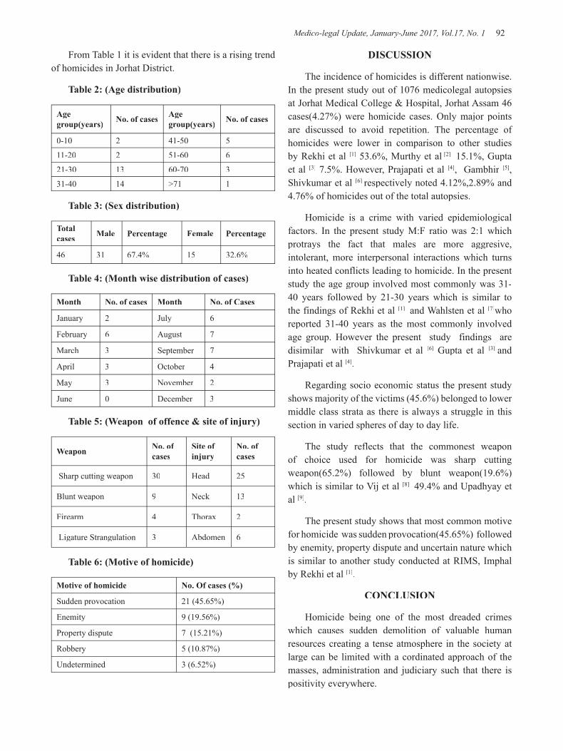
Medico-legal Update, January-June 2017, Vol.17, No. 1 92 93 -legal Update, January-June 2017, Vol.17, No. 1
From Table 1 it is evident that there is a rising trend of homicides in Jorhat District.
Table 2: (Age distribution)
Age group(years) No. of cases Age
group(years) No. of cases
0-10 2 41-50 5
11-20 2 51-60 6
21-30 13 60-70 3
31-40 14 >71 1
Table 3: (Sex distribution)
Total cases Male Percentage Female Percentage
46 31 67.4% 15 32.6%
Table 4: (Month wise distribution of cases)
Month No. of cases Month No. of Cases
January 2 July 6
February 6 August 7
March 3 September 7
April 3 October 4
May 3 November 2
June 0 December 3
Table 5: (Weapon of offence & site of injury)
Weapon No. of cases
Site of injury
No. of cases
Sharp cutting weapon 30 Head 25
Blunt weapon 9 Neck 13
Firearm 4 Thorax 2
Ligature Strangulation 3 Abdomen 6
Table 6: (Motive of homicide)
Motive of homicide No. Of cases (%)
Sudden provocation 21 (45.65%)
Enemity 9 (19.56%)
Property dispute 7 (15.21%)
Robbery 5 (10.87%)
Undetermined 3 (6.52%)
DISCUSSION
The incidence of homicides is different nationwise. In the present study out of 1076 medicolegal autopsies at Jorhat Medical College & Hospital, Jorhat Assam 46 cases(4.27%) were homicide cases. Only major points are discussed to avoid repetition. The percentage of homicides were lower in comparison to other studies by Rekhi et al [1] 53.6%, Murthy et al [2] 15.1%, Gupta et al [3] 7.5%. However, Prajapati et al [4], Gambhir [5], Shivkumar et al [6] respectively noted 4.12%,2.89% and 4.76% of homicides out of the total autopsies.
Homicide is a crime with varied epidemiological factors. In the present study M:F ratio was 2:1 which protrays the fact that males are more aggresive, intolerant, more interpersonal interactions which turns into heated conflicts leading to homicide. In the present study the age group involved most commonly was 31-40 years followed by 21-30 years which is similar to the findings of Rekhi et al [1] and Wahlsten et al [7]who reported 31-40 years as the most commonly involved age group. However the present study findings are disimilar with Shivkumar et al [6] Gupta et al [3] and Prajapati et al [4].
Regarding socio economic status the present study shows majority of the victims (45.6%) belonged to lower middle class strata as there is always a struggle in this section in varied spheres of day to day life.
The study reflects that the commonest weapon of choice used for homicide was sharp cutting weapon(65.2%) followed by blunt weapon(19.6%) which is similar to Vij et al [8] 49.4% and Upadhyay et al [9].
The present study shows that most common motive for homicide was sudden provocation(45.65%) followed by enemity, property dispute and uncertain nature which is similar to another study conducted at RIMS, Imphal by Rekhi et al [1].
CONCLUSION
Homicide being one of the most dreaded crimes which causes sudden demolition of valuable human resources creating a tense atmosphere in the society at large can be limited with a cordinated approach of the masses, administration and judiciary such that there is positivity everywhere.
-legal Update, January-June 2017, Vol.17, No. 1 92 93 Medico-legal Update, January-June 2017, Vol.17, No. 1
Conflict of Interest: Nil
Source of Funding : Self
Ethical Clearance : Not required
REFERENCES
1. Rekhi T, Singh K P and Nabachandra H. Study on Homicidal Blunt Force Injuries. Journal of Forensic Medicine & Toxicology. 2007; 24(2) :3-5.
2. Murthy OP, Keong KS, Ghazali MF et al. Study of Homicidal Deaths at University Malaya Medical Centre, Kuala Lumpur. International Journal Of Medical toxicology & LegalMedicine.
3. Gupta A, Mukta R, Mittal AK and Dikshit PC. A study of homicidal deaths in Delhi. Med Sci Law. 2004; 44(2) 127-32.
4. Prajapati P, Sheikh MI and Patel S. A study of homicidal deaths by mechanical injuries in Surat, Gujrat. Journal Indian academy of Forensic Medicine. 2010;32(2):134-38.
5. Gambhir O and Gupta BD. Evaluation of mehanical injury in homicidal deaths; JIAFM.2007; 23(3): 18-22
6. Shivakumar BC, Vishwanath D, Srivastava PC. Trends of homicidal deaths at a tertiary care centre Bengaluru; JIAFM. 2011; 33(2) 120-24.
7. Wahlsten P, Koiranen V and Saukko P. Survey of Medicolegal investigation of homicide in the city of Turku, Finland. Journal of Forensic & Legal Medicine.2007;14(2007):243-52.
8. Vij A, Menon A, Menezes RG, Kanchan T and Rastogi P. A retrospective review of homicides in Mangalore, South India. Journal of Forensic & Legal Medicine 2010; 17(6): 312-15.
9. Reddy KSN. The Essentials of Forensic Medicine & Toxicology.2015 Edition.
10. Modi JP. A textbook of medical jurisprudence & toxicology. 24th Edition, 2012.

95 -legal Update, January-June 2017, Vol.17, No. 1
Study of Patterns of Cranio-cerebral Injuries in Road Traffic Accidents Involving Two Wheelers in Gulbarga Area
Mohsenul Haq1, Ayesha Farheen2, S K Goli3
1Associate Professor, Department of Forensic Medicine, K.B.N.I.M.S, Gulbarga, 1Associate Professor, Department of Forensic Medicine, K.B.N.I.M.S, Gulbarga, 1 2Assistant Professor, 2Assistant Professor, 2
Department of Pathology, ESIC Medical College, Gulbarga, 3Professor & Head, Department of Forensic Medicine, K.B.N.I.M.S, Gulbarga
ABSTRACT
Road traffic accidents are the major causes of death worldwide. Head injury is the single most common cause of mortality in road traffic accidents1. Head injuries may result in injury to the contents of the skull either alone or with a fracture of the skull.
The present study was undertaken on 140 victims of road traffic accidents, died due to head injury and to find out the patterns of head injuries, their age and sex distribution and site distribution of different types of fractures.
This study was conducted at Khaja Banda Nawaz institute of medical science Gulbarga(North-Karnataka) from 1st April 2014 to 30st April 2014 to 30st th September 2015.
Among 140 cases autopsied, males 122(87.14%) predominated females 18(12.85%) in the age group 31-40 years.
Linear fracture of skull was the commonest type and temporo-parietal region was involved, predominantly subdural haemorrhage was the commonest intracranial haemorrhage associated with head injuries. More number of deaths is occurring in the late night hours.In this study 47 (33.57%) died on the spot, 65(46.42%) died in the hospital after few hours and 28(20%) died on the way to hospital.
Keywords: Road traffic accidents, skull fractures, intracranial haemorrhages, two wheelers.
INTRODUCTION
Road traffic accident is an unplanned event occurring suddenly, unexpectedly and inadvertently in unforeseen circumstances. Incidence is more common among the two wheeler vehicles. Head was the most common site to be injured in the RTA’s2.According to 2008 report of National Crime Record Bureau of India 1,18,239 persons were killed in fatal road traffic accidents(RTA) and out of these 23,552(19.9%) were killed while riding on two wheelers3.The mortality rate is steadily rising3.
Since the head contains brain, a very important vital organ, trauma to this region challenges the individual because of its anatomical position, size, and movements in all directions. Some of the factors that increase risk of RTA in India are lack of traffic laws, drunken and
rash driving, traffic accidents due to negligent act, poor conditions of the road, lack of infrastructures, traffic mix, encroachments that restrict safe area for pedestrian.
The aim of the present study is to find out the pattern and distribution of head injuries in deaths due to RTA with/without helmet and other associated risk factors and to suggest preventive measures so as to save precious human lives from the man made calamities.
MATERIALS AND METHOD
Study design- Cross-Sectional, Descriptive and non-randomized study
Study period- 01-04-2014 to 30-09-2015
Study Method-For this study, 140 cases with the
DOI Number: 10.5958/0974-1283.2017.00020.2
95 Medico-legal Update, January-June 2017, Vol.17, No. 1
history of road traffic accidents in and around Gulbarga city that have fulfilled the inclusion and exclusion criteria were selected on non-randomized purposive sampling basis.
This study was conducted on the injured and deceased who were brought to the hospital and mortuary of the civil and Khaja Banda Nawaz hospital, Gulbarga. The inquest reports were studied for various details such as name, age, gender and brief history of road traffic accidents viz, age, sex, site of death, time of death and period of survival. In hospital treated cases, case sheets were studied for details statistical analysis was done by using chi square test and calculating P value.
RESULTS
The present study revealed that, the total number of road traffic accident deaths was 140, which were autopsied, and it was found that 46(33%) of the victims was in the age group 31-40 years followed by 21-30 years. The lowest incidence was seen in age group of more than 70 years(1%). Males comprised 87.14% of the total victims.(Table No 2)
Maximum number of victims 65(46.42%) died in the hospital, followed by victims died on the spot 47(33.57%). The number of victims who died on the way to hospital was 28(20%).(Table No 3).
Most of the incidents irrespective of the cause occurred between 6pm to 12am comprising 65(46.42%) of the total cases. The least number of cases 22(15.71%) occurred between 6am to 12pm.(Table No-4)
Intracranial haemorrhages were seen in 108 cases, skull fractures were found in 97 cases and injury to brain in 62 cases. A combination of multiple injuries in the form of scalp injury, skull fracture, intracranial haemorrhage and injury to brain was predominantly seen in 51( 36,42%) cases. Commonest type of fracture present in the skull was linear fracture 54(38.57%) followed by crush fracture in 34(24.28%) cases. (Table No-5).
Alcohol was found in stomach contents of 16(11.42%) who are males of adult age group. In remaining 124(88.57%) people including males and females had no alcohol smell.
DISCUSSION
In the present study , a total of 140 cases of fatal two
wheeler accidents of all age group and both sex were studied for duration of 18 months, The percentage of deaths occurring due to involvement of two wheelers is constantly around 7% in all the years, but the number of deaths due to accidents is increasing in proportion to the total number of deaths 4,5.
Highest incidence of fatalities occurred in the age group 31-40 years (33%) followed by 21-30 years (23%). Since the age group 20-40 years is the most active phase of life physically and socially, they therefore accounted for the maximum number of accidental deaths. Similar findings were observed by other researchers6-9
In the present study , male comprised a majority and constituted 87.15% of 140 cases compared to females 12.85% . The male to female ratio in the present study is 5:1.
A male preponderance almost in consistence with the study reported10-11.
Peak time of occurrence of RTA was 6:00pm to 12:00 midnight followed by 12:00 noon to 6:00pm which is probably due to heavy and unequal distribution of the traffic at these closing hours of the people and the rider is generally exhausted after day’s work. Sirathanont12
demonstrated most of motorcycle crashes were between 6pm – 9pm.
Ding et al13 reported most of the head injuries occurred between 4pm – 11pm peaking at 9pm.
Majority of the deceased (84%) were not using helmet while riding at the time of accident. only 6% riders among deceased used helmets, which suggest that use of helmet can be lifesaving measure during an accident. Sirathranont and Kasantikul12 noted that 0nly 4% of the riders were wearing helmet at the time of accident which is in conformity with our observation.Pathak et al14 in their study of 39 cases of two wheelers accidental deaths reported that 12.82% victims used helmet while 87.18% did not used helmet at all. Thus, death rate was noted to be higher in non-helmet users when compared to helmet users, a finding which is in concurrence to our observation.
The Period of survival: 65(46.42%) victims died in the hospital after 6 hours of the accident and 47(33.57%) died on the spot and these findings co-relates with the present study15.

Medico-legal Update, January-June 2017, Vol.17, No. 1 96 97 -legal Update, January-June 2017, Vol.17, No. 1
The period of survival has not shown an increase despite the advances of medical facilities, this may be due to severity of injuries sustained, lack of knowledge of first aid among the people at the scene of accident, delay in shifting the patient to trauma center and no change in the attitude of people due to fear of getting into legal hassles.
In the present study contusion is seen in 128(91.4%) cases either alone or in combination with other injuries. Laceration with other injuries is seen in 28(20%) cases.90% of the victims suffered fracture of the skull- either vault or base of the skull or both. Among the fractures sustained 54% linear fracture and 34% crush fracture .Our findings were well supported by Pathak et al14who reported 120 cases of head injury deaths, linear fracture of the skull being the highest (43.04%).
The most commonly found intracranial haemorrhage was subdural haemorrhage(SDH) followed by subarachnoid Haemorrhage (SAH) in 96% of the victims of RTA which coincided with the observations of the other researchers [ 5,13,14,15,]
In the present study alcohol 16(11.42%) cases presence of alcohol was detected. These findings are not similar with Ghosh, who reported that put of 230 cases only in 6(2.6%) cases the alcohol played the role in causing accidents. Solheim et al, detected in 52.7% cases. This difference may be due to the fact that alcohol intake in Indian population is not much high degree as seen in western countries.
Table (1): Age wise distribution of RTA victims
Age No of RTA victims Percentage
0-10 03 2%
11-20 16 11%
21-30 32 23%
31-40 46 33%
41-50 23 16%
51-60 12 9%
61 -70 07 5%
>70 01 1%
Total 140 100%
Table (2) Sex wise Distribution of RTA victims
SEX No of RTA victims Percentage
Male 122 87.15%
Female 18 12.85%
Total 140 100
Table (3)Site of Death
Site of death No of RTA Victims Percentage
Died on spot 47 33.57%Hospital 65 46.42%On the way to hospital 28 20.00%Total 140 100%
Table (4) Time of Accident
Time of Accident Male Female Total %
6am – 12pm 21 01 22(15.71%)
12pm -6pm 24 05 29(20.71%)
6pm – 12am 54 11 65(46.42%)
12am – 6am 23 01 24(17.85%)
Total 122 18 140(100%)
Table (5) Injury to skull & types of intracranial haemorhages
Fracture of skull
No of RTAvictims
Intracranial haemorhages Total %
Linear fracture of skull 54 EDH 11(7.85%)
Comminuted fracture of skull 11 SDH 62(44.28%)
Depressed fracture of skull 27 SAH 51(36.42%)
Basal fracture of skull 14 ICH 12(8.57%)
Crush fracture of skull 34 IVH 04(2.85%)
CONCLUSION
The present study revealed that the human error is mainly responsible for the fatal RTA. Though it is a most difficult task to control human errors involved, sincere efforts can reduce the mortality and morbidity. For reducing fatalities among victims of two wheeler road traffic accidents, it is essential to study the cause of RTAs, which revolve around factors responsible as Human errors, Machine(vehicle) errors and environment.
-legal Update, January-June 2017, Vol.17, No. 1 96 97 Medico-legal Update, January-June 2017, Vol.17, No. 1
Males are predominant gender than females which are succumbing to the two wheeler accidents.The study reveals that males aged between 21- 40 years are more vulnerable for two wheeler accidents.
Most of the victims sustained fatal head injuries and this was most common among those who were not wearing helmets. Majority of the RTA victims who died on the spot had died due to primary head impact.
As the population and number of immigrants continued to increase, preventive measures need to be taken, based on the risk factors that had been identified. The following measures has to be considered to minimize the incidence of fatal head injuries in RTA victims.
-Total Ban on drunk and drive.
-Do not overtake unnecessarily.
-Reducing the speed of vehicles.
-Making good quality roads with lightings and signals.
-Make one way traffic and avoid haphazard road crossing by pedestrians.
-Build pavements for people to walk and ban of shops or sale on pavements.
-Impose hefty fine if one violates traffic rules with driving license cancellation for repeated violation of traffic rules.
-well equipped trauma centres with facilities and specialized professionals.
Conflict of Interest: The findings in this research work found to be similar to most of the research work conducted in India.
Source of Funding: Self Funding.
Ethical Clearance: Not Applicable.
REFERENCES
1) Reddy KSN. The essentials of Forensic Medicine and Toxicology, 29th edition,published by K.Sugana Devi,2010; p218.
2) Otto Saphir.Autopsy Diagnosis and technique.Paul B Hober Inc.third edition.
3) Accidental Deaths and Suicides in India-2008.National Crime Record bureau Report-2008. p 13-14.
4) Schelp L,Ekman R. ‘Road traffic accidents in a Swedish municipality’ Public Health.1990 Jan; 104(1): 55-64.
5) ‘Accident deaths & Suicides in India 1998, Published by National crime records Bureau,Ministry of Home Affairs,GOI, New Delhi.
6) B H Tirpude, R S Naik, A J Anjankar et al. A study of pattern of cranio-cerebral injuries in road traffic accidents.Journal of Indian Academy of Forensic Medicine,1998 vol.20, No 1 ISSN: 0971- 0973.
7) Harnam Singh, S K Dhattarwal. Pattern and distribution of injuries in fatal road traffic accidents in Rohtak (Haryana), Journal of Indian Academy of Forensic Medicine,2004;26(1),ISSN 0971- 0973.
8) B R Sharma, A K Sharma, Swati Sharma, Harshabad Singh. Fatal road traffic injuries in Northern India: Can they be prevented? Trends in medical Research 2(3):142-148,2007.
9) Gautam Biswas, S K Verma, N K Agrawal et al. Pattern of road traffic accidents in north east Delhi; Journal of Forensic Medicine and Toxicologyvol.20,No.1, Jan-Jun 2003.
10) Bereka Bruce et al. Outcome Following Severe Head Injuries in Children. Journal of Neuro-Surgery, 1978; 679-688.
11) Edward L. hanna et al. Motor Vehicle Crashes in New-York State. Journal of Trauma, 2001;50:1117-1124.
12) Sirathanout J. Kasantikul V. Mortality and injury from motorcycle collision in Phetchaburi province,J.Med Assoc. Thai 2003; 86; p97-102
13) Ding SL. Pai L, Wang Jd, Chen KT. Head injuries in traffic accidents with emphasis on The comparisons between motorcycle-helmet users and non users. J Formos Med Assoc. 1994; 93; S42- 48.
14) Pathak A, Desania NL, and Verma R, Profile of road traffic accidents and head injury in Jaipur (Rajasthan). J Indian Acad Forensic Med. 2008; 30(1): 6-9
15) Nandy A. Road Traffic Accidents. Ist Edition.New Centre of Book Agency Calcutta 1995;291.
99 -legal Update, January-June 2017, Vol.17, No. 1
Study of Correlation of Index Finger Lengths and Stature
Pramod Kumar GN1, Roopa Urs AN2
1Associate Professor, Department of Forensic Medicine & Toxicology, 1Associate Professor, Department of Forensic Medicine & Toxicology, 1 2Assistant Professor, Department 2Assistant Professor, Department 2
of Pathology, Mahatma Gandhi Medical College & Research Institute, Sri Balaji Vidyapeeth University, Pillaiyarkuppam, Pondicherry, India
ABSTRACT
The present study attempts to determine stature from Index finger length(IFL). Length of Index finger was measured of 200 individuals (100 males & 100 females) belonging to Mysore district, Karnataka, India aged between 21 and 30 years. Measurements of Index finger length (IFL) were recorded using a Vernier caliper and the individual height (stature) was recorded using an anthropometric rod and the data was subjected to statistical analysis using SPSS software. Mean stature was significantly higher in males than females. Mean IFL on right and left sides was 8.72cm and 8.67cm in males and 8.19cm and 8.11cm in females respectively. Mean IFL was greater in males than females of both hands. Statistically significant correlation was observed between stature and Index finger length of both hands. Pearson correlation (r) for stature and finger lengths was higher among females than males. Independent linear regression equations to calculate the height was formulated in males & females separately.
Keywords: Identification; Index finger length; Stature; Vernier caliper;
Corresponding author:Dr. Pramod Kumar GNAssociate Professor, Department of Forensic Medicine & Toxicology, Mahatma Gandhi Medical College & Research Institute, Sri Balaji Vidyapeeth UniversityPillaiyarkuppam, Pondicherry – 607402, India E mail: [email protected]@mgmcri.ac.in, Mobile: 09787690333
INTRODUCTION
Anthropometry refers to measurement of different dimensions of individual for the purpose of identification. Determination of stature forms the primary data of identification. Estimation of stature of an individual from the skeletal remains or from mutilated or amputated limbs or from parts of limbs has obvious significance in personal identification in the event of alleged homicide, accidents or natural disasters, mainly concerned with the forensic identification analysis. The retrieval of mutilated remains is not uncommon, because many a times the bodies are mutilated with the intention of either concealing the identity of the deceased after committing a crime or to facilitate the disposal of dead. In some circumstances fragmentary remains may also be recovered from forests or lonely places mutilated by
wild animals.1 The need to develop methods to construct stature from various bones has been stressed by many workers due to its application in forensic medicine, in medicolegal enquiries and in identifying war or mass disaster casualties.
Various studies in past have utilized various body parts such as upper and lower extremities including hand and foot dimensions for estimation of stature. 2,3 Krishan K et al study concluded that the living stature can be predicted from the Index finger length (IFL) and Ring finger length (RFL) with a reasonable accuracy in adolescent population of North India.4,7 Sen J et al conducted a study to find the correlation between stature and the lengths of the index and ring fingers & concluded that the regression models derived can be of significant utility in the estimation of stature.5 The present study attempts to determine the stature from Index finger length.
MATERIAL AND METHOD
The study was conducted in Mysore, India. In this study length of Index finger & height were measured of 200 individuals (100 males & 100 females) belonging to Mysore district, Karnataka, India aged between 21 and 30 years. Nonresident Indians and individuals from
DOI Number: 10.5958/0974-1283.2017.00021.4
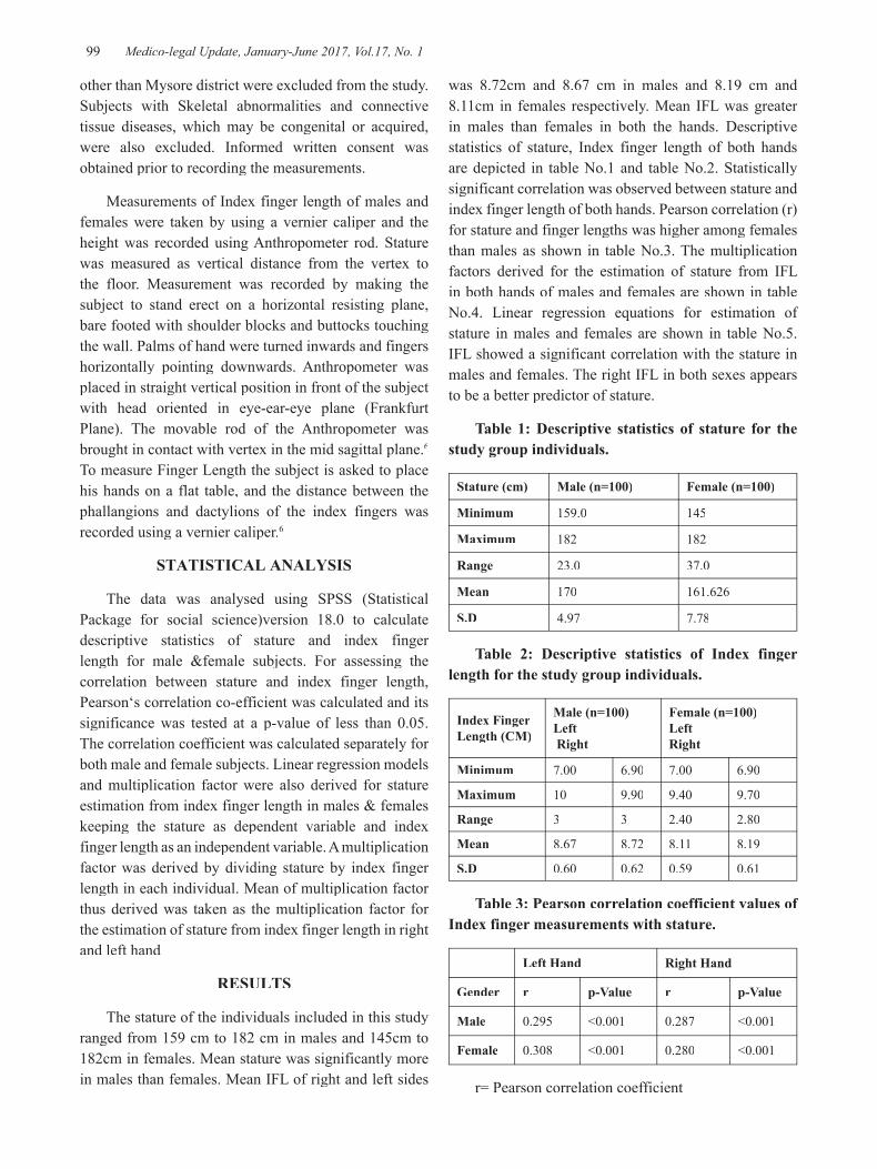
99 Medico-legal Update, January-June 2017, Vol.17, No. 1
other than Mysore district were excluded from the study. Subjects with Skeletal abnormalities and connective tissue diseases, which may be congenital or acquired, were also excluded. Informed written consent was obtained prior to recording the measurements.
Measurements of Index finger length of males and females were taken by using a vernier caliper and the height was recorded using Anthropometer rod. Stature was measured as vertical distance from the vertex to the floor. Measurement was recorded by making the subject to stand erect on a horizontal resisting plane, bare footed with shoulder blocks and buttocks touching the wall. Palms of hand were turned inwards and fingers horizontally pointing downwards. Anthropometer was placed in straight vertical position in front of the subject with head oriented in eye-ear-eye plane (Frankfurt Plane). The movable rod of the Anthropometer was brought in contact with vertex in the mid sagittal plane.6
To measure Finger Length the subject is asked to place his hands on a flat table, and the distance between the phallangions and dactylions of the index fingers was recorded using a vernier caliper.6
STATISTICAL ANALYSIS
The data was analysed using SPSS (Statistical Package for social science)version 18.0 to calculate descriptive statistics of stature and index finger length for male &female subjects. For assessing the correlation between stature and index finger length, Pearson‘s correlation co-efficient was calculated and its significance was tested at a p-value of less than 0.05. The correlation coefficient was calculated separately for both male and female subjects. Linear regression models and multiplication factor were also derived for stature estimation from index finger length in males & females keeping the stature as dependent variable and index finger length as an independent variable. A multiplication factor was derived by dividing stature by index finger length in each individual. Mean of multiplication factor thus derived was taken as the multiplication factor for the estimation of stature from index finger length in right and left hand
RESULTS
The stature of the individuals included in this study ranged from 159 cm to 182 cm in males and 145cm to 182cm in females. Mean stature was significantly more in males than females. Mean IFL of right and left sides
was 8.72cm and 8.67 cm in males and 8.19 cm and 8.11cm in females respectively. Mean IFL was greater in males than females in both the hands. Descriptive statistics of stature, Index finger length of both hands are depicted in table No.1 and table No.2. Statistically significant correlation was observed between stature and index finger length of both hands. Pearson correlation (r) for stature and finger lengths was higher among females than males as shown in table No.3. The multiplication factors derived for the estimation of stature from IFL in both hands of males and females are shown in table No.4. Linear regression equations for estimation of stature in males and females are shown in table No.5. IFL showed a significant correlation with the stature in males and females. The right IFL in both sexes appears to be a better predictor of stature.
Table 1: Descriptive statistics of stature for the study group individuals.
Stature (cm) Male (n=100) Female (n=100)
Minimum 159.0 145
Maximum 182 182
Range 23.0 37.0
Mean 170 161.626
S.D 4.97 7.78
Table 2: Descriptive statistics of Index finger length for the study group individuals.
Index Finger Length (CM)
Male (n=100)Left Right
Female (n=100)Left Right
Minimum 7.00 6.90 7.00 6.90
Maximum 10 9.90 9.40 9.70
Range 3 3 2.40 2.80
Mean 8.67 8.72 8.11 8.19
S.D 0.60 0.62 0.59 0.61
Table 3: Pearson correlation coefficient values of Index finger measurements with stature.
Left Hand Right Hand
Gender r p-Value r p-Value
Male 0.295 <0.001 0.287 <0.001
Female 0.308 <0.001 0.280 <0.001
r= Pearson correlation coefficient

Medico-legal Update, January-June 2017, Vol.17, No. 1 100 101 -legal Update, January-June 2017, Vol.17, No. 1
Table 4: Constant and regression coefficient values for stature estimation from Index figure measurements.
Gender Side Constant Regression coefficient
MalesRight 151.06 2.267
Left 149.92 2.412
FemalesRight 132.614 3.539
Left 128.581 4.071
Table 5: Linear regression equations for stature estimation from Index figure length.
Gender Side Equation
Male Left H= 149.92 + 2.412 (LI)
Right H= 151.06 + 2.267 (RI)
Female Left H= 128.581 + 4.071 (LI)
Right H = 132.614 + 3.539 (RI)
H=Height
DISCUSSION
Stature is one such parameter that can be established even in mutilated and dismembered bodies. In this study an attempt was made to establish the stature of a person by using Index finger length. Males and females aged between 21 and 30 years, those who were natives of Mysore district of Karnataka state were included in the study. In the present study the mean stature among males was 170.84 cm with a standard deviation of 4.9718. The minimum &maximum heights were being 159 cm and182 cm respectively. Among females mean stature was 161.62 cm with a standard deviation of 7.79. The minimum and maximum heights were being 145 cm and182 cm, respectively. The Pearson correlation coefficients showed a high degree of correlation and all the values were statistically significant (p value < 0.05). Krishan et al estimated stature from index and ring finger length in a North Indian population of age between 14-18 years with correlation coefficient ranging from 0.671 to 0.748 in males and from 0.367 to 0.531 in females.8 In the present study age group of subjects is between 21 -30 years and the correlation coefficient ranged from 0.287
to 0.295 in males and from 0.280 to 0.308 in females. Krishan et al had noted higher correlation coefficient for males than females whereas in present study we have noted higher correlation coefficient for females than males. This difference could be attributed to population difference as Krishan et al had utilized adolescent north Indian population whereas the present study is done on south Indian population. The findings of our study are similar to study done by Rajesh Bardale et al8 from Maharashtra (where in the correlation coefficient ranged from 0.546 to 0.576 in males and from 0.594 to 0.596 in females).
From this study it is found that, for more accurate prediction of stature, independent linear regression equation should be used. This study involved a small sample size and only native people of Mysore district of Karnataka state, South India, so there is scope for further work to determine similar regression equations with larger sample for people from other parts of the Country and World.
CONCLUSION
Present study shows that there is significant correlation between stature and index finger length. Hence this can be of help in identification of unidentified and dismembered bodies. This study is conducted in localized geographical area with limited sample size (200) hence similar studies are proposed in different populations of larger sample size.
Acknowledgment: Nil
Declaration of Interest Statement: The Authors declare that there is no conflict of interest.
Source of Funding: No source of financial assistance was obtained from any individual or agency.
Ethical Clearance: Written consent from the participant obtained.
REFERENCES
1. Tyagi AK, Kohli A, Verma SK andAggarwal BBL. Correlation betweenstature and fingers length. InternationalJournal of Medical Toxicology and LegalMedicine.1999; 1(2): 20-22.
2. Bhatnagar DP, Thapar SP, Batish MK. Identification of personal height from the somatometry of the hand in Punjabi males. Forensic Sci. Int. 1984; 24:
-legal Update, January-June 2017, Vol.17, No. 1 100 101 Medico-legal Update, January-June 2017, Vol.17, No. 1
137-41 4.
3. Agnihotri AK, Agnihotri S, Jeebun N, Googoolye K. Prediction of stature using hand dimensions. J Forensic Leg Med 2008; 15:479- 82.
4. Krishan K, Sharma A. Estimation of stature from dimensions of hand and feet in a north Indian population. J Forensic Leg Med 2007; 14:327-32.
5. Sen J, Kanchan T, Ghosh A, Mondal N, Krishan K. Estimation of stature from lengths of index and ring fingers in a North-eastern Indian population. J Forensic Leg Med 2014;22:10-5.
6. Singh IP and Bhasin MK.A manual ofbiological anthropology. 1st Ed. Delhi:Kamla Raj Enterprises; 2004: 168-69.
7. Krishan K, Kanchan T, Asha N. Estimation of stature from index and ring finger length in a north Indian adolescent population. J Forensic Leg Med 2012; 19:285-90.
8. Bardale RV, Dahodwala TM, Sonar VD. Estimation of Stature from Index and Ring Finger Length. J Indian Acad Forensic Med 2013;35(4):353-57.

103 -legal Update, January-June 2017, Vol.17, No. 1
Study of Pattern of Ligature Mark in Cases of Hanging in Kamrup Region, Assam State, India
Arunava Borah1, Rituraj Chaliha2
1Demonstrator. Department of Forensic Medicine, 2Professor & Head, Department of Forensic Medicine, Gauhati Medical College, Guwahati
ABSTRACT
Suicidal hanging still remains one of the commonest methods of committing suicide. A careful examination of the ligature material is very important while giving an opinion in hanging. This is an autopsy based study of hanging cases, conducted in the Department of Forensic Medicine and Toxicology, Gauhati Medical College in the year 2014-2015 to analyze the pattern of ligature mark. During this study period, a total number of 2964 autopsies were carried out in the department, out of which 379 cases were associated with hanging. Male female ratio was found as 2.32:1. Majority of the cases 111 (29.29%) were found using plastic ropes as a ligature material in hanging. The commonest position of the knot was behind the left mastoid 125 (32.98%). Only 17 cases (4.48%) showed presence of ligature material in situ. Position of the ligature was above the thyroid cartilage in 100% cases. The ligature mark showed marked grooved pattern in 135 cases (35.62%). Grooved pattern of ligature mark was associated with plastic rope 103 (27.18%) and coir rope 23 (6.07%). Majority of grooved pattern 24.52% was seen between 6-12 hours period of suspension.
Keywords: Hanging, ligature mark, ligature material, groove pattern.
Corresponding author : Arunava Borah, Demonstrator, Department of Forensic MedicineGauhati Medical College, Guwahati, Assam.Email: [email protected]: +919401270726
INTRODUCTION
Hanging is the process in which a body is suspended with a ligature around the neck as a result of which constriction of the air passage occur thus preventing exchange of air between the atmosphere and the alveoli of lungs. Death occurs chiefly due to asphyxia1. The principal external sign in a case of death due to hanging is the ligature mark. A detailed examination of the ligature mark is very significant in order to arrive at the probable cause of death. This also helps in differentiating between hanging and the ligature strangulation 2. Most of the times, the ligature mark may be the only evidence available in cases of hanging and its characteristics are well noted in the literature3. The type and position of knot also plays an important role in the causation of death in hanging. In many cases the ligature material is not available during autopsy examination.
Hence, the features of ligature material are not submitted to systematic analysis4. A victim of suicidal hanging uses any type of ligature available at hand in the house or place where they are found. Commonly used materials are rope, metallic chain and wireless, handkerchief, belt, bed sheet, scarf, dhoti, saree, dupatta etc5. The pattern of the ligature material gets impressed on the skin which forms a characteristic diagonal mark of the strands found when the rope is used. The wide band of cloth when used as a ligature on the bare skin may cause a narrow ligature mark, due to tension lines in the stretched cloth. If the ligature material is soft and the body is cut down immediately after death, there may be no mark6.
MATERIAL AND METHOD
This is a cross sectional study carried out in the Department of Forensic Medicine, Gauhati Medical College within the district administrative area of Kamrup, Assam and few referral cases from the neighboring districts. The study period extended from 1st July, 2014 to the 30th June, 2015. A total number of 2964 autopsies were carried out of which 379 cases were associated with hanging. All hanging cases were included in this study irrespective of history of
DOI Number: 10.5958/0974-1283.2017.00022.6
103 Medico-legal Update, January-June 2017, Vol.17, No. 1
incident, age group, sex, degree of decomposition.
The primary data in each case is collected from different sources including the Inquest report and the dead body challan, personal interview of the investigating officer, relatives, friends and neighbors of the deceased, autopsy examination performed on the dead body of the victim with use of magnifying lens, measuring tape and camera for photography. All these data collected from different sources were then recorded in a specially designed proforma for each case for further collective evaluation.
OBSERVATIONS
It was seen that majority of the cases 111 cases (29.29%) were found using plastic ropes as a ligature material out of which 84 cases (22.16%) were male who used plastic rope as their ligature material while 34 cases (8.97%) were females who used chunni as their ligature material. Rubber belt, crepe bandage, tie, towel were few other ligature materials used.
292 cases (77.04%) had complete type of hanging with both sexes and nature was suicidal in 100% of cases. The hanging victims suspended themselves from the roof in 185 (48.81%) cases, from ceiling fan 106 (27.97%) cases, from tree in 55 (16.36%) cases, grill 23 (6.07%) cases.
Majority of the knots were behind the left mastoid 125 (32.98%). Next common position was right mastoid 85 (22.43%). Likewise, occiput accounted 75 cases (19.79%), left angle of the mandible accounted 40 cases (10.55%), right angle of the mandible accounted 22 cases (5.80%). In both the sexes, knot was present mostly behind the left mastoid with male and female accounting for 93(24.53%) and 32(8.44%) respectively.
In 362 cases (95.51%), the ligature material was absent in situ. Only 17 cases (4.48%) showed presence of ligature material in situ.
In all the 379 recorded cases, where ligature was found above the thyroid cartilage and were non continuous all around the neck. 374 cases (98.68%) had a single turn of the ligature. Five (1.32%) cases showed double turn and zero cases were found with multiple turns. 314 (82.85%), there was no intervening material in between neck and ligature material. Intervening material in the form of scalp hair were present only in case of female 65 (17.15%).
In 135 cases (35.62%), the ligature mark showed marked grooved pattern. On the other hand 244 cases
(64.38%) showed non grooved pattern.
The period of suspension was 6-12 hours in 214 cases (54.46%) followed by 0-6 hours in 139 cases (36.68%).Only in 4 cases (1.05%), the period of suspension was more than 24 hours duration. The groove pattern was highest seen in 6-12 hours duration with 93 cases (24.52%).This was followed by 0-6 hour interval period with 33 cases (8.48%).
The grooved pattern of ligature mark was associated with plastic rope 103 (27.18%) and coir rope 23 (6.07%). This was followed by gamusha 4 (1.06%), chunni 3 (0.79%) and saree 29 (0.53%) which showed grooved pattern.
DISCUSSION
The incidence of hanging cases is increasing everyday so it is imperative that knowledge of this subject must increase rapidly both in social and practical aspects. In the present study 84 cases (22.16%) were male who used plastic rope as their ligature material while 34 cases (8.97%) were females who used chunni as their ligature material. Similar observations were made by Authors Sharma BR, Harish D, Singh VP, Singh P (2005)7, Momin SG, Mangal HM, Kyada HC, Vijapura MT, Bhuva SD (2008)2, Fimate L et al (1993)8. However soft material like dupatta was found in the study by Waghmare PB, Chikhalkar G, Nanandkar SD (2014)9 .
The type of hanging in the victims as per study was found to be complete type in 292 cases (77.04%) with both the sexes. These findings are consistent with the findings of study conducted by MeeraTh, Singh MBK (2011)10, Singh KP, Marak AR, Meera T (2013)11.
During the course of study, majority of the knots were observed behind the left mastoid 125 (32.98%). Next common position was right mastoid 85 (22.43%). These findings were consistent with the findings of study conducted by Baishya MK (2014)5, Ambade VN, Tumram N, Meshram S, Borkar J (2014)4.
It was also found that 374 cases (98.68%) had a single turn of the ligature, five (1.32%) cases showed double turn and zero cases were found with multiple turns. These findings were consistent with the findings of study conducted by Meera Th, Singh MBK (2011)10, Baishya MK (2014)5, Ambade VN, Tumram N, Meshram S, Borkar J (2014)4, Arif M (2015)12.
In all the 379 recorded cases, the ligature was found

Medico-legal Update, January-June 2017, Vol.17, No. 1 104
above the thyroid cartilage and non- continuous all around the neck. These results were similar to Some Authors Waghmare PB, Chikhalkar G, Nanandkar SD (2014)9, Rawat V, Rodrigues EJ (2015)13.
In the present study, marked grooved pattern was observed in 135 cases (35.62%) while 244 cases (64.38%) showed non grooved pattern. Nearly similar observations were made by the Authors Rawat V, Rodrigues EJ (2015)13, Ali E, Maksud M, Zubyra SJ (2014) 14.
As per the observations of the present study it was seen that the well-marked groove pattern of ligature mark was mostly associated with hard ligature materials which include plastic rope 103 (27.18%) and coir rope 23 (6.07%). Similar observations were made by authors Rawat V, Rodrigues EJ (2015)13.
In our study, we observed maximum victims were suspended for a period of 6-12 hours in 214 cases (54.46%) and grooved pattern was observed highest in this time interval. Similar observations were made by Authors Rawat V, Rodrigues EJ (2015)13 .
CONCLUSIONS
Hanging has emerged as one of the commonest form of asphyxial suicidal deaths in Assam .The observations of the present study tallied in almost all respects with studies conducted by previous researchers. A careful examination of the ligature mark and the ligature material is very important while giving an opinion in hanging. The type and position of knots are necessary to correlate the autopsy findings and mechanism of death in hanging. Any gross inconsistencies in this regard may create doubt of foul play or suspicious death.
Conflict of Interest: None declared.
Ethical Clearance: Taken.
Source of Funding: None declared.
Declarations: (1) The article is original with the author(s) and does not infringe any copyright or violate any other right of third parties; (2) The article has not been published elsewhere, and is not being considered for publication elsewhere in any form, except as provided herein; (3) All author(s) have contributed sufficiently in the article to take public responsibility for it; (4) All author(s) have reviewed the final version of the above manuscript and approve it for publication.
REFERENCES1. Nandy A. Principles of Forensic Medicine including
Toxicology. 3rd ed. Kolkata: New Central Book Agency(P) Ltd; 2012.p517
2. Momin SG, MangalHM,KyadaHC,Vijapura MT, Bhuva SD. Pattern of Ligature Mark in Cases of Compressed Neck in Rajkot Region: A Prospective Study.JIAFM,2012;34(1):p40-43.
3. Tumram NK, Ambade VN, Bardale RJ, Dixit PG. Injuries over neck in hanging deaths and its relation with ligature material: is it vital? J Forensic Leg Med 2014;22:p82–3.
4. Ambade VN, Tumram N, Meshram S, Borkar J, Ligature material in hanging deaths:The neglected area in forensic examination. Egyptian Journal of Forensic Sciences 2014;5(3):p109-113.
5. BaishyaMK.An epidemiological study of hanging cases brought to the Gauhati medical College and hospital mortuary for medicolegal autopsy. MD Thesis. SrimantaSankaradeva University of Health Sciences 2014, Guwahati, Assam, India.
6. Bhatia M S, Agarwal N K, Millo T, Murthy O P. Suicide, suicide note, and psychological autopsy. International Journal of Medical Toxicology and Legal Medicine. (1999); 16:p38 -39
7. Sharma BR, Harish D, Singh VP, Singh P. Ligature mark on neck-How informative?. JIAFM, 2005: 27 (1).p10-15.
8. Fimate L, Monochand A, Bijoy TH. Prolife of hanging in Manipur. Journals of Medical Society 1993;16:p29-35.
9. Waghmare PB, Chikhalkar G, Nanandkar SD. Analysis of Asphyxial Deaths Due To Hanging.JIAFM,2014:36(4).p343-345.
10. Meera T, Singh MBK, Pattern of Neck Findings in Suicidal Hanging, A Study in Manipur, J Indian Acad Forensic Med. October- December 2011;33:p352-54.
11. Singh KP, Marak AR, Meera T. Multifactorial analysis of hanging deaths. J Med Soc 2013;27:p49-51
12. Arif M. Ligature mark on the neck; How elucidative? Professional Med J 2015;22(6):p798- 803.
13. Rawat V, E. J. Rodrigues (2015) Medicolegal Study of Hanging Cases in North Goa. Int J Forensic SciPathol. 3(5);p110-118.
14. Ali E, Maksud M, Zubyra SJ, Hossain MS, Debnath PR, AlamA,Chakrabarty PK. Suicide by hanging: a study of 334 cases. Bangladesh Med J. 2014 May;43 (2).

-legal Update, January-June 2017, Vol.17, No. 1 104
Study of Post Mortem Interval Using Entomological Evidence
Shashikanth Naik C R1, S Venkata Raghava2
1Asst Prof, Dept of Forensic Medicine, St Johns Medical College, Bangalore, 2Assoc Prof, Dept of Forensic Medicine, Bangalore Medical College, Bangalore
ABSTRACT
The largest biological group on earth is arthropods and they are most important biological group which can be found in all locations including crime scenes. The study of insect are called entomology and application of entomological evidence in solving crime and estimation of time since death is called Forensic Entomology. In this study entomological evidence has been used in 51 cases of decomposed bodies to calculate time since death. Calculated time since death by entomological evidence is compared with estimated time since death by police enquiry and post mortem examination. Paired T test done which was found that, but for mean estimate for post mortem examination and larval length methods all other methods were not significantly different. The estimate using entomological evidence corroborate well with estimate made in police report and post mortem examination.
Keywords: Arthropods, Crime scene, Post mortem examination, Time since death.
INTRODUCTION
Its principle application is in elucidating the time elapsed since death. There are other methods also for this purpose mostly based on study of post mortem changes in the body including rigor mortis, post mortem lividity, post mortem cooling, retinal changes, stomach emptying time etc. and various putrefactive signs. The study of alteration in levels of different ions, enzymes and bio-molecules in body fluids and tissues for estimation of post mortem interval is a relatively new approach. None of these parameters have been found to be very effective, while a study of the cadaver fauna for this purpose has been found to be precise and reliable. (1)
Study of insects and maggots infesting a dead body may not only give some idea about the time since death, the place of death but also of the season when death occurred.(2)
The rationale of Forensic Entomology is that after death, invasion of an unprotected body by sarcosaprophagous insects and other small fauna comes in successive waves. Different species of arthropods colonize the corpse at different periods after death. (3)
From forensic entomological point of view, the necrophagous species, their predators and parasites are the important insects to be attracted to the body immediately after death even within minutes. (4)
The basic principle underlying estimation of the Post Mortem Interval with the help of cadaver insects involves consideration of 3 criteria: (5)
1. The exact sequence in which insects arrive to infest the corpse.
2. The determination of the stage of insect development on the dead body at a specific time.
3. The micro climatic and environmental factors in and around corpse and their influence.
OBJECTIVES OF STUDY
1. To estimate approximate time since death in decomposed bodies.
MATERIAL AND METHOD
Source of data
Medico legal autopsies of decomposed bodies conducted at the Department of Forensic Medicine, Victoria hospital, Bangalore Medical College and Research Institute, Bangalore for a period of 24 months from Nov-2012 to Oct-2014 were examined to obtain insect samples.
Collection of data
1. Study design: Cross sectional study
DOI Number: 10.5958/0974-1283.2017.00023.8
Medico-legal Update, January-June 2017, Vol.17, No. 1 106 107 -legal Update, January-June 2017, Vol.17, No. 1
a. Study period: Nov 2012- Oct 2014. (24 months)
b. Sample size: 51 Medico legal autopsies of decomposed bodies
c. Statistical test: A paired t-test was done to compare the means of PMI (TSD) by the various methods.
2. Inclusion Criteria:
a. All decomposed bodies with clear entomological evidence.
b. Exhumed bodies with clear entomological evidence
3. Exclusion Criteria:
a. Bodies with early decomposition without entomological evidence.
b. Body stored in cold storage after arrival to mortuary.
c. Body preserved by other means during transportation.
METHODOLOGY
The decomposed bodies subjected to autopsy as per the request by the police department were examined for the clear entomological evidence. Only those bodies with the presence of any stage or stages of insects were utilized for the study. Background history of the decomposed body was filled in the aforementioned proforma and the respective post mortem number was assigned for the individual cases. Samples of eggs and larvae were collected from different areas of the body and clothings. Fifty per cent of the samples collected from each body were preserved (see below) and remaining 50% of the sample was used for rearing. Beef liver served as food. The pupae were taken to the Department of Entomology, University of Agricultural Sciences, Bangalore along with the preserved samples in each case. Pupae were reared till adult emergence in the Entomology department.
For the identification of Calliphorid genera and species, the work of Senior White (1940) was used and the species were also got identified by Dr Meenakshi Bharti. The species of Sarcophagidae were identified using the work of Nandi (2002) and those of Muscidae by the work of van Embden (1965). All the identification were confirmed by Dr C. A. Viraktamath.
6. Methods of calculation of Post Mortem Interval
a) Based on enquiry by the police
Time since death estimated as per investigations done by police.
b) Based on the Post Mortem Examination (9)
1) Abdomen develops greenish discoloration - 24 to 36 hrs
2) Marbling of the body - 24 hrs to 48 hrs
3) Bloating stage (distension of body & distortion of face) - 48 hrs to 72 hrs
4) Epidermal Vesicle formation, skin slippage and
diffuse greenish black discoloration of the body - 48 hrs to 96 hrs
5) Active decay - > 96hrs
c) Based on the length of larva and species identified
The larva which were collected and preserved was taken and the length of the largest/oldest larva of the identified species in each case was measured in stereobionocular microscope and noted. Relevant life cycle of the identified species was studied to arrive at the Post Mortem Interval in each case as described by Kashyap and Pillay. (1) The PMI was calculated from the larval length using the formula:
T=A+B (cd)
Where T is the time since death (Post Mortem Interval), A is the wave of invasion of the insect species, B is the time elapsed since oviposition and cd is the climatic factor correction (wherever necessary).
d) Based on Accumulated Degree Hours
Other method of estimation of Post Mortem Interval using accumulated degree days were also calculated in each case as followed by Babu et al. (10) ADH method depends on stage of larva collected from the corpse.
Data needed for calculating Accumulated Degree Hours
A. Average daily temperature from the weather station close to the crime scene for 7 days.
B. Development time(hrs) of each species at constant temperature
C. Minimum threshold temperature for

-legal Update, January-June 2017, Vol.17, No. 1 106 107 Medico-legal Update, January-June 2017, Vol.17, No. 1
development of identified species.
D. Degree days (DD) for each day for the week before corpse found.
(Average temperature of that day – Minimum threshold temperature)
E. Calculate Accumulate degree day hours of each day. (Degree day x 24hrs)
Examples to calculate Accumulated Degree Hours
Table 1: For Chrysomya rufifacies case no 10, PM No 716/13 dated 7/3/13
Collection of maggots: 3rd instar larvae from at 14:00 hrs on 7th march 2013
Accumulated Degree Hours (ADH) taken by Chrysomya rufifacies species to reach the 3rd instar at 24rd instar at 24rd : [Table 2] (21+30+24) x 24 = 1800 ADH.
Total ADH of 7th , 6th ,5th , 4th , 3rd
march 2013 :
ADH = Degree Day (DD) x Development time(hrs)(15.5oC x 14) + (14oC x 24) + (16oC x 24) + (15oC x 24) + (15.5oC x 24) = 1669 ADH
Difference between Accumulated Degree Hours (ADH) taken by Chrysomya rufifacies species to reach the 3rd instar at 24rd instar at 24rd
- Total ADH of 7th , 6th , 5th , 4th , 3rd march 2013 :rd march 2013 :rd1800 - 1669 = 131 ADH.
Dividing 131 ADH by the Degree Day (DD) of 2nd march( i.e. nd march( i.e. nd
16): 131 / 16 = 8.1 hours.
Determination of PMI :
Time taken for female C. rufifacies species to lay its eggs on dead body: 0.5 hrs to 2 hrs
8.1 hrs + 0.5 hrs to 2hrs = 8.5 hrs to 10hrs before 00:00 hrs on 3rd march 2013.rd march 2013.rd
i.e. between 2pm to 3:30pm on 2nd marchnd marchnd
PMI : 118.5 hrs to 120hrs at the time of autopsy
RESULTS
1. Estimation of PMI by Post mortem examination and police enquiry
PMI as per police reports ranged from a minimum of 24 to 48 hrs to a maximum of 120 to 144 hrs with mean of 80 hrs. PMI as per PME ranged from a minimum of 24 to 48 hrs and a maximum of 96 to 120 hrs with a mean of 76.47 hrs.
2. Estimation of PMI by using larval length and ADH method
PMI as per larval length method ranged from a minimum of 8.5 to 22 hrs to a maximum of 192 to 240 hrs with mean of 92.52 hrs. PMI as per ADH method ranged from a minimum of 22.5 to 24 hrs and a maximum of 189.5 to 191 hrs with a mean of 82.07 hrs.
Table 2: Paired t-tests between mean estimates of PMI (in hrs) by using different methods of estimation of PMI
Police& PME
Police& LL
Police& ADH
PME& LL
PME& ADH
LL& ADH
Police 80.0 80.68 81.0 - - -PME 76.47 - - 76.8 76.36 -LL - 92.52 - 92.52 - 82.22ADH - - 82.07 - 82.07 82.07df 50 49 43 49 43 43t value 1.245 -1.814 -0.1815 -3.022 -1.47 0.063p value 0.219 0.076 0.857 0.004 0.15 0.95Significance NS NS NS * NS NS

Medico-legal Update, January-June 2017, Vol.17, No. 1 108 109 -legal Update, January-June 2017, Vol.17, No. 1
* Significant
NS - Not significant
The mean estimate of PMI (TSD) was done by different methods and is presented in Table 24. The means varied from 92.52 hrs by LL method to 76.36 by PME method.
The mean estimates of PMI (TSD) by PME (76.8 hrs) and larval length (92.52 hrs) method was significantly different at p< 0.05 while all other pairs of means of estimates of PMI (police & PME, police & LL, police & ADH, PME & ADH, LL & ADH) were not significantly different (Table 24).
Cases in which P. ruficornis and C. nigripes were found, PMI was not estimated because of lack of information on the biology and minimum threshold temperature of development.
As C. megacephala larvae were the larger than the larvae of other species, the developmental stage of only C. megacephala was taken to calculate TSD in cases of mixed infestation and developmental stage of C. rufifacies was taken when infested with Muscidae.
DISCUSSION
Insect development
Gravid females oviposit eggs on a corpse in an area that is an open orifice or an area that has exposed flesh due to some sort of injury or trauma. (6)
The first instar usually takes the least amount of time. Most maggots complete this stage in 11 to 38 hours, with most developing between 22 to 28 hours. At this time, the maggot is approximately 5 millimeters in length about the size of a grain of rice. (6)
The second instar stage runs from 8 to 54 hours. Maggots develop a maggot “mass” that continually devours flesh. Maggots need to breathe while eating, so they develop posterior or anal spiracles. Two posterior spiracles are present, and at this stage they are approximately 10 millimeters in length. Most species will go 11 to 22 hours before molting into the third stage.(6)
The third stage lasts the longest. It is divided into two parts. In the first part, there is continual feeding on the corpse. This can last anywhere from 20 to 96 hours. Maggots are approximately 17 millimeters in length. When it cannot eat anymore, it enters the second part, which is the post-feeding or wandering stage. This stage can last from 40 to 504 hours, but the average time is 80 to 112 hours. During this time, the stomach of the maggot begins to empty and it starts the pupal stage. (6)
Pupal stage is resistant to heat, cold, and flooding. While in the case, the larva changes into an adult fly. The tissues and structures of the maggot are dissolved by a process called histolysis. New structures begin to develop such as legs, eyes, and wings. This stage can take 4 to 18 days, but most will last 4 to 16 days. (6)
Chrysomya megacephala
The first-instar is muscoid-shaped and composed of 12 segments. The cephalic segment possesses a pair of dorsal organs, a pair of terminal organs, a pair of multi-branched mouth hooks and three oral grooves at each side. (7)
Table 3. Biology of C. megacephala under room temperatures at Bangalore (8)
Development Stage ofC. megacephala
Max Temp Min Temp
Development Time (hrs) ofC. megacephala
Egg stage 27.4 18.5 8 to 20
1st Instar larvae 25.5 17.1 20 to 24
2nd Instar larvae 26.7 19.2 20 to 24
3rd Instart larvae 26.4 17.5 65 to 72
Pupation 27.1 14.3 6 to 8 days
Chrysomya rufifacies
Adults are robust flies, metallic green in color with a distinct blue hue when viewed under bright sunlit conditions. The posterior margins of the abdominal tergites are a brilliant blue. (9)

-legal Update, January-June 2017, Vol.17, No. 1 108 109 Medico-legal Update, January-June 2017, Vol.17, No. 1
Table 4. Biology of Chrysomya rufifacies at constant temperature of 240C (10)
Development Stage ofCh. rufifacies
Development Time (hrs) of Ch. rufifacies (at 24 )
Egg stage 21
1st Instar larvaest Instar larvaest 30
2nd Instar larvaend Instar larvaend 24
3rd Instart larvaerd Instart larvaerd 24
Pupation-adult emergence 264
Accumulated degree hours/degree days
Accumulated degree days (ADD) are a measure of temperature over time that has traditionally been used to determine the rates of insect development, but can be dually applied to estimating the rate of human decomposition change. ADD is useful for estimating the age of insects because insects cannot maintain their own body temperature, and so insect development is largely dependent on ambient temperature. ADD models are species specific and are based on linear regression models that consider the maximum and minimum temperatures of which a specific species can develop. Accumulated degree days (ADD) are calculated as the “minimum developmental threshold” temperature multiplied by time. Insect’s minimum developmental thresholds are commonly regarded as 6˚C and 10 C.(11)
In their study in Secunderabad in 1985 estimated the post mortem interval in a case of 30 yr old that was found to be dead on 8th July and autopsy was conducted on 11th July. Same procedure applied for collecting, preserving and rearing. The length of largest larva was 5 mm which corresponds to about 2 to 3 days in a cycle. After rearing the live larva it is identified as Calliphora species. The time since death was calculated as follows:(22)
6 to 8 hrs + 2 to 3 days = 2 to 4 days
(Time of invasion of (as per the size of (time elapsed since Calliphora species largest larva)death) after death)
CONCLUSION
Estimates of PMI (TSD) done by different methods (police report, post mortem examination, larval length and accumulated degree hours) were compared and it was found that but for the mean estimates for PME and larval
length methods all other methods were not significantly different. The estimates using entomological evidence (larval length and ADH methods) corroborate well with the estimates made in police report and PME.
Conflict of Interest – Nil
Source of Funding – Nil
Ethical Clearence – Taken
REFERENCES
1. Kashyap VK, Pillay VV. Cadaver Fauna as Indicator of Time of Death. Police Research And Development 1986 Oct-Dec:13-5.
2. Mukherjee JB. Forensic Medicine and Toxicology. 4th ed. India: Academic publishers; 2011.P.276.
3. Saukko P, Knight B. Knight’s Forensic Pathology. 3rd ed. India. Edward Arnold publishers. 2004.P.76
4. Simth KGV. A Manual of Forensic Entomology. London: The Trustees, British Museum; 1986. p. 207.
5. Kashyap, pillay VV. Cadavar Insects of India and their use in estimation of Post Mortem Interval. Indian Journal of Forensic Sciences. Vol.2. 1998.p. 17-20
6. Decomposing dudes and morphing maggots [online]. [Cited 2014 october 8]. Available from URL http://media.johnwiley.com.au/product_data/excerpt/03/04700781/0470078103.pdf
7. Kabkaew LS, Sukontason K, Piangjai S, Boonchu N, Chaiwong T, Roy CV et al. Larval morphology of Chrysomya megacephala (Fabricius) (Diptera: Calliphoridae) using scanning electron microscopy. Journal of Vector Ecology. June, 2003. P. 47-52
8. Valendier SC. Some studies on Calliphorid flies in Karnataka with development of Chrysomya megacephala Fabricius (Diptera: Calliphoridae) in different meat. Graduate Theses and Dissertations. University of Agricultural Sciences, Bangalore. 1980. P. 113-114.
9. Common name: hairy maggot blow fly scientific name: Chrysomya rufifacies (Macquart) (Insecta: Diptera: Calliphoridae). [online]. 1998. [cited 2014 october 8]. Available from URL http://entnemdept.ufl.edu/creatures/livestock/hairy_

Medico-legal Update, January-June 2017, Vol.17, No. 1 110
maggot_blow_fly.htm
10. Babu BS, Sharma H and Bharthi M. Estimation of post mortem interval by rearing Chrysomya rufifacies (Macquart, 1842) ( Diptera: Calliphoridae) : A case study from central India. Anil Aggrawal’s Internet Journal of Forensic Medicine and Toxicology.Vol 14(2), July - December 2013
11. Jason H, Byrd, James L. The Utility of Arthropods in Legal Investigations. Boca Raton: CRC Press. 2001. P. 287-302.
12. Kashyap VK, Pillay VV. Cadaver Fauna As Indicator Of Time Of Death. Police Research And Development 1986 Oct-Dec:13-5

-legal Update, January-June 2017, Vol.17, No. 1 110
Profile of Medicolegal Cases at a Tertiary Care Hospital in North Karnataka
Hemanth Kumar R G1, Pratibha R Kulkarni2
1Associate Professor, 2Assistant Professor, Department of Forensic Medicine & Toxicology, SDM College of Medical Sciences and Hospital, Dharwad, Karnataka, India
ABSTRACT
Medicolegal cases form a part of our daily medical practice. Doctor should be well versed in handling these cases because we have an additional responsibility in the interest of the public. Intimation to police, preservation of the evidentiary materials and attending court form the additional duty in a medicolegal case. The present retrospective study was carried out at a tertiary care center with an intention to know the profile of medicolegal cases. Male subjects outnumbered the females, road traffic accidents formed the major chunk of medicolegal cases and patients in the age group of 21 to 30 years formed the majority. The study provides information on the types of medicolegal cases encountered in this region which can be useful to the hospitals to train their residents in handling them.
Keywords: Evidentiary, Medicolegal, Road traffic accidents.
Corresponding author:Dr. Hemanth Kumar R G Associate Professor, Department of Forensic Medicine & Toxicology, SDM College of Medical Sciences and Hospital, Dharwad 580009, KarnatakaEmail: [email protected] no: 9620507547
INTRODUCTION
A doctor, apart from treating the patient, has some legal responsibility on him and one of those is to judge whether it is a medicolegal case and intimate the same to the police for further action as it is a binding on them under section 39 CrPC. A medico-legal case is one where a doctor after taking history and examining the individual feels that some investigation is required by the law enforcement agencies to establish and fix the responsibility for the case in accordance with the law of land.1 A variety of cases comprise the same including assault, poisoning, burns, road traffic accident, fall, sexual assault case and many more. The hospital should be well prepared for handling these medicolegal cases, failing which they may face the blunt of legal system. The doctor should not feel it as a burden but he should think otherwise that by doing his legal duties properly he is helping the judiciary to uphold the justice and equity.
The doctors should be trained to recognize them, properly document the case details in the medico-legal register and intimate police about each and every case. This study is an attempt to know the profile of medicolegal cases which attended the casualty of a tertiary care center in North Karnataka so that the doctors are aware of the type of medicolegal case and be prepared.
MATERIALS AND METHOD
A retrospective study was carried out at SDM Medical College Dharwad by obtaining information from the medicolegal register. The study included patients who visited the hospital during January 2015 to December 2015. Details regarding the age and sex of the individual, type of medico-legal case, inpatient or outpatient and final outcome of case were noted by going through the medicolegal register and case sheet of the patient. The information obtained was tabulated and analysed. Burns patients were excluded from the study as during the study period, burns ward was not present in the hospital
RESULTS
In the present case study a total of 2107 cases attending the hospital were labeled as medicolegal cases. Among these 1614 were males and 493 were
DOI Number: 10.5958/0974-1283.2017.00024.X

Medico-legal Update, January-June 2017, Vol.17, No. 1 112 113 -legal Update, January-June 2017, Vol.17, No. 1
females, males outnumbered the females with a ratio 3.8: 1 (Table 1). Majority of the patients were in the age group of 21-30 years (32.99%), followed by 31-40, 11-20, 41-50 and 51-60 years (Table 2). Among the type of medicolegal cases road traffic accidents (69.81%) were majority followed by poisoning, fall, assault and snake bite. Unknown insect bite, hanging, drowning, bull gore injury, industrial accident, electrocution, food poisoning and penetrating injuries were among other few cases which were registered as medicolegal (Table 3). The case load was almost same throughout year. Majority (80.64%) of the medicolegal cases were inpatients (Table 4). Among the medicolegal registered cases, 94 (4.46%) patients died and rest of them were either discharged after treatment or discharged against medical advice or referred to other hospital (Table 5).
Table 1: Gender wise distribution of cases
MONTH MALES FEMALESJAN 141 44FEB 115 28MAR 169 46APR 146 48MAY 156 54JUN 107 35JUL 112 40AUG 115 42SEP 145 32OCT 127 47NOV 133 40DEC 148 37
1614 (76.60) 493 (23.40)
Table 2: Distribution of cases according to age
AGERANGE JAN FEB MAR APR MAY JUNE JULY AUG SEP OCT NOV DEC TOTAL %
0-10 4 2 11 11 13 6 6 7 7 9 6 5 87 04.13
11-20 25 17 50 31 30 12 23 22 21 18 15 22 286 13.57
21-30 65 52 66 66 70 43 47 44 61 63 62 56 695 32.99
31-40 42 28 34 40 39 38 30 42 30 38 33 35 429 20.36
41-50 17 20 33 19 20 20 24 19 22 22 23 27 266 12.62
51-60 23 15 12 19 18 16 13 12 22 19 22 27 218 10.35
>61 9 9 9 8 20 7 9 11 14 5 12 13 126 05.98
TOTAL 185 143 215 194 210 142 152 157 177 174.00 173 185 2107 100.00
Table 3: Distribution of cases according to cause of medicolegal case
CAUSE JAN FEB MAR APR MAY JUN JULY AUG SEP OCT NOV DEC TOTAL %
FALL 25 14 21 15 17 12 10 19 21 16 17 25 212 10.06
RTA* 126 105 132 143 151 115 96 104 123 120 130 126 1471 69.81
ASSAULT 8 0 3 11 2 4 4 8 9 10 6 8 73 3.46
POISONING 13 23 30 22 32 4 23 24 14 15 11 13 224 10.63
SNAKE BITE 10 1 6 2 2 5 7 1 6 7 3 10 60 2.85
SCORPION STING 0 0 0 0 1 1 1 0 0 1 0 0 4 0.19
UNKNOWN INSECT BITE 1 0 0 1 2 1 2 0 0 1 1 1 10 0.47
HANGING 1 0 0 0 0 0 2 0 1 0 0 1 5 0.24
DROWNING 0 0 0 0 1 0 2 0 0 0 0 0 3 0.14
FOOD POISONING 0 0 22 0 0 0 0 0 0 0 0 0 22 1.04
INDUSTRIAL ACCIDENT 0 0 0 0 0 0 4 0 2 2 0 0 8 0.38
BULL GORE INJURY 0 0 1 0 0 0 0 0 1 1 4 0 7 0.33
ELECTROCUTION 1 0 0 0 1 0 0 1 0 1 0 1 5 0.24
PENETRATING INJURIES 0 0 0 0 1 0 1 0 0 0 1 0 3 0.14
TOTAL 185 143 215 194 210 142 152 157 177 174 173 185 2107 100.00
*Road traffic accident
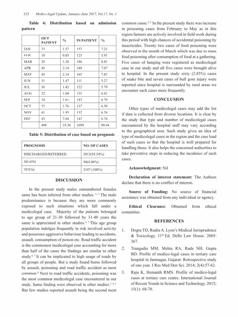
-legal Update, January-June 2017, Vol.17, No. 1 112 113 Medico-legal Update, January-June 2017, Vol.17, No. 1
Table 4: Distribution based on admission pattern
OUT PATIENT % IN PATIENT %
JAN 33 1.57 152 7.21
FEB 18 0.85 125 5.93
MAR 29 1.38 186 8.83
APR 45 2.14 149 7.07
MAY 45 2.14 165 7.83
JUN 31 1.47 111 5.27
JUL 30 1.42 122 5.79
AUG 22 1.04 135 6.41
SEP 34 1.61 143 6.79
OCT 37 1.76 137 6.50
NOV 41 1.95 132 6.26
DEC 43 2.04 142 6.74
408 19.36 1699 80.64
Table 5: Distribution of case based on prognosis
PROGNOSIS NO. OF CASES
DISCHARGED/REFERRED 2013(95.54%)
DEATH 94(4.46%)
TOTAL 2107 (100%)
DISCUSSION
In the present study males outnumbered females same has been inferred from other studies. 2, 3 The male predominance is because they are more commonly exposed to such situations which fall under a medicolegal case. Majority of the patients belonged to age group of 21-30 followed by 31-40 years the same is appreciated in other studies.4, 5 This age group population indulges frequently in risk involved activity and possesses aggressive behaviour leading to accidents, assault, consumption of poison etc. Road traffic accident is the commonest medicolegal case accounting for more than half of the cases the findings are similar to other study.6, 7 It can be implicated to high usage of roads by all groups of people. But a study found burns followed by assault, poisoning and road traffic accident as most common.8 Next to road traffic accidents, poisoning was the most common medicolegal case encountered in our study. Same finding were observed in other studies.3, 4, 6
But few studies reported assault being the second most
common cause.2, 5 In the present study there was increase in poisoning cases from February to May as in this region farmers are actively involved in field work during this period with high chances of accidental poisoning by insecticides. Twenty two cases of food poisoning were observed in the month of March which was due to mass food poisoning after consumption of food at a gathering. Five cases of hanging were registered as medicolegal case in our study and all five cases were brought alive to hospital. In the present study sixty (2.85%) cases of snake bite and seven cases of bull gore injury were reported since hospital is surrounded by rural areas we encounter such cases more frequently.
CONCLUSION
Other types of medicolegal cases may add the list if data is collected from diverse locations. It is clear by the study that type and number of medicolegal cases encountered by the hospital staff may vary according to the geographical area. Such study gives an idea of type of medicolegal cases in the region and the case load of such cases so that the hospital is well prepared for handling those. It also helps the concerned authorities to take preventive steps in reducing the incidence of such cases.
Acknowledgment: Nil
Declaration of interest statement: The Authors declare that there is no conflict of interest.
Source of Funding: No source of financial assistance was obtained from any individual or agency.
Ethical Clearance: Obtained from ethical committee.
REFERENCES
1. Dogra TD, Rudra A. Lyon’s Medical Jurisprudence & Toxicology. 11th Ed. Delhi Law House. 2005:367.
2. Trangadia MM, Mehta RA, Rada NH, Gupta BD. Profile of medico-legal cases in tertiary care hospital in Jamnagar, Gujarat: Retrospective study of one year. J Res Med Den Sci. 2014; 2(4):57-62.
3. Raju K, Hemanth RMN. Profile of medico-legal cases at tertiary care centre. International Journal of Recent Trends in Science and Technology. 2015; 15(1): 68-70.

Medico-legal Update, January-June 2017, Vol.17, No. 1 114
4. Santhosh CS, Anupam D. A Study Pattern of Medico-legal Cases Treated at a Tertiary Care Hospital in Central Karnataka. Indian Journal of Forensic and Community Medicine. 2015; 2(4):193-197.
5. Dileep KR, Siddaramanna TC, Shailesh VP, Hemanthraj MN. Retrospective Study of Profile of medico-legal cases in Tumkur region, Karnataka.International Journal Of Biomedical and Advance Research. 2015; 6(04): 339-340.
6. Vishal G, Verma SK. Profile of medicolegal cases at Adesh Institute of Medical Sciences and Research, Bathinda, Punjab. J Indian Acad Forensic Med. 2010; 32(2):150-152.
7. Yatoo GH, Jalali S, Malik A, Khan A. Profile and pattern of medico legal cases attending tertiary care hospital in north India. IJMPS. 2015; 5(5): 1-8.
8. Hussaini SN, Kulkarni CS, Batra AK. Profile of medico-legal cases coming to casualty of government medical college, Akola. Journal of Forensic Medicine, Science and Law.2013; 22(2):

-legal Update, January-June 2017, Vol.17, No. 1 114
Age Assessment from Vault Sutures Closure in Elderly Persons: An Autopsy Study in Haroti Region
Ashok Moondra1, Deepak Sharma2, B S Shekhawat3
1Associate Professor, 1Associate Professor, 1 2Assistant Professor Forensic Medicine, 2Assistant Professor Forensic Medicine, 2 3Professor, Psychiatry, Govt. Medical College, Kota
ABSTRACT
In India, even after frequent stress paid on Birth & death registration, most of the people are not following it and hence the age verification is a great problem, especially among the illiterates.
A correct estimation of age is important in medico legal& administrative matter’s i.e. regularization of temporary employee, retirement from service/superannuation, re-employment, and settlement of pension case, old age pension, and potency certificate premature release of prisoners for their good conduct in Jail, due to old age & provides senior citizen benefits.
In the dead person the age estimation at the time of autopsy is done to help the police or magistrate. In India the task of scientific confirmation of disputed age is issue of civil and criminal in nature is the duty of forensic experts. This study is done to determine the age of individual from 25 years and above. The maximum number of cases was in age group of 25-30 (26%) and male female ratio was 3: 1, 59% cases were urban & 41% were rural, 56% cases are Hindu and 2% were Christians, and maximum 39% cases from middle lower socio economic status.
Keywords: Vault sutures, Endocranium and Ecotocranium.
INTRODUCTION
Kota is the divisional headquarter of Hadoti region (consisting of mainly four districts i.e. Kota, Bundi, Jhalawar and Baran) and is situated in the southern-east part of Rajasthan. It is an industrial Manchester of the state, where there come a large number of labors from different villages and also from the neighboring tribal areas of Madhya Pradesh for employment most of these individuals do not posses correct knowledge about their age, hence very frequently the elderly persons (age 25 years and above) are referred for their age estimation by the employers to this institution.
In addition to this, Hadoti region has a network of river chambal and its tributaries crossing through, so many times unidentified dead bodies recovered from flowing water, are also brought to this institution, under various stages of decomposition. In decomposed bodies, age estimation again becomes an essential task for identification of the individual to help the inquest.
Age estimation after 25 years of age is done by
various criteria i.e. state of obliteration of cranial sutures at ectocranium and endocranium (observed through naked eye examination) the findings are compared with life time x-ray taken during treatment in some cases.
AIMS AND OBJECTIVES
In this study, estimation of age is done in elderly persons (Age 25 years and above). Cranial sutures examined for fusion with the following aims and objectives:
1. To find out age of obliteration of vault sutures: Sagittal, coronal and lambdoid on autopsy.
2. To see the incidence of closure of cranial sutures at different ages in both inner & outer tables.
3. To compare obliteration of vault sutures, in both males and females.
4. To find out variation in obliteration of vault sutures depending upon diet, habitat and socioeconomic status etc.
DOI Number: 10.5958/0974-1283.2017.00025.1

Medico-legal Update, January-June 2017, Vol.17, No. 1 116 117 -legal Update, January-June 2017, Vol.17, No. 1
5. To compare obliteration of sutures on autopsy study with that of Roentagenologic examination.
6. To compare the data obtained in present study with that of other available studies.
METHODOLOGY
This Prospective study was conducted in department of forensic medicine and toxicology of Govt. medical college & M.B.S. Hospital Kota i.e. June, 1996 to Oct. 2000.
The person selected for the study were in the age group 25-30 yrs, 31-35 yrs, 36-40 yrs, 41-45 yrs, 46-50 yrs, 51-55 yrs, 56-60 yrs and above 60 years. The following accessory features of age estimation are also taken into consideration 1-Greying of hair 2-Wrinkles on face, 3-Arcus senilis 4-Menopause, 5-Teeth, 6-Fusion of sternum.
The cases were divided in two groups:
Group-I : Subjected with almost confirm age as per birth certification/ service record. Total cases in this group were 37.
Group-II: Subject without any age proof, whose age was labeled from their apparent look or as per statement of relatives. There were 63 cases from this group.
The scalps were incised across the posterior vertex from the tip of one mastoid process to the other side. The calvarium were then removed by sawing and chiseling at a level 2.5 cm above the eye brow in front and about the level of occipital protuberance at the back it give rise to complete cut of the vault along with its three sutures i.e. coronal, sagittal and lambdoid. The separated calvarium were then thoroughly washed with water & dried. The vault sutures were measured with the help of thread & each suture was divided into its subdivision both on ectocranium and endocranium as follows (6)
A- Sagittal:
1. Pars Bregmatica
2. Pars Verticis
3. Pars Obelica
4. Pars Lambdica
B- Coronal:
1. Pars Bregmatica ( Lt &Rt)
2. Pars Complicata (Lt &Rt)
3. Pars Stephanica (Lt &Rt)
4. Pars Pterica (Lt &Rt)
C- Lambdoid:
1. Pars Lambdica ( Lt &Rt)
2. Pars Intermedia (Lt &Rt)
3. Pars Asterica (Lt &Rt)
OBSERVATIONS
A Performa is designed to record all findings. The photograph of all subjects showing front of face along with obliteration of sutures at outer table and inner table were also taken.
DISCUSSION & CONCLUSION
In this study 100 medico legal cases above the age of 25 years were selected. The accessory features for age estimation e.g. Graying, wrinkles, arcussenilis, menopause, teeth & sterna segments were also taken into consideration.
The fusion rating scale was introduced by Frederic (5)in 1905 and 1910 is as follows:
0= patent (not closed)
1= Less than half closed
2= Half closed
3= More than half closed
4= totally closed.
This scale is modified by me as:
0= Patent (not closed)
1 = partially closed.
2 = completely closed.
In the present study commencement of the sutural obliteration on the endocranium of sagittal suture was observed at the age of 25-30 years in both sexes which completed by the age of 46-50 years the fusion of
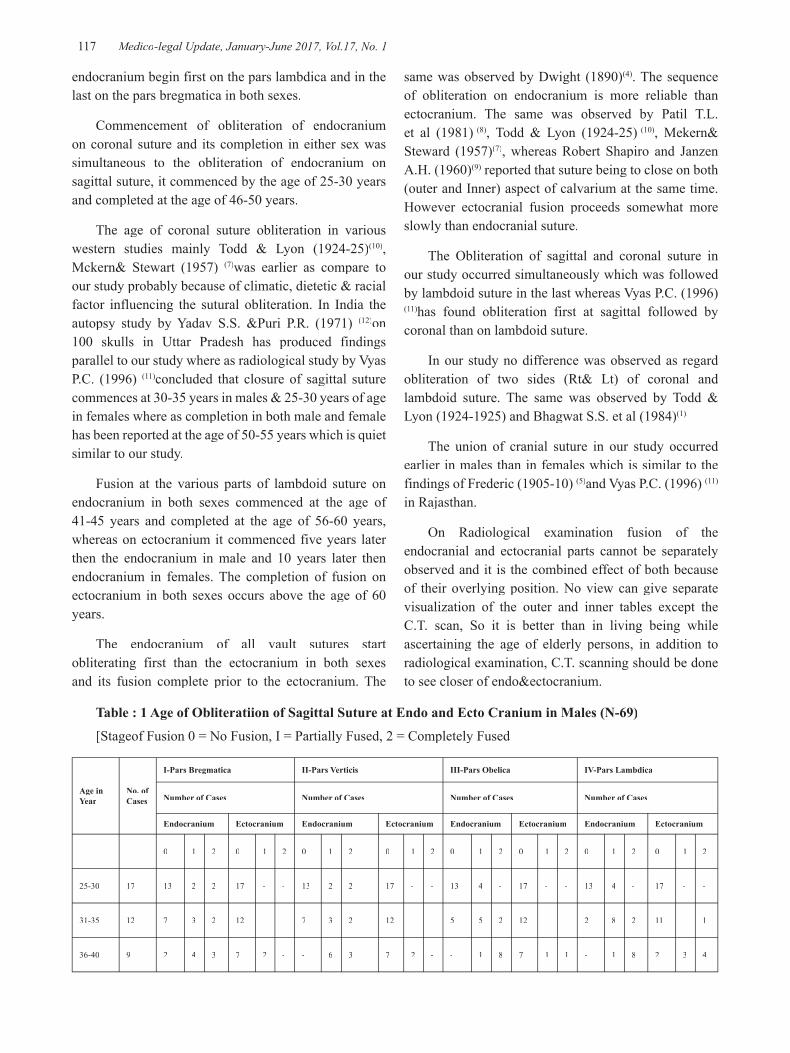
-legal Update, January-June 2017, Vol.17, No. 1 116 117 Medico-legal Update, January-June 2017, Vol.17, No. 1
endocranium begin first on the pars lambdica and in the last on the pars bregmatica in both sexes.
Commencement of obliteration of endocranium on coronal suture and its completion in either sex was simultaneous to the obliteration of endocranium on sagittal suture, it commenced by the age of 25-30 years and completed at the age of 46-50 years.
The age of coronal suture obliteration in various western studies mainly Todd & Lyon (1924-25)(10), Mckern& Stewart (1957) (7)was earlier as compare to our study probably because of climatic, dietetic & racial factor influencing the sutural obliteration. In India the autopsy study by Yadav S.S. &Puri P.R. (1971) (12)on 100 skulls in Uttar Pradesh has produced findings parallel to our study where as radiological study by Vyas P.C. (1996) (11)concluded that closure of sagittal suture commences at 30-35 years in males & 25-30 years of age in females where as completion in both male and female has been reported at the age of 50-55 years which is quiet similar to our study.
Fusion at the various parts of lambdoid suture on endocranium in both sexes commenced at the age of 41-45 years and completed at the age of 56-60 years, whereas on ectocranium it commenced five years later then the endocranium in male and 10 years later then endocranium in females. The completion of fusion on ectocranium in both sexes occurs above the age of 60 years.
The endocranium of all vault sutures start obliterating first than the ectocranium in both sexes and its fusion complete prior to the ectocranium. The
same was observed by Dwight (1890)(4). The sequence of obliteration on endocranium is more reliable than ectocranium. The same was observed by Patil T.L. et al (1981) (8), Todd & Lyon (1924-25) (10), Mekern& Steward (1957)(7), whereas Robert Shapiro and Janzen A.H. (1960)(9) reported that suture being to close on both (outer and Inner) aspect of calvarium at the same time. However ectocranial fusion proceeds somewhat more slowly than endocranial suture.
The Obliteration of sagittal and coronal suture in our study occurred simultaneously which was followed by lambdoid suture in the last whereas Vyas P.C. (1996) (11)has found obliteration first at sagittal followed by coronal than on lambdoid suture.
In our study no difference was observed as regard obliteration of two sides (Rt& Lt) of coronal and lambdoid suture. The same was observed by Todd & Lyon (1924-1925) and Bhagwat S.S. et al (1984)(1).
The union of cranial suture in our study occurred earlier in males than in females which is similar to the findings of Frederic (1905-10) (5)and Vyas P.C. (1996) (11)
in Rajasthan.
On Radiological examination fusion of the endocranial and ectocranial parts cannot be separately observed and it is the combined effect of both because of their overlying position. No view can give separate visualization of the outer and inner tables except the C.T. scan, So it is better than in living being while ascertaining the age of elderly persons, in addition to radiological examination, C.T. scanning should be done to see closer of endo&ectocranium.
Table : 1 Age of Obliteratiion of Sagittal Suture at Endo and Ecto Cranium in Males (N-69)
[Stageof Fusion 0 = No Fusion, I = Partially Fused, 2 = Completely Fused
Age in Year
No. of Cases
I-Pars Bregmatica II-Pars Verticis III-Pars Obelica IV-Pars Lambdica
Number of Cases Number of Cases Number of Cases Number of Cases
Endocranium Ectocranium Endocranium Ectocranium Endocranium Ectocranium Endocranium Ectocranium
0 1 2 0 1 2 0 1 2 0 1 2 0 1 2 0 1 2 0 1 2 0 1 2
25-30 17 13 2 2 17 - - 13 2 2 17 - - 13 4 - 17 - - 13 4 - 17 - -
31-35 12 7 3 2 12 - - 7 3 2 12 - - 5 5 2 12 - - 2 8 2 11 - 1
36-40 9 2 4 3 7 2 - - 6 3 7 2 - - 1 8 7 1 1 - 1 8 2 3 4
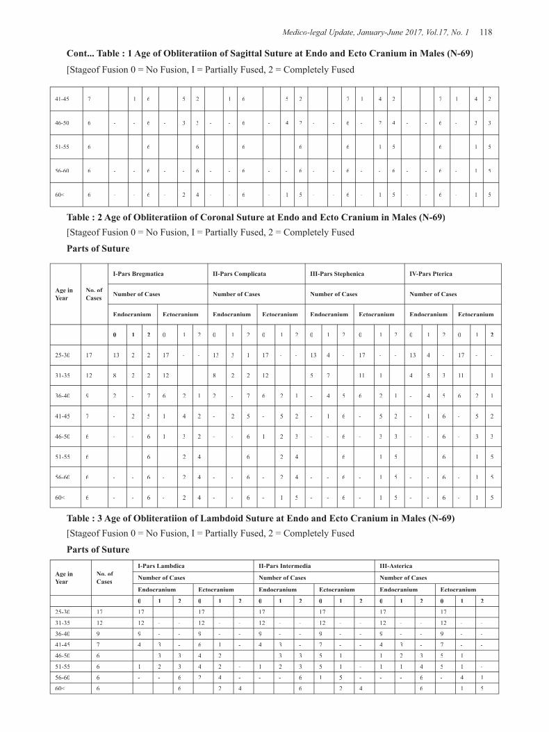
Medico-legal Update, January-June 2017, Vol.17, No. 1 118 119 -legal Update, January-June 2017, Vol.17, No. 1
41-45 7 - 1 6 - 5 2 - 1 6 - 5 2 - - 7 1 4 2 - - 7 1 4 2
46-50 6 - - 6 - 3 3 - - 6 - 4 2 - - 6 - 2 4 - - 6 - 3 3
51-55 6 - - 6 - - 6 - - 6 - - 6 - - 6 - 1 5 - - 6 - 1 5
56-60 6 - - 6 - - 6 - - 6 - - 6 - - 6 - - 6 - - 6 - 1 5
60< 6 - - 6 - 2 4 - - 6 - 1 5 - - 6 - 1 5 - - 6 - 1 5
Table : 2 Age of Obliteratiion of Coronal Suture at Endo and Ecto Cranium in Males (N-69)[Stageof Fusion 0 = No Fusion, I = Partially Fused, 2 = Completely Fused
Parts of Suture
Age in Year
No. of Cases
I-Pars Bregmatica II-Pars Complicata III-Pars Stephenica IV-Pars Pterica
Number of Cases Number of Cases Number of Cases Number of Cases
Endocranium Ectocranium Endocranium Ectocranium Endocranium Ectocranium Endocranium Ectocranium
0 1 2 0 1 2 0 1 2 0 1 2 0 1 2 0 1 2 0 1 2 0 1 2
25-30 17 13 2 2 17 - - 13 3 1 17 - - 13 4 - 17 - - 13 4 - 17 - -
31-35 12 8 2 2 12 - - 8 2 2 12 - - 5 7 - 11 1 - 4 5 3 11 - 1
36-40 9 2 - 7 6 2 1 2 - 7 6 2 1 - 4 5 6 2 1 - 4 5 6 2 1
41-45 7 - 2 5 1 4 2 - 2 5 - 5 2 - 1 6 - 5 2 - 1 6 - 5 2
46-50 6 - - 6 1 3 2 - - 6 1 2 3 - - 6 - 3 3 - - 6 - 3 3
51-55 6 - - 6 - 2 4 - - 6 - 2 4 - - 6 - 1 5 - - 6 - 1 5
56-60 6 - - 6 - 2 4 - - 6 - 2 4 - - 6 - 1 5 - - 6 - 1 5
60< 6 - - 6 - 2 4 - - 6 - 1 5 - - 6 - 1 5 - - 6 - 1 5
Table : 3 Age of Obliteratiion of Lambdoid Suture at Endo and Ecto Cranium in Males (N-69)[Stageof Fusion 0 = No Fusion, I = Partially Fused, 2 = Completely Fused
Parts of Suture
Age in Year
No. of Cases
I-Pars Lambdica II-Pars Intermedia III-Asterica
Number of Cases Number of Cases Number of Cases
Endocranium Ectocranium Endocranium Ectocranium Endocranium Ectocranium
0 1 2 0 1 2 0 1 2 0 1 2 0 1 2 0 1 2
25-30 17 17 - - 17 - - 17 - - 17 - - 17 - - 17 - -
31-35 12 12 - - 12 - - 12 - - 12 - - 12 - - 12 - -
36-40 9 9 - - 9 - - 9 - - 9 - - 9 - - 9 - -
41-45 7 4 3 - 6 1 - 4 3 - 7 - - 4 3 - 7 - -
46-50 6 - 3 3 4 2 - - 3 3 5 1 - 1 2 3 5 1 -
51-55 6 1 2 3 4 2 - 1 2 3 5 1 - 1 1 4 5 1 -
56-60 6 - - 6 2 4 - - - 6 1 5 - - - 6 - 4 1
60< 6 - - 6 - 2 4 - - 6 - 2 4 - - 6 - 1 5
Cont... Table : 1 Age of Obliteratiion of Sagittal Suture at Endo and Ecto Cranium in Males (N-69)
[Stageof Fusion 0 = No Fusion, I = Partially Fused, 2 = Completely Fused
-legal Update, January-June 2017, Vol.17, No. 1 118 119 Medico-legal Update, January-June 2017, Vol.17, No. 1
CONCLUSION
In this study total 100 medicolegal cases were examined. The male and female incidence was about 3: 1. In males age of commencement of obliteration on endocranium was 25-30 years for both sagittal & coronal suture and 41-45 years for lambdoid suture, where as it completed at the age of 46-50 years on sagittal and coronal suture and 56-60 years on lambdoid suture. In females the ag e of commencement of obliteration on endocranium was 25-30 years on both sagittal and coronal suture and 41-45 years on lambdoid suture, whereas its completion was at the age of 46-50 years on both sagittal and coronal suture and 56-60 years on lambdoid suture.
The obliteration occur 5-10 years in advance on endocranium in comparision to ectocranium the fusion of endocranium is more reliable than ectocranium, because on ectocranium there are chances of delayed union and lapsed union.
The obliteration of the sutures sets a little later and proceeds more slowly in the female then in males.
Conflict of Interest: Nil
Source of Funding : Self
Ethical Clearance: Taken from Ethical Committee of Government Medical College, Kota (Raj.)
REFERENCES
1. Bhagwat S.S., Pal G.P., Outal R.V.R. and Patel V.: A study of sutural bones in GujratiCrawa. J. Anat. Soc. India 33 (1) : 33, 1984
2. Boyde, A.: In manual of medico legal practice by Goel M.R. and Saraswat P.K.: 1st Ed. Ajmer, Unique st Ed. Ajmer, Unique st
Books, 115: 1996.
3. Chandra Shekhran, P: Skull suture: Forensic science as is what is: The forensic science society of India, Madras: 166, 1985.
4. Dwight, T.: The closure of the cranial sutures as a sign of age: Boston. Med. Surg. J. 122 : 389-392, 1890
5. Frederic, J:Untersuchungenuber die normale obliteration der Schadelnahtezeitschrift fur morphologie and Anthropologie. 9 : 373-456, 1906
6. Gray, H: Gray’s Anatomy. 37thed: London, Melbourn and New york, Churchill Living Stone. 341-342, 1989
7. Mekern, T.W. and Steward, T.D.:Sketal age changes in young American males. Headqu. Q.M. Res and Dev. Command. Tech. Rep Ep-45 Natick, mass, 1957
8. Patil, T.L. Bhargava and Qureshi, A.A. P: The study of cranial suture closure of the vault of the skull. J. Anat Soc. Of India 30-38, 1981
9. Shapiro, R and Janzen, H.A.: The normal Skull-A roentgen study, Paul B. Hober Inc. Medical Division of Harper and Brothers. 161, 1960
10. Todd, T.W. and Lyon Jr. D.W.:Endocranial suture closure, its progress and age relationship. Part-I, adult males of white stock. AJPA 7(3) : 325-384, 1924
11. Vyas, P.C.: pattern of skull suture obliteration on Roentegnologic examination – A study in elderly individuals for age estimation in Jaipur: Dissertation for M.D. (FMT), Univ. of Raj., 1996.
12. Yadav, S.S. &Puri, P.R.: Jr. Ind. Aca. For Sci. Jan. 11 : 1971

121 -legal Update, January-June 2017, Vol.17, No. 1
Anatomical Variation of Left Anterior Descending Artery and Atherosclerotic Coronary Artery Disease
in Sudden Cardiac Death
Sujith Sreenivas C Associate Professor & Dy. Police Surgeon, Dept. of Forensic Medicine, Govt. Medical College, Idukki
ABSTRACT
Evaluation of sudden cardiac death below 40 years of age, due to atherosclerotic coronary artery disease involving left coronary artery revealed a branching of the Proximal part of left anterior descending artery. There was an age dependent preponderance of atherosclerosis at this branching site. Hence a series of death due to Coronary artery disease below the age of 40 years were analysed which revealed the branching of proximal left anterior descending artery. Branching of proximal left anterior descending artery, an anatomical variation of coronary artery predisposes early atherosclerosis of the proximal left anterior descending artery resulting in death at a younger age compared to people with single non-branching left anterior descending artery.
Keyword: Anatomical variation – left anterior descending artery – Atherosclerotic coronary artery disease.
INTRODUCTION
Atherosclerosis is a chronic inflammatory response1 of the arterial wall precipitated by an injury to the endothelium. The primary event seems to be repeated subtle injury to the arterial inner lining (endothelium) through various mechanisms. These include physical stress from turbulent blood flow due to formation of eddy’s current. Repetitive endothelial injury at the site of branch point where there is a disturbed flow pattern makes the area vulnerable for developing atherosclerotic changes due to the interruption of smooth laminar flow. The evolution of atherosclerotic plaque occurs as per the hypothesis of endothelial injury.2 Monocytes and platelets adhere at the site of endothelial injury. The adherent monocyte from the lumen and smooth muscle cells from the media migrate into the intimal layer of arterial wall and they proliferate. The monocytes differentiates into macrophages and ultimately into foam cells following the entry of lipids. The smooth muscle cells which migrate from media into the intima proliferate and deposit extra cellular matrix which converts the fatty streaks into a fibrofatty atheroma, resulting in the growth of atherosclerosis. The extracellular matrix consist of collagen which stabilize the atheromatous plaques forming a fibrous cap for the atheromatous lesion. Endothelial dysfunction à Monocyte adhesion
à Macrophage à Lipid accumulation (Foam cells) àSmooth muscle proliferations à Extracellular matrix deposition (collagen) àFibrofatty atheroma.
MATERIALS AND METHOD
The autopsy based study was conducted at the Department of Forensic Medicine , Govt.Medical College, Kozhikode in the year 2014-15. Death due to ischemic heart disease were evaluated in all subjects below 40 years and the position of atherosclerotic plaque involving left anterior descending artery analysed. The extend of atherosclerosis from the origin of left anterior descending artery and branching of left anterior descending artery evaluated for the presence of atherosclerotic thickening of branching site , which was compared with other areas of left coronary artery. There were 14 cases of death below 40 years which were subjected for evaluation in the year 2014-15. The study was compared with a control group of 13 subjects who died of atherosclerotic coronary artery disease above the age of 55 years.
Ethical clearance obtained –procedure followed were in accordance with the Ethical standards of committee of human experimentation of institute and with the Helsinki Declaration of 1975 revised in 2000.
DOI Number: 10.5958/0974-1283.2017.00026.3
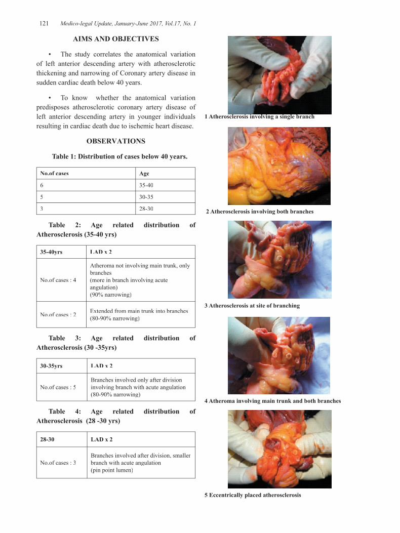
121 Medico-legal Update, January-June 2017, Vol.17, No. 1
AIMS AND OBJECTIVES
• The study correlates the anatomical variation of left anterior descending artery with atherosclerotic thickening and narrowing of Coronary artery disease in sudden cardiac death below 40 years.
• To know whether the anatomical variation predisposes atherosclerotic coronary artery disease of left anterior descending artery in younger individuals resulting in cardiac death due to ischemic heart disease.
OBSERVATIONS
Table 1: Distribution of cases below 40 years.
No.of cases Age
6 35-40
5 30-35
3 28-30
Table 2: Age related distribution of Atherosclerosis (35-40 yrs)
35-40yrs LAD x 2
No.of cases : 4
Atheroma not involving main trunk, only branches(more in branch involving acute angulation)(90% narrowing)
No.of cases : 2 Extended from main trunk into branches (80-90% narrowing)
Table 3: Age related distribution of Atherosclerosis (30 -35yrs)
30-35yrs LAD x 2
No.of cases : 5Branches involved only after division involving branch with acute angulation(80-90% narrowing)
Table 4: Age related distribution of Atherosclerosis (28 -30 yrs)
28-30 LAD x 2
No.of cases : 3Branches involved after division, smaller branch with acute angulation(pin point lumen)
1 Atherosclerosis involving a single branch
2 Atherosclerosis involving both branches
3 Atherosclerosis at site of branching
4 Atheroma involving main trunk and both branches
5 Eccentrically placed atherosclerosis
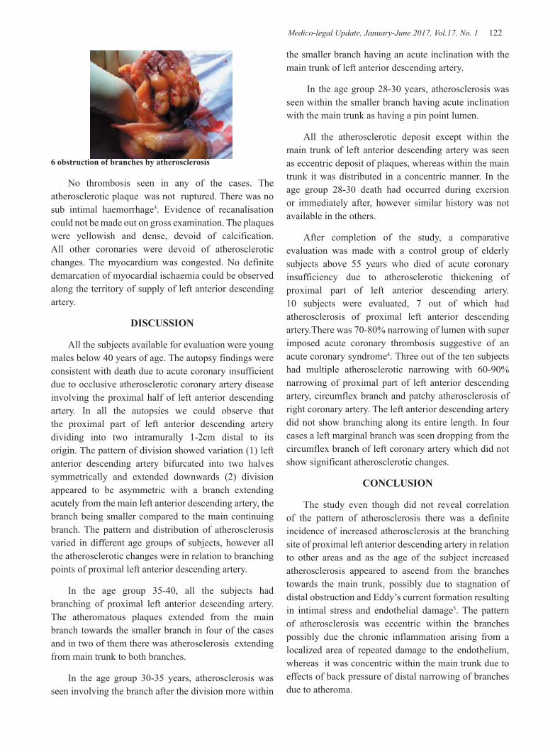
Medico-legal Update, January-June 2017, Vol.17, No. 1 122 123 -legal Update, January-June 2017, Vol.17, No. 1
6 obstruction of branches by atherosclerosis
No thrombosis seen in any of the cases. The atherosclerotic plaque was not ruptured. There was no sub intimal haemorrhage3. Evidence of recanalisation could not be made out on gross examination. The plaques were yellowish and dense, devoid of calcification. All other coronaries were devoid of atherosclerotic changes. The myocardium was congested. No definite demarcation of myocardial ischaemia could be observed along the territory of supply of left anterior descending artery.
DISCUSSION
All the subjects available for evaluation were young males below 40 years of age. The autopsy findings were consistent with death due to acute coronary insufficient due to occlusive atherosclerotic coronary artery disease involving the proximal half of left anterior descending artery. In all the autopsies we could observe that the proximal part of left anterior descending artery dividing into two intramurally 1-2cm distal to its origin. The pattern of division showed variation (1) left anterior descending artery bifurcated into two halves symmetrically and extended downwards (2) division appeared to be asymmetric with a branch extending acutely from the main left anterior descending artery, the branch being smaller compared to the main continuing branch. The pattern and distribution of atherosclerosis varied in different age groups of subjects, however all the atherosclerotic changes were in relation to branching points of proximal left anterior descending artery.
In the age group 35-40, all the subjects had branching of proximal left anterior descending artery. The atheromatous plaques extended from the main branch towards the smaller branch in four of the cases and in two of them there was atherosclerosis extending from main trunk to both branches.
In the age group 30-35 years, atherosclerosis was seen involving the branch after the division more within
the smaller branch having an acute inclination with the main trunk of left anterior descending artery.
In the age group 28-30 years, atherosclerosis was seen within the smaller branch having acute inclination with the main trunk as having a pin point lumen.
All the atherosclerotic deposit except within the main trunk of left anterior descending artery was seen as eccentric deposit of plaques, whereas within the main trunk it was distributed in a concentric manner. In the age group 28-30 death had occurred during exersion or immediately after, however similar history was not available in the others.
After completion of the study, a comparative evaluation was made with a control group of elderly subjects above 55 years who died of acute coronary insufficiency due to atherosclerotic thickening of proximal part of left anterior descending artery. 10 subjects were evaluated, 7 out of which had atherosclerosis of proximal left anterior descending artery.There was 70-80% narrowing of lumen with super imposed acute coronary thrombosis suggestive of an acute coronary syndrome4. Three out of the ten subjects had multiple atherosclerotic narrowing with 60-90% narrowing of proximal part of left anterior descending artery, circumflex branch and patchy atherosclerosis of right coronary artery. The left anterior descending artery did not show branching along its entire length. In four cases a left marginal branch was seen dropping from the circumflex branch of left coronary artery which did not show significant atherosclerotic changes.
CONCLUSION
The study even though did not reveal correlation of the pattern of atherosclerosis there was a definite incidence of increased atherosclerosis at the branching site of proximal left anterior descending artery in relation to other areas and as the age of the subject increased atherosclerosis appeared to ascend from the branches towards the main trunk, possibly due to stagnation of distal obstruction and Eddy’s current formation resulting in intimal stress and endothelial damage5. The pattern of atherosclerosis was eccentric within the branches possibly due the chronic inflammation arising from a localized area of repeated damage to the endothelium, whereas it was concentric within the main trunk due to effects of back pressure of distal narrowing of branches due to atheroma.

-legal Update, January-June 2017, Vol.17, No. 1 122 123 Medico-legal Update, January-June 2017, Vol.17, No. 1
LIMITATIONS
Dietary factors, type of personality, lipid profile and other contributory factors for atherosclerotic changes6 were not available for evaluation .
Conflict of Interest - Nil
Source of Funding - Self
REFERENCES
1) Ross R: Atherosclerosis : an inflammatory disease. N Engl J Med 340:115,1999 .
2) Ross R.The pathogenesis of atherosclerosis. A perspective for the 1990’s Nature 1993;362:801-809.
3) Fry DL.Hemodynamic forces in atherogenesis In: Scheinberg P;ed Cerebrovascular Diseases. Tenth Princeton conference. New York: Raven press;1976: 77-95.
4) Libby P.Molecular bases of the acute coronary syndrome.Circulation 1995;91:2844-2850.
5) Gimbrone M A Jr: Endothelial dysfunction, hemodynamic forces and atherogenesis. Ann Ny Acad sci 902:239,2000.
6) Libby P :The Vascular Biology of Atherosclerosis In Braunwald E, Zipes DP , Libby P (eds): Heart Disease, 6th ed Philadelphia,WB Saunders co,2001,P. 995-1009.

125 -legal Update, January-June 2017, Vol.17, No. 1
Effectiveness of Home Safety Supervisory Program (HSSP) on Childhood Injury, Caregivers’ Knowledge and Behavioural
Outcomes- A Randomized Controlled Trial Protocol
Renu G1, Mamatha S Pai2, Baby S Nayak3, Suneel C Mundkur4, Dinesh M Nayak5, Shashidhara YN6, Anice George7
1PhD Scholar, 2HOD Child Health Nursing, 3Professor, Dept of Child Health Nursing, Manipal College of Nursing, Manipal, 4Additional Professor, Dept.of Paediatrics Kasturba Hospital Manipal, 5Prof & HOD, Paediatrics Malaka Manipal Medical College (Manipal Campus), Dr. TMA Pai Hospital, Udupi, 6 HOD,
Community Health Nursing, 7Professor & Dean, Manipal College of Nursing, Manipal
ABSTRACTS
Background: Childhood injuries represent an important global health problem. Most unintentional injuries to children under 5 years occur in and around the home as this is where they spend the majority of their time.
Methodology: A study will be conducted among the caregivers’ of children between 2 years to 5 years who are the residents in the selected 30 villages of Udupi taluk.
Study consists of two phases: Phase I :A cross sectional prevalence survey (N= 2040), has already been done to find the prevalence of childhood injury among children between 2 to 5 years. The overall prevalence of injury among children was 98.6% 95%CI [97.6, 99.5].
Phase II A randomized controlled trial (RCT): A sample size of 150 (10 households/ village) from 30 villages will be included in the study, which was calculated statistically. Randomization of participants and villages for intervention and control was done by a person not involved in the study. A home safety supervisory program will be provided for the intervention group, the control group will receive a sham normal child care education.
A baseline assessment and repeated follow ups in one month, three month and six month will be done in both the groups. The study outcomes will be assessed at each time.
Keywords: Children, Injury, Home safety, Supervision,Childhood Injury
Corresponding author:Anice GeorgeDean, MCON,Manipal, e-mail: [email protected]
INTRODUCTION
Globally, each year around 9,50,000 children younger than 18 years die because of violence and injury [1]. Two third of all injury-related deaths worldwide, occurs in developing countries [2]. After the first birthday of the child, injury is the major cause of death among children under 5 years. Other than this, medical attention with or without hospitalization is required for millions
of children for non-fatal injuries. Lifelong disabilities will occur for many children because of various kinds of injuries [3].
Many of the intentional or unintentional injuries for children are reported from home, the reported reason for this is the relatively longer duration youngsters spend in the house and the reasonably high amount of dangers that are present at home [3, 4]. Some of the injuries that typically occur in young children at home are burns, choking [5, 6] falls and poisoning [4, 6, 7].
Importance of Proposed Research Investigation: Literature Support
DOI Number: 10.5958/0974-1283.2017.00027.5
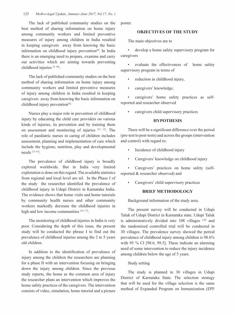
125 Medico-legal Update, January-June 2017, Vol.17, No. 1
The lack of published community studies on the best method of sharing information on home injury among community workers and limited preventive measures of injury among children in India resulted in keeping caregivers away from knowing the basic information on childhood injury prevention[8]
. In India there is an emerging need to prepare, examine and carry out activities which are aiming towards preventing childhood injuries [9, 10].
The lack of published community studies on the best method of sharing information on home injury among community workers and limited preventive measures of injury among children in India resulted in keeping caregivers away from knowing the basic information on childhood injury prevention[8].
Nurses play a major role in prevention of childhood injury by educating the child care providers on various kinds of injuries, its prevention and by training them on assessment and monitoring of injuries [11, 12]. The role of paediatric nurses in caring of children includes assessment, planning and implementation of care which include the hygiene, nutrition, play and developmental needs [13-15].
The prevalence of childhood injury is broadly explored worldwide. But in India very limited exploration is done on this regard. The available statistics from regional and local level are nil. In the Phase I of the study the researcher identified the prevalence of childhood injury in Udupi District in Karnataka India. The evidence shows that home visits and home tutorials by community health nurses and other community workers markedly decrease the childhood injuries in high and low income communities [16, 17].
The monitoring of childhood injuries in India is very poor. Considering the depth of this issue, the present study will be conducted the phrase I to find out the prevalence of childhood injuries among the 2 to 5 years old children.
In addition to the identification of prevalence of injury among the children the researchers are planning for a phase II with an intervention focusing on bringing down the injury among children. Since the previous study reports, the home as the common area of injury the researcher plans an intervention which improves the home safety practices of the caregivers. The intervention consists of video, simulation, home tutorial and a picture
poster.
OBJECTIVES OF THE STUDY
The main objectives are to
• develop a home safety supervisory program for caregivers
• evaluate the effectiveness of home safety supervisory program in terms of
• reduction in childhood injury,
• caregivers’ knowledge,
• caregivers’ home safety practices as self- reported and researcher observed
• caregivers child supervisory practices
HYPOTHESIS
There will be a significant difference over the period (pre-test to post-tests) and across the groups (intervention and control) with regard to:
• Incidence of childhood injury
• Caregivers’ knowledge on childhood injury
• Caregivers’ practices on home safety (self-reported & researcher observed) and
• Caregivers’ child supervisory practices
BRIEF METHODOLOGY
Background information of the study area.
The present survey will be conducted in Udupi Taluk of Udupi District in Karnataka state. Udupi Taluk is administratively divided into 108 villages [18] and the randomized controlled trial will be conducted in 30 villages. The prevalence survey showed the period prevalence of childhood injury among children is 98.6% with 95 % CI [98.6, 99.5]. These indicate an alarming need of some intervention to reduce the injury incidence among children below the age of 5 years.
Study setting
The study is planned in 30 villages in Udupi District of Karnataka State. The selection strategy that will be used for the village selection is the same method of Expanded Program on Immunization (EPI)
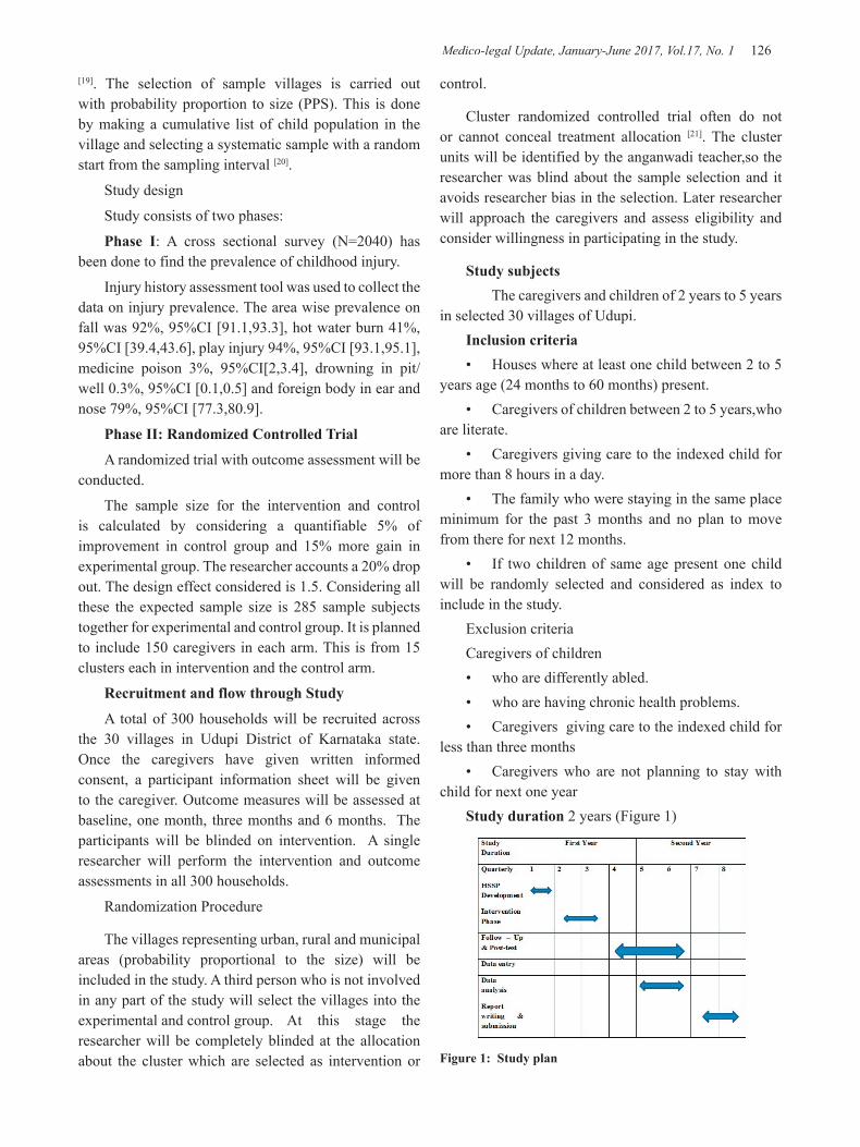
Medico-legal Update, January-June 2017, Vol.17, No. 1 126 127 -legal Update, January-June 2017, Vol.17, No. 1
[19]. The selection of sample villages is carried out with probability proportion to size (PPS). This is done by making a cumulative list of child population in the village and selecting a systematic sample with a random start from the sampling interval [20].
Study design
Study consists of two phases:
Phase I: A cross sectional survey (N=2040) has been done to find the prevalence of childhood injury.
Injury history assessment tool was used to collect the data on injury prevalence. The area wise prevalence on fall was 92%, 95%CI [91.1,93.3], hot water burn 41%, 95%CI [39.4,43.6], play injury 94%, 95%CI [93.1,95.1], medicine poison 3%, 95%CI[2,3.4], drowning in pit/well 0.3%, 95%CI [0.1,0.5] and foreign body in ear and nose 79%, 95%CI [77.3,80.9].
Phase II: Randomized Controlled Trial
A randomized trial with outcome assessment will be conducted.
The sample size for the intervention and control is calculated by considering a quantifiable 5% of improvement in control group and 15% more gain in experimental group. The researcher accounts a 20% drop out. The design effect considered is 1.5. Considering all these the expected sample size is 285 sample subjects together for experimental and control group. It is planned to include 150 caregivers in each arm. This is from 15 clusters each in intervention and the control arm.
Recruitment and flow through Study
A total of 300 households will be recruited across the 30 villages in Udupi District of Karnataka state. Once the caregivers have given written informed consent, a participant information sheet will be given to the caregiver. Outcome measures will be assessed at baseline, one month, three months and 6 months. The participants will be blinded on intervention. A single researcher will perform the intervention and outcome assessments in all 300 households.
Randomization Procedure
The villages representing urban, rural and municipal areas (probability proportional to the size) will be included in the study. A third person who is not involved in any part of the study will select the villages into the experimental and control group. At this stage the researcher will be completely blinded at the allocation about the cluster which are selected as intervention or
control.
Cluster randomized controlled trial often do not or cannot conceal treatment allocation [21]. The cluster units will be identified by the anganwadi teacher,so the researcher was blind about the sample selection and it avoids researcher bias in the selection. Later researcher will approach the caregivers and assess eligibility and consider willingness in participating in the study.
Study subjects The caregivers and children of 2 years to 5 years
in selected 30 villages of Udupi.Inclusion criteria• Houses where at least one child between 2 to 5
years age (24 months to 60 months) present.• Caregivers of children between 2 to 5 years,who
are literate. • Caregivers giving care to the indexed child for
more than 8 hours in a day.• The family who were staying in the same place
minimum for the past 3 months and no plan to move from there for next 12 months.
• If two children of same age present one child will be randomly selected and considered as index to include in the study.
Exclusion criteria Caregivers of children• who are differently abled.• who are having chronic health problems.• Caregivers giving care to the indexed child for
less than three months• Caregivers who are not planning to stay with
child for next one year Study duration 2 years (Figure 1)
Figure 1: Study plan

-legal Update, January-June 2017, Vol.17, No. 1 126 127 Medico-legal Update, January-June 2017, Vol.17, No. 1
Intervention Procedure
The intervention for the study is a Home Safety Supervisory Program (HSSP).
Home safety supervisory program will be prepared and contextualized according to the local settings and local language. Content and construct validity of the intervention program will be done before the administration of the program to the study setting.
The home safety supervisory program consists of 4 different parts.
Video on Safe Home Safe Child
A 10 minutes video showing the hazards at home which children usually encounter and the second half of the video will demonstrate the preventive measures for those injuries.
Simulation on Home Safety
A simulated setting will be created at anganwadi showing the safe and unsafe environments in the house.
Poster on Safe Home Safe Child
This will be a picture poster with calendar given to each house showing the safe and unsafe home environments.
Home Tutorial
The home visit will be performed by the trained personnel assessing the safety in the houses and suggesting the simple modification of the house which make the house safe for the child.
The picture poster will be given at the time of first home visit. Video and home tutorial will be used at first home visit and after a month during the second home visit. Individual home visits will be performed by the researcher.
The control group will receive a health teaching on normal child care. A home visit will be performed by the researcher to assess the home safety practices.
Post-Test and Follow - Up
The baseline assessment and administration of intervention will follow an assessment after 30 days. After the first month assessment all the interventions will be carried out as reinforcement. The second and
third follow ups and outcome assessments will be done at three months and six months. All the instruments used for the baseline assessments will be used for the post tests to measure the study outcomes.
Outcome Measures
• Incidence of childhood injury – 25 item questionnaire to assess the history of injury in the past 3 months.
• Knowledge of caregivers on childhood injury – a thirty item multiple response questionnaire
• Home safety practices – self-reported and researcher observed – a 31 item yes or no response question to assess the safety practices at home, which is expressed by the participant and the same will be assessed by the researcher.
• Caregiver supervisory practices: a 23 item questionnaire assessing the supervisory practices of caregivers.
Villages and Participant selection Blinded
The village selection and participant selection will be done by a person who will not be involved in the study.
Intervention and Follow up and Assessment
Intervention and follow up assessment will be given by a single researcher for all the thirty villages and all 300 houses (this include intervention and control group).
Data Analysis
The collected data will be coded, entered and analysed using the Predictive analytic software statistic.
Descriptive statistics of frequency and percentage will be used for presenting the demographic variables.
Homogeneity assessment of intervention and control group will be done using chi-square test with a p – values at .05 level of significance.
Since the study is planning to assess the change of mean score of same outcome measures more than three times, a repeated measure of ANOVA will be used.

Medico-legal Update, January-June 2017, Vol.17, No. 1 128 129 -legal Update, January-June 2017, Vol.17, No. 1
Ethical consideration
The study protocol has been approved by institutional review committee, later approval was sought from institutional ethics committee. The present study also got permission from Zilla Panchayat, Udupi, Women and Child Welfare Committee and Women and Child Welfare Department.
This trial is registered under Clinical Trial Registry of India, Trial number: CTRI/2015/09/006188.
Validation of Protocol
Protocol was submitted to review by the experts in the various fields. The protocol was presented in the institutional research committee and got approved.
Importance of the Proposed study
Research
The proposed study will provide further information on the effectiveness of home tutorial and education of caregivers in bringing down childhood injury. The data obtained may help in planning more studies with community participation
Education
This study may throw light about the need of health team workers utilization of health education opportunities.
Quality and effectiveness
This study will also assess the influence of improvement in knowledge and home tutorial supervision in the outcome of childhood injury.
Policy making
The information gathered will give adequate support for planning policies for regular home visits, supervision and caregiver training in the future, which can be implemented across India.
CONCLUSION
This randomized controlled trial aims to establish the effectiveness of home safety supervisory program on childhood injury. This intervention ensures more caregiver participation in the childcare. The trial has been registered with ctri.org. Results of the study will be published after the completion of the study.
Source of Funding Self
Conflict of Interest: Nil
REFERENCES
1. World Health Organization, R.o.f.S.-E.A.O. Child Injury Preventionin the South-East Asia Region. 2010; Available from: http://www.searo.who.int/LinkFiles/Disability,_Injury_Prevention_&_Rehabilitation.
2. Razzak, J.A., et al., A child an hour: burden of injury deaths among children under 5 in Pakistan. Arch Dis Child, 2013. 98(11): p. 867-71.
3. Adnan A. Hyder, et al., Childhood Unintentional Injuries: Need for a Community-Based Home Injury Risk Assessments in Pakistan. International Journal of Pediatrics, 2012. 2012: p. 7.
4. Zia, N., et al., Understanding unintentional childhood home injuries: pilot surveillance data from Karachi, Pakistan. BMC Res Notes, 2012. 5: p. 37.
5. Rai, A., et al., Electrical injuries in urban children in New Delhi. Pediatric Emergency Care, 2013. 29(3): p. 342-345.
6. Theurer, W.M. and A.K. Bhavsar, Prevention of unintentional childhood injury. American Family Physician, 2013. 87(7): p. 502-509.
7. Sznajder, M., et al., [Evaluation of the effectiveness of an injury prevention kit delivery for toddlers in four French cities]. Arch Pediatr, 2003. 10(6): p. 510-6.
8. Chandran, A., et al., Disseminating childhood home injury risk reduction information in Pakistan: results from a community-based pilot study. Int J Environ Res Public Health, 2013. 10(3): p. 1113-24.
9. Jagnoor, J., et al., Childhood and adult mortality from unintentional falls in India. World Health Organization. Bulletin of the World Health Organization, 2011. 89(10): p. 733-40.
10. Nath, A. and V.A. Naik, Profile of accidents in children less than five years of age belonging to a rural community in Belgaum district. 32, 2007. 2: p. 133-134.
11. Garzon, D.L., Contributing factors to preschool unintentional injury. J Pediatr Nurs, 2005. 20(6): p. 441-7.

-legal Update, January-June 2017, Vol.17, No. 1 128 129 Medico-legal Update, January-June 2017, Vol.17, No. 1
12. Woods, A.J., The role of health professionals in childhood injury prevention: a systematic review of the literature. Patient Educ Couns, 2006. 64(1-3): p. 35-42.
13. Cicutto, L., T. To, and S. Murphy, A randomized controlled trial of a public health nurse-delivered asthma program to elementary schools. J Sch Health, 2013. 83(12): p. 876-84.
14. Kenny, J. and D. Corkin, A children’s nurse’s role in the global development of a child with diabetes mellitus. Nurs Child Young People, 2013. 25(9): p. 22-5.
15. Murila, F., et al., Breast-feeding and human immunodeficiency virus infection: assessment of knowledge among clinicians in Kenya. Int J Nurs Pract, 2015. 21(1): p. 37-42.
16. Kendrick, D., et al., Home safety education and provision of safety equipment for injury prevention. Cochrane Database Syst Rev, 2012. 9: p. CD005014.
17. Russell, K., B. Morrongiello, and K.J. Phelan, Commentaries on ‘Home safety education and provision of safety equipment for injury prevention’. Evid Based Child Health, 2013. 8(3): p. 940-3.
18. Census Organization of India, C. Udupi District : Census 2011 data. [cited 2014 22/01/2014].
19. Bennett, C., et al., WITHDRAWN: Home-based support for disadvantaged adult mothers. Cochrane Database Syst Rev, 2008(1): p. CD003759.
20. Sivamani, M., V. Balraj, and J.P. Muliyil, Validity of a Surveillance System for Childhood Injuries in a Rural Block of Tamilnadu. Indian Journal of Community Medicine, 2009. 34(1): p. 43-47.
21. Hahn, S., et al., Methodological bias in cluster randomised trials. BMC Med Res Methodol, 2005. 5: p. 10.
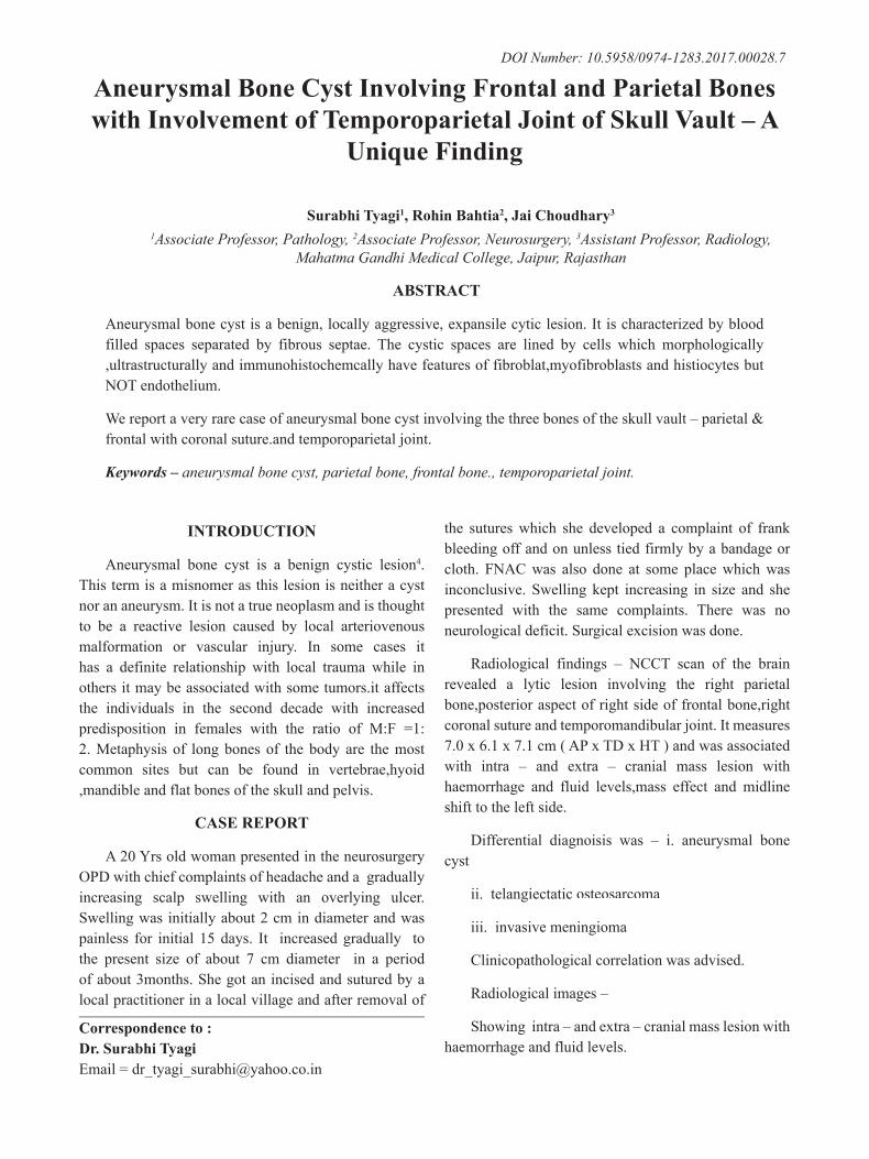
131 -legal Update, January-June 2017, Vol.17, No. 1
Aneurysmal Bone Cyst Involving Frontal and Parietal Bones with Involvement of Temporoparietal Joint of Skull Vault – A
Unique Finding
Surabhi Tyagi1, Rohin Bahtia2, Jai Choudhary3 1Associate Professor, Pathology, 2Associate Professor, Neurosurgery, 3Assistant Professor, Radiology,
Mahatma Gandhi Medical College, Jaipur, Rajasthan
ABSTRACT
Aneurysmal bone cyst is a benign, locally aggressive, expansile cytic lesion. It is characterized by blood filled spaces separated by fibrous septae. The cystic spaces are lined by cells which morphologically ,ultrastructurally and immunohistochemcally have features of fibroblat,myofibroblasts and histiocytes but NOT endothelium.
We report a very rare case of aneurysmal bone cyst involving the three bones of the skull vault – parietal & frontal with coronal suture.and temporoparietal joint.
Keywords – aneurysmal bone cyst, parietal bone, frontal bone., temporoparietal joint.
Correspondence to : Dr. Surabhi TyagiEmail = [email protected]
INTRODUCTION
Aneurysmal bone cyst is a benign cystic lesion4. This term is a misnomer as this lesion is neither a cyst nor an aneurysm. It is not a true neoplasm and is thought to be a reactive lesion caused by local arteriovenous malformation or vascular injury. In some cases it has a definite relationship with local trauma while in others it may be associated with some tumors.it affects the individuals in the second decade with increased predisposition in females with the ratio of M:F =1:2. Metaphysis of long bones of the body are the most common sites but can be found in vertebrae,hyoid ,mandible and flat bones of the skull and pelvis.
CASE REPORT
A 20 Yrs old woman presented in the neurosurgery OPD with chief complaints of headache and a gradually increasing scalp swelling with an overlying ulcer. Swelling was initially about 2 cm in diameter and was painless for initial 15 days. It increased gradually to the present size of about 7 cm diameter in a period of about 3months. She got an incised and sutured by a local practitioner in a local village and after removal of
the sutures which she developed a complaint of frank bleeding off and on unless tied firmly by a bandage or cloth. FNAC was also done at some place which was inconclusive. Swelling kept increasing in size and she presented with the same complaints. There was no neurological deficit. Surgical excision was done.
Radiological findings – NCCT scan of the brain revealed a lytic lesion involving the right parietal bone,posterior aspect of right side of frontal bone,right coronal suture and temporomandibular joint. It measures 7.0 x 6.1 x 7.1 cm ( AP x TD x HT ) and was associated with intra – and extra – cranial mass lesion with haemorrhage and fluid levels,mass effect and midline shift to the left side.
Differential diagnoisis was – i. aneurysmal bone cyst
ii. telangiectatic osteosarcoma
iii. invasive meningioma
Clinicopathological correlation was advised.
Radiological images –
Showing intra – and extra – cranial mass lesion with haemorrhage and fluid levels.
DOI Number: 10.5958/0974-1283.2017.00028.7

131 Medico-legal Update, January-June 2017, Vol.17, No. 1
Image–1
Image - 2
Histopathological examination of the haematoxylin and eosin stained sections of the bone and soft tissue pieces submitted, revealed multiple cystic spaces filled with blood and separated by fibrous septae alternating with soild areas. The cysts are lined by fibroblasts and NOT endocthelium. Occasional osteoclastic multinucleated giant cells are seen with loose spindly to cellular stroma with focal areas showing reactive woven bone, benign appearing osteoid and fibromyxoid tissue.
Histopathology images –
IMAGE 3 & 4 – Magnification 4 x
IMAGE 5 - Magnification 40 x -
IMAGE 6- Magnification 40 x -
DISCUSSION
ABC involves the skull vault very rarely. Only 63 cases have been reported in literature 5 which affect any bone of the skull (including skull base) of which 12 are of temporal bone 1,7 and very few of the parietal bone3
and frontal bone.2
No case has been reported to the best of my knowledge which presents ABC at this unusual site – involving the parietal bone ,frontal bone with coronal suture and marginal involvement of temporoparietal junction.
ABC is a non neoplastic cystic lesion which usually presents as a scalp mass but may present as an intracranial SOL6 or cerebral haemorrhage. It is associated with history of local trauma or may be other bone lesions like unicameral bone cyst,non ossifying fibroma,giant cell tumor ,chondroblastoma ,fibrous dysplasia.
Multiple fluid levels are important characteristics of ABC on CT scan,which represents sedimentation of RBCs in the blood filled cystic cavities. ABCs which do not show fluid levels on CT scan are often non homogenous and resemble some giant cell tumor.
Total surgical excision has been recommended as an ideal treatment.
Total excision has been recommended as an ideal treatment for ABC skull. Radiotherapy is advocated however for deeply situated ABCs at the base of the skull,dural involvement or where subtotal excision is done but its effect is not clear. RT is contraindicated in treatment of ABCs associated with fibrous dysplasia as there are increased chances of malignant transformation.
CONCLUSION
Aneurysmal bone cyst affects the skull vault very rarely and this is the first case reported at this unique site

Medico-legal Update, January-June 2017, Vol.17, No. 1 132
involving parietal bone, frontal bone and involving the coronal suture and temporoparietal joint. Total surgical excision was done as recommended and the patient was stable.
Ethical Clearance – Taken
Source of Funding – Self
Conflict of Interest – Nil
REFERENCES
1. Jeremiah BS: Aneurysmal bone cyst of the temporal bone. J International Coll Surgery 1965;43:179-183
2. Burns –Cox CJ,Higgins AT:Aneurysmal bone cyst of the frontal bone.J Bone Joint Surg 1969;344-345
3. Manuel A,Cacdac, M.D, Leonard I, Malls M.D, Paul J,Anderson M.D; Aneurysmal parietal bone cyst.J of Neurosurgery 1972;37;2;237-241
4. Lichtenstein L:Aneurysmal bone cyst :observations of fifty cases.J Bone Joint Surg 1957; 39 A: 873-882
5. Kumar R,Mukherjee K.K:Aneurysmal bone cyst of skull: report of three cases.Br J Neurosurg 1999;13:82-84
6. Costantini FE,Irad G,Benedetti A et al: Aneurysmal bone cyst as an intracranial space occupying lesion.J Neurosurg 1966;25:205-207
7. Parikh M.A, Dave R.J, Patel DD et al: Aneurysmal bone cyst of temporal bone. A case report. Neurol India 1996;44:131-133
-legal Update, January-June 2017, Vol.17, No. 1 132
A Faith or Fatal: A Case Report
Taware AA1, Jadhao VT2, Khade RV3, Tatiya HS4, Punpale SB5
1Associate Professor, Department of Forensic Medicine and Toxicology, B J Govt. Medical College and Sassoon General Hospitals Pune, 2Associate Professor, Department of Forensic Medicine and
Toxicology, SBH Govt. Medical College, Dhule, 3Junior Resident, 4Assistant Professor, 5Professor and Head, Department of Forensic Medicine and Toxicology, B J Govt. Medical College and Sassoon General
Hospitals Pune
ABSTRACT
Foreign body inhalation is quite common. Children are vulnerable because of exploration of their environment by putting objects into their mouths, inadequate dentition and immature swallowing coordination. This mostly occurs in children who play with toy parts, seeds etc. However we report a rare case where a live whole fish was responsible for the death of a 7 year old male child. The reason being not attributed to his activities but was due to superstition on a part of his parents. The case is reported not only to discuss this rare episode but also to discuss the medico legal aspects of such incidences.
Keywords: Live fish, superstition, foreign body inhalation, black magic act.
INTRODUCTION
Foreign body inhalation is an emergency condition as it can cause sudden upper airway obstruction. It is rare in adults1. Various kinds of foreign bodies have been reported of which coins, Groundnuts, Pea, or buttons are commonly encountered foreign bodies in pediatric age group, while dentures and bones are more common in adults 2. Ingestion of fish bone is extremely common. On the other hand one rarely ever heard of a live fish as a foreign body in upper aero digestive tract3. Accidental ingestion and pharyngeal lodging of a whole live fish usually occurs while catching live fish4. Here we report a rare case of live fish impaction and death of 7 month old child due to superstitious practice of his parents.
CASE REPORT
History: A dead body of 7 month old child of a poor socioeconomic family was brought to Department of Forensic Medicine and Toxicology of this institute.
Corresponding author:Taware AA Associate Professor, Department of Forensic Medicine and Toxicology, B J Govt. Medical College and Sassoon General Hospitals, Pune, Jaiprakash Narayan RoadPune Station 411 001. [email protected]/ 09422789579
The inquest and requisition was done by concerned investigating officer for deduction of cause of death. As per inquisition and on enquiry of parents we came to know that, he had history of excessive salivation for many months. Both the parents were illiterate and hence instead of going to a qualified person to stop this excessive salivation, parents took advice by an alleged godman to wipe out the inside of oral cavity by a live fish. The theory behind this was stated that the live fish would suck all the excessive salivation. The same practice was followed by his parents. While following advice of godman the fish accidentally slipped inside the oral cavity. Suddenly the boy went into unconsciousness and then was brought to the hospital, where he was declared brought dead. On postmortem examination we found following observations.
External examination:-
Body was of an averagely built and averagely nourished male child. There was no evidence of any external injury over body. Rigor mortis was well marked in neck and upper limbs. Postmortem lividity was fixed. There was cyanotic discoloration of finger nails and toe nails. Eyes showed subconjunctival petechial hemorrhages.
DOI Number: 10.5958/0974-1283.2017.00029.9

Medico-legal Update, January-June 2017, Vol.17, No. 1 134 135 -legal Update, January-June 2017, Vol.17, No. 1
Internal examination:-
All the visceral organs were grossly congested. Pericardium showed petechial hemorrhage. Lungs showed subpleural hemorrhages.
Larynx and Trachea
On dissection of larynx and trachea we observed a whole fish of size 04cm x 2.5cm found lodged in the aero digestive tract extending from oropharynx to laryngeal inlet leading to obstruction of airway. (Figure 1) Pharynx, larynx and tracheal mucosa were edematous and congested. Excessive thick mucoid and blood tinged secretions were present lining the mucosa.
DISCUSSION
Foreign bodies lodged in the upper aero digestive tract are classified as Animate and Inanimate foreign bodies. They may be exogenous or endogenous (dentures, loose tooth, etc.) and may get impacted traumatically or atraumatically. Incidence of foreign bodies is higher in younger age groups and is more commonly encountered in males than females. In children, foreign body impaction occurs accidentally while playing with objects like coins, buttons, etc. A live fish is a rare foreign body to be impacted accidentally in a child2.
It is intriguing when the causes of swallowing fish are explored. The habit of the victim biting the fish in mouth while trying to catch another fish in the fishing net is a commonest cause while other causes happened while elder child playing with younger victim, or the victim swallows fish for alleged medicinal purposes as in curing asthma in India.(Bathini fish medicine or Hyderabad fish therapy)5. Live fish impaction in the pharynx occurs accidentally in spear fisherman who holds the catch between their teeth while hunting for the others in the wet lands, which is a fairly common practice in eastern part of Indian subcontinent.
Deidiker reported the case of a 45 year old man who attempted to swallow a whole fish while fishing and drinking with friends who subsequently asphyxiated as a result of upper airway obstruction6. Pradipta kumar parida reported accidental entry of fish into throat of a 52 year old male while bathing in a pond4. Another case was reported where 20 year old fisherman succumbed to death due to ingestion of live tilapia fish while trying to stabilize a fish he had caught with his teeth while preventing another fish on the ground from slipping
back into the sea1. Alok bose et al reported accidental impaction of live fish in the throat of 1 year old female child while her elder siblings were playfully teasing her with a live fish2. In the present case 7 month old male child succumbed to death due to parental negligence and their superstitious practice.
Live fish ingestion as a faith healing of chronic diseases like asthma is a common practice at certain parts of Indian Subcontinent, but no accidental impaction of live fish following this practice has been reported in Indian literature2. Also we could not find any case relating to circumstances mentioned in our case report.
It is found that these inhaled /ingested foreign bodies can lodge anywhere from the larynx through the trachea and bronchi to the bronchioles and alveoli depending on the size2. In present case we found a fish lodged oropharynx and extending to laryngeal inlet obstructing the airway.
It is mentioned in the literature that poor socioeconomic condition of these cases poses a problem regarding their proper parental care and prevention of such accidents, which otherwise in a well parented family can be avoided2. However in present case even though the parents were from low socioeconomic group, it was not the lack of parental care but their illiteracy and negligent behavior along with superstitious faith which made them to select such a wrongful treatment and led to death of their child.
In the current case the investigating officer filed charges on parents under section 304 A of Indian Penal Code (IPC). S.304-A of I.P.C. deals with causing death by negligence. It mentions that “Whoever causes the death of any person, by doing any rash or negligent act not amounting to culpable homicide shall be punished with imprisonment for a term which may extend to two years or with fine, or with both7.
But the question is not only of parental negligence. It also revolves around superstitions and the prohibition of rightful treatment to the child due to the act of the godman. Hence to curtail such a kind of practices, The Maharashtra Prevention and Eradication of Human Sacrifice and other Inhuman, Evil and Aghori Practices and Black Magic Act 2013, also known as Anti-Superstition or Black Magic Bill came into force. This is an act to bring social awakening and awareness in the society and to create a healthy and safe social
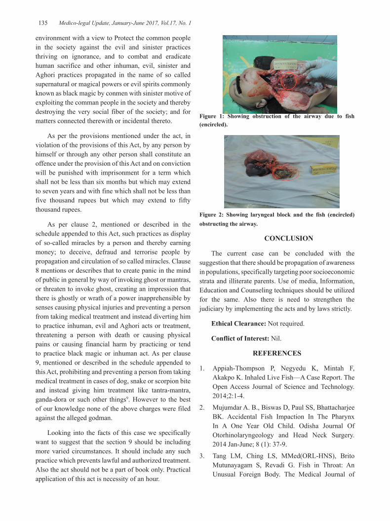
-legal Update, January-June 2017, Vol.17, No. 1 134 135 Medico-legal Update, January-June 2017, Vol.17, No. 1
environment with a view to Protect the common people in the society against the evil and sinister practices thriving on ignorance, and to combat and eradicate human sacrifice and other inhuman, evil, sinister and Aghori practices propagated in the name of so called supernatural or magical powers or evil spirits commonly known as black magic by conmen with sinister motive of exploiting the comman people in the society and thereby destroying the very social fiber of the society; and for matters connected therewith or incidental thereto.
As per the provisions mentioned under the act, in violation of the provisions of this Act, by any person by himself or through any other person shall constitute an offence under the provision of this Act and on conviction will be punished with imprisonment for a term which shall not be less than six months but which may extend to seven years and with fine which shall not be less than five thousand rupees but which may extend to fifty thousand rupees.
As per clause 2, mentioned or described in the schedule appended to this Act, such practices as display of so-called miracles by a person and thereby earning money; to deceive, defraud and terrorise people by propagation and circulation of so called miracles. Clause 8 mentions or describes that to create panic in the mind of public in general by way of invoking ghost or mantras, or threaten to invoke ghost, creating an impression that there is ghostly or wrath of a power inapprehensible by senses causing physical injuries and preventing a person from taking medical treatment and instead diverting him to practice inhuman, evil and Aghori acts or treatment, threatening a person with death or causing physical pains or causing financial harm by practicing or tend to practice black magic or inhuman act. As per clause 9, mentioned or described in the schedule appended to this Act, prohibiting and preventing a person from taking medical treatment in cases of dog, snake or scorpion bite and instead giving him treatment like tantra-mantra, ganda-dora or such other things9. However to the best of our knowledge none of the above charges were filed against the alleged godman.
Looking into the facts of this case we specifically want to suggest that the section 9 should be including more varied circumstances. It should include any such practice which prevents lawful and authorized treatment. Also the act should not be a part of book only. Practical application of this act is necessity of an hour.
Figure 1: Showing obstruction of the airway due to fish (encircled).
Figure 2: Showing laryngeal block and the fish (encircled) obstructing the airway.
CONCLUSION
The current case can be concluded with the suggestion that there should be propagation of awareness in populations, specifically targeting poor socioeconomic strata and illiterate parents. Use of media, Information, Education and Counseling techniques should be utilized for the same. Also there is need to strengthen the judiciary by implementing the acts and by laws strictly.
Ethical Clearance: Not required.
Conflict of Interest: Nil.
REFERENCES
1. Appiah-Thompson P, Negyedu K, Mintah F, Akakpo K. Inhaled Live Fish—A Case Report. The Open Access Journal of Science and Technology. 2014;2:1-4.
2. Mujumdar A. B., Biswas D, Paul SS, Bhattacharjee BK. Accidental Fish Impaction In The Pharynx In A One Year Old Child. Odisha Journal Of Otorhinolaryngeology and Head Neck Surgery. 2014 Jan-June; 8 (1): 37-9.
3. Tang LM, Ching LS, MMed(ORL-HNS), Brito Mutunayagam S, Revadi G. Fish in Throat: An Unusual Foreign Body. The Medical Journal of
Medico-legal Update, January-June 2017, Vol.17, No. 1 136
Malaysia. 2013 Dec;68(6):469-70.
4. Parida PK, Suriyanarayanan G. Accidental entry of fish into throat while bathing in a pond. Case reports in medicine.2013 Nov 25;2013.
5. Walavekar RR, Velankar H, Shiralkar P, Pradhan KA. “Live Fish” in The Throat – an Unusual Foreign Body. Bombay Hospital journal 2003 July; 45(3):496.
6. Deidiker R. Return of the killer fish: accidental choking death on a bluegill (Lepomis macrochirus).
The American Journal of Forensic Medicine and Pathology.2002 Jun 1;23(2):197-8.
7. The Indian Penal Code 1860. Available from: ncw.nic.in/acts/THEINDIANPENALCODE1860.pdf.
8. Maharashtra Prevention and Eradication of Human Sacrifice and Other Inhuman, Evil and Aghori Practices and Black Magic Act 2013 (Mah. Act No. XXX of 2013). Avialble from: bombayhighcourt.nic.in/libweb/acts/stateact/2013acts/2013.30.pdf.

-legal Update, January-June 2017, Vol.17, No. 1 136
Cardiac Changes Seen in Hanging Victims Who Survived for Varying Periods before Death: An Autopsy based Study
S Harish1, K Sasikala2, K S Meena3, A Sarath4
1Assistant Surgeon, Community Health Centre Kulathupuzha, Kollam, Kerala, 1Assistant Surgeon, Community Health Centre Kulathupuzha, Kollam, Kerala, 1 2Professor, 2Professor, 2 3Associate 3Associate 3
Professor, Department of Forensic Medicine, 4Associate Professor, Department of Pathology, Government 4Associate Professor, Department of Pathology, Government 4
Medical College, Thiruvananthapuram, Kerala
ABSTRACT
Hanging is a very simple, yet highly effective method of suicide. Death is instantaneous in most cases of hanging. Survival after hanging is a rare event. A long survival and recovery is rarer. The present cross sectional study was conducted in 25 cases of hanging victims who survived for varying periods before death and was brought for autopsy at Department of Forensic Medicine, State Medico-Legal Institute, Government Medical College, Thiruvananthapuram. Macroscopic and microscopic changes seen in the heart of these victims were studied for a period of one and a half years from January 2013 to June 2014 . The cases were studied in detail and are discussed below.
Keywords: hanging, survival after hanging, cardiac changes.
Corresponding author: Dr. Harish S
Assistant Surgeon, Community Health Centre Kulathupuzha, Kollam, KeralaEmail- [email protected]. Mobile No 9072245454
INTRODUCTION
Hanging is a very simple, yet highly effective method of suicide. According to the International suicide patterns derived from the WHO mortality database, hanging was the predominant method of suicide in most countries1. The departmental statistics of the Department of Forensic Medicine, State Medico-Legal Institute of Kerala, Government Medical College, Thiruvanathapuram for a period of 6 years from 2007 to 2012 had shown that around 22 to 29 % of deaths among the total medico-legal postmortems conducted per year were due to hanging. out of which about 15 to 16% of cases were brought down alive following the hanging attempt but died before getting any treatment. Only 1 to 2 % of the cases survived to reach a hospital. Hence survival after hanging is a rare event. A long survival and recovery is rarer. Once launched upon the suicide by hanging, there is no retreat and victims cannot save
themselves once they change their minds. Consciousness is lost as soon as the ligature tightens around the neck although death occurs some minutes after the body is suspended2,3 , In the present scenario, we could see many of the victims of hanging were brought down immediately by relatives or friends and they might have survived long enough to reach the hospital. Individual cases of delayed deaths after hanging have been reported from different parts of India.
The present study was an attempt to analyze the macroscopic and microscopic changes in the heart of hanging victims who survived for variable periods before death
MATERIALS AND METHOD
This cross sectional study was done on 25 cases of known dead bodies of both genders, brought for medico legal autopsy with a definite history of suicide by hanging to the Mortuary Wing of the Department of Forensic Medicine, State Medico-Legal Institute, Government Medical College, Thiruvananthapuram. Period of study was 1.5 years from January 2013 to June 2014. Data was collected from a police officer in charge of the concerned dead body, accompanying near relative and from clinical case records. After external examination, dissections were carried out by Modified Rokitansky procedure.
DOI Number: 10.5958/0974-1283.2017.00031.7

Medico-legal Update, January-June 2017, Vol.17, No. 1 138 139 -legal Update, January-June 2017, Vol.17, No. 1
5. Microscopic appearances
a. Congestion of heart was present in 48% of cases who had a survival time more than 12 hours (Figure 3), (Table 1)
Figure 3: Congestion of heart H&E x 100
b. Hemorrhage in the myocardium was present in 8% of cases who had survived for more than 3 days. (Figure 4),(Table 1)
Figure 4: Hemorrhage in the myocardium H&E x 100
c. Hypereosinophilia in the myocardium was present in 12% of cases who had survival period between 3 to 7 days. (Figure 5), (Table 1)
Figure 5: Hypereosinophila of myocardium H&E x 400
Neck was dissected in layers. Macroscopic changes in the heart were noted. Bits of tissue were collected heart, fixed in formalin, processed and stained with Harris Hematoxylin and Eosin stains. Microscopic examination of each slides were done in various powers. Data were entered in the proforma and were analyzed using SPSS (Statistical Package for Social Sciences) version 16.
RESULTS
On analyzing 25 cases of hanging victims who had survived for varying periods before death, the following findings were made.
1. Age
There were twenty five cases who had ages between 13 to 70 years. Maximum numbers of cases were in the age group of 21 to 30 years (8 cases). (Figure 1)
Figure 1: Age wise distribution of the study group
2. Gender
56% of the cases were females and 44 % were males
3. Survival Time
Death occurred within 24 hours in 68% of cases. Remaining cases had survival for a period between 1 to 11 days. 5 cases survived for more than 3 days, 1case for more than 7 days. (Table 1)
4. Macroscopic appearance
Epicardial Petechial hemorrhages were the only macroscopic finding observed in the heart. This finding was present in 20% of cases who had survived for a period of less than 3 hours (Figure 2), (Table 1)
Figure 2: Epicardial Petechial hemorrhages
-legal Update, January-June 2017, Vol.17, No. 1 138 139 Medico-legal Update, January-June 2017, Vol.17, No. 1
Table 1: Macroscopic and microscopic changes in the heart with progression of survival time
Period of survival FrequencyEpicardial petechial hemorrhage (Macroscopy)
Congestion(Microscopy)
Hemorrhage(Microscopy)
Congestion(Microscopy)
<1 hour 4 2
1-3 hours 5 3
3-6 hours 3
6-12 hours 1
12-24 hours 4 4
1-2 days 1 1
2-3 days 1 1
3-7 days 5 5 1 3
7-12 days 1 1 1
Total 25 5 12 2 3
DISCUSSION
25 cases of victims of attempted hanging who survived for varying periods before death were studied. A female preponderance was noted in the present study. This is not in agreement with findings noted by Sharija.S4. This may be because females usually use an easily available material from the vicinity like a shawl or a saree having soft consistency and when brought down immediately they might have survived long enough to reach the hospital when compared to males who may resort to harder but pliable ligatures like ropes, wires or cables.
76% of the victims were below the age of 40 years. As extremes of age were present, it substantiates the statement that ‘age is no bar for suicidal hanging’.5
This study revealed that suicide is increasing among the younger age groups. This might have been due to the increased stress and workload of the present curriculum and allied academic issues prevalent in the schools and colleges and reduction in job opportunities which should be viewed seriously. Psychological counseling, academic and job counseling are important and if given might have reduced the incidence.
Epicardial petechial hemorrhages are described as one of the cardinal signs of asphyxia.6 Many authors have described the presence of epicardial petechial hemorrhages in hanging victims.2,6,7,8,9,10 In a case report
from Delhi (2006) on delayed death after homicidal strangulation where the victim had survived for 5 days, heart was found to be normal at autopsy11. Similar findings were observed by Kumar V (2007) in a 33 year old lady who survived for 15 hours after attempted hanging.12
The mechanical compression of neck structures by the ligature leads to asphyxiation. Serious deprivation of oxygen for 5 to 10 minutes will result in irreversible damage to the brain tissues7. The brain is the most vulnerable organ. The more specialized and phylogenetically developed areas have a higher oxygen requirement and thus greater vulnerability to hypoxia. The severity of hypoxia required to produce brain damage also produces myocardial depression and reduction in cardiac output13. Anoxia results in electrolyte imbalance causing potassium ions to leave the cells which are replaced by sodium ions. This causes water retention inside the cell which interferes with cellular metabolism resulting in parenchymatous degeneration of cells10.14. This is why organs having lower metabolic rate than the brain like heart, lungs, liver, spleen and kidneys show less marked changes. These changes can be appreciated on histopathological examination 13,15.
Congestion of heart in a case of hanging may occur as a part of generalized venous congestion. Hypoxia or anoxia leads to capillary dilatation, capillary engorgement and stasis of blood in organs causing

Medico-legal Update, January-June 2017, Vol.17, No. 1 140
congestion 2,6,7,8,9,10.
Hemorrhage in the myocardium was not a consistent finding seen in the study groups. It was found scattered among various survival groups with varying distribution. Myocardial hemorrhage is an independent marker of ventricular remodeling after hypoxic insult16
Ischemia lasting upto 30 minutes will produce only reversible injury to the heart. No features will be visible grossly or on light microscopy. However electron microscopy will show relaxation of myofibrils; glycogen loss and mitochondrial swelling16. After anoxia for longer than 30 minutes, heart muscle will be irreversibly damaged15,16. According to Janssen W (1984), the following changes can be seen in the heart in 10-15 minutes of hypoxia on light microscopy. Perinuclear unstained vacuoles, slender, elongated , pyknotic and swollen nuclei, nuclei of endomysium and intra myocardial perivascular connective tissue become loose and swollen, hydropic degeneration of muscle cells, attenuation of cardiac muscle fibres, strong acidophilia or basophilia of muscle cells15. Increased affinity of myocardial fibres for acidic dyes such as eosin is probably the most important single sign of ischemic damage17.
CONCLUSION
Deaths due to hanging is very common but only very few cases are found to survive for varying periods before death. Survival after attempted hanging is a rare event hence the number of victims who survived for varying periods before death is also small. Many patients who suffer from hypoxic organ damage following hanging die very soon after the episode. Histopathological examination of bits of tissues from the heart will help to establish the fact that hanging results in a severe hypoxic episode leading to wide spread damage of the heart.
Conflict of Interest We hereby declare there is no conflict of interest
Source of Funding: Self
Ethical Clearance: Ethical clearance had been obtained from the Institutional ethical Committee of Government Medical College, Thiruvananthapuram.
REFERENCES
1. Vladeta Ajdacic-Gross et al. Methods of suicide: international suicide patterns derived from the WHO mortality database. Bulletin of the World Health Organization 2008;86:726–732.
2. Polson CJ, Gee DJ, Knight B.The Essentials of Forensic Medicine.4th ed. Oxford; 1985.P.351-88.
3. Dimaio VJ, DiMaio D.Forensic Pathology. 2nd ed. CRC Press USA;2001
4. Sharija.S, Sreekumari K, Geetha O. Epidemiological profile of suicide by hanging in southern parts of Kerala: an autopsy based study.
5. Kannan K, Mathiharan K. MODI A Textbook of Medical Jurisprudence and Toxicology.24thed.New Delhi: Butterworths; 2012. P.445-50.
6. Reddy K.S.N. The Essentials of Forensic Medicine and Toxicology.33st ed.NewDelhi.Jaypee Publishers ; 2014.P 338-58
7. Karmakar R.N.J.B Mukherjee’s Forensic Medicine and Toxicology.4th ed. Kolkota. Academic Publishers.P.499-523.
8. Umadethan. B. Principles and Practice of Forensic Medicine.1st ed.Kochi. Swami law publishers;2008.P 265-72.
9. Bardale R. Principles of Forensic Medicine & Toxicology. New Delhi: Jaypee; 2011.P.284-95
10. Vij K. Textbook of Forensic Medicine and Toxicology Principles and Practice.5th ed. Elsevier. New Delhi; 2011. P 110-29.
11. Naik. S.K, Murali.A. Delayed death after homicidal strangulation: A case report. Indian Internet journal of Forensic medicine and Toxicology; 2006;4(2).
12. Matsuyama T,Okuchi K, Seki T, Higuchi T, Ito S, Makita D,et al. Magnetic resonance images in hanging. Resuscitation. 2006 May;69(2):343-5.
13. Camps F.E, Robinson A.E. Grandwohl’s Legal Medicine.3rd ed .Bristol: John Right and Sons LTD;1976.
14. Guhraj PV, Chandran MR. Forensic Medicine.2nd ed. Hyderabad: Universities Press(India) Pvt Ltd;2009.P.175-79
15. Janssen W. Forensic Histopathology. Germany: Springer Verlag; 1984.
16. Kumar V, Abbas AK, Aster JC . Robbins Basic pathology.9th.New Delhi: Elsevier; 2013.
17. Perper J.A, Wecht C.H.Microscopic diagnosis in Forensic Pathology. Charles T Thomas publisher USA:1980.

-legal Update, January-June 2017, Vol.17, No. 1 140
Premortem Clinical Diagnoses and Postmortem Autopsy Findings in Traumatic Cases: A Comparative Study
Chepyala Lakshman Rao1, Kurasam Venkata Ramana Murthy2
1Assistant Professor, 2Associate Professor , Department of Forensic Medicine, Gandhi Medical College & Hospital, Secunderabad, Telangana
ABSTRACT
This study assessed the comparison of premortem and postmortem diagnoses of trauma cases that died during treatment and then autopsied. Autopsy reports of forensic cases which were referred to Gandhi Medical College & Hospital, Secunderabad between January 2011 and September 2014 were retrospectively reviewed. The cases that died because of trauma and which had complete medical reports were included in the study. Totally 626 cases were autopsied and 307 cases records were examined from them. The mean age was found to be 28.11. According the treatment period, it was detected that 116 (37.8%) cases died within the first 24 hours. Discrepancies between premortem and postmortem diagnoses were determined in 20.6% of cases. 5.9% had a main diagnostic discrepancy and 14.7% were of second lethal diagnoses. The discrepancies were observed mostly in the multiple injury cases. In our study, diagnostic discrepancy rate was higher in multiple injury cases especially who died by explosion. When clinicians focus on the treatment according to their main diagnosis, they overlook the fatal injuries in other parts of the body. This study shows that autopsy is the most reliable method for the confirmation of the clinical diagnosis in trauma patients.
Keywords: Clinical Diagnosis, Autopsies, Trauma.
Corresponding author:Dr. Chepyala Lakshman Rao Assistant Professor, Department of Forensic Medicine, Gandhi Medical College & HospitalSecunderabad - 500003Telangana. E Mail: [email protected]
INTRODUCTION
It has been reported that autopsy is the most accurate method used to confirm causes of death, clinical diagnoses and results of diagnostic tests . In a study, using a questionnaire, most of the participants noted that autopsy played an important role in practice and education. It has a great value especially in cases of medical malpractice. In fact, it helps reveal complications or malpractices which remain undetected 1-3.
There has been a gradual decrease in the number of autopsies in recent decades. It has been reported to be due to the availability of modern diagnostic methods and presentation of objective findings. However, it has been stated that advances in medical technologies do not bring a considerable reduction in the incidence of
misdiagnoses.
The incidence of misdiagnoses has been found to be 10% and similar across studies performed at different times. Several other studies have revealed a higher incidence of misdiagnoses. Cameron et al. found discrepancies in diagnoses in 15% of the cases in one study and in another study they found discrepancies in major diagnoses in 39% of the cases and other diagnoses contributing to death in 66% of the cases4-7. Ermenc compared clinical diagnoses and diagnoses made at autopsies in 911 cases and found that both diagnoses were completely consistent in 49.30% of the cases and were partly consistent in 20.68% of the cases, but were not consistent in 6.87% of the cases4. In another study, the cause of death was found to be misdiagnosed in at least one third of the cases and conditions not suspected before death that turned out to be diagnosed at autopsy in 50% of the cases1.
There have been two studies comparing premortem and postmortem diagnoses in Turkey. Both studies were performed on patients claimed to die after surgery due to malpractice. Premortem and postmortem diagnoses were
DOI Number: 10.5958/0974-1283.2017.00030.5

Medico-legal Update, January-June 2017, Vol.17, No. 1 142 143 -legal Update, January-June 2017, Vol.17, No. 1
found to have discrepancies in 18% of the cases in one study and in 49.1% of the cases in the other 8-9.
Data obtained before and after death from the cases of trauma are invaluable to determine the most appropriate approach to these patients. The objective of this study is to compare premortem and postmortem diagnoses of patients died after treatment and undergone autopsy.
METHOD
Autopsy reports of forensic cases which were referred to Gandhi Medical College & Hospital, Secunderabad between January 2011 and September 2014 were retrospectively reviewed. Cases exposed to the process of examinations, routine radiological investigations and having at least one diagnosis before death were included into the study. Findings about premortem period were collected from patient records or reports. Considering that data about nosocomial infections/complications are missing or insufficient, only diagnoses of trauma were evaluated. Data about age, gender, trauma and treatment until death were obtained from hospital patient records. Postmortem findings, information and documented cause of death are obtained from the death records and autopsy reports of same people. Premortem and postmortem diagnoses of death were categorized as completely consistent, inconsistent and partly consistent, that, have another lethal lesion apart from the primary diagnosis. Time from trauma to death was classified into “in 24 hours” and “in more than 24 hours”. Causes of death were categorized into gunshot wounds, explosions, traffic accidents, falls and others including injuries with sharp objects, burns and being under wreckage. Obtained data were analyzed with Statistical Package for the Social Sciences (SPSS) 14.0. Descriptive analyses for frequency and percentages chi-square test for variables are used.
RESULTS
A total of 626 cases underwent conventional autopsy between January 2011 and September 2014 and a total of 307 cases fulfilling the inclusion criteria mentioned in Material and Methods were included into the study. Of 307 cases, 277 (90.2%) were male and 30 (9.8%) were female. The youngest case was 7 months old and the oldest one was 89 years old and the mean age of the cases was 28.11 years. The mean age was 27.3 years in the cases without discrepancies between their premortem
and postmortem diagnosis and it was 41.8 years in the cases with discrepancies between their diagnosis . Premortem and postmortem diagnosis were consistent in 79.5% of the cases (Figure 1).
Of 307 cases, 116 (37.8%) died within 24 hours of their treatment, 191 (62.2%) died 24 hours after their treatment. Among the ones who died in 24 hours; 75% (n: 87) of the cases are without discrepancy, 6.9% (n: 8) are with discrepancy, 18% (n: 21) are with secondary diagnosis. Among the ones who died after 24 hours; 82.2% (n: 157) of the cases are without discrepancy, 5.2% (n: 10) are with discrepancy, 12.6% (n: 24) are with secondary diagnosis. Among the cases without discrepancy 35.7% (n: 87) died in first 24 hours, 64.3% (n: 157) died after 24 hours. Among the cases with discrepancy 44.4% (n: 8) died in first 24 hours, 55.6% (n: 10) died after 24 hours. Among the cases with secondary diagnosis 45.7% (n: 21) died in first 24 hours, 53.3% (n: 24) died after 24 hours .
The cause of death was canister shots in 172 cases (56.0%), gunshot wounds in 80 cases (26.1%), traffic accidents in 20 cases (6.5%) and falls from a high place in 25 cases (8.1%) and others in 10 cases (3.3%). The results were not statistically significant. When premortem and postmortem diagnoses were compared, a secondary lethal lesion was most frequently found to be in deaths due to explosions. In addition, out of 18 cases with discrepancies between premortem and postmortem diagnoses, 12 were found to die due to explosions or falls from a high place (table 1). Of these, 14 cases died within the first 24 hours and 8 cases were identified the major vascular injuries.
DISCUSSION
It has been reported that age, gender, quality of hospitals and types of diseases may play a role in discrepancies between premortem and postmortem diagnoses, but genders do not significantly differ in terms of changes in their major diagnoses 10. In this study, 90.2% of the cases were male. It may be because only patients having traumas were included into the study. In fact, most of the cases autopsied were the ones died from traumas in wars, which might have caused preponderance of the male gender in the sample.
Yayci et al. found that discrepancies between premortem and postmortem diagnoses most frequently appeared in the cases aged 16 - 45 years 8 . Goldman et

-legal Update, January-June 2017, Vol.17, No. 1 142 143 Medico-legal Update, January-June 2017, Vol.17, No. 1
al. reported no discrepancies in cases younger than 40 years and those older than 60 years5. In another study, cases aged 1-40 years were found to have changes in their major diagnoses9. Cameron reported that changes in diagnoses most frequently appeared in elderly patients7. In the present study, the mean age of the cases in which changes in major diagnoses occurred was 41.8 years. Although the cases included in this study had traumas, the age group with changes in their major diagnoses was consistent with that reported in the literature.
It has been shown that the rate of cases with discrepancies between premortem and postmortem diagnoses varies from 15% to 63% 7, 11 . Although it is claimed that advances in diagnostic procedures reduce needs for autopsies, comparative studies underline the fact that major diagnoses can still change at autopsy. In one study directed towards determining whether rates of misdiagnoses decreased despite advanced diagnostic methods, a retrospective analysis of the cases encountered in one hospital during four different periods of time at 10 years intervals showed changes in diagnoses at autopsy in 10% of the cases12 . In a study by Coradazzi et al., 50% of the cases had a secondary diagnosis clinically not suspected and the rate of discrepancies between clinical diagnoses and diagnoses at autopsies was higher in the cases with a secondary diagnosis13. In the current study, the rate of discrepancies in major diagnoses was 5.9%, which was lower than that reported in the literature. It can be explained by the fact that a multidisciplinary approach is adopted in cases of traumas. In addition, 14.7% of the cases had a trauma-related pathological condition not clinically diagnosed but contributing to death. This indicates that clinicians designing follow-up and treatment for a major diagnosis may overlook a secondary condition which can be fatal.
While some studies reveal that a prolonged hospital stay increases rates of misdiagnoses, others propose the opposite. The former group of studies shows that infections are most frequently overlooked in 25% of the misdiagnosed cases. Therefore, they argue that cases with a primary diagnosis dying within 48 hours of their admission to intensive care units are less likely to be misdiagnosed since they die before contracting an infection14,15 . The latter group of the studies indicate that major diagnoses have changed in 67% of the cases dying within the first 24 hours and attribute these changes to the fact that these deaths occur before medical interventions are performed and necessary consultations
are performed9 . In the current study, at autopsies, there was a change in major diagnoses of 6.9% of the cases dying within 24 hours of their admission and 18.1% of the cases had a secondary diagnosis the clinicians could not predict. Premortem and postmortem diagnoses were consistent in 82.2% of the cases which stayed in hospital for more than 24 hours. It was thought that the high discrepancies in dying within the first 24 hours is due to the lack of time to request the consultation and diagnostic tests and insufficient of history. However, if cases of nosocomial infections had been included in the sample, the rate of changes in diagnoses of cases surviving for more than 24 hours could have increased.
Studies comparing premortem and postmortem diagnoses have been performed on cases of natural medical conditions other than traumas. In the present study, when the relation between etiologies of traumas and discrepancies in diagnoses was examined, it turned out that 33.3% of 18 cases with diagnostic discrepancies and 71.1% of 45 cases with a secondary diagnosis died due to explosions. Since canister pieces cause injuries in many parts of the body in cases exposed to explosions, health professionals might have overlooked conditions posing risk of death while attempting to treat major conditions. Likewise, the rate of discrepancies was higher in cases of falls from a high place. These findings indicate that changes in major and secondary diagnoses were more frequent in cases of multiple traumas. It has been suggested in the literature that health staff should act as a trauma team in scenes where a high number of deaths occur due to traumas 16 . Rates of preventable deaths are lower in areas where there are trauma centers because these centers establish an integration of emergency health care and care offered in these centers and thus reduce death rates in cases of traumas17. A multidisciplinary approach and an experienced health care team will decrease misdiagnoses.
It has been reported that advances in medical technologies, especially radiological methods do not reduce rates of misdiagnoses12. In the light of the evidence from the literature and the present study, it is striking that discrepancies between premortem and postmortem diagnoses reported from different countries are similar despite social, cultural, economic and technological differences. All imaging techniques and treatments expected to be offered routinely are free of charge. Despite this, rates of discrepancies are high, which emphasizes the role of taking history, performing
Medico-legal Update, January-June 2017, Vol.17, No. 1 144 145 -legal Update, January-June 2017, Vol.17, No. 1
examinations and having a multidisciplinary approach in making accurate diagnoses. It should be kept in mind that patients with multiple traumas can have lethal lesions apart from a major medical condition.
The lack of minor lesion records in patients’ medical reports and inability to evaluate nosocomial infections and the complications due to injuries are important limitations of our study. Therefore, a prospective study evaluating those issues would be more helpful to support mentioned issues. Figure 1. Correspondence of premortem-postmortem
diagnosis
Table 1: Premortem-postmortem diagnosis according to Etiology of trauma
Diagnosis(n/%) Explosion Gunshot Traffic
accident Fall from high place Others
Without discrepancy 134 (54%) 67(27%) 18(7%) 18(7%) 7(3%)
Discrepancy 6(33%) 3(16%) 1(5%) 6(33%) 2(11%)
Secondary diagnosis 32(71%) 10(22%) 1(2%) 1(2%) 1(2%)
Total 172(56%) 80(26%) 20(6%) 25(8.1%) 10(3%)
CONCLUSION
To conclude, the rates of misdiagnoses were not low in spite of advances in imaging techniques. The rates of discrepancies between premortem and postmortem diagnoses were higher especially in cases of deaths due to explosions. It may be that unlike other studies reported so far, this study includes only trauma cases. Autopsy is the most reliable method to confirm clinical diagnoses in trauma cases.
Conflict of Interest : Nil
Source of Funding : Nil
Ethical Clearance: Obtained from Institutional ethics committee
REFERENCES
1. Roulson, J., Benbow, E.W. and Hasleton, P.S. Discrepancies between Clinical and Autopsy Diagnosis and the Value of Post Mortem Histology; a Meta-Analysis and Review. Histopathology 2005 ; 47: 551-559.
2. McGoogan, E. and Cameron, H.M.Clinical Attitudes to the Autopsy. Scottish Medical Journal
1978;23: 19-22.
3. Juvin, P, Teissière, F., Brion, F., Desmonts, J.M. and Durigon, M. Postoperative Death and Malpractice Suits: Is Autopsy Useful? Anesthesia & Analgesia 2000; 91 :344-346.
4. Ermenc, B. Comparison of the Clinical and Post Mortem Diagnoses of the Causes of Death. Forensic Science International 2000;114:117-119.
5. Goldman, L., Sayson, R., Robbins, S., Cohn, L.H., Bettmann, M. and Weisberg, M. The Value of the Autopsy in Three Different Eras. The New England Journal of Medicine 1983;308:1000-1005.
6. Cameron, H.M. The Autopsy as a Clinical Investigation. Journal of the Royal Society of Medicine 1981;74: 713-715.
7. Cameron, H.M., McGoogan, E. and Watson, H. Necropsy: A Yardstick for Clinical Diagnosis. British Medical Journal 1980; 281: 985-988.
8. Yayci, N., üzün, I., Arslan, M.M. and Iris, M. Postoperatif Ölümle Sonuçlanmisve Tibbi Uygulama Hatasi Iddiasi Bulunan Olgularda Otopsinin Degeri. Türkiye Klinikleri Adli Tip Dergisi 2004; 1:57-61.
-legal Update, January-June 2017, Vol.17, No. 1 144 145 Medico-legal Update, January-June 2017, Vol.17, No. 1
9. Pakis, I., Polat, O., Yayci, N. and Karapirli, M. Comparison of the Clinical Diagnosis and Subsequent Autopsy Findings in Medical Malpractice. American Journal of Forensic Medicine & Pathology 2010; 31: 218-221.
10. Battle, R.M., Pathak, D. and Humble, C.G. Factors Influencing Discrepancies between Premortem and Postmortem Diagnosis. JAMA 1987; 258, :339-344.
11. Mosquera, D.A. and Goldman, M.D. Surgical Audit without Autopsy: Talesof the Unexpected. Ann R CollSurg Engl 1993 ;75: 115-117.
12. Kirch, W. and Schafii, C. Misdiagnosis at a University Hospital in 4 Medical Eras. Medicine (Baltimore) 1996; 75:29-40.
13. Coradazzi, A.L., Morganti, A.L. and Montenegro, M.R. Discrepancies between Clinical Diagnoses and Autopsy Findings. Brazilian Journal of Medical and Biological Research 2003: 36, 385-391.
14. Stevanovic, G., Tucakovic, G., Dotlic, R.D. and Kanjuh, V.Correlation of Clinical Diagnoses and Autopsy Findings: A Retrospective Study of 2145 Consecutive Autopsies. Human Pathology 1986; 17:1225-1230.
15. Tai, D.Y., El-Bilbeisi, H., Tewari, S., Mascha, E.J., Wiedemann, H.P. and Arroliga, A.C. A Study of Consecutive Autopsies in a Medical ICU. A Comparison of Clinical Cause of Death and Autopsy Diagnosis. Chest 2001; 119:530- 536.
16. Holcomb, J., Caruso, J., McMullin, N., Wade, C.E., Pearse, L., Oetjen-Gerdes, L., Champion, H.R., Lawnick, M., Farr, W., Rodriguez, S. and Butler, F. Causes of Death in US Special Operations Forces in the Global War on Terrorism: 2001-2004. U.S. Army Medical Department Journal 2007;2:24-37.
17. Eyi, Y.E., Toygar, M., Karbeyaz, K., Kaldirim, ü., Tuncer, S.K. and Durusu, M. Evaluation of Autopsy Reports in Terms of Preventability of Traumatic Deaths. Ulusal Travma ve Acil Cerrahi Dergisi 2015;2:127-133.
147 -legal Update, January-June 2017, Vol.17, No. 1
A Study of Ossification Centres of Bones of Shoulder Joint in Adolescents in Hyderabad Area
Kankan Janardhan1, Bandi Sree Ramulu2, Harikrishna3
1 Assistant Professor, 3Professor, Department of Forensic medicine, Osmania Medical College, Hyderabad, 2Assistant Professor, Department of Forensic Medicine, Gandhi Medical College, Secunderabad2Assistant Professor, Department of Forensic Medicine, Gandhi Medical College, Secunderabad2
ABSTRACT
Background : The determination of age in adolescents is important in medicolegal work. It arises in relation to fixing up of criminal responsibility, rape, kidnapping, awarding judicial punishment and employment.
Objectives: To estimate age from epiphyseal fusions at shoulder joint in the local population of Hyderabad area in the age group of 14-16 years.
Method: 90 subjects (both male and female) between the age of 14 to 16 years in and around Hyderabad were studied. X-rays of the right shoulder joint was taken in the Anterior posterior view. Observations regarding the degree of epiphyseal union were recorded for each subject and tabulated in age groups.
Results: Overall mean height of adolescents was found to be between 152± 8.02 cm and 149.71±6.12 cm in Adolescent males and females ,respectively. Ossification was progressive with age in males of 14 to 16 years age and fusion of epiphysis with diaphysis was completed in about 46.6% of males of 16-years age group. In females, ossification was much more rapid that in males and by 16 years of age 93.4% of the female subjects showed fusion of the epiphysis with diaphysis.
Conclusion: The ossification at the Shoulder joint is completed in all instances at the age groups of 17-18 year. It was progressive in both sexes.
Keywords: Epiphyseal Union, Shoulder Joint, Proximal end of Humerus.
Correspondence: Dr. Bandi Sree RamuluAssistant Professor, Department of Forensic Medicine, Gandhi Medical College & Hospital,Secunderabad – 500003, TelanganaEmail ID : [email protected]
INTRODUCTION
The determination of age in adolescents is important in medicolegal work. It arises in relation to fixing up of criminal responsibility, rape, kidnapping, awarding judicial punishment and employment. Determination of age is much more important in homicidal cases where only skeletal remains are available for analysis. Sometimes, bones are found disposed off in jungle, in the open in ditches or rubbish dumps, etc., or may be found while digging foundations for buildings or skeleton may be exhumed. In cases of mass disaster,
where many persons die in the same area and same time from fire, air crashes, etc., the help of forensic expert is sought in identification1-4. Generally, ossification activities are earlier in our Indian population than in western population. The activities are generally earlier in females than in males. Radiological examination is a must as the courts give more importance to it, hence radiological examination of various bones is most valuable investigation with certain limitations, as the union of Epiphysis with diaphysis differs from region to region. Even in normal individuals the appearance and fusion of ossification centres of bone shows variations in relation to sex, race, region and nutrition. So, it is in this context, this study was aimed to estimate age from epiphyseal fusions at shoulder joint in the local population of Hyderabad area in the age group of 14-16
DOI Number: 10.5958/0974-1283.2017.00032.9

147 Medico-legal Update, January-June 2017, Vol.17, No. 1
years.
METHOD
This study was carried out between 2010 and 2013 in the Department of Forensic Medicine and Radiology, Gandhi Medical College & Hospital, Secunderabad and Osmania Medical College & Hospital, Hyderabad. The material for this study consisted of 90 subjects (both male and female) between the ages of 14 to 16 years. Subjects were from various schools of local area. The total number of cases studied for the present study is 90 (both male and female) between 14-16 years. The cases were divided into 3 groups in each sex.
Group I : 14 to 15 years (14 years to 14 years 11 months 29 days)
Group II : 15 to 16 years (15 years to 15 years 11 months 29 days)
Group III : 16 to 17 years (16 years to 16 years 11
months 29 days)
Total number of cases included in this study is 80 (both males and females).
Radiological Technique
X-rays of the right shoulder joint was taken in the Anterior posterior view (A.P. view). The epiphyseal appearance and Union was divided into nine stages
RESULTS
The overall mean weight of adolescents was found to be 40.97 ± 5.47Kg. There was a steady increase in the mean weight with increase in the age evidently due to the growth spurt in puberty which is also statistically significant. The overall mean height of adolescents was found to be 152.08 ± 8.02 Cm. There was a study increase in the mean height with increase in the age evidently due to the growth spurt in puberty which is
also statistically Significant.
Table 1: Ossification of Humerus by age Upper end (Common epiphysis) (Male adolescents)
Age (Years) G(i) G(iii) H(i) I Total
14 8 (57.1) 7 (42.9) 0 (0.0) 0 (0.0) 15 (100.0)
15 2 (13.3) 11 (73.3) 0 (0.0) 2 (13.3) 15 (100.0)
16 0 (0.0) 6 (53.3) 5 (33.3) 2 (13.3) 15 (100.0)
Total 10 (22.2) 26 (57.8) 5 (11.1) 4 (8.9) 45 (100.0)
Overall, 22.2% were in G(i) Stage, 57.8%in G(iii) Stage, 11.1% in H(i)Stage and 8.9% in I Stage. In the lower age group of 14 years, all were in the earlier stages of ossification [G(i) and G(iii)]. In the group of 16 years, 46.6%were in higher level of ossification [H(i) and I stages]. The difference among the various age groups was also statistically significant.
Table 2. Ossification of Acromion Process by age (Male adolescents)
Stage of OssificationAge of years
14 15 16 Total
A 8 (53.3) 3 (20.0) 0 (0.0) 11 (24.4)B 6 (40.0) 4 (26.7) 1 (6.7) 11 (24.4)C 0 (0.0) 2 (13.3) 1 (6.7) 3 (6.7)E2(Eii) 0 (0.0) 0 (0.0) 1 (6.7) 1 (2.2)F 1 (6.7) 3 (20.0) 2 (13.3) 6 (13.3)G(i) 0 (0.0) 0 (0.0) 5 (33.3) 5 (11.1)G(iii) 0 (0.0) 1 (6.7) 0 (0.0) 1 (2.2)Hi 0 (0.0) 1 (6.7) 4 (26.7) 5 (11.1)I 0 (0.0) 1 (6.7) 1 (6.7) 2 (4.4)
Total 15 (100.0) 15 (100.0) 15 (100.0) 45 (100.0)
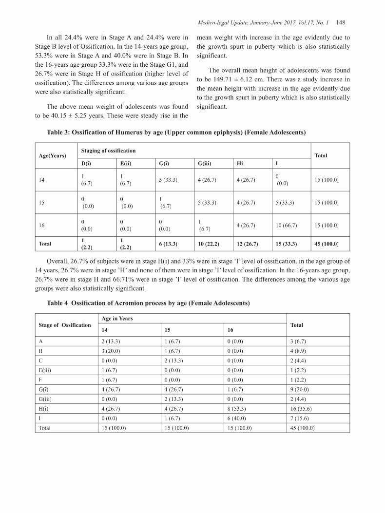
Medico-legal Update, January-June 2017, Vol.17, No. 1 148 149 -legal Update, January-June 2017, Vol.17, No. 1
In all 24.4% were in Stage A and 24.4% were in Stage B level of Ossification. In the 14-years age group, 53.3% were in Stage A and 40.0% were in Stage B. In the 16-years age group 33.3% were in the Stage G1, and 26.7% were in Stage H of ossification (higher level of ossification). The differences among various age groups were also statistically significant.
The above mean weight of adolescents was found to be 40.15 ± 5.25 years. These were steady rise in the
Table 3: Ossification of Humerus by age (Upper common epiphysis) (Female Adolescents)
Age(Years)Staging of ossification
TotalD(i) E(ii) G(i) G(iii) Hi I
14 1 (6.7)
1 (6.7) 5 (33.3) 4 (26.7) 4 (26.7) 0
(0.0) 15 (100.0)
15 0 (0.0)
0 (0.0)
1 (6.7) 5 (33.3) 4 (26.7) 5 (33.3) 15 (100.0)
16 0 (0.0)
0 (0.0)
0 (0.0)
1 (6.7) 4 (26.7) 10 (66.7) 15 (100.0)
Total 1 (2.2)
1 (2.2) 6 (13.3) 10 (22.2) 12 (26.7) 15 (33.3) 45 (100.0)
Overall, 26.7% of subjects were in stage H(i) and 33% were in stage ’I’ level of ossification. in the age group of 14 years, 26.7% were in stage ’H’ and none of them were in stage ’I’ level of ossification. ln the 16-years age group, 26.7% were in stage H and 66.71% were in stage ’I’ level of ossification. The differences among the various age groups were also statistically significant.
Table 4 Ossification of Acromion process by age (Female Adolescents)
Stage of OssificationAge in Years
Total14 15 16
A 2 (13.3) 1 (6.7) 0 (0.0) 3 (6.7)B 3 (20.0) 1 (6.7) 0 (0.0) 4 (8.9)C 0 (0.0) 2 (13.3) 0 (0.0) 2 (4.4)E(iii) 1 (6.7) 0 (0.0) 0 (0.0) 1 (2.2)F 1 (6.7) 0 (0.0) 0 (0.0) 1 (2.2)G(i) 4 (26.7) 4 (26.7) 1 (6.7) 9 (20.0)G(iii) 0 (0.0) 2 (13.3) 0 (0.0) 2 (4.4)H(i) 4 (26.7) 4 (26.7) 8 (53.3) 16 (35.6)I 0 (0.0) 1 (6.7) 6 (40.0) 7 (15.6)Total 15 (100.0) 15 (100.0) 15 (100.0) 45 (100.0)
mean weight with increase in the age evidently due to the growth spurt in puberty which is also statistically significant.
The overall mean height of adolescents was found to be 149.71 ± 6.12 cm. There was a study increase in the mean height with increase in the age evidently due to the growth spurt in puberty which is also statistically significant.

-legal Update, January-June 2017, Vol.17, No. 1 148 149 Medico-legal Update, January-June 2017, Vol.17, No. 1
In all, 35.6% were in stage ‘H’ and 15.6% were in stage ‘I’ level ofossification. In the 14-years age group, 26.7% were in stage ‘H’ and none of them were in stage ‘I’ level while in 16-years age group 53.3% were in ‘H’ level and 40.0% were in stage ‘I’ level of ossification. The differences among various age groups were also statistically significant.
DISCUSSION
Study of the appearance and fusion of the ossification centres around shoulder joint as visualised on radiograph has been time tested method. Numerous workers both in India and other countries have proved age ranges relevant to their population,
Mckern T.W. & Stewart. T.D. 5 conducted study in U.S.A. on males of 18 to 38 years age. They observed that Head of the Humerus separated from Diaphysis under 20 years, partially united with diaphysis between 19 to 20 years, united with diaphysis but joint was visible between 20 to 21 years and completely united with diaphysis at about 21 years and above.
Present study findings include that fusion was progressive in 16 years of age group in males where 46.6% were in higher level of ossification (Hi & I stages) when compared to 14 years of age group where all were in early stages of ossification (GI & G3 stages).
In females, fusion was much more progressive than males in 16-year group where 93.4% were in higher level of ossification (in stages H(i) & I) compared to 14 years of age group where 26.7% were in stage H and none of the them were in stage I level of ossification.
Davies and Parsons (1927) 6 recorded the age of fusion was occurring between 19 to 21 years of age in English subjects (both boys and girls). They did not give separate figures for males and females, so it is not possible to correlate present observations with their study.
The findings of present study showed that the union was early and progressive in both sexes, when compared to the findings of Davies and Parsons.
Pillai (1936) 7noted the age of fusion amongst boys and girls of Madras as 14 to 17 yrs. As he had given figures for mixed population, this work cannot be compared with the present study.
Basu (1938) 8found the age of union as 16 to 17 Years in Bengalis, Hindu females.
Present study revealed that the fusion was much more rapid and progressive in females than males especially in 16 Years age group when 93.4% were in higher level of ossification (in stages H(i) and I) compared to14 years of age group where 26.7% were in stage H(i) and none of them were in stage I level of ossification.
Present study showed that fusion was much more progressive than males in 16-years age group to females, where 93.4% were in higher level of ossification (in stages H(i) and I) compared to 14-years of age group where 26.7% were in stage H and none of them were in stage I level of ossification.
Apurba Nandi (2010) 9noted that fusion occurs as 16 to 17years in males.
Present study showed that the union was progressive in 16 years of age groups in Males as 46.6% were in higher level of ossification (H(i) and I Stages) compared to 14 years of age group where all were in earlier stages of ossification (G1 and G3 stages).According to him fusion in females occurs between 15 to 16 years of age.
Present study revealed that the union was much more progressive in females around 16 years of age where 93.4% were in higher level of Ossification than 14-years age group where 26.7% were in stage H and none of them were in stage I level of ossification.
According to flecker (1932) the age of appearance was around 14 years in females and 15 years in males 10. Fusion of Acromial Centre with body of scapula was around 17 years in both sexes.
According to the present study in females also, fusion of the ossification centre is progressive in 16 years of age group, where 53.3% were in stage H and 40.0% were in I level of ossification when compared to 14 years of age group Where 26.7% were in stage H and none of them were in stage I level of ossification.
The present study showed that the appearance and fusion of acromial ossification centre was progressive in 16 years of age group in Males were 33.3% were in stage G1 and 26.7% were in stage H of higher level of ossification when compared to 14 years of age group where 53.3% were in stage A and 40.0% were in stage B level of ossification.

Medico-legal Update, January-June 2017, Vol.17, No. 1 150
According to the present study in females also appearance and fusion of the ossification centre is much more progressive in 16 years of age groupwhere 53.3% were in stage H and 40.0% were in I level of ossification whencompared to 14 years of age group where 26.7% were in stage and none ofthem were in stage I level of ossification.
CONCLUSION
Ossification was progressive with age in males of 14 to 16 years ageand almost group and fusion of epiphysis with diaphysis was commenced complete in about 46.6% of males of 16-years age group. In females, ossification was much more rapid that in males and by 16 years of age 93.4% of the female subjects showed fusion of the epiphysis with diaphysis. This study will enable medico legal experts of this area to reach fairly definite conclusion of epiphyseal union, regarding age estimation in medicolegal cases.
Conflict of Interest : Nil
Sourceof Funding : Nil
Ethical Clearance: Obtained from Institutional ethics committee
REFERENCES
1. Aggarwal A. Ages of ossification-Personal Identification in Self-Assessment and Review of Forensic Medicine and Toxicology.1sted. Delhi: Jaypee Publishers and Distributers (P) Ltd.; 2006. P.51-59.
2. Alcazar ML, Alvear J, Muzzo S. Influence of Nutrition on the bone-development in the child.
Archivos Latinoamericanos De Nutrition 1984; 34 (2): 298-307.
3. Bajaj ID, Bharadwaj OP, Bharadwaj S. Appearance and fusion of important ossification centers - A study in Delhi population. Indian J. Med Res. 1967; 55:1064 – 1067.
4. Bargotra RN, Bargotra M and Singh J. The time of fusion of medial epicondyle of Humerus in residents of Jammu And Kashmir State. Paper read at 38th annual conference, 1989 of Anat. Soc. of India. Abstract published in Journal of Anatomical Society of India. 1990; 39:76.
5. Mckern T.W. & Stewart. T.D. :Skeletal age change in youngAmerican males. analysed from the stand point of age identification. Head qu. Qm, Res. And Dev. Comm& Tech 1957 . Ep-45, natick, Mass,l957.
6. Davies D.A. & Parsons F.G . The age order of appearance and union of normal epiphysis as seen by X-rays. Jr. Anat. London 1927 : 62 : 58-61.
7. Pillai MJS . The study of epiphyseal union for determining the age of South Indians. Indian J Med Res 1936;23 :1015–1017.
8. Basu, S.K.: Medicolegalaspects of the determination of age of Bengalese girls. Indian. Med. Rec, 63, (4) :97-100, l938
9. Nandy Apurba. Principles of Forensic Medicine Including Toxicology. 3rded: New Central Book Agency (Pvt) Ltd. London; 2010. 119-127
10. H. Flecker. Roentgenographic Observations of the Times of Appearance of Epiphyses and their Fusion with the Diaphyses. J Anat. 1932 ; 67(Pt 1): 118–164.
-legal Update, January-June 2017, Vol.17, No. 1 150
INTRODUCTION
Poisoning is and likely to remain one of the commonest cause of unnatural death. Knowledge of poisons and causing death by poisoning was prevalent in India right from ancient times. The present era being no exception, it is estimated that some form of poison is directly or indirectly responsible for more than 1 million illness worldwide and this problem is getting worse
A Study of Common Type of Poisoning in the Cases Referred for Medicolegal Autopsy at Sri Venkateswara Medical College,
Tirupati
Vishnu Vardhan Poluru1, Karnam Mamatha2
1 Senior Resident, 2Associate Professor, Department of Forensic Medicine, Sri Venkateswara Medical College, Tirupati, Andhra Pradesh, India
ABSTRACT
Back ground and objective: Acute poisoning is a medical emergency. It is important to know the nature, severity and outcome of acute poisoning cases in order to take up appropriate planning, prevention and management techniques . So this study aimed to acess the incidence of poisoning amongst the age, sex, to study and analyze the Postmortem findings in various cases of poisoning in relation to Chemical analysis.
Method: 196 fatal cases of acu te poisoning autopsied at Sri Venkateswara Medical College, Tirupati were studied. Complete autopsy of the body was carried out in each case.
Results: Incidence was more in the age group of 20-29 years in both sexes. It was more common in young males. Organophosphorus constituted 91 cases (46.93%. . It also showed that in 52 cases (26.53%) the odour of the stomach content was pungent, in 30 cases (15.30%) kerosene like smell, garlicky like in 25 cases (12.75%), peculiar smell noticed in 33 cases (16.85%) and in 56 cases (23.97%) no smell was detected. The stomach mucosa was congested in 128 of the total cases, followed by nil particular changes in 37 cases, congested and hemorrhagic appearance in 26 cases and charred/eroded appearance in 5 cases of corrosive acid poisoning.
Conclusion: Poisoning is more common in young males. The overall mortality is substantially high, mainly contributed by self-poisoning with insecticides and corrosives. Early care in a tertiary care center may help to reduce mortality in India.
Keywords: Acute poisoning, pattern and outcome, tertiary care hospital.
Corresponding author :Dr. Karnam MamathaAssociate Professor, Department of Forensic Medicine Sri Venkateswara Medical College, Tirupati- 517 507Andhra Pradesh, India Email id : [email protected]
from time to time as newer drugs and chemicals are developed in vast numbers owing to vast developments in the fields of biochemistry, agriculture, horticulture, microbiology, medicine and toxicology. Knowingly or unknowingly millions of people are exposed to danger by hazardous occupational practices and unsafe storage3
of toxic chemicals products in their day to day life. Lack of specialized toxicological services in developing countries like India has further contributed to the higher rate of morbidity and mortality4,5.
Poisoning being invariably medicolegal in nature among fatal cases, postmortem examination is done to establish the exact cause and manner of death. Manner of death in these cases is predominately suicidal because of the general belief that it terminates life with minimal
DOI Number: 10.5958/0974-1283.2017.00033.0

Medico-legal Update, January-June 2017, Vol.17, No. 1 152 153 -legal Update, January-June 2017, Vol.17, No. 1
sufferings or accidental but however homicidal cases are also reported and alleged which was more prevalent in the past as there were no well established means of detecting poison from the viscera, etc.. It is very difficult to draw a report to say which kind of poisoning is more frequent, has the nature of poisoning varies from one region to other region depending upon the poison availability and the knowledge of the local population regarding the properties of poisons6.
The rising incidences of fatal poisoning with prevalence of certain groups of poisons in this particular geographical area has prompted us to undertake this study to know the epidemiological aspects, patterns & other significant features of deaths due fatal poisoning and to compare it with the observations of various authors by scientific discussion. This study was taken up at a tertiary care hospital in AP to analyze these changing pattern of poisoning which is a ever-increasing medicolegal, epidemiological and social problem, also to stress the need to establish poison information centers so as to provide information and evidence concerning to diagnosis, treatment, prognosis and prevention of poisoning. So this study aimed to ascertain the common type of poisoning in the cases referred for medicolegal autopsy to Sri Venkateswara Medical College, Tirupati.
METHOD
The present study has been carried out in the Department of Forensic medicine Sri Venkateswara Medical College, Tirupati during the period January 2013 to June 2014. All the cases brought to the department for medicolegal autopsy with history of poisoning and
cases that were diagnosed as poisoning during the post mortem examination were selected. A sum total of 196 po i son ing cases were analyzed during this study period. Permission of the ethical committee on the use of human material for research purpose was obtained.
Detailed information of the deceased pertaining to the case was collected from the concerned police and from the relatives of the deceased by a questionnaire with their consent. Post mortem findings were analyzed with the chemical analysis reports. In treated cases information was acquired by perusal of hospital records. In cases of allegations, information was supplemented by either visit to the scene of crime or from the photographs of the scene of crime.
In all cases, viscera were subjected for chemical analysis at Regional Forensic Science Laboratory, Tirupati. Dissection of the body was carried out as per the procedure mentioned in “Autopsy Diagnosis and Technic” by Otto Sapphire7. Inclusion criteria included were all cases with the alleged history of poisoning, Cases diagnosed as poisoning after complete Post Mortem examination and Perusal of chemical analysis reports.
The data was analyzed using MS Excel and Epi-info. 7 version software and appropriate statistical tests of significance were employed in the needed situations in the presentation of data.
RESULTS AND DISCUSSION
Of the 1149 cases of postmortem examination, poisoning constituted 196 cases, amounting to 17.05% during this study period.
Table-1: Distribution of the study population according to Age.
Sl No Age Group Male Percent Female Percent Total Percent
1 1-9 yearsyears 1 0.75 4 6.30 5 2.55
2 10-19 years 2 1.50 9 14.28 11 5.61
3 20-29 years 25 18.75 27 42.85 52 26.52
4 30-39 years 27 20.25 12 19.12 39 19.89
5 40-49 years 30 22.50 05 7.93 35 17.85
6 50-59 years 26 19.75 0 0 26 13.30
7 60-69 years 133 9.75 3 4.76 16 8.16
8 70-79 years 9 6.75 3 4.76 12 6.12
Total 133 100 63 100 196 100
-legal Update, January-June 2017, Vol.17, No. 1 152 153 Medico-legal Update, January-June 2017, Vol.17, No. 1
It is observed from the above table 1 that maximum number of poisonings in the study population are seen in the age group of 20 to 29 years (26.52%). In males it amounted to 22.50% in the age group of 40-49 years followed by 30 to 39 years (20.25%). Least number were noticed in the age group of 1 to 9 years (0.75%) and in females maximum number of poisoning cases were observed in age group of 20 to 29 years accounting to 2 7 c a s e s (42.85%) followed by 30 to 39 years age group (19.12%).
Similar results were observed in the studies of Adarsh Kumar1 and Sharma B.R8
The maximum incidence of poisoning in the age group of 20 to 29 years is attributed to the factors like failures in facing the difficulties of academics, unemployment, love failures, family conflicts, marital disharmony, improper judgment of the problem, dowry harassment in case of females and ill health.
Males (67.86%) outnumbered the females (32.14%) in the study population. Similar results have been stated by Sharma B.R8In the present study population, poisoning is seen more among married people (81.12%) as compared to single/unmarried (18.88%).
These findings tallied with studies conducted by Gargi J9.
In the study population, it is observed that poisoning cases were predominantly seen in rural areas (61.22%) as compared to urban areas (38.78%).
Similar findings were seen in the studies of Karamjith Singh10.
In rural areas the predominant factors are financial losses in agriculture, family problems, draught conditions and ill health. In urban areas the reason being due to rapid urbanization leading to stiff competition, lack of employment opportunities, economic instability and high cost of living in urban setup.
In the present study it is observed that majority of the victims were Illiterates (31.12%) followed by Under-Matriculate (27.55%), Matriculates (23.47%) and least were among Postgraduates (1.02%).
Illiterates mainly being rural inhabitants, are dependent entirely on agriculture and related works, which are mainly confined to a season. They are left
without any work or income for remaining part of the year. Also lack of minimum education makes them prone for bad habits and unable to cope up with the stressful states, thereby losing mental balance and are prone to take drastic decisions and end their lives.
It is similar to the study of Manish Nigam11 and is in disagreement with study conducted by Karamjith Singh10.
As per the present study it is observed that 31 (15.82%) cases who died without receiving the treatment, included 21 victims who were categorized under spot deaths and 10 other cases of poisoning where death occurred before receiving any treatment.
The results differed from the study conducted by B.D.Gupta12.
Among treated cases, 29.07% of the victims survived for 1-3 days, followed by 21.93% of the victims surviving for 12-24 hours, 19.38% for 6-12 hours and the least in 5.11% of the victims, who survived for more than 7 days, of which the highest period of survival is 15 days by a male victim aged 28 years who had consumed organophosphorus insecticide.
Table 2 Distribution of the study population according to Manner of Death
Sl No Type of Poison Number Percent
1 Suicidal 185 94.39
2 Alleged Homicide 4 2.04
3 Accidental 7 3.57
Total 196 100
In the present study it is observed that, in 94.39% of the victims the manner of death were concluded as suicidal, 3.58% accidental and only in 2.04% there were allegations of homicide (Table 2 ).
The results were similar to the studies of B R Sharma11 and S K Dhattarwal13
The Manner of death that were concluded as suicide were based on the history furnished by the police and the relatives of the deceased, Suicide note, circumstantial evidence and on the Postmortem examination.
Of the 196 cases subjected for chemical analysis, 163 cases (83.16%) were positive for a poison and in 33
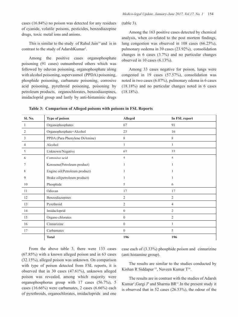
Medico-legal Update, January-June 2017, Vol.17, No. 1 154 155 -legal Update, January-June 2017, Vol.17, No. 1
cases (16.84%) no poison was detected for any residues of cyanide, volatile poisons, pesticides, benzodiazepine drugs, toxic metal ions and anions.
This is similar to the study of Rahul Jain14 and is in contrast to the study of AdarshKumar1.
Among the positive cases organophosphate poisoning (91 cases) outnumbered others which was followed by oduvan poisoning, organophosphate along with alcohol poisoning, supervasmol (PPDA) poisoning, phosphide poisoning, carbamate poisoning, corrosive acid poisoning, pyrethroid poisoning, poisoning by petroleum products, organochlorates, benzodiazepines, imidacloprid group and lastly by anti-histaminic drugs
(table 3).
Among the 163 positive cases detected by chemical analysis, when co-related to the post mortem findings, lung congestion was observed in 108 cases (66.25%), pulmonary oedema in 39 cases (23.92%), consolidation changes in 6 cases (3.7%) and no particular changes observed in 10 cases (6.13%).
Among 33 cases negative for poison, lungs were congested in 19 cases (57.57%), consolidation was noted in two cases (6.07%), pulmonary edema in 6 cases (18.18%) and no particular changes noted in 6 cases (18.18%).
Table 3: Comparison of Alleged poisons with poisons in FSL Reports
Sl. No. Type of poison Alleged In FSL report
1 Organophosphates 67 91
2 Organophosphate+Alcohol 23 16
3 PPDA (Para Phenylene DiAmine) 8 8
4 Alcohol 1 1
5 Unknown/Negative 63 33
6 Corrosive acid 5 5
7 Kerosene(Petroleum product) 1 1
8 Engine oil(Petroleum product) 1 1
9 Brake oil(petroleum product) 1 1
10 Phosphide 5 6
11 Oduvan 17 17
12 Benzodiazepines 2 2
13 Pyrethroid 2 4
14 Imidacloprid 0 2
15 Organo chlorates 0 2
16 Cinnarizine 0 1
17 Carbamates 0 5
Total 196 196
From the above table 3, there were 133 cases (67.85%) with a known alleged poison and in 63 cases (32.15%), alleged poison was unknown. On comparison with type of poison detected from FSL reports, it is observed that in 30 cases (47.61%), unknown alleged poison was revealed, among which majority were organophosphorus group with 17 cases (56.7%), 5 cases (16.66%) were carbamates, 2 cases (6.66%) each of pyrethroids, organochlorates, imidacloprids and one
case each of (3.33%) phosphide poison and cinnarizine (anti histamine group).
The results are similar to the studies conducted by Kishan R Siddapur 15, Naveen Kumar T16.
The results are in contrast with the studies of Adarsh Kumar1,Gargi J9 and Sharma BR11.In the present study it is observed that in 52 cases (26.53%), the odour of the

-legal Update, January-June 2017, Vol.17, No. 1 154 155 Medico-legal Update, January-June 2017, Vol.17, No. 1
stomach contents was pungent, in 30 cases (15.30%) kerosene like smell, garlicky like in 25 cases (12.75%), peculiar smell noticed in 33 cases (16.85%) and in 56 cases (23.97%) no smell was detected. It is in contrast to the study of Mrinal Haloi17.
On postmortem examination it is seen that in 128 cases (65.31%) the mucosa of the stomach is congested due to the fact that more number of victims received treatment causing the degradation of the poison. This was observed in maximum 69 cases (53.90%) of organophosphate poisoning. In 26 cases (13.26%) the mucosa of the stomach were congested and hemorrhagic suggesting the highly toxicity of the poison and were seen more in victims who were not treated indicating severe mucosal damage due to the erosive effect of the chemicals. This was seen in 12 cases (46.15%) of organophosphate poisoning. In 5 cases (2.56%) mucosa was charred or eroded due to corrosive acid poisoning which is highly specific and later was confirmed in chemical analysis reports.
In 37 (18.87%) cases, mucosa of stomach was found to be nil particular which is due to intensive and appropriate counteractive treatment measures. It was observed in 10 cases (27.02%) of organophosphate poisoning, 3 cases (8.1%) of organophosphate and alcohol poisoning.
CONCLUSION
A study on the fatal cases of acu te poisoning autopsied at Sri Venkateswara Medical College, Tirupati was under taken. Incidence of poisoning was more in the age group of 20-29 years in both sexes and was more in males as compared to females. Out of the positive chemical analysis report, organophosphorus constituted 91 cases (46.93%), followed by oduvan poisoning, organophosphate along with alcohol poisoning, supervasmol (PPDA) poisoning, phosphide poisoning, carbamate poisoning, corrosive acid poisoning, pyrethroid poisoning, poisonig by petroleum products, organochlorates, benzodiazepines, imidacloprid group and lastly by anti-histaminic drugs. Of the 33 negative chemical analysis reports due to consumption of unknown substance or unknown tablets as from the alleged history, the reasons may be attributed to any predisposing disease of the victim or small quantity of poison intake causing difficulties to detect or due to poison vomited out, treatment undergone or the poison
could have been detoxified, neutralized, conjugated or eliminated from the body causing difficulties in analytical procedures.
Conflict of Interest : Nil
Source of Funding : Nil
Ethical Clearance: Obtained from Institutional ethics committee
REFERENCES
1. Adarsh Kumar, Krishan Vij. Trends of Poisoning in Chandigarh-A Six Year Autopsy Study. Journal of Forensic Medicine and Toxicology, 2001; 18(1): 8-11.
2. Krishan Vij. Text Book Of Forensic Medicine And Toxicology. 2nd Edition, B.I. Churchill Livingstone, New Delhi; 2002:760-968.
3. Singh D.P, Acharya R.P. “Pattern of poisoning cases in Bir hospital.” Journal of Institute of Medicine, 2006;28(1):3-6.
4. Flemming Konradsen, Wim van der Hoek, Donald C. Cole, Gerard Hutchinson, Hubert Daisley, Surjit Singh, Michael Eddleston. Reducing acute poisoning in developing countries-options for restricting the availability of pesticides. Toxicology. 2003; 192: 249-261.
5. Suresh Kumar Gupta, Sharda Shah Peshin, Amita Srivastava and Thomas Kaleekal. A study of childhood poisoning at National Poisons Information Centre, All India Institute of Medical Sciences, New Delhi”. Journal of Occupational Health 2003; 45: 191-196.
6. Unnikrishnan B, Singh B, Rajeev A. “Trends of acute poisoning in south Karnataka”. Kathmandu University Medical Journal, 2005; Vol 3, No.2, 10: 149-154.
7. Otto Sapphire. “Autopsy Diagnosis and Technic”. 4th Edition, Illinois; Hoeber Harper.
8. Sharma.B.R, Dasari Harish, Vivek Sharma, Krishan Vij.”The Epidemiology of Poisoning: An Indian View Point”. Journal of Forensic Medicine and Toxicology, 2002; 19(2): 5-11.

Medico-legal Update, January-June 2017, Vol.17, No. 1 156
9. Gargi.J,Hakumat Rai, Ashok Chanana, Gurmanjit Rai, Bagga.I.J.S. “Current Trends of Poisoning- A Hospital Profile”. Journal of Punjab Forensic Medicine and Toxicology, 2003; 3:41-45.
10. Karamjit Singh, Oberoi.S.S, Bhullar.D.S. “Poisoning Trends In The Malwa Region Of Punjab”. Journal of Punjab Forensic Medicine and Toxicology, 2003; 3:26-29.
11. Manish Nigam, Ashok Kumar Jain,Dubey.B.P,Sharma.V.K. “Trends of Organophosphorus Poisoning In Bhopal Region An Autopsy Based Study”. Journal of Indian Forensic Medicine, 2004; 26 (2):62-65.
12. B.D.Gupta, P.C. Vaghela. “Profile of fatal poisoning in and around Jamnagar.” Journal of Indian Academy of Forensic Medicine, 2005; 27(3): 145-148.
13. Dhattarwal.S.K, Harnam Singh. “Profile of Deaths Due To Poisoning In Rohtak, Haryana”. Journal of Forensic Medicine and Toxicology, 2001; 18 (2) :28-29.
14. Rahul Jain, Sharikanth Asawa, Shishir Ruia. “Status of Poisoning In A Hospital Of Maharashtra”. Journal of Forensic Medicine and Toxicology, 2001; 18 (1): 12-16.
15. Naveen Kumar T, Jagannatha S R, Ananda K. “A Study of changingtrends and patterns of poisoning at Bangalore.” Indian Journal of Forensic medicine and Toxicology, July-December,2012; 6(2):120-122.
16. Kishan R Siddapur, Gurudatta S Pawar, Shashidar C Mestri. “Trends of Poisoning and Gross stomach mucosal appearance in fatal poisoning cases- an autopsy study”. Journal of Indian Academy of Forensic Medicine, April-June, 2011; 33(2):106-111.
17. Mrinal Haloi, Mamata Devi Haloi, Amarjyoti Patowari. “Death due poisoning in district of kamrup, Assam-A Medicolegal study”. Journal of Indian Academy of Forensic Medicine, January-March, 2013; 35(1): 18-21.
-legal Update, January-June 2017, Vol.17, No. 1 156
Study of Patterns of Sexual Offence Cases in Tripura Medical College
S C Sarkar1, P N Chakraborty2
1Associate Professor, 2Assistant Professor, Department of Forensic Medicine & Toxicology, Tripura Medical College & Dr. BRAM Teaching Hospital, Agartala
ABSTRACT
Sexual offences are the most heinous crimes against women and children. It is most barbarous and humiliating and the women and children remain most vulnerable group of this crime. Preservation of biological evidences of sexual offences is the important task and ethical duty of doctors. Poor medical evidences are often responsible for low conviction rate of criminals. The present study is a 5 years retrospective study which reflects the incidence and patterns of sexual offence cases brought to the Tripura Medical College along with the demographic variables, physical and genital examination findings of victims and results of medical evidences collected during the study period (January2010 to December2015). Total 67 victims and 103 accused of sexual offence were brought by police to the emergency block for medical examination. Victims 11- 20 years and accused 21 – 25 years are most vulnerable and dangerous for sexual assault.
Keywords: Sexual assaults, poor analysis, medical evidences, low conviction rate, younger group.
INTRODUCTION
Sexual offences are the acts of sexual intercourse with a second person or an animal to obtain sexual gratification. The law and customs of the society permits heterosexual intercourse between a man and his own wife as provided by the nature. Among all the crimes sexual offences are the most barbarous and humiliating. Women and children are the most vulnerable group of the crime. The alarming rise in the rate of sexual assault worldwide represents a major public Health problem1.
In USA, an estimate one in every four women and children2, in Nigeria four out of every ten women are victims of sexual assaults3. In South Africa, the incidence of rape is approximately 300 out of 100,000 population4. In India over the last five years, sexual assault cases show increasing and decreasing trends with increase of 6.6% in the year 2000 over the year 1999 and decline of 2.5% in the year 2001. A total of 16,075/1.6 per 100,000 population cases of child rape were reported all over the India. It shows that 11.2%, 19.5% of total crimes are against women and children respectively. Delhi rank 5th for the incidence of rape cases against women and 1st for that on the children (0.8%) per 100,000 population5.
Collecting and preserving the evidences in sexual offence cases is of crucial importance in medical profession for documentation in the court of law and to trace the actual criminal. Careful examination of biological specimens such as blood, semen, saliva often yields vital evidence for identification of a person in contemporary criminal investigation and to give the major part of conclusion of the cases6. In this present study, the incidence and patterns of sexual offences in Tripura Medical College & Dr. BRAM Teaching Hospital along with the demographic variables, physical and genital examination findings of victims and accused, results of medical evidences collected are presented.
MATERIAL AND METHOD
During the study period (January2010 to December2015) a total 67 victims and 103 accused of sexual offence cases were brought by police to the emergency block of Tripura Medical College for medical examination. From 9 am to 4 pm the victims were examined by the department of Forensic Medicine and Toxicology and after 4 pm to next day morning, were examined by Department of Obstetrics & Gynaecology. Details pertaining to the age, sex, religion, literacy, socio-economic status, place of incidence, number
DOI Number: 10.5958/0974-1283.2017.00034.2

Medico-legal Update, January-June 2017, Vol.17, No. 1 158 159 -legal Update, January-June 2017, Vol.17, No. 1
of assailants, relationship of assailants with victims, findings of physical and genital examination and results of evidence collected during the examination were noted in a predesigned format. Careful collection and analysis of preserved biological specimens were done which yielded the vital evidences for identification of person in criminal investigations.
RESULTS
A total 170 of sexual offence cases including victims and accused, 67(39.41%) were victims, 103(60.58%) were accused who were brought by police in the emergency block for medical examination. Out of 67 cases of rape victims, all are the female victims; no case of male victim (sodomy) was reported during the study period. The age of the victims ranged from 3 - 49 years, and of assailants ranged from 15 to 55 years, but the most effected group was 11- 20years(67.16%) and followed by 21 -30 years(11.94%)of victims. The assailants between 21 – 25 years (45.63%) followed by 26-35years (22.23%) were dangerous age group for sexual assault(Table-1).
Fifty eight (86.56%) of victims, 96(93.20%) assailants were Hindu. Muslim victims 9(13.43%), assailants 7(6.79%) were involved in sexual offences cases and no other religion were reported in this study and 57(85.07%) victims, 86(83.49%) assailants were
unmarried (Table-2).
Maximum number of victims, 31(46.26%) were illiterate, 27(26.21%) of accused studied from class V- Class IX standard, 59(88.05%)victims and 87(84.46%)assailants were from lower socioeconomic status who were involved in sexual assaults. (Table-3)
The vast majority of victims knew their assailants. In 29(43.28%)cases they were of acquaintance, in 7(10.44%) cases assailants were strangers, in 16(23.88%)cases they were closed friend and in 2(2.89%) cases victims were blood related (father) to her assailants. The commonest sites of sexual offences were in victim’s house 33(49.25%) followed by some isolated places or jungle23 (34.32%) (Table-4).
In 49(73.13%) cases there was invalid consensual sexual intercourse followed by forcible rape 15 (22.38%) and no case of unnatural offence was reported in this study (Table-5).
In 7(10.44%) cases victims had extra genital, 11(16.41%) genital and 4(5.97%) cases victims had combined extra genital and genital injuries. Rupture of hymen was found in 45 (67.16%) cases (Table-6).
There were 3 (4.47%) cases of gang rape and maximum 3 assailants were involved in one case. The vaginal swabs collected from 33 cases and showed positivity of spermatozoa only in 2(2.98%) cases (Table-7).
Table-1: Age of Victims and Accused of sexual offences
Victims(N=67) Accused(N=103)
Ages(Years) Female(N=67) Male(N=0) Total Number % Age Number(N=103) %
0 – 10 5 0 5 7.46 15 – 20 22 21.3511 – 20 45 0 45 67.16 21 – 25 47 45.6321 – 30 8 0 8 11.94 26 – 35 23 22.3331- 40 5 0 5 7.46 36-45 7 6.7941 – 50 3 0 3 4.47 46- 50 3 2.91>50 0 0 0 0.00 >50 1 097
Table-2: Religion and marital status of Victims and Accused of sexual offences
Victims(N-67) Accused(N=103)Religion Number % Religion Number %Hindu 58 86.56 Hindu 96 93.20Muslim 9 13.43 Muslim 7 6.79Christian 0 0.00 Christian 0 0.00Sikh 0 0.00 Sikh 0 0.00Married 9 13.43 Married 17 16.50Unmarried 57 85.07 Unmarried 86 83.49Widow 1 1.49

-legal Update, January-June 2017, Vol.17, No. 1 158 159 Medico-legal Update, January-June 2017, Vol.17, No. 1
Table-3: Literacy and Socioeconomic status of Victims and Accused of sexual offences
Victims(N=67) Accused(N=103)
Education(Class) Number % Education(Class) Number %
Illiterate 31 46.26 Illiterate 17 16.50
Class I- Class V 14 20.89 Class I- Class V 27 26.21
Class VI -Class IX 13 19.40 Class VI -Class IX 43 41.74
Class X- class XII 7 10.44 Class X- class XII 13 12.62
Above Class XII 2 2.98 Above Class XII 3 2.91
Low Socioeconomic 59 88.05 Low Socioeconomic 87 84.46
Middle Socioeconomic 5 7.46 Middle Socioeconomic 11 10.67
High Socioeconomic 3 4.47 High Socioeconomic 5 5.15
Table-4: Relationship of Assailants with Victims and places of sexual offence
Relationship of assailants with Victims Places of incidences of sexual offences
Types of relationship Number % Places incidences Number %
Acquaintance 29 43.28 Victim’s house 33 49.25
Stranger 7 10.44 Accused house 8 11.94
Closed friends 13 19.40 Relative house 3 4.47
Neighbour 16 23.88 Isolated places/Jungle 23 34.32
Blood relation 2 2.988
Table-5: Distribution of types of offences and patterns of injuries on victims of sexual offences
Types of offences No.(N=67) % Patterns of injuries No.(N=67) %
Forcible rape 15 22.38 Extra genital 7 1o.44
Invalid consensual Rape 49 73.13 Genital 11 16.41
Attempted Rape 3 4.47 Extra genital & genital 4 5.97
Unnatural sexual offences 0 0.00 Hymen ruptured 45 67.16
Table-6: Distribution of Assailants and Victims of sexual offences in gang rape
Number of Victims Number of Assailants1 31 31 2
Total (4.47%) 8 (Two assailant on each case)
Table-7: Results of Laboratory Tests of specimens collected from Victims
Test performed No.(N=67) +ve %Microscopic examination of spermatozoa 33 2 2.98Acid Phosphate 33 0 0.00Florence Test 33 0 0.00Barberior Test 33 0 0.00Hanging drop preparation 2 0 0.00
Medico-legal Update, January-June 2017, Vol.17, No. 1 160 161 -legal Update, January-June 2017, Vol.17, No. 1
DISCUSSION
Sexual offences are one of the most heinous crime against women and children. The various social Organizations and women Welfare Organizations, legal and administrative steps have taken by the Government to give protection to the women and children against such crimes. Sexual assault is a neglected public health issue in most of the developing countries and there is even smaller percentage of reporting sexual assaults7. Only 10- 50% female victims report such assaults8. The under reporting of sexual assault is due to various social stigma and delay in the administration of justice9.
In the present study, 67 victims and 103 assailants of sexual offences was studied in Tripura Medical College and Dr. BRAM Teaching Hospital from January2010 to December2015, all the victims were female and no male victim was found. The results are in agreement with the study of Sagar10, Bhardwas et al, 1 1 Grossing et al,12 Riggs et al, 15 17% victims were assaulted with anal intercourse.
Bhardwas et al11 and Malhotra et al7 found that majority of victims were in the age group of 13 – 20 years (40.70%), in Du Munt et al 13 study, victims were of age group of 15 – 20 years and in the study of Islam et al 14 victims were between 11 – 20 years (70.42%) and majority of assailants were in the age group of 21-25 years (37.60%). In present study, majority of victims were between 11- 20 years(67.16%) and majority of assailants were in the age group of 21 – 25 years47(45.63%). The study conducted, minimum age of victim was 3 years and maximum age was 49 years. Hence no age is considered to be safe from rape. Person belonging to extremes of age group cannot physically resist and defend themselves from sexual assaults 17.
In the present study, majority of victims(86.56%)and assailants(93.20%) were Hindus and finding was agreement with the study of Fimate et al 16 (57%). Majority of victims were illiterate (46.26%) and accused(41.74%) were educated upto class V-IX. Islam et al 14 reported that majority of victims were illiterate. In this study, both victims(88.05%) and accused(84.46%) were found in lower socioeconomic status.
In the present study majority of victims were known to their assailants (Acquaintance 43.28%, closed friend were23.88%) and 10.44% assailants were stranger. Similar findings have been observed by Fimate et al 16
of the victims and assailant relationship, acquaintance (69.7%), and stranger (25.6%). While Islam et al 14
reported that in the majority of cases of victims knew the assailants. Strangers have been reported as the common assailants in the study of Okonkwo et al13 (49.2%). Riggs et al 15 (39%) and Dumont et al 13 (49.2%). Malhotra et al reported that rape by person acquitted with victims is common for girls less than 10 years. Rape or assault by strangers increased significantly with age. Grossin et al reported that in half of the cases of victims examined within 72 hours, the assailants were stranger, while in those examined after 72 hours the assailant was mainly father (30%).
The most common site of offences was the victim’s houses (41%) as reported by Grossin et al 12 and Okokwo et al 13 and in the present study it was (49.25%). Majority of victims in this study were brought for medical examination after 5th days. In the grossin et al 12 , the victims presented for medic et al 13 within 72 hours, in Du Mont et al 13 the victims (40.1%) reported within2 – 6 hours, while Islam et al 14 reported that 23.7% of victims reported within 72 hours after incident.
There was invalid consensual sexual intercourse in 73.13% cases as found in this study. More than one assailant was involved in 4.47% cases (present study), 20% cases in Riggs et al15 and 10% cases in Grossin et al12.
Killing after sexual act could be either in panic or destroy a witness of crime17. Only 20% cases of murder are committed by the urge of sexual gratification18. Victims being under influence of alcohol (39%) and drugs (12%) was reported by Okonkwo et al13 and DuMontet al13 (alcohol 41.7%). Though it was not found in this study.
In the present study, 10.44% victims had extra genital injuries, 16.41% had genital injuries and 5.97% had combined extra genital and genital injuries. Rupture of hymen was found 67.16% cases of victims and maximum cases were old rupture. Islam et al14 reported extra genital injuries in 91 cases, rupture of hymen in38.9% cases with fresh tear in fourchette in 2–6% of cases. Malhotra et al17
reported genital injuries in 33.3% cases. DuMunt et al13
reported injuries on bodies of victims in 64.2% cases. Grossin et al12 reported general body trauma in 39.1% and genital trauma in 35.11% cases examined within 72 hours, 6.3% and 19.5% cases examine after 72 hours

-legal Update, January-June 2017, Vol.17, No. 1 160 161 Medico-legal Update, January-June 2017, Vol.17, No. 1
respectively. Riggs et al15 found general body trauma in 67% cases and genital injuries in 53% cases. Absence of genital injuries in the victims examined could be due to various reasons. Majorities of the victims were adults with prior sexual activities. Genital injuries are common in children and post menopausal women. The nature and time of assault determines whether injuries would normally be expected.19 The absence of physical injuries may not contradict the allegation of sexual assault. Rapidly healing injuries can be missed in cases with delayed examination or there may be false allegation.20 Similarly, value of examination immediately following an alleged incident is limited by the fact that bruises may not become apparent for at least 48 hours.
Vaginal swabs collected from 33 cases of victims and showing positivity for spermatozoa in 2(2.98%). Riggs et al15 reported that evidence of sperm and semen in 48 cases. Davies and Wilson(1974)21observed that seminal blood group antigens could be detected on swabs collected within 48 hours, acid phosphatase upto 2 days, choline within 3 days after sexual intercourse. The absence of sperm may be attributed to any of the following causes like erectile dysfunction, impotence, premature ejaculation, prolonged postcoital intercourse, oligospermic or orchidectomised assailants, penetration, use of condoms or spermicidal agents and menstruation and vaginal inflammation 21,22.
CONCLUSION
This study highlights the importance of addressing rape as a public health issue and focuses on the demographic profile of victims of sexual assault cases.
Ethical Clearance- Taken from Institutional Ethical committee
Source of Funding- Self
Conflict of Interest- Nil
REFERENCES
1. Walch AG, Broadhead WE. Prevalence of lifetime sexual victimization among female patient.JFam Pract. 1992Nov; 35(5):511-6.
2. Sexual Assalt and Domestic Violence. In Beckmann CRB,Ling FW, Herberr WPN, Laube DW, Smit RP, Barzansky BMeditors. Obstetrics and gynaecology. 3rd ed. Lippincott Williams & Wilkins.1998;576-82.
3. Okonkwo JEN, Ibeh CC. Female sexual assault in Nigeria. Int J Gynaecol obstet. 2003; 83(3):325-6.
4. Martin L J Forensic evidence collection for sexual assaults:a south Africa Perspective.INt J Gynaecol Obstet.2002;78 Suppl 1: S 105-10.
5. Crime in India-2009. New Delhi: 29 Dec 2010.640p.
6. Ansell R. Securing evidence after sexual offence is of importance task for physicians. Increasing severity of crimes and use of DNA analysis necessitate higher quality standards Lakartidningen. 1998 cct;95(42):4626-8,4630-11.
7. MalhotraN, Sexual assalat- a neglected public health problem in the developing world. IntJ Gynaecol Obstet. 2000;71(3):257-8.
8. Beebe DK. Emergency management of adult female rape victim. An Fam Physician.1991;43(6):2041-6.
9. Okonkwo CO. In:Rape (sexual assault), Society and children. Problems and papers of s symposium held at Enugu on May 7, 1986, FIDA(Anambra/Enugu). Family Law Publishers;1986: 50-9.
10. Sagar MS. Sexual offences in South Delhi. JEFMT. 1992; Vol.IX No. 8-11.
11. Bhardwaj DN, Sharma RK, Sagar MS, Murty OP. Study of sexual offences in South Delhi.JFMT.1992; Vol.XII No.3& 4:33-4.
12. Grossin C, Sibille I, lorin de la grandmaison G, Banasr A, Brion F, Durigon M. Analysis of 418 cases of sexual assault. Forensic Sci Int.2003 jan 28; 131(2-3):125-30.
13. Du Mont J, Parnis D. Sexual assault and legal resolution: querying the medical collection of Forensic evidence. Med Law.2000; 19(4): 779-92.
14. Islam MN, Islam MN. Retrospective study of alleged rape victims attended at Forensic Medicine Department of Dhaka medical College, Bangladesh. Leg Med ( Tokyo). 2003 Mar;5 suppl 1 : 351-3.
15. Riggs N Houry D Long G. Markovchick v, Feldhaus KM. Analysis of 1076 cases of sexual assault. Ann Emerg Med. 2000 pr; 35 (4): 358-62.
16. Fimate L., Devi M. An analytical study of rape in Manipur. Int J Med Toxicol Legal Med. 1998; 1: 1-2.
17. Subramanyam BV, editor. Modi’s Medical

Medico-legal Update, January-June 2017, Vol.17, No. 1 162
Jurisprudence and Toxicology. 22nd ed. New delhi; Butterworths India; 1999.
18. Gee DJ. Sexual offences . In Polson CJ, Gee DJ, Knight B., editors. The Essential of Forensic Medicine. 4th ed., Oxford: Pergamon Press:1985.
19. Gogers D. Physical aspects of alleged sexual assaults. Med Sci Law. 1996. Apr; 36(2) 117-22.
20. Cheistia CW, Lavelle JM, De Jong AR, Loiselle J, Brenner L, Joffe M. Forensic evidence finding in prepubertal victims of sexual assault. Paediatrics. 2000 Jul; 106 ( 1 pt 1): 1100-4.
21. Davies A. Wilaaon E. The persistence of seminal constituents in the human vagina. Forensic Sci. 1974 Feb;3(1): 45-55.

-legal Update, January-June 2017, Vol.17, No. 1 162
Demographic Profile of Fatal Road Traffic Accidents among Under 30 Years in Union Territory of Pondicherry
Ananda Reddy1, Pramod Kumar GN2, Hemanth Kumar R G3, Balaraman R4
1Associate Professor, Department of Forensic Medicine & Toxicology, The Oxford Medical College 1Associate Professor, Department of Forensic Medicine & Toxicology, The Oxford Medical College 1
Hospital and Research Centre, Bangalore. Karnataka, India, 2Associate Professor, Department of Forensic 2Associate Professor, Department of Forensic 2
Medicine & Toxicology, Mahatma Gandhi Medical College & Research Institute, Sri BalajiVidyapeeth University, Pillaiyarkuppam, Pondicherry, India, 3Associate Professor, Department of Forensic Medicine 3Associate Professor, Department of Forensic Medicine 3
& Toxicology, SDM College of Medical Sciences and Hospital, Dharwad, Karnataka, India, 4Grade I Senior Resident, Department of Forensic Medicine and Toxicology, Indira Gandhi Government General
Hospital & Post Graduate Institute, Pondicherry. India
ABSTRACT
The present study aims to highlight the demographic profile of fatal road traffic accidents (RTAs) in individuals under the age group of 30 years in Union Territory of Pondicherry, South India. The study is an autopsy based observation of fatal road traffic accidents during the year 2012-2014. Fatal road accidents are less common before noon. A male preponderance (82.5%) was observed with a male-female ratio of 4.7:1. Individuals in the age group of 21 to 30 years formed the most vulnerable (28.0%) group. Most of the accidents took place on National highways (36.9%). Majority of the victims were two wheeler occupants (48.5%) followed by pedestrians (23.7%). In most fatal accidents vehicle involved was two wheelers (56.4%) followed by four wheelers (14.5%). Head and facial injuries (46.5%) were the most common. 82.6% of victims belonged to Hindu religion. Head injury accounted as the cause of death in majority of cases (41.9%). 67.2% of victims were unmarried & 35.7% were uneducated.
Keywords: Fatal injuries, Pondicherry, Road traffic accidents (RTAs)
Corresponding author:Dr. Ananda ReddyAssociate Professor, Department of Forensic Medicine & Toxicology, The Oxford Medical College Hospital and Research Centre, Bangalore- 562107. IndiaEmail: [email protected]: 09845707755
INTRODUCTION
As per the World Health Organization (WHO) key facts, about 1.25 million people die each year as a result of road traffic crashes and it is the leading cause of death among young people, aged 15–29 years. The importance of road traffic accidents (RTAs) as health problem is perhaps not sufficiently recognized by public; even enough weightage is not given by health planners also. 90% of the world’s fatalities on the roads occur in low- and middle-income countries, even though these countries have approximately half of the world’s vehicles. Half of those dying on the world’s
roads are “vulnerable road users”: pedestrians, cyclists and motorcyclists. Without action, road traffic crashes are predicted to rise to become the 7th leading cause of death by 2030.1 The data presented to Parliament by the Ministry of Road Transport and Highways for year 2008, indicates that 1,19,860 people lost their lives in mishaps during that year.2 Deaths due to road accidents in 2009 were reported to be 126,896 and in 2010 it increased to 133,938. State of Tamil Nadu, alone is accounted for 11.5% of total “Road Accident” deaths in the country.3
The Law Commission of India has pointed out that the national and state highways account for nearly half of all road accidents.3 Overpopulation, increased number of vehicles on roads, poor road conditions and disregard for traffic rules and regulations are some of the major causes of increased injuries and fatalities in India.4, 5 The present study was carried out to provide a baseline data to policy makers to plan safer transportation routes and in setting up of health care centers in areas that report a higher number of accidents.
DOI Number: 10.5958/0974-1283.2017.00035.4

Medico-legal Update, January-June 2017, Vol.17, No. 1 164 165 -legal Update, January-June 2017, Vol.17, No. 1
MATERIALS AND METHOD
The present study is an analysis of 241 RTAs related deaths in population aged less than 30 years in the Union Territory of Pondicherry. This study was undertaken at Indira Gandhi Government General Hospital & Post Graduate Institute (IGGGH& PGI) between calendar years 2012 and 2014. The autopsies on these cases were conducted by the department of Forensic Medicine, IGGGH & PGI Pondicherry. The information regarding age, sex, marital status, and religion of the individual, time and location of the accident, type of vehicle involved in the accident, mode of transport of the victim on road, survival period of the victim were collected from the case sheets and police records. The site of fatal injuries on the body and cause of death of victim were recorded based on autopsy findings of the deceased. Total 4014 autopsies were conducted during the period of 2012 to 2014, out of which 832 were due to RTAs. Out of 832 RTAs 241 victims were under the age group of 30 years. In the present study, the subjects were divided into three age groups: 1–10, 11–20 and 21–30 years. The data thus obtained were analyzed by the SPSS version 16.
RESULTS
The study period included total 241 cases of fatal RTAs under the age of 30 years. Maximum RTA fatalities occurred in the year 2013 (Table 1). Individuals in the age group of 21 to 30 years formed the most vulnerable (28.0%) group, and a male preponderance (82.5%) was observed with a male-female ratio of 4.7:1(Table 2). 36.9% cases of RTAs occurred on National highways followed by state highways (21.6%). Majority of the victims were two wheeler occupants (48.5%) followed by pedestrians (23.7%). Two wheeler vehicles involved in 56.4% of cases followed by four wheelers (14.5%). 21.2% of victims died on the spot while 10.4% of them succumbed to injuries during transportation to hospital. Maximum fatalities occurred during second quarter of the year (April to June) accounting for 26.1% cases (Table 3). Most of fatal road traffic accidents took place during 3 AM to 9 PM accounting for 41.8% cases (Table 4). In 46.5% cases injuries were documented on Head and face (Table 5). Head injury was the cause of death in majority (41.9%) of cases (Table 5). 82.6% of victims belonged to Hindu religion, 67.2% of victim were unmarried & 35.7% of them were uneducated.
Table 1: Year wise distribution of Road Traffic accident (RTA) fatalities
Calendar year Total autopsies performed(A) RTA fatalities (B) Proportion of
fatal RTAs(B/A)
RTA fatalities under 30 years of age (C)
Proportion of RTA fatalities Under 30 years (C/B)
2012 1560 278 17.8 72 25.9
2013 1318 297 22.5 95 32
2014 1136 257 22.6 83 32.3
Total 4014 832 20.7 241 28.9
Table 2: Age and Gender wise distribution of RTA fatalities under 30 year’s victims
Age group (in years) Male (a) Female (b) Total Gender ratio
(a/b)
0-10 12 (5.0%) 04 6(1.%) 16 (6.6%) 3.0:1
11-20 53 (9.6%) 11 (1.6%) 64 (11.1%) 4.8:1
21-30 134 (24.9%) 27 (3.1%) 161 (28.0%) 5.0:1
Total 198 (82.5%) 42 (15.3%) 241(100%) 4.7:1
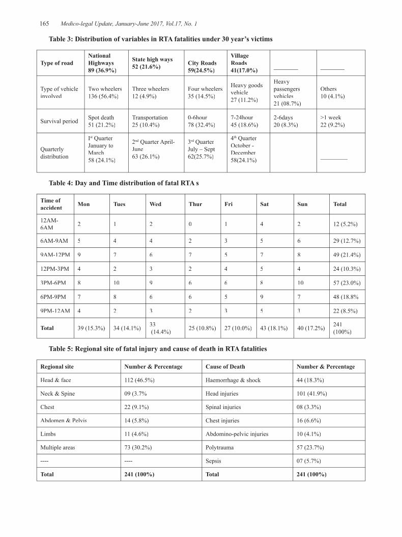
-legal Update, January-June 2017, Vol.17, No. 1 164 165 Medico-legal Update, January-June 2017, Vol.17, No. 1
Table 3: Distribution of variables in RTA fatalities under 30 year’s victims
Type of roadNational Highways89 (36.9%)
State high ways 52 (21.6%) City Roads
59(24.5%)
Village Roads41(17.0%) ________ ________
Type of vehicle involved
Two wheelers136 (56.4%)
Three wheelers12 (4.9%)
Four wheelers35 (14.5%)
Heavy goods vehicle27 (11.2%)
Heavy passengers vehicles21 (08.7%)
Others10 (4.1%)
Survival period Spot death51 (21.2%)
Transportation25 (10.4%)
0-6hour78 (32.4%)
7-24hour45 (18.6%)
2-6days20 (8.3%)
>1 week22 (9.2%)
Quarterly distribution
Ist Quarter st Quarter st
January to March58 (24.1%)
2nd Quarter April- nd Quarter April- nd
June63 (26.1%)
3rd Quarter rd Quarter rd
July – Sept62(25.7%)
4th Quarter October - December58(24.1%) _________ _________
Table 4: Day and Time distribution of fatal RTA s
Time of accident Mon Tues Wed Thur Fri Sat Sun Total
12AM-6AM 2 1 2 0 1 4 2 12 (5.2%)
6AM-9AM 5 4 4 2 3 5 6 29 (12.7%)
9AM-12PM 9 7 6 7 5 7 8 49 (21.4%)
12PM-3PM 4 2 3 2 4 5 4 24 (10.3%)
3PM-6PM 8 10 9 6 6 8 10 57 (23.0%)
6PM-9PM 7 8 6 6 5 9 7 48 (18.8%
9PM-12AM 4 2 3 2 3 5 3 22 (8.5%)
Total 39 (15.3%) 34 (14.1%) 33 (14.4%) 25 (10.8%) 27 (10.0%) 43 (18.1%) 40 (17.2%) 241
(100%)
Table 5: Regional site of fatal injury and cause of death in RTA fatalities
Regional site Number & Percentage Cause of Death Number & Percentage
Head & face 112 (46.5%) Haemorrhage & shock 44 (18.3%)
Neck & Spine 09 (3.7% Head injuries 101 (41.9%)
Chest 22 (9.1%) Spinal injuries 08 (3.3%)
Abdomen & Pelvis 14 (5.8%) Chest injuries 16 (6.6%)
Limbs 11 (4.6%) Abdomino-pelvic injuries 10 (4.1%)
Multiple areas 73 (30.2%) Polytrauma 57 (23.7%)
---- ---- Sepsis 07 (5.7%)
Total 241 (100%) Total 241 (100%)
Medico-legal Update, January-June 2017, Vol.17, No. 1 166 167 -legal Update, January-June 2017, Vol.17, No. 1
DISCUSSION
In the present study most fatalities were seen in males and most victims were in their second decade of life as males and people in this age group form the highest admission at emergency department as per studies.6,7,8 Majority of the victims were two wheeler occupants probably because riding a two wheeler is more risky & common than other modes of transport on road and also among the vehicles involved in accident two wheelers were leading the list. These finding are similar to another study carried out at Manipal.9 A study found pedestrians as more common victims but according to us this difference could be attributed to the study period, which includes victims of previous three decades, during which period the major users of roads were pedestrians.10 Majority of fatal accidents occurred on national highways and this can be explained by the high speeding vehicles on national highways compared to other roads. But one study has noted rise in fatal accidents in city roads.8 The discordance in the findings can be implicated to differences in the location of the hospital (city or outskirts) and the type of roads near it, as the victims attending the hospital are usually from nearby places. More than 25% of fatalities occurred before reaching hospital; the findings are comparable to another study.10 In the present study the incidence of fatal road traffic accidents was almost same throughout the year but other studies have noted a rise in winter but without any statistical significance.8, 11 Majority of fatal accidents were reported during 3pm to 6pm and 6pm to 9pm which could be due to poor light source, driving under alcohol intoxication and same findings were noted in Tamilnadu.3 A study at Chandigarh revealed higher incidence during 12-4 pm and 4-8 pm.11 From which we can infer that fatal road accidents are less common before noon because working population will be at office, women at home will be busy with household chores and the children will be at school and at noon people move out of the office for lunch, children return home and as the day passes there will be a slow increase in number of road users till dinner time. In the present study injuries to the head were commonest and also the most common cause of death. These findings are similar to other studies.8, 10, 11, 12 Majority of the victims belonged to Hindu religion as it is a Hindu predominant area. Most of the victims were uneducated as they might not be well versed with traffic rules. Unmarried people were major victims probably because such individuals have
rash driving practices & high risk taking behavior than married ones.
CONCLUSION
The present study helps to know the trends of fatal road traffic accident among individuals aged under 30 years in this region and to take preventive measures by the concerned authority.
Acknowledgement: Nil
Conflicts of Interest & Financial Support: We declare that we have no conflict of interest and no source of support in the form of grants.
Ethical Clearance: Ethical clearance taken from Institutional ethical committee.
REFERENCES
1. Road traffic injuries fact sheet [Internet]. WHO; 2016 May [cited 2016 June 25]. Available from: http://www.who.int/violence_injury_prevention/road_traffic/en
2. Ram N. Killer roads. The Hindu [Internet]. 2011 May 23 [cited 2014 June 25]. Available from: http://www.thehindu.com/opinion/editorial/article2042983.ece
3. National Crime Records Bureau. Accidental deaths & suicides in India 2014 [Internet]. New Delhi: The Bureau; 2015. [Cited 25 June 2016]. Available from: http://ncrb.nic.in/statpublications/adsi/adsi2014/chapter-1a%20traffic%20accidents.pdf
4. Singh SK, MisraA. Road accident analysis: a case study of Patna city Urban. Transp J. 2016; 2004 2(2):65-70.
5. Kakkar R, Aggarwal P, Kakkar M, Deshpande K,Gupta D. Road traffic accident: retrospective study. Indian J Sci Res. 2014; 5(1):59–62.
6. Srinivasa KPV, Srinivasan K. To study the socio demographic profile of road traffic accident victims in district hospital, karimnagar. Int J Res Dev Health. 2013; 1(3):136 – 40.
7. Shakeer KP, Altaf HR. An epidemiological study of road traffic accident cases attending a tertiary care hospital, Tirupati. Journal of Dental and Medical Sciences. 2015; 14(9):38-43.
8. Shruthi P, Venkatesh VT, Viswakanth B, Ramesh C, Sujatha PL, Dominic IR. Analysis of fatal road

-legal Update, January-June 2017, Vol.17, No. 1 166 167 Medico-legal Update, January-June 2017, Vol.17, No. 1
traffic accidents in a metropolitan city of south India.J Indian Acad Forensic Med. 2013; 35(4): 317-320.
9. Suresh KSB, Tanuj K, Ritesh GM, Shankar MB, Vinod CN, Yoganarasimha K. Victim profile and pattern of thoraco-abdominal injuries sustained in fatal road traffic accidents. J Indian Acad Forensic Med. 2012; 34(1):17-20.
10. Archana K, Sinha US, Pathak YK, Aparajita S, Kapoor AK, Susheel S, Sanju S. Fatal road traffic accidents, study of distribution, nature and type of injury. JIAFM. 2005; 27(2):71-76.
11. Dalbir S, Satinder PS, Kumaran M, Sonu G. Epidemiology of road traffic accident deaths in children in Chandigarh zone of North West India. Egyptian Journal of Forensic Sciences. Forthcoming 2015.
12. Chaudhary BL, Deepak S, Tirpude BH, Sharma RK, Veena M. Profile of road traffic accident cases in Kasturba hospital of M.G.I.M.S, Sevagram, Wardha, Maharashtra. Medico-Legal Update.2005; 59(4):10 -12.
169 -legal Update, January-June 2017, Vol.17, No. 1
Analysis of Pattern of Medico Legal Cases Referred to Medical Board Department of a Tertiary Care and Teaching
Institute of Northern India
Brijender Singh1, Sukhbir Singh2, Ashok Chauhan3
1Senior Professor & Head, Department of Hospital Administration, 2Assistant Professor Cum Dy. Medical 2Assistant Professor Cum Dy. Medical 2
Superintendent, 3Medical Superintendent, Pt. B. D. Sharma, PGIMS, Rohtak
ABSTRACT
Introduction: Medico legal case is a case of injury or illness resulting out of sexual assault, poisoning or any suspicious circumstances, where the attending doctor, after eliciting history of the patient and on medical examination, decides that an investigation by law enforcement agencies is essential to understand establish and fix the criminal responsibility for the case in accordance with the law of the land in the interest of truth and justice of victim/patient and state. Various Government agencies and court of law are referring different medico legal cases at PGIMS Rohtak for expert opinion being an apex tertiary acre institute of Haryana.
Aim & Objective: To study the pattern of medico legal cases referred to medical board department
Methodology: It was a retrospective study where the files of all medico legal cases referred to PGIMS, Rohtak for medical board opinion between Years Jan 2013 to December 2014 were retrieved and extensively analyzed.
Results : Total 140 cases were referred to the PGIMS, Rohtak for expert opinion from medical board during the study duration. Out of them maximum cases (28%) were in the age group of 10-20 years and the age was not mentioned in 36% of the case files. Majority of them were males (66%). Majority of the medico legal cases (69%) were referred from the different districts of Haryana and 7% were referred from the different jails. Majority of the cases were from the rural areas (64%) and had been referred from the civil surgeon office of different districts (39%) followed by the court of law (29%). In 31% cases the medico legal cases were referred to the PGIMS, Rohtak for age estimation by a board of doctors followed by opinion regarding nature of injury (25%). The doctors of sixteen (16) different departments were pooled as a member of different medical boards for examination of these patients. Out of them doctors from Forensic Medicine department contribute maximum times as board members (39%) followed by the doctors from the Dental College (19%).
Conclusion: It was observed that majority of the patients were males and were between the age group of 10-20 years. 66% patients were referred from the rural areas of the Haryana for medical board examination. Thus this study was carried out to show the load of medico legal cases at a tertiary care and teaching hospital.
Keywords: Medico legal cases, Medical Board.
Corresponding author:Dr. Brijender Singh DhillonSenior Professor & HeadDepartment of Hospital Administration, Pt.B.D.S. PGIMS, Rohtak, (Haryana) Pin Code -124001E Mail ID: [email protected] Mobile No: + 91-9999110455
INTRODUCTION
The medical science is advancing and law is encroaching in almost every area of medical practice. It also cannot be overlooked that humanity deserves the right to cultivate enormous expectation from the advances and contemporary medicine and legal requirements intended at their welbing1. For the care
DOI Number: 10.5958/0974-1283.2017.00036.6

169 Medico-legal Update, January-June 2017, Vol.17, No. 1
of misery the health professionals need scientific awareness, procedural proficiency, ethical understanding of line of work and knowledge about the applicable laws of the land. Thus they have several ethical and legal obligations in the performance of their duties. The majority of physicians, irrespective of their specialty, would have been faced certain cases, which at the time or subsequently, would be labeled as ―medico-legal2. In general doctor has two duties, medical and medico-legal. Medico legal duties become more significant if the doctor is working in government hospital. Accordingly medico-legal case is any case where the attending registered medical practitioners (RMP), after eliciting history and examining the patient, thinks that some inquiry by law enforcement agencies is crucial to ascertain and fix liability for the case in accordance with the law of the land3. In other words a medico-legal case is a case of injury/ illness where the attending doctor, after eliciting history and examining the patient, thinks that some exploration by law enforcement agencies is required to ascertain and fix responsibility for the case in accordance with the law of the land4.
The usual medico legal work like preparation of medico legal report (MLR) is being done by the Casualty Medical Officer (CMO) posted in the department of Accident and Emergency of our institute and in case of referred patient the medico legal work had already been done by the referring doctors. The other treatment aspect of these medico legal patients is looked after by the doctors of different specialties and super specialties like general patients. On review of literature many studies are available on the medico legal work load attended by the Medical Officers while performing duties in Accident and Emergency, but no or very little work has been done to assess the medico legal work load of doctors working in different specialties and super specialty of tertiary care academic institutes. Therefore, this study was planned to assess the medico legal work handled directly by the doctors working in different specialties and super specialties. Various Government agencies and court of law are referring different medico legal cases at PGIMS Rohtak for expert opinion being an apex tertiary care institute of Haryana. These cases were examined by the expert doctors of concerned field after constituting a medical board.
AIM & OBJECTIVE
● To study the pattern of medico legal cases referred to medical board department
MATERIAL & METHOD
It was a retrospective study where the files of all medico legal cases referred to PGIMS, Rohtak for medical board opinion between Years Jan 2013 to December 2014 were retrieved and extensively analyzed. Associated general information like the age, sex, types of medico-legal case etc. of the cases were collected from the case files maintained in the Medical Board branch. The above referred details of these medico-legal cases were then entered in the Performa and the master chart was prepared. The relevant details were analyzed, grouped and tabulated by taking various parameters as stated above.
RESULT
Total 140 medico legal cases were referred to the PGIMS, Rohtak for expert opinion by constituting medical board during the study duration.
Age wise Distribution of Medico Legal Cases
Out of them maximum cases (28%) were in the age group of 10-20 years and the age was not mentioned in 36% of the case files (Table 1).
TABLE 1: AGE WISE DISTRIBUTION
Sr. No. Age (In Years) No. of Cases
1 0-10 1
2 10-20 39
3 20-30 14
4 30-40 7
5 40-50 14
6 Above 50 14
7 Information not available 51
Sex wise Distribution of Medico Legal Cases
Majority of them were males (66%) (Table 2).
TABLE 2: SEX WISE DISTRIBUTION
Sr. No. Sex No. of Cases
1 Male 93
2 Female 47
Total 140
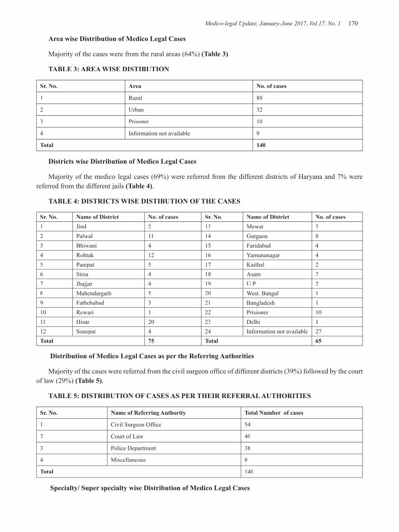
Medico-legal Update, January-June 2017, Vol.17, No. 1 170 171 -legal Update, January-June 2017, Vol.17, No. 1
Area wise Distribution of Medico Legal Cases
Majority of the cases were from the rural areas (64%) (Table 3)
TABLE 3: AREA WISE DISTIBUTION
Sr. No. Area No. of cases
1 Rural 89
2 Urban 32
3 Prisoner 10
4 Information not available 9
Total 140
Districts wise Distribution of Medico Legal Cases
Majority of the medico legal cases (69%) were referred from the different districts of Haryana and 7% were referred from the different jails (Table 4).
TABLE 4: DISTRICTS WISE DISTIBUTION OF THE CASES
Sr. No. Name of District No. of cases Sr. No. Name of District No. of cases 1 Jind 2 13 Mewat 32 Palwal 11 14 Gurgaon 83 Bhiwani 4 15 Faridabad 44 Rohtak 12 16 Yamunanagar 45 Panipat 5 17 Kaithal 26 Sirsa 4 18 Asam 27 Jhajjar 4 19 U.P 28 Mahendargarh 5 20 West. Bangal 19 Fathehabad 3 21 Bangladesh 110 Rewari 1 22 Prisioner 1011 Hisar 20 23 Delhi 112 Sonepat 4 24 Information not available 27Total 75 Total 65
Distribution of Medico Legal Cases as per the Referring Authorities
Majority of the cases were referred from the civil surgeon office of different districts (39%) followed by the court of law (29%) (Table 5).
TABLE 5: DISTRIBUTION OF CASES AS PER THEIR REFERRAL AUTHORITIES
Sr. No. Name of Referring Authority Total Number of cases
1 Civil Surgeon Office 54
2 Court of Law 40
3 Police Department 38
4 Miscellaneous 8
Total 140
Specialty/ Super specialty wise Distribution of Medico Legal Cases
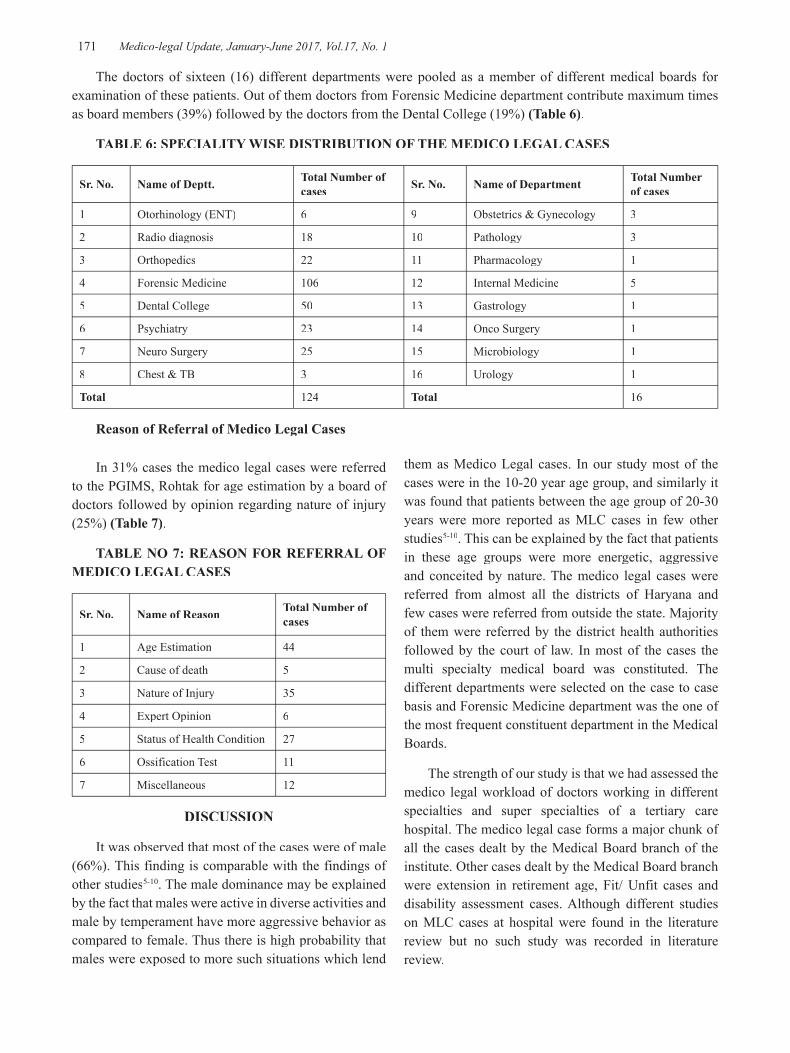
-legal Update, January-June 2017, Vol.17, No. 1 170 171 Medico-legal Update, January-June 2017, Vol.17, No. 1
The doctors of sixteen (16) different departments were pooled as a member of different medical boards for examination of these patients. Out of them doctors from Forensic Medicine department contribute maximum times as board members (39%) followed by the doctors from the Dental College (19%) (Table 6).
TABLE 6: SPECIALITY WISE DISTRIBUTION OF THE MEDICO LEGAL CASES
Sr. No. Name of Deptt. Total Number of cases Sr. No. Name of Department Total Number
of cases
1 Otorhinology (ENT) 6 9 Obstetrics & Gynecology 3
2 Radio diagnosis 18 10 Pathology 3
3 Orthopedics 22 11 Pharmacology 1
4 Forensic Medicine 106 12 Internal Medicine 5
5 Dental College 50 13 Gastrology 1
6 Psychiatry 23 14 Onco Surgery 1
7 Neuro Surgery 25 15 Microbiology 1
8 Chest & TB 3 16 Urology 1
Total 124 Total 16
Reason of Referral of Medico Legal Cases
In 31% cases the medico legal cases were referred to the PGIMS, Rohtak for age estimation by a board of doctors followed by opinion regarding nature of injury (25%) (Table 7).
TABLE NO 7: REASON FOR REFERRAL OF MEDICO LEGAL CASES
Sr. No. Name of Reason Total Number of cases
1 Age Estimation 44
2 Cause of death 5
3 Nature of Injury 35
4 Expert Opinion 6
5 Status of Health Condition 27
6 Ossification Test 11
7 Miscellaneous 12
DISCUSSION
It was observed that most of the cases were of male (66%). This finding is comparable with the findings of other studies5-10. The male dominance may be explained by the fact that males were active in diverse activities and male by temperament have more aggressive behavior as compared to female. Thus there is high probability that males were exposed to more such situations which lend
them as Medico Legal cases. In our study most of the cases were in the 10-20 year age group, and similarly it was found that patients between the age group of 20-30 years were more reported as MLC cases in few other studies5-10. This can be explained by the fact that patients in these age groups were more energetic, aggressive and conceited by nature. The medico legal cases were referred from almost all the districts of Haryana and few cases were referred from outside the state. Majority of them were referred by the district health authorities followed by the court of law. In most of the cases the multi specialty medical board was constituted. The different departments were selected on the case to case basis and Forensic Medicine department was the one of the most frequent constituent department in the Medical Boards.
The strength of our study is that we had assessed the medico legal workload of doctors working in different specialties and super specialties of a tertiary care hospital. The medico legal case forms a major chunk of all the cases dealt by the Medical Board branch of the institute. Other cases dealt by the Medical Board branch were extension in retirement age, Fit/ Unfit cases and disability assessment cases. Although different studies on MLC cases at hospital were found in the literature review but no such study was recorded in literature review.

Medico-legal Update, January-June 2017, Vol.17, No. 1 172
CONCLUSION
This study shows the volume of medico-legal cases and there pattern at the medical board branch of PGIMS, Rohtak. It was observed that majority of the patients were males and were between the age group of 10-20 years. 66% patients were referred from the rural areas of the Haryana for medical board examination. It was found that patients were received from almost all the districts of Haryana and ten patients were the prisoners from the different jails. Majority of them were referred from the civil surgeon’s office followed by the orders of court of law. In majority of cases multi specialty boards were constituted and forensic medicine department contributed maximum in the medical boards. Thus our hospital received patients from all over the Haryana and also performs the legal tasks to inspect and certify medico-legal cases. Therefore the doctors should be more trained in dealing with medico legal cases.
Conflict of Interest: None
Source of Funding: None
Ethical Clearance:- From Ethics Committee, UHS, Rohtak
REFERENCES
1. Legal Medicine Annual 1978, pp 245-255
2. Singh A.K., Singh K, Verma A. Study of Medico-legal Case Management in Tertiary Care Hospital. J Indian Acad Forensic Med. October- December 2011, Vol. 33, No. 4:337-342.
3. Agarwal SS, Kumar L, Chavali K. Legal medicine manual. New Delhi: Jaypee Brothers medical publishers (P) Ltd.; 2008. 12-3.
4. Dogra TD, Rudra A. Lyon’s Medical Jurisprudence & Toxicology. 11th Ed. Delhi Law House. 2005:367
5. Garg V, Verma SK. Profile of Medicolegal Cases at Adesh Institute of Medical Sciences and Research, Bthinda, Punjab. J Indian Acad Forensic Med. 2010; 32 (2):150-2.
6. Malik Y, Chawla R, Sharma G, Malik P, Singh R, Tripathi A. Profile of Medico-legal Cases in Casualty of a Rural Medical College of Haryana. J Indian Acad Forensic Med. 2013; 35 (4):367-8.
7. Yadav A, Singh NK. Pattern of Medico-legal Cases in rural Area of Faridabad, Haryana. J Indian Acad Forensic Med. 2013; 35 (1):60-2.
8. Hussaini SN, Kulkarni CS, Batra AK. Profile of Medico Legal Cases coming to Casualty of Government Medical College, Akola. Journal of Forensic Medicine, science and Law 2013; 22(2)
9. Benomran FA. The medico-legal science in dubai:2002-2007. Journal of Forensic and Legal Medicine 2009; 16: 332-7.
10. Gupta B, Singh S, Singh H, Sharma RK. A one Year Profile of Medico-legal Cases at Tertiary Care Hospital in Western Uttar Pradesh. MedicoLegal Update 2012; 12(2):30-5.
-legal Update, January-June 2017, Vol.17, No. 1 172
Role of Alar Index & Auricular Index in Determining Sex of Sacrum
M Mohan Reddy1, Raghavendra K M2, Prakash I Babladi3
1Assistant Professor, Forensic Medicine, Prathima Institute of Medical Sciences, Naganur, Karimnagar, 2Assistant Professor, Forensic Medicine, Sree Gokulam Medical College, Venjaramoodu,
Thiruvananthauram, 3Professor & Head, Department of Forensic Medicine, MRMCG, Gulbarga
ABSTRACT
Determination of sex from the skeletal remains is of medico-legal importance for establishing the identity of a deceased. The order of reliability of identifying the male bones in the current study is length of auricular surface and auricular index. Though almost all elements of human skeleton, show some degrees of sexual dimorphism, Sacrum shows characteristic sexual differences which have obstetric, forensic and anthropologic application. In the current study 200 sacral bones of known sex from Hyderabad Karnataka region were studied noting various parameters like width of sacrum, length of la & auricular surface of sacrum, transverse diameter of S1 deriving Alar index & auricular index. It is established that in the current study order of reliability in identifying female bone is alar index and transverse diameter of S1 & in male bones is length of auricular surface and auricular index. The most useful parameter for identifying sex in present study is length of auricular surface.
Keywords: Alar index, Auricular index, Sexual dimorphism.
Address for Correspondence:Dr. Raghavendra K M Assistant Professor in Forensic MedicineSree Gokulam Medical College, VenjaramooduThiruvananthauram -695607E mail; [email protected] : 09633295234
INTRODUCTION
Determination of sex from the skeletal remains is of medico-legal importance for establishing the identity of a deceased. The determination of sex of deceased is first step in skeletal analysis since estimation of age at death, race and stature depends on sex of the deceased. It is established that almost all elements of human skeleton, show some degrees of sexual dimorphism, but Sacrum shows characteristic sexual differences which have obstetric, forensic and anthropologic application.
It has long been customary among forensic experts, anatomists and anthropologist to judge the sex of skeletal material by nonmetric observations. Lately, sexual divergence has been based upon actual measurement in different regions. Metrical study of sacrum done by various authors like Wilder1, Faweet2, Davivongs3 and
Singh and Gangrade4 have reported that even within the same general population, mean value may be significantly different in bones from different zones. Jit and Singh5 advocated importance of the demarking point (D.P.), which identifies the sex with about 100% accuracy. The present study is an attempt to establish some of the parameters for Hyderabad – Karnataka region which will be of great help in sex determination both in medico-legal and anthropometric study.
MATERIALS AND METHOD
200 sacra of known sex (100 males & 100 females) used in this work are collected from Department of Forensic medicine & Toxicology, Department of Anatomy of Mahadevappa Rampure Medical College, Gulbarga & K.B.N. Institute of Medical Sciences, Gulbarga.
All the sacra were dry, free of damage or deformities. Sacra that are not ossified are excluded.
With the help of a stainless steel sliding caliper and flexible steel tape, the following measurements were taken (each linear measurement was recorded to the nearest millimeter).
DOI Number: 10.5958/0974-1283.2017.00037.8
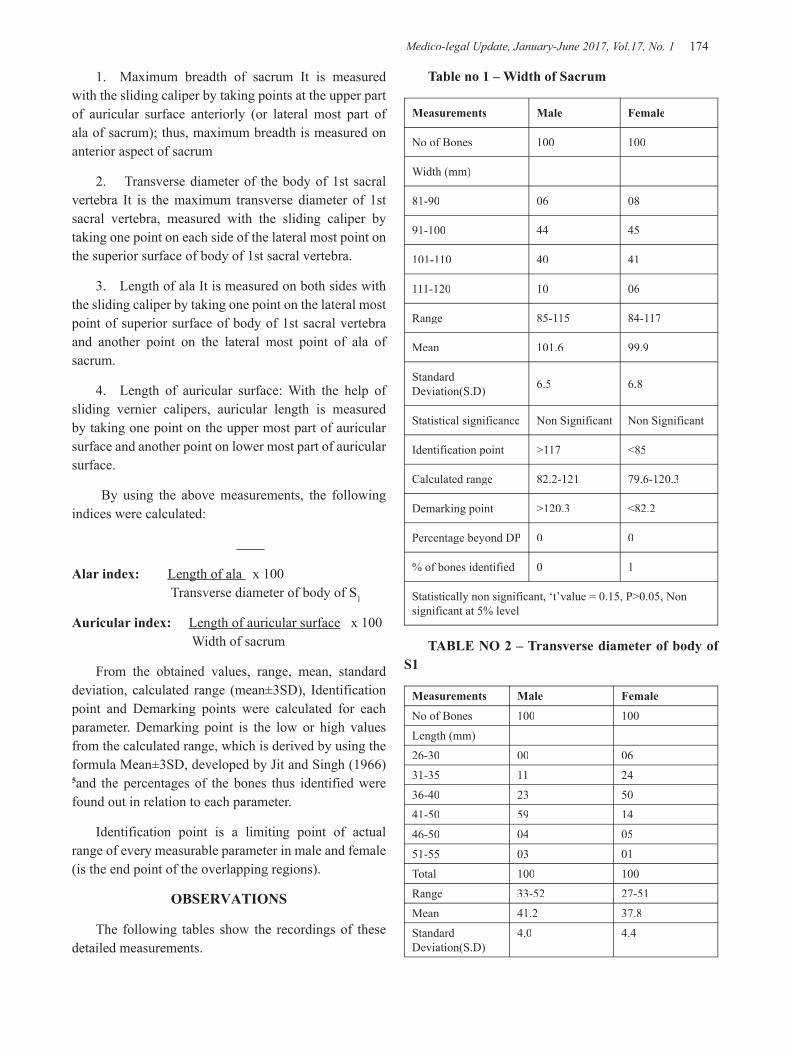
Medico-legal Update, January-June 2017, Vol.17, No. 1 174 175 -legal Update, January-June 2017, Vol.17, No. 1
1. Maximum breadth of sacrum It is measured with the sliding caliper by taking points at the upper part of auricular surface anteriorly (or lateral most part of ala of sacrum); thus, maximum breadth is measured on anterior aspect of sacrum
2. Transverse diameter of the body of 1st sacral vertebra It is the maximum transverse diameter of 1st sacral vertebra, measured with the sliding caliper by taking one point on each side of the lateral most point on the superior surface of body of 1st sacral vertebra.
3. Length of ala It is measured on both sides with the sliding caliper by taking one point on the lateral most point of superior surface of body of 1st sacral vertebra and another point on the lateral most point of ala of sacrum.
4. Length of auricular surface: With the help of sliding vernier calipers, auricular length is measured by taking one point on the upper most part of auricular surface and another point on lower most part of auricular surface.
By using the above measurements, the following indices were calculated:
Alar index: Length of ala x 100 Transverse diameter of body of S1
Auricular index: Length of auricular surface x 100 Width of sacrum
From the obtained values, range, mean, standard deviation, calculated range (mean±3SD), Identification point and Demarking points were calculated for each parameter. Demarking point is the low or high values from the calculated range, which is derived by using the formula Mean±3SD, developed by Jit and Singh (1966) 5and the percentages of the bones thus identified were found out in relation to each parameter.
Identification point is a limiting point of actual range of every measurable parameter in male and female (is the end point of the overlapping regions).
OBSERVATIONS
The following tables show the recordings of these detailed measurements.
Table no 1 – Width of Sacrum
Measurements Male Female
No of Bones 100 100
Width (mm)
81-90 06 08
91-100 44 45
101-110 40 41
111-120 10 06
Range 85-115 84-117
Mean 101.6 99.9
Standard Deviation(S.D) 6.5 6.8
Statistical significance Non Significant Non Significant
Identification point >117 <85
Calculated range 82.2-121 79.6-120.3
Demarking point >120.3 <82.2
Percentage beyond DP 0 0
% of bones identified 0 1
Statistically non significant, ‘t’value = 0.15, P>0.05, Non significant at 5% level
TABLE NO 2 – Transverse diameter of body of S1
Measurements Male Female
No of Bones 100 100Length (mm)26-30 00 0631-35 11 2436-40 23 5041-50 59 1446-50 04 0551-55 03 01Total 100 100Range 33-52 27-51Mean 41.2 37.8Standard Deviation(S.D)
4.0 4.4
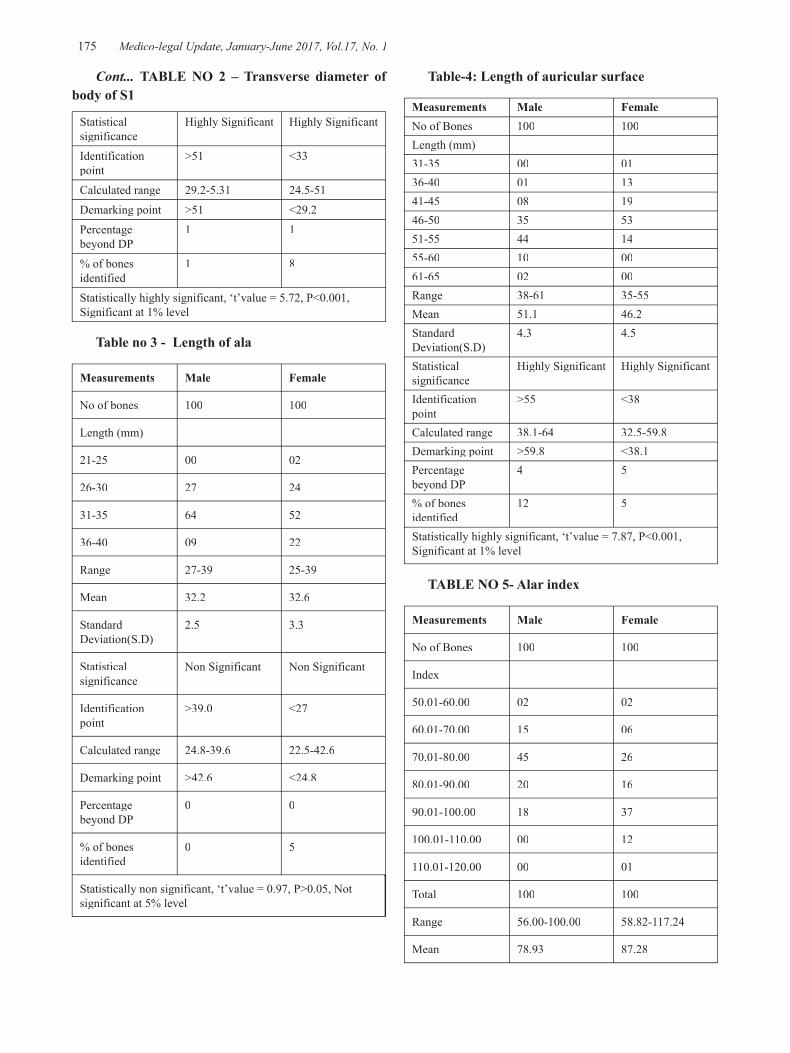
-legal Update, January-June 2017, Vol.17, No. 1 174 175 Medico-legal Update, January-June 2017, Vol.17, No. 1
Statistical significance
Highly Significant Highly Significant
Identification point
>51 <33
Calculated range 29.2-5.31 24.5-51Demarking point >51 <29.2Percentage beyond DP
1 1
% of bones identified
1 8
Statistically highly significant, ‘t’value = 5.72, P<0.001, Significant at 1% level
Table no 3 - Length of ala
Measurements Male Female
No of bones 100 100
Length (mm)
21-25 00 02
26-30 27 24
31-35 64 52
36-40 09 22
Range 27-39 25-39
Mean 32.2 32.6
Standard Deviation(S.D)
2.5 3.3
Statistical significance
Non Significant Non Significant
Identification point
>39.0 <27
Calculated range 24.8-39.6 22.5-42.6
Demarking point >42.6 <24.8
Percentage beyond DP
0 0
% of bones identified
0 5
Statistically non significant, ‘t’value = 0.97, P>0.05, Not significant at 5% level
Table-4: Length of auricular surface
Measurements Male FemaleNo of Bones 100 100Length (mm)31-35 00 0136-40 01 1341-45 08 1946-50 35 5351-55 44 1455-60 10 0061-65 02 00Range 38-61 35-55Mean 51.1 46.2Standard Deviation(S.D)
4.3 4.5
Statistical significance
Highly Significant Highly Significant
Identification point
>55 <38
Calculated range 38.1-64 32.5-59.8Demarking point >59.8 <38.1Percentage beyond DP
4 5
% of bones identified
12 5
Statistically highly significant, ‘t’value = 7.87, P<0.001, Significant at 1% level
TABLE NO 5- Alar index
Measurements Male Female
No of Bones 100 100
Index
50.01-60.00 02 02
60.01-70.00 15 06
70.01-80.00 45 26
80.01-90.00 20 16
90.01-100.00 18 37
100.01-110.00 00 12
110.01-120.00 00 01
Total 100 100
Range 56.00-100.00 58.82-117.24
Mean 78.93 87.28
Cont... TABLE NO 2 – Transverse diameter of body of S1

Medico-legal Update, January-June 2017, Vol.17, No. 1 176 177 -legal Update, January-June 2017, Vol.17, No. 1
Standard Deviation(S.D)
9.75 12.85
Statistical significance
Highly Significant Highly Significant
Identification point
<58.82 >100
Calculated range 49.67-108.18 48.73-125.82
Demarking point <48.73 >108.18
Percentage beyond DP
0 2
% of bones identified
2 13
Statistically highly significant, ‘t’value = 5.18, P<0.001, Significant at 1% level
Table No 6 – Auricular Index
Measurements Male Female
No of Bones 100 100
Index
30.01-40.00 01 08
40.01-50.00 46 79
50.01-60.00 48 13
60.01-70.00 05 00
Range 35.85-61.70 37.11-55.56
Mean 50.41 46.24
Standard Deviation(S.D) 4.81 4.21
Statistical significance
Highly Significant
Highly Significant
Identification point >55.56 <35.85
Calculated range 35.99-64.83 33.63-58.86
Demarking point >58.86 <35.99
Percentage beyond DP 5 0
% of bones identified 11 0
Statistically highly significant,‘t’value = 6.52, P<0.001, Significant at 1% level
DISCUSSION
In the discussion of sexual dimorphism of human sacra, the merits and demerits of each measurements, mean value for male and female, statistical significance were compared with other studies.
Accordingly, in the sexual dimorphism of human bones, Davivongs has stated that “as a general rule, the male bones are more massive and heavier than female bones. The crests, ridges, tuberosities and lines of muscle and ligament attachments are more strongly marked in males. This rule also governs the size of joint and articular surfaces as well”. 4
Krogman, W.M. (1949) ranked accuracy of sex determination using the pelvis at 95% followed by the skull at 92%. The mandible alone at 90% and the long bone measures at 80% accuracy 6.The value of determining the sex of a human bone is mostly medicolegal, where hundred percent accuracy is required. Jit & Singh (1966) 5 found that the maximum and minimum limits of parameter values which were determined on the basis of mean ± 3 S.D and which they have named Demarking Point (D.P), would be of great value in determining the sex of the sacrum with almost 100% accuracy .
Width of sacrumThe mean of the Sacral Width in male is 101.6 mm
and ranging between 85 to 115 mm. The mean of the Sacral Width in females is 99.9mm and range between 84-117 mms. Identification point for males is >117mm and for females <85mm. The Demarking point for males and females were >120.3mm & <82.2mm respectively. ‘t’ test was non significant with P>0.05.
Transverse diameter of S1The mean of the Transverse diameter of S1 in male is
41.2 mm and ranging between 33 to 52 mm. The mean of the Transverse diameter of S1 in females is 37.8 mm and range between 27-51 mm. Identification point for males is >51mm and for females <33mm. The Demarking point for males and females were >51mm & <29.2mm respectively. ‘t’ test was highly significant with P<001.
Length of Alae:The mean of Length of Alae in males is 32.2mm
and in females are 32.6mm. Sacra with length more than 32.2mm are definitely male and below 32.6mm are female. The sex difference between in mean values of width of sacrum is statistically non significant.
The mean of Length of Alae measurements of males and females agree nearly with the study conducted by
Cont... TABLE NO 5- Alar index
-legal Update, January-June 2017, Vol.17, No. 1 176 177 Medico-legal Update, January-June 2017, Vol.17, No. 1
Kanika et al 7 (3.35 & 3.00) and Jyothinath et al 8 (3.19 & 3.59) respectively.
Length of Auricular surface:The mean of Length of Auricular surface in males is
5.11 and in females are 4.62. The mean range for male bones is 3.80-6.10 and for female bones is 3.50-5.50. Sacra with length more than 5.11 are definitely male and below 3.80 are female. The sex difference between in mean values of width of sacrum is statistically highly significant. The mean of Length of Auricular surface measurements of males agree nearly with the study conducted by Jyothinath et al 8.
Alar Index:The mean of Alar Index in males is 78.93mm and in
females are 87.28mm. The mean range for male bones is 56.00-100mm and for female bones is 58.82-117.24mm. The sex difference between in mean values of width of sacrum is statistically highly significant. The values of mean of alar index of the current study are in agreement with that of Jyothinath et al 8 (71.31 &80.21) where as Mishra et al 9 (56.1 & 72.6) documented lower values.
Auricular Index:The mean of Auricular Index in males is 50.4mm and
in females are 46.24mm. The mean range for male bones is 35.85-61.70 and for female bones is 37.11-55.56. Sacra with index more than 55.56 are definitely male and below 35.85 are female. The sex difference between in mean values of width of sacrum is statistically highly significant. Mishra et al 9 (59.78 &51.69) and Jyothinath et al 8 (55.82 & 52.77) documented higher values for mean of auricular index in males & females in their respective studies.
SUMMARY AND CONCLUSIONSAfter a detailed study and comparison of the present
study with other studies, it can be concluded that:1 Identification point and demarking point help in
sexing of the sacrum with certainty.2 The order of reliability of identifying the male
bones in the current study is length of auricular surface and auricular index.
3 The order of reliability of identifying female bone is alar index and transverse diameter of S1.
4 The most useful parameter for identifying sex in present study is length of auricular surface.
5 However, not a single parameter could identify sex in 100% of the bones. Hence, it can be concluded that for sex determination of the sacrum with 100% accuracy is possible only when maximum number of parameters
are taken into consideration. It also substantiates observation of Singh and Gangrade (4) that sacrum or for that matter all bones show significant variations according to geographic location. Continuance of such studies in a defined geographic area over a period of time will definitely help in establishing anthropometric standards. Such studies will also be useful to observe the changing trends if any, in the metric measurements which is influenced by environmental, socioeconomic factors, physical stress and genetic factors.
Conflict of Interest : NilEthical Clearance: Taken from ethical clearance
committee, MRMCG, GulbargaSource of Funding: Self
REFERENCES1. Wilder HH. A Laboratory Manual of Anthropometry;
P. Blakistons Sons and Co. Philadelphia. 1920;193-200.
2. Faweet E. The Sexing of the Human Sacrum; Journal of Anatomy, London.72:638.
3. Davivongs, V. (1963): The pelvic girdle of the Australian Aborigine, sex differences and sex determination. American Journal of Physical Anthropology, 1963; 21(4): 443-455.
4. Singh S, Gangrade, K.C. Sexing of Adult Clavicle - Verification and Applicability ofDemarking Point; Journal of Indian Academy of Forensic Science. 1968;7:20- 30.
5. Jit I, Singh S. Sexing of the Adult Clavicle; Indian Journal of Medical Research.54:551 -571.
6. Krogman, W.M: The human skeleton in Forensic Medicine. Sexing of skeletal remains, Charles C. Thomas. Springfield, Illinois, U.S.A.: 1st edition; 1962; 112-113.
7. Kanika Sachdev, Rajan Kumar Singla, Gurudeep, Sharma: Role of sacrum in sexual dimorphism-A Morphometric Study, J Indian Acad of Forensic Med. July-September 2011; Vol. 33 (3): pp 206-210.
8. Jyothinath kothapalli, Subhadra Devi, Varalakshmi Desai, Roshan Zameer: Morphometric study of Sexual dimorphism in adult sacra of South Indian population, Int J Biol Med Res, 2012; 3 (3): pg.2076-2081.
9. Mishra S.R, Singh P.J, Agarwal A.K and Gupta R.N: Identification of sex of sacrum of Agra region: J Anat. Soc. 2003; India 52 (2): 132-136.
179 -legal Update, January-June 2017, Vol.17, No. 1
Analysis of Fatalities in Two Wheeler Road Traffic Injury and the Beneficial Effect of Implementation of Helmet Law- A
Study from, Madurai South India
T Selvaraj1, K Rajavelu2
1AssociateProfessor, Jaynagar, Ponmeni, Madurai, 2Assistant Professor, Raja Hospital, Chinnalapatti, Dindigul
ABSTRACT
According to National Crime Records Bureau, over 1,37,000 people were killed in road accidents in 2013 alone, that is more than the number of people killed in all our wars put together. Drunken driving and poor infra structures are among the leading causes of road fatalities. Among this, two wheelers account for 25% of total road crash deaths. The aim of this study is to find out the trend of mortality associated with two wheeler accident with respect to age, sex of the victim , type of injuries sustained, type of vehicle involved and time distribution, along with effect of implementation of helmet law over the mortality associated with two wheeler accidents during the period of one year from 1st January to 31st December of 2015.Main aim of this study was to establish the effect of helmet in the prevention and reduction of mortality due to head injury that occur in road traffic accidents. A number of reviews have previously been published regarding this protective effect of helmet. Madurai district is second largest in population of the 32 districts of the state of Tamil Nadu, in south-eastern India . According to 2011 census, Madurai district had a population of 3,038,252 up from 2,578,201 in the 2001 census, for a growth rate of 17.95%. It had a sex-ratio of 990 females for every 1,000 males, up from 978 in 2001, and much above the national average of 929. A total of 313,978 were under the age of six, constituting 162,517 males and 151,461 females. The average literacy of the district was 74.83%, compared to the national average of 72.99%. Total of 460 deaths occur because of two wheeler accidents. In the present study it was observed that 64% of victim used geared type vehicle. Accident rate among riders, male( 99%) was higher than that among female (1%). Accident rate among pillion riders males 80% and 20% female. These findings are very similar to those reported by other researchers. Injury concerned head injury contributes 60 % followed by multiple injuries 38 %. Most of the accident occurs in sub urban area78.4%, compared to urban area 21.6%.Most of the fatalities occur between 4pm to 11pm.Accidents are highest in the month of June followed by September and least in December followed by February.
Keywords: Helmet, collision, two wheeler, geared, non geared, traffic injury, mortality, road safety and traffic rules.
INTRODUCTION
Driving is a highly complex mechanism involving a wide range of cognitive, perceptual and motor activities. This study was undertaken to find the trend of mortality associated with two wheeler accident with respect to age, sex of the victim , type of injuries sustained, type of vehicle involved and time distribution ,along with effect of implementation of helmet law over the mortality associated with two wheeler accidents, during the period of one year from 1st January to 31st December of 2015.Main aim of this study was to establish the effect
of helmet in the prevention and reduction of mortality due to head injury that occur in road traffic accidents. A number of reviews have previously been published regarding this protective effect of helmet. Crash helmet reduces friction of the head against the ground and makes deceleration less drastic by allowing the protected head to skid across the ground.
Motor cyclist are 20 to 30 times more likely to be severely injured or die as a result of accident than are motor vehicle occupants. Statistics from U.S. which support our study indicate that most motor cycle incidents tends to occur in the late afternoon and
DOI Number: 10.5958/0974-1283.2017.00038.X
179 Medico-legal Update, January-June 2017, Vol.17, No. 1
early evening and fatal crashes within the evening and early morning. Accidents are uniform in number from Monday to Thursday, increase on Friday and peak on Saturday then decrease slightly on Sunday1 which is in contrary to our study.
As per the report published by the State transport authority in 2013, out of 66,238 accidents, two-wheelers were involved in 22,496 accidents, cars, jeeps, taxis and tempos in 18,658, trucks in 9,192, government buses in 3,765, private buses in 3,564, three-wheelers in 2,983 and others in 5,580. As per the same report, 20,686 accidents occurred in national highways, 20,984 in state highways, 17,401 in district roads and remaining 7,167 in village roads.
Urban ring road system and four way system in national highway have been rapidly developed in recent years. It has become one key part of the city roadways networks, as it carrying large traffic volumes and providing high travelling speed. Along with increase in traffic volume, traffic safety has become a major issue because of crash and the non recurrent congestion caused by them.
We think that this study helps to unveil the accident mechanism and further developing the active traffic management control strategies to improve traffic safety. 25th August 2006 /GENEVA-declares that each year about one million people die as a result of road traffic accident and millions more are injured or disabled. Most of the deaths are preventable. In many low income and middle income countries, user of two wheelers particularly motor cyclists make up more than 50% of those injured or killed on the roads.
Lethal injuries usually involve severe head injury and cervical spine fracture dislocation. Non lethal injuries involving bone, joint and soft tissue injuries to the lower limbs are the most common injuries sustained by motor cycle riders2. According to the investigation done by the major collision investigation group in Victoria, Australia examined 47 collision involving severe injuries and fatalities to motor cycle riders- 44 of the riders were male, age ranged from 17 to 56 years with a mean of 32 years which support this study. Examination showed excessive speed contributes 38% of the crashes3.In the above study indicates that unsafe practice by motor cycle riders were implicated in 76% of spot death. In other studies from Australia determine
motor cyclists exhibited different behaviour while riding a motor cycle when compared with other vehicle4.
Head injuries are main cause of death and disability, among motorcycle users and the cost of head injuries are high, because they frequently require specialized medical care or long term rehabilitation. Wearing helmet is the single most effective way of reducing head injuries and fatalities resulting from motor cycle and bicycle crashes. Wearing helmet reduces the risk and severity presented by previous data,continuous to prove that motor cycle helmet laws save lives.
In Tamilnadu helmet mandatory for two wheeler for both the riders from July-1-2015, under section 129 of Motor Vehicle Act 1988. Violators facing the risk of their vehicle documents being impounded. It was really disheartening to note that the number of lives were lost due to non wearing of helmet and the pathetic position was, inspite of judgement given, the authorities acted as per the statue nor followed the direction the judge noted. It is also noted that 6419 person have lost their lives in 2014 due to nonwearing of helmets.
METHOD
This study was conducted in the southern city of Tamilnadu Madurai, with a population of 3,038,252. The data for our study was collected retrospectively from three sources, they are records maintained at the office of super intendent of police, traffic wing and records maintained at forensic department of government madurai medical college.
Data regarding age and gender of the victims, time and place of accident, type of vehicle whether it is geared or un-geared involved in the accident, type of injuries, type of road users and outcome of the accident. Here the geared vehicles are motor cycle and scooters, whereas un-geared ones included mopeds, scootereffes and scooters with variomatic transmission.
Data collected was tabulated and entered into the computer using Microsoft excel package for further analysis. It was analysed manually and as well as using SPSS package version 10.
OBSERVATION AND RESULTS
Total of 460 deaths occur because of two wheeler accidents. In the present study it was observed that 64% of victim used geared vehicle (FIG-1) Accident rate

Medico-legal Update, January-June 2017, Vol.17, No. 1 180 181 -legal Update, January-June 2017, Vol.17, No. 1
among riders, male ( 99%) was higher than that among female (1%). Accident rate among pillion riders males 80% and 20% female. These findings are very similar to those reported by other researchers. Most of the accident occurs in sub urban area78.4% , compared to urban area 21.6% (FIG-3). Regarding the age of the victim, 47% of them are between 21 to 40 years and 35% between 41 to 60 years.
Total number of two wheeler mortality is 460.Among this total number of death of rider male is 342 , rider female is 0. Death of pillion rider male is 96 and female is 37. According to this study we come to an idea that mortality of female using the two wheeler as a rider is nil and as a pillion passenger is 8%..Mortality is highest in the month of May and June, and lowest in December and January. But the number of accident concerned, it is more in June and September and least in November and February (FIG-4). Weekday concerned there is no great variation and regarding time of occurrence most of the accidents (72%) occur in the afternoon particularly between 4pm to 11pm (FIG-2). Regarding implementation of helmet law – mortality due to head injury is reduced after implementation compared with before implementation.
Pattern of two wheeler accidents in Madurai are depicted in table. Rider was affected in maximum number of accidents, pillion and pedestrian also succumbed but to a lesser extent. The type of two wheeler mainly involved in accident was the geared vehicle. Number of such vehicle being64 % compared to 36% non gearing vehicle.
A significantly high number of males were involved than female. Data regarding age indicates that most commonly involved age groups being 20 to 40 years, followed by 41 to 60 and above 60 year. In this study nearly 72% of the two wheeler accidents were reputed between 4pm to 11pm. The year was divided into 3 season summer, rainy and winter. A large number of accidents took place during summer. This study revealed that type of injury that contribute for death was as follows- head injury 60%, multiple injury 38% followed by spinal injury 1.3%, abdominal injury 0.6% and 0.1% lower limb injury.
DISCUSSION
An accident also known as an unintentional injury, incidental and unplanned event that could have been
prevented if the circumstances leading up to the accident been recognized and acted upon, prior to its occurrence.
A study about two wheeler accidents from Germany found that 90.7% were male , age of 28.8 years, helmet were worn in 98.8% , 2.3% died of severe head injury.. Most common injuries in survivors- lower extremity injuries in 46% ,in particular compound tibial fracture 19.7%, fracture distal radius 18.8% of the cases5. Motor cycle riders who die as a consequence of a collision most commonly suffer significant head, neck and chest injuries. Severe brain injury and fracture dislocation of the upper cervical spine commonly results in spot death. The thoracic spine is the region most commonly injured, with multiple spinal injuries not uncommon5. But in our study we are unable to get the complete details that whether the victim worn the helmet at the time of accident in all the cases.
In the Yorkshire district of the U.K., approximately 4% of injured motorcycle riders were pillion passengers.6. Inguinal creases abrasion or lacerations caused from impact with the fuel tank is the characteristic injury which is useful in forensic investigation to find out the rider in two wheeler accident7. But in our study we mention the rider through the history obtained by the investigating officer, Motor cyclists are 20 to 30 times more likely to be severely injured or die as a result of crash than motor vehicle occupants8. In a comparison study motor cyclists were found to travel at higher speeds than the matched group of car drivers, although this may have reflected the spatial and performance characteristics of motor cycle in general9 .
The helmet provides protection from direct blunt impact to the head; however an important issue is whether the helmet provides protection from rotational head injury. Individual who do not wear a helmet suffer more significant head and neck injuries causing death and prolonged hospitalisation10. Previous studies from Arkansas reveal that repeal of the mandatory helmet law was associated with an increase in the non helmeted crash fatality rate and disproportionately higher hospital admission rate10. Further studies from U.S.A. that compares the data, indicates that states with implementation of helmet law show the lesser fatality rate than those without implementation11 which supports our study.
Role of helmet in the prevention of head injury depends upon the quality and type of the particular
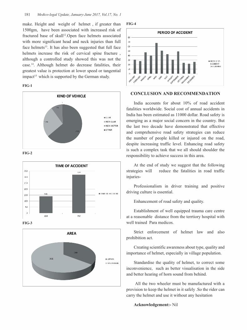
-legal Update, January-June 2017, Vol.17, No. 1 180 181 Medico-legal Update, January-June 2017, Vol.17, No. 1
make. Height and weight of helmet , if greater than 1500gm, have been associated with increased risk of fractured base of skull12.Open face helmets associated with more significant head and neck injuries than full face helmets13. It has also been suggested that full face helmets increase the risk of cervical spine fracture , although a controlled study showed this was not the case.14. Although helmet do decrease fatalities, their greatest value is protection at lower speed or tangential impact15 which is supported by the German study.
FIG-1
FIG-2
FIG-3
FIG-4
CONCLUSION AND RECOMMENDATION
India accounts for about 10% of road accident fatalities worldwide. Social cost of annual accidents in India has been estimated as 11000 dollar. Road safety is emerging as a major social concern in the country. But the last two decade have demonstrated that effective and comprehensive road safety strategies can reduce the number of people killed or injured on the road, despite increasing traffic level. Enhancing road safety is such a complex task that we all should shoulder the responsibility to achieve success in this area.
At the end of study we suggest that the following strategies will reduce the fatalities in road traffic injuries-
Professionalism in driver training and positive driving culture is essential.
Enhancement of road safety and quality.
Establishment of well equipped trauma care centre at a reasonable distance from the terrtiory hospital with well trained Para medicos.
Strict enforcement of helmet law and also prohibition act.
Creating scientific awareness about type, quality and importance of helmet, especially in village population.
Standardise the quality of helmet, to correct some inconvenience, such as better visualisation in the side and better hearing of horn sound from behind.
All the two wheeler must be manufactured with a provision to keep the helmet in it safely .So the rider can carry the helmet and use it without any hesitation
Acknowledgement:- Nil

Medico-legal Update, January-June 2017, Vol.17, No. 1 182
Conflict of Interest:- We declare that we have no conflict of interest and no source of support in the form of grants.
Source of Funding: - Self.
Ethical Clearance: - Sir, as it is a retrospective case study with department register and case sheet, we did not get the ethical clearance.
REFERENCES
1. Michael P. Burke –Forensic medical investigation of motor vehicle incidents; CRC press Tailor & Francis group : London : 2007 : page 102-104.
2. Michael P. Burke –Forensic medical investigation of motor vehicle incidents; CRC press Tailor & Francis group : London : 2007 : page 102-104.
3. Michael P. Burke –Forensic medical investigation of motor vehicle incidents; CRC press Tailor & Francis group : London : 2007 : page 104.
4. Michael P. Burke –Forensic medical investigation of motor vehicle incidents; CRC press Tailor & Francis group : London : 2007 : page 105.
5. Wick.M. muller.E.J, Ekkernkamp.A. : motorcyclist,easy rider (or) easy victim-An analysis of motor cycle Accidents in Germany. American Journal of Emergency Medicine 1998: 16: 20; 323.
6. Robertson.A , Giannoudis PV , Branfoot.T. , Barlow.I. ,Matthews.SJ ,Smith.RM. : spinal injuries in motor cycle crashes; pattern and outcomes . The Journal Of Trauma : 2002 : 53 ; page 5-8.
7. Shiono.H , Akane.A. , Matsubara.K. , Tanabe.K. , Takahashi.S. : identification of Driver in two rider motor cycle accidents. The American Journal of Forensic Medicine and Pathology. 1990 ; 11; 190-
192.
8. Mcgwin.G. Jr. Whatley.J. Metzger.J. , Valent.F. Barbone.f. , Ru LW III. : The effect of state motor cycle licensing laws on motor cycle drive mortality rates .The Journal of Trauma; 2004 :56: 415-419.
9. Horswill.M.S, Helman.S. , A behavioural comparision between motor cyclists and a matched group of non-motorcycling car drivers : Factors influencing accident risk. Accident analysis and prevention 2003 ; 35 ; 589-597.
10. Bledsoe GH., Schexnayder SM., Carey.MJ., Dobbins W.N., Gibson.WD., Hindman. JW., Collins.T., Wallace.BM., Cone.JB. , Ferrer. TJ. The negative impact of the repeal of the Arkansa motor cycle helmet law. The Journal of Trauma. 2002 ; 53 ; 1078-1087.
11. Branas.CC., Knudson.MM., Helmet laws and Motor cycle death rates. Accident analysis and prevention. 2001 ; 33 ; page 641-648.
12. Ritchter.M., offe.D., Lehmann.U., Chinn.B., Schuller.E, Doyle.D., Sturrock.K., Krateek.C. Head Injury mechanisms in Helmet protected motor cyclists; Prospective multicenter study. The Journal of Trauma. 2001: 51 ; 949-958.
13. Hitosugi.M. , shigeta.A., Takatsu.A., Yokoyama.T., Tokudome. S; Analysis of fatal injuries to motorcyclists by helmet type. The American Journal Of Forensic Medicine and Pathology; 2004; 25 ; page 125-128.
14. Oconnor.PJ. , Kloeden.C., Mclean.AJ., do full face helmets offer greater protection against cervical spinal cord injury than open faced helmets ? Traffic Injury Protection. 2002 ; 3 ; 247-250.
15. Dr.K.S.Narayan reddy and DR.O.P.Murty., The Essentials Of Forensic Medicine and Toxicology ; 2014: 33 ; 283-284.).

-legal Update, January-June 2017, Vol.17, No. 1 182
Drawing New Formulaes for Dental Age Estimation: An Attempt at Extending Kvaal’s Technique
Shruti Sinha1, Akanksha Srivastava2, Sunira Chandra3
1Senior Lecturer, 2MDS 3rd Year, 3Reader, Department of Oral Medicine and Radiology, Saraswati Dental College & Hospital, Lucknow, UP
ABSTRACT
Various methods like Gustafson’s method, Demirjian’s method, Nolla’s method, Kvaal’s method and amino acid racemisation have been used to estimate age. Present study aims to determine regression formulae and to check its accuracy in calculating age using Kvaals’s method on OPGs for a specific population and to check the applicability and reliability of the derived formulaes on our specified population. Sixty subjects aged between 20 to 60 years, were randomly selected and digital OPGs were taken using standard procedures. Measurements were recorded from which linear regression formulas were obtained and age was calculated. Statistically, the coefficient of determination (R²) was strongest when the ratios for all 6 teeth were included (0.244) and weakest when mandibular first premolar used alone (0.069). Among the mandibular and maxillary teeth, maxillary teeth showed better coefficient of determination (R² = 0.181) The derived formulas when applied to a 15 randomly selected subject’s IOPARs gave difference between calculated and chronological age within the acceptable limits. Thus, it proved the feasibility of Kvaal’s method of age estimation in adults using orthopantomograph in the selected population.
Keywords: Age estimation, Kvaal’s technique, Forensic Dentistry.
INTRODUCTION
One of the major problems faced by mankind is to establish the correct identity of an individual.1 Estimation of age is an important aspect in personal identification as it helps in attainment of various personal, social, civil and legal rights such as settlement of property between families, in facilitating remarriage, insurance claims and allowance of cremation or burial of the desceased.2
Age can be determined with the help of various long bones, skull bones and teeth.3 Teeth have been used successfully for determining the age of the individual.2
Teeth are considered better indicators of age than bones because they are the hardest structure present in the human body which can sustain trauma in cases of causalities and can be preserved long after every other tissue of the body gets disintegrated.2 Teeth continue to demonstrate changes even after attainment of maturity or death. 4 Most of the methods for dental age estimation are usually either destructive in nature or require time consuming procedures like tooth extraction and preparation of histological sections. Radiographs are simple and non-destructive tools for age estimation.4
They are reproducible and give a reliable results.5
Secondary dentine deposition gradually increases with the advancing age of the individual and can be visualised easily on the radiographs. Further, the dimensions of the pulp can be correlated with the chronological age of the individual.4
In 1994, Kvaal realised that changes seen in pulpal dimensions on extracted teeth could be used to assess the age and devised linear regression formulas for adult. In 1995 Kvaal used his own derived regression formulaes for age estimation using intraoral periapical radiographs.4
Later in 2005, Bosman et al used OPG on the Belgian population to perform the radiographic analysis and thus both concluded that there were no significant differences between estimated and chronological age when the original technique proposed by Kvaal was applied. 6
However, regression formulaes proposed by Kvaal et al when applied to Indian population were not found applicable due to environmental and genetic variation. Thus, this study was performed with an aim to derive linear regression formulaes for age estimation and to determine their accuracy on digital OPGs in our
DOI Number: 10.5958/0974-1283.2017.00040.8

Medico-legal Update, January-June 2017, Vol.17, No. 1 184 185 -legal Update, January-June 2017, Vol.17, No. 1
population (Lucknow).
MATERIALS AND METHOD
The study sample was chosen from the patients visiting a dental college in Northern India for routine dental examination and treatment. A total of 60 subjects, both males and females were selected having age range from 20 to 60 years. The ethical clearance and a written consent in patient’s own language were taken for every subject.
Inclusion criteria consisted of having healthy 6 single rooted teeth from both the jaws. Subjects having any developmental or morphological abnormalities were excluded from the study.
For each subject digital OPG using KODAK 8000C Digital Panoramic & Cephalometric system operated at 70 kVp and 6.3 mA were recorded under adequate protective measures. (Figure 1)
The Six teeth were selected on each radiograph for analyses were Maxillary Central Incisor, Lateral Incisor, Second Premolar, Mandibular canine, First Premolar and Lateral Incisor. (Figure 2)
• For each tooth, linear measurements were made with the help of Kodak Digital Imaging Software version 6.12.32.0 (Figure 3):
• Maximum tooth length (T)
• Pulp length (P)
• Root length on the mesial surface (R)
• Root width at CEJ i.e. at level A (ARW)
• Pulp width at CEJ i.e. at level A (APW)
• Root width midway between A and C i.e. level B (BRW)
• pulpal width midway between A and C i.e. level B (BPW)
• Root width midway between CEJ and apex i.e. at level C (CRW)
• Pulp width midway between apex and CEJ i.e. at level C (CPW)
The measurements were followed by calculating ratios in order to compensate for possible magnification and angulation errors which are inherent in various radiographic techniques:
• Ratio between length of pulp and root (RP)
• Ratio between length of pulp and tooth (RR)
• Ratio between length of tooth and root (RT)
• Ratio between width of pulp and root at enamel–cementum junction (RA)
• Ratio between width of pulp and root at midpoint between level C and A (RB)
• Ratio between width of pulp and root at mid root level (RC)
• Mean value of all ratios (M)
• Mean value of width ratios from levels B and C (W)
• Mean value of the length ratios P and R (L)
• Difference between W and L (W-L)
STATISTICAL ANALYSIS
The data was subjected to statistical evaluation using SPSS version 20 and 7 linear regression formulae were obtained for individual teeth, 3 maxillary teeth, 3mandibular teeth, and all 6 teeth together. The correlation coefficient (p) was calculated. A paired t test and the correlation coefficient were used to investigate inter- and intra-observer variations. Thereafter regression analysis was made employing age as a dependent variable and gender as an independent variable. A prediction which included measurement of teeth from both the jaws were calculated, as were also separate predictions restricted exclusively to either maxillary or mandibular teeth. Separate predictions were also calculated for each of the six type of teeth included in the study. For each of the derived formula, Pearson correlation (P) and Standard Deviation (SD) were estimated in order to check the accuracy of our derived formulaes.
RESULTS
The correlation coefficient between the age and the calculated ratio are shown in table 1.
The result indicated that for both maxillary and mandibular lateral incisors the calculated ratios were either weakly or not significantly correlated with age. For maxillary central incisor the ratio between length of pulp and root(RP) was most strongly correlated with the age, for maxillary second premolar ratio between length of pulp and root(RP) and ratio between width of pulp and root at CEJ (RA) showed the maximum correlation with age, for mandibular canine strongest correlation was between ratio of length of tooth to root(RT) and age and
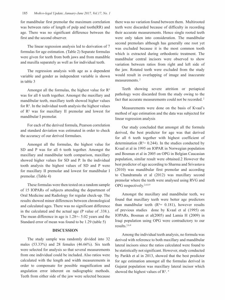
-legal Update, January-June 2017, Vol.17, No. 1 184 185 Medico-legal Update, January-June 2017, Vol.17, No. 1
for mandibular first premolar the maximum correlation was between ratio of length of pulp and tooth(RR) and age. There was no significant difference between the first and the second observer.
The linear regression analysis led to derivation of 7 formulas for age estimation. (Table 2) Separate formulas were given for teeth from both jaws and from mandible and maxilla separately as well as for individual teeth.
The regression analysis with age as a dependent variable and gender as independent variable is shown in table 3
Amongst all the formulas, the highest value for R² was for all 6 teeth together. Amongst the maxillary and mandibular teeth, maxillary teeth showed higher values for R². In the individual tooth analysis the highest values of R² was for maxillary II premolar and lowest for mandibular I premolar.
For each of the derived formula, Pearson correlation and standard deviation was estimated in order to check the accuracy of our derived formulaes.
Amongst all the formulas, the highest value for SD and P was for all 6 teeth together. Amongst the maxillary and mandibular teeth comparison, maxillary showed higher values for SD and P. In the individual tooth analysis the highest values of SD and P were for maxillary II premolar and lowest for mandibular I premolar. (Table 4)
These formulas were then tested on a random sample of 15 IOPARs of subjects attending the department of Oral Medicine and Radiology for regular check-up. The results showed minor differences between chronological and calculated ages. There was no significant difference in the calculated and the actual age (P value of .338.). The mean difference in age is 1.28+- 5.02 years and the Standard error of mean was found to be 1.29 (table 5)
DISCUSSION
The study sample was randomly divided into 32 males (53.33%) and 28 females (46.66%). Six teeth were selected for analysis so that several measurements from one individual could be included. Also ratios were calculated with the length and width measurements in order to compensate for possible magnification and angulation error inherent on radiographic methods. Teeth from either side of the jaw were selected because
there was no variation found between them. Multirooted teeth were discarded because of difficulty in recording their accurate measurements. Hence single rooted teeth were only taken into consideration. The mandibular second premolars although has generally one root yet was excluded because it is the most common tooth which is extracted during orthodontic treatment. The mandibular central incisors were observed to show variation between ratios from right and left side of the jaw. Rotated teeth were excluded from the study would result in overlapping of image and inaccurate measurements. 2
Teeth showing severe attrition or periapical pathology were discarded from the study owing to the fact that accurate measurements could not be recorded. 2
Measurements were done on the basis of Kvaal’s method of age estimation and the data was subjected for linear regression analysis
Our study concluded that amongst all the formula derived, the best predictor for age was that derived for all 6 teeth together with highest coefficient of determination (R²= 0.244). In the studies conducted by Kvaal et al in 1995 on IOPAR in Norwegian population and Bosman et al in 2005 on OPG in Belgian Caucasian population, similar result were obtained.2 However the best predictor of age according to Sharma and Srivastava (2010) was mandibular first premolar and according to Chandramala et al (2012) was maxillary second premolar where the teeth were analysed using RVG and OPG respectively.2,6,8,9
Amongst the maxillary and mandibular teeth, we found that maxillary teeth were better age predictors than mandibular teeth (R²= 0.181), however results of previous studies done by Kvaal et al (1995) on IOPARs, Bosman et al(2005) and Lamia H (2009) in Iraqi population using OPG were contradictory to our results.2,6,4
Among the individual teeth analysis, no formula was derived with reference to both maxillary and mandibular lateral incisors since the ratios calculated were found to be statistically not significant. However, study conducted by Parikh et al in 2013, showed that the best predictor for age estimation amongst all the formulas derived in Gujarat population was maxillary lateral incisor which showed the highest values of R². 4
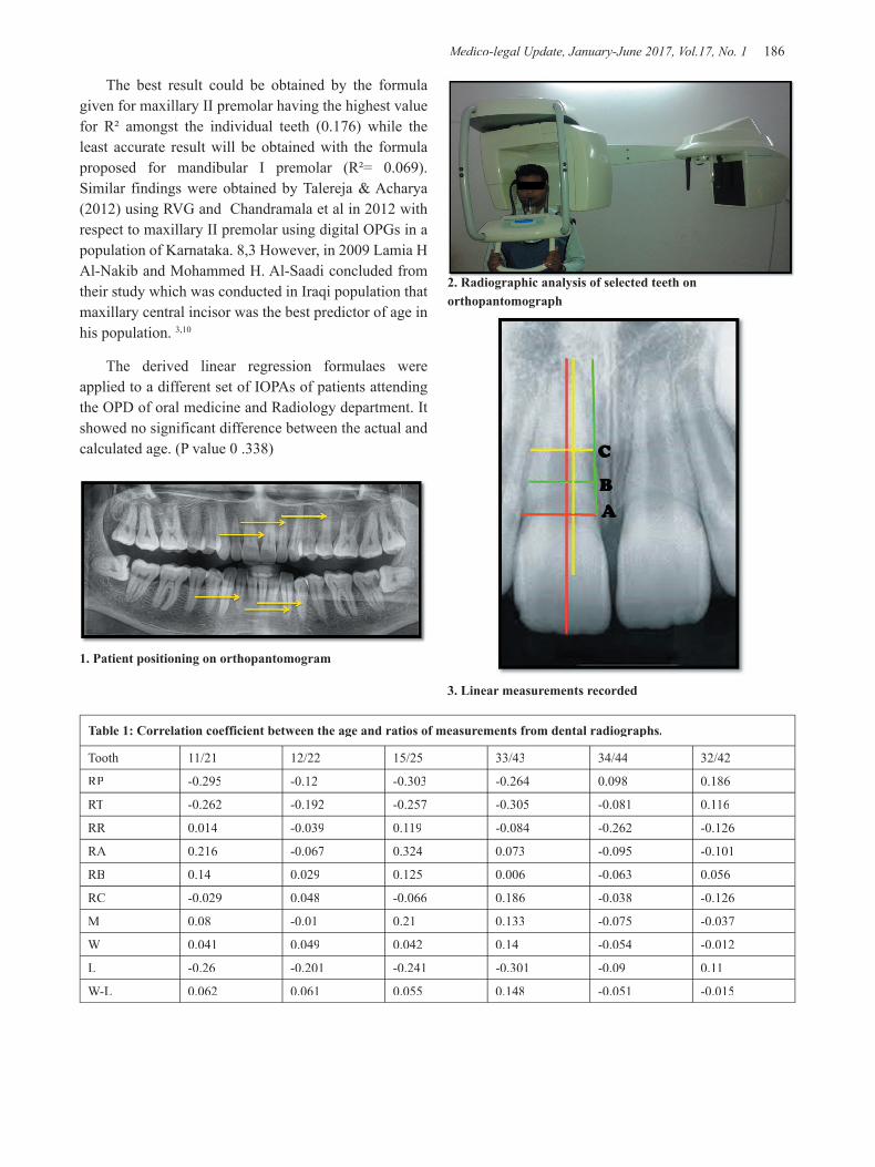
Medico-legal Update, January-June 2017, Vol.17, No. 1 186 187 -legal Update, January-June 2017, Vol.17, No. 1
2. Radiographic analysis of selected teeth on orthopantomograph
3. Linear measurements recorded
The best result could be obtained by the formula given for maxillary II premolar having the highest value for R² amongst the individual teeth (0.176) while the least accurate result will be obtained with the formula proposed for mandibular I premolar (R²= 0.069). Similar findings were obtained by Talereja & Acharya (2012) using RVG and Chandramala et al in 2012 with respect to maxillary II premolar using digital OPGs in a population of Karnataka. 8,3 However, in 2009 Lamia H Al-Nakib and Mohammed H. Al-Saadi concluded from their study which was conducted in Iraqi population that maxillary central incisor was the best predictor of age in his population. 3,10
The derived linear regression formulaes were applied to a different set of IOPAs of patients attending the OPD of oral medicine and Radiology department. It showed no significant difference between the actual and calculated age. (P value 0 .338)
1. Patient positioning on orthopantomogram
Table 1: Correlation coefficient between the age and ratios of measurements from dental radiographs.
Tooth 11/21 12/22 15/25 33/43 34/44 32/42
RP -0.295 -0.12 -0.303 -0.264 0.098 0.186
RT -0.262 -0.192 -0.257 -0.305 -0.081 0.116
RR 0.014 -0.039 0.119 -0.084 -0.262 -0.126
RA 0.216 -0.067 0.324 0.073 -0.095 -0.101
RB 0.14 0.029 0.125 0.006 -0.063 0.056
RC -0.029 0.048 -0.066 0.186 -0.038 -0.126
M 0.08 -0.01 0.21 0.133 -0.075 -0.037
W 0.041 0.049 0.042 0.14 -0.054 -0.012
L -0.26 -0.201 -0.241 -0.301 -0.09 0.11
W-L 0.062 0.061 0.055 0.148 -0.051 -0.015

-legal Update, January-June 2017, Vol.17, No. 1 186 187 Medico-legal Update, January-June 2017, Vol.17, No. 1
Table 2: Regression formulae for age estimation in years based on radiographic analysis of 6 selected teeth.
Maxillary Central Incisor AGE=63.827-24.753*RP1
Maxillary 2nd Pre Molar nd Pre Molar nd AGE = 55.828+1.460*RA3- 4.435*RP3
Mandibular Canine AGE = 74.699-29.030*RT4
Mandibular 1st Pre Molarst Pre Molarst AGE=82.203-38.488*RR5
3 maxillary teeth AGE = 55.56 + 1.53 *RA3 -23.152*RP1
3 mandibular teeth AGE = 74.699-29.03 *RT4
All 6 teeth together AGE=107.606+1.515*RA3-5.637*RT4-5.489*RR5
Table 3: Calculated coefficient of determination (R²) and standard error (SEE) of estimate (years) in the study
(R²) (SEE)
Formula 1 (All 6 Teeth) 0.244 8.0452997
Formula 2 (3 Maxillary teeth) 0.181 8.2987252
Formula 3 (3 Mandibular teeth ) 0.093 8.6582124
Formula 4 (Maxillary Central incisor) 0.087 8.6853607
Formula 5 (Maxillary 2nd Pre Molar) nd Pre Molar) nd 0.176 8.3221949
Formula 6 (Mandibular Canine) 0.093 8.6582124
Formula 7 (Mandibular 1st Pre Molar) 0.069 8.7731156
Table 4: Calculated Standard Deviation (SD) and Pearson Correlation (R)
(SD) (R)
Formula 1 (All 6 Teeth) 4.44920 0.494
Formula 2 (3 Maxillary teeth) 3.83399 0.425
Formula 3 (3 Mandibular teeth ) 2.74596 0.305
Formula 4 (Maxillary Central incisor) 2.66036 0.295
Formula 5 (Maxillary 2nd Pre Molar) nd Pre Molar) nd 3.78451 0.420
Formula 6 (Mandibular Canine) 2.74596 0.305
Formula 7 (Mandibular 1st Pre Molar) 2.36037 0.262
Table 5: Paired T Test between the actual and estimated age of randomly selected IOPARs (n=15) based on formula derived from our study.
Paired Differences
t df Sig. (2-tailed)
Mean Std. Deviation
Std. Error Mean
Pair 1 Actual age (in yrs) - Calculated age (in yrs) -1.2886667 5.0297442 1.2986744 -0.992 14 0.338
CONCLUSION
Thus for dental age estimation, a number of methods are available for use depending on the needs of the odontologist and the specific case. Each method
is blessed with few advantages and a few drawbacks as well. 11 So it’s important to remember not to restrict ourselves to only one method but apply different methods and perform repetitive measurements and calculations to
Medico-legal Update, January-June 2017, Vol.17, No. 1 188
establish the maximum reproducibility of the result.
Hereby, we conclude, that these formulas based on Kvaal’s method presented over here, for estimation of chronological age in adults of our population gives inference for the feasibility of this technique in the Indian population. Thus it would be interesting to find out whether these formulas can be applied to estimate age in further studies in people of Indian origin.
Conflicts of Interest: Nil
Source of Funding: Self funded
Ethical Clearance: Taken from the institutional ethical committee
REFERENCES
1. Misra N, Umapati D, Misra P. Chelioscopy for sex determination. Medicolegal update 2014; 14(2):77-82.
2. Kvaal S I, Kottveit K M, Thomson I B O, Solheim T. Age estimation of adults from dental radiographs. Forensic Sci Int 1995; 74:175-185.
3. Talreja et al. An assessment of the versatility of Kvaal’s mehod of adult dental age estimation in Indians. Archives of oral biology 2012; 57:277-284.
4. Parikh N et al. Application of Kvaal’s dental age estimation technique on Orthopantomographs on
A Population of Gujarat – A Short Study. BUJOD 2013; 3(3):18-24.
5. Singaraju S, Sharda P. Age estimation using pulp/tooth area ratio: A digital image analysis. J Forensic Dent Sci 2009; 1(1):37-41.
6. Bosmans N et al. the application of Kvaal’s dental age calculation technique on panoramic dental radiographs. Forensic Sci Int 2005; 153:208-212.
7. Sinha S, Umapati D. Dental Age Estimation. Germany: Lambert Academic Publishing. 2013.
8. Chandramala et al. application of Kvaal’s technique of age estimation on digital radiographs. Dentistry 2012;2(6)
9. Sharma R, Srivastava A. Radiographic evaluation of dental age of adults using Kvaal’s method. J Forensic Dent Sci 2010; 2(1):22-26.
10. Lamia H, Mohd H. Chronological age estimation by radiological measurements on digital panoramic image among Iraqi sample (Forensic odontology study). J Bagh College Dentistry 2009:21(4):75-79.
11. Bagherpour A, Imanimoghaddam M, Bagherpour MR,. Einolghozati M Dental age assessment among Iranian children aged 6–13 years using the Demirjian method. Forensic Sci Int 2010; 197:121 e1- 121 e4.

-legal Update, January-June 2017, Vol.17, No. 1 188
Analysis of Mandibular Fractures in Road Traffic Accident Cases- A Retrospective Autopsy Study
Vijay Kumar AG1, Hemanth Raj M N2
1Assistant Professor, Department of Forensic Medicine & Toxicology, Adichunchanagiri Institute of Medical Sciences, B G Nagara, Nagamangala Taluk, Mandya, Karnataka State, India, 2Assistant
Professor, Department of Forensic Medicine & Toxicology, Shridevi Institute of Medical Sciences & Research Hospital, NH-4, Sira Road, Tumkur District, Karnataka State, India
ABSTRACT
Mandibular fractures are discontinuation of the mandibular bone. They usually occur during road traffic accident. In this study, all fatal road traffic accident cases autopsied during the period January 2011 to December 2014 were analyzed at the Department of Forensic Medicine & Toxicology, AIMS, BG Nagar, and Karnataka. The incidence, age and sex wise distribution of cases and site of MANDIBULAR fractures were noted. In our study total numbers of autopsied cases during 2011 to 2014 are 453. In that 21 cases had MANDIBULAR fracture. Maximum number of victims belongs to 21-30 years (08 cases) decade followed by 31-40 years (06 cases). Fracture of body of mandible accounts for maximum number of cases (09 cases) followed by Para-symphyseal region (04 cases). In six cases fracture were seen at multiple sites. X-rays and CT scan report were referred in most of the cases to confirm the fracture. In India, Mandible fracture is mainly due to road traffic accidents. Since most of the drivers are male, so maximum numbers of victims are also male. Not using the helmet and seat belt, are also important reason for facial injuries in all road traffic accident cases leading to permanent disfigurement of face.
Keywords: Mandible, fractures, road traffic accident cases, facial injuries, autopsy.
Corresponding author :Dr. Hemanth Raj M N, Assistant Professor, Department of Forensic Medicine & Toxicology, Shridevi Institute of Medical sciences & Research Hospital, NH-4, Sira Road, Tumkur District, Karnataka state, India, Email id: [email protected]
INTRODUCTION
Mandibular fracture (fractures of the jaw) are discontinuation of the mandibular bone. They usually occur during road traffic accident.
Egyptian papyrus described the mandibular fracture at 1650 BC. The re-approximation and immobilization of mandibular fracture was first described by Hippocrates. The first textbook regarding the treatment of mandibular fracture was written in Salerno, Italy, in 1180. Maxillo-mandibular fixation was first described in 1492, in a book called Cirugia. Chopart and Desault used dental prosthetic devices and other treatment modalities to
stabilize the fracture fragments of mandible.1
Anatomical Classification of Mandibular fracture
Symphysis - Fracture in the region of superior border to the inferior border of the mandible
Parasymphyseal - Fractures occurring in the region of vertical lines distal to the canine teeth
Body - From the distal symphysis to the alveolar border of the mandible
Angle – region between body and ramus
Condylar process - region of the condylar process of the mandible superior to ramus region
Coronoid process – region of the coronoid process of the mandible superior to the ramus region
Alveolar process - mandibular teeth region2
MATERIAL AND METHOD
In this study, all fatal road traffic accident cases
DOI Number: 10.5958/0974-1283.2017.00039.1

Medico-legal Update, January-June 2017, Vol.17, No. 1 190 191 -legal Update, January-June 2017, Vol.17, No. 1
autopsied during the period January 2011 to December 2014 were analyzed at the Department of Forensic Medicine & Toxicology, AIMS, BG Nagar, and Karnataka. The incidence, age and sex wise distribution of cases and site of MANDIBULAR fractures were noted.
RESULTS
Table 1: INCIDENCE OF MANDIBULAR FRACTURE CASES
TOTAL NUMBER AUTOSPIED CASES TOTAL NUMBER OF MANDIBULAR FRACTURE CASES
453 24
Table 2: AGE AND SEX WISE DISTRIBUTION OF CASES
SI.NO. AGE GROUP NUMBER OF CASES MALE FEMALE TOTAL
1 <10 YEARS 01 1 0 01
2 11-20 YEARS 02 01 01 02
3 21-30 YEARS 08 06 02 08
4 31-40 YEARS 06 05 01 06
5 41-50 YEARS 04 04 00 04
6 >50 YEARS 03 02 01 03
Table 3: SITE OF MANDIBULAR FRACTURE
SITE OF FRACTURE NUMBER OF CASES
Body of Mandible 09
parasymphysis 04
Symphysis 03
Angle 02
Ramus 02
Condyle 02
Coronoid 02
Combination(more than one site) 06
DISCUSSION
In our study total numbers of autopsied cases during 2011 to 2014 are 453. In that 21 cases had MANDIBULAR fracture. Maximum number of victims belongs to 21-30 years (08 cases) decade followed by 31-40 years (06 cases). Fracture of body of mandible accounts for maximum number of cases (09 cases) followed by Para-symphyseal region (04 cases). In six cases fracture were seen at multiple sites. X-rays and CT scan report were referred in most of the cases to confirm the fracture.
According to the study by B Rai, most of the patient with mandibular fracture was seen in the age group of 21-30 years with male dominance. Maximun number mandibular fracture were seen at canine region
(65%) followed by angle and condyle region.road traffic accident was the major reason for the above said fractures.3
According to the study by Amit A, maximum number of victims was seen at 21-30 year age group with male to female ratio was 4.6:1. In 28% of the cases fracture was at parasymphyseal region followed by angle and body of the mandible. Road traffic accident was accounted in 43% of the cases followed by fall from height in 275 of the cases.4
According to the study by Sunita M and Gurdarshan S, Out of 474 patients, 86 cases were happened to be mandibular fractures with male predominance. Maximum number of cases were seen in the 18-34 year group. Manner of injury was road traffic accidents in 48.83% of the cases, followed by assaults in 26.74% of the cases and sporting activities in13.95% of the cases. Most of the fracture was seen at parasymphysis region followed by angle of mandible.5,6,7,8
CONCLUSION
In India, Mandible fracture is mainly due to road traffic accidents. Since most of the drivers are male, so maximum numbers of victims are also male. Not using the helmet and seat belt, are also important reason for facial injuries in all road traffic accident cases leading to permanent disfigurement of face.
-legal Update, January-June 2017, Vol.17, No. 1 190 191 Medico-legal Update, January-June 2017, Vol.17, No. 1
Consent: Obtained from institutional ethical committee
Conflict of Interest: Nil
Source of Finance: Nil
REFERENCES
1. Thoma KH. Historical review of methods advocated for treatment of jaw fractures with Ten Commandments for modern fracture treatment. Am J Orthodont Oral Surg. 1944:30;399.
2. Mandibular Fractures. Available at http://emedicine.medscape.com/article/1283150.
3. Rai B, Dhattarwal S, Jain R, Kangra V, Anand S, Bhardawaj D. Road Traffic Accidents: Site of Fracture of the Mandible. The Internet Journal of Epidemiology. 2006:4(2);1-4.
4. An Epidemiological Study of Pattern and Etiology of Mandibular Fractures in 153 Patients. National Journal of Medical and Dental Research 2013:1(3);11-16.
5. Sunita M, Gurdarshan S. Incidence, Aetiology and Pattern of Mandible Fractures in Sonepat, Haryana (INDIA). International Journal of Medical Dentistry. 2014: 4(1);51-59.
6. Barton JR. A systemic bandage for fractures of the lower jaw. Am Med Recorder Phila. 1819. 2:153.
7. Moon H. Mechanical appliances for treatment of fracture of the jaws. Br J Dent Sci. 1874. 17:303.
8. Ellis E 3rd, Moos KF, el-Attar A. Ten years of mandibular fractures: an analysis of 2,137 cases. Oral Surg Oral Med Oral Pathol. 1985 Feb. 59(2):120-9.

193 -legal Update, January-June 2017, Vol.17, No. 1
Hanging Deaths in Imphal: An Autopsy Analysis
Anant Prakash Rank1, Th. Meera2, Th. Bijoy Singh3, Memchoubi Ph.4
1Postgraduate Trainee, 2Professor, 3Professor & Head, 4Associate Professor, Department of Forensic Medicine, Regional Institute of Medical Sciences, Imphal, Manipur
ABSTRACT
Hanging, a form of violent asphyxial death, is a common method of committing suicide in India. A retrospective analysis of hanging deaths brought for autopsy to the mortuary of a tertiary care teaching institute at Imphal during the period of 2011-2015 was carried out to assess these deaths as regards the demographic profile, time and place of occurrence and the pattern of injuries present. The autopsy records of the cases were studied and findings were statistically analysed.
All together 80 cases of hanging deaths were brought for autopsy during the period of 2011 to 2015. Males in the age group of >20-30 years (17.5%) and females in the age group of >10-30 years (33.75%) were the common victims, the mean age being (29.61 years). Married victims constituted 56.25% of the cases and the common site of occurrence was indoors (76.25%), mostly during afternoon hours (42.5%). Tear of the intimal layer of the carotid artery was observed in (7.5%), while only (3.75%) showed fracture of the hyoid bone. From the present study, it may be concluded that suicidal hanging among young adults is a common finding in both sexes in this part of the country, which was constituted mainly by married victims with a female preponderance. More studies will help in finding out the factors associated with such deaths.
Keywords: Hanging, autopsy, suicide
Corresponding author: Dr. Anant Prakash Rank Department of Forensic Medicine, Regional Institute of Medical Sciences, Imphal, Manipur-795004E-mail- [email protected]
INTRODUCTION
Hanging is that form of asphyxia which is caused by suspension of the body by ligature which encircles the neck, the constricting force being the weight of the body.1 All across the world, death due to hanging is not unusual constituting majority of the asphyxial deaths. Most of the hanging deaths are suicidal. In India hanging is among the top five methods of choice for committing suicide.2 According to National Crime Reports Bureau, hanging was the most commonly used method of suicide (41.8%) followed by consumption of poison (26%) in the year 2014. Hanging may also occur by accident especially in children, and homicidal hanging is extremely rare and the victim needs to be intoxicated or unconscious.3
Hanging produces painless death for the victims and there are no costs involvement other than that of the ligature material.4 A detailed knowledge of various factors associated with suicidal hanging in certain geographical regions is important to prevent such deaths. This retrospective study has been carried out to assess hanging deaths which were brought for autopsy with regard to demographic profile, time and place of occurrence, injuries present on the body, nature of death, etc.
MATERIALS AND METHOD
This retrospective study was conducted at a tertiary care teaching institute at Imphal after obtaining approval of the institutional ethics committee on cases of death due to hanging brought for autopsy during January 2011 to December 2015. Eighty cases of hanging were selected for the present study. The cases were studied to find out age group, sex, marital status, time and place of occurrence, injuries sustained, if any, etc. Both external and internal post mortem findings were studied meticulously from the post mortem reports. Then, the
DOI Number: 10.5958/0974-1283.2017.00041.X

193 Medico-legal Update, January-June 2017, Vol.17, No. 1
findings were statistically analyzed.
RESULTS
A total of 1426 autopsies were conducted during the period of January 2011 to December 2015, of which 80 cases were deaths due to hanging. The total number of deaths by hanging was highest in 2012 and 2014(25%), followed by 2015(18.75%) as shown in Fig 1.
The incidence of hanging death among males was (51.25%) and in females was (48.75%) and highest no. of victim were observed in age group of >20–30 years (17.5%) followed by >10-20 years (33.75%) and >30–40 years (20%) respectively(Fig 2). Out of the 80 cases, 56.25% were married, the mean age of married victims being the 29.61 years (Fig 3). Most of the cases (76.25%) occurred indoors viz. bedroom, bathroom, kitchen, etc. and the remaining occurred outdoors (23.75%) as shown in (Fig 4). Maximum cases number of suicide by hanging (41.25%) occurred during afternoon hours followed by morning hours (25%), (Fig 5). In 80 cases of hanging, thyroid fracture was seen in 2 cases (2.5%) while hyoid bone fracture is seen in 1.25% cases whereas in 7.5% cases intimal tear of carotid artery was noted (Table 1).
DISCUSSION
The present study showed highest number of deaths in year 2012 and 2014(25%). As per National Crime Record Bureau, 2013 during 2011-2013, number of hanging deaths was 33.2%, 37.0% and 39.8% respectively in India. In the present study, the incidence of hanging deaths was 5.61% with males constituting 51.25% and females constituting 48.75% of cases thus showing a slightly higher preponderance of males, male-female ratio being 1.02:1, which in contrast with the study of Ambade et al.5, where 84.3% of the victims were male and 15.7% were females with the male: female ratio of 5.4:1. Other studies reported that males account for most of the deaths by hanging, confirming that violent death is more commonly associated with males.6-9 Interestingly, Ahmed and Hossain10 noted female preponderance in hanging.
The present study showed maximum number of cases (33.75%) in the age group of 20-30 years, the mean age being 29.61years which is consistent with observations of Momin et al. (40.2%)11, Sheikh and Agarwal (42.4%)12 and Pravin (39.3%)13, which could
be due to increased pressures and burdens of life. The common pressure situations faced could be domestic quarrels, failure in love affairs, mental illness, poverty, failure in examinations, dowry torture, unemployment etc. as per the National Crime Record Bureau, (1994). Saisudheer and Nagaraja14 reported that most of the victims were in the age group of 20-40 years followed by the period of adolescence, more particularly in females. This may be due to early physical and mental maturity with influence of emotional factors in the females.Similarly in our study suicides by the females in the age group of >10-30 years almost doubled as compared to the males. The incidence of hanging was minimal in the extreme of ages which is consistent with that reported by Saisudheer and Nagaraja14.
In the present study, the incidence of hanging deaths was more common in married people (56.25%) as compared to unmarried ones (43.75%) with female predominance which is similar with the study done by Saisudheer and Nagaraja14 in which 82% of the female victims were married. In another study by Saini et al.15 in Bikaner, it was observed that 51.42% of the victims were married and 48.58% were unmarried. However, in a study by Singh et al.16, 84% were married and 16% were unmarried. The home of the victims (indoor) is the most common place for committing suicide by hanging. This is probably due to the secluded environment and easy accessibility of different ligature materials available at home for ligation. In the present series, indoor (76.25%) was the most common place for hanging followed by outdoor (23.75%). Vijayakumari17, Ahmed and Hossain10
reported that more than 95% of the victims preferred homes for committing suicide by hanging.
In a study conducted by Aurthur18, he found that 43.5% committed suicide in the morning, 26.1% in the afternoon and 30.4% in the evening; whereas, in present study, it was observed that maximum cases occurred during afternoon (42.5%), followed by morning (25%) and night (21.25%) and least cases were reported during evening hours (11.25%).
In our study, hyoid bone was fractured in 1.25% and thyroid cartilage in 2.5% of hanging deaths, predominantly seen in males. Some studies have shown that these fractures of neck structures were more frequent in women19 and others in contrast state the same for men20. Morlid21 also observed that the different age composition may be the reason behind the variations

Medico-legal Update, January-June 2017, Vol.17, No. 1 194 195 -legal Update, January-June 2017, Vol.17, No. 1
in the incidence of hyoid bone fractures. The other factors which can be considered as the reason for these variations are gender, height of suspension, the difference in the types of ligature material and type of hanging. But Feigin22 concluded that the height of suspension, gender of the decedent and ligature width were not predictive of the fractures.
CONCLUSION
From the present study, it may be concluded that
suicidal hanging among young adults is a common finding in both sexes in this part of the country which was constituted mainly by married victims with a female preponderance. More studies will help in finding out the factors associated with such deaths.
-legal Update, January-June 2017, Vol.17, No. 1 194 195 Medico-legal Update, January-June 2017, Vol.17, No. 1
Conflict of Interest – Nil
Sources of Funding- Nil
Acknowledgement- Nil
REFERENCES
1. Reddy KSN.Mechanical asphyxia.The essential of Forensic Medicine and toxicology, 33rd Edn. K. Suguna Devi, Hyderabad. 2014:338.
2. Pillay VV. Mechanical asphyxia, Text of Forensic Medicine and toxicology 15th Edn. Paras Medical Publishers,Hyderabad,New Delhi;2010: 279-286.
3. Hanging: Available from http://www.encyclopedia.com/topic/Hanging.aspx. [Last accessed 20 Apr 2016]
4. Ali E, Maksud M, Zubyra SJ, Hossain MS, Debnath PR, Alam A et al. Suicide by hanging: a study on 334 cases. Bangladesh Med Jour 2014;43(2):90-3.
5. Ambade VN, Keoliya AN,Wankhede AG.Availability of means of suicides. Int J Med Toxicology Leg Med 2012; 14:83-9.
6. Nikolic S, Micic J, Atanasigevic T, Djokic V, Djonic D. Analysis of neck injuries in hanging. Am J Forensic Med Pathol 2003; 24:179-92.
7. Byard RW, Klitte A, Gilbert JD.Changing patterns of female suicide: 1986-2000. J Clin Forensic Med 2004; 11:123-8.
8. Stark C, Hopkins P, Gibbs D, Rapson T, Belbin A, Hay A. Trends in suicide in Scotland 1981-1999: age, method and geography. BMC Public Health 2004;4: 49-58.
9. Havasi B, Magori K, Toth A, Kiss L. Fatal suicide cases from 1991 to 2000 in Szeged, Hungry. Forensic Sci Int 2005;147S: S25-8.
10. Ahmed M, Hossain MZ.Hanging as a method of suicide: retrospective analysis of postmortem cases. J AFMC, Bangaladesh 2010; 6(2):37-9.
11. Momin SG, Mangal HM, Kyada HC,Vijapura MT,Bhuva SD. Pattern of Ligature Mark in Cases of Compressed Neck in Rajkot Region: A Prospective Study. J Indian Acad Forensic Med. 2012; 34( 1):40- 43.
12. Sheikh MI and Agarwal SS. Medico-legal implications of hyoid bone fracture- a study paper. J Indian Acad Forensic Med 2001; 23( 4): 61 – 63.
13. Pravin JT. Medico legal study of hanging and strangulation. Int J Med Toxicology & Legal Med 2010;13(2):13-17.
14. Saisudheer T, Nagaraja TV. A study of ligature mark in cases of hanging deaths. Int J Pharm Biomed 2012;3(3):80-4.
15. Saini OP, Saini PK, Jain R, Mathur PN. Posotion of knot in neck and relation with working hand in cases of hanging. IIJFMT. 2005;3(3).
16. Singh D, Kumar A, Vij K, Garji J. A comparative study of deaths due to hanging and strangulation in the north-west region of Punjab.IIJFMT.2006;4(4).
17. Vijayakumari N. Suicidal hanging: a prospective study. J Indian Forensic Sci 2011;33(4):355-7.
18. Arthur RC. Fatal suicidal hanging among prisoners in jail. Med Sci Law. 1989;29(4):341-44.
19. 18.Paparo G, Siegal H. Neck marking and fractures in suicidal hanging. Forensic Sci Int 1984;24:27-35.
20. Simonsen J. Patho-anatomic findings in neck structure in asphyxiation due to hanging: a survey of 80 cases. Forensic Sci Int 1998;38 :83-91.
21. Morild I. fractures of neck structures in suicidal hanging. Med Sci Law 1996;36(1):80-4.
22. Feigin G. Frequency of Neck Organ Fractures in Hanging, Am. J. Forensic Med Pathol, 1999, 20(2); 128 – 30.
197 -legal Update, January-June 2017, Vol.17, No. 1
Age Determination from Sternum
RenjuRaveendran1, Sasikala K2, S Sivasuthan2
1Associate Professor, 1Associate Professor, 1 2Professor, Forensic Medicine, Government Medical College, Thiruvananthapuram2Professor, Forensic Medicine, Government Medical College, Thiruvananthapuram2
ABSTRACT
Determination of age is a crucial step in the process of establishing identity. Skeletal growth changes are utilized to determine age from various bones. Sternum as an indicator of age has often been used in unknown dead bodies and skeletal remains as part of the identification process. In this study sternum from 153 cases with known identity brought for medicolegal autopsy have been utilized for observing and recording the state of fusion of its various components including the sternal body segments, manubrium and xiphoid process with a view to determining the age. Based on these observations it was found that fusion between sternal body segments or sternebrae occurred much earlier than hitherto documented. Complete fusion of all four segments of the body denoted an age of at least 19 whereas fusion of manubrium and xiphoid process were found to occur anywhere between 40 and 70 years.
Keywords: Sternum, manubrium, xiphoid process, sternebrae, fusion.
Corresponding author:Dr RenjuRaveendranAssociate Professor of Forensic MedicineGovernment Medical College, ThiruvananthapuramEmail: [email protected]: 9846808533
INTRODUCTION
Of the various parameters used for identification, age and sex determination are of vital importance. Determination of age is a time honoured problem in medicolegal practice. Different methods have been adopted for determining age of which skeletal age changes is the most important.
Skeletal growth changes are orchestrated by a complicated interplay of genetic, environmental and cultural factors.1 Though it is not possible to formulate a uniform standard data based on skeletal changes, still the appearance and fusion of ossification centres are by and large employed for skeletal age determination within a reasonable range.2,3.
Various studies using sternum as an indicator of age have been carried out within the country as well as internationally where the fusion of the components of this bone including the manubrium, xiphoid process and its four body segments (or sternebrae) have been taken
into consideration. A perusal of literature indicates that the four middle pieces constituting the body fuse with one another from below upwards between 14 and 25 years. The xiphoid unites with body at about the 40th year while th year while th
the manubrium rarely unites with the body except in old age4,5,6. Indian studies including one conducted in the present institution do also reveal findings that more or less parallel the observations of earlier researchers7,8.
In the light of available data, this study was conducted to obtain a fresh perspective on the subject matter of age determination from sternal fusion utilizing specimens from the local population.
MATERIALS AND METHOD
A cross-sectional study utilizing 153sternii collected during medicolegal autopsy in the Department of Forensic Medicine, Government Medical College, and Thiruvananthapuram. Only bodies with known identity were selected for the study. For age determination,fusion between the following regions was studied by naked eye examination: between the various segments of the body, between the manubrium and body, and between the xiphoid process and the body. The data thus collected was statistically analyzed in SPSS.
DOI Number: 10.5958/0974-1283.2017.00042.1

197 Medico-legal Update, January-June 2017, Vol.17, No. 1
RESULTS
Age distribution
The lowest age in this study group was 4 years and the highest was 86. Maximum number of cases (91, 59.5%) was in the 20 to 49 age group.
Age of fusion between 3rd and 4thsegments (Segments of body of sternum are numbered from above downwards).
Fusion between 3rd and 4rd and 4rd th segments or sternebrae had started even before the age of 10 years. 50% of cases in this particular age group had showed fusion. In the 10-19 years age group, 89% of cases showed fusion while in the above 19 group, 98% of cases were fused.
Table 1: Fusion between 3rd and 4th Segments of body (N=153)
Agen
Total% Showing fusionFused Not
fused<10 1 1 2 5010-19 17 2 19 8920-29 25 1 26 9630-39 30 0 30 10040-49 34 1 35 9750-59 20 0 20 10060-69 10 0 10 100> 70 11 0 11 100Total 148 5 153
Fusion between 2nd and 3nd and 3nd rd segments.rd segments.rd
Fusion between these segments was not observed below the age of 10 years. In the 20 to 29 years age group, 96% of cases showed fusion while 99% of cases above 19 years were fused.
Table 2: Fusion between 2nd and 3rd segments of body (N=153)
Agen
Total% Showing fusionFused Not
fused<10 0 2 2 010-19 13 6 19 6820-29 25 1 26 9630-39 30 0 30 10040-49 35 0 35 10050-59 20 0 20 10060-69 10 0 10 100> 70 11 0 11 100Total 144 9 153
Fusion between 1st and 2st and 2st nd segments.nd segments.nd
Fusion between these two segments was not observed below the age of 10 years. Similar to the 2nd
and 3rd segment fusion category, here too 96% of cases rd segment fusion category, here too 96% of cases rd
in the age group 20-29 showed fusion and 99% of cases above 19 years were fused.
Fusion between xiphoid process and body of sternum
Here fusion was not observed below 20 years of age, while 15% of cases showed fusion in the 20-29 years age group. Above 39 years the percentage of cases showing fusion was found to be consistently increasing with the advancement of age. However in the above 70 years age category, the percentage showing fusion was minimal, only 54%.
Fusion between manubrium and body of sternum.Fusion of these segments was not observed below
20 years. 40% of cases in the age groups 40-49 and 50-59 showed fusion. In the 60-69 groups, it was 70%. But as with the case of fusion between xiphoid and sternal body, only 54% of cases in the above 70 years group were fused.
DISCUSSION
Age estimation from skeletal remains is a challenging assignment vested in a Forensic pathologist. Since biological and geographical variations influence skeletal growth, actual age determination becomes a vexatious task. So for a given geographical area, the data obtained from that specific region would go a long way in evolving a regional standard for age estimation.
What is most outstanding as regards the present study is the surprisingly younger age group (below 10 years) that manifested with fusion between the 3rd and 4rd and 4rd th
sternal segments. Noticeably, this particular observation is in stark contradistinction to those of most workers and authors4,5,6,9,10,11. According to earlier works, the four middle pieces constituting the body of sternum fuse with one another from below upwards between 14 and 25 years. Studies conducted by Jit. I and Kaur.H7 in North West India have shown that fusion between 3rd and 4rd and 4rd th
segments occurred in the 15-17 age group. Similarly a study conducted in Thiruvananthapuram8by MNVijayan states that the minimum age for fusion was 13 years.
Fusion between 1st to 3st to 3st rdsegments started above the age of 10 and almost all cases above 19 years of age have shown fusion between all segments of the sternal body. This is consistent with the views of Mckern and Stewart12, Sydney Smith13, MNVijayan, Jit.I and Kaur.H,
Medico-legal Update, January-June 2017, Vol.17, No. 1 198
all of whom have noticed similar findings.
When fusion of xiphoid process with the body of sternum was considered, the present study did not reveal even a single case below 20 years of age. Whereas fusion was found to begin only from the 20-29 age group. Above 39 years, fusion was seen to be steadily and progressively increasing up to 80% by the age of 60-69. Paradoxically the percentage of fusion in individuals above 70 years was low and found to be only 54%. This observation is in reasonable agreement with those of MN Vijayan who had noticed fusion at 22-24 years and Dwight14 who stated that union of xiphoid with body was extremely variable. The sudden fall in percentage above 70 years is quite contrary to expectation and is hence unique to this study.
As concerns manubrial fusion, no case of fusion was observed below the age of 20. 40% and 70% of cases showed fusion in the age categories 40-59 and 60-69 respectively. But just as with fusion of xiphoid process, the percentage of fused cases above 70 years was interestingly less (54%). This observation seems to closely conform to that of MN Vijayan who suggested that the incidence of fusion of manubrium with body is very low. It is pertinent here to highlight the observation made by Keith Simpson who states that the disappearance of the manubriosternal joint may occur in a period from 50 years to death, but this has been observed to remain cartilaginous even in a centenarian15.
CONCLUSIONS
1. Fusion between 3rd and 4rd and 4rd th segments of the sternal body could start even before age 10.
2. Minimum age of the individual could be 19 years if all segments are fused.
3. The individual is definitely above 10 years if either 2nd and 3nd and 3nd rd or 1rd or 1rd st and 2st and 2st nd body segments are fused.nd body segments are fused.nd
4. If manubrium or xiphoid process are fused with body of sternum, the age of the individual could be anywhere between 40 and 70 years. But above 70 years the results are unpredictable.
5. Fusion between segments of the sternal body seems to be a more reliable indicator for age estimation than those between either the xiphoid or manubrium with the body.
6. Age of fusion between the various components of the sternum in this study, has been noted to occur at a younger age compared to those previously documented, which could possibly be explained to be due to the
influence of genetic, nutritional and environmental factors on skeletal growth.
Statement: There are neither ethical nor issues of conflict of interest involved in this study. Ethical clearance was obtained from Government Medical College, Thiruvananthapuram.
Source of Funding: Self funded original bonafide research work carried out by the author.
REFERENCES
1. Siegel, Saukko, Knupfer. Encyclopaedia of Forensic Sciences. Vol1. 1992: 242,251
2. Brash, Determination of age from rib phase analysis. Thesis of GopalakrishnaPillai. 1986: 19-23.
3. ApurbaNandy. Principles of Forensic Medicine. 200: 68
4. Modi. Textbook of Medical Jurisprudence and Toxicology.21st edition.1988: 41st edition.1988: 41st
5. Parikh CK. Jurisprudence and Toxicology. 5th
edition. 1990: 41-26. MKR Krishnan. Handbook of Forensic Medicine
3rd edition. 1971: 33rd edition. 1971: 33rd
7. Jit. I and Kaur. H Time of fusion of the human sternebrae in North West India. American Journal of Physical Anthropology 1989: 195-202
8. M.N Vijayan. A study of Sternum with a view to determine age and sex, Thesis. 1982
9. RJ Last. Anatomy Regional and applied 4th edition. 1970: 315
10. J.C Boileau Grant. A method of Anatomy-Descriptive and Deductive. 6th edition. 1958: 509-11
11. P.V Guharaj. Forensic Medicine. 1982: 27, 33.12. Mckern.T.W, Stewart T.W. Skeletal age changes in
young American Males Technical Report EP, US Army Quartermaster Research and Development Centre, Natick.1957: 45.
13. Sydney Smith, Frederick Smith, Fiddes. Forensic Medicine- A textbook for students and practitioners 10th edition. 1955: 69, 77.
14. Wilton Marion Krogman. Human Skeleton in Forensic Medicine. 1962: 215-17
15. Keith Simpson. Modern Trends in Forensic Medicine. Butterworth and Co. 1953: 139.

-legal Update, January-June 2017, Vol.17, No. 1 198
Giant Malignant Peripheral Nerve Sheath Tumor with Intraspinal & Extraspinal Extension – A Rare Entity
Surabhi Tyagi1, Pankaj Gupta2, K K Dangayach3, Jitendra Verma4
1Associate Professor, Pathology, 1Associate Professor, Pathology, 1 2Professor & Head of the Department, Neurosurgery, 2Professor & Head of the Department, Neurosurgery, 2 3Professor & Unit Head, General Surgery, 4Assistant Professor, Neurosurgery, Mahatma Gandhi Medical College, 4Assistant Professor, Neurosurgery, Mahatma Gandhi Medical College, 4
Jaipur, Rajasthan
ABSTRACT
Malignant peripheral nerve sheath tumors are a rare variety of soft tissue sarcomas of ectomesenchymal origin.They originate from peripheral nerves or from cells associated with the nerve sheath such as schwann cells, perineural cells or fibroblasts. Due to their origin from many cell types,the presentation varies from case to case.They cause spinal cord compression with secondary changes in the surrounding bone and extension of tumor mass in the paraspinal and reterospinal region to a variable extent.
We present a case of giant MPNST with intraspinal component and extension to paraspinal and reterospinal region.
Keywords – MPNST, intraspinal , extraspinal, paraspinal, retrospinal.Keywords – MPNST, intraspinal , extraspinal, paraspinal, retrospinal.Keywords –
Corresponding author:Dr. Surabhi TyagiAssociate Professor, Pathology, Mahatma Gandhi Medical College, Jaipur, Rajasthan, Mobile - 09602779752 Email = [email protected]
INTRODUCTION
MPNSTs occur generally in adults in the age group of 20 to 50 years with equal prelidiction in both sexes. They present usually as enlarging palpable masses.1,2 Pain is a variable component and rapid enlargement is seen in setting of NF1.7,9 Clinical presentation can be seen in the form of radicular pain, parasthesias and motor weakness. Mostly occur in conjunction with large peripheral nerves such as sciatic nerve, brachial plexus and sacral plexus4. Total surgical resection is the main stay of treatment even though the tumor is biologically aggressive. If complete removal is not possible then excision combined with high dose radiotherapy may be used. Chemotherapy is usually not effective.
CASE REPORT
A 55 years male presented in the neurosurgery department with chief complaints of backache associated with pain in postero-lateral aspect of thighs radiating to
the ankle bilaterally. Patient was having diminished sensations of all modalities below L1 and paraparesis, grade 3/5 power with hypertonia.
Radiological findings – MRI revealed an intraspinal tumor with extraspinal reteroperitoneal extension.5
Extraspinal tumor measured 98x 48x69 mm extending from D11– L3 vertebrae in the left paraspinal and left peri hilar region causing anterosuperior displacement of the left kidney. Left renal vein was splayed on anterior aspect with possible partial encasement of left renal artery. It had solid, cystic and necrotic components and was seen splaying and partially compressing the left psoas muscle.
The intraspinal component of this soft tissue mass measured 53x24 mm, extending into and compressing the left L1-2 neural foramina. It extended from D12 – L2 vertebrae compressing and displacing the tip of the conus laterally on right side with compression of its proximal nerve roots. There was bony erosion of left half of L1 body, left articular facets & left pedicle of L1 vertebra.
The operation was done in two stages. The first stage for intraspinal component of the tumor in which left side laminectomy was performed at D12,L1 & L2 vertebrae with removal of intraspinal tumor. Patient
DOI Number: 10.5958/0974-1283.2017.00043.3
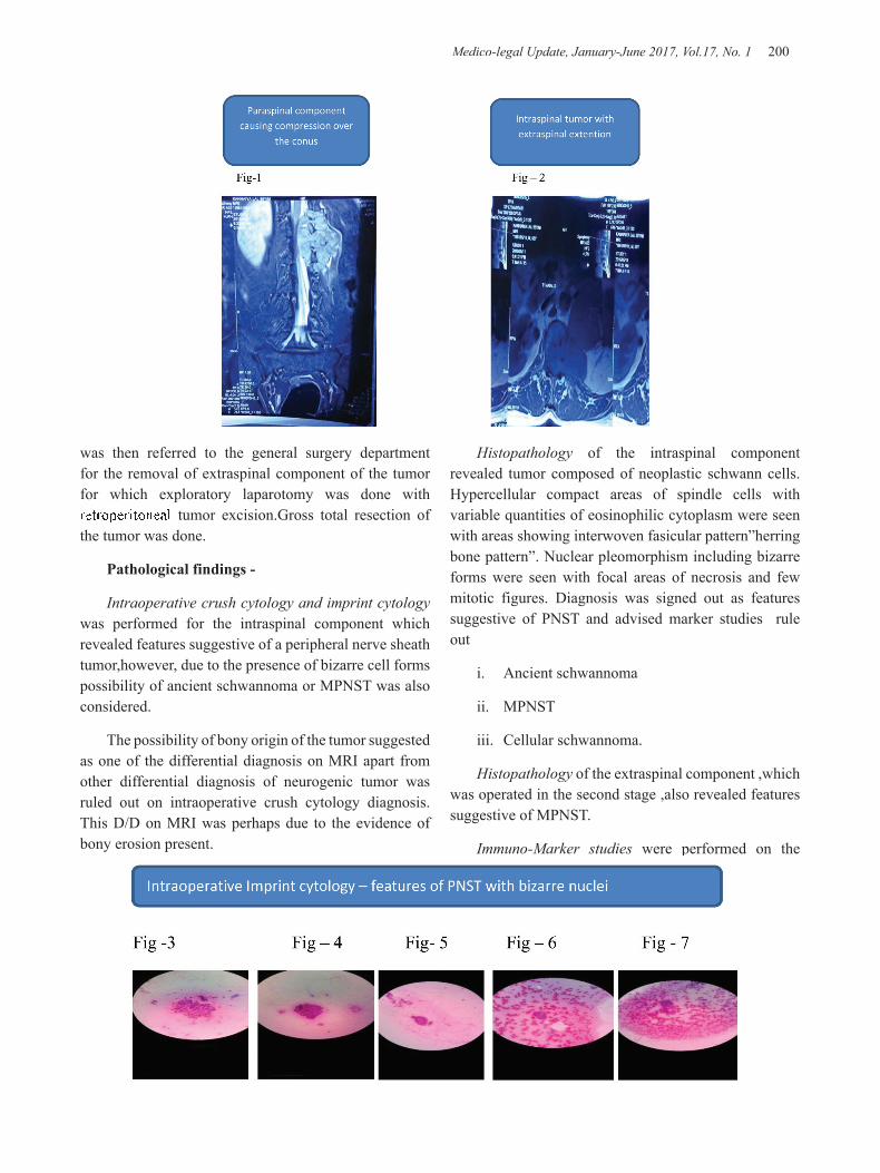
Medico-legal Update, January-June 2017, Vol.17, No. 1 200 201 -legal Update, January-June 2017, Vol.17, No. 1
Histopathology of the intraspinal component revealed tumor composed of neoplastic schwann cells. Hypercellular compact areas of spindle cells with variable quantities of eosinophilic cytoplasm were seen with areas showing interwoven fasicular pattern”herring bone pattern”. Nuclear pleomorphism including bizarre forms were seen with focal areas of necrosis and few mitotic figures. Diagnosis was signed out as features suggestive of PNST and advised marker studies rule out
i. Ancient schwannoma
ii. MPNST
iii. Cellular schwannoma.
Histopathology of the extraspinal component ,which was operated in the second stage ,also revealed features suggestive of MPNST.
Immuno-Marker studies were performed on the
was then referred to the general surgery department for the removal of extraspinal component of the tumor for which exploratory laparotomy was done with retroperitoneal tumor excision.Gross total resection of the tumor was done.
Pathological findings -
Intraoperative crush cytology and imprint cytology was performed for the intraspinal component which revealed features suggestive of a peripheral nerve sheath tumor,however, due to the presence of bizarre cell forms possibility of ancient schwannoma or MPNST was also considered.
The possibility of bony origin of the tumor suggested as one of the differential diagnosis on MRI apart from other differential diagnosis of neurogenic tumor was ruled out on intraoperative crush cytology diagnosis. This D/D on MRI was perhaps due to the evidence of bony erosion present.

-legal Update, January-June 2017, Vol.17, No. 1 200 201 Medico-legal Update, January-June 2017, Vol.17, No. 1
blocks of both intra- and extra- spinal components which favour the diagnosis of a malignant peripheral nerve sheath tumor with –
Vimentin & S 100 positive in tumor cells.
CD 34 is positive in few cells and vasculature is markedly increased.
Calretinin is focal positive in the tumor cells.
GFAP,NF,Desmin,Factor XIIIa , CD 56 ,CD 68,EMA and HMB 45 are negative in the tumor cells.
P53 is positive in very occasional cells.
Ki 67/ MIB is approximately 1-3 %
DISCUSSION
The patient presented with unusually large malignant peripheral nerve sheath tumor (MPNST) with intraspinal and reterospinal extension accompanied by erosion of bone. The intraoperative crush cytology excluded the possibility of bone tumor as suggested as D/D on MRI and it ascertained the origin of tumor as a peripheral
nerve sheath tumor (PNST). On biopsy differential diagnosis of –
i. Ancient schwannoma
ii. MPNST
iii. Cellular schwannoma ,was considered and marker studies was advised, which was in favour of malignant peripheral nerve sheath tumor.
MPNST is a rare variety of soft tissue sarcoma and is very rare in the spine. It arises from proliferation of schwann cells which are neural crest derivatives.3,8 They can occur sporadically or in patients with NF1.
Tumors are often aggressive and high grade with tendency to recur or metastasize. They account for upto 10% of all soft tissue sarcomas and are associated with poor prognosis unless wide excision of the tumor is undertaken before local invasion or distant metastasis occurs. It is well documented that these tumors can extend considerable distances along nerves.
Recurrance rate is reported to be as high as 40 %

Medico-legal Update, January-June 2017, Vol.17, No. 1 202
and approximately 2/3 rd cases metastasize to lungs and bone. Five year survival rate is reported to be 26 – 60 % and ten year survival rate as 45 %.
Total surgical resection is the main stay of treatment6
even though the tumor is biologically aggressive. It complete resection is not possible ,then excision combined with high dose radiotherapy may be used. Chemotherapy is usually not effective .
Ethical Clearance – Taken
Source of Funding – Self Source of Funding – Self Source of Funding –
Conflict of Interest – Nil
REFERENCES
1. Ducatman BS, Scheithauer BW, Piepgras DG, Reiman HM, Ilstrup DM.Malignant peripheral nerve sheath tumors:A clinicopathologic study of 120 cases.Cancer.1986;57:2006-21.
2. Wanebo JE ,Malik JM,VandenBerg ,Wanebo JH ,Driesen N, Persing JA.Malignant peripheral nerve sheath tumors: A clinicopathological study of 28 cases.Cancer 1993;71:1247-53.
3. Hajdu SI.Peripheral nerve sheath tumors.Histogenesis, classification and prognosis.Cancer.1993;72:3549-52.
4. Wick M,Swanson P,Scheithauer B,Manivel J (1987) Malignant peripheral nerve sheath tumor.Am J Clin Pathol 87:425-433
5. Lang N,Liu XG,Yuan HS.Malignant peripheral nerve sheath tumor in spine.Imaging manifestations.Clin imaging.2012;36:209-15.
6. Mc Laughlin EJ,Heuer GG,Whitmore RG,Birknes JK,Belasco JK, Belasco J,Sterman D et al. Treatment of a malignant peripheral nerve sheath tumor and its complications through a multidisciplinary approach.J Neurosurg Pediatr.2011;7:543-8.
7. Ramanathan RC,Thomas JM.Malignant peripheral nerve sheath tumors associated with von recklinghausens neurofibromatosis.Eur J Surg Oncol 1999;25:190-3
8. Stark AM, Buhl R, Hugo HH et al.Malignant peripheral nerve sheath tumors – report of 8 cases and review of literature.Acta Neurochir(Wien)2001;143:357-64
9. Dunnick NR,Image interpretation session:1999.Intraosseous malignant peripheral nerve sheath tumor (malignant schwannoma) in a patient with neurofibromatosis.Radiographics 2000;20:271-273.

-legal Update, January-June 2017, Vol.17, No. 1 202
Cranio-cerebral Trauma
B Karunaker1, Abhijit Subhedar2, Jakkam Surendar3, Vamshi Madav4
1Associate Professor, 2Professor-in charge, Department of Forensic Medicine& Toxicology, Osmania Medical College, Hyderabad, Telangana, 3Assistant Professor, Department of Forensic Medicine,
ESIC Medical College, Hyderabad, Telangana, 4Assistant Professor, Department of Forensic Medicine, Mallareddy Medical College, Hyderabad, Telangana
ABSTRACT
As head is the most prominent exposed part of human body by virtue of its Situation, it bears the brunt of violence namely accidents, suicidal and homicides. The diagnosis of the exact lesion due to trauma is a difficulty even to the neuro -physician. The treatment is an enigma to Neuro-surgeon. And the evaluation of mechanism is a puzzle to the medico-legal expert. the present study conducted in 200 cases in the department of forensic medicine , Osmania Medical College, Hyderabad. During the period of June 2009 to the end of May 2012. Number of cranio cerebral trauma case are more in male especially in the age group of 21-30 years.this trauma is mainly due to RTA. 46% cases have fissured fracture and 51% cases have subdural haemorrhage.
Keywords : Trauma, skull injury, intra cranial hemorrhage
Corresponding author: Dr. Jakkam SurendarAssistant Professor, Department of Forensic Medicine, ESIC Medical College, Hyderabad, Telangana, [email protected], Ph.: 9951197087
INTRODUCTION
The subject of “CRANIO CEREBRAL TRAUMA” has assumed paramount importance in recent times owing to enormous mechanization of various aspects of life, increasing instances of brutal assault and innumerable and variegated accidents. Mostly “CRANIO CEREBRAL TRAUMA” are a result of blunt force either local or general and direct or indirect. They are common injuries and alone can account for one fourth of all deaths due to violence and about one fifth of the Head injuries prove fatal. Polson (1955) said “It is prudent to assume that every unconscious patient has sustained a Head injury and that his condition is due to this cause, unless or until it is shown to be due to some other cause.”(1) Hence understanding the underlying mechanism, the basic physiological disturbances and the rational principals of treatment in acute cranio-cerebral trauma become a necessity. All resultant deaths need medico legal investigation.
AIMS AND OBJECTIVES
This study is taken up to evaluate the actual burden of mortality due to RTA and the trends of increasing burden of human loss due to cranio cerebral trauma.
MATERIAL AND METHOD
The present work, is done in the department of forensic medicine, Osmania Medical College, Hyderabad. During the period of June 2009 to the end of may 2012. 200 cases observed. The object of this piece of work is to analyze cases in general in only those cases with a clear cut history of injury to head e.g., fall from height, vehicular accidents assault by blunt weapon etc. Decomposed bodies, mutilated bodies, and bones were not taken up for this study. Brief history pertaining to the injury to the head was recorded after verifying from the relatives, eye witnesses, hospital records and the police inquest report.
RESULTS AND DISCUSSION
Detailed postmortem study was conducted on 200 bodies who died of “cranio cerebral trauma” , 122 of which from the clinical series and 78 died before being admitted to hospital .
DOI Number: 10.5958/0974-1283.2017.00044.5

Medico-legal Update, January-June 2017, Vol.17, No. 1 204 205 -legal Update, January-June 2017, Vol.17, No. 1
1) INCIDENCE OF SEX IN RELATION TO AGE
Age incidence: there were cases that belonged to every decade.
From the above table we find the largest number of cases in 21-30 age group next highest being in 11-20 years. In 0-10years, 41-50, 51-60 , 61-70 , and 71 years and above the number of cases gradually decreasing .The differentiation in deaths in different age groups depend upon the activities of the person of different age group. The middle age groups i.e., 21-30 and 31-40 years though more stable and steady sustain injuries as a result of quarrels, assault, falls and vehicular accidents. In the present series, the youngest victim is one year child and older is 75 years .
Sex incidence:
In the total 200 cases 164 (83%) are males and only 36(17%) are females. The high percentage of mortality in males may be attributed to their activities in life. The high incidence is seen in the age group 21-30 years. The incidence of mortality in females is very low as they are not involved generally in such activities as men, like assaults, climbing up etc and majority of women are confined to their homes.
2) ETIOLOGY OF CRANIO-CEREBRAL TRAUMA IN RELATION TO AGE AND SEX:
a) falls : 20 cases are the total number of deaths from fall from height .14 males and 6 females were involved. In these 20 cases 7 belong to age group 0-10; 5 males 2 females both these two females are children and had fallen into dry well In the age group 11-20 ; 5 cases all males , three has fallen from roof and two from tree. In age group 21-30 ; 5 cases recorded. In 31-40 ,41-50 and 50 above only one each case is recorded ,being fall from height and bridge respectively. The incidence of falls from males to females is 16:4. In all these cases there is no suspicion of foul play and death due to falls may be attributed as accidental.
b) vehicular incidence: the incidence in vehicular accidents are 120 (75%) of which 110 are males and 30 females .The highest incidence is in 21-30 years group i.e., 60 ; males 45 and females 15. Next highest group is 11-20 years 20 ; males 18 and females 2. Third highest group is 31-40 years 35; males 33 and females 2 . Next group is 0-10 years 10; males 6 and females 4 . This
is followed by the groups 41-50 , 51-60 , 61-70 and 71 above with 6,4, 3, 2 victims respectively.The mortality among children is quite high and the sex incidence is almost equal . The average Indian child is less protected from traffic accidents than his western counterparts .This may partly be explained by the habit of children of playing in the streets , walking suddenly into the streets or crossing the streets without proper precautions . The ratio between male and female taking all the age groups is 7:1 . men are more vulnerable for the accidents , as most of them are pedestrians , passengers , drivers and so on .
Table-1: Victims involved in accidents
Category Total number of victims Percentage
Pedestrians 25 23
Two wheelers 57 47
Four wheelers 18 18
Occupants of vehicle 12 10
Cyclist 3 2
Total 120 100
Pedestrians from the largest group of victims in urban road accidents because , in contrast to motor ways , the streets of a city are usually thronged with pedestrians. Ejection of occupants out of the vehicle adds a further sequence of force as they make impact with the road and are likely to be injured again by the vehicle or other passing vehicles. The pedestrians are injured by the direct contact with the vehicle and is forcibly thrown on the ground. These injuries are mostly due to blunt trauma . In some cases the injuries are caused by the wheel passing over the head resulting in crushing.
c) assaults: there were history of assault in 30 cases and most of them are males .
The incidence is from 2nd to 6th decade age group. Stones, sticks, rods have been used as the weapon of offense .
d) miscellaneous: The incidence of this group is 30 cases and most of them are males belonging to 21-60 age group .
3) EXTERNAL INJURIES OF THE HEAD
External injuries of the head in relation to
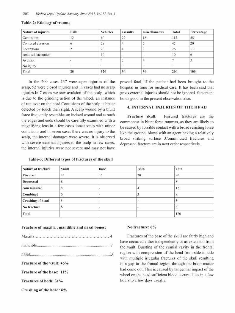
-legal Update, January-June 2017, Vol.17, No. 1 204 205 Medico-legal Update, January-June 2017, Vol.17, No. 1
Table-2: Etiology of trauma
Nature of injuries Falls Vehicles assaults miscellaneous Total PercentageContusions 12 60 22 18 112 58Contused abrasion 6 28 4 7 45 20Lacerations 2 20 1 3 26 13contused-laceration - 10 - - 10 6Avulsion - 2 3 2 7 3No injury - - - - -Total 20 120 30 30 200 100
In the 200 cases 137 were open injuries of the scalp, 52 were closed injuries and 11 cases had no scalp injuries.In 7 cases we saw avulsion of the scalp, which is due to the grinding action of the wheel, an instance of run over on the head.Contusions of the scalp is better detected by touch than sight. A scalp wound by a blunt force frequently resembles an incised wound and as such the edges and ends should be carefully examined with a magnifying lens.In a few cases intact scalp with minor contusions and in seven cases there was no injury to the scalp, the internal damages were severe. It is observed with severe external injuries to the scalp in few cases, the internal injuries were not severe and may not have
proved fatal, if the patient had been brought to the hospital in time for medical care. It has been said that gross external injuries should not be ignored. Statement holds good in the present observation also.
4. INTERNAL INJURIES OF THE HEAD
Fracture skull: Fissured fractures are the commonest in blunt force traumas, as they are likely to be caused by forcible contact with a broad resisting force like the ground, blows with an agent having a relatively broad striking surface .Comminuted fractures and depressed fracture are in next order respectively.
Table-3: Different types of fractures of the skull
Nature of fracture Vault base Both Total
Fissured 45 15 20 80
Depressed 8 - - 8
com minuted 8 - 4 12
Combined 6 - 3 9
Crushing of head 5 - - 5
No fracture 6 - - 6
Total 120
Fracture of maxilla , mandible and nasal bones:
Maxilla……………………………………………… 4
mandible.......................................................................7
nasal..............................................................................3
Fracture of the vault: 46%
Fracture of the base: 11%
Fractures of both: 31%
Crushing of the head: 6%
No fracture: 6%
Fractures of the base of the skull are fairly high and have occurred either independently or as extension from the vault. Bursting of the cranial cavity in the frontal region with compression of the head from side to side with multiple irregular fractures of the skull resulting in a gap in the frontal region through the brain matter had come out. This is caused by tangential impact of the wheel on the head sufficient blood accumulates in a few hours to a few days usually.
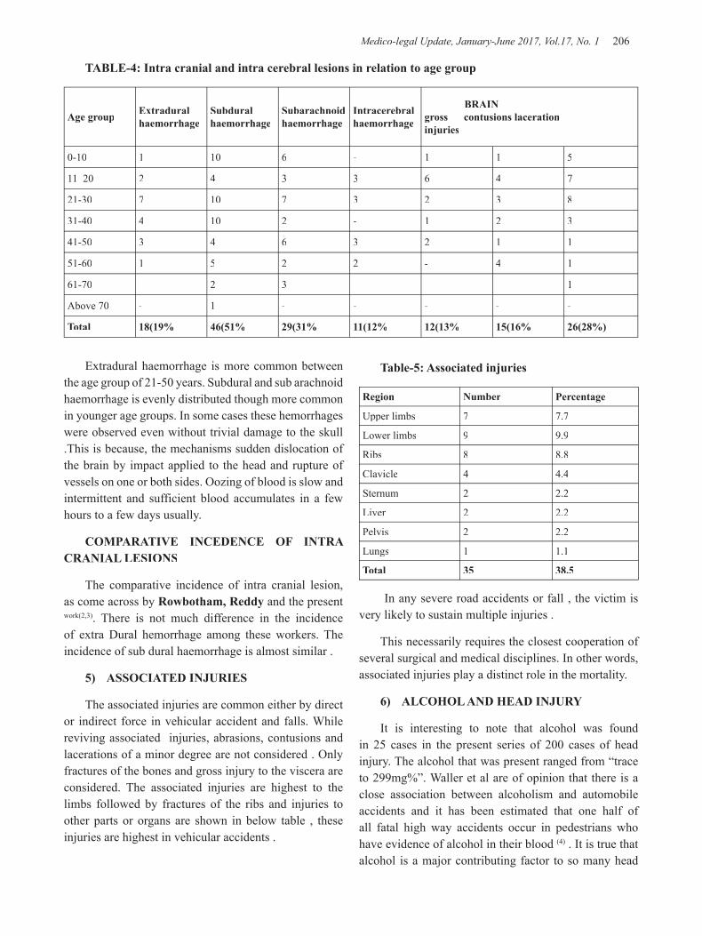
Medico-legal Update, January-June 2017, Vol.17, No. 1 206 207 -legal Update, January-June 2017, Vol.17, No. 1
TABLE-4: Intra cranial and intra cerebral lesions in relation to age group
Age group Extradural haemorrhage
Subdural haemorrhage
Subarachnoid haemorrhage
Intracerebral haemorrhage
BRAINgross contusions lacerationinjuries
0-10 1 10 6 - 1 1 5
11 20 2 4 3 3 6 4 7
21-30 7 10 7 3 2 3 8
31-40 4 10 2 - 1 2 3
41-50 3 4 6 3 2 1 1
51-60 1 5 2 2 - 4 1
61-70 - 2 3 - - - 1
Above 70 - 1 - - - - -
Total 18(19% 46(51% 29(31% 11(12% 12(13% 15(16% 26(28%)
Extradural haemorrhage is more common between the age group of 21-50 years. Subdural and sub arachnoid haemorrhage is evenly distributed though more common in younger age groups. In some cases these hemorrhages were observed even without trivial damage to the skull .This is because, the mechanisms sudden dislocation of the brain by impact applied to the head and rupture of vessels on one or both sides. Oozing of blood is slow and intermittent and sufficient blood accumulates in a few hours to a few days usually.
COMPARATIVE INCEDENCE OF INTRA CRANIAL LESIONS
The comparative incidence of intra cranial lesion, as come across by Rowbotham, Reddy and the present work(2,3). There is not much difference in the incidence of extra Dural hemorrhage among these workers. The incidence of sub dural haemorrhage is almost similar .
5) ASSOCIATED INJURIES
The associated injuries are common either by direct or indirect force in vehicular accident and falls. While reviving associated injuries, abrasions, contusions and lacerations of a minor degree are not considered . Only fractures of the bones and gross injury to the viscera are considered. The associated injuries are highest to the limbs followed by fractures of the ribs and injuries to other parts or organs are shown in below table , these injuries are highest in vehicular accidents .
Table-5: Associated injuries
Region Number Percentage
Upper limbs 7 7.7
Lower limbs 9 9.9
Ribs 8 8.8
Clavicle 4 4.4
Sternum 2 2.2
Liver 2 2.2
Pelvis 2 2.2
Lungs 1 1.1
Total 35 38.5
In any severe road accidents or fall , the victim is very likely to sustain multiple injuries .
This necessarily requires the closest cooperation of several surgical and medical disciplines. In other words, associated injuries play a distinct role in the mortality.
6) ALCOHOL AND HEAD INJURY
It is interesting to note that alcohol was found in 25 cases in the present series of 200 cases of head injury. The alcohol that was present ranged from “trace to 299mg%”. Waller et al are of opinion that there is a close association between alcoholism and automobile accidents and it has been estimated that one half of all fatal high way accidents occur in pedestrians who have evidence of alcohol in their blood (4) . It is true that alcohol is a major contributing factor to so many head
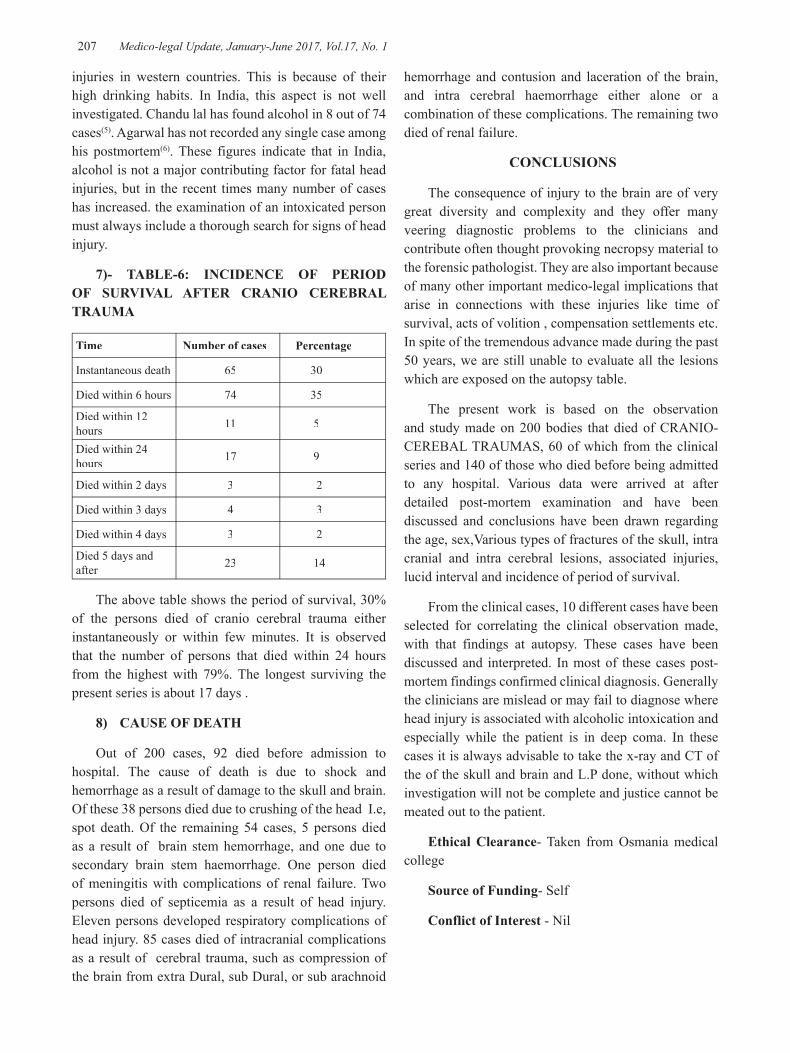
-legal Update, January-June 2017, Vol.17, No. 1 206 207 Medico-legal Update, January-June 2017, Vol.17, No. 1
injuries in western countries. This is because of their high drinking habits. In India, this aspect is not well investigated. Chandu lal has found alcohol in 8 out of 74 cases(5). Agarwal has not recorded any single case among his postmortem(6). These figures indicate that in India, alcohol is not a major contributing factor for fatal head injuries, but in the recent times many number of cases has increased. the examination of an intoxicated person must always include a thorough search for signs of head injury.
7)- TABLE-6: INCIDENCE OF PERIOD OF SURVIVAL AFTER CRANIO CEREBRAL TRAUMA
Time Number of cases Percentage
Instantaneous death 65 30
Died within 6 hours 74 35
Died within 12 hours 11 5
Died within 24 hours 17 9
Died within 2 days 3 2
Died within 3 days 4 3
Died within 4 days 3 2
Died 5 days and after 23 14
The above table shows the period of survival, 30% of the persons died of cranio cerebral trauma either instantaneously or within few minutes. It is observed that the number of persons that died within 24 hours from the highest with 79%. The longest surviving the present series is about 17 days .
8) CAUSE OF DEATH
Out of 200 cases, 92 died before admission to hospital. The cause of death is due to shock and hemorrhage as a result of damage to the skull and brain. Of these 38 persons died due to crushing of the head I.e, spot death. Of the remaining 54 cases, 5 persons died as a result of brain stem hemorrhage, and one due to secondary brain stem haemorrhage. One person died of meningitis with complications of renal failure. Two persons died of septicemia as a result of head injury. Eleven persons developed respiratory complications of head injury. 85 cases died of intracranial complications as a result of cerebral trauma, such as compression of the brain from extra Dural, sub Dural, or sub arachnoid
hemorrhage and contusion and laceration of the brain, and intra cerebral haemorrhage either alone or a combination of these complications. The remaining two died of renal failure.
CONCLUSIONS
The consequence of injury to the brain are of very great diversity and complexity and they offer many veering diagnostic problems to the clinicians and contribute often thought provoking necropsy material to the forensic pathologist. They are also important because of many other important medico-legal implications that arise in connections with these injuries like time of survival, acts of volition , compensation settlements etc. In spite of the tremendous advance made during the past 50 years, we are still unable to evaluate all the lesions which are exposed on the autopsy table.
The present work is based on the observation and study made on 200 bodies that died of CRANIO-CEREBAL TRAUMAS, 60 of which from the clinical series and 140 of those who died before being admitted to any hospital. Various data were arrived at after detailed post-mortem examination and have been discussed and conclusions have been drawn regarding the age, sex,Various types of fractures of the skull, intra cranial and intra cerebral lesions, associated injuries, lucid interval and incidence of period of survival.
From the clinical cases, 10 different cases have been selected for correlating the clinical observation made, with that findings at autopsy. These cases have been discussed and interpreted. In most of these cases post-mortem findings confirmed clinical diagnosis. Generally the clinicians are mislead or may fail to diagnose where head injury is associated with alcoholic intoxication and especially while the patient is in deep coma. In these cases it is always advisable to take the x-ray and CT of the of the skull and brain and L.P done, without which investigation will not be complete and justice cannot be meated out to the patient.
Ethical Clearance- Taken from Osmania medical college
Source of Funding- Self
Conflict of Interest - Nil

Medico-legal Update, January-June 2017, Vol.17, No. 1 208
REFERENCES
1. Polson CJ.Essentials of forensic medicine, English universities press, London.1955.
2. Rowbotham G.F.:Acute Injuries of Head 1964.4th edition.edinburgh and London:E.and S. and Livingstone.
3. Reddy D.J.: Clinico-pathological observation of cases of head injury. Ind.Aea. Of Foresic Sciences Vol.3 Jan 64.
4. Walker A, emergency medicine journak,2001,sep;18 (5),352.
5. Chandulal R.; Fatal road accidents; J.Police Research and Development. 1971 July-September; 17-19.
6. Agarwal : Fatal Road accidents and analysis of 64 cases joun of Ind:Academy of Forensic Medicine Science. Vol.6.1967.
-legal Update, January-June 2017, Vol.17, No. 1 208
Proposed Legislative: Human DNA Profiling Bill: Indian Scenario
Singh Sukhdeep1, Chaudhary B L1 , Kumar Arvind1, Sharma G K2
1Associate Professor, Forensic Medicine, 2Director Professor, Forensic Medicine, Lady Hardinge Medical College, New Delhi
ABSTRACT
Human DNA Profiling Bill is a proposed legislation in India which will allow the Government to establish a National DNA Data Bank and a DNA Profiling Board, and use the data for various specified forensic purposes. The DNA samples may be collected from unidentified dead bodies, missing persons, habitual offenders, and suspects and accused to create a DNA database. The bill has raised concerns of costs of this exercise to the government exchequer and that of privacy among citizen right group. The DNA (Deoxyribose Nucleic Acid) test would be a powerful tool to identify people in criminal and civil proceedings. The National DNA database can be proved to be backbone in this era of rising crime and international terrorism. Many countries are benefitted by this scientific advancement. A brief review of literature on proposed Human DNA Profiling Bill is analysed and important aspects of this bill discussed.
Keyword; Human DNA Profiling Bill; DNA Data Bank; Crime; Identification.
Corresponding author: Dr. Sukhdeep Singh Associate Professor, Forensic Medicine,Lady Hardinge Medical College, New Delhi,11001Email id- [email protected], Mob: +91-9582032160
INTRODUCTION
Human DNA Profiling Bill 2007, seeks to regulate the use of deoxyribonucleic acid analysis of human body substances profile and to establish DNA profiling board for laying down standards for laboratories and collection of human body substances, custody trail from collection to reporting and also to establish a National DNA bank and for matters connected therewith and incidental thereto Considering the huge number of missing persons and unidentified dead bodies Government of India has been contemplating Human DNA profiling act since 2003.The bill was to be introduced in 2007 but was held back due to some legal, moral and ethical issues. This was again drafted in 2012 and was again to be introduced in monsoon session of 2015 but couldn’t be introduced due to some concerns on privacy, safety of data and costs of the exercise.1,2 It has been alleged that there may be scientific ambitions in the backdrop of the whole exercise which could be in conflict with the
safety and privacy of the data which belongs to private citizens. The control of collection of samples and its use may provide overarching powers to the Department of Biotechnology and Centre for DNA fingerprinting & Diagnostics Hyderabad. However, there are many countries having a National DNA Data bank, which are reaping the benefits of this scientific advancement. The National DNA database can be of great utility in this era of rising crime and international terrorism, if India has a meticulously drafted legislation for the regulation and standardisation of DNA profiling.
MATERIAL AND METHOD
The original Human DNA bill June 2015 was obtained from website of PRS Legislative research and data was also retrieved from National crime records Bureau of India. In addition Research material was collected and incorporated from articles published in eminent newspaper like Indian express, Financial express, Times of India and Hindu etc.
REVIEW OF LITERATURE
The Human DNA profiling bill was originally proposed in 2007 and drafting of the bill was initiated in 2012 by the Department of Biotechnology and was expected to be presented in the parliament in
DOI Number: 10.5958/0974-1283.2017.00045.7

Medico-legal Update, January-June 2017, Vol.17, No. 1 210 211 -legal Update, January-June 2017, Vol.17, No. 1
the monsoon session of 2015.1,2 The proposed DNA Profiling Board will consist of molecular biology, human genetics, population biology, bioethics, social sciences, and law and criminal justice experts. The Board will define standards &controls for DNA profiling and will also certify labs and handle access of the data by law enforcement agencies. There will be similar bodies at state levels.3 The bill will also create a National DNA Data Bank, which will collect data from offenders, suspects, missing persons, unidentified dead bodies and volunteers.4 Indices for various categories will be created like missing persons index, Unknown deceased persons index, Offender’s index, Suspect’s index Volunteer’s index.4 The National DNA data bank will profile and store DNA data in criminal cases like homicide, sexual assault, adultery and other crimes from suspects, convicts, relatives of missing people as well as (research) volunteers, whose DNA profiles would help “further our knowledge of genetic markers in different Indian subpopulations.5The data will be restricted and will be available only to the accused or the suspect when a person facing imprisonment or death sentence can send a request for DNA profiling of related evidence to the court that convicted him.3,5 In most countries, the DNA database is used only for criminal investigations.In India, it will also be used to also identify victims of accidents, missing persons etc in addition to criminal investigation in culpable homicide, murder, death by negligence, miscarriage, dowry deaths, causing death of new born child, sexual assault, unnatural offences, outraging the modesty of a woman, co-habitation with a woman by deceit, adultery, enticing a married woman with criminal intent, among others. The bill has the provision that any misuse of data will carry a punishment of up to three years imprisonment and also fine. 3
DNA Database is the need of the hour as per data compiled by the National Crime Records Bureau of India, the number of unidentified bodies recovered and inquest conducted was 37,282 in (2007), 37,668 (2008), 34,902 (2009), 33,857 (2010) and 37,193 (2011).The figures for various states are Maharashtra(6,313), followed by Tamil Nadu (4,479), Uttar Pradesh (4,084), West Bengal (3,704), Delhi (2,748), Andhra Pradesh (2,639), Karnataka (2,440), Gujarat (2,099), Madhya Pradesh (1,191), Rajasthan (1,170), Haryana (1,159) and Punjab (1,004). It said a total of 11,846, 13,586 and 13,268 people went missing in Delhi alonein 2011; the highest number of unidentified bodies was recovered in Maharashtra in 2006, 2007 and 2008 respectively. From Andhra Pradesh, a total of 47,936
went missing during 2009-12 and in Gujarat another 37,395 were reported missing during 2007-11.2, 5Going by collecting DNA of everyone arrested in India, the numbers are huge. According to the National Crime Records Bureau, over 3.2 million people were arrested in 2012 alone in the country.2, 5
The facts and figures reflect that National Database for DNA if established in India shall be of great benefit to the public as well as law enforcement agencies. While the costs are huge, it does make sense to retain DNA information of people in an era of rising crime and terrorism but consensus is yet to be reached on the issues regarding privacy & safety of the data.2, 5
CONCERNS
In a representation submitted to the National Human Rights Commission, The Citizens Forum for Civil Liberties has said DNA profiling is “undesirable, particularly as forensic DNA developments are intertwined with significant changes in legislation and contentious issues of privacy, civil liberty and social justice.1 Even though privacy has been held to be a fundamental right as long back as in 1962, India does not have a law that specifies safeguards to privacy. 6While the Supreme Court has held privacy to be a fundamental right, it is restricted to certain aspects of a person’s life like the privacy of one’s home, family, marriage, motherhood, procreation and child-rearing. Therefore, to claim privacy in any other aspect, individuals have to substantiate these are ‘private’ and should not be subjected to state or private interference.6Expert Group on Privacy recommended that the proposed legislation on privacy should ensure that safeguards are technology neutral. This means that the enactment should provide protections that are applicable to information, regardless of the manner in which it is stored: digital or physical form.Therefore the new legislation should protect all types of privacy, such as bodily privacy (DNA and physical privacy); privacy against surveillance (unauthorised interception, audio and video surveillance); and data protection. The safeguards under the Bill should apply to both government and private sector entities.6
Addressing issues related to protecting privacy of individuals, the draft Bill envisages that access to the information in the National DNA Data Bank will be restricted to those related to the victim or suspect; any individual undergoing a sentence of imprisonment or death sentence can apply to the court which convicted him, for an order of DNA testing of specific evidence

-legal Update, January-June 2017, Vol.17, No. 1 210 211 Medico-legal Update, January-June 2017, Vol.17, No. 1
under specific conditions.1There are clearly scientific ambitions fuelling the DNA database project says, Usha Ramanathan legal researcher and a member of the Committee formed by the Centre in 2013 to review this Bill..In the same 12th plan document, CDFD is also described as aiming to work on molecular genetics, cytogenetics, biochemical genetics, newborn screening centre and develop a national database for genetic disorders.7In identification form for DNA sample tests, put up on the CDFD website included a column for filling information. ‘caste and origin of State’ Ms. Ramanathan raised concerns regarding how an agency could be empowered by the proposed law whisch might use DNA samples to profile & distinguish people on the basis of caste.Further, Schedule I under the Act, in the ‘List of Matters for DNA Profiling’ allows data collection on maternity or paternity disputes, issues relating to pedigree, surrogacy and immigration or emigration as well.There is no provision in UID, Adhaar to opt out of the database once enrolled, as there was no specified consent clause or guidelines for sample collection .The DNA Profiling Bill too brings similar concerns. The Director of the Centre for DNA Fingerprinting and Diagnostics in India (CDFD), based in Hyderabad, will always be the ex-officio member-secretary of the DNA Profiling Board as per the Bill. That the agency has been given considerable powers to take decisions regarding DNA sample usage and regulate DNA profiling in India is itself a cause for concern.2 In the DNA profiling Board, CDFD will play a central role and also, control the addressing of concerns regarding breach of privacy.
A process of appeals against the retention of data should also be made available to individuals, and individuals should be able to have a second sample taken and reanalysed in court,” says the group, which is chaired by the former Chief Justice of the Delhi High Court, A.P. Shah. No process has been provided to verify the correctness of the analysis and the information parked in the database.The report points out that samples collection must be done with consent (from a victim or for the purpose of elimination), and circumstances wherein collection takes place without consent (e.g, from crime scenes) should be distinguished. The DNA data bank has been entitled to holding volunteer indices. However, no procedure for consent requirement, and no mechanism for volunteers to withdraw their information if they so desire, have been provided.
The proposed law should incorporate that notice be given to individuals and their consent should be taken if their information is disclosed to a third party,
unless the disclosure is sought by authorised agencies, the report says. “Bodies and organisations collecting, analysing and storing DNA samples should publish a transparency report, on an annual basis, detailing their internal governance structure, practices, finances and success and error rates.”
Another important recommendation made by the group is that wherever applicable, notice of the purpose for collection of DNA samples and personal information must be made public and the use of samples and personal information should be limited to the purposes and time frame specified under the bill.A procedure should also be established for the destruction of all samples and personal information after they have served their purpose.An NGO “Lokniti “had raised concerns about the thousands of unidentified dead bodies that remain unclaimed in the country and filed a petition in Supreme Court regarding delay in establishment of a national DNA database, so as to identify missing persons. It said that over 40,000 unclaimed bodies are found in railway stations and other places in India every year, and over 20,000 children disappear or get kidnapped. It argued that DNA samples alone can aid the authorities track their relatives down. Lokniti also mentioned that a DNA data bank can help the police crack unsolved crimes.The Union government of India a submitted an affidavit in the Supreme Court stating the concerns of experts and NGO’s on the privacy-related aspects and costs of establishing a National DNA Data Bank.8
The proposed DNA profiling mechanism needs a large number of trained personnel and infrastructure which would amount to an annual expenditure of Rs 80crores. As per DNA database annual report ,UK Home Office spent £2.2 million in 2013-14 in running the National DNA Database .The apex court suggested the Centre to create a DNA data bank, while agreeing with a plea by NGO Lokniti Foundation that establishment of identity was an essential feature of individual dignity and the government must resort to modern scientific methods.9
A bench of Justices Dipak Misra and V Gopala Gowda said it would like the Centre to think it over seriously so that an appropriate roadmap is brought into existence on making it (DNA profiling) functional that would echo the voice and cry of the feelings (of relatives of missing persons.5The UID/Adhaar card will give every citizen a 12-digit number ,to be primarily used for identifying people eligible for subsidies while DNA profiling can be a valuable tool in civil and criminal

Medico-legal Update, January-June 2017, Vol.17, No. 1 212
cases. Across the world, about 60 countries have various versions of DNA databases that largely relate to data on criminals. According to the National Crime Records Bureau of India over 3.2 million people were arrested in 2012 and If DNA sample of each individual arrested is collected the cost incurred on DNA profiling will be huge but it certainly does make sense in an era of rising crime and terrorism.10 In most countries, the DNA database is used only for criminal investigations. In India, the proposed plan will use it to also identify victims of accidents, missing persons in addition to criminal investigations. There is no plan to seek civilian DNA data as of now, that could happen later.
DISCUSSION
The statistics for missing persons from National crime records bureau amply justify the need for a National database of DNA irrespective of the huge cost to the government.11 There should be no hitches in using modern scientific advancements for the protection and welfare of society. The consent of the individual should always be taken for collection of the sample and he should be apprised of the safety and secrecy of the data except in certain circumstances of crime and national interest. The DNA profiling bill has to be coherent with the pending legislation on privacy, which has been recognised as a fundamental right by Supreme court.6 The use of National DNA data generated can be encouraged in cytogenetics &gene therapy for the advancement of Medical sciences but with due compliance to the Professional secrecy of the data of subjects.7 There should be an inbuilt mechanism in the act itself to verify the correctness of data by second examination on the demand of affected party.The authority of the agencies like CDFD should be kept in proper checks and balances and data generated should be used in National interest and not to further the caste identities based discrimination.7
CONCLUSION
India with its massive population and the rising crime graph needs substantially specific and sensitive standardised scientific methods for fixation of the individuality of a person. The Biometric systems of identification like Iris scan and Fingerprinting scan serve this purpose well in identifying of live individuals for civil purposes. There are a large number of unidentified dead
bodies; some of them decomposed beyond recognition a number of missing persons in the records of police every year. It is possible that many of them could have succumbed to violent crimes. Most of such crimes and the accused are not brought to justice and their victims perish unidentified.Legalisation and standardisation of DNA profiling in India can go along way in bridging the gaps in criminal justice system however the concerns regarding safety, security and privacy of data generated needs to be addressed simultaneously.
Conflict of Interest –No
Sourse of Funding-No
Ethical Clearance – Not required since it is a review article.
REFERENCES
1. Centre’s ambitious DNA Profiling Bill hits privacy roadblock. India Today. 15 September 2014. Retrieved 5 January 2015.
2. Human DNA profiling bill 9 June 2015/prsindia.org.
3. Bill to create bank for DNA profiling of accused coming. The Hindu. 21 October 2012. Retrieved 5 January 2015.
4. Experts moot more transparency in bill on DNA profiling.The Hindu. 23 October 2012. Retrieved 5 January 2015.
5. SC wants roadmap for DNA profiling of all unclaimed bodies.The Times of India. 15 July 2014. Retrieved 5 January 2015.
6. The PRS Blog/The official blog site of PRS legislative.
7. Scientific ambitions behind DNA Profiling Bill’New Delhi, August 16, 2015.
8. Bill stuck so cannot create DNA data bank: Centre to SC. The Indian Express. 15 September 2014. Retrieved 5 January 2015.
9. DNA bank plan unveiled. The Telegraph (India). 22 September 2014. Retrieved 5 January 2015.
10. Human DNA Profiling Bill: A tool that could be misused. The Financial Express (India). 27 July 2015. Retrieved 4 August 2015.
11. National Crime records Bureau/ncrb.gov.in.

-legal Update, January-June 2017, Vol.17, No. 1 212
Morphological Study of Lip Print Pattern among Medical Students: An Anthropological Study
Sidramappa Gouda1, M Surender Rao2
1Assistant Professor, Department of Forensic Medicine, Navodaya Medical College, Raichur, 1Assistant Professor, Department of Forensic Medicine, Navodaya Medical College, Raichur, 1
2Assistant Professor, Department of Forensic Medicine, DRVRK Women’s Medical College, Hyderabad2Assistant Professor, Department of Forensic Medicine, DRVRK Women’s Medical College, Hyderabad2
ABSTRACT
Similar to finger prints, appearance of lip prints are heritable and individualistic. Forensic researchers are giving more attention towards studying lip prints (Chieloscopy) which is helping them to solve many of crime and legal related cases. The present study was carried out in the Department of Forensic Medicine, Navodaya Medical College, Raichur, to evaluate different pattern of lip prints in all 4 quadrants of lip, among students of our college. A total of 100 students (50 Males and 50 Females) were taken as study subjects. Suzuki and Tsuchihashi classification was used to classify different types of grooves in lips and the results were statistically analysed and tabulated. No identically similar lip print pattern was noticed among study subjects. Type I pattern was most common among males in all four quadrants (28.7%) fallowed by Type III (27.7%). Further in males Type I was prominently seen in upper right quadrant (38%) and lower left quadrant (21.8%) and Type III in upper left (28.2%) and lower right (31%). Similarly females too showed Type I pattern as commonest one (34%) in all quadrants. Further in females Type I pattern was more frequently seen in lower left quadrant (37%) fallowed by upper right quadrant (34.6%). Lower right and upper left quadrant had Type I pattern of 33% each.
Keywords: Identification, Lip print, Sex.
Corresponding author:Dr. Sidramappa GoudaAssistant Professor, Department of Forensic Medicine, Navodaya Medical College, Raichur.Contact No: 7899606175E mail : [email protected]
INTRODUCTION
Study of lip print is called as chieloscopy and is derived from Greek word chelios-lips skopein-see1. In the year 1968-71, Y. Tsuchihashi and T. Suzuki found that groves present on the red part of human lips were unique2. This unique feature of grooves on human lips can be used as identification data3. Lip prints are ordinary lines and gaps in the types of wrinkles and sections present in the zone of move of human lip, between the inward labial mucosa and external skin, examination of which is known as cheiloscopy. This is extraordinary for people, as fingerprints4. Cheiloscopic strategies have an equivalent worth in connection to other sorts of legal proofs for individual identification
and sex determination5. In a crime scene examination, lip prints can connect a subject to a particular area if found on fabrics or different subjects, for example, glasses, containers or even cigarette butts6. Examination of the lip prints left at the scene of crime, and their examination with those of suspected individual might be valuable for identification7. Dental identification remains a standout amongst the most solid and much of the time connected strategies for distinguishing proof, overwhelmingly by the correlation of ante mortem and post mortem records8. This bio-optical wonder was initially noted by anthropologists, R.Fischer, who was the first to portray it in 19029. Several studies have been done in the past using lip prints patterns for human identification10. The present study was taken to assess the lip print patterns of different individuals in four quadrants of lip and find out the incidence of most common pattern in the given population.
MATERIALS AND METHOD
A sample of 100 students comprising 50 males
DOI Number: 10.5958/0974-1283.2017.00046.9

Medico-legal Update, January-June 2017, Vol.17, No. 1 214 215 -legal Update, January-June 2017, Vol.17, No. 1
and 50 females were considered as study subject, aged between 18 to 23 years. Students having normal transition zone between the mucosa and skin and free from any pathology were included in the study. Consent from all study subjects was taken. Red colour lip stick which is non glossy and non metallic was used to get lip prints on white thick paper. Initially lips were cleaned, dried and left for 5 minutes. A thin film of lip stick was applied and lip prints were taken by rolling the paper on to the lips with application of slight, gentle pressure. The lips were kept slightly separated and relaxed during manipulation. After taking the impression a cellophane strip was stuck on the paper were lip impression is taken for permanent record purpose. All lip prints were examined by using magnifying hand lens with direct light focused on it. Each subject’s lips were divided into four compartments, i.e., two compartments on each lip, and were allotted the digits 1-4 in a clock-wise sequence starting from the subject’s upper right. Classification of lip prints was done based on Suzuki and Tsuchihashi classification, which is as fallows.
1. Type I - A clear-cut groove running vertically across the lip.
2. Type II – Partial length groove of Type I.
3. Type III – A branched groove.
4. Type IV – An intersected groove.
5. Type V – A reticular pattern.
6. Type VI – Other patterns.
RESULTS
All data were statistically analyzed for determining the frequencies of the pattern types in each area of the lip, frequencies of the pattern types in each sex and
for comparison between males and females regarding upper and lower lip. In this present study, out of 100 subjects, no identically similar lip print pattern appeared in two individuals. In all subjects, when it was evaluated among all four lip compartment, it was found that Type I pattern was most common, both in males and females having 28.7% and 34% respectively. Next prominent pattern was Type III in both sexes with 27.7% in males and 22.9% in females. Frequency of different patterns of lip print on all four quadrants if lips (Table 2) are as fallows.
Type I: this was most prominent pattern in males, overall in all four quadrants, but was more prominent in upper right quadrant (33%) and least prominent in upper left quadrant (18%). Even in females this was common pattern among all quadrants with highest being (25%) and there was not much difference in other quadrants as compared to males.
Type II: In males, this pattern was seen least (17%) in lower left quadrant and highest (39%) in upper left quadrant. In females it was low in lower right quadrant and high (32%) in upper left quadrant.
Type III: In males this pattern was highest (28%) in lower right quadrant and least (22%) upper right quadrant. In females upper right quadrant had high distribution (33%) and least in upper left (15%)
Type IV: In males it was least (17%) in upper left quadrant and highest (31%) in lower left quadrant. Where as in females it was least in(10%) in lower left and predominant in upper left quadrant (37%).
In females 18.8% of lip prints were undetermined where as in males 15.2% of lip prints were undetermined.
Table 1: Frequency of lip prints in Males and Females
Lip print Pattern Males (N=50X4) Females (N=50X4)
Vertical 57(28.7%) 69(34%)
Branched 33(16.2%) 28(13.6%)
Intersected 55(27.7%) 45(22.9%)
Reticular 24(11.9%) 20(9.5%)
Undetermined 31(15.2%) 38(18.8%)
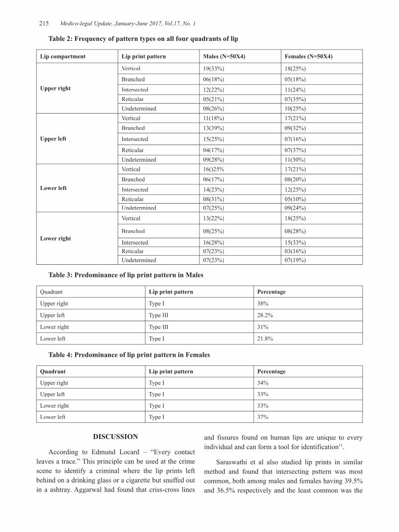
-legal Update, January-June 2017, Vol.17, No. 1 214 215 Medico-legal Update, January-June 2017, Vol.17, No. 1
Table 2: Frequency of pattern types on all four quadrants of lip
Lip compartment Lip print pattern Males (N=50X4) Females (N=50X4)
Upper right
Vertical 19(33%) 18(25%)
Branched 06(18%) 05(18%)
Intersected 12(22%) 11(24%)Reticular 05(21%) 07(35%)Undetermined 08(26%) 10(25%)
Upper left
Vertical 11(18%) 17(21%)Branched 13(39%) 09(32%)
Intersected 15(25%) 07(16%)
Reticular 04(17%) 07(37%)Undetermined 09(28%) 11(30%)
Lower left
Vertical 16()25% 17(21%)
Branched 06(17%) 08(20%)
Intersected 14(23%) 12(25%)Reticular 08(31%) 05(10%)Undetermined 07(25%) 09(24%)
Lower right
Vertical 13(22%) 18(25%)
Branched 08(25%) 08(28%)
Intersected 16(28%) 15(33%)Reticular 07(23%) 03(16%)Undetermined 07(23%) 07(19%)
Table 3: Predominance of lip print pattern in Males
Quadrant Lip print pattern Percentage
Upper right Type I 38%
Upper left Type III 28.2%
Lower right Type III 31%
Lower left Type I 21.8%
Table 4: Predominance of lip print pattern in Females
Quadrant Lip print pattern Percentage
Upper right Type I 34%
Upper left Type I 33%
Lower right Type I 33%
Lower left Type I 37%
DISCUSSION
According to Edmund Locard – “Every contact leaves a trace.” This principle can be used at the crime scene to identify a criminal where the lip prints left behind on a drinking glass or a cigarette but snuffed out in a ashtray. Aggarwal had found that criss-cross lines
and fissures found on human lips are unique to every individual and can form a tool for identification11.
Saraswathi et al also studied lip prints in similar method and found that intersecting psttern was most common, both among males and females having 39.5% and 36.5% respectively and the least common was the
Medico-legal Update, January-June 2017, Vol.17, No. 1 216
reticular pattern in both sexes12. Singh et al also used Tsuchihashi and Suzuki classification to categorise the lip prints. They found that Type II pattern was common in both sexes. They stated that no two individuals had similar pattern and no one has single type of lip prints in sh compartments13. However in South India data related on Lip prints are meagre. Our study categorised lip prints into five types similar to that of Suzuki and Tsuchihashi. Our study results were similar to their reports that lip prints had different patterns in different individual. A similar type of study was also done by Uma Maheshwari in south India and found that lip prints are unique and no one had a single groove type in all compartments14, whereas our study found similar grooves in more than one compartments in many individuals.Our study showed that males had a vertical and intersected grooves as the most common pattern whereas females had only vertical grooves as the prominent one in all the compartments of lips.
CONCLUSION
Our study showed that the lip prints are unique to each individual similar to finger prints. So similar to finger print, lip prints can also be used as tool for identification. In future software programme can be developed as that of dactylography for chieloscopy also.
Conflict of Interest: Nil
Source of Funding: Self
Ethical Clearance: Taken from ethical clearance committee of our college.
REFERENCES
1. K. Randhawa, R. S. Narang, P. C. Arora. Study of the Effect of Age Changes on Lip Print Pattern and its Reliability in Sex Determination. Sri Guru Ramdas Institute of Dental Sciences and Research, Amritsar, Punjab, India.143006.
2. Lip Prints in Personal Identification 1 2 Suman Jaishankar , Jaishankar N , Shanmugam.
3. Rohit M, Sumit G (2011) Cheiloscopy: A Deterministic Aid for Forensic Sex Determination. Journal of Indian academy of Oral Medicine and Radiology 23: 17-19.
4. Saraswathi TR, Gauri Mishra, Raganthan K (2009) Study of lip prints. J Forensic Dent Sci 1: 28-31.
5. Bindal U, Jethani SL, Mehrotra N, Rohatgi RK, Arora M, etal. (2009) Lip Prints as a Method of Identification in Human being.J Anat Soc 58: 152-155.
6. Singh NN, Brave VR, Khanna S (2010) Natural dyes versus lysochrome dyes in cheiloscopy: A comparative evaluation. J Forensic Dent Sci 2:11-17.
7. El Domiaty MA, Al-gaidi SA, Elayat AA, Safwat MD, Galal SA (2010) Morphological patterns of lip prints in Saudi Arabia at Almadinah Almonawarah province..Forensic Sci Int 179.e1-179.e9.
8. Ranganathan K, Rooban T, Vidya Lakshminarayanan (2008) Forensic odontology: a review. J Forensic Odontol 1: 4-12.
9. Vahanwalla S, Nayak CD, Pagare SS, Study of lip prints as a aid to sex determination. Medicolegal Udate 2005;5:93-98.
10. T Suchihashi Y. Studies on Personal Identification by Means of Lip Prints. Forensic Sci.1974; 3:233-48.
11. A Aggarwal. The Importance of Lip Prints. Mystery Magazine web.2004; II:2.http://lifeloom.com/archives.htm.
12. Saraswathi TR, Mishra G, Ranganathan K. Study of Lip Prints. J Forensic Dental science.2009;1:28-31.
13. Singh NN, Brave VR, Khanna S. Natural dyes versus lysochrome dyes in chieloscopy:A comparative evaluation. J Forensic Dent Sci.2010; 2(1):11-17.
14. Maheshwari UT. Lip Prints, Master Thesis of Dental Surgery Saveetha dental College and Hospitals, Chennai, 2005.
-legal Update, January-June 2017, Vol.17, No. 1 216
Correlation between Fingerprint and Lip Print Pattern in Gujarati Population
Senthil Kumaran M1, Binaya Kumar Bastia2, Lavlesh Kumar3, Sweta H Patel4
1Senior Resident, JIPMER, 2Additional Professor, Department of Forensic Medicine, All India Institute 2Additional Professor, Department of Forensic Medicine, All India Institute 2
of Medical Science, Rishikesh, Uttarakhand, India, 3Professor & HOD, 4Junior Resident, Department of 4Junior Resident, Department of 4
Forensic Medicine, SBKS Medical Institute & Research Centre, Vadodara, Gujarat, India
ABSTRACT
Background: Identification of an individual plays an important role in both civil and criminal investigations. Fingerprinting is widely accepted and practiced method of identification throughout the world because of its uniqueness. Similarly researches done previous shows that lip print pattern is also unique and is helpful in identification.
Objective: To correlate fingerprint with lip print pattern and also to determine the predominant fingerprint and lip print pattern in males and females, in Gujarati population.
Materials and Method: Cross-sectional survey was conducted in a teaching hospital Vadodara, Gujarat, India. A total of 200 subjects (100 males and 100 females), between 18 – 24 years of age, were the study subjects. Fingerprint and lip impressions of all the subjects were recorded, and the results were analyzed based on Henry system for fingerprints and Suzuki & Tsuchihashi classification for lip prints.
Results: There exists a correlation between the fingerprint and the lip print pattern in both males and females (p <0.001), which was highly significant sex-wise.
In the male (63.9%) and female (66.6%) loop fingerprint pattern was the predominant type. In the case of lip print pattern in male Type III (42%) and female Type I (37%) was the most predominant type.
Conclusion: There is a correlation between fingerprint and lip print patterns in both males and females. The study also revealed that finger and lip prints are unique to each and can be used as an identification tool in civil and criminal investigation.
Keywords: dactyloscopy; cheiloscopy; fingerprint; lip print; correlation; identification.
INTRODUCTION
Human identification is the basis of civilization, and the identification of unknown individuals has always been of chief importance to society.1 Identification of any individual living or dead was based on the theory that all individuals are unique. When an unidentified body or trace was found, it is assumed that it could be anybody. The more unique the characteristic, the smaller the group becomes. As more unique characteristics are noted, the comparison group becomes smaller until it reaches unity.2
There are successful methods of personal identification include anthropometry, dermatoglyphics,
DNA fingerprinting, sex determination, estimation of age, measurement of height, differentiation by blood groups, handwriting and bite marks.3
Study of fingerprints is known as dermatoglyphics or dactyloscopy or Galton – Henry system. Fingerprints are impressions of patterns formed by the papillary, or epidermal ridges of the finger tips.4 Fingerprints are widely used for identification because, the ridge pattern once developed remains unchanged for the rest of the life, individuality stored in a systematic way for future reference, can be taken from even highly decomposed dead bodies, age of fingerprint can be accessed from migration of the chloride ions. Moreover fingerprints
DOI Number: 10.5958/0974-1283.2017.00047.0
Medico-legal Update, January-June 2017, Vol.17, No. 1 218 219 -legal Update, January-June 2017, Vol.17, No. 1
collected at a crime scene can be used to identify suspects, victims and other persons who touched the surface, fingerprint scans can be used to validate electronic registration.5
Cheiloscopy or queiloscopy, the study of furrows or grooves present in the zone of transition of human lip, between the inner labial mucosa and outer skin. Lip print can give valuable information about the individual and his habits, e.g., lip prints found on cigarette or drinking glass, etc. The use of lipstick is not compulsory for obtaining lip prints as the edges of lips have sebaceous and sweat glands, which provide secretions enabling the development of ‘latent’ lip prints, which can be developed later on using various techniques similar to fingerprints and used for the establishment of identity of the victim.
Among fingerprint and lip print, finger print is widely used throughout the world for identification. In recent times, there is more awareness regarding fingerprint, and this leads to criminals in taking extra care not to leave any fingerprint mark at scene of the crime. So it’s high time that we follow other methods of identification that criminals are not usually aware like cheiloscopy method, because criminals tend to leave their lip print mark in glasses, cigarettes, on the skin of the victim in the case of sexual offences that help in identification of the accused.
Though extensive research work has been carried out regarding dermatoglyphics and cheiloscopy independently, combined study correlating the two entities are very few. Due to the immense potential of fingerprints as an effective method of identification, an attempt had been made to analyze their correlation with the lip print in an individual, when either of the evidence detected at the scene of an incident.
The main aim of this study is to find out possibility of correlation between the fingerprint and lip print patterns among males and females in Gujarati population. And also to determine the predominant patterns of finger and lip print among males and females in Gujarati population.
MATERIAL AND METHOD
The study sample of 200 subjects (100 males and 100 females) in the age group 18-24 years from a teaching hospital Vadodara, Gujarat. The inclusion criterion
was Gujarati subjects in age group 18-24 years having healthy lips and finger tips. Subjects with a history of skin allergy, trauma, malformation, deformity, surgical scars, active lesion of fingers and lips, composite finger pattern and Type V lip pattern were excluded. After explaining about the nature of the study to subjects, informed written consent was obtained prior to taking finger and lip prints.Materials used were brown and pink colored non-glossy lipstick, cellophane tape, white sheet, blue inked stamp pad, and magnifying lens.
The subjects were asked to wash their hands thoroughly with soap and water and dry them using tissue paper to remove dirt and grease. Then they were asked to press their finger tip on the stamp pad, and then the rolled impressions of each finger were obtained in the white sheet. Care was taken to avoid sliding of fingers to prevent smudging of the print. The subjects were asked to keep their arm relaxed and not to try to help in rolling the fingers as this may cause smearing. The fingerprints so obtained were studied with the help of a magnifying lens and classified according to Henry’s system of classification.6 The classification is as follows:Loops, Whorls, Arches and composite.
In composite variety, there is a combination of more than one pattern, either a whorl and loop patterns, or two different loop patterns. So in order to avoid confusion, the subjects with the composite pattern of fingerprints were excluded from the present study.
The subjects were asked to clean their lips thoroughly with water and dry them with tissue paper. And then were asked to open the mouth, and dark colored lipstick was applied uniformly on the lips starting at the midline and moving up to the vermillion border in a single motion, evenly on the upper lip and then on the lower lip. Then the subjects were asked to rub the upper and lower lips together prior to taking the print ensured that the lipstick was spread evenly over the lips. Then the lipstick was allowed to dry for about two minutes. A strip of cellophane tape, 10 cm long was cut out with scissors. The subjects were asked to keep the mouth stationary during the procedure. The glued portion of the cellophane tape strip was placed, and the subjects were asked to make a lip impression in the normal rest position of the lips by dabbing it in the centre first and then pressing it uniformly toward the corners of the lips, to allow the print to form on the tape. Then the tape was carefully lifted from the lip, from one end of
-legal Update, January-June 2017, Vol.17, No. 1 218 219 Medico-legal Update, January-June 2017, Vol.17, No. 1
the strip to the other, avoiding any smudging of the print. The strip of cellophane tape was held up against the light to determine its quality. In cases of unsatisfactory print, the above steps were repeated. The cellophane strip was then stuck in the allotted space for lip print on a white sheet for permanent record purpose.
For the classification, the middle part of the lower lip (10-mm wide) was taken as study area as proposed by Sivapathasundaram et al.7 Since this fragment is almost always visible in any trace, the determination of the pattern depends on numerical superiority of properties of the lines on this study area.
The lip prints so obtained were studied with the help of a magnifying lens and classified according to Suzuki and Tsuchihashi system of classification.8 The classification is as follows:
Type I: A clear cut groove running vertically across the lip
Type I’: Partial length groove of type-I
Type II: Branched groove
Type III: An intersecting groove
Type IV: A reticular groove
Type V: Other patterns
Type V of the lip print was excluded from the study; since it does not fall into any of the categories and cannot be differentiated morphologically.
After recording fingerprint and lip print patterns from the subjects’, data were entered in MS Excel 2007 and then analysed using statistical package SPSS version 16 for windows.
RESULTS AND OBSERVATION
Table 1: General distribution of fingerprint patterns in all fingers of both hands
Patterns of fingerprint Frequency Percentage
Loops 1305 65.25%
Whorls 520 26%
Arches 175 8.75%
Total 2000 100%
Table 2: Distribution of fingerprint patterns among males and females
Fingerprint patterns Males Females
LoopsFrequency 639 666
Percentage 63.9% 66.6%
WhorlsFrequency 270 250
Percentage 27% 25%
ArchesFrequency 91 84
Percentage 9.1% 8.4%
Table 3: General distribution of Lip print patterns in the middle part of the lower lip (10-mm wide)
Lip print patterns Frequency Percentage
I 48 24%
I’ 17 8.5%
II 64 32%
III 55 27.5%
IV 16 8%
Table 4: Distribution of lip print pattern among males and females
Lip print patterns Males Females
IFrequency 11 37
Percentage 11% 37%
I’Frequency 4 13
Percentage 4% 13%
IIFrequency 31 33
Percentage 31% 33%
IIIFrequency 42 13
Percentage 42% 13%
IVFrequency 12 4
Percentage 12% 4%
Medico-legal Update, January-June 2017, Vol.17, No. 1 220 221 -legal Update, January-June 2017, Vol.17, No. 1
Table 5: Cross-tabulation of lip print pattern with fingerprint pattern in males
Lip Print Patterns Fingerprint Pattern Total
Loops Whorls Arches
I 64 (58.18%) 34 (30.91%) 12 (10.91%) 110
I’ 26 (52%) 7 (14%) 17 (34%) 50
II 214 (67.51%) 78 (24.61%) 25 (7.89%) 317
III 247 (62.85%) 116 (29.52%) 30 (7.63%) 393
IV 88 (67.69%) 35 (26.93%) 7 (5.38%) 130
Table 6: Correlation of lip print pattern with fingerprint pattern in males
Pearson’s Chi-Square df p value
45.900 8 <0.001
Table 7: Cross-tabulation of lip print pattern with fingerprint pattern in females
Lip Print Patterns Finger print Pattern Total
Loops Whorls Arches
I 249 (70.54%) 72 (20.40%) 32 (9.06%) 353
I’ 90 (69.23%) 28 (21.54%) 12(9.23%) 130
II 217 (62.54%) 107 (30.83%) 23 (6.63%) 347
III 71 (59.17 %) 41 (34.16%) 8 (6.67%) 120
IV 39 (78%) 2(4%) 9(18%) 50
Table 8: Correlation of lip print pattern with fingerprint pattern in females
Pearson’s Chi-Square df p value32.489 8 <0.001
DISCUSSION
In the present study, the general distribution of the fingerprint pattern observed was of the order that, loops (65.25%) were the most common pattern followed by whorls (26%), and arches (8.75%) was the least frequently observed pattern, in the total subject population in all the ten digits respectively. (Table1)
Among males, it was recorded that loop (63.9%) was the predominant pattern followed by whorls (27%) and arches (9.1%). In females also the predominant fingerprint pattern was loops (66.6%) followed by whorls (25%) and arches (8.4%) (Table2).
The frequency of fingerprint patterns observed in the present study found that the most frequent fingerprint pattern was loop followed by whorls and arches in the total population, as well as in the sex wise distribution. Which was consistent with studies conducted earlier by Gangadhar MR and Rajashekara RK, 9 Igbigbi PS and Msamati BC, 10 Nithan et al., 11 Bharadwaja et al., 12
Prateek R and Keerthi R13 and Muralidhar RS et al.14
In the present study, it was observed that the lip print pattern Type II (32%) was the predominant pattern, followed by Type III (27.5%) and Type I (24%), and the least common was Type I’ (8.5%) and Type IV (8%) respectively in the total subjects. (Table 3)
In males, the most predominant lip print pattern observed was Type III (42%), followed by Type II (31%) and Type IV (12%) in order, and the least common Type I (11%) and Type I’ (4%) respectively. In females, the most predominant lip print pattern observed was Type I (37%), followed by Type II (33%) and Type I’ & III (13%) in order; and least common was Type IV (4%) respectively. (Table 4)
In the present study, the most predominant lip print pattern observed in both the sexes was Type II and our findings matched with that of Gondivkar et al.,15 and Patel et al., 16 whereas the least common pattern was, Type IV and it was corresponding with Gondivkar et

-legal Update, January-June 2017, Vol.17, No. 1 220 221 Medico-legal Update, January-June 2017, Vol.17, No. 1
al.15 Differences in the lip print patterns in the present study and the other studies performed in the past may be due to differences in the ethnic origins. As Gondivkar’s study was done on Marathi population, and Patel’s study was on Rajasthan population, we may conclude, Type II is more common in the western part of India.
The present study showed there exist a correlation between the fingerprint pattern and the lip print pattern in both males and females (p <0.001), which was highly significant according to sex-wise. Which was consistent with a study conducted by Nagasupriya et al.17 Therefore the lip print of the subject can be predicted by his or her fingerprint. (Table 5-8)
CONCLUSION
The results show that fingerprints and lip prints are a constant anatomical structure and are unique to the individual and thus are a foolproof means of personal identification if collected and analyzed scientifically.
The fingerprint and lip print patterns can be consistently matched, thus making them useful for personal identification in sexual assault and other cases with either of the trace evidence present at the scene of the crime.
Hence, it can be concluded that, time has come that fingerprint and lip prints correlating methods should be used like other new age evidence systems. So that it can be used for criminal investigation and personal identification to take the existing systems to new heights of accuracy and dependability.
Acknowledgement – NilAcknowledgement – NilAcknowledgement –Ethical Clearance – Taken From Institute Ethical Ethical Clearance – Taken From Institute Ethical Ethical Clearance –
CommitteeSource of Funding: NilPrevious Presentation: NilConflict of Interest: Nil
REFERENCES
1. Rothwell BR. Pr dental identification. Dent Clin North Am. 2001; 45(2):253-70.
2. O′Shaughnessy PE. Introduction to forensic science. Dent Clin North Am. 2001; 45(2):217-27.
3. Epidermis (skin) - Wikipedia, the free encyclopedia [Internet]. [cited 2014 June 25]. Available from: http://en.wikipedia.org/wiki/Epidermis_(skin)
4. Reddy KSN. Identification. In: The Essentials
of Forensic Medicine and Toxicology. 30th ed. Hyderabad: Devi KS; 2000. p. 75-6.
5. Nandy A. Identificatidividual. In: Principles of Forensic Medicine. 2nd edition. Kolkata: New Central Book Agency (P) Ltd. 2007. p. 97.
6. The History of Fingerprints [Internet]. [cited 2014 June 25]. Available from: http://onin.com/fp/fphistory.html
7. Sivapathasundharam B, Prakash PA, Sivakumar G. Lip prints (cheiloscopy). Indian Journal of Dental Research. 2001; 12(4):234-7.
8. Tsuchihashi Y. Studies on personal identification by means of lip prints. Forensic Sci. 1974; 3:233-48.
9. Gangadhar MR, Rajashekara RK. Finger Dermatoglyphics of Adikarnatakas: a scheduled caste population of Mysore, Karnataka. Man in India 83. 1983; (1&2):183-193.
10. Igbigbi PS, Msamati BC. Palmar and digital dermatoglyphic traits of Kenyan and Tanzanian subjects. West Afr J Med. 2005 Jan-Mar; 24(1):26-30.
11. Nithan MD, Balaraj BM, Manjunatha B, Mestri SC. A study of fingerprint classification and their gender distribution among the South Indian population. J Forensic Leg Med 2009; 16(8):460-3.
12. Bharadwaja A, Saraswat PK, Aggarwal SK, BanerjiP, Bharadwaja S. Pattern finger prints in different ABO blood groups. J Indian Acad Forensic Medicine. 2004; 26(1):49-52.
13. Prateek R, Keerthi RP. A study fingerprints in relation to gender and blood group. J Indian Acad Forensic Medicine. 2010; 32(1):11-3.
14. Muralidhar RS, Karumanchi K, Kolla A. A study fingerprints: bilateral asymmetry and sex difference in the region of Andhra Pradesh. Journal of Diagnostic and Clinical Research. 2011; 5(3):497-500.
15. Gondivkar SM, Indurkar A, Degwekar S, Bhowate R. Cheiloscopy for sex determination. J Forensic Dent Sci. 2009; 1(2):56-60.
16. Patel S, Paul I, Madhusudan AS, Ramesh G, Sowmya GV. A study of lip prints in relation to gender, family and blood group. International J Oral & Maxillofacial Pathol. 2010; 1(1):4-7.
17. Nagasupriya A, Raghu D, Reena K, Saraswathi TR, Ramachandran CR. Patterns - A crime solver. J Forensic Dent Sci. 2011 Jan-Jun; 3(1):3-7.
223 -legal Update, January-June 2017, Vol.17, No. 1
What Can affect Clinical Training Programs? A Cross Sectional Study on Surgical Technology Students
in Zahedan 2016
Zahra Pournamdar1, Ali Ghanbari Bonjar2, Zahra Mahdavinia2, Mahshid Nazemzadeh Shoaei1, Mahnaz Shahrakipour3
1Community Nursing Research Center, Pregnancy Health Research Center, 2Student Scientific Research Center, 3Department of Biostatistics and Epidemiology, School of Health, Pregnancy Health Research 3Department of Biostatistics and Epidemiology, School of Health, Pregnancy Health Research 3
Center, Zahedan University of Medical Sciences, Zahedan, Iran
ABSTRACT
Introduction: Clinical environments are dynamic. Due to this situation teaching programs are different in these kinds of environments. This would emphasis the important role of clinical mentors in clinical education. For sure recognizing the positive and negative factors which effect on clinical training programs would help to reduce so many problems. This study aimed to find out what are these positive and negative factors.
Method: 42 surgical technology students containing graduated associated degree to bachelor students and senior straight bachelor students were included in this survey. The respondents were chosen through a randomized sampling method. The data were analyzed through SPSS v19 by descriptive statistics and T test after collection.
Results: Almost young students participated this study while the mean of age was 26.10±6.81. 17 students were males while 25 were female. 23 were senior straight bachelor students while 19 were associated degree to bachelor students. The students mostly agreed with the following questions. In aims and educational program dimension it was: “the prerequisite lessons should be considered for clinical lessons”, in mentor’s dimension it was:” the mentor comes to work on time”, in interactions with student dimension it was: “the staff have a good corporation with students”, in educational environment dimension it was: “the number of students are proper for learning” and in supervising and evaluating dimension it was: “the educational program in clinics is being well supervised”.
Conclusion: All the factors effecting clinical education were in a good range of score in this study, but improving this situation can be a huge step to clinical education development.
Keywords: Effecting factors, clinical training, students.
Corresponding author: Ali Ghanbari Bonjar, Student Scientific Research Center, Zahedan University of Medical Sciences, Zahedan, Iran.
INTRODUCTION
Nursing is a dynamic profession. Nursing education is a complex of theories and activities which would complete with creativity and experience [1]. For
providing safe nursing services nurses should have both the knowledge and clinical skills. Clinical training is an important part of nursing education program [2]. Clinical courses build nursing student’s personality in clinical environment and have an important role in improving their clinical and professional skills [3]. In medical education centers the most important part for students are the clinical trainings. Clinical training make the students able to use their basic information in the clinical environment. Clinical learning needs clinical
DOI Number: 10.5958/0974-1283.2017.00048.2
223 Medico-legal Update, January-June 2017, Vol.17, No. 1
experience so the students learn in clinical environments with observation, interaction and designing approaches while they are getting supervise by an expert mentor [4]. In this step the students earn clinical experience by doing clinical activities, so they can convert their knowledge into cognitive, dynamic and social skills which are the basic needs for providing a good care to the patient [5]. Clinical environments have various characteristics which effects on clinical training programs. Due to this some mentors believe that clinical training is much more important than basics which are taught in class [6,
7]. The results of some studies had shown that factors such as: lack of coordination between basic lessons and clinical trainings, lack of aims in clinical trainings, fake evaluating system and shortage of educational resources could be barriers to achieve the aims of clinical training programs [8]. Certainly recognizing the positive and negative factors which effect on clinical training programs can be useful to solve these problems [9]. For this reason evaluating student’s attitude about the clinical environment and analyzing it can be a good step for start [10]. This can motivate students and make them learn more in clinical environment [11]. For improving the quality of educational programs the coordination between educational programs and professional demands should be assessed and the shortages must be covered [12, 13]. According to this that the bachelor of surgical technology is a new university major and to reduce the weaknesses and improve the strengths in this major this study aimed to find out what are the positive and negative factors effecting the clinical training program.
MATERIAL AND METHOD
42 surgical technology students containing graduated associated degree to bachelor students and senior straight bachelor students were included in this survey. The respondents were chosen through a randomized sampling method. The data was collected through a questionnaire containing 33 questions which were answered with (yes, almost, no). This questionnaire had 5 dimensions including aims and educational program dimension (11 questions), mentor’s dimension (9 questions), interactions with student dimension (4
questions), educational environment dimension (6 questions) and supervising and evaluating dimension (3 questions). The mean score of each dimension was compared with each other. This questionnaire was used in similar studies previously and its reliability and validity was proved [14]. The content of this questionnaire was proved by mentors of nursing and midwifery. The reliability of this tool was proved by re test method. The questionnaire was given to a group of students containing 17 students in a period of two weeks. In both answering times internal similarities was obtained (0.88). The data were analyzed through SPSS v19 by descriptive statistics and T test after collection.
RESULTS
Almost young students participated this study while the mean of age was 26.10±6.81. 17 (40.5%) students were males while 25 (59.5%) were female. 23 (54.8%) were senior straight bachelor students while 19 (45.2%). were associated degree to bachelor students. The students mostly agreed with the following questions. In aims and educational program dimension it was: “the prerequisite lessons should be considered for clinical lessons”, in mentor’s dimension it was:” the mentor comes to work on time”, in interactions with student dimension it was: “the staff have a good corporation with students”, in educational environment dimension it was: “the number of students are proper for learning” and in supervising and evaluating dimension it was: “the educational program in clinics is being well supervised”.
In other hand the students mostly disagreed with the following questions. In aims and educational program dimension it was: “there is a coordination between basic lessons and clinical trainings”, in mentor’s dimension it was:” mentors completely support their students in clinical environments”, in interactions with student dimension it was: “the educational supervisor has a good interaction with students”, in educational environment dimension it was: “the number of students are proper in clinical environment” and in supervising and evaluating dimension it was: “mentors are evaluated by students” (table 1).
Medico-legal Update, January-June 2017, Vol.17, No. 1 224 225 -legal Update, January-June 2017, Vol.17, No. 1
Table1. Students answer to the questions
Yes% Almost% No % aims and educational program dimension
45.3 45.2 9.5 Student’s roles are specified in the ward42.9 47.6 9.5 Aims of program were explained to the students in the first day23.8 61.9 14.3 Trainings were coordinated with aims28.6 45.2 26.2 There was a coordination between educational aims and staff expectations 26.2 52.4 21.4 Students are focused on topics of lessons 40.5 47.6 11.9 Scientific conferences are held every week to improve students scientific knowledge 33.3 47.6 19 Professional ethics and patient communication methods are taught to students 47.6 33.3 19 the prerequisite lessons should be considered for clinical lessons45.2 38.1 16.7 Clinical training steps (observation, doing job with mentor, direct act) are considered 38.1 42.9 19 Students opinions are considered in clinical environment 16.7 50 33.3 there is a coordination between basic lessons and clinical trainings
Yes Almost No mentor’s dimension
83.3 14.3 2.4 the mentor comes to work on time
66.7 28.6 4.8 Mentors pay attention to students on time presenting on the ward35.7 50 14.3 mentors completely support their students in clinical environment54.8 35.7 9.5 Mentors treat students appropriately 54.8 35.7 9.5 Mentors are patience and kind to teach students 47.6 40.5 11.9 Mentors reduce their students stress 61.9 33.3 4.8 Mentors love clinical job45.3 45.2 9.5 Mentors are well expertise 35.8 45.2 19 Mentors have enough clinical skills Yes Almost No interactions with student dimension26.2 38.1 35.7 interactions with student dimension it was35.7 54.8 9.5 the staff have a good corporation with students28.6 50 21.4 Students can improve their confidence in clinical environment 26.2 40.5 33.3 Students are included to make decisions for patients treatmentYes Almost No educational environment dimension26.2 19 54.8 the number of students are proper in clinical environment40.5 38.1 21.4 Patient’s number are enough for learning 31 28.6 40.5 The resources are good in wards 31 59.5 9.5 Variable disease are accessible for learning 23.8 45.2 31 Additional tools are used to teach in clinical environment 21.4 50 28.6 Enough motivation for future working is made in clinical environment Yes Almost No supervising and evaluating dimension23.8 38.1 38.1 mentors are evaluated by students19 54.8 26.2 Students are aware of how are they evaluated in the first of course38.1 54.8 7.1 the educational program in clinics is being well supervised
DISCUSSION
The results of this study shown that most of students disagreed with the question “there is a coordination between basic lessons and clinical trainings” in aims and educational program dimension. This result was
consistent with Abdi and colleagues study. In their study solving this problem was highly requested by students [15]. It seems necessary to make a coordination between basic lessons and clinical lessons. The students stated in this study that their number in clinical environments are too high that disturbs their learning process. An
-legal Update, January-June 2017, Vol.17, No. 1 224 225 Medico-legal Update, January-June 2017, Vol.17, No. 1
educational program is effective when all the resources are gathered and number of students are proper [16, 17].
In the supervising and evaluating dimension most of students agreed with more effect of mentors to be evaluated by students. This result was consistent with other studies [4]. Hamy and colleagues study had shown that more than 50% of students said that their evaluation of their mentors and providing it to the faculty was the most effective factor on mentor’s better performance [18]. Mentors have an important role in students learning in clinical environment [11]. This was proved in Alavi and colleagues study also [19]. In most medical bachelors clinical training programs are really important [20]. In clinical trainings deep learning would be achievable [21]. Learning which is earned in clinical environment is necessary for educational and professional development [22], because facing patients is a particular experience which you can’t find it in labs [23]. So if students want to learn skills to work on their own in future this is the best place to learn [24]. It is suggested to authorities to plan a proper program to solve the problems in clinical environments.
CONCLUSION
All the factors effecting clinical education were in a good range of score in this study, but improving this situation can be a huge step to clinical education development.
This study was the result of a student research project (7565) approved at Zahedan University of Medical Sciences.
Source of Funding- Zahedan University of Medical Sciences
Conflict of Interest - Nil
REFERENCES
1. Hosoda Y. Development and testing of a Clinical Learning Environment Diagnostic Inventory for baccalaureate nursing students. Journal of advanced nursing. Wiley Online Library; 2006;56(5): 480–490.
2. Ghodsbin F, Shafakhah M. Facilitating and preventing factors in learning clinical skills from the viewpoints of the third year students of Fatemeh school of nursing and midwifery. Iranian Journal of Medical Education. Iranian Journal of
Medical Education; 2008;7(2): 343–352.
3. Salehi S, Hassan Zahrayi R, Ghazavi Z, Amini P, Ziaei S. The characteristics of effective clinical teachers as perceived by nursing faculty and students. Iranian journal of medical education. Iranian Journal of Medical Education; 2004;4(1): 37–44.
4. Taheri ALIR,Forghany S,Atapour S, Hassanzadeh A. The effective clinical teaching from faculty members’ and rehabilition students’ point of view. Iranian journal of medical education. 2012;
5. Bartz C, Dean-Baar S. Reshaping clinical nursing education: an academic-service partnership. Journal of Professional Nursing. Elsevier; 2003;19(4): 216–222.
6. Cox KR, Ewan CE. The medical teacher. Churchill Livingstone; 1988.
7. Atack L, Comacu M, Kenny R, LaBelle N, Miller D. Student and staff relationships in a clinical practice model: impact on learning. Journal of Nursing Education. SLACK Incorporated; 2000;39(9): 387–392.
8. Mollahadi M. Importance of clinical educating in nursery. Education Strategies in Medical Sciences. Education Strategies in Medical Sciences; 2010;2(4): 153–159.
9. Beech B. Aggression prevention training for student nurses: differential responses to training and the interaction between theory and practice. Nurse Education in Practice. Elsevier; 2008;8(2): 94–102.
10. Ghorbanian N, Abdollahzadeh Mahlani F, Kazemi Haki B. Effective Factors on Clinical Education Quality Anesthesiology and Operating Room Students View. Education Strategies in Medical Sciences. Education Strategies in Medical Sciences; 2014;6(4): 235–239.
11. Mogharab M, Khazaei T, Amouzeshi Z, Soruri M, Sharifzadeh GHR. Assessing clinical training fields from the perspective of nursing students and instructors in 2010-2011. Modern care.2011: 94-99.
12. Tagharrobi Z, Fakharian E, Mirhosseini FS, Rasoulinejad SA, Akbari H, Ameli H. Predictors of academic performance in nursing alumni of Kashan University of Medical Sciences. Education
Medico-legal Update, January-June 2017, Vol.17, No. 1 226
Strategies in Medical Sciences. Education Strategies in Medical Sciences; 2010;3(3): 127–131.
13. Mirzabeigi GH, Salemi S, Sanjari M, Shirazi F, Heidari SH, Maleki S. Job satisfaction among Iranian nurses. Journal of hayat. Journal of hayat; 2009;15(1): 49–59.
14. Delaram M. Clinical education from the viewpoints of nursing and midwifery students in Shahrekord University of Medical Sciences. Iranian journal of medical education. Iranian Journal of Medical Education; 2006;6(2): 129–135.
15. A’bdi K, Maddah SSB, Rahgozar M, Dalvandi A. Evaluation of Educational Situation of Rehabilitation Branches in Welfare and Rehabilitation University from the Student’s Viewpoint in Educational Year 2003-2004. Journal of Rehabilitation. Journal of Rehabilitation; 2007;7(4): 57–64.
16. Hemmati MM, Khalizadeh H. Nursing students’ perspectives on effective clinical education: A qulitative study. Iranian journal of medical education.2012:718-727.
17. Bazrafkan L, Dehghani M, Dehbozorgian M. A comparison of academic staff and students opinions toward medical education process. Iranian journal of medical education. Iranian Journal of Medical Education; 2002;2: 17.
18. Hamdy H, Williams R, Tekian A, Benjamin S, El Shazali H, Bandaranayake R. Application
of‘ VITALS’: visual indicators of teaching and learning success in reporting student evaluations of clinical teachers. Education for health-abingdon-carfax publishing limited.Carfax publishing .2001;14(2): 267–276.
19. Alavi M, Abedi H. Nursing students’ experiences and perceptions of effective instructor in clinical education. Iranian Journal of Medical Education. Iranian Journal of Medical Education; 2008;7(2): 325–334.
20. Sevenhuysen SL, Haines T. The slave of duty: Why clinical educators across the continuum of care provide clinical education in physiotherapy. Hong Kong Physiotherapy Journal. Elsevier; 2011;29(2): 64–70.
21. Laitinen-Väänänen S, Luukka M-R, Talvitie U. Physiotherapy under discussion: A discourse analytic study of physiotherapy students’ clinical education. Advances in Physiotherapy. Taylor & Francis; 2008;10(1): 2–8.
22. Moore A, Morris J, Crouch V, Martin M. Evaluation of physiotherapy clinical educational models: comparing 1: 1, 2: 1 and 3: 1 placements. Physiotherapy. Elsevier; 2003;89(8): 489–501.
23. Currens JAB, Bithell CP. Clinical education: listening to different perspectives. Physiotherapy. Elsevier; 2000;86(12): 645–653.
24. Pang MYC. Physiotherapy clinical education: Pros and cons and the way forward. Hong Kong Physiotherapy Journal. Elsevier; 2011;29(2): 41.

-legal Update, January-June 2017, Vol.17, No. 1 226
Analysis of Burns Cases – in the Forensic Department of Government Madurai Medical College, Madurai during the
Period from 1st January to 31 December 2015
T Selvaraj1, K Rajavelu2 1AssociateProfessor, Prathiba Illam, First Cross Street, Jaynagar, Ponmeni. Madurai,
2Assistant Professor, Raja Hospital, Saibaba Complex, Poonjolai, Main Road, Chinnalapatti, Dindigul
ABSTRACT
Despite many medical advances, burns continue to remain a challenging problem. It is because of lack of infrastructure and trained professionals as well as the increased cost of treatment, which have an impact on the outcome. Here in this retrospective study, we try to give very little information on the pattern of outcomes among burn victims – in relation to demographic aspects. pattern and magnitude of all the death due to burns which is registered in the post-mortem register in the forensic department of Government madurai medical college, was analyzed between January to December 2015. This study shows out of 380 burns cases the female victims constitute the majority 246(64.73%) then male 134(35.26%). The most of the cases lies in the age group 1-30 years (28.52%). The maximum cases were reported in June followed by April. Again most of the cases reported in summer followed by winter. The objective of the study was to analyze the various demographic aspects of deaths due to burns so as to suggest some remedial measures1.
Keywords: Burns, medico legal cases, forensic department, season.
INTRODUCTION
Despite many medical advances burns continue to remain a challenging problem. It is because of lack of infrastructure and trained professionals as well as the increased cost of treatment. As analysis and profiling of burns cases is an integral aspect of preventable causalities in future. In present study an attempt is made to know the pattern and magnitude of burns cases in the aspect of age and sex of cases, month and season of occurrence, and place of death is carried out. Burns are caused by the transfer of energy from a physical or chemical source into living tissue which causes disruption of their normal metabolic process and commonly leads to tissue death. It the heat is dry the resultant injury is called a burn1.
MATERIAL AND METHOD
This is the retrospective study which is record based, the cases that were registered in post-mortem register was analyzed in the department of Government Madurai medical college, during the period between January to December 2015. During these study 380 cases of death due to burns registered in the department
of forensic medicine was taken for analysis. Victim data was recorded for age and gender, month and season, and place of death. The data was entered in a Microsoft excel spreadsheet and analyzed using chi square test.
OBSERVATION AND RESULTS
In this one year retrospective study from 1 January 2015 to December 2015, a total number of 380 deaths due to burns were reported and studied, out of all cases maximum numbers of victims are female.
In our study maximum cases were between 21-30 years of age followed by between 31-40 years. Minimum cases reported were between 0-10 years and 81-90 years (extremes of age.)
Present study showed that out of total number of burns cases, female predominant than male, showing that female patients have higher risk for more extensive burns, which is supported by the study conducted by Maryam Ansari4. In our study maximum number of cases registered in June followed by April. Minimum numbers of cases were reported in October, November
DOI Number: 10.5958/0974-1283.2017.00049.4

Medico-legal Update, January-June 2017, Vol.17, No. 1 228 229 -legal Update, January-June 2017, Vol.17, No. 1
followed by February.
Present study revealed that out of the 380 cases died of burns, 344 cases died in hospital, 22 cases brought dead to the hospital, and 22 cases died at the spot.
DISCUSSION
Present study shows that in the maximum cases, female dominant male because the accelerant (kerosene) used is easily available and reachable within the house, the kitchen, which is the working place for the housewife is also one of the reason for accidental burns. Most of the accidents took place in home in which flame burns are commonest. It also supported in study conducted by Bortolani3.
In our study maximum numbers of cases reported were in the age group of 21-30 years. Suicidal burns may be due to the fact that in this age group the female
are exposed to new environment. After marriage and problems and stress faced in that environment. Also because of lack of capacity to make correct decisions.
Present study showed that maximum number of cases were hospital dead when compared to brought dead and spot dead. So thus we come to know that people are well aware about the complication of burns, also know about the facility available in the government hospital and service offered by 108 free ambulance services.
This study shows that maximum number of cases reported in the month of June and April may be because of high environment temperature when compared to other month.
In our study maximum number of cases occurred in summer season, followed by winter season and then rainy season.
Table I: Profile of Burns cases
Month Sex Place of death
Total casesMale Female Hospital dead Brought dead Spot deadJanuary 9 20 28 1 - 29February 6 20 21 4 1 26March 12 21 29 3 1 33April 15 23 36 2 0 38May 17 15 23 4 5 32June 16 24 39 1 - 40July 10 25 31 4 - 35August 7 21 28 - - 28September 14 21 34 1 - 35October 8 17 25 - - 25November 6 19 18 2 5 25December 14 20 32 0 2 34Total 134 246 344 22 14 380
Table II: Age wise distribution
Age (years) Cases(N) Percentage(N)0-10 10 2.6311-20 52 13.6821-30 108 28.5231-40 91 23.9441-50 52 13.6851-60 25 6.57
61-70 20 5.26
71-80 12 3.1581-90 10 2.63Total 380 100
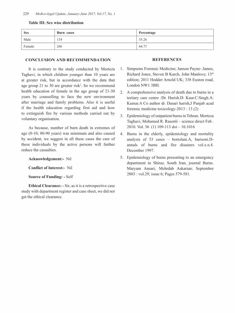
-legal Update, January-June 2017, Vol.17, No. 1 228 229 Medico-legal Update, January-June 2017, Vol.17, No. 1
Table III: Sex wise distribution
Sex Burn cases Percentage
Male 134 35.26
Female 246 64.73
CONCLUSION AND RECOMMENDATION
It is contrary to the study conducted by Morteza Taghavi, in which children younger than 10 years are at greater risk, but in accordance with the data that age group 21 to 30 are greater risk2. So we recommend health education of female in the age group of 21-30 years by counselling to face the new environment after marriage and family problems. Also it is useful if the health education regarding first aid and how to extinguish fire by various methods carried out by voluntary organisation.
As because, number of burn death in extremes of age (0-10; 80-90 years) was minimum and also caused by accident, we suggest in all these cases the care of these individuals by the active persons will further reduce the casualties.
Acknowledgement:- Nil
Conflict of Interest:- Nil
Source of Funding: - Self
Ethical Clearance: - Sir, as it is a retrospective case study with department register and case sheet, we did not get the ethical clearance.
REFERENCES
1. Simpsons Forensic Medicine; Janson Payne- James, Richard Jones, Steven B Karch, John Manlove; 13th
edition; 2011 Hodder Arnold UK; 338 Euston road, London NW1 3BH.
2. A comprehensive analysis of death due to burns in a tertiary care centre :Dr. Harish.D: Kaur.C:Singh.A:Kumar.A Co author dr. Dasari harish,J Punjab acad forensic medicine toxicology-2013 : 13 (2)
3. Epidemiology of outpatient burns in Tehran. Morteza Taghavi, Mohamed R. Rasonli – science direct Feb . 2010. Vol. 36 (1) 109-113 doi – 10.1016
4. Burns in the elderly, epidemiology and mortality analysis of 53 cases – bortolani.A, barisoni.D- annals of burns and fire disasters vol.x.n.4. December 1997.
5. Epidemiology of burns presenting to an emergency department in Shiraz, South Iran, journal Burns. Maryam Ansari, Mehrdab Askarian; September 2003 : vol.29; issue 6; Pages 579-581.

231 -legal Update, January-June 2017, Vol.17, No. 1
Why Students don’t do Research in Zahedan University of Medical Sciences? A Cross-sectional Study in Zahedan 2016
Zahra Pournamdar1, Asef Jahani2, Mahnaz Shahrakipour3
1Community Nursing Research Center, Pregnancy Health Research Center, 2Student Scientific Research Center, 3Department of Biostatistics and Epidemiology, School of Health, Pregnancy Health Research 3Department of Biostatistics and Epidemiology, School of Health, Pregnancy Health Research 3
Center,Zahedan University of Medical Sciences, Zahedan, Iran
ABSTRACT
Introduction: Finding the powers and weaknesses in research programs is the first step of organizing research activities in society. According to this present study aimed to find out why students don’t do research in Zahedan University of Medical Sciences?
Method: 98 students took part in this study. A two part questionnaire was used for data collection. The first part included personal factors which avoid students to do research and the second part included organizational factors. Finally the data was analyzed by SPSS v19 through descriptive statistics.
Results: The mean of age was 21.61 ± 2.09 in this study. 26 (26.8%) students were male while 72 (73.2%) were females. The highest percentage in personal factors were for “I am not familiar with statistical basics”, “I am not familiar with methodology” and “I am too busy”. In organizational factors the highest percentages were for “authorities do not encourage us to do research”, “shortage of funds and resources.
Conclusion: Due to the results of this study, giving enough consults in research, making funds and resources for research and encouraging the students can make students to do research. It seems that educating students in a way which they are doing the job themselves also can help to this matter.
Keywords: Barriers, inhibiting factors, research projects.
Corresponding author:Asef Jahani,Student Scientific Research Center, Zahedan University of Medical Sciences, Zahedan, Iran
INTRODUCTION
Research is the base of every kind of development in the society [1]. Research is so important that these days number of articles, number of researchers and resources in research are indicators of scientific development in countries [2]. Almost all the countries accepted that no development in health and economics are achievable without research and they can’t do anything without research and scientific work results. So they have invested their investments in research [3]. Universities are the place of producing the science. Organizing university researches is an important factor in society’s development in different dimensions [4]. Without
involving university students in research as young human resources and using their innovative minds in the way of country’s development, progress would not be reachable [5]. Research is the main difference between universities and primary and high schools. In universities students can get famous in the country and abroad with research in universities. Due to abovementioned importance of research, having a good resume in research for a student and a university can be helpful for further progress and development. Despite these it is noticed that there are some barriers for doing research. Some of these barriers are solvable and some are not [6, 7]. Administrative bureaucracy, considering the quantity of articles not quality and bias in reviewing the research projects are problems you may face in research [8]. Finding the powers and weaknesses in research programs is the first step of organizing research activities in society. Making better relationship between researchers and people who use the results of the research can solve so many
DOI Number: 10.5958/0974-1283.2017.00050.0
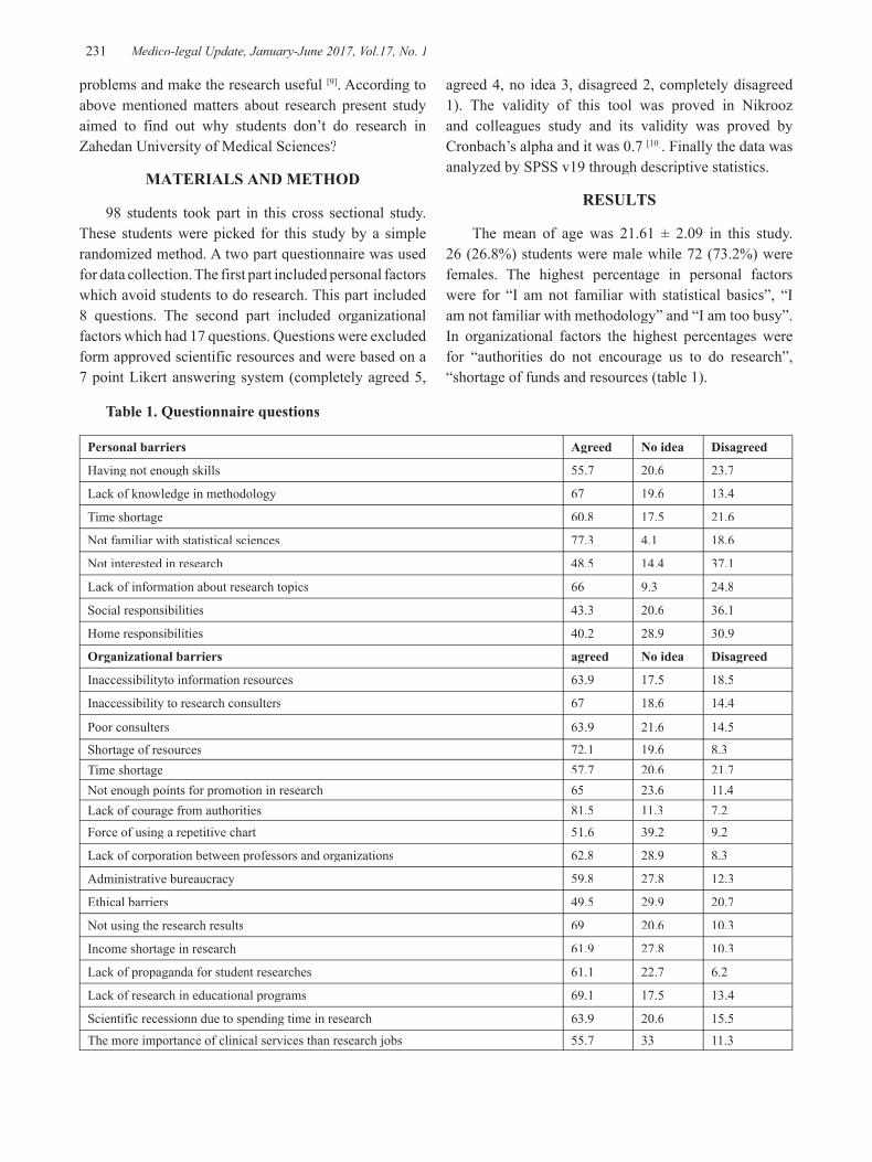
231 Medico-legal Update, January-June 2017, Vol.17, No. 1
problems and make the research useful [9]. According to above mentioned matters about research present study aimed to find out why students don’t do research in Zahedan University of Medical Sciences?
MATERIALS AND METHOD
98 students took part in this cross sectional study. These students were picked for this study by a simple randomized method. A two part questionnaire was used for data collection. The first part included personal factors which avoid students to do research. This part included 8 questions. The second part included organizational factors which had 17 questions. Questions were excluded form approved scientific resources and were based on a 7 point Likert answering system (completely agreed 5,
agreed 4, no idea 3, disagreed 2, completely disagreed 1). The validity of this tool was proved in Nikrooz and colleagues study and its validity was proved by Cronbach’s alpha and it was 0.7 [10]. Finally the data was analyzed by SPSS v19 through descriptive statistics.
RESULTS
The mean of age was 21.61 ± 2.09 in this study. 26 (26.8%) students were male while 72 (73.2%) were females. The highest percentage in personal factors were for “I am not familiar with statistical basics”, “I am not familiar with methodology” and “I am too busy”. In organizational factors the highest percentages were for “authorities do not encourage us to do research”, “shortage of funds and resources (table 1).
Table 1. Questionnaire questions
Personal barriers Agreed No idea Disagreed
Having not enough skills 55.7 20.6 23.7
Lack of knowledge in methodology 67 19.6 13.4
Time shortage 60.8 17.5 21.6
Not familiar with statistical sciences 77.3 4.1 18.6
Not interested in research 48.5 14.4 37.1
Lack of information about research topics 66 9.3 24.8
Social responsibilities 43.3 20.6 36.1
Home responsibilities 40.2 28.9 30.9
Organizational barriers agreed No idea Disagreed
Inaccessibilityto information resources 63.9 17.5 18.5
Inaccessibility to research consulters 67 18.6 14.4
Poor consulters 63.9 21.6 14.5
Shortage of resources 72.1 19.6 8.3Time shortage 57.7 20.6 21.7Not enough points for promotion in research 65 23.6 11.4Lack of courage from authorities 81.5 11.3 7.2Force of using a repetitive chart 51.6 39.2 9.2
Lack of corporation between professors and organizations 62.8 28.9 8.3
Administrative bureaucracy 59.8 27.8 12.3
Ethical barriers 49.5 29.9 20.7
Not using the research results 69 20.6 10.3
Income shortage in research 61.9 27.8 10.3
Lack of propaganda for student researches 61.1 22.7 6.2
Lack of research in educational programs 69.1 17.5 13.4
Scientific recessionn due to spending time in research 63.9 20.6 15.5The more importance of clinical services than research jobs 55.7 33 11.3
Medico-legal Update, January-June 2017, Vol.17, No. 1 232 233 -legal Update, January-June 2017, Vol.17, No. 1
DISCUSSION
Finding the barriers in research are really important and researchers must work to find them out. The results of this study had shown that the highest percentage in personal factors were for “I am not familiar with statistical basics”, “I am not familiar with methodology” and “I am too busy”. In organizational factors the highest percentages were for “authorities do not encourage us to do research”, “shortage of funds and resources. In Nikrooz and colleagues study the most common inhibiting factors was time shortage [10]. Financial problems are another barrier in research which was consistent with other studies [11-13]. Financial and funding problems are a common barriers in research in so many progressing countries [14]. It suggested that authorities held classes with the title of research methods, data analyzing and article writing to take the barriers of research away. Also it is suggested to make research consult offices in schools and hospitals and give them every kind of resources such as internet, printers, libraries, computers and Xeroxes [15]. Student research forces the student to study different surveys from all around the world and then use the best of them [16]. As the student researches profits are more explained to students they would come more to this field. Authorities are responsible to make a bridge between classes and research laboratories for students so they can come and do student researches [17].
CONCLUSION
Due to the results of this study, giving enough consults in research, making funds and resources for research and encouraging the students can make students to do research. It seems that educating students in a way which they are doing the job themselves also can help to this matter.
This study was the result of a student research project (7861) approved at Zahedan University of Medical Sciences.
Source of Funding- Zahedan University of Medical Sciences
Conflict of Interest - Nil
REFERENCES
1. Sedigh MJ, Karimian Eghbal M, Ghadiri F. Parkhaye tahghighati va toseaeye tahghighat. Encyclopedia of
Higher Education. 2004; 245–249.
2. Naghizadeh Baghi A, Khanbabazadeh Ghadim M, Samari E. Survey of Barriers and Research Problems from the Viewpoint of Faculty Members of Ardabil University of Medical Sciences. Journal of Ardabil University of Medical Sciences. Journal of Ardabil University of Medical Sciences; 2015;15(3): 255–264.
3. Khoshfar GR. The Role of research, research centers, research and economic-social development. J Rahyaft. 2001;22: 98–103.
4. Karimian Z, Sabbaghian Z, Saleh Sedghpour B, Lotfi F. Internal obstacles in research activities: faculty members’ viewpoints in Shiraz University of Medical Sciences. Iranian Journal of Medical Education. Iranian Journal of Medical Education; 2012;11(7): 750–763.
5. Entwistle N. Research-based university teaching: what is it and could there be an agreed basis for it? Psychology of Education Review. The british psychological society.2002;26: 3–9.
6. Dadkhah B, Mohammadi M, Poornaseri S, Mozaffari N, Adham D. View of Ardabil province universities scientific members’ about research and its limitations. Journal of Ardabil University of Medical Sciences. Journal of Ardabil University of Medical Sciences; 2008;8(1): 37–44.
7. Anbari Z, Jamshidifar A, Setareh M. The viewpoints of faculty members about research activities problems in Arak University of Medical Sciences. Iranian Journal of Medical Education. Iranian Journal of Medical Education; 2005;5(2): 196–198.
8. Changiz T, Adibi P, Hoseini M, Tootoonchi M. Didgahe asatide rahnama dar morede moshkelate ravande ejraye payannameye tahsilye pezeshkye omoomi dar daneshgahe oloom pezeshkye Esfehan. Iranian Journal of Medical Education. 2003;9: 24–32.
9. Alamdari A, Afshoon A. Barriers to research activities: Views of faculty members in Yasuoj university of medical sciences. Journal of Armaghane danesh. 2002;29: 27–34.
10. Nikrooz L, Rouzitalab M, Rayegan Shirazi AR, Naghizadeh MM, Taghavi F. Student Research Projects Inhibiting Factors from the Students Perspective. Journal of Fasa University of Medical Sciences. Journal of Fasa University of Medical

-legal Update, January-June 2017, Vol.17, No. 1 232 233 Medico-legal Update, January-June 2017, Vol.17, No. 1
Sciences; 2012;2(2): 113–119.
11. Zinaloo A, Mohammadi M, Geranmayeh M, Farsaam H, Khakbazan Z. Research perspective at Tehran University of Medical Sciences from the viewpoint of the faculty members. Iranian Journal of Medical Education. Iranian Journal of Medical Education; 2002;2: 60.
12. Jafari H, Yaghoubi T, Heidari J, Esmaeili R, Hekmat Ara M, Ataei M. Ideas of the Mazandaran University of Medical Sciences faculty members towards the internal and external organizational obstacles effective on their research process in 2000. Mazandaran Scientific Research Journal of Faculty of Nasibeh Nursing and Midwifery of University (Shakiba). 2004;3: 13–20.
13. Khanchamani J. Knowledge production and expanding the frontiers of knowledgeand basic strategies for cultural social and industrial development of Iran. Tehran: Islamic Azad University Publications. 2004.
14. Malekafzali H, Majdzade R, Fotouhi A, Tavakoli S. Ravesh shenasye pajooheshhaye karbordi dar oloom pezeshki. Tehran: Tehran University of Medical Science. 2004.
15. Zohor A, Fekri A. The barriers of research: Views of faculty members in Iran university of medical sciences. Payesh Quarterly. 2003;2(2): 113–120.
16. Farmanbar R, Askari F. Study of barriers related to research: views of faculty members in Gilan university of medical sciences. Journal of Medical faculty of Gilan university of medical sciences. 2005;14(54): 84–91.
17. Nabavi B. Introduction to research methodology in social sciences. Tehran, Iran: Farvardin Publications. 2006.
18. Pournamdar Z. A survey of the views of nursing school students to the benefits of using Clinical Skills Learning Center. Der Pharmacia Lettre. [Online] 2016;8(3): 148–153.

235 -legal Update, January-June 2017, Vol.17, No. 1
Profile of Poisoning Cases in a Tertiary Care Hospital
Sidramappa Gouda1, M Surender Rao2
1Assistant Professor, Department of Forensic Medicine, Navodaya Medical College, Raichur, 1Assistant Professor, Department of Forensic Medicine, Navodaya Medical College, Raichur, 1
2Assistant Professor, Department of Forensic Medicine, DRVRK Women’s Medical College, Hyderabad2Assistant Professor, Department of Forensic Medicine, DRVRK Women’s Medical College, Hyderabad2
ABSTRACT
In developing countries like India poisoning is one of the most common causes of morbidity and mortality. There is great variation in type of poisoning consumption and also method of poisoning. Hence to know the profile of poisoning, a retrospective study was conducted in Navodaya Medical College Hospital and research centre, Raichur. Study period was between Jan- Oct 2015. 200 cases of poisoning cases were studied during this period. The aim of the study was to know the age and sex distribution of the victims, commonest type of poison used and manner of poisoning. In the present study maximum cases of poisoning was seen in the age group of 21 to 30 years (35.5%) and males were more when compared to female. Among 200 subjects majority i.e 126 (63%) were due to organophosphorus campound. When manner of death was evaluated, suicide was the most common with 78.5 %( 157) of study subjects.
Keywords: Poisoning, Organophosphorous compound, Unnatural death.
Corresponding author:Dr.Sidramappa GoudaAssistant Professor, Department of Forensic Medicine, Navodaya Medical College, Raichur.Contact No: 7899606175E mail : [email protected]
INTRODUCTION
Poisoning is one of the most common health problem world wide with significant morbidity and mortality. Earlier reports show that on an average 700 people die from poisoning every day1, 2. It is estimated that half a million people die every year as a result poisoning due to pesticides2. Some of the previous studies clearly showed that the increase in poisoning is mainly because of medications and chemicals particularly pesticides3. The pattern of poisoning varies from country to country and region to region depending on many factors like geography, accessibility and availability of poison, socio economic conditions, cultural and religious influences. In countries like India poisoning by pesticides in more common as most of them are employed in agriculture4. Whereas in countries such as Norwy, benzodiazepines, ethanol and paracetomol are the most common causes for poisoning5.
MATERIALS AND METHOD
All poisoning cases admitted to Navodaya Medical College, Raichur and post-mortem done in district Hospital, Raichur, during the period Jan- Oct 2015 were included in this study. The data were collected from hospital case records, police inquest forms, post mortem report and forensic science laboratory report. The collected data was analysed and tabulated.
RESULTS
In this study following results were found. Total no. of deaths due to poisoning was 200 cases. Table no.1 shows age and sex wise distribution of poisoning deaths. Among 200 cases125 were males and 75 were females. The incidence of poisoning was more in third decade age group i.e. 71(35.5%). Table no 2 shows different types of poisons consumed, among all, organophorphorous compound was most common. 126(63%) deaths out of 200 were due to this compound followed by snake bite. Table no 3 shows manner of death. Among 200 deaths due to poisoning suicidal manner was most common i.e. 157(78.5%) out of 200, followed by accidental deaths. Table no 4 shows domicile pattern of the victims. Poisoning cases were most common in rural areas.
DOI Number: 10.5958/0974-1283.2017.00051.2

235 Medico-legal Update, January-June 2017, Vol.17, No. 1
Table 1: Age and sex wise distribution of poisoning deaths
Age Male Female Total Percentage
0-10 yrs 2 1 3 1.5%
11-20 yrs 10 16 26 13%
21-30 yrs 45 26 71 35.5%
31-40 yrs 30 13 43 21.5%
41-50 yrs 25 10 35 17.5%
51-60 yrs 8 6 14 7%
61-70 yrs 4 3 7 3.5%
>71 yrs 1 0 1 0.5%
Total 125 75 200 100%
Table 2: Types of poisons
Poison Male Female Total Percentage
Organophosphorous 85 41 126 63%
Snake bite 13 9 22 11%
Aluminium phosphide 7 13 20 10%
Organochlorine 8 7 15 7.5%
Alcohol 9 0 9 4.5%
Carbamates 1 2 3 1.5%
Pyrethroids 0 2 2 1%
Micellanenous 2 1 3 1.5%
Total 125 75 200 100%
Table 3: Manner of Death
Manner of Death Male Female Total Percentage
Suicidal 105 52 157 78.5%
Accidental 15 18 33 16.5%
Homicidal 5 5 10 5%
Total 125 75 200 100%
Table 4: Domicile pattern of victims.
Manner of Death Male Female Total Percentage
Rural 97 41 138 69%
Urban 28 34 62 31%
DISCUSSION
Poisoning is one of the most common causes of unnatural deaths worldwide. In this present study it was found that 62.5% were males. 37.5% females and most of them were from rural areas and oraganophosphorous compound was most commonly used for poisoning.
Hence it was concluded that individuals with rural background and agricultural occupation were the most common victims. Failure of the monsoon rains with improper yield in the farms was probably associated with poisoning6. In another study of poisoning cases presented to emergency department, the female to male ratio is 1.17:1. Most of poisoning occurred in
Medico-legal Update, January-June 2017, Vol.17, No. 1 236
the age group of 15-24 yrs. Snake bite was the most common among all with majority of the cases being farmers7. This result varied with our findings where organophosphorous was most common compound for poisoning and victims were most common in third decade. In another study of 137 cases during one year period maximum cases belong to 20-30 years of life. Suicidal was the most common manner occurring in the evening hours. Organophosphoros compound was most commonly used poison8 which was similar to our study results.
CONCLUSION
Proper counselling is required for the population deployed in agriculture to reduce the incidents of suicide. Knowledge of signs and symptoms of insecticide poisoning with quick medical measures can bring down the incidents of deaths due to poisoning. Strict laws should be made with the sale and distribution of toxic compounds.
Conflict of Interest: Nil
Source of Funding: Self.
Ethical Clearance: Taken from ethical clearance committee of our college.
REFERENCES
1. Zine KU, Mahanty AC. Pattern of acute poisoning at Indira Gandhi Medical College and Hospital, Nagpur. J Indian Acad Forensic Med 1998;20:37-9.
2. International Programme of Chemical Safety, World Health Organization. Guidelines for poison control. Geneva, Switzerland: WHO Press; 1997.
3. Kora SA, Doddamani GB, et al. Socio Demographic Profile of the Organophosphorous Poisoning cases in Southern India. J Clin Diagn Res 2011;5(5):953-6.
4. Aaron R, Joseph A, Abraham S, Muliyil J, George K, Prasad J, et al. Suicides in young people in rural Southern India. Lancet 2004;363 (9415) :1117-8.
5. Hovda KE, Bjornaas MA, Skog K, Opdhal A, Drottning P, Ekberg O, et al. Acute poisonings treated in hospitals in Oslo: A one year Prospective study (I): Pattern of poisoning. Clin Toxicol (Phila) 2008 Jan;46 (1) :35-41.
6. AK Batra*, AN Keoliya**, GU Jadhav Poisoning:An Unnatural Causes of Morbidity and Mortality in Rural India, JAPI, Vol.51, October 2003, Pg955-959.
7. Gurung CK, Dahal R, Khanal P, Nepal S, Jaiswal AK. Pateern of poisoning cases in a hospital in a Terai District of central Nepal. Nepal Med College. J.2011 Sep;13(3):160-3.
8. Karki R K, Risal. Study of Poisoning cases in a tertiary care hospital, Kathmandu university medical journal, vol.10, No.4, Issue 40, Oct-Dec 2012, page 70-72.

-legal Update, January-June 2017, Vol.17, No. 1 236
How Can I have a better Communication with my Professors? a Study form Student’s attitude in Zahedan 2016
Zahra Pournamdar1, Alireza Shameli 2, Mahnaz Shahrakipour3
1Community Nursing Research Center, Pregnancy Health Research Center, 2Student Scientific Research Center, 3Department of Biostatistics and Epidemiology, School of Health, Pregnancy Health Research 3Department of Biostatistics and Epidemiology, School of Health, Pregnancy Health Research 3
Center, Zahedan University of Medical Sciences, Zahedan, Iran
ABSTRACT
Introduction: Student’s best learning situation would come after best communication they have with their professors. The best thought, creativity and plan come after a good communication. Due to above mentioned facts this study aimed to find out what are the factors which effect on communication between students and professors from students attitude.
Method: 115 students from Zahedan University of medical students took part in this cross sectional study. The data was collected through a 33 question questionnaire. After data collection they were analyzed though SPSS v19 by descriptive statistics.
Results: The mean of age was 21.16 ± 1.88. 77 respondents were males while 38 were females. Most of “too much” answers went for “sincerity and sympathy to students” and “considering social and ethical values” in personal characteristic dimension. Most of “too much” answers went for “professor’s responsibility” and “fairness in dealing with students” in professor’s professional characteristics dimension. Most of “too much” answers went for “professors being up to dated” and “mastering scientific concepts and materials” in professor’s scientific characteristics dimension.
Conclusion: The results of this study had shown that “sincerity and sympathy to students” and “professor’s responsibility” had the most effect on the communication between students and their professors. These two important factors are mostly forgotten by authorities unfortunately.
Keywords: Communication, professor, student, attitude.
Corresponding author: Alireza Shameli, Student Scientific Research Center, Zahedan University of Medical Sciences, Zahedan, Iran
INTRODUCTION
Professors are the most important members of medical universities. The faculty members in medical universities are responsible for: education, research, patient care, management and other activities out of the university [1]. Educating the medical students is the main purpose of professors in medical universities. Increasing the quality of education among students could lead to student’s motivating and professor’s better performance. A good professor can ease the educating process and
even complete the books and educational resources [2]. Good teaching needs good communication with students [3, 4]. Professors can make a good learning situation for students by using communication skills, books and educational resources. A bad professor could influence and destroy a student’s whole carrier [5]. The best thought, creativity and plan come after a good communication. Communication means to share information with each other. Without sharing information no thoughts are shared either [6]. Effective communication is necessary to make coordination between human resources and make a proper network between them [7]. As it was mentioned in above text communication skills are too important to the professors for teaching in a good way [8]. Due to above mentioned facts this study aimed to find out what are the factors which effect on communication between
DOI Number: 10.5958/0974-1283.2017.00052.4

Medico-legal Update, January-June 2017, Vol.17, No. 1 238 239 -legal Update, January-June 2017, Vol.17, No. 1
students and professors from students attitude.
MATERIAL AND METHOD
115 students from Zahedan University of medical students took part in this cross sectional study. The samples were chosen by a simple random method. The data was collected through a 33 question questionnaire. This questionnaire was made in 2012 in Birjand University of medical sciences by some experts in medical education [2]. After data collection they were analyzed though SPSS v19 by descriptive statistics. After demographic information, the questions about the factors effecting on professor-student communication came in this tool. For better understanding and better analyzing questions were divided in three categories including: professor’s personal characteristics (12 questions), professor’s scientific characteristics (6 questions) and professor’s professional characteristics (14 questions). The effectiveness of each factor was
evaluated by “too much, a lot, moderate, few, and very few”. The validity of the questionnaire was proved by 10 experts after writing the content and this reliability was proved by Cronbach’s alpha and it was 0.88. After data collection they were analyzed though SPSS v19 by descriptive statistics.
RESULTS
The mean of age was 21.16 ± 1.88. 77 (67%) respondents were males while 38 (33%) were females. Most of “too much” answers went for “sincerity and sympathy to students” and “considering social and ethical values” in personal characteristic dimension. Most of “too much” answers went for “professor’s responsibility” and “fairness in dealing with students” in professor’s professional characteristics dimension. Most of “too much” answers went for “professors being up to dated” and “mastering scientific concepts and materials” in professor’s scientific characteristics dimension.
Effectiveness questions
Too much A lot Moderate Few Very few15.7 35.7 33.9 7.8 7 Age
Pers
onal
chr
acte
ristic
s
13 27 36.5 18.3 5.2 Sex 17.4 31.3 34.8 13 3.5 Apperience 28.7 38.3 23.5 7 2.6 High confidence 28.7 47.8 15.7 6.1 1.7 Flexibility 33 38.3 22.6 5.2 .9 Modesty and humility27 35.7 32.2 3.5 1.7 Fame and pupularity 44.3 33 15.7 7 0 sincerity and sympathy to students34.8 33.9 22.6 6.1 2.6 professor and student having same major38.3 32.2 21.7 7.8 0 considering social and ethical values26.1 37.4 25.2 10.4 0.9 Attending the class on time24.3 38.3 26.1 10.4 0.9 Criticism acceptance27 39.1 19.1 10.4 4.3 Respecting students
Prof
essi
onal
cha
ract
eris
tics
23.5 47.8 20.9 7 0.9 Trying to solve student’s problems15.7 27.8 40 12.2 4.3 Trusteeship and Secrecy44.3 32.2 18.3 3.5 1.7 responsibilty27 33.9 21.7 8.7 8.7 Including students in discussions18.3 33 32.2 13 3.5 Motivating students to study and learn30.4 33 27.8 6.1 2.6 fairness in dealing with students20.9 37.4 31.3 9.6 .9 Encouraging the students on its time20 36.5 29.6 12.2 1.7 Giving coreculim to students 18.3 24.3 41.7 7.8 7 Easey access to professor out of class22.6 33.9 27 12.2 4.3 Professor’s scientific level23.5 42.6 23.5 7.8 2.6 Conclusion of lessons in end of clases27 38.3 27 3.5 4.3 Using different educational methods and tools20 36.5 34.8 6.1 2.6 Transfering the knowledge all clearley to students24.3 25.2 33.9 9.6 7 Using examples in teaching
Scie
ntifi
c ch
arac
teris
tics
20.9 40.9 28.7 7.8 1.7 Keeping the conjuction between lessons42.6 3.3 19.1 4.3 2.6 mastering scientific concepts and materials19.1 34.8 32.2 9.6 4.3 Research experinces 27.8 30.4 34.8 4.3 2.6 Educational experiences 43.5 38.3 13 3.5 1.7 professors being up to dated
-legal Update, January-June 2017, Vol.17, No. 1 238 239 Medico-legal Update, January-June 2017, Vol.17, No. 1
DISCUSSION
The results of this study had shown that the “Modesty and humility” and “fairness in dealing with students” had the highest score among other questions. This result was consistent with other studies [6]. Ghadami and colleagues had shown in their study in Arak University of medical sciences that 68.5% of students stated that professor’s behavior is very important in communication [9]. Totally considering humanity and behavior by professors have an important role in improving student-professor communications. Professor’s age and appearance had the least effect on communication. The results of this study also prove this fact [2].
In professional dimension responsibility and fairness in dealing with students were most effective factors which other studies proved it too [2]. According to Khosravi and colleagues study respecting the student and professor’s behavior are the most effective factors on student-professor communication [10]. In Abidi and colleagues study it was Trusteeship and Secrecy which had the most effect [11]. In Scientific characteristics “mastering scientific concepts and materials” and “professors being up to dated” had the most effect [2]. In Khosravi and colleagues study professors scientific level and his experience were the most effective ones [10]. In Ghadami and colleagues study speech abilities, teaching skills, professors scientific level and his experience were the most effective ones [5]. Communication between student and professors causes confidence in students and increases their motivation for learning [12]. A good teaching would come after a good communication so it is suggested to professors to improve their communicational skills [13]. If professors do this issue this communication could help student’s behaviors and improve their educational performance
CONCLUSION
The results of this study had shown that “sincerity and sympathy to students” and “professor’s responsibility” had the most effect on the communication between students and their professors. These two important factors are mostly forgotten by authorities unfortunately.
This study was the result of a student research project (6192) approved at Zahedan University of Medical Sciences
Source of Funding- Zahedan University of Medical Sciences
Conflict of Interest - Nil
REFERENCES
1. Zohoor AR, Islami Nejad T. Shakheshaye tadrise asarbakhsh az didgahe daneshjooyane daneshgahe oloom pezeshkye Kerman. Payesh Journal. 2002;1(4): 5–13.
2. Abedini MRM, Abassi A, Mortazavi FF, Bijari B. Students’ viewpoint of factors affecting teacher-students communication, A study in Birjand University of Medical Sciences. Iranian Journal of Medical Education. Iranian Journal of Medical Education; 2012;12(6): 439–447.
3. Bland CJ, Wersal L, VanLoy W, Jacott W. Evaluating faculty performance: a systematically designed and assessed approach. Academic medicine. LWW; 2002;77(1): 15–30.
4. Pournamdar Z. A survey of the views of nursing school students to the benefits of using Clinical Skills Learning Center. Der Pharmacia Lettre. [Online] 2016;8(3): 148–153.
5. Ghadami A, Salehi B, Sajadi S, Naji H. Students’ points of view regarding effective factors in establishing communication between students and faculty members. Iranian Journal of Medical Education. Iranian Journal of Medical Education; 2007;7(1): 149–154.
6. Torabi A, Zahiri M. Study on Influencing Factors Relationship between Teacher and Student in View of Students of Ahvaz University of Medical Sciences. Ù… ج Ù „Ù‡ Ø1Ù „Ù… ÛŒ Ù3⁄4Ø2Ø´ Ú© ÛŒ ج Ù† د ÛŒ Ø´ Ø § Ù3⁄4ور. 2012;1(11): 549–557.
7. Irannezhad M, Sasangohar P. Organization and Management: From Theory to Practice. Tehran: Iran Banking Institute. 2008.
8. Moghimi SM. Organization and management research approach. Tehran, cashmere Publications. 2006.
9. Ghadami A. Study on affective factors on Relationship between Teacher and Student in view of Students of Arak University of Medical Sciences. IR J Edu in Medical Sci. 2007;7(1): 149–154.
10. Khosravi F, Shahouei R, Hesami K. Savadzade Sh.[Avamele moaser dar barghararye ertebat beine daneshjoo va ostad az didgahe daneshjooyane

Medico-legal Update, January-June 2017, Vol.17, No. 1 240
oloom pezeshkye Kordestan]. Abstract of First National Conference on Medical Education Development. Sanandaj. 2010.
11. Obeidi N. Effective factors on the communication between students and faculty members in the viewpoint of paramedical students. Educational strategies.2010:133-136.
12. Paterson B, Crawford M. Caring in nursing education: An analysis. Journal of Advanced Nursing. Wiley Online Library; 1994;19(1): 164–173.
13. Spencer J. Learning and teaching in the clinical environment. British Medical Journal. BMJ Publishing Group; 2003;326(7389): 591.
-legal Update, January-June 2017, Vol.17, No. 1 240
Are the Patients Satisfied with Healthcare Services Provided by Nurses in Hospital? A Descriptive Analytic Study in
Ali-ebne Abi Taleb Hospital in Zahedan 2016
Zahra Pournamdar1, Ali Alemifar2, Mahnaz Shahrakipour3
1Community Nursing Research Center, Pregnancy Health Research Center, 2Student Scientific Research Center, 3Department of Biostatistics and Epidemiology, School of Health, Pregnancy Health Research 3Department of Biostatistics and Epidemiology, School of Health, Pregnancy Health Research 3
Center, Zahedan University of Medical Sciences, Zahedan, Iran
ABSTRACT
Introduction: Patient’s satisfaction about the healthcare services is an important indicator for healthcare service’s quality assessment. Investigating patient’s satisfaction is important for efficiency assessment and healthcare services effectiveness. This study was conducted to answer this question that “Are the patients satisfied with healthcare services provided by nurses in hospitals?”
Method: 50 patients who were chosen by a random sampling method were included to this descriptive analytic study in 2016. LA Monika’s “nursing services satisfaction” questionnaire was used to collect the data. The data were analyzed thorough SPSS v19 by descriptive statistics, Pearson correlation and T test.
Results: The mean of age was 33.06 ± 15.43 in this study. 27 respondents were female while 23 were males. 13 were single while 37 were married. 27 respondents had an experience of hospitalization. The mean score of nursing services satisfaction questionnaire was 33.06 ± 15.43. This score shows a moderate satisfaction among patients. There wasn’t any significant relationship between demographic information (age, sex, marriage status, experience of hospitalization) and patient’s satisfaction of healthcare services.
Conclusion: Due to necessity of high quality healthcare services and the results of this study it is suggested to make committees of patient’s satisfaction assessment to assess the reasons of patient satisfaction and make plans to reduce the dissatisfactions. This would finally lead to high quality nursing services for patients.
Keywords: Patient satisfaction, nursing services, Zahedan.
Corresponding author: Ali alemifar, Student Scientific Research Center, Zahedan University of Medical Sciences, Zahedan, Iran. Email: [email protected]
INTRODUCTION
Medicine has considered patient’s satisfaction of healthcare services for years since 1950s. Today this satisfaction really matters to authorities [1]. In a large number of countries patient’s satisfaction about the healthcare services is an important indicator for healthcare service’s quality assessment. So then these countries try hard to build this satisfaction, keep it and improve it [2]and improve it [2]and improve it . Investigating patient’s satisfaction
is important for efficiency assessment and healthcare services effectiveness. It will assess the hospital’s performance in whole the nation [3]. In European countries patient’s satisfaction is assessed through a national program [4]. Ware showed in his study that 45 articles proved this that patient’s satisfaction is an appropriate indicator for assessing the quality of healthcare services [5]. Unfortunately there some problems in the way of providing healthcare services to patients in our countries hospitals. Due to this some basic changes must be conducted on the way of providing care to patients in Iranian hospitals [6]. Studies had shown that if the patients be satisfied with the healthcare services that they are getting they pay much more attention to the medical advices [7]. In 2006 Shahin and Tatar mentioned in their study that there five factors effecting
DOI Number: 10.5958/0974-1283.2017.00053.6
Medico-legal Update, January-June 2017, Vol.17, No. 1 242 243 -legal Update, January-June 2017, Vol.17, No. 1
patient satisfaction. These five factors are physician’s capability, information, quality of care, waiting time quality of hospital [8]. Patient’s satisfaction is usually seen by caring factors, trust, unity and attention. Some surveys proved that demographic information such as age, education, wellbeing, sex and marriage status have a direct relationship with patient’s satisfaction [9]. Bakker said in his study that a high percent of dissatisfactions accrued due misunderstandings and lack of good communication [10].
According to the importance of patient’s satisfaction this study was conducted to answer this question that “Are the patients satisfied with healthcare services provided by nurses in hospitals?”
MATERIAL AND METHOD
This study was the result of a student research project approved (7813) at Zahedan University of Medical Sciences. 50 patients who were chosen by a random sampling method were included to this descriptive analytic study in 2016. A two section questionnaire was used to collect data. The first section included demographic information (age, sex, hospitalization experience and marriage status). The second section was LA Monika’s “nursing services satisfaction” questionnaire. This tool was invented by LA Monika in 1986. It contains 41 questions. Each question is answered by a 77 point Likert method from completely disagree to completely agreed. The range of score in this tool was 41to 287. As the score is higher the more satisfaction patients have from the services. A checklist was used to collect the personal, social and disease related information. The validity of this questionnaire was proved in Moghadasian and colleagues study and its reliability was proved by Cronbach’s alpha and it was 0.9[11].
RESULTS
The mean of age was 33.06 ± 15.43 in this study. 27 (54%) respondents were female while 23 (46%) were males. 13 (26%) were single while 37 (74%) were married. 27 (54%) respondents had an experience of hospitalization. The mean score of nursing services satisfaction questionnaire was 33.06 ± 15.43. This score shows a moderate satisfaction among patients. There wasn’t any significant relationship between demographic information (age, sex, marriage status, experience of hospitalization) and patient’s satisfaction of healthcare
services (p<0.05).
DISCUSSION
Nursing is a profession with scientific and humanistic dimensions. Patients are the reason of nursing’s existence. Nurses aim to help patients get their needs that they can’t have them on their own while they are in the bed. If nurses succeed in this purpose patient’s satisfaction would come after that. If you one know the quality of care in hospitals you can take look on patient’s satisfaction [12]. The results of this study had shown a good satisfaction among patients. This was consistent with Ansari’s study [13]. But in a survey in Paris [14] and Hajian and colleagues study the rate of satisfaction was more than this study [15]. Despite this the satisfaction in this study was acceptable with all the problems which nurses face here every day and the job burnout they have. This shows that the nurses here closed their eyes on all the problems and they are doing their job properly and with love [16]. Making good and proper communication with patients and their family really helps to their satisfaction. Dairsen also proved in his study that communicational factors are really important for keeping the patient satisfied [17]. Some other studies in our country also insist on this matter [18, 19]. So it seems necessary to improve nurse’s communication skills. There are many factors which can effect on patient’s satisfaction. Some are related to healthcare providers and some others are related healthcare environment. It is true that 50 patients are not enough to find out any significant relationship between satisfaction and demographic information but conducting quantitative studies can help the society to collect valuable information and use them for planning and further studies [20, 21]
CONCLUSION
Due to necessity of high quality healthcare services and the results of this study it is suggested to make committees of patient’s satisfaction assessment to assess the reasons of patient satisfaction and make plans to reduce the dissatisfactions. This would finally lead to high quality nursing services for patients.
This study was the result of a student research project approved (7813) at Zahedan University of Medical Sciences.Source of funding- Zahedan University of Medical Sciences.
Source of funding- Zahedan University of Medical Sciences

-legal Update, January-June 2017, Vol.17, No. 1 242 243 Medico-legal Update, January-June 2017, Vol.17, No. 1
Conflict of Interest - Nil
REFERENCES
1. Williams B. Patient satisfaction: a valid concept? Social science & medicine. Elsevier; 1994;38(4): 509–516.
2. Jolaee S, Hajibabae F, Jalal AJ, Bahrani N. Evaluation of patients’ satisfaction with nursing care provided in hospitals. Hayat. 2011;17(1): 35–44.
3. Skurka MA. Health information management: principles and organization for health record services. American Hospital Publishing, Incorporated; 1998.
4. Sitzia J. How valid and reliable are patient satisfaction data? An analysis of 195 studies. International Journal for Quality in Health Care. ISQHC; 1999;11(4): 319–328.
5. Ware Jr JE. What Information Do Consumers Want and How Will They Use It?. Medical care. LWW; 1995;33(1): JS25.
6. Cohen L, Delaney P, Boston P. Listening to the customer: implementing a patient satisfaction measurement system. Gastroenterology Nursing. LWW; 1994;17(3): 110–115.
7. Bond S, Thomas LH. Measuring patients’ satisfaction with nursing care. Journal of advanced Nursing. Wiley Online Library; 1992;17(1): 52–63.
8. Commission J. National patient safety goals: 2006 critical access hospital and hospital national patient safety goals. 2012-04-28]. http://www. ed-qual. com/emergency_medici-ne_news/ed_news_2006_jca-ho_patient_safety_goals. htm. 2009.
9. McEwen M. Spiritual nursing care: state of the art. Holistic nursing practice. LWW; 2005;19(4): 161–168.
10. Bakker DA, Fitch MI, Gray R, Reed E, Bennett J. Patient–health care provider communication during chemotherapy treatment: the perspectives of women with breast cancer. Patient Education and Counseling. Elsevier; 2001;43(1): 61–71.
11. Mogadasiyan S, Abdolahzadeh F, Rahmani A, Nikanafar AR, Firoziyan AA. Satisfaction with nursing care and related factors in hospitalized cancer patients in shahid ghazi hospital in tabriz.
Journal of Urmia Nursing & Midwifery Faculty. 2013;11(7).
12. Pournamdar Z. A survey of the views of nursing school students to the benefits of using Clinical Skills Learning Center. Der Pharmacia Lettre. [Online] 2016;8(3): 148–153.
13. Ansari H, Ebadi F, Molasadeghi G. Patient satisfaction of Iran University of medical sciences. Birjand Univ Med Sci J. 2005;11(3): 38–45.
14. Goupy F, Ruhlmann O, Paris O, Thélot B. Results of a comparative study of in-patient satisfaction in eight hospitals in the Paris region. International Journal for Quality in Health Care. ISQHC; 1991;3(4): 309–315.
15. Hajian K. Evaluation of patients satisfaction with hospital care in shahid beheshti and yahanejad hospitals(BABOL; 2005). Journal of babol university of medical sciences(JBUMS); 2007:51-60.
16. Sitzia J, Wood N. Patient satisfaction: a review of issues and concepts. Social science & medicine. Elsevier; 1997;45(12): 1829–1843.
17. Dierssen-Sotos T, Rodríguez-Cundin P, Robles-García M, Brugos-Llamazares V, Gómez-Acebo I, Llorca J. [Factors associated with patient satisfaction with hospital care]. Anales del sistema sanitario de Navarra. 2008. p. 317–325.
18. Emadzadeh A, Yavari M, Ebrahimzadeh S, Ahmadian N. The effects of dental graduate students satisfaction in mashhad university of medical sciences in 1383. Journal of mashhad dental school. 2004:69-76.
19. Jafari H, Ainy E, Sh H, Kohyar E, Hajifathali A. Patient satisfaction and its related factors in Ayatollah taleghani hospital in 2006. Pajoohandeh Journal. Pajoohandeh Journal; 2008;12(6): 541–546.
20. Coyle J, Williams B. Seeing the wood for the trees: defining the forgotten concept of patient dissatisfaction in the light of patient satisfaction research. Leadership in Health Services. MCB UP Ltd; 1999;12(4): 1–9.
21. Avis M, Bond M, Arthur A. Satisfying solutions? A review of some unresolved issues in the measurement of patient satisfaction. Journal of advanced nursing. Wiley Online Library; 1995;22(2): 316–322.

245 -legal Update, January-June 2017, Vol.17, No. 1
Determination of Body Weight from Footprint Length Measurements among Melanau Population in Malaysia
T Nataraja Moorthy1, Hairunnisa Bt Mohd Anas Khan2
1Associate Professor of Forensic Sciences, Forensic Science Program, Faculty of Health and Life Sciences, Management and Science University, Shah Alam, Selangor, Malaysia,
2Forensic Division, Chemistry Department of Malaysia, Kuching, Sarawak State, East Malaysia2Forensic Division, Chemistry Department of Malaysia, Kuching, Sarawak State, East Malaysia2
ABSTRACT
Foot impressions found in crimes form a valuable physical evidence for person identification. Literature review shows that very few studies were conducted to correlate body weight with footprint for crime scene application. Body weight involves a variety of cultural and physical variables. The present study involved 200 Malanau ethnic subjects (100 males and 100 females) mostly residing in east Malaysia and aimed to derive population specific regression equations to determine live body weight from bilateral footprint lengths collected from the consented individuals. Investigation revealed that all footprint lengths exhibit statistically positive significant correlation with body weight (p<0.001). The correlation coefficient (R) in pooled sample (0.242-0.290) is found to be comparatively higher than those of male (0.145-0.215), and female footprints (0.166-0.274). The results were presented in the form of tables. The equations derived for pooled sample can be used even when the sex of the footprint owner remains unknown.
Keywords: Forensic anthropology, body weight, footprint length, Malanau ethnics, Malaysia.
Corresponding author:T Nataraja MoorthyAssociate Professor of Forensic Sciences, Forensic Science Program, Faculty of Health and Life Sciences, Management and Science University, Shah Alam, Selangor, Malaysia, E-mail: [email protected] & [email protected]
INTRODUCTION
An aspect of human identification that has received scant attention from forensic anthropologists is the study of human foot and foot impressions made by the feet1. Person identification through footprint analysis is also an emerging biometric technique2. The characteristic features of an individual’s footprint is as unique as his fingerprints and hence can be used for identification purpose3. The presence of complete or partial two dimensional (2D) and three dimensional (3D) foot impressions at the crime scenes form a valuable physical evidence to compare with suspects4,5. Foot impressions are found at crime scenes since offenders often remove their footwear, either to avoid noise or to gain better grip in climbing walls, etc., while entering or exiting 6. Researchers have conducted study on
footprints7-11, foot outlines12-14 and feet 15-19 to estimate stature for crime scene application. But it is unfortunate that very limited studies were conducted to determine body weight from footprints recovered for crime scene application. It is important that racial and cultural aspects of foot morphology must be considered while conducting the foot impression study20 and hence a single formula cannot represent for all races or regions in a country7,10,14,21-23. Hence, the present study is aimed to derive population specific formulae to determine body weight from footprint length measurements for Melanau ethnic, an indigenous population mostly living in Sarawak state, East Malaysia.
MATERIALS AND METHOD
The sample collection was conducted at Sarawak state, East Malaysia. The subjects were from colleges, universities and general public. Most of the Melanaus are living in Sarawak state, East Malaysia. The subjects’ age ranged from 18 to 60 years. Subjects with any apparent foot-related disease, pregnancy, orthopedic deformity, physical impairment, injury or disorders were excluded from the study. The foot prints were collected from the participants following the standard procedure
DOI Number: 10.5958/0974-1283.2017.00054.8

245 Medico-legal Update, January-June 2017, Vol.17, No. 1
4,6,8,12, 21. The five diagonal footprint length measurements were taken from the mid-rear heel point (P) to most anterior point of each left toes (LT1, LT2, LT3, LT4, and LT5). The left footprint length measurements were designated as PLT1, PLT2, PLT3, PLT4, and PLT5. The procedure was repeated for the right footprint and the right footprint length measurements were designated as PRT1, PRT2, PRT3, PRT4, and PRT5. The land marks on right footprint are shown in Figure 1. The weight of the subjects were measured and recorded following the procedure adopted by Irene24. All footprints and information relating to participants were coded with sample ID for anonymity.
Fig 1: Landmarks P, the mid-rear heel point, pternion to the most anterior point of toes LT1-LT5 and illustrative example of diagonal length measurements PRT1 and PRT4 on right
footprint
The data were analyzed using PASW Statistics version 20 (Predictive Analytic Software). The linear regression analysis method was employed to derive regression equations for body weight determination from various footprint length measurements25.
RESULTS
All footprint lengths exhibit statistically positive significant correlation with body weight. Table1 shows the descriptive statistics of body measurements in males, females and pooled sample. In males, the body weight ranges from 45.80 to 76.70 kg (mean 58.68 kg) and in females, the body weight ranges from 38.10 to 68.90 kg (mean 48.46 kg). In pooled sample (combining both males and females), the body weight ranges from 38.10 to 76.70 kg (mean 56.65 kg). The result showed that mean body weight is found to be significantly higher in males than females.
Table 1: Descriptive statistics of body weight (in kg) in males, females and pooled sample of adult Melanus in Malaysia.
Variable
Male (N = 100) Female (N =100) Pooled sample (N=200)
Min Max Mean SD Min Max Mean SD Min Max Mean SD
Body weight 45.80 76.70 58.68 8.13 38.10 68.90 48.46 7.19 38.10 76.70 56.65 10.16
Min: minimum; Max: maximum; N: sample size; SD: Standard deviation
Table 2 presents descriptive statistics of footprint lengths in males, females and pooled sample. The mean footprint length measurements in males (19.91-23.63 cm) are found to be significantly higher than females (18.01-21.46 cm) showing the existence of gender difference. It is interested to note that the mean second toe-heel footprint lengths (PLT2, PRT2) are found to be the longest in both sides and genders. Also the mean right footprint length is longer than the mean left footprint length of both gender. This shows the existence of bilateral asymmetry in both males and females.

Medico-legal Update, January-June 2017, Vol.17, No. 1 246 247 -legal Update, January-June 2017, Vol.17, No. 1
Table 2: Descriptive statistics of footprint length (in cm) measurements in males, females and pooled sample of adult Melanaus in Malaysia.
VariablesMale (N=100) Female (N=100) Pooled sample (N=200)
Min Max Mean SD Min Max Mean SD Min Max Mean SD
PLT1 21.0 26.6 23.31 1.0 19.6 23.2 21.26 0.9 19.6 26.6 22.38 1.3PLT2 21.9 26.7 23.62 1.0 19.2 23.3 21.38 0.9 19.2 26.7 22.60 1.4PLT3 21.0 25.7 22.86 0.9 18.6 22.9 20.67 0.9 18.6 25.7 21.85 1.3PLT4 19.6 25.7 21.63 0.9 17.5 21.5 19.61 0.9 17.5 25.7 20.71 1.3PLT5 17.9 24.2 19.94 0.9 15.8 20.0 18.06 0.8 15.8 24.2 19.09 1.2PRT1 21.6 26.2 23.37 0.9 19.6 23.4 21.40 0.9 19.6 26.2 22.49 1.3PRT2 21.9 26.7 23.63 0.9 19.4 23.3 21.46 0.9 19.7 26.7 22.65 1.3PRT3 21.2 25.6 22.85 0.9 18.9 22.8 20.69 0.8 18.9 25.6 21.87 1.3PRT4 19.9 24.0 21.60 0.9 17.8 21.6 19.60 0.8 18.0 24.0 20.70 1.2PRT5 17.9 22.7 19.91 0.8 16.5 19.9 18.01 0.8 16.5 22.7 19.07 1.2
Min: minimum; Max: maximum; PLT1 to PLT5: left lengths from anterior part of footprint toes LT1- LT5 to mid-rear heel point P; PRT1 to PRT5: right lengths from anterior part of toes RT1-RT5 to mid-rear heel point P; SD: standard deviation; N : sample size.
Table 3: Linear regression equations for body weight determination from different footprint length measurements on left and right sides among adult male Melanaus in Malaysia.
Variables Regression equations SEE R R2 ANOVA
PLT1 16.990 + 1.752PLT1 8.262 0.209 0.044 4.471(1, 98) ; p = 0.037
PLT2 12.618 + 1.914PLT2 8.237 0.222 0.049 5.092(1, 98) ; p = 0.026
PLT3 10.101 + 2.088PLT3 8.220 0.231 0.053 5.526(1, 98) ; p = 0.021
PLT4 15.921 + 1.937PLT4 8.250 0.215 0.046 4.767(1, 98) ; p = 0.031
PLT5 18.313 + 1.981PLT5 8.255 0.213 0.045 4.644(1, 98) ; p = 0.034
PRT1 34.256 + 1.008PRT1 8.398 0.109 0.012 1.185(1, 98) ; p = 0.279
PRT2 16.469 + 1.750PRT2 8.296 0.189 0.036 3.626(1, 98) ; p = 0.060
PRT3 24.215 + 1.471PRT3 8.348 0.154 0.024 2.373(1, 98) ; p = 0.127
PRT4 19.652 + 1.768PRT4 8.310 0.180 0.033 3.292(1, 98) ; p = 0.073
PRT5 28.861 + 1.455PRT5 8.359 0.145 0.021 2.115(1, 98) ; p = 0.149
PLT1 to PLT5: left lengths from anterior part of toes LT1- LT5 to footprint mid-rear heel point P; PRT1 to PRT5: right lengths from anterior part of toes RT1-RT5 to footprint mid-rear heel point P; SEE: standard error of estimate; R2: coefficient of determination.
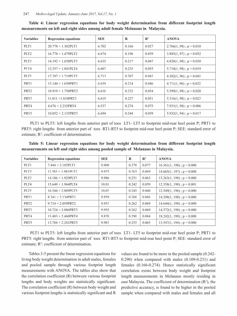
-legal Update, January-June 2017, Vol.17, No. 1 246 247 Medico-legal Update, January-June 2017, Vol.17, No. 1
Table 4: Linear regression equations for body weight determination from different footprint length measurements on left and right sides among adult female Melanaus in Malaysia.
Variables Regression equations SEE R R2 ANOVA
PLT1 20.778 + 1.302PLT1 6.702 0.166 0.027 2.766(1, 98) ; p = 0.010
PLT2 16.778 + 1.479PLT2 6.674 0.196 0.039 3.885(1, 97) ; p = 0.052
PLT3 14.192 + 1.658PLT3 6.635 0.217 0.047 4.828(1, 98) ; p = 0.030
PLT4 12.357 + 1.841PLT4 6.607 0.235 0.055 5.710(1, 98) ; p = 0.019
PLT5 17.397 + 1.719PLT5 6.713 0.207 0.043 4.302(1, 96) ; p = 0.041
PRT1 13.144 + 1.650PRT1 6.639 0.214 0.046 4.711(1, 98) ; p = 0.032
PRT2 10.919 + 1.750PRT2 6.610 0.232 0.054 5.599(1, 98) ; p = 0.020
PRT3 11.011 +1.810PRT3 6.619 0.227 0.051 5.316(1, 98) ; p = 0.023
PRT4 4.676 + 2.235PRT4 6.537 0.274 0.075 7.931(1, 98) ; p = 0.006
PRT5 10.052 + 2.135PRT5 6.694 0.244 0.059 5.932(1, 94) ; p = 0.017
PLT1 to PLT5: left lengths from anterior part of toes LT1- LT5 to footprint mid-rear heel point P; PRT1 to PRT5: right lengths from anterior part of toes RT1-RT5 to footprint mid-rear heel point P; SEE: standard error of estimate; R2: coefficient of determination.
Table 5: Linear regression equations for body weight determination from different footprint length measurements on left and right sides among pooled sample of Melanaus in Malaysia.
Variables Regression equations SEE R R2 ANOVA
PLT1 7.444 + 2.185PLT1 9.909 0.278 0.077 16.561(1, 198) ; p = 0.000
PLT2 11.583 + 1.981PLT2 9.975 0.263 0.069 14.665(1, 197) ; p = 0.000
PLT3 14.186 + 1.929PLT3 9.986 0.251 0.063 13.263(1, 198) ; p = 0.000
PLT4 15.649 + 1.964PLT4 10.01 0.242 0.059 12.358(1, 198) ; p = 0.001
PLT5 16.544 + 2.089PLT5 10.03 0.245 0.060 12.549(1, 196) ; p = 0.000
PRT1 8.341 + 2.134PRT1 9.959 0.260 0.068 14.398(1, 198) ; p = 0.000
PRT2 9.714 + 2.059PRT2 9.953 0.262 0.069 14.644(1, 198) ; p = 0.000
PRT3 10.724 + 2.086PRT3 9.955 0.262 0.069 14.572(1, 198) ; p = 0.000
PRT4 13.403 + 2.460PRT4 9.870 0.290 0.084 18.242(1, 198) ; p = 0.000
PRT5 13.784 + 2.241PRT5 9.983 0.255 0.065 13.547(1, 194) ; p = 0.000
PLT1 to PLT5: left lengths from anterior part of toes LT1- LT5 to footprint mid-rear heel point P; PRT1 to PRT5: right lengths from anterior part of toes RT1-RT5 to footprint mid-rear heel point P; SEE: standard error of estimate; R2: coefficient of determination.
Tables 3-5 present the linear regression equations for living body weight determination in adult males, females and pooled sample through various footprint length measurements with ANOVA. The tables also show that the correlation coefficient (R) between various footprint lengths and body weights are statistically significant. The correlation coefficient (R) between body weight and various footprint lengths is statistically significant and R
values are found to be more in the pooled sample (0.242-0.290) when compared with males (0.109-0.231) and females (0.166-0.274). Hence statistically significant correlation exists between body weight and footprint length measurements in Melanaus mostly residing in east Malaysia. The coefficient of determination (R2), the predictive accuracy, is found to be higher in the pooled sample when compared with males and females and all
Medico-legal Update, January-June 2017, Vol.17, No. 1 248 249 -legal Update, January-June 2017, Vol.17, No. 1
measurements are found to be positive and statistically significant for body weight determination.
DISCUSSION
The present investigation shows that body weight and footprint lengths are found to be larger in males than females, showing the existence of a statistically significant sex difference in Melanau population and this finding is in accordance with the previous studies11-
13,26-28. The investigation revealed that the right footprint length measurements are found to be larger at T2 in males and females and hence the existence of right-sided asymmetry. This finding is opposite with the findings of previous population studies viz. Malaysian Malays6, Malaysian Chinese12, Ibans of East Malaysia27, but is in accordance with the population studies viz. Kadazan Dusun22 and Lun Bawang female populations 23. But Iban 27 and Lun Bawang male 23 footprint study showed the non-existence of bilateral asymmetry. The minimum age of the subjects is fixed as 18 years and is considered appropriate. Commonly, stature at 18 years is accepted as adult, although there are small increments in stature after this25. The standard error of estimate (SEE) in the body weight determination from foot impression measurements shows higher value27 than SEE in the stature determination8-14. Hence, when the regression equation is applied to find the body weight from known footprint length, the body weight will be in the form of range, not an exact figure.
CONCLUSION
The present study provided regression equations for body weight determination from footprint length (complete or partial) measurements for Melanau population in Malaysia and Melanaus are mostly residing in east Malaysia. The linear regression equations derived for the pooled sample can be utilized to determine body weight when the sex of the footprint remains unknown, as in real crime scenarios. It is important to note that the people from different races and regions of Malaysia bear different morphological features and it would be incorrect to utilize the equations derived for Melanaus to any other populations either in Malaysia or any other populations in the world.
Conflict of Interest – Nil
Source of Funding – Self
Ethical Clearance - Done
REFERENCES
1. Jahar JK, Vijay P, OPalival PK. Estimation of height from measurements of foot length in Haryana region. J Indian Acad Forensic Med 2010; 32: 231-233.
2. Ambeth Kumar VD, Ramakrishnan M. Legacy of footprints recognition – A review. Int J Computer Applications 2011; 35:9–16.
3. Kennedy VD. Uniqueness of bare feet and its use as a possible means of identification. Forensic Sci Int 1996; 82:81–87.
4. Krishan K. Individualizing characteristics of footprints in Gujjars of north India—forensic aspects. Forensic Sci Int 2007;168:137–144.
5. Kennedy RB, Chen S, Pressman IS, Yamashita AB. A large scale statistical analysis of barefoot impressions. J Forensic Sci 2005; 50(5):1–10.
6. Nataraja Moorthy T, Mazidah K, Hadzri M, Jayaprakash PT. Estimation of stature based on foot length of Malays in Malaysia. Aust J Forensic Sci 2011; 43:13–26.
7. Naomi H, Ambika F, Nur-Intaniah I, Daniel F. Estimation of stature using anthropometry of feet and footprints in a Western Australian population. J Forensic and Legal Med 2013;20: 435-441.
8. Nataraja Moorthy T, Mustaqqim A, Boominathan R, Raman N. Stature estimation from footprint measurements in Indian Tamils by regression analysis. Egyptian J Forensic Sci 2014; 4:7-16.
9. Reel S, Rouse S, Vernon W, Doherty D. Estimation of stature from static and dynamic footprints. Forensic Sci Int 2012;e1–e5.
10. Kanchan T, Krishan K, Shyamsundar S, Aparna KR, Jaiswal S. Analysis of footprint and its parts for stature estimation in Indian population. The Foot 2012; 22:175–180.
11. Nataraja Moorthy T, Nurul A, Hairunnisa M. Stature estimation based on footprint measurements of Malays in Peninsula Malaysia by regression analysis. Int J Bio medl adv res 2013; 4(10):683-689.
12. Nataraja Moorthy T, Ang Yan Ling, Saufee Affandi Sarippudin. Estimation of stature from footprint and foot outline measurements in Malaysian Chinese, Australian J Forensic Sci 2014;46(2): 136-159.

-legal Update, January-June 2017, Vol.17, No. 1 248 249 Medico-legal Update, January-June 2017, Vol.17, No. 1
13. Nataraja Moorthy T, Hairunnisa M .Estimation of stature from foot outline measurements in Ibans of East Malaysia by regression analysis. Int J Bio medl adv res 2013;4(12): 889-895.
14. Krishan K. Estimation of stature from footprint and foot outline dimensions in Gujjars of North India. Forensic Sci Int 2008;175: 93-101.
15. Petra U, Radoslav B, Sona M. Stature estimation from various foot dimensions among Slovak population. J Forensic Sci 2013;58: 448-451.
16. Abdi O, Beytullah K, Melek O. Kolusayin, Ahsen K, Hüseyin A. Predictive role of hand and foot dimensions in stature estimation. Romanion J Leg Med 2012;20: 41-46.
17. Mansur DI, Haque MK,Sharma K, Karki RK,Khanal K,Karna R. Estimation of Stature from Foot Length in Adult Nepalese Population and its Clinical Relevance. Kathmandu Univ Med J 2012; 37(1):16-9.
18. Ogugua E, Nto NJ, Bello EF, Egwu EO, Ukoha UU, Ajah D. Stature Estimation from Foot Dimensions of An Adult Nigerian Population. Anatomica Karnataka 2012; 6(2):08-12.
19. Sonali K,Ashias A. Estimation of stature from the measurement of foot length, hand length and head length in Maharashtra region. Indian Journal of Basic & Applied Med Res 2012; 1(2): 77-85.
20. Gregory E, Laskowski BS, Vernon L, Kyle MS. Barefoot impressions – A preliminary study of identification characteristics and population frequency of their morphological features. J Forensic sci 1988;33(2): 378-388.
21. Hairunnisa MAK, Nataraja Moorthy T. Stature estimation from the anthropometric measurements
of footprints among Melanau: an indigenous population of Malaysian Borneo. Canadian Soc Forensic sci J 2015;48(2): 68-84.
22. Nataraja Moorthy T, Jessica RS. Stature estimation from the anthropometric measurements of foot outline in adult indigenous Kadazan Dusun ethnic of east Malaysia by regression analysis. J South Ind Med Leg Asso 2016;8(1): 15-20.
23. Hairunnisa MAK, Nataraja Moorthy T. Stature estimation from foot outline measurements in adult Lun Bawang ethnics of east Malaysia by regression analysis. Med Leg Upd 2016;16(2): 187-192.
24. Irene AF, Nashwa NK. Stature and body weight estimation from various footprint measurement among Egyptian population. J Forensic Sci 2010; 55:884–888.
25. Krishan K, Kanchan T, Sharma A. Multiplication factor versus regression analysis in stature estimation from hand and foot dimensions. J Forensic Leg Med 2012;19:211–214.
26. Nataraja Moorthy T, Jessica RS. Stature estimation from the anthropometric measurements of foot outline in adult indigenous Kadazan Dusun ethnic of east Malaysia by regression analysis. J South Ind Med Leg Assoc 2016;8(1):15–20.
27. Nataraja Moorthy T, Hairunnisa MAK. Body weight estimation from various footprint measurements among Ibans of east Malaysia. Malaysian App Bio J 2016;45(2):113–118.
28. Nataraja Moorthy T, Hairunnisa MAK. Stature estimation from anthropometric measurements of footprints in Lun Bawang, an indigenous ethnic groups of east Malaysia by linear regression analysis. Malaysian App Bio J 2016;45(2):69–74.

251 -legal Update, January-June 2017, Vol.17, No. 1
Bodyweight Estimation from Foot Impresson Breadth Measurements among Iban Population of East Malaysia
T Nataraja Moorthy1, Hairunnisa Bt Mohd Anas Khan2
1Associate Professor of Forensic Sciences, Forensic Science Program, Faculty of Health and Life Sciences, Management and Science University, Shah Alam, Selangor, Malaysia,
2Forensic Division, Chemistry Department of Malaysia, Kuching, Sarawak State, East Malaysia2Forensic Division, Chemistry Department of Malaysia, Kuching, Sarawak State, East Malaysia2
ABSTRACT
Forensic anthropologists are interested in body weight estimation through foot impressions. The present study is aimed to derive regression formulae to estimate body weight from breadths of foot impressions (footprint and foot outline) among Iban population in Malaysia. Following the standard procedure, footprint, foot outline breadths and body weights were recorded. The results showed significant positive correlation between body weight and breadth measurements. The correlation coefficient (R) in pooled sample (0.262-0.342) is found to be comparatively higher than those of individual male (0.082-0.265), and female (0.218-0.249) footprint breadth measurements. Similar observation was noted in foot outline breadth measurements also i.e., higher R values recorded in pooled sample (0.273-0.436) than male (0.162-0.384) and female (0.166-0.312) subjects. Thus, linear regression equations were derived for body weight estimation from foot impression breadth measurements among Iban population of east Malaysia.
Keywords: Forensic anthropology, body weight, footprint, foot outline, Ibans, Malaysia.
Corresponding author:T Nataraja MoorthyAssociate Professor of Forensic Sciences, Forensic Science Program, Faculty of Health and Life Sciences, Management and Science University, Shah Alam, Selangor, Malaysia, E-mail: [email protected] & [email protected]
INTRODUCTION
Forensic anthropologists are interested in body weight estimation through foot impression1. Person identification using footprint analysis is also an emerging biometric technique2. The whole body of a human has a strong relationship with each part of his/her body3. The characteristic features of foot impressions can provide useful clues to establish identity4. Foot impressions are found at crime scenes since offenders often remove their footwear, either to avoid noise or to gain better grip in climbing walls, etc, while entering or exiting5. Examination of footprints6-11, foot outlines12-15 and feet 16-20 can provide valuable information to estimate stature. The feet may produce 2D and 3D impressions in hard
and soft surfaces21. Researchers have conducted study on sex determination from foot 22-23 and footprint 24-25
for forensic application. Also studied the relationship between footprint 21,26-27, foot outline28 lengths and body weight. It is important that racial and cultural aspects of foot morphology must be considered29 and hence a single formula cannot represent for all races or regions in a country10-16. Hence, the present study attempts to derive regression equations to estimate stature from foot impression (footprint and foot outline) breadth measurements for Iban population.
MATERIALS AND METHOD
The study was carried out at east Malaysia and the participants (100 males and 100 females) ages ranged from 18 to 64 years. Subjects with any apparent foot-related diseaseor disorders were excluded from the study. Following the standard procedure, body weight 21,
26-28, footprint 6-10 and foot outlines 11-15 were collected and the anatomical marks of footprint and foot outline were shown in figure 1.
DOI Number: 10.5958/0974-1283.2017.00055.X

251 Medico-legal Update, January-June 2017, Vol.17, No. 1
Fig 1: Landmarks and measurements on left footprint and right foot outline
The data were analyzed using PASW Statistics version 20 (Predictive Analytic Software). The linear regression analysis method is reliable and was employed to derive regression equations for weight estimation from foot impression breadths30.
RESULTSTable 1 presents the descriptive statistics of body weights in males, females and pooled sample (combined male
and female subjects).
Table 1. Descriptive statistics of body weight (in kg) in adult males, females and pooled sample among Ibans of east Malaysia.
VariableMale (N=100) Female (N=100) Pooled sample (N=200)
Min Max Mean SD Min Max Mean SD Min Max Mean SD
Body weight (kg) 44.6 76.0 59.2 7.8 30.9 74.9 54.7 8.1 30.9 76.0 57.0 8.2
Min: minimum; Max: maximum; N: sample size; SD: standard deviation
The table shows that the mean body weight of male is found to be comparatively higher (59.2 kg) than the body weight of females (54.7 kg).
Anatomical landmarks
P – Mid-rear heel point on footprint
OP – Mid-rear heel point on foot outline
DLA – Designated longitudinal axis
BB – Ball breadth
HB – Heel breadth
LPBB – Ball breadth measurement in left footprint
RPBB – Ball breadth measurement in right footprint
LPHB – Heel breadth measurement in left footprint
RPHB – Heel breadth measurement in right footprint
ROBB – Ball breadth measurement in right foot outline
LOBB – Ball breadth measurement in left foot outline
ROHB – Heel breadth measurement in right foot outline
All footprints, foot outlines, body weight and information relating to participants were coded with sample ID for anonymity.

Medico-legal Update, January-June 2017, Vol.17, No. 1 252 253 -legal Update, January-June 2017, Vol.17, No. 1
Table 2: Descriptive statistics of ball and heel breadth measurements in the footprints of adult males, females and pooled sample among Ibans of east Malaysia (in cm).
VariableMale (N=100) Female (N=100) Pooled sample (N=200)
Min Max Mean SD Min Max Mean SD Min Max Mean SD
LPBB 8.00 10.5 9.04 0.5 7.30 9.70 8.43 0.5 7.30 10.5 8.74 0.6
LPHB 3.80 6.30 4.59 0.5 3.00 5.00 4.13 0.4 3.00 6.30 4.36 0.5
RPBB 7.70 10.5 9.03 0.5 7.40 9.90 8.45 0.5 7.40 10.5 8.74 0.6
RPHB 3.60 6.40 4.61 0.4 3.00 5.30 4.15 0.4 3.00 6.40 4.38 0.5
Min: minimum; Max: maximum; N: sample size; SD: standard deviation; LPBB: ball breadth in left footprint; RPBB: ball breadth in right footprint; LPHB: heel breadth in left footprint; RPHB: heel breadth in right footprint
Table 2 presents the descriptive statistics of various ball and heel breadth measurements in the footprints among male, female and pooled sample on both sides. The ball breadth measurements) are comparatively larger than the heel measurements which is common to normal human feet. The result shows that the size of both ball and heel breadth measurements are found to longer in
males (BB 9.03-9.04 cm; HB 4.59-4.61 cm)) compared to females (BB 8.43-8.45 cm; HB 4.13-4.15 cm)). The size of footprints show bilateral asymmetry in ball and heel measurements. In males the left ball breadth is longer than right ball breadth but not significant. The standard deviation values are very low in both genders.
Table 3: Descriptive statistics of ball and heel breadth measurements in the foot outlines of adult males, females and pooled sample among Ibans of east Malaysia (in cm).
VariableMale (N=100) Female (N=100) Pooled sample (N=200)
Min Max Mean SD Min Max Mean SD Min Max Mean SD
LOBB 8.40 11.1 10.05 0.5 8.00 10.9 9.362 0.6 8.00 11.1 9.704 0.6
LOHB 4.70 7.30 6.152 0.5 4.50 6.50 5.610 0.5 4.50 7.30 5.881 0.6
ROBB 8.70 11.1 10.08 0.5 8.20 10.4 9.407 0.5 8.20 11.1 9.744 0.6
ROHB 4.80 7.30 6.129 0.5 4.40 6.70 5.589 0.5 4.40 7.30 5.859 0.5
Min: minimum; Max: maximum; N: sample size; SD: standard deviation; LOBB: ball breadth in left foot outline; ROBB: ball breadth in right foot outline; LOHB: heel breadth in left foot outline; ROHB: heel breadth in right foot outline
Table 3 presents the descriptive statistics of various ball breadth and heel breadth measurements in foot outlines among males, females and pooled sample on both sides. The ball breadth measurements are
comparatively larger than the heel measurements in both genders as observed in footprints. The result indicated that the size of both ball and heel breadth measurements are found to longer in males compared to females.
Table 4. Linear regression equations to estimate body weight (kg) through ball and heel measurements and ANOVA in the footprints of adult males, females and pooled sample among Ibans of east Malaysia.
Gender Side Linear regression equations R R2 SEE ANOVA
MaleN=100
LPBB W= 20.446 + 4.285LPBB 0.265 0.070 7.523 8.918 (1, 98) ; p = 0.003
LPHB W= 51.889 + 1.589LPHB 0.096 0.009 7.766 1.102 ( 1, 98) ; p= 0.003
RPBB W= 28.782 + 3.368RPBB 0.211 0.045 7.626 5.497(1, 98) ; P = 0.002
RPHB W= 52.374 + 1.478RPHB 0.082 0.007 7.776 0.795(1, 98) ; p = 0.003
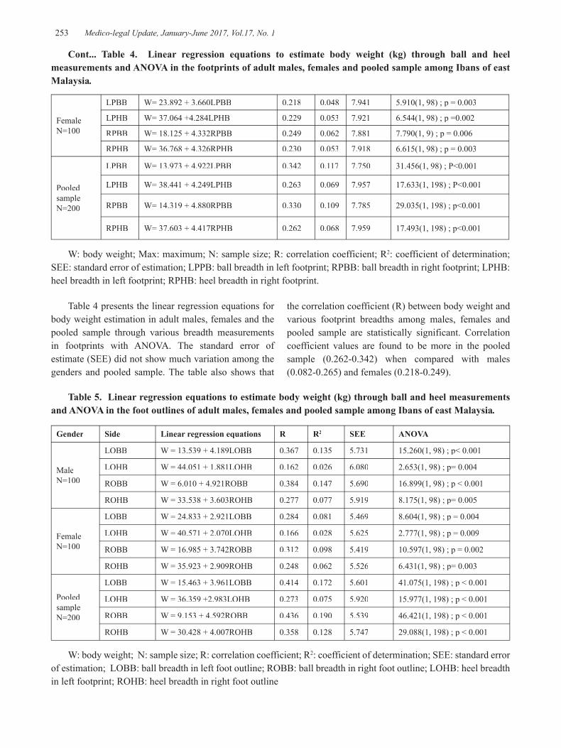
-legal Update, January-June 2017, Vol.17, No. 1 252 253 Medico-legal Update, January-June 2017, Vol.17, No. 1
FemaleN=100
LPBB W= 23.892 + 3.660LPBB 0.218 0.048 7.941 5.910(1, 98) ; p = 0.003
LPHB W= 37.064 +4.284LPHB 0.229 0.053 7.921 6.544(1, 98) ; p =0.002
RPBB W= 18.125 + 4.332RPBB 0.249 0.062 7.881 7.790(1, 9) ; p = 0.006
RPHB W= 36.768 + 4.326RPHB 0.230 0.053 7.918 6.615(1, 98) ; p = 0.003
Pooled sampleN=200
LPBB W= 13.973 + 4.922LPBB 0.342 0.117 7.750 31.456(1, 98) ; P<0.001
LPHB W= 38.441 + 4.249LPHB 0.263 0.069 7.957 17.633(1, 198) ; P<0.001
RPBB W= 14.319 + 4.880RPBB 0.330 0.109 7.785 29.035(1, 198) ; p<0.001
RPHB W= 37.603 + 4.417RPHB 0.262 0.068 7.959 17.493(1, 198) ; p<0.001
W: body weight; Max: maximum; N: sample size; R: correlation coefficient; R2: coefficient of determination; SEE: standard error of estimation; LPPB: ball breadth in left footprint; RPBB: ball breadth in right footprint; LPHB: heel breadth in left footprint; RPHB: heel breadth in right footprint.
Table 4 presents the linear regression equations for body weight estimation in adult males, females and the pooled sample through various breadth measurements in footprints with ANOVA. The standard error of estimate (SEE) did not show much variation among the genders and pooled sample. The table also shows that
Cont... Table 4. Linear regression equations to estimate body weight (kg) through ball and heel measurements and ANOVA in the footprints of adult males, females and pooled sample among Ibans of east Malaysia.
the correlation coefficient (R) between body weight and various footprint breadths among males, females and pooled sample are statistically significant. Correlation coefficient values are found to be more in the pooled sample (0.262-0.342) when compared with males (0.082-0.265) and females (0.218-0.249).
Table 5. Linear regression equations to estimate body weight (kg) through ball and heel measurements and ANOVA in the foot outlines of adult males, females and pooled sample among Ibans of east Malaysia.
Gender Side Linear regression equations R R2 SEE ANOVA
MaleN=100
LOBB W = 13.539 + 4.189LOBB 0.367 0.135 5.731 15.260(1, 98) ; p< 0.001
LOHB W = 44.051 + 1.881LOHB 0.162 0.026 6.080 2.653(1, 98) ; p= 0.004
ROBB W = 6.010 + 4.921ROBB 0.384 0.147 5.690 16.899(1, 98) ; p < 0.001
ROHB W = 33.538 + 3.603ROHB 0.277 0.077 5.919 8.175(1, 98) ; p= 0.005
FemaleN=100
LOBB W = 24.833 + 2.921LOBB 0.284 0.081 5.469 8.604(1, 98) ; p = 0.004
LOHB W = 40.571 + 2.070LOHB 0.166 0.028 5.625 2.777(1, 98) ; p = 0.009
ROBB W = 16.985 + 3.742ROBB 0.312 0.098 5.419 10.597(1, 98) ; p = 0.002
ROHB W = 35.923 + 2.909ROHB 0.248 0.062 5.526 6.431(1, 98) ; p= 0.003
Pooled sampleN=200
LOBB W = 15.463 + 3.961LOBB 0.414 0.172 5.601 41.075(1, 198) ; p < 0.001
LOHB W = 36.359 +2.983LOHB 0.273 0.075 5.920 15.977(1, 198) ; p < 0.001
ROBB W = 9.153 + 4.592ROBB 0.436 0.190 5.539 46.421(1, 198) ; p < 0.001
ROHB W = 30.428 + 4.007ROHB 0.358 0.128 5.747 29.088(1, 198) ; p < 0.001
W: body weight; N: sample size; R: correlation coefficient; R2: coefficient of determination; SEE: standard error of estimation; LOBB: ball breadth in left foot outline; ROBB: ball breadth in right foot outline; LOHB: heel breadth in left footprint; ROHB: heel breadth in right foot outline
Medico-legal Update, January-June 2017, Vol.17, No. 1 254 255 -legal Update, January-June 2017, Vol.17, No. 1
Table 5 shows the linear regression equations for body weight estimation in adult males, females and the pooled sample through various breadth measurements in foot outline with ANOVA. The standard error of estimate (SEE) is comparatively lower in females than male and pooled sample. Correlation coefficient (R) values are significant and found to be more in pooled sample (0.273-0.436) when compared with males (0.162-0.364) and females (0.166-0.312).
DISCUSSIONMalaysia is a multi-racial country and Iban is one
of the ethnic groups speak a dialect of Malay that is distinct. Farming is the main occupation of the Ibans33. The present investigation shows that body weight, heel and ball breadth measurements are found to be larger in males than females. This finding is in according with the previous studies 11,28,31,32. This may attributed to the general male-female differences and natural size in both sexes 34. The correlation coefficient (R) is found to be higher in pooled samples in breadth measurements. In many countries, footprint evidence is considered “unimportant” and hence neglected during their crime scene investigation32. But footprint can provide more valuable information than fingerprints during human identification process. The standard error of estimate (SEE) in the present investigation shows lower value and hence the regression equations can be used to estimate body weight from footprint and foot outline breadth measurements.
CONCLUSIONThe present study provided regression formulae to
estimate body weight from the breadth measurements of foot impressions. The equation are suitable for Iban population and the regression equations derived for the pooled sample can be employed to estimate body weight when the sex of the perpetrator remains unknown. Hence the researchers are suggested to conduct similar studies on other ethnic groups living in different regions and countries so that effect of genetics and regions can be investigated in forensic terms.
Conflict of Interest – Nil
Source of Funding – Self
Ethical Clearance - Done
REFERENCES1. Jahar JK, Vijay P, OPalival PK. Estimation of
height from measurements of foot length in
Haryana region. J indian Acad Forensic Med 2010; 32: 231-233.
2. Ambeth Kumar VD, Ramakrishnan M. Legacy of footprints recognition – A review. Int J Computer Applications 2011; 35:9–16.
3. Philip TA. Formulae for establishing stature from foot size by regression method. J Ind Acad Forensic Med 1990; 12:57-62.
4. Krishan K. Individualizing characteristics of footprints in Gujjars of north India—forensic aspects. Forensic Sci Int 2007;168:137–144.
5. Nataraja Moorthy T, Mazidah K, Hadzri M, Jayaprakash PT. Estimation of stature based on foot length of Malays in Malaysia. Aust J Forensic Sci 2011; 43:13–26.
6. Naomi H, Ambika F, Nur-Intaniah I, Daniel F.Estimation of stature using anthropometry of feet and footprints in a Western Australian population. J Forensic and Legal Med 2013;20: 435-441.
7. Nataraja Moorthy T, Mustaqqim A, Boominathan R, Raman N. Stature estimation from footprint measurements in Indian Tamils by regression analysis. Egyptian J Forensic Sci 2014; 4:7-16.
8. Reel S, Rouse S, Vernon W, Doherty D. Estimation of stature from static and dynamic footprints. Forensic Sci Int 2012:e1–e5.
9. Kanchan T, Krishan K, Shyamsundar S, Aparna KR, Jaiswal S. Analysis of footprint and its parts for stature estimation in Indian population. The Foot 2012; 22:175–180.
10. Nataraja Moorthy T, Nurul A, Hairunnisa M. Stature estimation based on footprint measurements of Malays in Peninsula Malaysia by regression analysis. Int J Bio medl adv res 2013; 4(10):683-689.
11. Nataraja Moorthy T, Ang Yan Ling, Saufee Affandi Sarippudin. Estimation of stature from footprint and foot outline measurements in Malaysian Chinese, Australian J Forensic Sci 2014;46(2): 136-159.
12. Nataraja Moorthy T, Hairunnisa M .Estimation of stature from foot outline measurements in Ibans of East Malaysia by regression analysis. Int J Bio Medl Adv Res 2013;4(12): 889-895.
13. Krishan K. Estimation of stature from footprint and foot outline dimensions in Gujjars of North India. Forensic Sci Int 2008;175: 93-101.

-legal Update, January-June 2017, Vol.17, No. 1 254 255 Medico-legal Update, January-June 2017, Vol.17, No. 1
14. Hairunnisa MAK, Nataraja Moorthy T. Stature estimation from foot outline measurements in adult Bidayuhs of east Malaysia by regression analysis. Indo J Leg Foren Sci 2013; 3(1):6-10.
15. Nataraja Moorthy T, Hairunnisa MAK. Stature estimation from foot outline measurements in adult Lun Bawang ethnics of east Malaysia by regression analysis. Med Leg Upd 2016;16(2): 187-192.
16. Petra U, Radoslav B, Sona M. Stature estimation from various foot dimensions among Slovak population. J Forensic Sci 2013;58: 448-451.
17. Abdi O, Beytullah K, Melek O. Kolusayin, Ahsen K, Hüseyin A. Predictive role of hand and foot dimensions in stature estimation. Romanion J Leg Med 2012;20: 41-46.
18. Mansur DI, Haque MK,Sharma K, Karki RK,Khanal K,Karna R. Estimation of Stature from Foot Length in Adult Nepalese Population and its Clinical Relevance. Kathmandu Univ Med J 2012; 37(1):16-9.
19. Ogugua E, Nto NJ, Bello EF, Egwu EO, Ukoha UU, Ajah D. Stature Estimation from Foot Dimensions of An Adult Nigerian Population. Anatomica Karnataka 2012; 6(2):08-12.
20. Sonali K,Ashias A. Estimation of stature from the measurement of foot length, hand length and head length in Maharashtra region. Indian Journal of Basic & Applied Med Res 2012; 1(2): 77-85.
21. Robbins LM. Estimating height and weight from size of footprints. J Forensic Sci 1986; 31:143–52.
22. Zeybek G, Ergur I, Demiroglu Z. Stature and gender estimation using foot measurements. Forensic Sci Int 2008; 181(1-3):54.
23. Phang SF, Khairul O, Sri Pawita AMH, Noor HH, Ab Halim M, Normaizatul AI. Stature and sex estimation using foot measurements for Malays and Chinese in Malaysia. Jurnal Sains Kesihatan Malaysia 2011; 9(2): 23-28.
24. Kanchan T, Krishan K, Aparna KR, Shyamsunder. Footprint ridge density: A new attribute for sexual dimorphim. HOMO – J compa Human Bio 2012; 63: 468-480.
25. Oberoi DV, Kuruvilla A, Saralaya KM, Rajeev A, Ashok B, Nagesh KR, Rao NG. Estimation
of stature and sex from footprint length using regression formulae and standard footprint length formula respectively. J Punjab Acd Forensic Med Tox 2006;6:5-8.
26. Nataraja Moorthy T, Hairunnisa MAK. Body weight estimation from various footprint length measurements among Ibans of east Malaysia.Malays Appl Biol 2016; 45(2): 113-118.
27. Irene AF, Nashwa NK. Stature and body weight estimation from various footprint measurements among Egyptian population. J Forensic Sci 2010;55:884–888.25.
28. Nataraja Moorthy T, Hairunnisa MAK. Estimation of body weight from foot outline length measurements in Melanau population of east Malaysia. Malays Appl Biol 2016; 45(2): 125-130.
29. Gregory E, Laskowski BS, Vernon L, Kyle MS. Barefoot impressions – A preliminary study of identification characteristics and population frequency of their morphological features. J Forensic sci 1988;33(2): 378-388.
30. Krishan K, Kanchan T, Sharma A. Multiplication factor versus regression analysis in stature estimation from hand and foot dimensions. J Forensic Leg Med 2012;19:211–214.
31. Irene AF, Nashwa NK. Stature and body weight estimation from various footprint measurements among Egyptian population. J Forensic Sci 2010;55:884–888.25.
32. Nataraja Moorthy, Hairunnisa MAK. Determination of body weight from footprint and foot outline breadth anthropometry among Melanau ethnis of Borneo Island, east Malaysia for forensic investigation. Malay App Biol 2016;45(2):139–144.
33. Hairunnisa MAK, Nataraja Moorthy T. Estimation of stature from foot outline measurements in Ibans of east Malaysia by regression analysis. Int J Biomed Adv Res 2013;4(12): 889-895.
34. Tanuj K, Krishan K, Shyamsundar S, Aparna KR, Sankalp J. Analysis of footprint and its parts for stature estimation. The foot 2012;22: 175-180.


Copyright © 2022 FDOKUMEN