PET Evaluation of Lung Cancer* - Journal of Nuclear Medicine
Upload
khangminh22Category
view
0download
0
The Final Frontier: A medical student’s mission to boldly dream where no dream has gone before...............................................5Laura Drudi
Kienbock’s disease and juvenile idiopathic arthritis................8Nicholas M. Desy, Mitchell Bernstein, Edward J. Harvey, Elizabeth Hazel
A New Palmo-Shoulder Compression Association.................14Hani Sinno, Teanoosh Zadeh
Teenage Female with Knee Pain and Instability.......................16Patricia Jo, David A Leswick, Lauren A Allen
A Man with a Neck Mass, Pleural Effusion and Hypoechoic Masses in the Right Atrium and Ventricle ...............................20Rabiya Jalil , Habib ur Rehman
CASE REPoRTS
LETTERS
MJMAN INTERNATIoNAL FoRuM FoR THE AdVANCEMENT oF MEdICAL SCIENCE by STudENTS
oRIgINAL ARTICLES
REVIEw ARTICLES
Evolutionary approaches to autism: an overview and integration ..................................................................................38Annemie Ploeger, Frietson Galis
breaking the Scope-of-Practice Taboo: where Multidisci-plinary Rhymes with Cost-Efficiency........................................44Nicholas Chadi
CRoSSRoAdS: AERoSPACE MEdICINE
The Canadian Space Agency Space Learning grants............53Jason Clement
Medical Education for Exploration Class Missions: NASA Aerospace Medicine Elective at the Kennedy Space Centre...55Gregory E. Stewart, Laura Drudi
ultrasound: From Earth to Space ............................................59Jennifer Law, Paul B. Macbeth
Medical Care in the Arctic and on orbit....................................66David Saint-Jacques
Physicians as Astronauts..........................................................69Robert Thirsk
Human Space Exploration – The Next Fifty years....................76David Williams, Matthew Turnock
Exploring the Possibility for a Common System for Joint Aero-medical Standards......................................................................82Justin Woodson, Walter M Dalitsch III, James L Persson, James McGhee, Charles Ciccone, Brian Parsa
Poverty Reduction Strategy Papers and their contribution to health: An Analysis of Three Countries....................................22Sam Bartlett
Salvage Resection for Isolated Local and/or Regional Failure of Head/Neck Cancer Following Definitive Concurrent Chemo-radiotherapy: Case Series and Review of the Literature.........29Patricia L. Kearney, John M. Watkins, Keisuke Shirai, Amy E. Wahlquist, John A. Fortney, Elizabeth Garrett-Mayer, M. Boyd Gil-lespie, Anand K. Sharma
MJMAN INTERNATIoNAL FoRuM FoR THE AdVANCEMENT oF MEdICAL SCIENCE by STudENTS
MJMAN INTERNATIoNAL FoRuM FoR THE AdVANCEMENT oF MEdICAL SCIENCE by STudENTS
VoLuME 13 ISSuE 2 2011
EdIToRIAL boARd
EdIToRS-IN-CHIEFLaura DrudiBindu Suresh
ExECuTIVE SENIoR EdIToRHoria Vulpe
ExECuTIVE CRoSSRoAdS EdIToRMarcel Edwards
ExECuTIVE PubLIC RELATIoNSMaheen Diwan
ExECuTIVE IT ANd oNLINE EdIToRGenevieve Mathis
ExECuTIVE TREASuRERNatalia Gorelik
LAyouT EdIToRSLaura Drudi
CoPy EdIToRSShreya JalaliMathura Thevarajah
CoNSuLTANTSRussel Brown
CoVER dESIgNMarina Martinez
SENIoR EdIToRSKirsten NilesKyle GibsonStephen GowingNicolas Thibodeau-JarryFaisal NaqibMatas MorkeviciusTudor BotnaruIvan LitvinovMireille Schnitzer
JuNIoR EdIToRSChristopher MaraczMadhur NayanAna BlanchardMichelle ZhangStephen HanleyMadeleine HopsonAriella ZbarChris CheungJonathan MicieliAkshay SharmaAnna NikonovaMelica NourmoussaviMatthew ShorofskyMazin AbdelghanySagar DuganiTristan Tham
FACuLTy AdVISoRPhil Gold, MD, PhD
The McGill Journal of Medicine (MJM), published by students at McGill University (Montreal, Canada), is devoted to the advancement of medical science by students. Materials in the MJM published by students include Original Articles, Case Reports, Review Articles, and Crossroads. Articles in the MJM Focus are the invited contributions of physicians and scientists. Letters to the MJM, Commentaries, and Book Reviews are submitted by all who wish to contribute. All materials reflect the individual views of the authors and do not represent the official views of the MJM or the institutions with which the authors are affiliated. Articles published in the MJM have been reviewed for accuracy; however, information presented and opinions expressed remain the sole responsibility of the author(s). Advertisements appearing herein do not reflect endorsement by the MJM. Although all advertisements have been reviewed to ensure compliance with general ethical and legal standards, ultimate responsibility for their content rests solely with the advertiser. The physician is therefore advised to verify all prescribing information. The Journal is published biannually (Summer and Winter) in one volume (two issues) per year. Student subscription prices for each volume are CAN$20(Canada), US$20 (USA), or US$20 (International). Non-student subscription prices are CAN$40 (Canada), US$40 (USA), or US$40 (International). Communications should be directed to [email protected]. The MJM head office is located at the Faculty of Medicine, 6th Floor, 3655 Promenade Sir William Osler, Montreal, QC, Canada H3G 1Y6. The online version of the MJM is available at: http://www.mjm.mcgill.ca.
ACKNowLEdgMENTS
The executive committee of the McGill Journal of Medicine would like to thank all of the sponsors for the the McGill Journal of Medicine 13.2.
The executive committee further extends their gratitude to all those who worked hard to make this issue of the McGill Journal of Medicine a reality.
Copyright © 2011 by MJM, all rights reserved
LETTER FRoM THE EdIToRS
MJM 2011 13(2): 5-6 5Copyright © 2011 by MJM
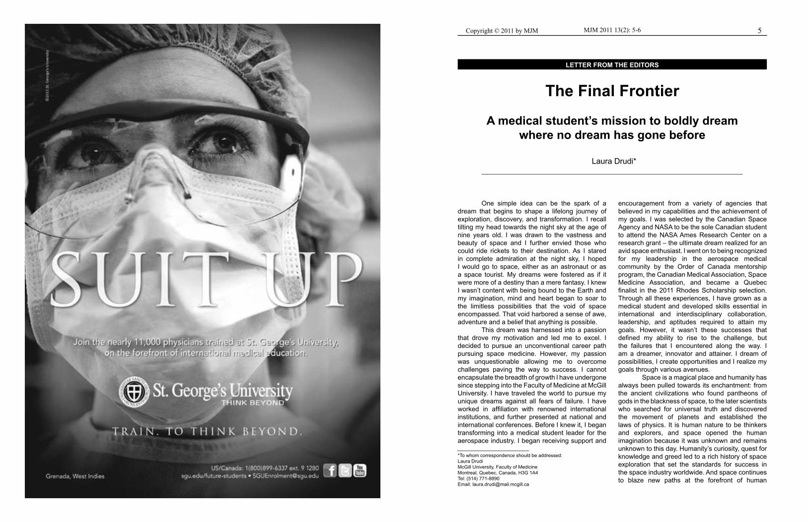
One simple idea can be the spark of a dream that begins to shape a lifelong journey of exploration, discovery, and transformation. I recall tilting my head towards the night sky at the age of nine years old. I was drawn to the vastness and beauty of space and I further envied those who could ride rickets to their destination. As I stared in complete admiration at the night sky, I hoped I would go to space, either as an astronaut or as a space tourist. My dreams were fostered as if it were more of a destiny than a mere fantasy. I knew I wasn’t content with being bound to the Earth and my imagination, mind and heart began to soar to the limitless possibilities that the void of space encompassed. That void harbored a sense of awe, adventure and a belief that anything is possible. This dream was harnessed into a passion that drove my motivation and led me to excel. I decided to pursue an unconventional career path pursuing space medicine. However, my passion was unquestionable allowing me to overcome challenges paving the way to success. I cannot encapsulate the breadth of growth I have undergone since stepping into the Faculty of Medicine at McGill University. I have traveled the world to pursue my unique dreams against all fears of failure. I have worked in affiliation with renowned international institutions, and further presented at national and international conferences. Before I knew it, I began transforming into a medical student leader for the aerospace industry. I began receiving support and
encouragement from a variety of agencies that believed in my capabilities and the achievement of my goals. I was selected by the Canadian Space Agency and NASA to be the sole Canadian student to attend the NASA Ames Research Center on a research grant – the ultimate dream realized for an avid space enthusiast. I went on to being recognized for my leadership in the aerospace medical community by the Order of Canada mentorship program, the Canadian Medical Association, Space Medicine Association, and became a Quebec finalist in the 2011 Rhodes Scholarship selection. Through all these experiences, I have grown as a medical student and developed skills essential in international and interdisciplinary collaboration, leadership, and aptitudes required to attain my goals. However, it wasn’t these successes that defined my ability to rise to the challenge, but the failures that I encountered along the way. I am a dreamer, innovator and attainer. I dream of possibilities, I create opportunities and I realize my goals through various avenues. Space is a magical place and humanity has always been pulled towards its enchantment: from the ancient civilizations who found pantheons of gods in the blackness of space, to the later scientists who searched for universal truth and discovered the movement of planets and established the laws of physics. It is human nature to be thinkers and explorers, and space opened the human imagination because it was unknown and remains unknown to this day. Humanity’s curiosity, quest for knowledge and greed led to a rich history of space exploration that set the standards for success in the space industry worldwide. And space continues to blaze new paths at the forefront of human
The Final Frontier
A medical student’s mission to boldly dream where no dream has gone before
Laura Drudi*
*To whom correspondence should be addressed:Laura DrudiMcGill University, Faculty of MedicineMontreal, Quebec, Canada, H3G 1A4Tel: (514) 771-8890Email: [email protected]
6
understanding. My goal is to be at that frontier and begin an expedition unparalleled by any other. To this very day, I tilt my head towards the night sky. I don’t know if I will ever have the chance to become one of the select few to travel beyond Earth’s gravity, but one thing I can guarantee is that I am going to make the attempt. As the frontiers of science and technology push forward, so too do the ideas, creativity and innovation of talented people. There is no boundary to space and there is no limitation to the imagination. The final frontier
isn’t space; I believe it’s the breath of the human imagination. With that in mind, this expedition that I have embarked on is bound to be an exciting one. It is with great pleasure the McGill Journal of Medicine Issue 13.2 will have aerospace medicine as the focus section. I hoped to share my passions for space and medicine with the community at large, and I am privileged and humbled to have participation and support from the space life sciences community.
Thank you.
The Final Frontier 2011
Laura drudi (M.D., C.M. candidate 2013) is a third year medical student at McGill Uni-versity. Her interest in combining her two passions of space and medicine has led her to conduct aerospace medicine research. She will be taking a one year’s leave of absence from the Faculty of Medicine and will be pursuing a Diploma of Space Studies and an MSc in Experimental Surgery prior to completing her MDCM degree. She hopes to work for the manned space program as a flight surgeon and to further continue her research in space life sciences.
CASE REPoRT
MJM 2011 13(2): 8-138 Copyright © 2011 by MJM
AbSTRACT: Kienbock’s disease or osteonecrosis of the lunate is an uncommon cause of wrist pain. . Though there have been several reports of cases in patients with various rheumatologic diseases, the precise etiology has currently not been established. we report a case of Kienbock’s disease that occurred in a patient with juvenile idiopathic arthritis. To our knowledge, this is the first case report with an association between these two conditions.
Keywords: Kienbock’s disease, osteonecrosis, juvenile idiopathic arthritis, lunatomalacia, avascular necrosis
*To whom correspondence should be addressed:Dr. Nicholas M. DesyMcGill University Health Centre Montreal General Hospital1650 Cedar Avenue Room Montreal, Quebec, Canada, H3G 1A4Tel: (514) 934-1934 ext. 42734; Fax: (514) 934-8453Email: [email protected]
INTRoduCTIoN The etiology of Kienbock’s disease, also known as (osteonecrosis of the lunate, remains controversial. It commonly occurs in patients twenty to fourty years old and presents with pain and stiffness in the dorsomedial aspect of the wrist. Several risk factors have been established to help explain its etiology: acute or repetitive trauma, variation in blood supply to the lunate, differences in the anatomy and shape of the lunate bone, and venous congestion (1, 2). Abnormal biomechanics at the radiocarpal joint between the distal radius and ulna has also been implicated in Kienbock’s disease (3, 4). Ulnar variance describes the length relationship between the articular surfaces of the radius and ulna at the radiocarpal joint. Positive ulnar variance indicates that the ulna is longer than the radius, while negative ulnar variance indicates that the ulna is shorter at the wrist joint. In neutral ulnar variance 80% of the axial load at the wrist is transmitted through the distal radius. As ulnar variance decreases to more negative values, the
load transmission across the distal radiocarpal joint increases, subsequently exposing the lunate to abnormally higher pressures and potentially increasing the risk of Kienbock’s disease (3, 4). Kienbock’s disease is also associated with systemic lupus erythematosus (SLE) (5-8), antiphospholipid antibody syndrome (9), sickle cell anemia (10), and Crohn’s enteritis (11). Multiple hereditary osteochondromata (12), carpal coalition (13, 14) and congenital shortening of the ulna in Langer-Giedion syndrome (15), are other anatomic abnormalities that have been reported with Kienbock’s disease. Rheumatic diseases, including scleroderma (16-18), rheumatoid arthritis (19), gout (20, 21) and dermatomyositis (22) have been published in association with Kienbock’s, but there have been no identifiable cases in patients with juvenile idiopathic arthritis (JIA). This report presents a case of osteonecrosis of the lunate in a patient with JIA and no prior history of trauma. Furthermore, a literature review is done to illustrate the proposed etiologies of Kienbock’s disease and its association with other rheumatologic conditions.
Kienbock’s disease and juvenile idiopathic arthritis
Nicholas M. Desy*, Mitchell Bernstein, Edward J. Harvey, Elizabeth Hazel
9Vol. 13 No. 2 Kienbock’s Disease
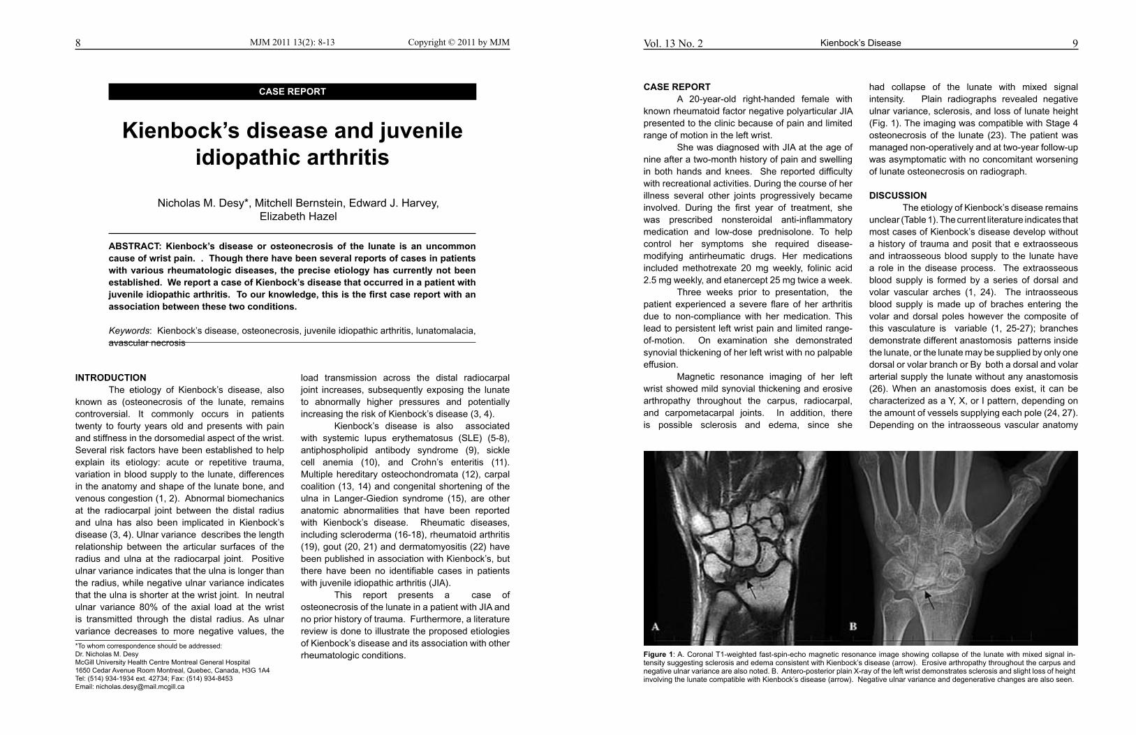
CASE REPoRT A 20-year-old right-handed female with known rheumatoid factor negative polyarticular JIA presented to the clinic because of pain and limited range of motion in the left wrist. She was diagnosed with JIA at the age of nine after a two-month history of pain and swelling in both hands and knees. She reported difficulty with recreational activities. During the course of her illness several other joints progressively became involved. During the first year of treatment, she was prescribed nonsteroidal anti-inflammatory medication and low-dose prednisolone. To help control her symptoms she required disease-modifying antirheumatic drugs. Her medications included methotrexate 20 mg weekly, folinic acid 2.5 mg weekly, and etanercept 25 mg twice a week. Three weeks prior to presentation, the patient experienced a severe flare of her arthritis due to non-compliance with her medication. This lead to persistent left wrist pain and limited range-of-motion. On examination she demonstrated synovial thickening of her left wrist with no palpable effusion. Magnetic resonance imaging of her left wrist showed mild synovial thickening and erosive arthropathy throughout the carpus, radiocarpal, and carpometacarpal joints. In addition, there is possible sclerosis and edema, since she
had collapse of the lunate with mixed signal intensity. Plain radiographs revealed negative ulnar variance, sclerosis, and loss of lunate height (Fig. 1). The imaging was compatible with Stage 4 osteonecrosis of the lunate (23). The patient was managed non-operatively and at two-year follow-up was asymptomatic with no concomitant worsening of lunate osteonecrosis on radiograph.
dISCuSSIoN The etiology of Kienbock’s disease remains unclear (Table 1). The current literature indicates that most cases of Kienbock’s disease develop without a history of trauma and posit that e extraosseous and intraosseous blood supply to the lunate have a role in the disease process. The extraosseous blood supply is formed by a series of dorsal and volar vascular arches (1, 24). The intraosseous blood supply is made up of braches entering the volar and dorsal poles however the composite of this vasculature is variable (1, 25-27); branches demonstrate different anastomosis patterns inside the lunate, or the lunate may be supplied by only one dorsal or volar branch or By both a dorsal and volar arterial supply the lunate without any anastomosis (26). When an anastomosis does exist, it can be characterized as a Y, X, or I pattern, depending on the amount of vessels supplying each pole (24, 27). Depending on the intraosseous vascular anatomy
Figure 1: A. Coronal T1-weighted fast-spin-echo magnetic resonance image showing collapse of the lunate with mixed signal in-tensity suggesting sclerosis and edema consistent with Kienbock’s disease (arrow). Erosive arthropathy throughout the carpus and negative ulnar variance are also noted. B. Antero-posterior plain X-ray of the left wrist demonstrates sclerosis and slight loss of height involving the lunate compatible with Kienbock’s disease (arrow). Negative ulnar variance and degenerative changes are also seen.
11Vol. 13 No. 2 Kienbock’s Disease
Scleroderma associated with Kienbock’s disease was first reported by Agus et al. in a patient with bilateral osteonecrosis of the lunate (16). This patient had severe Raynaud’s phenomenon and was never treated with corticosteroids. The contributing factors were hypothesized to be vasculopathy, Raynaud’s phenomenon, and a lunate consisting of a single nutrient vessel. Ribbans also reported a case of Kienbock’s disease in a patient with scleroderma and severe Raynaud’s phenomenon (18). The vasculopathy, scleroderma, and repeated use of the patient’s affected wrist predisposed this patient to Kienbock’s disease. Matsumoto et al. reported three cases of Kienbock’s disease in three patients with scleroderma, two without a history of steroid use and one who only used low dose steroids prior to the diagnosis of osteonecrosis (17). All three patients also had limited skin involvement, but had severe Raynaud’s phenomenon. They reported that scleroderma related vascular disease was likely the cause of the circulatory impairment leading to osteonecrosis. Mok et al. reported a patient with rheumatoid arthritis who was found to have osteonecrosis of the lunate. They believed that the Kienbock’s disease occurred in this patient due to an increase in intra-articular pressure within the wrist compartment, causing impedance of venous return, vascular insufficiency to the lunate, and subsequent osteonecrosis (19). In addition to the various risk factors mentioned above, Kienbock’s disease b is prevalent in specific patient populations. Rooker et al. found an increased prevalence of Kienbock’s disease in a group of patients with cerebral palsy (9.4%) (32). This was thought to be related to an abnormally flexed posture of a spastic wrist, which was present in all patients with Kienbock’s disease in this study and could impede blood flow. Joji et al. also found an increased prevalence of Kienbock’s disease in patients with cerebral palsy (2.7%) (33). They believed that the high muscle tone across the wrist in an ulnar negative wrist caused an increased pressure on the lunate. This cause is consistent with repeated microtrauma leading to vascular compromise and ultimately osteonecrosis. While the association between Kienbock’s disease and several of the reported cases could be coincidental, having a rheumatologic co-morbidity such as JIA could represent a risk factor
for Kienbock’s disease, as seen in the presented case report. The wrist is the second most common site of growth abnormality in patients with JIA (34). Several changes occur at the wrist, including narrowing of the intercarpal spaces, premature ossification of the carpal bones, and early fusion of the ulnar epiphysis leading to a shorter ulna (negative ulnar variance) (35). The wrist ultimately becomes displaced ulnarly and volarly leading to a dislocation of the wrist and bayonet deformity. Therefore, it is possible that the abnormalities in the wrist associated with JIA could lead to abnormal stresses and or pressures in the wrist that led to Kienbock’s disease. Furthermore, the erosive changes in other carpal bones may lead to a change on the normal force patterns in the patient’s carpus. Seven years prior to the onset of Kienbock’s disease, our patient was also treated with low-dose corticosteroids which could have also disrupted circulation and led to the development of lunate osteonecrosis. The precise etiology of Kienbock’s disease remains elusive. Several theories attempt to explain its pathogenesis, which suggests that it may be multifactorial. Many risk factors have also been identified; steroid treatment, a predisposing rheumatologic disease, a variation in lunate blood supply, and possibly negative ulnar variance. Our case demonstrates the possible relation between Kienbock’s disease and JIA. It also suggests that Kienbock’s disease could be a possible cause of wrist pain and stiffness in patients with JIA.
REFERENCES1. Gelberman RH, Bauman TD, Menon J et al. The vascularity
of the lunate bone and Kienbock’s disease. J Hand Surg
Am. 1980;5:272-278.
2. Jensen CH. Intraosseous pressure in Kienbock’s disease.
J Hand Surg Am. 1993;18:355-359.
3. Bonzar M, Firrell JC, Hainer M et al. Kienbock disease
and negative ulnar variance. J Bone Joint Surg Am.
1998;80:1154-1157.
4. Chen WS. Kienbock disease and negative ulnar variance.
J Bone Joint Surg Am. 2000;82:143-144.
5. Griffiths ID, Maini RN, Scott JT. Clinical and radiological
features of osteonecrosis in systemic lupus erythematosus.
Ann Rheum Dis. 1979;38:413-422.
6. Mok CC, Lau CS, Cheng PW et al. Bilateral Kienbock’s
disease in SLE. Scand J Rheumatol. 1997;26:485-487.
10
of the lunate, certain lunate bones are predisposed to Kienbock’s disease. This concept was highlighted in a case report of Kienbock’s disease associated with sickle cell anemia (10). The osteonecrosis was thought to have developed from an at-risk lunate - single volar arterial supply - along with significant vascular sickling and stasis. Venous congestion has also been attributed to the pathogenesis of Kienbock’s disease (2). During surgery, Jensen measured increased pressure inside the lunate compared with the radial styloid and capitate. He concluded that the higher pressure was caused by venous congestion leading to osteonecrosis of the lunate. Negative ulnar variance is also implicated in the pathogenesis of Kienbock’s disease (3, 4, 28). The altered relationship between the ulna and radius at the distal radioulnar joint modifies the biomechanics at the radiolunate joint and increases strains on the lunate. This postulation is still controversial because several studies, including a meta-analysis, have shown that negative ulnar variance is not a risk factor for developing Kienbock’s disease, (29, 30). On the contrary,Ledoux et al. performed a finite-element analysis on cadaveric lunate bones and found that the progression of a fracture of the lunate was present with negative ulnar variance, a high lunate uncovering index, which is the amount of lunate outside the lunate fossa of the radius compared to the amount of lunate articulating with the lunate fossa, and angulated trabeculae (31). This suggests that given the circumstance, the lunate can be at risk for developing osteonecrosis due to abnormal stresses.Further cases have also reportedpatients with conditions that may have caused altered stresses on the lunate. In particular, two cases have been reported involving carpal coalition (13, 14). It was
postulated in these cases that carpal coalition caused a progressively increasing stress on the lunate, which in turn led to Kienbock’s disease. Schuind et al. reported a case of Kienbock’s disease associated with congenital shortening of the ulna as seen in Langer-Giedion syndrome (15). It was suggested that Kienbock’s disease developed from microfractures sustained by an abnormal stress distribution (15). Multiple hereditary osteochondromata in the forearm was also found in association with Kienbock’s disease and was attributed to an excess load on the lunate by negative ulnar variance, but with no carpal slip (12). Systemic lupus erythematosus has been associated with avascular necrosis of bone. In 1977, Urman presented several cases of patients with SLE and osteonecrosis of the carpal bones, including a case report of a patient with SLE and Kienbock’s disease (8). The patient also had a history of Raynaud’s phenomenon and was taking high-dose corticosteroids. In SLE patients treated with corticosteroids and who developed osteonecrosis, there was one patient who developed Kienbock’s disease (5). This patient was treated with large doses of corticosteroids compared to those who did not develop Kienbock’s disease. Mok et al. reported a case of bilateral Kienbock’s disease in a patient with SLE (6). The patient described in this case report was never treated with corticosteroids and the etiology of Kienbock’s disease was thought to be due to a vasculopathy caused by either vasculitis or antiphospholipid antibodies, even though this patient tested negative for lupus anticoagulant and anticardiolipin antibodies. More recently, Taniguchi et al. described two cases of Kienbock’s disease in SLE after taking high doses of steroids (7). One of the cases was of a patient who developed bilateral osteonecrosis of the lunate. Both cases attributed corticosteroid use with the development of osteonecrosis. It is apparent that the cases of Kienbock’s disease in patients with SLE were either attributed to high dose corticosteroid use or to the disease itself. More over, patients with Crohn’s disease who use corticosteroids to control disease symptoms are known to develop Kienbock’s disease and osteonecrosis of the hip (11). The same scenario also occurred in a patient with dermatomyositis taking high doses of corticosteroids for sixteen months (22).
Kienbock’s Disease 2011
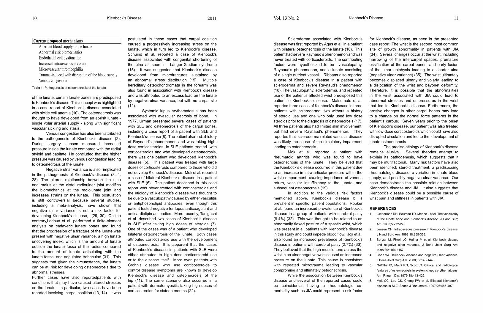
Table 1: Pathogenesis of osteonecrosis of the lunate
Current proposed mechanismsAberrant blood supply to the lunateAbnormal risk biomechanicsEndothelial cell dysfunctionIncreased intraosseous pressureMicrovascular thrombophiliaTrauma-induced with disruption of the blood supplyVenous congestion
13Vol. 13 No. 2 Kienbock’s Disease12 2011Kienbock’s Disease
7. Taniguchi Y, Tamaki T, Yoshida M. Kienbock’s disease in
systemic lupus erythematosus. Hand Surg. 2002;7:197-
200.
8. Urman JD, Abeles M, Houghton AN et al. Aseptic
necrosis presenting as wrist pain in SLE. Arthritis Rheum.
1977;20:825-828.
9. Alijotas J, Argemi M, Barquinero J. Kienbock’s disease
and antiphospholipid antibodies. Clin Exp Rheumatol.
1990;8:297-298.
10. Lanzer W, Szabo R, Gelberman R. A vascular necrosis
of the lunate and sickle cell anemia. A case report. Clin
Orthop Relat Res. 1984:168-171.
11. Culp RW, Schaffer JL, Osterman AL et al. Kienbock’s
disease in a patient with Crohn’s enteritis treated with
corticosteroids. J Hand Surg Am. 1989;14:294-296.
12. de Gauzy JS, Kany J, Darodes P et al. Kienbock’s disease
and multiple hereditary osteochondromata: a case report. J
Hand Surg Am. 1999;24:642-646.
13. Kaneko K, Uta S, Mogami A et al. Lunatomalacia in
association with congenital synostosis between the
capitate and the hamate. Chir Main. 2001;20:312-316.
14. Macnicol MF. Kienbock’s disease in association with carpal
coalition. Hand. 1982;14:185-187.
15. Schuind FA, Schiedts D, Fumiere E et al. Lunatomalacia
associated with congenital shortening of the ulna in
Langer-Giedion syndrome: a case report. J Hand Surg Am.
1997;22:404-407.
16. Agus B. Bilateral aseptic necrosis of the lunate in systemic
sclerosis. Clin Exp Rheumatol. 1987;5:155-157.
17. Matsumoto AK, Moore R, Alli P et al. Three cases of
osteonecrosis of the lunate bone of the wrist in scleroderma.
Clin Exp Rheumatol. 1999;17:730-732.
18. Ribbans WJ. Kienbock’s disease: two unusual cases. J
Hand Surg Br. 1988;13:463-465.
19. Mok CC, Wong RW, Lau CS. Kienbock’s disease in
rheumatoid arthritis. Br J Rheumatol. 1998;37:796-797.
20. Castagnoli M, Giacomello A, Argentina RS et al. Kienbock’s
disease in gout. Arthritis Rheum. 1981;24:974-975.
21. Shin AY, Weinstein LP, Bishop AT. Kienbock’s disease and
gout. J Hand Surg Br. 1999;24:363-365.
22. Kahn SJ, Sherry DD. Kienbock’s disease--avascular
necrosis of the carpal lunate bone--in a 7-year-old girl with
dermatomyositis. Clin Pediatr (Phila). 1994;33:752-754.
23. Lichtman DM, Mack GR, MacDonald RI et al. Kienbock’s
disease: the role of silicone replacement arthroplasty. J
Bone Joint Surg Am. 1977;59:899-908.
24. Gelberman RH, Panagis JS, Taleisnik J et al. The arterial
anatomy of the human carpus. Part I: The extraosseous
vascularity. J Hand Surg Am. 1983;8:367-375.
25. Lamas C, Carrera A, Proubasta I et al. The anatomy
and vascularity of the lunate: considerations applied to
Kienbock’s disease. Chir Main. 2007;26:13-20.
26. Lee ML. The intraosseus arterial pattern of the carpal
lunate bone and its relation to avascular necrosis. Acta
Orthop Scand. 1963;33:43-55.
27. Panagis JS, Gelberman RH, Taleisnik J et al. The arterial
anatomy of the human carpus. Part II: The intraosseous
vascularity. J Hand Surg Am. 1983;8:375-382.
28. Gelberman RH, Salamon PB, Jurist JM et al. Ulnar variance
in Kienbock’s disease. J Bone Joint Surg Am. 1975;57:674-
676.
29. Chung KC, Spilson MS, Kim MH. Is negative ulnar variance
a risk factor for Kienbock’s disease? A meta-analysis. Ann
Plast Surg. 2001;47:494-499.
30. D’Hoore K, De Smet L, Verellen K, et al. Negative Ulnar
Variance Is Not a Risk Factor for Kienbock’s Disease. J
Hand Surg 1994;19A:229-231.
31. Ledoux P, Lamblin D, Wuilbaut A et al. A finite-element
analysis of Kienbock’s disease. J Hand Surg Eur Vol.
2008;33:286-291.
32. Rooker GD, Goodfellow JW. Kienbock’s disease in cerebral
palsy. J Bone Joint Surg Br. 1977;59:363-365.
33. Joji S, Mizuseki T, Katayama S et al. Aetiology of Kienbock’s
disease based on a study of the condition among patients
with cerebral palsy. J Hand Surg Br. 1993;18:294-298.
34. Findley TW, Halpern D, Easton JK. Wrist subluxation
in juvenile rheumatoid arthritis: Pathophysiology and
management. Arch Phys Med Rehabil 1983;64:69-74.
35. Evans DM, Ansell BM, Hall MA. The wrist in juvenile
arthritis. J Hand Surg [Br]. 1991;16:293-304.
Nicholas M. desy, M.D.C.M. is a third year resident in Orthopaedic Surgery at McGill Uni-versity. He obtained a B.Sc. in Microbiology and Immunology from McGill University. He then completed his medical degree in 2008 from McGill University.
Mitchell bernstein, M.D. is a fourth year resident in Orthopaedic Surgery at McGill Univer-sity. He received a B.Sc. in Microbiology and Immunology from McGill University followed by an M.D. in Chicago.
Edward J. Harvey M.D., M.Sc. is an Associate Professor of Surgery in the department of Orthopaedic Surgery at McGill University. He is the Chief of Orthopaedic Trauma and Hand and Microvascular Surgery. He is also a co-director of the JTN Wong Labs for Bone Engi-neering where a large part of his research focuses on osteonecrosis and bone healing.
Elizabeth Hazel, M.D. is an Assistant Professor of Medicine in the division of Rheu-matology at McGill University. She completed her Internal Medicine and Rheuma-tology training at McGill University. Her interests include patients with juvenile idio-pathic arthritis.

CASE REPoRT
MJM 2011 13(2): 14-1514 Copyright © 2011 by MJM
*To whom correspondence should be addressed:Dr. Hani SinnoDivision of Plastic and Reconstructive Surgery Department of Surgery, McGill UniversityMontreal, QC, Canada H3G 1A4Email: [email protected]
INTRoduCTIoN Carpal tunnel syndrome is a most common peripheral compression neuropathy (1). It is caused by mechanical compression of the median nerve as it traverses the carpal tunnel of the wrist. Classic signs and symptoms are numbness of the lateral three digits and weakness of the thenar muscles due to atrophy (2). Important diagnostic tests in-clude electromyography (EMG) and nerve con-duction studies. The gold standard for the surgical treatment is transection of the transverse carpal ligament.
CASE REPoRT A thirty-eight year old right-handed con-struction worker presented to the McGill University Health Center Plastic Surgery clinic with complaints of bilateral carpal tunnel syndrome. He had no oth-er relevant past medical history, was not taking any medications, and had no known allergies. On fur-ther history, he complained of a ten-year period of slowly progressing symptoms of hand numbness, pain, and paresthesias in the median nerve distri-bution distal to the wrist. On physical examination he demonstrated positive Phalen and Tinel Test bilaterally. He had no obvious thenar eminence wasting and his grip strength was weakened. His EMG and nerve con-duction studies demonstrated moderate to severe carpal tunnel syndrome bilaterally. A routine open carpal tunnel release was performed on his right hand and the patient had complete resolution of his carpal tunnel symptoms with no complications. Two months later he was scheduled to have the same surgery for his left
hand. At this time, he said he was having bilateral shoulder weakness for the past two years and had not sought medical attention for it sincehe attributed it to strenuous physical work and fatigue secondary to his occupation. He explained that he was unable to elevate and abduct his arms above his shoulder prior to his right median nerve decompression, at which point he regained full range of motion and strength of his shoulder. Directly after his left carpal tunnel release the patient was able, with full strength, to elevate and abduct his left shoulder.
LITERATuRE REVIEw Using PubMed and Medline database, an online search using the headings “carpal tunnel re-lease” and “shoulder abduction” was done to deter-mine the occurrence and frequency of the observed phenomenon presented in the case report. No re-sults were found. Further searches with headings of “carpal tunnel release” and “shoulder weakness” also revealed no published material. The same was done with “shoulder extension”and “shoulder paral-ysis” and resulted in the same outcome. A search using the headings “carpal tunnel” and “shoulder flexion” demonstrated one paper by Vaught et al (3). The authors have concluded that the likelihood of patients with carpal tunnel syndrome having as-sociated thoracic outlet syndrome (TOS) is sixteen times higher than control subjects. They have dem-onstrated that patients with carpal tunnel syndrome may also concomitant proximal nerve entrapment. The case presented in this manuscript reveals that a distal release of an entrapped nerve compart-ment, in this case the median nerve within the car-pal tunnel, has relieved the weakness of a proximal muscle group, the shoulder.
A New Palmo-Shoulder Compression Association
Hani Sinno*, Teanoosh Zadeh
15Palmo-Shoulder CompressionVol. 13 No. 2
nerve compression in the carpal tunnel. Although it is known that proximal nerve compression such as those seen in TOS can be associated with carpal tunnel symptoms, it has never been shown that the distal release of the flexor retinaculum can relieve the proximal symptoms. We have presented a unique case that likely shows a new peripheral nerve phenonmenon between the axillary and median nerve. This find-ing is of interest to family physicians, neurologists, plastic surgeons, and orthopedic surgeons who are routinely involved in the diagnosis and manage-ment of carpal tunnel syndrome. If an association of TOS is demonstrated, further proximal nerve stud-ies should be made to rule-out this phenomenon. We suggest a treatment through surgical carpal tunnel release for patients that present with carpal tunnel syndrome combined with unilateral shoulder abduction weakness in the case where no other proximal injuries or abnormal anatomic or neurologic etiologies are found.
REFERENCES 1. Spinner M, Spencer PS. Nerve compression lesions of the
upper extremity. A clinical and experimental review. Clin Or-
thop Relat Res 1974: 46-67.
2. Chung KC. Current status of outcomes research in carpal
tunnel surgery. Hand (N Y) 2006: 1: 9-13.
3. Vaught MS, Brismee JM, Dedrick GS, Sizer PS, Sawyer
SF. Association of disturbances in the thoracic outlet in sub-
jects with carpal tunnel syndrome: a case-control study. J
Hand Ther: 24: 44-51; quiz 52.
dISCuSSIoN The median nerve is formed from the me-dial and lateral cords of the brachial plexus. The nerve roots are typically C5-C7. It courses in the arm supplying flexor muscles in the forearm and lateral muscle in the hand, and is responsible for sensation of the lateral part of the palmar surface of the hand. The typical sites of median nerve com-pression include the carpal tunnel, specifically, be-neath Struthers’ ligament at the distal humerus, and in the pronator teres muscle. This results in carpal tunnel syndrome, anterior interosseous syndrome, and the pronator syndrome respectively. The me-dian nerve and all its sites of compression have not been shown to cause shoulder weakness and an inability to abduct the arm. This is most likely due to the fact that this maneuver is the function of the axillary nerve (C5, C6 nerve root), a branch of the posterior cord of the brachial plexus. The only reported median and axillary nerve combined weakness occur secondarily from proximal compression or defects found in obstet-rical injuries, aberrant proximal rib anatomy, and traumatic injuries to the brachial plexus for exam-ple. All the described joined median and axillary nerve syndromes are a result of proximal defects. The present case presented with complete resolu-tion of median and axillary nerve weakness after a distal nerve compression surgical procedure. The patient in the present case appears to have an aberrant connection of his median nerve with his axillary nerve or distal compression of the com-mon nerve roots axons that could explain his up-stream nerve weakness secondary to the median
Hani Sinno (B.Sc., M.Eng., M.D.C.M.) is in his fourth year of plastic surgery residency at McGill University. He was a junior editor and then a senior editor for the McGill Journal of Medicine during his medical training at McGill University Medical School. His research inter-ests include reconstructive surgery, and biomedical engineering.
CASE REPoRT
MJM 2011 13(2): 16-1916 Copyright © 2011 by MJM
*To whom correspondence should be addressed:Dr. Patricia JoUniversity of Sakatchewan, Faculty of MedicineEmail: [email protected]
INTRoduCTIoN Congenital dislocation of the knee, result-ing from an absence of the cruciate ligaments, is a condition affecting 0.017 per 1000 live births (1). Although very rare, it has drawn the attention of or-thopaedic surgeons and radiologists because it is associated with other congential anomalies. This paper presents abnormalities that are isolated to the knee and without evidence of associated syndrome. The absent anterior cruciate ligament (ACL) is associated with a hypoplastic posterior cruciate ligament (PCL), a shallow femoral notch, and hypoplastic tibial spines seen with radiographic and magnetic resonance imaging. The objective of this article is to review the clinical presentation and imaging findings associated with congenitally absent ACL.
THE CASE A 16-year-old female presented to a pe-diatric orthopaedic surgeon with a history of right knee symptoms. Beginning at two years of age, her mother noticed atrophy of her right leg and abnor-mality in gait when she first began to walk, leading to recurrent falls. Her left leg was asymptomatic. At that time, a pediatric neurologist assessed her, and spine MRI and EMG studies were performed, showing no abnormalities. At age five, she began to function normally and she enrolled in dance and gymnastics. These activities appeared to help her with balance and strength. At the time of evaluation, the patient was very active and played soccer. However, she de-scribed a five month history of poorly localized in-termittent pain and tightness in the right knee after
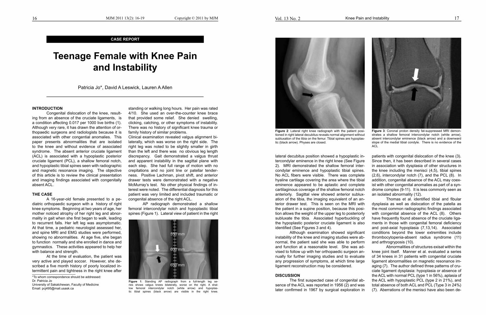
standing or walking long hours. Her pain was rated 4/10. She used an over-the-counter knee brace that provided some relief. She denied swelling, clicking, catching, or other symptoms of instability. There was no history of significant knee trauma or family history of similar problems. Clinical examination revealed valgus alignment bi-laterally, which was worse on the right side. The right leg was noted to be slightly smaller in girth than the left and there was no obvious leg length discrepancy. Gait demonstrated a valgus thrust and apparent instability in the sagittal plane with each step. She had full range of motion with no crepitations and no joint line or patellar tender-ness. Positive Lachman, pivot shift, and anterior drawer tests were demonstrated with a negative McMurray’s test. No other physical findings of in-terest were noted. The differential diagnosis for this patient was very limited and included traumatic or congenital absence of the right ACL. AP radiograph demonstrated a shallow femoral intercondylar notch and hypoplastic tibial spines (Figure 1). Lateral view of patient in the right
Teenage Female with Knee Pain and Instability
Patricia Jo*, David A Leswick, Lauren A Allen
Figure 1: Standing AP radiograph from a full-length leg se-ries shows valgus knees bilaterally, worse on the right. A shal-low femoral intercondylar notch (white arrow) and hypoplas-tic tibial spines (black arrow) are visible in the right knee.
17Knee Pain and InstabilityVol. 13 No. 2
patients with congenital dislocation of the knee (3). Since then, it has been described in several cases in association with dysplasia of other structures in the knee including the menisci (4,5), tibial spines (2,6), intercondylar notch (7), and the PCL (8). In addition, congenital absence of the ACL may coex-ist with other congenital anomalies as part of a syn-drome complex (9-11). It is less commonly seen as an isolated abnormality (12). Thomas et al. identified tibial and fibular dysplasia as well as dislocation of the patella as the most common radiographic findings associated with congenital absence of the ACL (8). Others have frequently found absence of the cruciate liga-ments in those with congenital femoral deficiency and post-axial hypoplasia (7,13,14). Associated conditions beyond the lower extremities include thrombocytopenia-absent radius syndrome (11) and arthrogryposis (10). Abnormalities of structures exisat within the knee joint itself. Manner et al. evaluated a series of 34 knees in 31 patients with congenital cruciate ligament abnormalities on magnetic resonance im-aging (7). The author defined three patterns of cru-ciate ligament dysplasia: hypoplasia or absence of the ACL with normal PCL (type 1 in 56%), aplasia of the ACL with hypoplastic PCL (type 2 in 21%), and total absence of both ACL and PCL (Type 3 in 24%) (7). Aberrations of the menisci have also been de-
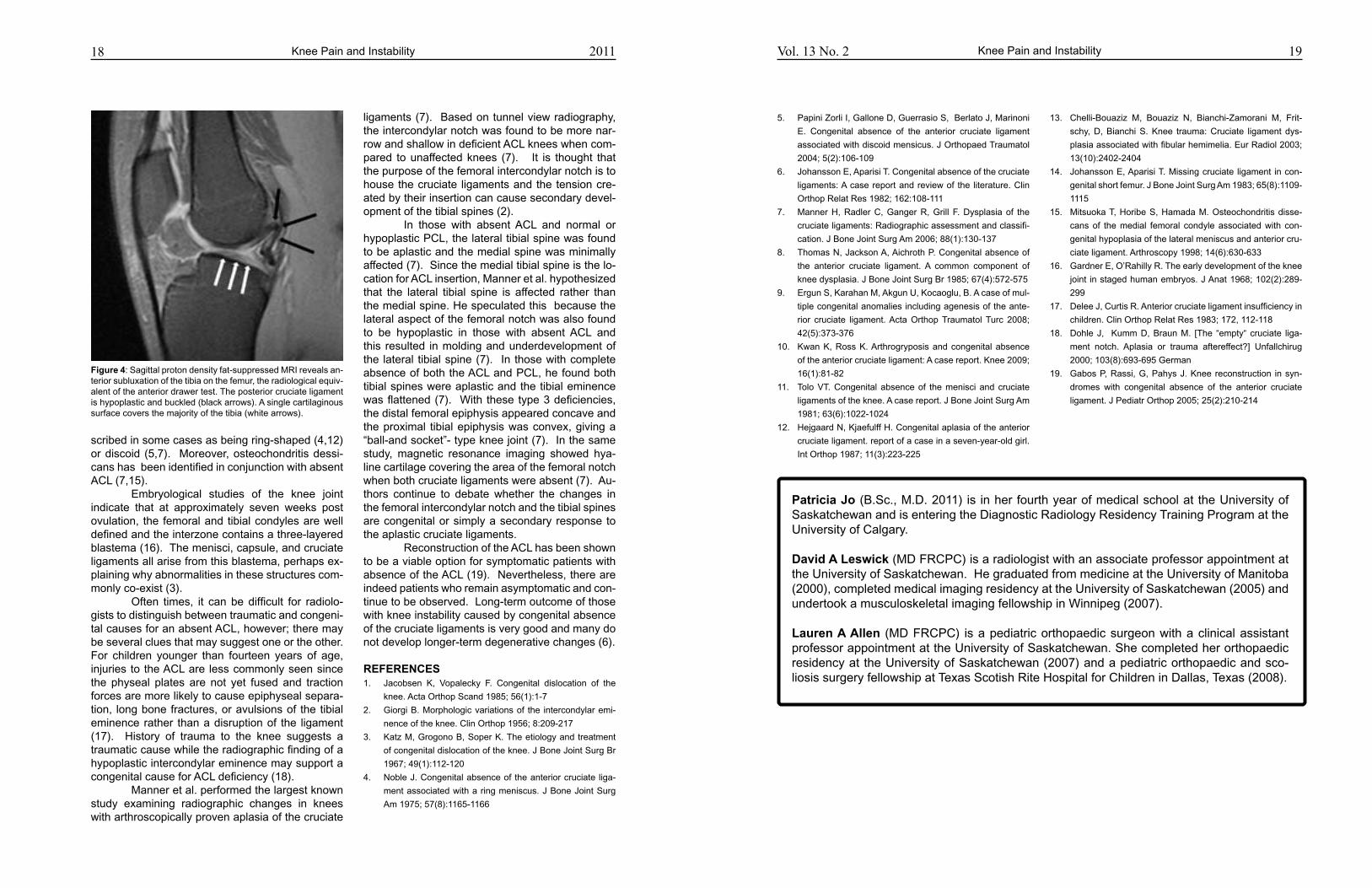
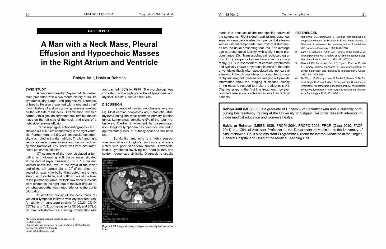
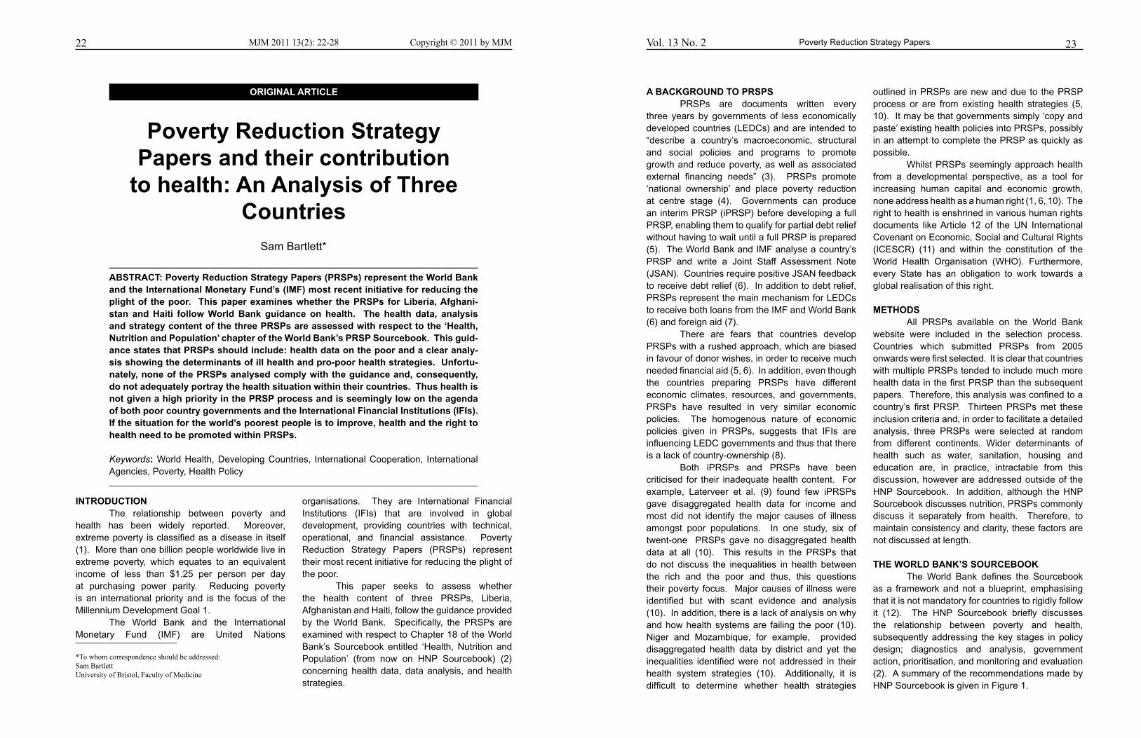
lateral decubitus position showed a hypoplastic in-tercondylar eminence in the right knee (See Figure 2). MRI demonstrated the shallow femoral inter-condylar eminence and hypoplastic tibial spines. No ACL fibers were visible. There was complete hyaline cartilage covering the area where the tibial eminence appeared to be aplastic and complete cartilaginous coverage of the shallow femoral notch anteriorly. Sagittal view showed anterior sublux-ation of the tibia, the imaging equivalent of an an-terior drawer test. This is seen on the MRI with the patient in a supine position, because this posi-tion allows the weight of the upper leg to posteriorly subluxate the tibia. Associated hyperbuckling of the hypoplastic posterior cruciate ligament is also identified (See Figures 3 and 4). Although examination showed significant instability of the knee and imaging studies were ab-normal, the patient said she was able to perform and function at a reasonable level. She was ad-vised to follow up with her orthopaedic surgeon an-nually for further imaging studies and to evaluate any progression of symptoms, at which time large ligament reconstruction may be considered.
dISCuSSIoN The first suspected case of congenital ab-sence of the ACL was reported in 1956 (2) and was later confirmed in 1967 by surgical exploration in
Figure 2: Lateral right knee radiograph with the patient posi-tioned in right lateral decubitus reveals normal alignment without subluxation of the tibia on the femur. Tibial spines are hypoplas-tic (black arrow). Physes are closed.
Figure 3: Coronal proton density fat-suppressed MRI demon-strates a shallow femoral intercondylar notch (white arrow), absent intercondylar eminence (black arrow) and a downward slope of the medial tibial condyle. There is no evidence of the ACL
18 19Vol. 13 No. 2 Knee Pain and Instability
5. Papini Zorli I, Gallone D, Guerrasio S, Berlato J, Marinoni
E. Congenital absence of the anterior cruciate ligament
associated with discoid mensicus. J Orthopaed Traumatol
2004; 5(2):106-109
6. Johansson E, Aparisi T. Congenital absence of the cruciate
ligaments: A case report and review of the literature. Clin
Orthop Relat Res 1982; 162:108-111
7. Manner H, Radler C, Ganger R, Grill F. Dysplasia of the
cruciate ligaments: Radiographic assessment and classifi-
cation. J Bone Joint Surg Am 2006; 88(1):130-137
8. Thomas N, Jackson A, Aichroth P. Congenital absence of
the anterior cruciate ligament. A common component of
knee dysplasia. J Bone Joint Surg Br 1985; 67(4):572-575
9. Ergun S, Karahan M, Akgun U, Kocaoglu, B. A case of mul-
tiple congenital anomalies including agenesis of the ante-
rior cruciate ligament. Acta Orthop Traumatol Turc 2008;
42(5):373-376
10. Kwan K, Ross K. Arthrogryposis and congenital absence
of the anterior cruciate ligament: A case report. Knee 2009;
16(1):81-82
11. Tolo VT. Congenital absence of the menisci and cruciate
ligaments of the knee. A case report. J Bone Joint Surg Am
1981; 63(6):1022-1024
12. Hejgaard N, Kjaefulff H. Congenital aplasia of the anterior
cruciate ligament. report of a case in a seven-year-old girl.
Int Orthop 1987; 11(3):223-225
13. Chelli-Bouaziz M, Bouaziz N, Bianchi-Zamorani M, Frit-
schy, D, Bianchi S. Knee trauma: Cruciate ligament dys-
plasia associated with fibular hemimelia. Eur Radiol 2003;
13(10):2402-2404
14. Johansson E, Aparisi T. Missing cruciate ligament in con-
genital short femur. J Bone Joint Surg Am 1983; 65(8):1109-
1115
15. Mitsuoka T, Horibe S, Hamada M. Osteochondritis disse-
cans of the medial femoral condyle associated with con-
genital hypoplasia of the lateral meniscus and anterior cru-
ciate ligament. Arthroscopy 1998; 14(6):630-633
16. Gardner E, O’Rahilly R. The early development of the knee
joint in staged human embryos. J Anat 1968; 102(2):289-
299
17. Delee J, Curtis R. Anterior cruciate ligament insufficiency in
children. Clin Orthop Relat Res 1983; 172, 112-118
18. Dohle J, Kumm D, Braun M. [The “empty“ cruciate liga-
ment notch. Aplasia or trauma aftereffect?] Unfallchirug
2000; 103(8):693-695 German
19. Gabos P, Rassi, G, Pahys J. Knee reconstruction in syn-
dromes with congenital absence of the anterior cruciate
ligament. J Pediatr Orthop 2005; 25(2):210-214
Knee Pain and Instability 2011
ligaments (7). Based on tunnel view radiography, the intercondylar notch was found to be more nar-row and shallow in deficient ACL knees when com-pared to unaffected knees (7). It is thought that the purpose of the femoral intercondylar notch is to house the cruciate ligaments and the tension cre-ated by their insertion can cause secondary devel-opment of the tibial spines (2). In those with absent ACL and normal or hypoplastic PCL, the lateral tibial spine was found to be aplastic and the medial spine was minimally affected (7). Since the medial tibial spine is the lo-cation for ACL insertion, Manner et al. hypothesized that the lateral tibial spine is affected rather than the medial spine. He speculated this because the lateral aspect of the femoral notch was also found to be hypoplastic in those with absent ACL and this resulted in molding and underdevelopment of the lateral tibial spine (7). In those with complete absence of both the ACL and PCL, he found both tibial spines were aplastic and the tibial eminence was flattened (7). With these type 3 deficiencies, the distal femoral epiphysis appeared concave and the proximal tibial epiphysis was convex, giving a “ball-and socket”- type knee joint (7). In the same study, magnetic resonance imaging showed hya-line cartilage covering the area of the femoral notch when both cruciate ligaments were absent (7). Au-thors continue to debate whether the changes in the femoral intercondylar notch and the tibial spines are congenital or simply a secondary response to the aplastic cruciate ligaments. Reconstruction of the ACL has been shown to be a viable option for symptomatic patients with absence of the ACL (19). Nevertheless, there are indeed patients who remain asymptomatic and con-tinue to be observed. Long-term outcome of those with knee instability caused by congenital absence of the cruciate ligaments is very good and many do not develop longer-term degenerative changes (6).
REFERENCES 1. Jacobsen K, Vopalecky F. Congenital dislocation of the
knee. Acta Orthop Scand 1985; 56(1):1-7
2. Giorgi B. Morphologic variations of the intercondylar emi-
nence of the knee. Clin Orthop 1956; 8:209-217
3. Katz M, Grogono B, Soper K. The etiology and treatment
of congenital dislocation of the knee. J Bone Joint Surg Br
1967; 49(1):112-120
4. Noble J. Congenital absence of the anterior cruciate liga-
ment associated with a ring meniscus. J Bone Joint Surg
Am 1975; 57(8):1165-1166
scribed in some cases as being ring-shaped (4,12) or discoid (5,7). Moreover, osteochondritis dessi-cans has been identified in conjunction with absent ACL (7,15). Embryological studies of the knee joint indicate that at approximately seven weeks post ovulation, the femoral and tibial condyles are well defined and the interzone contains a three-layered blastema (16). The menisci, capsule, and cruciate ligaments all arise from this blastema, perhaps ex-plaining why abnormalities in these structures com-monly co-exist (3). Often times, it can be difficult for radiolo-gists to distinguish between traumatic and congeni-tal causes for an absent ACL, however; there may be several clues that may suggest one or the other. For children younger than fourteen years of age, injuries to the ACL are less commonly seen since the physeal plates are not yet fused and traction forces are more likely to cause epiphyseal separa-tion, long bone fractures, or avulsions of the tibial eminence rather than a disruption of the ligament (17). History of trauma to the knee suggests a traumatic cause while the radiographic finding of a hypoplastic intercondylar eminence may support a congenital cause for ACL deficiency (18). Manner et al. performed the largest known study examining radiographic changes in knees with arthroscopically proven aplasia of the cruciate
Patricia Jo (B.Sc., M.D. 2011) is in her fourth year of medical school at the University of Saskatchewan and is entering the Diagnostic Radiology Residency Training Program at the University of Calgary.
david A Leswick (MD FRCPC) is a radiologist with an associate professor appointment at the University of Saskatchewan. He graduated from medicine at the University of Manitoba (2000), completed medical imaging residency at the University of Saskatchewan (2005) and undertook a musculoskeletal imaging fellowship in Winnipeg (2007).
Lauren A Allen (MD FRCPC) is a pediatric orthopaedic surgeon with a clinical assistant professor appointment at the University of Saskatchewan. She completed her orthopaedic residency at the University of Saskatchewan (2007) and a pediatric orthopaedic and sco-liosis surgery fellowship at Texas Scotish Rite Hospital for Children in Dallas, Texas (2008).
Figure 4: Sagittal proton density fat-suppressed MRI reveals an-terior subluxation of the tibia on the femur, the radiological equiv-alent of the anterior drawer test. The posterior cruciate ligament is hypoplastic and buckled (black arrows). A single cartilaginous surface covers the majority of the tibia (white arrows).
CASE REPoRT
MJM 2011 13(2): 20-2120 Copyright © 2011 by MJM
*To whom correspondence should be addressed:Dr. Rabiya JalilClinical Assistant Professor, Regina Qu’Appelle Health RegionRegina, SK, S4P 0W5, CanadaEmail: [email protected]
CASE STudy A previously healthy 55-year old Caucasian male presented with a two month history of flu-like symptoms, dry cough, and progressive shortness of breath. He also presented with a one and a half month history of a slowly growing painless swelling on the left side of his neck. Examination revealed normal vital signs, an erythematous, firm but mobile mass on the left side of the neck, and signs of a right sided pleural effusion. Transesophageal echocardiography (TEE)showed a 4.5 X 3 cm echodensity in the right ventri-cle. Furthermore, a 3.5 X 3.5 cm sessile echoden-sity was noted in the right atrium. The left and right ventricles were normal in size and function with an ejection fraction of 60%. There was trace circumfer-ential pericardial effusion. CT scanning of the neck displayed a fun-gating and ulcerating soft tissue mass situated at the dermal layer measuring 3.5 X 1.7 cm and located above the level of the hyoid at the lower end of the left parotid gland. CT of the chest re-vealed an extensive bulky filling defect in the right atrium, right ventricle, and outflow track at the level of the pulmonary valve. Multiple low density lesions were evident in the right lobe of the liver (Figure 1). Lymphadenopathy was noted inferior to the aortic bifurcation. In addition, biopsy of the neck mass re-vealed a lymphoid infiltrate with atypical features. A majority of cells were positive for CD20, CD10, CD79a, and TdT, but negative for CD34, and BCL-2 on immunohistochemical staining. Proliferation rate
approached 100% for Ki-67. The morphology was consistent with a high grade B-cell lymphoma with atypical Burkitt/Burkitt-like features.
dISCuSSIoN Incidence of cardiac neoplasia is very low (1). Most cardiac neoplasms are metastatic, atrial myxoma being the most common primary cardiac tumor. Lymphomas constitute 9% of the total me-tastases. Cardiac involvement by disseminated non-Hodgkin’s lymphoma has been documented in approximately 20% of autopsy cases to the heart (2). Burkitt-like lymphoma is a highly aggres-sive form of non-Hodgkin’s lymphoma and asso-ciated with poor short-term survival. Extranodal Burkitt Lymphoma involving the heart is rare and seldom recognized clinically. Diagnosis is usually
A Man with a Neck Mass, Pleural Effusion and Hypoechoic Masses in the Right Atrium and Ventricle
Rabiya Jalil*, Habib ur Rehman
Figure 1:CT image showing multiple low density lesions in the liver.
21
made late because of the non-specific nature of the symptoms. Right-sided heart failure, dyspnea, superior vena cava obstruction, pericardial effusion with or without tamponade, and rhythm disturbanc-es are the usual presenting features. The average age at presentation is sixty, with a slight male pre-dominance (3). Transesophageal echocardiogra-phy (TEE) is superior to transthoracic echocardiog-raphy (TTE) in assessment of cardiac lymphomas and typically shows a hypoechoic mass in the atria or ventricles that is often associated with pericardial effusion. Although multidetector computed tomog-raphy and magnetic resonance imaging will provide information about the staging of disease, biopsy of the mass is needed to make the diagnosis (4). Chemotherapy is the first line treatment, however complete remission is achieved in less than 60% of patients.
REFERENCES1. Rosenthal DS, Braunwald E. Cardiac manifestations of
neoplastic disease. In: Braunwald E, ed. Heart disease. A
Textbook of cardiovascular medicine. 3rd ed. Philadelphia:
WB Saunders Company, 1988;1744-1746.
2. Lam KY, Dickens P, Chan AC. Tumors of the heart. A 20-
year experience with a review of 12485 consecutive autop-
sies. Arch Pathol Lab Med 1993;117:1027–31.
3. Ceresoli GL, Ferreri AJ, Bucci E, Ripa C, Ponzoni M, Villa
E. Primary cardiac lymphoma in immunocompetent pa-
tients: diagnostic and therapeutic management. Cancer
1997; 80: 1479-506.
4. De Filippo M, Chernyschova N, Maffei E, Rovani C, De Bla-
si M, Beghi C, Zompatori M. Primary cardiac Burkett’s type
lymphoma: transthoracic echocardiography, multidetector
computed tomography and magnetic resonance findings.
Acta Radiologica 2006; 47: 167-71.
Vol. 13 No. 2
Rabiya Jalil (MD 2009) is a graduate of University of Saskatchewan and is currently com-pleting her residency training at the University of Calgary. Her other research interests in-clude medical education and women’s health.
Habib ur Rehman (MBBS 1989, FRCPI 2004, FRCPC 2008, FRCP (Glas) 2010, FACP 2011) is a Clinical Assistant Professor at the Department of Medicine at the University of Saskatchewan. He is also Assistant Programme Director for Internal Medicine at the Regina General Hospital and Head of the Medical Teaching Unit.
Cardiac Lymphoma
oRIgINAL ARTICLE
MJM 2011 13(2): 22-2822 Copyright © 2011 by MJM
AbSTRACT: Poverty Reduction Strategy Papers (PRSPs) represent the world bank and the International Monetary Fund’s (IMF) most recent initiative for reducing the plight of the poor. This paper examines whether the PRSPs for Liberia, Afghani-stan and Haiti follow world bank guidance on health. The health data, analysis and strategy content of the three PRSPs are assessed with respect to the ‘Health, Nutrition and Population’ chapter of the world bank’s PRSP Sourcebook. This guid-ance states that PRSPs should include: health data on the poor and a clear analy-sis showing the determinants of ill health and pro-poor health strategies. unfortu-nately, none of the PRSPs analysed comply with the guidance and, consequently, do not adequately portray the health situation within their countries. Thus health is not given a high priority in the PRSP process and is seemingly low on the agenda of both poor country governments and the International Financial Institutions (IFIs). If the situation for the world’s poorest people is to improve, health and the right to health need to be promoted within PRSPs.
Keywords: World Health, Developing Countries, International Cooperation, International Agencies, Poverty, Health Policy
*To whom correspondence should be addressed:Sam BartlettUniversity of Bristol, Faculty of Medicine
INTRoduCTIoN The relationship between poverty and health has been widely reported. Moreover, extreme poverty is classified as a disease in itself (1). More than one billion people worldwide live in extreme poverty, which equates to an equivalent income of less than $1.25 per person per day at purchasing power parity. Reducing poverty is an international priority and is the focus of the Millennium Development Goal 1.
The World Bank and the International Monetary Fund (IMF) are United Nations
organisations. They are International Financial Institutions (IFIs) that are involved in global development, providing countries with technical, operational, and financial assistance. Poverty Reduction Strategy Papers (PRSPs) represent their most recent initiative for reducing the plight of the poor.
This paper seeks to assess whether the health content of three PRSPs, Liberia, Afghanistan and Haiti, follow the guidance provided by the World Bank. Specifically, the PRSPs are examined with respect to Chapter 18 of the World Bank’s Sourcebook entitled ‘Health, Nutrition and Population’ (from now on HNP Sourcebook) (2) concerning health data, data analysis, and health strategies.
Poverty Reduction Strategy Papers and their contribution
to health: An Analysis of Three Countries
Sam Bartlett*
23Poverty Reduction Strategy Papers
A bACKgRouNd To PRSPSPRSPs are documents written every
three years by governments of less economically developed countries (LEDCs) and are intended to “describe a country’s macroeconomic, structural and social policies and programs to promote growth and reduce poverty, as well as associated external financing needs” (3). PRSPs promote ‘national ownership’ and place poverty reduction at centre stage (4). Governments can produce an interim PRSP (iPRSP) before developing a full PRSP, enabling them to qualify for partial debt relief without having to wait until a full PRSP is prepared (5). The World Bank and IMF analyse a country’s PRSP and write a Joint Staff Assessment Note (JSAN). Countries require positive JSAN feedback to receive debt relief (6). In addition to debt relief, PRSPs represent the main mechanism for LEDCs to receive both loans from the IMF and World Bank (6) and foreign aid (7).
There are fears that countries develop PRSPs with a rushed approach, which are biased in favour of donor wishes, in order to receive much needed financial aid (5, 6). In addition, even though the countries preparing PRSPs have different economic climates, resources, and governments, PRSPs have resulted in very similar economic policies. The homogenous nature of economic policies given in PRSPs, suggests that IFIs are influencing LEDC governments and thus that there is a lack of country-ownership (8).
Both iPRSPs and PRSPs have been criticised for their inadequate health content. For example, Laterveer et al. (9) found few iPRSPs gave disaggregated health data for income and most did not identify the major causes of illness amongst poor populations. In one study, six of twent-one PRSPs gave no disaggregated health data at all (10). This results in the PRSPs that do not discuss the inequalities in health between the rich and the poor and thus, this questions their poverty focus. Major causes of illness were identified but with scant evidence and analysis (10). In addition, there is a lack of analysis on why and how health systems are failing the poor (10). Niger and Mozambique, for example, provided disaggregated health data by district and yet the inequalities identified were not addressed in their health system strategies (10). Additionally, it is difficult to determine whether health strategies
outlined in PRSPs are new and due to the PRSP process or are from existing health strategies (5, 10). It may be that governments simply ‘copy and paste’ existing health policies into PRSPs, possibly in an attempt to complete the PRSP as quickly as possible.
Whilst PRSPs seemingly approach health from a developmental perspective, as a tool for increasing human capital and economic growth, none address health as a human right (1, 6, 10). The right to health is enshrined in various human rights documents like Article 12 of the UN International Covenant on Economic, Social and Cultural Rights (ICESCR) (11) and within the constitution of the World Health Organisation (WHO). Furthermore, every State has an obligation to work towards a global realisation of this right.
METHodS All PRSPs available on the World Bank website were included in the selection process. Countries which submitted PRSPs from 2005 onwards were first selected. It is clear that countries with multiple PRSPs tended to include much more health data in the first PRSP than the subsequent papers. Therefore, this analysis was confined to a country’s first PRSP. Thirteen PRSPs met these inclusion criteria and, in order to facilitate a detailed analysis, three PRSPs were selected at random from different continents. Wider determinants of health such as water, sanitation, housing and education are, in practice, intractable from this discussion, however are addressed outside of the HNP Sourcebook. In addition, although the HNP Sourcebook discusses nutrition, PRSPs commonly discuss it separately from health. Therefore, to maintain consistency and clarity, these factors are not discussed at length.
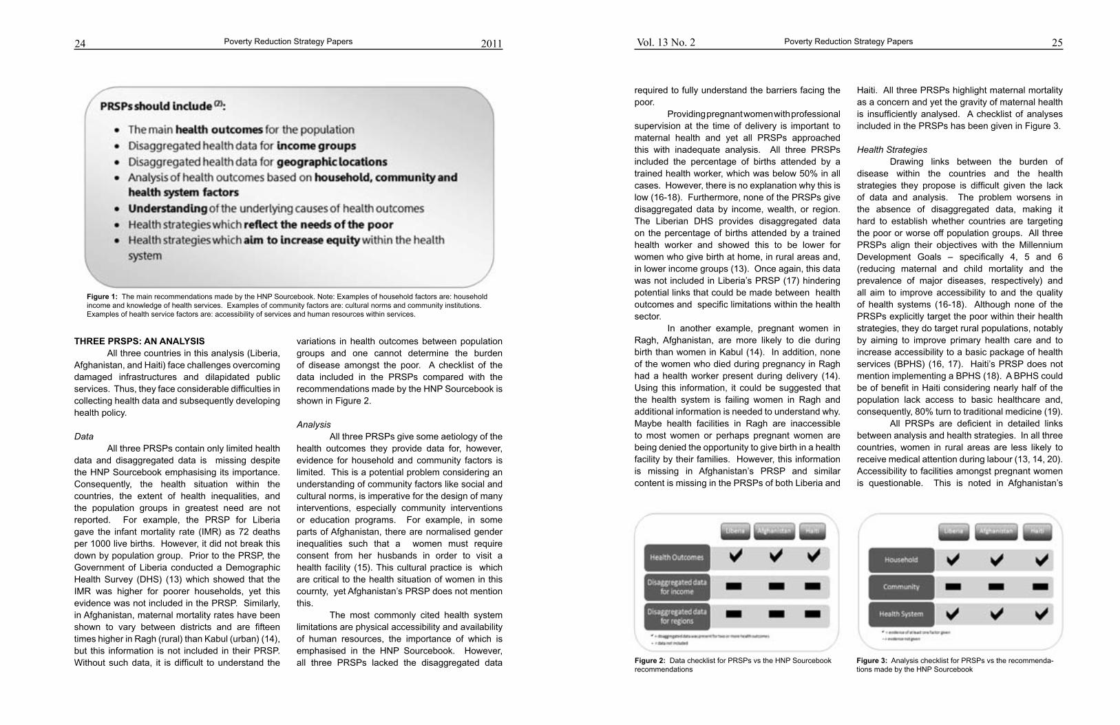
THE woRLd bANK’S SouRCEbooK The World Bank defines the Sourcebook as a framework and not a blueprint, emphasising that it is not mandatory for countries to rigidly follow it (12). The HNP Sourcebook briefly discusses the relationship between poverty and health, subsequently addressing the key stages in policy design; diagnostics and analysis, government action, prioritisation, and monitoring and evaluation (2). A summary of the recommendations made by HNP Sourcebook is given in Figure 1.
Vol. 13 No. 2
24 Poverty Reduction Strategy Papers
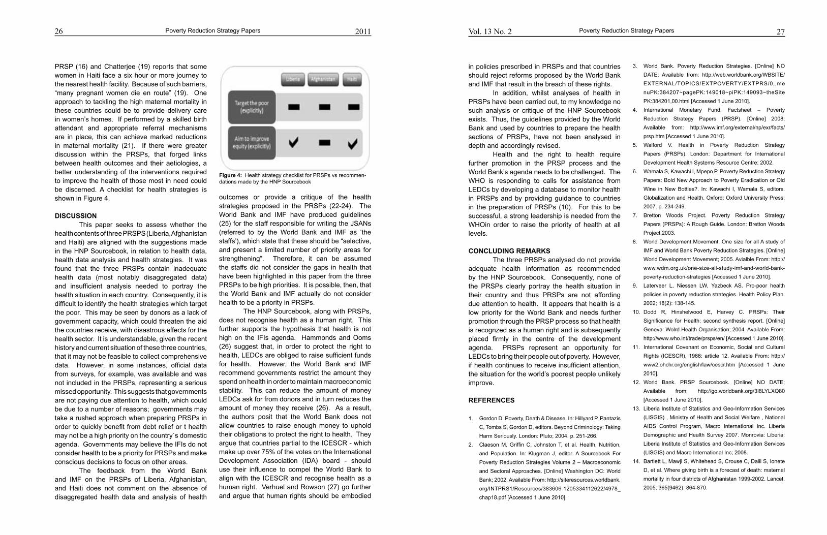
variations in health outcomes between population groups and one cannot determine the burden of disease amongst the poor. A checklist of the data included in the PRSPs compared with the recommendations made by the HNP Sourcebook is shown in Figure 2.
Analysis All three PRSPs give some aetiology of the health outcomes they provide data for, however, evidence for household and community factors is limited. This is a potential problem considering an understanding of community factors like social and cultural norms, is imperative for the design of many interventions, especially community interventions or education programs. For example, in some parts of Afghanistan, there are normalised gender inequalities such that a women must require consent from her husbands in order to visit a health facility (15). This cultural practice is which are critical to the health situation of women in this cournty, yet Afghanistan’s PRSP does not mention this. The most commonly cited health system limitations are physical accessibility and availability of human resources, the importance of which is emphasised in the HNP Sourcebook. However, all three PRSPs lacked the disaggregated data
THREE PRSPS: AN ANALySIS All three countries in this analysis (Liberia, Afghanistan, and Haiti) face challenges overcoming damaged infrastructures and dilapidated public services. Thus, they face considerable difficulties in collecting health data and subsequently developing health policy.
Data All three PRSPs contain only limited health data and disaggregated data is missing despite the HNP Sourcebook emphasising its importance. Consequently, the health situation within the countries, the extent of health inequalities, and the population groups in greatest need are not reported. For example, the PRSP for Liberia gave the infant mortality rate (IMR) as 72 deaths per 1000 live births. However, it did not break this down by population group. Prior to the PRSP, the Government of Liberia conducted a Demographic Health Survey (DHS) (13) which showed that the IMR was higher for poorer households, yet this evidence was not included in the PRSP. Similarly, in Afghanistan, maternal mortality rates have been shown to vary between districts and are fifteen times higher in Ragh (rural) than Kabul (urban) (14), but this information is not included in their PRSP. Without such data, it is difficult to understand the
2011 25Poverty Reduction Strategy Papers
Haiti. All three PRSPs highlight maternal mortality as a concern and yet the gravity of maternal health is insufficiently analysed. A checklist of analyses included in the PRSPs has been given in Figure 3.
Health Strategies Drawing links between the burden of disease within the countries and the health strategies they propose is difficult given the lack of data and analysis. The problem worsens in the absence of disaggregated data, making it hard to establish whether countries are targeting the poor or worse off population groups. All three PRSPs align their objectives with the Millennium Development Goals – specifically 4, 5 and 6 (reducing maternal and child mortality and the prevalence of major diseases, respectively) and all aim to improve accessibility to and the quality of health systems (16-18). Although none of the PRSPs explicitly target the poor within their health strategies, they do target rural populations, notably by aiming to improve primary health care and to increase accessibility to a basic package of health services (BPHS) (16, 17). Haiti’s PRSP does not mention implementing a BPHS (18). A BPHS could be of benefit in Haiti considering nearly half of the population lack access to basic healthcare and, consequently, 80% turn to traditional medicine (19). All PRSPs are deficient in detailed links between analysis and health strategies. In all three countries, women in rural areas are less likely to receive medical attention during labour (13, 14, 20). Accessibility to facilities amongst pregnant women is questionable. This is noted in Afghanistan’s
required to fully understand the barriers facing the poor. Providing pregnant women with professional supervision at the time of delivery is important to maternal health and yet all PRSPs approached this with inadequate analysis. All three PRSPs included the percentage of births attended by a trained health worker, which was below 50% in all cases. However, there is no explanation why this is low (16-18). Furthermore, none of the PRSPs give disaggregated data by income, wealth, or region. The Liberian DHS provides disaggregated data on the percentage of births attended by a trained health worker and showed this to be lower for women who give birth at home, in rural areas and, in lower income groups (13). Once again, this data was not included in Liberia’s PRSP (17) hindering potential links that could be made between health outcomes and specific limitations within the health sector. In another example, pregnant women in Ragh, Afghanistan, are more likely to die during birth than women in Kabul (14). In addition, none of the women who died during pregnancy in Ragh had a health worker present during delivery (14). Using this information, it could be suggested that the health system is failing women in Ragh and additional information is needed to understand why. Maybe health facilities in Ragh are inaccessible to most women or perhaps pregnant women are being denied the opportunity to give birth in a health facility by their families. However, this information is missing in Afghanistan’s PRSP and similar content is missing in the PRSPs of both Liberia and
Figure 1: The main recommendations made by the HNP Sourcebook. Note: Examples of household factors are: household income and knowledge of health services. Examples of community factors are: cultural norms and community institutions. Examples of health service factors are: accessibility of services and human resources within services.
Vol. 13 No. 2
Figure 2: Data checklist for PRSPs vs the HNP Sourcebook recommendations
Figure 3: Analysis checklist for PRSPs vs the recommenda-tions made by the HNP Sourcebook
Vol. 13 No. 226 Poverty Reduction Strategy Papers
outcomes or provide a critique of the health strategies proposed in the PRSPs (22-24). The World Bank and IMF have produced guidelines (25) for the staff responsible for writing the JSANs (referred to by the World Bank and IMF as ‘the staffs’), which state that these should be “selective, and present a limited number of priority areas for strengthening”. Therefore, it can be assumed the staffs did not consider the gaps in health that have been highlighted in this paper from the three PRSPs to be high priorities. It is possible, then, that the World Bank and IMF actually do not consider health to be a priority in PRSPs. The HNP Sourcebook, along with PRSPs, does not recognise health as a human right. This further supports the hypothesis that health is not high on the IFIs agenda. Hammonds and Ooms (26) suggest that, in order to protect the right to health, LEDCs are obliged to raise sufficient funds for health. However, the World Bank and IMF recommend governments restrict the amount they spend on health in order to maintain macroeconomic stability. This can reduce the amount of money LEDCs ask for from donors and in turn reduces the amount of money they receive (26). As a result, the authors posit that the World Bank does not allow countries to raise enough money to uphold their obligations to protect the right to health. They argue that countries partial to the ICESCR - which make up over 75% of the votes on the International Development Association (IDA) board - should use their influence to compel the World Bank to align with the ICESCR and recognise health as a human right. Verhuel and Rowson (27) go further and argue that human rights should be embodied
PRSP (16) and Chatterjee (19) reports that some women in Haiti face a six hour or more journey to the nearest health facility. Because of such barriers, “many pregnant women die en route” (19). One approach to tackling the high maternal mortality in these countries could be to provide delivery care in women’s homes. If performed by a skilled birth attendant and appropriate referral mechanisms are in place, this can achieve marked reductions in maternal mortality (21). If there were greater discussion within the PRSPs, that forged links between health outcomes and their aetiologies, a better understanding of the interventions required to improve the health of those most in need could be discerned. A checklist for health strategies is shown in Figure 4.
dISCuSSIoN This paper seeks to assess whether the health contents of three PRSPS (Liberia, Afghanistan and Haiti) are aligned with the suggestions made in the HNP Sourcebook, in relation to health data, health data analysis and health strategies. It was found that the three PRSPs contain inadequate health data (most notably disaggregated data) and insufficient analysis needed to portray the health situation in each country. Consequently, it is difficult to identify the health strategies which target the poor. This may be seen by donors as a lack of government capacity, which could threaten the aid the countries receive, with disastrous effects for the health sector. It is understandable, given the recent history and current situation of these three countries, that it may not be feasible to collect comprehensive data. However, in some instances, official data from surveys, for example, was available and was not included in the PRSPs, representing a serious missed opportunity. This suggests that governments are not paying due attention to health, which could be due to a number of reasons; governments may take a rushed approach when preparing PRSPs in order to quickly benefit from debt relief or t health may not be a high priority on the country`s domestic agenda. Governments may believe the IFIs do not consider health to be a priority for PRSPs and make conscious decisions to focus on other areas. The feedback from the World Bank and IMF on the PRSPs of Liberia, Afghanistan, and Haiti does not comment on the absence of disaggregated health data and analysis of health
27Poverty Reduction Strategy Papers
3. World Bank. Poverty Reduction Strategies. [Online] NO
DATE; Available from: http://web.worldbank.org/WBSITE/
EXTERNAL/TOPICS/EXTPOVERTY/EXTPRS/0,,me
nuPK:384207~pagePK:149018~piPK:149093~theSite
PK:384201,00.html [Accessed 1 June 2010].
4. International Monetary Fund. Factsheet – Poverty
Reduction Strategy Papers (PRSP). [Online] 2008;
Available from: http://www.imf.org/external/np/exr/facts/
prsp.htm [Accessed 1 June 2010].
5. Walford V. Health in Poverty Reduction Strategy
Papers (PRSPs). London: Department for International
Development Health Systems Resource Centre; 2002.
6. Wamala S, Kawachi I, Mpepo P. Poverty Reduction Strategy
Papers: Bold New Approach to Poverty Eradication or Old
Wine in New Bottles?. In: Kawachi I, Wamala S, editors.
Globalization and Health. Oxford: Oxford University Press;
2007. p. 234-249.
7. Bretton Woods Project. Poverty Reduction Strategy
Papers (PRSPs): A Rough Guide. London: Bretton Woods
Project,2003.
8. World Development Movement. One size for all A study of
IMF and World Bank Poverty Reduction Strategies. [Online]
World Development Movement; 2005. Avialble From: http://
www.wdm.org.uk/one-size-all-study-imf-and-world-bank-
poverty-reduction-strategies [Accessed 1 June 2010].
9. Laterveer L, Niessen LW, Yazbeck AS. Pro-poor health
policies in poverty reduction strategies. Health Policy Plan.
2002; 18(2): 138-145.
10. Dodd R, Hinshelwood E, Harvey C. PRSPs: Their
Significance for Health: second synthesis report. [Online]
Geneva: Wolrd Health Organisation; 2004. Available From:
http://www.who.int/trade/prsps/en/ [Accessed 1 June 2010].
11. International Covenant on Economic, Social and Cultural
Rights (ICESCR), 1966: article 12. Available From: http://
www2.ohchr.org/english/law/cescr.htm [Accessed 1 June
2010].
12. World Bank. PRSP Sourcebook. [Online] NO DATE;
Available from: http://go.worldbank.org/3I8LYLXO80
[Accessed 1 June 2010].
13. Liberia Institute of Statistics and Geo-Information Services
(LISGIS) , Ministry of Health and Social Welfare , National
AIDS Control Program, Macro International Inc. Liberia
Demographic and Health Survey 2007. Monrovia: Liberia:
Liberia Institute of Statistics and Geo-Information Services
(LISGIS) and Macro International Inc; 2008.
14. Bartlett L, Mawji S, Whitehead S, Crouse C, Dalil S, Ionete
D, et al. Where giving birth is a forecast of death: maternal
mortality in four districts of Afghanistan 1999-2002. Lancet.
2005; 365(9462): 864-870.
in policies prescribed in PRSPs and that countries should reject reforms proposed by the World Bank and IMF that result in the breach of these rights. In addition, whilst analyses of health in PRSPs have been carried out, to my knowledge no such analysis or critique of the HNP Sourcebook exists. Thus, the guidelines provided by the World Bank and used by countries to prepare the health sections of PRSPs, have not been analysed in depth and accordingly revised. Health and the right to health require further promotion in the PRSP process and the World Bank’s agenda needs to be challenged. The WHO is responding to calls for assistance from LEDCs by developing a database to monitor health in PRSPs and by providing guidance to countries in the preparation of PRSPs (10). For this to be successful, a strong leadership is needed from the WHOin order to raise the priority of health at all levels.
CoNCLudINg REMARKS The three PRSPs analysed do not provide adequate health information as recommended by the HNP Sourcebook. Consequently, none of the PRSPs clearly portray the health situation in their country and thus PRSPs are not affording due attention to health. It appears that health is a low priority for the World Bank and needs further promotion through the PRSP process so that health is recognzed as a human right and is subsequently placed firmly in the centre of the development agenda. PRSPs represent an opportunity for LEDCs to bring their people out of poverty. However, if health continues to receive insufficient attention, the situation for the world’s poorest people unlikely improve.
REFERENCES
1. Gordon D. Poverty, Death & Disease. In: Hillyard P, Pantazis
C, Tombs S, Gordon D, editors. Beyond Criminology: Taking
Harm Seriously. London: Pluto; 2004. p. 251-266.
2. Claeson M, Griffin C, Johnston T, et al. Health, Nutrition,
and Population. In: Klugman J, editor. A Sourcebook For
Poverty Reduction Strategies Volume 2 – Macroeconomic
and Sectoral Approaches. [Online] Washington DC: World
Bank; 2002. Available From: http://siteresources.worldbank.
org/INTPRS1/Resources/383606-1205334112622/4978_
chap18.pdf [Accessed 1 June 2010].
2011
Figure 4: Health strategy checklist for PRSPs vs recommen-dations made by the HNP Sourcebook
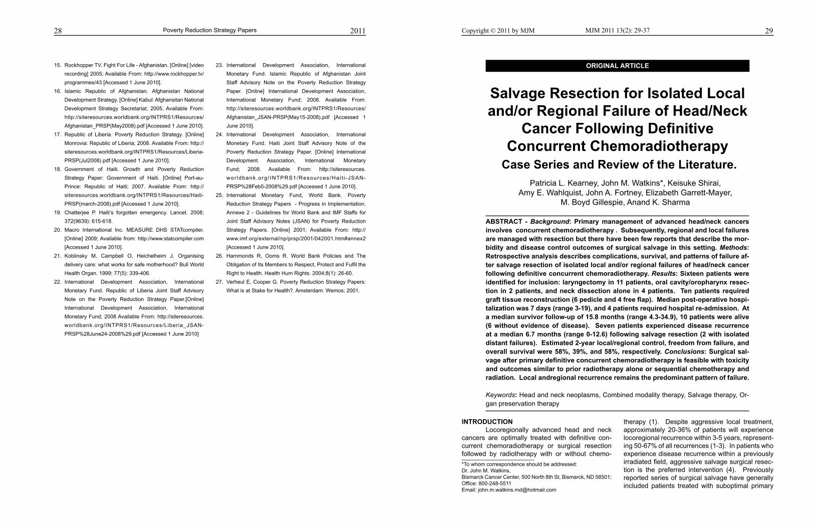
201128 Poverty Reduction Strategy Papers
23. International Development Association, International
Monetary Fund. Islamic Republic of Afghanistan Joint
Staff Advisory Note on the Poverty Reduction Strategy
Paper. [Online] International Development Association,
International Monetary Fund; 2008. Available From:
http://siteresources.worldbank.org/INTPRS1/Resources/
Afghanistan_JSAN-PRSP(May15-2008).pdf [Accessed 1
June 2010].
24. International Development Association, International
Monetary Fund. Haiti Joint Staff Advisory Note of the
Poverty Reduction Strategy Paper. [Online] International
Development Association, International Monetary
Fund; 2008. Available From: http://siteresources.
wor ldbank.org / INTPRS1/Resources/Hai t i -JSAN-
PRSP%28Feb5-2008%29.pdf [Accessed 1 June 2010].
25. International Monetary Fund, World Bank. Poverty
Reduction Strategy Papers - Progress in Implementation.
Annexe 2 - Guidelines for World Bank and IMF Staffs for
Joint Staff Advisory Notes (JSAN) for Poverty Reduction
Strategy Papers. [Online] 2001; Available From: http://
www.imf.org/external/np/prsp/2001/042001.htm#annex2
[Accessed 1 June 2010].
26. Hammonds R, Ooms R. World Bank Policies and The
Obligation of Its Members to Respect, Protect and Fulfil the
Right to Health. Health Hum Rights. 2004;8(1): 26-60.
27. Verheul E, Cooper G. Poverty Reduction Strategy Papers:
What is at Stake for Health?. Amsterdam: Wemos; 2001.
15. Rockhopper TV. Fight For Life - Afghanistan. [Online] [video
recording] 2005; Available From: http://www.rockhopper.tv/
programmes/43 [Accessed 1 June 2010].
16. Islamic Republic of Afghanistan. Afghanistan National
Development Strategy. [Online] Kabul: Afghansitan National
Development Strategy Secretariat; 2005. Available From:
http://siteresources.worldbank.org/INTPRS1/Resources/
Afghanistan_PRSP(May2008).pdf [Accessed 1 June 2010].
17. Republic of Liberia. Poverty Reduction Strategy. [Online]
Monrovia: Republic of Liberia; 2008. Available From: http://
siteresources.worldbank.org/INTPRS1/Resources/Liberia-
PRSP(Jul2008).pdf [Accessed 1 June 2010].
18. Government of Haiti. Growth and Poverty Reduction
Strategy Paper: Government of Haiti. [Online] Port-au-
Prince: Republic of Haiti; 2007. Available From: http://
siteresources.worldbank.org/INTPRS1/Resources/Haiti-
PRSP(march-2008).pdf [Accessed 1 June 2010].
19. Chatterjee P. Haiti’s forgotten emergency. Lancet. 2008;
372(9639): 615-618.
20. Macro International Inc. MEASURE DHS STATcompiler.
[Online] 2009; Available from: http://www.statcompiler.com
[Accessed 1 June 2010].
21. Koblinsky M, Campbell O, Heichelheim J. Organising
delivery care: what works for safe motherhood? Bull World
Health Organ. 1999; 77(5): 339-406.
22. International Development Association, International
Monetary Fund. Republic of Liberia Joint Staff Advisory
Note on the Poverty Reduction Strategy Paper.[Online]
International Development Association, International
Monetary Fund; 2008 Available From: http://siteresources.
worldbank.org/INTPRS1/Resources/Liberia_JSAN-
PRSP%28June24-2008%29.pdf [Accessed 1 June 2010]
oRIgINAL ARTICLE
MJM 2011 13(2): 29-37 29Copyright © 2011 by MJM
*To whom correspondence should be addressed:Dr. John M. Watkins,Bismarck Cancer Center, 500 North 8th St, Bismarck, ND 58501; Office: 800-248-5511Email: [email protected]
INTRoduCTIoN Locoregionally advanced head and neck cancers are optimally treated with definitive con-current chemoradiotherapy or surgical resection followed by radiotherapy with or without chemo-
therapy (1). Despite aggressive local treatment, approximately 20-36% of patients will experience locoregional recurrence within 3-5 years, represent-ing 50-67% of all recurrences (1-3). In patients who experience disease recurrence within a previously irradiated field, aggressive salvage surgical resec-tion is the preferred intervention (4). Previously reported series of surgical salvage have generally included patients treated with suboptimal primary
Salvage Resection for Isolated Local and/or Regional Failure of Head/Neck
Cancer Following Definitive Concurrent Chemoradiotherapy
Case Series and Review of the Literature.
Patricia L. Kearney, John M. Watkins*, Keisuke Shirai, Amy E. Wahlquist, John A. Fortney, Elizabeth Garrett-Mayer,
M. Boyd Gillespie, Anand K. Sharma
AbSTRACT - Background: Primary management of advanced head/neck cancers involves concurrent chemoradiotherapy . Subsequently, regional and local failures are managed with resection but there have been few reports that describe the mor-bidity and disease control outcomes of surgical salvage in this setting. Methods: Retrospective analysis describes complications, survival, and patterns of failure af-ter salvage resection of isolated local and/or regional failures of head/neck cancer following definitive concurrent chemoradiotherapy. Results: Sixteen patients were identified for inclusion: laryngectomy in 11 patients, oral cavity/oropharynx resec-tion in 2 patients, and neck dissection alone in 4 patients. Ten patients required graft tissue reconstruction (6 pedicle and 4 free flap). Median post-operative hospi-talization was 7 days (range 3-19), and 4 patients required hospital re-admission. At a median survivor follow-up of 15.8 months (range 4.3-34.9), 10 patients were alive (6 without evidence of disease). Seven patients experienced disease recurrence at a median 6.7 months (range 0-12.6) following salvage resection (2 with isolated distant failures). Estimated 2-year local/regional control, freedom from failure, and overall survival were 58%, 39%, and 58%, respectively. Conclusions: Surgical sal-vage after primary definitive concurrent chemoradiotherapy is feasible with toxicity and outcomes similar to prior radiotherapy alone or sequential chemotherapy and radiation. Local andregional recurrence remains the predominant pattern of failure.
Keywords: Head and neck neoplasms, Combined modality therapy, Salvage therapy, Or-gan preservation therapy
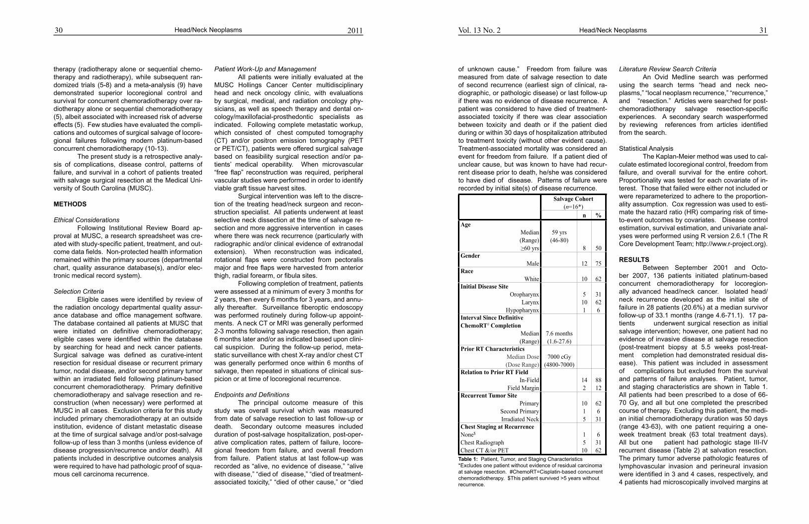
31
of unknown cause.” Freedom from failure was measured from date of salvage resection to date of second recurrence (earliest sign of clinical, ra-diographic, or pathologic disease) or last follow-up if there was no evidence of disease recurrence. A patient was considered to have died of treatment-associated toxicity if there was clear association between toxicity and death or if the patient died during or within 30 days of hospitalization attributed to treatment toxicity (without other evident cause). Treatment-associated mortality was considered an event for freedom from failure. If a patient died of unclear cause, but was known to have had recur-rent disease prior to death, he/she was considered to have died of disease. Patterns of failure were recorded by initial site(s) of disease recurrence.
Literature Review Search Criteria An Ovid Medline search was performed using the search terms “head and neck neo-plasms,” “local neoplasm recurrence,” “recurrence,” and “resection.” Articles were searched for post-chemoradiotherapy salvage resection-specific experiences. A secondary search wasperformed by reviewing references from articles identified from the search.
Statistical Analysis The Kaplan-Meier method was used to cal-culate estimated locoregional control, freedom from failure, and overall survival for the entire cohort. Proportionality was tested for each covariate of in-terest. Those that failed were either not included or were reparameterized to adhere to the proportion-ality assumption. Cox regression was used to esti-mate the hazard ratio (HR) comparing risk of time-to-event outcomes by covariates. Disease control estimation, survival estimation, and univariate anal-yses were performed using R version 2.6.1 (The R Core Development Team; http://www.r-project.org).
RESuLTS Between September 2001 and Octo-ber 2007, 136 patients initiated platinum-based concurrent chemoradiotherapy for locoregion-ally advanced head/neck cancer. Isolated head/neck recurrence developed as the initial site of failure in 28 patients (20.6%) at a median survivor follow-up of 33.1 months (range 4.6-71.1). 17 pa-tients underwent surgical resection as initial salvage intervention; however, one patient had no evidence of invasive disease at salvage resection (post-treatment biopsy at 5.5 weeks post-treat-ment completion had demonstrated residual dis-ease). This patient was included in assessment of complications but excluded from the survival and patterns of failure analyses. Patient, tumor, and staging characteristics are shown in Table 1. All patients had been prescribed to a dose of 66-70 Gy, and all but one completed the prescribed course of therapy. Excluding this patient, the medi-an initial chemoradiotherapy duration was 50 days (range 43-63), with one patient requiring a one-week treatment break (63 total treatment days). All but one patient had pathologic stage III-IV recurrent disease (Table 2) at salvation resection. The primary tumor adverse pathologic features of lymphovascular invasion and perineural invasion were identified in 3 and 4 cases, respectively, and 4 patients had microscopically involved margins at
Head/Neck Neoplasms201130 Vol. 13 No. 2Head/Neck Neoplasms
therapy (radiotherapy alone or sequential chemo-therapy and radiotherapy), while subsequent ran-domized trials (5-8) and a meta-analysis (9) have demonstrated superior locoregional control and survival for concurrent chemoradiotherapy over ra-diotherapy alone or sequential chemoradiotherapy (5), albeit associated with increased risk of adverse effects (5). Few studies have evaluated the compli-cations and outcomes of surgical salvage of locore-gional failures following modern platinum-based concurrent chemoradiotherapy (10-13). The present study is a retrospective analy-sis of complications, disease control, patterns of failure, and survival in a cohort of patients treated with salvage surgical resection at the Medical Uni-versity of South Carolina (MUSC). METHodS
Ethical Considerations Following Institutional Review Board ap-proval at MUSC, a research spreadsheet was cre-ated with study-specific patient, treatment, and out-come data fields. Non-protected health information remained within the primary sources (departmental chart, quality assurance database(s), and/or elec-tronic medical record system).
Selection Criteria Eligible cases were identified by review of the radiation oncology departmental quality assur-ance database and office management software. The database contained all patients at MUSC that were initiated on definitive chemoradiotherapy; eligible cases were identified within the database by searching for head and neck cancer patients. Surgical salvage was defined as curative-intent resection for residual disease or recurrent primary tumor, nodal disease, and/or second primary tumor within an irradiated field following platinum-based concurrent chemoradiotherapy. Primary definitive chemoradiotherapy and salvage resection and re-construction (when necessary) were performed at MUSC in all cases. Exclusion criteria for this study included primary chemoradiotherapy at an outside institution, evidence of distant metastatic disease at the time of surgical salvage and/or post-salvage follow-up of less than 3 months (unless evidence of disease progression/recurrence and/or death). All patients included in descriptive outcomes analysis were required to have had pathologic proof of squa-mous cell carcinoma recurrence.
Patient Work-Up and Management All patients were initially evaluated at the MUSC Hollings Cancer Center multidisciplinary head and neck oncology clinic, with evaluations by surgical, medical, and radiation oncology phy-sicians, as well as speech therapy and dental on-cology/maxillofacial-prosthedontic specialists as indicated. Following complete metastatic workup, which consisted of chest computed tomography (CT) and/or positron emission tomography (PET or PET/CT), patients were offered surgical salvage based on feasibility surgical resection and/or pa-tients’ medical operability. When microvascular “free flap” reconstruction was required, peripheral vascular studies were performed in order to identify viable graft tissue harvest sites. Surgical intervention was left to the discre-tion of the treating head/neck surgeon and recon-struction specialist. All patients underwent at least selective neck dissection at the time of salvage re-section and more aggressive intervention in cases where there was neck recurrence (particularly with radiographic and/or clinical evidence of extranodal extension). When reconstruction was indicated, rotational flaps were constructed from pectoralis major and free flaps were harvested from anterior thigh, radial forearm, or fibula sites. Following completion of treatment, patients were assessed at a minimum of every 3 months for 2 years, then every 6 months for 3 years, and annu-ally thereafter. Surveillance fiberoptic endoscopy was performed routinely during follow-up appoint-ments. A neck CT or MRI was generally performed 2-3 months following salvage resection, then again 6 months later and/or as indicated based upon clini-cal suspicion. During the follow-up period, meta-static surveillance with chest X-ray and/or chest CT was generally performed once within 6 months of salvage, then repeated in situations of clinical sus-picion or at time of locoregional recurrence.
Endpoints and Definitions The principal outcome measure of this study was overall survival which was measured from date of salvage resection to last follow-up or death. Secondary outcome measures included duration of post-salvage hospitalization, post-oper-ative complication rates, pattern of failure, locore-gional freedom from failure, and overall freedom from failure. Patient status at last follow-up was recorded as “alive, no evidence of disease,” “alive with disease,” “died of disease,” “died of treatment-associated toxicity,” “died of other cause,” or “died
Salvage Cohort(n=16*)
Salvage Cohort(n=16*)
Salvage Cohort(n=16*)
n %Age
Median(Range)>60 yrs
59 yrs(46-80)
8 50Gender
Male 12 75Race
White 10 62Initial Disease Site
OropharynxLarynx
Hypopharynx
5101
31626
Interval Since Definitive ChemoRT# Completion
Median(Range)
7.6 months(1.6-27.6)
Prior RT CharacteristicsMedian Dose(Dose Range)
7000 cGy(4800-7000)
Relation to Prior RT FieldIn-Field
Field Margin 14
28812
Recurrent Tumor SitePrimary
Second PrimaryIrradiated Neck
1015
626
31Chest Staging at RecurrenceNone$
Chest RadiographChest CT &/or PET
15
10
63162
Table 1: Patient, Tumor, and Staging Characteristics *Excludes one patient without evidence of residual carcinoma at salvage resection. #ChemoRT=Cisplatin-based concurrent chemoradiotherapy. $This patient survived >5 years without recurrence.
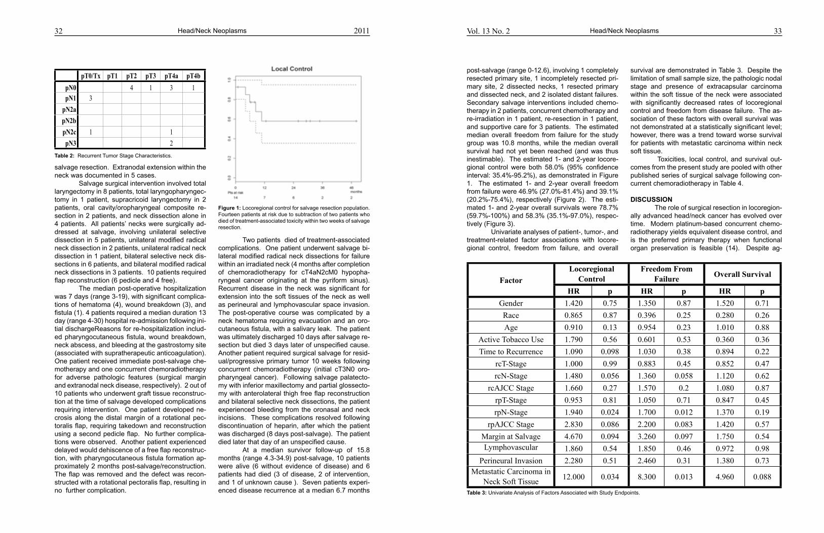
33Vol. 13 No. 2 Head/Neck Neoplasms
survival are demonstrated in Table 3. Despite the limitation of small sample size, the pathologic nodal stage and presence of extracapsular carcinoma within the soft tissue of the neck were associated with significantly decreased rates of locoregional control and freedom from disease failure. The as-sociation of these factors with overall survival was not demonstrated at a statistically significant level; however, there was a trend toward worse survival for patients with metastatic carcinoma within neck soft tissue. Toxicities, local control, and survival out-comes from the present study are pooled with other published series of surgical salvage following con-current chemoradiotherapy in Table 4.
dISCuSSIoN The role of surgical resection in locoregion-ally advanced head/neck cancer has evolved over time. Modern platinum-based concurrent chemo-radiotherapy yields equivalent disease control, and is the preferred primary therapy when functional organ preservation is feasible (14). Despite ag-
post-salvage (range 0-12.6), involving 1 completely resected primary site, 1 incompletely resected pri-mary site, 2 dissected necks, 1 resected primary and dissected neck, and 2 isolated distant failures. Secondary salvage interventions included chemo-therapy in 2 patients, concurrent chemotherapy and re-irradiation in 1 patient, re-resection in 1 patient, and supportive care for 3 patients. The estimated median overall freedom from failure for the study group was 10.8 months, while the median overall survival had not yet been reached (and was thus inestimable). The estimated 1- and 2-year locore-gional control were both 58.0% (95% confidence interval: 35.4%-95.2%), as demonstrated in Figure 1. The estimated 1- and 2-year overall freedom from failure were 46.9% (27.0%-81.4%) and 39.1% (20.2%-75.4%), respectively (Figure 2). The esti-mated 1- and 2-year overall survivals were 78.7% (59.7%-100%) and 58.3% (35.1%-97.0%), respec-tively (Figure 3). Univariate analyses of patient-, tumor-, and treatment-related factor associations with locore-gional control, freedom from failure, and overall
32
salvage resection. Extranodal extension within the neck was documented in 5 cases. Salvage surgical intervention involved total laryngectomy in 8 patients, total laryngopharyngec-tomy in 1 patient, supracricoid laryngectomy in 2 patients, oral cavity/oropharyngeal composite re-section in 2 patients, and neck dissection alone in 4 patients. All patients’ necks were surgically ad-dressed at salvage, involving unilateral selective dissection in 5 patients, unilateral modified radical neck dissection in 2 patients, unilateral radical neck dissection in 1 patient, bilateral selective neck dis-sections in 6 patients, and bilateral modified radical neck dissections in 3 patients. 10 patients required flap reconstruction (6 pedicle and 4 free). The median post-operative hospitalization was 7 days (range 3-19), with significant complica-tions of hematoma (4), wound breakdown (3), and fistula (1). 4 patients required a median duration 13 day (range 4-30) hospital re-admission following ini-tial dischargeReasons for re-hospitalization includ-ed pharyngocutaneous fistula, wound breakdown, neck abscess, and bleeding at the gastrostomy site (associated with supratherapeutic anticoagulation). One patient received immediate post-salvage che-motherapy and one concurrent chemoradiotherapy for adverse pathologic features (surgical margin and extranodal neck disease, respectively). 2 out of 10 patients who underwent graft tissue reconstruc-tion at the time of salvage developed complications requiring intervention. One patient developed ne-crosis along the distal margin of a rotational pec-toralis flap, requiring takedown and reconstruction using a second pedicle flap. No further complica-tions were observed. Another patient experienced delayed would dehiscence of a free flap reconstruc-tion, with pharyngocutaneous fistula formation ap-proximately 2 months post-salvage/reconstruction. The flap was removed and the defect was recon-structed with a rotational pectoralis flap, resulting in no further complication.
Two patients died of treatment-associated complications. One patient underwent salvage bi-lateral modified radical neck dissections for failure within an irradiated neck (4 months after completion of chemoradiotherapy for cT4aN2cM0 hypopha-ryngeal cancer originating at the pyriform sinus). Recurrent disease in the neck was significant for extension into the soft tissues of the neck as well as perineural and lymphovascular space invasion. The post-operative course was complicated by a neck hematoma requiring evacuation and an oro-cutaneous fistula, with a salivary leak. The patient was ultimately discharged 10 days after salvage re-section but died 3 days later of unspecified cause. Another patient required surgical salvage for resid-ual/progressive primary tumor 10 weeks following concurrent chemoradiotherapy (initial cT3N0 oro-pharyngeal cancer). Following salvage palatecto-my with inferior maxillectomy and partial glossecto-my with anterolateral thigh free flap reconstruction and bilateral selective neck dissections, the patient experienced bleeding from the oronasal and neck incisions. These complications resolved following discontinuation of heparin, after which the patient was discharged (8 days post-salvage). The patient died later that day of an unspecified cause. At a median survivor follow-up of 15.8 months (range 4.3-34.9) post-salvage, 10 patients were alive (6 without evidence of disease) and 6 patients had died (3 of disease, 2 of intervention, and 1 of unknown cause ). Seven patients experi-enced disease recurrence at a median 6.7 months
Head/Neck Neoplasms 2011
pT0/Tx pT1 pT2 pT3 pT4a pT4bpN0 4 1 3 1pN1 3pN2apN2bpN2c 1 1pN3 2
Table 2: Recurrent Tumor Stage Characteristics.
Figure 1: Locoregional control for salvage resection population. Fourteen patients at risk due to subtraction of two patients who died of treatment-associated toxicity within two weeks of salvage resection.
FactorLocoregional
ControlLocoregional
ControlFreedom From
FailureFreedom From
Failure Overall SurvivalOverall SurvivalFactor
HR p HR p HR pGender 1.420 0.75 1.350 0.87 1.520 0.71Race 0.865 0.87 0.396 0.25 0.280 0.26Age 0.910 0.13 0.954 0.23 1.010 0.88
Active Tobacco Use 1.790 0.56 0.601 0.53 0.360 0.36Time to Recurrence 1.090 0.098 1.030 0.38 0.894 0.22
rcT-Stage 1.000 0.99 0.883 0.45 0.852 0.47rcN-Stage 1.480 0.056 1.360 0.058 1.120 0.62
rcAJCC Stage 1.660 0.27 1.570 0.2 1.080 0.87rpT-Stage 0.953 0.81 1.050 0.71 0.847 0.45rpN-Stage 1.940 0.024 1.700 0.012 1.370 0.19
rpAJCC Stage 2.830 0.086 2.200 0.083 1.420 0.57Margin at Salvage 4.670 0.094 3.260 0.097 1.750 0.54Lymphovascular
Invasion1.860 0.54 1.850 0.46 0.972 0.98
Perineural Invasion 2.280 0.51 2.460 0.31 1.380 0.73Metastatic Carcinoma in
Neck Soft Tissue 12.000 0.034 8.300 0.013 4.960 0.088
Table 3: Univariate Analysis of Factors Associated with Study Endpoints.

34 35Vol. 13 No. 2
gressive combined-modality therapy, approximate-ly 20-36% of patients still experience locoregional recurrence within 3-5 years, representing approxi-mately half of all recurrences (1). In patients who experience disease recurrence within a previously irradiated field, aggressive salvage surgical resec-tion is the preferred intervention, yielding 55% lo-cal control and 32-39% survival at 5 years (4,15). These have primarily included patients treated with radiotherapy alone or sequential radiotherapy and chemotherapy, which have demonstrated inferior disease control when compared with concurrent chemoradiotherapy (5-10). Few reports have focused on the compli-cations, disease control, and survival outcomes for concurrent chemoradiotherapy patients. The RTOG 91-11 study provides the best comparative data for salvage resection in the setting of radio-therapy alone versus sequential chemotherapy/
studies (disease site, reconstruction techniques) and inconsistent scoring of self-limited or “minor” fistulae in the retrospective analyses relative to the RTOG 91-11 analysis. If only “major” fistulae are considered for the RTOG study, the rate (11%) is more consistent with the retrospective series. With-in our own series, 2 of the 3 fistulae were self-limited salivary gland leaks which resolved with conserva-tive measures. Thus, the overall risk of significant fistula formation after surgical salvage for concur-rent chemoradiotherapy failures is approximately 5-10%. Graft tissue reconstruction may have a beneficial impact on reducing these major com-plications. A single-institution analysis of salvage laryngectomy following radiotherapy or chemora-diotherapy demonstrated a reduced risk of fistula for free flap reconstruction compared with primary closure (18% versus 50%, p=0.08) (16). Similarly, the risk of stricture and feeding tube dependence was reduced with free flap reconstruction. It should be noted that this analysis was restricted to patients whose disease was generally limited to the larynx, excluding patients for whom partial pharyngectomy or total laryngopharyngectomy would have been re-quired. Within the present study population, com-plications were similar regardless of graft tissue re-construction (4 of 11 reconstructed patients versus 2 of 6 primary closure patients). The present study demonstrated an esti-mated 2-year overall survival of 58%, which is com-parable to other single-institution series of post-
radiotherapy versus concurrent chemoradiotherapy (5). Of 517 patients initially randomized, 129 re-quired total laryngectomy (95% for recurrent/resid-ual cancer) (10). While pharyngocutaneous fistula formation was less common in the radiotherapy alone arm (15%) compared to the the concurrent chemoradiotherapy arm (30%), this did not reach statistical significance (p>0.05). Similarly, the over-all incidence of major and minor complications was not significantly different between treatment groups (52-59%). These consisted mainly of fistulae for-mation, infection, wound dehiscence, dysphagia and/or inability to take fluids, which are consistent with complications reported by other published se-ries (10-12). Within the RTOG concurrent chemo-radiotherapy group, the rate of fistula was some-what higher than that described in single-institution series (30% versus 5-8%) (10-12). This may be associated with inherent differences between the
chemoradiotherapy salvage resection (11,13) as well as mixed-treatment populations (4). Patients with smaller tumor burden (rT1-2) appear to have im-proved survival over larger tumors, as do laryngeal recurrences over pharyngeal sites (4,10,15,17,18). Within our own population, all 4 patients with long-term disease-free and overall survivals (>2 years) were locoregional failures of larynx primary sites, despite locally advanced disease at recurrence (rp-T4aN0 in two patients, rpT3N0 in one, and rpTxN1 in one). The interval from primary therapy comple-tion to disease failure does not appear to impact disease control or survival outcomes (4). Though limited by small numbers, no association was iden-tifiable within the present study population. The role of immediate post-salvage che-motherapy and/or re-irradiation remains to be determined. A recently published French trial randomized 130irradiated patients to adjuvant chemoradiotherapy or observation following sal-vage resection (19). Despite improved local con-trol and disease-free survival, no overall survival benefit was observed, and severe late toxicities (in-cluding subcutaneous fibrosis, osteoradionecrosis, and trismus) were higher in the adjuvant chemo-radiotherapy arm (39% vs 11% at 2 years). It is possible that the benefit may be limited to a spe-cific “high-risk” subset of patients requiring salvage resection. One single-institution series of post-chemoradiotherapy failures demonstrated a trend toward poorer survival in patients with the specific
Head/Neck Neoplasms Head/Neck Neoplasms2011
Series NMethodol
ogy Site(s)2y Local Control
2y Overall Survival
Major Complications
Richey11
(UNC, 2007) 38Retrospect
ive Review
Multiple 42% 27%
Fistula (1/38)Wound Dehiscence
(2/38)Respiratory Failure
(2/38)Sepsis (1/38)
Weber10
(RTOG 91-11, 2003)
27 RCT Larynx 74% 71%Fistula (8/27)
Wound Dehiscence (4/27)
Morgan12
(MDACC, 2007)
38Retrospect
ive Review
Multiple N/R N/R Fistula (3/38)Infection (2/38)
Yom13
(Multi-Institutional,
2005)
14*Retrospect
ive Review
Multiple 64%* 57%*Wound Dehiscence
(3/14)
Kearney(MUSC)
17Retrospect
ive Review
Multiple 58%# 58%#
Fistula (3/17)Wound Dehiscence
(3/17)Flap Breakdown
(2/10)Table 4: Published Series of Toxicities and Outcomes for Salvage Resection of Concurrent Chemoradiotherapy Recurrences in Head/Neck Cancer. *Twelve of fourteen underwent surgical salvage for residual or progressive disease noted at post-chemoradiotherapy assessment, local control and survival reflect crude recurrence rates rather than 2-year estimates. #Survival data based upon 16 patients with pathologic disease at salvage, excludes one patient with squamous cell carcinoma at post-chemoradiotherapy biopsy but no evidence of disease following salvage laryngectomy.
Figure 2: Freedom from disease failure for salvage resection population.
Figure 3: Overall survival for salvage resection population.
36 37Vol. 13 No. 2
high-risk features of involved surgical margins and/or extranodal neck disease (11). Alternatively, these patients may simply have more advanced disease and would have a worse prognosis regard-less of post-salvage intervention. Within the pres-ent study, two patients underwent immediate post-salvage therapy for high-risk pathologic features. One patient received immediate chemotherapy for involved surgical margins and another underwent concurrent chemotherapy and re-irradiation for ex-tranodal carcinoma within their previously irradiated neck. Neither patient experienced significant post-salvage complications; however, both experienced local disease failure within 6 months. Despite aggressive surgical salvage, the majority of disease recurrences involve the primary tumor site. Of 7 patients from the present series who experienced disease recurrence, 5 recur-rences involved the tumor bed. This is consistent with other published data (11); however, the pat-tern of failure appears to shift toward more distant metastasis for advanced node-positive recurrences (11,13) and those treated with radiotherapy alone (10,20). The timing of post-salvage disease failure was also consistent with previously published se-ries (median 9 months) (11,18); all but one patient within the present series failed within one year (iso-lated distant failure at 12.6 months). As suggested by Goodwin’s comprehensive review (4), the rapidi-ty of post-salvage recurrence may be attributable to advanced stage at initial recurrence presentation. Specifically, the analysis demonstrated an inverse relationship between recurrent disease stage and post-salvage median disease-free survival (>22.1, >11.5, 14.4, and 5.5 months for stages I, II, III, and IV disease, respectively). Within the present se-ries, all patients who experienced post-salvage dis-ease failure had recurrent pathologic stage III (n=2) or IV (5) disease. Salvage resection of local and/or regional head/neck cancer failures following platinum-based concurrent chemoradiotherapy is feasible and can provide the opportunity for disease control and survival. Post-operative complication rates, timing and patterns of disease failure, locoregional con-trol, freedom from disease failure, and overall sur-vival rates are similar to those described in series of surgically salvaged patients treated with prior ra-diotherapy alone or sequential chemotherapy and radiation.
13. Yom SS, Machtay M, Biel MA, et al. Survival impact of
planned restaging and early surgical salvage following de-
finitive chemoradiation for locally advanced squamous cell
carcinomas of the oropharynx and hypopharynx. Am J Clin
Oncol 2005; 28:385-392.
14. Cognetti DM, Weber RS, Lai SY. Head and neck cancer:
an evolving treatment paradigm. Cancer 2008; 113(7 sup-
pl):1911-1932.
15. Agra IMG, Carvalho, AL, Ulbrich FS, et al. Prognostic fac-
tors in salvage surgery for recurrent oral and oropharyn-
geal cancer. Head Neck 2006; 28:107-113.
16. Withrow KP, Rosenthal EL, Gourin CG, et al. Free tissue
transfer to manage salvage laryngectomy defects after
organ preservation failure. Laryngoscope 2007; 117:781-
784.
17. Ganly I, Patel SG, Matsuo J, et al. Results of surgical sal-
vage after failure of definitive radiation therapy for early-
stage squamous cell carcinoma of the glottic larynx. Arch
Otolaryngol Head Neck Surg 2006; 132:59-66.
REFERENCES1. Forastiere A, Koch W, Trotti A, Sidransky D. Head and neck
cancer. N Engl J Med 2001; 345:1890-1900.
2. Cooper JS, Pajak TF, Forastiere AA, et al. Postoperative
concurrent radiotherapy and chemotherapy for high-risk
squamous-cell carcinoma of the head and neck. N Engl J
Med 2004; 350:1937-1944.
3. Bernier J, Domenge C, Ozsahin M, et al. Postoperative ir-
radiation with or without concomitant chemotherapy for lo-
cally advanced head and neck cancer. N Engl J Med 2004;
350:1945-1952.
4. Goodwin WJ. Salvage surgery for patients with recurrent
squamous cell carcinoma of the upper aerodigestive tract:
when do the ends justify the means? Laryngoscope 2000;
110;(suppl 93):1-18.
5. Forastiere AA, Goepfert H, Maor M, et al. Concurrent che-
motherapy and radiotherapy for organ preservation in ad-
vanced laryngeal cancer. N Engl J Med 2003; 349:2091-
2098.
6. Calais G, Alfonsi M, Bardet E, et al. Randomized trial of ra-
diation therapy versus concomitant chemotherapy and ra-
diation therapy for advanced-stage oropharynx carcinoma.
J Natl Cancer Inst 1999; 91:2081-2086.
7. Adelstein DJ, Li Y, Adams GL, et al. An Intergroup phase III
comparison of standard radiation therapy and two sched-
ules of concurrent chemoradiotherapy in patients with un-
resectable squamous cell head and neck cancer. J Clin
Oncol 2003; 21:92-98.
8. Adelstein DJ, Saxton JP, Lavertu P, et al. A phase III ran-
domized trial comparing concurrent chemotherapy and
radiotherapy with radiotherapy alone in resectable stage
III and IV squamous cell cancer: preliminary results. Head
Neck 1997; 19:567-575.
9. Pignon JP, le Maitre A, Bourhis J. Meta-analyses of chemo-
therapy in head and neck cancer (MACH-NC): an update.
Int J Radiat Oncol Biol Phys 2007; 69(2 suppl 1):S112-
S114.
10. Weber RS, Berkey BA, Forastiere A, et al. Outcome of
salvage total laryngectomy following organ preservation
therapy. The Radiation Therapy Oncology Group trial 91-
11. Arch Otolaryngol Head Neck Surg 2003; 129:44-49.
11. Richey LM, Shores CG, George J, et al. The effectiveness
of salvage surgery after the failure of primary concomi-
tant chemoradiation in head and neck cancer. Otolaryngol
Head Neck Surg 2007; 136:98-103.
12. Morgan JE, Breau RL, Suen JY, Hanna EY. Surgical wound
complications after intensive chemoradiotherapy for ad-
vanced squamous cell carcinoma of the head and neck.
Arch Otoloryngol Head Neck Surg 2007; 133:10-14.
18. Kim AJ, Suh JD, Sercarz JA, et al. Salvage surgery with
free flap reconstruction: factors affecting outcome after
treatment of recurrent head and neck squamous carcino-
ma. Laryngoscope 2007; 117:1019-1023.
19. Janot F, de Raucourt D, Benhamou E, et al. Randomized
trial of postoperative reirradiation combined with chemo-
therapy after salvage surgery compared with salvage sur-
gery alone in head and neck carcinoma. J Clin Oncol 2008;
26:5518-5523.
20. Forastiere AA, Maor M, Weber RS, et al. Long-term results
of Intergroup RTOG 91-11: a phase III trial to preserve the
larynx – induction cisplatin/5-FU and radiation therapy ver-
sus concurrent cisplatin and radiation therapy versus radia-
tion therapy. J Clin Oncol 2006; 24(18 suppl):A5517.
Head/Neck Neoplasms Head/Neck Neoplasms2011
39Autism
(i.e., with an average or above average IQ) tend to outperform unaffected people with similar IQs on systemizing tasks (8). Third, the behavioral differ-ences between people with and without autism are mediated by differences at the anatomical level of the brain (9). Fourth, prenatal exposure to testos-terone (i.e., an androgen) is positively related to the development of autistic traits (10). From an evolutionary point of view, in an-cestral times, men with well-developed systemizing skills and under-developed empathizing skills may have had an advantage over other men (11). It is generally accepted that most humans used to live in hunter-gatherer societies, in which men fulfilled the role of hunter, and women the role of gatherer. Systemizing may have been important in develop-ing tools and weapons, in hunting, tracking, and trading. Empathizing may have been disadvanta-geous in situations where rivals had to be eliminat-ed, and in situations where one had to tolerate soli-tude, while being far away from home for hunting. For women, empathizing may have been more im-portant because of mothering, making new friends (women used to marry into new groups), gossiping, and inferring the thoughts of a possible mate (to discover whether he is willing to invest in offspring). So having an extreme male brain, a condition which we strongly associate with autism, may have had practical advantages given demands of ancestral times. These advantages would have conferred greater reproductive success, thus ensuring the continued existence of this condition.
Autism as the result of an extreme imprinted brain A second theory on the evolution of au-tism is the imbalanced genomic imprinting theory (12). Genomic imprinting refers to the expression of genes from only one of the two parental chromo-somes (13). We inherit two copies of every allele, a maternal and a paternal copy. In most cases both copies are functional, but in some exceptional cas-es one of the copies is turned off (i.e., silenced) and thus not functional. This may be the consequence of imprinting: maternal imprinting ensures that only the maternal copy is expressed, and paternal im-printing ensures that only the paternal copy is ex-pressed. Imprinted genes show ‘parent-of-origin effects’ in the inheritance of traits: maternally ex-pressed genes are inherited down the matriline, whereas paternally expressed genes are inherited down the patriline.
The evolutionary function of imprinted genes is unknown. It has been suggested that ge-nomic imprinting originates in a conflict between the sexes about the amount of investment of the mother in the child (14). Paternally expressed im-printed genes tend to promote fetal growth, where-as maternally expressed imprinted genes tend to suppress fetal growth. From the father’s point of view, it is beneficial that the mother invests as much as possible in the child. From the mother’s point of view, it is important to preserve her resources. This implies that it is beneficial for her to invest in the child only as much as is necessary. It has been shown that imprinted genes are highly expressive in the central nervous sys-tem, and that they are involved in neurodevelop-ment (15). There is some evidence that maternally imprinted genes are expressed in the cerebral cor-tex and hippocampal regions, brain areas involved in cognitive processing, whereas paternally im-printed genes are expressed in the hypothalamic and septal brain areas (16), brain areas involved in emotional processing; however, this finding has not been replicated (17). Imprinted genes are often implicated in disorders, because a single change can dysregulate their function (18-19). Genomic imprinting has been linked to several disorders, in-cluding autism. Crespi and Badcock hypothesized that autism reflects reduced maternal brain func-tions, and enhanced paternal brain functions (12). This hypothesis is supported by the sex ratio in au-tism; more males are affected than females (7). A second source of evidence is provided by genome scans, which show that many of the genes involved in autism contain imprinted genes or genes that in-teract with imprinted genes (12). Crespi and Badcock suggested that au-tism is the low-fitness extreme of a condition that is beneficial for the father (20). Children with autism impose additional demands compared to normal children, especially on mothers, who tend to be the primary caregiver. These demands include dealing with tantrums, attempts to control others, lack of cooperative behavior, and lack of empathy. Normal children who display such behavior also impose ad-ditional demands on the mother, which is beneficial from the point of view of the father, because the mother will spend more of her time and resources on the child. However, in the case of autism, the behavior of the child assumes pathological propor-tions which no longer benefit either the mother or the father.
Vol. 13 No. 2
REVIEw ARTICLE
MJM 2011 13(2): 38-4338 Copyright © 2011 by MJM
*To whom correspondence should be addressed:Annemie PloegerDepartment of Psychology, University of AmsterdamRoetersstraat 15, 1018 WB Amsterdam, Netherlandse-mail: [email protected]
INTRoduCTIoN Autism is a highly heritable neurodevelop-mental disorder (1-2), with deleterious effects on reproductive success (3). The combination of high heritability and low reproductive success raises an evolutionary question (4): why was autism not elimi-nated by natural selection? In this paper we present an overview of theories that address the evolution of autism, and we formulate an integration of these different theories.
Autism as the result of an extreme male brain The extreme male brain theory of autism postulates that affected individuals are extremely focused on systemizing as opposed to empathiz-ing (5-6). Men, on average, appear to have a more systemizing brain than women, i.e., they are more interested in and better at analyzing variables in a system, and at deriving the rules that govern the behavior of a system. Women, on the other hand, seem to have a more empathizing brain, i.e., they are better at inferring mental states in other peo-ple, and to respond appropriately to these mental states. Empirical support for the extreme male brain theory of autism comes from several sources. First, more males are affected by autism than females (7). Second, high-functioning affected individuals
Evolutionary approaches to autism- an overview and
integrationAnnemie Ploeger*, Frietson Galis
AbSTRACT: Autism is a highly heritable neurodevelopmental disorder, which great-ly reduces reproductive success. The combination of high heritability and low re-productive success raises an evolutionary question: why was autism not eliminated by natural selection? we review different perspectives on the evolution of autism and propose an integration which emphasizes epistatic interactions between the ef-fects of genes during development. It is well-established that autism is a polygenic disorder, and that the genes contributing to autism interact. If a disorder is poly-genic, it is likely that the genes underlying the disorder are also involved in traits that are beneficial for the individual. For example, it is possible that genes involved in the development of autism are also involved in the development of intelligence. As intelligence is positively correlated with reproductive success, genes involved in autism can possibly spread in the population. we propose that in most individu-als, the interactions between genes result in normal or high intelligence and the absence of autism. However, in some unlucky situations, often in combination with spontaneous negative mutations, the interactions between genes can lead to the development of autism (or other pathologies). Thus, the combination of high herita-bility and low reproductive success in autism can be explained from an evolution-ary developmental perspective that emphasizes the role of epistatic interactions in polygenic disorders.
Keywords: Autism, evolutionary psychology, heritability, epistatis

41Autism
genes have been associated with autism (27), au-tism is clearly no Mendelian (single gene) disorder. An evolutionary theory of autism should take into account the developmental effects of both its poly-genic nature and of interactions among the genes (i.e., epistatic interactions) (28). Assume that there are 30 genes involved in the development of au-tism (this number is likely to be larger), and that this same set of genes is involved in the development of intelligence. Given the evidence that intelligence is positively correlated with potential reproduc-tive success (29), the 30 genes that are involved in autism can potentially spread in the population, thanks to the link with intelligence. In most people, interactions between these 30 genes result in an individual with normal or high intelligence, with-out autism. However, some unlucky interactions, especially in combination with negative spontane-ous mutations (30-31), lead to the development of autism, low intelligence, or other pathologies. With regard to autism, the unlucky effects of interactions between genes are negatively correlated to survival (32) and reproductive success (3). However, most of the time, interactions between the 30 genes lead to higher fitness (because of higher intelligence), and consequently, genes involved in autism can be beneficial. Thus, the combination of high heritability and low fertility in autism can be explained from an evolutionary developmental perspective that em-phasizes the role of epistatic interactions in poly-genic disorders. There are several lines of evidence that support this idea. The first line concerns the re-lation between autism and intelligence or other exceptional abilities. A behavioral genetic study showed that there is substantial overlap between genetic factors that influence individual differences in autistic traits and intelligence (33). In addition, on certain intelligence tests, individuals with autism show equal or better performance levels compared to normal individuals. These tests include the Ra-ven’s Progressive Matrices test (34) and the block design test (35). There is also evidence for the rela-tion between autism and exceptional abilities, with some famous examples of autistic savants (36-37). The co-occurrence of savant syndrome and autism is an example of the effect of epistatic interactions between genes, in which potentially beneficial ef-fects of genes are nullified by the negative effect of autism, mental retardation, or other disabilities, on reproductive success (38). The second line of evidence is the rela-tion between assortative mating and autism (39).
Assortative mating refers to the tendency of mat-ing with individuals who are phenotypically similar. It was found that mothers and fathers of children with Asperger syndrome, one of the autism spec-trum disorders, scored highly on the Embedded Figures test, a test that is often included in IQ test batteries (40). Both mothers and fathers of children with Asperger syndrome tend to have jobs that re-quire high intelligence (41). Other studies revealed a positive correlation between the score of fathers and mothers of children with autism on measures of autistic traits (42-43), although another study did not confirm this (2). A third piece of evidence for the involve-ment of epistatic interactions between genes in au-tism is the co-occurrence of autism and several oth-er disabilities. There is ample evidence that autism co-occurs with medical syndromes (44), and minor physical anomalies (45). There is also considerable comorbidity with other psychiatric conditions (46). Epistatic interactions between the effects of genes can explain the diverse array of phenotypes associ-ated with autism spectrum disorders: in people with severe autism, which often co-occurs with mental retardation and physical disabilities, the effects of interactions between genes are extremely unlucky and most likely co-occur with spontaneous muta-tions. The proposal that the combination of high heritability and low fertility in autism can be ex-plained by the effects of epistatic interactions be-tween genes that are involved in both intelligence and autism, provides an integration of the different approaches to the evolution of autism discussed above. The extreme male brain theory of autism is consistent with this proposal, as systemizing can be regarded as a subset of intelligence. The ex-treme imprinted brain theory of autism can also be integrated in the proposal, as imprinted genes are known to be highly epistatic (47). The theory that autism is a low-fitness extreme of a parentally se-lected fitness indicator is also consistent with the proposal, as it is likely that unlucky combinations of genes result in detectable indications that the individual is not fit. The co-occurrence of autism and syndromes is an example, but these signals can be more subtle, such as physical asymmetry. Some evidence suggests that autism is positively related to asymmetry (48-49), with symmetry being regarded as a signal of high fitness (50). Finally, the theory of autism as a consequence of a reptile brain describes what may have gone wrong with the brains of people with autism at the neurobio-
2011 Vol. 13 No. 240
Autism as a low-fitness extreme of a parentally selected fitness indicator A third theory postulates that autism is the low-fitness extreme of a parentally selected fitness indicator (21). A fitness indicator is a trait that takes considerable energy to develop, maintain, and dis-play. This cost makes it a reliable indicator of fit-ness, because the energy demands ensure that the indicator characterizes mainly the individuals who are sufficiently fit to meet them. The peacock’s tail is a famous example of a fitness indicator that does not contribute to survival, but does help to attract mates. The peacock’s tail is a sexually selected trait. It is likely that there are also parentally se-lected fitness indicators; parents are likely to invest more of their resources in offspring that are more likely to survive and reproduce. Thus, it is possible that parental selection resulted in offspring traits that evolved to win parental resources. Shaner and colleagues proposed that parental selection could have caused some aspects of the social repertoire of infants to evolve as a fitness indicator and that autism could represent its low-fitness, poor quality extreme. They termed the overall trait ‘charm’ and suggested that it may include babbling, smiling, and creative play. Infants that are characterized by high genetic quality (i.e., a low mutation load) and that are reared in a favorable environment would be able to develop the complex brain systems re-quired for highly charming behavior. Infants that are characterized by low genetic quality (i.e., a high mutation load) would not be able to develop these complex brain systems as well, therefore would not display the charming behaviors (at least not to the same degree). In a few, the behavioral deviations would be so severe that we would associate them with autism. Evidence for this theory is that infants develop charm at the time when mothers start to show signals of weaning (22), and that autism is correlated with early weaning (23).
Autism as the result of a reptile brain A different perspective on the evolution of autism is provided by the Polyvagal theory (24). Polyvagal theory postulates that through three stag-es of phylogeny, mammals, especially primates, in-cluding humans, have evolved a functional neural organization that regulates emotions and social be-havior. The vagus, i.e., the 10th cranial nerve is a major component of the autonomic nervous system that plays an important role in regulating emotions and social behavior. The three stages of phylogeny reflect the emergence of three distinct parts of the
autonomic nervous system, each with a different behavioral function. In the first evolutionary stage, the unmyelinated vagus emerged, which regu-lates immobilization for death feigning and passive avoidance. These are typical responses to danger-ous situations in reptiles, but atypical in mammals, including humans. In the second stage, the sym-pathic-adrenal system emerged, which is charac-terized by mobilization as a response to dangerous situations. In the third stage, the myelinated vagus emerged, which is involved in social communica-tion, self-soothing and calming. It is proposed that people with autism minimize the expression of the mammalian response, i.e., social communication. Rather, they rely on the defensive strategies that include both mobilization and immobilization. While normally primates and humans have a well-developed ability to shift adaptively between mobilization and social engagement behaviors, individuals with autism lack this ability. The result-ing behavioral features lead to adaptive benefits in focusing on objects, while minimizing the poten-tially dangerous interactions with people. Without a readily accessible social engagement system, the myelinated vagus is unable to efficiently inhib-it an autonomic state and is poised for flight and fight behaviors with the functional outcomes of fre-quently observed emotional outbursts or tantrums. The combination of a nervous system that favors defensive behaviors, and the inability to use social communication with people, places the autistic indi-vidual outside the realm of normal social behavior. Thus, due to the inability to engage the myelinated vagus to calm and dampen the defensive system (through social interactions), the nervous system of the autistic individual is in a constant state of hypervigilance or shutdown. These are generally adaptive responses in reptiles, but are severely maladaptive in mammals.
Towards an integration of different approaches on the evolution of autism: Autism as the result of epistatic interactions between the effects of genes Each theory described above has its merit; each theory explains some part of the phenotype of autism, and is supported to various degrees by empirical evidence. Is it possible to integrate these theories to arrive at a sensible account for mainte-nance of a highly heritable disorder, which is char-acterized by low reproductive success? It is well-established that autism is caused by many interacting genes (25-26). As nearly 30
Autism

43Autism
did the savant syndrome not spread in the population? A
psychiatric example of a developmental constraint. Psy-
chiatry Res. 2009; 166(1):85-90.
39. Baron-Cohen S. The hyper-systemizing, assortative mat-
ing theory of autism. Prog Neuropsychopharmacol Biol
Psychiatry. 2006; 30(5):865-872.
40. Baron-Cohen S, Hammer J. Parents of children with As-
perger Syndrome: what is the cognitive phenotype? J Cogn
Neurosci. 1997; 9(4):548-554.
41. Baron-Cohen S, Wheelwright S, Stott C, et al. Is there a link
between engineering and autism? Autism. 1997; 1(1):101-
109.
42. Virkud YV, Todd RD, Abbacchi AM, et al. Familial aggrega-
tion of quantitative autistic traits in multiplex versus simplex
autism. Am J Med Genet B. 2009; 150B(3):328-334.
43. Constantino JN, Todd RD. Intergenerational transmission
of subthreshold autistic traits in the general population. Biol
Psychiatry. 2005; 57(6):655-660.
44. Zafeiriou DI, Ververi A, Vargiami E. Childhood autism and
associated comorbidities. Brain Dev. 2007; 29(5):257-272.
45. Ozgen HM, Hop JW, Beemer FA, et al. Minor physical
anomalies in autism: a meta-analysis. Mol Psychiatry, in
press.
46. Leyfer OT, Folstein SE, Bacalman S, et al. Comorbid psy-
chiatric disorders in children with autism: Interview devel-
opment and rates of disorders. J Autism Dev Disord. 2006;
36(7):849-861.
47. Morison IM, Ramsay JP, Spencer HG. A census of mam-
malian imprinting. Trends Genet. 2005; 21(8):457-465.
48. Esposito G, Venuti P, Maestro S, et al. An exploration of
symmetry in early autism spectrum disorders: Analysis of
lying. Brain Dev. 2009; 31(2):131-138.
49. Manning JT, Baron-Cohen S, Wheelwright S, et al. The 2nd
to 4th digit ratio and autism. Dev Med Child Neurol. 2001;
43(3):160-164.
50. Thornhill R, Møller AP. The relative importance of size
and asymmetry in sexual selection. Behavior Ecol. 1998;
9(6):546-551.
27. Hughes JR. A review of recent reports on autism: 1000 stud-
ies published in 2007. Epilepsy Behav. 2008; 13(3):425-
437.
28. Ijichi S, Ijichi N, Ijichi Y, et al. For others: Epistasis and the
evolutionary survival of an extreme tail of the quantita-
tive distribution of autistic assets. Med Hypotheses. 2008;
70(3):515-521.
29. Arden R, Gottfredson LS, Miller G, et al. Intelligence and
semen quality are positively correlated. Intelligence. 2009;
37(3):277-282.
30. Xu S, Han JC, Morales A, et al. Characterization of 11p14-
p12 deletion in WAGR syndrome by array CGH for identi-
fying genes contributing to mental retardation and autism.
Cytogenet Genome Res. 2008; 122(2):181-187.
31. Hogart A, Leung KN, Wang NJ, et al. Chromosome 15q11-
13 duplication syndrome brain reveals epigenetic altera-
tions in gene expression not predicted from copy number. J
Med Genet. 2009; 46(2):86-93.
32. Mouridsen SE, Hansen HB, Rich B, et al. Mortality and
causes of death in autism spectrum disorders – An update.
Autism. 2008; 12(4):403-414.
33. Nishiyama T, Taniai H, Miyachi T, et al. Genetic correlation
between autistic traits and IQ in a population-based sam-
ple of twins with autism spectrum disorders (ASDs). J Hum
Genet. 2009; 54(1):56-61.
34. Dawson M, Soulières I, Gernsbacher MA, et al. The lev-
el and nature of autistic intelligence. Psychol Sci. 2007;
18(8):657-662.
35. Stewart ME, Watson J, Allcock AJ, et al. Autistic traits
predict performance on the block design. Autism. 2009;
13(2):133-142.
36. Fitzgerald M. Autism and creativity: Is there a link between
autism in men and exceptional ability? Philadelphia, PA:
Brunner-Routledge, 2003.
37. Treffert DA. The savant syndrome: an extraordinary condi-
tion. A synopsis: past, present, future. Philos Trans R Soc
B Biol Sci. 2009; 364(1522):1351-1357.
38. Ploeger A, van der Maas HLJ, Raijmakers MEJ, et al. Why
2011 Vol. 13 No. 242
logical level, and how this can be explained from a phylogenetic point of view. It does not explain the genetic background and the heritability of autism, so the reptile brain theory needs some extension to give a full explanation of the evolution of autism. The theory is not in disagreement with the idea that autism is the result of epistatic interactions between the effects of genes, so combining the two ideas may lead to a more complete account of the evolu-tion of autism. To sum up, this paper proposed a new the-ory that may explain the evolution of autism, which is a puzzle because of the combination of high heri-tability and low reproductive success. Epistatic in-teractions between potentially beneficial effects of genes may lead to unlucky gene expression that eventually leads to the development of autism. Ear-lier theories on the evolution of autism are not nec-essarily in disagreement with this new theory, but the new theory may serve as an integrative frame-work for different views on the evolution of autism.
ACKNowLEdgEMENTSWe would like to thank Conor Dolan, Stephen Porg-es and Andrew Shaner for their valuable comments on earlier drafts of the manuscript.
REFERENCES1. Bailey A, LeCouteur A, Gottesman I, et al. Autism as a
strongly genetic disorder: evidence from a British twin
study. Psychol Med. 1995; 25(1):63-77.
2. Hoekstra RA, Bartels M, Verweij CJH, et al. Heritability of
autistic traits in the general population. Arch Pediatr Ado-
lesc Med. 2007; 161(4):372-377.
3. Larsen FW, Mouridsen SE. The outcome in children with
childhood autism and Asperger syndrome originally diag-
nosed as psychotic. A 30-year follow-up of subjects hos-
pitalized as children. Eur Child Adolesc Psychiatry. 1997;
6(4):181-190.
4. Keller M, Miller G. Resolving the paradox of common,
harmful, heritable mental disorders: which evolutionary ge-
netic models works best? Behav Brain Sci. 2006; 29(4):
385-404.
5. Baron-Cohen S. The extreme male brain theory of autism.
Trends Cogn Sci. 2002; 6(6): 248-254.
6. Baron-Cohen S. Autism: The empathizing-systemizing (E-
S) theory. Ann N Y Acad Sci. 2009; 1156:68-80.
7. Fombonne E. The epidemiology of autism: a review. Psy-
chol Med. 1999; 29(4):769-786.
8. Wakabayashi A, Baron-Cohen S, Uchiyama T, et al. Empa-
thizing and systemizing in adults with and without autism
spectrum conditions: Cross-cultural stability. J Autism Dev
Disord. 2007; 37(10):1823-1832.
9. Baron-Cohen S, Knickmeyer RC, Belmonte MK. Sex differ-
ences in the brain: Implications for explaining autism. Sci-
ence. 2005; 310(5749):819-823.
10. Auyeung B, Baron-Cohen S, Ashwin E, et al. Fetal testos-
terone and autistic traits. Br J Psychol. 2009; 100(1):1-22.
11. Baron-Cohen S. The essential difference. New York: Basic
Books; 2003.
12. Badcock C, Crespi B. Imbalanced genomic imprinting in
brain development: an evolutionary basis for the aetiology
of autism. J Evolution Biol. 2006; 19(4):1007-1032.
13. Reik W, Walter J. Genomic imprinting: Parental influence
on the genome. Nature Rev Genet. 2001; 2(1):21-32.
14. Moore T, Haig D. Genomic imprinting in mammalian de-
velopment: a parental tug-of-war. Trends Genet. 1991;
7(2):45-49.
15. Davies W, Isles AR, Wilkinson LS. Imprinted gene expres-
sion in the brain. Neurosci Biobehav Rev. 2005; 29:421-
430.
16. Davies W, Smith RJ, Kelsey G, et al. Expression patterns
of the novel imprinted genes Nap115 and Peg13 and their
non-imprinted host genes in the adult mouse brain. Gene
Expr Patterns. 2004; 4(6):741-747.
17. Kim J, Noskov VN, Lu X, et al. Discovery of a novel, pa-
ternally expressed ubiquitin-specific processing protease
gene through comparative analysis of an imprinted re-
gion of mouse chromosome 7 and human chromosome
19q13.3. Genome Res. 2000; 10(8):1138-1147.
18. Falls JG, Pulford DJ, Wylie AA, et al. Genomic imprint-
ing: Implications for human disease. Am J Pathol. 1999;
154(3):635-647.
19. Wilkinson LS, Davies W, Isles AR. Genomic imprinting ef-
fects on brain development and function. Nature Rev Neu-
rosci. 2007; 8(11):832-843.
20. Crespi B, Badcock C. Psychosis and autism as diametri-
cal disorders of the social brain. Behav Brain Sci. 2008;
31(3):241-261.
21. Shaner A, Miller G, Mintz J. Autism as the low-fitness ex-
treme of a parentally selected fitness indicator. Hum Na-
ture-Int Bios. 2008; 19(4):389-413.
22. Quinlan RJ, Quinlan MB, Flinn MV. Parental investment
and age at weaning in a Caribbean village. Evol Hum Be-
hav. 2003; 24(1):1-16.
23. Tanoue Y, Oda S. Weaning time of children with infantile
autism. J Autism Dev Disord. 1989; 19(3):425-434.
24. Porges SW. The Polyvagal Theory: phylogenetic contribu-
tions to social behavior. Physiol Behav. 2003; 79(3):503-
513.
25. Gupta AR, State MW. Recent advances in the genetics of
autism. Biol Psychiatry. 2007; 61(4):429-437.
26. Freitag CM. The genetics of autistic disorders and its clini-
cal relevance: a review of the literature. Mol Psychiatry.
2007; 12(1):2-22.
Autism

45Scope-of-Practice Taboo
amounts of human and material resources involved in such “high-end, high-tech” innovative tech-niques, medical innovations tend to increase rather than decrease the costs of medical care (7). In an article published in the Harvard Business Review, Clayton M. Christensen, one of America’s most in-fluential business thinkers and writers, describes such innovations as “sustaining innovation” (7). In his opinion, sustaining innovations are necessary to solve complex medical problems affecting small groups of patients in specialized medical chimneys, but they do not lead to decreases in medical costs. Christensen also goes a step further in affirming that in most developed countries, the omnipresence of sustaining innovations has led to the maintenance of the status quo by way of an excessive amount of resources being allocated to organizations that are “wedded in their current solutions, delivery models and recipients”(8). In an interview with Mark D. Smith (9), Christensen describes another category of innova-tions: disruptive innovations. Contrarily to sustain-ing innovations, disruptive technologies or services are available at much more affordable prices than existing alternatives. They “disrupt” the market by changing the approach to a problem and by by-passing more complex alternate solutions. They also allow the opening of a whole new market formed by purchasers who traditionally could not afford such products and innovations. In the same interview, Christensen depicts a third category of innovations—catalytic innovations—which he de-scribes as being even more beneficial than dis-ruptive innovations in the context of modern day healthcare. This third category of breakthroughs not only lowers the prices of products or services, but also focuses on bringing social change through scaling and replication (9). By making changes to rigid scope-of-practice rules, healthcare systems have the opportunity of creating a great number of catalytic innovations. For example, by allowing nurses or other healthcare practitioners to conduct a certain number of simple and highly reproduc-ible medical acts that were traditionally completed by doctors, clinics can allow patients to be treated at lower costs while avoiding long waits. Yes, this perspective allows for the possibility that patients might receive healthcare services of an inferior quality due to the fact that the healthcare profes-sionals who are providing them do not have the same level of training as physicians. However, in a North American context of limited resources where no less than 25% of doctors willingly affirm that
their scope-of-practice is too wide (5), such catalyt-ic innovations should definitely be considered as a promising avenue for addressing some of the most complex issues in healthcare.
REdEFININg THE RoLES oF HEALTHCARE PRACTITIoNERS In the past few decades, with the progres-sive lengthening of life expectancies and an on-going “medicalization” of western societies (10), healthcare practitioners—and especially doctors—have been brought to play wider and wider roles in the lives of individuals. As mentioned above, this has led to important discrepancies between what healthcare professionals think their scope-of-prac-tice should comprise of and what their workload consists of on a day-to-day basis. To illustrate this point, an article which was recently published in the American journal Health Affairs (3), maintains that American doctors, if asked the question: “what percentage of your time do you perform functions that require a medical degree?” would most likely provide a figure neighbouring 50%. Building on this example, let’s now further analyze how scope-of-practice issues specifically impact the work of four groups of key players of the healthcare workforce: physicians, nurses, pharmacists and other health-care professionals.
DOCTORS Acknowledging the fact that physicians are highly trained professionals and that they represent one of the most important healthcare expenses for most industrialized nations (11, 12), there is no doubt that their time should be used wisely and that their practice should focus on what they do best. There appears to be a consensus in the medical literature regarding the fact that where physician attention is the most essential is in the treatment, diagnosis and management of complex medical issues (7). Who other than highly trained special-ists or experienced family physicians would be able to correctly perform a cardiac bypass surgery or detect a rare congenital disorder bringing subtle changes in a long followed patient’s health? However, this simplified view of what physi-cians should be responsible for overlooks the fact that there are many levels of specialization inside the medical profession itself. While general practi-tioners and medical specialists might at first glance be assumed to work in collaboration—referring patients to one another when issues are either too broad or too specialized for their scope-of-prac-
Vol. 13 No. 2
REVIEw ARTICLE
MJM 2011 13(2): 44-5244 Copyright © 2011 by MJM
*To whom correspondence should be addressed:Nicholas ChadiMcGill University Faculty of MedicineMontreal, QC, Canada, H3G 1A4. Email: [email protected]
INTRoduCTIoN In the past century, medical care in the western world has evolved tremendously. While in the early 1900s, healthcare was mostly a pri-vate affair, it has now become a major expense for all developed nations. Complex structures have emerged: modern-day healthcare professionals now evolve in highly diverse environments rang-ing from small private clinics to highly specialized teaching hospitals. With the rising costs of health-care and the rapidly increasing demand for health-care services, governments need to find new ways to render the delivery of healthcare services more cost-effective without compromising the quality of care or patient and healthcare worker satisfaction. The challenge is superb; obstacles are numerous and solutions are often complex. In recent years, many commissions and re-ports have strived to explore these obstacles and solutions. In Canada, the Final Report on the State of the Healthcare System (1), published in 2004, is one of many sources which support that the rigid-ity of healthcare structures and scope-of-practice rules—the rules defining which tasks different cat-egories of healthcare professionals are permitted to perform—represents an ominous barrier to increas-ing productivity in healthcare. Another important Canadian report published in 2002, the Romanow Report (2), also highlights the need for change in the way healthcare services are delivered. By plac-ing a special emphasis on “collaborative teams and networks of providers” the Romanow Report sug-gests that “traditional scopes of practice need to change [thereby suggesting] new roles for nurses, family physicians, pharmacists, case managers and
a host of new and emerging health professions”. While a certain number of studies have shown that a growing number of physicians (especially prima-ry care doctors) are very receptive to the idea of sharing part of their responsibilities with their fel-low healthcare professionals (3, 4, 5), many oth-ers, often in fear of losing some of their autonomy, exclusivity and prestige are still reluctant to support initiatives aiming to restrict or redefine the scope of their practice (6). In order to increase cost-efficiency in health-care, the taboo surrounding physicians’ rigid scope-of-practice should be broken; this would promote a stronger and more integrated multidisciplinary approach to medicine. The evidence supporting this thesis is growing at a breathtaking pace and revolves around five main themes. First, alterations to scope-of-practice rules fall into the very promis-ing realm of catalytic innovations. Second, the re-definition of roles for healthcare practitioners—with a special emphasis on doctors, nurses and phar-macists—allows for better patient and healthcare practitioner satisfaction and improved healthcare resource utilization. Third, a new generation of phy-sician assistants can successfully help address the issue of rising healthcare costs. Fourth, smartly or-ganized multidisciplinary teams can lead to better outcomes and resource utilization in healthcare. Finally, a certain number of compelling examples from the literature illustrate how multidisciplinary approaches have a high potential for encouraging better cost-effectiveness in healthcare.
CATALyTIC INNoVATIoNS ANd ALTERATIoN oF SCoPE-oF-PRACTICE RuLES Thanks to advances in technology, medi-cal research is now able to target such complex is-sues as heart transplants, gene therapy and robotic microscopic surgery. Because of the impressive
breaking the Scope-of-Practice Taboo: where Multidisciplinary Rhymes with Cost-Efficiency
Nicholas Chadi*

47Scope-of-Practice Taboo
ily considered integral members of multidisciplinary healthcare teams (6). However, a converging body of recent publications has shown that increasing the degree of involvement of pharmacists in patient care yields tremendous potential. Whether they act autonomously as independent outpatient case managers or as part of multidisciplinary inpatient teams, pharmacists can most definitely represent a very valuable resource in an environment where pharmaceutical products are becoming increasing-ly diverse and more difficult to understand. When working in tight collaboration with physicians, pharmacists can allow for a much more comprehensive and cost-effective way of prescrib-ing pharmaceutical products. In fact, in settings where pharmacists have successfully been inte-grated in family healthcare teams, doctors report: “an improved availability of easy-to-interpret […] drug information, an advantageous access to fresh perspectives regarding new and competing phar-maceutical products, more confidence about pre-scribing medications and more productive work relationships with pharmacists”(6). Furthermore, from the patient standpoint, this has allowed major improvements in patient education through ways of a facilitated access to high quality drug-related in-formation. In two separate American studies observ-ing the effects of integrating pharmacists in the care of patients with type 2 diabetes mellitus and chronic hypertension, pharmacists have been shown to lead the way towards better evaluation and modifica-tion of pharmacotherapy, better self-management of illness, improved reinforcement of screening for medical complications and better patient follow-up (16, 17). Also, in cases where pharmacists were involved in patient care they have been proven to allow better glycemic control, more sustainable life-style modifications and greater decreases in sys-temic blood pressure than in cases where patients with chronic illnesses were cared for following a tra-ditional physician-managed approach. Once again, as it was the case for nurses, all of these results have been obtained with a high potential for sig-nificant cost reductions and improved overall cost-effectiveness. In light of these benefits, one might won-der why systematic reforms aiming to fully integrate pharmacists in healthcare teams haven’t yet been undertaken. Once again, as it was the case for nurs-es, resistance from physician associations, which hesitate to disrupt the existing status quo, and the lack of appropriate financial incentives seem to be
the major obstacles (6). One of the most commonly mentioned arguments relates to the increased time commitment required for physicians to interact with pharmacists on a regular basis. As one might read-ily predict, this argument loses much of its signifi-cance once a short period of adaptation has been completed.
OTHER HEALTHCARE PROFESSIONALS While a great number of authors focusing on the effectiveness of multidisciplinary teams in healthcare have strived to describe the importance of programs involving such healthcare profession-als as dieticians, physical therapists, occupational therapists, psychotherapists and social workers in teams affected to direct patient management, there is still a lack of evidence regarding the cost-effectiveness and the changes in clinical outcomes related to the implementation of such programs (18). However, there is no doubt that these highly trained professionals can play an important role in the management of patients in situations concern-ing their field of expertise. For instance, how often are family physicians required to provide nutritional, psychological or social counselling to patients in a setting where they have very little time to do so? How often are patients given a note to consult a di-etician, a psychotherapist or an occupational thera-pist without there being adequate—if any—follow-up from their family physician? If patient access to the healthcare professionals who are best able to help them is facilitated and if adequate financial incentives to stimulate collaboration are created, there is a high potential for successfully decreasing the often overwhelming burden assumed by family doctors. Finally, although the medical literature very seldom mentions the importance of well-trained and efficient administrative staff in assuring the ef-fective functioning of healthcare institutions, these actors can also contribute enormously to making their workplaces much more cost-effective. Taking care of responsibilities which can otherwise be per-ceived as very cumbersome tasks for other health-care professionals (3), they should be more readily considered by their peers as essential members of a well-oiled medical team.
A NEw gENERATIoN oF PHySICIAN ASSIS-TANTS In the US and in an increasing number of OECD countries, a new generation of healthcare professionals has recently made its entrance on
2011 Vol. 13 No. 246
tice—recent evidence has shown that confusion often prevails when it comes time to determine who should be taking which role in the management of a patient’s illness (5). Before moving on to redefin-ing the scope-of-practice rules for nurses, pharma-cists and other healthcare professionals, it is im-portant to keep in mind that the medical profession itself has a highly varied array of members, each possessing different skills and levels of expertise. Thus, the elaboration of a strong stepped-care ap-proach, where the right patients are directed to the right physicians for optimal healthcare, accompa-nied by the installation of adequate financial incen-tives for doctors to follow this approach, might very well be the necessary first step to any healthcare reform aiming to address scope-of-practice redefi-nition (5).
NURSES While the diagnosis of medical conditions has traditionally been thought of as the most impor-tant aspect of a doctor’s practice, there is a grow-ing body of evidence showing how simpler illnesses presenting with an easily identifiable pattern and consistent clinical findings can be managed very efficiently by nurses without the need for doctors to intervene directly (13). In fact, healthcare teams in which registered nurses work independently, yet in tight collaboration with practising physicians, have not only been reported to provide adequate health-care services and diagnoses to patients; they have also been shown to do so with equal or increased levels of patient satisfaction, with no significant dif-ferences in clinical outcomes. Most importantly, these teams also yield the promise of improved cost-efficiency allowing for more medical acts to be performed by lower paid professionals (4). Whereas doctors are often thought of as the ones who treat patients, nurses are often con-sidered as the ones responsible for caring for pa-tients. This observation is usually correct, since over 60% of registered nurses in Canada possess a college-level diploma and focus mostly on provid-ing supportive attention to patients in hospitals and out-patient settings (14). However, increasing numbers of nurses who are trained and recognized as nurse practitio-ners are leading the way towards a new definition of nurses’ roles. Benefiting from a higher level of education, nurse practitioners can be defined as “unique healthcare providers […] who engage in ad-vanced practices in a variety of specialty areas such as family, adult, paediatric, gerontologic, women’s
health, school health, occupational health, emer-gency, neonatal care and acute care” (13). Due to the great complexity and to the large number of ar-eas where they can be affected, nurse practitioners (also referred to as nurse clinicians) typically com-plete their training in one of many different medi-cal specialties. In Canada, most nurse practitioners complete a two year graduate university course which allows them to assess and manage a certain number of medical problems. For instance, their training can allow them to prescribe common phar-macological agents, make simple medical diagno-ses or take charge of the management of patients with acute and chronic diseases while discussing such essential issues as health promotion and the importance of maintaining a healthy lifestyle (13). Also, taking advantage of their close relation with patients and of the larger amount of time they can spend with them (when compared to doctors) nurse practitioners can develop highly efficient individual-ized care plans in collaboration with their patients (15). Unfortunately, even though the early imple-mentation of a new generation of nurse practitio-ners in healthcare institutions has led to very prom-ising results in most industrialized countries (13), numerous obstacles including financial arguments and considerable opposition from organized medi-cal associations are slowing down the process of training more advanced practice nurses. Some au-thors have brought the idea that one of the main reasons for the slow speed of development of specialized nursing training programs—which can very well be considered as high-yield catalytic in-novations—might be that in the context of limited financial resources, nursing fellowships are not as “glamorous” as, say, the purchase of a new glis-tening MRI scanner or the development of a new artificial heart (8). Nonetheless, the fact that many governments are still adding more resources in highly specialized medical innovations which only pertain to a limited number of patients indicates that the much higher potential for innovations such as programs reforming nurses’ scope-of-practice rules and advanced nursing training programs for decreasing healthcare costs is all too often ignored.
PHARMACISTS Most North American pharmacists work in the private sector, often owning or co-owning their own pharmacy. For many patients, doctors, nurses and other healthcare practitioners, this has led to the idea that pharmacists are not necessar-
Scope-of-Practice Taboo
49Scope-of-Practice Taboo
disease management not only diminishes physi-cians’ workload; it has also been proven repeatedly to bring comparable or superior clinical outcomes such as lower levels of glycosylated haemoglobin A1C—the main laboratory indicator used for long-term monitoring of blood sugar control in diabetic patients (15, 17). Another reason for why improved health-care outcomes can be reached when multidisci-plinary approaches are used comes from the fact that teams comprising nurses, social workers and dieticians allow for patients to meet with healthcare professionals in a different setting than in a doctor’s office, where they are more likely to understand and initiate meaningful lifestyle changes essential to the management of their medical conditions. This will often allow them to manage their illness without needing to consult a doctor on a regular basis, thus avoiding considerable healthcare expenses (9). Finally, one of the most important aspects of multidisciplinary care comes from the fact that it allows the elaboration of more comprehensive and efficient case management plans for patients. By definition, case management represents a “collab-orative process that assesses, plans, implements, coordinates, monitors, and evaluates the options and services required to meet an individual’s health needs, using communications and available re-sources to promote quality and cost-effective out-comes” (17). For instance, these “communications and available resources”, sometimes referred to as telemedicine, comprise such practices as tele-phone counselling, email exchanges and web-based health services, all of which can be delivered effectively by more than one member of healthcare teams. The number of studies assessing the cost-effectiveness of intensive case management is sill very limited. Nevertheless, a fair number of tri-als have suggested that when patients are taken in charge by a multi-tiered team, they are much more likely to stay away from acute medical situa-tions, thus saving the medical system considerable amounts of healthcare resources (4, 18).
POTENTIAL HURDLES Multidisciplinary approaches to healthcare can also come with significant drawbacks, a large number of which have been reported on many oc-casions in the medical literature and the object of which is beyond the scope of this article. In fact, all of the above-mentioned advantages of team-based practice cannot be obtained without overcoming a significant number of hurdles. Most importantly,
individual physician and physician association ap-proval needs to be obtained before any major changes to healthcare systems and organizations can be made. When it comes to changes of this nature, doctors have traditionally adopted a very conservative mentality and usually request con-siderable amounts of “rock-hard” data before even envisioning undertaking major shifts in their prac-tices (22). Furthermore, the risks of obtaining sub-optimal results in early stages of multidisciplinary care program implementation and in the period of time following scope-of-practice changes are often considered as an unbearable short-term gamble which healthcare authorities are not always ready to take, especially without the presence of solid evi-dence. Thus, even though very promising trials and initiatives are starting to trace a clear path towards the advancement of multidisciplinary healthcare, there is still a pressing need for more credible and unbiased evidence comparing the two sides of the medal in order for industrialized nations to move ahead with ambitious multidisciplinary healthcare reforms.
PRoMISINg ExAMPLES FRoM THE MEdICAL LITERATuRE More and more healthcare practitioners and entrepreneurs are starting to acknowledge the potential of catalytic multidisciplinary healthcare reforms and innovations. In North America alone, many states and provinces have made clear men-tion of their intention of embracing new multidisci-plinary paths or have clearly underlined the need for a redefinition of scope-of-practice rules (1, 2). Here are three examples of promising Canadian and American initiatives which have recently made their way into the medical literature.
ONTARIO FAMILY HEALTH TEAMS In 2009, almost 2 million Ontarians had ac-cess to comprehensive family healthcare through an extending network of Family Health Teams (23). These teams, created by independent groups of healthcare practitioners since the beginning of the years 2000, have received numerous incentives and generous support from their provincial govern-ment. In fact, seeking to improve accessibility to primary healthcare for its citizens, the Ministry of Health of Ontario has created a vision allowing phy-sicians, nurse practitioners and other members of the team to practice in a productive working environ-ment where cooperation and knowledge exchange are extremely important. Among other roles, Family
2011 Vol. 13 No. 248
the healthcare market and is being considered by many as a very appealing solution for addressing cost-efficiency issues in healthcare in the context of limited financial resources. These professionals, most commonly referred to as physician assistants, first entered the American medical system in the late 1960s. Physician assistants, with their intermedi-ate status, which places them somewhere in be-tween doctors and nurses, have many advantages. Mainly, they allow palliating for an increased need for healthcare resources by taking over some of the tasks that were traditionally performed by sleepy-eyed junior doctors, overwhelmed primary care physicians or overworked nurses (19). In the early 2000s, there were close to 50 000 fully trained phy-sician assistants in the US. Thanks to favourable governmental incentives and to the emergence of more and more specialized education programs across the country, this number is rising consistent-ly from year to year. Most commonly, physician assistant de-grees consist of 2 years of graduate university education training following a previous degree, most commonly in the area of biomedical sciences, physiotherapy or occupational therapy. Students usually enter the program with a strong GPA, cer-tain amounts of clinical work experience and strong interpersonal skills (20). In 2007, there were 136 state-recognized physician assistant programs in the US; 76% of them were at the master’s level and offered what is often considered a broad-based “condensed medical degree” while the remaining 24% of the programs offered doctoral or physician assistant training specializing in a certain medical domain. In the US, physician assistants usually work under the close supervision of fully certified physicians. While many of their tasks can overlap with nurses’ job descriptions, they are usually not assigned to continuous patient care on hospital wards. Rather, their tasks are primarily directed toward outpatient groups or short interventions and most commonly include: taking patient histo-ries, completing full physical examinations, making simple clinical diagnoses, ordering laboratory tests, prescribing specific medications, suturing, applying casts, providing comprehensive patient education and doing rounds in nursing homes (20). The results of physician assistant imple-mentation in healthcare teams have been extreme-ly promising throughout the world in all or most countries where they are present (19). In the UK, a
small team of physician assistants has successfully provided a large number of patients with similar quality healthcare services as residents and doc-tors. When asked, patients reported that they were highly satisfied with the attention they had received and were impressed by the empathy with which their healthcare providers had treated them. In ad-dition, the doctors working with the team of physi-cian assistants reported excellent professional in-teractions with their new staff members, showed no resistance to the prolongation of their contract and were very appreciative of the help that they were providing them. Hence, by borrowing some of the simpler elements of physicians’ scope-of-practice, the wide scale implementation of physician assis-tants worldwide might be one of the well-needed catalytic innovations which will allow a shift towards more cost-effective healthcare.
AdVANTAgES ANd PoTENTIAL HuRdLES oF THE MuLTIdSCIPLINARy APPRoACH
ADVANTAGES Once all the players of a well-designed healthcare team have had a chance to collaborate in providing services for a certain period of time, the advantages of a multidisciplinary approach to healthcare are tremendous. However, achieving such a feat as the establishment of a well-function-ing multidisciplinary team is all but a simple walk in the park. According to Regina E. Herzlinger, profes-sor of Business Administration at Harvard, health-care is still a tremendously fragmented industry (21). Nevertheless, Herzlinger writes that in the cases where successful horizontal integration of independent players is achieved, multidisciplinary care can generate economies of scale by consider-ably increasing efficiency while at the same time improving quality of care. Multidisciplinary care has been shown to allow a stronger emphasis on preventative health-care, patient education and patient self-care. For example, the efficient management of chronic ill-nesses such as diabetes mellitus type II and hyper-tension requires an important component of patient education, which is considerably time and labour-intensive. When physicians are forced to deal with such complex issues as lifestyle changes and en-suring patient compliance to medical treatment without the help of other healthcare profession-als, the costs of adequate disease management are quite astounding (4). In cases like these, the benefits of a strong collective approach to chronic
Scope-of-Practice Taboo
51Scope-of-Practice Taboo
6. Pottie K, Farrell B, Haydt S, Dolovich L, Sellors C, Kennie
N, et al. Integrating pharmacists into family practice teams:
Physicians’ perspectives on collaborative care. Can Fam
Physician, December 1, 2008;54(12):1714-17175. URL:
http://cfp.highwire.org/cgi/content/abstract/54/12/1714
7. Christensen CM. Will disruptive innovations cure health-
care? Harvard business review. 2000;78(5):102. URL:
http://nyc.indymedia.org/media/application/disrup-
tivechangeinhealthcare-thumb.pdf
8. Christensen CM. Disruptive innovation for social change.
Harvard business review. 2006;84(12):94. URL: http://fac-
ulty.miis.edu/~levinger/disruptive_innovation.pdf
9. Smith MD. Disruptive innovation: Can healthcare learn
from other industries? A conversation with Clayton M.
Christensen. Health Affairs. 2007;26(3):w288. URL: http://
content.healthaffairs.org/cgi/content/full/26/3/w288?ijkey=
nWmsfqu4ihfVo&keytype=ref&siteid=healthaff
10. Conrad P. Medicalization and Social Control. Annual Re-
view of Sociology. 1992;18(1):209-32. URL : http://arjour-
nals.annualreviews.org/doi/pdf/10.1146/annurev.so.18.080
192.001233?cookieSet=1
11. Burton, Rachel and Peterson, Chris L. U.S. Healthcare
Spending: Comparison with Other OECD Countries. Con-
gressional Research Group, September 17, 2007. URL:
http://digitalcommons.ilr.cornell.edu/cgi/viewcontent.
cgi?article=1316&context=key_workplace
12. Pearson, Mark. Disparities in health expenditure across
OECD countries: Why does the United States spend so
much more than other countries. Written Statement to Sen-
ate Special Committee on Aging, Health Division, OECD,
30th September 2009. URL: http://www.oecdwash.org/PD-
FILES/Pearson_Testimony_30Sept2009.pdf
13. Buchan J. Skill-mix and policy change in the health work-
force: nurses in advanced roles, OECD Health Work-
ing Papers, 2005. URL: http://www.oecd.org/datao-
ecd/30/28/33857785.pdf
14. Association CN. 2007 Workforce profile of Registered
Nurses in Canada: Canadian Institute for Health Informa-
tion, July 2009. URL: http://www.cna-aiic.ca/CNA/docu-
ments/pdf/publications/2007_RN_Snapshot_e.pdf
15. Hogg W. Randomized controlled trial of Anticipatory and
Preventive multidisciplinary Team Care: For complex pa-
tients in a community-based primary care setting. Cana-
dian family physician. 2009;55(12):e76. URL: http://www.
cfp.ca/cgi/reprint/55/12/e76
16. Sookaneknun P, Richards RM, Sanguansermsri J, Teera-
sut C. Pharmacist Involvement in Primary Care Improves
Hypertensive Patient Clinical Outcomes. Ann Pharmaco-
ther. December 1, 2004;38(12):2023-8. URL: http://www.
theannals.com/cgi/content/abstract/38/12/2023
17. Hayward RA. Proactive case management of high-risk pa-
tients with type 2 diabetes mellitus by a clinical pharma-
cist: a randomized controlled trial. The American journal of
number of positive accounts taken from the medical literature, reforms that follow the idea of multidisci-plinary approaches to healthcare should definitely be undertaken: the benefits of such enterprises seem to widely out-measure the potential obstacles and hurdles which might affect their implementa-tion. At this point on the road, one might won-der: Where to start? What should be the next big step? While governments are striving to adapt their healthcare systems to the realities of the 21st cen-tury, the answer to these questions might very well lie in the hands of those who are in the best po-sition to implement change in the years to come: students. If medical and nursing students become aware of the potential benefits of redefining the scope of their practice and breaking the taboo which has traditionally surrounded the matter, they might just become the much needed vectors of change capable of increasing the cost-efficiency of 21st century healthcare.
REFERENCES
1. Kirby MJL. The Health of Canadians - The Federal Role,
Final Report on the State of the Healthcare System in Can-
ada, October 2002. URL: http://www.parl.gc.ca/37/2/parl-
bus/commbus/senate/Com-e/soci-e/rep-e/repoct02vol6-e.
2. Romanow Q.C. RJ. Building on Values: The Future of
Healthcare in Canada - Final Report. Saskatoon: Com-
mission on the Future of Healthcare in Canada, November
2002. URL: http://dsp-psd.pwgsc.gc.ca/Collection/CP32-
85-2002E.pdf
3. Margolius D, Bodenheimer T. Transforming Primary
Care: From Past Practice To The Practice Of The Future.
Health Aff. May 1, 2010;29(5):779-84. URL: http://content.
healthaffairs.org/cgi/content/abstract/29/5/779
4. Litaker D. Physician nurse practitioner teams in chronic dis-
ease management: the impact on costs, clinical effective-
ness, and patientsʼ perception of care. Journal of Interpro-
fessional Care. 2003;17(3):223. URL: http://www.ncbi.nlm.
nih.gov/pubmed/12850874
5. Katon W, Von Korff M, Lin E, Simon G. Rethinking practi-
tioner roles in chronic illness: the specialist, primary care
physician, and the practice nurse. General Hospital Psychi-
atry. 2001;23:138-44. URL: http://www.sciencedirect.com/
science?_ob=MImg&_imagekey=B6T70-439VBXW-5-1&_
cdi=5044&_user=458507&_pii=S0163834301001360&_
or ig=search&_coverDate=06%2F30%2F2001&_
sk=999769996&view=c&wchp=dGLbVzW-zSkzS&md5=0
5d2b705d87abfd98a1b751a965f129d&ie=/sdarticle.pdf
2011 Vol. 13 No. 250 Scope-of-Practice Taboo
KAYSER PERMANENTE A recent article published in The Economist, entitled Another American Way, draws an extremely flattering picture of Kaiser Permanente, an integrat-ed American healthcare firm which offers managed care packages to 8.6 million Americans via highly efficient primary healthcare teams (25). Each team follows a group-practice model composed of 3 to 5 clinicians (physicians, nurse practitioners or phy-sician assistants), 2 registered nurses, 1 to 2 re-ceptionists or clerks and 6 to 7 registered practical nurses or medical assistants that provide care to a sample of 8000 to 15 000 patients (22). One of Kayser Permanente’s biggest strengths is that it of-fers all of its employees a comprehensive training in team-oriented care prior to their first day of work. Also, teams have the freedom to adapt to the needs and conditions of their patient population. For in-stance, a team can decide to hire more or less physicians, more non-physician clinicians or more support staff depending on the patterns of illness contracted by the population it serves. In addition, each primary healthcare team receives a thorough report of its activities every three months, outlining patient and staff satisfaction as well as clinical out-comes. Monetary incentives and feedback are then provided by Kayser Permanente’s headquarters with the effect of promoting constant progress and improvement among healthcare teams.
CoNCLuSIoN Considering the size of the challenge of controlling healthcare expenses in a context of growing healthcare needs and aging demograph-ics, the application of new ways to improve the cost-effectiveness of healthcare systems is es-sential. One of the most promising avenues sug-gests that doctors should be encouraged to review the rules regulating the scope of their practice in order to promote a stronger multidisciplinary ap-proach to healthcare. In order for this solution to yield the most effective results, healthcare institu-tions should strive to follow the model of catalytic innovations, a model encouraging both simpler and more affordable solutions with a special emphasis on social change. In addition, scope-of-practice re-forms should not be limited to physicians; rather, they should extend to all healthcare practitioners. Also, the potential benefits of training a new gen-eration of physician assistants should be acknowl-edged. All of these elements have the potential of giving rise to an efficient and visionary multidisci-plinary approach to healthcare. Based on the large
Health Teams are meant to promote disease man-agement programs for chronic illnesses, self-care programs, health promotion, patient-centered care and facilitated navigation and care coordination for patients seeking services in multiple healthcare in-stitutions. Although the implementation of Family Health Teams in Ontario has been welcomed al-most unanimously by citizens and healthcare prac-titioners, there still exists an important gap between the reality of practising in a team-based setting and what is taught to medical and nursing students in Ontarian medical and nursing schools (24). Hence, even though they are extremely promising, multi-disciplinary approaches to medicine need not only be implemented on the field; they also need to be accompanied by pertinent reforms in healthcare education in order to ensure that the new genera-tion of workers will be better equipped to deal with the new challenges of team-based practice.
MINUTE CLINICS In the US, a very popular example of how scope-of-practice rules have been changed in or-der to provide patients with more affordable and convenient healthcare services is the advent of so-called “Minute Clinics” (21). These clinics are run entirely by nurse practitioners who use software-based protocols in order to offer vaccinations and basic medical attention for a limited set of health problems. If a patient presents with an illness that is beyond the scope of the nurse’s expertise, he or she is immediately referred to a doctor’s office or emergency room. Many factors can explain the booming suc-cess of this catalytic innovation which has success-fully reformed scope-of-practice rules for nurses in the US. First, Minute Clinics offer cheaper, quicker and more accessible healthcare for a great number of illnesses allowing patients to avoid more costly and inconvenient visits to the hospital. Second, these clinics have not met any significant resis-tance from physicians, simply because they are not seen as a threat to their practice; rather, they allow for the shortening of waiting lists and allow doctors to focus on more complex cases requiring more of their competencies and skills. Third, minute clinics are often used by uninsured, underserved popula-tions who otherwise would not have access to other healthcare resources. Finally, according to surveys, patients are equally if not superiorly satisfied with the quality of care they receive in Minute Clinics. (8).

CRoSSRoAdS
MJM 2011 13(2): 53-5453 Copyright © 2011 by MJM
*To whom correspondence should be addressed:Jason ClementCanadian Space AgencyPhone: (450) 926-4345Email: [email protected]
The Canadian Space Agency Space Learning grants
Jason Clement*
forwarded to the International Astronautical Federation (IAF) – the organization responsible for the IAC - for final selection. In 2011, close to 60 abstracts were submitted for consideration to be included at the congress in Cape Town, South Africa, with 21 Canadian students ultimately being selected for funding by the Canadian Space Agency to share their work with the conference delegation of international space professionals and other students. For anyone interested in applying to the 2012 edition of IAC , to be held in Naples, Italy, information on the application process will be posted on the student (17+) section of the CSA web site in the late fall. A second learning opportunity funded by the CSA is the NASA Academy summer program. NASA Academy provides students at the upper undergraduate or early graduate levels with an opportunity to spend 10 weeks paired with a researcher at one of the NASA centres. Students selected to participate are given the extraordinary opportunity to conduct space research with an experienced researcher in addition to developing their own group project with fellow students. NASA Academy participants are treated to a wonderful introduction to the space field through a series of presentations, meetings and visits at the various NASA centres across the United States. In the past two years, two McGill students have been selected through this competitive process - Medical student Laura Drudi in 2010 and Atmospheric Science student Alexandra Anderson-Frey for the summer of 2011. Information for those interested in applying to the 2012 NASA Academy will also be available via the student section of the CSA web site in the fall. Finally, the My Research section of the CSA website profiles the next generation of space leaders, providing a showcase for students involved in space-related research. The profiles
The Canadian Space Agency (CSA), via its Space Learning Program offers a bevy of opportunities that Canadian university students may wish to leverage. Through the Space Learning Grants Program, the CSA provides funding to upwards of 200 students each year – the majority being undergraduate and graduate students – which supports their participation in space-focused learning initiatives. This grant program, designed to assist students with funds to help cover travel, registration and living expenses, is open to students from primary school right up to the doctorate level, so long as the student is either a Canadian citizen or permanent resident of Canada. Over the past year, funding awarded through this program has allowed students to participate in a wide variety of initiatives covering an array of fascinating disciplines - from an annual Aerospace Medical Association Meeting, and international Lunabotics competitions to Solar-Terrestrial science conferences. While individual requests for funding can be submitted and considered, budget-permitting, on an ad-hoc basis year-round, there are also two opportunities both earmarked and funded through this program on an annual basis. The first is the International Astronautical Congress (IAC) – the largest annual international space conference. Each year in February, students are asked to submit abstracts to the CSA on relevant conference topics that will also allow them to highlight their research at the congress. Each abstract undergoes an internal evaluation by CSA scientists, engineers and medical professionals with those achieving the highest rankings
201152
managed care. 2005;11:253. URL: http://drtedwilliams.net/
cop/727/diabetes%20disease%20management%203.pdf
18. Gray D, Armstrong CD, Dahrouge S, Hogg W, Zhang W.
Cost-effectiveness of Anticipatory and Preventive multidis-
ciplinary Team Care for complex patients: Evidence from
a randomized controlled trial. Can Fam Physician. 2010
January 1, 2010;56(1):e20-9. URL: http://cfp.highwire.org/
cgi/content/abstract/56/1/e20
19. Stewart A. Can physician assistants be effective in the UK?
Clinical medicine. 2005;5(4):344. URL: http://docserver.
ingentaconnect.com/deliver/connect/rcop/14702118/v5n4/
s13.pdf?expires=1273371010&id=56660713&titleid=5200
003&accname=McGill+University+Library+-+Serials+%2
6+E-Resources+Unit&checksum=609B20671D98482650
3C02D008ECA15B
20. Jones PE. Physician Assistant Education in the United
States. Academic Medicine. 2007;82(9):882-7 10.1097/
ACM.0b013e31812f7c0c. URL: http://journals.lww.com/
academicmedicine/Fulltext/2007/09000/Physician_Assis-
tant_Education_in_the_United_States.14.aspx
21. Herzlinger RE. Why innovation in healthcare is so hard.
Harvard business review. 2006;84(5):58. URL: http://hbr.
org/hbr-main/resources/pdfs/comm/philips/innovation-
health-care-hard.pdf
22. Grumbach K, Bodenheimer T. Can Healthcare Teams
Improve Primary Care Practice? JAMA. March 10,
2004;291(10):1246-51. URL: http://jama.ama-assn.org/cgi/
content/abstract/291/10/1246
23. Care Ontario Ministry of Health. Roadmap to Implement-
ing a Family Health Team, June 23 2009. URL: http://hdl.
handle.net/1873/15110
24. Soklaridis S, Oandasan I, Kimpton S. Family health teams:
Can health professionals learn to work together? Can Fam
Physician. 2007 July 1, 2007;53(7):1198-9. URL: http://
www.cfp.ca/cgi/citmgr?gca=cfp;53/7/1198
25. Economist T. Controlling Healthcare Costs: Another Ameri-
can Way. The Economist. April 29, 2010. URL: http://www.
economist.com/business-finance/displaystory.cfm?story_
id=16009167&source=hptextfeature
Scope-of-Practice Taboo
Nicholas Chadi (M.D., C.M. candidate 2012) is a third year medical student at McGill Uni-versity.
CRoSSRoAdS
MJM 2011 13(2): 55-58 55Copyright © 2011 by MJM
*To whom correspondence should be addressed:Dr. Gregory E. StewartUniversity of Western OntarioEmail: [email protected]
Medical Education for Exploration Class Missions
NASA Aerospace Medicine Elective at the
Kennedy Space CentreGregory E. Stewart*, Laura Drudi
issue worth considering for long duration space missions, and effective countermeasures should be developed for CMOs to manage arising medical events. Also, extensive equipment and supplies for the medical interventions cannot be provided due to the severe weight and volume constraints of spaceflight (4,5,6). Thus, risk management strategies dictate that only those situations that are the most severe, or the most easily diagnosed and treated will be anticipated and supplied. The greatest medical concerns to a crew on an exploration class mission include (i) radiation exposure (ii) human behaviour and performance and (iii) physiologic alterations in the reduced gravitational environment (2,4,5). With the cancellation of the Constellation program, the current plan for NASA is to support the extension of the ISS through 2020. Thus, the ISS will serve as a platform for space life sciences research as well as preparation for future exploration class missions by increasing our understanding of space physiology (6,7). The standard of care on the ISS is to support the crew 24/7 from Mission Control and to stabilize & transport an astronaut to Earth for definitive medical care (2). For future exploration class missions, however, the medical care system will need to be very autonomous and self-sufficient due to the communication delay and extremely long separation from definitive medical care. Furthermore, procedural skill decay will become a mission-threatening medical consideration, as the expected rate of a significant medical event extrapolated to a 2.5 year Mars mission involving 6 crew members is approximately 1 event/mission (8).
bACKgRouNd oF AERoSPACE MEdICINE ELECTIVE For over a decade, the Canadian Space Agency (CSA) has selected Canadian medical students & residents to attend NASA’s prestigious Aerospace Medicine Elective at either the Kennedy Space Center (KSC) on the Space Coast in Florida or the Johnson Space Center (JSC) in Houston, Texas (1). Selected students have the privilege to learn from pioneers and leading experts in space life sciences about the physiologic adaptations that occur during space-flight as well as the preparations and medical support required for a Space Shuttle launch to the International Space Station (ISS).
INTRoduCTIoN The spaceflight environment poses many challenges to astronauts. Understanding the effects of long duration space travel and how a crew medical officer (CMO) operates in this extreme environment was the focus of the research project. The knowledge and skills set for future CMOs as the endeavours to space exploration continue, and Canada’s involvement in this initiative was further assessed in this project. Physicians are often chosen to be astronauts; however, non-physicians are often the CMO on the ISS. Forty hours of CMO training occurs during the two-year period leading up to the actual mission and there is no protocol for maintaining medical skills during a long duration mission (2,3). Therefore, procedural skill decay will be an important
Canadian Space Agency 201154
who may be at the forefront of leading the next wave of Canadian innovation. For more information on any of these programs, as well as many other learning opportunities, please visit the student section of the CSA website at: http://www.asc-csa.gc.ca/eng/youth-students/17/
featured in this section are constantly evolving and are written by the students themselves, with each profile sharing with the reader the individual’s story of where they came from, what they are currently doing and where they see themselves headed in the future. This section also provides a great opportunity for space industry representatives to identify some of the country’s brightest students
Jason Clement (B.A. Cultural Studies‘98) currently works as a Communications Officer for the Space Learning Program at the Canadian Space Agency (CSA). Prior to joining the CSA in December 1999, Jason worked in the promotions department at what is now Virgin Radio and wrote his own section- called “Fresh Meet” - in a national magazine titled Fresh, which profiled people in the 18-34 demographic from a variety of interesting fields. At the CSA, Jason is responsible for the coordination of the Space Learning Grants & Contribu-tions Program, the Student and Educator Professional Development Workshop Program, the Student/Youth section of the website as well as a variety of special projects including the development of student programming for a number of space-related international confer-ences. Jason also represents Canada at the Working Group level of the International Space Education Board.

57Aerospace Medicine Elective
9. Grantcharov TP, Reznick RK. Teaching Procedural Skills.
BMJ. 2008. 336: 1129-31
10. Wang EE, Quinones J, Fitch MT, Dooley-Hash S, Griswold-
Theodorson S, Medzon R, Korley F, Laack T, Robinett A,
Clay L. Developing Technical Expertise in Emergency
Medicine – The Role of Simulation in Procedural Skill Ac-
quistion. Acad Emerg Med. 2008. Nov 15(11).
11. Gurusamy K, Aggarwal R, Palanivelu L, Davidson BR.
Systematic review of randomized controlled trials on the
effectiveness of virtual reality training for laparoscopics sur-
gery. Brit Journ Surg. 2008. 95: 1088-1097
12. Lammers, RL. Learning and Retention Rates After Train-
ing in Posterior Epistaxis Management. Acad Emerg Med.
2008. Nov 15(11).
13. Kovacs G, Bullock G, Ackroyd-Stolarz S, Cain E, Petrie D.
A Randomized Controlled Trial on the Effect of Educational
Interventions in Promoting Airway Management Skill Main-
tenance. Ann Emerg Med. 2000. Oct, 36(4): 301-309.
14. Campbell MR, Billica RD, Johnston III SL, Muller MS. Per-
formance of ATLS procedures in microgravity. Aviat Space
Environ Med. 2002. Sept, 73(9): 907-911.
15. McFarlin K, Sargsyan AE, Melton S, Hamilton DR, Dul-
chavsky SA. A surgeon’s guide to the universe. Surgery.
2006. May, 139(5): 587-590.
16. Doerr H, Murray WB, Cuttino M, Broderick TJ. Training
Astronauts to Manage Trauma (Emergencies): Integrat-
ing Human Patient Simulation into Medical Operations for
NASA. Trauma Care. 2006. Winter 16(1): 26-30.
17. Sinclair D, Croskerry P. Patient safety and diagnostic error.
CFP. 2010. 56:28-30.
18. Helmreich RL. On error management: lessons from avia-
tion. BMJ. 2000. 320:781-5.
19. Amalberti R, Wioland L. Human error in aviation. In:
Soekha H, ed. Aviation safety: human factors, system
engineering, flight operations, economics, strategies, man-
agement. Brill Academic Publishers. 1997. pp 91-108.
20. Rathjen T, Whitmore M, McGuire K, Goel N, Dinges DF,
Tvaryanas AP, Zehner G, Hudson J, Dismukes RK, Mus-
son DM. An introduction to human factors in aerospace.
In: Davis JR, Johnson R, Stepanek J, Fogarty JA, eds.
Fundamentals of Aerospace Medicine. LWW. 2008. pp
491-515
21. Thomas EJ, Taggart B, Crandell S, Lasky RE, Williams AL,
Love LJ, Sexton JB, Tyson JE, Helmreich RL. Teaching
teamwork during the Neonatal Resuscitation Program: a
randomized trial. Journ of Perin. 2007. 27:409-414.
22. Hales BM, Pronovost PJ. The checklist – a tool for error
management and performance improvement. Journ of Crit
Care. 2006. 21: 231-235.
23. Kubose TT, Patel VL, Jordan D. Dynamic adaptation to
critical care medical environment: error recovery as cogni-
tive activity. Proceedings of the 2002 Cognitive Science
Society, 2002. pp 43.
established medical infrastructure necessary to support the training of future astronaut-physicians. Thus, as the International Space Station nears completion, it demonstrates how teamwork and collaboration foster the motivation and determination to overcome even the greatest of obstacles. Ultimately, efforts to better our world will undoubtedly inspire the next generation of scientists and explorers to improve their world as well. No matter how large or small the contribution, all those involved with the international space exploration effort can be proud of their motives.
ACKNowLEdgEMENTSDr. Stewart could not have taken part in the Aerospace Medicine Elective without the dedication and hard work of the individuals at NASA’s Kennedy Space Center and the Canadian Space Agency who made this incredible experience possible.
REFERENCES1. Canadian Space Agency. http://www.asc-csa.gc.ca/eng/
astronauts/osm_training_candidates.asp. Accessed Feb-
ruary 22, 2010.
2. Needs and Capacity Study: Provision of Medical Care
Solutions for Long Duration Human Space Flight Mis-
sions. NORTH Network. http://www.asc-csa.gc.ca/pdf/
CSEW2008_Medical_Care_Solutions.pdf. Accessed Feb-
ruary 22, 2010.
3. Ball JR, Evans CH, eds. Committee on Creating a Vision for
Space Medicine During Travel Beyond Earth Orbit, Board
on Health Sciences Policy, Institute of Medicine. Safe Pas-
sage: astronaut care for exploration missions. Washington,
DC: National Academy Press; 2001.
4. Fogleman G, Davis JR, Stegemoeller CM. Bioastronautics
Roadmap: A risk reduction strategy for Human Space Ex-
ploration. Houston, TX: JSC. 2005
5. Baisden DL, Beven GE, Campbel MR, Charles JB, Dervay
JP, Foster E, Gray GW, Hamilton DR, Holland DA, Jen-
nings RT, Johnston SL, Jones JA, Kerwin JP, Locke J, Polk
JD, Scarpa PJ, Sipes W, Stepanek J, Webb JT; Ad Hoc
Committee of Members of the Space Medicine Association;
Society of NASA Flight Surgeons. Human health and per-
formance for long-duration spaceflight. Aviat Space Envi-
ron Med. 2008. Jun, 79(6): 629-35.
6. Buckey JC. Space Physiology. New York, NY: Oxford Uni-
versity Press; 2006
7. NASA Fiscal Year 2011 Budget. http://www.nasa.gov/news/
budget/index.html Accessed February 22 2010.
8. Billica RD, Simmons SC, Mathes KL, McKinley BA, Ch-
uang CC, Wear ML, Hamm PB. Perception of medical risk
of spaceflight. Aviat Space Environ Med. 1996. May, 67(5):
467-73.
Vol. 13 No. 256 Aerospace Medicine Elective 2011
and injuries (21). Also, it was found that 30% of neonatal resuscitation steps are not performed or performed incorrectly. Certainly, check-lists can be a helpful memory aid in these safety critical environments, especially when all relevant human factors are not addressed (22). Human factors engineering is the study of the interaction between humans and their working environment (20,23). More specifically, its goal is to understand how human limitations, capabilities, characteristics, behaviours and responses will affect performance in a given environment. Furthermore, the application of our understanding of human factors to the design of an intuitive system will minimize risk and optimize performance (24,25). For example, telemedicine has been used in the design of a model for safe technology transfer to community surgeons in Southwestern Ontario, Canada (26). The study used a preceptor guided training schedule to meet minimum case requirements. The preceptor allowed progression from direct “scrubbed-in” supervision to “verbal-only” supervision and finally to telementoring only when competent skill and judgment was observed. The study demonstrated the feasibility of a training program for laparoscopic colon surgery that shortens hospital stays and ultimately improves patient outcomes. Telemedicine can also be applied to space travel. A case in point is Just-In-Time telemedicine for ultrasound exam, as it provides a means to investigate a wide variety of conditions in remote & austere environments (27). For the ISS, the training design uses a pre-mission familiarization with the equipment followed by on-board CD-ROM based skill enhancement, as well as remote expert guidance for patient exam. This telemedicine training algorithm developed for spaceflight has also been used to rapidly train medical and non-medical personnel to perform complex procedures (28). Furthermore, it has been used to confirm the diagnosis of High Altitude Pulmonary Edema (HAPE) in mountain climbers on Mount Everest (29).
CANAdA’S INVoLVEMENT IN THE FuTuRE oF SPACE ExPLoRATIoN A unique contribution Canada can make to future exploration class missions is to develop a remote medical training program for crew medical officers. This could be a niche sector for the Great White North as it offers a vast and largely uninhabited geographic area, harsh climate and
Current countermeasures for procedural skill decay include efficient and structured medical training design (9). Specifically, the educational experience can be enhanced by designing realistic simulations, also known as High-fidelity Environment Analog Training (HEAT) (10,11). Similar to flight simulators, medical simulation allows effective training and maintenance of skills, and has been successful in improving the training of physicians in safety critical environments including the Emergency Department, the Operating Room and the Intensive Care Unit (12,13). NASA has also developed a flight-ready human patient simulator that can operate in simulated microgravity (i.e. KC-135) and potentially spaceflight (14,15,16). The importance of simulation based learning is highlighted by the Dual process model which describes efficient reasoning and judgment as distinguishing crew characteristics in safety critical environments (17). Essentially, the model describes two cognitive systems for problem solving:
System 1: characterized by intuitive, rapid reasoning.System 2: characterized by deliberate, careful reasoning.
Thus, simulation based learning allows the student to develop essential reasoning and judgment skills (i.e. develop System 2) while continued practice allows unfamiliar situations to become more automated and efficient (i.e. develop System 1). This allows advancement to more complex tasks once competence in basic skills has been shown. Ideally, efficient training design mitigates human error and the risk of an adverse event to a safe and acceptable level. In aviation, it is accepted that errors and mistakes by crewmembers will occur in any flight and a non-blame approach to error is emphasized (18). By shifting the focus from blame to safety, the error is dealt with as any other threat to safety, and the best course of action is discussed in an open atmosphere to determine the most appropriate response to the new situation (19). This philosophy of error management has been formalized into simulation based training entitled Crew Resource Management. It was developed in the late 1970s when it was found that up to 70% of aviation accidents were due to crew issues including failure in communication, lack of situational awareness and poor error management (20). Similarly in medicine, communication issues have been implicated in 70% of perinatal deaths
CRoSSRoAdS
MJM 2011 13(2): 59-65Copyright © 2011 by MJM
*To whom correspondence should be addressed:Dr. Jennifer LawUniversity of Texas Medical BranchDivision of Aerospace MedicineEmail: [email protected]
INTRoduCTIoN The use of ultrasound to diagnose and facilitate therapeutic interventions has become routine in many areas of medicine and surgery (1). With advances in computing power and probe design, ultrasound systems have become a widely available imaging modality. Traditionally, ultrasound is best known for its assessment of pregnancy and fetal growth. A growing number of applications have developed to include detailed assessments of almost every organ system. Clinicians have also identified benefits in trauma, critical care, and remote diagnostics. Ultrasound is an ideal diagnostic tool as it is noninvasive, low-cost, and highly portable. Image generation and interpretation, however, is highly user-dependent. As a result, ultrasound has traditionally been limited to expert users. With new advances in ultrasound technology and personnel training, the use of ultrasound has expanded beyond these traditional boundaries and has become an extension of the physical examination to many. Bedside ultrasound assessments have enhanced physicians’ capabilities
to accurately diagnose and understand patient physiology with the benefit of real-time feedback (2). In this review we discuss the development of ultrasound technology and its expanded assessment of patients. A detailed description of its applications will be highlighted with discussion of its remote capabilities and utility for human space exploration.
bACKgRouNd History of ultrasound. The origins of ultrasonography can be traced back as far as the early 1800s, when Swiss physicist Jean-Daniel Colladon accurately determined the speed of sound through water. In the late 1800s, Pierre Curie and Jacques Curie demonstrated the connection between voltage and pressure in crystalline materials now known as the piezoelectric effect. This breakthrough led to the creation of the modern ultrasound transducer. It was not until the late 1930s when Austrian psychiatrist Dr. Karl Dussik demonstrated the clinical utility of ultrasound by generating images of brain tumors. A decade later, Dr. George Luwig characterized the differences of sound waves in different tissues. Early clinical applications primarily focused on clinical assessment of pregnancy and fetal development. As the technology matured, more clinical applications
ultrasound: From Earth to Space
Jennifer Law*, Paul. B. Macbeth
59
AbSTRACT: ultrasonography is a versatile imaging modality that offers many ad-vantages over radiography, computed tomography, and magnetic resonance imag-ing. on Earth, the use of ultrasound has become standard in many areas of medi-cine including diagnosis of medical and surgical diseases, management of obstetric and gynecologic conditions, assessment of critically ill patients, and procedural guidance. Advances in telecommunications have enabled remotely-guided ultraso-nography for both geographically isolated populations and astronauts aboard the International Space Station. while ultrasound has traditionally been used in space-flight to study anatomical and physiological adaptations to microgravity and evalu-ate countermeasures, recent years have seen a growth of applications adapted from terrestrial techniques. Terrestrial, remote, and space applications for ultrasound are reviewed in this paper.
Keywords: Ultrasound, Spaceflight, Telemedicine, Telesonography, Remote consultation
201158 Aerospace Medicine Elective
28. Kwon D, Bouffard JA, von Holsbeeck M, Sargsyan AE,
Hamilton DR, Melton SL, Dulchavsky SA. Battling fire and
ice: remote guidance ultrasound to diagnose injury on the
ISS and the ice rink. Am J Surg. 2007. 193: 417-420.
29. Otto C, Hamilton DR, Levine BD, Hare C, Sargsyan AE, Alt-
shuler P, Dulchavsky SA. Into thin Air: Extreme Ultrasound
on Mt Everest. Wild Env Med. 2009. 20, 283-289.
24. Patel LP, Cohen T. New Perspectives on error in critical
care. Cur Opin Crit Care. 2008. 14:456-459
25. Stone R, McCloy R. Ergonomics in Medicine and Surgery.
BMJ. 2004. 328:1115-8
26. Schlachta CM, Sorsdahl AK, Lefebvre KL, McCune ML,
Jayaraman S. A model for longitudinal mentoring and tele-
mentoring of laparoscopic colon surgery. Surgical Endos-
copy. 2009. 23:1634-1638.
27. Foale CM, Kaleri AY, Sargsyan AE, Hamilton DR, Melton
S, Martin D, Dulchavsky SA. Diagnostic Instrumentation
Aboard ISS: Just-In-Time Training for Non-Physician Crew-
members. Aviat Space Environ Med. 2005. June, 76(6):
594-598.
gregory E Stewart (BMSc MD CCFP(c)) completed medical school at The University of Ottawa and is now a resident at The University of Western Ontario in the Rural Family Medicine Program in Goderich. As a pilot and traveler as well as a physician in training, he investigated “Medical Education for Exploration Class Missions” because he was interested in learning about the medical concerns of long duration space travel and how a CMO oper-ates in this extreme environment.
Laura drudi (M.D., C.M. candidate 2013) is a third year medical student at McGill University. Her interest in combining her two passions of space and medicine has led her to conduct aerospace medicine research. She will be taking a one year’s leave of absence from the Faculty of Medicine and will be pursuing a Diploma of Space Studies and an MSc in Ex-perimental Surgery prior to completing her MD. She hopes to work for the manned space program as a flight surgeon and to further continue her research in space life sciences.

61Ultrasound: From Earth to Space2011
Medical and surgical applications. Ultrasound is increasingly being used in the emergency department for medical resuscitations. Various protocols have been described to evaluate the undifferentiated hypotensive patient, generally involving sonographic windows of the abdomen, heart, abdominal aorta, inferior vena cava, and pleura (19-21). In less emergent settings, comprehensive transthoracic or transesophageal echocardiograms are used to evaluate the anatomical structure and functioning of the heart, yielding information including valve integrity, ejection fraction, and disease states such as endocarditis, hypertrophic cardiomyopathy, and pericardial effusion. Other applications for ultrasound include diagnosis of arterial and venous thrombosis, biliary tree disease such as cholelithiasis and cholecystitis, appendicitis, hydronephrosis, testicular torsion, and soft tissue infections. Obstetric and gynecological applications. Ultrasound, which does not expose patients to ionizing radiation, has traditionally been the modality of choice for the confirmation of intrauterine pregnancy, monitoring of fetal growth, and evaluation of pregnancy-related complications including placenta previa and abruption. Ultrasound also enables excellent visualization of the uterus and adnexa to diagnose such conditions as uterine fibroids, ovarian cysts, and ovarian torsion. Procedural guidance. The application of ultrasound in interventional procedures has seen significant growth. Its use has become an established component of interventional procedures to assist physicians in the safer delivery of invasive procedures such as central venous access, arterial lines, chest tube placements, percutaneous fluid drainage including thoracentesis and paracentesis, abscess identification and drainage, and regional nerve blocks (1, 22). The use of ultrasound for central line placements has reduced procedure-related complications and is now considered standard of practice in many institutions (23). Ultrasound has also been shown to significantly improve speed, patient satisfaction, and safety for difficult peripheral vascular access in the emergency department (24, 25). Ultrasound guidance for fracture reduction is currently under investigation (26). Demonstration of remote guidance of interventional procedures has been described and is presented further in the next section.
REMoTE uLTRASouNd Ultrasonography is inherently well suited for remote application with transmission of signals for expert interpretation. The development of remote
ultrasound capabilities has expanded beyond terrestrial based activities to include applications in human spaceflight on the International Space Station (ISS). The benefits to patients on Earth are delivery of diagnostic and interventional capabilities in geographically isolated sites, where experts are not always available or there is a need for second opinion when diagnosis is difficult. In many remote locations, ultrasound may exist as the only potential imaging modality available. Several studies have documented utility using ultrasound for the detection of chronic, sub-acute and acute medical problems in isolated areas where advanced imaging capabilities are not available (27). Recent literature suggests that non-radiologist operators can reliably perform focused ultrasound examinations to facilitate on-site diagnosis (28). Ground-based. Geographically isolated patients often have limited access to health care resources including ultrasound services. This lack of access has resulted in programs to provide telesonography to remote communities. The portability and low cost of ultrasound equipment make it ideal for this application. Global telecommunication networks, using ISDN (ground based) or D or V SAT (satellite) protocols, allow transmission of communications signals between almost any two points on Earth. These established global networks enable transmission of ultrasound images for interpretation by a remotely located expert (29). Existing telesonography programs have focused mainly towards diagnosis of chronic or sub-acute medical conditions and follow-up assessments (30-33). Applications in remote areas of Australia and Canada have demonstrated its use for assessment of pregnancy and fetal health (34, 35). Recent advances have allowed for remote diagnostic and intervention guidance in critically ill patients (36, 37). A program based in Calgary, Alberta, has created a telemedicine link between a remote resuscitating hospital and the emergency department of a tertiary care trauma centre in the management of acutely injured patients. Using two-way video conferencing and one-way ultrasound transmission, the receiving physicians are able to mentor the remote clinician through the assessment of a trauma patient. These technologies have also been described in providing diagnostic capabilities in the battlefield. NEEMO. Remote ultrasound has been evaluated and tested aboard Aquarius, an underwater habitat off the Florida Keys, as part of the NASA Extreme Environment Mission Operations (NEEMO). The life sciences mission NEEMO 7 investigated
60 Vol. 13 No. 2Ultrasound: From Earth to Space
were identified. In the late 1970s, Europeans began using ultrasound in the assessment of critically ill trauma patients. It was nearly 15 years later when this application became more widespread in North America. Within the last two decades, ultrasound technology and technique have matured, allowing for wide availability. New techniques and applications continue to be developed. How ultrasound works. In contrast to radiography, computed tomography (CT), and magnetic resonance imaging (MRI), the acquisition and interpretation of ultrasound images are interconnected, as the ultrasonographer must be able to identify important structures and pathologies while scanning. As such, ultrasonographers require an understanding of the basic physical principles of ultrasound. Fundamentally, ultrasound image generation relies on the interaction of ultrasound waves with different tissues. Ultrasonography is based on the piezoelectric effect where quartz crystals are electrically stimulated, causing the crystals to change shape and produce sound waves. Conversely, when reflected sound waves hit the crystals, they produce electrical signals, which are used in combination to generate an image. Image generation relies on impedance differences between different tissues. These tissue interfaces result in the reflection of transmitted ultrasound waves, creating an echo. Many of the objects seen in ultrasound images are due to the physical properties of ultrasonic beams, such as reflection, refraction, and attenuation. The ultrasound computer measures the time to detect the reflected wave, then calculates the distance to the reflected surface. These signals and calculations are then combined to generate a two-dimensional real-time image on the screen. In a typical ultrasound, millions of pulses and echoes are sent and received each second. A probe is positioned on the surface of the body and moved to obtain various views. Ultrasound waves pass easily through fluids and soft tissues, however they are unable to penetrate bone or gas. Therefore, ultrasound is of limited use for examining regions surrounded by bone, or areas that contain gas or air. Despite this, ultrasound has been used to examine most parts of the body. Understanding these interactions is important for establishing a clinical diagnosis.
TERRESTRIAL APPLICATIoNS Ultrasound is an essential tool for diagnostics and interventional procedures and has been used to characterize almost every organ system in a variety of patient populations and specialties.
Trauma. The Focused Assessment with Sonography for Trauma (FAST), originally described in 1999 by consensus definition, is used to rapidly evaluate patients with blunt or penetrating thoracoabdominal trauma (3). The FAST examination is based on evaluation of dependent portions of the peritoneal cavity—the splenorenal, hepatorenal, and rectovesical/rectovaginal recesses—for evidence of free fluid (as illustrated in Figure 1) and the pericardium for evidence of pericardial effusion or tamponade. The purpose of this assessment is to extend the physical examination to rapidly identify diagnoses that require emergent interventions such as laparotomy or pericardiocentesis. In the setting of an unstable patient, the use of ultrasound for rapid diagnostic assessment is far superior to conventional CT or MRI modalities. The FAST examination is widely used in North America and has become standard teaching for emergency medicine and surgical trainees. Recently this evaluation technique has been expanded to include examination of the pleural surfaces to assess for the presence of fluid (hemothorax) and air (pneumothorax). This technique is referred to as the extended FAST (EFAST) originally described by Kirkpatrick, et al (4, 5). Other descriptions of using ultrasound in assessment of trauma patients include identification of intraperitoneal free air (6) and pulmonary contusion (7), assessment of elevated intracranial pressures by sonographic characterization of the optic sheath (8), identification of a ruptured globe (9), and diagnosis of maxillofacial fractures (10). Despite these advances in application and technique, further development is ongoing (11-18).
Figure 1: A positive Focused Assessment with Sonography for Trauma (FAST) examination. Ultrasound image demonstrating a small amount of free fluid adjacent to the liver in Morrison’s pouch.
63Ultrasound: From Earth to Space
ocular examination on himself with remote guidance from the MCC, visualizing the anatomical structures of the globe, iris, and pupil (54). Sinuses. Astronauts in space are predisposed to sinusitis due to cephalad fluid shift and altered drainage of the sinuses in microgravity; superinfection of the sinuses may result in acute bacterial rhinosinusitis. Beninnger et al. introduced fluid to porcine sinuses in parabolic flight and found that in microgravity, fluid could be visualized on ultrasound as a 2 to 3 mm thick air-fluid interface distributed along the entire sinus cavity. The authors further noted that ultrasound-guided sinus drainage procedures were possible (55). Decompression sickness. Space crews are susceptible to decompression sickness (DCS) when they transition from one environment to a more hypobaric environment, such as an extravehicular (EVA) suit. When the ambient pressure decreases, nitrogen dissolved in the bloodstream comes out of solution and forms bubbles, which may circulate in the body or get trapped, causing local symptoms. Nitrogen bubbles are readily seen on Doppler ultrasound. An in-suit system has been recommended by the NASA Medical Operations EVA Integrated Product Team to better understand bubble formation and arterialization in flight to quantify the risk of DCS (56). Future applications. For future Exploration Class missions, astronauts will require autonomous medical capabilities given communication delays between the crew and medical support staff on the ground; real-time remote guidance will likely be replaced by one or more trained physician-astronauts onboard. Timely evacuation to Earth will not be possible. Thus, ultrasound will play a greater role in the medical armamentarium for diagnosis and treatment of medical contingencies in space. Sargsyan et al. (42) has catalogued an extensive list of ultrasound applications that have been tested in microgravity and/or with remote guidance, as well as those that are potentially feasible in space. In addition to the previously described applications, more novel uses for ultrasound include evaluation for dental periapical abscesses, thyroiditis, and retroperitoneal hematoma. Finally, low-intensity ultrasound has been suggested to promote bone formation in vitro (57) and may one day be used as a countermeasure against microgravity-induced osteopenia.
CoNCLuSIoN Ultrasound is a well-proven diagnostic modality on Earth and is becoming increasingly useful in space. Its versatility, portability, noninvasiveness,
lack of ionizing radiation, and tele-transmittability make ultrasound an ideal imaging method for space crews. Although ultrasound does not provide the same resolution for evaluating gas-filled or osseous structures as CT or MRI, the role of ultrasound continues to expand, both on Earth and in space. New applications being investigated for spaceflight may be adapted for use on Earth, especially in remote environments that do not have ready access to advanced imaging modalities or expert radiologists, and vice versa. Indeed, ultrasound shows much promise in benefitting both astronauts and patients on Earth.
REFERENCES1. Kirkpatrick AW, Šustic A, Blaivas M. Introduction to the
use of ultrasound in critical care medicine. Crit Care Med.
2007;35(5):S123-5.
2. Satava RM: Disruptive visions: A robot is not a machine…
systems integration for surgeons. Surg Endosc. 2004 Apr;
18(4):617-20.
3. Scalea TM, Rodriguez A, Chiu WC, Brenneman FD, Fallon
WF Jr, Kato K, et al. Focused Assessment with Sonogra-
phy for Trauma (FAST): results from an international con-
sensus conference. J Trauma. 1999 Mar;46(3):466-72.
4. Kirkpatrick AW, Sirois M, Laupland KB, Liu D, Rowan K,
Ball CG, et al. Hand-held thoracic sonography for detecting
post-traumatic pneumothoraces: the Extended Focused
Assessment with Sonography for Trauma (EFAST). J Trau-
ma. 2004 Aug;57(2):288-95.
5. Blaivas M, Lyon M, Duggal S. A prospective comparison of
supine chest radiography and bedside ultrasound for the
diagnosis of traumatic pneumothorax. Acad Emerg Med.
2005;12:844-9.
6. Blaivas M, Kirkpatrick AW, Rodriguez-Galvez M, Ball CG.
Sonographic depiction of intraperitoneal free air. J Trauma.
2009 Sep;67(3):675.
7. Stone MB, Secko MA. Bedside ultrasound diagno-
sis of pulmonary contusion. Pediatr Emerg Care. 2009
Dec;25(12):854-5.
8. Soldatos T, Chatzimichail K, Papathanasiou M, Gouliamos
A. Optic nerve sonography: a new window for the non-
invasive evaluation of intracranial pressure in brain injury.
Emerg Med J. 2009 Sep;26(9):630-4.
9. Chandra A, Mastrovitch T, Ladner H, Ting V, Radeos MS,
Samudre S. The utility of bedside ultrasound in the detec-
tion of a ruptured globe in a porcine model. West J Emerg
Med. 2009 Nov;10(4):263-6.
10. Blazic RJ, Wolf S, Hamilton DR, Sargsyan AE, Melton
SL, Diebel LN, et al. Rapid ultrasound diagnosis of max-
illofacial injury [abstract]. Aviat Space Environ Med. 2003
Apr;74(4):438-9.
201162 Vol. 13 No. 2Ultrasound: From Earth to Space
complete evaluation of the heart in flight. On ISS Expedition 7, a study demonstrated the feasibility of coupling the HRF US to the cycle ergometer to perform stress echocardiography (42). To measure fluid shift, systems such as the French Compact Doppler System have been used to evaluate cerebral and femoral blood flow before flight, during reentry and landing, and post-flight (43). Musculoskeletal. Many musculoskeletal complaints such as back pain, contusion, and strain are common among space crews. Ultrasound has demonstrated that the intervertebral distance between L1 and L5 increases significantly in microgravity (44), which may be one of the contributing factors to back pain in space. Ultrasound can theoretically be used to evaluate any tendon, ligament, and bursa (42). A specific protocol for sonographic evaluation of the shoulder, including the articular cartilage surface and the biceps and supraspinatus tendons was demonstrated by the Expedition 9 crew (45). Trauma. Blunt and penetrating trauma can occur when astronauts engage in tasks such as extravehicular activity, habitat construction, and vehicle operations. The FAST examination has been evaluated both in parabolic flight and aboard the ISS. In the former case, fluid was introduced to the peritoneal cavity of restrained porcine models and it was found that fluid in the subhepatic space was the most sensitive in microgravity (46). In the latter case, a crewmember was able to perform the exam on herself without difficulty (47). Sonographic diagnosis of pneumothorax, hemothorax (48) and ultrasound-guided percutaneous aspiration of intraperitoneal fluid to treat peritonitis (49) have also been demonstrated in porcine models in parabolic flight. Genitourinary. Urinary tract infections, urosepsis, urinary retention, and nephrolithiasis have all occurred in past space flights. The HRF US has been used to conduct renal and bladder surveys for evaluation of the renal anatomy, vascular flow, and ureteral patency (50, 51). Ultrasound-guided percutaneous bladder catheterization to relieve urinary obstruction was demonstrated in porcine models in parabolic flight, which can be adapted to space in case luminal catheterization is not possible (52). Ocular. Ocular foreign bodies are a common problem in microgravity, where small particles float freely, sometimes undetected. During the Shuttle-Mir Program, there was also an incidence of blunt trauma to the orbit when a bungee cord restraint system broke (53). Recently, a non-physician crewmember was able to use the HRF US to perform a comprehensive
the role of ultrasound examination of the abdominal organs and structures. Ultrasound-trained and untrained aquanaut crewmembers conducted a series of diagnostic and interventional procedures under remote guidance from experts over 3,000 km away (38). Researchers demonstrated that mean efficiencies were slightly higher with telementoring than with the use of a procedure manual. Robotic-guided ultrasound . The capabilities of ultrasound using an audio/video link with a remote expert have also been augmented with robotic control and guidance of the ultrasound probe. Several groups are working on the development of master-slave type remote ultrasound diagnostic systems (39-41). These systems, based on a communications link between two robotic systems, allow the expert ultrasonographer to extend a virtual hand onto the ultrasound probe. The motion of a master manipulator is controlled by the expert and is reproduced by a slave manipulator carrying an ultrasound probe. Haptic technologies have also been developed and integrated into these systems to remotely provide the expert with tactile feedback. Currently, these systems are prohibitively expensive and used primarily on a research basis.
SPACE APPLICATIoNS Ultrasound is currently the only medical imaging method available aboard the ISS, which hosts an ultrasound system in its Human Research Facility (HRF) that is capable of high-definition sonographic imaging for cardiac, vascular, general/abdominal, thoracic, musculoskeletal, and other ultrasound applications, with remote guidance from experts in the Mission Control Center (MCC) (42). While to date ultrasound has been primarily used to characterize anatomical or physiological changes in microgravity and evaluate countermeasures (43), terrestrial applications for ultrasound are increasingly being adapted for spaceflight, for diagnostic purposes. In microgravity, organs may shift position and free fluid does not pool in dependent areas, so many existing ultrasound techniques require modification. Parabolic flight offers the opportunity to refine adapted techniques before they are used in space. Cardiovascular. Prolonged exposure to microgravity can result in cardiac deconditioning and orthostatic intolerance upon return to Earth due to fluid shift and loss. The first ultrasound system in space, Argument, was flown on Salyut 6 and 7 to study chamber sizes and left ventricular systolic function. More advanced systems, ranging from the American Flight Echograph to the HRF Ultrasound System (HRF US), have subsequently enabled more

65Ultrasound: From Earth to Space2011
50. Jones J, Sargsyan AE, Melton SL, Martin D, Hamilton DR.
Development of imaging protocols for in-flight screening for
genitourinary diseases on the International Space Station
[abstract]. Aviat Space Environ Med. 2003 Apr;74(4):439.
51. Jones JA, Sargsyan AE, Barr YR, Melton S, Hamilton DR,
Dulchavsky SA, et al. Diagnostic ultrasound at MACH 20:
retroperitoneal and pelvic imaging in space. Ultrasound
Med Biol. 2009 Jul;35(7):1059-67.
52. Jones JA, Kirkpatrick AW, Hamilton DR, Sargsyan AE,
Campbell M, Melton S, et al. Percutaneous bladder cathe-
terization in microgravity. Can J Urol. 2007 Apr;14(2):3493-
8.
53. Gontcharov IB, Kovachevich IV, Pool SL, Navinkov OL,
Barratt MR, Bogomolov VV, et al. In-flight medical incidents
in the NASA-Mir program. Aviat Space Environ Med. 2005
Jul;76(7):692-6.
54. Chiao L, Sharipov S, Sargsyan AE, Melton S, Hamilton
DR, McFarlin K, Dulchavsky SA. Ocular examination for
trauma; clinical ultrasound aboard the International Space
Station. J Trauma. 2005 May;58(5):885-9.
55. Benninger MS, McFarlin K, Hamilton DR, Rubinfeld I, Sarg-
syan AE, Melton SM, et al. Ultrasound evaluation of sinus
fluid levels in swine during microgravity conditions. Aviat
Space Environ Med. 2009 Dec;80(12):1063-5.
56. Medical Operations Extravehicular Activity Integrated Prod-
uct Team. Recommendations regarding PFO screening
[internal report]. Houston (TX): National Aeronautics and
Space Administration; 1999.
57. Monici M, Bernabei PA, Vasile V, Romano G, Conti A, Bre-
schi L. Can ultrasound counteract bone loss? Effect of low-
intensity ultrasound stimulation on a model of osteoclastic
precursor. Acta Astron. 2007;60:383-90.
41. Tateishi N, Kimura E, Ishihara K. Development of a tele-
echography system by using an echographic diagnosis ro-
bot. Igaku Butsuri. 2003;23(1):24-9.
42. Sargsyan AE, Hamilton DR, Melton SL, Young J (Wyle
Laboratories and NASA Johnson Space Center). The Inter-
national Space Station ultrasound imaging capability over-
view for prospective users. Technical report. Houston (TX):
National Aeronautics and Space Administration; 2006 Dec.
Report No.: TP-2006-213731.
43. Martin DS, South DA, Garcia KM, Arbeille P. Ultrasound
Med Biol. 2003 Jan;29(1):1-12.
44. Ledsome JR. Spinal changes in microgravity. In: Snyder
RS, compiler. Second International Microgravity Labora-
tory (IML-2) final report, NASA reference publication 1405.
Huntsville (AL): NASA; 1997. p. 182-5.
45. Fincke EM, Padalka G, Lee D, van Holsbeeck M, Sargsyan
AE, Hamilton DR, et al. Evaluation of shoulder integrity in
space: first report of musculoskeletal US on the Interna-
tional Space Station. Radiology. 2005 Feb;234(2):319-22.
46. Kirkpatrick AW, Hamilton DR, Nicolaou S, Sargsyan AE,
Campbell MR, Feiveson A, et al. Focused Assessment with
Sonography for Trauma in weightlessness: a feasibility
study. J Am Coll Surg. 2003 Jun;196(6):833-44.
47. Sargsyan AE, Hamilton DR, Jones JA, Melton S, Whitson
PA, Kirkpatrick AW, et al. FAST at MACH 20: clinical ultra-
sound aboard the International Space Station. J Trauma.
2005 Jan;58(1):35-9.
48. Hamilton DR, Sargsyan AE, Kirkpatrick AW, Nicolaou S,
Campbell M, Dawson DL, et al. Sonographic detection
of pneumothorax and hemothorax in microgravity. Aviat
Space Environ Med. 2004 Mar;75(3):272-7.
49. Kirkpatrick AW, Nicolaou S, Campbell MR, Sargsyan AE,
Dulchavsky SA, Melton S, et al. Percutaneous aspiration
of fluid for management of peritonitis in space. Aviat Space
Environ Med. 2002 Sep;73(9):925-30.
Jennifer Law (M.D.) is an aerospace medicine resident and the 2011-2012 chief resident of aerospace medicine at the University of Texas Medical Branch (UTMB) in Galveston, Texas. She received her Bach-elor of Science degree in electrical engineering from the Massachusetts Institute of Technology and Medi-cal Doctor degree from the University of Southern California. She subsequently completed her emergency medicine residency at the University of California-Davis and currently moonlights as a clinical instructor in the emergency department at UTMB. Prior to her medical career, she worked at NASA’s Jet Propulsion Laboratory on the Mars Exploration Rovers project and supported pre-launch operations at the Kennedy Space Center. She has authored a number of publications in the fields of aerospace medicine, emergency medicine, and trauma.
Paul Mcbeth (MASc, MD) is currently a General Surgery residency at the University of Calgary, where earlier he had earned his MD. He began his career as an engineer and completed a Masters of Applied Science degree in Mechanical Engineering at the University of British Columbia where he studied surgical robotics and human factors. He served as a Robotics Research Engineer where he assisted in the design and development of a MR compatible, image-guided neurosurgical robot system. Dr. McBeth is also a graduate of the International Space University. His research interests include applications of remote ultra-sound and the resuscitation of critically ill patients.
64 Vol. 13 No. 2
11. Boulanger BR, McLellan BA, Brenneman FD, Ochoa J,
Kirkpatrick AW. Prospective evidence of the superiority of
a sonography-based algorithm in the assessment of blunt
abdominal injury. J Trauma. 1999 Oct;47(4):632-7.
12. Rozycki GS, Ochsner MG, Jaffin JH, Champion HR. Pro-
spective evaluation of surgeon’s use of ultrasound in the
evaluation of trauma patients. J Trauma 1993;34:516-26.
13. Healey MA, Simons RK, Winchell RJ, Gosink BB, Casola
G, Steele JT, et al. A prospective evaluation of abdominal
ultrasound in blunt trauma: is it useful? J Trauma. 1996
Jun;40(6):875-83; discussion 883-5.
14. Rozycki GS, Ballard RB, Feliciano DV, Schmidt JA, Pen-
nington SD. Surgeon-performed ultrasound for the as-
sessment of truncal injuries: lessons learned from 1540
patients. Ann Surg. 1998 Oct;228(4):557-67.
15. Boulanger BR, Kearney PA, Tsuei B, Ochoa JB. The rou-
tine use of sonography in penetrating torso injury is benefi-
cial. J Trauma 2001;51(2):320-5.
16. Udobi KF, Rodriguez A, Chiu WC, Scalea TM. Role of ultra-
sonography in penetrating abdominal trauma: a prospec-
tive clinical study. J Trauma. 2001 Mar;50(3):475-9.
17. Kirkpatrick AW, Simons RK, Brown DR, Ng AK, Nicolaou S.
Digital hand-held sonography utilised for the focused as-
sessment with sonography for trauma: a pilot study. Ann
Acad Med Singapore. 2001 Nov;30(6):577-81.
18. Kirkpatrick AW, Simons RK, Brown R, Nicolaou S, Dul-
chavsky S. The hand-held FAST: experience with hand-
held trauma sonography in a level-I urban trauma center.
Injury. 2002 May;33(4):303-8.
19. Rose JS, Bair AE, Mandavia D, Kinser DJ. The UHP ultra-
sound protocol: a novel ultrasound approach to the empiric
evaluation of the undifferentiated hypotensive patient. Am
J Emerg Med. 2001 Jul;19(4):299-302.
20. Atkinson PR, McAuley DJ, Kendall RJ, Abeyakoon O, Reid
CG, Connolly J, et al. Abdominal and Cardiac Evalua-
tion with Sonography in Shock (ACES): an approach by
emergency physicians for the use of ultrasound in patients
with undifferentiated hypotension. Emerg Med J. 2009
Feb;26(2):87-91.
21. Perera P, Mailhot T, Riley D, Mandavia D. The RUSH exam:
Rapid Ultrasound in SHock in the evaluation of the critically
ill. Emerg Med Clin North Am. 2010 Feb;28(1):29-56, vii.
22. American College of Emergency Physicians. Emergency
ultrasound guidelines [policy statement]. Dallas (TX):
ACEP; 2008.
23. Kumar A, Chuan A. Ultrasound guided vascular access: ef-
ficacy and safety. Best Pract Res Clin Anaesthesiol. 2009
Sep;23(3):299-311.
24. Bauman M, Braude D, Crandall C. Ultrasound-guidance vs.
standard technique in difficult vascular access patients by
ED technicians. Am J Emerg Med. 2009 Feb;27(2):135-40.
25. Dargin JM, Rebholz CM, Lowenstein RA, Mitchell PM,
Feldman JA. Ultrasonography-guided peripheral intrave-
nous catheter survival in ED patients with difficult access.
Am J Emerg Med. 2010 Jan;28(1):1-7.
26. Chinnock B, Khaletskiy A, Kuo K, Hendey GW. Ultrasound-
guided Reduction of Distal Radius Fractures. J Emerg
Med. 2009 Dec 1. [Epub ahead of print]
27. Shuster M, Abu-Laban RB, Boyd J, Gauthier C, Mergler
S, Shepherd L, et al. Focused abdominal ultrasound for
blunt trauma in an emergency department without ad-
vanced imaging or on-site surgical capability. CJEM. 2004
Nov;6(6):408-15.
28. Shackford SR, Rogers FB, Osler TM, Trabulsy ME, Clauss
DW, Vane DW. Focused abdominal sonogram for trauma:
the learning curve of nonradiologist clinicians in detecting
hemoperitoneum. J Trauma. 1999 Apr;46(4):553-62; dis-
cussion 562-4.
29. Chan FY, Soong B, Watson D, Whitehall J. Realtime fetal
ultrasound by telemedicine in Queensland. A successful
venture? J Telemed Telecare. 2001;7 Suppl 2:7-11.
30. Tachakra S, Uko Uche C, Stinson A. Four years’ experi-
ence of telemedicine support of a minor accident and treat-
ment service. J Telemed Telecare. 2002;8 Suppl 2:87-9.
31. Beach M, Goodall I, Miller P. Evaluating telemedicine for
minor injuries units. J Telemed Telecare. 2000;6 Suppl
1:S90-2.
32. Salmon S, Brint G, Marshall D, Bradley A. Telemedicine
use in two nurse-led minor injuries units. J Telemed Telec-
are. 2000;6 Suppl 1:S43-5.
33. Tachakra S, Dutton D, Newson R, Hayes J, Sivakumar A,
Jaye P, et al. How do teleconsultations for remote trauma
management change over a period of time? J Telemed
Telecare. 2000;6 Suppl 1:S12-5.
34. Duchesne JC, Kyle A, Simmons J, Islam S, Schmieg RE
Jr, Olivier J, et al. Impact of telemedicine upon rural trauma
care. J Trauma. 2008 Jan;64(1):92-7; discussion 97-8.
35. Tachakra S, Jaye P, Bak J, Hayes J, Sivakumar A. Su-
pervising trauma life support by telemedicine. J Telemed
Telecare. 2000;6 Suppl 1:S7-11.
36. Smith P, Brebner E. Tele-ultrasound for remote areas. J
Telemed Telecare. 2002;8 Suppl 2:80-1.
37. Wootton R, McKelvey A, McNicholl B, Loane M, Hore D,
Howarth P, et al. Transfer of telemedical support to Corn-
wall from a national telemedicine network during a solar
eclipse. J Telemed Telecare. 2000;6 Suppl 1:S182-6.
38. Doarn CR, Anvari M, Low T, Broderick TJ. Evaluation of
teleoperated surgical robots in an enclosed undersea envi-
ronment. Telemed J E Health. 2009 May;15(4):325-35.
39. Courreges F, Vieyres P, Istepanian RS, Arbeille P, Bru C.
Clinical trials and evaluation of a mobile, robotic tele-ultra-
sound system. J Telemed Telecare. 2005;11 Suppl 1:46-9.
40. Courrèges F, Al Bassit L, Novales C, Rosenberger C, Smith-
Guerin N, Brù C, Gilabert R, Vannoni M, Poisson G, Vieyres P.
A tele-operated mobile ultrasound scanner using a light-weight
robot. IEEE Trans Inf Technol Biomed. 2005 Mar;9(1):50-8.
Ultrasound: From Earth to Space

CRoSSRoAdS
MJM 2011 13(2): 66-6866 Copyright © 2011 by MJM
*To whom correspondence should be addressed:Dr. David Saint-JacquesEmail: [email protected]
I joined the Canadian Space Program in 2009. I am currently undergoing basic astronaut training leading, hopefully for a future mission aboard the International Space Station (ISS). My perspective is therefore that of a newcomer without spaceflight experience. I do, however, have some experience working as a family physician in northern Canada and, as my understanding of how medical care is provided on orbit grows, it has been interesting for me to see the parallels with my former practice.
MacGyver spirit The first thing that comes to mind when discussing medical care “up north” is that, compared to their colleagues working in large centers, northern first-line physicians are more on their own. The absence
Medical Care in the Arctic and on orbit
David Saint-Jacques*
67Vol. 13 No. 2 Remote Medical Care
off between support from Earth and medical autonomy becomes an issue. Basically, the further away, the more autonomy is required. This is driven firstly by the increasing delay in communications. Since we can’t transmit radio signals faster than the speed of light, from Mars it could take up to 40 minutes to get an answer back, making consultations very cumbersome. Secondly, increasing distance makes an emergency medical evacuation less and less practical. Whereas evacuation from the low-earth orbit where ISS is located would be expensive but possible, an evacuation from Mars would probably not be an option.
Environmental and cultural issues There are other obvious parallels between medical practice in northern Canada and on-orbit. One is the harsh and unforgiving nature of these beautiful environments; this modifies the frequency of various pathologies and drives the requirements for preparedness. For example, up north, the occurrence of exposure is relatively high, whereas on orbit decompression accidents are a threat - fortunately, this has not happened yet! The other parallel one can draw is the cross-cultural element. Physicians working in northern Canada serve an Inuit population with different lifestyles and different expectations towards healthcare compared to
Telehealth Of course, up north, one is not completely alone: help is always available in one form or another thanks to telehealth. For physicians working in northern communities, telehealth most often simply means a phone call to a specialist. Satellite internet now allows the transmission of clinical photographs and locally obtained x-rays. Where and when videoconferencing bandwidth permits, one can even perform ultrasound under the live guidance of a remote specialist. This has, for example, revolutionized pre-partum care for Inuit women, who don’t have to fly “south” just for a regular exam. Telehealth is also a great way for specialists to provide mentoring to remote personnel. All these tools currently apply to on-orbit medical care. There is always a flight surgeon available for advice, and selected imagery can be downlinked to Earth. For example, commonly transferred images include cardiovascular ultrasound loops, fundoscopic images and dermatological photographs. X-Ray, MRI and CT are not available due to the prohibitive launch weight of the required equipment. Interestingly, telehealth is a good example of bidirectional technology transfer, where tools and protocols developed for terrestrial applications are used on-orbit, and vice-versa. However, as we contemplate deep-space missions back to the Moon, and on to Mars, the trade-
of readily available consultants has a big impact on their practice; there is a strong incentive to strive for maximum autonomy and to stay current on as many topics as possible. Diagnostic tools and therapeutic pharmacopeia are also limited, and sometimes you just have to make do with what you have!
All of that is also true of the Crew Medical Officer (CMO) on a space mission. Readiness, inventiveness, outside-the-box thinking and a broad knowledge base are important to all physicians, but are particularly key to those working on their own in remote locations. Incidentally, there is not always a physician on-board the ISS; every crewmember receives basic emergency medical training, and the designated CMO receives more advanced training.
Figure 1: Intubation training at the McGill Simulation Center with fellow CSA astronaut candidate, Major Jeremy Hansen, a former CF-18 fighter pilot.
Figure 2: Patient transfer by plane in Nunavik. These sturdy DeHavilland DHC-6 are used for regular passenger transport between northern communities, but can be quickly turned into an ambulance and carry a stretcher or an infant incubator.
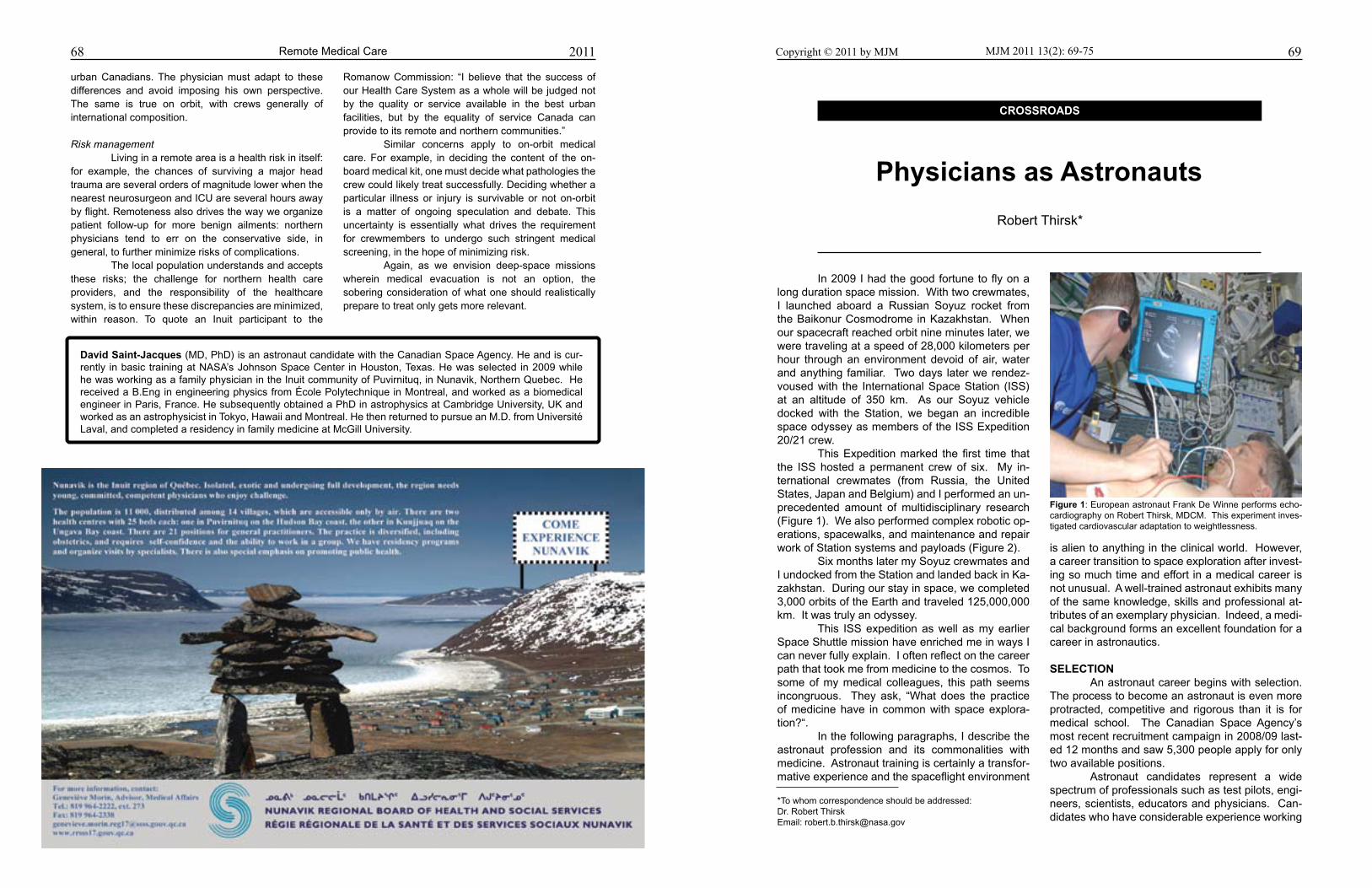
68 Remote Medical Care 2011
urban Canadians. The physician must adapt to these differences and avoid imposing his own perspective. The same is true on orbit, with crews generally of international composition.
Risk management Living in a remote area is a health risk in itself: for example, the chances of surviving a major head trauma are several orders of magnitude lower when the nearest neurosurgeon and ICU are several hours away by flight. Remoteness also drives the way we organize patient follow-up for more benign ailments: northern physicians tend to err on the conservative side, in general, to further minimize risks of complications. The local population understands and accepts these risks; the challenge for northern health care providers, and the responsibility of the healthcare system, is to ensure these discrepancies are minimized, within reason. To quote an Inuit participant to the
CRoSSRoAdS
MJM 2011 13(2): 69-75 69Copyright © 2011 by MJM
*To whom correspondence should be addressed:Dr. Robert ThirskEmail: [email protected]
In 2009 I had the good fortune to fly on a long duration space mission. With two crewmates, I launched aboard a Russian Soyuz rocket from the Baikonur Cosmodrome in Kazakhstan. When our spacecraft reached orbit nine minutes later, we were traveling at a speed of 28,000 kilometers per hour through an environment devoid of air, water and anything familiar. Two days later we rendez-voused with the International Space Station (ISS) at an altitude of 350 km. As our Soyuz vehicle docked with the Station, we began an incredible space odyssey as members of the ISS Expedition 20/21 crew. This Expedition marked the first time that the ISS hosted a permanent crew of six. My in-ternational crewmates (from Russia, the United States, Japan and Belgium) and I performed an un-precedented amount of multidisciplinary research (Figure 1). We also performed complex robotic op-erations, spacewalks, and maintenance and repair work of Station systems and payloads (Figure 2). Six months later my Soyuz crewmates and I undocked from the Station and landed back in Ka-zakhstan. During our stay in space, we completed 3,000 orbits of the Earth and traveled 125,000,000 km. It was truly an odyssey. This ISS expedition as well as my earlier Space Shuttle mission have enriched me in ways I can never fully explain. I often reflect on the career path that took me from medicine to the cosmos. To some of my medical colleagues, this path seems incongruous. They ask, “What does the practice of medicine have in common with space explora-tion?“. In the following paragraphs, I describe the astronaut profession and its commonalities with medicine. Astronaut training is certainly a transfor-mative experience and the spaceflight environment
is alien to anything in the clinical world. However, a career transition to space exploration after invest-ing so much time and effort in a medical career is not unusual. A well-trained astronaut exhibits many of the same knowledge, skills and professional at-tributes of an exemplary physician. Indeed, a medi-cal background forms an excellent foundation for a career in astronautics.
SELECTIoN An astronaut career begins with selection. The process to become an astronaut is even more protracted, competitive and rigorous than it is for medical school. The Canadian Space Agency’s most recent recruitment campaign in 2008/09 last-ed 12 months and saw 5,300 people apply for only two available positions. Astronaut candidates represent a wide spectrum of professionals such as test pilots, engi-neers, scientists, educators and physicians. Can-didates who have considerable experience working
Physicians as Astronauts
Robert Thirsk*
Figure 1: European astronaut Frank De Winne performs echo-cardiography on Robert Thirsk, MDCM. This experiment inves-tigated cardiovascular adaptation to weightlessness.
Romanow Commission: “I believe that the success of our Health Care System as a whole will be judged not by the quality or service available in the best urban facilities, but by the equality of service Canada can provide to its remote and northern communities.” Similar concerns apply to on-orbit medical care. For example, in deciding the content of the on-board medical kit, one must decide what pathologies the crew could likely treat successfully. Deciding whether a particular illness or injury is survivable or not on-orbit is a matter of ongoing speculation and debate. This uncertainty is essentially what drives the requirement for crewmembers to undergo such stringent medical screening, in the hope of minimizing risk. Again, as we envision deep-space missions wherein medical evacuation is not an option, the sobering consideration of what one should realistically prepare to treat only gets more relevant.
david Saint-Jacques (MD, PhD) is an astronaut candidate with the Canadian Space Agency. He and is cur-rently in basic training at NASA’s Johnson Space Center in Houston, Texas. He was selected in 2009 while he was working as a family physician in the Inuit community of Puvirnituq, in Nunavik, Northern Quebec. He received a B.Eng in engineering physics from École Polytechnique in Montreal, and worked as a biomedical engineer in Paris, France. He subsequently obtained a PhD in astrophysics at Cambridge University, UK and worked as an astrophysicist in Tokyo, Hawaii and Montreal. He then returned to pursue an M.D. from Université Laval, and completed a residency in family medicine at McGill University.

71Physicians as Astronauts
body operates independently, each spacecraft sys-tem interacts with several others. Physicians and astronauts would be equal-ly handicapped if we could not resort to our unique mnemonics. Acronyms and jargon pepper our con-versations with colleagues. I smile to myself when-ever I encounter a new space acronym that shares a meaning from my medical past (e.g. ER, D&C).
SKILLS Having acquired the fundamentals of spaceflight operations, astronauts next begin a training phase known as Advanced Training. This phase of training is analogous to clinical clerkship and residency since we learn highly specialized skills that are unique to our profession. It is fast-paced and fulfilling. We learn skills that allow us to launch to and return from orbit, to rendezvous and dock with other spacecraft, to perform spacewalks (also known as EVAs) and to operate robotic systems such as the Canadarm2. After acquiring proficiency in a skill, we become mentors to the next trainees. ‘See one, do one, teach one’ is a mantra that also applies to spaceflight.
Practice makes perfect. We train thou-sands of hours for nominal as well as off-nominal situations. Crew coordination, situational aware-ness and speed of reaction are critical factors to save our lives, the spacecraft and the mission un-der contingency situations. It is during Advanced Training that simula-tors play a major role. In fact, simulators are the basis for much of our skills acquisition. They are used to prepare astronauts for a variety of flight situations. For instance, the robotics simulator at the Canadian Space Agency in Montréal uses virtual reality to model Canadarm2, other robotic systems and the Space Station environment. Astronauts from all ISS partner nations use this facility to de-velop skills such as Station assembly and the cap-ture and berthing of cargo vehicles. The Neutral Buoyancy Laboratory (NBL) in Houston, Texas is another type of simulator. It is basically a huge pool (much larger than an Olym-pic swimming pool). We exploit water buoyancy to simulate the weightless condition experienced by astronauts during space walks. Small flotation devices or weights are strapped to the space suits
Figure 3: During her STS-42 Shuttle mission Roberta Bondar, MD, begins an experiment to investigate visual and vestibular responses to head and body movements.
201170
in a team setting and who habitually work outside their comfort zones are highly regarded (Figure 3). Like prospective medical students, astro-naut candidates complete questionnaires and sub-mit university transcripts, letters of reference and a personal essay (“why I want to be an astronaut”) as part of the application process. During the latter stages of the selection process, a series of inter-views are conducted with the selection committee. A very thorough medical evaluation is performed since many medical conditions that are considered to be minor on Earth can become problematic in a spaceflight environment. The work of an astronaut regularly takes us to the limits of our physical, mental and emo-tional abilities. Accordingly, the selection process includes physical fitness, cognitive and psychiat-ric evaluations. Hours of psychometric tests and psychological interviews ensure that the chosen candidates have the motivation, personalities and support mechanisms to do well in the demanding training and flight environments. Aptitude tests assess a candidate’s team
skills, creativity and tolerance to stress. During the most recent recruitment campaign, the candidates’ potential to learn specialized skills such as robotics, EVA (Extra Vehicular Activity) and foreign languag-es was also considered. Finally, interpersonal and communication skills are evaluated. In addition to operational du-ties, astronauts function as spokespersons for the Canadian Space Agency. We advocate for a national economy based upon innovation and ad-vanced training. In the same way that we were in-spired by explorers and scientists when we were young, it is our responsibility to engage the public and to instill a passion for discovery in Canada’s next generation of leaders.
KNowLEdgE Following selection (a gratifying day for a fortunate few!), astronauts begin the first phase of our education called Basic Training. This phase typically lasts a couple of years. Basic Training pro-vides each new astronaut (no matter what prior pro-fessional background) with a common knowledge base and builds the foundation for more advanced training to follow in the ensuing years. Each new re-cruit acquires broad background knowledge about the scientific, technical and operational aspects of human spaceflight. The structure of the basic training program for an astronaut shares a lot in common with a medical school curriculum. Our training resources include manuals, lectures, computer-based instruc-tion and field trips. The first couple of years of medical school were challenging for me due to the massive amount of knowledge that needed to be learned quickly. The same is true in astronautics. Astronaut train-ing is like drinking from a fire hose. The Russian Soyuz and the American Space Shuttle are com-plex space vehicles. Even more challenging to understand and operate is the International Space Station (Figure 4). This marvel of engineering has a mass of 420 tons, dimensions that are two times greater than that of a CFL football field, and living space equivalent to a four-bedroom house. 120 telephone-booth-size racks house spacecraft sys-tems and research experiments. The Station’s on-board computers process four million lines of code. A systems approach is used to instruct astronauts about space operations. We consider the composition of our spacecraft to include ther-mal control, electrical power, life support and many other systems. Just as no system in the human
Physicians as Astronauts Vol. 13 No. 2
Figure 2: Robert Thirsk, MDCM, performs maintenance on the Station’s Water Recovery System. This recycling system pro-cesses waste water and crew urine into potable water.
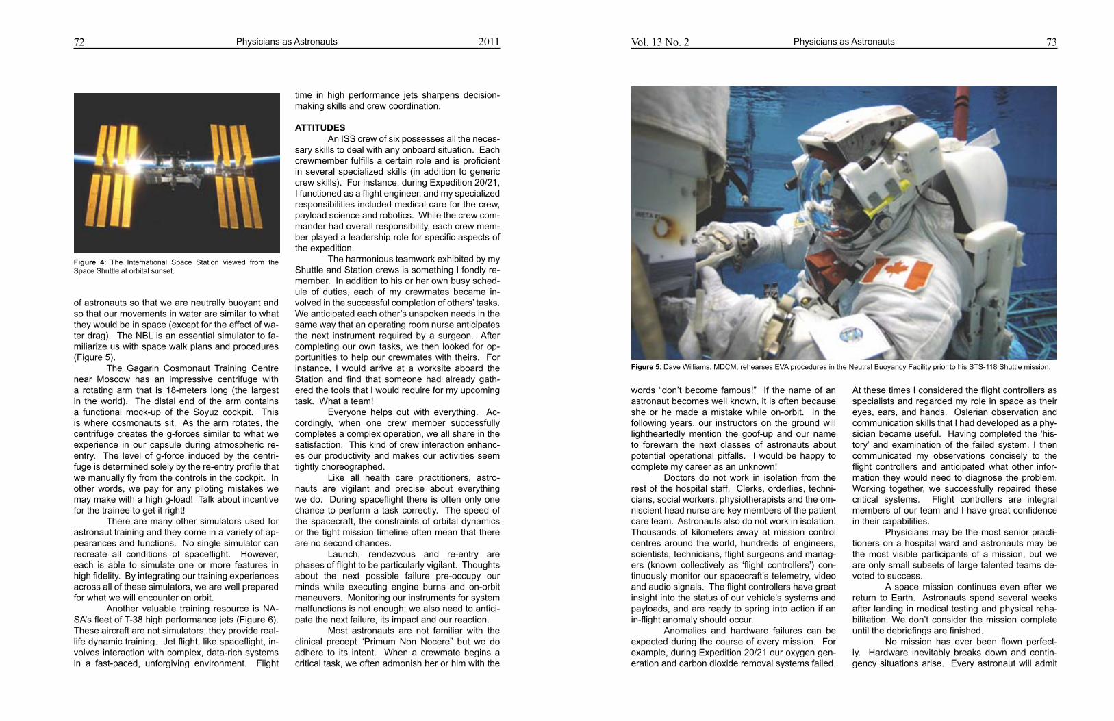
72
of astronauts so that we are neutrally buoyant and so that our movements in water are similar to what they would be in space (except for the effect of wa-ter drag). The NBL is an essential simulator to fa-miliarize us with space walk plans and procedures (Figure 5). The Gagarin Cosmonaut Training Centre near Moscow has an impressive centrifuge with a rotating arm that is 18-meters long (the largest in the world). The distal end of the arm contains a functional mock-up of the Soyuz cockpit. This is where cosmonauts sit. As the arm rotates, the centrifuge creates the g-forces similar to what we experience in our capsule during atmospheric re-entry. The level of g-force induced by the centri-fuge is determined solely by the re-entry profile that we manually fly from the controls in the cockpit. In other words, we pay for any piloting mistakes we may make with a high g-load! Talk about incentive for the trainee to get it right! There are many other simulators used for astronaut training and they come in a variety of ap-pearances and functions. No single simulator can recreate all conditions of spaceflight. However, each is able to simulate one or more features in high fidelity. By integrating our training experiences across all of these simulators, we are well prepared for what we will encounter on orbit. Another valuable training resource is NA-SA’s fleet of T-38 high performance jets (Figure 6). These aircraft are not simulators; they provide real-life dynamic training. Jet flight, like spaceflight, in-volves interaction with complex, data-rich systems in a fast-paced, unforgiving environment. Flight
time in high performance jets sharpens decision-making skills and crew coordination.
ATTITudES An ISS crew of six possesses all the neces-sary skills to deal with any onboard situation. Each crewmember fulfills a certain role and is proficient in several specialized skills (in addition to generic crew skills). For instance, during Expedition 20/21, I functioned as a flight engineer, and my specialized responsibilities included medical care for the crew, payload science and robotics. While the crew com-mander had overall responsibility, each crew mem-ber played a leadership role for specific aspects of the expedition. The harmonious teamwork exhibited by my Shuttle and Station crews is something I fondly re-member. In addition to his or her own busy sched-ule of duties, each of my crewmates became in-volved in the successful completion of others’ tasks. We anticipated each other’s unspoken needs in the same way that an operating room nurse anticipates the next instrument required by a surgeon. After completing our own tasks, we then looked for op-portunities to help our crewmates with theirs. For instance, I would arrive at a worksite aboard the Station and find that someone had already gath-ered the tools that I would require for my upcoming task. What a team! Everyone helps out with everything. Ac-cordingly, when one crew member successfully completes a complex operation, we all share in the satisfaction. This kind of crew interaction enhanc-es our productivity and makes our activities seem tightly choreographed. Like all health care practitioners, astro-nauts are vigilant and precise about everything we do. During spaceflight there is often only one chance to perform a task correctly. The speed of the spacecraft, the constraints of orbital dynamics or the tight mission timeline often mean that there are no second chances. Launch, rendezvous and re-entry are phases of flight to be particularly vigilant. Thoughts about the next possible failure pre-occupy our minds while executing engine burns and on-orbit maneuvers. Monitoring our instruments for system malfunctions is not enough; we also need to antici-pate the next failure, its impact and our reaction. Most astronauts are not familiar with the clinical precept “Primum Non Nocere” but we do adhere to its intent. When a crewmate begins a critical task, we often admonish her or him with the
Physicians as Astronauts 2011 73Vol. 13 No. 2
words “don’t become famous!” If the name of an astronaut becomes well known, it is often because she or he made a mistake while on-orbit. In the following years, our instructors on the ground will lightheartedly mention the goof-up and our name to forewarn the next classes of astronauts about potential operational pitfalls. I would be happy to complete my career as an unknown! Doctors do not work in isolation from the rest of the hospital staff. Clerks, orderlies, techni-cians, social workers, physiotherapists and the om-niscient head nurse are key members of the patient care team. Astronauts also do not work in isolation. Thousands of kilometers away at mission control centres around the world, hundreds of engineers, scientists, technicians, flight surgeons and manag-ers (known collectively as ‘flight controllers’) con-tinuously monitor our spacecraft’s telemetry, video and audio signals. The flight controllers have great insight into the status of our vehicle’s systems and payloads, and are ready to spring into action if an in-flight anomaly should occur. Anomalies and hardware failures can be expected during the course of every mission. For example, during Expedition 20/21 our oxygen gen-eration and carbon dioxide removal systems failed.
At these times I considered the flight controllers as specialists and regarded my role in space as their eyes, ears, and hands. Oslerian observation and communication skills that I had developed as a phy-sician became useful. Having completed the ‘his-tory’ and examination of the failed system, I then communicated my observations concisely to the flight controllers and anticipated what other infor-mation they would need to diagnose the problem. Working together, we successfully repaired these critical systems. Flight controllers are integral members of our team and I have great confidence in their capabilities. Physicians may be the most senior practi-tioners on a hospital ward and astronauts may be the most visible participants of a mission, but we are only small subsets of large talented teams de-voted to success. A space mission continues even after we return to Earth. Astronauts spend several weeks after landing in medical testing and physical reha-bilitation. We don’t consider the mission complete until the debriefings are finished. No mission has ever been flown perfect-ly. Hardware inevitably breaks down and contin-gency situations arise. Every astronaut will admit
Physicians as Astronauts
Figure 5: Dave Williams, MDCM, rehearses EVA procedures in the Neutral Buoyancy Facility prior to his STS-118 Shuttle mission.
Figure 4: The International Space Station viewed from the Space Shuttle at orbital sunset.
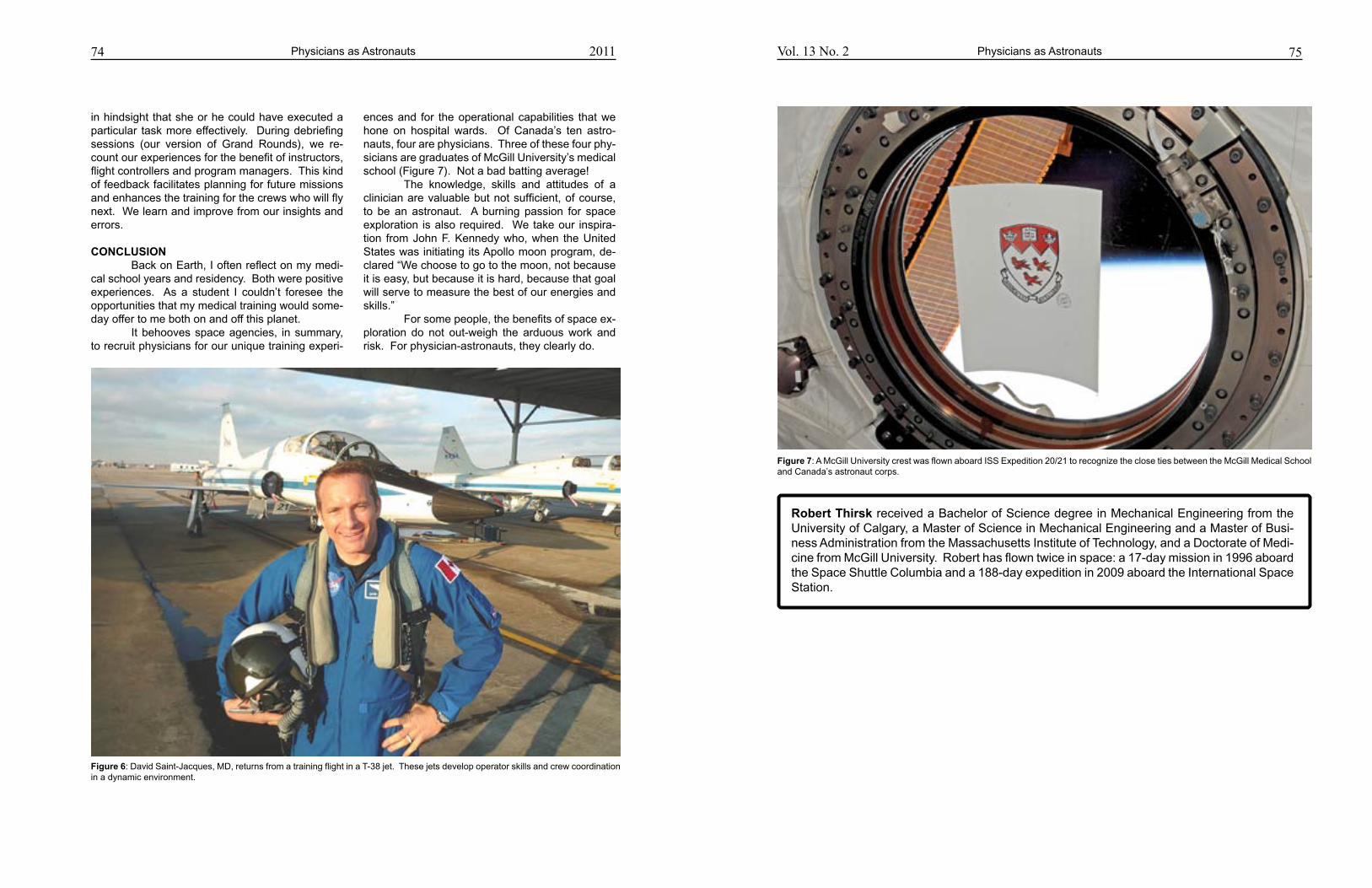
75Vol. 13 No. 2 Physicians as Astronauts74
in hindsight that she or he could have executed a particular task more effectively. During debriefing sessions (our version of Grand Rounds), we re-count our experiences for the benefit of instructors, flight controllers and program managers. This kind of feedback facilitates planning for future missions and enhances the training for the crews who will fly next. We learn and improve from our insights and errors.
CoNCLuSIoN Back on Earth, I often reflect on my medi-cal school years and residency. Both were positive experiences. As a student I couldn’t foresee the opportunities that my medical training would some-day offer to me both on and off this planet. It behooves space agencies, in summary, to recruit physicians for our unique training experi-
ences and for the operational capabilities that we hone on hospital wards. Of Canada’s ten astro-nauts, four are physicians. Three of these four phy-sicians are graduates of McGill University’s medical school (Figure 7). Not a bad batting average! The knowledge, skills and attitudes of a clinician are valuable but not sufficient, of course, to be an astronaut. A burning passion for space exploration is also required. We take our inspira-tion from John F. Kennedy who, when the United States was initiating its Apollo moon program, de-clared “We choose to go to the moon, not because it is easy, but because it is hard, because that goal will serve to measure the best of our energies and skills.” For some people, the benefits of space ex-ploration do not out-weigh the arduous work and risk. For physician-astronauts, they clearly do.
Physicians as Astronauts 2011
Figure 7: A McGill University crest was flown aboard ISS Expedition 20/21 to recognize the close ties between the McGill Medical School and Canada’s astronaut corps.
Robert Thirsk received a Bachelor of Science degree in Mechanical Engineering from the University of Calgary, a Master of Science in Mechanical Engineering and a Master of Busi-ness Administration from the Massachusetts Institute of Technology, and a Doctorate of Medi-cine from McGill University. Robert has flown twice in space: a 17-day mission in 1996 aboard the Space Shuttle Columbia and a 188-day expedition in 2009 aboard the International Space Station.
Figure 6: David Saint-Jacques, MD, returns from a training flight in a T-38 jet. These jets develop operator skills and crew coordination in a dynamic environment.

CRoSSRoAdS
MJM 2011 13(2): 76-8176 Copyright © 2011 by MJM
AbSTRACT: Preparation for the fiftieth anniversary of human spaceflight in the spring of 2011 provides the space faring nations with an opportunity to reflect on past achievements as well as consider the next fifty years of human spaceflight. The International Space Station is a unique platform for long duration life science research that will play a critical role in preparing for future human space exploration beyond low earth orbit. Some feel the future path back to the Moon and on to Mars may be delayed with the current commitment of the united States to support the development of human-rated commercial spacecraft. others see this as a unique opportunity to leverage the capability of the private sector in expanding access to space exploration. This article provides an overview of the past achievements in hu-man spaceflight and discusses future missions over the next fifty years and the role space medicine will play in extending the time-distance constant of human space exploration.
Keywords: Human spaceflight, space medicine, career astronauts, spaceflight partici-pants, commercial spaceflight
*To whom correspondence should be addressed:Dr. David R. WilliamsDepartment of Surgery McMaster University39 Chalrton Avenue East, Hamilton, ON. L8N 4A6 email: [email protected]
The past five decades will stand for eternity as that period in history when humans became a space faring species. From the first flights of Yuri Gagarin and Alan Shepard in the spring of 1961 to the Apollo 11 lunar landing in 1969, people through-out the world were mesmerized by the incredible progress of space exploration. The decade culmi-nated with the achievement of President Kennedy’s vision of human exploration of the lunar surface and safe return to Earth. Meeting this goal required the combination of sophisticated engineering, life sci-ence and medical research with a dedicated team committed to making what appeared impossible, possible. From a clinical perspective, bioastronautics (1) and space medicine (2) (3) research demon-strated that humans could survive in space, work in space and perform complex scientific missions on the surface of another celestial body. After the ex-
ponential growth in short duration flight objectives of the sixties, the past four decades have focused on the development of a long duration capability for human spaceflight. The vision of sending humans farther into the solar system was shared by many experts within the United States and Russia. For that vision to become reality, the acclimation of hu-mans to space had to be studied over the course of months not days. The Russian Salyut series of space stations and the NASA Skylab (4) (5) (6) pro-gram that highlighted the next decade of human spaceflight were used to evaluate the capacity of humans to live and work in microgravity for long pe-riods of time. Both programs provided fundamen-tal data on space physiology (7) relevant to space medicine, but they also demonstrated the need for extended long duration missions on board a new generation of space stations. The scientific utilization of space stations as microgravity research platforms provided an additional technical challenge in developing the capability to bring payloads to and from low earth orbit. The Space Shuttle was designed to meet this
Human Space ExplorationThe Next Fifty years
David R. Williams and Matthew Turnock*
77Human Space Exploration
unique requirement along with additional roles as an autonomous science platform and as a vehicle that could be used to launch and repair satellites. The need for onboard robotics as a critical enabling technology was identified and Canada was invited to design and produce a robotic arm for the Shut-tle program. Referred to as the Canadarm, or “the arm” for short, this contribution to the Shuttle pro-gram led to the first selection of six Canadian astro-nauts in 1983, with the prospect of a series of three flights for Canadian scientist astronauts referred to by NASA as payload specialists. Twenty-five years ago, Mark Garneau became the first Canadian to fly in space aboard the Space Shuttle Challenger. A number of dedicated Canadian experiments were selected for the STS-41G mission, creating an opportunity for Canadian scientists to obtain first-hand experience with microgravity research. These experiments were referred to with the acronym CANEX, a descriptor which was used for the re-maining two flights of Canadian payload specialists that took place in 1992. By this time, the concept of a partnership of the major space faring nations working together to create a world-class orbiting research platform had become a reality, and the newly emerging space station program led to the need for, and selection of a second group of Canadian astronauts. During a six-month selection process, the Canadian Space Agency used a complex set of selection criteria to hire four new astronauts that would train as mis-sion specialists for long duration missions on board the space station. Roberta Bondar and her back-up Ken Money retired after participating in the Inter-national Microgravity Laboratory (IML-2) mission in January 1992, leaving Marc Garneau, Bob Thirsk, Bjarni Tryggvason and Steve Maclean to be joined by Chris Hadfield, Julie Payette, Mike McKay and Dave Williams for mission specialist training and potential assignment to shuttle flights or space sta-tion construction missions. This was a pivotal time for the Canadian Space Agency that had raised the Canadian profile as a major space faring nation, now with an expanded team of 8 astronauts, two with mission experience, capable of leveraging the Canadian robotic and scientific expertise. The initial design requirements for the pro-posed space station included a health maintenance facility (HMF) (8) in recognition of the potential medi-cal issues that could arise during long duration mis-sions in low earth orbit. In addition to the HMF, the proposed airlock design provided both a hypobaric capability necessary for suited astronauts to egress
the station for spacewalks as well as a hyperbaric capability to treat potential episodes of decom-pression sickness (DCS) that could arise during a space walk. The designated operating pressure of the space station was 1 atmosphere (14.7 p.s.i.), similar to that of the Space Shuttle, while the suit pressure of the extravehicular mobility unit (EMU) was 4.3 p.s.i. thereby introducing the risk of DCS while transitioning to the lower suit pressure. De-spite the rigorous design requirements for on board healthcare, these facilities were not implemented in the construction of the International Space Station primarily due to cost constraints. Historically, during the Shuttle era, the clini-cal approach to prevention, diagnosis and treat-ment of illness and injury in space had a strong emphasis on prevention. This was accomplished through medical selection criteria, regular medical screening, and the development of countermea-sures to mitigate the many physiologic changes associated with exposure to microgravity. This ap-proach evolved from the early work in the Mercury, Gemini and Apollo programs that was based pri-marily on a preventive strategy with rudimentary on orbit diagnostic and treatment capabilities based on the use of small medical kits with support from flight surgeons in mission control. The Skylab program provided an excellent opportunity for biomedical re-search during long duration missions that helped further delineate the physiologic changes associ-ated with exposure to microgravity. These results were used to develop the exercise, cardiovascular and neurovestibular countermeasures implement-ed in the early Shuttle program and led to a further series of life science experiments conducted on dedicated Shuttle research missions. These stud-ies were concluded by the launch of the first ele-ment of the International Space Station (ISS) in the fall of 1998 and were published a year later as a comprehensive extended duration orbiter medical project (EDOMP) (9) (10). Canadian researchers participated in a number of collaborative Shuttle experiments throughout this time to help understand the many physiologic changes associated with acclimation to microgravity and to evaluate potential preventive countermeasures. In addition, researchers at the Canadian Space Agency (CSA) worked in collabo-ration with experts in DCS at the Defence Research and Development Canada Centre in Toronto to par-ticipate as one of three NASA supported research sites to develop the new pre-breathe protocols for use in preparation for spacewalks from the Interna-
Vol. 13 No. 2
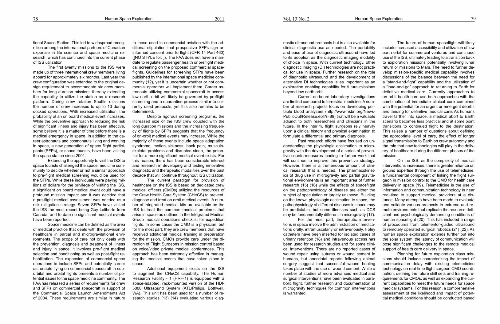
79Human Space Exploration2011
The future of human spaceflight will likely include increased accessibility and utilization of low earth orbit for commercial ventures and continued use of the ISS, ultimately leading to a transition back to exploration missions potentially involving lunar return or missions to Mars. The need to further de-velop mission-specific medical capability involves discussions of the balance between the need for a “stand-and-fight” capability and the utilization of a “load-and-go” approach to returning to Earth for definitive medical care. Currently approaches to on orbit health care use both approaches with the combination of immediate clinical care combined with the potential for an urgent or emergent deorbit and landing for definitive medical care. As humans travel farther into space, a medical abort to Earth scenario becomes less practical and at some point transitions to continued flight to the destination. This raises a number of questions about defining the appropriate level of care, the effect of longer signal transmission to Earth on crew autonomy and the role that new technologies will play in the deliv-ery of healthcare during the different phases of the mission. On the ISS, as the complexity of medical interventions increases, there is greater reliance on ground expertise through the use of telemedicine, a fundamental component of linking the flight sur-geon in mission control to the CMO for health care delivery in space (19). Telemedicine is the use of information and communication technology in near real-time to support medical providers at a dis-tance. Many attempts have been made to evaluate and validate various protocols in extreme and re-mote environments that replicate the resource defi-cient and psychologically demanding conditions of human spaceflight (20). This has included a range of procedures from telementor-guided ultrasound to remotely operated surgical robotics (21) (22). As human space exploration extends further out into the solar system, the latency of communication will pose significant challenges to the remote medical support of health care delivery in space. Planning for future exploration class mis-sions should include characterizing the impact of communication delay with existing telemedicine technology on real-time flight surgeon CMO coordi-nation, defining the future skill sets and training re-quirements for CMOs, as well as expanding the cur-rent capabilities to meet the future needs for space medical systems. For this reason, a comprehensive assessment of the likelihood and impact of poten-tial medical conditions should be conducted based
nostic ultrasound protocols but is also available for clinical diagnostic use as needed. The portability and ease of use of diagnostic ultrasound have led to its adoption as the diagnostic imaging modality of choice in space. With current technology, other diagnostic imaging (DI) technologies are not practi-cal for use in space. Further research on the role of diagnostic ultrasound and the development of alternative DI technologies is as important as an exploration enabling capability for future missions beyond low earth orbit. Current on-board laboratory investigations are limited compared to terrestrial medicine. A num-ber of research projects focus on developing por-table blood analyzers (http://www.nsbri.org/News-PublicOut/Release.epl?r=89) that will be a valuable adjunct to both researchers and clinicians in the future. In the interim, flight surgeons rely heavily upon a clinical history and physical examination to formulate a differential and primary diagnosis. Past research efforts have focused on un-derstanding the physiologic acclimation to micro-gravity with the development of a series of preven-tive countermeasures leading to further work that will continue to improve this preventive strategy. However, there is a tremendous amount of clini-cal research that is needed. The pharmacokinet-ics of drug use in microgravity and partial gravita-tional environments is an important area of further research (15) (16) while the effects of spaceflight on the pathophysiology of disease are either the subject of speculation or largely unknown. Building on the known physiologic acclimation to space, the pathophysiology of different diseases in space may be predictable, but some illnesses such as DCS may be fundamentally different in microgravity (17). For the most part, therapeutic interven-tions in space involve the administration of medica-tions orally, intramuscularly or intravenously. Foley catheters have been inserted for isolated cases of urinary retention (18) and intravenous access has been used for research studies and for some clini-cal interventions. There are no reported cases of wound repair using sutures or wound cement in humans, but anecdotal reports following animal surgery suggest that successful wound healing takes place with the use of wound cement. While a number of studies of more advanced medical and surgical interventions have been evaluated in para-bolic flight, further research and documentation of microgravity techniques for common interventions is warranted.
Vol. 13 No. 278 Human Space Exploration
to those used in commercial aviation with the ad-ditional stipulation that prospective SFPs sign an informed consent prior to flight (CFR 14 Part 460) ([NO STYLE for: ]). The FAA does not have a man-date to regulate passenger health or preflight medi-cal screening on the proposed commercial space-flights. Guidelines for screening SFPs have been published by the international space medicine com-munity (12), yet it is uncertain whether or not com-mercial operators will implement them. Career as-tronauts utilizing commercial spacecraft to access low earth orbit will likely be governed by preflight screening and a quarantine process similar to cur-rently used protocols, yet this also remains to be determined. Despite rigorous screening programs, the increased size of the ISS crew coupled with the long duration missions and the increasing frequen-cy of flights by SFPs suggests that the frequency of on-orbit medical events may increase. While the majority of these events include space adaptation syndrome, motion sickness, back pain, musculo-skeletal problems and disrupted sleep, the poten-tial for a more significant medical event exists. For this reason, there has been considerable interest and research in developing and testing innovative diagnostic and therapeutic modalities over the past decade that will continue throughout ISS utilization. The current paradigm for provision of healthcare on the ISS is based on dedicated crew medical officers (CMOs) utilizing the resources of the Crew Health Care System (CHeCS) to prevent, diagnose and treat on orbit medical events. A num-ber of integrated medical kits are available on the ISS to treat the common medical problems that arise in space as outlined in the Integrated Medical Group medical operations checklist for expedition flights. In some cases the CMO is a physician, but for the most part, they are crew members that have received additional medical training in preparation for the mission. CMOs provide care under the di-rection of Flight Surgeons in mission control based on voice/video private medical conferences. This approach has been extremely effective in manag-ing the medical events that have taken place in space. Additional equipment exists on the ISS to augment the CHeCS capability. The Human Research Facility - 1 (HRF-1) is equipped with a space-adapted, rack-mounted version of the HDI-5000 Ultrasound System (ATL/Philips, Bothwell, WA). This unit has been used for a number of re-search studies (13) (14) evaluating various diag-
tional Space Station. This led to widespread recog-nition among the international partners of Canadian expertise in life science and space medicine re-search, which has continued into the current phase of ISS utilization. The first twenty missions to the ISS were made up of three international crew members living aboard for approximately six months. Last year the crew configuration was extended to the original de-sign requirement to accommodate six crew mem-bers for long duration missions thereby extending the capability to utilize the station as a research platform. During crew rotation Shuttle missions the number of crew increases to up to 13 during docked operations. With increased utilization, the probability of an on board medical event increases. While the preventive approach to reducing the risk of significant illness and injury has been effective, some believe it is a matter of time before there is a medical emergency in space. In addition to the ca-reer astronauts and cosmonauts living and working in space, a new generation of space flight partici-pants (SFPs), or space tourists, have been visiting the space station since 2001. Extending the opportunity to visit the ISS to space tourists challenged the space medicine com-munity to decide whether or not a similar approach to pre-flight medical screening would be used for the SFPs. While these individuals were paying mil-lions of dollars for the privilege of visiting the ISS, a significant on board medical event could have a profound mission impact and it was decided that a pre-flight medical assessment was needed as a risk mitigation strategy. Seven SFPs have visited the ISS the most recent being Guy Laliberte from Canada, and to date no significant medical events have been reported. Space medicine can be defined as the area of medical practice that deals with the provision of healthcare in partial and microgravitational envi-ronments. The scope of care not only deals with the prevention, diagnosis and treatment of illness and injury in space, it involves pre-flight medical selection and conditioning as well as post-flight re-habilitation. The expansion of commercial space operations to include SFPs and potentially career astronauts flying on commercial spacecraft in sub-orbital and orbital flights presents a number of po-tential issues to the space medicine community. The FAA has released a series of requirements for crew and SFPs on commercial spacecraft in support of the Commercial Space Launch Amendments Act of 2004. These requirements are similar in nature
80 Human Space Exploration 2011 81Vol. 13 No. 2
17. Williams DR. The biomedical challenges of space flight.
Annu Rev Med 2003;54:245-56.
18. Stepaniak PC, Ramchandani SR, Jones JA. Acute urinary
retention among astronauts. Aviat Space Environ Med
2007, Apr;78(4 Suppl):A5-8.
19. Williams DR, Bashshur RL, Pool SL, Doarn CR, Mer-
rell RC, Logan JS. A strategic vision for telemedicine and
medical informatics in space flight. Telemed J E Health
2000;6(4):441-8.
20. Doarn CR. Telemedicine in extreme environments: Analogs
for space flight. Stud Health Technol Inform 2003;97:35-41.
21. Dulchavsky S, Otto C, Comtois J-M, Sargsyan A, Dul-
chavsky A, Rubinfeld I. The martian chronicles: Remotely
guided diagnosis and treatment in the arctic circle. Surg
Endosc . doi:10.1007/s00464-010-0917-1
22. Thirsk R, Williams D, Anvari M. NEEMO 7 undersea mis-
sion.
23. Horneck G, Comet B. General human health issues for
moon and mars missions: Results from the HUMEX
study. Advances in Space Research, 37(1), 100-108.
doi:10.1016/j.asr.2005.06.077
24. Issenberg SB, McGaghie WC, Hart IR, Mayer JW, Felner
JM, Petrusa ER, et al. Simulation technology for health
care professional skills training and assessment. JAMA
1999, Sep 1;282(9):861-6.
Human Space Exploration
REFERENCES1. Parker JF, West VR. Bioastronautics data book, second
edition NASA-SP-3006. Parker JF, West VR, editors. NASA
Scientific and Technical Information Office; 1973a.
2. Link MM. Space medicine in project mercury NASA-
SP-4003. Link MM, editors. Washington, DC: NASA Scien-
tific and Technical Information Office; 1965b.
3. Johnston RS, Dietlein LA, Berry CA. Biomedical Results of
Apollo. Johnston RS, Dietlein LA, Berry CA, editors. Wash-
ington, DC: NASA Scientific and Technical Information Of-
fice; 1975c.
4. The proceedings of the skylab life sciences symposium,
Volume 1, 27-29 Aug. 1974, Houston, TX, United States.
1974.
5. The proceedings of the skylab life sciences symposium,
Volume 2, 27-29 Aug. 1974, Houston, TX, United States.
1974.
6. Michel EL, Johnston RS, Dietlein LF. Biomedical results of
the skylab program. Life Sci Space Res 1976;14:3-18.
7. Pitts JA. The human factor: Biomedicine in the manned
space program to 1980 NASA-SP-4213. Washington, DC:
NASA Scientific and Technical Information Office; 1985.
8. Billica RD, Doarn CR. A health maintenance facility for
space station freedom. Cutis 1991, Oct;48(4):315-8.
9. Sawin CF. Biomedical investigations conducted in sup-
port of the extended duration orbiter medical project. Aviat
Space Environ Med 1999, Feb;70(2):169-80.
10. Sawin CF, Taylor GR, Smith WL. Extended Duration Orbiter
Medical Project Final Report - NASA/SP-1999-534. Sawin
CF, Taylor GR, Smith WL, editors. Washington, DC: NASA
Scientific and Technical Information Office; 1999.
12. Jennings RT, Murphy DM, Ware DL, Aunon SM, Moon RE,
Bogomolov VV, et al. Medical qualification of a commercial
spaceflight participant: Not your average astronaut. Aviat
Space Environ Med 2006, May;77(5):475-84.
13. Dulchavsky, Scott A. MD, PhD; Schwarz, Karl L. MD; Kirk-
patrick, Andrew W. MD; Billica, Roger D. MD; Williams,
David R. MD; Diebel, Lawrence N. MD; Campbell, Mark
R. MD; Sargysan, Ashot E. MD, and; Hamilton, Douglas
R. MD, PhD. Prospective evaluation of thoracic ultrasound
in the detection of pneumothorax. The Journal of Trauma:
Injury, Infection, and Critical Care. February 2001 - Volume
50 - Issue 2 - Pp 201-205 2001, Feb 1;50(2):201-5.
14. Sargsyan AE, Hamilton DR, Jones JA, Melton S, Whitson
PA, Kirkpatrick AW, et al. FAST at MACH 20: Clinical ul-
trasound aboard the international space station. J Trauma
2005, Jan;58(1):35-9.
15. Levy G. Pharmacodynamic aspects of spaceflight. The
Journal of Clinical Pharmacology 1991;31(10):956.
16. Klaus DM, Howard HN. Antibiotic efficacy and microbial
virulence during space flight. Trends Biotechnol 2006,
Mar;24(3):131-6.
on a combination of historical data, expert opinion, analogue studies and epidemiological studies from other related high-risk occupations (23) to facilitate development of future medical protocols. Unfortunately, the rarity and complexity of medical illness during spaceflight makes it difficult to evaluate the effectiveness of these protocols and new medical technologies. High-fidelity medical simulation has been suggested as an effective tool to assess the performance of high-level medical systems and interdependent medical teams (24). Electromechanical robotic mannequins can be used to simulate a wide variety of physiologic pa-rameters, medical emergencies and illnesses in a controlled, reproducible, and risk-free environment to evaluate clinical protocols. Beyond research and testing, medical simulation is also an ideal platform for providing medical education and training oppor-tunities for CMOs who may not be exposed to the required breadth of clinical experience necessary for supporting a space mission. In addition, it pro-vides a context-specific opportunity for CMOs skill retention during a mission, or to provide just-in-time medical training to deal with an in-flight medical emergency. The next decade provides an opportunity for further ISS research to develop new diagnostic and treatment capabilities, assess new technolo-gies and evaluate strategies for CMO skill reten-tion and just-in-time training. This research will be important to prepare for exploration class missions beyond low earth orbit in addition to developing on board clinical care for commercial space complex-es. Based on the terrestrial approach of providing on-board healthcare for commercial ocean cruises, it is likely that commercial space complexes will have an on-board clinic with a physician or other health care professional providing clinical care. The evolution of commercial space travel in the decades to come will extend the scope of space medicine beyond the realm of the government supported human spaceflight into the realm of civilian space-flight. While the objectives of human space travel will differ between the government and commercial groups, there will be a shared interest amongst practitioners of space medicine in developing the best approaches to prevent and treat illnesses and injuries during a mission. Clearly, the future oppor-tunities for those interested in space medicine are very exciting.
dave R. williams obtained a Bachelor of Science, Major in Biology in 1976, a Master of Sci-ence from the Physiology Department, and a Doctorate of Medicine and a Master of Surgery from the Faculty of Medicine, McGill University. He further completed a residency in family practice in the Faculty of Medicine, University of Ottawa in 1985, and he obtained a fellow-ship in emergency medicine from the Royal College of Physicians and Surgeons of Canada, following completion of a residency in emergency medicine at the University of Toronto in 1988. In June 1992 the Canadian Space Agency selected Williams as one of four successful candidates from a field of 5330 applicants to begin astronaut training and flew aboard STS-90 and STS-118. He retired from active astronaut status in 2008.
Matt Turnock (BSc., MSc) is a second year medical student at the University of Toronto, hav-ing recently completed his Master’s of Science degree in eHealth at McMaster University, and prior to that a Masters of Space Studies at the International Space University. His research experience includes areas in telemedicine technology, high-fidelity simulation, neural network modelling, balance and posture analysis, and pre-hospital care in aeromedical transport. For fun he directs the Wilderness Medicine Interest Group, serves as section editor for the Uni-versity of Toronto Medical Journal, and is an avid Dragonboat paddler.

CRoSSRoAdS
MJM 2011 13(2): 82-9282 Copyright © 2011 by MJM
*To whom correspondence should be addressed:Dr. Justin Woodson LTC, MC, SFS, US ArmyUniformed Services University of Health SciencesBethesda, MD Email: [email protected]
INTRoduCTIoN The clinical practice of Aviation Medicine in the U.S. Military revolves around an administrative landscape of aeromedical policy and physical stan-dards which are specific to the four main branches of the U.S. Armed Forces including the U.S. Air Force, U.S. Army, U.S. Navy/Marine Corps, and the U.S. Coast Guard. In today’s operational envi-ronment, military Flight Surgeons are increasingly practicing in a joint military environment in which service members from different services must col-laborate in order to accomplish the mission. In this joint military environment, Flight Surgeons are ex-pected to be equally well versed in the policies and procedures of their sister services, as they provide
aeromedical care for aviators and aircrew mem-bers from all services. Despite commonalities in aeromedical concerns, each service’s aeromedical policies have diverged over time, resulting in an of-ten confusing and unnecessarily complicated joint framework for aviation physical standards. There is increasing pressure from high levels of U.S. De-partment of Defense administration to consolidate practices across services in many areas of support including but not limited to aviation medicine. This paper explores the hurdles encountered in devel-oping a set of joint Aeromedical physical standards and administrative procedures and proposes po-tential solutions to some of these problems. While these proposals are not intended to be comprehen-sive in nature, they are presented to raise aware-ness and initiate dialog between administrators throughout the aeromedical communities, with the goal of moving toward the authors’ vision of a single common system of aeromedical administration for
Exploring the Possibility for a Common System for
Joint Aeromedical Standards
Justin Woodson*, Walter M Dalitsch III, James L Persson, James McGhee, Charles Ciccone, Brian Parsa
ABSTRACT: The Physical qualification standards for aviation service used by the united States Army, Navy/Marine Corps, Air Force, and Coast guard developed in parallel, diverging in many instances due to differences ranging from terminology to mission. Presently, standards and requirements for waiver vary widely between the services, in spite of minimal differences in aeromedical concerns for any given medical condition. Standardization or increased concordance between the services would have several advantages leading to more efficient and effective delivery of aviation medical support to the operational forces. This is particularly true in an increasingly joint operational environment. The authors have identified four major hurdles that must be overcome before the concept of joint aviation physical stan-dards can be explored. These include: a difference in terminology including aviator classification, a difference in mission definitions and requirements, a difference in the processes of policy development, and a difference in the review and application of those policies. These hurdles are explored, and suggestions for their mitigation are presented with open discussion following.
Keywords: Aerospace Medicine, Aviation Medicine, Physical Standards, Military Medicine
83Joint Aeromedical Standards
the U.S. Military. The scope of this article is limited to the U.S. Military for the sake of brevity, but paral-lel analysis with our international brethren is invited, as many of the same lessons clearly apply in the international military aviation medicine community.
bACKgRouNd The idea of a unified approach to medical service for all branches of the U.S. Armed Forces is not new. With four separate medical departments in the U.S. Navy/Marine Corps, U.S. Army, U.S. Air Force, and U.S. Coast Guard, the efficient delivery of health care to armed service members and their dependents has long been complicated by stove-piping of resources and programs. One of the most energetic attempts to consolidate US Department of Defense medical services was put forth by Ma-jor General Norman Kirk in 1947. While Kirk did not originate this idea, he materialized the concept in a detailed plan that he presented to the Senate Armed Forces Committee (13). Since that time, sev-eral more attempts have been made to propose a sweeping unification of all the armed forces medi-cal services into one integrated service. In fact, the Civilian Health and Medical Program for the Uni-formed Services (CHAMPUS) was a direct spin-off of these attempts. Efforts to establish physical standards for selection and retention in aviation date back to World War One when Allied Forces were wrestling with integration of the airplane into normal opera-tions. Recognizing the need for standardization in aviation medicine, Drs. Theodore Lyster and Isaac Jones developed a system of physical standards and education throughout the U.S. Army, which laid the foundation for the standardized approach to physical qualification for aviation service which we utilize today in all branches of the U.S. Armed Forc-es (6). Those early efforts of Doctors Lyster and Jones evolved into the modern practice of aviation medicine in the military, and as the Air Force broke off from the Army Air Corps in 1948, this framework for aviation medicine carried forward. But with five United States armed forces entering the modern era of aviation, Aeromedical policies and physical standards have necessarily diverged to meet op-erational demands of the specific service. Advances in technology drive the evolution of military tactics, which in turn results in changes to organizational structures, changes in Techniques, Tactics, and Procedures (TTPs) and the applica-tion of Combat Services Support on the battlefield. Changes in our current operational climate dictate the need to reconsider the practice of aviation med-
icine in the military. Increasingly, Flight Surgeons and aircrew are deployed in settings where crew-members must rely on Aeromedical support from sister service Flight Surgeons. Under current prac-tices, Flight Surgeons are rarely trained and are even less frequently familiar with the aeromedical standards of their sister services. As a result, they will do their best to wade through the necessary bu-reaucracy in order to meet the mission, frequently without achieving success and often failing to meet requirements. Differences in administrative frame-works between military aviation medicine programs unnecessarily inhibit the Flight Surgeon’s ability to provide needed services. Proponents of unification of aeromedi-cal systems across service boundaries tout an increased efficiency of care and decreased costs of administration. These advantages may be par-ticularly true during peacetime, and in stationary military medical facilities. Opponents to unification rightly point out the differences in mission and op-erational environment between the services, and the requirement for medical assets organic to in-dividual units that are able to provide optimal and timely care in these unique settings. Frequently, this requires divergence in training in order to pro-vide an operationally relevant product. While the authors do not necessarily ad-vocate the unification of the medical services as a whole, the adoption of a common language and a set of common tools within the aeromedical com-munities of the individual services has several ad-vantages. Issues as simple as which form to use for a flight physical, or which labs to order, or as complex as how to classify an aeromedical disposi-tion or how to process a waiver evaluation frustrate the Flight Surgeon and often lead to duplication of work, or worse. Standardization or increased con-cordance between the services would lead to more efficient and effective delivery of aviation medical support to the operational forces both at the individ-ual Flight Surgeon level as well as at the program administration level. Clearly, each service has both shared (e.g. rotary wing) and unique (e.g. carrier landings) avia-tion functions, and each operational environment places unique physiological and psychological stresses on the aviator. However, equally clear should be the reality that the vast majority of medi-cal conditions (e.g. cardiovascular disease) will have the same implications in an aviator who straps into any aircraft, regardless of type or paint color. Evidence-based practice mandates that Flight Sur-geons separate their aeromedical analysis from
Vol. 13 No. 2
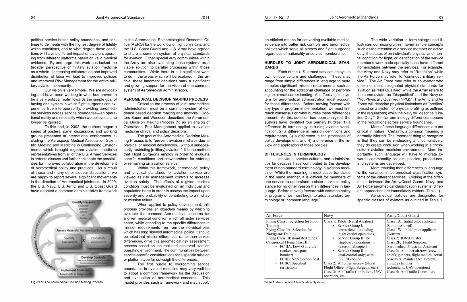
Vol. 13 No. 284
political service-based policy boundaries, and con-tinue to delineate with the highest degree of fidelity which conditions, and to what degree these condi-tions will have a different impact on aviators operat-ing from different platforms based on valid medical evidence. By and large, this work has lacked the broader perspective of military aviation medicine as a whole. Increasing collaboration and improved distribution of labor will lead to improved policies and improved Risk Management for the entire mili-tary aviation community. Our vision is very simple. We are advocat-ing and have been working in what has proven to be a very political realm towards the simple goal of having one system in which flight surgeons can ex-perience true interoperability, providing aeromedi-cal services across service boundaries—an opera-tional reality and necessity which we believe can no longer be ignored. To this end, in 2002, the authors began a series of posters, panel discussions and working groups presented at international conferences in-cluding the Aerospace Medical Association Scien-tific Meeting and Medicine in Challenging Environ-ments which brought together aviation medicine representatives from all of the U.S. Armed Services in order to discuss and further delineate the possibil-ities for improved collaboration in the development of Aeromedical policy and practices. As a result of these and many other sidebar discussions, we are happy to report several significant movements in the direction of Aeromedical jointness. In 2009, the U.S. Navy, U.S. Army, and U.S. Coast Guard have adopted a common administrative framework
in the Aeromedical Epidemiological Research Of-fice (AERO) for the workflow of flight physicals, and the U.S. Coast Guard and U.S. Army have agreed to share a common system of physical standards for aviation. Other special duty communities within the Army are also evaluating these systems as a viable solution to parallel processes within those communities. While there is still significant work to do in the areas which will be explored in this ar-ticle, these landmark decisions mark a significant and growing support for the vision of one common system of Aeromedical administration.
AERoMEdICAL dECISIoN MAKINg PRoCESS Critical to the process of joint aeromedical administration, must be a common system of evi-dence based decision making and analysis. Doc-tors Sauer and Woodson described the Aeromedi-cal Decision Making Process (1) as an analog of Operational Risk Management applied to aviation medicine clinical and policy decisions. The goal of the Aeromedical Decision Mak-ing Process is to “prevent aviation mishaps due to physical or medical deficiencies…without unneces-sarily restricting [military] aviation.” It is the method that Flight Surgeons employ in order to evaluate specific conditions and crewmembers for entering or remaining on aviation service. Within this framework, aeromedical policy and physical standards for aviation service are viewed as risk management controls to increase aviation safety. The effects of a given medical condition must be evaluated on an individual and population basis in order to assess the impact upon severity and probability of contributing to a mishap or mission failure. When applied to policy development, this process provides an objective means by which to evaluate the common Aeromedical concerns for a given medical condition which all sister services share, while attending to the specific differences in mission requirements free from the individual bias which has long skewed aeromedical policy. It should be noted that mission differences, rather than service differences, drive this aeromedical risk assessment process based on the real and observed aviation operating environment. The commonalities between service-specific considerations for a specific mission or platform type far outweigh the differences. The first hurdle to overcoming service boundaries in aviation medicine may very well be to adopt a common framework for the discussion and evaluation of aeromedical concerns. This model provides such a framework and may supply
Joint Aeromedical Standards
Figure 1: The Aeromedical Decision Making Process
85Joint Aeromedical Standards
The wide variation in terminology used il-lustrates our incongruities. Even simple concepts such as the retention of a service member on active duty, the status of an individual’s physical and men-tal condition for flight, or identification of the service member’s work code specialty each have different nomenclature between the services. For example, the Army and Navy may refer to “Retention” while the Air Force may refer to “continued military ser-vice.” The Air Force may refer to an aviator who does not meet designated physical standards for aviation as “Not Qualified” while the Army refers to the same aviator as “Disqualified” and the Navy as “Not Physically Qualified (NPQ).” The Army and Air Force will describe physical limitations as “profiles” (based on a system of physical profiling as outlined in the regulations) while the Navy will describe “Lim-ited Duty.” Similar terminology differences abound in the regulations across service boundaries. Most of these language differences are not critical in nature. Certainly, a common meaning is normally inferred. The important thing to recognize is that they can be misleading and cumulatively, they do create confusion when working in a cross-cultural aviation medicine environment. More im-portantly, such language will have to migrate to-wards commonality as joint policies, procedures, and systems are developed. More troubling than differences in language is the variance in aeromedical classification sys-tems of the different services. Looking at the differ-ences between the Army/Coast Guard, Navy, and Air Force aeromedical classification systems, differ-ent approaches are immediately evident (Table 1). Aeromedical policies are designated for specific classes of aviators as outlined in Table 1.
an efficient means for converting available medical evidence into better risk controls and aeromedical policies which serve all aircrew and flight surgeons regardless of nationality or service membership.
HuRdLES To JoINT AERoMEdICAL STAN-dARdS Each of the U.S. armed services enjoys its own unique culture and challenges. These may range from simple differences in language to more complex significant mission requirements such as accounting for the additional challenge of perform-ing an aircraft carrier landing. An effective joint sys-tem for aeromedical administration must account for these differences. Before moving forward with any type of program implementation, we must first reach consensus on what hurdles these differences present. As this question has been analyzed, the authors have identified four primary hurdles: 1) a difference in terminology including aviator classi-fication, 2) a difference in mission definitions and requirements, 3) a difference in the processes of policy development, and 4) a difference in the re-view and application of those policies.
dIFFERENCES IN TERMINoLogy Individual service cultures and administra-tive landscapes have contributed to the develop-ment of non-standard terminology in aviation medi-cine. While the meaning in most cases translates in the same manner, it is difficult for members of one service to understand a sister service’s policy stance for no other reason than differences in lan-guage. Before moving forward with common policy or programs, we must begin to adopt standard ter-minology or “common language.”
2011
Table 1: Aeromedical Classification Systems
Air Force Navy Army/Coast GuardFlying Class I: Selection for Pilot TrainingFlying Class IA: Selection for Navigator TrainingFlying Class III: non-rated dutiesCategorical Flying Class II
• FCIIA:Low-Gaircraft (tanker, transport, bomber)• FCIIB:Non-ejectionSeat• FCIIC:Specified restrictions
Class 1: Pilots (Naval Aviators)• ServiceGroupI: unrestricted (including night carrier operations)• ServiceGroupII:no shipboard operations (except helicopter)• ServiceGroupIII: dual-control only; with SG I/II copilot
Class 2: All other aircrew (Naval FlightOfficer,FlightSurgeon,etc.)Class3:AirTrafficControllers,UAVoperators, etc.
Class 1A: Initial pilot applicant (Commissioned)Class 1W: Initial pilot applicant (Warrant)Class 2: Rated aviatorClass 2F: Flight Surgeon, Aeromedical Physician AssistantClass 3: All other aircrew (crew chiefs,gunners,flightmedics,aerialobservers, maintenance aircrew, altitude chamber technicians, UAV operators)Class4:AirTrafficControllers

86 Vol. 13 No. 2
Aeromedical disposition in each service is grounded in its own aeromedical classification system, each of which have developed through an amalgam of service culture and regulatory framework entirely outside the realm of aviation medicine. It is fairly easy to recognize that these classes are defined in each service based on fundamentally different frameworks. In many cases, the difficulty in inter-preting aeromedical physical standards is rooted in the differences in these classification systems. It is difficult to see a truly joint aeromedi-cal system that uses the current service-specific aircrew classification structure. Standardization of this system into a common inter-service aircrew classification would seem the only plausible solu-tion to this problem and a vital step towards unifica-tion of aeromedical systems in the U.S. Military. Several solutions may present themselves, but one potential solution could be based upon crewmember type and basic aeromedical distinc-tion. There are only four essential types of indi-viduals who require aeromedical clearance, each of which represents unique job-related physical re-quirements: 1) flight crew who control aircraft , 2) flight crew who do not control aircraft 3) crewmem-bers who perform ancillary duties in flight (aerial ob-servers, weapons system operators, equipment op-erators, etc) unrelated to the control of the aircraft 4) individuals who perform flight-related duties, but not involving actual flight duties (ground crew, ATC, UAS operators etc). Accordingly, one potential in-terservice classification system might look like that seen in table 2a, which separates mission specific considerations and initial vs. retention consider-ations from the basic element of disposition clas-sification. A second option could link aeromedical risk to aeromedical threat (Table 2b).
Any classification system will require the Flight Surgeon to make decisions based on indi-vidual crewmembers and their specific job require-ments, but an effective classification system must account for differences in physiological require-ments. The key point is that adoption of a com-mon inter-service classification structure such as that presented in tables 2a or 2b would facilitate cross-service communication and allow for a com-mon framework of aeromedical regulations, moving us much further down the road towards a unified joint aeromedical system.
FRAMEwoRK FoR STANdARdS dEVELoP-MENT (MISSIoN VS. PHySIoLogy) The major services of the U.S. Armed Forc-es frequently distinguish themselves based upon their stated mission. On the most basic level, these missions may be categorized based upon service distinct missions (e.g. land-based vs. carrier-based in the Navy), the platform flown (e.g. fixed vs. ro-tary wing) or the complement of crew (e.g. single pilot vs. multi-crew aircraft). However, each type of aviation platform places its own unique set of physi-cal demands on the aviator, while many demands are common to all aviation platforms. Aeromedical concerns are more appropri-ately described in reference to the mission the air-crew member is serving than to the branch of service of which he or she is a member. There is no doubt that due diligence must be paid to the physiologi-cal demands of specific missions and equipment on the crewmember. It must be noted, however, that these demands are grouped into categories that transcend service boundaries. The present system effectively prevents aeromedical categorization of missions across the services. More importantly, it frequently does not even account for actual physi-cal stresses on different categories of crew mem-bers within a given service. An effective framework for aeromedical standards would appropriately account for differences in physiological demands based on mission, equipment, environment and other job requirements. One example of the failure to consider mission specific physical demands is illustrated by looking at the standards for stereopsis in Army air-crew. Current policy allows deficiencies in stereop-sis for crewmembers (non-pilots) but not for pilots (9,11). This may represent a leftover policy from the primarily fixed wing days of Army aviation medicine and is probably based on reasonable rationale: pi-lots need advanced stereopsis on final approach and landing phases of flight which are within the
Joint Aeromedical Standards
Table 2a: Proposed Interservice Aircrew Classification System, version 1
Table 2b: Proposed Interservice Aircrew Classification System, version 2version 1
87
stereoptic range, and back-end helicopter crew-members’ duties would not routinely call on their stereoptic capability as they manage payload. Yet evidence supports the idea that monocular pilots (without stereopsis) do just as well as binocular pi-lots in landing aircraft (15,16) and mission analysis readily demonstrates that the army crewmember may require greater degrees of stereopsis than the front end crew as he controls aircraft position in fine detail during air assault, fast-rope, sling-load, and routine hot-loading operations. The basic (reason-able) premise for this inversion of standards is most likely based on a general impression that pilots re-quire higher standards than non-pilots. This is a seemingly simple oversight, but it highlights the difficulties encountered when we fail to consider the actual physical demands of mission and equipment on crewmembers. Compound this phenomenon by comparing the differences between high-Gz platforms, carrier-based landings, rotary wing platforms, and Unmanned Aerial Systems (UAS). Other critical differences in demands exist based on differences in operating altitude and G-Forces. One approach to developing a functional joint system would be to examine the unique physi-ological aspects of broad categories of aircraft (Ta-ble 3).
When examining physical standards from this perspective, a more suitable framework for physical standards emerges; one which would serve the needs of all services, and would be based on physiological demands rather than political boundaries. A very important consideration is that each service does not necessarily operate within its own traditional boundaries; missions tradition-ally reserved for one service may be conducted by aircrew from another service. One commonly cited difference is the Navy requirement for carrier-based operations, an obviously demanding aviation task. Yet Army, Coast Guard, and Air Force rotary wing aviators are often called on to land on ship-based platforms. Current aeromedical administrative structure would not account for these challenges and their associated physical demands. If modified to address platform and mission based differences, a new joint aeromedical structure would allow us to speak the same aeromedical language across service boundaries and would better reflect an evi-dence based approach to aviation medicine. An aeromedical evaluation of an aviator could be made in the context of platform/mission-based parameters. A pilot, flight officer or aircrew member can be effectively authorized or restricted to fly on different types of missions, based on the
Joint Aeromedical Standards2011
Table 3: Aeromedical Aspects of Broad Categories of Aircraft
Aircrew Class Description
Class A Non-flightcrewperformingaerialduties
Class B Non-flying,flightrelatedpersonnel
Class C Flight Crew, Pilot in Control, single-control aircraft
Class D Flight Crew, Pilot in Control, dual-control aircraft
Class E Flight Crew, Non-pilot
In-Flight Crew Ground-based
Flight Critical Class A Class C
Flight Important Class B Class D
Rotary Wing Fixed Wing – Low Gz (tanker, transport, bomber)
Tactical Jet – Hi Gz
Unmanned Aerial Systems (UAS)
• Lower Gz • Vibration (2-35 Hz) • Intervertebral disc dz 4-5 Hz) • Pregnancy• ↓ Hypoxia Concern• Visual Acuity • Contrast sensitivity • Close proximity to the ground • Constantly clearing for obstacles, wires • Ground Target acquisition• Monocular displays/NVG • Binocular rivalry • Stereopsis eliminated (Terrain within limit) • Neck pain • ↓ Acuity (20/40) • ↓ Fields of view (40 deg) • Color vision eliminated • ↑ Ocular Motility demand • Depth perception • NVGs • NOE flying• MOPP• Living conditions/Heat stress
• Lower Gz • Long Duration Missions• Large Crews, often dual piloted• ↑ Hypoxia Concern• Stereopsis less critical• Decompression (also pressure suits)• Radiation (high altitude recon)• ↑ Circadian rhythm shifting
• High, rapid-onset Gz •↑↑ Hypoxia Concern• Long Duration Missions• Decompression• Visual Acuity • Increased visual demand for tactical mission • Close proximity to the ground• Air Target acquisition• Ejection Seats
• Ground Based • No Hypoxia Concern • No pressure differentials• Color Vision critical• Stereopsis not required• Dual Pilot• Potential for in-flight crew changes• Increased demands on decision making and situational awareness challenge traditional views of UAS requirements • Flying in Class A Airspace • Weapons and targeting systems
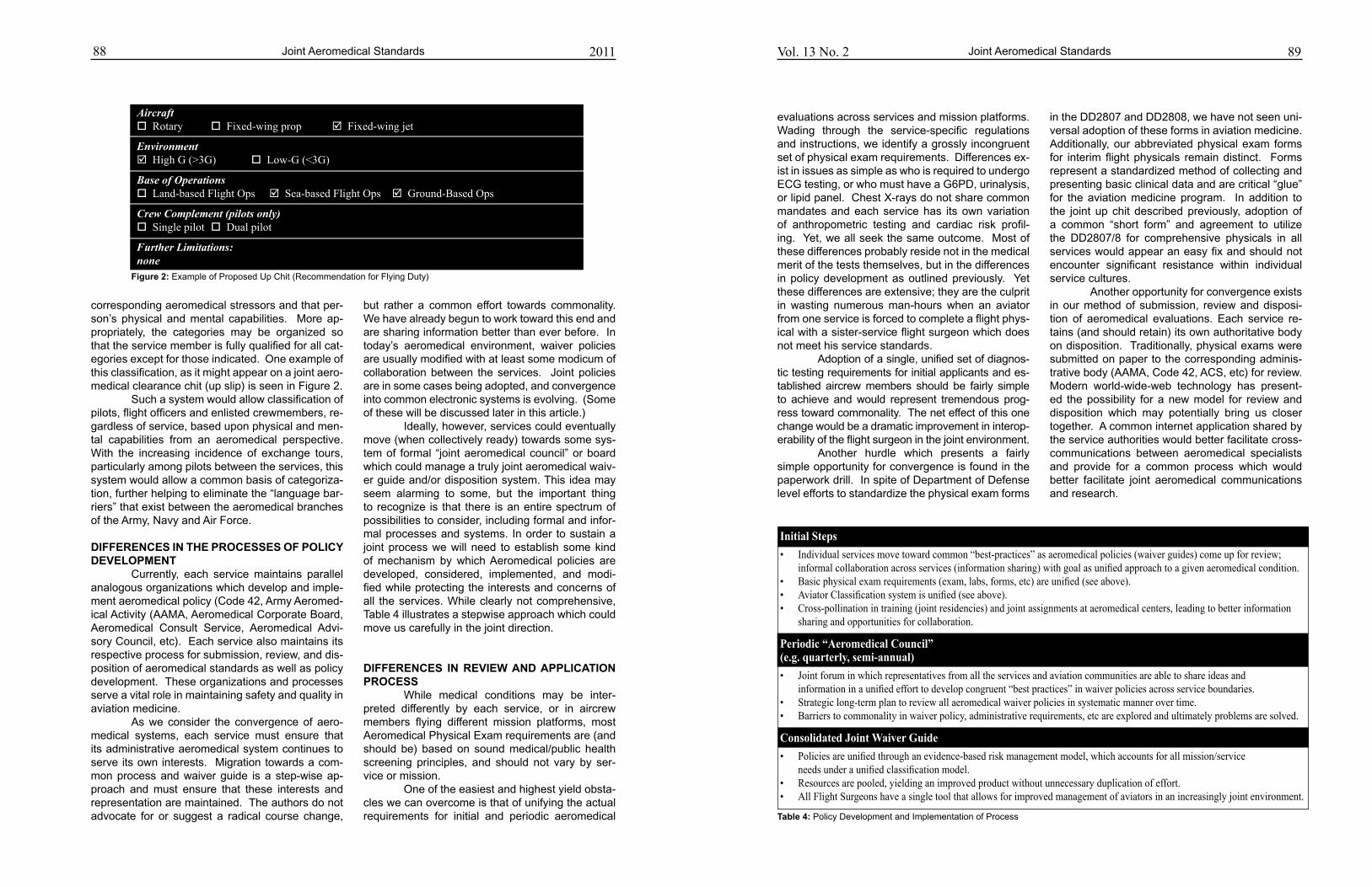
88 Vol. 13 No. 2
but rather a common effort towards commonality. We have already begun to work toward this end and are sharing information better than ever before. In today’s aeromedical environment, waiver policies are usually modified with at least some modicum of collaboration between the services. Joint policies are in some cases being adopted, and convergence into common electronic systems is evolving. (Some of these will be discussed later in this article.) Ideally, however, services could eventually move (when collectively ready) towards some sys-tem of formal “joint aeromedical council” or board which could manage a truly joint aeromedical waiv-er guide and/or disposition system. This idea may seem alarming to some, but the important thing to recognize is that there is an entire spectrum of possibilities to consider, including formal and infor-mal processes and systems. In order to sustain a joint process we will need to establish some kind of mechanism by which Aeromedical policies are developed, considered, implemented, and modi-fied while protecting the interests and concerns of all the services. While clearly not comprehensive, Table 4 illustrates a stepwise approach which could move us carefully in the joint direction.
dIFFERENCES IN REVIEw ANd APPLICATIoN PRoCESS While medical conditions may be inter-preted differently by each service, or in aircrew members flying different mission platforms, most Aeromedical Physical Exam requirements are (and should be) based on sound medical/public health screening principles, and should not vary by ser-vice or mission. One of the easiest and highest yield obsta-cles we can overcome is that of unifying the actual requirements for initial and periodic aeromedical
Joint Aeromedical Standards
Figure 2: Example of Proposed Up Chit (Recommendation for Flying Duty)
89
evaluations across services and mission platforms. Wading through the service-specific regulations and instructions, we identify a grossly incongruent set of physical exam requirements. Differences ex-ist in issues as simple as who is required to undergo ECG testing, or who must have a G6PD, urinalysis, or lipid panel. Chest X-rays do not share common mandates and each service has its own variation of anthropometric testing and cardiac risk profil-ing. Yet, we all seek the same outcome. Most of these differences probably reside not in the medical merit of the tests themselves, but in the differences in policy development as outlined previously. Yet these differences are extensive; they are the culprit in wasting numerous man-hours when an aviator from one service is forced to complete a flight phys-ical with a sister-service flight surgeon which does not meet his service standards. Adoption of a single, unified set of diagnos-tic testing requirements for initial applicants and es-tablished aircrew members should be fairly simple to achieve and would represent tremendous prog-ress toward commonality. The net effect of this one change would be a dramatic improvement in interop-erability of the flight surgeon in the joint environment. Another hurdle which presents a fairly simple opportunity for convergence is found in the paperwork drill. In spite of Department of Defense level efforts to standardize the physical exam forms
in the DD2807 and DD2808, we have not seen uni-versal adoption of these forms in aviation medicine. Additionally, our abbreviated physical exam forms for interim flight physicals remain distinct. Forms represent a standardized method of collecting and presenting basic clinical data and are critical “glue” for the aviation medicine program. In addition to the joint up chit described previously, adoption of a common “short form” and agreement to utilize the DD2807/8 for comprehensive physicals in all services would appear an easy fix and should not encounter significant resistance within individual service cultures. Another opportunity for convergence exists in our method of submission, review and disposi-tion of aeromedical evaluations. Each service re-tains (and should retain) its own authoritative body on disposition. Traditionally, physical exams were submitted on paper to the corresponding adminis-trative body (AAMA, Code 42, ACS, etc) for review. Modern world-wide-web technology has present-ed the possibility for a new model for review and disposition which may potentially bring us closer together. A common internet application shared by the service authorities would better facilitate cross-communications between aeromedical specialists and provide for a common process which would better facilitate joint aeromedical communications and research.
Joint Aeromedical Standards2011
Table 4: Policy Development and Implementation of Process
Aircraft Rotary Fixed-wing prop Fixed-wingjet
Environment High G (>3G) Low-G(<3G)
Base of OperationsLand-basedFlightOps Sea-based Flight Ops Ground-Based Ops
Crew Complement (pilots only) Single pilot Dual pilot
Further Limitations:none
Initial Steps
• Individualservicesmovetowardcommon“best-practices”asaeromedicalpolicies(waiverguides)comeupforreview; informalcollaborationacrossservices(informationsharing)withgoalasunifiedapproachtoagivenaeromedicalcondition.• Basicphysicalexamrequirements(exam,labs,forms,etc)areunified(seeabove).• AviatorClassificationsystemisunified(seeabove).• Cross-pollinationintraining(jointresidencies)andjointassignmentsataeromedicalcenters,leadingtobetterinformation sharing and opportunities for collaboration.
Periodic “Aeromedical Council” (e.g. quarterly, semi-annual)
• Jointforuminwhichrepresentativesfromalltheservicesandaviationcommunitiesareabletoshareideasand informationinaunifiedefforttodevelopcongruent“bestpractices”inwaiverpoliciesacrossserviceboundaries.• Strategiclong-termplantoreviewallaeromedicalwaiverpoliciesinsystematicmannerovertime.• Barrierstocommonalityinwaiverpolicy,administrativerequirements,etcareexploredandultimatelyproblemsaresolved.
Consolidated Joint Waiver Guide
• Policiesareunifiedthroughanevidence-basedriskmanagementmodel,whichaccountsforallmission/service needsunderaunifiedclassificationmodel.• Resourcesarepooled,yieldinganimprovedproductwithoutunnecessaryduplicationofeffort.• AllFlightSurgeonshaveasingletoolthatallowsforimprovedmanagementofaviatorsinanincreasinglyjointenvironment.
corresponding aeromedical stressors and that per-son’s physical and mental capabilities. More ap-propriately, the categories may be organized so that the service member is fully qualified for all cat-egories except for those indicated. One example of this classification, as it might appear on a joint aero-medical clearance chit (up slip) is seen in Figure 2. Such a system would allow classification of pilots, flight officers and enlisted crewmembers, re-gardless of service, based upon physical and men-tal capabilities from an aeromedical perspective. With the increasing incidence of exchange tours, particularly among pilots between the services, this system would allow a common basis of categoriza-tion, further helping to eliminate the “language bar-riers” that exist between the aeromedical branches of the Army, Navy and Air Force.
dIFFERENCES IN THE PRoCESSES oF PoLICy dEVELoPMENT Currently, each service maintains parallel analogous organizations which develop and imple-ment aeromedical policy (Code 42, Army Aeromed-ical Activity (AAMA, Aeromedical Corporate Board, Aeromedical Consult Service, Aeromedical Advi-sory Council, etc). Each service also maintains its respective process for submission, review, and dis-position of aeromedical standards as well as policy development. These organizations and processes serve a vital role in maintaining safety and quality in aviation medicine. As we consider the convergence of aero-medical systems, each service must ensure that its administrative aeromedical system continues to serve its own interests. Migration towards a com-mon process and waiver guide is a step-wise ap-proach and must ensure that these interests and representation are maintained. The authors do not advocate for or suggest a radical course change,
90 Vol. 13 No. 2
THE AERoMEdICAL ELECTRoNIC RESouRCE oFFICE (AERo) ANd CuRRENT JoINT INITIA-TIVES AS AN ExAMPLE oF CoNVERgENCE Opportunities to converge towards com-mon systems, policies, and practices abound and do not necessarily require a monumental overhaul of what is currently in place. While compromise is important, the service-specific aeromedical authori-ties do not need compromise on their standards or requirements in order to find common ground. One example of such endeavors can be found in recent developments in the integration of the Aeromedical Electronic Resource Office (AERO) at Fort Rucker, Alabama. In 2002, the U.S. Army Aeromedical Activ-ity (USAAMA) adopted AERO as an internet-based solution to aeromedical review and disposition, re-placing a cumbersome paper-based submission process in the Army. This government owned and developed system was fielded, and over a short pe-riod of time, resulted in significant improvements in the disposition of Army flight physicals. In addition to improving the submission process, internal pro-cessing times at USAAMA were reduced from 150 days to 1-2 days on average, while also making provisions for immediate review when necessary. AERO provided for data checking and was eas-ily integrated into the Flight Surgeon’s office, both CONUS and OCONUS in the deployed setting. Administrative errors on submitted physicals were reduced from 40% on the paper-based system to <1% on AERO, and immediate feedback was pro-vided to the Flight Surgeon on the disposition of air-crew physicals. Backlogs were cleared and overall efficiency was dramatically improved. In 2008, with problems similar to those ex-perienced using the Army’s paper-based systems, steps were taken independently in the U.S. Navy and U.S. Coast Guard to implement AERO as their system for aeromedical disposition and review. While still undergoing testing and implementation in both services, it is already clear that this system has the potential to make significant improvements in the process of disposition and allows for commonal-ity on an entirely different level than ever before. There are several points about this AERO migration which must be emphasized. Firstly, the Army, Coast Guard, and Navy all shared a simi-lar pathway for review and disposition within their own organizational structures (e.g. all three use a centralized review authority). Secondly, the Coast Guard and Army share a common footing in aero-medical culture as a result of the sharing of a common training base for Flight Surgeons, and a
common migration of Army Flight Surgeons into the Coast Guard medical service. Thirdly, in the case of the Coast Guard, the aeromedical physi-cal exam parameters (items required for physical exams) were already very similar. To cement these similarities, the Coast Guard agreed to adopt the same standards utilized by the Army, and Code 42 in the Navy has worked diligently to more closely align physical exam parameters with the Army and Coast guard in order to facilitate AERO integration. The opportunity presented by Navy and Coast Guard AERO integration allowed AAMA to make some minor modifications to AERO to ac-count for differences in requirements within the Na-vy’s aeromedical policy. Without modification, the process of review within AERO very easily accom-modated differences in the waiver process in both services. Because AERO utilizes a role-based sys-tem, the actual waiver authority could be retained in the service and allow for service-specific review while allowing all three services to utilize a common system and begin to migrate specific physical exam parameters, beginning to overcome one of the pre-viously mentioned hurdles to commonality. While AERO is only one system and one example, it serves the purpose of this article, as a vivid example of the capacity for convergence to-wards the authors’ vision of a single common aero-medical system, while simultaneously raising the program standards within each individual service.
CoNCLuSIoN The prospect of developing a joint Aero-medical System and Waiver Guide is clearly daunting and is not without its challenges. Service culture, existing systems, policies, and service spe-cific regulatory landscape all play important roles in keeping aeromedical systems separate. It is clear with the continuing evolution of the military opera-tional environment that each of the services must work to migrate towards commonality while time permits, before higher authorities mandate such a move. In the meantime, the benefits of increased jointness include increased efficiency, increased interperability, and the facilitation of aeromedical epidemiological research. Despite aeromedical divergence over the course of the last 100 years since the beginning of military aviation medicine, we are beginning to see a convergence towards a common system. As we continue to explore and overcome hurdles to joint aeromedical systems, we will see that the authors’ vision of a single common system of Aeromedical Administration for the U.S. Military is indeed within reach.
Joint Aeromedical Standards 91
REFERENCES1. Sauer S, Woodson J. The Aeromedical Decision Making
Process. Society of US Army Flight Surgeons Newsletter
2002;5(1):1, 13-16.
2. Woodson J, Thomas D. Rotary Wing Aeromedical Stan-
dards Compared. 2002 May 7; Montreal.
3. Dalitsch W, Woodson J. Overcoming Obstacles in the De-
velopment of Tri-Service Aviation Physical Standards. 2003
May 6; San Antonio.
4. Woodson J, Dalitsch W, Parsa B, Harman K, Bernstein
S, Ciccone C. Quad-Service Standards: It’s Time to Make
it Happen. Aviation, Space, and Environmental Medicine
2009;80(3):243-4.
5. Woodson J, Dalitsch W. Overcoming Obstacles in the De-
velopment of Tri-Service Aviation Physical Standards. 2009
Feb 3-5; Galveston, TX.
6. Jones IH. Flying Vistas: the Human Being as Seen Through
the Eyes of the Flight Surgeon. J. B. Lippincott Company;
1937.
7. Van Syoc D. Aerospace Medicine Practice Guidelines. Aero-
space Medical Association 73rd Scientific Meeting. Montre-
al; 2002.
8. AFI 148-123: Medical Examinations and Standards. In: AF-
MOA/SGOA UAF, editor. 2001.
9. AR 40-501: Standards of Medical Fitness. In: Headquarters
DotA, editor. 2007.
10. Navy Aeromedical Reference and Waiver Guide. In: (NAMI)
NAMI, editor. 2010.
11. AAMA UA. Aeromedical Policy Letters and Technical Bulle-
tins. 2008.
12. Ginn R. Of Purple Suits And Other Things: An Army Officer
Looks At Unification Of The Department of Defense medical
services. Military Medicine 1978;143(1):15-24.
13. Kirk NT. The Surgeon General, U.S. Army. Senate Armed
Forces Committee. 1947.
14. USAFSAM UAF. AFPAM 48-133 Physical Examination
Techniques. 2000.
15. Grosslight JH, Fletcher HJ, Masterton RB, Hagen R. Mon-
ocular vision and landing performance in general aviation
pilots: Cyclops revisited. Hum Factors 1978;20(1):27-33.
16. Kochhar DS, Fraser TM. Monocular peripheral vision
as a factor in flight safety. Aviat Space Environ Med
1978;49(5):698-706.
Joint Aeromedical Standards2011
Justin Trevor woodson (MD, MPH) is a Lieutenant Colonel in the United States Army. He is currently a faculty member in the Department of Military and Emergency Medicine and fel-low in Military Medical History at the Uniformed Services University of the Health Sciences (USUHS) in Bethesda, MD. LTC Woodson is a Flight Surgeon and a Dive Medical Officer (DMO) and is Board Certified in Aerospace Medicine. LTC Woodson’s primary career em-phasis has been in applied operational medicine in unusual environments and he enjoys a diverse training background and series of personal pursuits that suits his passion for new experience and hands-on discovery.
walter dalitsch III (MD, MPH) is a Navy and Marine Corps aerospace medicine specialist who has just completed a three-year tour as the faculty flight surgeon for the Navy’s School of Aviation Safety in Pensacola, Florida. He has assumed duties as the Force Surgeon for the Naval Air Forces, Pacific Command, headquartered in San Diego. Prior to the Navy he worked as the daytime emergency room physician and nighttime radio deejay in the Alaskan Yup’ik Eskimo village of Bethel. He has published and presented extensively on the history of aviation medicine, is a life member and Fellow of the Aerospace Medical Association, and enjoys flying in his spare time.
James S. Mcghee (MD, MPH) is certified in aerospace medicine and is the senior consult-ing flight surgeon for the US Army Aeromedical Activity, Fort Rucker, AL. He earned his medical degree from the Medical College of Virginia and his public health degree from the Johns Hopkins University. He completed a distinguished career in the United States Army serving in a variety of capacities including Dean of the US Army School of Aviation Medi-cine, Commander of the US Army Aeromedical Research Lab and Consultant to the Army Surgeon General for Aerospace Medicine. Prior to his medical career, he earned a master’s degree in environmental engineering and worked for the US Navy. He has authored a num-ber of publications, principally in the field of aerospace medicine.
AuTHoR INSTRuCTIoNS
The McGill Journal of Medicine (MJM) provides an international forum for student contributions to the field of medicine. We pub-lish student research ranging from basic laboratory science to clinical work to humanities analyses of medicine in society to medically related artwork. Our goal is to reach our broad inter-national readership with multiple perspectives of medicine. The McGill Journal of Medicine currently accepts submissions of the following types: original articles, review articles, letters, research letters, case reports, “crossroads” articles in the humanities, as well as artwork. These are defined below. All submissions must adhere to the following three criteria where applicable:
AuTHoRSHIPThe student must be first author. Authors should have partici-pated sufficiently in the work to take public responsibility for the content. Editors may require contributors to justify the assign-ment of authorship. Manuscripts submitted to the MJM must contain original work not previously published elsewhere.
MANuSCRIPT STyLEThe MJM conforms to the style defined in the CBE Style Manual, 5th ed., Council of Biology Editors, Inc., Bethesda, MD, 1983.
TyPES oF ARTICLESNon-peer reviewedLetter to the MJM – short comment on a specific topic, on a paper previously published in the MJM, or on the MJM itself. Unsolicited letters are welcome.Commentary – longer than letters but generally consisting of fewer than 700 words, on a topic of general interest. Unsolic-ited Commentaries are welcome but authors should contact the Editor-in-Chief to discuss the topic before submitting.Focus Article – solicited articles on a subject chosen in advance by the Editorial Board.Book Review – comments on a book of general interest. Recent-ly published books are preferred but older books still considered to have wide appeal are appropriate. Unsolicited book reviews are welcome but potential authors should contact the Editor-in-Chief to discuss the book before submitting. Artwork - originals or digital can be submitted
Peer reviewed (First author must be a student)Original Article – full-length article reporting the results of an original research project. In addition to presenting the project and its findings, the project’s rationale and the significance of its findings must be clearly explained.Case Report – report and discussion of a clinical case that il-lustrates some important teaching points. Authors must clearly explain the significance of the chosen case and the teaching points illustrated by it.Review Article – overview of the understanding and outstand-ing questions in a particular field of research. Review articles that explore a specific hypothesis and/or combine information from different areas of research to advance an original idea are especially encouraged, as opposed to review articles that simply enumerate past findings. Crossroads – similar in style to review articles. “Crossroads” ar-ticles should explore the relationship between medicine and the humanities (visual arts, literature, history, philosophy, etc.).
oRgANIzATIoNFor all peer reviewed articles, each required component should begin on a new page and appear sequentially, as follows. Origi-nal articles: title page, abstract and key words, introduction, methods, results, discussion, acknowledgements, references,
tables and figure titles and figure legends. Case report: title page, introduction, the case, discussion, acknowledgments, references, and tables. Review and “Crossroads” articles: title page, introduction, body of text, conclusion, acknowledgments, references, tables, and illustrations. Figures must be submitted as separate files.
TablesTables must be submitted typewrit ten in the order corresponding to their first citation in the text and accompanied by brief titles. All non-standard abbreviations must appear below the table with an accompanying definition.
Illustrations and FiguresFigures and legends should contain sufficient information such that each figure is intelligible without reference to the text. If il-lustrations or photographs are used, they must be submitted in electronic format separate from the manuscript. Full instructions are available on the MJM website (see below).
ReferencesReference citations should appear in Vancouver style (http://www.nlm.nih.gov/bsd/uniform_requirements.html). They should be numbered in parentheses throughout the text and listed in their order of appearance. Papers accepted but not yet pub-lished may appear with the name of the journal followed by the words “In press.” Should these instructions not be followed, the paper may be returned to you for proper formatting. List all au-thors when six or less; otherwise list only first three and add et al. First author’s last name, initials, second author’s last name, initials, etc. Title of article. Name of Journal Volume. Year; Vol-ume (issue): inclusive pages.
EdIToRIAL REVIEw PRoCESSAll submissions of “peer reviewed articles” undergo a formal and anonymous evaluation process conducted by the MJM Edito-rial Board, comprised of trained students with diverse areas of expertise. This peer review process is performed in two stages. During the first stage, the manuscript is evaluated individually by each member of the Editorial Board. For submissions requiring revision, a detailed manuscript evaluation summary is returned to the author with suggestions for improvement. Manuscripts of high caliber proceed to the second stage, in which a review is performed by a McGill medical faculty member whose field of expertise correlates with the subject matter of the paper. Com-ments from the faculty expert are synthesized with the evalua-tions of the Editor-in-Chief, Executive and Senior editors, and members of the Editorial Board to achieve a decision to accept, defer, or reject the paper for publication. In all cases, a detailed manuscript evaluation summary is returned to the author along with any suggestions for revision.
After appropriate revision, the manuscript text must be resubmit-ted and a copyright transfer agreement signed. Prior to publi-cation, authors will be required to review edited proofs without delay.
SubMISSIoN oF MANuSCRIPTSManuscripts must be submitted by e-mail in MS Word format to [email protected]. You may contact the McGill Journal of Medicine through the Faculty of Medicine, McGill Univer sity, 6th floor 3655 Promenade Sir William Osler, Montreal, Que-bec, Canada H3G 1Y6. The MJM Editorial Division may also be contacted by telephone at (514) 398-6987, or by e-mail at [email protected].
Copyright © 2022 FDOKUMEN