Volume: 4 - Issue: 8 - Journal of Surgery and Medicine
102
-
Upload
khangminh22 -
Category
Documents
-
view
2 -
download
0
Transcript of Volume: 4 - Issue: 8 - Journal of Surgery and Medicine


Journal of Surgery and Medicine (http://jsurgmed.com/en/) / Issue
Volume: 4 - Issue: 8 106 | 336
Contents
Research article
PDF (/en/dow nload/article-file/1244389)Comparison of three techniques for appendiceal stump closure during laparoscopy (http://jsurgmed.com/en/pub/issue/56401/781614 ) / Pages : 618-622 Eyüp GEMİCİ, Turgut DÖNMEZ, Ahmet SÜREK, Seymur ABDULLAYEV, Hüsnü AYDIN, Mehmet Abdussamet BOZKURT,Mehmet KARABULUT
PDF (/en/dow nload/article-file/1196180)Is overactive bladder a risk factor for erectile dysfunction? A cross-sectional study (http://jsurgmed.com/en/pub/issue/56401/767471 ) / Pages : 623-626 Aykut BAŞER, Mehmet Murat BAYKAM
PDF (/en/dow nload/article-file/1234505)Assessment of quality and accuracy of YouTube videos on percutaneous transthoracic biopsy (http://jsurgmed.com/en/pub/issue/56401/778941 ) / Pages : 627-630 Okan GÜRKAN, Canan GUNDUZ
PDF (/en/dow nload/article-file/1206193)Is language disability a risk factor for complicated appendicitis? A retrospective cohort study (http://jsurgmed.com/en/pub/issue/56401/770774 ) / Pages : 631-635 Dursun Özgür KARAKAŞ, Metin YEŞİLTAŞ, Berk GÖKÇEK, Seracettin EĞİN, Semih HOT
PDF (/en/dow nload/article-file/1197590)Rhabdomyosarcoma as a very rare tumor in adult: Case series ( http://jsurgmed.com/en/pub/issue/56401/767956 ) /Pages : 636-639 Ferit ASLAN, Erkan ERDUR, Fatih YILDIZ
PDF (/en/dow nload/article-file/1214337)The effects of first-trimester hemoglobin on adverse pregnancy outcomes (http://jsurgmed.com/en/pub/issue/56401/773306 ) / Pages : 640-644 Gülten ÖZGEN, Gültekin ADANAS, Levent ÖZGEN
PDF (/en/dow nload/article-file/1227284)
Evaluation of axillary nerve integrity and shoulder functions in patients w ho underw ent lateral deltoid splittingapproach ( http://jsurgmed.com/en/pub/issue/56401/777069 ) / Pages : 645-648 Ömer Kays UNAL, Miruna Florentina ATEŞ, Mirza Zafer DAĞTAŞ, EnderUGUTMEN
(//dergipark.org.tr/en/)
PDF (/en/dow nload/article-file/1230579)
Know ledge, attitudes, and practices of orthopedic patients tow ards COVID-19 outbreak (http://jsurgmed.com/en/pub/issue/56401/777851 ) / Pages : 649-653 İ̇brahim Deniz CANBEYLİ, Meriç ÇIRPAR, Caner BAYSAN, Fatma HAYVACICANBEYLİ, Ali TEPE
PDF (/en/dow nload/article-file/1203316)The value of adenosine deaminase level in assessing activation of inflammatory bow el disease (http://jsurgmed.com/en/pub/issue/56401/769877 ) / Pages : 654-659 Yasemin GÖKDEN, Semih HOT, Can GÖNEN, Semih KALYON, Züleyha AKKAN ÇETİNKAYA
PDF (/en/dow nload/article-file/1240191)Fixation of femoral neck fractures w ith three cannulated screw s: biomechanical changes at critical fracture angles (http://jsurgmed.com/en/pub/issue/56401/780442 ) / Pages : 660-663 Kerim ÖNER, Ahmet Emre PAKSOY, Alaettin ÖZER
PDF (/en/dow nload/article-file/1160928)
The relation of visceral adiposity index and lipid accumulation product w ith metabolic, anthropometric, and hormonalparameters in patients w ith polycystic ovary syndrome ( http://jsurgmed.com/en/pub/issue/56401/755729 ) / Pages :664-668 Gültekin ADANAS, Gülten ÖZGEN
PDF (/en/dow nload/article-file/1234641)Parental anxiety and depression levels associated w ith challenge tests in children w ith suspected drug and foodallergies ( http://jsurgmed.com/en/pub/issue/56401/778980 ) / Pages : 669-673Hulya ANIL, Begüm ŞAHBUDAK
PDF (/en/dow nload/article-file/1235884)Evaluation of cancer-related deaths in Turkey betw een 2009-2018: An epidemiological study (http://jsurgmed.com/en/pub/issue/56401/779292 ) / Pages : 674-677 Havva Hande KESER ŞAHİN, Orhan ASLAN, Mustafa ŞAHİN
PDF (/en/dow nload/article-file/1230657)Impact of tumor necrosis factor alpha antagonist treatment on antibody titer of hepatitis B surface antigen (http://jsurgmed.com/en/pub/issue/56401/777871 ) / Pages : 678-681 Demet YALÇIN KEHRİBAR, Muhammed OKUYUCU, Metin ÖZGEN, Yusuf Bünyamin KETENCİ, Talat AYYILDIZ, BeytullahYILDIRIM
PDF (/en/dow nload/article-file/1100001)
Evaluation of mean platelet volume before and after cyclophosphamide treatment in systemic sclerosis associatedinterstitial lung disease ( http://jsurgmed.com/en/pub/issue/56401/736242 ) / Pages : 682-684 Nurhan ATİLLA, Gözde YILDIRIM ÇETİN
PDF (/en/dow nload/article-file/1224431)Delay in starting insulin therapy in patients w ith type 2 Diabetes Mellitus (http://jsurgmed.com/en/pub/issue/56401/776346 ) / Pages : 685-688 Semih KALYON, Perihan ÖZKAN GÜMÜŞKAYA, Neslihan ÖZSOY, Mustafa ÖZCAN, Ayşe PALA, Ayşe BASMAKÇI,Yücel ARMAN, Tufan TÜKEK
PDF (/en/dow nload/article-file/1241118)Investigation of the effects of tadalafil and telmisartan in bleomycin-induced pulmonary fibrosis on rats (http://jsurgmed.com/en/pub/issue/56401/780681 ) / Pages : 689-692 İ̇lke Onur KAZAZ, Güner Kemal ÖZGÜR, Ümı̇t ÇOBANOĞLU, Nuri İ̇hsan KALYONCU, Ersagun KARAGÜZEL, MuratTOPBAŞ, Hüseyin EREN, Seher Nazlı KAZAZ, Rasin ÖZYAVUZ
PDF (/en/dow nload/article-file/1223038)
Anxiety level and risk factors among pediatric patients in endoscopic procedures outside the operating room: Across-sectional study ( http://jsurgmed.com/en/pub/issue/56401/775935 ) / Pages : 693-697 Şule ARICAN, Aylin YÜCEL, Resul YILMAZ, Gülçin HACIBEYOĞLU, MerveYUSİFOV, Sait YÜCE, Ahmet TOPAL
PDF (/en/dow nload/article-file/1217730)Assessment of ear metric properties in young Turkish adults ( http://jsurgmed.com/en/pub/issue/56401/774357 ) /Pages : 698-701 Emine PETEKKAYA, Sema ÖZANDAÇ POLAT, Ayşe Gül KABAKCI, Yiğit ÇEVİK
Case report
PDF (/en/dow nload/article-file/1286552)Management of resistant de Quervain tenosynovitis w ith local anesthetic (neural therapy): A case report (http://jsurgmed.com/en/pub/issue/56401/679149 ) / Pages : 702-703 Hüma BÖLÜK ŞENLİKCİ, Özden YILMAZ, Hüseyin NAZLIKUL

ULAKBİM Dergi Sistemleri (//dergipark.org.tr/en/)
PDF (/en/dow nload/article-file/1286557)Shock requiring thoracotomy after penetrating thoracic trauma: A case report (http://jsurgmed.com/en/pub/issue/56401/697034 ) / Pages : 704-706 Elif KOÇKARA, Hatice Şeyma AKÇA, Abdullah ALGIN, Serdar ÖZDEMİR, Serkan Emre EROĞLU
PDF (/en/dow nload/article-file/1286559)Primary retroperitoneal hydatid cyst in pancreatic head region ( http://jsurgmed.com/en/pub/issue/56401/680810 ) /Pages : 707-709 Shilpi KARMAKAR, Sanjay KALA, Saurabh KARMAKAR
PDF (/en/dow nload/article-file/1286562)Sebaceous carcinoma of scalp w ith parietal bone destruction: A rare case presentation (http://jsurgmed.com/en/pub/issue/56401/710507 ) / Pages : 710-712 Kafil AKHTAR, Noora SAEED, Sumbul WARSI, Shafaque ZABIN
PDF (/en/dow nload/article-file/1286565)A rare case of cardiac tumor: Malignant fibrous histiocytoma ( http://jsurgmed.com/en/pub/issue/56401/698065 ) /Pages : 713-715 Hasan SARI, Sefa TATAR, Yakup ALSANCAK
ULAKBİM Dergi Sistemleri (//dergipark.org.tr/en/)
P a g e / S a y f a | 618
Comparison of three techniques for appendiceal stump closure
during laparoscopy Laparoskopik apendektomide güdük kapatmak için kullanılan üç tekniğin karşılaştırılması
Eyüp Gemici 1, Turgut Dönmez 1, Ahmet Sürek 1, Seymur Abdullayev 1, Hüsnü Aydın 1, Mehmet Abdussamet Bozkurt 1, Mehmet Karabulut 1
How to cite/Atıf için: Gemici E, Dönmez T, Sürek A, Abdullayev S, Aydın H, Bozkurt MA, Karabulut M. Comparison of three techniques for appendiceal stump closure during
laparoscopy. J Surg Med. 2020;4(8):618-622.
J Surg Med. 2020;4(8):618-622. Research article DOI: 10.28982/josam.781614 Araştırma makalesi
1 Health Science University, Bakirkoy Dr Sadi
Konuk Education and Research Hospital,
Department of General Surgery, Istanbul, Turkey
ORCID ID of the author(s)
EG: 0000-0001-6769-3305
TD: 0000-0003-3095-2195
AS: 0000-0002-5192-2481
SA: 0000-0003-2102-0169
HA: 0000-0002-3364-3649
MAB: 0000-0003-3222-9363
MK: 0000-0002-1889-5637
Corresponding author/Sorumlu yazar:
Eyüp Gemici
Address/Adres: Sağlık Bilimleri Üniversitesi,
Bakırköy Dr Sadi Konuk Eğitim ve Araştırma
Hastanesi, İstanbul, Türkiye
E-mail: [email protected]
�
Ethics Committee Approval: Ethics committee
approval was not received due to the retrospective
design of the study. All procedures in this study
involving human participants were performed in
accordance with the 1964 Helsinki Declaration and its
later amendments.
Etik Kurul Onayı: Etik kurul onayı çalışmanın
retrospektif dizaynından dolayı alınmamıştır. İnsan
katılımcıların katıldığı çalışmalardaki tüm
prosedürler, 1964 Helsinki Deklarasyonu ve daha
sonra yapılan değişiklikler uyarınca
gerçekleştirilmiştir.
�
Conflict of Interest: No conflict of interest was
declared by the authors.
Çıkar Çatışması: Yazarlar çıkar çatışması
bildirmemişlerdir.
�
Financial Disclosure: The authors declared that this
study has received no financial support.
Finansal Destek: Yazarlar bu çalışma için finansal
destek almadıklarını beyan etmişlerdir.
�
Published: 8/30/2020
Yayın Tarihi: 30.08.2020
Copyright © 2020 The Author(s)
Published by JOSAM This is an open access article distributed under the terms of the Creative
Commons Attribution-NonCommercial-NoDerivatives License 4.0 (CC
BY-NC-ND 4.0) where it is permissible to download, share, remix,
transform, and buildup the work provided it is properly cited. The work
cannot be used commercially without permission from the journal.
Abstract
Aim: Laparoscopic appendectomy is the gold standard treatment of acute appendicitis. However, there is no consensus about the
technique to apply when closing the appendix stump. This study compares three techniques to close the appendix stump: Laparoscopic
purse-string suture (LPS), metal clips, and Hem-o-lok clips. The aim is to evaluate the advantages, safety, and costs of these three
methods.
Methods: We conducted a retrospective cohort study which included 220 patients who underwent laparoscopic appendectomy
operations for acute appendicitis between May 2017 and December 2019. The cases were divided into three groups and evaluated.
Group A received LPS, group B received metal clips, and group C received Hem-o-lok clips. The demographic features of the patients,
American Society of Anesthesiology (ASA) scores, duration of surgery, postoperative complications, hospital stay, and cost were
evaluated retrospectively from patient files.
Results: There were 79 patients in group A, 91 patients in group B, and 50 patients in group C. There was no difference between the
groups with respect to demographic features, ASA physiological state scores, and laboratuary values. The operation time and
postoperative complication rates did not differ between groups (P>0.05). Group C had longer hospital stays (P=0.001), and group A had
lower costs (P=0.001).
Conclusion: In the laparoscopic appendectomy technique, the use of LPS for appendix stump closure is safe and effective. Furthermore,
technical consumables and hospital treatment costs are significantly reduced.
Keywords: Acute appendicitis, Laparoscopic appendectomy, Laparoscopic purse string, Hem-o-lok clip
Öz
Amaç: Laparoskopik apendektomi akut apandisit tedavisinde altın standarttır. Bununla birlikte, apendiks güdüğünü kapatırken
uygulanacak teknik hakkında henüz bir fikir birliği yoktur. Bu çalışma, apendiks güdüğünü kapatmak için uygulanan teknikleri
karşılaştırmaktadır: Laparoskopik sütur uygulama, metal klips uygulama ve Hem-o-lok klips uygulama. Amacımız bu üç yöntemin
avantajlarını, güvenliğini ve maliyetlerini değerlendirmektir.
Yöntemler: Mayıs 2017-Aralık 2019 tarihleri arasında akut apandisit nedeniyle laparoskopik apendektomi operasyonu geçiren 220
hastanın dahil edildiği retrospektif kohort çalışma planlandık. Olgular üç gruba ayrılarak değerlendirildi. A Grubunda apendiks güdüğü
laparoskopik sütur yöntemi ile kapatılanlar yer aldı. B grubunda metal klips C grubunda ise Hem-o-lok klips uygulanarak apendiks
güdüğü kapatılan olgular yer aldı. Hastaların demografik özellikleri, Amerikan Anesteziyoloji Derneği (ASA) skorları, ameliyat süresi,
postoperatif komplikasyonlar, hastanede kalış süresi ve maliyeti değerlendirildi.
Bulgular: Grup A'da 79 hasta, grup B'de 91 hasta ve grup C'de 50 hasta vardı. Demografik özellikler, ASA skorları, laboratuar değerleri
açısından gruplar arasında fark yoktu. Ameliyat süresi ve ameliyat sonrası komplikasyon oranları gruplar arasında farklılık göstermedi
(P>0,05). Grup C'de daha uzun hastanede kalış süresi vardı (P=0,001) ve grup A daha düşük maliyete sahipti (P=0,001).
Sonuç: Laparoskopik apendektomide, apendiks güdüğünün sütur kullanılarak kapatılması tekniği güvenli ve etkilidir. Ayrıca bu tekniğin
sarf malzeme kullanımını ve hastane tedavi maliyetlerini önemli ölçüde azalttığı görülmüştür.
Anahtar kelimeler: Akut apandisit, Laparoskopik apendektomi, Laparoskopik sütur, Hem-o-lok klip
J Surg Med. 2020;4(8):618-622. Comparison of techniques for appendiceal stump closure
P a g e / S a y f a | 619
Introduction
Laparoscopic appendectomy (LA) is a globally accepted
surgical method for the treatment of acute appendicitis [1]. The
benefits of LA compared to open treatment include faster
recovery, less surgical pain, reduced wound infections, shorter
hospitalization, and early return to daily activities [1,2].
Although the technique is an accepted method, concerns remain
regarding the technique that should be used for appendiceal
stump closure [3]. A number of techniques have been described,
such as endoloops, the intracorporeal suture technique, bipolar
coagulation, metal clips, Hem-o-lok polymeric clips, and
endostaplers [3-7]. Discussions about the effectiveness and
safety of these new materials are still ongoing [3-9].
All the techniques offer obvious advantages and
disadvantages at various clinical stages of acute appendicitis.
Prospective clinical studies have evaluated the effectiveness, but
the number of patients in these studies is low, and sufficient data
on cost are not included [10,11]. Given the materials used in LA
surgery, a safe and low-cost technique is required to reduce costs
for the hospital and the patient.
A polymeric clip seems easier to use, faster, and at least
as secure as a knot. It is also cheaper than an endostapler.
Polymeric clips have found a wider range of application in daily
practice [7,10,12-14]. With the widespread use of titanium
endoclips in surgery, endoscopic procedures have been made
easier, and operation times have significantly shortened. They
can also be easily applied and do not require surgeons to have
advanced surgical skills.
Many studies have been conducted on the use of metal
clips to close the appendix stump [3,6,15]. However, there are
serious concerns that clips do not provide adequate security,
especially in cases where the appendix diameter increases
significantly [16]. In a study on laparoscopic purse-string sutures
(LPS), no difference was found between the groups regarding the
use of polymeric clips and intracorpereal sutures [17].
The aim of the present clinical study is to evaluate the
safety and effectiveness of three techniques under routine
conditions: LPS, metal clips, and Hem-o-lok clips. The aim is to
evaluate the advantages, safety, and costs of the methods.
Materials and methods
The clinical, paraclinical, and intraoperative data files of
patients who underwent LA between May 2017 and December
2019 were examined. A retrospective comparative analysis was
performed for appendix stump closure for three groups with
different surgical techniques. Group A received LPS, group B
received metal clips, and group C received Hem-o-lok clips.
Patients were excluded if they had an American Society
of Anesthesiology (ASA) score ≥ III, a history of anesthetic or
narcotic analgesic allergy, abdominal surgery, were pregnant or
aged less than 18 years. The diagnosis of acute appendicitis and
surgery was always made by an experienced surgeon. All
operations were performed by surgeons who are experienced in
LA. The groups were compared in terms of age, gender, ASA
score, body mass index (BMI), comorbidity, complications,
duration of surgery, length of hospital stay, and cost of
hospitalization. After discharge, the patients were followed up in
the outpatient clinic at one-week intervals for the monitoring of
complications and full recovery. Post-operative outpatient
records were reviewed. 220 patients who came to the outpatient
clinic controls and whose file records were accessed were
included. Fifty patients whose data were missing or did not show
up for control visits were excluded from the study.
The surgical procedure followed a standard protocol.
All patients were given a dose of first-generation cephalosporin
for antibiotic prophylaxis before surgery. LA was performed
using the classic three-port technique. Pneumoperitoneum was
created using an open technique or a closed Veress needle
technique depending on the surgeon's preferences, with carbon
dioxide (CO2). Intra-abdominal pressure was adjusted to 10-12
mmHg. An 11-mm trocar (Johnson and Johnson, USA) was
placed under the navel. A 5-mm trocar was then inserted into the
left lower quadrant with a 5-mm trocar under direct vision of the
right iliac fossa.
A 30-degree 10-mm laparoscope and 5-mm
laparoscopic instruments such as an endograsper and an
endoligasure were used. The patients were placed in the reverse
inclined Trendelenburg position. The distal ileum was pushed to
the left side of the abdomen to help reveal the appendix. After
the appendix became visible, the mesoappendix was ligated with
endoligasure (LigaSure, Covidien, Boulder, CO). After the
appendix radix was introduced, the appendix stump was
managed as follows:
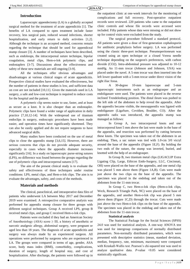
In Group A, two intracorporeal knots and one
intracorporeal knot 5 mm above the last were made in the base of
the appendix, and resection was performed by cutting between
these knots. The specimen was taken out of the abdomen in an
endobag. Then, a sac suture was tied with 1.0-cm 3/0 vicryl
around the base of the appendix (Figure 1E,F). By holding the
two ends of the suture, the stump was inverted, buried, and
knotted with the help of an endograsper.
In Group B, two titanium metal clips (LIGACLIP Extra
Ligating Clip, Large, Ethicon Endo-Surgery, LLC, Cincinnati,
OH) were placed on the base of the appendix, and one metal clip
was placed 5 mm above them (Figure 1A,B). Cuts were made
just above the two clips on the base of the appendix. The
specimen was placed in the endobag and taken out of the
abdomen from the 11-mm trocar.
In Group C, two Hem-o-lok clips (Hem-o-lok clips,
Weck, Research Triangle Park, NC) were placed on the base of
the appendix, and another Hem-o-lok clip was placed 5 mm
above them (Figure 1C,D) through the trocar. Cuts were made
just above the two Hem-o-lok clips on the base of the appendix.
The specimen was placed in the endobag and taken out of the
abdomen from the 11-mm trocar.
Statistical analysis
The Statistical Package for the Social Sciences (SPSS)
24.0 was used for statistical analysis. A one-way ANOVA test
was used for intergroup comparisons of normally distributed
parameters. Non-normally distributed parameters, which were
presented with descriptive statistics (mean, standard deviation,
median, frequency, rate, minimum, maximum) were compared
with Kruskall-Wallis test. Pearson’s chi-squared test was used to
analyze qualitative data. P-value <0.05 were considered
statistically significant.

J Surg Med. 2020;4(8):618-622. Comparison of techniques for appendiceal stump closure
P a g e / S a y f a | 620
Figure 1: Laparoscopic view of closing the appendix stump. (1A) Intraoperative view of
applied metal clips in laparoscopic appendectomy. (1B) Intraoperative view of appendix
stump after applied metal clips. (1C) Intraoperative view of applied hem-o-lok clips in
laparoscopic appendectomy. (1D) Intraoperative view of appendix stump after applied hem-
o-lok clips. (1E) Intraoperative view of intracorporeal knots in laparoscopic appendectomy.
(1F) Intraoperative view of appendix stump after applied intracorporeal knots.
Results
This retrospective study initially included 270 patients
with acute appendicitis. Their history, physical examination,
abdominal ultrasonography, and computerized tomography
results were used for the diagnosis. Fifty patients were excluded
from the study due to missing data and loss to follow-up. There
were 79 patients in group A, 91 patients in group B, and 50
patients in group C. All surgical operations were completed
successfully with the laparoscopic technique. The diagnosis was
uncomplicated acute appendicitis for 188 patients and
complicated acute appendicitis for 32 patients.
There was no significant difference between the two
groups with respect to demographic features, ASA scores, white
blood cell (WBC) counts, and neutrophil counts (Table 1). The
mean durations of the procedure were 53.7 (19.6) minutes in
group A, 51.9 (20.2) minutes in group B, and 47.8 (16.3) minutes
in group C (Table 2). The rates of postoperative complications
did not significantly differ between groups (P=0.474) (Table 2).
Patients were discharged at postoperative days 1–3 and
controlled daily for 1 week. The Douglas space was drained due
to perforated appendicitis in 8 patients in group A, 8 patients in
group B, and 4 patients in group C. All drains were pulled out on
postoperative days 5 or 6. There was no significant difference
between three groups (P=0.256) (Table 2).
The mean hospitalization period after LA was 27.8
(48.3) hours in group A, 22.8 (19.0) hours in group B, and 34.1
(34.6) hours in group C. The total length of hospital stay varied
significantly (P=0.001) (Table 2). According to the binary
comparisons, the total length of stay was significantly higher in
group C than groups B (P=0.002) and A (P=0.001). There was
no significant difference between groups A and B (P=0.785)
(Table 3).
The mean cost of the procedure was 247.9 (26.4) dollars
in group A, 260.4 (24.9) dollars in group B, and 281.8 (38.5)
dollars in group C. The costs differed significantly between
groups (P=0.001) (Table 2). According to the binary
comparisons, group A’s costs were significantly lower than those
of group B (P=0.001) and group C (P=0.001). Group B’s costs
were significantly lower than those of group C (P=0.001) (Table
3).
Table 1: Demographic data of patients
Suture
(n:79)
Metal clip
(n:91)
Hem-o-lok clip
(n:50)
P-
value
Age (years) Mean
(SD)
30.01
(2.59)
30.07
(13.42)
33.70 (14.74) a0.220
Gender (n)(%) Male 43(54.4) 59(64.8) 29(58) b0.111
Female 36(45.6) 32(35.2) 21(42)
BMI (kg/m2) Mean (SD) 28.12
(4.07)
27.84 (4.52) 28.23 (3.59) a0.912
ASA score (n)(%) I 44(55.7) 49(53.8) 29(58)
a0.564
II 31(39.2) 32(35.1) 17(34)
III 4(5.1) 10(11.1) 4(8)
WBC (cell/mm3) Mean (SD) 14006
(4049)
14398
(3987)
13381 (3472) a0.335
Neutrophil (cell/mm3) Mean
(SD)
11010
(4163)
11785
(3706)
10366 (3507) a0.099
Co-morbidities (n) (%) 18(22.7) 21(23.1) 11(22) a0.756
Diagnostic imaging
(n) (%)
CT 54(68.3) 61(67.1) 32(64) a0.876
US 25(31.7) 30(32.9) 18(36)
BMI: Body mass index, ASA score: American Society of Anesthesiologists physical classification system,
WBC: White blood cell, CT: Computed tomography, US: Ultrasonography
Table 2: The characteristics of the operation results
Suture
(n:79)
Metal clip
(n:91)
Hem-o-lok clip
(n:50)
P-
value
LGS* Score
(n) (%)
LGS* 0,
1, 2
52(65.8) 78 (85.7) 42 (84)
a0.032 LGS* 3a 12 (15.2) 8 (8.7) 3 (6)
LGS* 3b 8 (10.1) 0 0
LGS* 4a 3 (3.7) 2 (2.1) 2 (4)
LGS* 4b 2 (2.5) 1 (1.1) 1 (2)
LGS* 5 2 (2.5) 2 (2.1) 2 (2)
Postoperative complications
(n) (%)
8 (10.1) 5 (5.5) 5 (10) b0.474
Trocar site infection (n) (%) 4 (5.1) 7 (7.7) 7 (14) b0.192
Drain (n) (%) 8 (10.1) 8 (8.8) 4 (8) b0.256
Hospital stay (hours) Mean
(SD)
27.8 (48.3) 22.8 (19.0) 34.1 (34.6) c0.001
Operative time (minute)
Mean (SD)
53.7(19.6) 51.9 (20.2) 47.8 (16.3) a0.233
Hospital cost (dollar) Mean
(SD)
247.9
(26.4)
260.4 (24.9) 281.8 (38.5) c0.001
Price (dollar) 3.2x 6.42y 20.71z c0.001
a One-Way Anova Test, b Pearson Chi-Square Test, c Kruskall Wallis, LGS*: Laparoscopic grading system of
acute appendicitis according to Gomes Score [16] (the appendix was graded based upon its appearance:
Grade 0 (normal looking), 1 (redness and edema), 2 (fibrin), 3a (segmental necrosis), 3b (base necrosis), 4a
(abscess), 4b (regional peritonitis), and 5 (diffuse peritonitis), x: The price of three silk suture, y: The price
of three metal clips, z: The price of three Hem-o-lok clips.
Table 3: Post-Hoc results
Techniques P-value
Hospital stay
Suture/Metal clip 0.785
Suture/Hem-o-lok clip <0.001*
Metal clip/Hem-o-lok clip <0.002*
Hospital cost
Suture/Metal clip <0.001*
Suture/Hem-o-lok clip <0.001*
Metal clip/Hem-o-lok clip <0.001*
Mann Whitney U test, * P<0.01
The total complication rates of Group A, Group B, and
Group C were 15.1, 13.1 and 24%, respectively (Table 2). There
was no significant difference between the groups in terms of
wound infection, intra-abdominal abscess, and postoperative
ileus. Antibiotic treatment was administered in 5 patients in
Group A, 4 patients in group B, and 4 patients in group C. In
group B, 2 patients were treated with USG-guided percutaneous
drainage and antibiotic therapy. Ileus developed in 1 patient in
each group and healed with medical treatment. Four patients in
group A, 7 patients in group B and 7 patients in group C
J Surg Med. 2020;4(8):618-622. Comparison of techniques for appendiceal stump closure
P a g e / S a y f a | 621
developed wound infections that were treated with antibiotic
therapy.
Discussion
Due to faster recovery, less pain, and less surgical
complications in the treatment of acute appendicitis, laparoscopic
treatment has been widely accepted worldwide [1,2]. LA is a safe
procedure for the treatment of uncomplicated acute appendicitis,
but there is no common consensus for the laparoscopic treatment
of complicated appendicitis due to studies showing high and low
rates of post-operative intra-abdominal abscess (POIAA) [2,18-
22]. Peroperative classification of complicated and
uncomplicated acute appendicitis is highly valuable [16].
Postoperative complication rates are higher in
complicated appendicitis, regardless of the appendix stump
closure technique, and include intraabdominal abscess formation,
wound infection, and paralytic ileus [1-8,23]. This suggests that
the main determinant of postoperative complications depends on
the degree of the disease rather than the method used. It is
reasonable to consider that the presence and extent of peritonitis
may be risk factors for POIAA, and perforated appendicitis
without pus in the abdominal cavity may have lower rates than in
those complicated with peritonitis.
Despite the shorter hospital stay and lower perioperative
complication rates of LA, the hospital cost is still high compared
to open appendectomy [22]. The appendix stump closure
technique and the materials used are important in LA. Safe
closure of the appendix stump is important to prevent potential
postoperative complications (such as postoperative peritonitis,
sepsis, and fistulas) and reoperation. Numerous studies have
been carried out on techniques such as the endoloop, endostapler,
metal clips, and Hem-o-lok clips, and intracorporeal knot for the
closure of the appendix stump [3-13]. However, there is still no
consensus on the ideal technique. The closure technique of
embedding the stump used in open appendectomy was attempted
using alternative techniques in a laparoscopic procedure.
Studies using endostaples to close the appendix stump
have been carried out, but this method requires advanced
laparoscopic training [5,11,14,15,24]. Staples are safe to use but
very expensive. In addition, lost staple clips have been shown to
cause peritoneal adhesions that lead to complications such as
small bowel obstruction or ileus [15,24,25]. The appendix stump
closure selection is up to the surgeon's preference, but an
endostapler is recommended in cases of necrosis and
inflammation of the base of the appendix [25].
Endoloops are also widely used and are one of surgeons'
preferences [4,5,12,25]. The endostapler and endoloop methods
were compared in a clinical prospective, observational,
multicenter, high-case cohort study conducted by Van Rossem et
al. There was no significant difference between the groups in
terms of postoperative intraabdominal infections. Nevertheless,
they recommended using endoloops in terms of cost. In this
study, a double endoloop proximal to base of the appendix was
proposed in the presence of inflammation.
Another method used for appendix stump closure is the
titanium metal clip technique. Studies suggest that titanium metal
clips are safe and cost effective for fixing the base of the
appendix in LA [3,6,16]. In a clinical study, the metal clip
technique was evaluated in patients with complicated
appendicitis. Acute appendicitis grading was evaluated and
classified according to peroperative laparoscopic findings. It was
emphasized that the presence of local and diffuse peritonitis does
not cause difficulty for the metal clip technique. Nine of twelve
patients with appendicular base necrosis were safely treated with
laparoscopic suture and laparotomy, and the metal clip technique
is not recommended in these cases [16].
Recently, the use of simple non-absorbable clips, such
as the Hem-o-lok clip, has become widespread for the closure of
the appendix stump. The advantages of these clips are effortless
application, low cost, and robust and safe stump closure [10-15].
In a clinical study, Delibegovic et al. stated that the method is
effective and safe for closure of the appendix stump.
In our study, a secure closure was achieved by tying the
appendix stump with 2 intracorporeal knots, suturing 1 cm from
the stump, and embedding it in the cecum. Our technique
provides a safe closure in cases with appendix radix necrosis,
which is defined as 3D in the classification made by Gomez et al.
We defined the LPS technique in our clinical study in 2017 [26].
In a similar study, Shadhu et al. compared the intracorporeal
knot, Hem-o-lok clip, and laparoscopic purse string suture
techniques and stated that all techniques are safe in cases of
complicated appendicitis [17].
The stump closure techniques affect the operation times.
While devices such as the Hem-o-lok clip, metal clip, and
endostapler can be applied easily [6-13,27], techniques such as
the intracorporeal knot, endoloop, and LPS techniques require
surgical skills and experience [3-5]. A Cochrane review
published in 2017 compared the results of endostapler and
ligation methods (endoloop and intracorporeal knot) and found
that the endostapler technique significantly reduced the operation
time [28]. Again, both Hem-o-lok clip and metal clip
applications significantly decreased the operation times
compared to ligation techniques.
In a randomized clinical study conducted by Begovic et
al. [27], the endoloop group had significantly longer operations
than the endostapler group with clip techniques. The metal clip
technique had a shorter time than the endostapler technique, but
there was no significant difference between the Hem-o-lok clip
and the endostapler group. In the same study, the operation time
was significantly shorter in the Hem-o-lok group than the metal
clip group [27]. In a retrospective clinical study by Shadhu et al.
there was no significant difference in the duration of surgery in
the intracorporeal knot, Hem-o-lok clip, and LPS techniques. It is
much more difficult to apply LPS near the appendicular stump
during LA, and it requires some experience in laparoscopic
suturing [17].
In our study, even the LPS technique could not be
applied in some serious cases due to cecum edema, and partial
cecal resection was performed with an endostapler in these cases.
The Hem-o-lok polymeric clip technique has been found feasible
and safe for the appendicular stump [6-11]. However, the safe
use of Hem-o-lok and metal clips is significantly limited by the
maximum diameter of the 10-mm closure insert. In our study, the
LPS technique was applied to patients who could not receive the
Hem-o-lok clip technique.

J Surg Med. 2020;4(8):618-622. Comparison of techniques for appendiceal stump closure
P a g e / S a y f a | 622
The most common complications after LA are of
infectious origin, and the most serious is intra-abdominal abscess
formation [1,2]. This complication has important clinical
consequences because frequent intervention or hospitalization is
required. In a meta-analysis of 11 studies, wound-related
infections were present in 92 (4.2%) of 2175 operated patients
with acute complicated appendicitis [29]. In the same study, the
postoperative intraabdominal abscess rate was 5.9% (1059/63)
[29]. Abundant irrigation of the abdominal cavity with 0.9%
saline solution has been stated as a possible cause of this
development, and rational local irrigation with aspiration and
gauze application was recommended [2]. The type of appendix
stump closure has not been proven to affect this complication.
In a Cochrane systematic review, there was no
significant difference in the postoperative complications between
endoscopic clip and ligation techniques (endoloop and
intracorporeal knots) for closure of the appendix stump [28].
This meta-analysis showed a significant decrease in
postoperative complications with the use of the endostapler
device compared to the ligation techniques [28]. This decrease in
postoperative complications revealed that the endostapler
technique triggered a reduction in postoperative superficial
wound infections compared to ligation techniques.
There was no significant difference between groups in
terms of POIAA or postoperative ileus [28]. In a retrospective
clinical study, Shadhu et al. stated that there was no significant
difference between LPS, Hem-o-lok clip and intracorporeal knot
groups in terms of wound infection and POIAA. In our study, no
significant difference was found between LPS, metal clips and
Hem-o-lok clips in terms of postoperative complications.
It is accepted worldwide that LA is a costly method
compared to open appendectomy. For this reason, a safe and
low-cost technique is required to close the appendix stump to
reduce costs for the hospital and the patient. Cost analysis is
limited to data on consumable costs, and data on indirect costs
are not available [28]. Consumable prices also differ from
country to country [28,30]. Unfortunately, there is no study in
which appendix suture closure techniques are compared in terms
of hospital costs. Shadhu et al. compared LPS, hemoclip, and
intracorporeal knot techniques, but no cost was given.
In our study, hospital costs differed significantly among
groups. According to binary comparisons, the hospital costs in
LPS were significantly different than those in which clips were
used. Hospital costs in patients with metal clips were
significantly lower than those with local clips. Consumable costs
differed significantly by group.
Limitations
Our study has some limitations. The study was
retrospective and allocated to one or another treatment arm
because of the individual decision of the treating surgeon, so
both groups of patients differed in disease severity. Therefore, no
conclusions could be drawn regarding the equivalence of stump
closure by LPS, metal clip or Hem-o-lok clip at the same disease
stage. In addition, because of inflammation and necrosis (Gomez
classification 3B), the LPS technique was applied in patients in
which metal clip could not be used, because the appendix stump
diameter was over 10 mm.
Conclusion
The use of LPS for appendix stump closure in LA
operations is safe and effective. Our data clearly show that a
significant percentage of routine LA (32%) is suitable for clip
closure without an increase in intra- and postoperative
complications. This technique significantly reduces consumables
and hospital treatment costs. Our study supports the use of the
LPS technique in complicated appendicitis cases with necrosis
and perforation of the appendix base.
References
1. Sauerland S, Jaschinski T, Neugebauer EA. Laparoscopic versus open surgery for suspected
appendicitis. Cochrane Database Syst Rev. 2010;4;10:CD001546.
2. Pedersen AG, Petersen OB, Wara P, Rønning H, Qvist N, Laurberg S. Randomized clinical trial of
laparoscopic versus open appendicectomy. Br J Surg. 2001;88:200-5.
3. Ates M, Dirican A, Ince V, Ara C, Isik B, Yilmaz S. Comparison of intracorporeal knot-tying suture
(polyglactin) and titanium endoclips in laparoscopic appendiceal stump closure: a prospective
randomized study. Surg Laparosc Endosc Percutan Tech. 2012;22:226-31.
4. Sahm M, Kube R, Schmidt S, Ritter C, Pross M, Lippert H. Current analysis of endoloops in
appendiceal stump closure. Surg Endosc. 2011;25:124-9.
5. Safavi A, Langer M, Skarsgard ED. Endoloop versus endostapler closure of appendiceal stump in
pediatric laparoscopic appendectomy. Can J Surg. 2012;55:37-40.
6. Rickert A, Bönninghoff R, Post S, Walz M, Runkel N, Kienle P. Appendix stump closure with
titanium clips in laparoscopic appendectomy. Langenbecks Arch Surg. 2012;397:327-31.
7. Delibegovic S, Matovic E. Hem-o-lok plastic clips in securing of the base of the appendix during
laparoscopic appendectomy. Surg Endosc. 2009;23:2851-4.
8. Martín del Olmo JC, Blanco Alvarez JI, Carbajo Caballero MA, de la Cuesta de la Llave C, Vaquero
Puerta C, Arenal J. Laparoscopic appendectomy by ultrasonically activated scalpel in acute
appendicitis: preliminary report. J Laparoendosc Adv Surg Tech A. 2002;12:111-3.
9. Suttie SA, Seth S, Driver CP, Mahomed AA. Outcome after intra- and extra-corporeal laparoscopic
appendectomy techniques. Surg Endosc. 2004;18:1123-5.
10. Knight SR, Ibrahim A, Makaram N, Patil P, Wilson MSJ. The use of polymeric clips in securing the
appendiceal stump during laparoscopic appendicectomy: a systematic review. Eur J Trauma Emerg
Surg. 2019;45:665-70.
11. Al-Temimi MH, Berglin MA, Kim EG, Tessier DJ, Johna SD. Endostapler versus Hem-O-Lok clip to
secure the appendiceal stump and mesoappendix during laparoscopic appendectomy. Am J Surg.
2017;214:1143-8.
12. Colak E, Kement M, Ozlem N, Mutlu T, Yildirim K, Gurer A, et al. A comparison of nonabsorbable
polymeric clips and endoloop ligatures for the closure of the appendicular stump in laparoscopic
appendectomy: a prospective, randomized study. Surg Laparosc Endosc Percutan Tech. 2013;23:255-
8.
13. Varghese G. Feasibility and efficacy of using Hem-o-lok polymeric clips in appendicular stump
closure in laparoscopic appendectomy. Cureus 2018;10(6):e2871.
14. Graham CW, Komidar L, Perger L. Comparison of polymeric clips and endoscopic staplers for
laparoscopic appendectomy. J Laparoendosc Adv Surg Tech A. 2019;29:240-2.
15. Kliuchanok K, Keßler W, Partecke I, Walschus U, Schulze T, Heidecke CD, et al. A comparison of
non-absorbable polymeric clips and staplers for laparoscopic appendiceal stump closure: analysis of
618 adult patients. Langenbecks Arch Surg. 2019;404:711-6.
16. Gomes CA, Junior CS, de Peixoto RO, Netto JM, Gomes CC, Gomes FC. Appendiceal stump closure
by metal endoclip in the management of complicated acute appendicitis. World J Emerg Surg.
2013;18;8:35.
17. Shadhu K, Ramlagun D, Wang Y, Ping X, Chen T, Zhu Y, et al. Re-evaluation of purse string suture
in laparoscopic appendectomy. Surg Endosc. 2020;34:779-86.
18. Pokala N, Sadhasivam S, Kiran RP, Parithivel V. Complicated appendicitiseis the laparoscopic
approach appropriate? A comparative study with the open approach: outcome in a community hospital
setting. Am Surg. 2007;73;737-41.
19. Pirro N, Berdah SV. Appendicitis: yes or no to laparoscopic approach? J Chir (Paris). 2006;143:155-9.
20. Kouwenhoven EA, Repelaer van Driel OJ, van Erp WF. Fear for the intraabdominal abscess after
laparoscopic appendectomy: not realistic. Surg. Endosc. 2005;19:923-6.
21. Katkhouda N, Mason RJ, Towfigh S, Gevorgyan A, Essani R. Laparoscopic versus open
appendectomy: a prospective randomized double-blind study. Ann Surg. 2005;242:439-48.
22. Biondi A, Di Stefano C, Ferrara F, Bellia A, Vacante M, Piazza L. Laparoscopic versus open
appendectomy: a retrospective cohort study assessing outcomes and cost-effectiveness. World J
Emerg Surg. 2016;30;11:44.
23. Çalışkan Y. Original Alvarado scoring system in the diagnosis of acute appendicitis: A cohort study. J
Surg Med. 2017;1:28-31.
24. Wagner M, Aronsky D, Tschudi J, Metzger A, Klaiber C. Laparoscopic stapler appendectomy. A
prospective study of 267 consecutive cases. Surg Endosc. 1996;10:895-9.
25. van Rossem CC, van Geloven AA, Schreinemacher MH, Bemelman WA. Endoloops or endostapler
use in laparoscopic appendectomy for acute uncomplicated and complicated appendicitis: No
difference in infectious complications. Surg Endosc. 2017;3:178-84.
26. Gunes ME, Gemici E, Donmez T. Comparison of Laparoscopic Embedding Technique and Other
Techniques for Appendiceal Stump Closure. Turk J Colorectal Dis. 2019;29:121-6.
27. Delibegović S, Mehmedovic Z. The influence of the different forms of appendix base closure on
patient outcome in laparoscopic appendectomy: a randomized trial. Surg Endosc. 2018 ;32:2295-9.
28. Mannu GS, Sudul MK, Bettencourt-Silva JH, Cumber E, Li F, Clark AB, et al. Closure methods of the
appendix stump for complications during laparoscopic appendectomy. Cochrane Database Syst Rev.
2017 Nov 13;11:CD006437.
29. Markides G, Subar D, Riyad K. Laparoscopic Versus Open Appendectomy in adults with
Complicated Appendicitis: Systematic Review and Metaanalysis. World J Surg. 2010;34:2026-40.
30. Śmigielski J, Piskorz Ł, Koptas W. Comparison of treatment costs of laparoscopic and open surgery.
Videosurgery and Other Miniinvasive Techniques 2015;10:437-41.
This paper has been checked for language accuracy by JOSAM editors.
The National Library of Medicine (NLM) citation style guide has been used in this paper.

P a g e / S a y f a | 623
Is overactive bladder a risk factor for erectile dysfunction? A cross-
sectional study Aşırı aktif mesane erektil disfonksiyon için bir risk faktörü müdür? Kesitsel çalışma
Aykut Başer 1, Mehmet Murat Baykam 1
How to cite/Atıf için: Başer A, Baykam MM. Is overactive bladder a risk factor for erectile dysfunction? A cross-sectional study. J Surg Med. 2020;4(8):623-626.
J Surg Med. 2020;4(8):623-626. Research article DOI: 10.28982/josam.767471 Araştırma makalesi
1 Hitit University, Medical Faculty, Department of
Urology, Corum, Turkey
ORCID ID of the author(s)
AB: 0000-0003-0457-512X
MMB: 0000-0001-9006-4275
Corresponding author/Sorumlu yazar:
Aykut Başer
Address/Adres: Hitit Üniversitesi, Tıp Fakültesi,
Üroloji Anabilim Dalı, Çorum, Türkiye
E-mail: [email protected]
�
Ethics Committee Approval: Approval was received
from Hitit University noninvasive clinical research
ethics committee with the number 2020-206. All
procedures in this study involving human participants
were performed in accordance with the 1964 Helsinki
Declaration and its later amendments.
Etik Kurul Onayı: Onay Hitit Üniversitesi noninvaziv
Klinik Araştırma Etik Kurulundan 2020-206 onay
numarası ile alınmıştır. İnsan katılımcıların katıldığı
çalışmalardaki tüm prosedürler, 1964 Helsinki
Deklarasyonu ve daha sonra yapılan değişiklikler
uyarınca gerçekleştirilmiştir. �
Conflict of Interest: No conflict of interest was
declared by the authors.
Çıkar Çatışması: Yazarlar çıkar çatışması
bildirmemişlerdir.
�
Financial Disclosure: The authors declared that this
study has received no financial support.
Finansal Destek: Yazarlar bu çalışma için finansal
destek almadıklarını beyan etmişlerdir.
�
Published: 8/30/2020
Yayın Tarihi: 30.08.2020
Copyright © 2020 The Author(s)
Published by JOSAM This is an open access article distributed under the terms of the Creative
Commons Attribution-NonCommercial-NoDerivatives License 4.0 (CC
BY-NC-ND 4.0) where it is permissible to download, share, remix,
transform, and buildup the work provided it is properly cited. The work
cannot be used commercially without permission from the journal.
Abstract
Aim: Erectile dysfunction (ED) is a sexual dysfunction characterized by the inability to achieve or maintain penile erection during
sexual activity. Many risk factors have been identified in ED-related epidemiological studies. In experimental studies, a relationship is
thought to exist between Overactive Bladder (OAB) and ED. We investigated the relationship between OAB and ED, its clinical
reflections, and the odds ratio of OAB for ED.
Methods: We conducted this cross-sectional prospective study between January-July 2020. Sixty patients referred to the urology
outpatient clinic with complaints of OAB (Group 1) and 66 patients without urological complaints (Group 2) were included in the study.
Patients’ erectile functions were evaluated with the IIEF-5 form. OAB was evaluated with the OAB-V8 form.
Results: The groups were similar in terms of age, body mass index, comorbidity, and smoking status. IIEF-5 scores were higher in
Group 2 [20.52 (3.51)] compared to Group 1 [18.17 (5.46)] (P=0.036). Correlation analysis between IEF-5 and OAB-V8 scores
revealed a negative correlation; it was observed that the IIEF-5 score decreased as the OAB-V8 score increased (r=-0.260, P=0.045).
The odds ratio of decreasing IIEF-5 score with each 1-unit increase of OAB-V8 score was 0.164 (P=0.04). It was observed that the
patients diagnosed with OAB had lower IIEF-5 scores when they had nocturia.
Conclusion: OAB is a risk factor for ED. The presence of nocturia symptoms is remarkable for ED in OAB patients. The effect of OAB
should not be ignored in the treatment of ED.
Keywords: Erectile dysfunction, IIEF-5, OAB, Overactive bladder
Öz
Amaç: Erektil disfonksiyon (ED), cinsel aktivite sırasında penis ereksiyonunun sağlanamaması veya sürdürülememesi ile karakterize bir
cinsel işlev bozukluğudur. ED ile ilişkili epidemiyolojik çalışmalarda birçok risk faktörü tanımlanmıştır. Deneysel çalışmalarda Aşırı
Aktif Mesane (OAB) ile ED arasında bir ilişki olduğu düşünülmektedir. Bu çalışmamızda OAB ve ED arasındaki ilişkiyi, klinik
yansımalarını ve ED için OAB'nin risk oranını araştırmayı amaçladık.
Yöntemler: Bu kesitsel çalışmayı Ocak 2020 ve Temmuz 2020 arasında gerçekleştirdik. OAB (Grup 1) şikayetleri ile üroloji
polikliniğine başvuran 60 hasta ve ürolojik şikayeti olmayan 66 hasta (Grup 2) çalışmaya dahil edildi. Hastaların erektil fonksiyonları
IIEF-5 formu ile aşırı aktif mesane sorgulaması OAB-V8 formu ile değerlendirildi.
Bulgular: Gruplar yaş, vücut kitle indeksi, komorbidite ve sigara içme durumu açısından benzerdi. IIEF-5 skorları Grup 2'de [20,52
(3,51)] Grup-1'e [18,17 (5,46)] göre daha yüksek bulundu (P=0,036). IEF-5 score ve OAB-V8 score arasında yapılan korelasyon
analizinde negatif korelasyon saptandı; OAB-V8 skoru arttıkça IIEF-5 skorunun düştüğü gözlendi (r -0,260, P=0,045). OAB-V8
skorunun her 1 birim artışında IIEF-5 puanınında ki azalma risk oranı 0,164 olarak bulunmuştur (P=0,04). OAB tanısı alan hastalarda
noktüri varlığında IIEF-5 skorlarının daha düşük olduğu gözlendi.
Sonuç: OAB, ED için bir risk faktörüdür. Noktüri semptomlarının varlığı OAB hastalarında ED için dikkat çekicidir. ED tedavisinde
OAB'nin etkisi göz ardı edilmemelidir.
Anahtar kelimeler: Erektil disfonksiyon, IIEF-5, OAB, Aşırı aktif mesane

J Surg Med. 2020;4(8):623-626. Risk factor assessment for erectile dysfunction
P a g e / S a y f a | 624
Introduction
Erectile dysfunction (ED) is a sexual dysfunction
characterized by the inability to achieve or maintain penile
erection during sexual activity [1]. It is a quite common disease
among adult males and its incidence increases with age. With the
aging US population, it is predicted that more than 35 million
American men will experience ED and that 50% of men will be
affected by ED until the age of 50 [2]. In epidemiological studies
on ED, the main risk factors were divided into 4 categories: 1-
Urological and andrological risk factors, 2- Cardiovascular and
metabolic risk factors, 3- Psychiatric diseases and 4- Lifestyle-
related risk factors [3]. The relationship between ED and LUTS
under the title of urological and andrological risk factors is
confirmed by epidemiological studies and potential common
biological mechanisms [4].
According to OAB definition of ICS 2002 [5],
overactive bladder (OAB) is a syndrome characterized by
symptoms of urgency, with or without urgency incontinence,
usually with increased daytime frequency and nocturia
(increased night-time urination). It includes lower urinary system
symptoms (LUTS). The association of OAB, including lower
urinary tract symptoms, with Erectile Dysfunction (ED), which is
a urological disorder, was also investigated in the literature [6-
10]. It was also indicated that ED had a strong relationship with
OAB symptoms [9]. OAB affects 11.8-27.2% of men [8,11,12].
The prevalence of overactive bladder also increases with age, as
in ED. Both diseases negatively affect health-related quality of
life and psychology [13].
Experimental studies demonstrated that mirabegron,
which is used in the treatment of OAB, led to the relaxation of
the corpus cavernosum by alpha-1 adrenoceptor blockade and
that avanafil, which is a type 5 PDH inhibitor, inhibited the
activation of detrusor activated by potassium chloride [14,15].
Both experimental studies and the relationship of ED with LUTS
suggest that these two urological diseases may be each other's
risk factors or comorbid conditions.
In these studies, the relationship between the
Overactive Bladder 8-Question Awareness Tool (OAB-V8)
scores and The International Erectile Function Index
Questionnaire (IIEF-5) scores of OAB patients and the OAB-V8
scores and IIEF-5 scores of control patients randomly selected
from the community without OAB, and the clinical reflections of
their responses to the IIEF-5 questions were not investigated.
In this study, we conducted an observational study to
investigate this relationship and its clinical reflections and
calculate the odds ratio of OAB for ED.
Materials and methods
Study design
This study was designed as a cross-sectional study. The
present study protocol was reviewed and approved by the
Institutional Review Board of Hitit University School of
Medicine Ethics Committee (ethics committee approval date-
number: 2020-207). Informed consent was obtained from all
subjects when they were enrolled. This study was conducted in
accordance with STROBE guidelines for reporting observational
studies (www.strobestatement.org).
Study population
As a result of the sample size analysis based on other
research findings in the literature, the minimum number of
participants in each group was determined as 60 with a 95%
confidence level and 80% power.
Sixty patients referred to the urology outpatient clinic
with complaints of OAB (diagnosed with OAB for the first time
and never received anticholinergic treatment) between January
2020 - July 2020, and 66 control patients without urological
complaints were included in the study. The patients were
informed about the study and written consent forms were
received for participation. Patients' age, height, weight, Body
Mass Index (BMI), comorbid disease states, and smoking status
were examined.
Evaluation of ED
The sexual activity status of the patients with active
sexual lives for the last 12 months in the last 4 weeks was
evaluated using the 5-question version of the International
Erectile Function Index questionnaire (IIEF-5) (16). This
questionnaire consists of 4 questions about sexual function and 1
question about sexual satisfaction. Each question is scored
between 1-5 points. Patients who stated that they were using
PDH-5 inhibitor were not included in the study.
Evaluation of OAB
OAB was evaluated according to the 2002 ICS
definition using the OAB-V8 (Overactive Bladder 8-Question
Awareness Tool- V8) form consisting of 8 questions developed
by Acquadro et al. [17] for symptom scoring in 2006. In this
form, each question is scored between 0-5 points. Among the
patients with complaints of OAB, those with active urinary tract
infections, interstitial cystitis, neurogenic bladder, excessive
fluid intake, and patients using diuretics and similar drugs were
excluded from the study. In addition, patients were evaluated in
terms of benign prostatic hyperplasia. Patients with
uroflowmetry maximum flow rate (Qmax) below 15
milliliters/second and patients with obstructive, stenotic,
intermittent voiding patterns were not included in the study.
Likewise, according to the EAU and AUA guidelines on digital
rectal examination and PSA, patients with suspicious findings
were excluded.
Statistical analysis
Statistical analyses were performed using the SPSS
version 22 software. The distribution of variables was tested by
the Shapiro-Wilk test. Descriptive statistics were presented as
mean and standard deviation for normally distributed variables,
median, minimum, maximum values for ordered ordinal data,
and number and percentage for categorical variables. In the
evaluation of numerical data between the groups, the parameters
with normal distribution were evaluated by student t test, ordinal
data were evaluated by Mann-Whitney U test, and categorical
data were evaluated by chi-square test. The relationship between
the IIEF-5 score and the OAB-V8 score was examined with the
Pearson and Spearman’s correlation test. The independent effects
of the OAB-V8 score on IIEF-5 score were examined with the
linear regression model. The model fit was analyzed with the
required residual and fit statistics. The cases with a Type-1 error
level below 0.5% were statistically evaluated.
J Surg Med. 2020;4(8):623-626. Risk factor assessment for erectile dysfunction
P a g e / S a y f a | 625
Results
A total of 126 patients, including 60 patients diagnosed
with OAB (Group 1) and 66 patients in the control group (Group
2), were included in our study. The mean ages of Groups 1 and 2
were 44.30 (12.63) years and 40.15 (13.41) years, respectively.
BMI values were 27.95 (5.85) kg/m2 and 26.62 (4.68) kg/m
2 in
Groups 1 and 2, respectively. There was no difference between
the groups in terms of age and BMI values (P=0.056, P=0.073).
IIEF-5 scores were higher in Group 2 [20.52 (3.51)] compared to
Group 1 [18.17 (5.46)] (P=0.036). No difference was found
between the groups in terms of diabetes, hypertension
comorbidity and smoking. The main demographic data of the
groups are presented in Table 1.
When the responses to the questions of the IIEF-5 form
between the groups were examined, OAB patients were found to
have lower scores in question 2 (When you had erections with
sexual stimulation, how often were your erections hard enough
for penetration?), question 3 (During sexual intercourse, how
often were you able to maintain your erection after you had
penetrated your partner?) and question 5 (When you attempted
sexual intercourse, how often was it satisfactory for you?)
compared to control patients (P=0.010, P=0.048, P=0.006,
respectively). The scores of IIEF-5 questions among the groups
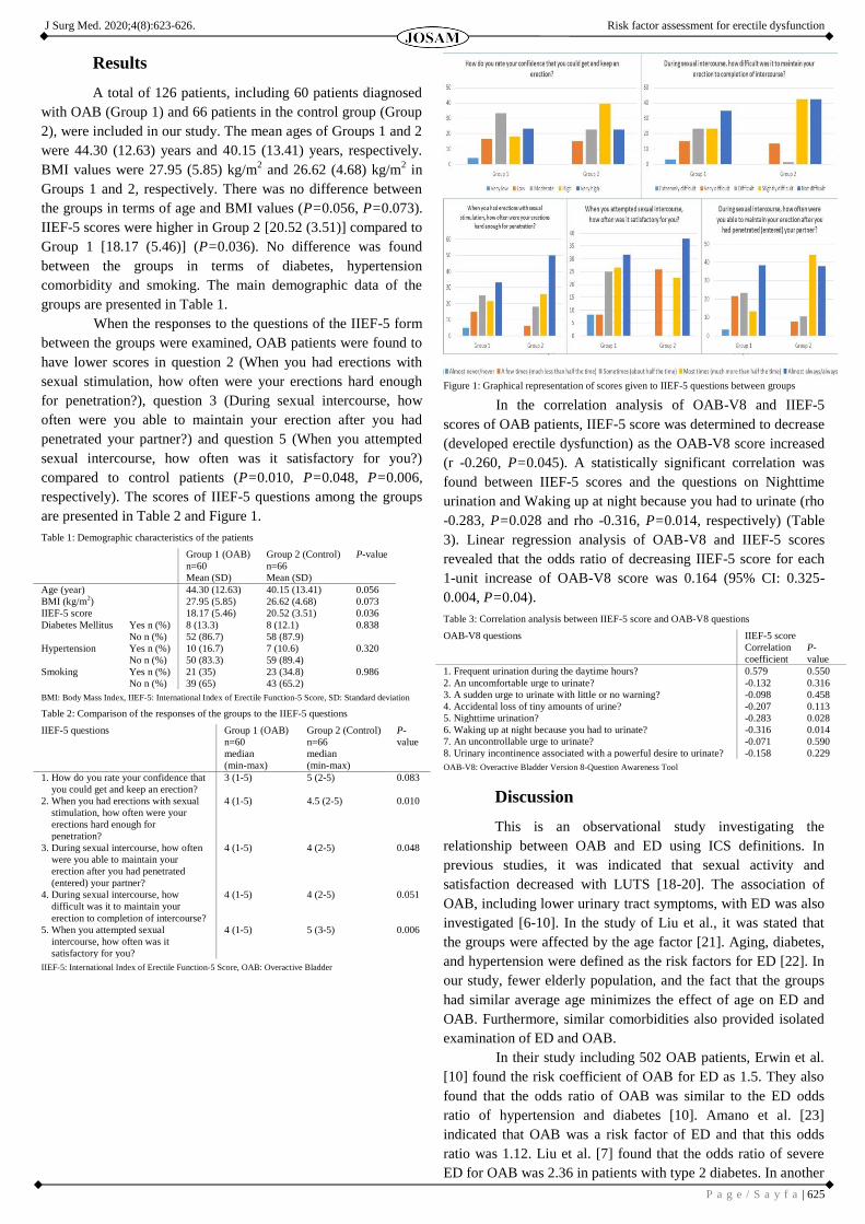
are presented in Table 2 and Figure 1.
Table 1: Demographic characteristics of the patients
Group 1 (OAB)
n=60
Mean (SD)
Group 2 (Control)
n=66
Mean (SD)
P-value
Age (year) 44.30 (12.63) 40.15 (13.41) 0.056
BMI (kg/m2) 27.95 (5.85) 26.62 (4.68) 0.073
IIEF-5 score 18.17 (5.46) 20.52 (3.51) 0.036
Diabetes Mellitus Yes n (%) 8 (13.3) 8 (12.1) 0.838
No n (%) 52 (86.7) 58 (87.9)
Hypertension Yes n (%) 10 (16.7) 7 (10.6) 0.320
No n (%) 50 (83.3) 59 (89.4)
Smoking Yes n (%) 21 (35) 23 (34.8) 0.986
No n (%) 39 (65) 43 (65.2)
BMI: Body Mass Index, IIEF-5: International Index of Erectile Function-5 Score, SD: Standard deviation
Table 2: Comparison of the responses of the groups to the IIEF-5 questions
IIEF-5 questions Group 1 (OAB)
n=60
median
(min-max)
Group 2 (Control)
n=66
median
(min-max)
P-
value
1. How do you rate your confidence that
you could get and keep an erection?
3 (1-5) 5 (2-5) 0.083
2. When you had erections with sexual
stimulation, how often were your
erections hard enough for
penetration?
4 (1-5) 4.5 (2-5) 0.010
3. During sexual intercourse, how often
were you able to maintain your
erection after you had penetrated
(entered) your partner?
4 (1-5) 4 (2-5) 0.048
4. During sexual intercourse, how
difficult was it to maintain your
erection to completion of intercourse?
4 (1-5) 4 (2-5) 0.051
5. When you attempted sexual
intercourse, how often was it
satisfactory for you?
4 (1-5) 5 (3-5) 0.006
IIEF-5: International Index of Erectile Function-5 Score, OAB: Overactive Bladder
Figure 1: Graphical representation of scores given to IIEF-5 questions between groups
In the correlation analysis of OAB-V8 and IIEF-5
scores of OAB patients, IIEF-5 score was determined to decrease
(developed erectile dysfunction) as the OAB-V8 score increased
(r -0.260, P=0.045). A statistically significant correlation was
found between IIEF-5 scores and the questions on Nighttime
urination and Waking up at night because you had to urinate (rho
-0.283, P=0.028 and rho -0.316, P=0.014, respectively) (Table
3). Linear regression analysis of OAB-V8 and IIEF-5 scores
revealed that the odds ratio of decreasing IIEF-5 score for each
1-unit increase of OAB-V8 score was 0.164 (95% CI: 0.325-
0.004, P=0.04).
Table 3: Correlation analysis between IIEF-5 score and OAB-V8 questions
OAB-V8 questions IIEF-5 score
Correlation
coefficient
P-
value
1. Frequent urination during the daytime hours? 0.579 0.550
2. An uncomfortable urge to urinate? -0.132 0.316
3. A sudden urge to urinate with little or no warning? -0.098 0.458
4. Accidental loss of tiny amounts of urine? -0.207 0.113
5. Nighttime urination? -0.283 0.028
6. Waking up at night because you had to urinate? -0.316 0.014
7. An uncontrollable urge to urinate? -0.071 0.590
8. Urinary incontinence associated with a powerful desire to urinate? -0.158 0.229
OAB-V8: Overactive Bladder Version 8-Question Awareness Tool
Discussion
This is an observational study investigating the
relationship between OAB and ED using ICS definitions. In
previous studies, it was indicated that sexual activity and
satisfaction decreased with LUTS [18-20]. The association of
OAB, including lower urinary tract symptoms, with ED was also
investigated [6-10]. In the study of Liu et al., it was stated that
the groups were affected by the age factor [21]. Aging, diabetes,
and hypertension were defined as the risk factors for ED [22]. In
our study, fewer elderly population, and the fact that the groups
had similar average age minimizes the effect of age on ED and
OAB. Furthermore, similar comorbidities also provided isolated
examination of ED and OAB.
In their study including 502 OAB patients, Erwin et al.
[10] found the risk coefficient of OAB for ED as 1.5. They also
found that the odds ratio of OAB was similar to the ED odds
ratio of hypertension and diabetes [10]. Amano et al. [23]
indicated that OAB was a risk factor of ED and that this odds
ratio was 1.12. Liu et al. [7] found that the odds ratio of severe
ED for OAB was 2.36 in patients with type 2 diabetes. In another
J Surg Med. 2020;4(8):623-626. Risk factor assessment for erectile dysfunction
P a g e / S a y f a | 626
study, LUTS ED odds ratio was reported as 2.2 [24]. In our
study, OAB was determined as a risk factor for ED. This odds
ratio was lower than the odds ratios indicated in the literature.
However, in our study, the study groups were fewer, they were
diagnosed with OAB for the first time and had not received prior
treatment for OAB. The similarity between the patients with
OAB and the control group patients in terms of risk factors such
as hypertension, diabetes, and smoking defined for ED may have
led to the lower result of this ratio.
With regards to the relationship between OAB and ED,
Erwin et al. [10] showed that urinary symptoms decreased the
frequency of sexual activity or prevented participation in sexual
activity. In the same study, it was also stated that nocturia and
urge incontinence had strong negative effects on ED. Similarly,
Liu et al. [7] determined that urge and nocturia had strong effects
on ED. In our study, we determined that the patients with OAB
had lower IIEF-5 scores compared to the healthy control group.
We think that OAB may have a negative effect on Erectile
function. Akin to the literature, we determined that nighttime
urination and waking up at night because you had to urinate
negatively affected IIEF-5 scores. However, we could not find
comparable results for urge incontinence. In our study, unlike the
literature, we determined that the scores of questions 2, 3 and 5
(erection for penetration, maintaining the erection and
satisfaction with sexual intercourse) in the IIEF-5 inquiry form
were lower among men with OAB compared to healthy controls.
One of the results we obtained in our study was that the
responses given to these questions (although there is a need to be
supported by studies with a larger number of patients) may be
inexpressible OAB symptoms. The effect of OAB should not be
ignored in the treatment of ED. OAB should be investigated in
those who do not respond to ED treatment. In the literature,
sexual functions were reported to improve after anticholinergic
treatment in female patients with OAB [25-27]. This was not
studied for male patients. There is a need for studies examining
the erectile functions of ED patients with OAB after OAB
treatment.
Limitations
This study has some limitations, one of which was the
small study population. Regarding other reasons, although the
study groups were similar in terms of hypertension, diabetes, and
smoking, which are known risk factors for ED, patients with
these factors were affected by the time they had these risk
factors. The effects of nocturia on sleep quality and quality of
life may also have effects on erectile function. Furthermore,
patients diagnosed with OAB may also be affected by these
factors since it was unknown how long the disease existed.
Conclusion
OAB and ED prevalence increase with age. OAB is a
risk factor for ED. ED patients with OAB have difficulties in
achieving and maintaining erection for penetration and sexual
satisfaction. The presence of the symptoms of nocturia is
remarkable for ED in OAB patients. The effect of OAB should
not be ignored in the treatment of ED, and there is a need for
studies examining the erectile functions of ED patients with
OAB following OAB treatment.
References
1. Valiquette, L. A historical review of erectile dysfunction. The Canadian Journal of Urology.
2003;10(Suppl 1):7-11. PMID: 12625844
2. Aytac IA, Araujo AB, Johannes CB, Kleinman KP, McKinlay JB. Socioeconomic factors and
incidence of erectile dysfunction: findings of the longitudinal Massachusetts male aging study. Soc Sci
Med 2000;51(5):771-8. doi:.10.1016/s0277-9536(00)00022-8
3. Beutel ME, Weidner W, Brähler E. Epidemiology of sexual dysfunction in the male population.
Andrologia. 2006;38(4):115-21. doi: 10.1111/j.1439-0272.2006.00730.x
4. De Nunzio C, Roehrborn CG, Andersson KE, McVary KT. Erectile Dysfunction and Lower Urinary
Tract Symptoms. Eur Urol Focus. 2017;3(4-5):352-63. doi: 10.1016/j.euf.2017.11.004
5. Abrams P, Cardozo L, Fall M, Griffiths D, Rosier P, Ulmsten U, et al. The standardisation of
terminology of lower urinary tract function: report from the Standardisation Sub-committee of the
International Continence Society. Neurourol Urodyn. 2002;21:167-78. doi: 10.1002/nau.10052
6. Jiann BP. Dose Overactive Bladder (OAB) Type Affect the Strength of the Association between OAB
and Erectile Dysfunction? J Sex Med. 2013;10:1187. doi: 10.1111/jsm.12053
7. Liu RT, Chung MS, Chuang YC, Lee JJ, Lee WC, Chang HW, et al. The presence of overactive
bladder wet increased the risk and severity of erectile dysfunction in men with type 2 diabetes. J Sex
Med 2012;9:1913-22. doi: 10.1111/j.1743-6109.2012.02738.x
8. Coyne KS, Sexton CC, Thompson C, Kopp ZS, Milsom I, Kaplan SA. The impact of OAB on sexual
health in men and women: Results from EpiLUTS. J Sex Med 2011;8:1603-15. doi: 10.1111/j.1743-
6109.2011.02250.x
9. Oliveira P, Castro NM, Muniz AL, Tanajura D, Brandão JC, Porto AF, et al. Prevalence of erectile
dysfunction in HTLV-1-infected patients and its association with overactive bladder. Urology.
2010;75(5):1100-3. doi: 10.1016/j.urology.2009.11.041
10.Debra E. Irwin, Ian Milsom, Kate Reilly, Steinar Hunskaar, Zoe Kopp, Sender Herschorn, et al.
Overactive Bladder Is Associated with Erectile Dysfunction and Reduced Sexual Quality of Life in
Men. J Sex Med 2008;5:2904-10 doi: 10.1111/j.1743-6109.2008.01000.x
11.Zumrutbas AE, Bozkurt AI, Tas E, Acar CI, Alkis O, Coban K, et al. Prevalence of lower urinary tract
symptoms, overactive bladder and urinary incontinence in western Turkey: results of a population-
based survey. Int J Urol. 2014;21(10):1027-33. doi: 10.1111/iju.12519
12.Irwin DE, Milsom I, Hunskaar S, Reilly K, Kopp Z, Herschorn S, et al. Population-based survey of
urinary incontinence, overactive bladder, and other lower urinary tract symptoms in five countries:
results of the EPIC study. Eur Urol 2006;50:1306-14. doi: 10.1016/j.eururo.2006.09.019
13.Quek KF. Factors affecting health-related quality of life among patients with lower urinary tract
symptoms. Int J Urol 2005;12:1032-6. doi: 10.1111/j.1442-2042.2005.01198.x
14.de Oliveira MG, Rojas-Moscoso JA, Bertollotto GM, Candido TZ, Kiguti LRA, Pupo AS, et al.
Mirabegron elicits rat corpus cavernosum relaxation and increases in vivo erectile response. Eur J
Pharmacol. 2019;5;858:172447. doi: 10.1016/j.ejphar.2019.172447
15.Dhruva A, Hamsavardhini VK, Kamatham S, Kataria A, Kumar A, Shanthi M, et al. Avanafil Inhibits
the Contractility of the Isolated Caprine Detrusor Muscle. Int J Appl Basic Med Res. 2019;9(4):231-5.
doi: 10.4103/ijabmr.IJABMR_339_18.
16.Rosen RC, Cappelleri JC, Smith MD, Lipsky J, Pena BM. Development and evaluation of an abrigged,
5-item version of the International Ondex of Erectile Function (IIEF-5) as a diagnostic tool for erectile
dysfuntion. International Journal of Impotence Research. 1999;11(6):319-26. doi:
10.1038/sj.ijir.3900472
17.Acquadro C, Kopp Z, Coyne KS, Corcos J, Tubaro A, Choo MS, et al. Translating overactive bladder
questionnaries in 14 languages. Urology. 2006;67:536. doi: 10.1016/j.urology.2005.09.035
18.Rosen R, Altwein J, Boyle P, Kirby RS, Lukacs B, Meuleman E, et al. Lower urinary tract symptoms
and male sexual dysfunction: The multinational survey of the aging male (MSAM-7). Eur Urol.
2003;44:637-49. doi: 10.1016/j.eururo.2003.08.015
19.Li MK, Garcia LA, Rosen R. Lower urinary tract symptoms and male sexual dysfunction in Asia: A
survey of ageing men from five Asian countries. BJU Int. 2005;96:1339-54. doi: 10.1111/j.1464-
410X.2005.05831.x
20.Aslan G, Cavus E, Karas H, Oner O, Duran F, Esen A. Association between lower urinary tract
symptoms and erectile dysfunction. Arch Androl. 2006;52:155-62. doi: 10.1080/01485010500379871
21.Bang-Ping Jiann. Dose Overactive Bladder (OAB) Type Affect the Strength of the Association
between OAB and Erectile Dysfunction? J Sex Med. 2013;10:1187. doi: 10.1111/jsm.12053
22.Braun M, Wassmer G, Klotz T, Reifenrath B, Mathers M, Engelmann U. Epidemiology of erectile
dysfunction: results of the ‘Cologne Male Survey’. Int J Import Res. 2000;12:305-11. doi:
10.1038/sj.ijir.3900622
23.Amano T, Earle C, Imao T, Takemae K. Are urge inkontinence and aging risk factors of erectile
dysfunction in patients with male lower urinary tract symptoms? Aging Male. 2016;19(1):54-7. doi:
10.3109/13685538.2015.1103219.
24.Ponholzer A, Temml Ch, Marszalek M, Obermayr R, Madersbacher S. Prevalence and risk factors for
erectile dysfunction in 2869 men using a validated questionnaire. Eur Urol. 2005;47(1):80-6. doi:
10.1016/j.eururo.2004.08.017
25.Hajebrahimi S, Azaripour A, Sadeghi-Bazargani H. Tolterodine immediate release improves sexual
function in women with overactive bladder. J Sex Med. 2008;5:2880-5. doi: 10.1111/j.1743-
6109.2008.00976.x
26.Rogers RG, Omotosho T, Bachmann G, Sun F, Morrow JD. Continued symptom improvement in
sexually active women with overactive bladder and urgency urinary incontinence treated with
tolterodine ER for 6 months. Int Urogynecol J Pelvic Floor Dysfunct. 2009;20:381-5. doi:
10.1007/s00192-008-0782-9
27.Sand PK, Goldberg RP, Dmochowski RR, McIlwain M, Dahl NV. The impact of the overactive
bladder syndrome on sexual function: a preliminary report from the Multicenter Assessment of
Transdermal Therapy in Overactive Bladder with Oxybutynin trial. Am J Obstet Gynecol.
2006;195(6):1730-5. doi: 10.1016/j.ajog.2006.08.013.
This paper has been checked for language accuracy by JOSAM editors.
The National Library of Medicine (NLM) citation style guide has been used in this paper.
P a g e / S a y f a | 627
Assessment of quality and accuracy of YouTube videos on
percutaneous transthoracic biopsy Transtorasik akciğer biyopsisi hakkındaki YouTube videolarının kalite ve doğruluğunun değerlendirilmesi
Okan Gürkan 1, Canan Gündüz 2
How to cite/Atıf için: Gürkan O, Gündüz C. Assessment of quality and accuracy of YouTube videos on percutaneous transthoracic biopsy. J Surg Med. 2020;4(8):627-630.
J Surg Med. 2020;4(8):627-630. Research article DOI: 10.28982/josam.778941 Araştırma makalesi
1 Gaziosmanpaşa Taksim Training and Research
Hospital, Department of Radiology, Istanbul,
Turkey 2 Istanbul Sureyyapasa Chest Diseases and
Thoracic Surgery Training and Research Hospital,
Department of Pulmonary Diseases, Istanbul,
Turkey
ORCID ID of the author(s)
OG: 0000-0002-7934-9154
CG: 0000-0002-4746-529X
Corresponding author/Sorumlu yazar:
Okan Gürkan
Address/Adres: Karayolları Mahallesi, Osmanbey
Caddesi 621 sokak, Gaziosmanpaşa, İstanbul, Türkiye
E-mail: [email protected]
�
Ethics Committee Approval: This article is not a
study with human participants. There are no
experiments on animals. There is no identifying
information of participants.
Etik Kurul Onayı: Bu makale, insan katılımcılarla
yapılan bir çalışma değildir. Hayvanlar üzerinde
deney yoktur. Katılımcıların tanımlayıcı bilgisi
yoktur. �
Conflict of Interest: No conflict of interest was
declared by the authors.
Çıkar Çatışması: Yazarlar çıkar çatışması
bildirmemişlerdir.
�
Financial Disclosure: The authors declared that this
study has received no financial support.
Finansal Destek: Yazarlar bu çalışma için finansal
destek almadıklarını beyan etmişlerdir.
�
Published: 8/30/2020
Yayın Tarihi: 30.08.2020
Copyright © 2020 The Author(s)
Published by JOSAM This is an open access article distributed under the terms of the Creative
Commons Attribution-NonCommercial-NoDerivatives License 4.0 (CC
BY-NC-ND 4.0) where it is permissible to download, share, remix,
transform, and buildup the work provided it is properly cited. The work
cannot be used commercially without permission from the journal.
Abstract
Aim: YouTube is an essential source of medical information for patients, but may also be misleading, depending on the kind of content
quality. The aim of this study is to evaluate the quality of YouTube videos on percutaneous transthoracic lung biopsy.
Methods: A search on YouTube was performed with the terms ‘Lung Biopsy,’ ‘CT-Guided Lung Biopsy,’ ‘US-Guided Lung Biopsy,’
and ‘Percutaneous Transthoracic Biopsy.’ Relevant English videos were examined and scored by two reviewers. Video characteristics,
uploaded sources, and content quality were assessed using different indices. Correlation analysis was conducted to investigate the
possible correlation for global quality score.
Results: Fifteen videos (47%) were uploaded by a healthcare professional, nine videos (28%) by a product company, six videos (19%)
by an individual user, and 2 (6%) by an academic institution. The videos had a median (IQR) quality score of 2 (range 1-5), and a
median (IQR) length of 235 (46-751) seconds. The quality of the videos varied, depending on the uploaded sources as well as whether
they were uploaded by academic institutions, which had the highest quality. Some important components regarding the biopsy
procedure, such as complications, were not evaluated in most videos.
Conclusion: YouTube videos on percutaneous transthoracic lung biopsy (PTLB) had a low median content quality score, while some
important points were grossly overlooked. Thus, YouTube videos may be a misleading source of patient information. Academic
institutions and healthcare professionals should be creating accurate multimedia content for patients seeking informative medical
information.
Keywords: YouTube, Social media, Biopsy, Patient education
Öz
Amaç: YouTube, hastalar için önemli bir tıbbi bilgi kaynağıdır. Ancak içerik kalitesine bağlı olarak yanıltıcı bilgiler de verebilmektedir.
Bu çalışmanın amacı, perkütan transtorasik akciğer biyopsisi ile ilgili YouTube videolarının kalitesini değerlendirmektir.
Yöntemler: YouTube’da ‘Lung Biopsy’, ‘CT-Guided Lung Biopsy’ ‘US-Guided Lung Biopsy’ ve ‘percutaneous Transthoracic Biopsy’
terimleri ile arama yapıldı. İlintili İngilizce videolar iki kişi tarafından incelendi. Video karakteristikleri, yüklendiği kaynak ve içerik
kalitesi değerlendirildi. Global kalite skorunu değerlendirmek için korelasyon analizi yapıldı.
Bulgular: On beş video (%47) profesyonel sağlık çalışanları, 9 video (%28) firmalar, 6 video (%19) kişisel hesaplar ve 2 (%6) video ise
akademik kurum tarafından yüklenmişti. Videoların ortanca (IQR) kalite skoru 2 (aralık 1-5), ortanca uzunluğu ise 235 (46-751) saniye
olarak hesaplandı. Videoların kalitesi, yüklendiği kaynağa göre değişiklik göstermekte olup, akademik kurumlar tarafından yüklenen
videolar en yüksek kaliteye sahipti. Komplikasyonlar gibi önemli biyopsi prosedürü komponentleri, çoğu videoda değerlendirilmemişti.
Sonuç: Perkütan transtorasik akciğer biyopsisi (PTLB) ile ilgili YouTube videolarında bazı önemli noktalar göz ardı edildiğinden dolayı,
düşük bir ortanca içerik kalite puanına sahip oldukları gözlendi. Bu nedenle, YouTube videoları yanıltıcı bir hasta bilgisi kaynağı
olabilir. Akademik kurumlar ve sağlık uzmanları, bilgilendirici tıbbi bilgi arayan hastalar için doğru multimedya içeriği oluşturmalıdır.
Anahtar kelimeler: YouTube, Sosyal medya, Biyopsi, Hasta eğitimi

J Surg Med. 2020;4(8):627-630. Assessment of YouTube videos on transthoracic biopsy
P a g e / S a y f a | 628
Introduction
YouTube is the second most popular website, which
serves 95% of web surfers in 80 different languages [1,2].
Entertainment and educational sources, including medical
videos, are also commonly used with YouTube. Explanatory
videos about medical procedures and operations are often
appreciated by medical students, as well as patients.
Lung cancer is the 3rd
most frequent malignancy in both
genders, and the most frequent malignancy in males.
Approximately 2 million new patients were diagnosed with lung
cancer in 2018. Although lung cancer mortality rates tend to
decrease in the male population, as a function of decreased
smoking prevalence, overall rates in women continue to increase.
Lung cancer accounts for the highest mortality rate in the overall
population [3,4]. Its presentation for most patients in the
advanced stages emphasizes the importance of early diagnosis
and treatment.
Lung biopsy procedures, as part of the diagnosis for
lung cancer, are either performed by bronchoscopy, percutaneous
interventions, or open surgery. Percutaneous transthoracic lung
biopsy (PTLB) is an image-guided radiological biopsy method
used in the diagnosis of lung cancer for patients with peripheral
masses. Fluoroscopy, computed tomography (CT) and ultrasound
(US) are often selected for imaging during the PTLB procedure
[5]. Occasionally, patients find the information from their
primary physicians about the PTLB procedure insufficient, since
it is usually performed by radiologists. As patients try to
overcome the lack of information from website sources,
YouTube videos serve as a substantial resource. Even if most
videos enlighten the patients and answer their questions
accurately, there is a risk of misinformation, which cannot be
underestimated. Misleading video content may have negative
consequences, such as avoidance of the procedure. The quality of
information received by patients via medical YouTube videos
has immense value.
The primary aim of this study was to evaluate the
quality of YouTube videos on PTLB. As such, video
characteristics via the upload source, reliability scores, and
popularity indices were assessed.
Materials and methods
A search was performed on YouTube with the search
terms ‘Lung Biopsy,’ ‘CT-Guided Lung Biopsy,’ ‘US-Guided
Lung Biopsy,’ and ‘Percutaneous Transthoracic Biopsy.’ The
study was performed on 6.6.2019. The videos were sorted by
relevance as well as other default filter settings. The first 60
videos for each search term were analyzed and viewed, since
internet search engine studies show that users primarily view the
first three pages [6]. Non-English videos are irrelevant, while
nonverbal and duplicate videos were excluded. Number of
likes/dislikes, total number of views, video length, uploading
date, and number of comments and likes per 1000 views were
recorded for the enrolled videos. They were analyzed and scored
independently by two investigators. One was a radiologist,
experienced with lung biopsies, the other was an experienced
pulmonary physician.
The videos were categorized according to four
uploading sources: Academic institutions, healthcare
professionals (physician and non-physician), product companies,
and individual users. Four questions adapted from the DISCERN
tool [7] were used for determining reliability of YouTube videos
(ROV): (1) Are the aims clearly stated? (2) Is the source of
information informative and reliable? (3) Is the presented
information objective and unbiased? (4) Are uncertain elements
noted? ‘Yes’ answers scored 1 point and ‘no’ answers, 0 points.
A lung biopsy scoring tool (LBST) was created for evaluating
the accuracy of the videos, which were adjusted from previous
studies [8,9] (Table 1). Each item scored 1 point, with a
maximum of 12 points. The educational content value of the
videos was evaluated by Global Quality Score (GQS), which was
adapted from a previous study [10], using a scale of 1 to 5: (1)
Poor quality, not useful for patients; (2) Poor quality and flow,
but partially useful; (3) Moderate quality, useful with some
important details missing; (4) Generally good flow and quality;
and (5) Excellent quality and flow, including very useful
information. Video power index (VPI) could evaluate popularity
of the videos, described by the formula: ratio*view ratio/100
[11].
Table 1: Lung biopsy scoring tool for accuracy of the videos (scored from 0 to 12)
Topic Content Maximum
points
available
Purpose of the
procedure
To determine if the lesion is benign or malignant and
to analyze the tumor stage, for evaluating diffuse lung
disease
3
Alternative to
percutaneous
transthoracic lung
biopsy
Follow-up, excisional biopsy 2
Preparation Stop antiaggregant 5-7 days, oral anticoagulants 3-5
days before the procedure. No intake of solid foods
before procedure for 6 hours and water for 2 hours
2
Method The entry localization should be sterilized and then
local anesthetic is injected. Position depends on depth
and size of the lesion. Biopsy needle should be
inserted while patient holds inspiration. Duration is
20 to 120 minutes.
2
Side effects Px, pulmonary hemorrhage, hemothorax, tumor
seeding cardiac tamponade, chest infection.
2
After procedure Chest radiograph should be performed 0.5-1 hour
later than the procedure.
1
Results 2-10 days after the procedure 1
Further
information
Further information sources 1
Px: pneumothorax
Statistical analysis
The statistical analysis was performed with IBM SPSS
Statistics 22.0 (Armonk, NY, USA: IBM Corp.). Shapiro-Wilk
test was used to assess the normality of variable distributions,
which were also included. A Spearman correlation analysis was
conducted to examine associations between GQS and VPI, and
ROV and ICGIP. For all analyses, a P-value < 0.05 was
considered significant.
Results
Among 240 screened videos, 32 fulfilled inclusion
criteria and were analyzed for the study (Excluded videos
(n=208): Non-English (61), Irrelevant (95), Non-verbal (22),
Duplicate videos (31)). Fifteen videos (47%) were uploaded by a
healthcare professional, 9 videos (28%) by a product company, 6
videos (19%) by an individual user, and 2 (6%) by an academic
institution. Median (IQR) length of the videos was 235 (46-751)
seconds, while median (IQR) number of views was 6385 (57-
63128). There was a significant correlation between duration of

J Surg Med. 2020;4(8):627-630. Assessment of YouTube videos on transthoracic biopsy
P a g e / S a y f a | 629
the video and number of views (P=0.04) (Table 2). Based on
reviewers’ GQSs, 12 videos were rated 1 point (poor quality, not
useful for patients), and only 1 was rated as 5 points (excellent
quality and flow, includes very useful information), out of a total
of 32 videos. Median quality score was 2.00. The highest median
quality score (3) belonged to videos uploaded by academic
institutions and the lowest median score (1) belonged to videos
uploaded by healthcare professionals (Table 3). The median
ROV was 2 out of 4. Academic institutions had the highest ROV
median score (2.5). The median number of likes from product
companies and individual user videos were 28 and 26,
respectively, which were higher than other uploaded sources.
The video with the highest number of dislikes was uploaded by a
product company with the longest duration (751 seconds). Video
content was not assessed for all components of LBST. The
highest score was 11 out of 12, uploaded by individual users.
Thirteen videos (40%) mentioned side effects, which was shown
in 5 videos (33%) uploaded by healthcare providers. The purpose
of the procedure was noted in most (81%) videos. Those that
focused on methods had higher dislike counts (P=0.02). The
videos with the highest and lowest median VPI scores were from
academic institutions and healthcare workers, respectively (38.83
and 1.64, respectively). GQS demonstrated a significant
correlation with ROV and ICIGP, but not with VPI (P<0.001,
P<0.001, P=0.067, respectively) (Table 4). GQS did not show a
significant correlation between number of views and video
length (P=0.31, P=0.62, respectively). Videos clarifying the
purpose of the procedure showed a significant correlation with
GQS (P<0.001 in both). Interclass correlation coefficients
ranged between 0.911 and 0.984 for intra-rater reliability, and
between 0.925 and 0.941 for interrater reliability. Ethical
committee approval was not required for the present study, since
open access data were used.
Table 2: Characteristics of the evaluated videos
Mean SD Median Minimum Maximum
Video length (seconds) 259 161 235 46 751
Number of likes 58 123 17 1 676
Number of dislikes 5 7 1 1 26
Total number of views 16079 19778 6385 57 63128
Total number of comments 9 29 1 0 162
Days since upload 1333 961 1165 28 3547
View ratio 19.68 40.48 7.33 0.07 209.43
Like ratio 80.61 17.04 87.30 50.00 98.67
VPI 17.66 38.33 5.78 0.04 205.75
ROV (0-4) 1.94 1.22 2.00 0.00 4.00
LBST (0-12) 3.44 2.82 3.00 1.00 11.00
GQS (1-5) 2.16 1.14 2.00 1.00 5.00
LBST: lung biopsy scoring tool, VPI: video power index, ROV: reliability of YouTube videos, GQS: global
quality score
Table 3: Characteristics of the YouTube videos by source
Source VPI ROV
(0-4)
ICIGP
(0-12)
GQS
(1-5)
Academic institutions Mean 38.83 2.50 5.50 3.00
Median 38.83 2.50 5.50 3.00
SD 44.15 2.12 6.36 2.83
Healthcare professionals(physician
and non-physician)
Mean 7.83 1.73 2.40 1.80
Median 1.64 1.00 2.00 1.00
SD 10.71 1.10 2.10 1.01
Product companies Mean 29.28 2.22 3.22 2.44
Median 5.97 2.00 3.00 2.00
SD 66.40 1.09 1.99 0.88
Individual users Mean 17.77 1.83 5.67 2.33
Median 11.71 2.00 5.00 2.50
SD 25.07 1.60 3.39 1.21
Abbreviations: LBST, lung biopsy scoring tool; VPI, video power index; ROV, reliability of YouTube
videos; GQS, global quality score
Table 4: Correlation of GQS
Correlation coefficient P-value
Number of likes 0.050 0.976
ROV 0.813 <0.001
ICIGP 0.799 <0.001
VPI 0.078 0.673
LBST: lung biopsy scoring tool, VPI: video power index, ROV: reliability of YouTube videos, GQS: global
quality score
Discussion
While there is a large number of medical studies on
YouTube related to patient education, over the last 10 years,
these studies have generally focused on diseases and treatments,
the use of medical materials, and operations. In our literature
review, we found only one study about image-guided biopsy. For
lung biopsy, no such studies were conducted so far.
Patients receiving detailed information from their
physicians before the biopsy and operation becomes more
difficult as the number of patients increase, as the time allocated
for each patient shortens. Healthcare associations provide
information to patients and their families in numerous ways for
educational purposes. However, special procedures such as a
biopsy become more understandable when supported by visual
content, such as videos. Although pulmonary and oncology
clinicians examine the patient with malignant diseases of the
lung and refer them for biopsy, the procedures are often
performed by radiologists. Disorderly information regarding the
procedure cause confusion. Lung cancer has the highest
mortality rate and early diagnosis is crucial for quality medicine.
To increase patient compliance prior to the biopsy, which is
necessary for an early diagnosis, online information is especially
helpful [12].
In this study, the median values of GQS, ROV, and
LBST of the videos were extremely low. These results show that
the quality, reliability, and accuracy of the videos on
transthoracic lung biopsy were also low. In terms of upload
source, the highest median ROV, LBST, and GQS scores were
affiliated with academic institutions. The lowest median of all
groups belonged to healthcare professionals. In terms of VPI, the
videos uploaded had the lowest average. It was also found that
these videos included little explanation, often containing biopsy
patient images in the CT unit with poor informative content and
quality. The popularity scores for these videos were also low,
due to lack of satisfactory information for the audience. For
example, one of the most important subjects for a patient is the
complication rate that may occur after the procedure. The most
common is pneumothorax, which can be seen in a wide range of
patients, from 0 to 61%. Moreover, 3.3% to 15% of patients may
also need a chest tube [13,14]. When patients and their relatives
do not receive enough information about these issues, they can
be troubled by hearsay information, and sometimes refuse the
procedure due to misleading information. Videos uploaded by
healthcare groups stated that this was rare. This shows that
videos displaying the process content, with too much visual
detail for patients (and not acknowledging focus topics, such as
side effects), receive negative reactions.
LBST was a tool that was adapted by us from two
separate studies, to evaluate videos about lung biopsy in terms of
accuracy. LBST being significantly correlated with GQS
suggested that this tool was useful. It also allowed us to make
unique inferences about the videos during a subgroup evaluation.

J Surg Med. 2020;4(8):627-630. Assessment of YouTube videos on transthoracic biopsy
P a g e / S a y f a | 630
For example, the dislike ratio of videos that discussed the
method was high. The most mentioned topic was the purpose of
the procedure. In general, this was noted in all videos uploaded
by individual users, focusing on their biopsy stories from
subjective experiences. We find that the mean LBST score of
individual users, being higher than other groups, needs further
consideration.
As stated, the highest VPI scores belonged to academic
institutions and product companies, respectively. We attributed
this to the fact that both groups create their videos with higher
quality and offer their content in a more organized manner. Since
it may be possible to get higher search rankings on YouTube
with Search Engine Optimization (SEO) [15], increasing the
number of views via various advertising routes by the product
companies may have positively contributed to the VPI scores. To
keep up with changing YouTube search algorithms, appropriate
optimization of title, description, and keywords must also be
performed for videos uploaded by healthcare professionals.
The lack of correlation between GQS and VPI revealed
that video uploaders neglected factors that created quality
content, while tending to increase video popularity for users.
ROV developed from the DISCERN tool, which was created
about 10 years before the first YouTube video was made, was
also correlated with GQS. This indicates that the tool was up-to-
date and could evaluate educational videos.
Limitations
The fact that only videos in English were examined was
one of the limitations of our study. Like previous studies, two
reviewers were included, which may also be considered a
potential limitation. Furthermore, new content could be available
in YouTube, while old content may have been removed. This is
also a limitation for our study.
Conclusion
In this study where we examined the contents of the
educational videos on YouTube for transthoracic lung biopsy
procedure, we found that the quality of the videos was low.
Moreover, we observed that the subjects about which the patients
were often curious, such as complications that may occur after
the procedure, were not adequately addressed. Professional
associations and academic institutions need to keep up with
changes in ways of learning, provide online healthcare
information more accurately and address a broader audience to
minimize patients' anxiety about the procedure and any refusal of
procedure.
References
1. Alexa. 2019. The top 500 sites on the web. [Online] Available at: https://www.alexa.com/topsites.
[Accessed 7 May 2019]
2. YouTube. 2019. YouTube for Press. [Online] Available at: https://www.youtube.com/intl/en-
GB/yt/about/press/. [Accessed 7 May 2019]
3. Bray F, Ferlay J, Soerjomataram I, Siegel RL, Torre LA et al. Global cancer statistics 2018:
GLOBOCAN estimates of incidence and mortality worldwide for 36 cancers in 185 countries. CA
Cancer J Clin 2018; 68: 394-424. doi: 10.3322/caac.21492
4. Torre LA, Siegel RL, Ward EM, Jemal A. International variation in lung cancer mortality rates and
trends among women. Cancer Epidemiol Biomarkers Prev. 2014;23:1025-36. doi: 10.1158/1055-
9965.EPI-13-1220
5. Charig M, Clelland C, Gleeson F, R Miller, H Moss et al. Guidelines for radiologically guided lung
biopsy. Thorax. 2003;58:920-36. doi: 10.1136/thorax.58.11.920
6. iProspect Search Engine User Behaviour Study. [Accessed January 26, 2012.] Available from:
http://district4.extension.ifas.ufl.edu/Tech/TechPubs/WhitePaper_
2006_SearchEngineUserBehavior.pdf.
7. Charnock D, Shepperd S, Needham G, Gann R. DISCERN: An instrument for judging the quality of
written consumer health information on treatment choices. J Epidemiol Community Health.
1999;53:105-11. doi: 10.1136/jech.53.2.105
8. Lehmann S, Frank N. An Overview of Percutaneous CT-Guided Lung Biopsies. J Radiol Nurs 2018;
37: 2-8. doi: 10.1016/j.jradnu.2017.12.002
9. American T. Acr – Sir – Spr Practice Parameter on Informed Consent for Image-Guided Procedures.
2016;1076:1-8.
10.Singh AG, Singh S, Singh PP. YouTube for information on rheumatoid arthritis--a wakeup call? J
Rheumatol. 2012;39:899-903. doi: 10.3899/jrheum.111114
11.Erdem MN, Karaca S. Evaluating the Accuracy and Quality of the Information in Kyphosis Videos
Shared on YouTube. Spine. 2018;43:E1334-E1339. doi: 10.1097/BRS.0000000000002691
12.Sechrest RC. The internet and the physician-patient relationship. Clin Orthop Relat Res.
2010;468:2566-71. doi: 10.1007/s11999-010-1440-3
13.Swischuk JL, Castaneda F, Patel JC, Jitendra C. Patel, Ruizong Li, et al. Percutaneous Transthoracic
Needle Biopsy of the Lung: Review of 612 Lesions. J Vasc Interv Radiol. 1998;9:347-52. doi:
10.1016/S1051-0443(98)70279-9.
14.Kazerooni EA, Lim FT, Mikhail A, Martinez FJ. Risk of pneumothorax in CT-guided transthoracic
needle aspiration biopsy of the lung. Radiology. 1996;198:371-5. doi:
10.1148/radiology.198.2.8596834
15.Jackson R, Schneider A, Baum N. Social media networking: YouTube and search engine optimization.
J Med Pract Manage. 2011;26:254-7.
This paper has been checked for language accuracy by JOSAM editors.
The National Library of Medicine (NLM) citation style guide has been used in this paper.
P a g e / S a y f a | 631
Is language disability a risk factor for complicated appendicitis? A
retrospective cohort study Dil engeli, komplike apandisit için bir risk faktörü müdür? Geriye dönük kohort çalışma
Dursun Özgür Karakaş 1, Metin Yeşiltaş 1, Berk Gökçek 1, Seracettin Eğin 1, Semih Hot 1
How to cite/Atıf için: Karakaş DÖ, Yeşiltaş M, Gökçek B, Eğin S, Hot S. Is language disability a risk factor for complicated appendicitis? A retrospective cohort study. J Surg
Med. 2020;4(8):631-635.
J Surg Med. 2020;4(8):631-635. Research article DOI: 10.28982/josam.770774 Araştırma makalesi
1 Okmeydanı Teaching and Educating Hospital,
Department of General Surgery, İstanbul, Turkey
ORCID ID of the author(s)
DÖK: 0000-0002-2466-9855
MY: 0000-0002-2080-1572
BG: 0000-0001-9989-4152
SE: 0000-0002-4090-5205
SH: 0000-0001-9623-356X
Corresponding author/Sorumlu yazar:
Dursun Özgür Karakaş
Address/Adres: Okmeydanı Eğitim ve Araştırma
Hastanesi, Genel Cerrahi Kliniği, İstanbul, Türkiye
E-mail: [email protected]
�
Ethics Committee Approval: Approval was received
from Okmeydanı Training and Research Hospital
Ethical Committee, May 14, 2019 date and 1148
number. All procedures in this study involving human
participants were performed in accordance with the
1964 Helsinki Declaration and its later amendments.
Etik Kurul Onayı: Onay Okmeydanı Eğitim ve
Araştırma Hastanesi Etik Kurulu, 14 Mayıs 2019 tarih
ve 1148 sayı onay numarası ile alınmıştır. İnsan
katılımcıların katıldığı çalışmalardaki tüm
prosedürler, 1964 Helsinki Deklarasyonu ve daha
sonra yapılan değişiklikler uyarınca
gerçekleştirilmiştir. �
Conflict of Interest: No conflict of interest was
declared by the authors.
Çıkar Çatışması: Yazarlar çıkar çatışması
bildirmemişlerdir.
�
Financial Disclosure: The authors declared that this
study has received no financial support.
Finansal Destek: Yazarlar bu çalışma için finansal
destek almadıklarını beyan etmişlerdir.
�
Published: 8/30/2020
Yayın Tarihi: 30.08.2020
Copyright © 2020 The Author(s)
Published by JOSAM This is an open access article distributed under the terms of the Creative
Commons Attribution-NonCommercial-NoDerivatives License 4.0 (CC
BY-NC-ND 4.0) where it is permissible to download, share, remix,
transform, and buildup the work provided it is properly cited. The work
cannot be used commercially without permission from the journal.
Abstract
Aim: Acute appendicitis is the most common emergent surgical disease and complicated appendicitis is an indicator of disrupted access
to healthcare. Language disability has become more important in the healthcare sector with increasing migration in the last decade. We
aimed to evaluate language disability as a risk factor for complicated appendicitis.
Methods: From January 2014 to December 2018, patients who were operated for acute appendicitis were evaluated retrospectively.
Patients’ age, gender, language disability (LD) (no (C) or yes (F)), whether surgical drainage was required, pathological findings
(appendix diameter, severity as uncomplicated (UCA) or complicated (CA), and presence of local peritonitis), levels of C-Reactive
Protein (CRP), White Blood Cells (Wbc), Neutrophil% (Neu%), and ultrasonography (USG) and computed tomography (CT) results
were noted and compared.
Results: Six hundered twenty-eight patients were included in the study, among which 15.1% (n=95) were considered F, and 12% (n=74)
were CA. Age and gender did not significantly differ in terms of LD and severity (P=0.15, P=0.24 and P=0.2, P=0.21, respectively).
Drainage requirement, local peritonitis, levels of CRP, Wbc, and Neu% were significantly higher in the CA group (P<0.001, P<0.001,
P<0.001, P=0.009, and P<0.001, respectively). Drainage, appendix diameter, levels of CRP, and Neu% were significantly higher in the
F group (P=0.01, P=0.04, P=0.007, and P=0.046, respectively). CA rate was insignificantly higher in the F group (17% vs 11%)
(P=0.72). The false-negative ratio of USG and CT was higher in F patients with CA (56.2% vs. 37.5%).
Conclusion: This study showed that language disability could be a risk factor for complicated appendicitis with higher drainage ratio,
appendix diameter, levels of CRP, and Neu%.
Keywords: Appendicitis, Severity, Language disability
Öz
Amaç: Akut apandisit en sık acil cerrahi hastalık ve komplike apandisit sağlık kuruluşlarına erişebilme göstergesidir. Son on yılda
göçlerin artması ile dil engeli sağlık için önemli hale gelmiştir. Dil engelinin komplike apandisit için risk faktörü olarak değerlendirmeyi
amaçladık.
Yöntemler: Ocak 2014’den Aralık 2018 tarihleri arasında akut apandisit nedeni ile opere edilen hastalar geriye dönük değerlendirildi.
Hastaların yaş, cinsiyet, dil engeli (DE) (var (Y) yok (V)), drenaj uygulanması, patolojisi (apandiks çapı, şiddeti komplike olan (KA),
komplike olmayan (KOA), ve lokal peritonit varlığı), C-Reaktif Proteini (CRP), beyaz küre (BK), nötrofil yüzdesi (Nöt%),
ultrasonografi (US) ve bilgisayarlı tomografi (BT) sonuçları istatistiksel olarak değerlendirildi.
Bulgular: Altı yüz yirmi sekiz hasta çalışmaya dahil edildi. %15,1’i (n=95) Y, ve %12’si (n=74) KA idi. DE ve şiddet açısından yaş ve
cinsiyet farkı istatistiksel olarak anlamlı saptanmadı (sırasıyla P=0,15, P=0,24 ve P=0,2, P=0,21). Drenaj, lokal peritonit, CRP, BK ve
Nöt% KA grubunda anlamlı olarak yüksek saptandı (sırasıyla P<0,001, P<0,001, P<0,001, P=0,009, ve P<0,001). Drenaj, apandiks
çapı, CRP ve Nöt% seviyesi Y gurunda anlamlı olarak yüksek saptandı (sırasıyla P=0,01, P=0,04, P=0,007 ve P=0,046). KA oranı Y
grubunda daha fazla olmasına rağmen (%17 karşı %11) istatistiksel olarak anlamlı (P=0,72). US ve BT’nin yalancı negatiflik oranı
KA’lı Y grubunda daha yüksek saptandı (%56,2 karşı %37,5).
Sonuç: Bu çalışma dil engelinin; yüksek drenaj oranı, apandiks çapı, CRP ve Nöt% seviyeleri ile komplike apandisit için bir risk faktörü
olabileceğini göstermiştir.
Anahtar kelimeler: Apandisit, Şiddet, Dil engeli

J Surg Med. 2020;4(8):631-635. Complicated appendicitis and language disability
P a g e / S a y f a | 632
Introduction
Ten percent of Europe and 4.8% of Turkey are
foreigners (immigrants, tourists, etc.) with language disability
[1,2]. The language disability, difficulty in accessing healthcare,
or lack of health insurance are the most important disadvantages
of foreign healthcare [3,4]. Language disability decreased with
the second generation of immigrants due to their learning of the
primary language. Lack of access to healthcare or health
insurance are not valid factors for all foreigners admitted to the
emergency department Turkey. However, language disability
remains a severe problem and causes complications.
Acute abdominal pain is an important, frequent
complaint which was observed in 10% of the emergency
department admissions and only 1.9% were caused by acute
appendicitis [5,6]. Acute abdominal pain has a 10.5% false-
positive and 18,6% false-negative decision risk. False-positive
decisions cause unnecessary appendectomies with increased
morbidity and mortality, and false-negative decisions increase
perforation or abscess risk [7]. Complicated acute appendicitis is
considered an indicator of lack of access to healthcare in children
based on communication disabilities [8]. Foreigners have
increased risk of perforated/complicated appendicitis or
unnecessary surgery for acute abdominal pain [9,10].
Radiological (USG and CT), scoring systems (Alvarado,
etc.), or inflammatory markers such as C-Reactive Protein
(CRP), White Blood Cells (Wbc), and Neutrophil % (Neu%) are
used to evaluate the severity of acute appendicitis [11-14].
Ten percent of admissions for emergency surgery
consisted of foreigners with or without language disability, and
appendicitis is the most common emergency surgical disease in
our hospital. We aimed to evaluate language disability as a risk
factor for complicated appendicitis.
Materials and methods
After receiving institutional approval from the ethics
committee of Okmeydanı Training and Research Hospital (14
May 2019 date, and 1148 number), records of patients who were
operated for acute appendicitis between January 2014 and
December 2018 were evaluated retrospectively. The foreigner
patients who can speak Turkish, English or other common
languages were excluded from the group of patients with
language disability (Figure 1).
Figure 1: Flow diagram of the study. (CRP: C-reactive protein, Wbc: White Blood Cell,
Neu%: Neutrophil %, USG: Ultrasonography, CT: Computed Tomography)
Age, gender, language disability, operation type,
whether surgical drainage was performed, pathological findings
(diameter of the appendix, severity, the presence of local
peritonitis, and fecaloid), levels of C-reactive protein (CRP),
white blood cells (Wbc), neutrophil % (Neu%), results of
ultrasonography (USG) and computed tomography (CT) were
evaluated retrospectively.
Language disability was evaluated as without (C) or
with (F), and gender, as male or female. Operation methods were
either open or laparoscopic, and whether surgical drainage was
performed was noted. The severity of pathology was evaluated as
uncomplicated (UCA) (appendicitis, phlegmonous, and
suppurative) or complicated appendicitis (CA) (gangrenous and
perforated). Local peritonitis and fecaloids were evaluated as yes
or no. USG and CT were either (0) not performed, (1) performed
but negative, or (2) performed and positive for acute
appendicitis.
Severity of appendicitis and language disability were
assessed with respect to age, gender, need for surgical drainage,
appendix diameter, presence of local peritonitis, levels of CRP,
Wbc, and Neu%. USG and CT results of patients with and
without language disability were compared with regards to
severity of appendicitis.
Statistical analysis
Statical analysis was performed with SPSS 15.0. T-test
was used to evaluate age, diameter, CRP (mg/dl), Wbc (103/uL),
Neu% (%) (Mean (Standard derivation)). The ratio of males to
females, language disability, drainage requirement, local
peritonitis, USG, and CT were calculated in percentages. Chi-
Square, Mann Whitney U, and T-Test were used for assessment,
and P<0.05 was considered statistically significant. ROC
analysis was performed for language disability, CRP, WBC,
Neu% according to CA. Cut off values, sensitivity, and
specificity of CRP, Wbc and Neu% were noted.
Results
Six hundred twenty-eight patients were included in the
study, 15.1% (n=95) of which had language disability (foreigners
(F) not citizens (C)). The mean age was 30.31 (12.6) years, and
67% (n=421) of the patients were males. Among all, 55.7%
(n=350) of the operations were performed laparoscopically, 13%
(n=81) had surgical drainage. The mean appendix diameter was
10.54 (5.4) mm. Twelve percent (n=74) of the patients had CA,
63% (n=397) had local peritonitis, and 62.9% (n=395) had
fecaloids. The mean CRP, WBC, and Neu% values were 54.5
(79.4) mg/dL, 14.6 (4.6) 106/uL, and 77.4% (10.3), respectively
(Table 1).
The mean age was 29.86 (12.28) years among the UCA
group, and 33.66 (14.86) years among those with CA. 66.2% of
the UCA and 73% of the CA patients were male. 8.3% (n=46) of
patients with UCA and 47.3% (n=35) of CA patients required
surgical drainage, the difference between which was statistically
significant (P<0.001). The mean diameter of the appendix was
10.39 (5.56) mm in UCA, and 11.68 (3.69) mm in CA groups
(P=0.055). The language disability rates were 14.3% (n=79) and
21.6% (n=16) among those with UCA and CA, respectively,
which were similar (P=0.97). 59% (n=327) of the UCA and

J Surg Med. 2020;4(8):631-635. Complicated appendicitis and language disability
P a g e / S a y f a | 633
94.6% (n=70) of the CA patients had local peritonitis, which was
significantly higher in the CA group (P<0.001) (Table 2).
Table 1: Age, gender, language disability, operation type, drainage, diameter, severity of
appendicitis, local peritonitis, fecaloids, CRP, Wbc and Neu% values of included patients
Age (years)* 30.31 (12.6)
Gender n %
Male 421 67
Female 207 33
Language Disability n %
No (C) 533 84.9
Yes (F) 95 15.1
Operation type n %
Open 278 44.3
Laparoscopic 350 55.7
Drainage n %
No 547 87
Yes 81 13
Diameter (mm)* 10.54 (5.4)
Severity of Appendicitis n %
Uncomplicated 554 88
Complicated 74 12
Local Peritonitis n %
No 231 37
Yes 397 63
Fecaloid n %
No 233 37.1
Yes 395 62.9
CRP (mg/dl)* 54.5 (79.4)
Wbc (103/uL)* 14.6 (4.6)
Neu%* 77.4 (10.3)
* Mean (Standard Derivation), C: Citizens, F: Foreigners. CRP= C-reactive protein, Wbc: White Blood Cell,
Neu%: Neutrophil %
Table 2: Comparison of the severity of appendicitis with regards to age, gender, drainage,
appendix diameter, language disability, local peritonitis, CRP, Wbc and Neu%
Severity of Appendicitis P-value
Uncomplicated (n=554) Complicated
(n=74)
Age (years)* 29.86 (12.28) 33.66 (14.86) 0.15
Gender n % n % 0.24
Male 367 66.2 54 73
Female 187 33.8 20 27
Drainage n % n %
<0.001 No 508 91.7 39 52.7
Yes 46 8.3 35 47.3
Diameter (mm)* 10.39 (5.56) 11.68 (3.69) 0.55
Language Disability N % n % 0.97
No 475 85.7 58 78.4
Yes 79 14.3 16 21.6
Local Peritonitis n % n % <0.001
C 227 41 4 5.4
F 327 59 70 94.6
CRP (mg/dl)* 44.97 (71.77) 124.61 (97.17) <0.001
Wbc (103/uL)* 14.47 (4.43) 15.97 (5.59) 0.009
Neu%* 76.83 (10.5) 81.87 (7.36) <0.001
* Mean (Standard Derivation), C: Citizens, F: Foreigners. CRP: C-reactive protein, Wbc: White Blood Cell,
Neu%: Neutrophil %
The mean CRP value of those in the CA group was
significantly higher than those in the UCA group (124.61 (97.17)
mg/dl vs. 44.97 (71.77)) (P=0,001). The same applied for WBC
and Neu% values (15.97 (5.59) 103/uL vs. 14.47 (4.43) 10
3/uL
(P=0.009) and 81.87% (7.36) vs. 76.83% (10.5) (P=0.001),
respectively) (Table 2).
The mean ages of C and F patients were 30.58 (13.16)
and 28.77 (9.26) years, respectively. 66% (n=352) of C and
72.6% (n=69) of F patients were male. 11% (n=59) of C and
23.2% (n=22) of F patients required surgical drainage, the
difference between which was statistically significant (P=0.01).
The mean diameter of the appendix was 10.36 (4.29) mm among
C, and 11.59 (9.39) mm among F groups (P=0.04). 11% (n=58)
of the C and 17% (n=16) of F patients had CA (P=0.72). 63.4%
(n=338) of the C and 62.1% (n=59) of the F patients had local
peritonitis (P=0.80) (Table 3).
The mean CRP value was 50.76 (77.38) mg/dl among C
and 74.64 (87.62) mg/dl among F patients (P=0.007). In the C
and F groups, the mean WBC values were 14.68 (4.47) 103/uL
and 14.45 (5.34) 103/uL, respectively (P=0.654), and mean
Neu% values were 77.08 (9.91) % and and 79.34 (12.18) %
(P=0.046), respectively (Table 3).
Comparison of USG and CT results between UCA and
CA groups is presented in Table 4. USG or CT was not
performed in 0.8% (n=4) of C, and 1.3% (n=1) of F patients in
the UCA group. CT was not performed in 63.3% (n=301) of C,
and 64.5% (n=51) of F patients in UCA, and 48.3% (n=28) of
those in C, and 50% (n=8) of F patients in CA. The false
negative USG rate in UCA was 27.8% (n=131/470) among C,
and 26.9% (n=21/78) among F patients. The false negative USG
rates among the CA group was 43.1% (n=25/58) in C, and 56.2%
(n=9/16) in F patients. The false negative CT among UCA was
9.3% (n=16/173) in C, and 21.4% (n=6/28) in F patients. The
false negative CT in CA was 3.3% (n=1/30) in C, and 37.5%
(n=3/8) in F patients. Comparisons of USG and CT yielded
statistically significant results in both C and F patients with UCA
(P<0.001, P=0.014 respectively), and significant results in F
patients with CA (P=0.036).
In CA, the Area Under Curve (AUC) value was 0.530
(P=0.354) for language disability, and 0.782 (P<0.001) for CRP.
The cut off value was 44.17 with 77% sensitivity and 72%
specificity in CA. The AUC value was 0.567 (P=0.063) for
WBC, while the cut off value was 14.5 with 55% sensitivity and
54% specificity in CA. The AUC value for Neu% was 0.641
(P<0.001), the cut off value being 80.45 with 65% sensitivity
and 57% specificity in CA (Table 5) (Figure 2).
Table 3: Comparison age, gender, drainage, diameter, severity of appendicitis, local
peritonitis, CRP, Wbc and Neu% with respect to language disability
Language disability P-value
C (n=533) F (n=95)
Age (years)* 30.58 (13.16) 28.77 (9.26) 0.20
Gender n % n % 0.21
Male 352 66 69 72.6
Female 181 34 26 27.4
Drainage n % n %
0.01 No 474 89 73 76.8
Yes 59 11 22 23.2
Diameter (mm)* 10.36 (4.29) 11.59 (9.39) 0.04
Severity of appendicitis n % n % 0.72
Uncomplicated 475 89 79 83
Complicated 58 11 16 17
Local peritonitis n % n % 0.80
No 195 36.6 36 37.9
Yes 338 63.4 59 62.1
CRP (mg/dl)* 50.76 (77.38) 74.64 (87.62) 0.007
Wbc (103/uL)* 14.68 (4.47) 14.45 (5.34) 0.654
Neu%* 77.08 (9.91) 79.34 (12.18) 0.046
* Mean (Standard Derivation), C: Citizens, F: Foreigners. CRP: C-reactive protein, Wbc: White Blood Cell,
Neu%: Neutrophil %
Table 4: Comparison the severity of pathology and language disability between
ultrasonography (USG), computed tomography (CT)
Pathology Language disability USG BT P-value
0 1 2
UCA C 0 4 1 0 <0.001
1 44 6 81
2 253 10 76
F 0 1 0 0 0.014
1 8 1 12
2 42 5 10
CA C 1 9 0 16 0.146
2 19 1 13
F 1 2 3 4 0.036
2 6 0 1
C: Citizens, F: Foreigners, USG: Ultrasonography, CT: Computed Tomography, 0: Not Performed, 1:
Performed but Negative, 2: Performed and Positive
Table 5: ROC analysis results of language disability, CRP, Wbc, and Neu% according to
severity of appendicitis
Parameters AUC SE P-
value
95% CI Cut-
off
value
Sensitivity
(%)
Specificity
(%) Lower Upper
Language
Disability
0.530 0.033 0.354 0.465 0.595 --- --- ---
CRP 0.782 0.029 <0.001 0.726 0.839 44.17 77 72
Wbc 0.567 0.036 0.063 0.496 0.638 14.5 55 54
Neu% 0.641 0.031 <0.001 0.579 0.702 80.45 65 57
ROC: Reciever Operating Caracteristics, CRP: C-reactive protein, Wbc: White blood cell, Neu: Neutrophil.
AUC: Area Under Curve, SE: Standard Error, CI: Confidence Interval

J Surg Med. 2020;4(8):631-635. Complicated appendicitis and language disability
P a g e / S a y f a | 634
Figure 2: ROC curve of language disability, CRP, Wbc, and Neu% according to severity of
appendicitis. (CRP: C-reactive protein, Wbc: White Blood Cell, Neu%: Neutrophil %)
Discussion
Among all, 16.5-24.4% of the appendicitis cases were
complicated. CA is reported as an indicator of delayed access to
healthcare, and a sign of late diagnosis. Prediction of
complicated appendicitis is important for preventing further
complications, morbidity, and mortality. Numerous studies are
performed for predicting the risk factors of CA with clinical,
radiological, biochemical, or mixed parameters. Various scoring
systems were also developed for predicting CA [11-16].
The number of patients with language disability has
increased with migration. Diagnosis of the disease can be
difficult, and misdiagnosis is possible when the patient and
doctor have language disabilities, which is a potential risk factor
for complicated diseases such as CA [17,18]. The ratio of acute
appendicitis in patients with language disability is reportedly 1.5-
3.28% [9,10]. In a recent study, the ratio of acute appendictis in
patients with language disability was higher than that reported in
the literature, with 5.8% (n=111/2088).
Both CA and UCA are mostly seen in males. 53.6% of
all appendectomies, 54% of CA, and 53.3% of the F
appendectomies were performed males, as reported in the
literature [2,9,10,19,20]. Another recent study found that the
ratio of males was insignificantly higher than the literature.
Requiring drainage at surgery and finding of local
peritonitis at pathological examination are signs of CA, for
which increased diameter appendix (>12 mm) is reported as a
risk factor [12,21]. A recent study reported higher rates of
drainage requirement, local peritonitis, and increased appendix
diameter in CA. Increased rate of the above-mentioned findings
in F patients showed that language disability was a risk factor for
CA.
For CA, the sensitivity and specificity of CRP, WBC,
and Neu% were 51.4-78.57% and 60.31-85.7%, 43-67.5% and
36.3-73.8%, 58.5-60.1% and 60.1-90.9%, respectively [22-25].
In another study, the sensitivity and specificity of CRP for CA
was 77% and 72%, with a cut-off value of 44.17. Mean CRP
levels were significantly higher in CA as well as in F patients.
The sensitivity and specificity of WBC were 55% and 54%,
respectively, for CA, with a cut-off value of 14.5. Mean WBC
levels were significantly higher in CA, but not in F patients. The
sensitivity and specificity of Neu% were 65% and 57%,
respectively, for CA with a cut-off value of 80.45. Mean Neu%
levels were significantly higher in CA and F patients. In a recent
study, increased CRP, and Neu% were significant predictors of
CA. Also, higher CRP, and Neu% in F patients showed that
language disability was a risk factor for CA.
The false diagnosis of appendicitis varies from 12 to
42% in literature. USG is the initial imaging modality for
diagnosis of acute appendicitis with 44-90% sensitivity and 47-
95% specificity. It has a 15-30% false-negative ratio for acute
appendicitis. The sensitivity and specificity of CT is 72-97% and
91-99% respectively; however, the prevalence of positive
findings was 23.5% [26,27]. In one of the latest studies, the
false-negativity ratios of USG in CA among C and F patients
(27.8% vs 26.9%) were similar to that reported in the literature,
but the ratio in CA was higher in both C and F (43.1% vs
56.2%). The false-negativity ratios of CT in both CA and UCA
were higher in F than C patients (21.4% vs 9.3% in UCA, and
37.5% vs 3.3% in CA). The development of complicated
appendicitis for patients with language disability is also affected
from false negative radiologic results.
Limitations
The retrospective nature of this study was its first
limitation. Prospective randomized clinical trials with large
numbers of patients, and translators, will provide further
evidence regarding language disability being a risk factor for
severe diseases.
Conclusions
Language disability becomes a more important risk
factor for complicated diseases among immigrants until they can
speak the main language of that country or an international
language such as English. It could be a risk factor for
complicated appendicitis with significantly higher drainage
requirement rates, increased CRP, and Neu%. Higher false
negative ratios of USG and CT must keep in the mind when
evaluating the patients with language disability for acute
appendicitis.
References
1. Health of Refuges and Migrants. Regional situation analysis, practices, experiences, lessons learned
and ways forward. WHO European Region 2018. available at
https://www.who.int/migrants/publications/EURO-report.pdf
2. Republic of Turkey Ministry of Interior Directorate General of Migration Managment, Migration
Statistics 2018. available at http://www.goc.gov.tr/icerik/migration-statistics_915_1024
3. Ponce NA, Hays RD, Cunningham WE. Linguistic disparities in health care access and health status
among older adults. J Gen Intern Med. 2006;21:786e91.
4. Braveman P, Schaaf VM, Egerter S, Bennett T, Schecter W. Insurance-related differences in the risk
of ruptured appendix. N Engl J Med. 1994;331:444e9.
5. Gans SL, Pols MA, Stoker J, Boermeester MA. Guideline for the Diagnostic Pathway in Patients with
Acute Abdominal Pain. Dig Surg. 2015;32:23–31.
6. Viniol A, Keunecke C, Biroga T, Stadje R, Dornieden K, Bösner S, et al. Studies of the symptom
abdominal pain--a systematic review and meta-analysis. Fam Pract. 2014;31(5):517-29.
7. Graff L, Russell J, Seashore J, Tate J, Elwell A, Prete M, et al. False-negative and false-positive errors
in abdominal pain evaluation failure to diagnose acute appendicitis and unnecessary surgery. Acad
Emerg Med. 2000;7(11):1244-55.
8. Gadomski A, Jenkins P. Ruptured appendicitis among children as an indicator of access to care.
Health Serv Res. 2001;36(1 Pt 1):129-42.
9. Liu TL, Tsay JH, Chou YJ, Huang N. Comparison of the perforation rate for acute appendicitis
between nationals and migrants in Taiwan, 1996-2001. Public Health. 2010;124(10):565-72. doi:
10.1016/j.puhe.2010.05.009.
10. Tatsioni A, Charchanti A, Kitsiou E, Ioannidis JP. Appendicectomies in Albanians in Greece:
outcomes in a highly mobile immigrant patient population. BMC Health Serv Res. 2001;1:5.
11. Yeşiltaş M, Karakaş DÖ, Gökçek B, Hot S, Eğin S. Can Alvarado and Appendicitis Inflammatory
Response scores evaluate the severity of acute appendicitis? Ulus Travma Acil Cerrahi Derg.
2018;24:557-62.
12. Shindoh J, Niwa H, Kawai K, Ohata K, Ishihara Y, Takayabashi N, et al. Diagnostic power of
inflammatory markers in predicting severity of appendicitis. Hepatogastroenterology.
2011;58(112):2003-6. doi: 10.5754/hge10329.

J Surg Med. 2020;4(8):631-635. Complicated appendicitis and language disability
P a g e / S a y f a | 635
13. Avanesov M, Wiese NJ, Karul M, Guerreiro H, Keller S, Busch P, et al. Diagnostic prediction of
complicated appendicitis by combined clinical and radiological appendicitis severity index (APSI).
Eur Radiol. 2018;28(9):3601-10. doi: 10.1007/s00330-018-5339-9.
14. Sanz-Peláez O, Angel-Moreno A, Tapia-Martín M, Conde-Martel A, Carranza-Rodríguez C, Carballo-
Rastrilla S, et al. [Reference values in the usual laboratory data for sub-Saharan F. Importance in the
management of infectious diseases]. Rev Clin Esp. 2008;208(8):386-92.
15. D'Souza N, Nugent K. Appendicitis. Am Fam Physician. 2016;15;93(2):142-3.
16. Ferris M, Quan S, Kaplan BS, Molodecky N, Ball CG, Chernoff GW, et al. The Global Incidence of
Appendicitis: A Systematic Review of Population-based Studies. Ann Surg. 2017;266(2):237-41. doi:
10.1097/SLA.0000000000002188.
17. Jaeger FN, Pellaud N, Laville B, Klauser P. The migration-related language barrier and professional
interpreter use in primary health care in Switzerland. BMC Health Serv Res. 2019; 27;19(1):429. doi:
10.1186/s12913-019-4164-4.
18. Torun P, Mücaz Karaaslan M, Sandıklı B, Acar C, Shurtleff E, Dhrolia S, et al. Health and health care
access for Syrian refugees living in İstanbul. Int J Public Health. 2018;63(5):601-8. doi:
10.1007/s00038-018-1096-4.
19. Affronti M, Affronti A, Pagano S, Soresi M, Giannitrapani L, Valenti M, et al. The health of irregular
and illegal F: analysis of day-hospital admissions in a department of migration medicine. Intern Emerg
Med. 2013;8(7):561-6. doi: 10.1007/s11739-011-0635-2.
20. Eddama M, Fragkos KC, Renshaw S, Aldridge M, Bough G, Bonthala L, et al. Logistic regression
model to predict acute uncomplicated and complicated appendicitis. Ann R Coll Surg Engl.
2019;101(2):107-18. doi: 10.1308/rcsann.2018.0152.
21. Li Z, Zhao L, Cheng Y, Cheng N, Deng Y. Abdominal drainage to prevent intraperitoneal abscess
after open appendectomy for complicated appendicitis. Cochrane Database of Systematic Reviews.
2018, Issue 5. Art. No.: CD010168. doi: 10.1002/14651858.CD010168.pub3.
22. Van den Worm L, Georgiou E, De Klerk M. C-reactive protein as a predictor of severity of
appendicitis. S Afr J Surg. 2017 Jun;55(2):14-7.
23. Şahbaz NA, Bat O, Kaya B, Ulukent SC, İlkgül Ö, Yiğit M, et al. The clinical value of leucocyte
count and neutrophil percentage in diagnosing uncomplicated (simple) appendicitis and predicting
complicated appendicitis. Ulus Travma Acil Cerrahi Derg. 2014;20(6):423-6
24. McGowan DR, Sims HM, Zia K, Uheba M, Shaikh IA. The value of biochemical markers in
predicting a perforation in acute appendicitis. ANZ J Surg. 2013;83(1-2):79-83. doi:
10.1111/ans.12032.
25. Virmani S, Prabhu PS, Sundeep PT, Kumar V. Role of laboratory markers in predicting severity of
acute appendicitis. Afr J Paediatr Surg. 2018;15(1):1–4. doi:10.4103/ajps.AJPS_47_16
26. Petroianu A. Diagnosis of acute appendicitis. Int J Surg. 2012;10(3):115-9. doi:
10.1016/j.ijsu.2012.02.006.
27. Lembcke B. Ultrasonography for acute appendicitis - the way it looks today. Z Gastroenterol.
2016;54(10):1151-65. doi: 10.1055/s-0042-116949.
This paper has been checked for language accuracy by JOSAM editors.
The National Library of Medicine (NLM) citation style guide has been used in this paper.

P a g e / S a y f a | 636
Rhabdomyosarcoma as a very rare tumor in adult: Case series Erişkinde nadir bir tümör olarak rabdomiyosarkom: Vaka serisi
Ferit Aslan 1, Erkan Erdur 2, Fatih Yıldız 3
How to cite/Atıf için: Aslan F, Erdur E, Yıldız F. Rhabdomyosarcoma as a very rare tumor in adult: Case series. J Surg Med. 2020;4(8):636-639.
J Surg Med. 2020;4(8):636-639. Research article DOI: 10.28982/josam.767956 Araştırma makalesi
1 Yüksek İhtisas University, Medicalpark Ankara
Hospital, Department of Medical Oncology,
Ankara, Turkey 2 Health Sciences Unıversity, Gazi Yaşargil
Training and Research Hospital, Department of
Medical Oncology, Diyarbakır, Turkey 3 Health Sciences Unıversity, Ankara Dr
Abdurrahman Yurtaslan Oncology Training and
Research Hospital, Department of Medical
Oncology, Ankara, Turkey
ORCID ID of the author(s)
FA: 0000-0002-9153-6921
EE: 0000-0002-9123-2688
FY: 0000-0003-2295-7332
Corresponding author/Sorumlu yazar:
Ferit Aslan
Address/Adres: Yüksek İhtisas Üniversitesi,
Medicalpark Ankara Hastanesi, Tıbbi Onkoloji
Kliniği, Ankara, Türkiye
E-mail: [email protected]
�
Ethics Committee Approval: Ethics committee
approval was not received due to retrospective design
of the study. All procedures in this study involving
human participants were performed in accordance
with the 1964 Helsinki Declaration and its later
amendments.
Etik Kurul Onayı: Etik kurul onayı çalışmanın
retrospektif dizaynından dolayı alınmamıştır. İnsan
katılımcıların katıldığı çalışmalardaki tüm
prosedürler, 1964 Helsinki Deklarasyonu ve daha
sonra yapılan değişiklikler uyarınca
gerçekleştirilmiştir. �
Conflict of Interest: No conflict of interest was
declared by the authors.
Çıkar Çatışması: Yazarlar çıkar çatışması
bildirmemişlerdir.
�
Financial Disclosure: The authors declared that this
study has received no financial support.
Finansal Destek: Yazarlar bu çalışma için finansal
destek almadıklarını beyan etmişlerdir.
�
Published: 8/30/2020
Yayın Tarihi: 30.08.2020
Copyright © 2020 The Author(s)
Published by JOSAM This is an open access article distributed under the terms of the Creative
Commons Attribution-NonCommercial-NoDerivatives License 4.0 (CC
BY-NC-ND 4.0) where it is permissible to download, share, remix,
transform, and buildup the work provided it is properly cited. The work
cannot be used commercially without permission from the journal.
Abstract
Aim: Rhabdomyosarcoma is more frequent and has a better prognosis in children. In adults, it is relatively rare and has a worse
prognosis. The most effective treatment is achieved with a multimodal approach. We aimed to share the clinical, pathological and
survival results of 14 patients with adult rhabdomyosarcoma.
Methods: In our study, we evaluated 14 patients with RMS who were followed up and treated between January 2000 and January 2018
in three medical oncology departments in Turkey. The uses of surgery, chemotherapy and radiotherapy for curative and palliative
purposes were considered multimodal in all patients.
Results: The median age of all patients was 44.5 years (range: 16-83). Ten (71.4%) of our patients were male. The tumors of nine
(64.3%) of 14 patients were localized and 5 (35.7%) patients had metastatic disease. Five (55.6%) of 9 patients with localized disease
developed relapse. Histological examination of the patients revealed that 10 (71.4%) had pleomorphic, 3 (21.4%) had alveolar and 1
(7.1%) had undifferentiated RMS. The median follow-up period of all patients was 14.6 (range; 2.3-267) months. Relapse-free survival
(RFS) was 15.17 months (95% CI; 1.1-29.2). The time to progression of disease after metastatic first-line treatment (PFS) was 10.18
(95% CI; 7.08-13.2) months. At evaluation of the data, 9 patients had died. Median overall survival (OS) at local and metastatic stages
were 29.3 months (95% CI; 20.8-37.9) and 11.2 months (95% CI; 9.29-13.1), respectively, while the OS of all participants was 22.8
months (95% CI; 0-47). Five-year OS was 28.2% (Standard error (SE); 13.4%) and 5-year relapse-free survival was 41.2% (SE; 17.3%).
Conclusions: The multimodal approach is the best option in early and advanced stage rhabdomyosarcoma. Among our few patient
series, clinic and survival results are consistent with the literature.
Keywords: Adult rhabdomyosarcoma, Multimodality, Survival, Clinic and pathology
Öz
Amaç: Çocuklarda daha sık ve daha iyi prognozlu olan rabdomiyosarkom, erişkinlerde çocukların aksine daha nadir ve daha kötü
prognozludur. En etkili tedavi yöntemi multimodaliter yaklaşımdır. Bu çalışmamızda 14 hastalık erişkin rabdomiyosarkom hastamızın
klinik, patolojik ve sağkalım sonuçlarını paylaşmayı amaçladık.
Yöntemler: Çalışmamızda, Türkiye'nin üç tıbbi onkoloji bölümünde 2000 Ocak ve Ocak 2018 tarihleri arasında takip edilen ve tedavi
edilen 14 RMS hastası değerlendirildi. Hastaların tamamında, küratif ve palyatif amaçlı cerrahi, kemoterapi ve radyoterapinin
kullanılması multimodaliter olarak benimsenmiştir.
Bulgular: Tüm hastaların ortanca yaşı 44,5 (dağılım: 16-83) yıl olarak bulundu. Hastalarımızın 10'u (%71,4) erkektir. On dört hastanın
9'unda (%64,3) lokalize, 5'inde (%35,7) metastatik hastalık mevcuttu. Lokalize hastalığı olan 9 hastanın 5 (%55,6) inde nüks gelişti.
Hastaların histolojik özelliklerine bakıldığında 10 (%71,4) hasta pleomorfik, 3 (%21,4) alveoler ve 1 (%7,1) hastada pleomorfik
rabdomiyosarkom (RMS )vardı. Tüm hastaların ortanca takip süresi 14,6 (dağılım; 2.3-267) aydı. Nükssüz geçen sağkalım (RFS) 15,17
aydı (%95 Güvenlik Aralığı (GA); 1,1-29,2). Metastatik hastalarda birinci basamak tedavi için progresyonsuz sağkalım süresi (PFS)
10,18 (%95 CI; 7,08-13,2) ay olarak saptandı. Hastalardan 9 unda ölüm gerçekleşmişti. Lokal evrede toplam sağkalım (OS) 29,3 ay
(%95 GA; 20,8-37,9), metastatik evrede 11,2 ay (%95 GA; 9,29-13,1) ve tüm hasta grubunda 22,8 (%95 GA; 0-47) ay olarak bulundu.
Beş yıllık OS %28,2 (Standart hata (SH); %13,4) ve 5 yıllık RFS %41,2 (SH; %17,3) idi.
Sonuç: Multimodaliter tedavi yaklaşımı erken ve ileri evre rabdomiyosarkom için en iyi seçenektir. On dört hastalık serimizin klinik ve
sağkalım sonuçları literatürle uyumludur.
Anahtar kelimeler: Yetişkin rabdomiyosarkom, Multimodalite, Sağkalım, Klinik ve patoloji
J Surg Med. 2020;4(8):636-639. Adult rhabdomyosarcoma
P a g e / S a y f a | 637
Introduction
Soft tissue sarcomas constitute 1% of all adult
malignancies [1,2]. Although rhabdomyosarcoma (RMS) is
common in children, it is very rare in adults [3]. In a study in
Europe where adult sarcomas were evaluated retrospectively,
rhabdomyosarcomas constituted 10.6% of all soft tissue
sarcomas [4,5].
While the overall 5-year overall survival (OS) in
localized RMS in children exceeds 70%, the prognosis is worse
in the adult age group. Five-year overall survival in localized
disease in RMS in the adult age group is around 20-43%, while
this rate is around 5% in metastatic disease. Treatment of RMS
in adult patients is difficult, the most important reasons being its
rarity and heterogeneous distribution [6,7].
Alveolar and embryonic RMS are treated according to
pediatric guidelines. RMSs seen in the adult age group are
treated by adhering to pediatric treatment protocols and with
multidisciplinary approaches [8,9].
In systemic treatment of alveolar and embryonic RMS,
agents such as doxorubicin, actinomycin, cyclophosphamide,
vincristine, ifosfamide and etoposide are used. Pleomorphic
RMS is treated like other soft tissue sarcomas in the adult age
group [4,10,11].
We aimed to share the clinical features, treatment, and
follow-up results of 14 adult rhabdomyosarcoma patients, 9 of
which had localized and 5 had metastatic disease.
Materials and methods
This study involved the evaluation of 14 patients who
were treated for and followed-up with a diagnosis of RMS at
three medical centers between January 2000 and December
2018.
Soft tissue sarcoma staging of the American joint
committee on cancer (AJCC, 2017, 8th edition) staging system
was used. The classification determined by the World Health
Organization was utilized for side effect assessment. Treatment
choices were made taking into account the studies of
international sarcoma study groups and the National
Comprehensive Cancer Network guideline (NCCN).
VAC (vincristine, actinomycin, cyclophosphamide,
mesna), ICE (ifosfamide, carna, adriamycin) and oral pazopanib
were used for chemotherapy. Multidisciplinary treatment,
including surgery, adjuvant chemotherapy and radiotherapy, was
required in 9 patients with localized disease. Considering
adjuvant treatment protocols, 7 pleomorphic RMS and 1 alveolar
RMS patients received VAC chemotherapy, while 1 alveolar
RMS patient received IMA. When all stage 4 patients were
evaluated in terms of 1st line treatment, 1 undifferentiated RMS,
2 alveolar RMS and 4 pleomorphic RMS patients were
administered VAC chemotherapy, and 3 pleomorphic RMS
patients, IMA chemotherapy. Total anthracycline dose was
decisive in the selection of first-line treatment in patients with
recurrence. In the second line treatment of stage 4 patients, 2
patients with pleomorphic RMS received oral pazopanib
treatment and 1 patient with alveolar RMS received ICE
chemotherapy. No patients received third-line treatment.
Statistical analysis
The SPSS 18.0 program was used to estimate survival
rate, and descriptive data were calculated through the use of the
same program. Kaplan-Meier curves and a Log-rank test were
used to analyze the survival data, and P-values of <0.05 were
considered statistically significant.
Results
The median age of all patients was 44.5 (Range: 16-83)
years. Ten (71.4%) patients were male. Nine (64.3%) of 14
patients had localized tumors, while 5 (35.7%) were in stage 4 at
diagnosis. Five localized patients (35.7%) had stage 2, 4 patients
(28.6%) had stage 3 disease. During the diagnosis, 1 (20%) RMS
patient with a single metastatic nodule in the lung underwent
metastasectomy. In 4 (80%) patients with metastatic disease,
surgery could not be performed due to extensive metastasis. R1
resection was performed in 2 (22%) of 9 patients with localized
disease, while R0 resection was performed in the remaining 7
(88%) patients. The primary sites of 5 patients (35.7%) were
lower limbs, 2 patients (14.3%), upper limbs, 2 patients (14.5%),
the head and neck, 2 patients (14.5%), the trunk, 2 patients
(14.5%), the genitourinary system, and 1 patient (7.1%), the
ophthalmic area. Histopathologically, 10 (71.4%) patients had
pleomorphic, 3 (21.4%) patients had alveolar, 1 (7.1%) patients
had undifferentiated RMS (Table 1, 2).
Recurrence occurred in 5 (55.6%) of 9 patients with
localized disease. Local recurrence was more common, followed
by inguinal region and lung recurrence. Considering the
metastasis sites in patients who had metastases at diagnosis or
later, 5 patients (55.6%) had metastasis in the lung, 2 patients
(22.2%), in the skin, 3 patients (33.3%), in the bone, 1 patient
(11.1%) in the breast, and 1 (11.1%) in the inguinal lymph node.
Primary GCSF prophylaxis was performed in the
treatment of VAC, IMA and ICE. Hematological toxicity was
grade 3-4 in seven patients and grade 1-2 in 5 patients. Among
non-hematologic toxicities, 4 patients had grade 3 mucositis and
one patient using VAC developed severe type demyelinating
polyneuropathy. The use of pazopanib in the second-line
treatment of metastatic pleomorphic sarcoma was well tolerated.
Median overall survival (OS) at local and metastatic
stages were 29.3 months (95% CI; 20.8-37.9) and 11.2 months
(95% CI; 9.29-13.1), respectively, while the OS of all
participants was 22.8 months (95% CI; 0-47). Five-year OS was
28.2% (Standard error (SE); 13.4%), the median survival time to
relapse (RFS) was 15.17 months (95% CI; 1.1-29.2), and 5-year
RFS was 41.2% (SE; 17.3%). Progression-free survival (PFS)
was 10.18 (95% CI; 7.08-13.2) months after first-line treatment
in metastatic disease. Three patients were able to receive second-
line treatment in metastatic disease: Two patients with
Pleomorphic RMS received pazopanib, and PFS was 3.2 and 7.2
months. The PFS of the patient with alveolar RMS who received
ICE chemotherapy in the second line was 7.2 months (Tables 1,
2).

J Surg Med. 2020;4(8):636-639. Adult rhabdomyosarcoma
P a g e / S a y f a | 638
Discussion
We shared the clinical, follow-up and treatment results
of 14 patients with adult rhabdomyosarcoma. In this case series,
the 5-year OS was 28%. In the literature, the 5-year OS in adults
is reported as 27%, regardless of stage [3]. Among histologic
sub-types, alveolar and pleomorphic RMS were the majority in
our case series. While alveolar histology was seen in early ages,
pleomorphic histology was dominant in later years, which was
coherent with the literature. Since RMS is less common in the
adult age group, chemotherapy protocols used in childhood were
predominantly applied in our cases. A multidisciplinary approach
was demonstrated in all of our patients. Combined treatment
approach including surgery, radiotherapy and chemotherapy was
adopted in those with localized disease. While median OS was
22.8 months in all patient groups, it was 11.2 months in the
metastatic group. Although the number of patients in our study
was low, survival data were similar to the literature. The OS of
one patient was 17.9 years, and he was still alive. Even though
grade 3-4 toxicities developed from time to time with the
chemotherapy protocols applied, they were manageable.
In the study with the largest patient series regarding
adult RMS and comparing adult and child age group RMS, the 5-
year OS was around 82% in children with localized disease, and
around 47% in adults. Age, histological subtype, primary
location, stage, surgery and radiotherapy and local control were
the most important predictors for survival in multivariate
analysis [3].
In a study in which French national data were compiled,
when survival according to histopathologic subtypes were
examined, the median OS in localized disease was 24, 42, 66
months for alveolar, pleomorphic, and embryonic RMS,
respectively, and 9, 13 and 28 months for metastatic alveolar,
pleomorphic and embryonic RMS, respectively. In the
Multivariate analysis performed for OS in localized disease, it
was observed that patients with non-alveolar histology, young
age, R0 resection, radiotherapy and pediatric KT protocol had
better survival. In the multivariate analysis for advanced RMS,
non-alveolar histology, R0 resection and RT use were found to
have better OS [6].
In the study from MD Anderson cancer center involving
239 patients, median OS was 3.8 years in the nonmetastatic
group. In multivariate analysis of localized disease, age >50
years was associated with shorter OS and RFS. Median OS in
metastatic disease was 1.4 years. Multimodal therapy has been
shown to be associated with longer survival in localized and
metastatic disease [7].
At the 2018 American Society of Clinical oncology
(ASCO) congress, the European Pediatric Soft Tissue Sarcoma
Study Group (EpSSG) phase 3 study showed that maintenance
metronomic chemotherapy increased OS in patients with high-
risk RMS after standard chemotherapy in patients aged 6-21
years. Additional studies and time are needed to evaluate this as
a standard approach [12].
Limitations
The main deficiencies in our study are the low number
of patients, lack of multi-center data and retrospective
evaluation. Due to the low number of patients, a complete
statistical evaluation of the factors affecting treatment efficacy
and survival could not be made. Case reports and case series on
adult rhabdomyosarcoma have an important place in the
literature. The number of studies with large patient series and
prospective studies is very low. For these reasons, we think that
our series of 14 cases will contribute to the literature.
Table 1: Underwent surgery patients who are early stage (stage 2-3) at diagnosis
Patient
Number
Age
Gender Localization Pathology Stage Adjuvant
Treatment
Recurrence
Area
RFS
(Month)
Name and Response of
First line Treatment in
advance disease
PFS of First line
Treatment in advance
disease
( Month)
Name and Response of
second line treatment of
advance disease
PFS of second line
treatment of advance
disease
OS
(Month)
Exitus
1 30 Male Genitourinary Alveolar
Stage 3 6×IMA
Local RT
Local
Recurrence
Lung
15.1 4×VAC
SD
4.99 3×ICE
SD
7.23 28.85 Yes
2 53 Male Trunk Pleomorphic
Stage 3 6×VAC
Local RT
İnguinal
LAP
8.8 6×IMA
PR
8.28 6 ×Pazopanib (Month)
SD
7.2 29.37 Yes
3 43 Male Limbs Pleomorphıc
Stage 2 6×VAC
Local RT
No 59.3 No
4 16 Male Genitourinary Alveolar
Stage 2 6×VAC
Local RT
No 215.1 No
5 68 Male Limbs Pleomorphıc
Stage 2 5×VAC
Local RT
No 13.37 No
6 33 Female Limbs Pleomorphıc
Stage 2 6×VAC
Local RT
Lung 7.26 6×IMA
SD
8.05 3 ×Pazopanib (Month)
PD
3.22 23.85 Yes
7 46 Male Head and
Neck
Pleomorphıc Stage 2 3×VAC
9.43 10.3 Yes
8 16 Male Head and
Neck
Pleomorphıc Stage 3 6×VAC
No 267 No
9 83 Female Limbs Pleomorphıc Stage 3 No No 2.43 2.43 Yes
IMA: Iphosphamide-Mesna-Adriamycin, VAC: Vincristin-Actynomycin-Cyclophosphamide-Mesna, ICE: Iphosphamide-Carboplatin-Etoposide-Mesna, RT: Radiotherapy, LAP: Lymphadenopathy, RFS: Recurrence
Free Survival, PFS: Progression Free Survival, OS: Overall Survival, PR: Partial Response, SD: Stabil Disease, PD: Progression Disease
Table 2: Patients who are advance disease at diagnosis
Patient
Number
Age
(Years)
Gender Primary
Localization
Pathology Palliative
RT
Metastasıs
Area
Name and response of
firstline in advance disease
PFS of Firstline in advance disease
(Month)
OS Exitus
10 26 Female Ophthalmic Undifferentiated Bone Bone 6×VAC
PR
9.17 10.32 Yes
11 68 Male Scapula Pleomorphıc Bone Lung
Skin
1×VAC
PD
0.43 0.79 Yes
12 32 Female Limbs Alveolar Bone Bone
Breast
3×VAC
PD
4.47 14.52 No
13 52 Male Limbs Pleomorphic
Bone Bone 6×VAC
PR
11.20 11.20 Yes
14 48 Male Limbs Pleomorphic
No Lung 6×VAC
PD
8.02 11.96 Yes
J Surg Med. 2020;4(8):636-639. Adult rhabdomyosarcoma
P a g e / S a y f a | 639
Conclusion
In our series of patients with rhabdomyosarcoma, which
we rarely see in adults, we have seen that our patients with
multimodal approach can have better results on a case-by-case
basis. In our patients, we experienced that multimodal treatment
was tolerable with a manageable toxicity profile.
References
1. Wibmer C, Leithner A, Zielonke N, Sperl M, Windhager R. Increasing incidence rates of soft tissue
sarcomas? A population-based epidemiologic study and literature review. Ann Oncol. 2010
May;21(5):1106-11.
2. Allouche F, Maztı̇ A, Terrab F, Ghammad S, Bouhafa T, El Mazghı̇ A, Hassounı̇ K. Kidney clear cell
sarcoma: About one case. J Surg Med. 2018;2(3):380-2.
3. Sultan I, Qaddoumi I, Yaser S, Rodriguez-Galindo C, Ferrari A. Comparing adult and pediatric
rhabdomyosarcoma in the surveillance, epidemiology and end results program, 1973 to 2005: an
analysis of 2,600 patients. J Clin Oncol. 2009 Jul 10;27(20):3391-7.
4. Nagar SP, Mytelka DS, Candrilli SD, Dyachkova Y, Lorenzo M, Kasper B, et al. Treatment patterns
and survival among adult patients with advanced soft tissue sarcoma: a retrospective medical record
review in the United Kingdom, Spain, Germany, and France. Sarcoma, 2018 May;2018:5467057.
10.1155/2018/5467057
5. Demı̇rlı̇ Atıcı S, Yeşı̇lyurt D, Dı̇kmeer E, Salı̇moğlu S, Ayaz D, Aydın C. Biphasic synovial sarcoma:
A rare cause of axillary mass. J Surg Med. 2019; 3(7): 533-535.
6. Bompas E, Campion L, Italiano A, Le Cesne A, Chevreau C, Isambert N, et al. Outcome of 449 adult
patients with rhabdomyosarcoma: an observational ambispective nationwide study. Cancer Med. 2018
Aug;7(8) 4023–35.
7. Elsebaie MA, Amgad M, Elkashash A, Elgebaly AS, Shash E, Elsayed Z. Management of low and
intermediate risk adult rhabdomyosarcoma: a pooled survival analysis of 553 patients. Sci Rep. 2018
Jun 19;8(1):9337. doi: 10.1038/s41598-018-27556-1.
8. Kojima Y, Hashimoto K, Ando M, Yonemori K, Hirakawa A, Kodaira M, et al. Clinical outcomes of
adult and childhood rhabdomyosarcoma treated with vincristine, d-actinomycin, and
cyclophosphamide chemotherapy. J Clin Oncol. 2009 Nov 1;27(31):5182–8.
9. Ferrari A, Dileo P, Casanova M, Bertulli R, Meazza C, Gandola L, et al. Rhabdomyosarcoma in
adults: a retrospective analysis of 171 patients treated at a single institution. Cancer. 2003 Aug
1;98(3):571-80
10.Schöffski P, Cornillie J, Wozniak A, Li H, Hompes D. Soft tissue sarcoma: an update on systemic
treatment options for patients with advanced disease. Oncol Res Treat. 2014;37(6):355-62
11.Garcia del Muro X, de Alava E, Artigas V, Bague S, Brana A, Cubedo R, et al. Spanish Group for
Research on Sarcoma. Clinical practice guidelines for the diagnosis and treatment of patients with soft
tissue sarcoma by the Spanish group for research in sarcomas (GEIS). Cancer Chemother Pharmacol.
2016 Jan;77(1):133-46.
12.Dumont SN, Araujo DM, Munsell MF, Salganick JA, Dumont AG, Raymond KA, Linassier C, Patel
S, Benjamin RS, Trent JC. Management and outcome of 239 adolescent and adult rhabdomyosarcoma
patients. Cancer Med. 2013 Aug;2(4):553-63.
This paper has been checked for language accuracy by JOSAM editors.
The National Library of Medicine (NLM) citation style guide has been used in this paper.
P a g e / S a y f a | 640
The effects of first-trimester hemoglobin on adverse pregnancy
outcomes İlk trimester hemoglobin değerinin olumsuz gebelik sonuçları üzerine etkileri
Gülten Özgen 1, Gültekin Adanaş Aydın 2, Levent Özgen 2
How to cite/Atıf için: Özgen G, Aydın GA, Özgen L. The effects of first-trimester hemoglobin on adverse pregnancy outcomes. J Surg Med. 2020;4(8):640-644.
J Surg Med. 2020;4(8):640-644. Research article DOI: 10.28982/josam.773306 Araştırma makalesi
1 Bursa Yüksek Ihtisas Training and Research
Hospital, Department of Obstetrics and
Gynecology, Bursa, Turkey 2 Bursa City Hospital, Department of Obstetrics
and Gynecology, Bursa, Turkey
ORCID ID of the author(s)
GÖ: 0000-0002-7888-7583
GAA: 0000-0002-9113-9846
LÖ: 0000-0003-0070-2646
Corresponding author/Sorumlu yazar:
Gülten Özgen
Address/Adres: Bursa Yüksek İhtisas Eğitim ve
Araştırma Hastanesi, Kadın Hastalıkları ve Doğum
Kliniği, Bursa, Türkiye
E-mail: [email protected]
�
Ethics Committee Approval: The study protocol was
approved by the University of Health Sciences, Bursa
Yüksek İhtisas Training and Research Hospital,
Ethics Committee (2011-KAEK-25-2020/06-07). All
procedures in this study involving human participants
were performed in accordance with the 1964 Helsinki
Declaration and its later amendments.
Etik Kurul Onayı: Çalışma protokolü Sağlık Bilimleri
Üniversitesi Bursa Yüksek İhtisas Eğitim ve
Araştırma Hastanesi Etik Kurulu tarafından onaylandı
(2011-KAEK-25-2020/06-07). İnsan katılımcıların
katıldığı çalışmalardaki tüm prosedürler, 1964
Helsinki Deklarasyonu ve daha sonra yapılan
değişiklikler uyarınca gerçekleştirilmiştir. �
Conflict of Interest: No conflict of interest was
declared by the authors.
Çıkar Çatışması: Yazarlar çıkar çatışması
bildirmemişlerdir.
�
Financial Disclosure: The authors declared that this
study has received no financial support.
Finansal Destek: Yazarlar bu çalışma için finansal
destek almadıklarını beyan etmişlerdir.
�
Published: 8/30/2020
Yayın Tarihi: 30.08.2020
Copyright © 2020 The Author(s)
Published by JOSAM This is an open access article distributed under the terms of the Creative
Commons Attribution-NonCommercial-NoDerivatives License 4.0 (CC
BY-NC-ND 4.0) where it is permissible to download, share, remix,
transform, and buildup the work provided it is properly cited. The work
cannot be used commercially without permission from the journal.
Abstract
Aim: Previous studies have demonstrated that variable hemoglobin levels are associated with adverse pregnancy outcomes such as
postpartum hemorrhage, pregnancy-induced hypertension, intrauterine growth restriction, gestational diabetes mellitus and perinatal
mortality. Here, we aimed to investigate the effects of hemoglobin (Hb) levels measured in the first trimester of pregnancy on pregnancy
outcomes and identify the predictive value of Hb on adverse pregnancy outcomes.
Methods: This single-center, retrospective study included a total of 8,916 pregnant women who were diagnosed, followed, and delivered
their babies in our Obstetrics and Gynecology clinic. The patients were divided into three groups according to their Hb levels as anemic
(Hb <11 g/dL, n=1,846), normal (Hb <12.5 g/dL, n=4,898), and elevated (Hb ≥12.5 g/dL, n=2,172). Demographic and clinical features
of the patients were obtained from hospital records. Adverse pregnancy outcomes were also noted.
Results: The mean age of the patients was 27.50 (4.45) years and BMI value was 25.03 (3.39) kg/m2. The mean Hb level was 11.83
(0.88) g/dL and the mean hematocrit value was 36.004 (2.75%). Irrespective of Hb levels, among all patients, rate of pregnancy loss was
6.5%, rate of impaired glucose tolerance, 4.9%, gestational diabetes mellitus, 5.1%, pregnancy-induced hypertension, 7.4%, preterm
birth 3.8%. The rate of newborns in need of neonatal intensive care unit was 3.5%, while 3.8% were born with low APGAR scores.
Placenta previa was observed in 2.4% of patients, and placental abruption was seen in 1.3%. About 39.3% had a cesarean-section (C/S)
delivery, 5.9% gave birth to low birth-weight neonates, and premature rupture of membranes was observed in 9.7% patients. There were
no significant differences with respect to adverse pregnancy outcomes between the three groups (P>0.05 for all).
Conclusion: Our study results showed no significant differences between the three groups categorized by Hb concentrations measured in
the first trimester in terms of adverse pregnancy outcomes.
Keywords: Anemia, First trimester, Hemoglobin, Pregnancy outcomes
Öz
Amaç: Literatürde yapılan çalışmalarda farklı hemoglobin (Hb) düzeylerinin postpartum hemoraji, gebelik ile indüklenen hipertansiyon,
intrauterin gelişme geriliği, gestasyonel diabetes mellitus ve perinatal mortalite gibi olumsuz gebelik sonuçları ile ilişkisi gösterilmiştir.
Bu çalışmanın amacı gebeliğin ilk üç ayında ölçülen hemoglobin (Hb) düzeylerinin gebelik sonuçları üzerindeki etkilerini araştırmak ve
Hb'nin olumsuz gebelik sonuçları üzerindeki prediktif değerini belirlemektir.
Yöntemler: Bu tek merkezli, retrospektif çalışma, kadın hastalıkları ve jinekoloji kliniğimizde teşhis, takip ve doğumları yapılan toplam
8.916 gebeyi kapsamaktadır. Hastalar Hb düzeylerine göre anemik (Hb<11 g/dL, n=1.846), normal (Hb 11-12,5 g/dL, n=4.898) ve
yüksek (Hb ≥12,5 g/dL, n=2.172) olmak üzere üç gruba ayrıldı. Hastaların demografik ve klinik özellikleri hastane kayıtlarından elde
edilerek olumsuz gebelik sonuçları kaydedildi.
Bulgular: Hastaların ortalama yaşı 27,50 (4,45) yıl ve vücut kitle indeksi değeri (VKİ) değeri 25,03 (3,39) kg/m2 idi. Ortalama Hb
düzeyi 11,83 (0,88) g/dL ve ortalama hematokrit değeri %36 (%2,75)'ti. Hemoglobin seviyelerinden bağımsız olarak tüm çalışma
grubunda gebelik kaybı %6,5, bozulmuş glukoz toleransı %4,9, gestasyonel diabetes mellitus %5,1, gebelik ile indüklenen hipertansiyon
%7,4, erken doğum %3,8, yenidoğan yoğun bakım ünitesine duyulan ihtiyaç %3,5, düşük APGAR skorları %3,8, plasenta previa %2,4,
plasental ablasyon %1,3, sezaryen (C/S) doğum %39,3, düşük doğum ağırlığı %5,9 ve erken membran rüptürü %9,7 oranında saptandı.
Hemoglobin seviyeleri ve olumsuz gebelik sonuçlarını oluşturan değişkenler açısından anlamlı bir fark yoktu (Tümü için P>0,05).
Sonuç: Çalışmamızda ilk trimesterde ölçülen Hb konsantrasyonlarına göre ayrılan gruplar arasında olumsuz gebelik sonuçları açısından
anlamlı bir farklılık saptanmadı.
Anahtar kelimeler: Anemi, İlk trimester, Hemoglobin, Gebelik sonuçları

J Surg Med. 2020;4(8):640-644. Hemoglobin and pregnancy outcomes
P a g e / S a y f a | 641
Introduction
Alterations in the hematological parameters occur
depending on the trimester of pregnancy. In healthy pregnancies,
major hematological changes which activate adaptation
mechanisms of the human body include expanded plasma
volume and red blood cell count, physiological anemia, mild
neutrophilia, and mildly prothrombotic state to accommodate the
growing and developing fetoplacental unit [1].
Hemoglobin (Hb) is the standard measure to assess
physical capacity and anemia of pregnant women in the first
perinatal visit. Physiological anemia is characterized by reduced
Hb levels with a peak at late second trimester, followed by an
increase during the third trimester [2].
Anemia is typically caused by decreased oxygen-
carrying capacity of the blood or red blood cells depending on
the age, gender, and pregnancy [3]. Due to physiological
alterations in pregnant women, the diagnosis of anemia differs
among pregnant women and non-pregnant women of
reproductive age [3]. According to the World Health
Organization (WHO), anemia is defined as Hb levels of <11
g/dL (hematocrit <33%) in the first and third trimesters and as
<10.5 g/dL (hematocrit <32%) in the second trimester [4].
Accordingly, the prevalence of anemia is about 24.8% among
pregnant women worldwide, leading to a major maternal and
fetal health concern [5].
Previous studies have demonstrated controversial
findings regarding the effects of Hb on pregnancy outcomes. The
discrepancy between the findings can be attributed to the use of
different study designs and sample sizes and the measurement of
Hb at different time points. Nonetheless, it has been well-
established that maternal anemia dramatically affects the fetal
health [6].
Anemic hypoxia plays a key role in the dilation of the
placental blood vessels. Although iron deficiency is the leading
cause of anemia, compromised immune system, malnutrition
with vitamin B12 and folic acid deficiency, decreased physical
and mental capacity, sedentary lifestyle, low socioeconomic
status, hemoglobinopathies, age (<20 years or >35 years),
adolescent pregnancy, alcohol and tobacco use, short intervals
between pregnancies, and infections can also pose an increased
risk for anemia [7]. In a recent systematic review, an increased
incidence of low-birthweight (LBW) and preterm birth was
reported among anemic patients with low Hb levels related to
maternal iron deficiency [8]. In addition, maternal anemia was
shown to increase perinatal morbidity and mortality due to
preterm birth and intrauterine growth restriction (IUGR) [9].
Although previous studies have demonstrated that
maternal anemia is associated with adverse pregnancy outcomes
with an increased rate of postpartum hemorrhage (PPH) and
cesarean-section (C/S) delivery, the effects of maternal anemia
on pregnancy outcomes are still unclear. Recent studies have,
however, shown elevated red blood cell counts and
hypercoagulability due to increased plasma volume during
pregnancy, leading to pregnancy-induced hypertension (PIH),
IUGR, gestational diabetes mellitus (GDM), and perinatal
mortality [5,10].
In the present study, we aimed to investigate the effects
of Hb levels measured in the first trimester of pregnancy on
pregnancy outcomes and to identify the predictive value of Hb
on adverse pregnancy outcomes.
Materials and methods
Study population and study design
This single-center, cross-sectional, retrospective study
was conducted at a tertiary hospital between January 2018 and
June 2019. A total of 8,916 pregnant women who were
diagnosed, followed, and delivered their babies in our Obstetrics
and Gynecology clinic were retrospectively reviewed. According
to the WHO criteria, the patients were divided into three groups
based on their Hb levels as anemic (Hb <11 g/dL, n=1,846),
normal (Hb 11-12.5 g/dL, n=4,898), and elevated (Hb ≥12.5
g/dL, n=2,172). The anemic patients were considered to have
mild anemia (10 to 10.9 g/dL) according to the WHO criteria, as
the lowest Hb level was 10 g/dL in our study. None of the
patients had moderate (7 to 9.9 g/dL) or severe (<7 g/dL)
anemia. Exclusion criteria were as follows: Having irregular
screening visits during pregnancy, missing birth data, missing Hb
levels in the first trimester, age <16 years or >35 years, multiple
pregnancies, congenital abnormalities, hepatic and renal
impairment, diabetes mellitus, PPH, a history of preterm birth,
recurrent pregnancy loss, previous placental abnormalities,
complicated pregnancy, a body mass index (BMI) of ≥30 kg/m2,
hypothyroidism or hyperthyroidism, and alcohol or tobacco use.
A written informed consent was obtained from each participant.
The study protocol was approved by the University of Health
Sciences, Bursa Yüksek İhtisas Training and Research Hospital,
Ethics Committee (2011-KAEK -25-2020/06-07). The study was
conducted in accordance with the principles of the Declaration of
Helsinki.
Data collection
Demographic and clinical characteristics of the patients
were obtained from hospital records. In addition, pregnancy
complications, type of delivery, birth weight, Appearance, Pulse,
Grimace, Activity, and Respiration (APGAR) scores, the need
for neonatal intensive care unit (NICU) were noted. Complete
blood count analysis was performed using the Coulter LH780
analyzer (Beckman Coulter Inc., CA, USA). Adverse pregnancy
outcomes included pregnancy loss, GDM, PIH, preterm birth,
need for NICU, placenta previa or placental abruption, C/S
delivery, premature rupture of membranes (PROM), and low
APGAR scores (<7). Pregnancy loss was defined as any natural
miscarriage occurring before 20 weeks of gestation. LBW was
defined as a birth weight of less than 2,500 g, while preterm birth
was defined as a live birth occurring at <37 completed weeks of
gestation. PROM was defined as spontaneous rupture of fetal
membranes prior to the onset of labor. The NICU stay was
needed in case of jaundice, respiratory distress, preterm birth,
neonatal sepsis requiring cardiorespiratory support [11].
Impaired glucose tolerance (IGT) was defined as an
elevated fasting plasma glucose concentration (100 to 126
mg/dL). The diagnosis of GDM was made based on a 75-g oral
glucose tolerance test (OGTT) results (a fasting blood glucose of
>92 mg/dL, ≥180 mg/dL at one hour, and ≥153 mg/dL at two
hours). The patients with elevated glucose levels (≥140 mg/dL)

J Surg Med. 2020;4(8):640-644. Hemoglobin and pregnancy outcomes
P a g e / S a y f a | 642
in 50-g OGTT underwent 100-g OGTT and those with two or
more elevated glucose results were diagnosed as having GDM (a
fasting blood glucose level of ≥95 mg/dL, ≥180 mg/dL at one
hour, ≥155 mg/dL at two hours, and ≥140 mg/dL at three hours).
The patients with a fasting blood glucose level of ≥200 mg/dL at
two hours were also diagnosed with GDM [12]. The diagnosis of
PIH was made based on a systolic blood pressure of ≥140 mmHg
and/or a diastolic blood pressure of ≥90 mmHg after 20 weeks of
gestation [13]. Placenta previa was defined as the abnormal
implantation of the placenta into the lower uterine segment,
covering the cervical opening partially or completely [14].
Placental abruption was defined as the separation of a normally
implanted placenta before delivery of the fetus [14].
Statistical analysis
Statistical analysis was performed using the SPSS
version 22.0 software (IBM Corp., Armonk, NY, USA).
Descriptive data were expressed in mean (standard deviation
(SD)), median (min-max), or number and frequency. The
Kolmogorov-Smirnov test was used to assess the normality of
data. One-way analysis of variance (ANOVA) was performed to
analyze significant differences between the groups and the
Bonferroni test was used to test significance. The Pearson chi-
square test was applied to evaluate the relationship between
categorical variables. Binary multiple logistic regression analysis
was carried out to identify the effects of age, BMI, and Hb and
hematocrit levels on adverse pregnancy outcomes. A p value of
<0.05 was considered statistically significant.
Results
Of a total of 8,916 pregnant women included in the
study, 20.7% (n=1,846) were anemic, 54.9% (n=4,898) had
normal Hb levels, and 24.4% (n=2,172) had elevated Hb levels.
Irrespective of Hb levels, pregnancy loss was observed in 6.5%,
IGT in 4.9%, GDM in 5.1%, PIH in 7.4%, preterm birth in 3.8%,
need for NICU in 3.5%, low APGAR scores in 3.8%, placenta
previa in 2.4%, placental abruption in 1.3%, C/S delivery in
39.3%, delivery with LBW in 5.9%, and PROM in 9.7% patients.
The patient groups according to the Hb levels and adverse
pregnancy outcomes are shown in Table 1.
The mean age of the patients was 27.50 (4.45) years.
The mean BMI value was 25.032 (3.39) kg/m2. The mean Hb
level was 11.827 (0.882) g/dL and the mean hematocrit value
was 36.004 (2.75%) Descriptive data of the patients are
summarized in Table 2.
Age and BMI values were evaluated among the three
groups according to the Hb levels (Table 3). There was a
significant difference only in the mean BMI value among the
groups (P=0.001). Multivariate analysis was performed to
examine the reason for significance. Accordingly, the mean BMI
value was significantly lower in the anemic group than those
with normal and elevated Hb levels (P=0.001 and P=0.001,
respectively).
Categorical variables were compared between three
groups. There was no statistically significant difference between
three Hb groups in terms of pregnancy loss (P=0.165), impaired
glucose tolerance (P=0.769), gestational diabetes mellitus
(P=0.916), pregnancy induced hypertension (P=0.841), preterm
delivery (P=0.257), NICU admission (P=0.482), low APGAR
scores (P=0.118), placenta previa (P=0.708), placental abruption
(P=0.311), cesarean-section (C/S) delivery (P=0.474), low birth
weight (P=0.588) and PROM (P=0.956) (Table 4).
Table 1: Patient groups according to hemoglobin levels and adverse pregnancy outcomes
Variable n %
Hb (g/dL) Mild anemic 1,846 20.7
Normal 4,898 54.9
Elevated 2,172 24.4
Pregnancy loss No 8,334 93.5
Yes 582 6.5
IGT No 7,924 95.1
Yes 411 4.9
GDM No 7,910 94.9
Yes 425 5.1
PIH No 7,722 92.6
Yes 613 7.4
Preterm birth No 8,021 96.2
Yes 314 3.8
NICU No 8,040 96.5
Yes 295 3.5
APGAR <7 No 8,012 96.2
Yes 315 3.8
Placenta previa No 6,093 97.6
Yes 150 2.4
Placental abruption No 6,159 98.7
Yes 84 1.3
C/S delivery No 3,787 60.7
Yes 2,456 39.3
LBW No 5,875 94.1
Yes 368 5.9
PROM No 5,637 90.3
Yes 606 9.7
Data are given in number and percentage, unless otherwise stated. Hb: hemoglobin, IGT: impaired glucose
tolerance, GMD: gestational diabetes mellitus, PIH: pregnancy-induced hypertension, NICU: neonatal
intensive care unit, APGAR: Appearance, Pulse, Grimace, Activity, and Respiration; C/S: cesarean-section,
LBW: low birth weight, PROM: premature rupture of membranes.
Table 2: Descriptive characteristics of patients
Mean Median SD Min Max IQR
Age, year 27.50 28.00 4.458 18 35 7
BMI, kg/m2 25.032 25.100 3.394 18.5 34.7 5.3
Hb (g/dL) 11.827 11.800 0.882 10.0 13.8 1.3
SD: standard deviation, min: minimum, max: maximum, IQR: interquartile range, BMI: body mass index,
Hb: hemoglobin
Table 3: Quantitative measurements according to hemoglobin concentrations
Mild decrease
(Hb <11 g/dL)
Normal
(Hb 11-12.5 g/dL)
Elevated Hb
(Hb ≥12.5 g/dL)
P-value a
Mean SD Mean SD Mean SD
Age, year 27.49 4.480 27.50 4.466 27.50 4.424 0.997
BMI, kg/m2 24.606 3.529 25.106 3.310 25.225 3.434 0.001
a One-way analysis of variance (ANOVA), SD: standard deviation, BMI: body mass index
Table 4: Categorical variables according to hemoglobin concentrations
Mild decrease
(Hb <11
g/dL)
Normal
(Hb 11-12.5
g/dL)
Elevated Hb
(Hb ≥12.5
g/dL)
P-
valuea
n % n % n %
Pregnancy loss No 1,729 93.7 4,558 93.1 2,047 94.2 0.165
Yes 117 6.3 340 6.9 125 5.8
IGT No 1,638 94.7 4,339 95.2 1,947 95.1 0.769
Yes 91 5.3 220 4.8 100 4.9
GDM No 1,642 95.0 4,329 95.0 1,939 94.7 0.916
Yes 87 5.0 230 5.0 108 5.3
PIH No 1,597 92.4 4,224 92.7 1,901 92.9 0.841
Yes 132 7.6 335 7.3 146 7.1
Preterm birth No 1,656 95.8 4,384 96.2 1,981 96.8 0.257
Yes 73 4.2 175 3.8 66 3.2
NICU No 1,663 96.2 4,391 96.3 1,986 97.0 0.482
Yes 66 3.8 168 3.7 61 3.0
APGAR <7 No 1,651 95.6 4,379 96.2 1,982 96.9 0.118
Yes 76 4.4 175 3.8 64 3.1
Placenta previa No 1,254 97.9 3,322 97.5 1,517 97.6 0.708
Yes 27 2.1 86 2.5 37 2.4
Placental
abruption
No 1,260 98.4 3,369 98.9 1,530 98.5 0.311
Yes 21 1.6 39 1.1 24 1.5
C/S delivery No 791 61.7 2,071 60.8 925 59.5 0.474
Yes 490 38.3 1,337 39.2 629 40.5
LBW No 1,200 93.7 3,217 94.4 1,458 93.8 0.558
Yes 81 6.3 191 5.6 96 6.2
PROM No 1,154 90.1 3,080 90.4 1,403 90.3 0.956
Yes 127 9.9 328 9.6 151 9.7
a Pearson chi-square test. Data are given in number and percentage, unless otherwise stated. Hb, hemoglobin;
IGT, impaired glucose tolerance; GMD, gestational diabetes mellitus; PIH, pregnancy-induced hypertension;
NICU, neonatal intensive care unit; APGAR, Appearance, Pulse, Grimace, Activity, and Respiration; C/S,
cesarean-section; LBW, low birth weight; PROM: premature rupture of membranes.

J Surg Med. 2020;4(8):640-644. Hemoglobin and pregnancy outcomes
P a g e / S a y f a | 643
Discussion
Anemia is one of the most common medical disorders
during pregnancy and its prevalence and severity vary depending
on the populations studied. Although there are many studies
investigating the relationship between anemia and maternal
morbidity, mortality, and adverse pregnancy outcomes in the
literature, controversial results have been reported [15-17]. The
discrepancies among the studies primarily result from the
gestational week in which Hb levels are measured and the
severity of anemia. Due to increased plasma volume, Hb and
hematocrit levels tend to decrease with increasing gestational
week. Screening for anemia is recommended before 20 weeks of
gestation due to the occurrence of physiological anemia in later
weeks [15]. In the light of these data, we, therefore, investigated
the effects of Hb levels measured in the first trimester of
pregnancy on pregnancy outcomes and identified the predictive
value of Hb on adverse pregnancy outcomes. According to the
WHO criteria, we divided the patients into three groups
according to their Hb levels as anemic (Hb <11 g/dL), normal
(Hb <12.5 g/dL), and elevated (Hb ≥12.5 g/dL). Our study
results showed that 20.7% of the patients had mild anemia,
which is consistent with a previous study conducted in Europe
[15,18]. However, we were unable to find a significant difference
in the incidence of PIH, GDM, preterm birth, LBW, C/S
delivery, PROM, and placental abruption among the three
groups.
In a study conducted in Turkey, Vural et al. [10]
examined the effect of anemia on preterm birth (<37 weeks),
LBW (<2,500 g), and anemia (Hb <11 g/dL) in different stages
of pregnancy and reviewed medical records of 39,587 Turkish
pregnant women. They divided the participants into three groups
as Hb <10 g/dL, Hb 10-11 g/dL, and Hb >11 g/dL. They found a
significant increase in LBW and preterm birth rates in the Hb
<10 g/dL group compared to the non-anemic group. In addition,
C/S rates were significantly higher in the anemic group. In
another study, Hamalainen et al. [19] evaluated adverse
pregnancy outcomes in pregnant women with anemia during
three trimesters and found that maternal anemia detected only in
the first trimester was associated with LBW, using a cut-off
value of 100 g/L for anemia. However, they were unable to find
a significant correlation between anemia in the first trimester and
small for gestational age and preterm birth. In their population-
based study, Levy et al. [20] showed that maternal anemia (Hb
<10 g/dL) measured in the first trimester was an independent risk
factor for both preterm birth and LBW. However, they were
unable to identify a significant correlation between maternal
anemia and adverse perinatal outcomes. Similarly, Sehgal et al.
[21] examined the effect of anemia on course and pregnancy
outcomes in anemic (8-10.9 g/dL) and non-anemic (≥11 g/dL)
primigravidas aged 20 to 30 years with a gestational age of 16 to
18 weeks and found no significant difference in the IUGR and
LBW rates between the study groups. In addition, there was no
increase in the LBW and preterm birth in the anemic group. No
significant correlation between anemia and week and type of
delivery was observed. Based on these findings, the authors
concluded that mild to moderately anemic pregnant women had
similar outcomes as a normal pregnancy, if anemia could be
detected early in pregnancy and treated appropriately. Unlike the
aforementioned studies, patients with moderate (7 to 9.9 g/dL) or
severe (<7 g/dL) anemia were excluded from our study. The
inclusion of only mild anemic women in the study yielded no
significant correlation between mild anemia and preterm birth,
PROM, LBW, and C/S rates.
Preeclampsia (PE) is another pregnancy complication
which has been extensively studied for its possible association
with Hb levels in the first trimester. Some authors have proposed
that the link between PE and Hb concentrations lies beneath
increased blood viscosity: Hyperviscosity decreases blood flow
in the placental tissue, leading to impaired perfusion and reduced
oxygenation. In addition, Hb has been thought to have a direct
effect on nitric oxide (NO) regulation and endothelial functions.
Free Hb binds to NO and depletes it, resulting in
vasoconstriction [22]. In patients with PE, the increase in blood
volume is not likely to be adequate compared to those with
normal Hb levels and, thus, Hb concentrations tend to increase
[23]. In the light of these data, several studies have been
conducted considering that these alterations may occur even in
early stage of pregnancy, i.e., the first trimester [16,22,23]. In a
study, Wang et al. [22] reported that the risk for PE increased,
when Hb levels exceeded ≥150 g/L. However, there was a
significant correlation between elevated Hb levels and an
increased risk for PE in women with a pre-pregnancy BMI of
≥24 kg/m2, although it became non-significant after adjusting for
confounders. Cakmak et al. [16] evaluated the relationship
between Hb levels in the first trimester and adverse pregnancy
outcomes in a Turkish population and found PIH to be more
common in the women with a Hb level of ≥13 g/dL compared to
the others. However, in this study, the effect of BMI on PE
development could not be assessed due to missing BMI data of
the participants. In another study, Aghamohammadi et al. [23]
examined whether high maternal Hb levels in the first trimester
was associated with PIH in Iranian nulliparous women aged 20
to 34 years with a BMI value of <26 kg/m2 and found that high
maternal Hb levels (≥13.2 g/dL) in the first trimester was a risk
factor for PE. Consistent with this study, we included a similar
study population and found no significant increase in the PE risk
in the patients with elevated Hb levels, although the sample size
was relatively high in our study.
Iron is an essential micronutrient which plays a role in
several physiological processes. Previous studies have shown
that excess iron increases oxidative stress and insulin resistance,
resulting in GDM by exerting toxic effects to the β-cells [24-26].
Gao et al. [27] examined the relation of Hb concentrations in the
first trimester and GDM and found significantly higher Hb levels
in GDM patients compared to the control group. In addition, the
number of patients who were overweight or obese according to
the pre-pregnancy BMI values was significantly higher among
those with elevated Hb levels, yielding a higher number of GDM
patients. Logistic regression analysis also revealed that a Hb
level of ≥13 g/dL was an independent risk factor for GDM. In
another study including 21,577 singleton pregnancies, Wang et
al. [22] investigated whether Hb levels in early pregnancy were
associated with the risk of GDM, PE, and preterm birth. The
women with GDM and PE had significantly elevated Hb levels
in the first trimester of pregnancy compared to the control group.
In addition, the Hb levels were significantly higher in the women

J Surg Med. 2020;4(8):640-644. Hemoglobin and pregnancy outcomes
P a g e / S a y f a | 644
with a pre-pregnancy BMI of ≥24 kg/m2; however, the
correlation between GDM and pre-pregnancy BMI values among
the women with a BMI of ≥24 kg/m2 and <24 kg/m
2 became
non-significant, after adjusting for confounders. Beibei et al. [24]
assessed the relation of serum iron concentration, Hb level, and
iron supplements before and during pregnancy with GDM risk
and observed a U-shaped correlation between serum iron
concentration and GDM risk, indicating that iron supplements
before pregnancy increased the risk of GDM. However, although
elevated Hb levels were associated with an increased GDM risk
in the early pregnancy, this association became non-significant
after pre-pregnancy BMI values and systolic and diastolic blood
pressures were included in the analysis as adjustments. In
addition, the authors found no significant correlation between
iron supplementation in the first and second trimester and GDM
risk. Consistent with these findings, we observed no significant
difference in GDM risk among the women with mild anemia and
normal and elevated Hb levels. Similarly, the women with mild
anemia in the first trimester had lower BMI values, consistent
with previous studies. Also, although the women with normal
and elevated Hb levels had similar BMI values in our study, we
found no significant increase in the GDM risk in the women with
elevated Hb concentrations.
Strength and limitations
There are some limitations to this study. Due to the
retrospective nature of the study, the data regarding iron
supplementation are missing in the women with mild anemia
detected in the first trimester. Although iron supplementation is
recommended in our routine clinical practice for all pregnant
women, the number of women receiving iron supplements is
unclear. However, some of the previous studies demonstrated
that anemia treatment in early pregnancy did not significantly
affect the incidence of PPH, preterm birth, small for gestational
age, LBW, and stillbirth [17,28]. In a study, treatment of mild
anemia in early pregnancy reduced adverse maternal and
perinatal outcomes [15]. Another limitation of the current study
is that it included Hb levels measured only in the early trimester.
However, several studies reported that the first-trimester Hb
measurement was more valuable and clinically relevant than the
second- and third-trimester measurements [29]. The main
strength of the present study is the large sample size, which can
theoretically detect statistically significant effects. Also, it is
optimally representative of the Turkish young healthy pregnant
women.
Conclusions
In conclusion, our study results showed no significant
difference between the Hb concentrations in the first trimester
and adverse pregnancy outcomes. However, the present study
included low-risk women with mild anemia rather than women
with complicated pregnancies and, therefore, further large-scale,
prospective studies including different severities of anemia and
data regarding iron supplementation are needed to draw a
definite conclusion.
References
1. Horowitz KM, Ingardia CJ, Borgida AF. Anemia in Pregnancy. Clin Lab Med. 2013;33(2):281-91.
2. Moeller SL, Schmiegelow C, Larsen LG, Nielsen K, Msemo OA, Lusingu JPA, et al. Anemia in Late
Pregnancy Induces an Adaptive Response in Fetoplacental Vascularization. Placenta. 2019;80:49-58.
3. Tran K, Mc Cormack S. Screening and Treatment of Obstetric Anemia: A Review of Clinical
Effectiveness, Cost-Effectiveness, and Guidelines. Ottawa (ON): CADTH. 2019
4. Global Health Observatory Data Repository. Prevalence of anemia in pregnant women. Estimates by
WHO region2018.
5. Smith C, Teng F, Branch E, Chu S, Joseph KS. Maternal and Perinatal Morbidity and Mortality
Associated With Anemia in Pregnancy. Obstet Gynecol. 2019;134:1234–44.
6. Scanlon KS, Yip R, Schieve LA, Cogswell ME. High and low hemoglobin levels during pregnancy:
differential risks for preterm birth and SGA. Obstet Gynecol. 2000;96:741–7.
7. Kumar KJ, Asha N, Murthy DS, Sujatha MS, Manjunath VG. Maternal anemia in various trimesters
and its effect on newborn weight and maturity: an observational study. Int J Preven Med.
2013;4(2):193-9.
8. Badfar G, Shohani M, SoleymanA, Azami M. Maternal anemia during pregnancy and small for
gestational age: a systematic review and meta-analysis. J Matern Fetal Neonatal Med.
2019;32(10):1728-34.
9. Pena Rozas JB, De-Regil LM, Malave HG, Flores-Urrutia MC, Dowswell T. Intermittent Oral Iron
Supplementation During Pregnancy Cochrane Database Syst Rev. 2015;19;2015(10):CD009997.
10.Vural T, Toz E, Ozcan A, Biler A, İleri A, Inan AH. Can anemia predict perinatal outcomes in
different stages of pregnancy? Pak J Med Sci. 2016;32:1354-9.
11.Suryanarayana R, Chandrappa M, Santhuram AN, Prathima S, Sheela S R. Prospective study on
prevalence of anemia of pregnan two menandits outcome: A community based study. J Family Med
Prim Care. 2017;6:739-43.
12.Sacks BD, Arnold B, Bakris GL, Brunns DE, Horward AE, Kirkman SE, et al. Guidelines and
recommendations for labaratory analysis in the diagnosis and management of diabetes mellitus. Clin
Chem. 2011,57(6):1-47.
13.Mancia G, Fagard R, Narkiewicz K, Redon J, Zanchetti A, Böhm M, et al. 2013 ESC/ESC guidelines
for the management of arteriel hypertension of the European Society of Hpertension (ESH) and of the
European Society of Cardiology (ESC). Eur Heart J. 2013;34:2159-219.
14.Asıcıoğlu O, Sahbaz A, Gungorduk K, Yıldırım G, Asıcıoğlu BB, Ülker V. Maternal and perinatal
outcomes in women with placenta praevia and accreta in teaching hospitals in Western Turkey. J
Obstet Gynaecol. 2014;34:462–6.
15.Bencaiova G,Breymann C. Mild Anemia and Pregnancy Outcome in a Swiss Collective. J Pregnancy.
2014;2014:307535.
16.Çakmak BD, Türker ÜA, Öztaş S, Arık M, Üstünyurt E. The effect of first trimester hemoglobin levels
on pregnancy outcomes. Turk J Obstet Gynecol. .2018;15:165-70.
17.Randall DA, Patterson JA, Gallimore F, Morris JM, McGee TM, Ford JB, et al. The association
between haemoglobin levels in the first 20 weeks of pregnancy and pregnancy outcomes. PLoS ONE.
2019;14(11):e0225123.
18.Hamalainen H, Hakkarainen, K and Heinonen S. Anaemia in the first but not in these condor third
trimester is a risk factor for low birth weight. Clinic Nutr. 2003;22(3):271–5.
19.Hercberg S, Preziosi P and Galan P. Iron deficiency in Europe. Public Health Nutr. 2001;4(2B):537–
45.
20.Levy A, Fraser D, Katz M, Mazor M, Sheiner E. Maternal anemia during pregnancy is an independent
risk factor for low birth weight and preterm delivery. Eur J Obstet Gynecol Reprod Biol.
2005;122:182-6.
21.Sehgal R, Kriplani A, Vanamail P, Maiti L, Kandpal S, Kumar N. Assessment and comparison of
pregnancy outcome among anaemic and nonanaemic primigravida mothers. Indian J Public Health.
2016;60:188-94.
22.Wang C, Lin L, Su R, Zhu W, Wei Y, Yan J, et al. Hemoglobin levels during the first trimester of
pregnancy are associated with the risk of gestational diabetes mellitus, pre-eclampsia and preterm birth
in Chinese women: retrospective study. BMC Pregnancy and Childbirth. 2018;18(1):263.
23.Aghamohammadi A, Zafari M, Tofighi M. High maternal hemoglobin concentration in first trimester
as risk factor for pregnancy induced hypertension. Caspian J Intern Med. 2011;2:194-7.
24.Zhu B, Liang C, Xia X, Huang K, Yan S, Hao J et al. Iron-Related Factors in Early Pregnancy and
Subsequent Risk of Gestational Diabetes Mellitus: the Ma’anshan Birth Cohort (MABC) Biol Trace
Elem Res. 2019;191(1):45-53.
25.Casanueva E, Viteri FE. Iron and oxidative stress in pregnancy. J Nutr 2003;133(2):1700-8.
26.Fernandez-Real JM, Penarroja G, Castro A, Garcia-Bragado F, Hernandez-Aguado I, Ricart W. Blood
letting in high ferritin type 2 diabetes: effects on insulin sensitivity and beta-cell function. Diabetes.
2002;51(4):1000–4.
27.Gao CJ, Huang XM, Chen ZP, Sheng L, Xu J, Li Y, et al. High Level of Hemoglobin During the First
Trimester of Pregnancy Associated With the Risk of Gestational Diabetes Mellitus. Zhonghua Fu
Chan KeZaZhi. 2019;54(10):654-9.
28.Pena-Rosas JP, De-Regil LM, Garcia-Casal MN, Dowswell T. Daily oral iron supplementation during
pregnancy. Cochrane Database Syst Rev. 2015;(7).
29.Bakacak M, Avci F,Ercan Ö, Köstü B, Serin S, Kiran G, et al. The effect of maternal hemoglobin
concentration on fetal birth weight according to trimesters. J Matern Fetal Neonatal Med.
2015;28(17):2106-10.
This paper has been checked for language accuracy by JOSAM editors.
The National Library of Medicine (NLM) citation style guide has been used in this paper.
P a g e / S a y f a | 645
Evaluation of axillary nerve integrity and shoulder functions in
patients who underwent lateral deltoid splitting approach Lateral deltoid splitting yaklaşım uygulanan hastalarda aksiller sinir bütünlüğünün ve omuz fonksiyonlarının
değerlendirilmesi
Ömer Kays Ünal 1, Miruna Florentina Ates 2, Mirza Zafer Dağtaş 1, Ender Ügütmen 1
How to cite/Atıf için: Ünal ÖK, Ates MF, Dağtaş MZ, Ügütmen E. Evaluation of axillary nerve integrity and shoulder functions in patients who underwent lateral deltoid
splitting approach. J Surg Med. 2020;4(8):645-648.
J Surg Med. 2020;4(8):645-648. Research article DOI: 10.28982/josam.777069 Araştırma makalesi
1 Department of Orthopedics and Traumatology,
Maltepe University Faculty of Medicine, Istanbul,
Turkey 2 Department of Neurology, Maltepe University
Faculty of Medicine, Istanbul, Turkey
ORCID ID of the author(s)
OKU: 0000- 0002-9445-1552
MFA: 0000-0001-5953-4240
MZD: 0000-0001-6861-6555
EU: 0000-0003-1829-5700
Corresponding author/Sorumlu yazar:
Ömer Kays Ünal
Address/Adres: Maltepe Üniversitesi Tıp Fakültesi
Ortopedi ve Travmatoloji Anabilim Dalı, Bağlarbaşı
Mahallesi Feyzullah Sokak No: 36, 34844 Maltepe,
İstanbul, Türkiye
E-mail: [email protected]
�
Ethics Committee Approval: The study protocol was
approved by Maltepe University Clinical Research
Ethics Committee (08 May 2020, 2020/900/24). All
procedures in this study involving human participants
were performed in accordance with the 1964 Helsinki
Declaration and its later amendments.
Etik Kurul Onayı: Çalışma protokolü Maltepe
Üniversitesi Klinik Araştırmalar Etik Kurulu (08
Mayıs 2020, 2020/900/24) tarafından onaylandı. İnsan
katılımcıların katıldığı çalışmalardaki tüm
prosedürler, 1964 Helsinki Deklarasyonu ve daha
sonra yapılan değişiklikler uyarınca
gerçekleştirilmiştir. �
Conflict of Interest: No conflict of interest was
declared by the authors.
Çıkar Çatışması: Yazarlar çıkar çatışması
bildirmemişlerdir.
�
Financial Disclosure: The authors declared that this
study has received no financial support.
Finansal Destek: Yazarlar bu çalışma için finansal
destek almadıklarını beyan etmişlerdir.
�
Published: 8/30/2020
Yayın Tarihi: 30.08.2020
Copyright © 2020 The Author(s)
Published by JOSAM This is an open access article distributed under the terms of the Creative
Commons Attribution-NonCommercial-NoDerivatives License 4.0 (CC
BY-NC-ND 4.0) where it is permissible to download, share, remix,
transform, and buildup the work provided it is properly cited. The work
cannot be used commercially without permission from the journal.
Abstract
Aim: The most common complication of the Lateral deltoid splitting approach (LDSA), which is used in the shoulder area, especially
for posterior extension fractures and other soft tissue pathologies, is axillary nerve injury. Determining the frequency of nerve injuries
that may occur after LDSA is decisive for the applicability of this approach. Therefore, in our study, we aimed to evaluate the axillary
nerve integrity and shoulder functions in patients who underwent LDSA.
Methods: In this prospective cohort study, 55 patients who were operated with LDSA for proximal humerus fractures between February
2015 and July 2018 were evaluated. Among these patients, 35 were selected and included in the study. Six months later
Electrophysiological tests (Electroneuromyelography – ENMG) and Constant Shoulder Score (CSS) were used for evaluation of each
operated and non-operated shoulder. CSS difference between the operated and non-operated sides was graded as mild (11-20 point),
moderate (21-30) and severe (>30).
Results: Mean age of the group was 66 (9) years. Twenty-five patients were female and 10 were male. Mean follow-up time was 4 (1)
years. Mean latencies of axillary nerve were 4.6 (1.8) msn, 3.7 (0.54) msn and mean amplitudes of axillary nerve were 6.6 (2.21) mV,
8.4 (2.80) mV in the operated and non-operated shoulders, respectively. There was no statically significant difference between the
operated and non-operated sides according to latency and amplitude (latency P=0.25, amplitude P=0.16). Mean CSS of the patients
were 28.7. CSS of 12 patients were severe (mean: 39.08), 18 patients, moderate (mean 25.4) and 5 patients, mild (mean 16). There was
no statically significant correlation between CSS and axillary nerve latency / amplitude (P= 0.62, r=0.267 / P=0.98, r=-0.339). Fracture
type and CSS showed a statically significant correlation (P=0.032, r=0.829).
Conclusion: This study revealed that LDSA provides wide and versatile fracture control without compromising the deltoid muscle
functions and axillary nerve, especially in fractures extending to the posterior part of the proximal humerus.
Keywords: Proximal humeral fractures, Deltoid-splitting approach, Nerve crush, Electromyography
Öz
Amaç: Omuz bölgesinin özellikle posterior uzanımlı kırıklarında ve diğer yumuşak doku patolojilerinde uygulanmakta olan Lateral
deltoid splitting yaklaşım (LDSY)’ın en bilinen komplikasyonu aksiller sinir yaralanmasıdır. LDSY sonrası oluşabilecek sinir
yaralanmalarının sıklığını belirlemek bu yaklaşımın uygulanabilirliği açısından belirleyicidir. Bu nedenle çalışmamızda, LDSY
uygulanan vakalarda aksiller sinir bütünlüğünü ve omuz fonksiyonlarını değerlendirmek amaçlanmıştır.
Yöntemler: Prospektif kohort tipteki bu çalışmada Şubat 2015-Temmuz 2018 tarihleri arasında proksimal humerus kırığı nedeniyle
LDSA uygulanarak opere edilen 55 hastanın verileri incelendi. Bu hastalar arasından seçilen 35 hasta çalışmaya alındı. Altı ay sonra tüm
hastaların opere edilen ve edilmeyen omuzları elektrofizyolojik testler (Elektronöromiyelografi - ENMG) ve Constant Omuz Skoru
(CSS) ile karşılaştırmalı olarak değerlendirildi. Opere edilen ve edilmeyen omuzların CSS farkları hafif (11-20), orta (21-30), şiddetli
(>30) olarak derecelendirildi.
Bulgular: Hastaların yaş ortalaması 66(9) idi. Hastaların 25’i kadın, 10’u erkek hasta idi. Ortalama takip süresi 4(1) yıl idi. Opere olan
ve olmayan omuz bölgelerinde yapılan ENMG incelemelerinde aksiller sinirin ortalama latansı sırasıyla 4,6(1,8) msn, 3,7(0,54) msn ve
ortalama amplitude’u sırasıyla 6,6(2,21) mV, 8,4(2,80) mV idi. Latans ve amplitude değerlerine göre ameliyat edilen ve ameliyat
edilmeyen taraflar arasında istatistiksel olarak anlamlı bir fark saptanmadı (latans P=0,25, amplitude P=0,16). Hastaların ortalama CSS
skoru 28,7 idi. CSS’in 12 hastada şiddetli (ortalama: 39,08), 18 hastada orta (ortalama 25,4) ve 5 hastada hafif (ortalama 16) olduğu
saptandı. CSS ve aksiller sinir latans / amplitude değerleri arasında istatistiksel olarak anlamlı bir korelasyon saptanmadı (P=0,62,
r=0,267 / P=0,98, r=-0,339). Kırık tipi ve CSS arasında ise istatistiksel olarak anlamlı korelasyon olduğu görüldü (P=0,032, r=0,829).
Sonuç: Bu çalışma sonucunda, LDSA’nın aksiller sinir’e ve omuz fonksiyonlarına zarar vermeden özellikle proksimal humerusun arka
kısmına uzanan kırıklarda geniş ve çok yönlü kırık kontrolü sağladığı saptanmıştır.
Anahtar kelimeler: Proksimal humerus kırıkları, Deltoid-splitting yaklaşım, Sinir yaralanması, Elektromiyografi
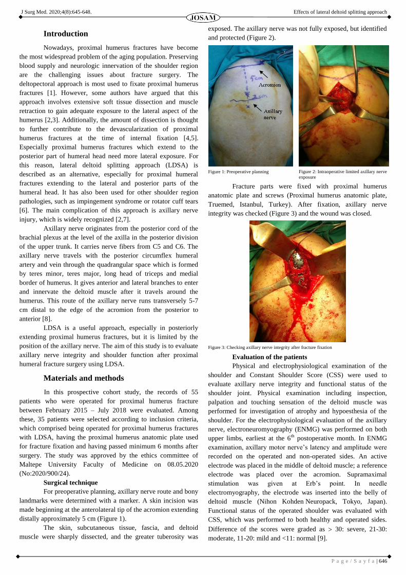
J Surg Med. 2020;4(8):645-648. Effects of lateral deltoid splitting approach
P a g e / S a y f a | 646
Introduction
Nowadays, proximal humerus fractures have become
the most widespread problem of the aging population. Preserving
blood supply and neurologic innervation of the shoulder region
are the challenging issues about fracture surgery. The
deltopectoral approach is most used to fixate proximal humerus
fractures [1]. However, some authors have argued that this
approach involves extensive soft tissue dissection and muscle
retraction to gain adequate exposure to the lateral aspect of the
humerus [2,3]. Additionally, the amount of dissection is thought
to further contribute to the devascularization of proximal
humerus fractures at the time of internal fixation [4,5].
Especially proximal humerus fractures which extend to the
posterior part of humeral head need more lateral exposure. For
this reason, lateral deltoid splitting approach (LDSA) is
described as an alternative, especially for proximal humeral
fractures extending to the lateral and posterior parts of the
humeral head. It has also been used for other shoulder region
pathologies, such as impingement syndrome or rotator cuff tears
[6]. The main complication of this approach is axillary nerve
injury, which is widely recognized [2,7].
Axillary nerve originates from the posterior cord of the
brachial plexus at the level of the axilla in the posterior division
of the upper trunk. It carries nerve fibers from C5 and C6. The
axillary nerve travels with the posterior circumflex humeral
artery and vein through the quadrangular space which is formed
by teres minor, teres major, long head of triceps and medial
border of humerus. It gives anterior and lateral branches to enter
and innervate the deltoid muscle after it travels around the
humerus. This route of the axillary nerve runs transversely 5-7
cm distal to the edge of the acromion from the posterior to
anterior [8].
LDSA is a useful approach, especially in posteriorly
extending proximal humerus fractures, but it is limited by the
position of the axillary nerve. The aim of this study is to evaluate
axillary nerve integrity and shoulder function after proximal
humeral fracture surgery using LDSA.
Materials and methods
In this prospective cohort study, the records of 55
patients who were operated for proximal humerus fracture
between February 2015 – July 2018 were evaluated. Among
these, 35 patients were selected according to inclusion criteria,
which comprised being operated for proximal humerus fractures
with LDSA, having the proximal humerus anatomic plate used
for fracture fixation and having passed minimum 6 months after
surgery. The study was approved by the ethics committee of
Maltepe University Faculty of Medicine on 08.05.2020
(No:2020/900/24).
Surgical technique
For preoperative planning, axillary nerve route and bony
landmarks were determined with a marker. A skin incision was
made beginning at the anterolateral tip of the acromion extending
distally approximately 5 cm (Figure 1).
The skin, subcutaneous tissue, fascia, and deltoid
muscle were sharply dissected, and the greater tuberosity was
exposed. The axillary nerve was not fully exposed, but identified
and protected (Figure 2).
Figure 1: Preoperative planning
Figure 2: Intraoperative limited axillary nerve
exposure
Fracture parts were fixed with proximal humerus
anatomic plate and screws (Proximal humerus anatomic plate,
Truemed, Istanbul, Turkey). After fixation, axillary nerve
integrity was checked (Figure 3) and the wound was closed.
Figure 3: Checking axillary nerve integrity after fracture fixation
Evaluation of the patients
Physical and electrophysiological examination of the
shoulder and Constant Shoulder Score (CSS) were used to
evaluate axillary nerve integrity and functional status of the
shoulder joint. Physical examination including inspection,
palpation and touching sensation of the deltoid muscle was
performed for investigation of atrophy and hypoesthesia of the
shoulder. For the electrophysiological evaluation of the axillary
nerve, electroneuromyography (ENMG) was performed on both
upper limbs, earliest at the 6th
postoperative month. In ENMG
examination, axillary motor nerve’s latency and amplitude were
recorded on the operated and non-operated sides. An active
electrode was placed in the middle of deltoid muscle; a reference
electrode was placed over the acromion. Supramaximal
stimulation was given at Erb’s point. In needle
electromyography, the electrode was inserted into the belly of
deltoid muscle (Nihon Kohden Neuropack, Tokyo, Japan).
Functional status of the operated shoulder was evaluated with
CSS, which was performed to both healthy and operated sides.
Difference of the scores were graded as 30: severe, 21-30:
moderate, 11-20: mild and <11: normal [9].

J Surg Med. 2020;4(8):645-648. Effects of lateral deltoid splitting approach
P a g e / S a y f a | 647
Statistical analysis
SPSS 25.0 statistics program was used for statistical
analysis of results. Frequency analysis was used for demographic
data. Bilateral latency and amplitude values of the axillary nerve
were compared with student-t test. CSS, latency, and amplitude
values of the axillary nerve were compared with correlation
analysis. ANOVA test was used to compare CSS groups and
latency – amplitude values. A P-value <0.05 was considered
statically significant.
Results
Mean age of the patients was 66(9) years. Twenty-five
patients were females and 10 patients were males. Mean follow-
up time was 4(1) years. According Neer classification, 10
patients had surgical neck fractures (group III), 19 patients had a
3-part fracture (13 group IV, 4 group V, and 2 group VI), and 6
patients had a 4-part fracture (4 group IV, 2 group VI) (Table 1).
In physical examination, none of the patients had deltoid atrophy
or hypoesthesia at the lateral side of the shoulder. Mean latency
and amplitude were 4.6(1.8) msn and 6.6(2.21) mV, respectively,
on the operated side and 3.7(0.54) msn and 8.4(2.80) mV on the
non-operated side, the difference between which were
insignificant (latency P=0.25, amplitude P=0.16). There were no
denervation potentials in needle electromyography, neither on
the operated side nor on the non-operated sides (Table 2).
Mean CSS of all patients was 28.7. 12 patients were in
bad condition (mean 39.08), 18 patients were in fair condition
(mean 25.4), and 5 patients were in good condition (mean 16) at
the last control visit. According to the correlation analysis,
axillary nerve latency was prolonged and amplitude was low in
patients with high CSS but there was no statically significant
correlation between CSS and axillary nerve latency / amplitudes
(P=0.62, r=0.267 / P=0.98, r=-0.339).When CSS groups were
compared, there was no statically significant difference between
groups with regards to EMG parameters (P=0.084). Correlation
analysis between CSS and fracture type revealed that there was a
negative correlation between functional scores and comminution
of the fracture which means functional scores were bad or fair
for more fragmented fractures (P=0.032, r=0.829).
Table 1: Demographics of the patients
Demographics n
Mean age (min. – max.) 66.7 (53 - 79)
Gender (W/M) (n) 25 / 10
Mean follow-up (min. – max.) (year) 4.8 (3 – 7)
Side of humerus fracture (R / L) 19 / 16
Neer Classification
Surgical Neck Fracture 10 (group III)
3-part fracture 19 (13 group IV, 4 group V, 2 group VI)
4-part fracture 6 (4 group IV, 2 group VI)
Min: minimum, max: maximum, W: woman, M: men, R: right, L: left
Table 2: The electrophysiological evaluation of axillary nerve
ENMG parameters Operated side Non-operated side P-value
Mean latency (msn) (SD) 4.6 (1.8) 3.7 (0.54) 0.25
Mean amplitude (mV) (SD) 6.6 (2.21) 8.4 (2.8) 0.16
ENMG: Electroneuromyography, msn: millisecond, mV: millivolt, SD: standard deviation
Discussion
The deltopectoral approach is the most used approach in
the shoulder region [10]. However, in some cases, LDSA may be
preferred due to characteristics of the pathology and necessity of
extended approach. The main complication of the LDSA is
iatrogenic axillary nerve injury [11]. In this study, we evaluated
the patients who were operated for proximal humerus fracture
with LDSA with regards to axillary nerve integrity and shoulder
functions. The results revealed that LDSA is a safe method for
axillary nerve injury with careful dissection and fracture fixation.
Additionally, functional scores of the shoulder joint were
dependent to severity of the fracture.
One of the major upper extremity fractures is proximal
humerus fracture and surgical treatment is usually necessary for
comminuted fractures. The deltopectoral approach is well known
and more commonly used for proximal humerus fractures. It has
a relatively low complication rate and enhances exposure for
fracture fixation, but deltopectoral approach may be insufficient
for some comminuted and posterior extended fractures [11].
Some complications were defined for this exposure in these
fractures [4,12]. Extended dissection to reach posterior part of
humeral head may cause more disruption of the integrity of
periosteum. Additionally, reduction of displaced greater
tuberosity may become difficult with the deltopectoral approach.
Deltoid muscle retraction during this approach may cause
dysfunction of the muscle [13].
LDSA can solve this problem and provide more control
on extreme fractures for fixing the posterior part of humeral head
[14]. Especially for posteriorly extending proximal humerus
fractures, LDSA can provide adequate exposure with a smaller
incision than the deltopectoral approach. It affects postoperative
functional outcomes of the shoulder joint [14, 15]. Isiklar et al.
[16] demonstrated that constant scores in patients operated with
LDSA were significantly better than patients who were operated
using the deltopectoral approach at an earlier time. Additionally,
this approach preserves periosteal blood supply by adequate
exposure with limited incision. Despite these benefits, branches
of axillary nerve are in danger due to its proximity to the surgical
field, and iatrogenic axillary nerve injury is the main
complication. Cheung et al. [17] investigated axillary nerve
placement with a cadaver study and revealed that the axillary
nerve lies about 5 cm distal from the mid-acromion. The risky
area for axillary nerve is between 5 cm to 9 cm from mid-
acromion.
Axillary nerve injury and shoulder functions after
fixation of proximal humerus fracture were evaluated in many
studies [15,18]. Khan et al. [15] reported that none of patients
had an axillary nerve injury after lateral deltoid splitting
approach with shoulder strap incision. In their surgical technique,
axillary nerve is visualized and protected during fixation.
Laflamme et al. [18] also reported that axillary nerve injury was
not seen in their case series who were operated due to proximal
humerus fractures with 2 different incisions, using mini open
lateral deltoid splitting approach. They did not visualize the
axillary nerve. In our study, postoperative 6th
month ENMG
results revealed that there were no significant differences
between the operated and non-operated side axillary nerve
functions. None of the patients complained about hypoesthesia at
the lateral side of the shoulder.
Many injuries, especially proximal humerus fractures
affect the functional status of the shoulder joint negatively.
Severity of the fracture is the main predictor of the functional
outcome of the shoulder joint. Robinson et al. [19] pointed that
proximal humerus fractures with tuberosity involvement had
poor functional outcome. Especially proximal humerus fractures
J Surg Med. 2020;4(8):645-648. Effects of lateral deltoid splitting approach
P a g e / S a y f a | 648
with varus angulation have high complication rates [20,21].
Disruption of the medial vascular supply to the humeral head is
stated as the possible cause [22]. Besides, metaphysical
impaction and displacement of medial hinge are strong predictors
of osteonecrosis of the humeral head [20,23]. Fisher et al. [24]
revealed that more complex fracture patterns may be associated
with worse outcomes after proximal humerus fracture surgery. In
this study, a correlation between CSS and fracture type was
determined and lower CSS scores were recorded more frequently
in comminuted fractures.
Due to the spherical anatomical structure of the shoulder
area, it is difficult to control the entire shoulder with a single
surgical approach. Although the deltopectoral approach is a
suitable surgical approach for many shoulder pathologies, it may
be insufficient, especially in surgeries involving the posterior
shoulder area. LDSA provides adequate exposure at the lateral
and posterior parts of the shoulder but may cause axillary nerve
injury due to its proximity. The results of our study, in which we
evaluated the rate of axillary nerve injury and its effect on
shoulder functions in patients undergoing LDSA, show that this
approach can be used safely.
Limitations
Main limitation of this study is the number of the
participants which should be much higher for more accurate
results. Another limitation is that only patients who underwent
LDSA were evaluated and the effects of deltopectoral approach
could not be compared with LDSA, especially for posteriorly
extending proximal humerus fractures.
Conclusions
Clinical and electrophysiological findings of this study
have revealed that LDSA is an effective and safe approach if it is
performed by carefully exploring the axillary nerve. It provides
wide and versatile fracture control without compromising the
deltoid muscle functions and axillary nerve integrity, especially
in fractures extending to the posterior part of the proximal
humerus.
References
1. Buecking B, Mohr J, Bockmann B, Zettl R, Ruchholtz S. Deltoid-split or deltopectoral approaches for
the treatment of displaced proximal humeral fractures? Clin Orthop Relat Res. 2014;472:1576–85.
doi:10.1007/s11999-013-3415-7.
2. Hoppenfeld S. deBoer P. Surgical exposures. Philadelphia: Lippincott, Williams and Wilkins; 2003.
3. Saran N, Bergeron SG, Benoit B, Reindl R, Harvey EJ, Berry GK. Risk of axillary nerve injury during
percutaneous proximal humerus locking plate insertion using an external aiming guide. Injury.
2010;41:1037–40. doi:10.1016/j.injury.2010.04.014.
4. Gardner MJ, Voos JE, Wanich T, Helfet DL, Lorich DG. Vascular implications of minimally invasive
plating of proximal humerus fractures. J Orthop Trauma. 2006;20:602–7.
doi:10.1097/01.bot.0000246412.10176.14.
5. Gerber C, Werner CML, Vienne P. Internal fixation of complex fractures of the proximal humerus. J
Bone Joint Surg - Series B. 2004;86:848–55. doi:10.1302/0301-620X.86B6.14577.
6. Kayaokay K, Mirzazade C, Küçük L, Coşkunol E. Comparison of open acromioplasty outcomes
according to approach type; anterior and lateral. J Surg Med. 2017;1:49–51.
doi:10.28982/josam.351717.
7. Perlmutter GS. Axillary nerve injury. In: Clin Orthop Relat Res. Lippincott Williams and Wilkins;
1999. p. 28–36. doi:10.1097/00003086-199911000-00005.
8. Gurushantappa PK, Kuppasad S. Anatomy of axillary nerve and its clinical importance: a cadaveric
study. J Clin Diagn Res. 2015 Mar;93:13-7.
9. Constant CR, Murley AHG. A clinical method of functional assessment of the shoulder. Clin Orthop
Relat Res. 1987;214:160–4.
10. Westphal T, Woischnik S, Adolf D, Feistner H, Piatek S. Axillary nerve lesions after open reduction
and internal fixation of proximal humeral fractures through an extended lateral deltoid-split approach:
electrophysiological findings. J Shoulder Elb Surg. 2017;26:464–71. doi:10.1016/j.jse.2016.07.027.
11. Singh H, Batra A, Patel D. Lateral transdeltoid approach to proximal humerus fractures. Int Surg J.
2015;2:337–40. doi:10.18203/2349-2902.isj20150400.
12. Robinson CM, Murray IR. The extended deltoid-splitting approach to the proximal humerus:
Variations and extensions. J Bone Joint Surg. 2011;93 B:387–92. doi:10.1302/0301-
620X.93B3.25818.
13. Wu CH, Ma CH, Yeh JJH, Yen CY, Yu SW, Tu YK. Locked plating for proximal humeral fractures:
Differences between the deltopectoral and deltoid-splitting approaches. J Trauma. 2011;71:1364–70.
doi:10.1097/TA.0b013e31820d165d.
14. Gardner MJ, Griffith MH, Dines JS, Briggs SM, Weiland AJ, Lorich DG. The extended anterolateral
acromial approach allows minimally invasive access to the proximal humerus. Clin Orthop Relat Res.
2005;123–9. doi:10.1097/01.blo.0000152872.95806.09.
15. Khan LAK, Robinson CM, Will E, Whittaker R. Assessment of axillary nerve function and functional
outcome after fixation of complex proximal humeral fractures using the extended deltoid-splitting
approach. Injury. 2009;40:181–5. doi:10.1016/j.injury.2008.05.031.
16. Isiklar Z, Kormaz F, Gogus A, Kara A. Comparision of deltopectoral versus lateral deltoid split
approach in operative treatment of proximal humeral fractures. J Bone Joint Surg Br. 2010;92:352.
17. Cheung S, Fitzpatrick M, Lee TQ. Effects of shoulder position on axillary nerve positions during the
split lateral deltoid approach. J Shoulder Elbow Surg. 2009;18:748–55. doi:10.1016/j.jse.2008.12.001.
18. Laflamme GY, Rouleau DM, Berry GK, Beaumont PH, Reindl R, Harvey EJ. Percutaneous humeral
plating of fractures of the proximal humerus: Results of a prospective multicenter clinical trial. J
Orthop Trauma. 2008;22:153–8. doi:10.1097/BOT.0b013e3181694f7d.
19. Robinson CM, Stirling PHC, Goudie EB, Macdonald DJ, Strelzow JA. Complications and Long-Term
Outcomes of Open Reduction and Plate Fixation of Proximal Humeral Fractures. J Bone Joint Surg.
2019;101:2129–39. doi:10.2106/JBJS.19.00595.
20. Solberg BD, Moon CN, Franco DP, Paiement GD. Locked plating of 3- and 4-part proximal humerus
fractures in older patients: The effect of initial fracture pattern on outcome. J Orthop Trauma.
2009;23:113–9. doi:10.1097/BOT.0b013e31819344bf.
21. Jung SW, Shim SB, Kim HM, Lee JH, Lim HS. Factors that influence reduction loss in proximal
humerus fracture surgery. J Orthop Trauma. 2015;29:276–82. doi:10.1097/BOT.0000000000000252.
22. Hardeman F, Bollars P, Donnelly M, Bellemans J, Nijs S. Predictive factors for functional outcome
and failure in angular stable osteosynthesis of the proximal humerus. Injury. 2012;43:153–8.
doi:10.1016/j.injury.2011.04.003.
23. Hertel R, Hempfing A, Stiehler M, Leunig M. Predictors of humeral head ischemia after intracapsular
fracture of the proximal humerus. J Shoulder Elbow Surg. 2004;13:427–33.
doi:10.1016/j.jse.2004.01.034.
24. Fisher ND, Barger JM, Driesman AS, Belayneh R, Konda SR, Egol KA. Fracture severity based on
classification does not predict outcome following proximal humerus fracture. Orthopedics.
2017;40:368–74. doi:10.3928/01477447-20170925-04..
This paper has been checked for language accuracy by JOSAM editors.
The National Library of Medicine (NLM) citation style guide has been used in this paper.

P a g e / S a y f a | 649
Knowledge, attitudes, and practices of orthopedic patients towards
COVID-19 outbreak Ortopedi hastalarının COVID-19 salgınına yönelik bilgi, tutum ve uygulamaları
İbrahim Deniz Canbeyli 1, Meriç Çırpar 1, Caner Baysan 2, Fatma Hayvacı Canbeyli 3, Ali Tepe 1
How to cite/Atıf için: Canbeyli İD, Çırpar M, Baysan C, Canbeyli FH, Tepe A. Knowledge, attitudes, and practices of orthopedic patients towards COVID-19 outbreak. J Surg
Med. 2020;4(8):649-653.
J Surg Med. 2020;4(8):649-653. Research article DOI: 10.28982/josam.777851 Araştırma makalesi
1 Kirikkale University, School of Medicine,
Department of Orthopedics and Traumatology,
Kirikkale, Turkey 2 Ankara University, School of Medicine,
Department of Public Health / Epidemiology,
Ankara, Turkey 3 Kirikkale University, School of Medicine,
Department of Pediatric Cardiology, Kirikkale,
Turkey
ORCID ID of the author(s)
İDC: 0000-0003-3880-4779
MÇ: 0000-0001-9669-6513
CB: 0000-0002-7675-1391
FHC: 0000-0003-0619-075X
AT: 0000-0002-3030-959X
Corresponding author/Sorumlu yazar:
İbrahim Deniz Canbeyli
Address/Adres: Kırıkkale Üniversitesi Yenişehir
Kampüsü, Yahşihan, Kırıkkale, Türkiye
E-mail: [email protected]
�
Ethics Committee Approval: The study was approved
by Kirikkale University Ethics Committee (Date:
5/20/2020, No. 2020.04.08) and performed in the
Department of Orthopedics and Traumatology at
Kirikkale University School of Medicine. All
procedures in this study involving human participants
were performed in accordance with the 1964 Helsinki
Declaration and its later amendments.
Etik Kurul Onayı: Çalışma Kırıkkale Üniversitesi Etik
Kurulu tarafından onaylanmış (Tarih: 20.05.2020, No.
2020.04.08) Kırıkkale Üniversitesi Tıp Fakültesi
Ortopedi ve Travmatoloji Anabilim Dalı'nda
gerçekleştirilmiştir. İnsan katılımcıların katıldığı
çalışmalardaki tüm prosedürler, 1964 Helsinki
Deklarasyonu ve daha sonra yapılan değişiklikler
uyarınca gerçekleştirilmiştir. �
Conflict of Interest: No conflict of interest was
declared by the authors.
Çıkar Çatışması: Yazarlar çıkar çatışması
bildirmemişlerdir.
�
Financial Disclosure: The authors declared that this
study has received no financial support.
Finansal Destek: Yazarlar bu çalışma için finansal
destek almadıklarını beyan etmişlerdir.
�
Published: 8/30/2020
Yayın Tarihi: 30.08.2020
Copyright © 2020 The Author(s)
Published by JOSAM This is an open access article distributed under the terms of the Creative
Commons Attribution-NonCommercial-NoDerivatives License 4.0 (CC
BY-NC-ND 4.0) where it is permissible to download, share, remix,
transform, and buildup the work provided it is properly cited. The work
cannot be used commercially without permission from the journal.
Abstract
Aim: Public education and awareness levels play a prominent role in effective, timely prevention and control of a public health crisis.
We aimed to determine the level and defective sides of knowledge, perceptions, and awareness of the population who were referred to
the outpatient clinic of the Orthopedics and Traumatology on pandemic days.
Methods: In this prospective cross-sectional study, a total of 467 patients who were referred to the orthopedics outpatient clinic between
May 21, 2020 and June 21, 2020 were surveyed. We used the questionnaire which was previously described by Khan et al. A total of
276 volunteer patients aged over 16 years were included. Patients’ knowledge, perceptions, and awareness level regarding COVID-19
were evaluated in terms of spread of the COVID-19 pandemic, prevention, and control policies.
Results: Out of these respondents, 58.3% were males and 41.7% were females. Around 50% of patients were aged less than 45 years,
while 50% were above 45 years. The majority of the participants (question-5 [n=271; 98.2%], question-6 [n=231; 83.7%], question-7
[n=221; 80.1%]) had knowledge about the name, origin, signs and symptoms of COVID-19 infection, although their knowledge about
the spread of coronavirus was relatively low. 225 participants (81.5%) did not receive any form of training or orientation about infection
prevention and control. The mean age those who preferred newspapers and advertisements, friends and family, and other sources (51.09
(17.63) years) was higher than those who preferred social media and the internet (37.85 (16.45) years) (P<0.001).
Conclusion: There is a lack of information about spread routes, hence, protection from COVID-19 in the society. We suggest that health
care providers develop, and release content-checked health education programs that aim to increase the knowledge level among the
people and control COVID-19 spread.
Keywords: COVID-19, Knowledge, Attitudes, Practice, SARS-CoV-2, Coronavirus
Öz
Amaç: Halkın eğitimi ve farkındalık seviyeleri, bir halk sağlığı krizinin etkili, zamanında önlenmesi ve kontrolünde önemli bir rol
oynar. Biz pandemi günlerinde Ortopedi ve Travmatoloji polikliniğine başvuran insanların bilgi, algı ve farkındalık düzeylerini ve
kusurlu taraflarını belirlemeyi amaçladık.
Yöntemler: Bu prospektif kesitsel çalışmada, 21 Mayıs 2020 ile 21 Haziran 2020 tarihleri arasında ortopedi polikliniğine başvuran
toplam 467 hasta incelendi. Çalışmada Khan ve arkadaşlarının tanımladığı anket kullanılmıştır. Çalışmaya 16 yaş üstü toplam 276
gönüllü hasta dahil edildi. Hastaların COVID-19 hakkındaki bilgi, algı ve farkındalık düzeyleri COVID-19 pandemisinin yayılma,
önleme ve kontrol politikalarına göre değerlendirildi.
Bulgular: Katılımcıların %58,3'ü erkek, %41,7'si kadındı. Hastaların toplam %50'si 45 yaş altındayken, %50'si 45 yaş üzerindeydi.
Katılımcıların çoğunluğunun koronavirüsün yayılmasına ilişkin bilgi düzeyleri nispeten düşük olmasına rağmen (soru-5 [n=271; %98,2],
soru-6 [n=231;% 83,7], soru-7 [n=221; %80,1]) isim, köken, COVID-19 enfeksiyonunun belirti ve semptomları hakkında yeterli bilgi
sahibi idi. 225 katılımcı (%81,5) enfeksiyon önleme ve kontrol konusunda herhangi bir eğitim veya yönlendirme almamıştı. Bilgiye
erişimde gazete, reklam, arkadaş, aile ve diğer kaynakları tercih edenlerin yaş ortalaması (51,09 (17,63) yıl), sosyal medya ve interneti
tercih edenlerden (37,85 (16,45) yıl) daha yüksekti (P<0.001).
Sonuç: Toplumda yayılma yolları ve dolayısıyla COVID-19'dan korunma hakkında bilgi eksikliği vardır. Sağlık hizmeti sağlayıcılarının,
insanlar arasında bilgi düzeyini ve dolayısıyla COVID-19'un yayılmasını kontrol altına almayı amaçlayan içerik kontrollü sağlık eğitimi
programları geliştirmelerini ve yayınlamalarını öneriyoruz.
Anahtar kelimeler: COVID-19, Bilgi, Tutumlar, Uygulama, SARS-CoV-2, Koronavirüs

J Surg Med. 2020;4(8):649-653. A quick cross-sectional survey of COVID-19
P a g e / S a y f a | 650
Introduction
The novel coronavirus disease-2019 (COVID‐19, also
known as severe acute respiratory syndrome coronavirus 2
[SARS-CoV-2]) was first reported as unknown cases of systemic
acute respiratory syndrome from Wuhan, Hubei, mainland China
on December 8th
, 2019 [1-4]. COVID‐19, which is thought to
have spread from a seafood market in Wuhan, was declared a
pandemic by the World Health Organization (WHO) after having
been reported in more than 100 countries on March 11th
, 2020
[5].
Turkey was threatened by the COVID-19 pandemic,
like the rest of the world. Strict measures were taken to prevent
this pandemic, so it did not lead to a national health crisis. The
Republic of Turkey, Ministry of Health declared a guideline for
infection control, which was based on science board
recommendations, after the first case was seen on March 11th
,
2020. According to this guideline, only emergency patients,
patients with severe comorbid disease, oncological diseases, and
those in the postoperative follow-up period were able to present
to the outpatient clinics to prevent transmission of the
coronavirus among patients with comorbid diseases [6].
Moreover, patients who presented to the hospital were checked
in terms of signs and symptoms of COVID-19. Thus, the number
of patients who were referred to the outpatient clinics decreased.
However, the orthopedic outpatient clinic was the most crowded
one in those days, especially due to the high population of
elderly patients with degenerative musculoskeletal diseases and
those in the postoperative follow-up period. In addition, the
pediatric patient population also raised as the schools were
closed and the children became more active in daily life.
The implementation of basic infection control protocols
is only possible when individuals are aware of public health
policies. Public education and awareness levels play a prominent
role in effective, timely prevention and control of a public health
crisis. In addition, the assessment of knowledge and perceptions
of populations about rapidly spreading infectious disease
outbreaks, such as COVID-19, should be accomplished in a brief
period to provide information to public health institutions.
In the present study, we aimed to determine the level
and defective sides of knowledge, perceptions, and awareness of
the population who visited the Orthopedics and Traumatology
outpatient clinic regarding COVID-19, and provide data to guide
health care providers and hospital managers in informing the
patients about COVID-19 and probable future pandemics.
Materials and methods
This study was approved by the Kırıkkale University
Ethics Committee (Date: 5/20/2020, No. 2020.04.08). A written
informed consent was obtained from each patient. The study was
conducted in accordance with the principles of the Declaration of
Helsinki.
In this prospective cross-sectional study, a total of 467
patients who visited the orthopedics outpatient clinic between
May 21, 2020 and June 21, 2020 were surveyed. A total of 276
volunteers aged over 16 years were included. The sample size
was calculated with the formula which was previously used by
Zhong et al. [7] as follows: Sample size= (Zα2 * (P) * (1-P)) /
C2. Z value for 99% confidence interval =2.576, P=0.900,
C=0.05). It was determined that at least 239 individuals had to be
recruited to the study for 99% confidence interval, 5% margin of
error, and 90.0% prevalence. Patients under 16 years of age and
those who did not volunteer to participate were excluded.
Patients’ knowledge, perceptions, and awareness levels on
COVID-19 were evaluated in terms of COVID-19 spread,
prevention, and control policies. We used the questionnaire
which was previously described by Khan et al. [8]. The
questionnaire was translated into Turkish by the authors (Table
1). It includes demographic features and 21 different questions to
measure patients’ basic knowledge of infection, their attitudes,
standard practices during the infection outbreak, control
programs and policies, awareness of origin, transmission, and
signs and symptoms.
Table 1: Questionnaire on COVID-19
Questionnaire-based analysis of infection prevention and control in Turkey regarding Coronavirus
* Required
Q1 Age *
Q2
Gender *
Male
Female
Other….
Q3 Profession *
Q4 Organization *
Government Hospital
Private Hospital
Educational Institutes
Q5 Do you know the name of Coronavirus? *
Yes
No
Maybe
Q6 Do you know the origin of Coronavirus? *
Yes No Maybe
Q7 Are you aware of common signs and symptoms of Coronavirus? *
Yes No Maybe
Q8 Does having fever, flue, and cough mean that you are infected? *
Yes No Maybe
Q9 Can you get Coronavirus from a parcel coming from china? *
Yes No Maybe
Q10 Can you get it from pets? *
Yes No Maybe
Q11 Do you have knowledge of other pandemic viral infections in the past? *
Yes No Maybe
Q12 Can we stop the viral spread in Turkey? *
Yes No Maybe
Q13 Is there an infection control program in the hospital you are applying for? *
Yes No Maybe
Q14 Does your country have any infection control policies and guidelines? *
Yes No Maybe
Q15 Have you received some form of training or orientation about infection prevention and control? *
Yes No Maybe
Q16 Does your organization have an emerging infectious disease task force (dealing with outbreaks)? *
Yes No Maybe
Q17 Do you think that all residents in your city are promptly following infection control policies and
rules? *
Yes No Maybe
Q18 Do you think your country is prepared for any infection outbreak? *
Yes No Maybe
Q19
Is Coronavirus curable? *
Yes No Maybe
Q20 Do you think that going to school, hospital or any organization is safe? *
Yes No Maybe
Q21
Where can you get further information about Coronavirus? *
Internet
Social media
Newspapers and advertisements
Friends and family
Others …………………..
Q: Question
Statistical analysis
Statistical analysis was performed with the Statistical
Package for the Social Sciences (SPSS) for Windows version
25.0 (IBM Corp., Armonk, NY, USA). Descriptive statistics
were presented in mean (standard deviation (SD)), median (min-
max), or number and frequency. The variables were examined
using visual (histogram and probability plots) and analytical
methods (Shapiro-Wilk test) to determine whether they were
normally distributed. The Kruskal-Wallis test was conducted to
compare the non-normally distributed parameters and three and
more independent variables. The chi-square automatic
interaction detector (CHAID) analysis was performed to identify
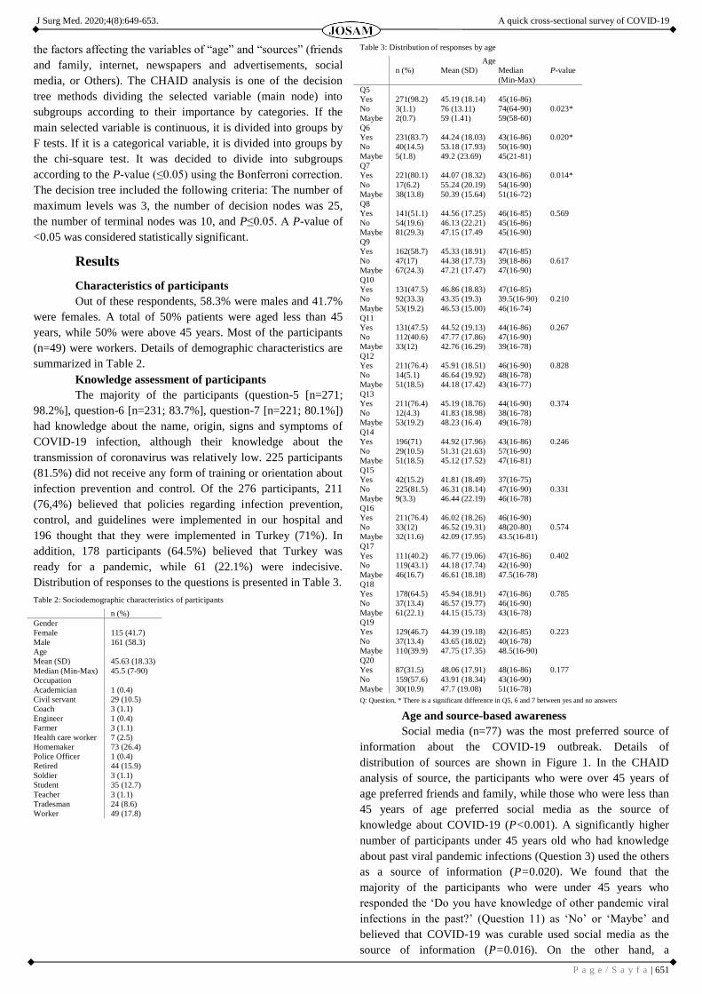
J Surg Med. 2020;4(8):649-653. A quick cross-sectional survey of COVID-19
P a g e / S a y f a | 651
the factors affecting the variables of “age” and “sources” (friends
and family, internet, newspapers and advertisements, social
media, or Others). The CHAID analysis is one of the decision
tree methods dividing the selected variable (main node) into
subgroups according to their importance by categories. If the
main selected variable is continuous, it is divided into groups by
F tests. If it is a categorical variable, it is divided into groups by
the chi-square test. It was decided to divide into subgroups
according to the P-value (≤0.05) using the Bonferroni correction.
The decision tree included the following criteria: The number of
maximum levels was 3, the number of decision nodes was 25,
the number of terminal nodes was 10, and P≤0.05. A P-value of
<0.05 was considered statistically significant.
Results
Characteristics of participants
Out of these respondents, 58.3% were males and 41.7%
were females. A total of 50% patients were aged less than 45
years, while 50% were above 45 years. Most of the participants
(n=49) were workers. Details of demographic characteristics are
summarized in Table 2.
Knowledge assessment of participants
The majority of the participants (question-5 [n=271;
98.2%], question-6 [n=231; 83.7%], question-7 [n=221; 80.1%])
had knowledge about the name, origin, signs and symptoms of
COVID-19 infection, although their knowledge about the
transmission of coronavirus was relatively low. 225 participants
(81.5%) did not receive any form of training or orientation about
infection prevention and control. Of the 276 participants, 211
(76,4%) believed that policies regarding infection prevention,
control, and guidelines were implemented in our hospital and
196 thought that they were implemented in Turkey (71%). In
addition, 178 participants (64.5%) believed that Turkey was
ready for a pandemic, while 61 (22.1%) were indecisive.
Distribution of responses to the questions is presented in Table 3.
Table 2: Sociodemographic characteristics of participants
n (%)
Gender
Female
Male
115 (41.7)
161 (58.3)
Age
Mean (SD)
Median (Min-Max)
45.63 (18.33)
45.5 (7-90)
Occupation
Academician
Civil servant
Coach
Engineer
Farmer
Health care worker
Homemaker
Police Officer
Retired
Soldier
Student
Teacher
Tradesman
Worker
1 (0.4)
29 (10.5)
3 (1.1)
1 (0.4)
3 (1.1)
7 (2.5)
73 (26.4)
1 (0.4)
44 (15.9)
3 (1.1)
35 (12.7)
3 (1.1)
24 (8.6)
49 (17.8)
Table 3: Distribution of responses by age
Age
n (%) Mean (SD) Median
(Min-Max)
P-value
Q5
Yes
No
Maybe
271(98.2)
3(1.1)
2(0.7)
45.19 (18.14)
76 (13.11)
59 (1.41)
45(16-86)
74(64-90)
59(58-60)
0.023*
Q6
Yes
No
Maybe
231(83.7)
40(14.5)
5(1.8)
44.24 (18.03)
53.18 (17.93)
49.2 (23.69)
43(16-86)
50(16-90)
45(21-81)
0.020*
Q7
Yes
No
Maybe
221(80.1)
17(6.2)
38(13.8)
44.07 (18.32)
55.24 (20.19)
50.39 (15.64)
43(16-86)
54(16-90)
51(16-72)
0.014*
Q8
Yes
No
Maybe
141(51.1)
54(19.6)
81(29.3)
44.56 (17.25)
46.13 (22.21)
47.15 (17.49
46(16-85)
45(16-86)
45(16-90)
0.569
Q9
Yes
No
Maybe
162(58.7)
47(17)
67(24.3)
45.33 (18.91)
44.38 (17.73)
47.21 (17.47)
47(16-85)
39(18-86)
47(16-90)
0.617
Q10
Yes
No
Maybe
131(47.5)
92(33.3)
53(19.2)
46.86 (18.83)
43.35 (19.3)
46.53 (15.00)
47(16-85)
39.5(16-90)
46(16-74)
0.210
Q11
Yes
No
Maybe
131(47.5)
112(40.6)
33(12)
44.52 (19.13)
47.77 (17.86)
42.76 (16.29)
44(16-86)
47(16-90)
39(16-78)
0.267
Q12
Yes
No
Maybe
211(76.4)
14(5.1)
51(18.5)
45.91 (18.51)
46.64 (19.92)
44.18 (17.42)
46(16-90)
48(16-78)
43(16-77)
0.828
Q13
Yes
No
Maybe
211(76.4)
12(4.3)
53(19.2)
45.19 (18.76)
41.83 (18.98)
48.23 (16.4)
44(16-90)
38(16-78)
49(16-78)
0.374
Q14
Yes
No
Maybe
196(71)
29(10.5)
51(18.5)
44.92 (17.96)
51.31 (21.63)
45.12 (17.52)
43(16-86)
57(16-90)
47(16-81)
0.246
Q15
Yes
No
Maybe
42(15.2)
225(81.5)
9(3.3)
41.81 (18.49)
46.31 (18.14)
46.44 (22.19)
37(16-75)
47(16-90)
46(16-78)
0.331
Q16
Yes
No
Maybe
211(76.4)
33(12)
32(11.6)
46.02 (18.26)
46.52 (19.31)
42.09 (17.95)
46(16-90)
48(20-80)
43.5(16-81)
0.574
Q17
Yes
No
Maybe
111(40.2)
119(43.1)
46(16.7)
46.77 (19.06)
44.18 (17.74)
46.61 (18.18)
47(16-86)
42(16-90)
47.5(16-78)
0.402
Q18
Yes
No
Maybe
178(64.5)
37(13.4)
61(22.1)
45.94 (18.91)
46.57 (19.77)
44.15 (15.73)
47(16-86)
46(16-90)
43(16-78)
0.785
Q19
Yes
No
Maybe
129(46.7)
37(13.4)
110(39.9)
44.39 (19.18)
43.65 (18.02)
47.75 (17.35)
42(16-85)
40(16-78)
48.5(16-90)
0.223
Q20
Yes
No
Maybe
87(31.5)
159(57.6)
30(10.9)
48.06 (17.91)
43.91 (18.34)
47.7 (19.08)
48(16-86)
43(16-90)
51(16-78)
0.177
Q: Question, * There is a significant difference in Q5, 6 and 7 between yes and no answers
Age and source-based awareness
Social media (n=77) was the most preferred source of
information about the COVID-19 outbreak. Details of
distribution of sources are shown in Figure 1. In the CHAID
analysis of source, the participants who were over 45 years of
age preferred friends and family, while those who were less than
45 years of age preferred social media as the source of
knowledge about COVID-19 (P<0.001). A significantly higher
number of participants under 45 years old who had knowledge
about past viral pandemic infections (Question 3) used the others
as a source of information (P=0.020). We found that the
majority of the participants who were under 45 years who
responded the ‘Do you have knowledge of other pandemic viral
infections in the past?’ (Question 11) as ‘No’ or ‘Maybe’ and
believed that COVID-19 was curable used social media as the
source of information (P=0.016). On the other hand, a

J Surg Med. 2020;4(8):649-653. A quick cross-sectional survey of COVID-19
P a g e / S a y f a | 652
significantly higher number of participants who were aware of
the previous viral outbreaks and considered that all cough and
fever symptoms were related to COVID-19 infection (Question
8) used social media as the source (P=0.023). Details of CHAID
analysis of source are shown in Figure 2.
The mean age those who preferred newspapers and
advertisements, friends and family, and other sources (51.09
(17.63) years) was higher than those who preferred social media
and the internet (37.85 (16.45) years) (P<0.001). The mean age
of those who did not know the name of coronavirus (76 (13.11)
years) was significantly higher than those who knew its name
(45.19 (18.14) years; P=0.023). Moreover, the mean age of those
who did not know the origin of coronavirus (Question 6) (53.18
(17.93) years) was significantly higher than those who did (44.24
(18.03) years; P=0.020). The mean age of those who recognized
the common signs and symptoms of COVID-19 (44.07 (18.32)
years) was significantly less than those who did not know (55.24
(20.19) years; P=0.014). However, there was no significant
difference between the mean ages of those who knew or did not
know the common signs and symptoms of COVID-19
(P=0.569). There was no significant difference in terms of mean
age between those who answered “Yes”, “No” or “Maybe” to
other questions. Details of distribution of age-based responses
are summarized in Table 3. In CHAID analysis, the mean age of
those who use social media and internet as a source of
information about COVID-19 was significantly less than those
who used the newspapers and advertisements, friends and family,
and others (P<0.001). A significantly higher number (n=131) of
those who used the newspapers and advertisements, friends and
family, and others as sources had knowledge about the origin of
COVID-19 (P=0.007). Among these 131 individuals, the mean
age of women (52.40 (18.33) years) who knew the origin of
COVID-19 was significantly higher than that of men (45.67
(16.38) years) (P=0.029). Details of CHAID analysis of age are
shown in Figure 3.
Figure 3: Aged-based decision tree of CHAID analysis
Figure 2: Source-based decision tree of CHAID analysis

J Surg Med. 2020;4(8):649-653. A quick cross-sectional survey of COVID-19
P a g e / S a y f a | 653
Discussion
Turkey is one of the largest countries between the
Middle East and Europe with more than 80 million citizens. To
the best of our knowledge, this is the first study in the Turkish
population examining the knowledge, attitudes, and practices
toward COVID-19 among patients.
The implementation of basic infection control protocols
is possible when individuals are aware of public health policies.
Moreover, public education and awareness levels play a vital role
in timely prevention and control of a public health crisis such as
COVID-19. The most attractive finding of our study was the lack
of knowledge of the participants about the spread of coronavirus.
It was well documented by WHO [9] that the disease spreads
primarily from human to human through small droplets from the
nose or mouth, which are expelled when a person with COVID-
19 coughs, sneezes, or speaks. People can catch COVID-19 if
they breathe in these droplets from a person infected with the
virus. It is important to stay at least 1 meter away from others.
These droplets can land on objects and surfaces around the
person such as tables, doorknobs, and handrails [9]. People can
become infected by touching these objects or surfaces, then
touching their eyes, nose, or mouth. Thus, it was highly
recommended to wash your hands regularly with soap and water
or clean with alcohol-based hand rubs. The lack of information
about the transmission routes of coronavirus may play a
significant role in the spread of COVID-19. People may
misbehave with regards to individual protection from COVID-
19. Incorrect usage of the masks and gloves, insufficient
handwashing, and application errors of social distance in
business life and in the specific events to our country, such as
weddings and enlistment ceremonies, increase the transmission
of the virus around substantial number of citizens. Thus, the
effectiveness of common public education programs plays the
key role for protection in epidemics.
Our study also revealed that, 81.5% of the participants
did not receive any form of training or orientation about infection
prevention and control. Zhong et al. [7] stated that health
education programs aimed at improving COVID-19 knowledge
are helpful for maintaining safe practices. In another study,
Abdelhafiz et al. [10] reported that vaccine development is
estimated to require months, and thus management of the
COVID-19 outbreak depends primarily on people’s knowledge,
attitudes, and practices. Practicing hand and respiratory hygiene
is important at outbreaks such as SARS and COVID-19 and is
the best way to protect oneself. It was also underlined by WHO
that since some infected individuals may be asymptomatic or
their symptoms may be mild, maintaining at least a 1-meter
distance with everyone is needed if you are in an area where
COVID-19 is circulating, and particularly important when you
are standing by someone who is coughing or sneezing [9].
Therefore, we believe that in preventing the spread of epidemic
diseases, the public education programs must include detailed
data about transmission routes of the agent, protection methods
and public and individual protection policies.
In the current study, patients had a vast knowledge of
name, origin, signs, and symptoms of COVID-19, and optimistic
attitudes toward SARS-CoV-2. In a pandemic, people should not
be pessimistic, however, ignorant optimism may lead to
inadequate implementation of protective measures, which can
lead to an out-of-control increase of infected individuals. Even if
the ideal training program is implemented, indifference due to
optimism can cause a deficiency in the purpose of education
programs.
In our study, social media and internet were the most
preferred sources of information, instead of more traditional
media platforms; namely, newspapers and advertisements for
patients under 45 years of age, while friends and family were the
most popular sources for patients over 45 years of age. A
concern in this regard is the spread of dis- and misinformation
about COVID-19 on social media. WHO Director General
Tedros Adhanom Ghebreyesus stated that a global epidemic of
misinformation spreading rapidly through social media platforms
and other outlets pose a severe problem for public health [11].
Since social media can include unverified information, it may be
a source of false information. We believe that in such a public
health crisis, both the ministry of health and non-governmental
health organizations should work together to reveal the most
accurate information and deliver it to all parts of the population
using all informing platforms such as the social media, visual,
and printed media, while keeping content under control.
Limitations
The main limitation of the present study includes the
methodological limitations of the survey. There may be some
reporting bias. Data were self-reported and data from non-
respondents could not be obtained. In addition, the questionnaire
was not pilot tested, which would have increased its reliability.
Finally, this study included only those living in a single city;
thus, further large-scale, and multi-center studies are needed.
Conclusion
There is a lack of information about spread routes and
hence protection from COVID-19 in the society due to the
limited ways of accurately informing large populations.
Therefore, the content-checked health education programs such
as printed brochures and advertisements, public information
videos on TVs and social media that aim to increase knowledge
level among the people and control the spread of COVID-19
should be developed and released in cooperation with the
ministry of health and non-governmental health organizations.
References
1. Deng SQ, Peng HJ. Characteristics of and Public Health Responses to the Coronavirus Disease 2019
Outbreak in China. J Clin Med. 2020;9.
2. Shereen MA, Khan S, Kazmi A, Bashir N, Siddique R. COVID-19 infection: Origin, transmission, and
characteristics of human coronaviruses. J Adv Res. 2020;24:91-8.
3. Palacios Cruz M, Santos E, Velazquez Cervantes MA, Leon Juarez M. COVID-19, a worldwide public
health emergency. Rev Clin Esp. 2020.
4. Giwa AL, Desai A, Duca A. Novel 2019 coronavirus SARS-CoV-2 (COVID-19): An updated
overview for emergency clinicians. Emerg Med Pract. 2020;22:1-28.
5. WHO. WHO health topics 2020. https://www.euro.who.int/en/health-topics/health-
emergencies/coronavirus-covid-19/novel-coronavirus-2019-ncov
6. Cakmak G, Ceyhan E, Demirtas Y, Berk H. The management of orthopedics and traumatology
patients during SARS-CoV-2 pandemic. Acta Orthop Traumatol Turc. 2020;54:223-7.
7. Zhong BL, Luo W, Li HM, Zhang QQ, Liu XG, Li WT, et al. Knowledge, attitudes, and practices
towards COVID-19 among Chinese residents during the rapid rise period of the COVID-19 outbreak:
a quick online cross-sectional survey. Int J Biol Sci. 2020;16:1745-52.
8. Khan S, Khan M, Maqsood K, Hussain T, Noor Ul H, Zeeshan M. Is Pakistan prepared for the
COVID-19 epidemic? A questionnaire-based survey. J Med Virol. 2020.
9. WHO. How does COVID-19 spread? https://www.who.int/emergencies/diseases/novel-coronavirus-
2019/question-and-answers-hub/q-a-detail/q-a-coronaviruses. 2020.
10.Abdelhafiz AS, Mohammed Z, Ibrahim ME, Ziady HH, Alorabi M, Ayyad M, et al. Knowledge,
Perceptions, and Attitude of Egyptians Towards the Novel Coronavirus Disease (COVID-19). J
Community Health. 2020.
11.Zarocostas J. How to fight an infodemic. The Lancet. 2020 Feb;395(10225):676.
This paper has been checked for language accuracy by JOSAM editors.
The National Library of Medicine (NLM) citation style guide has been used in this paper.

P a g e / S a y f a | 654
The value of adenosine deaminase level in assessing activation of
inflammatory bowel disease İnflamatuar bağırsak hastalığının aktivasyonunun değerlendirilmesinde adenosin deaminaz düzeyinin kullanımı
Yasemin Gökden 1, Semih Hot 2, Can Gönen 3, Semih Kalyon 4, Züleyha Akkan Çetinkaya 5
How to cite/Atıf için: Gökden Y, Hot S, Gönen C, Kalyon S, Çetinkaya ZA. The value of adenosine deaminase level in assessing activation of inflammatory bowel disease. J
Surg Med. 2020;4(8):654-659.
J Surg Med. 2020;4(8):654-659. Research article DOI: 10.28982/josam.769877 Araştırma makalesi
1 Health Sciences University, Prof. Dr. Cemil
Taşçıoğlu City Hospital, Department of
Gastroenterology, Istanbul, Turkey 2 Health Sciences University, Prof. Dr. Cemil
Taşçıoğlu City Hospital, Department of General
Surgery, Istanbul, Turkey 3 Acıbadem Mehmet Ali Aydınlar University,
Department of Gastroenterology, Istanbul, Turkey 4 Health Sciences University, Prof. Dr. Cemil
Taşçıoğlu City Hospital, Department of Internal
Medicine, Istanbul, Turkey 5 İstanbul Gelişim University Memorial Ataşehir
Hospital, Department of Gastroenterology,
Istanbul, Turkey
ORCID ID of the author(s)
YG: 0000-0001-6767-3072
SH: 0000-0001-9623-356X
CG: 0000-0002-8882-9272
SK: 0000-0003-4207-0800
ZA: 0000-0003-3831-1819
Corresponding author/Sorumlu yazar:
Semih Hot
Address/Adres: Sağlık Bilimleri Üniversitesi İstanbul
Cemil Taşçıoğlu Şehir Hastanesi Genel Cerrahi
Kliniği Şişli, İstanbul, Türkiye
E-mail: [email protected]
󠄀
Ethics Committee Approval: This study was approved
by the Ethics Committee of Health Science University
İstanbul Haydarpaşa Training and Research Hospital
(HNEAH-KAEK 2013/KK/62). All procedures in this
study involving human participants were performed in
accordance with the 1964 Helsinki Declaration and its
later amendments.
Etik Kurul Onayı: Bu çalışma, Sağlık Bilimleri
Üniversitesi İstanbul Haydarpaşa Eğitim ve Araştırma
Hastanesi Etik Kurulu tarafından onaylanmıştır
(HNEAH-KAEK 2013/KK/62). İnsan katılımcıların
katıldığı çalışmalardaki tüm prosedürler, 1964
Helsinki Deklarasyonu ve daha sonra yapılan
değişiklikler uyarınca gerçekleştirilmiştir. 󠄀
Conflict of Interest: No conflict of interest was
declared by the authors.
Çıkar Çatışması: Yazarlar çıkar çatışması
bildirmemişlerdir.
󠄀
Financial Disclosure: The authors declared that this
study has received no financial support.
Finansal Destek: Yazarlar bu çalışma için finansal
destek almadıklarını beyan etmişlerdir.
󠄀
Published: 8/30/2020
Yayın Tarihi: 30.08.2020
Copyright © 2020 The Author(s)
Published by JOSAM This is an open access article distributed under the terms of the Creative
Commons Attribution-NonCommercial-NoDerivatives License 4.0 (CC
BY-NC-ND 4.0) where it is permissible to download, share, remix,
transform, and buildup the work provided it is properly cited. The work
cannot be used commercially without permission from the journal.
Abstract
Aim: There is still a need for an ideal laboratory test that can determine the type of disease, the degree of its activity, predict its course,
and monitor treatment response in patients with inflammatory bowel disease (IBD). This study aims to investigate the relationship
between disease types and activity with Adenosine deaminase (ADA) levels in patients with IBD.
Methods: A total of 92 patients with IBD [43 with Crohn’s disease (CD) and 49 with ulcerative colitis (UC)] and 31 healthy control
(HC) volunteers were included in this case-control study. Patients’ age, gender, body mass index, location and severity of the disease,
medication, endoscopic examination, hemogram, C-reactive Protein (CRP), and ADA results were evaluated.
Results: The mean ADA level was 24.87 (9.6 - 74.9) IU/L in the IBD group and 20.8 (13.7 - 38.9) IU/L in the HC group. The difference
between the IBD and HC groups was statistically significant (P<0.013), while that between UC and CD groups was not (P=0.76). Mean
ADA level was significantly higher in active UC patients than in inactive ones (P<0.001). To distinguish active UC patients from those
in remission, a cut-off level of 21.64 U/L was determined for ADA with 77.6% confidence interval, 89% sensitivity and 60% specificity.
Mean ADA level was significantly higher in the CD group compared to the HC group.
Conclusion: ADA level may be used as an alternative marker to distinguish active UC patients from those in remission, regardless of the
disease location and the extent of the affected area.
Keywords: Ulcerative colitis, Crohn’s disease, Adenosine deaminase
Öz
Amaç: Günümüzde, inflamatuar barsak hastalığı (İBH) hastalarda hastalığın türünü, aktivitesinin derecesini belirleyebilen, seyrini
tahmin edebilen ve tedavi yanıtını izleyebilen ideal bir laboratuvar testine hala ihtiyaç vardır. Bu çalışmada İBH olan hastalarda hastalık
aktivitesi ile adenozin deaminaz ADA düzeyleri arasındaki ilişki araştırıldı.
Yöntemler: Bu vaka-kontrol çalışmasına toplam olarak 92 IBH hastası [43 Crohn hastalığı (CD) ve 49 ülseratif kolit (UC)] ve 31
sağlıklı kontrol (HC) gönüllüsü dahil edildi. Hastaların yaşı, cinsiyeti, vücut kitle indeksi, hastalığın yeri ve şiddeti, ilaç tedavisi,
endoskopik muayene, hemogram, C-reaktif Protein (CRP) ve ADA sonuçları retrospektif olarak değerlendirildi.
Bulgular: Ortalama ADA seviyesi IBH grubunda 24,87 (9,6 - 74,9) IU/L ve HC grubunda 20,8 (13,7 - 38,9) IU/L idi. IBH ve HC grubu
arasındaki fark istatistiksel olarak anlamlıydı (P<0,013), ancak UC ve CD grubu arasındaki fark istatistiksel olarak anlamlı değildi
(P=0,76). Ortalama ADA düzeyi aktif UC hastalarında inaktif olanlardan anlamlı olarak yüksekti (P<0,001). Aktif UC hastalarını
remisyonda UC hastalarından ayırt etmek için, eğer ADA düzeyinin eşik değeri 77,6 güven aralığında 21,64 U/L olarak kabul edilirse,
duyarlılık %89 ve özgüllük %60 olarak hesaplandı. Ortalama ADA düzeyi CD grubunda HC grubuna göre anlamlı derecede yüksekti.
Sonuç: ADA seviyesi, hastalığın bulunduğu yere ve etkilenen alanın boyutuna bakılmaksızın, aktif UC hastalarını remisyondaki UC
hastalarından ayırmak için alternatif bir belirteç olarak kullanılabilir.
Anahtar kelimeler: Ülseratif kolit, Crohn hastalığı, Adenozin deaminaz

J Surg Med. 2020;4(8):654-659. Adenosine deaminase in inflammatory bowel disease
P a g e / S a y f a | 655
Introduction
Inflammatory bowel diseases (IBD) are chronic
inflammatory pathologies without any curative medical
treatment, which develop due to recurrent immune system
activation and inflammation, and course with gastrointestinal
tract remission and exacerbations. Crohn’s disease (CD) and
ulcerative colitis (UC) are two types of IBD with similar
epidemiology, etiology, and clinical features [1-3]. Many clinical
activity predictors and noninvasive markers have been used to
identify disease activity and plan treatment for patients with IBD.
However, none of them has yet provided a definitive finding in
detecting inflammatory activity as much as histopathological and
endoscopic examinations [4]. An ideal laboratory test that would
be able to identify the type of disease, determine the degree of
disease activity, predict the course of the disease, and monitor
the treatment response in patients with IBD would be beneficial
for physicians. However, there is no laboratory marker with
sufficient sensitivity and specificity to provide this convenience
alone.
Adenosine deaminase (ADA) enzyme plays a role in the
catabolism of purine nucleotides and catabolizes the conversion
of adenosine and deoxyadenosine to inosine and deoxycinosine
irreversibly [5]. It is widely available in body fluids and tissues.
The most important biological activity of ADA is seen in
lymphoid tissues: It plays an essential role, especially in the
differentiation and proliferation of T lymphocytes [6]. ADA
level is ten times higher in lymphocytic cells than in
erythrocytes, and more common in T lymphocytes than B
lymphocytes [7]. It is considered a non-specific marker of T cell
activation and cellular immunity. ADA level increases in
autoimmune diseases and inflammation and is used as an
indicator of cellular immunity for tuberculosis, rheumatoid
arthritis, Graves' disease, celiac disease, systemic lupus
erythematosus, acute pancreatitis, acute appendicitis, and other
infectious diseases [8-14]. However, studies regarding the level
of ADA as an effective biomarker in the evaluation of intestinal
inflammation and disease activity are ongoing [15].
The aim of this study is to investigate the relationship
between ADA levels and diagnosis, as well as clinical and
endoscopic activity of the disease in patients with UC and CD.
Materials and methods
A case-control study was conducted on patients with
IBD followed in the gastroenterology clinic. All patients were
definitively diagnosed with IBD by clinical, endoscopic,
histopathologic, and radiologic examinations. Patients with
abdominal abscess, intestinal obstruction, active gastrointestinal
bleeding, chronic liver disease, other active infections,
tuberculosis, malignant disease, chronic kidney failure,
pregnancy, and those younger than 18 years of age were not
included in the study. Thirty-one volunteer participants with
similar characteristics in terms of age, gender, and body mass
index included to constitute the healthy control (HC) group.
These participants were selected from individuals who visited
our hospital for routine health checks and without a history of
any disease, malignancy, or chronic drug use.
The patients' ages, genders, body mass indexes,
findings, durations, and severity of the disease, affected area of
the intestine, medications, and previous operations were
recorded. Endoscopic examination and laboratory blood tests of
the patients were performed on the same day. The endoscopic
examination of the patients was carried out by
gastroenterologists with at least five years of experience.
Smoking and alcohol use habits of the patients were recorded in
detail.
In patients with UC, Truelove-Witts [16] and clinical
colitis activity index (CCAI) criteria [17] were used to calculate
clinical severity. Endoscopic Mayo Score [18] was used to
categorize endoscopic severity. UC patients with CCAI index
scores ≤4 were considered clinically inactive. Bloodless
defecation 1-2 times a day, normal hemoglobin, and erythrocyte
sedimentation rate, as well as the absence of fever and
tachycardia, are characteristics of inactive disease according to
the Truelove-Witts score.
UC patients with Mayo Scores between 0 and 1
considered to be in endoscopic remission. CD activity index
(CDAI) [19] and a simple endoscopic score (SES-CD) [20] were
used for patients with CD. CD patients with a CDAI value <150
were considered to have clinically inactive disease. Clinically
active CD patients were divided into three subgroups as mildly
active (CDAI was between 150-220), moderately active (CDAI
was between 221-450), and severely active (CDAI> 450).
Patients were also evaluated endoscopically according to SES-
CD, and scores between 0-2 were considered as endoscopically
inactive, and scores above 2 were considered as endoscopically
active disease.
ADA level was determined in all patients for
biochemical evaluation of disease activity. For laboratory tests,
venous blood was collected from the patients and control groups
after 10-12 hours of fasting (to eliminate possible lipemia) into
tubes containing EDTA, sodium citrate, and gel (Becton
Dickinson, USA). The gel tubes were centrifuged for 10 minutes
in 3500 RPM (1300g) after resting for 30 minutes. Whole blood
count (EDTA blood samples) and erythrocyte sedimentation
rates (ESR) (performed using the Westergreen method from
sodium citrate blood sample) were measured with the Sed Rate
Screener 100 (SRS 100, Greiner Bio-one GmbH, Austria)
device. CRP was measured from serum samples with the
nephelometric method (Immage, Beckmann Coulter, USA).
The serum samples were kept at -70ºC for measuring
ADA levels later, and frozen samples were thawed and studied
just before the analysis. Repeated freezing and thawing were
avoided. ADA levels of all patients were measured using the
Enzymatic-spectrophotometric method. Total ADA levels were
evaluated by the Giusti and Galanti method [21]. Samples
incubated with adenosine and free ammonium ions were
measured. The concentration of ammonium ions formed in 1
minute was expressed in U/L and defined as the ADA level.
Ethical considerations
This study was approved by the Ethics Committee of
Health Science University İstanbul Haydarpaşa Training and
Research Hospital (HNEAH-KAEK 2013/KK/62) and carried
out according to the principles of the Helsinki Declaration.

J Surg Med. 2020;4(8):654-659. Adenosine deaminase in inflammatory bowel disease
P a g e / S a y f a | 656
Written informed consent was obtained from all patients and
volunteer participants.
Statistical analysis
Nonparametric tests were used to evaluate non-normally
distributed study data. Descriptive statistical methods (median)
were used, and the nonparametric Kruskal Wallis test was
utilized to compare quantitative data between groups. Mann
Whitney U test was used to identify groups with differences. The
mean rank values were considered in determining the source of
group differences. Nonparametric Kendall's Taub relation test
was made use of for the correlation analysis of parameters.
Power analysis was performed using the computer-aided
statistics program G-power. According to previous studies
[22,23] in the literature, the smallest sample size to represent the
population with 95% strength was calculated as 36 participants
with 1.25 effect size and 5% alpha error margin. IBM SPSS
(Statistical Package for Social Sciences) Statistics 20 program
was used for statistical analysis in evaluating the study findings.
Results
Ninety-two IBD patients (43/49, CD/UC) and 31
healthy volunteers were included in this study. There were 19
female and 24 male patients in the CD group, 27 female, and 22
male patients in the UC group, and 15 female, 16 male patients
in the control group. The median ages of the CD, UC and control
groups were 39 years (24-66), 37 years (16-74), and 39 years
(25-55), respectively. IBD group patients and healthy volunteers,
as well as UC and CD subgroup patients were similar in terms of
mean age and gender distribution (Table 1).
The mean ADA level was 24.87 (9.6 - 74.9) IU/L in the
IBH group and 20.8 (13.7 - 38.9) IU/L in the healthy volunteer
group. The mean ADA level of all IBD patients was significantly
higher than that of the healthy volunteer group (P=0.013). Also,
the mean ADA levels of CD [26.48 (13.8-74.9) IU/L] and UC
[23.8 (9.6-43) IU/L] groups were statistically significantly higher
(P=0.028, P=0.027 respectively) compared with the healthy
volunteer control group. However, ADA levels of patients in the
UC and CD groups were similar (P=0.76).
When ADA levels were compared in the CD subgroups
according to disease localization (Normal: no active disease as
diagnosed by endoscopy; Localization: Colonic, ileal,
ileocolonic) and CDAI (<150; mild, 150-219; moderate and
>220; severe), the results were similar (Table 2). When the
patients with CD were classified into those with clinically active
and inactive disease according to CDAI (<150; inactive, ≥150;
active) and SES-CD (0-2; inactive, >2; active), there was no
significant difference between ADA levels and disease activity
(Table 3). However, in patients with CD, ESR, and CRP values
were significantly higher in the active group than the inactive
group with respect to the CDAI classification (P=0.002,
P=0.035, respectively).
No significant differences were found between the
disease types in the CD subgroups (stricting, penetrating,
nonpenetrating-nonstricturing) and the ADA levels (Table 2).
When the patients with UC were compared between subgroups
according to CCAI (score between 0-4: subgroup 0, between 5-
10: subgroup 1, 11-17: subgroup 2, 18 and above: subgroup 3),
subgroup 0, which had the lowest disease activity, was
found to significantly differ in terms of ADA levels, ESR, and
CRP values from the other groups (P=0.002, P=0.001, P=0.010
respectively) (table 2).
Table 1: The characteristics of study and control groups
Characteristic HC IBD CD UC P-
value
Patients, n 31 92 43 49
Gender / Male
Female
15/16 46/46 19/24 27/22 0.30
Median Age (Range,
Years)
39 (24-55) 36.8 (16-74) 36 (19-63) 37.2 (16-74) 0.29
Median BMI (Range
kg/m2)
24.2 (18.4-
37.9)
22.82 (13.7-
37.5)
22.4 (16.2-
35.2)
23.15 (13.7-
37.5)
0.38
Smoke (+/-) 10 /21 28 / 64 11 / 32 17 / 32 0.28
Years of Disease
(Median)
--- 5.5 (1-16) 6.1 (1-15) 5.0 (1-16) ---
HC: healthy controls, IBD: inflammatory bowel disease, CD: crohn’s disease, UC: ulcerative colitis, BMI:
body mass index
Table 2: Serum ADA, CRP and ESR levels in IBD classified by potential categorical
coverable
Variable n ADA (IU/L) P-
value
CRP (mg/dl) P-
value
ESR (mm/h) P-
value
Crohn’s disease
Disease location
Normal 5 17.98(13.8-
23.4)
1.03(0.13-
2.13)
22.8(10-45) 0.53
Colonic 6 33.25(15-74.9) 0.093 5.5(0.13-14) 0.142 45.6(19-64)
Ileal 16 22.43(14.9-
47.6)
2.71(0.1-12) 22.8(21-71)
Ileocolonic 16 28.6(14.5-48.7) 3.07(0.16-14) 39.06(21-
14.9)
CDAI
<150 24 23.5(13.8-74.9) 1(0.1-14) 19(0.21-58) <0.001
150-219 11 23(15-38.4) 0.832 2(0.13-14) 0.009 46(20-79)
>220 8 22.1(14.9-34.9) 3.51(2-10) 55(35-90)
Disease behavior
B1 21 24.3(13.8-74.9) 1.65(0.1-14) 3510-90) 0.724
B2 12 23(14.9-47.6) 0.606 1.94(011-14) 0.980 29(0.21-71)
B3 10 21.65(14.5-41) 1.64(0.13-
6.44)
35(14-54)
Ulcerative colitis
Disease location
Proctitis 4 19.95(9.6-28) 0.5(0.26-1.3) 26(16-34) 0.946
Left side 27 22(12.2-42.9) 1.2(0.1-6) 28(6-70)
Pancolitis 18 23.8(11-43) 0.615 1.17(0.1-7.3) 0.522 29(10-83)
CCAI .
<4 21 20.9(11-28.7) 0.31(0.1-7.3) 19(6-54) 0.010
5-10 14 28.95(20-43) 0.94(0.11-
2.28)
0.001 42(12-70)
11-17 14 23.4(9.6-36.3) 0.002 4.4(0,4-6) 34(28-83)
>18 7 24.9(20.9-42.9) 1.54(0.4-5.3) 25(10-83)
Truleove-Wits
index
0 4 28.35(17-29.4) 0.28(0.26-
0.31)
21.5(12-32) 0.031
1 12 21.35(11-42.7) 0.29(0.1-3.59) 18.5(6-61)
2 22 22(9.6-43) 0.757 1.25(0.11-6) <0.001 34(12-70)
3 11 24.7(12.4-42.9) 4.4(0.4-7.3) 28(10-83)
Mayo score
0 5 28(17-29.4) 0.26(0.1-0.31) 14(12-32) 0.110
1 4 29.95(18.5-
29.9)
.596 0.44(0.28-
0.63)
.001 12(6-61)
2 23 22(11-43) 0.98(0.11-6) 28(12-70)
3 17 22.7(9.6-36.3) 2.2(0.4-7.3) 30(10-83)
Data presented as median (IQR), ADA: Adenosine deaminase, IBD: Inflammatory bowel disease, CDAI:
Crohn's disease activity index, CCAI: Clinical colitis activity index
Table 3: Serum ADA, CRP and ESR levels in inactive-active IBD
Variable n ADA (IU/ml) P-value CRP (mg/dl) P-value ESR (mm/h) P-value
Crohn’s disease
CDAI
Inactive
Active
26
12
24.25(13.8-74.9)
21.70(14.9-38.4)
0.032
1.05(0.1-14)
2(0.13-14)
0.035
20(0.21-67)
45(20-90)
0.002
SES-CD
Inactive
Active
31
8
21.90(13.8-74.9)
23(14.5-47.6)
0.832
0.47(0.11-14)
1.65(0.1-14)
0.003
18.5(10-57)
35(0.21-90)
<0.001
Ulcerative Colitis
CCAI
Inactive
Active
28
21
20.9(11-28.7)
25.65(9.6-43)
0.001
0.31 (0.1-7.30)
1.53(0.11-6)
0.005
19(6-54)
34(10-83)
0.002
Data are presented as median (IQR), ADA: Adenosin deaminase, IBD: Inflammatory bowel disease, CDAI:
Crohn disease activity index, CCAI: Clinical colitis activity index, SES-CD: Simple Endoscopic Score for
Crohn's Disease
The patients with CD were divided into six subgroups
regarding the medicine they used. ADA levels were compared
between the medicine subgroups (mesalazine (n:3), azathioprine
(n:23), budesonide (n:2), azathioprine + budesonide (n:3), anti-

J Surg Med. 2020;4(8):654-659. Adenosine deaminase in inflammatory bowel disease
P a g e / S a y f a | 657
TNF (n:8), and azathioprine + steroid (n:4)), and no differences
found (P>0.05).
As the UC group was classified into active and inactive
subgroups according to CCAI, ADA levels, CRP, and ESR
values were higher in the active UC subgroup than the inactive
subgroup (P=0.001, P=0.005, P=0.002, respectively), but ADA
levels were comparable (Table 2). The cut-off level for the value
of ADA in differentiating active UC patients from inactive ones
was determined as 21.64 IU/L, with 76.8% power of
discernment, 89% sensitivity, and 60% specificity by the ROC
curve. The cut-off for CRP was 0.80, with 74% power of
discernment, 68% sensitivity, and 42% specificity, and for ESR,
it was 18mm/s, with 85% discrimination power, sensitivity
85.6%, and specificity 30% (Figure 1).
There was no significant difference in ADA levels
between UC patients according to endoscopic Mayo score and
Truelove-Witts clinical activity index (P=0.596, P=0.757,
respectively).
ADA levels of UC patients did not differ according to
the drugs they used, which were as follows: 5-ASA (n=30),
steroid (n=1), 5-ASA+azathioprine (n=6), 5-
ASA+azathioprine+steroid (n=3), 5-ASA+steroid (n=8), anti-
TNF+azathioprine (n=1). In addition, the ADA levels of the
smokers and non-smokers among CD and UC patients were
similar (P=0.29, P=0.27 respectively).
Figure 1: ROC curve of CRP, ESR, and ADA levels in Ulcerative Colitis patients
Discussion
IBD is a chronic inflammatory pathology of the
gastrointestinal tract without curative medical treatment and is
triggered by environmental, genetic, microbial, or
immunoregulatory factors. IBD has remission and exacerbation
periods. CD and UC are two main forms of IBD, and the
underlying causes are similar in terms of epidemiological and
clinical features [1]. Diagnosis, activity, and treatment results in
patients with IBD are evaluated by the combination of clinical
examination, laboratory tests, radiology, endoscopic and
histological findings. Although clinical, endoscopic, and
histopathological methods are one step ahead in terms of
diagnostic importance, the benefits of laboratory findings should
not be overlooked.
In the recent years, many noninvasive tests have been
studied in the evaluation of intestinal inflammation, but a simple,
widely available, and successful one is yet to be found. CRP,
ESR, white blood cell (WBC) count, fecal calprotectin, and
polymorphonuclear neutrophil elastase were noninvasive tests
used for this purpose. While CRP value correlates with disease
activity, especially in patients with CD, this correlation is weaker
in patients with UC [4]. Fecal calprotectin has been used in
recent years as a noninvasive sensitive stool test to detect
intestinal inflammation in both UC and CD patients. It has
proven to be a better predictor, especially in patients with UC.
On the other hand, ESR is less used in clinical practice because it
has a longer half-life, and its relationship with inflammation is
weaker compared to that of CRP. Due to the limitations of the
current noninvasive tests, the search for the optimal test, which
can be used in both diagnosis and activity determination of
patients with IBD will likely continue.
ADA plays a role in the maturation and function of
monocytes and macrophages. It is the main enzyme involved in
lymphoid cell differentiation, and the highest ADA activity is
detected in lymphocytes, especially T lymphocytes and
monocytes. ADA activity is higher in CD + 4 lymphocytes than
in CD + 8 lymphocytes [24-27]. As a non-specific marker of T
cell activation and cellular immunity, ADA levels increase in a
variety of autoimmune and inflammatory diseases that cause
cell-mediated immune response [8-14].
It seems rational to use inflammatory markers for
diagnosis and activity determination in IBD, the main
pathogenetic mechanism of which is T-cell activation and
chronic intestinal inflammation. In our study investigating the
diagnostic benefit of ADA level as a marker of T cell activation
and cellular immunity in patients with IBD, it was significantly
higher in patients with IBD compared to the control group. ADA
levels were also higher in patients with active disease compared
to the HC group and patients with inactive disease in the UC
group. However, there was no significant difference in ADA
levels between patients with clinically active and inactive disease
in the CD group.
Although it is known that ADA level increases in IBD,
few studies have evaluated its relationship with disease activity
in the literature. The relationship between ADA levels and
clinical and endoscopic disease activation was investigated in
patients with CD by Maor et al. [22] and in patients with UC by
Beyazıt et al. [23]. In both studies, akin to the results in our
study, ADA level was higher in patients with UC and CD
compared to the HC group. WBC, CRP, and ESR values were
frequently used inflammatory markers to determine IBD activity
in clinical practice. These parameters may vary with the severity
of inflammation. However, they are not sufficient to reflect
disease activity, as they have low sensitivity and specificity for
intestinal inflammation [4,28].
In our study, ADA level was significantly higher in the
UC group compared to the CD group, in which CRP and ESR
values were also higher. When patients with active and inactive
UC were compared, CRP, ESR, and ADA levels were
significantly higher in the active disease group. In their study,
Beyazıt et al. [23] evaluated ADA level as a marker of disease
activity in patients with UC and found it to be significantly
higher in patients with active UC compared to those with
inactive disease. In that study, ADA cut-off value was 9.45 U/L,
and its predictive accuracy of active disease was 83.7%

J Surg Med. 2020;4(8):654-659. Adenosine deaminase in inflammatory bowel disease
P a g e / S a y f a | 658
(sensitivity 83.3%, specificity 84.2%). In our study, the results
were similar, and ADA level was higher in patients with UC
compared to the HC group. ADA was significantly higher in the
active disease group than the inactive disease group. According
to our calculations, the diagnostic value of ADA was similar to
other non-invasive laboratory parameters such as CRP and ESR.
In our study, there was no correlation between ADA
levels, CRP, and WBC values in the UC patient group. Contrary
to our study, Beyazıt et al. [23] found a correlation between
ADA level, WBC, and CRP values in patients with UC.
However, in their study, ADA cut off value was lower than ours.
Differences in the kits used, the time of blood drawing, and in
the process of handling and processing serum samples may play
a role in the formation of divergent results. As a result of their
study, Maor et al. [22] found that ADA and CRP values were
higher in patients with active CD compared to HC participants.
Additionally, they reported a significantly lower ADA level and
CRP value in patients with inactive CD (CDAI <150). They also
reported a significantly positive correlation between ADA and
CRP values in patients with CD. They claimed that ADA levels
could distinguish between active and nonactive CD. However, in
our study, while ADA level was higher in the CD group
compared to the HC group, there were no significant differences
between active and inactive patients with CD. Additionally, we
did not observe any correlation between ADA levels and other
inflammatory parameters (CRP, ESR, WBC) in the CD group.
The results of ADA levels in patients with CD by Sajjadi M et al.
[29] were similar to our results. They also could not demonstrate
that ADA level was associated with disease activity and other
inflammatory markers (CRP, ESR, and fecal calprotectin) in
patients with CD.
The effects of drugs used by patients on inflammation
may alter ADA levels. Therefore, patient groups were also
compared in terms of the drugs they used in our study. There was
no difference in ADA levels between patient groups using
different pharmacological agents. Additionally, in patients with
active UC, although ADA level was high, no relationship was
demonstrated between the degree of severity and endoscopic and
clinical activity. ADA levels were negatively correlated with
drugs used in patients with UC. Evaluation of all these data
suggest that with the use of more potent immunoregulatory drugs
in patients with higher inflammation and clinical activity, ADA
levels are expected to be relatively lower.
Limitation
Possible limitations of ADA level measurement and its
use in clinical practice should be considered. Because
tuberculosis patients are prominent in our country, the use of
ADA levels in determining IBD activity is limited, especially in
patients with suspected intestinal tuberculosis. In these patients,
the diagnosis of intestinal tuberculosis should be ruled out first.
Undoubtedly, easily applicable, and reproducible noninvasive
tests will be preferred more. This feature should also be provided
for ADA measurement. The cut-off values used in studies
conducted with ADA levels differ from each other, and even its
mean value in one study may be the same as that of healthy
controls in another study. For this reason, ADA level
measurements should be standardized before common use
recommendations. As inflammatory markers, ADA levels, which
are elevated in patients with IBD and appear to be associated
with high inflammatory response, especially in patients with
active UC, seem to be usable in determination of the clinical
activity.
In our study, the potential of ADA levels to distinguish
the active disease from inactive disease was similar to that of
commonly used CRP since it is cheaper, reproducible, and easier
in routine. However, an elevated serum CRP level is not a
specific finding and may vary due to several inflammatory and
non-inflammatory responses. It has been shown that serum CRP
levels are independently related with serum albumin level and
cardiovascular diseases [30]. Many factors such as age, gender,
smoking addiction, body mass index, liver failure, lipid levels,
and blood pressure can affect baseline CRP levels. In addition, it
has been shown that healthy postmenopausal women receiving
hormone replacement therapy have high CRP levels and, in these
cases, CRP is the most affected inflammatory marker. Also,
some drugs used for other diseases, such as HMG CoA-reductase
inhibitors, have also been shown to reduce high CRP levels [31].
Conclusion
ADA level was higher in patients with IBD than HC
volunteers, reflecting inflammation in the gastrointestinal tract. It
was found effective in distinguishing active disease from inactive
disease in UC patients. However, its effectiveness was similar
that of CRP, which is widely used in daily practice. Especially
considering the low sensitivity of CRP in the determination of
activity in the UC patient group, ADA level measurement may
be beneficial for patients in which a definitive decision regarding
disease activity cannot be made, despite evaluation of the CRP
value. Although its routine use is not yet recommended, ADA
level is useful in determining the clinical activity of IBD and can
be preferred in selected cases. Additionally, before the
widespread use of ADA levels is recommended, it should be
ensured that it is easily applicable in larger populations.
References
1. Bruce E. Sands and Corey A. Siegel. Crohn’s Disease. Feldman M, Friedman LS, Brandt LJ, editors.
Sleisenger & Fordtran’s Gastrointestinal and Liver Disease. Pathophysiology/Diagnosis/Management
Philadelphia: Saunders Elsevier; 2010. P.1041-1973.
2. Pulat H, Yalaki S. Can red blood cell distribution width (RDW) predict clinical and endoscopic
activity in ulcerative colitis patients? J Surg Med. 2020;4;271-5. doi: 10.28982/josam.712289.
3. Aziz K, El Marouni A, Belghali H, Souiki T, Ibn Majdoub K, Toughrai I, et al. Multiple enterogluteal
fistulas, Crohn's disease: A case report. J Surg Med. 2019;3:763-5. doi: 10.28982/josam.547091.
4. Vermeire S, Van Assche G, Rutgeerts P. Laboratory markers in IBD: useful, magic, or unnecessary
toys? Gut. 2006;55:426–31. doi: 10.1136/gut.2005.069476.
5. Fox IH, Kelley WN. The role of adenosine and 2’-deoxyadenosine in mammalian cells. Annu Rev
Biochem. 1978;47:655-86. doi: 10.1146/annurev.bi.47.070178.003255.
6. Sullivan JL, Osbrne WR, Wedgewood RJ. Adenosine deaminase activity in lymphocytes. Br J
Haematol. 1977;37:157-8. PMID: 412512
7. Piras MA, Gakis C, Budroni M, Andreoni G. Adenosine deaminase avtivity in pleural effusions: an
aid to differential diagnosis. Br Med J. 1978;2:1751-2. doi: 10.1136/bmj.2.6154.1751-a.
8. Erer B, Yilmaz G, Yilmaz FM, Koklu S. Assessment of adenosine deaminase levels in rheumatoid
arthritis patients receiving anti-TNF-alpha therapy. Rheumatol Int. 2009;29:651–4. doi:
10.1007/s00296-008-0750-1.
9. Oztürk ZA, Köklü S, Erol MF, Yılmaz FM, Basar O, Yuksel O, et al. Serum adenosine deaminase
levels in diagnosis of acute appendicitis. Emerg Med J. 2008;25:583–5. doi:
10.1136/emj.2007.054452.
10. Hitoglou S, Hatzistilianou M, Gougoustamou D, Athanassiadou F, Kotsis A, Catriu D. Adenosine
deaminase activity and its isoenzyme pattern in patients with juvenile rheumatoid arthritis and
systemic lupus erythematosus. Clin Rheumatol. 2001;20:411– 6. doi: 10.1007/s100670170005.
11. Cakal B, Beyazit Y, Koklu S, Akbal E, Biyikoglu I, Yilmaz G. Elevated adenosine deaminase levels
in celiac disease. J Clin Lab Anal. 2010;24:323–6. doi: 10.1002/jcla.20410.
12. Ibiş M, Köklü S, Yilmaz FM, Basar O, Yılmaz G, Yuksel O, et al. Serum adenosine deaminase levels
in pancreatic diseases. Pancreatology. 2007;7:526–30. doi: 10.1159/000108970.
13. Canpolat F, Unver M, Eskioğlu F, Kösebalaban S, DurmazlarSP. Serum and erythrocyte adenosine
deaminase activities in patients with Behçet's disease. Int J Dermatol. 2006;45:1053–6. doi:
10.1111/j.1365-4632.2006.02892.x.
14. Nishikawa Y, Nakamura M, Fukumoto K, Matsumoto M, Matsuda T, Tanaka Y, et al. Adenosine
deaminase isoenzymes in patients with Graves' disease. Rinsho Byori. 1995;43:1057–60. PMID:
8531390.
15. Yordanova M, Gerova D, Atanassova A, Galunska B. Adenosine Deaminase as a Useful Biomarker
for Diagnosis and Monitoring of Inflammatory Bowel Disease.Clin Lab. 2020 Jul 1;66(7). doi:
10.7754/Clin.Lab.2019.191124.
J Surg Med. 2020;4(8):654-659. Adenosine deaminase in inflammatory bowel disease
P a g e / S a y f a | 659
16. Truelove SC, Witts LJ. Cortisone in ulcerative colitis: final report on a therapeutic trial. Br Med J.
1955;2:1041–48. doi: 10.1136/bmj.2.4947.1041.
17. Rachmilewitz D. Coated mesalazine (5-aminosalicylic acid) versus sulphasalazine in the treatment of
active ulcerative colitis. BMJ. 1989;298:82–6. doi: 10.1136/bmj.298.6666.82.
18. Schroeder KW, Tremaine WJ, Ilstrup DM. Coated oral 5-aminosalicylic acid therapy for mildly to
moderately active ulcerative colitis. A randomized study. N Engl J Med. 1987;317:1625–9. doi:
10.1056/NEJM198712243172603.
19. Best WR, Becktel JM, Singleton JW, Kern F. Development of a Crohn’s disease activity index.
National Cooperative Crohn’s Disease Study. Gastroenterology. 1976;70:439–44. PMID: 1248701.
20. Daperno M, D'Haens G, Van Assche G, Baert F, Bulois P, Maunoury V, et al. Development and
validation of a new, simplified endoscopic activity score for Crohn's disease: the SES-CD Gastrointest
Endosc. 2004;60:505-12. doi: 10.1016/s0016-5107(04)01878-4.
21. Giusti G, Galanti B. Adenosine deaminase: colorimetric method. Methods of Enzymatic Analysis. 5th
ed. Edited by: Bergmeyer HU. Weinheim (Germany). Verlag Chemie 1984;315-23.
22. Maor I, Rainis T, Lanir A, Lavy A. Adenosine deaminase activity in patients with Crohn's disease:
distinction between active and nonactive disease. Eur J Gastroenterol Hepatol. 2011;23:598-602. doi:
10.1097/MEG.0b013e328346e205.
23. Beyazit Y, Koklu S, Tas A, Purnak T, Sayilir A, Kurt M, et al. Serum adenosine deaminase activity as
a predictorof disease severity in ulcerative colitis. J Crohns Colitis. 2012;6:102-7. doi:
10.1016/j.crohns.2011.07.010.
24. Ungerer JPJ, Oosthuizen HM, Bissbort SH, Vermaak WJH. Serum Adenosine Deaminase:
Isoenzymes and Diagnostic Application. Clin Chem 1992;38:1322-6. PMID: 1623598.
25. Cristalli G, Costanzi S, Lambertucci C, Lupidi G, Vittori S, Volpini R, et al. Adenosine deaminase:
functional implications and different classes of inhibitors. Med Res Rev. 2001;21:105-28. doi:
10.1002/1098-1128(200103)21:2<105::aid-med1002>3.0.co;2-u.
26. Lee SJ, Hwang HS, Kim BN et al. Changes in serum adenosine deaminase activity during normal
pregnancy. J Korean Med Sci. 2007;22:718-21. doi: 10.3346/jkms.2007.22.4.718.
27. Baganha MF, Pego A, Lima MA, Gaspar EV, Cordeiro AR. Serum and pleural adenosine deaminase.
Correlation with lymphocytic populations. Chest. 1990;97:605-10. doi: 10.1378/chest.97.3.605.
28. Khan K, Schwarzenberg SJ, Sharp H, Greenwood D, Weisdorf-Schindele S. Role of serology and
routine laboratory tests in childhood inflammatory bowel disease. Inflamm Bowel Dis. 2002;8:325–9.
doi: 10.1097/00054725-200209000-00003.
29. Sajjadi M, Gholamrezaei A, Daryani NE. No association between serum adenosine deaminase activity
and disease activity in Crohn’s disease. Dig Dis Sci. 2015;60:1755–60. doi: 10.1007/s10620-014-
3510-y.
30. Heidari B. C-reactive protein and other markers of inflammation in hemodialysis patients. Caspian J
Intern Med. 2013;4:611–6. PMCID: PMC3762236.
31. Sproston NR, Ashworth JJ. Role of C Reactive Protein at Sites of Inflamation and Infection. Front
Immunol. 2018;9:754. doi: 10.3389/fimmu.2018.00754.
This paper has been checked for language accuracy by JOSAM editors.
The National Library of Medicine (NLM) citation style guide has been used in this paper.

P a g e / S a y f a | 660
Fixation of femoral neck fractures with three cannulated screws:
biomechanical changes at critical fracture angles Femur boyun kırıklarında üç kanüllü vida ile tespit: kritik kırık açılarında biyomekanik değişiklikler
Kerim Öner 1, Ahmet Emre Paksoy 2, Alaettin Özer 2
How to cite/Atıf için: Öner K, Paksoy AE, Özer A. Fixation of femoral neck fractures with three cannulated screws: Biomechanical changes at critical fracture angles. J Surg
Med. 2020;4(8):660-663.
J Surg Med. 2020;4(8):660-663. Research article DOI: 10.28982/josam.780442 Araştırma makalesi
1 Yozgat Bozok University Faculty of Medicine,
Department of Orthopedics and Traumatology,
Yozgat, Turkey 2 Yozgat Bozok University Faculty of Engineering,
Department of Mechanical, Yozgat, Turkey
ORCID ID of the author(s)
KÖ: 0000-0001-8415-1057
AÖ: 0000-0002-3499-1215
AEP: 0000-0002-8333-6137
Corresponding author/Sorumlu yazar:
Kerim Öner
Address/Adres: Yozgat Bozok Üniversitesi Tıp
Fakültesi Eğitim ve Araştırma Hastanesi, Ortopedi ve
Travmatoloji Bölümü, Yozgat, Türkiye
E-mail: [email protected]
󠄀
Ethics Committee Approval: This article is not a
study with human participants. There are no
experiments on animals. There is no identifying
information of participants.
Etik Kurul Onayı: Bu makale, insan katılımcılarla
yapılan bir çalışma değildir. Hayvanlar üzerinde
deney yoktur. Katılımcıların tanımlayıcı bilgisi
yoktur. 󠄀
Conflict of Interest: No conflict of interest was
declared by the authors.
Çıkar Çatışması: Yazarlar çıkar çatışması
bildirmemişlerdir.
󠄀
Financial Disclosure: The authors declared that this
study has received no financial support.
Finansal Destek: Yazarlar bu çalışma için finansal
destek almadıklarını beyan etmişlerdir.
󠄀
Published: 8/30/2020
Yayın Tarihi: 30.08.2020
Copyright © 2020 The Author(s)
Published by JOSAM This is an open access article distributed under the terms of the Creative
Commons Attribution-NonCommercial-NoDerivatives License 4.0 (CC
BY-NC-ND 4.0) where it is permissible to download, share, remix,
transform, and buildup the work provided it is properly cited. The work
cannot be used commercially without permission from the journal.
Abstract
Aim: Increased fracture angle in the coronal plane results in more instability and complications in femoral neck fractures. Our aim in
this study was to analyze biomechanical changes at critical fracture angles (30 degrees, 50 degrees, and 70 degrees) as described in
Pauwels classification.
Methods: A femur model was obtained by 3D computerized tomography (CT) scanning. The angle of femoral neck fracture in the
coronal plane observed on the CT image was created on the model at 30, 50 and 70-degree angles. Three cannulated screws were placed
in the inverted triangle position. Screws were named “anterior-superior” (A), “posterior -superior” (B), and “inferior” (C). The obtained
three different models were transferred to the ANSYS Workbench program. Von Mises stress distribution on the screws and distal
fracture surfaces were recorded.
Results: In the 30-degree fracture model, the maximum stress was 18.062 MPa on the "A" screw. It was 22.13 MPa on screw "B" and
16.21 MPa on screw "C". In the 50-degree fracture model, the maximum stress values were 68.04 MPa, 89.52 MPa and 48.94 MPa in
screws "A", "B", and "C", respectively. In the 70-degree fracture model, the maximum stress values were 120.02 MPa, 138.32 MPa and
98.37 MPa in screws "A", "B", and "C", respectively. The stress values on the distal fracture surfaces were 13.54 MPa, 43.80 MPa, and
50.07 MPa in the 30, 50, and 70-degree models, respectively.
Conclusion: Increasing fracture angle from 30 to 50 degrees in femoral neck fractures significantly increases the stress on the distal
fracture surface and implants. However, this increase is minimal at angles higher than 50 degrees.
Keywords: Femoral neck fractures, Pauwels classification, Cannulated screw fixation, Finite element study
Öz
Amaç: Femur boyun kırıklarında koronal planda kırık açısının artması instabiliteyi ve komplikasyonları arttırmaktadır. Bu çalışmadaki
amacımız Pauwels sınıflamasında belirlenmiş olan kritik açılardaki (30 derece, 50 derece, 70 derece) biyomekanik değişiklikleri analiz
etmektir.
Yöntemler: 3D bilgisayarlı tomografi taramasından elde edilen femur modelinde koronal plandaki kırık açısına göre 30, 50 ve 70 derece
femur boyun kırığı oluşturuldu. Inverted triangle pozisyonunda 3 adet kanüllü vida yerleştirildi. Vidalar anterior-superior (A), posterior
–süperior (B) ve inferior (C) olarak isimlendirildi. Üç model Ansys Workbench programına aktarılarak vidalardaki ve distal kırık
yüzeyindeki von mises stres dağılımları kaydedildi.
Bulgular: Maximum stres 30 derece kırık modelinde A vidasında 18,06 mpa idi. B vidasında ise 22,13 MPa, C vidasında 16,21 MPa
olarak bulundu. 50 derece kırık modeline baktığımızda max stres değerleri A vidasında 68,04 MPa iken B vidasında 89,52 MPa, C
vidasında ise 48,94 MPa olarak bulundu. 70 derece kırık modelinde A vidasında maximum stres 120,02 MPa, B vidasında 138,32 MPa
idi. C vidasında ise 98,37 MPa olarak bulundu. Distal kırık yüzeyindeki stres değerleri ise 30, 50, 70 derece modellerde sırası ile 13,54
MPa, 43,80 MPa ve 50,07 MPa idi.
Sonuç: Femur boyun kırıklarında kırık açısının 30 dereceden 50 dereceye yükseltilmesi distal kırık yüzeyi ve implantlar üzerindeki
gerilimi önemli ölçüde artırmaktadır. Ancak bu artış 50 derecenin üzerindeki açılarda minimumdur.
Anahtar kelimeler: Femur boyun kırığı, Pauwels sınıflaması, Kanüllü vida ile fiksasyon, Sonlu elemanlar analizi

J Surg Med. 2020;4(8):660-663. Fixation biomechanics in femoral neck fractures
P a g e / S a y f a | 661
Introduction
Femoral neck fractures are common injuries [1].
Anatomically, they occur in the area between the
intertrochanteric region and the femoral head [2]. While they
occur due to high-energy traumas in young patients, the
mechanism is mostly a low-energy trauma in the elderly [3,4].
Anatomical reduction and stable fixation in femoral
neck fractures are critical factors to achieve success. Fixation
with cannulated screws is a frequently used treatment method
[5]. The most common complications of femoral neck fractures
are avascular necrosis (AVN) and nonunion. The occurrence of
these complications requires reoperations at a rate of 20% [6].
Pauwels classification is a frequently used system to
classify femoral neck fractures. Based on the angle of the
fracture line in the coronal plane, angles of up to 30 degrees are
classified as type 1, angles between 30 degrees-50 degrees are
classified as type 2, and angles higher than 50 degrees are
classified as type 3 [7]. Several studies report that high fracture
angles in the coronal plane increase the likelihood of failure after
fixation with cannulated screws, thereby, resulting in increased
rates of postoperative complications [8,9].
Despite many studies investigating this subject matter in
the literature, more studies are needed to understand
biomechanical changes and complications that may occur in
association with different fracture configurations [10].
Our aim in this study is to contribute to the treatment of
femoral neck fractures by examining the stress changes on the
implants and distal fracture surface at different fracture angles.
Materials and methods
Finite Element Method (FEM) is a mathematical based
computational technique used in solving complicated analytically
structural problems. In this way, one creates a model similar to
the real body with solid modeling programs such as Solid Works.
This model is obtained by using real computerized tomography
(CT) images from real CT scans. Modified solid models are
generated in a solid modeling program based on the problem,
then sent to a Finite Element Analysis software such as Ansys
Workbench, which is a useful tool for especially engineers to
solve various engineering problems.
The femur model used in our study was obtained from a
three-dimensional (3D) computerized tomography (CT) scan.
Fracture angles of 30 (30o), 50 (50o), and 70 degrees (70o) were
created on the obtained femur model based on the fracture angles
in the coronal plane. Three cannulated screws were placed in the
inverted triangle position (Figure 1).
Screws were named as anterior-superior (A), posterior-
superior (B), and inferior (C) (Figure 2). The obtained three
different models were transferred to the ANSYS Workbench
program and von Misess stress distribution on the screws and
distal fracture surfaces were recorded.
All models were applied a force of 1650N at a 15-
degree (15o) angle with the femoral shaft axis. The force applied
was limited along the femoral shaft (Figure 3).
High-resolution 3D elements of 1mm are used for the
construction of the mesh. The Elasticity Modules were taken as
16.8G Pa and 206 GPa for the femur and screws, respectively.
The Poisson ratio was selected to be 0.3 for both the femur and
the screws. The Linear Elastic Isotropic Model was used for
material deformation in the analyses [11].
Figure 1: Shows the fracture angle models of 30o, 50o, and 70o
Figure 2: Screw placements: anterior-superior (A), posterior –superior (B), and inferior (C)
Figure 3: Schematic representation of the loads applied to the models. A 1650N load at an
angle of 15 o
Results
The examination of the von Mises stress distribution on
the screws revealed that maximum values occurred at the points
where the screws crossed the fracture line and superiorly. In the
30-degree fracture model, the maximum stress was 18.062 MPa
on the "A" screw. It was 22.13 MPa on screw "B" and 16.21
MPa on screw "C". In the 50-degree fracture model, the
maximum stress values were 68.04 MPa, 89.52 MPa and 48.94
MPa in screws "A", "B", and "C", respectively. In the 70-degree
fracture model, the maximum stress values were 120.02 MPa,
138.32 MPa and 98.37 MPa in screws "A", "B", and "C",
respectively (Table 1) (Figure 4).
The maximum stress values on the distal fracture
surfaces, obtained in the inferior cortex, were 13.54 MPa, 43.80
MPa, and 50.07 MPa in the 30, 50, and 70-degree models,
respectively. (Table 2) (Figure 5).
Table 1: Maximum stress values on screws in fracture models
Max stress (MPa) on screw locations
Angle A(screw) B(screw) C(screw)
30o 18.06 MPa 22.13 MPa 16.21 MPa
50o 68.04 MPa 89.52 MPa 48.94 MPa
70o 120.02 MPa 138.32 MPa 98.37 MPa

J Surg Med. 2020;4(8):660-663. Fixation biomechanics in femoral neck fractures
P a g e / S a y f a | 662
Table 2: Maximum stress values on distal fracture surfaces in fracture models
Angle Distal fracture surface stresses (MPa)
30o 13.54 MPa
50o 43.80 MPa
70o 50.07 MPa
Figure 4: Von Mises stress distribution on screws by the fracture angles
Figure 5: The stress distribution on distal fracture surfaces by fracture angles
Discussion
Despite the availability of several studies regarding the
effects of fracture configuration and the type of implants on the
success of stabilization in femoral neck fractures, no consensus
has been achieved on this subject matter [12-17]. Jiantao et al.
[18] investigated the optimal placement of cannulated screws in
the treatment of femoral neck fractures. In their study, they stated
that the most stable model was the inverted triangle model. In
our study, we examined biomechanical changes by fracture
angles in cannulated screw fixation performed by applying an
inverted triangle model.
Hoshono et al. [3] and Collinge et al. [19] reported that
a high fracture angle may lead to instability and implant and
treatment failures. In our study, we observed that as the fracture
angle increases, so does the stress intensity on both the implant
and the distal fracture surface. In this sense, we suggest that high
angles of fractures may impair stability, increase complications,
and lead to treatment failures. When we analyzed the data we
obtained on the distal fracture surface in detail, we observed that
increasing the angle of the fracture from 30o to 50o raised the
maximum stress value on the distal fracture surface
approximately by fourfold. However, an increase in the angle
from 50 o to 70o is associated only with a 16% increase. These
findings demonstrate that the critical level of a biomechanical
change is more notable with fracture angles ranging from 30o to
50o and that angles of more than 50o are not associated with
significant differences across models. When these data are
interpreted clinically, it is obvious that increasing the angle of
fracture results in increased instability. In the Pauwels
classification, fracture angles of more than 50o are classified as
Type 3 and these angles are associated with high instability and
complication rates [3,8,20,21]. However, our study results show
that notable differences occur across fracture angles ranging
from 30o to 50o, whereas biomechanical differences occur less at
angles of more than 50 o. When the loads on the screws are
examined, it is observed that a shift in the angle from 30o to 50o
increases the load on all screws approximately by 4-fold but an
increase from 50o to 70o results in an increase by approximately
2-fold. Furthermore, the load on the screws increases with the
increasing load on the distal fracture surface. After examining
the overall results of the study, we can argue that Pauwels type 2
fractures are at least as much unstable as type 3 fractures.
Pauwels classification is important from a biomechanical aspect;
however, we suggest that critical angle values for instability be
reviewed and the classification be revised accordingly.
In a study on young patients with displaced femoral
neck fractures, Hoshino et al. [19] reported lower complication
rates with the use of fixed-angle devices used in anatomical
reduction compared to fixation with screws. The authors reported
that they did not know the reason for this finding, warranting
further studies. Studies comparing various implant systems
report that many implants provide sufficient stability despite
some differences across implant types [5,7,11,22]. In our study,
we obtained the stress values on inferior cortices of distal
fracture surfaces. In cases with no inferior cortical contact, that
is, when an anatomical reduction cannot be performed, the stress
generated in the proximal fracture fragment will not be
transferred to the distal fracture surface and the calcar region,
impacting on the implant directly. Consequently, implant and
treatment failures result. From this point of view, we argue that
achieving a proper anatomical reduction is important for stability
and success rather than the type of the implant in the treatment of
femoral neck fractures.
In our study, we measured the highest stress value on
the superior-posterior (B) screw compared to A and C screws.
We think that the cause of high stress levels may be the 15o
anteversion angle in the femoral neck. More comprehensive
biomechanical studies are needed on this subject matter.
Limitations
A limitation of our study is its biomechanical computer-
based design, warranting further clinical studies and studies on
cadavers to support the results. Secondly, in our study, we
examined the biomechanical effects of different angles but only
on 3 models. Further studies to be conducted on more models
with different angles may demonstrate biomechanical changes
more appropriately. The results we obtained in this study should
be supported by more comprehensive clinical and biomechanical
studies.
Conclusion
In femoral neck fractures, a higher fracture angle in the
coronal plane is associated with increased stress values on the
distal fracture surface and cannulated screws. While the changes
are more notable with increasing fracture angles from 30 o to 50o,
they become minimal with angles of more than 50o.
Acknowledgements
We would like to thank Ahmet Çankaya, who made the
modelling work for the purposes of this study.
References
1. Grosso MJ, Danoff JR, Murtaugh TS, Trofa DP, Sawires AN, Macaulay WB. Hemiarthroplasty for
Displaced Femoral Neck Fractures in the Elderly Has a Low Conversion Rate. J Arthroplasty. 2017
Jan;32(1):150-4. doi: 10.1016/j.arth.2016.06.048.
2. Panteli M, Rodham P, Giannoudis PV. Biomechanical rationale for implant choices in femoral neck
fracture fixation in the non-elderly. Injury. 2015 Mar;46(3):445-52. doi: 10.1016/j.injury.2014.12.031.
3. Collinge CA, Mir H, Reddix R. Fracture morphology of high shear angle "vertical" femoral neck
fractures in young adult patients. J Orthop Trauma. 2014 May;28(5):270-5. doi:
10.1097/BOT.0000000000000014.
4. Pauyo T, Drager J, Albers A, Harvey EJ. Management of femoral neck fractures in the young patient:
A critical analysis review. World J Orthop. 2014 Jul 18;5(3):204-17. doi: 10.5312/wjo.v5.i3.204.
J Surg Med. 2020;4(8):660-663. Fixation biomechanics in femoral neck fractures
P a g e / S a y f a | 663
5. Li J, Wang M, Li L, Zhang H, Hao M, Li C, Han L, Zhou J, Wang K. Finite element analysis of
different configurations of fully threaded cannulated screw in the treatment of unstable femoral neck
fractures. J Orthop Surg Res. 2018 Oct 29;13(1):272. doi: 10.1186/s13018-018-0970-3.
6. Slobogean GP, Sprague SA, Scott T, McKee M, Bhandari M. Management of young femoral neck
fractures: is there a consensus? Injury. 2015 Mar;46(3):435-40. doi: 10.1016/j.injury.2014.11.028.
7. Tianye L, Peng Y, Jingli X, QiuShi W, GuangQuan Z, Wei H, Qingwen Z. Finite element analysis of
different internal fixation methods for the treatment of Pauwels type III femoral neck fracture. Biomed
Pharmacother. 2019 Apr;112:108658. doi: 10.1016/j.biopha.2019.108658.
8. Cha YH, Yoo JI, Hwang SY, Kim KJ, Kim HY, Choy WS, Hwang SC. Biomechanical Evaluation of
Internal Fixation of Pauwels Type III Femoral Neck Fractures: A Systematic Review of Various
Fixation Methods. Clin Orthop Surg. 2019 Mar;11(1):1-14. doi: 10.4055/cios.2019.11.1.1.
9. Biz C, Tagliapietra J, Zonta F, Belluzzi E, Bragazzi NL, Ruggieri P. Predictors of early failure of the
cannulated screw system in patients, 65 years and older, with non-displaced femoral neck fractures.
Aging Clin Exp Res. 2020 Mar;32(3):505-513. doi: 10.1007/s40520-019-01394-1.
10.Shen M, Wang C, Chen H, Rui YF, Zhao S. An update on the Pauwels classification. J Orthop Surg
Res. 2016 Dec 12;11(1):161. doi: 10.1186/s13018-016-0498-3.
11.Mei J, Liu S, Jia G, Cui X, Jiang C, Ou Y. Finite element analysis of the effect of cannulated screw
placement and drilling frequency on femoral neck fracture fixation. Injury. 2014 Dec;45(12):2045-50.
doi: 10.1016/j.injury.2014.07.014.
12.Li J, Wang M, Zhou J, Han L, Zhang H, Li C, Li L, Hao M. Optimum Configuration of Cannulated
Compression Screws for the Fixation of Unstable Femoral Neck Fractures: Finite Element Analysis
Evaluation. Biomed Res Int. 2018 Dec 9;2018:1271762. doi: 10.1155/2018/1271762.
13.Noda M, Saegusa Y, Takahashi M, Tezuka D, Adachi K, Naoi K. Biomechanical Study Using the
Finite Element Method of Internal Fixation in Pauwels Type III Vertical Femoral Neck Fractures.
Arch Trauma Res. 2015 Aug 26;4(3):e23167. doi: 10.5812/atr.23167.
14.Zhang YL, Zhang W, Zhang CQ. A new angle and its relationship with early fixation failure of
femoral neck fractures treated with three cannulated compression screws. Orthop Traumatol Surg Res.
2017 Apr;103(2):229-234. doi: 10.1016/j.otsr.2016.11.019.
15.Noda M, Nakamura Y, Adachi K, Saegusa Y, Takahashi M. Dynamic finite element analysis of
implants for femoral neck fractures simulating walking. J Orthop Surg (Hong Kong). 2018 May-
Aug;26(2):2309499018777899. doi: 10.1177/2309499018777899.
16.Zhou L, Lin J, Huang A, Gan W, Zhai X, Sun K, Huang S, Li Z. Modified cannulated screw fixation
in the treatment of Pauwels type III femoral neck fractures: A biomechanical study. Clin Biomech
(Bristol, Avon). 2020 Apr;74:103-110. doi: 10.1016/j.clinbiomech.2020.02.016.
17.Zhang B, Liu J, Zhu Y, Zhang W. A new configuration of cannulated screw fixation in the treatment
of vertical femoral neck fractures. Int Orthop. 2018 Aug;42(8):1949-1955. doi: 10.1007/s00264-018-
3798-x.
18.Li J, Wang M, Zhou J, Han L, Zhang H, Li C, Li L, Hao M. Optimum Configuration of Cannulated
Compression Screws for the Fixation of Unstable Femoral Neck Fractures: Finite Element Analysis
Evaluation. Biomed Res Int. 2018 Dec 9;2018:1271762. doi: 10.1155/2018/1271762.
19.Hoshino CM, Christian MW, O'Toole RV, Manson TT. Fixation of displaced femoral neck fractures in
young adults: Fixed-angle devices or Pauwel screws? Injury. 2016 Aug;47(8):1676-84. doi:
10.1016/j.injury.2016.03.014.
20.Cordeiro M, Caskey S, Frank C, Martin S, Srivastava A, Atkinson T. Hybrid triad provides fracture
plane stability in a computational model of a Pauwels Type III hip fracture. Comput Methods Biomech
Biomed Engin. 2020 Jul;23(9):476-483. doi: 10.1080/10255842.2020.1738404.
21.Kemker B, Magone K, Owen J, Atkinson P, Martin S, Atkinson T. A sliding hip screw augmented
with 2 screws is biomechanically similar to an inverted triad of cannulated screws in repair of a
Pauwels type-III fracture. Injury. 2017 Aug;48(8):1743-1748. doi: 10.1016/j.injury.2017.05.013.
22.Samsami S, Augat P, Rouhi G. Stability of femoral neck fracture fixation: A finite element analysis.
Proc Inst Mech Eng H. 2019 Sep;233(9):892-900. doi: 10.1177/0954411919856138.
This paper has been checked for language accuracy by JOSAM editors.
The National Library of Medicine (NLM) citation style guide has been used in this paper.
P a g e / S a y f a | 664
The relation of visceral adiposity index and lipid accumulation
product with metabolic, anthropometric, and hormonal parameters in
patients with polycystic ovary syndrome Polikistikover sendromlu hastalarda visseral adiposite indeksi ve lipid birikim ürünlerinin metabolik, antropometrik ve
hormonal parametrelerle ilişkisi
Gültekin Adanaş Aydın 1, Gülten Özgen 1
How to cite/Atıf için: Aydın GA, Özgen G. The relation of visceral adiposity index and lipid accumulation product with metabolic, anthropometric, and hormonal parameters in
patients with polycystic ovary syndrome. J Surg Med. 2020;4(8):664-668.
J Surg Med. 2020;4(8):664-668. Research article DOI: 10.28982/josam.755729 Araştırma makalesi
1 Bursa Yüksek İhtisas Training and Research
Hospital, Department of Obstetrics and
Gynecology, Bursa, Turkey
ORCID ID of the author(s)
GA: 0000-0002-9113-9846
GÖ: 0000-0002-7888-7583
Corresponding author/Sorumlu yazar:
Gültekin Adanaş Aydın
Address/Adres: Bursa Yüksek İhtisas Eğitim ve
Araştırma Hastanesi, Kadın Hastalıkları ve Doğum
Kliniği, 16330, Bursa, Türkiye
E-mail: [email protected]
󠄀
Ethics Committee Approval: The study protocol was
approved by the Ethics Committee of Bursa Yüksek
Ihtisas Education and Research Hospital (2011-
KAEK-25 2019/10-02). All procedures in this study
involving human participants were performed in
accordance with the 1964 Helsinki Declaration and its
later amendments.
Etik Kurul Onayı: Çalışma protokolü Bursa Yüksek
İhtisas Eğitim ve Araştırma Hastanesi Etik Kurulu
(2011-KAEK-25 2019/10-02) tarafından onaylandı.
İnsan katılımcıların katıldığı çalışmalardaki tüm
prosedürler, 1964 Helsinki Deklarasyonu ve daha
sonra yapılan değişiklikler uyarınca
gerçekleştirilmiştir. 󠄀
Conflict of Interest: No conflict of interest was
declared by the authors.
Çıkar Çatışması: Yazarlar çıkar çatışması
bildirmemişlerdir.
󠄀
Financial Disclosure: The authors declared that this
study has received no financial support.
Finansal Destek: Yazarlar bu çalışma için finansal
destek almadıklarını beyan etmişlerdir.
󠄀
Published: 8/30/2020
Yayın Tarihi: 30.08.2020
Copyright © 2020 The Author(s)
Published by JOSAM This is an open access article distributed under the terms of the Creative
Commons Attribution-NonCommercial-NoDerivatives License 4.0 (CC
BY-NC-ND 4.0) where it is permissible to download, share, remix,
transform, and buildup the work provided it is properly cited. The work
cannot be used commercially without permission from the journal.
Abstract
Aim: Polycystic ovary syndrome (PCOS) is the most common endocrinopathy in women of reproductive age and is associated with
glucose intolerance, central obesity, hypertension, and dyslipidemia. The Visceral Adiposity Index (VAI) and Lipid Accumulation
Product (LAP) are effective indices for predicting insulin resistance are associated with cardiovascular and cerebrovascular events. In
this study, we aimed to investigate the relationship of VAI and LAP with metabolic, anthropometric, and hormonal parameters in PCOS
patients.
Methods: A total of 106 patients with PCOS who were diagnosed according to the Rotterdam criteria and 66 healthy controls without
PCOS aged 18-35 years were included in this prospective, case-control study. Patients with diabetes mellitus, Cushing syndrome,
hyperprolactinemia, congenital adrenal hyperplasia, hypertension, or thyroid disorder were excluded. The VAI and LAP were calculated
based on the high-density lipoprotein cholesterol (HDL-C) and triglyceride (TG) levels.
Results: There was a negative, significant correlation between LAP and HDL-C (r=-0.644), a positive, significant correlation between
TG (r=0.706) and hip circumference (r=0.872), and a positive, significant correlation between VAI and waist circumference (r=0.625) in
the PCOS group. There was also a positive, significant correlation between HOMA-IR and VAI in the PCOS group (r=0.462).
Conclusion: Our study results suggest that VAI seems to be a more useful index for predicting insulin resistance in PCOS patients.
Keywords: Lipid accumulation product, Polycystic ovary syndrome, Visceral adiposity index
Öz
Amaç: Polikistik over sendromu (PKOS) üreme çağında en sık görülen endokrinopati olup santral obezite, glukoz intoleransı,
dislipidemi ve hipertansiyon ile ilişkilidir. Visseral adipozite indeksi (VAI) ve lipid birikim ürünleri (LAP) insülin duyarlılığı dışında
kardiyovasküler ve serebrovasküler olaylar ile de ilişkili olduğu saptanmıştır. Bizler de, bu çalışmamızda PKOS ve kontrol grubu
hastalarımızda VAI ve LAP ile metabolik, antropometrik ve hormonal parametreler arasındaki ilişkileri değerlendirdik.
Yöntemler: Çalışmaya 18-35 yaş aralığında 106’sı Rotterdam kriterlerine göre PKOS grubunda olmak üzere toplam 172 hasta dahil
edildi. Diabetes mellitus, Cushing sendromu, hiperprolaktinemi, konjenital adrenal hiperplazi, hipertansiyon ve tiroid bozukluğu olan
hastalar çalışmaya dahil edilmedi. LAP ve VAI indekslerinin hesaplanması için HDL ve trigliserid düzeylerine bakıldı.
Bulgular: PKOS’lu hasta grubunda LAP ile HDL-C (r=-0,644) arasında anlamlı negatif korelasyon saptanırken, trigliserid (r=0,706) ve
kalça çevresi (r=0,872) arasında pozitif korelasyon saptandı. Yine VAI ile bel çevresi (r=0,625) ve HOMA-IR (r=0,462) arasında pozitif
korelasyon saptandı.
Sonuç: Bu çalışmada PKOS hasta grubunda VAI’nın insülin direncini belirlemede yararlı bir indeks olduğu sonucuna varıldı.
Anahtar kelimeler: Polikistik over sendromu, Visseral adipozite indeksi, Lipid birikim ürünleri

J Surg Med. 2020;4(8):664-668. VAI and LAP index in PCOS
P a g e / S a y f a | 665
Introduction
Polycystic ovary syndrome (PCOS) is a common
hormonal disorder in women of reproductive age characterized
by ovulatory dysfunction, chronic anovulation, irregular
menstruation, and clinic and/or biochemical hyperandrogenism
[1]. A higher rate of women with PCOS suffer from insulin
resistance, type 2 diabetes mellitus (DM), dyslipidemia,
hypertension, and central obesity than those with regular
menstruation. Insulin resistance is the main underlying cause of
the pathophysiology of these metabolic disorders [2]. About 70%
of women with PCOS have insulin resistance, while metabolic
syndrome affects about 8 to 25% of these women with
anovulatory PCOS phenotype [3,4].
Several studies have demonstrated that metabolic
disorders are more frequently associated with the distribution of
adipose tissue rather than absolute amount of the body fat [5].
Visceral obesity has been associated with increased insulin
resistance, low-grade chronic inflammation, type 2 DM,
dyslipidemia, and metabolic and cardiovascular diseases [6-8]. In
about 50 to 60% of insulin-resistant PCOS patients, central
adipose tissue distribution is present, irrespective of obesity [9].
Ultrasonography (USG), computed tomography (CT),
and magnetic resonance imaging (MRI) are helpful imaging
modalities for the assessment of visceral adiposity distribution.
However, radiation exposure and excessive costs of these
imaging modalities limit their use in daily clinical practice [10].
In previous studies, waist circumference (WC) and waist-to-hip
ratio (WHR) were calculated for the assessment of visceral
adipose tissue [11]. In more recent studies, the Visceral
Adiposity Index (VAI) and Lipid Accumulation Product (LAP),
which are effective indices for predicting insulin resistance, were
used and shown to be useful for the evaluation of
cardiometabolic risk-related adipose tissue dysfunction [12,13].
To date, several studies have used VAI for visceral
adiposity dysfunction in patients with metabolic syndrome, type
2 DM, and PCOS [12,14-18]. In the present study, we aimed to
investigate the relationship between VAI and LAP with
metabolic, anthropometric, and hormonal parameters in PCOS
patients.
Materials and methods
This prospective, case-control study was conducted at
the Obstetrics and Gynecology outpatient clinics between
January 2019 and July 2019. The patient group included a total
of 106 patients diagnosed with PCOS according to the Rotterdam
criteria [19]. The control group included 66 healthy individuals
without PCOS. Those with DM, Cushing syndrome,
hyperprolactinemia, thyroid disorder, hypertension, or congenital
adrenal hyperplasia, and patients who received oral
contraceptives, anti-androgens, insulin-sensitizing agents, or
statins within the past six months were excluded. A written
informed consent was obtained from each participant. The
protocol of the study, which was conducted in accordance with
the principles of the Declaration of Helsinki, was approved by
the Ethics Committee (2011-KAEK-25 2019/10-02).
Blood samples were collected after a 12-hour overnight
fast between 08.00 and 10.00 AM on Days 2 and 5 of the
menstrual cycle. Laboratory tests including follicle-stimulating
hormone (FSH), estradiol (E2), total testosterone, prolactin
(PRL), luteinizing hormone (LH), dehydroepiandrosterone
sulfate (DHEA-S), insulin, and 17-hydroxyprogesterone (17-
OHP) were analyzed (ARCHITECT®, Abbott Laboratories,
Singapore). Fasting blood glucose, total cholesterol (TC), low-
density lipoprotein cholesterol (LDL-C), HDL-C, and TG were
measured using the Synchron LX20 analyzer (Beckman Coulter,
CA, USA).
Insulin resistance was calculated using the Homeostatic
Model Assessment Insulin Resistance (HOMA-IR) formula
(fasting glucose (mg/dL) x fasting insulin (μU/mL)/405). LAP
was calculated using the following formula: (WC [cm]-58) x
(TG [mmol/L]) [13]. VAI was calculated using the following
formula: (WC in cm/36.58 + [1.89 x BMI in kg/m2] x [TG/0.81]
x [1.52/HDL-C mmol/L]) [12].
Statistical analysis
Statistical analysis was performed using the SPSS
version 22.0 software (IBM Corp., Armonk, NY, USA).
Descriptive data were expressed in mean (standard deviation),
median (IQR) values, and number and frequency. The
Kolmogorov-Smirnov test was used to check the normality
assumption. The Mann-Whitney U test was performed to
compare unadjusted age and BMI variables between the patient
and control groups. The analysis of covariance (ANCOVA) was
used to compare adjusted age and BMI variables between the
patient and control groups. The Spearman’s rank correlation
analysis was utilized to investigate the relationship between VAI
and LAP and other variables. A P-value of <0.05 was considered
statistically significant.
Results
Of a total of 172 participants included in the study, 106
were in the PCOS group and 66 were in the control group.
Descriptive demographic and clinical characteristics of the
patient and control groups (unadjusted) are shown in Table 1.
The mean BMI, Ferriman-Gallwey Hirsutism scores, WC, HC,
WHR, LH, total testosterone, 17-OHP, insulin, fasting blood
glucose, TC, and LAP (P<0.05 for all) were significantly higher
in the PCOS group compared to the control group.
Due to a significant difference in age and BMI values
between the groups, these variables were included in the model
as covariates. Descriptive demographic and clinical
characteristics of the patient and control groups (adjusted) are
shown in Table 2. Accordingly, there was no significant
difference in the VAI and LAP between the groups. However,
the mean WHR (P=0.049), LH (P=0.005), 17-OHP (P=0.001),
TC (P=0.049), and HOMA-IR (P=0.049) were significantly
higher in the PCOS group compared to the control group.
Correlation analysis results are summarized in Table 3.
There was a significant difference in WC between the groups
(P=0.020), indicating a positive, significant correlation of WC
with VAI in the PCOS group(r=.625). However, there was a
significant difference in the HC (P=0.022), HDL-C (P=0.006),
and TG levels (P=0.016) between the groups, indicating a
positive, significant correlation of HC(r=.872), HDL-C(r=-.644),
TG(r=.706) with LAP in the PCOS group. Furthermore, there
was a positive, significant correlation between the HOMA-IR

J Surg Med. 2020;4(8):664-668. VAI and LAP index in PCOS
P a g e / S a y f a | 666
(r=0.462) and VAI in the PCOS group. There was a similar
correlation between HOMA-IR and LAP measurements in both
groups.
Table 1: Descriptive demographic and clinical characteristics of the patient and control
groups (unadjusted)
Variable
Control group
n=66
PCOS group
n=106
Mean SD Mean SD
P-value
Age (years) 27.67 4.68 24.90 4.95 0.001
BMI (kg/m2) 23.69 3.87 26.44 5.65 0.001
FGH score 11.13 4.56 16.29 6.52 0.001
Waist circumference (cm) 74.29 12.37 81.05 15.51 0.007
Hip circumference (cm) 99.52 8.70 103.67 12.44 0.016
Waist-to-hip ratio (cm) 0.74 0.07 0.78 0.07 0.003
TSH (µU/ml) 2.48 1.15 2.14 1.29 0.019
PRL (ng/mL) 15.79 8.45 14.65 7.78 0.461
FSH (mIU/mL) 5.93 1.88 5.21 2.35 0.002
LH (mIU/mL) 5.02 3.02 7.40 6.30 0.005
Estradiol (pg/mL) 76.16 56.58 71.68 6476 0.248
Total testosterone , (ng/dL ) 16.25 16.13 21.39 19.98 0.026
DHEA-S (µg/dL) 202.97 81.29 228.52 96.84 0.067
17-OHP (ng/mL) 0.52 0.49 0.94 0.84 0.001
Insulin (µIU/mL) 9.54 13.08 10.65 6.78 0.004
HOMA-IR 1.612 .916 2.430 1.742 0.001
FBG (mg/dL) 85.35 13.04 89.56 9.20 0.023
HDL-C (mg/dL) 54.26 12.72 53.49 11.64 0.788
LDL-C (mg/dL) 97.94 28.99 108.29 29.22 0.052
TC (mg/dL) 167.83 30.47 181.71 35.10 0.015
TG (mg/dL) 90.05 49.88 97.15 56.81 0.297
VAI 3.20 2.57 3.58 2.73 0.242
LAP 16.67 21.03 25.10 24.89 0.010
PCOS: polycystic ovary syndrome, TSH: thyroid-stimulating hormone, PRL: prolactin, FSH: follicle-
stimulating hormone, LH: luteinizing hormone, DHEA-S: dehydroepiandrosterone sulfate, 17-OHP: 17-
hydroxyprogesterone, HOMA-IR: Homeostatic Model Assessment for Insulin Resistance, FBG: fasting
blood glucose, HDL-C: high-density lipoprotein cholesterol, LDL-C: low-density lipoprotein cholesterol,
TC: total cholesterol, TG: triglyceride, VAI: Visceral Adiposity Index, LAP: Lipid Accumulation Product
Table 2: Descriptive demographic and clinical characteristics of the patient and control
groups (adjusted)
Variable Control group
n=66
PCOS group
n=106
P-value
Mean SD Mean SD Waist circumference (cm) 77.715 8.798 78.914 8.597 0.403
Hip circumference (cm) 102.831 5.825 101.605 5.693 0.197
Waist-to-hip ratio (cm) 0.751 0.065 0.771 0.062 0.049
TSH (µU/ml) 2.608 1.300 2.058 1.277 0.010
PRL (ng/mL) 15.930 8.603 14.569 8.412 0.331
FSH (mIU/mL) 5.905 2.332 5.221 2.275 0.073
LH (mIU/mL) 4.904 5.549 7.475 5.426 0.005
Estradiol (pg/mL) 64.405 62.011 78.992 60.621 0.149
Total testosterone (ng/dL) 16.450 18.466 21.271 17.997 0.109
DHEA-S (µg/dL) 213.536 94.255 221.940 92.146 0.584
17-OHP (ng/mL) 0.462 0.772 0.975 0.752 0.001
Insulin (µIU/mL) 9.857 10.082 10.450 9.863 0.718
HOMA-IR 86.728 10.919 88.697 10.677 0.268
FBG (mg/dL) 1.843 1.44 2.287 1.76 0.049
HDL-C (mg/dL) 53.636 11.431 53.877 11.181 0.897
LDL-C (mg/dL) 98.566 30.741 107.902 30.053 0.063
TC (mg/dL) 169.374 35.388 180.749 34.604 0.049
TG (mg/dL) 95.726 55.463 93.614 54.227 0.815
VAI 3.506 2.608 3.386 2.553 0.777
LAP 21.959 18.271 21.803 17.863 0.958
Table 3: Correlation analysis results
r P-value r P-value
Control n=66 Age (years) 0.397 0.001 0.227 0.067
BMI (kg/m2) 0.221 0.074 0.625 0.001
FGH score -0.143 0.253 -0.448 0.001
Waist circumference (cm) 0.344 0.005 0.911 0.001
Hip circumference (cm) 0.221 0.075 0.752 0.001
Waist-to-hip ratio (cm) 0.432 0.001 0.828 0.001
TSH (µU/ml) 0.006 0.959 -0.269 0.029
PRL (ng/mL) 0.197 0.113 0.362 0.003
FSH (mIU/mL) -0.004 0.974 0.004 0.976
LH (mIU/mL) -0.078 0.534 -0.100 0.426
Estradiol (pg/mL) -0.168 0.177 -0.429 0.001
Total testosterone ( ng/dL) -0.169 0.179 -0.515 0.001
DHEA-S (µg/dL) -0.017 0.891 -0.047 0.708
17-OHP (ng/mL) -0.142 0.257 -0.364 0.003
Insulin (µIU/mL) 0.268 0.030 0.386 0.001
FBG (mg/dL) 0.077 0.538 0.348 0.004
HDL-C (mg/dL) -0.657 0.001 -0.318 0.009
LDL-C (mg/dL) 0.243 0.049 0.035 0.779
TC (mg/dL) 0.231 0.062 0.150 0.229
TG (mg/dL) 0.844 0.001 0.455 <0.001
HOMA-IR 0.242 0.051 0.435 <0.001
PCOS
n=106
Age (years) 0.145 0.139 0.304 0.002
BMI (kg/m2) 0.503 0.001 0.771 0.001
FGH score 0.010 0.919 0.102 0.300
Waist circumference (cm) 0.625 0.001 0.905 0.001
Hip circumference (cm) 0.617 0.001 0.872 0.001
Waist-to-hip ratio (cm) 0.519 0.001 0.771 0.001
TSH (µU/ml) 0.092 0.348 0.064 0.517
PRL (ng/mL) -0.007 0.944 0.047 0.634
FSH (mIU/mL) -0.010 0.921 -0.057 0.565
LH (mIU/mL) -0.115 0.241 -0.219 0.024
Estradiol (pg/mL) -0.391 0.001 -0.508 0.001
Total testosterone ( ng/dL) -0.336 0.001 -0.488 0.001
DHEA-S (µg/dL) 0.127 0.196 0.166 0.090
17-OHP (ng/mL) -0.068 0.486 0.033 0.740
Insulin (µIU/mL) 0.463 0.001 0.525 0.001
FBG (mg/dL) 0.251 0.009 0.306 0.001
HDL-C (mg/dL) -0.730 0.001 -0.644 0.001
LDL-C (mg/dL) 0.250 0.010 0.194 0.047
TC (mg/dL) 0.230 0.018 0.207 0.033
TG (mg/dL) 0.909 0.001 0.706 0.001
HOMA-IR 0.462 <0.001 0.530 <0.001
Discussion
Polycystic ovary syndrome is typically characterized by
chronic anovulation, hyperandrogenism, and morphological
polycystic ovaries. Irrespective of insulin resistance and obesity,
increased central adipose tissue is one of the main clinical
manifestations of PCOS, suggesting that PCOS is a metabolic
disorder [20,21]. Even with a normal body weight, women with
PCOS have usually increased visceral adipose tissue [20,21].
Visceral obesity is associated with increased adipocytokine
production, proinflammatory activity, and insulin resistance and
hypertension, high TG and low HDL-C levels, atherosclerosis
and high mortality rates [12,22,23]. Therefore, it is of utmost
importance to assess visceral adiposity in PCOS patients to
diagnose and treat cardiovascular diseases, irrespective of the
obesity status.
The International Diabetes Federation recommends CT
or MRI for the evaluation of visceral adiposity distribution [24].
Although both imaging modalities yield reliable results, radiation
exposure and excessive costs limit their use in daily clinical
practice. Previous studies have also established the role of
anthropometric measurements in predicting insulin resistance,
metabolic syndrome, and cardiovascular risks [15,25]. Recent
studies have reported that VAI and LAP are more useful tools to
estimate visceral obesity and visceral adiposity functionality
[12,13]. VAI and LAP seem to be helpful in identifying
cardiometabolic risk in many settings including PCOS [26,27].
Review of the literature reveals several studies investigating the
relationship of VAI and LAP with cardiometabolic risk factors in
women with PCOS. Agrawal et al. [28] examined the possible
relationship between VAI and cardiometabolic risk factors in

J Surg Med. 2020;4(8):664-668. VAI and LAP index in PCOS
P a g e / S a y f a | 667
patients with PCOS phenotypes classified according to the
Rotterdam criteria and in healthy controls. They showed higher
mean VAI values in the PCOS group. Although the patient and
control groups were BMI-matched, higher VAI values were
attributed to the increased WC and impaired lipid functions in
PCOS patients. The authors concluded that VAI showed a strong
correlation with cardiometabolic risk in PCOS patients and was,
therefore, a useful index. The precise cut-off reference value of
VAI for assessing cardiometabolic risk in this patient group
should be identified in future studies.
In another study, which evaluated the performance of
adiposity indices according to phenotypes of PCOS for the first
time, Mario et al. [26] examined different adiposity indices to
predict preclinical metabolic alterations and cardiovascular risk
in PCOS patients with classic and ovulatory phenotype and
healthy controls. They observed that LAP with a cut-off value of
≥34 and VAI with a cut-off value of ≥1.32 were the best markers
for classic and ovulatory PCOS, respectively. Also, both indices
were more useful than other indices for screening insulin
resistance in PCOS. Similarly, in the present study, we examined
the relation of VAI and LAP with metabolic, anthropometric,
and hormonal parameters. Our study results showed a significant
correlation between LAP and HDL-C, TG, and HC and between
VAI and WC in the PCOS group.
It has been well established that insulin resistance plays
a key role in the formation of reproductive and metabolic
alterations in PCOS patients; however, it is often challenging to
diagnose [14]. In a study using hyperinsulinemic-euglycemic
clamp to assess insulin sensitivity in PCOS patients, Oh et al.
[14] reported that VAI with a cut-off value of 1.79 could replace
visceral CT scanning as a marker for visceral adiposity defined
as a visceral fat area of >100 cm2 on CT. In another study, VAI
was strongly correlated with insulin resistance, indicating an
independent correlation with cardiovascular and cerebrovascular
events in the general population [12]. Similarly, in our study, we
observed a significant correlation between HOMA-IR and VAI
in the PCOS group.
On the other hand, there are controversial reports in the
literature suggesting a stronger correlation between the insulin
resistance, WC and BMI than those with VAI and LAP indices.
Borruel et al. [11] compared several surrogate indices of visceral
adiposity with USG and determined that WC and BMI showed
the strongest correlations with USG measurements of visceral
adiposity, while the WHR and VAI showed a weaker, but
statistically significant correlation with USG measurements of
the thickness of visceral adipose tissue depots. In addition, WC
and BMI showed a stronger correlation with insulin resistance
than VAI. In another study, Huang et al. [9] assessed insulin
resistance in Chinese women with PCOS according to the body
fat indices and reported that VAI and LAP were reliable
indicators of insulin resistance in the normal weight group, while
LAP, BMI, and waist-to-height ratio were more sensitive in
overweight/obese group. Similarly, Ramezani et al. [15]
compared the validity of available indicators in PCOS patients
who participated in a large population-based study and found
both VAI and LAP indices to be the best indicators for predicting
insulin resistance; however, WC and VAI were the best
predictors for predicting metabolic syndrome.
Limitations
Nonetheless, there are some limitations to this study.
First, we used the HOMA-IR for screening insulin resistance.
The hyperinsulinemic-euglycemic clamp is the gold standard for
the evaluation of insulin sensitivity; however, HOMA-IR shows
a strong correlation with this method and is helpful in identifying
cardiovascular risk [29,30]. Second, we were unable to examine
the performance of these indices according to PCOS phenotypes.
However, previous studies have demonstrated that
cardiovascular risk factors are less frequent in patients with
normoandrogenic patients than hyperandrogenic ones [31,32].
Conclusions
Our study results showed a significant correlation
between the HOMA-IR and VAI in the PCOS group than the
control group. These findings suggest that VAI seems to be a
more useful index for predicting insulin resistance in PCOS
patients. However, further large-scale studies are needed to draw
a definitive conclusion.
References
1. Norman RJ, Dewailly D, Legro RS, Hickey TE. Polycystic ovary syndrome. Lancet. 2007;370:685–
97.
2. Escobar-Morreale HF, San Millan JL. Abdominal adiposity and the polycystic ovary syndrome.
Trends Endocrinol Metab. 2007;18:266-72.
3. Wild RA, Carmina E, Diamanti-Kandarakis E, Dokras A, Hector F. Assessment of cardiovascular risk
and prevention of cardiovascular disease in women with the polycystic ovary syndrome: A consensus
statement by the Androgen Excess and Polycystic Ovary Syndrome (AE-PCOS) Society. J Clin
Endocrinol Metab. 2010;95:2038–49.
4. Yildrim B, Sabir N, Kaleli B. Relationship of intra-abdominal fat distribution to metabolic disorders in
nonobese patients with polycystic ovary syndrome. Fertil Steril. 2003;79:1358–64.
5. Kalkhoff RK, Hartz AH, Rupley D, Kissebah AH, Kelber S. Relationship of body fat distribution to
blood pressure, carbohydrate tolerance, and plasma lipids in healthy obese women. J Lab Clin Med.
1983;102:621–7.
6. Fernandez-Real JM, Ricart W. Insulin resistance and chronic cardiovascular inflammatory syndrome.
Endocr Rev.2003;24:278–301.
7. Chan DC, Barrett HP, Watts GF. Dyslipidemia in visceral obesity: mechanisms, implications, and
therapy. Am J Cardiovasc Drugs. 2004;4:227–46.
8. Bjorntorp P. Visceral obesity: a civilization syndrome. Obes Res. 1993:1;206–22.
9. Huang X, Wang Q, Liu T, Pei T, Liu D, Zhu H, et al. Body fat indices as effective predictors of insulin
resistance in obese/non-obese polycystic ovary syndrome women in the Southwest of China.
Endocrine. 2019 Jul;65(1):81-5.
10.Shuster A, Patlas M, Pinthus JH, Mourtzakis M. The clinical importance of visceral adiposity: a
critical review of methods for visceral adipose tissue analysis. Br J Radiol. 2012;85:1–10.
11.Borruel S, Moltó JF, Alpañés M, Fernández-Durán E, Álvarez-Blasco F, Luque-Ramírez M, et al.
Surrogate Markers of Visceral Adiposity in Young Adults: Waist Circumference and Body Mass
Index Are More Accurate than Waist Hip Ratio, Model of Adipose Distribution and Visceral
Adiposity Index. PLoS One. 2014;9(12):e114112.
12.Amato MC, Giordano C, Galia M, Criscimanna A, Vitabile S, Midiri M. et al. Visceral Adiposity
Index: a reliable indicator of visceral fat function associated with cardiometabolic risk. Diabetes Care.
2010;33:920–2.
13.Kahn HS. The "lipid accumulation product" performs better than the body mass index for recognizing
cardiovascular risk: a population-based comparison. BMC cardiovascular disorders. 2005;5:26.
14.Oh JY, Sung YA, Lee HJ. The Visceral Adiposity Index as a Predictor of Insulin Resistance in Young
Women with Polycystic Ovary Syndrome. Obesity (Silver Spring). 2013;21(8):1690-4.
15.Ramezani Tehrani F, Minooee S, Azizi F. Comparison of various adiposity indexes in women with
polycystic ovary syndrome and normo-ovulatory non-hirsute women: a population-based study. Eur J
Endocrinol. 2014 Aug;171(2):199-207.
16.Amato MC, Giordano C, Pitrone M, Galluzzo A. Cut-off points of the visceral adiposity index (VAI)
identifying a visceral adipose dysfunction associated with cardiometabolic risk in a Caucasian Sicilian
population. Lipids Health Dis. 2011;10:183.
17.Amato MC, Pizzolanti G, Torregrossa V, Misiano G, Milano S, Giordano C. Visceral adiposity index
(VAI) is predictive of an altered adipokine profile in patients with type 2 diabetes. PLoS One.
2014;9:9196–9.
18.Amato MC, Magistro A, Gambino G, Vesco R, Giordano C. Visceral adiposity index and DHEAS are
useful markers of diabetes risk in women with polycystic ovary syndrome. Eur J Endocrinol.
2015;172:79–88.
19.Abe T, Kawakami Y, Sugita M, Yoshikawa K, FukunagaT.Use of B-mode ultrasound for visceral fat
mass evaluation: comparisons with magnetic resonance imaging. Appl Human Sci. 1995;14:133–9.
20.Galluzzo A, Amato MC, Giordano C. Insulin resistance and polycystic ovary syndrome. Nutr Metab
Cardiovasc Dis. 2008;18:511-8.
21.Taksali SE, Caprio S, Dziura J, Dufour S, Calí AM, Goodman TR, et al. High visceral and low
abdominal subcutaneous fat stores in the obese adolescent: a determinant of an adverse metabolic
phenotype. Diabetes. 2008;57:367-71.
22.Neeland IJ, Turer AT, Ayers CR, Powell-Wiley TM, Vega GL, Farzaneh-Far R, et al. Dysfunctional
adiposity and the risk of prediabetes and type 2 diabetes in obese adults. JAMA 2012;308:1150–9.
23.Hajer GR, van Haeften TW, Visseren FL. Adipose tissue dysfunction in obesity,diabetes, and vascular
diseases. Eur Heart J. 2008;29:2959–71.
24.Alberti KG, Zimmet P, Shaw J. Metabolic syndrome—a new world-wide definition: a consensus
statement from the International Diabetes Federation. Diabet Med. 2006;23:469-80.
25.Oh JY, Sung YA, Lee HJ, Oh JY, Chung HW, Park H. Optimal waist circumference for prediction of
metabolic syndrome in young Korean women with polycystic ovary syndrome. Obesity.
2010;18(3):593–7.

J Surg Med. 2020;4(8):664-668. VAI and LAP index in PCOS
P a g e / S a y f a | 668
26.Mario FM, Graff SK, Spritzer PM. Adiposity Indexes as Phenotype-Specific Markers of Preclinical
Metabolic Alterations and Cardiovascular Risk in Polycystic Ovary Syndrome: A Cross-Sectional
Study. Exp Clin Endocrinol Diabetes. 2017 May;125(5):307-15.
27.Abruzzese GA, Cerrrone GE, Gamez JM, Graffigna MN, Belli S, Lioy G, et al. Lipid Accumulation
Product (LAP) and Visceral Adiposity Index (VAI) as Markers of Insulin Resistance and Metabolic
Associated Disturbances in Young Argentine Women with Polycystic Ovary Syndrome. Horm.Met
Res. 2017 Jan;49(1):23-9.
28.Agrawal H, Aggarwal K, Jain A. Visceral Adiposity Index: Simple Tool for Assessing
Cardiometabolic Risk in Women with Polycystic Ovary Syndrome. Indian J Endocrinol Metab.
2019;23(2):232–7.
29.Bonora E, Targher G, Alberiche M, Bonadonna RC, Saggiani F, Zenere MB, et al. Homeostasis model
assessment closely mirrors the glucose clamp technique in the assessment of insulin sensitivity: studies
in subjects with various degrees of glucose tolerance and insulin sensitivity. Diabetes Care.
2000;23:57–63.
30.Bonora E, Kiechl S, Willeit J, Oberhollenzer F, Egger G, Meigs JB, et al. Insulin resistance as
estimated by homeostasis model assessment predicts incident symptomatic cardiovascular disease in
Caucasian subjects from the general population: the Bruneck study. Diabetes Care. 2007;30:318–24.
31.Jamil AS, Alalaf SK, Al-Tawil NG, Al-Shawaf T, et al. A case-control observational study of insulin
resistance and metabolic syndrome among the four phenotypes of polycystic ovary syndrome based on
Rotterdam criteria. Reprod Health. 2015;12:7.
32.Daan NM, Louwers YV, Koster MP, Eijkemans MJC, Rijke YB, Lentjes WG, et al. Cardiovascular
and metabolic profiles amongst different polycystic ovary syndrome phenotypes: who is really at risk?
Fertil Steril. 2014;102:1444–51.
This paper has been checked for language accuracy by JOSAM editors.
The National Library of Medicine (NLM) citation style guide has been used in this paper.

P a g e / S a y f a | 669
Parental anxiety and depression levels associated with challenge tests
in children with suspected drug and food allergies Şüpheli ilaç ve gıda alerjisi olan çocukların ebeveynlerinde provokasyon testleri ile ilişkili anksiyete ve depresyon düzeyleri
Hülya Anıl 1, Begüm Şahbudak 2
How to cite/Atıf için: Anıl H, Şahbudak B. Parental anxiety and depression levels associated with challenge tests in children with suspected drug and food allergies. J Surg Med.
2020;4(8):669-673.
J Surg Med. 2020;4(8):669-673. Research article DOI: 10.28982/josam.778980 Araştırma makalesi
1 Department of Pediatric Allergy and
Immunology Eskişehir Şehir Hastanesi, Eskişehir,
Turkey 2 Child and Adolescent Psychiatry Department,
Katip Çelebi University Medical School, Atatürk
Training and Research Hospital, Karabağlar,
Izmir, Turkey
ORCID ID of the author(s)
HA: 0000-0002-0889-2491
BŞ: 0000-0003-4928-780X
Corresponding author/Sorumlu yazar:
Hülya Anıl
Address/Adres: Eskişehir Şehir Hastanesi, Pediatrik
Alerji ve İmmünoloji Bölümü, Eskişehir, Türkiye
E-mail: [email protected]
󠄀
Ethics Committee Approval: The study protocol was
approved by the Eskisehir Osmangazi University
Noninvasive Ethics Committee (9/11/2018-07). All
procedures in this study involving human participants
were performed in accordance with the 1964 Helsinki
Declaration and its later amendments.
Etik Kurul Onayı: Çalışma protokolü Eskişehir
Osmangazi Üniversitesi Noninvaziv Etik Kurulu
(11.09.2018-07) tarafından onaylandı. İnsan
katılımcıların katıldığı çalışmalardaki tüm
prosedürler, 1964 Helsinki Deklarasyonu ve daha
sonra yapılan değişiklikler uyarınca
gerçekleştirilmiştir. 󠄀
Conflict of Interest: No conflict of interest was
declared by the authors.
Çıkar Çatışması: Yazarlar çıkar çatışması
bildirmemişlerdir.
󠄀
Financial Disclosure: The authors declared that this
study has received no financial support.
Finansal Destek: Yazarlar bu çalışma için finansal
destek almadıklarını beyan etmişlerdir.
󠄀
Published: 8/30/2020
Yayın Tarihi: 30.08.2020
Copyright © 2020 The Author(s)
Published by JOSAM This is an open access article distributed under the terms of the Creative
Commons Attribution-NonCommercial-NoDerivatives License 4.0 (CC
BY-NC-ND 4.0) where it is permissible to download, share, remix,
transform, and buildup the work provided it is properly cited. The work
cannot be used commercially without permission from the journal.
Abstract
Aim: Oral food challenges (OFCs) and drug provocation tests (DPTs) are currently the gold standards for evaluating food and drug
hypersensitivity, respectively; however, use of these tests on children may generate anxiety and depression among their parents. Our aim
was to explore depression and anxiety level among parents of children who were undergoing clinical evaluation via a DPT or OFC.
Methods: This cross-sectional study included parents of patients diagnosed with food or drug hypersensitivity reactions in the pediatric
allergy clinic between September 2018 and October 2019. Children in Group 1 were subjected to DPT, Group 2 underwent OFC, and
Group 3 were healthy controls. Before a child underwent an OFC or DPT, all parents (one parent per child) completed the Hospital
Anxiety and Depression Scale, which is a serial assessment for symptoms of depression and anxiety.
Results: The study included parents of 105 children, among which Group 1 (n=50 children) were subjected to DPT, Group 2 (n=35
children) underwent OFC, and Group 3 (n=20 children) were healthy controls. In all three Groups, parents were primarily female
(62.9%). Anxiety and depression scores were significantly higher among the parents of children in Groups 1 and 2 vs. those of children
in Group 3 (P=0.002 and P=0.028, respectively). Anxiety scores were significantly higher among parents of children in Group 2 than
those of children in Group 1 (P=0.017).
Conclusions: DPTs and OFCs have a significant impact on the emotional status of parents. Parents of children with suspected or
documented food allergy experience particularly elevated levels of anxiety and depression before an OFC.
Keywords: Oral food challenge, Drug provocation test, Parental, Anxiety, Depression
Öz
Amaç: Oral besin ve ilaç provokasyon testleri, günümüzde besin ve ilaç aşırı duyarlılığını değerlendirmek için altın standart testlerdir;
ancak bu testlerin çocuklar üzerinde kullanılması ebeveynleri arasında anksiyete ve depresyon oluşturabilir. Amacımız, bir provokasyon
testi aracılığıyla klinik değerlendirmeye giren çocukların ebeveynleri arasındaki depresyon ve anksiyete düzeylerini araştırmak idi.
Yöntemler: Bu kesitsel çalışmaya Eylül 2018 ile Ekim 2019 tarihleri arasında çocuk alerji kliniğinde gıda veya ilaç aşırı duyarlılık
reaksiyonları tanısı alan hastaların ebeveynleri dahil edilmiştir. Çalışma çocukların ebeveynlerini içeriyordu; grup 1 DPT'ye, grup 2'ye
OFC'ye tabi tutulan ve grup 3 sağlıklı kontrollerden oluşmakta idi. Bir çocuğa provokasyon testi uygulanmadan önce tüm ebeveynlerden
(çocuk başına bir ebeveyn) depresyon ve anksiyete belirtileri için seri bir değerlendirme olan Hastane Anksiyete ve Depresyon Ölçeğini
doldurmaları istenmiştir.
Bulgular: Çalışmaya 105 çocuk alındı, grup 1 (n=50 çocuk) ilaç provokasyonu yapılan, grup 2 (n=35 çocuk) besin provokasyonu
yapılan ve grup 3 (n=20 çocuk) sağlıklı kontrollerdi. Her üç grupta da, ebeveynler esas olarak kadındı (%62,9). Grup 1 ve 2'deki
çocukların ebeveynlerinde anksiyete ve depresyon puanları, grup 3'teki çocukların ebeveynlerine göre anlamlı olarak daha yüksekti
(sırasıyla P=0,002 ve P=0,028). Grup 2'deki çocukların ebeveynlerinin anksiyete puanları, grup 1'deki çocuklarınkinden anlamlı
derecede yüksekti (P=0,017).
Sonuçlar: Çocuğa yapılan ilaç ve besin provokasyon testlerinin ebeveynlerinin duygusal durumu üzerinde önemli bir etkisi vardır.
Özellikle şüpheli veya belgelenmiş besin alerjisi olan çocukların ebeveynleri, besin provokasyon testi öncesinde, daha yüksek düzeyde
anksiyete ve depresyon yaşarlar.
Anahtar kelimeler: Besin provokasyon testi, İlaç provokasyon testi, Ebeveyn, Anksiyete, Depresyon

J Surg Med. 2020;4(8):669-673. Parental anxiety and challenges
P a g e / S a y f a | 670
Introduction
Food allergy is a developing problem with a growing
incidence in industrialized countries. Food allergies are estimated
to have an impact on ~10% of children worldwide, which can
result in a substantial deterioration in the quality of life of both
the child and the family [1]. They can also be a source of
significant morbidity and mortality [2].
It is well known that diagnosing children with food
allergies can result in significant stress for parents [3]. Results of
previous studies indicate that quality of life both the child and
the families underwent deterioration if food allergy is suspected
or diagnosed [4-7]. Interestingly, impaired quality of life due to
food allergy has been shown to be more significant than that
resulting from chronic diseases such as rheumatoid arthritis and
diabetes mellitus [8]. The factors with direct impact on quality of
life include the overall emotional impact as well as ongoing
social, and dietary restrictions. When quality of life scores are
low, all family members may experience symptoms of anxiety
and/or depression [8].
Like food allergy, drug allergy is a widespread problem
for practicing allergists. An incorrect diagnosis of drug allergy
can result in the use of less effective and/or more expensive
therapeutic modalities [9]. Drug provocation tests (DPTs) and
oral food challenges (OFCs) are currently gold standards for
diagnosis of suspected allergies; these tests can also be used to
find safe alternatives for allergenic substances [10]. Previous
studies showed that DPTs may cause a significant anxiety in
adults with drug hypersensitivity, who underwent these tests
[11]. Parents are routinely informed about the risks of these tests,
which may result in increased anxiety.
Although anxiety associated with DPTs and OFCs has
been reported [12-14], to the best of our knowledge, no studies
that provide a quantitative assessment of parental anxiety and
depression associated with these evaluations have yet been
performed.
In the present study, our goal was to evaluate parental
anxiety and depression symptoms related to the performance of
these provocation tests in children with suspected food or drug
allergies.
Materials and methods
Study design and participants
This study included parents of children who were
diagnosed with food or drug hypersensitivity in our pediatric
allergy clinic between September 2018 and October 2019. A
total of 105 children and their parents (one parent per child) were
enrolled in this study. Power analysis was performed before the
initiation of the study to determine the minimum number of
subjects that should be included in the patient and control
groups. In this study, the required minimum sample numbers
were determined as 60 patients and 15 controls (using Cohen
criteria), alpha=0.05 and power=0.80. The number of subjects
who were with both parents were 4 in Group 1, 3 in Group 2,
none in the control Group. Five of them were mothers. Control
Group was defined as patients who visited the general pediatrics
polyclinics with any reason and agreed to be participants in the
study. Demographic data and a detailed history of drug or food
allergy (the type of reaction, duration and culprit drug/food and
related phenomena...) were recorded for each child.
Exclusion criteria
The children who had a history of severe life-threating
drug reactions (toxic epidermal necrolysis, Stevens-Johnson
syndrome, drug rash with eosinophilia and/or acute generalized
exanthematous pustulosis) were excluded from the study, as
were parents with a past or current psychiatric history, including
diagnoses of depression or anxiety. Also excluded were children
who had an acute reaction within the previous 4 to 6 weeks, who
used antihistamines and steroids on a regular basis, and who
were diagnosed with another disorder, including urticaria,
uncontrolled asthma, cardiac dysfunction, renal-hepatic disease
or current upper airway infection.
Drug provocation tests
Provocations with the suspected drugs were performed
according to the European Network for Drug Allergy Guidelines
[15]. Reactions were classified as immediate if they were
detected within the first hour of drug administration and non-
immediate (delayed) type when responses were detected >1 hour
after administration [16]. The children who experienced both
immediate (urticaria, angioedema and anaphylaxis) as well as
non-immediate reactions, (maculopapular exanthema) were
included in the study. If the suspected reaction was compatible
with drug hypersensitivity, skin provocation tests (prick and
intradermal) with the suspected drug were performed according
to guidelines. For diagnosis, skin tests were performed if a skin
testing material was available. Skin testing was not performed in
children with mild non-immediate skin reactions. Drugs that
elicited reactions in the Group 1 patient cohort included
antibiotics, paracetamol, ibuprofen, and macrolides.
Oral food challenges
OFCs were performed to confirm the diagnosis of a
suspected food allergy or to determine whether tolerance has
developed in children who have already been diagnosed. The
OFC begins with a minimal challenge followed by incremental
increases in dose provided in 20-minute intervals until the total
challenge dose is reached and/or the child experiences an adverse
reaction. Parents of children who experienced an immediate
(urticaria, angioedema and/or anaphylaxis) or a nonimmediate
reaction (maculopapular exanthem) were included in Group 2.
OFCs were performed to detect responses to both
immunoglobulin E (IgE) and non-IgE mediated food allergy
[17]. Foods that elicited reactions among the children in Group 2
included cow’s milk, egg, and banana.
Hospital Anxiety and Depression Scale
Parents (one parent per child) were evaluated by the
Hospital Anxiety and Depression Scale (HADS), which is a
series of standardized screening tests designed to identify
symptoms of depression and anxiety. The HADS includes 2
subscales each with 7 questions that independently assess both
anxiety and depression. For each question, parent participants
were invited to indicate the best option related to their emotional
state on a scale from 0 to 3; the participants were permitted only
one response for each question. The cutoff points for the Turkish
population include Hospital Anxiety and Depression Scale-
Anxiety (HADS-A) scores of 10/11 for anxiety and Hospital
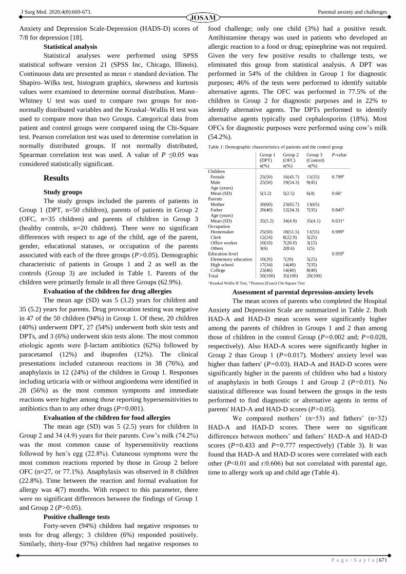
J Surg Med. 2020;4(8):669-673. Parental anxiety and challenges
P a g e / S a y f a | 671
Anxiety and Depression Scale-Depression (HADS-D) scores of
7/8 for depression [18].
Statistical analysis
Statistical analyses were performed using SPSS
statistical software version 21 (SPSS Inc, Chicago, Illinois).
Continuous data are presented as mean ± standard deviation. The
Shapiro–Wilks test, histogram graphics, skewness and kurtosis
values were examined to determine normal distribution. Mann–
Whitney U test was used to compare two groups for non-
normally distributed variables and the Kruskal–Wallis H test was
used to compare more than two Groups. Categorical data from
patient and control groups were compared using the Chi-Square
test. Pearson correlation test was used to determine correlation in
normally distributed groups. If not normally distributed,
Spearman correlation test was used. A value of P ≤0.05 was
considered statistically significant.
Results
Study groups
The study groups included the parents of patients in
Group 1 (DPT, n=50 children), parents of patients in Group 2
(OFC, n=35 children) and parents of children in Group 3
(healthy controls, n=20 children). There were no significant
differences with respect to age of the child, age of the parent,
gender, educational statuses, or occupation of the parents
associated with each of the three groups (P>0.05). Demographic
characteristic of patients in Groups 1 and 2 as well as the
controls (Group 3) are included in Table 1. Parents of the
children were primarily female in all three Groups (62.9%).
Evaluation of the children for drug allergies
The mean age (SD) was 5 (3.2) years for children and
35 (5.2) years for parents. Drug provocation testing was negative
in 47 of the 50 children (94%) in Group 1. Of these, 20 children
(40%) underwent DPT, 27 (54%) underwent both skin tests and
DPTs, and 3 (6%) underwent skin tests alone. The most common
etiologic agents were β-lactam antibiotics (62%) followed by
paracetamol (12%) and ibuprofen (12%). The clinical
presentations included cutaneous reactions in 38 (76%), and
anaphylaxis in 12 (24%) of the children in Group 1. Responses
including urticaria with or without angioedema were identified in
28 (56%) as the most common symptoms and immediate
reactions were higher among those reporting hypersensitivities to
antibiotics than to any other drugs (P=0.001).
Evaluation of the children for food allergies
The mean age (SD) was 5 (2.5) years for children in
Group 2 and 34 (4.9) years for their parents. Cow’s milk (74.2%)
was the most common cause of hypersensitivity reactions
followed by hen’s egg (22.8%). Cutaneous symptoms were the
most common reactions reported by those in Group 2 before
OFC (n=27, or 77.1%). Anaphylaxis was observed in 8 children
(22.8%). Time between the reaction and formal evaluation for
allergy was 4(7) months. With respect to this parameter, there
were no significant differences between the findings of Group 1
and Group 2 (P>0.05).
Positive challenge tests
Forty-seven (94%) children had negative responses to
tests for drug allergy; 3 children (6%) responded positively.
Similarly, thirty-four (97%) children had negative responses to
food challenge; only one child (3%) had a positive result.
Antihistamine therapy was used in patients who developed an
allergic reaction to a food or drug; epinephrine was not required.
Given the very few positive results to challenge tests, we
eliminated this group from statistical analysis. A DPT was
performed in 54% of the children in Group 1 for diagnostic
purposes; 46% of the tests were performed to identify suitable
alternative agents. The OFC was performed in 77.5% of the
children in Group 2 for diagnostic purposes and in 22% to
identify alternative agents. The DPTs performed to identify
alternative agents typically used cephalosporins (18%). Most
OFCs for diagnostic purposes were performed using cow’s milk
(54.2%).
Table 1: Demographic characteristics of patients and the control group
Group 1
(DPT)
n(%)
Group 2
(OFC)
n(%)
Group 3
(Control)
n(%)
P-value
Children
Female
Male
Age (years)
Mean (SD)
Parents
Mother
Father
Age (years)
Mean (SD)
Occupation
Homemaker
Clerk
Office worker
Others
Education level
Elementary education
High school
College
Total
25(50)
25(50)
5(3.2)
16(45.7)
19(54.3)
5(2.5)
11(55)
9(45)
6(4)
0.799b
0.66a
30(60) 23(65.7) 13(65)
20(40)
35(5.2)
25(50)
12(24)
10(10)
3(6)
10(20)
17(34)
23(46)
50(100)
12(34.3)
34(4.9)
18(51.5)
8(22.9)
7(20.0)
2(8.6)
7(20)
14(40)
14(40)
35(100)
7(35)
35(4.1)
11(55)
5(25)
3(15)
1(5)
5(25)
7(35)
8(40)
20(100)
0.845b
0.631a
0.999b
0.959b
a Kruskal Wallis H Test, b Pearson (Exact) Chi-Square Test
Assessment of parental depression-anxiety levels
The mean scores of parents who completed the Hospital
Anxiety and Depression Scale are summarized in Table 2. Both
HAD-A and HAD-D mean scores were significantly higher
among the parents of children in Groups 1 and 2 than among
those of children in the control Group (P=0.002 and; P=0.028,
respectively). Also HAD-A scores were significantly higher in
Group 2 than Group 1 (P=0.017). Mothers' anxiety level was
higher than fathers' (P=0.03). HAD-A and HAD-D scores were
significantly higher in the parents of children who had a history
of anaphylaxis in both Groups 1 and Group 2 (P=0.01). No
statistical difference was found between the groups in the tests
performed to find diagnostic or alternative agents in terms of
parents' HAD-A and HAD-D scores (P>0.05).
We compared mothers’ (n=53) and fathers’ (n=32)
HAD-A and HAD-D scores. There were no significant
differences between mothers’ and fathers’ HAD-A and HAD-D
scores (P=0.433 and P=0.777 respectively) (Table 3). It was
found that HAD-A and HAD-D scores were correlated with each
other (P˂0.01 and r:0.606) but not correlated with parental age,
time to allergy work up and child age (Table 4).

J Surg Med. 2020;4(8):669-673. Parental anxiety and challenges
P a g e / S a y f a | 672
Table 2. The mean scores of parents in the Hospital Anxiety and Depression Scale
Parents of
Group 1
(DPT)
(n=50)
Parents of
Group 2
(OFC)
(n=35)
Parents of
control
Group
(n=20)
P
df
Comparison
pattern
HADS-Anxiety
Mean(SD)
5.82(4.14)
8.20(3.37)
4.83(2.85)
0.002c
2
0.002a
0.017b
HADS-Depression
Mean(SD)
5.00(3.80)
5.85(2.83)
3.63(3.01)
0.028c
2
0.027a
HADS: Hospital Anxiety and Depression Scale *, a Comparison pattern indicates the standardized mean
difference between Group 2 and Group 3, b Comparison pattern indicates the standardized mean difference
between Group 1 and Group 2, c Kruskal- Wallis
Table 3: Comparison of fathers and mothers’ HAD scores
Fathers
(n=32)
Mothers
(n=53)
Total
(n=85)
P-value
HADS-Anxiety
Mean(SD)
5.00(0.67)
5.66(0.50)
5.41(3.68)
0.433a
HADS-Depression
Mean(SD)
4.41(0.67)
4.45(0.47)
4.44(3.54)
0.777a
a Mann-Whitney U test
Table 4: Correlation test of variables
Child
age (r)
Parental
age (r)
Time between reaction and
allergy work- up (r)
HADS-
A
HADS-
D
HADS-A 0.162 0.160 -0.119 1 0.606a
HADS-D 0.185 0.065 0.023 0.606a 1
a Spearman’s rho test
Discussion
The results of the current study indicate that the parents
of children with suspected food allergy have higher anxiety and
depression scores before OFC. In our study, the anxiety and
depression levels of parents of the children with suspected drug
allergy were higher than healthy controls’ parents but lower than
those with suspected or documented food allergy. The reasons
may include the presence of fewer opportunities to encounter
agents that elicit drug-related allergic reactions, and the fact that
they are more easily managed than allergies to food substances.
This information may decrease anxiety and depression levels in
parents of children with suspected drug allergy. There might be
many reasons to explain the differences regarding children’s
individual characteristics, family characteristics, previous
medical history, and life events. One study compared the anxiety
and depression levels with HADS of the mothers of children with
suspected food allergy on the first clinical visit and second visit
after 4–6 weeks [19]. The study reported no higher anxiety and
depression scores; however, the children were not evaluated by
OFC and the parents were evaluated during two visits only. The
studies related to anxiety of parents of the children with
suspected food allergy have been compared before and after
OFC. The parents’ anxiety level decreased after OFC [12-14].
In a randomized controlled adult study, it was aimed to
define placebo reactions during DPT, the subjects’ anxiety and
depression symptoms were evaluated by HADS before DPT. It
was shown that HADS positivity before the test was correlated to
placebo reactions. It was recommended that evaluating
psychiatric symptoms before DPT was important to define
placebo reactions [20].
Anxiety and depression scale scores can differ between
parents of an individual child [21,22]. King et al. [22] found that
mothers of children with food allergies express more anxiety and
stress than the fathers. Another study reported that the mothers of
children with food allergy reported higher levels of impairment
with respect to quality of life than did fathers, regardless of the
type or severity of the allergy or the existence of comorbidities
[14]. Knibb et al [23]. reported that the mothers experienced
comparatively prominent levels of anxiety prior to an OFC. Not
the only suspicion of food allergy, but the actual performance of
the OFC may increase parental anxiety levels. However, quality
of life of the families was positively affected by having access to
clear-cut results [23].
Most of the parents involved in our study were mothers
(62.9%); overall, their anxiety level was higher than that of the
fathers. The findings were consistent with the literature
[12,22,23]. Beken et al [12]. reported that the mothers of children
with suspected food allergy had higher anxiety before OFC than
the mothers of healthy control children. Parents’ reactions to any
condition impact children’s feelings and behavior, and parental
anxiety and depression level may impact compliance to the tests.
Mothers typically play a larger role in caring for children,
including making decisions on medications, preparing food,
reading labels, and related activities, all of which may cause their
anxiety levels to increase [22]. Thus, mothers of children who
were to undergo challenge tests might be more anxious and
depressive than the fathers. In our study, there were no
significant differences between anxiety and depression scores of
fathers and mothers. However, due to the difference in the
number of mothers visiting the clinic, gender differences could
not be determined.
To the best our knowledge, this is the first study which
evaluates parents’ anxiety and depression level before DPT that
are performed to children with suspected drug allergy.
Limitations
The first limitation of this study is the fact that we did
not reevaluate and compare anxiety and depression scores of the
parents sometime after the results of these provocation tests. The
second limitation is the lack of comparison of parents with same
gender. It must be considered that there are differences between
with mothers’ and fathers’ anxiety and depression level. The
third limitation is that we did not evaluate parents’ physical and
mental health problems, children’s’ mental health and other
medical problems, family relationship quality, family functioning
and other familial or individual factors that may affect
depression and anxiety levels. Further larger and well-controlled
observational studies are required in the future to consolidate the
association between parental anxiety-depression and challenges.
Conclusion
We determined that the anxiety and depression scores of
parents whose children require DPT or OFC testing are
uniformly high regardless of the test type. Parents’ anxiety and
depression level were higher when parents were informed about
the risks of these tests. However, informing parents that DPT
will reveal alternative agents decreases their anxiety and
depression levels. It may be useful to limit parents’ anxiety
before any type of allergy-provocation test, as that will increase
compliance with both the test and the treatments offered.
References
1. Sicherer SH, Sampson HA. Food Allergy: A Review and Update on Epidemiology, Pathogenesis,
Diagnosis, Prevention, and Management. J Allergy Clin Immunol. 2018;141:41-58
2. Vickery BP, Burks W. Oral immunotherapy for food allergy. Curr Opin Pediatr. 2010;22:765-770.
3. Feng C, Kim JH. Beyond Avoidance: The Psychosocial Impact of Food Allergies. Clin Rev Allergy
Immunol. 2019;57(1):74-82.
4. Venter C, Sommer I, Moonesinghe H, Grundy J, Glasbey G, Patil V, et al. Health-related quality of
life in children with perceived and diagnosed food hypersensitivity. Pediatr Allergy Immunol.
2015;26(2):126-32.
5. Cummings AJ, Knibb RC, Erlewyn-Lajeunesse M, King RM, Roberts G, Lucas JS. Management of
nut allergy influences quality of life and anxiety in children and their mothers. Pediatr Allergy
Immunol. 2010;21:586-94.
6. Primeau MN, Kagan R, Joseph L, Lim H, Dufresne C, Duffy C, et al. The psychological burden of
peanut allergy as perceived by adults with peanut allergy and the parents of peanut-allergic children.
Clin Exp Allergy. 2000;30(8):1135-43.

J Surg Med. 2020;4(8):669-673. Parental anxiety and challenges
P a g e / S a y f a | 673
7. Avery NJ, King RM, Knight S, Hourihane JO. Assessment of quality of life in children with peanut
allergy. Pediatr Allergy Immunol. 2003;14(5):378-82.
8. Flokstra-de Blok BM, Dubois AE, Vlieg-Boerstra BJ, Oude Elberink JN. Health-related quality of life
of food allergic patients: comparison with the general population and other diseases. Allergy.
2010;65(2):238-44.
9. Demoly P, Pichler W, Pirmohamed M, Romano A. Important questions in allergy:Drug
allergy/hypersensitivity. Allergy. 2008; 63:616-9.
10. Aberer W, Kränke B. Provocation tests in drug hypersensitivity. Immunol Allergy Clin North Am.
2009;29:567-84.
11. Soyyiğit Ş, Aydın Ö, Yılmaz İ, Özdemir SK, Cankorur VŞ, Atbaşoğlu C, et al. Evaluation of drug
provocation test-related anxiety in patients with drug hypersensitivity. Ann Allergy Asthma Immunol.
2016 ;117(3):280-4.
12. Beken B, Celik V, Gokmirza Ozdemir P, Sut N, Gorker I, Yazicioglu M. Maternal anxiety and
internet-based food elimination in suspected food allergy. Pediatr Allergy Immunol. 2019;30(7):752-
59.
13. Zijlstra WT, Flinterman AE, Soeters L, Knulst AC, Sinnema G, L’Hoir MP, et al. Parental anxiety
before and after food challenges in children with suspected peanut and hazelnut allergy. Pediatr
Allergy Immunol. 2010;21:439-45.
14. Kajornrattana T, Sangsupawanich P, Yuenyongviwat A. Quality of life among caregivers and growth
in children with parent-reported food allergy. Asian Pac J Allergy Immunol. 2018;36:22-6.
15. Aberer W, Bircher A, Romano A, Blanca M, Campi P, Fernandez J, et al. Drug provocation testing in
the diagnosis of drug hypersensitivity reactions: General considerations. Allergy. 2003;58:854-63.
16. Demoly P, Kropf R, Bircher A, Pichler WJ. Drug hypersensitivity:Questionnaire. EAACI interest
Group on drug hypersensitivity. Allergy. 1999;54:999-1003.
17. Muraro A, Werfel T, Hoffmann-Sommergruber K, Roberts G, Beyer K, Bindslev-Jensen C, et al.
EAACI Food Allergy and Anaphylaxis Guidelines Group. EAACI food allergy and anaphylaxis
guidelines:diagnosis and management of food allergy. Allergy. 2014;69:1008-25.
18. Aydemir O, Güvenir T, Küey L, Kültür S. Validity and reliability of Turkish version of Hospital
Anxiety and Depression Scale. Turk Psikiyatri Derg. 1997;8:280-7.
19. Knibb RC, Semper H. Impact of suspected food allergy on emotional distress and family life of
parents prior to allergy diagnosis. Pediatr Allergy and Immunol. 2013;24(8): 798-803.
20. De Pasquale T, Nucera E, Boccascino R, Romeo P, Biagini G, Buonomo A, et al. Allergy and
psychologic evaluations of patients with multiple drug intolerance syndrome Intern Emerg Med.
2012;7(1):41-7.
21. Warren CM, Gupta RS, Sohn MW, Oh EH, Lal N, Garfield CF, et al. Differences in empowerment
and quality of life among parents of children with food allergy. Ann Allergy Asthma Immunol.
2015;114(2):117-25.
22. King RM, Knibb RC, Hourihane JO. Impact of peanut allergy on quality of life, stress and anxiety in
the family. Allergy. 2009;64(3):461-8.
23. Knibb RC, Ibrahim NF, Stiefel G, Petley R, Cummings AJ, King RM, et al. The psychological impact
of diagnostic food challenges to confirm the resolution of peanut or tree nut allergy. Clin Exp Allergy.
2012;42:451-9.
This paper has been checked for language accuracy by JOSAM editors.
The National Library of Medicine (NLM) citation style guide has been used in this paper.
P a g e / S a y f a | 674
Evaluation of cancer-related deaths in Turkey between 2009-2018: An
epidemiological study 2009-2018 yılları arasında Türkiye'de kansere bağlı ölümlerin değerlendirilmesi: Bir epidemiyolojik çalışma
Havva Hande Keser Şahin 1, Orhan Aslan 2, Mustafa Şahin 3
How to cite/Atıf için: Şahin HHK, Aslan O, Şahin M. Evaluation of cancer-related deaths in Turkey between 2009-2018: An epidemiological study. J Surg Med. 2020;4(8):674-
677.
J Surg Med. 2020;4(8):674-677. Research article DOI: 10.28982/josam.779292 Araştırma makalesi
1 Department of Pathology, Hitit University
Faculty of Medicine, Çorum, Turkey 2 Department of General Surgery, Hitit University
Erol Olçok Training and Research Hospital,
Çorum, Turkey 3 Department of Medical Biochemistry, Hitit
University Erol Olçok Training and Research
Hospital, Çorum, Turkey
ORCID ID of the author(s)
HHKŞ: 0000-0003-1827-1039
OA: 0000-0002-1982-0792
MŞ: 0000-0001-6073-563X
Corresponding author/Sorumlu yazar:
Mustafa Şahin
Address/Adres: Hitit Üniversitesi, Erol Olcok Eğitim
ve Araştırma Hastanesi, Tıbbi Biyokimya
Laboratuvarı, Çorum, Türkiye
E-mail: [email protected]
󠄀
Ethics Committee Approval: By the design of the
study, Hitit University Faculty of Medicine Clinical
Research Ethics Board approval was not required.
The Turkish Statistical Institute permits the use of
data on its official website for research purposes.
Etik Kurul Onayı: Hitit Üniversitesi Tıp Fakültesi
Klinik Araştırmalar Etik Kurulu başkanlığı tarafından
çalışmanın tasarımı nedeniyle onayı gerekmedi.
Türkiye İstatistik Kurumu, resmi web sitesindeki
verilerin araştırma amacıyla kullanılmasına izin
vermektedir. 󠄀
Conflict of Interest: No conflict of interest was
declared by the authors.
Çıkar Çatışması: Yazarlar çıkar çatışması
bildirmemişlerdir.
󠄀
Financial Disclosure: The authors declared that this
study has received no financial support.
Finansal Destek: Yazarlar bu çalışma için finansal
destek almadıklarını beyan etmişlerdir.
󠄀
Previous presentation: This study was presented as an
oral presentation at the 3rd International Hippocrates
Congress on Medical and Health Sciences, held in
Ankara, Turkey, on March 6, 2020.
󠄀
Published: 8/30/2020
Yayın Tarihi: 30.08.2020
Copyright © 2020 The Author(s)
Published by JOSAM This is an open access article distributed under the terms of the Creative
Commons Attribution-NonCommercial-NoDerivatives License 4.0 (CC
BY-NC-ND 4.0) where it is permissible to download, share, remix,
transform, and buildup the work provided it is properly cited. The work
cannot be used commercially without permission from the journal.
Abstract
Aim: Determination of cancer incidence and mortality data for all countries is essential for cancer control. These data can affect public
health and clinical service planning. In this study, we aimed to examine the variability of cancer deaths in Turkey in terms of age
groups, gender, geographic region, and years according to the cancer types between 2009-2018.
Methods: In this epidemiological study, cancer-related mortality statistics data presented at the website of Turkey Statistical Institute
(TSI) were used. Cancer-related deaths between 2009-2018 were evaluated in terms of ratio to total deaths, age groups, cancer types,
geographical regions, and gender. Data were presented as percentages.
Results: Cancer-related deaths were observed to increase between 2009-2018. The highest cancer-related mortality rate was seen in the
Marmara Region, while it was lowest in the Southeastern Anatolia Region. Cancer-related death rate was higher in males than females,
and it was highest in the 65-74 years age range. The most common cause of death in males is lung cancer, and in females, breast cancer.
Conclusion: Cancer is responsible for 1/6 of all deaths in the world. Similarly, cancer-related deaths are responsible for approximately
1/5 of all deaths in our country. The risk of cancer in >60 years age group is higher in males than in females. In our country, all kinds of
cancers, except female cancers, cause more deaths in males than females.
Keywords: Cancer, Mortality, Pathology, Death
Öz
Amaç: Tüm ülkeler için kanser insidansı ve mortalite verilerinin belirlenmesi; kanserin kontrolü için esastır. Bu veriler halk sağlığına ve
klinik hizmet planlamasına etki edebilmektedir. Bu çalışmada 2009-2018 yılları arasında Türkiye'de kansere bağlı ölümlerin yıllara göre
değişimini, yaş gruplarına, cinsiyete ve coğrafi bölgelere göre farklılıklarını, kanser türlerine göre değişkenlikleri incelemeyi amaçladık.
Yöntemler: Çalışmada Türkiye İstatistik Kurumu (TÜİK) web sitesinde sunulan kanser ilişkili ölüm istatistik verileri kullanıldı. 2009-
2018 yılları arasında kansere bağlı ölümlerin coğrafi bölgelere göre toplam ölümlere oranı değerlendirildi. 2009-2018 yılları arasında
yaş gruplarına ve cinsiyete göre kansere bağlı ölümler değerlendirildi. 2009-2018 yılları arasında kanser türlerine ve cinsiyete göre
kansere bağlı ölümler değerlendirildi. Veriler yüzde olarak sunuldu. Çalışma epidemiyolojik olarak tasarlanmıştır.
Bulgular: Kansere bağlı ölümlerin 2009-2018 yılları arasında arttığı görülmüştür. Toplam ölümlere kıyasla kansere bağlı ölüm oranının
en yüksek olduğu bölge Marmara Bölgesi iken en düşük olduğu bölge Güneydoğu Anadolu Bölgesidir. Tüm yaş gruplarında; erkekler,
kadınlara göre kanser nedeniyle daha fazla ölmektedir. Kansere bağlı ölümler en yüksek seviyesine 65-74 yaş aralığında ulaşmaktadır.
Erkeklerde ölüme en çok neden kanser türü akciğer, kadınlarda ise meme kanseridir.
Sonuç: Kanser dünyada meydana gelen tüm ölümlerin 1/6’sından sorumludur. Benzer şekilde ülkemizde kansere bağlı ölümler, tüm
ölümlerin yaklaşık 1/5’inden sorumludur. 60 yaşın üzerinde erkeklerde kanser riski kadınlara oranla daha fazladır. Ülkemizde kadın
kanserleri dışında her türlü kanserin erkeklerde daha fazla ölüme neden olduğu tespit edilmiştir.
Anahtar kelimeler: Kanser, Mortalite, Patoloji, Ölüm

J Surg Med. 2020;4(8):674-677. Distribution of cancer in Turkey
P a g e / S a y f a | 675
Introduction
Investigation of rates, numbers, and causes of death is
important for the development of health systems, preventive
medicine, understanding the effects of diseases on people and the
effective use of treatment resources [1-3]. Cancer is a group of
diseases characterized by uncontrolled proliferation and spread
of abnormal cells. If the spread cannot be controlled, it can result
in death [4]. Cancer is the leading cause of death for all levels of
income and socioeconomic groups worldwide [5]. Determination
of cancer incidence and mortality data is essential for the control
of cancer in all countries. These data may affect public health
and clinical service planning [6]. Ratings and measurements
made according to age groups are not affected by population size
and structure. Changes in incidence rates are the main indicator
of changes in prevalence [7]. In 2018, it was estimated that there
were 18 million cancer cases worldwide, and 9.8 million people
died of cancer [6,8]. Age, alcohol, familial predisposition,
cancer-causing substances, chronic inflammation, diet,
hormones, immunosuppression, infectious agents, obesity,
radiation, sunlight, and tobacco are the most frequently
researched cancer risk factors. Although some of these risk
factors can be avoided, some are unavoidable [9]. Today, there
are changes in behavior and habits of society due to
industrialization and rapid urbanization, which shows that cancer
risk factors may change over time [10]. With the increase in
urbanization and development, cancer risk factors, such as the
adoption of a sedentary lifestyle, increase in the elderly
population, decrease in fertility, diet, and environmental
pollution, change. However, with the development of healthcare
and imaging methods, early diagnosis and effective treatment
have become more possible [11]. Cancer risk factors and
treatment options are affected by the regions and socioeconomic
status [12,13]. By evaluating cancer mortality and incidence
from a socioeconomic and regional perspective, inequalities in
the healthcare system can be recognized, and beneficial health
arrangements can be made [14,15]. In this study, we aimed to
investigate the changes by years, age group, gender, geographic
region according to the types of cancer seen in Turkey between
2009-2018.
Materials and methods
In this epidemiological study, we used the death
statistics data shared by Turkey Statistical Institute (TSI) on its
website [16]. TSI allows the use of their data for research. The
causes of death, mortality rates, and population data between
2009 and 2018 were collected, analyzed, classified in terms of
age, gender, geographical region, and disease groups, and
evaluated with descriptive statistics using the Statistical Package
for Social Sciences (SPSS) 22.0 software to be presented in
percentage (%).
Results
Cancer-related deaths are more frequent in males than in
females. It is observed that deaths due to cancer regularly
increased among both genders between 2009 and 2018.
Compared to 2009, cancer-related deaths had increased by 28.6%
in total, by 27.5% among males, and 30.5% among females by
2018. Deaths due to cancer by gender between 2009-2018 are
shown in the figure 1.
Figure 1: Cancer-related deaths between 2009 and 2018
The highest cancer-related mortality rate was seen in the
Marmara Region, while it was lowest in the Southeastern
Anatolia Region. In all regions but the Southeastern Anatolia,
cancer-related deaths are responsible for 1/5 of all deaths (Table
1).
Table 1: The ratio of cancer-related deaths to total deaths between 2009-2018 by
geographical regions
Geographical Regions Total Death Cancer-related deaths %
Marmara 1070733 249186 23.3
Aegean 574355 116268 20.2
Mediterranean 412813 76908 18.6
Black Sea 470107 92003 19.6
Eastern Anatolia 215586 42894 19.9
Southeastern Anatolia 214454 31864 14.9
Central Anatolia 597801 124559 20.8
The cancer-related mortality rate is higher in males than
females across all age groups. It reaches its highest level between
the ages of 65-74 years. Cancer-related mortality rates are close
to each other among males and females from birth until the age
of 44 years; however, after 45 years of age, male predominance
is observed (Table 2).
Table 2: Cancer-related deaths by age groups and gender between 2009 and 2018
Age groups Total % Male % Female %
0 – 14 6235 0.8 3149 0.7 3086 1.2
15 – 24 6191 0.9 3747 0.8 2444 0.9
25 – 34 10824 1.5 5843 1.2 4981 1.9
35 – 44 30597 4.1 15184 3.2 15413 5.9
45 – 54 89017 12.0 55643 11.5 33374 12.9
55 – 64 174697 23.6 123136 25.7 51561 19.9
65 – 74 202035 27.3 139350 29.1 62685 24.1
75 – 84 171138 23.2 108309 22.6 62829 24.2
85+ 48435 6.6 25168 5.2 23267 9.0
Total 739169 100 479529 100 259640 100
Deaths due to all cancer types, except breast and
gynecological cancers, are more frequent in males. The most
common cancer types are lung, bronchus, larynx, and trachea
cancer among both genders. Breast cancer was the most common
cause of cancer-related mortality in females. Other common
cancers included the lung, larynx, and trachea cancers (Table 3).

J Surg Med. 2020;4(8):674-677. Distribution of cancer in Turkey
P a g e / S a y f a | 676
Table 3: Cancer-related deaths by cancer types and gender between 2009 and 2018
Cancer types Cancer related
deaths
% Male % Female %
Lip, Oral cavity,
Pharynx
7965 1.3 5293 68.8 2672 31.2
Esophagus 7746 1.2 4544 58.7 3202 41.3
Stomach 62488 10.0 40970 65.6 21518 34.4
Column 51316 8.2 29311 57.1 22005 42.9
Rectum and anus 12218 2.0 7276 59.6 4942 40.4
Liver and bile ducts 27608 4.4 17775 64.4 9833 35.6
Pancreas 41351 6.6 24124 58.3 17227 41.7
Lung, larynx, trachea 225726 36.1 192410 85.2 33316 14.8
Skin 4457 0.7 2589 58.1 1868 41.9
Breast 35627 5.7 758 2.1 34869 97.9
Cervix 4703 0.8 0 0 4703 100
Other areas of the uterus 8241 1.3 0 0 8241 100
Over 13599 2.2 0 0 13599 100
Prostate 33042 5.3 33042 100 0 0
Kidney 9785 1.6 6661 68.1 3124 31.9
Bladder 19240 3.1 15993 83.1 3247 16.9
Lymphoid and
hematopoietic
59738 9.6 34898 59.4 24839 40.6
Total 624850 100 415644 66.6 209206 33.4
Discussion
Old age is the most important risk factor for many types
of cancer [17]. It is known that the proportion of elderly
population has increased gradually in our country since 1935
[18]. Life expectancy in Turkey was 50 years in 1970, and it has
risen to 78 years according to 2018 data. It is predicted that along
with the ratio of elderly population, cancer cases will also
increase. The number of cancer patients is growing due to the
escalation of risk factors in underdeveloped and developing
countries and due to the advances in diagnosis and treatment
methods in developed countries. The increase in the number of
cancer patients and the decrease in infection-related deaths
because of the use of more effective antibiotics compared to the
past also lead to a higher cancer-related mortality rate. In our
country, cancer-related deaths increased by 28.6% in 2018
compared to 2009, and we think that this is due to the increase in
the elderly population.
Cancers are the second most common causes of death in
the world after cardiovascular diseases. It is also reported that
cancer is responsible for 1/6 of all deaths worldwide [19].
Similarly, in our study, deaths due to cancer constituted
approximately 1/5 of all deaths in all regions, except
Southeastern Anatolia (Table 1). According to a study conducted
by the National Cancer Institute in 2015, 25.4% of cancer cases
were detected between the ages of 65-74 years (16). In our study,
27.3% of cancer-related deaths were seen in the 65-74 year-age
group (Table 2). In Turkey, another study conducted by the
Ministry of Health reported that breast cancer in women and lung
cancer in men were increasing [20]. Currently, lung cancer is the
most lethal type of cancer worldwide [19]. The most prominent
reason is that tobacco use is more common among males. In
European countries, the prevalence of lung cancer in females has
approached that of males in the recent years [7]. A study
conducted at a center in the eastern Anatolia region of Turkey
examining lung biopsy reports showed that lung cancer has
increased within the last decade [21].
In our study, the most common mortality-causing types
were respiratory system malignancies among males and breast
cancer among females (Table 3). Turkey's young population
resides in Southeastern Anatolia the most and in Black Sea and
Marmara regions the least [22]. Cancer risk increases with age
[9]. In our study, in accordance with the above findings, we
determined that cancer-related mortality rate was lower in the
Southeastern Anatolia Region, where the proportion of the young
population was higher. Expectedly, it was higher in the Marmara
Region, where the rate of the elderly population was higher
(Table 1). We think that the increase in some cancer risk factors
such as diet, decrease in physical activity, obesity, and decrease
in fertility rate may also contribute to the increase in cancer
mortality rate in Marmara region. In general, it is known that the
risk of cancer and cancer-related mortality are higher in males
compared to that in females over the age of 60 years [22]. This
may be caused by the fact that males consume more alcohol,
cigarettes, are more exposed to the sun, and prone to various risk
factors such as obesity [23]. Our study shows that all cancer
types, except gynecological cancers, cause more deaths in men
(Table 3). Classification according to geographical regions is
especially important in terms of determining different risk
factors, evaluating access to diagnosis and treatment, and
healthcare planning. Although the most common type of cancer
varies from country to country, different types of cancer can be
predominant in different regions of the same country. While a
decrease in lung cancer incidence is expected due to the tobacco
ban in developed countries, it is still increasing in developing
countries. Similarly, in terms of female cancers, while breast,
ovarian and endometrial cancers increase with the decrease in
fertility in developed countries, cervical cancer associated with
HPV and multiparity is more common in developing countries
[24-27]. In addition, liver cancer due to hepatitis B and C
infections is more common because of inadequate hygiene in
underdeveloped and developing countries, and rate of gastric
cancer is relatively high because of contamination with
Helicobacter pylori [28,29].
Around 85% of cancer risk factors are changeable and
preventable. For this reason, risk factors, and their distribution
according to regions, gender, and socioeconomic status can be
revealed through statistics and studies. Thus, the necessary study
for the target community will be performed more easily. In
Turkey, ‘Early Cancer Detection Screening and Training
Centers' (KETEM) perform mammography for breast cancer.
Family health centers perform fecal occult blood tests for colon
cancer screening to those over the age of 50 years. It is also
possible to detect cancer at an early stage and treat it effectively
by recommending endoscopy and colonoscopy screening for
patients with a family history of colon and stomach cancer. In
2008, the smoking ban in closed areas took effect in Turkey for
the fight against cancer, reducing exposure to tobacco smoke.
The government's publications about the harms of smoking and
warnings about the relationship between tobacco and cancer on
cigarette packages are positive developments in terms of fighting
against cancer. Including the hepatitis A and B vaccines in the
routine vaccination program will also likely reduce the risk of
liver cancer in Turkey. Similarly, in some countries where HPV
infection is prevalent, the routine use of the HPV vaccine is a
preventive health plan for cervical cancer. Obesity is also an
important problem associated with cancer in our era. It is
predicted to increase the risk of many cancers such as those of
the esophagus, colon, gallbladder, kidney, and breast. Obesity is
a preventable disorder because it is associated with excessive
calorie intake and reduced physical activity. By encouraging
healthy and natural nutrition, raising awareness about obesity by

J Surg Med. 2020;4(8):674-677. Distribution of cancer in Turkey
P a g e / S a y f a | 677
health institutions can reduce the incidence of cancer and
contribute to the prevention of various diseases, such as
cardiovascular diseases and diabetes [30]. We believe that
inability to evaluate mortality rates of cancer types due to the
lack of continuous reporting of annual new cancer case statistics
is the primary limitation of our study. The distribution and
mapping of cancer types according to geographical regions will
help determine focus areas for the healthcare planning. In that
regard, since mesothelioma is more frequent in Nevsehir,
Turkey, the region's soil was examined, and it was found that soil
comprises an excess amount of asbestos mineral. Therefore,
some villages were relocated. Regarding geography, it is thought
that some types of cancer, such as leukemia and thyroid cancer
have increased in the Black Sea region after the Chernobyl
disaster in 1986. In terms of cancer, differences in geographical
regions may also be related to diet. In the eastern regions of
Turkey, esophageal and stomach cancers are more common, and
colorectal cancers predominate in the western regions. This is
likely related to eating habits. It is known that pickled and
smoked foods cause stomach cancer. In addition, excessively hot
food consumption is a predisposing factor for esophageal cancer.
A decrease in the incidence of stomach cancer is predicted due to
easier access to fresh vegetables and better food preservation
methods. Smoking also varies over time, according to the
regions. While smoking rates decrease in developed countries, it
continues to increase in underdeveloped and developing ones
[31]. As a result, these countries, which are already challenged
economically, will be in a difficult position against the global
economic forces because of the cancer burden due to smoking
[5]. Grouping new cancer cases according to their age of
occurrence, reporting the time from the detection of the disease
to death, and similar statistics will be important for decreasing
cancer-related mortality and morbidity.
Limitations
As a result of incomplete reporting of data on cancer
statistics, institutional errors can be considered as the limitation
of our study.
Conclusions
We think that our study is notable since it covers a 10-
year period and reveals differences according to age groups,
gender, cancer types, and geographical regions. Cancer statistics
are important in terms of developing health policies, bringing
preventive healthcare to the fore, increasing cancer screening
rates in risky individuals, and conducting studies for early cancer
diagnosis. Early diagnosis of cancer will reduce the treatment
costs of countries.
References
1. Civil Registration: Why counting births and deaths is important? World Health Organization. Erişim
Adresi: http://www.who.int/mediacentre/factsheets/fs324/en/ Erişim Tarihi: 09.02.2020.
2. Sümbüloğlu V, Gören A, Sümbüloğlu K.Ölüm Raporu Yazım Kılavuzu, TC Sağlık Bakanlığı Sağlık
Projesi Genel Koordinatörlüğü Sağlık Enformasyon Sistemleri Eğitim Dizisi. Değiştirilmiş 2. Basım.
No:3. Ankara: Aydoğdu Ofset; 1995. 3.
3. The top 10 causes of death. Why do we need to know the reasons people die? Media Center. World
Health Organization. Erişim Tarihi: 09.02.2020. Erişim Adresi:
http://www.who.int/mediacentre/factsheets/fs310/en/index2.html
4. Cancer Facts&Figures: 2019. American Cancer Society. 2019
5. Global Cancer Incidence and Mortality Rates and Trends—An Update Cancer. Epidemiol Biomarkers
Prev. 2015;25(1):16–27.
6. World Cancer Report 2014. Geneva, Switzerland: World Health Organization, International Agency
for Research on Cancer, WHO Press, 2015.
7. Cancer incidence and mortality projections in the UK until 2035. British Journal of Cancer.
2016;115:1147-55.
8. Global cancer statistics for the most common cancers. World wide cancer data. Erişim Adresi:
https://www.wcrf.org/dietandcancer/cancer-trends/worldwide-cancer-data. Erişim Tarihi: 09.02.2020.
9. Risk Factors for Cancer. Cancer Causes and Prevention. National Cancer İnstitute. Erişim Adresi:
https://www.cancer.gov/about-cancer/causes-prevention/risk. Erişim Tarihi: 09.02.2020.
10. Trends and Patterns of Cancer Mortality in North China (Hebei Province), 1973–2013. ScientifiC
Reports. 2018;8:311.
11. Socioeconomic and Racial/Ethnic Disparities in Cancer Mortality, Incidence, and Survival in the
United States, 1950–2014: Over Six Decades of Changing Patterns and Widening Inequalities.
Hindawi Journal of Environmental and Public Health Volume 2017, Article ID 2819372, 19 pages
12. Singh GK, Siahpush M, Altekruse SF. Timetrendsin liver cancer mortality, incidence, and risk factors
by unemployment level and race/ethnicity, United States,1969–2011. Journal of Community Health.
2013;38(5):926-40.
13. Blackwell DL, Lucas JW, Clarke TC. Summary health statistics for U.S. adults: national health
interview survey, 2012. Vital and Health Statistics, vol.10, no.260, pp.1–161, 2014.
14. Singh GK, Williams SD, Siahpush M, Mulhollen A. Socioeconomic, rural-urban, and racial
inequalities in US cancer mortality: Part I-all cancers and lung cancer and part II-colorectal, prostate,
breast, and cervical cancers, Journal of Cancer Epidemiology. 2011;27:2011.
15. Singh GK, Miller BA, Hankey BF, Edwards BK. Area Socioeconomic Variations in U.S. Cancer
Incidence, Mortality, Stage, Treatment, and Survival, 1975–1999, NCI Cancer Surveillance
Monograph Series No. 4, NIH Publication No. 03-5417, National Cancer Institute, Bethesda, Md,
USA, 2003, http://seer.cancer.gov/publications/ses/index.html.
16. Türkiye İstatistik Kurumu. Erişim Adresi: www.tuik.gov.tr. Erişim Tarihi: 09.02.2020.
17. Engelli ve Yaşlı İstatistik Bülteni. Engelli ve Yaşlı Hizmetleri Genel Müdürlüğü. 2019.
18. Ergör G. Türkiye Kronik Hastalılar ve Risk Faktörleri Sıklığı Çalışması: Diğer Bulaşıcı Olmayan
Hastalıklar ve Kazalar. Türkiye Cumhuriyeti Sağlık Bakanlığı. Türkiye Halk Sağlığı Kurumu.
Ankara. 2013.
19. Cancer. World Health Organization. [Internet]. [2018]. [cited 2019 February 28]. Available from:
https://www.who.int/news-room/fact-sheets/detail/cancer
20. Türkiye’nin Nüfus Haritası. İç İşleri Bakanlığı, Yayın Tarihi: 11.07.2019. Erişim Adresi:
https://www.icisleri.gov.tr/turkiyenin-nufus-haritasi
21. Özmen S, Ceylan O. Trends in lung cancer incidence of last 10 years: An Eastern Anatolian single
center experience. J Surg Med. 2020;4(2):112-5.
22. Kutluk T, Kars A. Kanser Konusunda Genel Bilgiler. Kanser Savaş Dairesi Başkanlığı. Sağlık
Bakanlığı. 2001.
23. Men’s Health Week: Men more likely to develop and die from cancer. Cancer Australia. Australian
Goverment. Erişim Adresi: https://cancer australia.gov.au/about-us/news/mens-health-week-men-
more-likely-develop-and-die-cancer. Erişim Tarihi: 09.02.2020.
24. Chlebowski RT, Manson JE, Anderson GL, Cauley JA, Aragaki AK, Stefanick ML, et al. Estrogen
plus progestin and breast cancer incidence and mortality in the Women's Health Initiative
Observational Study. J Natl Cancer Inst 2013;105:526–35.
25. Colditz GA, Baer HJ, Tamimi RM. Breast cancer. In:Schottenfeld D, Fraumeni JF Jr, editors. Cancer
epidemiology and prevention. 3rd ed. New York: Oxford University Press; 2006. p. 995–1012.
26. Bruni L, Diaz M, Castellsagué X, Ferrer E, Bosch FX, de Sanjosé S. Cervical human papillomavirus
prevalence in 5 continents: meta-analysis of 1 million women with normal cytological findings. J
Infect Dis. 2010;202(12):1789-99. doi:10.1086/657321
27. Forman D, de Martel C, Lacey CJ, Soerjomataram I, Lortet-Tieulent J, Bruni L et al. Global burden
of human papillomavirus and related diseases. Vaccine 2012;30 Suppl 5:F12–23
28. Herrero R, Parsonnet J, Greenberg ER. Prevention of gastric cancer. JAMA 2014;312:1197–8.
29. London WT, Mc Glynn KA. Liver cancer. In: Schottenfeld D, Fraumeni J Jr, editors. Cancer
epidemiology and prevention. 3rd ed. New York: Oxford University Press; 2006. p. 763–86.
30. Global burden of cancer attributable to high body-mass index in 2012: a population-based study.
Lancet Oncol 2015;16:36–46
31. Mathers CD, Loncar D. Projections of global mortality and burden of disease from 2002 to 2030.
PLoS Med, 2006;3(11):e442.
This paper has been checked for language accuracy by JOSAM editors.
The National Library of Medicine (NLM) citation style guide has been used in this paper.

P a g e / S a y f a | 678
Impact of tumor necrosis factor alpha antagonist treatment on
antibody titer of hepatitis B surface antigen Tümör nekroz faktörü alfa antagonisti tedavisinin hepatit B yüzey antijeninin antikor düzeyine etkisi
Demet Yalçın Kehribar 1, Muhammed Okuyucu 1, Metin Özgen 2, Yusuf Bünyamin Ketenci 1, Talat Ayyıldız 3, Beytullah Yıldırım 3
How to cite/Atıf için: Kehribar DY, Okuyucu M, Özgen M, Ketenci YB, Ayyıldız T, Yıldırım B. Impact of tumor necrosis factor alpha antagonist treatment on antibody titer of
hepatitis B surface antigen. J Surg Med. 2020;4(8):678-681.
J Surg Med. 2020;4(8):678-681. Research article DOI: 10.28982/josam.777871 Araştırma makalesi
1 Department of Internal Medicine, Medical
Faculty, Ondokuz Mayıs University, Samsun,
Turkey 2 Department of Rheumatology, Medical Faculty,
Ondokuz Mayıs University, Samsun, Turkey 3 Department of Gastroenterology, Medical
Faculty, Ondokuz Mayıs University, Samsun,
Turkey
ORCID ID of the author(s)
DYK: 0000-0002-1852-7981
MO: 0000-0002-6026-2024
MÖ: 0000-0002-6842-2918
YBK: 0000-0003-0372-9166
TA: 0000-0003-1075-7499
BY: 0000-0003-1457-5721
Corresponding author/Sorumlu yazar:
Demet Yalçın Kehribar
Address/Adres: Ondokuz Mayıs Üniversitesi Tıp
Fakültesi, İç Hastalıkları Anabilim Dalı, Körfez
Mahallesi, 55270, Atakum, Samsun, Türkiye
E-mail: [email protected]
󠄀
Ethics Committee Approval: The study was approved
by Ondokuz Mayıs University Ethics Committee
(OMU KAEK, 12/26/2019, 2019/842). All procedures
in this study involving human participants were
performed in accordance with the 1964 Helsinki
Declaration and its later amendments.
Etik Kurul Onayı: Çalışma Ondokuz Mayıs
Üniversitesi Etik Kurulu (OMÜ KAEK, 26.12.2019,
2019/842) tarafından onaylanmıştır. İnsan
katılımcıların katıldığı çalışmalardaki tüm
prosedürler, 1964 Helsinki Deklarasyonu ve daha
sonra yapılan değişiklikler uyarınca
gerçekleştirilmiştir. 󠄀
Conflict of Interest: No conflict of interest was
declared by the authors.
Çıkar Çatışması: Yazarlar çıkar çatışması
bildirmemişlerdir.
󠄀
Financial Disclosure: The authors declared that this
study has received no financial support.
Finansal Destek: Yazarlar bu çalışma için finansal
destek almadıklarını beyan etmişlerdir.
󠄀
Published: 8/30/2020
Yayın Tarihi: 30.08.2020
Copyright © 2020 The Author(s)
Published by JOSAM This is an open access article distributed under the terms of the Creative
Commons Attribution-NonCommercial-NoDerivatives License 4.0 (CC
BY-NC-ND 4.0) where it is permissible to download, share, remix,
transform, and buildup the work provided it is properly cited. The work
cannot be used commercially without permission from the journal.
Abstract
Aim: Tumor necrosis factor alpha antagonists (anti-TNF-α) have recently been used successfully in various diseases. On the other hand,
due to their potential immunogenic effects, they cause hepatitis B virus (HBV) reactivation. The objective of this study is to examine the
change in the levels of hepatitis B surface antigen (HBsAg) and antibody (anti-HBs) in patients with negative HBsAg who were
administered different anti-TNF-α drugs as well as the factors that influence this change.
Methods: This research was designed as a retrospective cohort study. Patients with autoimmune diseases who were followed up at
General Internal Medicine outpatient clinics between 2012 and 2019, screened for hepatitis B virus (HBV) infection prior to treatment,
checked for serological markers (HBsAg, anti-HBs, hepatitis B core antigen antibody (anti-HBc)), and treated with anti-TNF for at least
a year were included. The inclusion criteria were as follows: Patients with negative HBsAg and whose liver function tests were within
normal limits, and the exclusion criteria included patients with positive HBsAg and whose liver function tests were above the normal
upper limit.
Results: A total of 221 adult patients who were treated with anti-TNF-α were included in the study. The pretreatment anti-HBs levels of
the patients were significantly higher than the posttreatment levels (P<0.001). Of the 211 patients with positive pretreatment anti-HBs
levels, 17 patients had posttreatment anti-HBs levels below 10 IU/L. The anti-HBs levels of five patients with positive anti-HBc dropped
below 10 IU/L.
Conclusions: The anti-HBs levels of the patients who were administered anti-TNF-α agent decreased significantly, whereas there was no
reactivation of HBV or de novo HBV infection in HBsAg-negative patients. It was observed in a small group of patients (8.0%) that the
levels of anti-HBs decreased to a risky level.
Keywords: Tumor necrosis factor antagonist, Hepatitis B surface antigen antibody, Autoimmune diseases
Öz
Amaç: Tümör nekroz faktör alfa antagonisleri (anti-TNF-α) bir çok hastalıkta başarıyla kullanılmaktadır. Öte yandan, potansiyel
immünojenik etkileri nedeniyle, hepatit B virüsü (HBV) yeniden aktivasyonuna neden olurlar. Bu çalışmanın amacı, farklı anti-TNF- α
ilaçları ile uygulanan hepatit B yüzey antijeni (HBsAg) negatif hastalarda, HBsAg antikoru (anti-HBs) düzeylerindeki değişimi ve
etkileyen faktörleri incelemektir.
Yöntemler: Bu araştırma, retrospektif kohort çalışma olarak tasarlanmıştır. Otoimmün hastalıkları nedeniyle 2012-2019 yılları arasında
Genel Dahiliye polikliniğine başvuran ve anti-TNF-α tedavi ile tedavi edilen ve en az 1 yıl bu tedaviyi alan ve Hepatit B virusu için
tarama amaçlı serolojik belirteçleri (HBsAg, anti-HBs, hepatit B kor antijen antikoru (anti-HBc)) değerlendirilen hastalar çalışmaya
alındı. Dahil edilme kriterleri; HBsAg negatif olan ve karaciğer fonksiyon testleri normal sınırlar içinde olan hastalar ve dışlama
kriterleri ise; HBsAg pozitif ve karaciğer fonksiyon testleri yüksek olan hastalar.
Bulgular: Anti-TNF-α ile tedavi edilen toplam 221 erişkin hasta çalışmaya dahil edildi. Hastaların tedavi öncesi anti-HBs düzeyleri,
tedavi sonrası düzeyler ile karşılaştırıldığında, anlamlı derecede yüksekti (P<0,001). Tedavi öncesi anti-HBs düzeyleri pozitif (titre ve
aşılı veya virusla karşılaşmış) 211 hastanın 17'sinde tedavi sonrası anti-HBs düzeyleri 10 IU / L'nin altındaydı. Anti-HBc pozitif beş
hastanın anti-HBs seviyeleri 10 IU / L'nin altına düştü.
Sonuç: Anti-TNF-α tedavi uygulanan hastalarda anti-HBs düzeyleri önemli ölçüde azalırken, HBsAg-negatif hastalarda HBV veya de
novo HBV enfeksiyonu için reaktivasyon yoktu. Küçük bir grup hastada (%8,0) anti-HBs düzeylerinin riskli bir seviyeye düştüğü
gözlendi.
Anahtar kelimeler: Tümör nekroz faktör antagonisti, Hepatit B yüzey antijeni antikoru, Otoimmun hastalık

J Surg Med. 2020;4(8):678-681. Tumor necrosis factor alpha antagonist and hepatitis B
P a g e / S a y f a | 679
Introduction
Tumor necrosis factor alpha antagonists (anti-TNF-α)
have recently been used successfully in various areas, primarily
in autoimmune-based dermatological, gastroenterological, and
rheumatologic diseases [1,2]. Besides, anti-TNF-α drugs increase
the risks of infection, latent tuberculosis, and hepatitis B virus
(HBV) reactivation due to their potential immunogenic side
effects [3]. Due to such risks, patients who are administered anti-
TNF-α are advised to get screened for tuberculosis and HBV [4].
Data on HBV reactivation under anti-TNF-α treatment are quite
limited and mostly include retrospective studies and case reports
[5-7].
The serologic markers screened for HBV infection in
patients receiving anti-TNF-α treatment include hepatitis B
surface antigen (HBsAg), hepatitis B surface antigen antibody
(anti-HBs), and hepatitis B core antigen antibody (anti-HBc)
[4,8]. Theoretically, TNF-α assists the elimination of HBV in
hepatocytes and synergistically prevents HBV replication
through interferons [9]. In patients receiving anti-TNF treatment,
the risk of HBV infection may increase, the existing HBV
replication may escalate, and clinical hepatitis may occur
[10,11]. Studies in the literature focus on the reactivation risk in
patients with chronic HBV infection who receive anti-TNF-α
treatment [12,13]. The infection risk in HBsAg-positive patients
was defined as 12%–39% when anti-TNF-α agents such as
infliximab, adalimumab, and certolizumab were used [14]. It is
reported that anti-TNF-α agents with lower potency such as
etanercept pose lower risk of reactivation (1%–5%) in HBsAg-
positive patients [6]. In addition, this risk was lower in HBsAg-
negative and anti-HBc-IgG-positive patients [15].
There is limited information about the relationship
between anti-HBs levels and anti-TNF-α treatment. Tamori et al.
[16] reported that anti-HBS levels of the patients who were
administered anti-TNF-α decreased significantly. Similarly,
Vassilopoulos et al. [12] reported that the anti-HBS levels in 19
patients inoculated with HBV decreased significantly following
anti-TNF-α treatment. Preclinical studies showed that TNF-α is
essential in host defense against external pathogens [10,17].
Additionally, TNF-α is thought to play a crucial role in anti-HBs
acquisition after HBV inoculation [18].
The objective of this study is to examine the change in
pretreatment and posttreatment anti-HBs levels in patients with
negative HBsAg who were administered anti-TNF-α drugs as
well as the factors that influence this change.
Materials and methods
Patients with autoimmune diseases who were followed
up at General Internal Medicine outpatient clinics between 2012
and 2019 and who received anti-TNF-α treatment for at least 1
year were included in the study. The study was approved by the
local ethics committee (OMU KAEK, 12/26/2019, 2019/842).
This research was designed as a retrospective cohort study. The
inclusion criteria were as follows: Patients with negative HBsAg
(negative ≤0.9 COI, gray zone ≥0.9 but <1 COI, positive ≥1
COI), whose liver function tests were within normal limits,
whose anti-HBs (negative ≤10 IU/L, positive ≥10 IU/L) titers
were checked at least once before and during the treatment, and
who had positive or negative anti-HBc antibody (negative ≥1
COI, positive ≤1 COI) levels. The exclusion criteria included
patients with positive HBsAg and whose liver function tests were
higher than the normal upper limit. Hepatitis markers were
examined using the COBAS E 601 device. Patients with positive
HBsAg and HBV-DNA, those with positive hepatitis C virus
(HCV), and alcoholic liver disease, primary biliary cholangitis,
or autoimmune hepatitis were excluded from the study. Genders
and ages of the patients, their autoimmune diseases,
immunosuppressive drugs used, drug exposure time, and HBV
markers checked during and throughout the treatment were
retrospectively examined and recorded.
Statistical analysis
Statistical analysis was performed using the Wilcoxon
rank test for comparison of quantitative variables between the
different patient subgroups. For repeated measurements, the
Wilcoxon signed ranks test for paired samples was used. The
results were expressed as mean (standard deviation). Statistical
significance was set at P-value <0.05 in a two-tailed test.
Results
A total of 221 adult patients were included in the study.
The mean age of the patients was 47.3 (15.1) years, and 46.1% of
them were female. The disease age was 2.54 (1.23) years, and
most patients received anti-TNF-α treatment due to ankylosing
spondylitis (32.1%). Total anti-HBc was positive in 52.3% of the
patients. Table 1 summarizes the sociodemographic and clinical
data of the patients.
The pretreatment anti-HBs levels (316.39 (356.57),
mean rank: 102.99) in the anti-HBs–positive patient group were
significantly higher than the posttreatment levels (251.62
(312.059), mean rank: 92.60) (Z:-6.839, P<0.001). The anti-HBs
levels in the adalimumab, infliximab, and etanercept drug groups
decreased significantly. The comparison of pre- and
posttreatment anti-HBs levels in anti-TNF drug groups is
presented in Table 2.
Of the 221 patients, 10 had no HBV exposure and their
pre- and posttreatment anti-HBc / anti-HBs levels were negative.
It was observed that the anti-HBs levels of 17 patients out of 221
patients with positive anti-HBs levels before the treatment fell
below 10 IU/L posttreatment. Anti-HBs levels in five patients
with positive anti-HBc dropped below 10 IU/L. Table 3 shows
the change in anti-HBs levels throughout the treatment.
Table 1: Clinical and sociodemographic data of the patients
Age (year) 47.3 (15.1)
Gender (female) 46.4%
Disease age (years) 2.54 (1.23)
Primary Diagnosis Ankylosing Spondylitis 32.1% (n: 71)
Psoriasis 27.6% (n: 61)
Rheumatoid arthritis 13.1% ( n: 29)
Ulcerative colitis 11.8% (n: 26)
Crohn's disease 9.0% (n: 20)
Behçet's disease 6.3% (n: 14)
Anti-TNF drugs Adalimumab 47.5% (n:105)
Infliximab 32.6% (n: 72)
Etanercept 14.5% (n: 32)
Golimumab 3.1% (n: 7)
Certolizumab 2.3% (n: 5)
HBV antibodies No HBV exposure:
HBsAg (-), Anti HBc total (-), Anti HBs (-)
10
HBV vaccinated:
HBsAg (-), Anti HBc total (-), Anti HBs (+)
91
Previous HBV:
HBsAg (-), Anti HBc total (+), Anti HBs (+)
110
Anti-TNF: Anti-Tumor Necrosis Factor, HBV: Hepatitis B Virus, HBsAg: Hepatitis B Surface Antigen, Anti
HBc: Hepatitis B Core Antigen Antibody, Anti HBs: Hepatitis B Surface Antigen Antibody

J Surg Med. 2020;4(8):678-681. Tumor necrosis factor alpha antagonist and hepatitis B
P a g e / S a y f a | 680
Table 2: Comparison of anti-HBs levels before and after treatment in anti-TNF drug groups
Anti-TNF drug Anti-HBs level before the
treatment (mlU/mL)
Anti-HBs level after the
treatment (mlU/mL)
P-
value*
Adalimumab
(n:105)
329.00 (348.15) 281.63 (318.09) 0.002
Infliximab (n:
72)
289.96 (365.72) 217.69 (306.70) <0.001
Etanercept
(n:32)
330.28 (382.25) 235.96 (326.12) 0.008
Golimumab
(n:7)
422.63 (396.81) 328.25 (294.12) 0.068
Certolizumab
(n:5)
169.80 (150.43) 93.20 (110.15) 0.052
Anti-TNFα: Anti-Tumor Necrosis Factor α, Anti-HBs: Hepatitis B Surface Antigen Antibody, *Wilcoxon
Signed-Ranks was used.
Table 3: Effects of anti-TNF drugs on anti-HBs levels according to HBV exposure
HBV exposure Anti-HBs level before
treatment (mlU/mL)
Anti-HBs level after
treatment (mlU/mL)
P-
value*
HBV vaccinated
(n: 91)
289.96 (365.72) 217.69 (306.70) <0.001
Previous HBV
(n: 110)
330.28 (382.25) 235.96 (326.12) 0.008
No HBV exposure: HBsAg (-), Anti HBc total (-), Anti HBs (-), HBV vaccinated: HBsAg (-), Anti HBc
total (-), Anti HBs (+), Previous HBV: HBsAg (-), Anti HBc total (+), Anti HBs (+), *Wilcoxon Signed-
Ranks was analyzed.
Discussion
This study evaluated 221 patients who were receiving
anti-TNF-α drugs. Most of the patients were followed up for
ankylosing spondylitis (32.1%) and psoriasis (27.6%). Biological
therapies have improved disease management in many rheumatic
diseases, and they will probably be used in various other fields in
the coming years [19]. In many rheumatologic and dermatologic
diseases, anti-TNF therapies in early phases are more successful
than standard treatments and are recommended as primary
therapy [20,21]. It is anticipated that the increased use of
biological agents in the future may increase the concerns about
the infection risk from anti-TNF agents as they inhibit
mechanisms that play essential roles in host defense [22].
In the present study, reactivation was not observed in
patients who received anti-TNF-α treatment, and the
posttreatment anti-HBs levels of 17 patients (8.0%) out of 221
patents with positive pretreatment anti-HBs levels were observed
to decrease below 10 IU/L posttreatment. In addition, anti-HBs
levels in five anti-HBc–positive / anti-HBs–negative patients fell
below 10 IU/L. In anti-HBc–positive patients, anti-HBs
negativity is the only known risk factor for HBV reactivation.
The low level of 10 mIU/mL in anti-HBs titer poses a significant
risk [22]. Pauly et al. [23] reported that 178 patients out of 4.620
who received anti-TNF treatment were HBsAg-negative and
anti-HBc-positive, and reactivation was observed in none of
these patients. HBsAg-negative/anti-HBc–positive patient group
is heterogeneous and anti-HBs test is advised for these patients
[24]. In the present study, although there is no reactivation, the
anti-HBs levels decreased to a risky level for HBV, which poses
an apparent risk for our country where HBV is endemic [25].
A statistically significant decrease has been observed in
the posttreatment anti-HBs levels in the adalimumab, infliximab,
and etanercept groups. Charpin et al. [26] demonstrated that the
anti-HBs levels in 21 HBsAg-negative / anti-HBs–positive
patients who received adalimumab, infliximab, and etanercept
decreased by >30%. It has been speculated that the decrease in
anti-HBs may be related to the underlying chronic disease in
addition to immunosuppressive treatment. Ballanti et al. [27]
examined the effects of etanercept and adalimumab in 32
patients with rheumatoid arthritis and previous HBV or HCV
infection and reported that there was no reactivation. However,
there have been reports of occult HBV infection reactivation and
de novo hepatitis B in patients with rheumatoid arthritis [28].
Moreover, fulminant, and fatal hepatitis have been reported in
HBV carriers who were treated with immunosuppressants [29].
Mori et al. [30] have demonstrated an increase in HBV-DNA
titers of only 1 patient out of 239 patients with rheumatoid
arthritis who have been infected with HBV and reported that
anti-TNF agents are safe and effective. Tamori et al. [16] have
reported reactivation in only one (2.2%) patient throughout the 2-
year period in patients who had undergone HBV infection and
received anti-TNF treatment.
Limitations
As the present study is retrospective, the inability to
examine HBV-DNA levels can be considered as a limitation
regarding activation. However, no transaminase increase was
observed in the patient population that may suggest activation.
Moreover, anti-HBS levels were not compared as comparable
numbers could not be attained among patient groups.
Conclusions
We observed that the anti-HBs levels of patients who
received anti-TNF agents decreased significantly; however, there
was no reactivation of HBV or de novo HBV infection in
HBsAg-negative patients. It was observed that the anti-HBs
levels of a small group of patients (8.0%) dropped to a risky
level. Thus, intermittent booster doses of HBV vaccination can
be useful after administering anti-TNF-α. Although anti-TNF
agents are considered safe in anti-HBs positive patients, it would
be appropriate to follow these patients in cooperation with a
gastroenterologist or an infectious diseases specialist.
References
1. Donahue KE, Gartlehner G, Schulman ER, Jonas B, Coker-Schwimmer E, Patel SV, et al. Drug
therapy for early rheumatoid arthritis: a systematic review update. Agency for Healthcare Research
and Quality (US); 2018 Jul. Report No: 18-EHC015-EFReport No: 2018-SR-02
2. Adegbola SO, Sahnan K, Warusavitarne J, Hart A, Tozer P. Anti-TNF therapy in Crohn’s disease. Int
J Mol Sci. 2018;19:2244.
3. Rahier JF, Magro F, Abreu C, Armuzzi A, Ben-Horin S, Chowers Y, et al. Second European evidence-
based consensus on the prevention, diagnosis and management of opportunistic infections in
inflammatory bowel disease. J Crohns Colitis. 2014;8:443-68.
4. Vaughn BP, Doherty GA, Gautam S, Moss AC, Cheifetz AS. Screening for tuberculosis and hepatitis
B prior to the initiation of anti-tumor necrosis therapy. Inflamm Bowel Dis. 2011;18:1057-63.
5. Zingarelli S, Frassi M, Bazzani C, Scarsi M, Puoti M, Airò P. Use of tumor necrosis factor-α-blocking
agents in hepatitis B virus-positive patients: reports of 3 cases and review of the literature. J
Rheumatol. 2009;36:1188-94.
6. Chung SJ, Kim JK, Park MC, Park YB, Lee SK. Reactivation of hepatitis B viral infection in inactive
HBsAg carriers following anti-tumor necrosis factor-α therapy. Journal Rheumatol. 2009;36:2416-20.
7. Murdaca G, Spanò F, Contatore M, Guastalla A, Penza E, Magnani O, et al. Infection risk associated
with anti-TNF-α agents: a review. Expert Opin Drug Saf. 2015;14:571-82.
8. Nathan DM, Angus PW, Gibson PR. Hepatitis B and C virus infections and anti‐tumor necrosis factor‐
α therapy: Guidelines for clinical approach. J Gastroenterol Hepatol. 2006;21:1366-71.
9. Herbein G, O'brien WA. Tumor Necrosis Factor (TNF)–α and TNF Receptors in Viral Pathogenesis.
Proc Soc Exp Biol Med. 2000;223:241-57.
10.Kasahara S, Ando K, Saito K, Sekikawa K, Ito H, Ishikawa T, et al. Lack of tumor necrosis factor
alpha induces impaired proliferation of hepatitis B virus-specific cytotoxic T lymphocytes. J Virol.
2003;77:2469-76.
11.Carroll MB, Bond MI, editors. Use of tumor necrosis factor-α inhibitors in patients with chronic
hepatitis B infection. Semin Arthritis Rheum. 2008;38:208-17.
12.Vassilopoulos D, Apostolopoulou A, Hadziyannis E, Papatheodoridis GV, Manolakopoulos S,
Koskinas J, et al. Long-term safety of anti-TNF treatment in patients with rheumatic diseases and
chronic or resolved hepatitis B virus infection. Ann Rheum Dis. 2010;69:1352-5.
13.Loomba R, Liang TJ. Hepatitis B Reactivation Associated With Immune Suppressive and Biological
Modifier Therapies: Current Concepts, Management Strategies, and Future Directions.
Gastroenterology. 2017;152:1297-309.
14.Esteve M, Saro C, Gonzalez-Huix F, Suarez F, Forne M, Viver J. Chronic hepatitis B reactivation
following infliximab therapy in Crohn’s disease patients: need for primary prophylaxis. Gut.
2004;53:1363-5.
15.Kato M, Atsumi T, Kurita T, Odani T, Fujieda Y, Otomo K, et al. Hepatitis B virus reactivation by
immunosuppressive therapy in patients with autoimmune diseases: risk analysis in Hepatitis B surface
antigen-negative cases. J Rheumatol. 2011;38:2209-14.
16.Tamori A, Koike T, Goto H, Wakitani S, Tada M, Morikawa H, et al. Prospective study of reactivation
of hepatitis B virus in patients with rheumatoid arthritis who received immunosuppressive therapy:
evaluation of both HBsAg-positive and HBsAg-negative cohorts. J Gastroenterol. 2011;46:556-64.
17.Stoop JN, Woltman AM, Biesta PJ, Kusters JG, Kuipers EJ, Janssen HL, et al. Tumor necrosis factor
alpha inhibits the suppressive effect of regulatory T cells on the hepatitis B virus–specific immune
response. Hepatology. 2007;46:699-705.

J Surg Med. 2020;4(8):678-681. Tumor necrosis factor alpha antagonist and hepatitis B
P a g e / S a y f a | 681
18.Velu V, Saravanan S, Nandakumar S, Shankar EM, Vengatesan A, Jadhav SS, et al. Relationship
between T-lymphocyte cytokine levels and sero-response to hepatitis B vaccines. World j
Gastroenterol: WJG. 2008;14:3534.
19.Kerdel FA. UpdateonTNF Inhibitors. Semin Cutan Med Surg. 2016;35(4 Suppl 4):S67-70
20.Khanna I, Kozicky O, Fischer H. Use of FDA‐Approved Medications: Biologics for Psoriatic Arthritis
in Patients at an Urban Outpatient Rheumatology Clinic. ACR Open Rheumatology. 2019;1:580-4.
21.Taylor PC, Feldmann M. Anti-TNF biologic agents: still the therapy of choice for rheumatoid arthritis.
Nat Rev Rheumatol 2009;5:578-82.
22.Kusumoto S, Tanaka Y, Suzuki R, Watanabe T, Nakata M, Takasaki H, et al. Monitoring of hepatitis
B virus (HBV) DNA and risk of HBV reactivation in B-cell lymphoma: a prospective observational
study. Clin Infect Dis. 2015;61:719-29.
23.Pauly MP, Tucker LY, Szpakowski JL, Ready JB, Baer D, Hwang J, et al. Incidence of hepatitis B
virus reactivation and hepatotoxicity in patients receiving long-term treatment with tumor necrosis
factor antagonists. Clin Gastroenterol Hepatol. 2018;16:1964-73.
24.Ozaras R, Ozaras N, Demirel A. The Risk of Hepatitis B Virus Reactivation in HBsAg-Negative and
Anti-HBc-Positive Patients Receiving Tumor Necrosis Factor Antagonists. Clin Gastroenterol
Hepatol. 2019;17:210.
25.Tozun N, Ozdogan O, Cakaloglu Y, Idilman R, Karasu Z, Akarca U, et al. Seroprevalence of hepatitis
B and C virus infections and risk factors in Turkey: a fieldwork TURHEP study. Clin Microbiol
Infect. 2015;21:1020-6.
26.Charpin C, Guis S, Colson P, Borentain P, Mattéi J-P, Alcaraz P, et al. Safety of TNF-blocking agents
in rheumatic patients with serology suggesting past hepatitis B state: results from a cohort of 21
patients. Arthritis Res Ther. 2009;11:R179.
27.Ballanti E, Conigliaro P, Chimenti MS, Kroegler B, Di Muzio G, Guarino MD, et al. Use of Anti‐
Tumor Necrosis Factor Alpha Therapy in Patients with Concurrent Rheumatoid Arthritis and Hepatitis
B or Hepatitis C: A Retrospective Analysis of 32 Patients. Drug Dev Res. 2014;75:42-5.
28.Caporali R, Bobbio‐Pallavicini F, Atzeni F, Sakellariou G, Caprioli M, Montecucco C, et al. Safety of
tumor necrosis factor α blockers in hepatitis B virus occult carriers (hepatitis B surface antigen
negative/anti–hepatitis B core antigen positive) with rheumatic diseases. Arthritis Care Res
(Hoboken). 2010;62:749-54.
29.Yeo W, Zee B, Zhong S, Chan P, Wong W, Ho W, et al. Comprehensive analysis of risk factors
associating with Hepatitis B virus (HBV) reactivation in cancer patients undergoing cytotoxic
chemotherapy. Br J Cancer. 2004;90:1306-11.
30.Mori S. Past hepatitis B virus infection in rheumatoid arthritis patients receiving biological and/or
nonbiological disease-modifying antirheumatic drugs. Mod Rheumatol. 2011;21:621-7.
This paper has been checked for language accuracy by JOSAM editors.
The National Library of Medicine (NLM) citation style guide has been used in this paper.

P a g e / S a y f a | 682
Evaluation of mean platelet volume before and after
cyclophosphamide treatment in systemic sclerosis associated
interstitial lung disease Sistemik skleroz ile ilişkili interstisyel akciğer hastalığında siklofosfamid tedavisi öncesi ve sonrası ortalama trombosit
hacminin değerlendirilmesi
Nurhan Atilla 1, Gözde Yıldırım Çetin 2
How to cite/Atıf için: Atilla N, Çetin GY. Evaluation of mean platelet volume before and after cyclophosphamide treatment in systemic sclerosis associated interstitial lung
disease. J Surg Med. 2020;4(8):682-684.
J Surg Med. 2020;4(8):682-684. Research article DOI: 10.28982/josam.736242 Araştırma makalesi
1 Kahramanmaras Sutcu Imam University
Medicine Faculty, Department of Chest Diseases,
Kahramanmaras, Turkey 2 Kahramanmaras Sutcu Imam University
Medicine Faculty, Department of Rheumatology,
Kahramanmaras, Turkey
ORCID ID of the author(s)
NA: 0000-0003-4127-4924
GYÇ: 0000-0001-9680-7535
Corresponding author/Sorumlu yazar:
Nurhan Atilla
Address/Adres: Kahramanmaraş Sütçü İmam
Üniversitesi, Tıp Fakültesi, Göğüs Hastalıkları
Anabilim Dalı, Kahramanmaraş, Türkiye
E-mail: [email protected]
󠄀
Ethics Committee Approval: Approval for the study
was granted by the Clinical Research Ethics
Committee of Kahramanmaraş Sütçü Imam
University (Date:19.09.2016, number:237). All
procedures in this study involving human participants
were performed in accordance with the 1964 Helsinki
Declaration and its later amendments.
Etik Kurul Onayı: Çalışmanın onayı Kahramanmaraş
Sütçü İmam Üniversitesi Klinik Araştırmalar Etik
Kurulu tarafından verildi (Tarih:19.09.2016,
Sayı:237). İnsan katılımcıların katıldığı
çalışmalardaki tüm prosedürler, 1964 Helsinki
Deklarasyonu ve daha sonra yapılan değişiklikler
uyarınca gerçekleştirilmiştir. 󠄀
Conflict of Interest: No conflict of interest was
declared by the authors.
Çıkar Çatışması: Yazarlar çıkar çatışması
bildirmemişlerdir.
󠄀
Financial Disclosure: The authors declared that this
study has received no financial support.
Finansal Destek: Yazarlar bu çalışma için finansal
destek almadıklarını beyan etmişlerdir.
󠄀
Published: 8/30/2020
Yayın Tarihi: 30.08.2020
Copyright © 2020 The Author(s)
Published by JOSAM This is an open access article distributed under the terms of the Creative
Commons Attribution-NonCommercial-NoDerivatives License 4.0 (CC
BY-NC-ND 4.0) where it is permissible to download, share, remix,
transform, and buildup the work provided it is properly cited. The work
cannot be used commercially without permission from the journal.
Abstract
Aim: Studies carried out in patients with systemic sclerosis (SSc) have shown that mean platelet volume (MPV) is associated with more
advanced stage of SSc. However, the effect of cyclophosphamide (CP) on MPV in systemic sclerosis-associated interstitial lung disease
(SSc-ILD) is less clear. In this study, we aimed to investigate the MPV response to CP treatment in SSc-ILD.
Methods: Thirty-eight SSc-ILD patients who responded positively to CP were included in this retrospective cohort study. MPV values
before the treatment were compared with third, sixth, and ninth months’ values during treatment.
Results: Over the nine-year period, 82 patients were diagnosed with SSc in the rheumatology clinic of our hospital. Forty-three of them
had SSc-ILD and all were administered CP treatment for 9 months. Thirty-eight clinically benefited from CP, among which 32 were
females (84%) and 6 (16%) were males. The MPV levels in SSc-ILD patients after CP (9.44 fL) were significantly higher than those
before CP (7.89 fL) (P=0.001).
Conclusion: Cyclophosphamide treatment causes an increase in MPV in SSc-ILD. MPV, a negative acute phase reactant, is considered
an independent risk factor for coronary and peripheral artery diseases. Patients receiving CP should be followed more closely for such
diseases.
Keywords: Cyclophosphamide, Mean platelet volume, Systemic sclerosis
Öz
Amaç: Sistemik sklerozlu (SSc) hastalarda yapılan çalışmalar ortalama trombosit hacminin (MPV) SSc ileri evresi ile ilişkili olduğunu
göstermiştir. Bununla birlikte, sistemik skleroza sekonder interstisyel akciğer hastalığında (SSc-İAH), siklofosfamid (CP) tedavisinin
MPV değeri üzerindeki etkisi bilinmemektedir. Bu çalışmada SSc-İAH'de, CP tedavisine MPV yanıtını araştırmayı amaçladık.
Yöntemler: Bu retrospektif kesitsel çalışmaya CP tedavisinden fayda görmüş 38 SSc-İAH hastası dahil edildi. Tedavi öncesi MPV
değerleri ile tedavi sırasında üçüncü, altıncı ve dokuzuncu ayların MPV değerleri karşılaştırıldı.
Bulgular: Dokuz yıllık bir süre içinde hastanemiz romatoloji kliniğinde 82 hastaya SSc tanısı kondu. Hastaların 43’ünde SSc-İAH vardı
ve bunların hepsinde 9 ay süre ile CP tedavisi uygulandı. CP'den klinik yarar gören 38 hastanın, 32’si (%84) kadın ve 6’sı (%16) erkek
idi. SSc-İAH hastalarında CP tedavisi sonrası MPV düzeyleri (9,44 fL), CP tedavisi öncesine (7,89 fL) göre belirgin yüksek tespit edildi
(P=0,001).
Sonuç: Siklofosfamid tedavisi, SSc-İAH'de MPV'de bir artışa neden olmaktadır. Negatif akut faz reaktanı olarak MPV'nin koroner ve
periferik arter hastalığı için bağımsız bir risk faktörü olarak kabul edildiği bilinmektedir. Siklofosfamid alan hastalar bu tür hastalıklar
için özellikle takip edilmelidir.
Anahtar kelimeler: Siklofosfamid, Ortalama trombosit volümü, Sistemik skleroz

J Surg Med. 2020;4(8):682-684. Effects of cyclophosphamide on mean platelet volume
P a g e / S a y f a | 683
Introduction
The diagnosis of systemic sclerosis-related interstitial
lung disease (SSc-ILD) is based on the presence of various
respiratory symptoms, such as non-productive cough, dyspnea,
fatigue and bibasilar fine inspiratory crackles at the lung bases on
physical examination. Pulmonary function tests and high-
resolution computed tomography remain the mainstay for the
diagnosis of SSc-ILD [1]. Nonselective immunosuppressors are
still the main treatment for SSc-ILD, with cyclophosphamide
(CP) most widely used to obtain remission [1].
In our rheumatology clinic, we evaluate our systemic
sclerosis (SSc) patients at least monthly. At the control visits, we
perform a thorough physical examination, chest X-ray, and
obtain a complete blood count (CBC), erythrocyte sedimentation
rate (ESR), and C-reactive protein (CRP) values. Spirometry and
DLCO were performed at three-month intervals, while HRCT
was not performed routinely. We usually request HRCT when
there is a worsening of symptoms or pulmonary function tests. If
the involvement in HRCT is 20% or more in patients diagnosed
with SSc-ILD, we switch the treatment to CP. In our clinic, we
prefer monthly intravenous CP administration for the
management of patients with SSc-ILD. The initial dose is based
upon the body surface area and adjusted for renal function,
obesity, cachexia, and advanced age. Subsequent doses are based
on response to treatment and white blood cell (WBC) counts.
Platelets play a large and complex physiological role in
both health and disease, as they contribute to hemostasis,
inflammation, tissue repair, and innate and adaptive immunity
[2,3]. Increased mean platelet volume (MPV) may reflect either
increased platelet activation or increased numbers of large,
hyperaggregable platelets. There are reports in the literature that
repeatedly document ongoing and chronic activation of platelets
and/or their release of biologically of active molecules, which
may contribute to vascular, immunologic, and connective tissue
pathology in SSc [4,5]. However, responses of MPV and similar
inflammatory markers to the CP treatment have not been
investigated before. In this study, we aimed to study whether CP
treatment influences MPV values.
Materials and methods
Patient selection
All patients diagnosed with SSc-ILD from January 2006
to December 2015 in Kahramanmaras Sutcu Imam University
Hospital were retrospectively reviewed. Patients who benefited
from CP and received the treatment for at least 9 months were
included in the study. Over the nine-year period, 82 patients were
diagnosed with SSc in the rheumatology clinic of our hospital.
Of these, 43 (52%) had SSc-ILD and all of them were treated
with CP, but only 38 (88%) patients benefited from CP
treatment. CP was administered intravenously to all patients at a
dose of 1000 mg / month. We compared the MPV values
immediately preceding the initiation of CP with the third, sixth,
and ninth month values after initiation of treatment.
Exclusion criteria included an involvement of less than
20% in HRCT, not benefiting from CP treatment, deterioration in
FVC, worsening of dyspnea according to MMRC (Modified
Medical Research Council), and radiologic progression. Flow
diagram with exclusion criteria is summarized in Figure 1.
Figure 1: Flow diagram of the study (FVC:forced vital capacity, HRCT: high-resolution
computed tomography, SSc-ILD: systemic sclerosis-related interstitial lung disease, CP:
cyclophosphamide, MPV: mean platelet volume, MMRC: Modified Medical Research
Council Dyspnea Scale)
Ethical approval
Approval for the study was granted by the Clinical
Research Ethics Committee of Kahramanmaraş Sütçü Imam
University. The study was conducted in accordance with the
principles of Helsinki Declaration.
Statistical analysis
Statistical analysis was performed using the SPSS 22.0
statistics package (SPSS, Inc., Chicago, IL, USA). This was a
retrospective cohort study. Descriptive statistics and T test were
used to describe the features of the data and analysis of related
samples (paired sample T test).
Results
The basic demographic findings of the patients are
presented in Table 1. There were 32 females (84%) and 6 males
(16%). Their mean age was 50.5 (13.6) years. Age at the onset of
SSc was 34.2 years, mean disease duration (years) was 4.1 (4.2)
years, mean pulmonary artery pressure (PAP) value was 24.4,
scl-70 value was positive in 52%, antinuclear antibody (ANA)
was positive in 89%, concomitant esophageal dilatation was
present in 5 patients (13%), concomitant cardiomegaly was
present in 7 patients (5.4%), and 8 patients had pleural
thickening (21%).
Table 1: Sociodemographic characteristics of the study population
Age (years), mean (SD) 50.5 (13.6)
Gender (F/M) 32/6
Age at onset of SSc (years) 34.2
Disease duration (years), mean (SD) 4.1 (4.2)
Mean PAP 24.4
Scl-70 +/- 24/14
ANA +/- 34/4
Esophageal dilatation +/- 5/33
Cardiomegaly +/- 7/31
Pleural thickening +/- 8/30
SD: standard deviation, F: female, M: male, PAP: pulmonary artery pressure, ANA: antinuclear antibody
We found that the posttreatment MPV values were
significantly higher than those before the treatment (P=0.001).
The mean MPV levels of SSc-ILD patients before CP (7.89 fL)
were significantly lower than those after 3 months (9.44 fL), 6
months (9.51 fL), and 9 months (9.48 fL) of treatment (P=0.001)
(Figure 2).
According to the results in Table 2, there is a positive
and significant relationship between the values before CP and 3
months (t= -10.957, P=0.001), 6 months (t= -12.34, P=0.001),
and 9 months (t= -11.28, P=0.001) after treatment).

J Surg Med. 2020;4(8):682-684. Effects of cyclophosphamide on mean platelet volume
P a g e / S a y f a | 684
Table 2: Comparison of the difference between before and after CP treatment (CP:
cyclophosphamide, MPV: mean platelet volume)
MPV values
Before CP After CP t P-value
7.89
3th month 9.44 -10.95 0.00
6th month 9.51 -12.34 0.00
9th month 9.48 -11.28 0.00
Discussion
To the best of our knowledge, this was the first study
which showed the effect of CP on MPV in SSc-ILD patients. Our
results showed that MPV values significantly increased after CP
administration.
Cyclophosphamide is an alkylating agent of the nitrogen
mustard type, which is used to treat cancers and autoimmune
disorders such as SSc-ILD [6]. It is the most recommended agent
in SSc-ILD, but the changes in blood parameters related to CP
treatment in SSc-ILD is still unknown.
Platelets are small cells which include various granules,
a microtubular system and an active membrane [7]. It is known
that these granules produce and secrete many bodies which play
an essential role in atherosclerotic coronary artery disease,
atherothrombosis, coagulation, and inflammation processes [8].
On the other hand, MPV, which represents platelet volume, is
one of the most studied parameters indicating the level of
inflammation. Larger size platelets are more active metabolically
and enzymatically than small ones. In other words, increase in
MPV increases the risk of thrombosis [9]. The mean platelet
volume may play a significant role in atherosclerotic and
thrombotic pathways [10]. Aksoy et al. [11] found that low MPV
values were associated with bone marrow aplasia after cytotoxic
chemotreatment, splenomegaly, reactive thrombocytosis, and
high-grade inflammation.
Systemic sclerosis, especially SSc-ILD, is a systemic
inflammatory disease which leads to secretion of various pro-
inflammatory cytokines, some of which cause secretion of
hematopoietic cells into the circulation by affecting the
maturation of hematopoietic cells in the bone marrow.
Conditions associated with a high degree of inflammation (active
inflammatory bowel disease, rheumatoid arthritis, FMF attack)
are usually related to low MPV values [12]. In remission or anti-
inflammatory drug use, MPV values remain high [13]. Patients
included in our study were considered to have high degree
inflammation, because all had active ILD. Some of the recent
studies associated with rheumatologic disorders have
investigated the potential relationship between MPV levels and
disease activity in rheumatoid arthritis, ankylosing spondylitis,
and synovitis with osteoarthritis. Previous studies have
demonstrated decreased MPV levels in active patients with
rheumatic diseases in contrast to the healthy population and
inactive SSc patients [14-16]. In their study consisting of 596
patients with various rheumatic diseases, Sahin et al. [17] found
a negative correlation between MPV and CRP, ESR. In another
study involving 76 SSc patients and 45 healthy volunteers,
Soydinc et al. [18] found that the mean MPV levels of SSc
patients were significantly higher than those of the control group.
On the other hand, as expected in any systemic high-grade
inflammatory response, they found MPV levels decreasing in
clinically active SSc patients. In this study, the authors compared
the MPV values of patients with and without lung involvement.
MPV values, regardless of CP usage in patients with lung
involvement, were 9.42, lower than in patients without lung
involvement (9.51). However, this result was not statistically
significant. In our study, when we assessed SSc-ILD patients,
MPV values were 9.07 and similar to the previous report.
The most crucial difference of our study from other
studies is that we evaluated the relationship between CP
treatment and MPV values. We found posttreatment MPV values
significantly higher than those prior to treatment. Similarly,
Gasparyan et al. [16] compared MPV values before and after
TNF-α in 21 patients with rheumatoid arthritis. They found a
statistically significant increase in MPV values after TNF-α
administration (7.7 (0.9), 7.8 (1.1), and 8.4 (1.1) fL at baseline,
2, and 12 weeks, respectively).
The elevated level of MPV might increase the risk of
arterial and venous thromboembolism. Therefore, physicians
should be alert for thrombus in patients receiving CP treatment.
It should also be remembered that MPV is not specific to the
disease, and that the results may vary depending on many
factors. Therefore, the changes in MPV level may be influenced
by the other factors.
Limitations
Our study contains several limitations, one being its
retrospective nature, the other, including results from a single
center, and a relatively small sample size.
Conclusions
Our study shows that CP has led to the rise of MPV
levels. However, further studies which are covering larger groups
are required to verify our findings.
References
1. Cappelli S, Bellando Randone S, Camiciottoli G, De Paulis A, Guiducci S, Matucci-Cerinic M.
Interstitial lung disease in systemic sclerosis: where do we stand? Eur Respir Rev. 2015;24(137):411-
9.
2. Gear AR, Camerini D. Platelet chemokines and chemokine receptors: Linking hemostasis,
inflammation, and host defense. Microcirculation. 2003;10:335–50.
3. Weyrich AS, Lindemann S, Zimmerman GA. The evolving role of platelets in inflammation.
Thromand Hematol. 2003;1:1897-905.
4. Friedhoff LT, Seibold JR, Kim HC, Simester KS. Serotonin induced platelet aggregation in systemic
sclerosis. Clin and Exp Rheu. 1984;2:119-23.
5. Postlethwaite AE, Chiang TM. Platelet contributions to the pathogenesis of systemic sclerosis. Cur
Opin in Rhe. 2007;19:574-9.
6. Giraud B, Hebert G, Deroussent A, Veal GJ, Vassal G, Paci A.Oxazaphosphorines: new therapeutic
strategies for an old class of drugs. Expert Opin Drug Metab Toxicol. 2010;6(8):919-38.
7. Eren R, Doğu MH, Koç A, Altındal Ş, Yokuş O, Suyanı E. The association of mean platelet volume
and platecrit and bone marrow fibrosis in patients with essential thrombocythemia: A cohort study. J
Surg Med. 2019;3(2):176-9.
8. İnanır M. An investigation of platelet parameters in smoking patients with coronary slow flow
detected during coronary angiography. J Surg Med. 2020;4(4):281-4.
9. Abanoz M. Predictive value of plateletcrit in the diagnosis of lower extremity deep vein thrombosis. J
Surg Med. 2020;4(2):148-51.
10.Bıyık İ, Aslan MM, Keskin F. Association between platelet indices and missed abortion. J Surg Med.
2019;3(8):549-52.
11.Aksoy S, Kilickap S, Hayran M, Harputluoglu H, Koca E, Dede DS. Platelet size has diagnostic
predictive value for bone marrow metastasis in patients with solid tumors. Int J Lab Hematol.
2008;30:214-9.
12.Korniluk A, Koper-Lenkiewicz OM, Kamińska J, Kemona H, Dymicka-Piekarska V. Mean platelet
volume (MPV): new perspectives for an old marker in the course and prognosis of inflammatory
conditions. Mediators of Inflammation. 2019;2019:14.
13.Gasparyan AY, Ayvazyan L, Mikhailidis DP, Kitas GD. Mean platelet volume: a link between
thrombosis and inflammation? Curr Pharm Des. 2011;17:47-58.
14.Balbaloglu O, Korkmaz M, Yolcu S, Karaaslan F, Beceren NG. Evaluation of mean platelet volume
(MPV) levels in patients with synovitis associated with knee osteoarthritis. Platelets. 2014;25(2):81-5.
15.Kisacik B, Tufan A, Kalyoncu U, Karadag O, Akdogan A, Ozturk MA et al. Mean platelet volume
(MPV) as an inflammatory marker in ankylosing spondylitis and rheumatoid arthritis. Joint, Bone,
Spine. 2008;75:291-4.
16.Gasparyan AY, Sandoo A, Stavropoulos-Kalinoglou A, Kitas GD. Mean platelet volume in patients
with rheumatoid arthritis: The effect of anti-TNF-α treatment. Rheumatology International.
2010;30:1125-9.
17.Sahin A, Yetisgin A, Sahin M, Durmaz Y, Cengiz AK. Can Mean Platelet Volume be a Surrogate
Marker of Inflammation in Rheumatic Diseases? West Indian Med J. 2015;65:165-9.
18.Soydinc S, Turkbeyler IH, Pehlivan Y, Soylu G, Goktepe MF, Bilici M et al. Mean Platelet Volume
Seems To Be a Valuable Marker in Patients with Systemic Sclerosis Inflammation. 2014;37(1):100-6.
This paper has been checked for language accuracy by JOSAM editors.
The National Library of Medicine (NLM) citation style guide has been used in this paper.

P a g e / S a y f a | 685
Delay in starting insulin therapy in patients with type 2 Diabetes
Mellitus Tip 2 diyabetes mellitus hastalarında insülin tedavisine geç başlama
Semih Kalyon 1, Perihan Özkan Gümüşkaya 1, Neslihan Özsoy 1, Mustafa Özcan 1, Ayşe Selcen Pala 1, Ayşe Basmakçı 2, Yücel Arman 1, Tufan Tükek 3
How to cite/Atıf için: Kalyon S, Gümüşkaya PÖ, Özsoy N, Özcan M, Pala AS, Basmakçı A, Arman Y, Tükek T. Delay in starting insulin therapy in patients with type 2 diabetes
mellitus. J Surg Med. 2020;4(8):685-688.
J Surg Med. 2020;4(8):685-688. Research article DOI: 10.28982/josam.776346 Araştırma makalesi
1 Department of Internal Medicine, Prof. Dr.
Cemil Taşcıoğlu City Hospital, Istanbul, Turkey 2 Department of Internal Medicine, Beykoz State
Hospital, Istanbul, Turkey 3 Department of Internal Medicine, Istanbul
University Istanbul Faculty of Medicine, Istanbul,
Turkey
ORCID ID of the author(s)
SK: 0000-0003-4207-0800
PÖG: 0000-0002-0838-9220
NÖ: 0000-0001-8660-1648
MÖ: 0000-0002-5613-0336
ASP: 0000-0002-6423-5429
AB: 0000-0003-3539-1112
YA: 0000-0002-9584-6644
TT: 0000-0002-4237-1163
Corresponding author/Sorumlu yazar:
Semih Kalyon
Address/Adres: Prof. Dr. Cemil Taşcıoğlu Şehir
Hastanesi, İç Hastalıkları Kliniği, İstanbul, Türkiye
E-mail: [email protected]
󠄀
Ethics Committee Approval: This study was approved
by the Ethics Committee of Prof. Dr. Cemil Taşcıoğlu
City Hospital (Date: 9/25/2019, No:986). All
procedures in this study involving human participants
were performed in accordance with the 1964 Helsinki
Declaration and its later amendments.
Etik Kurul Onayı: Bu çalışma, Prof. Dr. Cemil
Taşcıoğlu Şehir Hastanesi Etik Kurulu tarafından
onaylanmıştır (Tarih: 25.09.2019, No: 986). İnsan
katılımcıların katıldığı çalışmalardaki tüm
prosedürler, 1964 Helsinki Deklarasyonu ve daha
sonra yapılan değişiklikler uyarınca
gerçekleştirilmiştir. 󠄀
Conflict of Interest: No conflict of interest was
declared by the authors.
Çıkar Çatışması: Yazarlar çıkar çatışması
bildirmemişlerdir.
󠄀
Financial Disclosure: The authors declared that this
study has received no financial support.
Finansal Destek: Yazarlar bu çalışma için finansal
destek almadıklarını beyan etmişlerdir.
󠄀
Published: 8/30/2020
Yayın Tarihi: 30.08.2020
Copyright © 2020 The Author(s)
Published by JOSAM This is an open access article distributed under the terms of the Creative
Commons Attribution-NonCommercial-NoDerivatives License 4.0 (CC
BY-NC-ND 4.0) where it is permissible to download, share, remix,
transform, and buildup the work provided it is properly cited. The work
cannot be used commercially without permission from the journal.
Abstract
Aim: There are many problems with insulin initiation though required in diabetes mellitus patients. This study aims to determine why
patients are unable to receive insulin treatments on time.
Methods: Approval from the ethics committee and consent from the volunteers was obtained for this cross sectional, single center study,
in which patients with type 2 diabetes mellitus over the age of 18 years were included. Pregnant women, patients with cirrhosis, and
psychiatric disorders were excluded. A questionnaire was used for data collection. Statistical analysis was performed with SPSS 18.0.
Results: A total of 1062 patients were included in this study. Diabetes mellitus was regulated in 34% of the patients. The number of
patients who did not use insulin even though they should was 105. While physicians did not recommend any insulin treatment to 34
patients, 33 patients did not want insulin treatment due to fear of injection and 32 patients did not start insulin treatment because they
had incorrect information regarding insulin. When 36 patients who stopped insulin treatment while they were using were questioned for
the reason, it was learnt that 8 patients' insulin treatment was stopped by their physicians. The remaining 28 patients, on the other hand,
stopped their insulin treatments most frequently because of the difficulty of injection and incorrect information they heard about insulin.
Conclusions: Providing outpatient conditions that increase patient-physician dialogue, ensuring that the injection pen and needle are
seen and tested by the patient in person, and conferences for the patients and medical doctors to prevent getting the incorrect information
would be the solution of the delay in starting insulin in diabetes mellitus.
Keywords: Diabetes Mellitus, Insulin, Early treatment
Öz
Amaç: Diabetes mellitus hastalarında gerekmesine rağmen insülin başlatılmasıyla ilgili birçok sorun vardır. Bu çalışmanın amacı
hastaların niçin zamanında insülin tedavilerini alamadıklarını saptamak ve bu sorunlara değinmektir.
Yöntemler: Tek merkezde kesitsel yapılan bu çalışma için etik kurul’dan izin ve gönüllülerden onam alındı. 18 yaşından büyük, tip 2
diyabetes mellituslu hastalar çalışmaya dahil edildi. Hamileler, 18 yaşından küçükler, dekompanse karaciğer hastalığı olanlar,
psikiyatrik bozukluğu olanlar, Tip 1 diyabeti olanlar çalışmaya alınmadı. Veri toplanması için anket kullanıldı. İstatistiksel analiz SPSS
18.0'da yapıldı.
Bulgular: Toplam 1062 hasta çalışmaya alındı. Hastaların %34’ü regüle idi. İnsülin kullanması gerektiği halde kullanmayan hasta sayısı
105’di. 105 hastadan 34 hastaya hekimler hiç insülin tedavisi önermemişken 33 hasta enjeksiyon korkusu nedeniyle insülin istememiş
ve 32 hasta ise insülin hakkında yanlış bilgileri olduğu için insülin kullanmaya başlamamıştı. İnsülin kullanırken insülin tedavisine son
veren 36 hasta sorgulandığında 8 hastanın insülin tedavisi hekimleri tarafından durdurulmuştu. Kalan 28 hasta ise insülin tedavilerini en
sık enjeksiyon zorluğu ve insülin hakkında duyduğu yanlış bilgiler nedeni ile kendileri bırakmıştı.
Sonuçlar: Hasta hekim diyalogunu artıran poliklinik şartlarının sağlanması, enjeksiyon kaleminin ve iğnesinin bizzat hasta tarafından
görülüp denenmesi, yanlış bilgi edinilmesinden kaçınmak için hastalara ve doktorlara yönelik konferanslar diyabetes mellitus’ta
insülinin geç başlanmasına çözüm olabilir.
Anahtar kelimeler: Diyabetes Mellitus, İnsülin, Erken tedavi

J Surg Med. 2020;4(8):685-688. Delay in starting insulin therapy
P a g e / S a y f a | 686
Introduction
Diabetes was first recognized by the ancient Egyptians
3500 years ago. The first clinical definition was made by
Aretaeus, who lived in the Cappadocia region in about 120 years
AD [1]. Diabetes Mellitus (DM) develops when the pancreas
cannot produce enough insulin from beta cells or if the insulin
produced cannot be used effectively.
DM is a chronic disease that can cause serious
complications. Its complications can be macrovascular in the
form of coronary, cerebral or peripheral artery disease, or
microvascular, such as retinopathy, nephropathy, or neuropathy.
The necessity of an effective treatment in preventing
complications is an indisputable fact [2].
In addition to oral antidiabetic drugs, insulin also plays
an important role in the treatment of DM. In the case of insulin
deficiency, hyperglycemia and glucolipotoxicity due to increased
fatty acids lead to apoptosis. In the pre-diabetes period, even
during the impaired fasting glucose phase, more than 40% of
pancreatic beta cells are lost. Therefore, insulin therapy is
important in maintaining the beta-cell reserve [3-5].
Despite the long-term glycemic control and improving
effects of insulin on the quality of life, the delay in starting
insulin although treatment is indicated is due to various reasons.
The reasons for this delay are associated with the physician,
patient or both the physician and the patient [6].
This study aims to identify the main causes of delay in
starting insulin treatment and provide solutions.
Materials and methods
Before starting this study, approval was obtained from
the Ethics Committee of Prof. Dr. Cemil Taşcıoğlu City Hospital
(9/25/2019/986) and informed consent was acquired from all
patients who were willing to participate in the study before the
data was received.
Patients with type 2 diabetes mellitus over the age of 18
years who applied to the internal medicine outpatient clinic of
our tertiary hospital were included in this single center study.
Pregnant women, those under the age of 18, patients with
decompensated liver disease, or psychiatric disorders that
prevent cognition or compliance, and those with Type 1 DM
were excluded from the study.
All participants received a questionnaire specially
designed by the physician. The survey included demographic
information and personal diabetes treatment. Laboratory
evaluations (fasting blood glucose, post prandial blood glucose,
HbA1C) were performed at the hospital where the interviews
were held. All laboratory parameters were measured using
standard procedures.
Statistical analysis
Statistical analysis was performed with SPSS 18.0
(SPSS Inc., Chicago, IL, USA). Data were expressed as mean
(SD) or continuous variables or as percentages for categorical
variables. In the statistical evaluation of the data, P-value of
<0.05 was considered significant.
Results
Within one year, 1062 patients with type 2 DM were
included in the study. Among participants, 681 were female and
381 were male. The mean age of the study group was 59 (10)
years, and the mean body mass index was 30 (5) kg/m2. The
mean fasting blood glucose level was 169 (70) mg/dl, the mean
postprandial blood glucose, 234 (93) mg/dl, and the mean
HbA1c, 8.1 (3.9) mg/dl. The mean diabetes duration was 9 (6)
years, and the mean insulin usage period in patients under insulin
treatment was 6 (5) years.
Considering that the HbA1c target was set at 7%, 330
patients (34%) were regulated, while 655 patients were not
(Table 1). When the patients were divided into two groups
according to HbA1c levels as 7% and >7%, it was seen that the
group that was not regulated despite the treatment had diabetes
for a significantly longer period.
The number and distribution of medications according
to the drug active substances used by the patients were as
follows: Metformin 842 (79.3%), Sulfonylurea 233 (21.9%),
Acarbose 20 (1.9%), Glitazone 84 (7.9%), Glinides 6 (0.6),
Gliptins 430 (40.5%), SGLT-2 inhibitors 103 (9.7%), GLP-1
agonists 9 (0.8%), rapid-acting insulins 210 (19.8%), basal
insulins 340 (32%), ready-mix analog insulins 68 (6.4%) (Table
2).
Table 1: Demographic data
HbA1c ≤7
(n=330)
HbA1c ≥7
(n=655)
Total
(n=1062)
P-value
Gender (F/M) 207/123 426/229 681/381 0.609
Age (years) 59 (10) 60 (11) 59 (10) 0.300
BMI (kg/m²) 30 (5) 29 (5) 30 (5) 0.400
FBG (mg/dl) 118 (23) 194 (72) 169 (70) 0.001
PPBG (mg/dl) 163 (43) 270 (91) 234 (93) 0.001
Diabetes duration (years) 7 (5) 10 (6) 9 (6) 0.001
Insulin usage period (years) 5 (4) 6 (5) 6 (5) 0.307
BMI: Body mass index, FBG: Fasting Blood Glucose, PPBG: Postprandial Blood Glucose
Table 2: Distribution of patients included in the study according to the drug active substances
they used
Drug active substances n %
Metformin 842 79.3
Gliptins 430 40.5
Sulfonylurea 233 21.9
SGLT-2 inhibitors 103 9.7
Glitazone 84 7.9
Acarbose 20 1.9
GLP-1 agonists 9 0.8
Glinides 6 0.6
Basal insulins 130 32
Ready-mix analog insulins 68 6.4
Rapid-acting insulins+basal insulins 210 19.8
Metformin was the most commonly used agent (n=239,
22.5%), which was followed by a combination of metformin and
gliptin (n=109, 10.3%). Among insulin treatments, the
combination of basal-acting insulin and fast-acting insulin was
the most preferred (n=92, 8.7%) compared to all treatment
options (Table 3).
Considering the patients who were using four or more
oral antidiabetic agents or had HbA1c ≥9, 105 (9.9%) of the
patients were not using insulin although it was indicated. When
those 105 patients were asked why they did not use insulin, 34
(32.7%) stated that physicians did not recommend any insulin
treatment so far, 33 (31.7%) rejected insulin because of fear of
injection, and 32 (30.8%) did not start insulin because of the
dreadful comments they had heard about insulin before. These
dreadful comments included that insulin became habitual and
caused kidney failure. Remaining three of our patients had not
switched to insulin due to insufficient educational status, two

J Surg Med. 2020;4(8):685-688. Delay in starting insulin therapy
P a g e / S a y f a | 687
patients thought they would have hypoglycemia and one patient
was afraid of gaining weight (Table 4).
Patients who interrupted insulin treatment while using
insulin were questioned what the reason was. Based on the
responses, they were divided into two as physician-induced and
patient-induced to stop insulin treatment. In 8 (22%) of our
patients, the treatment of insulin was stopped by the physician
while 28 (78%) of our patients had stopped their insulin
treatments themselves. In patients who interrupted their insulin
treatment with the physicians’ advice, the mean age (years),
fasting blood sugar (mg/dl), postprandial blood sugar (mg/dl),
HbA1c (mg/dl), diabetes duration (years), insulin usage period
(years) and body mass index (kg/m2) were 51, 174, 184, 7.8, 6, 8,
and 24, respectively. In the same order, the values of the patients
who quit insulin treatment on their terms were 57, 251, 308, 11,
9, 3, and 28 (Table 5). Reasons for physicians to stop insulin
treatment in 8 patients included regulation of blood glucose in 6
patients and hypoglycemia in 2 patients. On the other hand, the
reasons for 28 patients interrupting their insulin treatment were
insufficient information on insulin in 10 (35%) patients,
difficulty in injection in 9 (32%) patients, inadequate educational
level and lack of helpers in 4 (14%) patients, hypoglycemia in 4
(14%) patients, and weight gain in one patient.
Table 3: The distribution of patients according to the drugs they use most frequently and the
ratio of these treatments to the treatment combinations
Treatment choices n %
Barely Metformin 239 22.5
Barely Gliptin 18 1.7
Barely Sulfonylurea 14 1.3
Metformin+Gliptin 109 10.3
Metformin+Sulfonylurea 56 5.3
Metformin+Gliptin+SGLT-2 29 2.7
Metformin+Glitazone 14 1.3
Metformin+Sulfonylurea+Gliptin 72 6.8
Metformin+Glitazone+Gliptin 15 1.4
Metformin+Sulfonylurea+Glitazone 11 1
Barely Basal Insulin 14 1.3
Basal Insulin+Metformin 23 2.2
Basal Insulin+Metformin+Gliptin 31 2.9
Basal Insulin+Metformin+SGLT-2 12 1.1
Basal Insulin+Metformin+Sulfonylurea +Gliptin 14 1.3
Basal Insulin+Rapid-acting insulin 92 8.7
Basal Insulin+Rapid-acting insulin+Metformin 33 3.1
Basal Insulin+Rapid-acting insulin+Gliptin 12 1.1
Basal Insulin+Rapid-acting insulin+Metformin+Gliptin 45 4.2
Mix Insulin +Metformin 19 1.8
Mix Insulin+Metformin+Gliptin 13 1.2
Table 4: Distribution of reasons for not using insulin among patients using four or more oral
antidiabetic drugs or HbA1c greater than 9 mg/dl
Reasons for not using insulin (n=105) n %
Physicians have not recommended 34 32.3
Fear of injection 33 31.4
Dreadful comments they had heard about insulin 32 30.4
Insufficient educational status 3 2.8
Fear of hypoglycemia 2 1.9
Afraid of gaining weight 1 0.9
Table 5: Comparison of patients whose insulin therapy was discontinued by the physician
and those who discontinued their insulin treatments themselves
Patients whose insulin therapy was
discontinued (n:36)
by the physician
(n:8)
by themselves
(n:28)
P-
value
Age (years) 51 (9) 57 (9) 0.43
FBG (mg/dl) 174 (42) 251 (84) 0.01
PPBG(mg/dl) 184 (30) 308 (86) 0.10
HbA1c (mg/dl) 7.8 (1) 11 (2) 0.38
Diabetes duration (years) 6 (5) 9 (5) 0.69
Insulin usage period (years) 8 (9) 3 (5) 0.28
BMI (kg/m²) 24 (13) 28 (3) 0.03
FBG: Fasting Blood Glucose, PPBG: Postprandial Blood Glucose, BMI: Body mass index
Discussion
Type 2 DM has an increasing prevalence worldwide.
This brings both increased treatment costs and complications of
diabetes together with increased morbidity and mortality. Blood
glucose regulation is important to prevent the development of
complications of diabetes, and compliance with treatment
algorithms is tremendously essential.
When looking at the insulin regimens used by patients,
mixed insulins are the least preferred by physicians in treatment
with a ratio of 16%, while basal + bolus treatment is
administered with a rate of 51%. However, in parallel with the
previous studies, in our study, we observed that we are still not at
the desired point in the treatment of diabetes. In the recent
TEMD study on this subject, Sonmez et al. [7] determined the
mean HbA1c level as 7.7% in Type 2 diabetic patients followed
in the endocrinology outpatient clinic, while in two separate
studies conducted by Ilkova et al. [8] and Satman et al. [9] the
HbA1c levels were reported as 8.1% and 8.6%, respectively. In
our study, the mean HbA1c level was 8.1% in patients who
visited the internal medicine outpatient clinic. Again, in the
TEMD study, it was reported that only 40% of patients with
Type 2 diabetes were achieving HbA1c targets. These rates were
29% and 23% in the studies of Ilkova et al. and Satman et al.,
respectively. The differences in the two ratios probably result
from different HbA1c targets and the 5-year interval between the
two study periods. In our study, when we accepted HbA1c ≤7 as
the regulation criteria, we found that only 34% of our patients
who visited the internal medicine outpatient clinic reached target
HbA1c levels [10].
Insulin therapy has an important place in the treatment
of diabetes mellitus. Achieving desired goals and maintaining
pancreatic cell reserves should be the primary goals by ensuring
the regulation of blood glucose levels in patients. There are more
than one study showing the effectiveness of insulin therapy and
early blood glucose regulation slowing the progression from
prediabetes to diabetes and preventing its complications. The
UKPDS study showed that while early glucose control reduces
the risk of both macrovascular and microvascular complications,
late control of diabetes is of little benefit to macrovascular
complications [11-13].
In the study of Pennartz et al. [14] on patients with type
2 diabetes mellitus, who were not controlled sufficiently with
metformin, a significant improvement in residual pancreatic cell
function with early use of basal insulin was demonstrated.
However, when necessary, the delay in the transition to
insulin therapy may be due to the physician, the patient or both
the physician and the patient. The two most important reasons
that emerged in our study were the patients' fear of injections and
their dreadful ideas about insulin. Contrary to the expectations,
lack of education is no longer an obstacle to starting insulin
therapy. Besides, when the patients were divided into three
classes according to HbA1c levels, the group with the highest
HbA1c had the lowest rate of insulin offered by the physician,
which suggests that the physicians had difficulty in changing the
ossified misconceptions of the diabetic patients and they have
stopped repeating their proposal.
Barriers to insulin initiation or continuation are
worldwide problems. The main obstacles encountered after
starting insulin therapy were determined by Lee et al. [15] as
fear, misperception, and side effects. Insulin follow-up programs,
multidisciplinary diabetes care teams, making glucometers and
more easily accessible strips have become necessary to overcome
these obstacles.

J Surg Med. 2020;4(8):685-688. Delay in starting insulin therapy
P a g e / S a y f a | 688
On the other hand, Karter et al. [16] attributed the
reasons for the failure of starting insulin usually to the
misinterpretations of the patients like the insulin itself has side
effects such as blindness, kidney failure, causing amputation,
increasing the risk of a heart attack, causing stroke or premature
death, to their injection phobia, anxiety of hypoglycemia,
negative impact on social life and work, and insufficient literacy.
The more difficult it is to start insulin for the patient
who needs insulin treatment, the more difficult it is for the
patient to continue the treatment. Oliveria et al. [17] reported that
86% of patients who required insulin treatment had never been
recommended it by healthcare professionals, and 46% of patients
who started insulin treatment had stopped. In this study, the
average time between the patients' first and last prescriptions was
4.9 years. The most common causes of discontinuation of insulin
were injection difficulty and the physicians’ advice to no longer
continuing. In their study, Khunti et al. [18] also emphasized the
graveness of the delays to start insulin treatment and increase the
dosages.
Yavuz et al. [19] reported that having negative
perceptions about insulin treatment and basal-bolus insulin
treatment regimen, young age, and starting new treatment were
the most common difficulties for conformity to treatment.
In our study, when the reasons for quitting insulin were
questioned, incorrect information previously obtained about
insulin (45%) and fear of injection (47%) were the foremost
patient reasons. Also, when the causes of insulin discontinuation
associated with the physician or the patient are compared, the
fasting blood glucose and BMI of the group in which the
physicians stopped insulin treatment were statistically
significantly lower than the other group. It is observed that
physicians stop insulin treatment when they reach moderate
HbA1c values with moderate postprandial glucose levels in
young and thinner patients with near-normal fasting glucose
levels. It is noteworthy that the patients who quit insulin by
themselves were older, fatter, with a history of older diabetes,
having poorly controlled diabetes, and having more newly
started insulin therapy. Contrary to the expectations, only 14% of
patients who quit insulin reveal the cause as hypoglycemia, while
hypoglycemia was the reason for 25% of physicians to quit
insulin. Therefore, it is possible to say that the fear of
experiencing hypoglycemia resides in the physician rather than
the patient.
Limitation
One potential limitation of this study is that it is based
on data from a single center; therefore, composition of
population, departmental protocols, resources, and staffing
characteristics are potential limits to the generalizability of our
results.
Conclusion
The most important physician-induced reasons of delay
starting insulin are not to adequately inform the patient about
insulin therapy and fear of hypoglycemia that the patient will
experience. The most common patient-induced causes are fear of
injections and dreadful ideas about insulin. The educational
meetings for hypoglycemia and injection fears and enough time
in outpatientclinic for patients can solve the problem of delay
starting insulin in diabetes mellitus.
References
1. King P, Peacock J, Donnelly R. The UK Prospective Diabetes Study (UKPDS): clinical and
therapeutic implications for type 2 diabetes. Br J Clin Pharmacol. 1999;48:643–8.
2. ORIGIN Trial Investigators. Cardiovascular and Other Outcomes Postintervention With Insulin
Glargine and Omega-3 Fatty Acids (ORIGINALE). Diabetes Care 2016;39:709-16.
3. Del Prato S. Role of glucotoxicity and lipotoxicity in the pathophysiology of Type 2 diabetes mellitus
and emerging treatment strategies. Diabet Med. 2009;26:1185–92.
4. Raz I, Mosenzon O. Early insulinization to prevent diabetes progression. Diabetes Care. 2013;36:190-
7.
5. Chon S, Oh S, Kim SW, Kim JW, Kim YS, Woo JT. The effect of early insulin therapy on pancreatic
beta-cell function and long-term glycemic control in newly diagnosed type 2 diabetic patients. Korean
J Intern Med. 2010;25:273–81.
6. Lagunes-Córdoba R, Galindo-Guevara I, Reyes AC, Romero-Aparicio C, Rosas-Santiago FJ. Why
don't doctors use early insulinization therapy in patients with diabetes mellitus type 2? A qualitative
approach in a Mexican city. Salud Colect. 2017;13(4):693-712.
7. Sonmez A, Haymana C, Bayram F, Salman S, Dizdar O.S, Gurkan E et al. Turkish nationwide survEy
of glycemic and other Metabolic parameters of patients with Diabetes mellitus (TEMD study).
Diabetes Research and Clinical Practice 2018;146:138-47.
8. Ilkova H, Damcı T, Karşıdağ K, Cömlekçi A, Ayvaz G. The international diabetes management
practices study (IDMPS) - Turkey’s 5th wave results. Turk J Endocrineol Metab 2016;20:88–96.
9. Satman I, Imamoglu S, Yilmaz C. ADMIRE Study Group. A patient-based study on the adherence of
physicians to guidelines for the management of type 2 diabetes in Turkey. Diab Res Clin Pract
2012;98:75–82.
10. Damci T, Kultursay H, Oguz A, Pehlivanoglu S, Tokgozoglu L. Vascular Risk Study Group. Sub-
optimal drug treatment of diabetes and cardiovascular risk in diabetic patients in Turkey. A
countrywide survey. Diabetes Metab 2004;30:327–33.
11. Heller SR. A Summary of the ADVANCE Trial. Diabetes Care. 2009; 32:357–61.
12. Patel A, MacMahon S, Chalmers J, Neal B, Billot L, Woodward M et al. Intensive blood glucose
control and vascular outcomes in patients with type 2 diabetes. N Engl J Med. 2008;358:2560–72.
13. Hanefeld M, Bramlage P. Insulin use early in the course of type 2 diabetes mellitus: the ORIGIN trial.
Current Diabetes Reports. 2013;13:342-9.
14. Pennartz C, Schenker N, Menge BA, Schmidt WE, Nauck MA, Meier JJ. Chronic reduction of fasting
glycemia with insulin glargine improves first- and second-phase insulin secretion in patients with type
2 diabetes. Diabetes Care. 2011;34:2048–53.
15. Lee YK, Lee PY, Ng CJ. A qualitative study on healthcare professionals’ perceived barriers to insulin
initiation in a multi-ethnic population. BMC Family Practice. 2012;13:28.
16. Karter AJ, Parker MM, Swain BE, Moffet HH, Subramanian U, Saha C. Barriers to Insulin Initiation.
The Translating Research Into Action for Diabetes Insulin Starts Project. Diabetes Care. 2010;33:733-
5.
17. Oliveria SA, Menditto LA, Yood MU, Koo YH, Wells KE, McCarthy BD. Barriers to the initiation of,
and persistence with, insulin therapy. Current Medical Research and Opinion. 2008;23:3105-12.
18. Khunti K, Nikolajsen A, Thorsted BL, Andersen M, Davies MJ, Paul SK. Clinical inertia with regard
to intensifying therapy in people with type 2 diabetes treated with basal insulin. Diabetes Obes Metab.
2016;18:401–9.
19. Yavuz DG, Ozcan S, Deyneli O. Adherence to insulin treatment in insulin-naïve type 2 diabetic
patients initiated on different insulin regimens. Patient Prefer Adherence. 2015;9:1225–31.
This paper has been checked for language accuracy by JOSAM editors.
The National Library of Medicine (NLM) citation style guide has been used in this paper.

P a g e / S a y f a | 689
Investigation of the effects of tadalafil and telmisartan in bleomycin-
induced pulmonary fibrosis on rats Tadalafil ve telmisartanın bleomisine bağlı akciğer fibrozisi üzerindeki etkilerinin sıçanlarda incelenmesi
İlke Onur Kazaz 1, Güner Kemal Özgür 1, Ümit Çobanoğlu 2, Nuri İhsan Kalyoncu 3, Ersagun Karagüzel 1, Murat Topbaş 4, Hüseyin Eren 5, Seher Nazlı Kazaz 6, Rasin Özyavuz 1
How to cite/Atıf için: Kazaz İO, Özgür GK, Çobanoğlu Ü, Kalyoncu Nİ, Karagüzel E, Topbaş M, Eren H, Kazaz SN, Özyavuz R. Investigation of the effects of tadalafil and
telmisartan in bleomycin-induced pulmonary fibrosis on rats. J Surg Med. 2020;4(8):689-692.
J Surg Med. 2020;4(8):689-692. Research article DOI: 10.28982/josam.780681 Araştırma makalesi
1 Department of Urology, Faculty of Medicine, Karadeniz
Technical University, 61080, Trabzon, Turkey 2 Department of Pathology, Faculty of Medicine, Karadeniz
Technical University, 61080, Trabzon, Turkey 3 Department of Pharmacology, Faculty of Medicine,
Karadeniz Technical University, 61080, Trabzon, Turkey 4 Department of Public Health, Faculty of Medicine,
Karadeniz Technical University, 61080, Trabzon, Turkey 5 Department of Urology, Research and Training Hospital,
Recep Tayyip Erdoğan University, 53020, Rize, Turkey 6 Department of Medical Oncology, Faculty of Medicine,
Karadeniz Technical University, 61080, Trabzon, Turkey
ORCID ID of the author(s)
İOK: 0000-0002-2106-0016
GKÖ: 0000-0002-6872-2355
ÜÇ: 0000-0001-6429-427X
NİK: 0000-0002-7362-6560
EK: 0000-0003-4270-0147
MT: 0000-0002-0660-197X
HE: 0000-0002-1406-8781
SNK: 0000-0002-8181-000X
RÖ: 0000-0002-7362-6560
Corresponding author/Sorumlu yazar:
İlke Onur Kazaz
Address/Adres: Karadeniz Teknik Üniversitesi Tıp
Fakültesi, Üroloji Anabilim Dalı, Ortahisar, Trabzon,
Türkiye
E-mail: [email protected]
󠄀
Ethics Committee Approval: This study was approved
by Karadeniz Technical University Animal Care and
Ethical Committee (Date: 5/25/2011, Protocol
number: 2011/22). All procedures in this study
involving human participants were performed in
accordance with the 1964 Helsinki Declaration and its
later amendments.
Etik Kurul Onayı: Bu çalışma, Karadeniz Teknik
Üniversitesi Hayvan Deneyleri Etik Kurulu (Tarih:
25.05.2011, Protokol numarası: 2011/22) tarafından
onaylanmıştır. İnsan katılımcıların katıldığı
çalışmalardaki tüm prosedürler, 1964 Helsinki
Deklarasyonu ve daha sonra yapılan değişiklikler
uyarınca gerçekleştirilmiştir.
󠄀
Conflict of Interest: No conflict of interest was
declared by the authors.
Çıkar Çatışması: Yazarlar çıkar çatışması
bildirmemişlerdir.
󠄀
Financial Disclosure: The authors declared that this
study has received no financial support.
Finansal Destek: Yazarlar bu çalışma için finansal
destek almadıklarını beyan etmişlerdir.
󠄀
Published: 8/30/2020
Yayın Tarihi: 30.08.2020
Copyright © 2020 The Author(s)
Published by JOSAM This is an open access article distributed under the terms of the Creative
Commons Attribution-NonCommercial-NoDerivatives License 4.0 (CC
BY-NC-ND 4.0) where it is permissible to download, share, remix,
transform, and buildup the work provided it is properly cited. The work
cannot be used commercially without permission from the journal.
Abstract
Aim: Pulmonary toxicity related to bleomycin, an antitumor drug used in the treatment of several malignancies, is a challenge, and
studies to find out molecules to prevent it are ongoing. In this study, we aimed to investigate the effectiveness of telmisartan and
tadalafil in an experimental rat model of bleomycin-induced lung fibrosis.
Methods: A total of 32 male rats were divided into four groups: Control group, Bleomycin group, Bleomycin-plus-Tadalafil group and
Bleomycin-plus-Telmisartan group. Lung fibrosis was achieved by intratracheal administration of bleomycin, and the same procedure
was performed to the control group, but saline was substituted for bleomycin. Tadalafil and telmisartan were administered with an
orogastric catheter for 14 days. Tissue malondialdehyde levels (MDA) were determined using colorimetric methods. Masson’s
trichrome staining was used in the histological examination of the tissue samples.
Results: The fibrosis scores of bleomycin-plus-tadalafil and bleomycin-plus-telmisartan groups were lower than that of the bleomycin
group (P=0.007 and P=0.007, respectively). MDA levels did not differ among study groups.
Conclusion: Tadalafil and telmisartan were found to decrease fibrosis scores, which were increased with bleomycin, concluding that
pulmonary toxicity was related to multiple processes and preventable.
Keywords: Pulmonary fibrosis, Bleomycin, Telmisartan, Tadalafil
Öz
Amaç: Farklı malignitelerin tedavisinde kullanılan bir antitümör ilaç olan bleomisin ile ilişkili pulmoner toksisite, önemli bir sorundur
ve bu toksisiteyi önleyecek molekülleri bulmak için çalışmalar devam etmektedir. Bu deneysel çalışmada, ratlarda, bleomisinle
indüklenen akciğer fibrozisinde telmisartan ve tadalafilin etkinliğini araştırmayı amaçladık.
Yöntemler: 32 erkek rat dört gruba ayrıldı: Kontrol grubu, Bleomisin grubu, Bleomisin+Tadalafil grubu, Bleomisin+Telmisartan grubu.
Akciğer fibrozisi intratrakeal bleomisin uygulaması ile oluşturulurken aynı prosedür kontrol grubunda bleomisin yerine salin
kullanılarak uygulandı. Tadalafil ve telmisartan gruplara göre 14 gün boyunca orogastrik kateter vasıtasıyla verildi. Doku MDA
düzeyleri kalorimetrik yöntemler kullanılarak ölçüldü. Masson trikrom boyama uygulanarak histolojik değerlendirme yapıldı.
Bulgular: Bleomisin+tadalafil ve bleomisin+telmisartan gruplarının fibrozis skorları bleomisin grubuna göre daha düşüktü (P=0,007 ve
P=0,007, sırasıyla). Gruplar arasında MDA düzeyleri açısından farklılık görülmedi.
Sonuç: Tadalafil ve telmisartanın bleomisine bağlı artan fibrozis skorlarını düşürdüğü ve pulmoner toksisitenin birden fazla süreçle
ilişkili olduğu ve önlenebilir olduğu kanısındayız.
Anahtar kelimeler: Pulmoner fibrozis, Bleomisin, Telmisartan, Tadalafil

J Surg Med. 2020;4(8):689-692. Effects of tadalafil and telmisartan on lung fibrosis
P a g e / S a y f a | 690
Introduction
Bleomycin is an antitumor antibiotic that is used to treat
a wide variety of malignancies, predominantly, germ cell tumors
and Hodgkin lymphomas. However, pulmonary toxicities,
interstitial pulmonary fibrosis being the most life-threatening
variant, can be observed in around ten percent of patients
receiving the drug [1,2]. The phosphodiesterase type 5 (PDE 5)
inhibitors cause vasodilation in many tissues by blocking the
degradation of cyclic guanosine monophosphate (cGMP), which
results in prolongation of the action of various mediators, such as
nitric oxide (NO). This effect of PDE 5 inhibitors are mostly
seen in the penis and lung. There are diverse studies which
demonstrated antiapoptotic, antifibrotic and anti-inflammatory
effects of PDE 5 inhibitors in different tissues [3-6].
Angiotensin-II receptor blockers (ARBs) are used to treat high
blood pressure, heart failure and diabetic kidney disease. Anti-
apoptotic and antifibrotic effects of ARBs in lungs, heart and
kidneys have been demonstrated in several studies [7-10].
Molecules which harbor antifibrotic potential are currently being
studied in experimental settings, and we aimed to investigate the
effects of tadalafil, a phosphodiesterase type-5 inhibitor, and
telmisartan, an angiotensin receptor blocker, in bleomycin-
treated rat model.
Materials and methods
The study was carried out with the approval of
Karadeniz Technical University Animal Care and Ethical
Committee (Date: 5/25/2011, Protocol number: 2011/22). A total
of 32 male Sprague-Dawley rats, weighing between 250-300
grams, were used. The rats were randomized into four groups as
follows: Control group, Bleomycin group, Bleomycin-plus-
Tadalafil group and Bleomycin-plus-Telmisartan group. All rats
were kept in steel cages at a room temperature of 22°C and fed
on standard chow pellet diet, with ad libitum access to tap water.
After 8 hours of fasting, the rats were anesthetized with
intraperitoneal injection of 50 mg/kg ketamine and 10 mg/kg
xylazine. The Pulmonary Fibrosis Model was achieved by
intratracheal administration of 0.2 ml of the designated solution
(bleomycin 5 mg/kg in saline in all groups excluding controls,
and 0.2 ml saline only in the control group) on day 0. In the
following 14 days, saline, tadalafil or telmisartan were
administered to the related groups through an orogastric catheter
(10 mg/kg of tadalafil or 10 mg/kg telmisartan). On day 15, all
rats were sacrificed, and their lungs were excised. The weights of
right and left lungs of all rats were noted and the right lungs were
used for histopathological examination, whereas the left lungs
were used for biochemical assays.
Histopathological examination
The right lungs were fixed in 10% formaldehyde, after
which paraffin blocks were prepared for light microscopic
investigations. 3 μm-thick serial sections were made with a
microtome and stained with hematoxylin-eosin and Masson’s
trichrome for evaluation of general morphology and fibrosis.
Then the sections were analyzed under a light microscope
(Olympus BX 51, Tokyo, Japan) at a high magnification by a
histologist blinded to the animal groups. The histological
sections were graded for pulmonary fibrosis, scores ranging from
0 (normal lung) to 8 (total fibrosis), using the grading system
described by Aschcroft et al. (Grade 0 = Normal lung, Grade 1 =
Minimal fibrous thickening of alveolar or bronchiolar walls,
Grade 2-3 = Moderate thickening of walls obvious damage to
lung architecture, Grade 4-5 = Increased fibrosis with definite
damage to lung structure and formation of fibrous bands or small
fibrous masses, Grade 6-7 = Severe distortion of structure and
large fibrous areas, “honeycomb lung” is placed in this category
and Grade 8 = Total fibrous obliteration of the field) [11] .
Biochemical assay (tissue MDA levels)
Tissue levels of MDA were measured
spectrophotometrically with the method described by Uchiyama
and Mihara on the thiobarbituric acid reactive substance
(TBARS) [12]. Left lung tissues of the rats were cut on ice molds
and weighed at 100 mg on a precision scale (Mettler Toledo AB
204-S Greifensee, Switzerland). 1 mL of cold homogenization
buffer (1.15% KCl and 0.05% Triton X-100 solution) was placed
and homogenized at 5000 rpm in a homogenizer (IKA- ultra
turrax T 18, Staufen, Germany) for 30 s in a cold environment.
After homogenization, samples were centrifuged at 1800g for 10
min. The supernatant fractions were analyzed at 512 nm by
microplate reader (Molecular devices Versa Max, California,
United States). Tetramethoxypropane was used as a standard,
and MDA levels were calculated as nanomoles per gram wet
tissue.
Statistical analysis
Data were expressed as median (interquartile range for
25-75 %). SPSS (version 23.0, Chicago, IL, USA) was used for
statistical analysis of the data, where the groups were compared
by Kruskal-Wallis analysis of variance (post-hoc evaluations
were done by Mann-Whitney U test). A P-value of less than 0.05
was considered significant.
Results
The weights of the left lungs were similar in all groups.
The weights of right lungs in two groups (bleomycin group and
bleomycin-plus-tadalafil group) were higher compared to
controls (P=0.015 and P=0.003, respectively).
The histopathological analysis of lung tissues from
untreated, tadalafil and telmisartan-treated bleomycin groups
revealed that both drugs had moderately protective effects on
lung injury (Figure 1). The fibrosis scores of the bleomycin
group were higher compared to the control group (P<0.05). The
fibrosis scores of bleomycin-plus-tadalafil and bleomycin-plus-
telmisartan groups were lower than the bleomycin group
(P=0.007 and P=0.007, respectively). The scores from these two
groups were comparable to the scores of the control group.
Analysis for tissue MDA levels did not show any
difference for the studied groups. The results of the statistical
analysis are presented in Table 1.

J Surg Med. 2020;4(8):689-692. Effects of tadalafil and telmisartan on lung fibrosis
P a g e / S a y f a | 691
Table 1: A comparison of lung weight, MDA levels and fibrosis score in the groups
Variables Control
(n:8)
Bleomycin
(n:8)
Bleomycin/
tadalafil
(n:8)
Bleomycin/
telmisartan
(n:8)
P-value
Lung Weight
Right lung weight (mg) 1100
(1000-1275)
1450
(1300-1575)a
1550
(1325-1950)a
1300
(1125-1475)
0.015
Left lung weight (mg) 800
(625-1000)
850
(800-1050)
950
(800-1000)
1000
(850-1075)
0.421
Biochemical Variable
MDA (nmol/mg wet tissue) 4.80
(4.32-5.44)
5.22
(4.31-5.70)
4.35
(3.43-4.95)
4.56
(4.19-5.60)
0.713
Histopathologic Variable
Fibrosis Score 3.0 (1.0-3.0) 3.0
(1.0-6.50)a
1.0
(1.0-3.0)b
1.0
(1.0-3.0)b
0.018
Data were expressed as median (interquartile range for 25-75%). P-values according to Kruskal-Wallis Test,
post hoc Mann Whitney U test. a P<0.05 compared with control group; b P<0.05 compared with Bleomycin
group
Figure 1: A: Normal appearance of lung parenchyma in control group B: Bleomycin group
C: Bleomycin /Tadalafil Group D: Bleomycin/Telmisartan Group (Masson’s trichrome
staining, original magnifications ×40)
Discussion
Bleomycin is a prevalent drug in regimens for the
treatment of testicular germ cell tumors (Bleomycin-Etoposide-
Cisplatin (BEP), Cisplatin-Vinblastine-Bleomycin or
Carboplatin-Vinblastine-Bleomycin) [13,14]. Pulmonary
complications were found in 5% to 16% of patients treated with
bleomycin for germ cell cancers [15]. In a study, it has been
shown that long-term pulmonary complications persisted in 8%
of the patients who received three cycles of BEP for germ cell
tumors [16]. The pathophysiological process leading to
pulmonary toxicity has not been clarified but oxidative damage,
deficiency of bleomycin hydrolase, genetic susceptibility and
inflammatory cytokines are thought to be relevant. Bleomycin
hydrolase is known to inactivate bleomycin and decrease its
effects [2]. Oxidative damage or chronic inflammation and
fibrosis of pulmonary interstitial tissues may also play a role in
bleomycin-related pulmonary injury. Bleomycin-induced
oxidative stress, DNA breakage and epithelial cell obliteration
provoke the accumulation of activated inflammatory cells, which
release proinflammatory cytokines and growth factors. These
fibrotic cytokines such as TNF-𝛼, IL-1 and TGF-𝛽 are thought to
increase inflammatory response, leading to myofibroblast
activation, collagen deposition and remodeling progress.
Imbalance between oxidants and antioxidants may play a
significant role in pulmonary inflammation that induce fibrosis
[17,18].
Antifibrotic and/or anti-inflammatory effects of PDE-5
inhibitors were shown in experimental studies. In a study,
Marcus et al. showed that vardenafil inhibited myofibroblast
transformation, collagen gel contraction and extracellular matrix
production in a rat model of Peyronie’s Disease [19]. In other
studies, it was shown that tadalafil can reduce hepatic and
circulating levels of TNF-𝛼 and limit the upregulation of pro-
inflammatory cytokines [20-22]. Yildirim et al. reported that
sildenafil lowered MDA levels and corrected antioxidant
glutathione in a pulmonary fibrosis model (bleomycin-treated
rats) [23]. However, our results were different from that of
Yildirim’s study, showing no significant changes in MDA levels.
On the other hand, histopathologically, our results concluded that
tadalafil was beneficial in preventing fibrosis in lungs.
TGF-𝛽 is an important profibrotic growth factor,
controlling cell growth, extracellular matrix, and collagens. The
role of reactive oxygen species and angiotensin on this process is
well studied. Thus, angiotensin receptor blocked by telmisartan,
is thought to be a potential target for preventing fibrosis in lungs
[24]. Our histopathological findings were parallel to the
hypothesis. We found that telmisartan can ameliorate the fibrosis
in lungs. In a study where TGF- 𝛽 levels were investigated,
Waseda and colleagues showed that olmesartan, another
angiotensin receptor blocker, had antifibrotic effects in rats with
pulmonary fibrosis due to bleomycin, and they reported a
decrease in TGF- 𝛽 levels [8].
Limitations
The most important limitation of our study is the
inadequate biochemical parameters. Another limitation of this
research is that in vivo studies cannot be predicted in terms of
possible effects.
Conclusion
We conclude that bleomycin-induced pulmonary
fibrosis may be related to multiple processes and preventable.
Our results with a phosphodiesterase type-5 inhibitor tadalafil
and an angiotensin receptor blocker telmisartan may be
considered promising and direct us to molecular studies to find
the pathways related to pulmonary complications.
References
1. O’Sullivan JM, Huddart RA, Norman AR, Nicholls J, Dearnaley DP, Horwich A. Predicting the risk
of bleomycin lung toxicity in patients with germ-cell tumours. Ann Oncol. 2003;14:91–6.
2. Sleijfer S. Bleomycin-Induced Pneumonitis. Chest. 2001;120:617–24.
3. Bae EH, Kim IJ, Joo SY, Kim EY, Kim CS, Choi JS, et al. Renoprotective effects of sildenafil in
DOCA-salt hypertensive rats. Kidney Blood Press Res. 2012;36:248–57.
4. Ferrini MG, Kovanecz I, Sanchez S, Vernet D, Davila HH, Rajfer J, et al. Long-Term Continuous
Treatment with Sildenafil Ameliorates Aging-Related Erectile Dysfunction and the Underlying
Corporal Fibrosis in the Rat1. Biol Reprod. 2007;76:915–23.
5. Kovanecz I, Rambhatla A, Ferrini MG, Vernet D, Sanchez S, Rajfer J, et al. Chronic daily tadalafil
prevents the corporal fibrosis and veno-occlusive dysfunction that occurs after cavernosal nerve
resection. BJU Int. 2008;101:203–10.
6. Iacono F, Prezioso D, Somma P, Chierchia S, Galasso R, Micheli P. Histopathologically proven
prevention of post-prostatectomy cavernosal fibrosis with sildenafil. Urol Int. 2008;80:249–52.
7. Hao J, Wang B, Jones SC, Jassal DS, Dixon IMC. Interaction between angiotensin II and Smad
proteins in fibroblasts in failing heart and in vitro. Am J Physiol-Heart Circ Physiol.
2000;279:H3020–30.
8. Waseda Y, Yasui M, Nishizawa Y, Inuzuka K, Takato H, Ichikawa Y, et al. Angiotensin II type 2
receptor antagonist reduces bleomycin-induced pulmonary fibrosis in mice. Respir Res. 2008;9:43.
9. Wamsley-Davis A, Padda R, Truong LD, Tsao CC, Zhang P, Sheikh-Hamad D. AT 1A -mediated
activation of kidney JNK1 and SMAD2 in obstructive uropathy: preservation of kidney tissue mass
using candesartan. Am J Physiol-Ren Physiol. 2004;287:F474–80.
10. Liu S-S, Wang H-Y, Tang J-M, Zhou X-M. Hypoxia-Induced Collagen Synthesis of Human Lung
Fibroblasts by Activating the Angiotensin System. Int J Mol Sci. 2013;14:24029–45.
11. Ashcroft T, Simpson JM, Timbrell V. Simple method of estimating severity of pulmonary fibrosis on
a numerical scale. J Clin Pathol. 1988;41:467–70.
12. Uchiyama M, Mihara M. Determination of malonaldehyde precursor in tissues by thiobarbituric acid
test. Anal Biochem. 1978;86:271–8.
13. Motzer RJ, Nichols CJ, Margolin KA, Bacik J, Richardson PG, Vogelzang NJ, et al. Phase III
Randomized Trial of Conventional-Dose Chemotherapy With or Without High-Dose Chemotherapy
and Autologous Hematopoietic Stem-Cell Rescue As First-Line Treatment for Patients With Poor-
Prognosis Metastatic Germ Cell Tumors. J Clin Oncol. 2007;25:247–56.
14. Huddart RA, Gabe R, Cafferty FH, Pollock P, White JD, Shamash J, et al. A Randomised Phase 2
Trial of Intensive Induction Chemotherapy (CBOP/BEP) and Standard BEP in Poor-prognosis Germ
Cell Tumours (MRC TE23, CRUK 05/014, ISRCTN 53643604). Eur Urol. 2015;67:534–43.
15. Necchi A, Miceli R, Oualla K, Sonpavde G, Giannatempo P, Raggi D, et al. Effect of Bleomycin
Administration on the Development of Pulmonary Toxicity in Patients With Metastatic Germ Cell
A B
C D
J Surg Med. 2020;4(8):689-692. Effects of tadalafil and telmisartan on lung fibrosis
P a g e / S a y f a | 692
Tumors Receiving First-Line Chemotherapy: A Meta-Analysis of Randomized Studies. Clin
Genitourin Cancer. 2017;15:213-220.e5.
16. de Wit R, Roberts JT, Wilkinson PM, de Mulder PHM, Mead GM, Fosså SD, et al. Equivalence of
Three or Four Cycles of Bleomycin, Etoposide, and Cisplatin Chemotherapy and of a 3- or 5-Day
Schedule in Good-Prognosis Germ Cell Cancer: A Randomized Study of the European Organization
for Research and Treatment of Cancer Genitourinary Tract Cancer Cooperative Group and the
Medical Research Council. J Clin Oncol. 2001;19:1629–40.
17. Mansour HM, Salama AAA, Abdel-Salam RM, Ahmed NA, Yassen NN, Zaki HF. The anti-
inflammatory and anti-fibrotic effects of tadalafil in thioacetamide-induced liver fibrosis in rats. Can J
Physiol Pharmacol. 2018;96:1308–17.
18. Sriram N, Kalayarasan S, Sudhandiran G. Epigallocatechin-3-gallate augments antioxidant activities
and inhibits inflammation during bleomycin-induced experimental pulmonary fibrosis through Nrf2–
Keap1 signaling. Pulm Pharmacol Ther. 2009;22:221–36.
19. Ilg MM, Mateus M, Stebbeds WJ, Milenkovic U, Christopher N, Muneer A, et al. Antifibrotic
Synergy Between Phosphodiesterase Type 5 Inhibitors and Selective Oestrogen Receptor Modulators
in Peyronie’s Disease Models. Eur Urol. 2019;75:329–40.
20. Rocha F, Silva Jr F, Leite A, Leite A, Girão V, Castro R, et al. Tadalafil analgesia in experimental
arthritis involves suppression of intra‐articular TNF release. Br J Pharmacol. 2011;164:828–35.
21. Varma A, Das A, Hoke NN, Durrant DE, Salloum FN, Kukreja RC. Anti-Inflammatory and
Cardioprotective Effects of Tadalafil in Diabetic Mice. PLoS ONE. 2012;7:e45243.
22. Dina AAL, Walaa YA, Olfat GS, Lobna OE. Evaluation of the colo-protective effects of tadalafil in
an experimental model of ulcerative colitis in rats. Afr J Pharm Pharmacol. 2017;11:385–93.
23. Yildirim A, Ersoy Y, Ercan F, Atukeren P, Gumustas K, Uslu U, et al. Phosphodiesterase-5 inhibition
by sildenafil citrate in a rat model of bleomycin-induced lung fibrosis. Pulm Pharmacol Ther.
2010;23:215–21.
24. Shang P, Liu T, Liu W, Li Y, Dou F, Zhang Y, et al. Telmisartan improves vascular remodeling
through ameliorating prooxidant and profibrotic mechanisms in hypertension via the involvement of
transforming growth factor-β1. Mol Med Rep. 2017;16:4537–44.
This paper has been checked for language accuracy by JOSAM editors.
The National Library of Medicine (NLM) citation style guide has been used in this paper.

P a g e / S a y f a | 693
Anxiety level and risk factors among pediatric patients in endoscopic
procedures outside the operating room: A cross-sectional study Pediatrik hastalarda ameliyathane dışı endoskopik işlemlerde anksiyete seviyesi ve risk faktörleri: Kesitsel çalışma
Şule Arıcan 1, Aylin Yücel 2, Resul Yılmaz 1, Gülçin Hacıbeyoğlu 1, Merve Yusifov 1, Sait Yüce 1, Ahmet Topal 1
How to cite/Atıf için: Arıcan Ş, Yücel A, Yılmaz R, Hacıbeyoğlu G, Yusifov M, Yüce S, Topal A. Anxiety level and risk factors among pediatric patients in endoscopic
procedures outside the operating room: A cross-sectional study. J Surg Med. 2020;4(8):693-697.
J Surg Med. 2020;4(8):693-697. Research article DOI: 10.28982/josam.775935 Araştırma makalesi
1 Department of Anesthesiology, Necmettin
Erbakan University, Medical Faculty, Konya,
Turkey 2 Department of Pediatric Gastroenterology,
Hepatology and Nutrition, Necmettin Erbakan
University, Medical Faculty, Konya, Turkey
ORCID ID of the author(s)
ŞA: 0000-0002-8634-1150
AY: 0000-0002-7442-4549
RY: 0000-0002-5527-2893
GH: 0000-0002-8634-1150
MY: 0000-0003-2560-3855
SY: 0000-0002-1000-5484
AT: 0000-0001-9832-9741
Corresponding author/Sorumlu yazar:
Şule Arıcan
Address/Adres: Necmettin Erbakan Üniversitesi Tıp
Fakültesi, Anesteziyoloji Anabilim Dalı Yunus Emre
Mah. Bağlarbaşı Sok. No; 281 42080 Meram, Konya,
Türkiye
E-mail: [email protected]
�
Ethics Committee Approval: This study was approved
by Necmettin Erbakan University Ethics Committee
(2019/2059). All procedures in this study involving
human participants were performed in accordance
with the 1964 Helsinki Declaration and its later
amendments.
Etik Kurul Onayı: Bu çalışma, Necmettin Erbakan
Üniversitesi Etik Kurulu (2019/2059) tarafından
onaylanmıştır. İnsan katılımcıların katıldığı
çalışmalardaki tüm prosedürler, 1964 Helsinki
Deklarasyonu ve daha sonra yapılan değişiklikler
uyarınca gerçekleştirilmiştir.
�
Conflict of Interest: No conflict of interest was
declared by the authors.
Çıkar Çatışması: Yazarlar çıkar çatışması
bildirmemişlerdir.
�
Financial Disclosure: The authors declared that this
study has received no financial support.
Finansal Destek: Yazarlar bu çalışma için finansal
destek almadıklarını beyan etmişlerdir.
�
Published: 8/30/2020
Yayın Tarihi: 30.08.2020
Copyright © 2020 The Author(s)
Published by JOSAM This is an open access article distributed under the terms of the Creative
Commons Attribution-NonCommercial-NoDerivatives License 4.0 (CC
BY-NC-ND 4.0) where it is permissible to download, share, remix,
transform, and buildup the work provided it is properly cited. The work
cannot be used commercially without permission from the journal.
Abstract
Aim: Endoscopy under anesthesia creates stress and anxiety in children and their parents. In this study, we aimed to determine the
preoperative anxiety levels of parents and patients who were routinely informed before endoscopy and examine the relationship between
children and parents' state and trait anxiety levels.
Methods: A cross-sectional study was planned in which pediatric patients and parents were included. The study comprised 104 ASA I-II
patients, aged 6-14 years, who were scheduled for endoscopy under deep sedation, and their parents. Immediately after the child patient
was taken to the treatment room, parents were asked to fill out the State-Trait Anxiety Inventory. The demographic characteristics of the
patients were recorded. The children’s anxiety level immediately after separation from parents was evaluated with Modified Yale
Preoperative Anxiety Scale (m-YPAS). The patients were divided into two groups according to anxiety levels. Those with m-YPAS >30
were considered the anxiety (+) group, m-YPAS ≤30 were defined as the anxiety (-) group.
Results: The mean age of the patients was 11.8 (2.6) years (6-14). Twenty-nine patients had m-YPAS> 30, and 75 patients had m-YPAS
≤30. Parent anxiety levels were not significantly different between the two groups. There was a strong negative correlation (r=-0.589,
P<0.001) between child’s anxiety and child age, a negative moderate correlation between child’s anxiety and parent age (r=-0.259,
P=0.008), and weak positive correlation between child’s anxiety and complications (r=0.218, P=0.026). Young age was determined as
an independent risk factor for increased child anxiety (OR: 0.501, P<0.001, 95% CI: 0.390-0.643).
Conclusion: There is no relationship between preoperative anxiety in school-age children and parental anxiety. Young age is an
independent risk factor for the development of anxiety in the child.
Keywords: Anxiety, m-YPAS, Parent anxiety, STAI
Öz
Amaç: Anestezi altında gerçekleştirilen endoskopi işlemi çocuklarda ve ebeveynlerinde stres ve kaygı yaratır. Bu çalışmada Endoskopi
işlemi öncesi rutin bilgilendirilme yapılan ebeveynlerin ve hastaların preoperatif kaygı düzeylerini belirlemek ve çocuklar ile
ebeveynlerin durum-sürekli kaygıları arasındaki ilişkiyi incelemeyi amaçladık.
Yöntemler: Çocuk hasta ve ebeveynlerin dahil edildiği kesitsel bir çalışma planlandı. Çalışmaya derin sedasyon altında endoskopi
planlanan 6-14 yaş arası, ASA I-II, 104 çocuk hasta ve ebeveynleri dahil edildi. Çocuk hasta işlem odasına alındıktan hemen sonra
ebeveynlerden Durum-Sürekli Kaygı Anketini doldurmaları istendi. Hastaların demografik özellikleri kayıt edildi. Çocukların kaygı
seviyesi ebeveynlerinden ayrıldıktan hemen sonra Modifiye Yale Preoperatif Kaygı Skoru (m-YPAS) ile değerlendirildi. Hastalar
anksiyete düzeylerine göre iki gruba ayrıldı. m-YPAS >30 anxiety (+) grup, m-YPAS ≤30 anxiety (-) grup olarak belirlendi.
Bulgular: Hastaların yaş ortalaması 11,8 (2,6) (6-14) yıldı. 29 hasta da m-YPAS >30, 75 hasta da m-YPAS ≤30’du. Ebeveyn kaygı
seviyeleri iki grup arasında istatistiksel olarak anlamlı değildi. Çocuk kaygısı ile çocuk yaşı arasında negatif yönde kuvvetli (r=-0,589,
P<0,001), ebeveyn yaşı arasında negatif yönde orta (r=-0,259, P=0,008), komplikasyon arasında pozitif yönde düşük (r=0,218,
P=0,026) korelasyon vardı. Çocuk kaygı seviyesi için düşük çocuk yaşı bağımsız risk faktörü olarak belirlendi. (OR: 0,501, P<0,001,
95% CI: 0,390-0,643).
Sonuç: Okul çağı çocuklarda preoperatif kaygı ile ebeveyn kaygı arasında etkileşim yoktur. Küçük yaş, çocukta kaygı gelişimi için
bağımsız risk faktörüdür.
Anahtar kelimeler: Kaygı, m-YPAS, Ebeveyn kaygı, STAI

J Surg Med. 2020;4(8):693-697. Anxiety and predictors
P a g e / S a y f a | 694
Introduction
Endoscopy is a diagnostic and treatment tool used by
pediatric gastroenterologists to detect underlying organic
pathologies in diseases of the gastrointestinal tract in the
pediatric population. Endoscopy is usually performed under
general anesthesia or deep sedation in children [1]. Medical
procedures such as endoscopy can be a negative experience for
children. Not only the child undergoing endoscopy, but the
whole family can experience stress and anxiety [2]. The most
common subject of anxiety among patients and parents are
"Pain" and "Endoscopy-related accidents," respectively, before
the procedure. The difficulty both patients and parents
experience before and after the procedure regards “Hunger.” By
explaining these specific concerns, it is possible to reduce the
anxiety of both patients and parents before endoscopy [3].
In addition, research has shown that the child's social
compatibility, cognitive ability, temperament (sociability and
activity) and age are predictors of preoperative anxiety [4,5].
Age and cognitive maturation are the most important predictors
of preoperative anxiety in children [5]. In the preschool group,
parents' anxiety level is higher. There is a close relationship with
preoperative anxiety between preschool children and parents [6].
The presence of a close correlation between the periprocedural
anxiety of parents and children provided further support for this
observation [7,8].
The aim of this study is to determine the anxiety levels
of the parents and patients who were routinely informed before
the procedure and examine the relationship between children’s
and parents' state and trait anxiety.
Materials and methods
Setting and participants
Necmettin Erbakan University ethics committee
(2019/2059) approval and written consent from the legal
guardian of each patient were obtained for the study. The study
included children aged 6-14 years undergoing endoscopy under
deep sedation, with American Society of Anesthesiology (ASA)
scores of I-II, and their parents.
Patients with major chronic disease and neurological
disease, patients and parents with communication difficulties and
psychiatric illnesses, illiterate parents, patients and parents who
did not volunteer were excluded from the study.
G*Power Software was used to determine the sample
size. A total of 100 patients were needed for the F test with 90%
power, 5% type I error level, and 25% effect size. 104 patients
were enrolled, considering the possibility of exclusion.
Survey study
Immediately after the pediatric patient was taken to the
treatment room, the parents were given a questionnaire to
complete in privacy and without any pressure. The parental
questionnaire consisted of 2 groups: The parental demographic
questionnaire and the State-Trait Anxiety Inventory (STAI). The
first group demographic questionnaire includes questions
regarding age, gender, siblings of the child, level of parent
education (primary / high school / higher education), parental
residence (province / district / town / village), the order of birth
of the child who underwent the endoscopic procedure and
whether they received anesthesia before.
State-Trait Anxiety Inventory
STAI is a self-reporting questionnaire that examines
anxiety in parents. The scale consists of two separate
measurements for trait (temperament or normal) anxiety and
state (current or situational) anxiety, respectively [9]. The score
ranges from 20 to 80 for each subscale and higher scores indicate
higher levels of anxiety.
For each statement in the questionnaire, the participant
was asked to choose between "almost never", "sometimes",
"often" and "almost always". Items 3, 4, 6, 7, 9, 12, 13, 14, 17
and 18 were scored positively, while 1, 2, 5, 8, 10, 11, 15, 16, 19
and 20 were scored negatively. During the assessment, each item
was scored between 1 (or -1) and 4 (or -4) (depending on its
negative or positive value), and an additional 50 points were
added to the total score.
Modified Yale Preoperative Anxiety Score
M-YPAS is used to assess children's preoperative
anxiety. It contains 22 items in 5 categories (activity, emotional
expression, state of arousal, vocalization, and use of parents).
Activity, emotional expression, state of arousal, and use
of parents include four items, while vocalization includes six
items. Each item is given 1 point.
Total corrected score is calculated by the following
formula: (Activity / 4 + emotional expression / 4 + state of
arousal / 4 + use of parents / 4 + vocalization / 6) × 100/5 [10].
M-YPAS can be used both in the premedication unit
and at the beginning of anesthesia [11]. In this study, m-YPAS
was applied before anesthesia induction. m-YPAS score ≥ 30
indicates anxiety [12,13].
Patient characteristics and anesthesia management
The day before the procedure, during preoperative
preparation, the anesthesiologist explained the method and risks
of administering anesthesia to parents, after which consent was
obtained. On the same day, the pediatric gastroenterologist told
the parents about the procedure and the risks that may occur in
detail, and consent was obtained.
Demographic data (age, gender, ASA, weight) of the
patients were recorded. Intravenous cannula was placed by an
experienced nurse with topical (lidocaine) local anesthesia in the
gastroenterology pediatric ward. No premedication was
administered to the patients. The children’s anxiety level,
immediately after separation from parents, was evaluated with
m-YPAS by an anesthesiologist who was not included in the
study. Routine monitoring (electrocardiography, heart rate (HR),
non-invasive blood pressure, peripheral oxygen saturation
(SpO2), end-tidal carbon dioxide (Et-CO2)) were performed to
the patients, and basal values and values after induction were
recorded. Bispectral Index Monitoring (BIS 40-60) was
performed and the depth of anesthesia was noted. Sedation level
was evaluated with the Ramsay sedation scale (RSS). All
patients were administered O2 at 4 L / min with nasal cannula. In
accordance with the standard practice, midazolam 1 mg,
ketamine 1 mg / kg, and propofol to obtain 40 <BIS <60 were
used in anesthesia. Total propofol dose and anesthetic agent
doses consumed at the end of the procedure were noted.
Anesthesia management was carried out by the same experienced
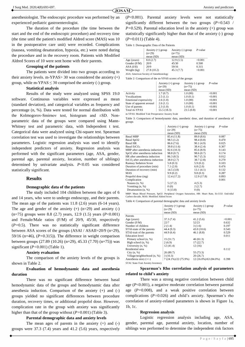
J Surg Med. 2020;4(8):693-697. Anxiety and predictors
P a g e / S a y f a | 695
anesthesiologist. The endoscopic procedure was performed by an
experienced pediatric gastroenterologist.
The duration of the procedure (the time between the
start and the end of the endoscopic procedure) and recovery time
(the time until the patient's modified Aldred score (MAS) was 10
in the postoperative care unit) were recorded. Complications
(nausea, vomiting desaturation, hypoxia, etc.) were noted during
the procedure and in the recovery room. Patients with Modified
Aldred Scores of 10 were sent home with their parents.
Grouping of the patients
The patients were divided into two groups according to
their anxiety levels. m-YPAS> 30 was considered the anxiety (+)
group, while m-YPAS ≤ 30 comprised the anxiety (-) group.
Statistical analysis
Results of the study were analyzed using SPSS 19.0
software. Continuous variables were expressed as mean
(standard deviation), and categorical variables as frequency and
percentage (n, %). Data were tested for normal distribution with
the Kolmogorov-Smirnov test, histogram and ±SD. Non-
parametric data of the groups were compared using Mann-
Whitney test and parametric data, with Independent t test.
Categorical data were analyzed using Chi-square test. Spearman
correlation test was used to investigate the relationships between
parameters. Logistic regression analysis was used to identify
independent predictors of anxiety. Regression analysis was
performed with the significant parameters (age, ASA, gender,
parental age, parental anxiety, location, number of siblings)
determined by univariate analysis. P<0.05 was considered
statistically significant.
Results
Demographic data of the patients
The study included 104 children between the ages of 6
and 14 years, who were to undergo endoscopy, and their parents.
The mean age of the patients was 11.8 (2.6) years (6-14 years).
The age and gender of the anxiety (+) (n=29) and anxiety (-)
(n=75) groups were 8.8 (2.7) years, 12.9 (1.5) years (P<0.001)
and Female/Male ratios (F/M) of 20/9, 45/30, respectively
(P=0.5). There was no statistically significant difference
between ASA scores of the groups (ASAI / ASAII=20/9 (n=29),
59/16 (n=46), (P=0.315)). The difference in weight comparison
between groups (27.89 (10.26) (n=29), 45.33 (7.70) (n=75)) was
significant (P<0.001) (Table 1).
Anxiety evaluation
The comparison of the anxiety levels of the groups is
shown in Table 2.
Evaluation of hemodynamic data and anesthesia
duration
There was no significant difference between basal
hemodynamic data of the groups and hemodynamic data after
anesthesia induction. Comparison of the anxiety (+) and (-)
groups yielded no significant differences between procedure
duration, recovery times, or additional propofol dose. However,
complication rate in the group with anxiety was significantly
higher than that of the group without (P=0.001) (Table 3).
Parental demographic data and anxiety levels
The mean ages of parents in the anxiety (+) and (-)
groups were 37.3 (7.4) years and 41.2 (5.6) years, respectively
(P<0.001). Parental anxiety levels were not statistically
significantly different between the two groups (P=0.543 /
P=0.529). Parental education level in the anxiety (+) group was
statistically significantly higher than that of the anxiety (-) group
(P=0.011) (Table 4).
Table 1: Demographic Data of the Patients
Anxiety (+) group
(n=29)
mean (SD)
Anxiety (-) group
(n=75)
mean (SD)
P-value
Age (years) 8.8 (2.7) 12.9 (1.5) <0.001
Gender (F/M) 20/9 45/30 0.500
ASA (I/II) 20/9 59/16 0.315
Weight (kg) 27.8 (10.2) 45.3 (7.7) <0.001
ASA: American Society of Anesthesiology
Table 2: Comparison of the m-YPAS scores of the groups
Anxiety (+) group
(n=29)
mean (SD)
Anxiety (-) group
(n=75)
mean (SD)
P-value
Activity 1.7 (0.7) 1.0 (00) <0.001
Vocalizations 2.5 (1.1) 1.0 (0.1) <0.001
Emotional expressivity 2.6 (1.0) 1.0 (00) <0.001
State of apparent arousal 2.6 (1.1) 1.0 (00) <0.001
Use of parents 2.3 (1.0) 1.0 (0.1) <0.001
Total m-YPAS 55.5 (18.3) 23.5 (0.8) <0.001
m-YPAS: Modified Yale Preoperative Anxiety Scale
Table 3: Comparison of hemodynamic data, anesthetic dose, and duration of anesthesia of
groups
Anxiety (+) group
(n=29)
mean (SD)
Anxiety (-) group
(n=75)
mean (SD)
P-value
Basal MBP 85.4 (11.8) 89.0 (8.6) 0.087
Basal SpO2 98.5 (1.2) 98.4 (0.8) 0.470
Basal HR 86.6 (7.6) 90.1 (4.9) 0.025
Basal EtCO2 39.8 (11.6) 38.4 (2.4) 0.307
MBP after anesthesia induction 82.6 (10.3) 85.6 (8.9) 0.143
SpO2 after anesthesia induction 98.8 (0.7) 98.6 (0.7) 0.174
HR after anesthesia induction 86.3 (8.5) 90.7 (5.3) 0.012
EtCO2 after anesthesia induction 38.0 (2.7) 38.7 (2.8) 0.270
Ramsay Sedation Score 5.8 (0.3) 5.9 (0.2) 0.103
Duration of procedure (min) 7.1 (2.0) 6.8 (2.0) 0.476
Duration of recovery (min) 14.5 (3.8) 13.1 (4.1) 0.132
MAS 9.9 (0.2) 9.8 (0.3) 0.287
Propofol dose (mg) 12.4 (17.2) 12.9 (17.8) 0.893
Complication 0.001
Nausea (n, %) 5 (17.2) 4 (5.3)
Vomiting (n, %) 0 (0) 2 (2.7)
Desaturation (n, %) 4 (13.8) 0 (0)
MBP: Mean Blood Pressure, SpO2: Peripheral Oxygen Saturation, HR: Heart Rate, Et-CO2: End-tidal
Carbon dioxide, MAS: Modified Aldred Score
Table 4: Comparison of parental demographic data and anxiety levels
Anxiety (+) group
(n=29)
mean (SD)
Anxiety (-) group
(n=75)
mean (SD)
P-value
Parents
Age (years) 37.3 (7.4) 41.2 (5.6) <0.001
Gender (F/M) 22/7 52/23 0.632
Number of children 2 (1-4) 3 (1-6) 0.197
STAI-state of the parents 44.4 (9.3) 43.0 (10.6) 0.543
STAI-trait of the parents 44.9 (6.4) 46.1 (8.8) 0.529
Education level 0.011
Primary school (n, %) 15 (51.7) 46 (61.3)
High school (n, %) 2 (6.9) 17 (22.7)
University (n, %) 12 (41.4) 12 (16)
Residential area 0.112
City (n, %) 26 (89.7) 55 (73.3)
Village/neighborhood (n, %) 3 (10.3) 20 (26.7)
Anesthesia story (+/-) 7 (24.1%)/22 (75.9%) 12 (16.0%)/63 (84.0%) 0.398
STAI: State-Trait Anxiety Inventory
Spearman’s Rho correlation analysis of parameters
related to child’s anxiety
There was a strong negative correlation between child
age (P<0.001), a negative moderate correlation between parental
age (P=0.008), and a weak positive correlation between
complications (P=0.026) and child’s anxiety. Spearman’s rho
correlation of anxiety-related parameters is shown in Figure 1a,
1b, 1c.
Regression analysis
Logistic regression analysis including age, ASA,
gender, parental age, parental anxiety, location, number of
siblings was performed to determine the independent risk factors

J Surg Med. 2020;4(8):693-697. Anxiety and predictors
P a g e / S a y f a | 696
that are effective in the development of child anxiety. Young age
was determined as an independent risk factor for increased levels
of child anxiety (OR: 0.501, P<0.001, 95% CI: 0.390-0.643).
Figure 1: Spearman’s Rho Correlation Analysis of parameters related to anxiety
Discussion
In this study, in which the anxiety level of school-age
children who underwent gastrointestinal endoscopy was
evaluated, young age was an important risk factor for anxiety
development.
Many patients and parents are most concerned about the
safety of endoscopy. Therefore, healthcare professionals
performing pediatric gastrointestinal endoscopy should consider
the anxiety of patients and parents before endoscopy [3]. Studies
have found that the presence of an overly anxious parent during
the induction of anesthesia does not benefit an anxious child and
can increase anxiety in a calm child [14,15]. Intense anxiety and
fear can cause poor cooperation, longer procedure time,
increased sedation dose, and even increased complications [16].
It is known that there is a correlation between preoperative
anxiety of children and parents' anxiety. The upcoming surgical
stress can be perceived and transmitted between children and
parents [17,18]. Emotional behaviors of parents have a
significant impact on children modeling their parents’ behavior
[19], and coping mechanisms affect children's coping
mechanisms [20]. However, such an interaction has been shown
to be strong only in preschool children and their parents (state
anxiety) [6].
In this study, routine information was given about the
procedure and anesthesia. In this study, in which the study group
was constituted by school-age children, it was observed that
there was no relationship between child’s preoperative anxiety
and parental anxiety. There was a correlation between child age,
parental age and child anxiety, and the rate of complications was
significantly higher in highly anxious children. Schoolchildren
are more independent and less affected by their parents, while
preschoolers are still more dependent on their parents [6].
A study evaluating the preoperative anxiety levels in the
parents of outpatients determined a relationship between the
maternal anxiety score and the child's age [21]. In a child cohort
evaluation including children between the ages of 2-16 years,
they found that anxiety decreased with increasing age of children
[22]. Young children are a risk factor for poor behavioral
compatibility during induction [23]. Older children express less
fear for preoperative preparations than younger children [24].
Older children can understand the necessity and requirements of
the procedure, which helps them withstand it. Preparation of the
child for the procedure and a pain-sensitive temperament can
positively affect the child's response to a procedure [25,26].
In contrast to these studies, in a study that assessed the
3-12-year age group in Turkey, there was no significant
correlation between the age of the child and maternal anxiety
score [8].
While the mean age of the group with anxiety in the
current study was 8.8 (2.7) years, it was 12.9 (1.5) years in the
group without anxiety. In this study, although the study group
comprised children in school age, the mean age of the children
with anxiety was significantly lower. Young age was an
independent risk factor anxiety. To reduce the preoperative
anxiety of school-age children, it is useful to provide appropriate
information directly to them [27].
Informing parents about procedures, communicating
with doctors, and letting parents talk to other parents in a
comparable situation reduces anxiety in parents and helps them
support their children more effectively [4]. Factors that mothers
thought would reduce maternal anxiety include getting more
information from the doctor, talking with mothers who had
anesthetic experience before, and being provided audiovisual
information. It is thought that accompanying the child to the
operating room and being present during the induction of
anesthesia will decrease maternal anxiety [8].
A systematic psychological preparation provided to both
parents and patients before endoscopy logically reduces the level
of anxiety on procedures. Although the level of education affects
the outcome of psychological preparation, it may not have a
significant effect on the level of anxiety in parents [2]. One study
reported that maternal education level had no effect on anxiety
[28], while another stated that anxiety level increased with
increasing maternal education level [29].
In our study, no information was provided to patients
and parents other than routine information, since exclusive
information and systematic psychological preparation is not
always possible in intensive polyclinic conditions. It was
observed that the education level of the parents of children with
high anxiety was higher.
Limitations
This study was conducted in Turkey. There may be
differences between pediatric gastrointestinal endoscopy-related
anxiety situations between countries with diverse cultures.
However, matching results have been emphasized in studies
conducted in dissimilar cultures [6,22-25].
The second limitation of this study is that child anxiety
risk factors were determined but their solutions were not
mentioned. Future studies are needed to reduce anxiety, based on
the results of this study.
Conclusion
This study showed that there was no interaction between
preoperative anxiety in school-age children and parental anxiety.
a b
c

J Surg Med. 2020;4(8):693-697. Anxiety and predictors
P a g e / S a y f a | 697
Young age was determined as an independent risk factor for the
development of anxiety in children. Attention should be paid to
the height of anxiety in younger patients in the school age group
and necessary precautions should be taken.
References
1. Fredette ME, Lightdale JR. Endoscopic sedation in pediatric practice. Gastrointest Endosc Clin N Am.
2008;18:739-51.
2. Riddhiputra P, Ukarapol N. Effect of systematic psychological preparation using visual illustration
prior to gastrointestinal endoscopy on the anxiety of both pediatric patients and parents. J Med Assoc
Thai. 2006;89:231-5.
3. Hagiwara SI, Nakayama Y, Tagawa M, Arai K, Ishige T, Murakoshi T, et al. Pediatric Patient and
Parental Anxiety and Impressions Related to Initial Gastrointestinal Endoscopy: A Japanese
Multicenter Questionnaire Study. Scientifica (Cairo). 2015;2015:797564. doi: 10.1155/2015/797564.
4. Kain ZN, Mayes LC, O'Connor TZ, Cicchetti DV. Preoperative anxiety in children Predictors and
outcomes. Arch Pediatr Adol Med. 1996;150:1238-45
5. Kain ZN, Mayes LC, Weisman SJ, Hofstadter MB. Social adaptability, cognitive abilities, and other
predictors for children's reactions to surgery. J Clin Anesth. 2000;12:549-54.
6. Cui X, Zhu B, Zhao J, Huang Y, Luo A, Wei J. Parental state anxiety correlates with preoperative
anxiety in Chinese preschool children. J Paediatr Child Health. 2016;52:649-55.
7. Karibe H, Aoyagi-Naka K, Koda A. Maternal anxiety and child fear during dental procedures: a
preliminary study. J Dent Child. 2014;81:72-7.
8. Cagiran E, Sergin D, Deniz MN, Tanattı B, Emiroglu N, Alper I. Effects of sociodemographic factors
and maternal anxiety on preoperative anxiety in children. J Int Med Res. 2014;42:572-80.
9. Spielberger CD, ed. Manual for the State-Trait Anxiety Inventory (STAI: FormY). Palo Alto,
California: Consulting Psychologists Press,1983; 4–26.
10. Kain ZN, Mayes LC, Cicchetti DV, Bagnall AL, Finley JD, Hofstadter MB. The Yale Preoperative
Anxiety Scale: how does it compare with a “gold standard”? Anesthesia & Analgesia. 1997;85:783-8.
11. Kim H, Jung SM, Yu H, Park SJ. Video distraction and parental presence for the management of
preoperative anxiety and postoperative behavioral disturbance in children: a randomized controlled
trial. Anesthesia & Analgesia. 2015;121:778-84.
12. Thung A, Tumin D, Uffman JC, Tobias JD, Buskirk T, Garrett W, et al. The Utility of the Modified
Yale Preoperative Anxiety Scale for Predicting Success in Pediatric Patients Undergoing MRI
Without the Use of Anesthesia. J Am Coll Radiol. 2018;15:1232-7.
13. Hatipoğlu Z, Kırdök O, Özcengiz D. Validity and reliability of the Turkish version of the modified
Yale Preoperative Anxiety Scale Turk. J Med Sci. 2019;49:730-7.
14. Kain ZN, Caldwell-Andrews AA, Maranets I, Nelson W, Mayes LC. Predicting which child–parent
pair will benefit from parental presence during induction of anesthesia: a decision-making approach.
Anesth Analg. 2006;102:81-4.
15. Caldwell-Andrews AA, Kain ZN, Mayes LC, Kerns RD, Ng D. Motivation and maternal presence
during induction of anesthesia. Anesthesiology. 2005;103:478-83.
16. Drossman DA, Brandt LJ, Sears C, Li Z, Nat J, Bozymki EM. A preliminary study of patients’
concerns related to GI endoscopy. Am J Gastroenterol. 1996;91:287-91.
17. Fincher W, Shaw J, Ramelet AS. The effectiveness of a standardised preoperative preparation in
reducing child and parent anxiety: A single-blind randomised controlled trial. J Clin Nurs.
2012;21:946-55.
18. Arai YC, Kandatsu N, Ito H, Sato Y, Satake Y, Mizutani M. Induction and emergence behavior of
children undergoing general anesthesia correlates with maternal salivary amylase activity before the
anesthesia. Acta Anaesthesiol. 2008;52:285-8.
19. McGraw T. Preparing children for the operating room: psychological issues. Can J Anaesth.
1994;41:1094-103.
20. Perrin EC, Ayoub CC, Willett JB. In the eyes of the beholder: family and maternal influences on
perceptions of adjustment of children with a chronic illness. J Dev Behav Pediatr. 1993;14:94-105.
21. Litman RS, Berger AA, Chhibber A. An evaluation of preoperative anxiety in a population of parents
of infants and children undergoing ambulatory surgery. Paediatr Anaesth 1996;6:443-7.
22. MacLaren JE, Thompson C, Weinberg Metal. Prediction of preoperative anxiety in children:who is
most accurate? Anesth Analg. 2009;108:1777-82.
23. Varughese AM, Nick TG, Gunter J, Wang Y, Kurth CD. Factors predictive of poor behavioral
compliance during inhaled induction in children. Anesth Analg. 2008;107:413
24. Hatava P, Olsson GL, Lagerkranser M. Preoperative psychological preparation for children
undergoing ENT operations: a comparison of two methods. Paediatr Anaesth. 2000;10:477-86
25. Kleiber C, Craft-Rosenberg M, Harper DC. Parents as distraction coaches during i.v. insertion: a
randomized study. J Pain Symptom Manage. 2001;22:851-61.
26. Ghazali DA, Faure JP, Breque C, Oriot D. Evaluation of stress patterns during simulated laparoscopy
in residency. Minerva Chir. 2016;71:252-61.
27. Litke J, Pikulska A, Wegner T. Management of perioperative stress in children and parents. Part I the
preoperative period. Anaesthesiol Intensive Ther. 2012;44:165-9.
28. Shevde K and Panagopoulos G. A survey of 800 patients’ knowledge, attitudes and concerns
regarding anesthesia. Anesth Analg. 1991;73:190-8.
29. Power NM, Howard RF, Wade AM, Franck LS. Pain and behaviour changes in children following
surgery. Arch Dis Child. 2012;97:879-84.
This paper has been checked for language accuracy by JOSAM editors.
The National Library of Medicine (NLM) citation style guide has been used in this paper.
P a g e / S a y f a | 698
Assessment of ear metric properties in young Turkish adults Genç Türk erişkinlerde kulak metrik özelliklerinin değerlendirilmesi
Emine Petekkaya 1, Sema Polat 2, Ayşe Gül Kabakçı 2, Yiğit Çevik 2
How to cite/Atıf için: Petekkaya E, Polat S, Kabakçı AG, Çevik Y. Assessment of ear metric properties in young Turkish adults. J Surg Med. 2020;4(8):698-701.
J Surg Med. 2020;4(8):698-701. Research article DOI: 10.28982/josam.774357 Araştırma makalesi
1 Department of Anatomy, Kastamonu University,
Faculty of Medicine, Kastamonu, Turkey 2 Department of Anatomy, Cukurova University,
Faculty of Medicine, Adana, Turkey
ORCID ID of the author(s)
EP: 0000-0002-5366-2425
SP: 0000-0003-4695-0514
AGK: 0000-0001-7144-8759
YÇ: 0000-0002-2691-3284
Corresponding author/Sorumlu yazar:
Emine Petekkaya
Address/Adres: Kastamonu Üniversitesi, Tıp
Fakültesi, Anatomi Anabilim Dalı, Kastamonu,
Türkiye
E-mail: [email protected]
�
Ethics Committee Approval: This study was approved
by Ethics Committee of Cukurova University on
1/10/2020; Meeting number: 95; Decision number:
36. All procedures in this study involving human
participants were performed in accordance with the
1964 Helsinki Declaration and its later amendments.
Etik Kurul Onayı: Bu çalışma, Çukurova Üniversitesi
Etik Kurulu (10.01.2020: Toplantı no: 95; Karar no:
36) tarafından onaylanmıştır. İnsan katılımcıların
katıldığı çalışmalardaki tüm prosedürler, 1964
Helsinki Deklarasyonu ve daha sonra yapılan
değişiklikler uyarınca gerçekleştirilmiştir.
�
Conflict of Interest: No conflict of interest was
declared by the authors.
Çıkar Çatışması: Yazarlar çıkar çatışması
bildirmemişlerdir.
�
Financial Disclosure: The authors declared that this
study has received no financial support.
Finansal Destek: Yazarlar bu çalışma için finansal
destek almadıklarını beyan etmişlerdir.
�
Published: 8/30/2020
Yayın Tarihi: 30.08.2020
Copyright © 2020 The Author(s)
Published by JOSAM This is an open access article distributed under the terms of the Creative
Commons Attribution-NonCommercial-NoDerivatives License 4.0 (CC
BY-NC-ND 4.0) where it is permissible to download, share, remix,
transform, and buildup the work provided it is properly cited. The work
cannot be used commercially without permission from the journal.
Abstract
Aim: The ear is a critical component of the human face functionally and aesthetically. Studies in recent years have reported that the
morphological properties of the outer ear show substantial differences based on sex and ethnic group. The data obtained in these studies
are guiding for plastic surgeons.
Methods: This cross-sectional study was carried out on a Turkish population. Ear axis, antihelix angle, ear length, ear width, earlobe
length and earlobe width measurements were made on 191 healthy adult volunteers (106 women, 85 men) between the ages of 18-25
years. Ear metric values mentioned above were compared between the genders, and correlation analysis was conducted for all.
Results: All values except the antihelix angle were larger in males (P=0.0486; P<0.001; P=0.004; P<0.001; P=0.038 for ear axis,
length, width, ear lobule length and width respectively). The ear axis was positively correlated with the antihelix angle, the ear width
and the ear lobule width and the ear length was positively correlated with the ear width and ear lobule length (P<0.001 for both).
Conclusion: Although our study has some limitations it includes noteworthy data about outer ear metric values of the young adult
Turkish population which we believe is going to be beneficial to standardization and optimization in ear surgery.
Keywords: Pinna, Metric, Aesthetic, Anatomy
Öz
Amaç: Kulak, fonksiyonel ve estetik olarak insan yüzünün son derece önemli bir bileşenidir. Son yıllarda yapılan çalışmalar dış kulağın
morfolojik özelliklerinin cinsiyete ve etnik gruba göre makul farklılıklar gösterdiğini raporlamıştır. Bu çalışmalardan elde edilen veriler
plastik cerrahlar için yönlendiricidir.
Yöntemler: Çalışmamız Türk popülasyonu üzerinde gerçekleştirilmiştir. 18-25 yaşları arasında 191 sağlıklı yetişkin gönüllünün (106
kadın, 85 erkek) kulak ekseni, antiheliks açısı, kulak uzunluğu, kulak genişliği, kulak memesi uzunluğu ve kulak memesi genişliği
ölçümleri yapılmıştır. Tüm parametreler cinsiyetler arasında karşılaştırılmıştır ve hepsi için korelasyon analizi yapılmıştır.
Bulgular: Anti heliks açısı dışında tüm değerler erkeklerde kadınlara göre daha büyük bulundu (P=0,0486; P<0,001; P=0,004; P<0,001;
P=0,038 sırasıyla kulak ekseni, uzunluğu, genişliği, kulak memesi uzunluğu ve genişliği için). Kulak ekseni, antiheliks açısı, kulak
genişliği ve kulak memesi genişliği ile pozitif korelasyon gösterdi ve kulak uzunluğu, kulak genişliği ve kulak memesi uzunluğu ile
pozitif korelasyon gösterdi (her ikisi için P<0,001).
Sonuç: Çalışmamızın bazı sınırlılıkları olmakla birlikte, kulak cerrahisinde standardizasyon ve optimizasyon için faydalı olacağına
inandığımız genç erişkin Türk popülasyonunun dış kulak ölçüm değerleri hakkında dikkate değer veriler içermektedir.
Anahtar kelimeler: Pinna, Metrik, Estetik, Anatomi
J Surg Med. 2020;4(8):698-701. Assessment of ear metric properties in Turkish youths
P a g e / S a y f a | 699
Introduction
Reconstruction of the auricle (pinna) is technically
difficult due to some factors. Before changing the shape of the
ear, plastic surgeons need to have normal anatomical data that
determine the normal shape, size, and direction limits of the ear.
The auricle is one of the descriptive properties of the face. It is
not a focus of beauty itself, but deviations in its symmetry,
direction or projection may be easily noticed and divert a face
from a sense of aesthetics [1]. Accordingly, some studies
conducted recently have reported that every part of the outer ear
is morphologically unique and shows substantial differences in
individuals and population groups [2-4]. Additionally, the lateral
notch found in its three-dimensional topography is a unique
structure that contributes to a normal outer ear with its vertical
placement and characteristic contour structure [1,2]. This unique
structure of the ear may be helpful in determining the identity of
individuals and in terms of anthropology and forensic medicine
[4, 5].
A deformation in the shape and size of the auricle may
indicate a possible congenital anomaly or syndrome in the
patient [3]. The auricle is a flexible structure consisting of five
main anatomical regions as the concha, helix, antihelix, tragus,
and lobule formed by skin, cartilage and fat tissues [6]. A
cartilaginous notch on the helix is known as “Darwin’s tubercle”,
and it is a vestigial residue corresponding to the tip of the ear of
some animals [4]. The contour of the auricle is determined by the
configuration of the elastic cartilage frame [1, 4]. The skin of its
lateral and medial surfaces contains hair with both sebaceous and
sudoriferous glands. While the skin is laterally tightly connected
to the perichondrium, medially, it is only loosely connected. The
cartilage of the auricle is an extension of the meatus acusticus
externus cartilage, and it is fixed on the cranium with a few
ligaments and muscle connections [1].
Embryologically, the outer ear starts to form within 6
weeks of pregnancy and completes its formation up to the 18th
week. Six tissue elevations known as the hillocks of His develop.
Each of these elevations shape different parts of the outer ear.
The first three elevations originate from the first branchial arch
and combine to form the tragus, helix, and antitragus, whereas
the next three elevations originate from the second branchial arch
and form the antihelix and concha. The ear lobule (lobule) does
not originate from these elevations [6]. Tragus, on the other
hand, does not contain cartilage tissue although it develops from
the first branchial arch. Incomplete fusional conglomerations of
the branchial arches lead to malformations of the outer ear.
During arrangement of the formed deformities with otoplasty,
instead of individual aesthetical perceptions, some ratios need to
be used. Therefore, measurements that will provide
reconstruction and aesthetical perception compatible with the
social population are required. In many studies, the structure of
the ear has been examined metrically to reveal societal
differences. Most of such studies have reported that ear
dimensions are metrically variable in individuals and
populations, ear measurement parameters are larger in men than
women, and the ears show bilateral asymmetry [2,4]. It may be
stated that data obtained from studies making metric
observations provide evidence-based indicators regarding the
ratios of an aesthetically pleasing ear. In summary, ear
morphology is aesthetically important and shows differences
between ethnic groups and genders [2,3]. For this reason, in our
study, which we want to become a significant guide for the
Turkish population in auricular surgery, in Turkish male and
female individuals, ear axis, antihelix angle, ear length, ear
width, lobule length and lobule width measurements were made,
and they were compared between the genders. Correlation
analysis was conducted for all parameters.
Materials and methods
All authors declare that the study was carried out in
accordance with the World Medical Association Helsinki
Declaration "Ethical Principles for Medical Research on Human
Subjects".
Participants
This cross-sectional study was conducted with 191
healthy adult volunteers (106 women, 55.497%; 85 men,
44.503%) between the ages of 18-25 years after obtaining
approval from the Ethics Board of Cukurova University (Ethical
permission no:2020/95). Participants were informed about the
study and gave individual consent. It was aimed to include
healthy individuals in the study. For this reason, the inclusion
criteria were determined as not having had any facial surgical
operation and not having a history of any neurological disease.
Among the 200 volunteers who were included at the beginning
of the study, 9 volunteers who later refused to participate were
excluded.
Measurements
The photographs of the participants which covered their
entire profile were taken with a Digital SLR camera with
constant photographing values (Canon EOS 80D; ISO 100 f /
4.5). The photography system was set up by fixing the camera
with an adjustable tripod. The tripod height was adjusted based
on the height of each individual. In the photography procedure,
each individual was firstly asked to stand up and relax, and they
were then put into the Frankfurt Horizontal Plane (FHP) position,
which is the natural head position. FHP is considered the natural
anatomical position of the human skull. This plane passes
through the lower edge of the orbita and the upper edge of the
external auditory meatus and is a significant reference for
surgeons [7]. Reports have stated that FHP is an adequately
reliable reference plane for different treatments and studies [8].
For this reason, in our study, we preferred to use FHP while
making the measurements. The images taken on this plane were
then transferred to a computer. The ear axis, antihelix angle
(angle between the antihelix and the vertical), ear length, ear
width, lobule length and lobule width were measured by using
the Image J 1.52a program. The physiognomic ear length was
measured by following the long axis of the ear as the direct
distance between the supra-auricula and sub-auricula. While
measuring the ear length, the distance between the very tip of the
lobule to the farthest point of the auricle was used. The
physiognomic ear width was measured as the direct distance
between the pre-auricula and post-auricula. The ear width was
measured in two ways as a and b (Figure 1). The lobule length
was measured as the direct distance between the incisura
intertragica and the deepest point in the sub-auricula. The lobule

J Surg Med. 2020;4(8):698-701. Assessment of ear metric properties in Turkish youths
P a g e / S a y f a | 700
width was measured at the center point of the lobular length as
the distance between the anterior lobule and posterior lobule. The
ear axis angle was considered the angle between the axis where
the ear length measurement was made and the vertical plane. The
measurements were made with a precision of 1/100 mm.
Figure 1: Presentation of ear width a and b measurement method on a volunteer
Statistical analysis
For the statistical analysis of the measurement results,
the SPSS Windows version 24.0 package software was used. As
n>30, Student’s t-test was used to compare the variables between
genders. The values are presented as mean (standard deviation).
As n>30, the Pearson’s correlation coefficient was used for
correlation analysis. In all analyses, P<0.05 was considered
statistically significant.
Results
In the comparison of the metric properties of the ear
between the genders, almost all parameters turned out to be
larger in the males than the females: The ear axis, ear length, ear
width a and b, lobule length and width were all significantly
higher in males (P=0.049, P<0.001, P<0.001, P=0.004,
P<0.001 and P=0.039, respectively). There was no statistically
significant difference between the genders in terms of the
antihelix angle (Table 1).
Correlation analyses between ear metric properties
yielded that there was a positive strong correlation between the
ear axis and antihelix angle (r=0.417, P=0.001), the ear axis and
ear width b (r=0.307, P=0.001) and the ear axis and lobule width
(r=0.270, P=0.001). There was a significant positive correlation
between the antihelix angle and ear width b (r=0.168, P=0.020),
the ear length and ear width a (r=0.153, P=0.034), a positive
strong correlation between the ear length and ear width b
(r=0.660, P=0.001), ear length and lobule length (r=0.603,
P=0.001) and ear length and lobule width (r=0.286, P=0.001).
There were positive, strong and significant correlations between
the ear width b and lobule length (r=0.361, P=0.001), ear width
b and lobule width (r=0.530, P=0.001) and lobule length and
lobule width (r=0.437, P=0.001) (Table 2).
Table 1: Comparison of ear metric properties between sexes
Male (n=85) Female (n=106) P-value
Mean SD Mean SD
Ear axis (°) 16.29 2.203 15.62 2.416 0.049
Antihelix angle (°) 61.95 8.586 60.76 9.152 0.360
Ear length (cm) 74.19 6.714 68.75 7.01 <0.001
Ear width a (cm) 41.04 4.584 37.6 3.987 <0.001
Ear width b (cm) 31.69 3.848 30.13 3.492 0.004
Ear lobule length (cm) 18.18 3.083 16.45 2.652 <0.001
Ear lobule width (cm) 22.32 3.848 21.21 3.306 0.039
SD: Standard deviation, cm: Centimeters
Table 2: Correlation analysis between ear metric properties
Variable 1 Variable 2 r P-value
Ear axis (°) Antihelix angle (°) 0.417 <0.001
Ear axis (°) Ear width b (cm) 0.307 <0.001
Ear axis (°) Earlobe width (cm) 0.270 <0.001
Antihelix angle (°) Ear width b (cm) 0.168 0.020
Ear length (cm) Ear width a (cm) 0.153 0.034
Ear length (cm) Ear width b (cm) 0.660 <0.001
Ear length (cm) Earlobe length (cm) 0.603 <0.001
Ear length (cm) Earlobe width (cm) 0.286 <0.001
Ear width b (cm) Earlobe length (cm) 0.361 <0.001
Ear width b (cm) Earlobe width (cm) 0.530 <0.001
Earlobe length (cm) Earlobe width (cm) 0.437 <0.001
cm: Centimeters, r: Correlation coefficient
Discussion
Knowing about the morphological properties and
normal anatomical dimensions of the auricle is important in the
diagnosis and surgical planning of congenital and acquired
malformations [5]. The objectives of ear surgery include basing
the ratios of an aesthetically pleasing ear on not the personal
aesthetical views of surgeons but evidence-based data.
Therefore, it is important to create standard measurements.
Human auricles have a unique shape and are a well-known part
of our auditory system. The shape of the outer ear is far from
random, and it has developed to allow spatial localization of
sounds. As it performs shape-specific filtering, it mediates
monoaural cues, while it mediates binaural cues due to the
bilateral localizations of the right and left ears [9]. The distance
of the ear to the lateral orbital edge is approximately the same as
the height of the ear [6]. The apex of the ear should be on the
level of the eyebrows. The auriculocephalic angle is ideally
between 25 and 35 degrees, while angles of larger than 45
degrees are considered too high. A scapha-conchal angle of
lower than 90 degrees is ideal, and an angle larger than 90
degrees leads to a less aesthetical appearance by causing the
scapha and fossa triangularis to protrude anteriorly rather than
laterally. The helical edge needs to protrude from the head by 15
to 20 mm [1].
According to published reports, the average length of an
ear is 55 to 65 mm, and the average width is 55% of the length
[1]. Moreover, it is known that the morphology of the ear differs
between genders [2,3]. In their study which investigated the
dimensions of the auricle based on the factor of gender, Verma et
al. [5] found that the two ears were symmetrical, and
measurements (upper-tragus ear length, lower-tragus ear length,
ear width, concha length, concha width, lobular height, lobular
width) were higher among men than women [5]. In our study
with healthy Turkish volunteers between the ages of 18-25 years,
similar to previous studies, the ear axis, ear length and ear width
were larger in the males than females. On the other hand, Senthil
Kumar and Selvi [3] found that, although the total ear length was
larger in men, the lobule length and width were almost the same
between men and women [3]. In our study, the lobule length and
width were significantly larger in males than females. It is
possible that this significant finding could have been caused by
the differences in ethnic groups and ages. However, it should still
be kept in mind looking at our findings that the difference
between the genders was larger in terms of the ear length and ear
width a. As it was previously reported that there is no significant
relationship between head size (height /width) and ear size
(length / width) [2], our study did not assess head dimensions.

J Surg Med. 2020;4(8):698-701. Assessment of ear metric properties in Turkish youths
P a g e / S a y f a | 701
Previous studies have observed that the antihelix angle
does not vary based on the ethnic group, but there is a
statistically significant reduction by increasing age in both men
and women. It is thought that the elongation of the ear might
affect this [2]. The results of our study also showed that
difference in genders does not create a significant difference in
terms of the antihelix angle. Not finding a significant difference
between ethnic groups and gender in this parameter may be
related to the fact that the antihelix angle shows a large variation
among individuals. Consequently, the data suggest that the
antihelix angle plays an exceedingly small role in defining a
normal ear. In addition to this, another significant aspect of the
shape of the outer ear is its uniqueness from one person to
another on the level seen in fingerprints and facial properties [9].
Considering that, in the embryonic period, the antihelix and
concha develop from different tissue elevations than the tragus,
helix and antitragus [6], we recommend the necessity to
investigate the place of the antihelix in more detail in terms of
the significance of the ear in identification in forensic cases. The
correlation between the ear axis and the antihelix suggests that
antihelix characteristics may be also important for the function of
sound localization.
The auricle grows based on age. The auricle grows fast
in the first 3 years of life and reaches approximately 85% of the
adult size at ages 6 to 8 and 95% at ages 8 to 10 [1]. The auricle
reaches its adult size at the age of 13 in men and 12 in women
[3]. In the last few years, ear dimensions in different ethnic
groups have been investigated in various studies where direct
and indirect anthropometrics and photography were used [10].
Although there are ethnic differences, all these studies have
reported that the auricle’s dimensions significantly increase by
age in both men and women due to changes in elastic fibers after
adulthood (even after reaching skeletal maturity) [3,10]. Previous
studies showed that age-related dimensional changes are not the
same for all variables; the ear length continues to increase faster
and for a longer time in comparison to the ear width [10]. While
our study provides findings on young adults, considering this
continuing development of the ear, it will be important to collect
data from different age groups to create a broad guide for
Turkish plastic surgery. Our study showed a correlation between
ear length and ear width. The further increase in the ear length in
further years than the ear width may reduce this correlation.
The RoB 2 tool (22 August 2019 version) was used to
estimate potential bias. Analysis showed low risk for
randomization process, deviations from intended interventions,
missing outcome data and some concerns for measurement of the
outcome. The low risk for overall bias can be counted among the
strengths of this study.
Limitations
The auricle reaches its adult size at the age of 12-13 [3].
However, the ear length and width continue to increase by age
[10]. In our study, measurements were made only in a certain age
group, and the lack of age-related comparisons are an important
limitation. Besides, body mass index, which is an indicator of
body fat level, was associated with soft tissue thickness and
positively correlated with ear length and width [11]. Body mass
index of the volunteers was not calculated in this study. This
prevents the study from providing more extensive information.
Furthermore, the contribution of the outer ear to the aesthetic
perception is not limited to its own metric properties. The
distance of the outer ear to the eyes, the location on the head, the
auriculocephalic angle, scapha-conchal angle are also critical
parameters for aesthetic perception. Although our data give clues
about aesthetic appearance, we do not deal with all aspects of
aesthetic perception in the absence of this information.
If future studies are designed to overcome the
limitations of our study in more comprehensive groups, it will be
possible to create a more consistent and useful atlas for ear
surgeries. In addition, there is a need of literature for studies
examining the contribution of the outer ear shape to hearing
function. The morphology of the external auditory canal has
previously been studied radiologically [12]. The evaluation of
the features of the outer ear shape and the outer ear canal can be
an interesting and unexpected study.
Conclusions
Studies on the ears usually aim at treatment of
congenital deformities and development of surgical techniques
for reconstruction of ears that are traumatically injured. Every
individual wants to have ears that look normal and aesthetically
pleasing, and this demand is the highest among individuals with
congenital or acquired deformities of the ear. Achievement of
good functional and aesthetical rehabilitation does not only
increase the self-confidence of the person, but it also means
better social acceptance. The dimensions of the outer ear and its
various parts vary in different ethnic groups, and this requires
surgeons to base their reconstructions on data collected from
each ethnic group. In this sense, we believe our study includes
noteworthy data regarding the young adult Turkish population.
Acknowledgements
We would like to thank for our gone but not forgotten
Prof. Dr. Ahmet Hilmi Yücel for supervising on us.
References
1. Pham TV, Early SV, Park SS. Surgery of the auricle. Facial Plast Surg. 2003;19(1):53-74.
2. Alexander KS, Stott DJ, Sivakumar B, Kang N. A morphometric study of the human ear. J Plast
Reconstr Aesthet Surg. 2011;64(1):41-7.
3. Senthil Kumar B, Panneer Selvi G. Morphometry of Ear Pinna in Sex Determination. Int J Anat Res.
2016;4(2):2480-4.
4. Krishan K, Kanchan T, Thakur S. A study of morphological variations of the human ear for its
applications in personal identification. Egypt J Forensic Sci. 2019;9(6). doi:.10.1186/s41935-019-
0111-0
5. Verma P, Sandhu HK, Verma KG, Goyal S, Sudan M, Ladgotra A. Morphological Variations and
Biometrics of Ear: An Aid to Personal Identification. J Clin Diagn Res. 2016;10(5):138-42.
6. Cubitt JJ, Chang LY, Liang D, Vandervord J, Marucci DD. Auricular reconstruction. J Paediatr Child
Health. 2019;55(5):512-7.
7. Pittayapat P, Jacobs R, Bornstein MM, Odri GA, Lambrichts I, Guy Willems G, et al. Three-
dimensional Frankfort horizontal plane for 3D cephalometry: a comparative assessment of
conventional versus novel landmarks and horizontal planes. Eur J Orthod. 2018;40(3):239-48.
8. Meiyappan N, Tamizharasi S, Senthilkumar KP, Janardhanan K. Natural head position: An overview.
J Pharm Bioallied Sci. 2015;7(2):424-7.
9. Claes P, Reijniers J, Shriver MD, Snyders J, Suetens P, Nielandt J, et al. An investigation of matching
symmetry in the human pinnae with possible implications for 3D ear recognition and sound
localization. J Anat. 2015;226(1):60-72.
10.Japatti SR, Engineer PJ, Reddy BM, Tiwari AU, Siddegowda CY, Hammannavar RB. Anthropometric
Assessment of the Normal Adult Human Ear. Ann Maxillofac Surg. 2018;8(1):42-50.
11.Surmeli M, Deveci I, Canakci H, Canpolat MS, Karabulut B, Yilmaz AAS. Effect of Body Mass Index
on Auricular Morphology and Auditory Functions. Ear Nose Throat J. 2019;98(7):E81-E86.
12.Çalışkan S, Çetı̇n H, Akkaşoğlu S. Morphometry of the external auditory canal: Radiological study. J
Surg Med. 2020;4(1):76-9.
This paper has been checked for language accuracy by JOSAM editors.
The National Library of Medicine (NLM) citation style guide has been used in this paper.

P a g e / S a y f a | 702
Management of resistant de Quervain tenosynovitis with local
anesthetic (neural therapy): A case report
Dirençli de Quervain tenosinovitinin lokal anestezik ile tedavisi (nöralterapi): Olgu sunumu
Hüma Bölük Şenlikci 1, Özden Sibel Odabaşı Yılmaz 2, Hüseyin Nazlıkul 3
How to cite / Atıf için: Şenlikci HB, Yılmaz ÖSO, Nazlıkul H. Management of resistant de Quervain tenosynovitis with local anesthetic (neural therapy): A case report. J Surg
Med. 2020;4(8):702-703.
J Surg Med. 2020;4(8):702-703. Case report DOI: 10.28982/josam.679149 Olgu sunumu
1 Başkent University, Faculty of Medicine,
Department of Physical Medicine and
Rehabilitation, Ankara, Turkey 2 Sincan State Hospital, Ankara, Turkey
3 International Federation of Medical Association
of Neural Therapy (IFMANT), Turkey
ORCID ID of the author(s)
HŞ: 0000-0001-6771-3265
ÖSOY: 0000-0003-1727-0050
HN: 0000-0002-9746-5220
Corresponding author / Sorumlu yazar:
Hüma Bölük Şenlikci
Address / Adres: Başkent Üniversitesi Tıp
Fakültesi, Fiziksel Tıp ve Rehabilitasyon Bölümü,
Ankara, Türkiye
E-mail: [email protected]
�
Informed Consent: The authors stated that the
written consent was obtained from the patients
presented with images in the study.
Hasta Onamı: Yazarlar çalışmada görüntüleri ile
sunulan hastalardan yazılı onam alındığını ifade
etmiştir.
�
Conflict of Interest: No conflict of interest was
declared by the authors.
Çıkar Çatışması: Yazarlar çıkar çatışması
bildirmemişlerdir.
�
Financial Disclosure: The authors declared that
this study has received no financial support.
Finansal Destek: Yazarlar bu çalışma için finansal
destek almadıklarını beyan etmişlerdir.
�
Published: 8/30/2020
Yayın Tarihi: 30.08.2020
Copyright © 2020 The Author(s)
Published by JOSAM This is an open access article distributed under the terms of the Creative
Commons Attribution-Non Commercial-NoDerivatives License 4.0 (CC
BY-NC-ND 4.0) where it is permissible to download, share, remix,
transform, and buildup the work provided it is properly cited. The work
cannot be used commercially without permission from the journal.
Abstract
De Quervain tenosynovitis is the most common reason of lateral wrist pain. Treatment consists of splinting, non-steroid anti-
inflammatory drugs, and corticosteroid injections. Neural therapy (NT) is a treatment modality using injections with local
anesthetics for diagnosis and treatment. Musculoskeletal disorders can be cured by NT. We herein present the management of
resistant de Quervain tenosynovitis by neural therapy in a 45-year-old male patient, who was referred to our outpatient clinic
with the complaint of right wrist pain lasting over 2 years. His pain was 7/10 based on visual analog scale which had gradually
increased. The patient also stated that he could not lift bags and had experienced neck pain 2 years ago. He was engaged in the
neural therapy program with the diagnosis of cervical discopathy and tenosynovitis. After 3 sessions he had improvement in
functional outcome and pain. In the treatment of resistant de Quervain tenosynovitis accompanying cervical discopathy, neural
therapy is an effective and safe method.
Keywords: De Quervain tenosynovitis, Local anesthtetic, Neuraltherapy
Öz
De Quervain tenosinoviti lateral el bilek ağrısının en sık nedenidir. Tenosinovitin tedavisinde splintleme, non-steroid anti
inflamatuar ilaçlar ve kortikosteroid enjeksiyonları kullanılır. Nöralterapi (NT), lokal anestezikler kullanılarak yapılan bir tanı
ve tedavi yöntemidir. Kas iskelet sistemi bozuklukları NT yaklaşımıyla tedavi edilebilir. Bu vaka sunumunda dirençli de
Quervain tenosinovitinin nöralterapi yaklaşımıyla yönetimi sunulmuştur.45 yaşında masabaşı işe sahip erkek hasta
polikliniğimize 2 yıldan uzun süredir devam eden sağ el bilek ağrısı şikayetiyle başvurdu. Vizüel analog skalaya göre hastanın
ağrısı 7/10 idi ve günden güne ağrısının arttığını ve çanta taşımada sorun yaşadığından şikayetçi idi. Hikayesinde 2 yıl önce
boyun ağrısı mevcuttu. Nöralterapi programına servikal diskopati ve tenosinovit tanılarıyla alındı. 3 seans sonra hastanın
fonksiyonel son durumu ve ağrısında iyileşme saptandı. Servikal diskopati ile beraber dirençli de Quervain tenosinovitinin
tedavisinde nöral terapi efektif ve güvenli bir method olarak uygulanabilir.
Anahtar kelimeler: De Quervain tenosinoviti, Lokal anestezik, Nöralterapi
Introduction
De Quervain tenosynovitis, which was identified by Fritz de Quervain in 1895, occurs
by stenosing abductor pollicis longus and extensor pollicis brevis tendons in the first dorsal
extensor compartment of the wrist. Being the most common reason of lateral wrist pain, it is
more frequently seen in the middle-aged women’s dominant hand. Symptoms exacerbate with
repeated ulnar deviation and thumb extension [1].
While its etiology is unclear, mixoid degeneration, fibrose tissue deposits and
increased vascularity seems responsible in the acute inflammation of the synovial sheath.
Tendon sheath thickening due to fibrose deposits in dorsal extensor compartment is blamed for
stenosis [2]. It is diagnosed by physical examination: Pain in the wrist is present in Finkelstein
test, which involves ulnar deviation. Plain radiography may be helpful in the absence of clinical
findings [1,2]. Treatment of the tenosynovitis consists of splinting, non-steroid anti-
inflammatory drugs and corticosteroid injections. Clinical symptoms may regress without any
intervention. Surgery is required when the patient does not respond to medical treatment and
injections [2].
Neural therapy (NT) is a treatment modality involving injections of local anesthetics
for diagnosis and therapy. Its indications include functional disorders, inflammatory diseases,
and acute or chronic pain [3-5]. NT can be divided into local (ex. Infiltration of trigger points)
and segmental therapy (ex. sympathetic ganglia, nerve roots, and peripheral nerves) [6-8].
We herein present the management of resistant de Quervain tenosynovitis by neural
therapy.

J Surg Med. 2020;4(8):702-703. Neural therapy in de Quervain tenosynovitis
P a g e / S a y f a | 703
Case presentation
A 45-year-old male patient with a desk job was referred
to our outpatient clinic with the complaint of right wrist pain
lasting over 2 years. He stated that his pain had increased, and he
was unable to lift bags recently. Prolonged computer use on his
desk lead to pain in his right wrist, which was 7/10 on visual
analog scale (VAS). 2 years ago, he underwent cervical magnetic
resonance imaging due to neck pain, which revealed C5-6 and
C6-7 protrusions without root compression. Medical treatment
reduced his neck pain.
He had no history of any chronic diseases or surgery.
He did not have obesity or intestinal problems, but he led a
sedentary life. In addition, he underwent treatment in his tooth
number 46.
In physical examination, Finkelstein test was positive on
his right wrist. Bilaterally C2 and right C3 transverse process
sensitivity was present on palpation. Increased skin turgor and
hyperalgesia were detected on the right C6-7 segments. Motor
and sensory examination were normal. Right rotation of lower
cervical vertebrae was limited, and while there was no limitation
in neck flexion, it was painful. Investigation of trigger points on
flexor and extensor muscles of forearm was negative. Although
he had received physical therapy, medical treatment,
corticosteroid injection and used splints before, there had been
no significant decrease in pain or improvement in function.
He was engaged in the neural therapy program with the
diagnosis of cervical discopathy, tenosynovitis and interference
field (teeth). First session included tendon sheath and segmental
intradermal injections (C5-6-7-8) along with T1-8 intradermal
injections to regulate the sympathetic nervous system that
innervates the upper extremity. His symptoms substantially
decreased following the first session, but he had discomfort in
using his hand in daily activities. Lower cervical vertebral
limitation persisted. Injection of interference field (teeth), facet
joints of C5-6-7-8 vertebras and tendon sheath was performed in
the second session of the therapy. After the third session, his
symptoms and sensitivity with palpation of the transverse
processes showed significant improvement. Informed consent
was obtained from the patient.
Discussion
De Quervain tenosynovitis is a stenosing tenosynovitis
which occurs with repeated ulnar deviation and thumb extension.
Diagnosis can be made with physical examination. Splinting,
non-steroid anti-inflammatory drugs, physical therapy modalities
and corticosteroid injections can be used for treatment [1].
Recently, it has been shown that de Quervain tenosynovitis
involves tissue inflammation [9]. Local anesthetics have anti-
inflammatory effects around the tendons in the fibro-osseous
canal. This anti-inflammatory effect may arise from the injection
of local anesthetics. Additionally, segmental intradermal therapy
involving the C5-6-7-8 facet joints may help decrease
sympathetic activity and pathological signs in supra-spinal
levels. Sympathetic nervous system is known to play a role in
chronic musculoskeletal pain due to the memory of its neurons.
Local anesthetics also may cancel this memory [5,10].
Interference fields are regions that lead to increased
sympathetic system activation, sending pathological signals to
distant body areas [7]. The most prominent example of the
interference field is non-living teeth. Karakan et al. [11] showed
the importance of interference field in cervical pathologies from
a holistic perspective. In our case there was just one non-living
teeth which may lead to chronic and resistant musculoskeletal
pain.
In cases which do not resolve with local injections,
contributing cervical discopathy should be kept in mind. The
patient described neck pain 2 years ago but since that time he has
had only wrist pain, which may be the leading reason of missing
the cervical pathology. Tenosynovitis can be assessed
individually, however, co-existence of two processes resulted in
resistance of symptoms.
Previously, Rankin et al. [12] showed positive effects of
corticosteroid/local anesthetic injections on de Quervain
tenosynovitis, but there is need for further studies which evaluate
the effect of local anesthetic injections.
Conclusion
In the treatment of resistant de Quervain tenosynovitis
accompanying cervical discopathy, neural therapy may be an
effective and safe method.
References
1. Ippolito JA, Hauser S, Patel J, Vosbikian M, Ahmed I. Nonsurgical Treatment of De Quervain
Tenosynovitis: A Prospective Randomized Trial. Hand (N Y). 2018:30:1558944718791187.
2. Pensak MJ, Bayron J, Wolf JM. Current treatment of de Quervain tendinopathy. J Hand Surg Am.
2013;38:2247-9.
3. Nazlikul H. Nöralterapi Ders Kitabı. Nobel Kitabevi, Istanbul, 2010.
4. Weinschenk S. Neural therapy—A review of the therapeutic use of local anesthetics. Acupuncture and
Related Therapies 2012;1:5–9.
5. Egli S, Pfister M, Ludin SM, Puente de la Vega K, Busato A, Fischer L. Long-term results of
therapeutic local anesthesia (neural therapy) in 280 referred refractory chronic pain patients. BMC
Complement Altern Med. 2015;15:200.
6. Mermod J, Fischer L, Staub L, Busato A. Patient satisfaction of primary care for musculoskeletal
diseases: a comparison between Neural Therapy and
7. conventional medicine. BMC Complement Altern Med. 2008;8:33.
8. Fischer L. [Pathophysiology of pain and neural therapy]. Praxis (Bern 1994). 2003;92:2051–59.
9. Mermod J, Fischer L, Staub L, Busato A. Patient satisfaction of primary care for musculoskeletal
diseases: a comparison between Neural Therapy and conventional medicine. BMC Complement
Altern Med. 2008;8:33.
10.Kuo YL, Hsu CC, Kuo LC, Wu PT, Shao CJ, Wu KC, et al. Inflammation is present in De Quervain’s
disease – Correlation study between biochemical and histopathological evaluation. Ann Plast Surg
2015;74:146-51.
11.Karakan M, Hasanoğlu Erbaşer GN, Torun B, Elçi M, Tamam Y, Nazlıkul H. Etiology of cervical
distonia and epilepsy molar embedded teeth an neuraltherapy treatment. Barnat. 2019;2:25-31.
12.Rankin ME, Rankin EA. Injection therapy for management of stenosing tenosynovitis (de Quervain's
disease) of the wrist. J Natl Med Assoc. 1998;90:474-6.
This paper has been checked for language accuracy by JOSAM editors.
The National Library of Medicine (NLM) citation style guide has been used in this paper.

P a g e / S a y f a | 704
Shock requiring thoracotomy after penetrating thoracic trauma: A
case report
Penetran toraks travması sonrası torakotomi gerektiren şok kliniği: Olgu sunumu
Elif Koçkara 1, Hatice Şeyma Akça 1, Abdullah Algın 1, Serdar Özdemir 1, Serkan Emre Eroğlu 1
How to cite / Atıf için: Koçkara E, Akça HŞ, Algın A, Özdemir S, Eroğlu SE. Shock requiring thoracotomy after penetrating thoracic trauma: A case report. J Surg Med.
2020;4(8):704-706.
J Surg Med. 2020;4(8):704-706. Case report DOI: 10.28982/josam.697034 Olgu sunumu
1 University of Health Sciences, Ümraniye
Education and Research Hospital, Department of
Emergency Medicine, Istanbul, Turkey
ORCID ID of the author(s)
EK: 0000-0001-8912-172X
HŞA: 0000-0003-2823-9577
AA: 0000-0002-9016-9701
SÖ: 0000-0002-6186-6110
SEE: 0000-0002-3183-3713
Corresponding author / Sorumlu yazar:
Hatice Şeyma Akça
Address / Adres: Sağlık Bilimleri Üniversitesi,
Ümraniye Eğitim ve Araştırma Hastanesi, Acil
Tıp Kliniği, İstanbul, Türkiye
E-mail: [email protected]
�
Informed Consent: The authors stated that the
written consent was obtained from the patients
presented with images in the study.
Hasta Onamı: Yazarlar çalışmada görüntüleri ile
sunulan hastalardan yazılı onam alındığını ifade
etmiştir.
�
Conflict of Interest: No conflict of interest was
declared by the authors.
Çıkar Çatışması: Yazarlar çıkar çatışması
bildirmemişlerdir.
�
Financial Disclosure: The authors declared that
this study has received no financial support.
Finansal Destek: Yazarlar bu çalışma için finansal
destek almadıklarını beyan etmişlerdir.
�
Published: 8/30/2020
Yayın Tarihi: 30.08.2020
Copyright © 2020 The Author(s)
Published by JOSAM This is an open access article distributed under the terms of the Creative
Commons Attribution-Non Commercial-NoDerivatives License 4.0 (CC
BY-NC-ND 4.0) where it is permissible to download, share, remix,
transform, and buildup the work provided it is properly cited. The work
cannot be used commercially without permission from the journal.
Abstract
Thoracic trauma is one of the leading causes of 25-50% of all traumatic injuries and death in the entire population. The main
problem in thoracic traumas is the disruption of respiratory and hemodynamic functions. We aimed to investigate emergency
thoracotomy performed in thoracic injury and its effect on patient mortality. In this case, we examined the time from the arrival
of a 41-year-old woman with penetrating multiple injuries to the thorax to the decision to perform an emergency thoracotomy.
Although the shock approach and resuscitation protocols were properly applied, an emergency thoracotomy was decided on,
since there was no improvement in the patient's clinical condition and active bleeding was observed in FAST ultrasonography.
The patient, whose vital values improved after thoracotomy, was discharged from the hospital after 1 week of intensive care
and service follow-up. It should not be forgotten that performing an emergency thoracotomy in intrathoracic penetrating
injuries is a key point in the patient’s survival.
Keywords: Thorax, Trauma, Penetrating injuries
Öz
Toraks travması tüm travmatik yaralanmaların %25-50'sinin ve tüm yaş grubunda ölümün önde gelen sebeplerinden biridir.
Toraks travmalarında ana sorun, solunumun ve hemodinamik fonksiyonların bozulmasıdır. Biz bu çalışmada acil servise
ulaştığında yaşamsal değerleri bozulmuş ve resüsitasyona cevap vermeyen bir toraks yaralanmasında yapılan acil torakotomiyi
sunmayı amaçladık. Bu vakada toraksa penetre multipl yaralanmaları olan 41 yaşında bir kadın olgunun hastaneye gelişinden
acil torakotomi kararı alınmasına kadar olan süreyi inceledik. Şok yaklaşımının ve resüsitasyon protokollerinin uygun biçimde
uygulanmasına rağmen hastanın klinik durumunda iyileşme olmaması ve yapılan FAST ultrasonografisinde aktif kanama
görülmesi üzerine hastaya acil torakotomi kararı alındı. Torakotomi sonrası yaşamsal değerlerinde düzelme görülen hasta 1
haftalık yoğun bakım ve servis takibinin ardından ayaktan taburcu edildi. Şok yaklaşımı doğru bir şekilde uygulanmış olmasına
rağmen, toraks içi penetran yaralanmalara acil torakotomi endikasyonu kararının alınmasının ve uygulanmasının hastanın
mortalitesinde kilit bir nokta olduğu unutulmamalıdır.
Anahtar kelimeler: Toraks, Travma, Penetran yaralanma
Introduction
One of the significant causes of mortality among young adults is multiple traumas,
reason mostly being accompanying solid organ injuries [1]. Eighty-five percent of the thoracic
traumas are blunt traumas [2]. In their study on 34120 trauma cases between 1993 and 2002,
Demetriades et al. [3] reported 65% blunt and 35% penetrating thoracal traumas [3]. Among all
thoracic traumas (including blunt or penetrating), approximately 10% require surgical
intervention [4].
The main problem in thoracic traumas is the disruption of respiratory and
hemodynamic functions. The first group of patients die at the scene of accident; in most cases
the cause is cardiovascular injuries. The second group of patients are lost within the first hour,
because of uncontrolled bleeding and respiratory failure. The third group of patients die due to
late complications such as infection, myocardial damage, and pulmonary embolism. In this
case, we aimed to examine the importance of emergency thoracotomy in the management of
uncontrolled bleeding in patients included in the first and second groups.

J Surg Med. 2020;4(8):704-706. Shock requiring thoracotomy
P a g e / S a y f a | 705
Case presentation
A 41-year-old female patient with no known diseases
was stabbed in many places in front of her apartment and
brought to the hospital by the ambulance. When the patient was
found, she had many penetrating injuries, the majority of which
were on the thorax. Her Glasgow coma scale score was 8. The
patient was fixed with a trauma board and a neck collar by the
first-response team, and fluid resuscitation was started during
transport. When the patient arrived at our hospital, her Glasgow
coma scale score had dropped to 7. Her blood pressure was 69/25
mmHg, oxygen saturation at room air, 100%, and pulse was 105
beats/min. On physical examination, there was a stab wound
about 10 cm-long, involving the skin, subcutaneous tissue, and
the platysma muscle in the lower 1/3 half of the neck, from
which there was no active bleeding. There was a 2-cm-long stab
incision at the level of the 3rd
costa on the sternum, which was
actively bleeding. Additionally, there were two 7-cm-long
incisions on the left scapula, one 8 cm-long incision on the right
forearm, one 4-cm incision on the first and second fingers in the
right hand, one 10-cm-long incision lateral to the left humerus,
and numerous about 3-cm-long incisions in the distal
interphalangeal joints on palmar aspect of all fingers of the left
hand. The patient, who was unconscious and had difficulty
breathing, was considered to have stage 4 shock, and intubated.
She was administered 1000 cc 0.9% NaCl intravenously within
20 minutes, 2 units of non-specific 0 Rh-erythrocyte suspension
in 20 minutes, and 1 gram of tranexamic acid in 100 cc of 0.9%
NaCl in 10 minutes.
FAST ultrasonography performed at the bedside
revealed fluid in the right upper quadrant in the Morrison space
and B line in the left lung. Her hemoglobin value was 8 g/dL,
platelet count was 224 000/mm3 and leukocyte count was
13,300/l at presentation. Biochemistry results were as follows:
ALT (Alanine Aminotransferase): 11 U/l, AST (Aspartate
Aminotransferase): 21 U/l, creatinine 1.12. mg/dl, BUN (blood
urea nitrogen): 32.1 mg/dl, K: 3.5 mEq/l, Na: 131 mEq/l. After
administration of erythrocyte suspension, her hemoglobin value
was 6.7 g/dl in blood gas. Massive hemothorax was detected in
the left lung (Figure 1), and there was pulsatile hemorrhage from
the left scapular and left mammarian incisions. The thoracic
surgery team was notified.
The patient, whose vascular injuries, lung parenchymal
incisions and diaphragmatic incisions were repaired during the
operation, was taken to the intensive care unit postoperatively
(Figure 2). After the operation, the patient's hemoglobin value
was 8.2 g/dl, platelet and leucocyte counts were 98.000/mm3 and
16,400/l, respectively. Biochemistry analysis of the blood
sample revealed the following results: ALT: 13U/l, AST: 39 U/l,
BUN: 27.82 mg/dl, creatinine: 0.63mg/dl, K: 4.1 mEq/l, Na: 144
mEq/l. The patient's troponin value after cardiopulmonary
resuscitation was 1.527 ng/ml. The patient, whose clinical and
laboratory values improved after 1 week of intensive care
follow-up, was transported to the ward, and 10 days later, she
was discharged. During discharge, her hemoglobin value was 9.7
g/dl ALT: 44 U/l, AST: 56 U/l, BUN: 12.4 mg/dl, creatinine 0.56
mg/dl, Na: 137mEq/l, and K: 3.7 mEq/l. She was followed-up
uneventfully.
Figure 1: Chest x-ray at the presentation of the patient
Figure 2: Chest x-ray after thoracotomy
Discussion
The form and time of trauma is highly significant in a
patient with thorax trauma. If the patient is conscious, anamnesis
should be obtained from the patient and eyewitnesses of the
event in detail and quickly. Meanwhile breathing and circulation
should be checked. It should be remembered that cervical spine
injury may occur during evaluation of the head and neck region
for airway obstruction. In case of unconsciousness, airway
obstruction can easily occur due to tongue obstructing
posteriorly, blood, vomit, secretion, foreign body aspiration, and
crushing of the larynx and trachea. During inspiration, chest
movements or paradoxical movements that do not participate in
breathing should be evaluated by monitoring chest movements.
Pulse and skin color should be checked to monitor circulation.
With palpation, pain, irregularities and crepitations in the chest
wall can be detected. Percussion may reveal hyper or
hyporesonance in case of pneumothorax or hemothorax. The
presence and characteristics of respiratory sounds should be
evaluated with auscultation. Depending on the severity of the
trauma, the patient may present with shock. Our patient was in
stage-4 shock at presentation [1-4].
Emergency thoracotomy is performed on patients who
are faced with death due to trauma in emergency conditions. 10-
15% of all thorax traumas require emergency thoracotomy.
J Surg Med. 2020;4(8):704-706. Shock requiring thoracotomy
P a g e / S a y f a | 706
These cases usually progress with massive hemothorax, cardiac
tamponade, extensive thoracic injury, or major thoracic vascular
injury. Rarely, tracheobronchial or esophageal injuries occur. In
the remaining 85-90% cases, chest tube insertion, pain control
and observation are sufficient. Thoracotomy performed from the
4th
or 5th
intercostal space is best for penetrating cardiac injuries.
Clamshell incision on the left should be preferred in the event of
a right hemithorax injury. The survival chances of patients who
are emergency thoracotomy candidates increase with appropriate
pre-hospital intervention and rapid transport. ET is still a life-
saving attempt in a small patient population. Limited injuries to
the chest wall rarely require surgical intervention. In this case,
there were multiple isolated injuries to the chest wall, as well as
penetration to the thoracic cavity. If not within the thorax,
wounds can be sutured after bleeding control. Intercostal
vascular injuries and hemothorax-causing injuries such as those
to the internal mammarian artery can be detected. These injuries
can be controlled by cauterization or ligation [5].
Hemothorax is the presence of blood in the thoracic
cavity. Depending on the location, duration, and amount of
bleeding, it can be followed up only by tube thoracostomy, or
emergency thoracotomy may be required. FAST ultrasonography
performed in our patient detected massive hemothorax in the left
lung and a decision for a thoracotomy was made. Traumatic
diaphragmatic rupture is seen in 4.5-6% of trauma cases.
Although it usually occurs with penetrating traumas, blunt
traumas may also be the cause. Possible diaphragmatic, thoracic,
and abdominal joint injuries should be considered in penetrating
injuries of the thoracic region below the nipple line. In our case,
a penetrating injury was present in the midclavicular line at the
left 2nd
-3rd
ribs, and FAST USG showed pulsatile hemorrhage in
the internal mammarian artery. The defect in penetrating
diaphragmatic injuries was smaller than those which occur with
blunt trauma. Rupture is usually observed in the left
diaphragmatic leaf (80-90%) [6].
Our patient was classified as stage 4 shock at the time of
arrival at the hospital, a blood transfusion protocol was initiated,
the patient was intubated, and surgical teams were informed.
Blood transfusion in stage 3-4 shock patients is not the first
option and indicated if the patient is unresponsive to intubation
and fluid resuscitation. It should be taken into consideration that
active bleeding may continue in the patient and physical
examination and imaging should be further expanded.
Emergency thoracotomy in patients with penetrating thoracic
trauma is a key point in the patient's survival.
Conclusion
Even if shock is managed appropriately, it should not be
forgotten that performing an emergency thoracotomy in
intrathoracic penetrating injuries is a key point in the patient’s
survival.
References
1. Haberal MA, Dikiş ÖŞ, Lewis Jakar E. Thoracic Trauma: Analysis of 440 Cases. Kafkas J Med Sci.
2019;9(2):97–102. doi: 10.5505/kjms.2019.02223
2. Emircan Ş, Özgüç H, Aydın ŞA, Özdemir F, Köksal Ö. Factors affecting mortality in patients with
thorax trauma. Ulus Travma Acil Cerrahi Derg. 2011;17(4):329-33. doi: 10.5505/tjtes.2011.76158.
3. Demetriades D, Murray J, Charalambides K, Alo K, Velmahos G, Rhee P, et al. Trauma fatalities: time
and location of hospital deaths. J Am Coll Surg. 2004;198(1):20-6.
doi:10.1016/j.jamcollsurg.2003.09.003.
4. Baumgartner FJ, Thoracic Trauma. In: Baumgartner FJ, editors. Cardiothoracic Surgery, 3rd ed. Texas:
Landes Bioscience, 2004:245-262.
5. Brock MV, Mason DP, Yang SC, Thoracic trauma. In: del Nido PJ, Swanson SJ, editors. Sabiston,
Spencer Surgery of the Chest, 7th ed. Philadelphia: Elsevier Saunders, 2005:79-102.
6. Çobanoğlu U, Kara V, Yalçınkaya İ, Er M, Işık AF, Sayır F, et al. traumatic diaphragmatic ruptures:
diagnostic and therapeutic approaches. Türk Göğüs Kalp Dama. 2012;20(1):85-93. doi:
10.5606/tgkdc.dergisi.2012.014.
This paper has been checked for language accuracy by JOSAM editors.
The National Library of Medicine (NLM) citation style guide has been used in this paper.
P a g e / S a y f a | 707
Primary retroperitoneal hydatid cyst in pancreatic head region
Pankreas başında primer retroperitoneal hidatik kist
Shilpi Karmakar 1, Sanjay Kala 2, Saurabh Karmakar 3
How to cite / Atıf için: Karmakar S, Kala S, Karmakar S. Primary retroperitoneal hydatid cyst in pancreatic head region. J Surg Med. 2020;4(8):707-709.
J Surg Med. 2020;4(8):707-709. Case report DOI: 10.28982/josam.680810 Olgu sunumu
1 Department of Plastic Surgery, King George’s
Medical University, Lucknow, India 2 Department of General Surgery, Ganesh
Shankar Vidyarthi Memorial Medical College,
Kanpur, India 3 Department of Pulmonary Medicine, All India
Institute of Medical Sciences, Phulwarisharif,
Patna, India
ORCID ID of the author(s)
SK: 0000-0001-7423-9186
SK: 0000-0001-8669-7512
SK: 0000-0002-8138-4864
Corresponding author / Sorumlu yazar:
Shilpi Karmakar
Address / Adres: Department of Plastic Surgery,
King George’s Medical University, Lucknow,
India
E-mail: [email protected]
�
Informed Consent: The authors stated that the
written consent was obtained from the patients
presented with images in the study.
Hasta Onamı: Yazarlar çalışmada görüntüleri ile
sunulan hastalardan yazılı onam alındığını ifade
etmiştir.
�
Conflict of Interest: No conflict of interest was
declared by the authors.
Çıkar Çatışması: Yazarlar çıkar çatışması
bildirmemişlerdir.
�
Financial Disclosure: The authors declared that
this study has received no financial support.
Finansal Destek: Yazarlar bu çalışma için finansal
destek almadıklarını beyan etmişlerdir.
�
Published: 8/30/2020
Yayın Tarihi: 30.08.2020
Copyright © 2020 The Author(s)
Published by JOSAM This is an open access article distributed under the terms of the Creative
Commons Attribution-Non Commercial-NoDerivatives License 4.0 (CC
BY-NC-ND 4.0) where it is permissible to download, share, remix,
transform, and buildup the work provided it is properly cited. The work
cannot be used commercially without permission from the journal.
Abstract
A hydatid cyst in retroperitoneal space is uncommon and one in the pancreatic head region is rare. We hereby report a case of a
20-year-old male presenting with a retroperitoneal hydatid cyst of the pancreatic head region. Investigations revealed no
hydatid cysts in other organs of the body. The diagnosis was confirmed at laparotomy and partial pericystectomy was
performed. Hydatid cyst should be considered in the differential diagnosis of cystic lesions of retroperitoneal area.
Keywords: Retroperitoneal cyst, Hydatid disease, Echinococcosis, Abdominal mass, Cystectomy
Öz
Hidatik kistler genel olarak retroperitoneal boşlukta görülmez, pankreas başı bölgesinde ise oldukça nadir saptanır. Bu vaka
raporunda, pankreas başı bölgesinde retroperitoneal hidatik kist ile başvuran 20 yaşında bir erkek olguyu sunmaktayız.
Araştırmalar vücudun diğer organlarında hidatik kist olmadığını ortaya çıkardı. Tanı laparotomide doğrulandı ve parsiyel
perikistektomi yapıldı. Retroperitoneal alandaki kistik lezyonların ayırıcı tanısında hidatik kist düşünülmelidir.
Anahtar kelimeler: Retroperitoneal kist, Hidatik hastalık, Ekinokokkoz, Abdominal kitle, Kistektomi
Introduction
Hydatid disease (HD) is an anthropozoonosis that is present endemically in the
grassland or temperate regions of the world, particularly the Mediterranean region, South
America, and Australia. However, due to increased migration, hydatid disease is becoming a
worldwide health problem [1].
HD is caused by the infection of the larval stage of Echinococcus granulosus parasite
belonging to family Taeniidae of the Cestode class [3].
In 85-95% of the cases, the liver and/or the lungs are involved and in only 5-15% of
patients, the cyst occurs at other sites [2]. Hydatid cyst (HC) developing in retroperitoneal
space without accompanied lesion in other organs is rare, and one in the pancreatic head region
is extremely rare. To the best of our knowledge, no such case of HC developing in
retroperitoneal region in close proximity to the pancreatic head, without its involvement, has
been reported. Rarity of this entity prompted us to report the case.

J Surg Med. 2020;4(8):707-709. Retroperitoneal hydatid cyst
P a g e / S a y f a | 708
Case presentation
A healthy looking, 20-year old male visited the
outpatient clinic of General Surgery with complaints of early
satiety, a feeling of fatigue and fullness in the upper abdomen for
three months. He had no history of trauma, nausea, vomiting,
anorexia, yellowish discoloration of eyes or alteration of bowel
habits. His medical history was not significant. He had no history
of prior surgery.
On examination, his vital signs were within normal
limits. A cystic lump measuring approximately 15 cm x 12 cm
was palpated over the epigastrium, right hypochondrium and part
of the left hypochondrium. The lump was spherical, well-
defined, had a normal surface temperature, and non-tender. It did
not move with respiration and was not contiguous with liver
dullness. On examining the patient in the knee-elbow position
the lump did not fall forward, signifying retroperitoneal location
of the swelling. Hemogram revealed a hemoglobin of 11.2 g/dl,
total leukocyte count of 14,900/cu.mm and differential count
revealed 10% eosinophilia. His biochemical investigations
including liver function test and renal function tests, coagulation
profile and serum amylase and lipase levels were within normal
limits. Contrast enhanced computed tomogram (CECT) of
abdomen revealed thin walled, decreased attenuating lesions with
multiple thin walled septations, measuring around 94 x 98 mm in
longest dimensions (Figure 1). Focal posterior wall calcification
was seen in the pancreatic head region. The mass displaced and
compressed the common bile duct posterolaterally and the gall
bladder posteriorly, with mild dilatation of intra hepatic biliary
tract. It medially abutted the superior mesenteric artery (SMA)
and vein and posteriorly abutted the right kidney and right renal
vessels (Figure 2). Pancreas was mildly displaced. Investigations
revealed no other cyst in liver, lungs or other organs.
Written informed consent was obtained from the patient
for the surgery, photography and for publication of photographs.
Under general anesthesia, laparoscopic surgical exploration was
planned. Cyst was approached via the intraperitoneal route. It
was adherent to the medial wall of duodenum, pancreas and
SMA. It was decided to convert to open laparotomy. Abdomen
was opened in upper midline. The lesser sac was entered by
taking the omentum off the transverse colon and the attachments
of the posterior gastric wall to the anterior surface of the
pancreas. The large cyst was seen nested in the retroperitoneal
tissue in duodenal C-curvature. The cyst was isolated from the
abdominal cavity and other structures by four 20% hypertonic
saline soaked mops. After placing stay sutures, a stab incision
was performed in the cyst wall, cyst fluid was aspirated and
hypertonic saline was poured in the cavity, which was re-
aspirated after fifteen minutes. Due to thick adhesions with the
SMA, partial pericystectomy with cauterization of the residual
endocyst was performed. There was no communication with the
pancreatic duct. A drain was placed and the abdomen was closed
in three layers.
The drain showed minimal output. The patient, who
recovered uneventfully, was discharged after three days on oral
albendazole. The drain was removed on day seven. Sutures
healed well and were removed on day 10. Histopathological
examination of the aspirate from the cyst and tissue from cyst
wall confirmed the diagnosis. The patient is in our follow-up for
more than a year now and has no recurrence.
Figure 1: CT of abdomen showing thin walled decreased attenuating lesion ( indicated by
star) having multiple thin walled septations, measuring approximately 94 x 98 mm in longest
dimensions.
Figure 2: CT of abdomen showing Superior Mesenteric Artery (SMA) arising from the aorta.
(indicated by arrow) Superior Mesenteric Vein is to the right of the SMA. SMA and SMV
are adjacent to the cyst.
Figure 3: The transverse colon is reflected inferiorly, and mops are placed to cover the
duodenum and liver. The forceps holds the pancreatic tissue. The pancreas head is stretched
over the large cyst.
Discussion
Adult E. Granulosus lives in the intestinal tract of
infected dogs. The parasite is transmitted to humans by the
accidental consumption of soil, water, or food contaminated by
the fecal matter of an infected dog. Echinococcus eggs deposited

J Surg Med. 2020;4(8):707-709. Retroperitoneal hydatid cyst
P a g e / S a y f a | 709
in soil can remain viable for up to a year. In the stomach, the
outer capsule of the egg gets digested and the freed larva
(oncosphere) penetrate the duodenal/jejunal mucosa, enter the
mesenteric venules and portal vein and reach the liver [3].
Larvae with diameters less than 0.3 mm pass through the sinus
capillaries of liver into the vena cava and right side of heart and
reach the lungs, where they are entrapped by pulmonary
capillaries. Larvae that escape the lung may then pass to any
organ of the body via arterial circulation.
Thus, the liver and lung account for the highest
percentage of involvement. Isolated HC of the retroperitoneum is
extremely rare [4]. A primary retroperitoneal HC without other
organ involvement was first reported by Lockhart and Sapinza in
1958 and till 1973, only 9 cases had been reported in the
literature [5,6]. Various modes of spread have been suggested to
explain the escape of liver and lung involvement via lymphatics
from the intestinal vessels to the thoracic duct or via veno-
venous shunts within the liver and in the space of Retzius [7-9]
The larvae form cysts within the infected organs. The
HC is a fluid-filled, spherical, unilocular cyst that consists of an
inner germinal layer of cells (endocyst) and acellular laminated
membrane. The outer layer, pericyst, forms due to the host
response to the parasite and consists of modified host cells,
fibroblasts, giant cells, and eosinophils [10].
Symptoms are usually due to compression as cysts
increase in size [11]. When there are complications such as
rupture and secondary infection, acute symptoms arise [5,7].
Rupture of the HC is the most common complication (about 15%
of all cases). It may occur in the biliary tree causing acute
cholangitis, or into the free peritoneal cavity with seeding
daughter cysts in the peritoneum [1,2].
The differential diagnosis of a cystic retroperitoneal
mass includes cystic lymphangioma, abscess, chronic hematoma,
necrotic malignant soft tissue tumor, pancreatic cyst and
hydronephrosis [12,13]. The establishment of diagnosis is based
on the combination of clinical findings, imaging modalities,
laboratory data and cytological information.
Abdominal ultrasonography is a sensitive tool for
diagnosing HC with characteristic findings like floating
membranes, hydatid sand and daughter cysts [14]. Presence of an
undulating membrane and multiple daughter cysts within a
mother cyst can suggest the diagnosis on CT and Magnetic
Resonance Imaging [4,15]. The characteristic radiological
findings described for hydatid cysts are often not present, as in
our case.
Chemical tests include Casoni's test which is accurate in
90% of cases and Ghedini-Weinberg test which is accurate in
80% cases [16]. However, neither test is specific for this disease.
Recently, many serological methods have replaced Casoni skin
test to diagnose human hydatidosis. These methods include latex
agglutination, indirect hemagglutination, counter immune-
electrophoresis, enzyme-linked immunosorbent assay (ELISA)
with whole hydatid fluid and with antigen 5 [17]. Indirect
hemagglutination and ELISA are the most sensitive tests.
Eosinophilia is detected in 20-50% of patients. [16,18,19]. Thus,
serological tests are of limited value. A definite pre-operative
diagnosis without histological examination is often difficult [20].
Delayed diagnosis and misdiagnosis increase the risk of
impairment, recurrence, and sepsis.
Surgery is the cornerstone for treatment of HD [16,21].
Total cystectomy without contamination of the field is the
procedure of choice. If total cystectomy is not possible, because
of dense adhesions to important anatomical structures, partial
cystectomy should be done [22]. A careful search for other
abdominal and liver hydatid cysts should be made because
secondary retroperitoneal hydatids far outnumber primary
retroperitoneal HC.
Chemotherapy is effective when used in conjunction
with surgery. A course of albendazole administered prior to
surgery facilitates surgical manipulation of the cysts by
inactivating protoscoleces, altering the integrity of the cystic
membranes, and reducing the turgidity of the cysts [23].
Conclusion
Due to varied and vague symptomology, suspicion helps
in the diagnosis of the HD. HC should be ruled out in cystic
retroperitoneal lesions. HC can masquerade as more common
lesions of the retroperitoneum. Surgical exploration with
histopathological examination is the gold standard treatment.
Total cyst excision must be aimed. Precautions should be taken
to prevent catastrophic anaphylaxis.
References
1. Eckert J, Deplazes P. Biological, epidemiological and clinical aspects of echinococcosis, a zoonosis of
increasing concern. Clin Microbiol Rev. 2004;17:107-35. doi: 10.1128/cmr.17.1.107-135.2004.
2. Saidi, F. Surgery of Hydatid Disease, 1st edition. Philadelphia: W. B. Saunders Company Ltd; 1976.
3. Alfonso J. Rodriguez-Morales, Lauren Sofia Calvo-Betancourt, Camila Alarcon-Olave, Adrian
Bolivar-Mejia. Echnococcosis in Coumbia – A neglected zoonosis? In: Alfonso J. Rodriguez-Morales,
ed. Current Topics in Echinococcosis, 1st edition. Rijeka: InTech; 2015.pp.8.
4. Shah OJ. Hydatid cyst of the pancreas. An experience with six cases. JOP. 2010;11:575–81.
5. Lockhart J, Sapinza VC. Primary retroperitoneal hydatid cyst. Excerpta Med Surg. 198;12:4513.
6. Mukherjee S, Nigam M, Saraf JC. Primary retroperitoneal hydatid cyst. Brit J Surg. 1973;60:916-8.
doi: 10.1002/bjs.1800601117.
7. Barret NR. The anatomy and the pathology of multiple hydatid cysts in the thorax. Arris and Gale
lecture. Ann Roy Col Surg Engl. 1960;26:362-79.
8. McPhail JL, Arora TS. Intra thoracic hydatid disease. Dis Chest. 1967;52:772-81. doi:
10.1378/chest.52.6.772.
9. Angulo JC, Escribano J, Diego A, Sanchez-Chapado M. Isolated retrovesical and extrarenal
retroperitoneal hydatosis: clinical study of 10 cases and literature review. J Urol. 1998;159:76-82. doi:
10.1016/s0022-5347(01)64016-3.
10.Pedrosa I, Saíz A, Arrazola J, Ferreirós J, Pedrosa CS. Hydatid disease: radiologic and pathologic
features and complications. Radiographics. 2000;20:795–817. doi:
10.118/radiographics.20.3.g00ma06795.
11.Prousalidis J, Tzardinoglou K, Sgouradis L, Katsohis C, Aletras H. Uncommon sites of hydatid
disease. World J Surg 1998; 22:17-22. doi: 10.1007/s002689900343.
12.Anas B, Et-tayeb O, Ahmed Z, Tarı̇k S, Hassanı̇ KIM, Toughraı̇ İ, et al. A case of pancreatic cyst
hydatid misdiagnosed as pancreatic cancer. J Surg Med. 2018;2:396-8.
13.Yang DM, Jung DH, Kim H, Kang JH, Kim SH, Kim JH, et al. Retroperitoneal cystic masses: CT,
clinical, and pathologic findings and literature review. Radiographics. 2004; 24:1353–65. doi:
10.1148/rg.245045017.
14.Lewall DB, McCorkell SJ. Hepatic echinococcal cysts: sonographic appearance and classification.
Radiology. 1985; 155:773–5. doi: 10.1148/radiolog.155.3.3890008.
15.Masoodi MI, Nabi G, Kumar R, Lone MA, Khan BA, Sayari KNA. Hydatid cyst of the pancreas: a
case report and brief review. Turk J Gastroenterol. 2011;22:430–2. doi: 10.4318/tjg.2011.0259.
16.Sofioleas M, Misiakos E, Manti C. Surgical treatment for splenic hydatidosis. World J Surg.
1997;21:374-8 . doi: 10.1007/pI00012256.
17.Safioleas M, Misiakos EP, Kakisis J, Manti C, Papachristodoulou A, Lambrou P, et al. Surgical
treatment of human echinococcosis. Int. Surg 2000;85:358.
18.Buckley RJ, Smith S, Herschorn S, Comisarow RH, Barkin M. Echinococcal disease of the kidney
presenting as a renal filling defect. J Urol. 1985;133:660-1. doi:10.1016/s0022-5347(17)49134-8.
19.Torricelli P, Martinelli C, Biagini R, Ruggieri P, Cristofaro RD. Radiographic and computed
tomographic findings in hydatid disease of bone. Skel Radiol. 1990;19:435-9. doi:
10.1007/BF00241799.
20.Alonso Casado O, Moreno Gonzalez E, Loinaz Segurola C, Gimeno Calvo A, Gonzalez Pinto I, Perez
Saborido B, et al. Results of 22 years of experience in radical surgical treatment of hepatic hydatid
cysts. Hepatogastroenterology. 2001;48:235-43.
21.Chautems R, Buhler LH, Gold B, Giostra E, Poletti P, Chilcott M, et al. Surgical management and
long-term outcome of complicated liver hydatid cysts caused by Echinococcus granulosus. Surgery.
2005;137:312-6. doi: 0.1016/j.sur.2004.09.004.
22.Tali S, Aksu A, Bozdağ PG, Bozdağ A. Primary Retroperitoneal Hydatid Cyst. Turkiye Parazitol
Derg. 2015;39:241-3. doi: 10.5152/tpd.2015.3905.
23.Shams-ul-Bari, Arif SH, Malik AA, Khaja AR, Dass TA, Naikoo ZA. Role of Albendazole in the
Management of Hydatid Cyst Liver. Saudi J Gastroenterol. 2011;17:343–7. doi: 10.4103/1319-
3767.84493.
This paper has been checked for language accuracy by JOSAM editors.
The National Library of Medicine (NLM) citation style guide has been used in this paper.

P a g e / S a y f a | 710
Sebaceous carcinoma of scalp with parietal bone destruction: A rare
case presentation
Parietal kemik yıkımlı saçlı derinin sebasöz karsinomu: Nadir bir olgunun sunumu
Kafil Akhtar 1, Noora Saeed 1, Sumbul Warsi 1, Shafaque Zabin 1
How to cite / Atıf için: Akhtar K, Saeed N, Warsi S, Zabin S. Sebaceous carcinoma of scalp with parietal bone destruction: A rare case presentation. J Surg Med. 2020;4(8):710-
712.
J Surg Med. 2020;4(8):710-712. Case report DOI: 10.28982/josam.710507 Olgu sunumu
1 Department of Pathology, Jawaharlal Nehru
Medical College, Aligarh Muslim University,
Aligarh (U.P), India
ORCID ID of the author(s)
KA: 0000-0002-3482-1195
NS: 0000-0002-3245-1234
SW: 0000-0002-1234-2134
SZ: 0000-0002-4321-3214
Corresponding author / Sorumlu yazar:
Kafil Akhtar
Address / Adres: Department of Pathology,
Jawaharlal Nehru Medical College, Aligarh
Muslim University, Aligarh (U.P), India
E-mail: [email protected]
�
Informed Consent: The authors stated that the
written consent was obtained from the patients
presented with images in the study.
Hasta Onamı: Yazarlar çalışmada görüntüleri ile
sunulan hastalardan yazılı onam alındığını ifade
etmiştir.
�
Conflict of Interest: No conflict of interest was
declared by the authors.
Çıkar Çatışması: Yazarlar çıkar çatışması
bildirmemişlerdir.
�
Financial Disclosure: The authors declared that
this study has received no financial support.
Finansal Destek: Yazarlar bu çalışma için finansal
destek almadıklarını beyan etmişlerdir.
�
Published: 8/30/2020
Yayın Tarihi: 30.08.2020
Copyright © 2020 The Author(s)
Published by JOSAM This is an open access article distributed under the terms of the Creative
Commons Attribution-Non Commercial-NoDerivatives License 4.0 (CC
BY-NC-ND 4.0) where it is permissible to download, share, remix,
transform, and buildup the work provided it is properly cited. The work
cannot be used commercially without permission from the journal.
Abstract
Sebaceous carcinoma is a rare malignant tumor of the skin appendages, which occurs in the elderly. The most commonly
involved site is the orbital region and it is rarely seen in the extra-orbital sites. We report a rare case of extra-orbital sebaceous
carcinoma of the scalp with parietal bone destruction in a 45-year-old male. He was treated with wide local excision of the
tumor with no regional or distant metastases. The patient received adjuvant chemo-radiation therapy, with no evidence of any
local recurrence after one year of follow-up period. It is imperative to diagnose sebaceous carcinoma at an early stage to
execute the primary treatment i.e., wide local excision and prevent recurrence of the tumor.
Keywords: Scalp, Sebaceous carcinoma, Bone destruction, Histopathology
Öz
Sebasöz karsinom, yaşlılarda ortaya çıkan, deri eklerinin nadir görülen kötü huylu tümörüdür. En sık tutulan bölge orbita
bölgesidir ve orbita dışı bölgelerde nadiren görülür. 45 yaşında bir erkekte pariyetal kemik yıkımı ile ilerleyen, kafa derisinde
orbita dışı sebasöz karsinomlu nadir bir olguyu sunuyoruz. Bölgesel veya uzak metastaz olmaksızın, tümörün geniş lokal
eksizyonu ile tedavi edildi. Hasta, bir yıllık takip süresinden sonra herhangi bir lokal nüks olmaksızın adjuvan kemo-radyasyon
tedavisi aldı. Birincil tedaviyi, yani geniş lokal eksizyonu gerçekleştirmek ve tümörün nüksetmesini önlemek için sebasöz
karsinomu erken aşamada teşhis etmek zorunludur.
Anahtar kelimeler: Saçlı deri, Sebasöz karsinom, Kemik yıkımı, Histopatoloji
Introduction
Sebaceous carcinoma is a rare aggressive malignant tumor derived from the adnexal
epithelium of sebaceous glands, seen frequently in the Asian population [1,2]. It commonly
occurs in the peri-ocular region. Extra orbital sebaceous cell carcinoma is extremely rare,
occurs on head and scalp due to the presence of abundant sebaceous glands [3]. Most
sebaceous carcinomas have no obvious etiology, but few are associated with Muir-Torre
syndrome and thought to arise from sebaceous glands in the skin [4].
Sebaceous carcinoma is classified into 2 groups, those arising from the ocular adnexa,
particularly the Meibomian glands and glands of Zeiss, and those arising in extra ocular sites
[4]. Ocular sebaceous carcinomas comprise 1% to 5.5% of all eyelid malignancies [5].
These
tumors have a high incidence of local recurrence and regional metastasis. Early diagnosis is
crucial in reducing the morbidity and mortality associated with the tumor [6]. We report a case
of sebaceous carcinoma of the scalp with parietal bone destruction in a 45-year-old male, who
presented with a painful gradually increasing scalp swelling over the parietal bone for the last 4
months.
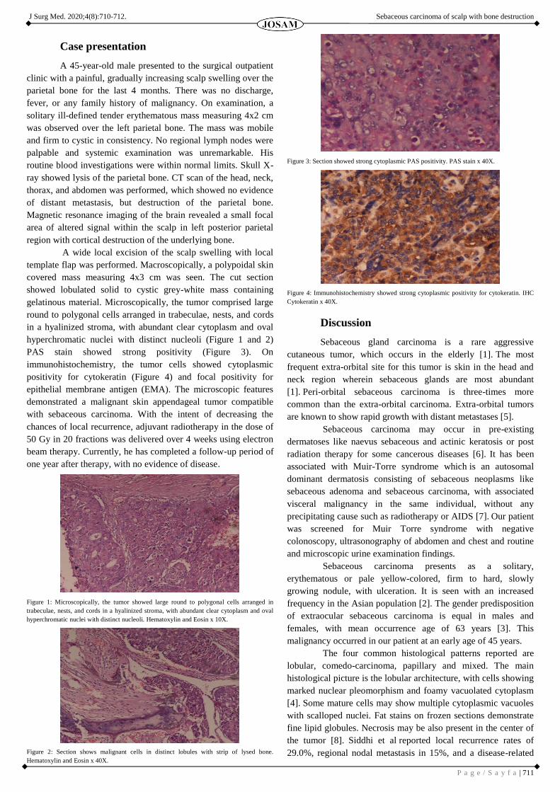
J Surg Med. 2020;4(8):710-712. Sebaceous carcinoma of scalp with bone destruction
P a g e / S a y f a | 711
Case presentation
A 45-year-old male presented to the surgical outpatient
clinic with a painful, gradually increasing scalp swelling over the
parietal bone for the last 4 months. There was no discharge,
fever, or any family history of malignancy. On examination, a
solitary ill-defined tender erythematous mass measuring 4x2 cm
was observed over the left parietal bone. The mass was mobile
and firm to cystic in consistency. No regional lymph nodes were
palpable and systemic examination was unremarkable. His
routine blood investigations were within normal limits. Skull X-
ray showed lysis of the parietal bone. CT scan of the head, neck,
thorax, and abdomen was performed, which showed no evidence
of distant metastasis, but destruction of the parietal bone.
Magnetic resonance imaging of the brain revealed a small focal
area of altered signal within the scalp in left posterior parietal
region with cortical destruction of the underlying bone.
A wide local excision of the scalp swelling with local
template flap was performed. Macroscopically, a polypoidal skin
covered mass measuring 4x3 cm was seen. The cut section
showed lobulated solid to cystic grey-white mass containing
gelatinous material. Microscopically, the tumor comprised large
round to polygonal cells arranged in trabeculae, nests, and cords
in a hyalinized stroma, with abundant clear cytoplasm and oval
hyperchromatic nuclei with distinct nucleoli (Figure 1 and 2)
PAS stain showed strong positivity (Figure 3). On
immunohistochemistry, the tumor cells showed cytoplasmic
positivity for cytokeratin (Figure 4) and focal positivity for
epithelial membrane antigen (EMA). The microscopic features
demonstrated a malignant skin appendageal tumor compatible
with sebaceous carcinoma. With the intent of decreasing the
chances of local recurrence, adjuvant radiotherapy in the dose of
50 Gy in 20 fractions was delivered over 4 weeks using electron
beam therapy. Currently, he has completed a follow-up period of
one year after therapy, with no evidence of disease.
Figure 1: Microscopically, the tumor showed large round to polygonal cells arranged in
trabeculae, nests, and cords in a hyalinized stroma, with abundant clear cytoplasm and oval
hyperchromatic nuclei with distinct nucleoli. Hematoxylin and Eosin x 10X.
Figure 2: Section shows malignant cells in distinct lobules with strip of lysed bone.
Hematoxylin and Eosin x 40X.
Figure 3: Section showed strong cytoplasmic PAS positivity. PAS stain x 40X.
Figure 4: Immunohistochemistry showed strong cytoplasmic positivity for cytokeratin. IHC
Cytokeratin x 40X.
Discussion
Sebaceous gland carcinoma is a rare aggressive
cutaneous tumor, which occurs in the elderly [1]. The most
frequent extra-orbital site for this tumor is skin in the head and
neck region wherein sebaceous glands are most abundant
[1]. Peri-orbital sebaceous carcinoma is three-times more
common than the extra-orbital carcinoma. Extra-orbital tumors
are known to show rapid growth with distant metastases [5].
Sebaceous carcinoma may occur in pre-existing
dermatoses like naevus sebaceous and actinic keratosis or post
radiation therapy for some cancerous diseases [6]. It has been
associated with Muir-Torre syndrome which is an autosomal
dominant dermatosis consisting of sebaceous neoplasms like
sebaceous adenoma and sebaceous carcinoma, with associated
visceral malignancy in the same individual, without any
precipitating cause such as radiotherapy or AIDS [7]. Our patient
was screened for Muir Torre syndrome with negative
colonoscopy, ultrasonography of abdomen and chest and routine
and microscopic urine examination findings.
Sebaceous carcinoma presents as a solitary,
erythematous or pale yellow-colored, firm to hard, slowly
growing nodule, with ulceration. It is seen with an increased
frequency in the Asian population [2]. The gender predisposition
of extraocular sebaceous carcinoma is equal in males and
females, with mean occurrence age of 63 years [3]. This
malignancy occurred in our patient at an early age of 45 years.
The four common histological patterns reported are
lobular, comedo-carcinoma, papillary and mixed. The main
histological picture is the lobular architecture, with cells showing
marked nuclear pleomorphism and foamy vacuolated cytoplasm
[4]. Some mature cells may show multiple cytoplasmic vacuoles
with scalloped nuclei. Fat stains on frozen sections demonstrate
fine lipid globules. Necrosis may be also present in the center of
the tumor [8]. Siddhi et al reported local recurrence rates of
29.0%, regional nodal metastasis in 15%, and a disease-related

J Surg Med. 2020;4(8):710-712. Sebaceous carcinoma of scalp with bone destruction
P a g e / S a y f a | 712
mortality of 20.0% in 91 cases with extra-ocular sebaceous
carcinoma [4]. Adverse prognostic factors include poor
differentiation, infiltrative growth pattern, multicentric origin of
the tumor and size of the tumor more than 10 mm.
Samarasinghe et al. have reported a case of sebaceous carcinoma
of scalp with nodal metastasis [2].
The tumors of sebaceous glands are separated into three
major categories: Sebaceous adenoma, basal cell carcinoma with
sebaceous differentiation and sebaceous carcinoma [1].
Sebaceous carcinoma cells are large and may show squamous
changes, and it should be differentiated from squamous cell
carcinoma by hydropic changes in the cell cytoplasm. Tumor
cells may show basaloid differentiation with presence of
inconspicuous lipid vacuoles, and hence the tumor must be
distinguished from basal cell carcinoma with sebaceous
differentiation [9]. Immunohistochemical staining for EMA can
differentiate sebaceous carcinoma from basal cell carcinoma and
squamous cell carcinoma [10].
Complete surgical excision is the treatment of choice
[11]. Sebaceous carcinomas tend to show 9 to 36% local
recurrence within 5 years [12]. Metastasis to liver, lungs, bones,
and brain have been reported in 14-25% of the patients [13].
Radiation therapy and chemotherapy have been used for regional
and metastatic disease, with varying degrees of response. Only a
few studies have shown satisfactory results of adjuvant
chemotherapy in the treatment of sebaceous carcinoma with
metastasis. There is one such report of complete response to
systemic chemotherapy in metastatic sweat gland carcinoma to
pleura, pericardium, and chest wall with 5‑ fluorouracil after 3
months of follow up period [3].
Conclusions
Sebaceous carcinoma needs to be considered as a
differential diagnosis for the cutaneous malignancies in all age
groups. The timely recognition enables execution of the primary
treatment i.e., wide local excision. Adjuvant radiotherapy may be
considered to improve the clinical outcome of recurrent tumors.
Acknowledgements
To the technical staff of the Histopathology and
Immunohistochemistry Laboratory.
References
1. Schwartz RA and Torre DP. Muir-Torre syndrome: A 25-year retrospect. J Am Acad Dermatol.
1995;33:90-104.
2. Samarasinghe V, Marsden J, Roberts C. Sebaceous carcinoma of the scalp presenting with nodal
metastasis. J Plast Reconstr Aesthet Surg. 2010;63:2193-4.
3. Ranajoy G, Bidhu KM, Prerna N, Puja S, Chitra S, Aman S, Karuna S. Recurrent sebaceous carcinoma
of the scalp in a young male treated with adjuvant radiotherapy. J Cancer Res Ther. 2013;9(4):730-2.
4. Siddhi C, Gaurav G, Rameshwar G, Uday K. Sebaceous carcinoma of scalp with proliferating
trichilemmal cyst. Ind Dermatol Online J. 2012;3(2):138-40.
5. Song A, Carter KD, Syed NA, Song J, Nerad JA. Sebaceous cell carcinoma of the ocular adnexa:
Clinical presentations, histopathology, and outcomes. Ophthal Plast Reconstr Surg. 2008;24:194-200.
6. Samarasinghe V. Sebaceous carcinoma of the scalp. J Plast Reconstr Aesthet Surg. 2011;1:16-8.
7. Dasgupta T. A Retrospective Review of 1349 Cases of Sebaceous Carcinoma. Cancer.
2009;115(1):158-65.
8. Khan S, Husain M, Ansari MM. Sebaceous Gland carcinoma - Case Report and Literature Review.
JSM Clin Case Rep. 2014;2(2):1025-8.
9. Joshi P, Joshi A, Norohna V, Prabhash K, Kane S, D’cruz AK. Systemic chemotherapy in sebaceous
carcinoma with lung metastasis. Ind J Med Paedr Oncol. 2012;33(4):239-41.
10.Bhatia SK, Atri S, Anjum A, Sardha M, Ali SA, Zaheer S. Postauricular sebaceous cell carcinoma. Int
J Case Reports and Images. 2012;3(9):29-32.
11.Mathur SK, Singh S, Yadav R, Duhan A, Sen R. Extraocular Sebaceous Carcinoma - a Rare Tumour
at a Rare Site. Egyptian Dermatol Online J. 2010;6(2):14-6.
12.JoonHo L, Hea-Kyeong S, Tae Jung J. A Case of Rapidly Growing Extraocular Sebaceous Carcinoma.
Cancer. 2014;15(1):32-5.
13.Karthika N, Reena R, Suma BP. Extra ocular sebaceous carcinoma: A rare case report. Ind Dermatol
Online J. 2011;2(2):91-3.
This paper has been checked for language accuracy by JOSAM editors.
The National Library of Medicine (NLM) citation style guide has been used in this paper.
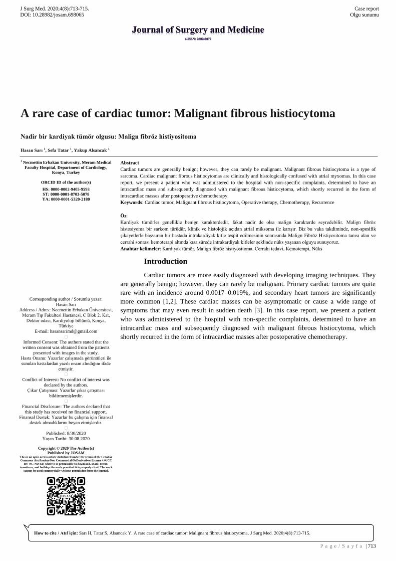
P a g e / S a y f a | 713
A rare case of cardiac tumor: Malignant fibrous histiocytoma
Nadir bir kardiyak tümör olgusu: Malign fibröz histiyositoma
Hasan Sarı 1, Sefa Tatar 1, Yakup Alsancak 1
How to cite / Atıf için: Sarı H, Tatar S, Alsancak Y. A rare case of cardiac tumor: Malignant fibrous histiocytoma. J Surg Med. 2020;4(8):713-715.
J Surg Med. 2020;4(8):713-715. Case report DOI: 10.28982/josam.698065 Olgu sunumu
1 Necmettin Erbakan University, Meram Medical
Faculty Hospital, Department of Cardiology,
Konya, Turkey
ORCID ID of the author(s)
HS: 0000-0002-9405-9593
ST: 0000-0001-8703-5078
YA: 0000-0001-5320-2180
Corresponding author / Sorumlu yazar:
Hasan Sarı
Address / Adres: Necmettin Erbakan Üniversitesi,
Meram Tıp Fakültesi Hastanesi, C Blok 2. Kat,
Doktor odası, Kardiyoloji bölümü, Konya,
Türkiye
E-mail: [email protected]
�
Informed Consent: The authors stated that the
written consent was obtained from the patients
presented with images in the study.
Hasta Onamı: Yazarlar çalışmada görüntüleri ile
sunulan hastalardan yazılı onam alındığını ifade
etmiştir.
�
Conflict of Interest: No conflict of interest was
declared by the authors.
Çıkar Çatışması: Yazarlar çıkar çatışması
bildirmemişlerdir.
�
Financial Disclosure: The authors declared that
this study has received no financial support.
Finansal Destek: Yazarlar bu çalışma için finansal
destek almadıklarını beyan etmişlerdir.
�
Published: 8/30/2020
Yayın Tarihi: 30.08.2020
Copyright © 2020 The Author(s)
Published by JOSAM This is an open access article distributed under the terms of the Creative
Commons Attribution-Non Commercial-NoDerivatives License 4.0 (CC
BY-NC-ND 4.0) where it is permissible to download, share, remix,
transform, and buildup the work provided it is properly cited. The work
cannot be used commercially without permission from the journal.
Abstract
Cardiac tumors are generally benign; however, they can rarely be malignant. Malignant fibrous histiocytoma is a type of
sarcoma. Cardiac malignant fibrous histiocytomas are clinically and histologically confused with atrial myxomas. In this case
report, we present a patient who was administered to the hospital with non-specific complaints, determined to have an
intracardiac mass and subsequently diagnosed with malignant fibrous histiocytoma, which shortly recurred in the form of
intracardiac masses after postoperative chemotherapy.
Keywords: Cardiac tumor, Malignant fibrous histiocytoma, Operative therapy, Chemotherapy, Recurrence
Öz
Kardiyak tümörler genellikle benign karakterdedir, fakat nadir de olsa malign karakterde seyredebilir. Malign fibröz
histosiyoma bir sarkom türüdür, klinik ve histolojik açıdan atrial miksoma ile karışır. Biz bu vaka takdiminde, non-spesifik
şikayetlerle başvuran bir hastada intrakardiyak kitle tespit edilmesinin sonrasında Malign Fibröz Histiyositoma tanısı alan ve
cerrahi sonrası kemoterapi altında kısa sürede intrakardiyak kitleler şeklinde nüks yaşanan olguyu sunuyoruz.
Anahtar kelimeler: Kardiyak tümör, Malign fibröz histiyositoma, Cerrahi tedavi, Kemoterapi, Nüks
Introduction
Cardiac tumors are more easily diagnosed with developing imaging techniques. They
are generally benign; however, they can rarely be malignant. Primary cardiac tumors are quite
rare with an incidence around 0.0017–0.019%, and secondary heart tumors are significantly
more common [1,2]. These cardiac masses can be asymptomatic or cause a wide range of
symptoms that may even result in sudden death [3]. In this case report, we present a patient
who was administered to the hospital with non-specific complaints, determined to have an
intracardiac mass and subsequently diagnosed with malignant fibrous histiocytoma, which
shortly recurred in the form of intracardiac masses after postoperative chemotherapy.
J Surg Med. 2020;4(8):713-715. Recurrent cardiac malignant fibrous histiocytoma
P a g e / S a y f a | 714
Case presentation
A 61-year-old female patient who did not suffer from
any chronic disease presented to the cardiology department of a
medical center with the complaints of back pain, chest tightness,
and palpitations which started following an upper respiratory
tract infection. Echocardiography revealed moderate mitral
insufficiency and a hyperechogenic mass (25x22 mm) in the
posterior leaflet of the mitral valve, after which the patient was
referred to our clinic. The physical examination did not reveal
any pathology other than 2/6 mitral regurgitation murmur at the
mitral focus. The echocardiography indicated the maximum
pulmonary artery systolic pressure was 45 mmHg with moderate
mitral and tricuspid regurgitation. We determined a hyperechoic
mass (28x15 mm) adhering to the posterior leaflet of the mitral
valve. Furthermore, we determined a mean diastolic pulmonary
gradient of 17 mmHg which we attributed to the mitral stenosis
caused by the mass on the mitral valve. We planned a
transesophageal echocardiography (TEE) to more clearly
evaluate the mass. The TEE revealed a mass (42x22 mm) in the
left atrium adhering to the posterior region that extended towards
the mitral valve, suggestive of a myxoma (Figure 1). There was
moderate mitral insufficiency, but the myxoma was thought to
affect mitral valve functions and cause mitral regurgitation. After
the evaluation of the heart team, the patient was decided to
undergo tumor resection and mitral valve replacement. The
patient was preoperatively evaluated with a PET/CT scan to
determine any other primary tumor or metastases. There only
was focal pathological 18F-FDG retention in the right
atrioventricular region as per the initial findings. The
pathological examination of the excised surgery material
confirmed the malignant fibrous histiocytoma diagnosis. Medical
oncologists decided on chemotherapy after relevant
examinations. The patient underwent a follow-up PET/CT scan
to evaluate tumoral activity after the fourth round of
chemotherapy. This examination revealed increased focal FDG
retention similar to the metallic hyperdensity in the left atrium
adjacent to the pulmonary trunk, and increased FDG retention in
multiple hypodense mass lesions in the liver. The patient was
subsequently referred to our clinic for cardiac evaluation. The
apical 4-chamber (A4C) view of the echocardiogram showed a
solid mass (25x20 mm) in the left ventricle that infiltrated the
septum with thin pedicles. Also, there was a solid mass (20x17
mm) extending over the right ventricular tricuspid lateral valve
(Figure 2). The image also revealed the functional prosthetic
metal valve at the position of the mitral valve. The patient
underwent cardiac MRI to confirm the solid lesions. The MRI
results affirmed the masses that were detected by
echocardiography (Figure 3). The patient was evaluated by the
cardiovascular department once more. The mass was evaluated
as malignant due to rapid progression. It was decided that the
patient should be closely followed up with medical treatment,
and reevaluated in case of impaired cardiac function. The
patient's consent was obtained for the clinical presentation.
Figure 1: Transesophageal echocardiogram showing mass (42x22 mm) in the left atrium
adhering to the posterior region that extended towards the mitral valve, suggestive of a
myxoma
Figure 2: The apical 4-chamber view of the echocardiogram showing a solid mass (25x20
mm) in the left ventricle that infiltrated the septum with thin pedicles and a solid mass
(20x17 mm) extending over the right ventricular tricuspid lateral valve
Figure 3: MRI showing masses in the left and right ventricle
Discussion
Malignant fibrous histiocytoma is a type of sarcoma that
typically occurs in the extremities and the trunk. There are less
than 100 reported cases of primary cardiac malignant fibrous
histiocytomas in the literature [4]. The reported patients are aged
between 14 and 77 years [5]. Cardiac malignant fibrous
histiocytomas are clinically and histologically confused with
atrial myxomas [6,7]. The first cardiac malignant fibrous
histiocytoma case was reported in 1978 [8]. The majority of the
J Surg Med. 2020;4(8):713-715. Recurrent cardiac malignant fibrous histiocytoma
P a g e / S a y f a | 715
recently reported cases were associated with rapid prognosis in
the form of recurrence, local infiltration, and distant metastasis,
and eventual death. Complete surgical excision is the only
treatment that has been shown to prolong survival [9]. However,
even with complete resection, the recurrence is common in most
patients with different sources reporting a median survival of 6 to
12 months [10]. Adriamycin and doxorubicin have been shown
to improve survival outcomes compared to other drugs [11].
Radiation significantly reduces the incidence of local recurrence
[12,13]. One study analyzed 42 cardiac sarcoma patients who
received treatment between 1988 and 2013, and found that a
multimodal therapy (any combination of surgery, radiotherapy
and chemotherapy) was associated with better survival than
surgery alone [14]. Patients with a cardiac mass should be
examined in detail to determine surgical requirements. Patients
with postoperative recurrence should be evaluated in terms of
cardiac functions instead of mass involvement. In addition, the
physician should consider medical follow-up together with
surgical intervention.
Conclusion
In this case study, we report a rare case of malignant
fibrous histiocytoma. We conclude that these cases must be
carefully monitored for recurrence and metastasis. A
multidisciplinary approach including cardiovascular and
oncology specialists is crucial for determining the optimal
treatment.
References
1. Straus R, Merliss R. Primary tumor of the heart. Arch Pathol. 1945;39:74–8.
2. Neragi-Miandoab S, Kim J, Vlahakes GJ. Malignant tumours of the heart: A review of tumour type,
diagnosis and therapy. Clin Oncol (R Coll Radiol). 2007;19:748–56. doi: 10.1016/j.clon.2007.06.009.
3. Basant K, Bhupendra KS, Vivek SG. Left Atrial Myxoma from Anterior Mitral Valve. Int J Appl
Basic Med Res. 2019 Oct-Dec;9(4):251–2. doi: 10.4103/ijabmr.IJABMR_288_18
4. Bandyopadhyay S, Banerjee S, Paul A, Das RK. Primary malignant fibrous histiocytoma involving the
left pulmonary vein presenting as a left atrial tumor. Ann Card Anaesth. 2013;16(4):293–5. doi:
10.4103/0971-9784.119184.
5. Okamoto K, Kato S, Katsuki S, Wada Y, Toyozumi Y, Morimatsu M, et al. Malignant fibrous
histiocytoma of the heart: case report and review of 46 cases in the literature. Intern Med.
2001;40(12):1222–6. doi: 10.2169/internalmedicine.40.1222.
6. Stevens CW, Sears-Rogan P, Bitterman P, Torrisi J. Treatment of malignant fibrous histiocytoma of
the heart. Cancer. 1992;69(4):956–61.
7. Laya MB, Mailliard JA, Bewtra C, Levin HS. Malignant fibrous histiocytoma of the heart. A case
report and review of the literature. Cancer. 1987;59(5):1026–31.
8. Shah AA, Churg A, Sbarbaro JA, Sheppard JM, Lamberti J. Malignant fibrous histiocytoma of the
heart presenting as an atrial myxoma. Cancer. 1978;42:2466 71.
9. Sato M, Suenaga E, Senaha S, Furutachi A. Primary malignant fibrous histiocytoma of the heart. Gen
Thorac Cardiovasc Surg. 2007;55:29 31.
10. Truong PT, Jones SO, Martens B, Alexander C, Paquette M, Joe H, et al. Treatment and outcomes in
adult patients with primary cardiac sarcoma: the British Columbia Cancer Agency experience. Ann
Surg Oncol. 2009;16:3358. doi: 10.1245/s10434-009-0734-8.
11. Putnam JB Jr, Sweeney MS, Colon R, Lanza LA, Frazier OH, Cooley DA. Primary cardiac sarcomas.
Ann Thorac Surg. 1991;51:906 10.
12. Barkley HT, Martin RG, Romsdahl MM, Lindberg R, Zagars GK. Treatment of soft tissue sarcomas
by preoperative irradiation and conservative surgical resection. Int J Radiat Oncol Biol Phys.
1988;14:693–9.
13. Lindberg RD, Martin RG, Romsdahl MM, Barkley HT. Conservative surgery and postoperative
radiotherapy in 300 adults with soft-tissue sarcomas. Cancer. 1981;47:2391-7.
14. Randhawa JS, Budd GT, Randhawa M, Ahluwalia M, Jia X, Daw H. Primary cardiac sarcoma: 25-
year Cleveland Clinic experience. Am J Clin Oncol. 2016;39(6):593–9.
This paper has been checked for language accuracy by JOSAM editors.
The National Library of Medicine (NLM) citation style guide has been used in this paper.