Journal of - Contemporary Medicine - DergiPark
118
Journal of Contemporary Medicine YEAR: 2019 VOLUME: 9 ISSUE: 3
-
Upload
khangminh22 -
Category
Documents
-
view
1 -
download
0
Transcript of Journal of - Contemporary Medicine - DergiPark

Journal ofContemporary MedicineYEAR: 2019 VOLUME: 9 ISSUE: 3

Resul YILMAZ
Fikret ERDEMİR
Mustafa ÖZÇETİN
Mustafa ALTAY
Atilla ŞENAYLI
EDITOR-IN-CHIEF
ASSOCIATE EDITORS
ADVISORY BOARD
JOURNAL OF CONTEMPORARY MEDICINE
Formerly Çağdaş Tıp Dergisi
The Owner and Publishing Manager on behalf of the Journal of Contemporary Medicine
Address:
Phone:Fax:
e-mail:web:
Doç. Dr. Resul YILMAZSelçuk Üniversitesi, Tıp Fakültesi Çocuk Yoğun Bakım Bilim Dalı Alaeddin Keykubat Yerleşkesi Selçuklu/Konya 42075 Türkiye+90 (332) 241 50 00-44513+90 (332) 241 21 84cagdastipdergisi @gmail.comhttp://www.jcontempmed.com
e-ISSN 2667-7180
VOLUME 9 ISSUE 3 YEAR 2019
Hülya BAYIR, USA
Maciej BURA, Poland
Sancak YÜKSEL, USA
Ashrarur Rahman MITUL, Bagladesh
Yau Sui YU, Hong Kong
Mustafa ALTAY, Turkey
Zhiqiang LIU, England
Abid QAZI, Istanbul
Ahmet DEMİR, Turkey
Obehi H OKOJIE, Nijerya
Ilhama JAFARLI, King Fahad Med. City
Zafar ZAHEER, Pakistan
İsmail SARI, Turkey
İlknur BOSTANCI, Turkey
Yücel YAVUZ, Turkey
Ahmet BAYDIN, Turkey
Sevil ÇAYLI, Turkey
Nihal HATIPOĞLU, Turkey
Cengiz ÇOKLUK, Turkey
Atilla ŞENAYLI, Turkey
Keramettin AYDIN, Turkey
Ömer ERDEVE, Turkey
Bahtiyar DEMİRALP, Turkey
İbrahim TÜRKCÜER, Turkey
Mehmet ÇETİNKAYA, Turkey
Sebahattin VURUCU, Turkey
Mehmet Ali KURÇER, Turkey
Murat ERDOĞAN, Turkey
Metin AKBULUT, Turkey
Journal ofContemporary MedicineYEAR: 2019 VOLUME: 9 ISSUE: 2 Journal of
Contemporary
Medicine
YEAR: 2019
VOLU
ME: 9
ISSUE: 2

INSTRUCTIONS FOR AUTHORS
AIM AND SCOPEJournal of Contemporary Medicine is published quarterly for four issues. Its purpose is to publish high-quality original clinical and experimental studies, case reports and letters to the editor.
Journal of Contemporary Medicine provides open access for academic publications. The journal provides free access to the full texts of all articles immediately upon publication.
The Journal will not consider manuscripts any that have been published elsewhere, or manuscripts that are being considered for another publication, or are in press. Studies previously announced in the congresses are accepted if this condition is stated. If any part of a manuscript by the same author(s) contains any information that was previously published, a reprint or a copy of the previous article should be submitted to the Editorial Office with an explanation by the authors.
All manuscripts are reviewed by the Editor, Associate Editor or a member of the Editorial Board. The Editor, Associate Editor and the member of the Editorial Board have right not to publish or send back to author(s) to be amended, edit or reject the manuscript. For further review, the Associate Editor or Editorial Board member sends the article to the refree(s). If necessary, author(s) may be invited to submit a revised version of the manuscript. This invitation does not imply that the manuscript will be accepted for publication. Revised manuscripts must be sent to the Editorial Office within 21 days, otherwise they will be considered as a new application. The corresponding author will be notified of the decision to accept or reject the manuscript for publication.
Statements and suggestions published in manuscripts are the authors’ responsibility and do not reflect the opinions of the Editor, Associate Editors and the Editorial Board members.
The manuscript will not be returned to the authors whether the article is accepted or not. Copyright fee is not paid for the articles published in the journal. A copy of the journal will be sent to the corresponding author.
Language of the Journal The official languages of the Journal are Turkish and English. The
manuscripts that are written in Turkish have abstracts in English, which makes the abstracts available to a broader audience.
Authorship Criteria After accepted for publication, all the authors will be asked to sign “Coyright Transfer Form” which states the following: “ This work is not under active consideration for publication, has not been accepted for publication, nor has it been published, in full or in part (except in abstract form). I confirm that the study has been approved by the ethics committee. ” All authors should agree to the conditions outlined in the form.
Journal of Contemporary Medicine has agreed to use the standards of the International Committee of Medical Journal Editors. The author(s) should meet the criteria for authorship according to the "Uniform Requirements for Manuscripts Submitted to Biomedical Journals: Writing and Editing for Biomedical Publication. It is available at www.icmje.org.
Ethical Responsibility Patient anonymity should be preserved and all studies on patients must include a statement that informed consent and approval of ethical committee were obtained. Written permission from identifiable patients appearing in photographs (as in case reports) must be obtained by the author(s) and must be surface mailed or faxed to the Editorial Office.
Any experiments involving animals must include a statement in the Materials and Methods section giving assurance that all animals have received humane care in compliance with the Guide for the Care and Use of Laboratory Animals (www.nap.edu/catalog/5140.html) and indicating approval by the institutional ethical review board.
Note also that for publishing purposes, the Journal requires acknowledgement of any potential conflicts of interest. This should involve acknowledgement of grants and other sources of funds that support reported research and a declaration of any relevant industrial links or affiliations that the authors may have.
TYPES OF MANUSCRIPT Manuscripts should be submitted online via www.jcontempmed.com
Original Articles should not exceed 3000 words and should
JOURNAL OF CONTEMPORARY MEDICINE
Formerly Çağdaş Tıp Dergisi
Journal ofContemporary MedicineYEAR: 2019 VOLUME: 9 ISSUE: 2
Journal ofContemporary Medicine YEAR: 2019 VOLUME: 9 ISSUE: 2

be arranged under the headings of Abstract (not more than 250 words), Introduction, Materials and Methods, Results, Discussion, Conclusion and References.
Case Reports should not exceed 1000 words and 10 references, and should be arranged as follows: Abstract, Introduction, Case Report, Discussion and References. It may be accompanied by only one figure or table.
Letter to the Editor should not exceed 500 words. Short relevant comments on medical and scientific issues, particularly controversies, having no more than five references and one table or figure are encouraged. Where letters refer to an earlier published paper, authors will be offered right of reply.
Reviews are not accepted unless written on the invitation of the Editorial Board.
PREPARATION OF MANUSCRIPTS All articles submitted to the Journal must comply with the following instructions:
a) Submissions should be doubled-spaced and typed in Arial 10 points.
b) All pages should be numbered consecutively in the top right-hand corner, beginning with the title page.
c) The title page should not include the names and institutions of the authors.
d) The manuscript should be presented in the following order: Title page, Abstract (English, Turkish), Keywords (English, Turkish), Introduction, Materials and Methods, Results, Discussion, Conclusion, Acknowledgements (if present), References, Figure Legends, Tables (each table, complete with title and foot-notes, on a separate page) and Appendices (if present) presented each on a separate page.
Title The title should be short, easy to understand and must define the contents of the article.
Abstract Abstract should be in both English and Turkish and should consist “Aim, Materials and Methods, Results and Conclusion”.
The purpose of the study, the setting for the study, the subjects, the treatment or intervention, principal outcomes measured, the type of statistical analysis and the outcome of the study should be stated in this section (up to 250 words). Abstract should not include reference. No abstract is required for the letters to the Editor.
Keywords Not more than five keywords in order of importance for indexing purposes should be supplied below the abstract and should be selected from Index Medicus Medical Subject Headings (MeSH), available at www.nlm.nih.gov/meshhome.html.
Text Authors should use subheadings to divide sections regarding the type of the manuscript as described above. Statistical methods used should be specified in the Materials and Methods section.
References In the text, references should be cited using Arabic numerals in parenthesis in the order in which they appear. If cited only in tables or figure legends, they should be numbered according to the first identification of the table or figure in the text. Names of the journals should be abbreviated in the style used in Index Medicus. The names of all authors should be cited when there are six or fewer; when seven or more, the first three should be followed by et al. The issue and volume numbers of the referenced journal should be added.
References should be listed in the following form:
Journal articleTeke Z, Kabay B, Aytekin FO et al. Pyrrolidine dithiocarbamate prevents 60 minutes of warm mesenteric ischemia/reperfusion injury in rats. Am J Surg 2007;194(6):255-62.
Supplement Solca M. Acute pain management: Unmet needs and new advances in pain management. Eur J Anaesthesiol 2002; 19(Suppl 25): 3-10.
Online article not yet published in an issueButterly SJ, Pillans P, Horn B, Miles R, Sturtevant J. Off-label use of rituximab in a tertiary Queensland hospital. Intern Med J doi: 10.1111/j.1445-5994.2009.01988.x
JOURNAL OF CONTEMPORARY MEDICINE
Formerly Çağdaş Tıp Dergisi
Journal ofContemporary MedicineYEAR: 2019 VOLUME: 9 ISSUE: 2
Journal ofContemporary Medicine YEAR: 2019 VOLUME: 9 ISSUE: 2
Journal ofContemporary Medicine YEAR: 2019 VOLUME: 9 ISSUE: 2

Book Sample1: Murray PR, Rosenthal KS, Kobayashi GS, Pfaller MA. Medical microbiology. 4th ed. St. Louis: Mosby; 2002. Sample 2: Sümbüloğlu K, Akdağ B. Regresyon Yöntemleri ve Korelasyon Analizi. Hatiboğlu Yayınevi: Ankara; 2007.
Chapter in a bookMeltzer PS, Kallioniemi A, Trent JM. Chromosome alterations in human solid tumors. I n: Vogelstein B, Kinzler KW, editors. The genetic basis of human cancer. New York: McGraw-Hill; 2002. p. 93113.
Journal article on the InternetAbood S. Quality improvement initiative in nursing homes: The ANA acts in an advisory role. Am J Nurs [serial on the Internet] 2002 [cited 12 Aug 2002]; 102. Available from:www.nursingworld.org/AJN/2002/june/wawatch.htm
Website Cancer-pain.org [homepage on the Internet]. New York: Association of Cancer Online Resources [updated 16 May 2002; cited 9 Jul 2002]. Available from: www.cancer-pain.org
An organization as an authorThe Intensive Care Society of Australia and New Zealand. Mechanical ventilation strategy in ARDS: Guidelines. Int Care J Aust 1996;164:282-4.
Acknowledgements The source of financial grants and the contribution of colleagues or institutions should be acknowledged.
Tables Tables should be complementary, but not duplicate information contained in the text. Tables should be numbered consecutively in Arabic numbers, with a descriptive, self-explanatory title above the table. All abbreviations should be explained in a footnote. Footnotes should be designated by symbols in the following order: *,†, ‡, §, ¶.
Figures All illustrations (including line drawings and photographs) are classified as figures. Figures must be added to the system as separate .jpg or .gif files (approximately 500x400 pixels, 8 cm
in width and at least 300 dpi resolution). Figures should be numbered consecutively in Arabic numbers and should be cited in parenthesis in consecutive order in the text.
Figure LegendsLegends should be self-explanatory and positioned on a separate page. The legend should incorporate definitions of any symbols used and all abbreviations and units of measurements should be explained. A letter should be provided stating copyright authorization if figures have been reproduced from another source.
Measurements and Abbreviations All measurements must be given in metric system (Système International d'Unités, SI). Example: mg/kg, µg/kg, mL, mL/kg, mL/kg/h, mL/kg/min, L/min, mmHg, etc. Statistics and measurements should always be given in numerals, except where the number begins a sentence. When a number does not refer to a unit of measurement, it is spelt out, except where the number is greater than nine.
Abbreviations that are used should be defined in parenthesis where the full word is first mentioned. Some common abbreviations can be used, such as iv, im, po, and sc.
Drugs should be referred to by their generic names, rather than brand names.
Editorial Correspondence Doç. Dr. Resul YILMAZSelçuk Üniversitesi, Tıp Fakültesi Çocuk Yoğun Bakım Bilim Dalı Alaeddin Keykubat Yerleşkesi Selçuklu/Konya 42075 TürkiyePhone: +90 (332) 241 50 00-44513Faks: +90 (332) 241 21 84
Çağdaş Tıp Dergisi
(Journal of Contemporary Medicine)http://www.jcontempmed.come-posta: cagdastipdergisi @gmail.com
Checklist for Manuscripts Review guide for authors and instructions for submitting manuscripts through the electronic submission, website at
http://www.jcontempmed.com
JOURNAL OF CONTEMPORARY MEDICINE
Formerly Çağdaş Tıp Dergisi
Journal ofContemporary MedicineYEAR: 2019 VOLUME: 9 ISSUE: 2
Journal ofContemporary Medicine YEAR: 2019 VOLUME: 9 ISSUE: 2
Journal ofContemporary Medicine YEAR: 2019 VOLUME: 9 ISSUE: 2

YAZARLARA BİLGİ
AMAÇ ve KAPSAMÇağdaş Tıp Dergisi, üç ayda bir yayımlanır ve dört sayı ile bir cilt tamamlanır. Dergi; tüm tıp alanlarıyla ilgili nitelikli klinik ve deneysel araştırmaları, olgu sunumlarını ve editöre mektupları yayımlar.
Çağdaş Tıp Dergisi, bilimsel yayınlara açık erişim sağlar. Dergi basımından hemen sonra, makalelerin tam metinlerine ücretsiz ulaşılabilir.
Dergide yayımlanmak üzere gönderilen yazıların daha önce başka bir yerde yayımlanmamış veya yayımlanmak üzere gönderilmemiş olması gerekir. Daha önce kongrelerde sunulmuş çalışmalar, bu durum belirtilmek koşuluyla kabul edilir. Makale, yazar(lar)ın daha önce yayımlanmış bir yazısındaki konuların bir kısmını içeriyorsa bu durum belirtilmeli ve yeni yazı ile birlikte önceki makalenin bir kopyası da Yayın Bürosu’na gönderilmelidir.
Gönderilen yazılar; Editör, Editör Yardımcısı ya da Yayın Kurulu Üyesi tarafından incelenir. Editör, Editör Yardımcısı ya da Yayın Kurulu Üyesi, yayın koşullarına uymayan yazıları yayınlamamak, düzeltmek üzere yazar(lar)a geri göndermek, biçimce düzenlemek veya reddetmek yetkisine sahiptir. Editör, Editör Yardımcısı ya da Yayın Kurulu Üyesi, uygun gördüğü yazıyı incelenmek üzere danışman(lar)a gönderir. Gerekli olduğu durumlarda, yazar(lar)dan düzeltme istenebilir. Yazardan düzeltme istenmesi, yazının yayımlanacağı anlamına gelmez. Bu düzeltmelerin en geç 21 gün içinde tamamlanıp dergiye gönderilmesi gereklidir. Aksi halde yeni başvuru olarak değerlendirilir. Sorumlu yazara yazının kabul veya reddedildiğine dair bilgi verilir.
Dergide yayımlanan yazıların etik, bilimsel ve hukuki sorumluluğu yazar(lar)a ait olup Editör, Editör Yardımcısı ve Yayın Kurulu’nun görüşlerini yansıtmaz.
Dergide yayımlanması kabul edilse de edilmese de, yazı materyali yazarlara geri verilmez. Dergide yayımlanan yazılar için telif hakkı ödenmez. Bir adet dergi, sorumlu yazara gönderilir.
Derginin Yazı Dili Derginin yazı dili Türkçe ve İngilizcedir. Dili Türkçe olan yazılar, İngilizce özetleri ile yer alır. Yazının hazırlanması sırasında, Türkçe kelimeler için Türk Dil Kurumundan (www.tdk.gov.
tr), teknik terimler için Türk Tıp Terminolojisinden (www.tipterimleri.com) yararlanılabilir.
Yazarlık Kriterleri Dergide yayınlanması uygun bulunan tüm yazıların araştırma ve yayın etiğine uygun hazırlandığı, varsa sağlanan fonun kaynağının tanımlandığı, başka yerde yayımlanmadığı veya yayımlanmak üzere gönderilmediği, çalışmaya katılan tüm yazarlar tarafından yazının son halinin onaylandığı, yayımlanacak yazı ile ilgili telif haklarının dergiye devredildiği, tüm yazarların imzaları ile “Yayın Hakkı Devir Formu”nda belirtilmesi gerekir.
Çağdaş Tıp Dergisi, Uluslararası Tıp Dergileri Editörleri Kurulu’nun (International Committee of Medical Journal Editors) “Biyomedikal Dergilere Gönderilen Makalelerin Uyması Gereken Standartlar: Biyomedikal Yayınların Yazımı ve Baskıya Hazırlanması (Uniform Requirements for Manuscripts Submitted to Biomedical Journals: Writing and Editing for Biomedical Publication)” standartlarını kullanmayı kabul etmektedir. Bu konudaki bilgiye www.icmje.org adresinden ulaşılabilir.
Etik Sorumluluk Hastaların gizlilik haklarına saygı gösterilmeli, aydınlatılmış onamları mutlaka alınmalı, aydınlatılmış onam ile Etik Kurul onayı alındığı bilimsel yazının içinde belirtilmelidir. Fotoğraflarda yüzü belli olan hastalardan yazılı izin alınmalı ve Dergi Editörlüğüne posta ya da faks yoluyla iletilmelidir.
Çağdaş Tıp Dergisi, deney hayvanları ile yapılan çalışmalarda, genel kabul gören ilgili etik kurallara uyulması zorunluluğunu hatırlatır. Alınmış Etik Kurul Onayı, makale ile birlikte sisteme yüklenmelidir.
Yazar(lar), ticari bağlantı veya çalışma için maddi destek veren kurum varlığında; kullanılan ticari ürün, ilaç, firma vb. ile nasıl bir ilişkisi olduğunu sunum sayfasında Editöre bildirmelidir. Böyle bir durumun yokluğu da yine ayrı bir sayfada belirtilmelidir.
YAZI TÜRLERİ Yazılar, elektronik ortamda www.cagdastipdergisi.com adresine gönderilir.
Orijinal makaleler , 3000 sözcük sayısını aşmamalı, “Öz (250 sözcükten fazla olmamalı), Giriş, Gereç ve Yöntem, Bulgular, Tartışma, Sonuç, Kaynaklar” bölümlerinden oluşmalıdır.
JOURNAL OF CONTEMPORARY MEDICINE
Formerly Çağdaş Tıp Dergisi
Journal ofContemporary MedicineYEAR: 2019 VOLUME: 9 ISSUE: 2
Journal ofContemporary Medicine YEAR: 2019 VOLUME: 9 ISSUE: 2
Journal ofContemporary Medicine YEAR: 2019 VOLUME: 9 ISSUE: 2

Olgu Sunumu , “Öz, Giriş, Olgu Sunumu, Tartışma, Kaynaklar” şeklinde düzenlenmelidir. En fazla 1000 sözcük ve 10 kaynak ile sınırlıdır. Sadece bir tablo veya şekil ile desteklenebilir.
Editöre Mektup , yayımlanan metinlerle veya mesleki konularla ilgili olarak 500 sözcüğü aşmayan ve beş kaynak ile bir tablo veya şekil içerecek şekilde yazılabilir. Ayrıca daha önce dergide yayınlanmış metinlerle ilişkili mektuplara cevap hakkı verilir.
Yayın Kurulu’nun daveti üzerine yazılanlar dışında derleme kabul edilmez.
MAKALENİN HAZIRLANMASI Dergide yayınlanması istenilen yazı için aşağıdaki kurallara uyulmalıdır.
a) Yazı; iki satır aralıklı olarak, Arial 10 punto ile yazılmalıdır. b) Sayfalar başlık sayfasından başlamak üzere, sağ üst köşesinde numaralandırılmalıdır.
c) Online makale sistemine yüklenen word dosyasının başlık sayfasında (makalenin adını içeren başlık sayfası), yazarlara ait isim ve kurum bilgileri yer almamalıdır.
d) Makale, şu bölümleri içermelidir: Her biri ayrı sayfada yazılmak üzere; Türkçe ve İngilizce Başlık Sayfası, Öz, Abstract, Anahtar Sözcükler, Keywords, Giriş, Gereç ve Yöntem, Bulgular, Tartışma, Sonuç, Açıklamalar (varsa), Kaynaklar, Şekil Alt Yazıları, Tablolar (başlıkları ve açıklamalarıyla beraber), Ekler (varsa).
Yazının Başlığı Kısa, kolay anlaşılır ve yazının içeriğini tanımlar özellikte olmalıdır.
Özetler Türkçe (Öz) ve İngilizce (Abstract) olarak yazılmalı, Amaç, Gereç ve Yöntem, Bulgular ve Sonuç (Aim, Materials and Methods, Results, Conclusion) olmak üzere dört bölümden oluşmalı, en fazla 250 sözcük içermelidir. Araştırmanın amacı, yapılan işlemler, gözlemsel ve analitik yöntemler, temel bulgular ve ana sonuçlar belirtilmelidir. Özette kaynak kullanılmamalıdır. Editöre mektup için özet gerekmemektedir.
Anahtar Sözcükler Türkçe Öz ve İngilizce Abstract bölümünün sonunda, Anahtar Sözcükler ve Keywords başlığı altında, bilimsel yazının ana başlıklarını yakalayan, Index Medicus Medical Subject Headings
(MeSH)’e uygun olarak yazılmış en fazla beş anahtar sözcük olmalıdır. Anahtar sözcüklerin, Türkiye Bilim Terimleri’nden (www.bilimterimleri.com) seçilmesine özen gösterilmelidir.
Metin Yazı metni, yazının türüne göre yukarıda tanımlanan bölümlerden oluşmalıdır. Uygulanan istatistiksel yöntem, Gereç ve Yöntem bölümünde belirtilmelidir.
Kaynaklar Çağdaş Tıp Dergisi, Türkçe kaynaklardan yararlanmaya özel önem verdiğini belirtir ve yazarların bu konuda duyarlı olmasını bekler.
Kaynaklar metinde yer aldıkları sırayla, cümle içinde atıfta bulunulan ad veya özelliği belirten kelimenin hemen bittiği yerde ya da cümle bitiminde noktadan önce parantez içinde Arabik rakamlarla numaralandırılmalıdır. Metinde, tablolarda ve şekil alt yazılarında kaynaklar, parantez içinde Arabik numaralarla nitelendirilir. Sadece tablo veya şekil alt yazılarında kullanılan kaynaklar, tablo ya da şeklin metindeki ilk yer aldığı sıraya uygun olarak numaralandırılmalıdır. Dergi başlıkları, Index Medicus’ta kullanılan tarza uygun olarak kısaltılmalıdır. Kısaltılmış yazar ve dergi adlarından sonra nokta olmamalıdır. Yazar sayısı altı veya daha az olan kaynaklarda tüm yazarların adı yazılmalı, yedi veya daha fazla olan kaynaklarda ise üç yazar adından sonra et al. veya ve ark. yazılmalıdır. Kaynak gösterilen derginin sayı ve cilt numarası mutlaka yazılmalıdır.
Kaynaklar, yazının alındığı dilde ve aşağıdaki örneklerde görüldüğü şekilde düzenlenmelidir.
Dergilerdeki yazılarTeke Z, Kabay B, Aytekin FO et al. Pyrrolidine dithiocarbamate prevents 60 minutes of warm mesenteric ischemia/reperfusion injury in rats. Am J Surg 2007;194(6):255-62.
Ek sayı (Supplement) Solca M. Acute pain management: Unmet needs and new advances in pain management. Eur J Anaesthesiol 2002;19(Suppl 25):3-10.
Henüz yayınlanmamış online makaleButterly SJ, Pillans P, Horn B, Miles R, Sturtevant J. Off-label use of rituximab in a tertiary Queensland hospital. Intern Med J doi: 10.1111/j.1445-5994.2009.01988.x
JOURNAL OF CONTEMPORARY MEDICINE
Formerly Çağdaş Tıp Dergisi
Journal ofContemporary MedicineYEAR: 2019 VOLUME: 9 ISSUE: 2
Journal ofContemporary Medicine YEAR: 2019 VOLUME: 9 ISSUE: 2
Journal ofContemporary Medicine YEAR: 2019 VOLUME: 9 ISSUE: 2

KitapÖrnek 1: Murray PR, Rosenthal KS, Kobayashi GS, Pfaller MA. Medical microbiology. 4th ed. St. Louis: Mosby; 2002. Örnek 2: Sümbüloğlu K, Akdağ B. Regresyon Yöntemleri ve Korelasyon Analizi. Hatiboğlu Yayınevi: Ankara; 2007.
Kitap bölümüMeltzer PS, Kallioniemi A, Trent JM. Chromosome alterations in human solid tumors. I n: Vogelstein B, Kinzler KW, editors. The genetic basis of human cancer. New York: McGraw-Hill; 2002. p. 93113.
İnternet makalesi
Abood S. Quality improvement initiative in nursing homes: The ANA acts in an advisory role. Am J Nurs [serial on the Internet] 2002 [cited 12 Aug 2002]; 102. Available from: www.nursingworld.org/AJN/2002/june/wawatch.htm
Web SitesiCancer-pain.org [homepage on the Internet]. New York: Association of Cancer Online Resources [updated 16 May 2002; cited 9 July 2002]. Available from: www.cancer-pain.org
Yazar olarak bir kuruluşThe Intensive Care Society of Australia and New Zealand. Mechanical ventilation strategy in ARDS: Guidelines. Int Care J Aust 1996;164:282-4.
Açıklamalar Varsa finansal kaynaklar, katkı sağlayan kurum, kuruluş ve kişiler bu bölümde belirtilmelidir.
Tablolar Tablolar metni tamamlayıcı olmalı, metin içerisinde tekrarlanan bilgiler içermemelidir. Metinde yer alma sıralarına göre Arabik sayılarla numaralandırılıp tablonun üstüne kısa ve açıklayıcı bir başlık yazılmalıdır. Tabloda yer alan kısaltmalar, tablonun hemen altında açıklanmalıdır. Dipnotlarda sırasıyla şu semboller kullanılabilir: *, †, ‡, §, ¶.
Şekiller Şekil, resim, grafik ve fotoğrafların tümü “Şekil” olarak adlandırılmalı ve ayrı birer .jpg veya .gif dosyası olarak (yaklaşık 500x400 piksel, 8 cm eninde ve en az 300 dpi çözünürlükte) sisteme eklenmelidir. Şekiller metin içinde kullanım sıralarına
göre Arabik rakamla numaralandırılmalı ve metinde parantez içinde gösterilmelidir.
Şekil Alt Yazıları Şekil alt yazıları, her biri ayrı bir sayfadan başlayarak, şekillere karşılık gelen Arabik rakamlarla çift aralıklı olarak yazılmalıdır. Şeklin belirli bölümlerini işaret eden sembol, ok veya harfler kullanıldığında bunlar alt yazıda açıklanmalıdır. Başka yerde yayınlanmış olan şekiller kullanıldığında, yazarın bu konuda izin almış olması ve bunu belgelemesi gerekir.
Ölçümler ve Kısaltmalar Tüm ölçümler metrik sisteme (Uluslararası Birimler Sistemi, SI) göre yazılmalıdır. Örnek: mg/kg, µg/kg, mL, mL/kg, mL/kg/h, mL/kg/min, L/min, mmHg, vb. Ölçümler ve istatistiksel veriler, cümle başında olmadıkları sürece rakamla belirtilmelidir. Herhangi bir birimi ifade etmeyen ve dokuzdan küçük sayılar yazı ile yazılmalıdır.
Metin içindeki kısaltmalar, ilk kullanıldıkları yerde parantez içinde açıklanmalıdır. Bazı sık kullanılan kısaltmalar; iv, im, po ve sc şeklinde yazılabilir.
İlaçların yazımında jenerik isimleri kullanılmalıdır.
İletişimDoç. Dr. Resul YILMAZSelçuk Üniversitesi, Tıp Fakültesi Çocuk Yoğun Bakım Bilim Dalı Alaeddin Keykubat Yerleşkesi Selçuklu/Konya 42075 TürkiyeTel: +90 (332) 241 50 00-44513Faks: +90 (332) 241 21 84
Journal of Contemporary Medicine
(Çağdaş Tıp Dergisi)http://www.cagdastipdergisi.come-posta: cagdastipdergisi @gmail.com
Kontrol Listesi · Türkçe ve İngilizce başlık,· Türkçe ve İngilizce özet · Türkçe ve İngilizce anahtar sözcükler (En fazla 5 sözcük) · İki satır aralıklı yazılmış metin (Arial, 10 punto) · Kurallara uygun hazırlanmış tablo ve şekiller · Kurallara uygun yazılmış kaynaklar · İmzalı “Yayın Hakkı Devir Formu” (makale yayın için kabul edildikten sonra istenmektedir)
JOURNAL OF CONTEMPORARY MEDICINE
Formerly Çağdaş Tıp Dergisi
Journal ofContemporary MedicineYEAR: 2019 VOLUME: 9 ISSUE: 2
Journal ofContemporary Medicine YEAR: 2019 VOLUME: 9 ISSUE: 2
Journal ofContemporary Medicine YEAR: 2019 VOLUME: 9 ISSUE: 2

CONTENTS VOLUME 9 ISSUE 3 YEAR 2019 e-ISSN 2667-7180
Comparison of Pro-BNP levels and myocardial performance index before and after iron treatment in children with congenital cyanotic heart disease with iron deficiency anemia /Demir eksikliği anemisi olan doğumsal siyanotik kalp hastalıklı çocuklarda demir tedavisi öncesi ve sonrası pro-bnp düzeyleri ve miyokardiyal performans indekslerinin karşılaştırılmasıBarutçu A, Erdem S, Demir F, Barutçu S, Leblebisatan G .......................................................................................................................... 197
Possible protective effect of quercetin against oxidative stress in liver from metabolic syndrome rats /Metabolik sendromlu sıçanların karaciğerlerinde oksidatif strese karşı kuersetinin olası koruyucu etkisiBilginoğlu A ............................................................................................................................................................................................................ 203
Is higher mean platelet volume an additional predictive marker of oligohydramnios and polyhydramnios? /Yüksek ortalama trombosit hacmi oligohidramnios ve polihidramnios için ek bir prediktif belirteç midir?Çetin Benli N, Mutlu S, Yıldız A, Benli AR ..................................................................................................................................................... 209
Comparative evaluation of dural venous sinuses and cerebral veins using contrast-enhanced spoiled gradient recalled echo and time-of-flight magnetic resonance venography / Dural venöz sinüsler ve serebral venlerin kontrastlı spoiled gradient recalled echo ve time-of-flight manyetik rezonans venografiler ile karşılaştırmalı değerlendirilmesiDeniz Ç, Gökçe E, Acu B, Kuyucu YE ............................................................................................................................................................ 214
The evaluation of the effects of passive smoking on children's health with detection of urine cotinine levels / İdrar kotinin düzeylerinin tespiti ile pasif sigara maruziyetinin çocuk sağlığına etkilerinin değerlendirilmesiKahvecioğlu D, Bostancı İ, Taşar MA, Dindar Badem N, Dallar YB ........................................................................................................ 222
Association between ABO blood group with Parkinson’s disease / ABO kan grubunun Parkinson hastalığı ile ilişkisiBulur O, Korucu O ............................................................................................................................................................................................. 227
Skin prick test results in patients with chronic allergic rhinitis: Housewives are risky occupational group for the development of allergic rhinitis due to house dust mites / Kronik alerjik rinitli hastalarda cilt prick test sonuçları: Ev hanımları ev tozu akarlarına bağlı alerjik rinit gelişimi için riskli meslek grubundadırÖzdemir T, Kasapoğlu B, Türkkanı MH, Özdilekcan Ç, Erel F ................................................................................................................. 230
The effect of duloxetine on ECoG activity of absence-epilepsy model in WAG/Rij rats / Absans epilepsi modeli olan WAG/Rij sıçanlarda duloksetinin ECoG aktivitesi üzerine etkisiAygün H ................................................................................................................................................................................................................. 235
Yenidoğan yoğun bakım ünitesinde izlenen üst solunum yolu patolojilerinin incelenmesi / Investigation of upper respiratory tract pathologies in neonatal intensive care unitKonak M, Erdur Ö, Kılınç MY, Soylu H .......................................................................................................................................................... 241
Bicarbonate may alters bacterial susceptibility to antibiotics by targeting Pseudomonas aeruginosa, Escherichia coli and Staphylococcus aureus / Bikarbonat, Pseudomonas aeruginosa, Escherichia coli ve Staphylococcus aureus'a karşı antibiyotiklerin duyarlılığını değiştirebilirKesici S, Demirci M, Kesici U............................................................................................................................................................................ 245
ORIGINAL ARTICLES
JOURNAL OF CONTEMPORARY MEDICINE
Formerly Çağdaş Tıp Dergisi
Journal ofContemporary MedicineYEAR: 2019 VOLUME: 9 ISSUE: 2
Journal ofContemporary Medicine YEAR: 2019 VOLUME: 9 ISSUE: 2
Journal ofContemporary Medicine YEAR: 2019 VOLUME: 9 ISSUE: 2

CONTENTS VOLUME 9 ISSUE 3 YEAR 2019 e-ISSN 2667-7180
Acil serviste bilgisayarlı tomografi görüntüleme istemleri ne kadar etkin? / How effective are the computerized tomography imaging prompts in the emergency department?Özkan Yıldız Ö, Eraybar S, Kaya H, Armağan E ........................................................................................................................................... 249
Tıkayıcı uyku apne sendromlu olgularda uyku MRG ve uyku endoskopisi bulgularının karşılaştırılması / Comparison of sleep mri and sleep endoscopy findings in patients with obstructive sleep apnea syndromeÇağlayan V, Özçelik Korkmaz M, Civelek Ş................................................................................................................................................... 255
Sternotomi kapatılmasında rijit fiksasyon yerine sadece aşırı kuvvet karşısında esneyebilen fiksasyon yönteminin bilinen yöntemlerle karşılaştırılması / Comparison of the sternotomy fixation methods: Stiff-rigid fixation vs. adjustable semi-elastic fixation for excessive forces: An experimental biomechanic studyKabalcı M, Günal N, Bolat A, Dural K, Özpolat B, Akkoyunlu T ............................................................................................................. 262
Pediatristler için tanı ya da tedavisi zor deri lezyonları / Skin lesions difficult to diagnose or treat for pediatriciansOğrum A ................................................................................................................................................................................................................ 269
Yoğun bakım ünitelerinden izole edilen çok ilaca dirençli Acinetobacter ve Pseudomonas izolatlarında karbapenem direncinin araştırılması / Investigation of the carbapenem resistance in multi-drug resistant Acinetobacter and Pseudomonas isolates in intensive care unitsRağbetli C, Güdücüoğlu H, Parlak M ............................................................................................................................................................... 275
Hatalı radikal mastoidektomi boşluğundaki inatçı otore problemini çözmek için yapılan timpanoplasti uygulaması / Application of tympanoplasty to solve the persistent otorrhea problem in the faulty radical mastoidectomy cavityKepekçi AH, Çelikyurt C ................................................................................................................................................................................... 280
Böbrek ve üriner sistemin doğumsal anomalileri: 806 olgunun analizi / Congenital anomalies of the kidney and the urinary tract: Analysis of 806 casesElmacı AM, Dönmez Mİ ...................................................................................................................................................................................... 284
Adölesanlarda kemik mineral dansite ölçüm sonuçlarının değerlendirilmesi / Evaluation of bone mineral density measurement results in adolescentsKonak M, Alp H .................................................................................................................................................................................................... 288
Autism and vaccinations: Does google side with science? / Otizm ve aşılar: Google bilimin yanında mı?Erden S, Nalbant K, Ferahkaya H ..................................................................................................................................................................... 295
Süt çocukluğu döneminde tanı alan Williams-Beuren sendromu vakası: Erken tanının önemi / The case of Williams-Beuren syndrome diagnosed in infancy: The importance of early diagnosisBuğrul F, Duymuş F ............................................................................................................................................................................................. 301
ORIGINAL ARTICLES
JOURNAL OF CONTEMPORARY MEDICINE
Formerly Çağdaş Tıp Dergisi
Journal ofContemporary MedicineYEAR: 2019 VOLUME: 9 ISSUE: 2
Journal ofContemporary Medicine YEAR: 2019 VOLUME: 9 ISSUE: 2
CASE REPORT

Corresponding (İletişim): Adnan Barutçu, Halfeti Devlet Hastanesi, Çocuk Hastalıkları Kliniği, Şanlıurfa, TurkeyE-mail (E-posta): [email protected]
Received (Geliş Tarihi): 29.07.2019 Accepted (Kabul Tarihi): 07.08.2019
DOI: 10.16899/jcm.598034J Contemp Med 2019;9(3):197-202
Original Article / Orjinal Araştırma
JOURNAL OF
CONTEMPORARY MEDICINEJournal ofContemporary MedicineYEAR: 2018 VOLUME: 8 ISSUE: 1
Comparison of Pro-BNP levels and myocardial performance index before and after iron treatment in children with
congenital cyanotic heart disease with iron deficiency anemia
Demir eksikliği anemisi olan doğumsal siyanotik kalp hastalıklı çocuklarda demir tedavisi öncesi ve sonrası pro-bnp düzeyleri ve miyokardiyal
performans indekslerinin karşılaştırılması
Adnan Barutçu,1 Sevcan Erdem,2 Fadli Demir,2 Saliha Barutçu,3 Göksel Leblebisatan4
1Department of Pediatrics, Halfeti State Hospital, Şanlıurfa, Turkey2Department of Pediatric Cardiology, Çukurova University Faculty of Medicine, Adana, Turkey
3Department of Family Medicine, Halfeti State Hospital, Şanlıurfa, Turkey4Department of Pediatric Hematology and Oncology, Çukurova University Faculty of Medicine, Adana, Turkey
Introduction: Our aim was to compare NT-proBNP levels and car-diac functions after iron therapy in children with congenital cyan-otic heart disease who had iron deficiency anemia.Methods: We included 40 children with pre-established cyanotic congenital heart disease and accompanying iron deficiency ane-mia, between the age of 6 months and 17 years, who were admitted to the outpatient clinic of Çukurova University Faculty of Medicine, Department of Pediatric Cardiology between September 2015 and March 2016. We recorded demographic data and performed following investigations: complete blood count, peripheral blood smear, reticulocyte count, measurement of serum iron levels, total iron-binding capacity, ferritin levels, transferrin saturation and NT-proBNP levels, and echocardiographic examination.Results: There was a statistically significant increase in following laboratory parameters after iron therapy: hemoglobin, hemat-ocrit, MCV, MCHC, serum iron and ferritin, transferrin saturation and oxygen saturation. During the follow-up period, RDW and NT-proBNP levels were significantly decreased. In left ventricular tissue doppler; there was a significant difference in MPI measurements before and after treatment. There was no significant difference in other echocardiography findings.Discussion and Conclusion: Iron therapy has improved cardiac functions in children with cyanotic congenital heart disease, and NT-proBNP levels can be used to evaluate the efficacy of treatment in the follow-up periodKeywords: Cyanotic congenital heart disease; echocardiography; iron deficiency anemia; NT-proBNP.
Amaç: Demir eksikliği anemisi olan doğumsal siyanotik kalp hastalık-lı çocuklarda demir tedavisi sonrası NT-proBNP düzeyleri ve kardiyak fonksiyonların karşılaştırılmasıdır.Gereç ve Yöntem: Eylül 2015–Mart 2016 tarihleri arasında Çukurova Üniversitesi Tıp Fakültesi Çocuk Kardiyoloji Bilim Dalı polikliniğine başvuran, siyanotik doğuştan kalp hastalığı tanılı, demir eksikliği anemisi saptanan altı ay ile 17 yaşları arasındaki 40 çocuk hasta ça-lışmaya alınmıştır. Çeşitli sebeplerden dolayı 26 hasta ile sonuçlar değerlendirilmiştir. Hastaların ilk geliş ve üçüncü ay kontrollerinde; demografik verileri, tam kan sayımı, periferik yayma, retikülosit, se-rum demir, total demir bağlama kapasitesi, ferritin düzeyleri, trans-ferrin satürasyonlarıu, NT-proBNP düzeyleri ve Eko bulguları değer-lendirilmiştir.Bulgular: Hastaların demir tedavisi öncesi ve sonrası laboratuvar değerlerinde; hemoglobin, hematokrit, MCV, MCHC, demir, ferritin, transferin saturasyonu, oksijen saturasyonu değerleri tedavi sonrası is-tatistiksel olarak anlamlı derece artmıştır. RDW ve NT-proBNP düzeyleri yüksekliği kontrol sonuçlarında anlamlı olarak düşmüştür. Sol ventri-kül doku dopplerinde; MPİ ölçümlerinde tedavi öncesi ve sonrasında anlamlı farklılık saptanmıştır. Diğer EKO bulgularında anlamlı farklılık saptanmamıştır.Sonuç: Demir tedavisi sonrası siyanotik doğuştan kalp hastalıklı ço-cuklarda kardiyak fonksiyonlarda iyileşme saptanmış olup NT-proBNP takipte, tedavinin değerlendirilmesinde kullanılabilir.Anahtar Sözcükler: Siyanotik doğuştan kalp hastalığı; ekokardiyografi; demir eksikliği anemisi; NT-proBNP.
ÖzetAbstract

198 Journal of Contemporary Medicine
The most common hematologic disease in childhood is iron deficiency anemia (IDA) characterized by insufficient
hemoglobin synthesis due to iron deficiency.[1] This is even more important because the blood circulation factors are af-fected in children with heart disease who have IDA, and the hemodynamics of normal circulation may vary, particularly in some heart diseases (e.g., complex cyanotic congenital heart diseases).Normal hemoglobin levels indicate relative anemia in patients with congenital cyanotic heart disease (CCHD). In patients with CCHD with a hemoglobin level of 15 g/dL, IDA cannot be easily recognized and there may be an increase in clinical findings due to insufficient oxygenation. Although less cyan-otic, these patients are usually more symptomatic. Due to iron deficiency, microspherocytic erythrocytes with higher de-formability and rigidity are produced instead of bi-concave re-sistant erythrocytes. A decrease in hemoglobin concentration causes further increase in blood viscosity. In order to transport oxygen to the tissues, there is a right shift of the oxygen-he-moglobin curve, but with a decrease in pulmonary affinity for oxygen.[2] Severe iron deficiency can produce ventricular dys-function and overt heart failure.Brain (B-type) natriuretic peptide (BNP) is sensitive to changes in ventricular function. It is also a ventricular hormone that is a specific determinant of these changes. An effective treatment for heart failure is known to decrease serum BNP levels. The reduction in ventricular load with a suitable treatment also reduces wall tension and BNP levels. As a result, BNP is an important neurohumoral marker for the diagnosis and treat-ment of acute heart failure and for the evaluation of treatment success.[3–5]
In our study, we aimed to prospectively collect the data of children with CCHD who have IDA and to make a comparative evaluation for iron parameters, cardiac functions and BNP lev-els before and after 3 months of therapy.This study is expected to shed light on the importance of NT-proBNP levels in the management and follow-up of IDA, which may cause mortality and morbidity in patients with CCHD, to determine the effect of iron therapy on cardiac functions in children with CCHD who have IDA, to identify the role of iron therapy in the follow-up of these patients, and to lead to a reduction in the complications and associated mortality and morbidity of iron deficiency.
Materials and MethodThis prospective study was carried out to investigate the ef-fect of iron therapy on cardiac functions in children with CCHD who had no clinical heart failure, between the age of 6 months to 17 years, who have IDA and were admitted to Çukurova University Faculty of Medicine, Department of Pediatric Cardi-ology, between September 2015 and March 2016. Forty chil-dren who met the inclusion criteria were enrolled in the study.The patients were excluded from the study if they had bleed-ing symptoms leading to anemia, received blood transfusion
in the previous week, received iron therapy in the last three months and had an infection in the last month. In addition, patients who were diagnosed with CCHD, but were scheduled for operation during the evaluation period (less than three months), and who did not want to participate in the study were excluded from the study.We excluded a total of fourteen patients, including five pa-tients who did not participate in regular follow-ups, four pa-tients who did not use regular medication, two patients who could not continue treatment due to side effects, one patient who died during follow-up, and two patients who had under-gone emergent operation and received blood transfusions during follow-up. Therefore, 26 patients were included in the study and their results were evaluated.Ethical approval was obtained from the Ethics Committee of Çukurova University Faculty of Medicine on 06 March 2015. The parents of all patients were informed about the purpose of the study and their verbal and written consent was obtained.The data including age, gender, weight, height, pulse rate, oxygen saturation measured by pulse oximetry and blood pressure were recorded by a pediatric cardiology nurse at the first admission and at the third month follow-up. In addition, a detailed physical examination was performed.Transthoracic echocardiography was performed using a Philips EPIQ 7 ultrasound system (Philips Medical Systems, An-dover, MA, USA) with multifrequency transducers (3–5 MHz). Initially, routine diagnostic imaging was performed. Myocar-dial performance index (MPI) by tissue Doppler imaging, was calculated as the sum of isovolumic contraction time (ICT) and isovolumic relaxation time (IRT) divided by ventricular ejec-tion time (ET).The patients with IDA, bivalent oral iron preparations (5 mg/kg/day in two equally divided doses) were commenced. Parents were instructed to administer iron treatment on an empty stomach half an hour before meals and were informed about the use, maintenance and side effects of iron prepara-tions. Laboratory and clinical examinations were repeated af-ter 3 months.
Statistical analysisThe data was analyzed using the Statistical Package for Social Sciences (SPSS) version 20.0. McNemar-Bowker test was used for the pre- and post-evaluation of categorical measurement data. The Kolmogorov–Smirnov probability test was used to determine the normal distribution of numerical data. Wil-coxon Signed Rank test was used to compare two dependent numerical data with non-normal distribution. The Kruskal-Wallis test was used for general comparison of non-normal distributed numerical data among more than two groups. The level of statistical significance was set at 0.05 in all tests.
ResultsThere were 26 patients with CCDH who had IDA included in our study. The age range of the patients was between 6

199Adnan Barutçu, Comparison of pro-bnp levels and myocardial performance index before and after iron treatment
months and 17 years, with a mean age of 52.7±54.6 (median: 34.5) months. Fourteen of the patients were female and 12 were male.
In our study, pre- and post-treatment laboratory values were examined on days 0 and 90. Accordingly, there was a statis-tically significant increase in hemoglobin, hematocrit, MCV, MCHC, serum iron, serum ferritin, transferrin saturation and oxygen saturation values. A comparison of all laboratory val-ues on days 0 and 90 showed a p value of 0.002 for MCV, but showed a p value of <0.001 for remaining parameters (Table 1).
In our study, mean NT-proBNP levels at first visit and after the treatment were 285.0±270.8 pg/ml and 236.2±231.2 pg/ml, respectively, with a p value of 0.012. The results were statisti-cally significant (Table 1).
Reticulocyte counts were performed on day 0 and at post-treatment 7th day to evaluate the efficacy of iron therapy. There was a statistically significant increase in reticulocyte
counts from day 0 to day 7, with a p value of <0.001.Nineteen of the patients had univentricular physiology (11 pa-tients had right ventricular (RV) dominant, 8 had left ventricu-lar (LV) dominant), and 7 biventricular physiology. None of the patients had evidence of heart failure by physical examination or routine diagnostic imaging by echocardiography. Twelve patients had no atrioventricular (AV) valve regurgitation, 7 had mild and 7 had moderate regurgitation.
No significant differences were found between the IRT, ICT, ET and MPI parameters of the patients with RV dominance (p>0,05). However, there was a significant difference between ET and MPI values of the left ventricle on days 0 and 90 (p<0.05) (Table 2).
DiscussionCongenital heart diseases (CHD) are the most common car-diac malformations of fetal and neonatal periods, which rep-resent a heterogeneous group of defects with a lesser known
Table 1. Laboratory findings before and after treatment of iron deficiency anemia
Number (n=26) Day
Day 0 Day 90
Mean±SD Median Range Mean±SD Median Range p
Hb (g/dL) 15.2±1.9 15.1 (10.3–18.5) 16.7±1.9 17.1 (12.5–21.3) <0.001Htc (%) 47.6±5.3 48.3 (33.6–584) 50.2±5.1 50.3 (38.9–62.2) <0.001MCV (fl) 78.9±7.8 79.9 (62.8–94.6) 81.8±5.8 81.5 (69.7–91.1) 0.002MCHC (g/dL) 31.4±2.0 318.0 (24.9–343.0) 32.9±2.0 33.3 (24.9–35.8) <0.001RDW (%) 17.0±2.9 16.2 (12.7–24.5) 15.2±2.0 14.6 (12.9–22.5) 0.001Iron (ug/dL) 60.0±32.4 54.0 (16.0–139.0) 123.8±66.6 112.5 (29.0–30.0) <0.001TIBC (ug/dL) 441.3±75.2 421.5 (338.0–567.0) 362.0±52.3 353.0 (280.0–485.0) <0.001Transferrin saturation (%) 13.8±7.8 13.1 (4.3–30.4) 34.7±19.4 31.6 (9.0–86.4) <0.001Ferritin (ng/dL) 9.5±2.5 10.5 (3.5–12.0) 60.6±53.1 40.8 (13.0–254.0) <0.001Oxygen saturation (%) 81.6±6.1 83.5 (69.0–89.0) 83.9±5.6 85.5 (73.0–93.0) <0.001Pro-BNP (pg/ml) 285.0±270.8 204.2 (76.6–1272.0) 236.2±231.2 149.1 (61.5–1090.0) 0.012
Table 2. Myocardial performance index of the ventricles
Day
Day 0 Day 90
Mean±SD Median Range Mean±SD Median Range p
RV - IRT (ms) 47.7±14.5 40.0 30.0–77.0 50.1±13.8 42.0 35.0–75.0 0.475RV - ICT (ms) 48.8±8.8 50.0 26.0–61.0 48.5±8.2 50.0 30.0–63.0 0.692RV - ET (ms) 255.5±35.0 248.0 214.0–310.0 252.4±40.2 240.0 198.0–320.0 0.529RV - MPI 0.37±0.06 0.38 0.25–0.49 0.39±0.10 0.38 0.26–0.66 0.651LV- IRT (ms) 46.4±12.7 43.0 29.0–79.0 45.8±8.9 42.0 35.0–68.0 0.887LV - ICT (ms) 51.8±11.0 50.0 34.0–82.0 49.0±7.6 48.0 35.0–65.0 0.195LV - ET (ms) 249.2±40.7 238.0 156.0–330.0 256.9±38.4 245.0 170.0–330.0 0.022LV - MPI 0.39±0.07 0.38 (0.31–0.63) 0.37±0.06 0.36 (0.30–0.60) 0.004
RV: Right ventricle; LV: Left ventricle; ICT: Isovolumic contraction time; IRT: Isovolumic relaxation time; ET: Ejection time; MPI: Myocardial performance index.

200 Journal of Contemporary Medicine
cause. In other words, CHD includes congenital structural or functional abnormalities in the cardiovascular system, which can be defined at birth or later.[6,7] The frequency of congenital heart diseases is known to be approximately 0.5-0.8% of all live births.[8–10]
Iron deficiency anemia is not only a condition affecting the hematological system, but also a clinical condition that may lead to multisystemic disorders.[11–13] It is known that anemia has a negative effect on cardiovascular system and hemody-namics and long-term severe anemia can lead to congestive heart failure.[14]
In our study, there were 26 patients with CCHD who had IDA. The age range of the patients was between 6 months and 17 years, with a mean age of 57.7±54.6 months. These patients were evaluated during routine outpatient visits. Of the pa-tients, 46.1% were male and 53.9% were female.The literature review showed no gender difference for con-genital heart defects. However, there is a known relationship between certain types of diseases and genders. The frequency of severe, especially cyanotic and complex heart defects was higher in males, whereas the frequency of less severe defects was higher in females. A study has shown that the frequency of double-outlet right ventricle, hypoplastic left heart syndrome, transposition of the great vessels and aortic stenosis was al-most two times higher, and of pulmonary atresia and tricuspid atresia was approximately one and a half times higher in males than females. The prevalence of less severe defects, including atrial septal defect, PDA and AVSD, was higher in women.[15–18] In our study, nineteen of the patients had univentricular phys-iology (11 patients had right ventricular (RV) dominant, 8 had left ventricular (LV) dominant), and 7 biventricular physiology. The number of patients was not sufficient to demonstrate this relationship between heart pathologies and gender.In this study, hemoglobin and hematocrit levels were found to be higher than age-expected values of the patients. This is a natural result of the expected secondary erythrocytosis in cyanotic patients. Serum ferritin levels were measured to determine iron deficiency in our patients. Additionally, serum transferrin saturation was calculated to support the diagnosis.Onur et al.,[19] in their study on 44 children with CCHD, found iron deficiency in 28 patients by measuring serum ferritin levels and administered oral iron treatment in these patients. However, 16 patients were followed-up without any treat-ment. Three months later, the laboratory analysis showed a decrease in Hb, Hct and ferritin values in the untreated group. Onur et al. included children aged between 6 and 48 months, which is the period with the highest physiological iron requirement. Therefore, there may be a higher need for iron in this age group due to secondary erythrocytosis. How-ever, although this study included children over two years of age, laboratory studies showed a decrease in iron and ferritin levels even after three months in patients not receiving iron treatment. For all patients, Hb, Htc, RBC, MCV, MCH, MCHC, RDW, SI, TIBC, TS and SF levels were studied at baseline and
at the end of 3rd month. In conclusion, the prevalence of iron deficiency was found to be 63.6% in 44 children with CCHD. In patients with iron deficiency, three months of iron therapy resulted in an increase in Hb, Htc, MCV, MCH, MCHC, RDW, SI, TIBC and SF levels. These results were comparable with the results of our study. In patients with adequate iron levels, MCV, MCH, RDW, SI, TIBC and SF levels were normal at base-line, but at the end of the 3-month follow-up, they reached levels consistent with iron deficiency. In this study, it was emphasized that it is possible to diagnose iron deficiency in children with CCHD by complete blood count, or even by measuring only RDW, MCV and MCH levels, and that these patients should be given iron prophylaxis even without iron deficiency.All of the 26 patients in our study had IDA. In our study, lab-oratory studies found significantly lower MCHC, TS, MCV val-ues, but significantly higher RDW and TIBC values. Our results were comparable with the results of other study that have addressed this issue.[19] In the present study, lower levels of MCHC and MCV and higher levels of RDW were correlated with lower ferritin levels.In our study, we measured NT-proBNP levels in order to deter-mine the effect of iron deficiency on myocardium in children with CCHD who had IDA, and tried to determine the role of NT-proBNP in clinical practice. In our study, the mean NT-proBNP levels at first visit and after treatment were 285.0±270.0 (mean 204.2) pg/ml and 236.2±231.2 (mean 149,1) pg/ml, respec-tively, with a p value of 0.012. There was a statistically signifi-cant decrease in NT-proBNP levels.Literature review showed a limited number of studies on NT-proBNP as a marker of myocardial involvement in children with anemia. Nybo et al.,[20] in their study including 6238 adult patients, and Willis et al.,[21] in their study including 209 adult patients with no evidence of heart failure, reported that NT-proBNP levels in patients with anemia were higher than those without anemia. They emphasized that, despite lower concen-trations of Hb in females, NT-proBNP levels in females were not as high as in male patients. Arati et al.[22] found a negative correlation between hemoglobin levels and serum NT-proBNP levels in 809 adult patients without heart failure. Mika et al.[23] found a negative correlation between plasma BNP levels and Hb levels in 1036 healthy adults.A study conducted in 2010 in Inonu University, Faculty of Medicine divided the patients with iron deficiency anemia into three groups as mild, moderate and severe, based on the Ross classification for heart failure, and made a comparison between these groups. There was a statistically significant negative correlation between pre-treatment NT-proBNP and Hb levels in the patient group (p=0.004). The comparison of pre-treatment and post-treatment NT-proBNP levels in the pa-tient group showed a decrease in NT-proBNP levels inversely with the increase in Hb levels after treatment (p=0.0001).[24]
The results of all these studies are consistent with our results and support the decrease in NT-proBNP levels with increasing

201Adnan Barutçu, Comparison of pro-bnp levels and myocardial performance index before and after iron treatment
Hb concentrations after treatment. Although our patients had no clinical evidence of congestive heart failure, it was thought that NT-proBNP levels decreased as a result of treatment of the negative effects of iron deficiency on myocardium.Echocardiography is the most important diagnostic modality for ventricular dysfunction in heart failure. It is not technically correct to evaluate the systolic functions of heart with stan-dard echocardiographic methods in CHD patients with single ventricular physiology. However, MPI by tissue Doppler imag-ing can be used in these patients because it is not affected by the geometric shape of the ventricle since it is the ratio of time intervals. It evaluates both systolic and diastolic functions of the ventricles. Because the age ranges of the patients included in the study were different and the anatomical heart structures were very complex, the Simpson method could not be used for measurements. This is one of the limitations of the study. In our study, there was no significant difference between 0th day and 90th day in right ventricular measurements. In left ven-tricular parameters; there was a significant difference between the 0th and 90th days of MPI measurements (p<0.05). MPI is a good indicator of the assessment of cardiac function and is an important and easy to determine parameter that provides information for clinical course and prognosis. Although there was no statistically significant difference in right ventricular physiology in our study, with iron therapy MPI was better in patients with left ventricular physiology.Groenning et al.[25] found that serum NT-proBNP measurement in adult patients with heart failure was a stronger marker than the classical echocardiographic parameters for demonstrating left ventricular dysfunction. In our study, no statistically signif-icant correlation was found between echocardiographic pa-rameters and NT-proBNP levels. The higher serum NT-proBNP levels of patients before treatment was considered to be the biochemical reflection of changes in the histological level that we could not detect clinically and with echocardiography. It can be concluded that NT-proBNP measurement can detect early myocardial involvement in which echocardiographic pa-rameters are not affected.In conclusion, our study evaluating children with CCHD who have IDA suggests that iron therapy improves cardiac symp-toms and that IDA should not be missed in patients with CCHD. Evaluation of children with CCHD who have IDA should be performed considering that their laboratory findings may differ from children without heart disease. In our study, the relatively low number of patients could be considered as the limitation of our study. Measurement of NT-proBNP levels, which show a significant decrease after iron therapy, can be used more frequently in follow-up. However, more extensive and prospective studies are needed to make more accurate judgments.
Ethics Committee Approval: Ethical approval was obtained from the Ethics Committee of Çukurova University Faculty of Medicine on 06 March 2015 with Decision No: 40/15).
Informed Consent: The parents of all patients were informed about the purpose of the study and their verbal and written con-sent was obtained.
Conflict of interest: There are no relevant conflicts of interest to disclose.
References1. Andrews N, Ullrich CK, Fleming MD. Disorders of iron metabolism
and sideroblastic anemia. In: Nathan and Oski’s Hematology of In-fancy and Childhood. 7th ed. Philadelphia: Saunders, 2008:521–570.
2. Perloff JK, Roseve MH. Adults with cyanotic congenital heart dis-ease: Hematologic management. Ann Intern Med. 1988;109:406–413.
3. Gershwin K. Davis, Fiona Bamforth, Amrita Sarpal, Frank Dicke, Ya-cov Rabi, Martha E. Lyon: B- type natriuretic peptide in pediatrics. Clinical Biochemistry 39. 2006;600–605.
4. Amıram Nır, MD, and Nadera Nasser, MD. Clinical Value of NT-ProBNP and BNP in Pediatric Cardiology Journal of Cardiac Failure. 2005;11:5.
5. Hoffman JIE, Kaplan S. The incidence of congenital heart disease. J Am Coll Cardiol. 2002;39:(12)1890–1900.
6. Candan I, Oral D. Cardiology. Ankara: Antıp Inc.- Baran Offset, 2002.
7. Capozzi G, Caputo S, Pizzuti R, Martina L, Santoro M, Santoro G. Congenital heart disease in live-born children: incidence, distri-bution, and yearly changes in the Campania Region. J Cardiovasc. Med. 2008;9(4):368–374.
8. Bernstein D. Epidemiology and genetic basis of congenital heart disease. Nelson Textbook of Pediatrics. 17th Ed, Philadelphia: Saunders, 2004.
9. Flanagan MF, Yeager SB, Weindling SN. Cardiac disease. Neona-tology Pathophysiology & Management of the Newborn,5th ed., Philadelphia: Lippincott, Williams&Wilkins, 1999.
10. Goel M, Shome DK, Singh ZN, Bhattachariee J, Khalil A. Hemo-static changes in children with cyanotic and acyanotic congenital heart disease. İndian Heart J, 2000; 52(5):559–63.
11. Prasad AN, Prasad C. Iron deficiency; non-hematological manifes-tations, Prog Food Nutr Sci 1991;15:225–283.
12. Gümrük F, Altay Ç. Iron metabolism and iron deficiency anemia. In: Özalp I (ed). KATKI Journal of Pediatrics 1995;3(16):265–87.
13. Beard JL. Iron biology in immune function, muscle metabolism and neuronal functioning. J Nutr 2001;131(2):568–80.
14. Celkan T, Apak H, Özkan A, Bal Ş, Erener T, Çelik M, Yüksel L, Yıldız I. Prevention and treatment of iron deficiency anemia. Turkish Archive of Pediatrics. 2000;35(4):226–231.
15. Gürkan B. Evaluation of congenital heart diseases. In: Yurdakök M, Erdem G (eds). Turkish Neonatology Society, The Book of Neona-tology, 1st edition. Ankara: Alp Offset; 2004.
16. Morris CD. Lessons from epidemiology for the care of women with congenital heart disease. Prog Pediat. Cardiol 19 2004;1: p.5–13.
17. Rosenthal G. Prevalence of congenital heart disease. İn: Garson A, Bricker JT, Fisher DJ, Neish SR (eds). The Science and Practice of Pediatric Cardiology, 2nd ed. Vol II. Baltimore: Williams and Wilkins, 1998.
18. Samanek M. Boy/girl ratio in children born with different forms of cardiac malformation: a population-based study. Pediatr Cardiol 15. 1994;53–57.
19. Onur CB, Sipahi T, Tavil B, Karademir S, Yoney A. Diagnos-ing iron deficiency in cyanotic heart disease. Indian J Pediat.

202 Journal of Contemporary Medicine
2003;70(1):29–31.20. Nybo M, Benn M, Mogelvang R, Jensen JS, Schnohr P, Rehfeld
JF, Goetze JP Impact of hemoglobin on plasma pro-B-type na-triuretic peptide concentrations in the general population. Clin Chem. 2007 Nov; 53(11):1921–7. Epub 2007 Sep 14.
21. Willis MS, Lee ES, Grenache DG. Effect of anemia on plasma con-centrations of NT-proBNP. Clin chim acta 2005;358:175–81.
22. Arati SD, Kirsten BD, Michael G. Association Between Anemia and N-Terminal Pro-B-Type Natriuretic peptide (NT-proBNP): Findings from the Heart and Soul Study. European Journal of Heart Failure 9 2007;886–891.
23. Mika M, Takeshi T, Yoshiro N, Masaaki LK. Anemia as a factor that elevates plasma brain natriuretic peptide concentration in appar-ently healthy subjects. Int Heart J 2008;49:577–586.
24. Tekin Nas, S. Clinical significance of serum nt-probnp levels in the diagnosis of heart failure and follow-up of treatment in chil-dren with iron deficiency anemia. Inonu University, Faculty of Medicine 2010:1–141.
25. Groenning BA, Nilson JC, Sondergaard L. Detection of left ven-tricular enlargement and impaired systolic function with plasma N-terminal pro brain natriuretic peptite concentrations. Am heart J 2002;143:923–9.

Corresponding (İletişim): Ayça Bilginoğlu, Ankara Yıldırım Beyazıt Üniversitesi Tıp Fakültesi, Biyofizik Anabilim Dalı, Ankara, TurkeyE-mail (E-posta): [email protected]
Received (Geliş Tarihi): 11.06.2019 Accepted (Kabul Tarihi): 22.08.2019
DOI: 10.16899/jcm.575413J Contemp Med 2019;9(3):203-208
Original Article / Orjinal Araştırma
JOURNAL OF
CONTEMPORARY MEDICINEJournal ofContemporary MedicineYEAR: 2018 VOLUME: 8 ISSUE: 1
Possible protective effect of quercetin against oxidative stress in liver from metabolic syndrome rats
Metabolik sendromlu sıçanların karaciğerlerinde oksidatif strese karşı kuersetinin olası koruyucu etkisi
Ayça Bilginoğlu
Department of Biophysics, Ankara Yıldırım Beyazıt University Faculty of Medicine, Ankara, Turkey
Introduction: Metabolic syndrome (MS) is linked to a type of type 2 diabetes mellitus associated with high glucose level and insulin resistance. Thioredoxin-1 (TRX-1) is localized in the cytoplasm and the mitochondria and controls cellular reactive oxygen species. The purpose of this study is to examine the relation between MS and oxidative stress, and effect of quercetin on oxidative stress via TRX-1 in liver of MS rats.Methods: Male Wistar rats (200–250g in weight) were used. They were divided three groups. Control group, MS group receiving (935 mM sucrose in drinking water) and quercetin treated (15 mg/kg/day, administered by gavage) MS group. Protein level of TRX-1 was determined by Western blot.Results: Aspartate transaminase (AST), alanine transaminase (ALT), lactate dehydrogenase (LDH), levels increased in MS group as com-pared with the Con group. Total-antioxidant-status (TAS), superox-ide-dismutase (SOD), and glutathione-peroxidase (GSH-Px) levels decreased in MS group when compared to Con group. Total-oxi-dant-status (TOS) levels increased in MS group as compared with the Con group. Triglycerides, total-cholesterol and LDL-cholesterol increased in MS group when compared with the Con group. TRX-1 level decreased in MS group and TRX-1 activity was lower in MS group than Con group.Discussion and Conclusion: Treatment of quercetin decreased AST, ALT, LDH, and TOS levels while it increased GSH-Px, SOD, and TAS levels. Also, lipid profile changed with quercetin. In conclusion, treatment of quercetin significantly increased TRX-1 level and ac-tivity of TRX-1 in MS group. These data suggest that elevated oxida-tive stress in liver of MS may be reduced by quercetin.Keywords: Insulin resistance; liver; metabolic syndrome; oxidative stress; quercetin.
Amaç: Metabolik sendrom (MS), yüksek şeker düzeyi ve insülin direnci ile ilişkilendirilen tip 2 diyabetin tipine bağlıdır. Tiyoredoksin-1 (TRX-1) sitoplazma ve mitokondride yerleşmiştir ve hücresel reaktif oksijen tür-lerini kontrol eder. Bu çalışmanın amacı, MS sıçanların karaciğerinde, MS ve oksidatif stres arasındaki ilişkiyi ve kuersetinin TRX-1 bağlantılı oksidatif stres üzerindeki etkisini araştırmaktır.
Gereç ve Yöntem: Erkek Wistar sıçanlar (200–250g ağırlığında) kulla-nıldı. Üç gruba ayrıldılar. Kontrol grup, MS grup (935 mM sükroz içe-ren içme suyu) ve kuersetin uygulanmış (15 mg/kg/gün, gavaj ile) MS grup. TRX-1 protein seviyesi Western blot ile belirlenmiştir.
Bulgular: MS gruptaki aspartat transaminaz (AST), alanin transami-naz (ALT), laktat dehidrojenaz (LDH) seviyeleri Kon grubu ile karşı-laştırıldığında arttı. MS grubundaki toplam-antioksidan-durumu (TAS), süperoksit-dizmutaz (SOD), ve glutatyon-peroksidaz (GSH-Px) seviyeleri Kon grubu ile karşılaştırıldığında azaldı. MS grubundaki toplam-oksidan-durumu (TOS) seviyesi Kon grubu ile karşılaştırıldı-ğında azaldı. MS grubundaki trigliserit, toplam-kolesterol ve LDL-ko-lesterol Kon grubu ile karşılaştırıldığında arttı. MS grubundaki TRX-1 seviyesi Kon grubu ile karşılaştırıldığında azalırken TRX-1 aktivitesi ise düştü.
Sonuç: Kuersetin tedavisi AST, ALT, LDH, ve TOS seviyelerini azaltırken GSH-Px, SOD, ve TAS seviyelerini artırdı. Ayrıca, yağ profili de kuerse-tin ile değişti. Sonuç olarak, Kuersetin tedavisi, MS grubundaki TRX-1 seviyesini ve aktivitesini önemli derecede artırdı. Bu veriler MS kara-ciğerinde artmış olan oksidatif stresin kuersetin ile azalabildiğini ileri sürmektedir.
Anahtar Sözcükler: İnsülin direnci; karaciğer; metabolik sendrom; ku-ersetin; oksidatif stres.
ÖzetAbstract

204 Journal of Contemporary Medicine
Metabolic syndrome (MS) is a medical disorder that it is linked to insulin resistance, overweight, adiposity, hy-
pertriglyceridemia.[1] MS is also associated with oxidative stress which reflects an imbalance between the production and inactivation of reactive oxygen species.[2]
Oxidative stress is resulted by when reactive forms of oxygen would produce faster than they could be neutralized by an-tioxidants.[3] Also, oxidative stress can produce an alteration of protein and nucleic acid structure, damage to membrane ion transport and permeability, destruction of the cells by lipid peroxidation.[4] In chronic hyperglycemia, there is a depletion of the activity of the antioxidative defense system. Thus it pro-motes free radical generation.[5,6]
Non-alcoholic fatty liver disease (NAFLD) may progress to cir-rhosis and its complications. The pathogenesis of cellular in-jury and steatosis is thought to be related mostly to insulin resistance and oxidative stress. Moreover, lots of studies re-ported that nonalcoholic fatty liver disease precedes and is a risk factor for the future development of the MS.[7,8]
Quercetin, a natural flavonoid, is a compound with antioxi-dant and anti-inflammatory activity.[9] Quercetin, like other flavonoids, acts as a quencher for reactive oxygen species generated by any physical or chemical action.[10] Xanthine oxi-dase and lipid peroxidation are inhibited by quercetin.[11]
Thioredoxin (TRX) system maintains proteins in a reduced state under physiological conditions and modulates the physiologi-cal function of proteins which are susceptible to oxidation.[12] TRX is characterized by the presence of three conserved pro-lines. This proline is the key residue that determines the reduc-ing power of TRX and replacing it by a serine or a threonine has a dramatic effect on the redox and stability properties of the protein.[13] Cellular protection against oxidative damage of proteins involves the activity of TRX-1 system.[14]
The aim of this present study was to evaluate the beneficial and preventive effects of quercetin on oxidative stress and TRX-1 in liver tissues of MS rats.
Materials and Method
Animals and induction of metabolic syndromeThree-months-old male Wistar Albino rats (200–250g) were used and maintained under standardized conditions (12-hour (h) light/ dark cycle, 24±2°C, 35–60% humidity). Female hor-mones may protect the animals from the oxidative stress of MS. So in the present study, we used male rats.[15] Rats were fed with standard laboratory chow (dietary composition of rat diet contained (as percentage): torula yeast 30.0, corn oil 2.0, sucrose 59.0, DL-methionine 0.3 and AIN-76 TM mineral mixture 5.0 and AIN-76 TM vitamin mixture 1.0 with digestible energy 12.59 MJ/kg from Horland Tekland, Madison, WI, USA) with free access to water. At the end of the experimental pe-riod, animals were anesthetized after being deprived of food for 12 h. The animals were randomly divided into the three groups consisting of 8 rats each. Control group (Con) received
standard laboratory diet and drinking water. Metabolic syn-drome induced group (MS) received 32% sucrose (935 mM) including drinking water for 20 weeks.[16] Quercetin treated MS group (MS-Q) received quercetin treatment (15 mg/kg/day, via oral gavage, Sigma) for two weeks at the end of the 18th weeks of MS group. Quercetin was dissolved in corn oil. Homeostatic model assessment (HOMA) is a method for as-sessing β-cell function and insulin resistance (IR) from basal (fasting) glucose and insulin concentrations. HOMA-IR is cal-culated using following formula: HOMA-IR=fasting blood glu-cose (mmol/L) x fasting insulin (mU/L)/22.5. HOMA-β is calcu-lated using following formula: HOMA-β=[20 x fasting insulin (mU/L)]/[fasting glucose (mmol/L)–3.5].[17,18] The units used ac-cording to IU (International units of system). Insulin was mea-sured using commercial kits (Cayman). All animal procedures and experiments described in present study were approved by the Animal Ethics Committee of Ankara University Faculty of Medicine (2015-2-37).
Tissue homogenizationLivers were homogenized with a motor-driven teflon to glass homogenizer in cold (mM) TrisHCl 20 (pH 7.4), NaCl 150, KCl 2, EDTA 2, DTT 0.5, protease inhibitor cocktail 100, PMSF 0.4 and 2% NP-40. And then centrifugation step was done to separate the cell membrane and cytosol. Protein content of cytosol was used in biochemical assays and western blot measurement.
Biochemical assaysAfter homogenization of liver tissues, protein content was analyzed using the Bradford method (Bio-Rad), and bovine serum albumin was used as the standard. Then, important en-zymes such as aspartate aminotransferase (AST) (Biovision), alanine transaminase (ALT) (Biovision), lactate dehydroge-nase (LDH) (Cusabio), total-antioxidant-status (TAS) (Rel assay diagnostics), and total –oxidant-status (TOS) (Rel assay diag-nostics) were measured using commercial kits. Triglycerides were measured using commercial kits (Cayman). Serum total cholesterol, high-density lipoprotein (HDL)-cholesterol, and low-density lipoprotein (LDL)-cholesterol levels were analyzed with a Cobas C 6000 autoanalyzer. Superoxide-dismutase (SOD) was estimated by the enzymatic methods.[19] Briefly, the principle of SOD activity determination method was based on the inhibition of nitroblue tetrazolium reduction by the xan-thine-xanthine oxidase system as a superoxide radical gen-erator. One unit of SOD was defined as the enzyme activity causing 50% inhibition in the nitroblue tetrazolium reduction rate. Glutathione-peroxidase (GSH-Px) was estimated by the enzymatic methods.[20] Briefly, GSH-Px as measured by the enzymatic reaction, which was initiated by addition of H2O2 (Sigma-Aldrich) to the reaction mixture containing reduced glutathione, nicotinamide adenine dinucleotide phosphate (NADPH, Sigma-Aldrich), and glutathione reductase (Sigma-Aldrich). The change in the absorbance at 340 nm was moni-tored using a spectrophotometer (Amersham Biosciences and Cayman Chemical, Ann Arbor, Mich., USA).

205Ayça Bilginoğlu, Quercetin effects oxidative stress in metabolic syndrome
Thioredoxin-1 (TRX-1) protein level and activityProtein level of thioredoxin-1 (TRX-1) was determined by Western blot. Shortly, equal amount of proteins (20 µg) from samples were loaded and separated on 10 % sodium dodecyl sulfate–polyacrylamide gel electrophoresis under reducing conditions. After electrophoresis (150 V, 1.5 h), the samples were electro blotted onto a PVDF membrane (20 V, 2 h). TRX-1 contents in the samples were identified us-ing anti-TRX-1 (12 kD, 1/1000, rabbit, Abcam) antibody. Im-munoreactive protein bands were visualized using the ECL plus detection system. TRX-1 protein levels in liver tissues normalized according to the β-actin levels in liver tissues from experimental group. TRX-1 activity was measured us-ing commercial kit (Elabscience).
ChemicalsUnless specified, the reagents used were obtained from Sigma–Aldrich Chemie (Steinheim, Germany). Molecular weight markers and PVDF membranes were purchased from Bio-Rad. To detect proteins with Western blotting, we used ECL Plus reagants, purchased from GE Healthcare.
Statistical analysisAll parameters were expressed as mean±standard error of mean (S.E.M.). Statistical analyses were performed using one-way analysis of variance followed by Bonferroni post-hoc anal-ysis. The p values less than 0.05 were considered to be statis-tically significant.
ResultsAt the end of the 20th weeks of experimental period, com-pared with control rats, the sucrose-fed rats exhibited several characteristics of MS, including central obesity, adiposity, in-sulin resistance, hyperinsulinemia, and hypertriglyceridemia. MS group blood glucose level was increased significantly ap-proximately 2.5 fold compared with Con group (Table 1). Fur-thermore, these group rats gain weight, increase food and wa-ter intake, also increase adiposity during experimental period. Quercetin didn’t affect the food intake of treated rats but the steatosis of liver was observed lower in MS-Q group. Serum in-sulin level of MS group increased significantly approximately
2.0 fold compared with Con group. The other marker of MS, HOMA (homeostasis model of assessment) index, for measur-ing insulin resistance increased compared with the Con group. The serum insulin level of MS-Q group significantly increased compared with Con group (Table 1). Table 2 showed that AST level significantly increased approx-imately 3.5 fold in MS as compared to the Con group. MS-Q group showed increases in AST level approximately 4.0 fold as compared with the Con group. The level of ALT increased in MS as compared to the Con group. The level of ALT decreased (not significantly) in quercetin treatment from MS group as compared to the MS group. In MS group, the LDH level sig-nificantly increased as compared to the Con group. Quercetin treatment reduced (not significantly) the LDH level in MS group as compared with the MS group.Figure 1A represents SOD as a graph in tissues of liver. SOD in MS group decreased (approximately 1.5 fold) as compared to the Con group. Quercetin treatment significantly increased the level of SOD in liver as compared to the MS group. GSH-Px level was shown as a graph in Figure 1B. GSH-Px level sig-nificantly decreased (approximately 1.8 fold) in MS group as compared to the Con group. Treatment of quercetin signifi-cantly decreased GSH-Px level in MS group. The activities of TAS in MS and MS-Q group in liver tissues showed in Figure 1C. TAS level was significantly decreased (approximately 2.0 fold) in MS as compared to the Con group. Quercetin treat-ment significantly increased the activities of TAS in liver as compared to the MS rat. The activities of TOS were shown as a graph in Figure 1D in liver tissues. MS group showed sig-nificantly increased levels of TOS (approximately 1.5 fold) as compared to the Con group. Quercetin treatment restored these elevated TOS levels in liver tissues of MS but it wasn’t significantly.The effects of quercetin treatment on triglycerides, total cholesterol, HDL-cholesterol, and LDL-cholesterol in the serum of control and MS groups were listed in Table 3. MS group showed significantly an increase in triglycerides (ap-proximately 1.5 fold), total cholesterol (approximately 1.2 fold) and LDL-cholesterol (approximately 3.4 fold) as compared to the Con group. There was a significant decrease in the HDL-cholesterol level (approximately 1.8 fold) in the MS group as compared to the Con group. MS-Q group did not show any
Table 1. Characteristics of experimental animals
Con (n=8) MS (n=8) MS-Q (n=8)
Body weight (g) 353.4±15.2 455.4±7.3a 420.5±13.4a
Blood glucose (mg/dL) 139.3 ±12.1 365.3±35.4a 280.4±33.2a
Insulin (ng/ml) 1.49±0.3 3.41±0.1a 2.25±0.2a
HOMA-IR 9.43±1.2 26.4±2.3a 17.4±1.3a
HOMA-β 0.54±0.07 0.44±0.04 0.47±0.06
All parameters were expressed as mean±standard error (S.E.M.). Con: Control; MS: Metabolic syndrome; MS-Q: Quercetin treated metabolic syndrome group; n: Number of rat for each group; HOMA: Homeostatic model assessment; IR: Insulin resistance; ap<0.05 vs. Con.
Table 2. Effect of quercetin on AST, ALT, LDH levels in liver tissues of male rats
Con (n=8) MS (n=8) MS-Q (n=8)
AST (U/L) 56.3±9.3 197.3±15.4a 147.4±12.5a
ALT (U/L) 27.3±6.5 113.4±9.5a 86.2±7.4a
LDH (U/L) 420.5±36.4 793.5±58.1a 635.4±35.2a
All parameters were expressed as mean±standard error (S.E.M.). Con: Control; MS: Metabolic syndrome; MS-Q: Quercetin treated metabolic syndrome group; n: number of rat for each group; AST: Aspartate transaminase; ALT: Alanine transaminase; LDH: Lactate dehydrogenase; ap<0.05 vs. Con.

206 Journal of Contemporary Medicine
significant changes in lipid profile except LDL-cholesterol as compared with the Con group, but there were some decreases in the levels of triglycerides, and LDL-cholesterol of MS-Q group when compared with MS group.
Figure 2A and 2B showed TRX-1 protein level and activity in experimental groups, respectively. TRX-1 protein level in MS group significantly decreased as compared to the Con group. Quercetin treatment significantly increased TRX-1 protein level in MS group. TRX-1 activity was lower in MS group than control group, but the treatment of quercetin enhanced the activity of TRX-1.
DiscussionQuercetin’s protective role has been reported in animal models of type 1 and type 2 diabetes. In the present study, quercetin shows as an antidiabetic effect via targeting both hyperglycemia and oxidative stress. This was shown by the reduction in blood glucose levels in the quercetin supple-mented group. One of the traits of MS is high insulin resistance.[21] Homeo-static model assessment (HOMA) gives information about the β-cell function and insulin resistance. In the present study, this HOMA level increased in MS group. Moreover, blood glucose
15
2.0 25
20
15
10
5
1.5
1.0
0.5
20
1015
5
10
5
0
0.0 0
0
U/m
lµm
ol/T
rolo
x Eq
/L
µmol
/H2O
2 Eq/
LU
/mg
SOD
TAS TOS
GSH–Px
Con
Con Con
Con
a
MS
a
MS
a
MS
a
MS
b
b b
b
MS–Q
MS–Q MS–Q
MS–Q
A
C D
B
Figure 1. Bar graphs represent the oxidative stress parameters in liver tissues from each experimental group: changes of superoxide dismutase (SOD) (A) and changes of glutathione peroxidase (GSH-Px) (B) total antioxidant status (TAS) (C) and total oxidant status (TOS) (D) in liver of experimental groups. Bar graph was expressed as mean±standard error of mean (S.E.M.) from control group (Con, n=8), metabolic syndrome group (MS, n=8), quercetin treated metabolic syndrome group (MS-Q, n=8). ap<0.05 versus Con, bp<0.05 versus MS.
Figure 2. Thioredoxin-1 (TRX-1) protein level and activity. Representative western blot of TRX-1 level (A) for liver in experimental groups is given at the top of the bar graphs. TRX-1 activity is represented in B. Bar graphs were expressed as mean ± standard error of mean (S.E.M.) from control group (Con, n=8), metabolic syndrome group (MS, n=8), quercetin treated metabolic syndrome group (MS-Q, n=8). TRX-1 protein level normalized according to the β-actin level (not showed). ap<0.05 versus Con, bp<0.05 versus MS.
250 kDa
150 kDa
100 kDa
75 kDa
50 kDa
37 kDa
25 kDa 20 kDa
15 kDa
10 kDa
1.2
0.0
0.4
0.8
TRX-
1 (a
.u.)
Con MS MS-Q
a
b
1.5
1.0
0.5
0.0
Opt
ical
den
sity
: 450
nm
TRX+1 (ng/mL)
120967248240
A B

207Ayça Bilginoğlu, Quercetin effects oxidative stress in metabolic syndrome
and insulin levels increased in MS group. These results showed that the animal model of MS truly occurred.In MS, oxidative stress develops and alters both non-enzymatic and enzymatic antioxidant defenses.[22] Intracellular redox bal-ance is maintained by GSH. GSH detoxifies various xenobiotics as well as scavenging free radicals.[23] Enzymes of glutathione cycle namely GST and GPx utilize GSH during their reactions.In a previous study about the model of sodium azide induced hepatic toxicity from sera showed that ALT, AST levels in-creased and treatment of quercetin decreased to these values to that of control animals.[24] Sonmez et al.[25] studied the toxic-ity of hydroxyapatite nanoparticles in rat liver cell model. They observed that LDH level increased in this group. Similarly, in the present study, AST, ALT and LDH level in liver tissues in-creased in MS group. Treatment of quercetin had drawn closer to the values of the control group (not significantly).SOD catalyzes the dismutation of the superoxide (O2
−) radicals. Also, it is an important antioxidant defense in all living cells when exposed to oxygen. In a previous study, SOD activity de-creased in liver tissues from animal model of type 2 diabetes.[26] Supplementation of quercetin increased significantly this level to that of control. In the present study, SOD and GSH-Px levels in liver tissues of MS group decreased significantly com-pared with the Con group. Treatment of quercetin increased these levels significantly when compared to Con group. These decrements occur may be due to lipid peroxyl radicals and from inactivation of their destruction products.TAS is an agent for investigating the association between diet and oxidative stress.[27] In the present study, TAS level in MS group decreased significantly compared with the Con group. Oral application of quercetin in MS group increased signifi-cantly to that of Con group. Furthermore, TOS level increased significantly in MS group compared with the Con group and quercetin treatment decreased significantly compared with the MS group.The daily supplementation of 250 mg quercetin in patients with type 2 diabetes in our study exerted no beneficial effect on triacylglycerol, total, LDL and HDL cholesterol.[28] Sahebkar et al.[29] showed that quercetin is a possible new agent to im-prove metabolic health, since it lowers plasma triglycerides
levels. Also, another previous study showed that triglycerides-lowering effect of quercetin may, at least in part, be due to increased triglycerides-derived fatty acid uptake by subcuta-neous white adipose tissue as a consequence of browning. However, the results of our present study showed elevated triglycerides, total cholesterol and LDL cholesterol level while decreased HDL cholesterol level. Quercetin supplementation resulted in significant alterations in lipid profile compared with Con group.In this study, MS induced significant decrease in TRX-1 level. Thioredoxins are redox-active proteins that play a protective role against degenerative diseases associated with oxidative stress, including diabetes.[30] Okuyama et al.[31] showed that TRX inhibits thioacetamide-induced acute hepatitis and ethanol-induced hepatocyte damage via its anti-oxidative action. It has been shown that patients with glucose intolerance have high plasma levels of TRX-1[30] and serum TRX-1 level of type 2 diabetic patients was elevated.[32] However, in the present study, we found that tissue TRX-1 level is significantly lower than Con group. Quercetin treatment increased significantly this level to that of Con level.In conclusion, it has been found that administration of quercetin enhances tissue protection in MS rats via the im-provement of the compromised antioxidant defense system. Quercetin has a protective effect on upregulation of TRX-1 in MS rats. Our results may provide further research on the role of quercetin supplementations to protect against MS and its complications.
Acknowledgment: I would like to thank Prof. Dr. Belma Turan and Dr. M. Fulya Tutar Selçuk for their departmental encouragement.
Conflict of interest: There are no relevant conflicts of interest to disclose.
Financial Support and Sponsorship: This study was supported by TUBITAK-SBAG-115S827 and Ankara Yıldırım Beyazıt University Projects Office-2864.
References1. Ando K, Fujita T. Metabolic syndrome and oxidative stress. Free
Radic Biol Med 2009;47:213–18. 2. Vincent HK, Taylor AG. Biomarkers and potential mechanisms of
obesity induced oxidant stress in humans. Int J Obes 2006;30:400–18.
3. Sies H. Oxidative stress: Oxidants and antioxidants London: San Diego; 1991.
4. Cabre M, Comps J, Paternain JL et al. Time course of changes in hepatic lipid peroxidation and glutathione metabolism in rats with carbon tetrachloride-induced cirrhosis. Clin Exp Pharmacol Physiol 2000;27:694–9.
5. Vincent A.M, Russell JW, Low P et al. Oxidative stress in the patho-genesis of diabetic neuropathy. Endocrine Reviews 2004;25:612–28.
6. Altan N, Dinçel AS, Koca C. Diabetes mellitus and oxidative stress. Turkish J Biochem 2006;31:51–6.
Table 3. Effect of quercetin on lipid profile in liver tissues of male rats
Con (n=8) MS (n=8) MS-Q (n=8)
Triglycerides (mg/dL) 37.5±3.2 66.2±4.5a 47.5±5.3b
Total cholesterol (mg/dL) 127.2±5.3 153.1±6.3a 138.2±4.5HDL cholesterol (mg/dL) 57.0±6.1 31.5±4.5a 44.3±3.2LDL cholesterol (mg/dL) 8.5±0.5 29.3±3.2a 17.2±2.2a,b
At the end of the experimental period, animals were anesthetized after being deprived of food for 12 h. All parameters were expressed as mean±standard error (S.E.M.). Con: Control; MS: Metabolic syndrome; MS-Q: Quercetin treated metabolic syndrome group; n: Number of rat for each group; HDL: High-density lipoprotein; LDL: Low-density lipoprotein; ap<0.05 vs. Con; bp<0.05 vs. MS.

208 Journal of Contemporary Medicine
7. Lonardo A, Ballestri S, Marchesinin G et al. Nonalcoholic fatty liver disease: A precursor of the metabolic syndrome. Dig Liver Dis 2015;47:181–90.
8. Lonardo A, Nascimbeni F, Mantovani A et al. Hypertension, dia-betes, atherosclerosis and NASH: Cause or consequence? J Hepa-tol 2018;68:335–52.
9. Aguirre L, Arias N, Macarulla MT et al. Beneficial effects of quercetin on obesity and diabetes. The Open Nutraceuticals J 2011;4:189–98.
10. Saija A, Scalese M, Lanza M et al. Flavonoids as antioxidant agents: importance of their interaction with biomembranes. Free Radic Biol Med 1995;19:481–6.
11. Kim HP, Mani I, Ziboh VA. Effects of naturally-occurring flavonoids and bio-flavonoids on epidermal cyclooxygenase from guinea-pigs. Prostaglandins Leukot Essent Fatty Acids 1998;58:17–24.
12. Arne´r ES, Holmgren A. The thioredoxin system in cancer. Semin Cancer Biol 2006;16:420–6.
13. Collet JF, Messens J. Structure, function, and mechanism of thioredoxin proteins. Antioxid Redox Signal. 2010;13:1205–16.
14. Yamawaki H, Berk BC. Thioredoxin: a multifunctional antioxidant enzyme in kidney, heart and vessels. Curr Opin Nephrol Hyper-tens 2005;14:149–53.
15. Ballestri S, Nascimbeni F, Baldelli E et al. NAFLD as a Sexual Dimorphic Disease: Role of Gender and Reproductive Status in the Development and Progression of Nonalcoholic Fatty Liver Disease and Inherent Cardiovascular Risk. Adv Ther 2017;34:1291–326.
16. Ruiz-Ramirez A, Chavez-Salgado M, Peneda-Flores JA et al. High-sucrose diet increases ROS generation, FFA accumulation, UCP2 level, and proton leak in liver mitochondria. Am J Physiol En-docrinol Metab 2011;301:E1198–207.
17. Matthews DR, Hosker JP, Rudenski AS et al. Homeostasis model assessment: insulin resistance and beta-cell function from fasting plasma glucose and insulin concentrations in man. Diabetologia 1985;28:412–9.
18. Vasques AC, Rosado LE, Cassia GR et al. Critical analysis on the use of the homeostasis model assessment (HOMA) indexes in the evaluation of the insulin resistance and the pancreatic beta cells functional capacity. Arq Bras Endocrinol Metabol 2008;52:32–9.
19. Sun Y, Oberley LW, Li Y. A simple method for clinical assay of su-peroxide dismutase. Clin Chem 1988;34:497–500.
20. Paglia DE, Valentine WN. Studies on the quantitative and qualita-
tive characterization of erythrocyte glutathione peroxidase. J Lab Clin Med 1967;70:158–69.
21. Kim SH, Sung KC, Reaven GM. Utility of Homeostasis Model Assessment of β-Cell Function in Predicting Diabetes in 12,924 Healthy Koreans. Diabetes Care 2010;33: e72.
22. Delbosc S, Paizanis E, Magous R et al. Involvement of oxidative stress and NADPH oxidase activation in the development of car-diovascular complications in a model of insulin resistance, the fructose-fed rat. Atherosclerosis 2005;179:43–9.
23. Pompella A, Visvikis A, Paolicchi A et al. The changing faces of glutathione, a cellular protagonist. Biochem Pharmacol 2003; 66:1499–503.
24. Somade OT, Akinloye OA, Adeyeye MO et al. Quercetin, a natural phytochemical and antioxidant protects against sodium azide-induced hepatic and splenic oxidative stress in rats. A J Physiol Biochem Pharmacol 2015;4: 69–74.
25. Sonmez E, Cacciatore I, Bakan F et al. Toxicity assessment of hy-droxyapatite nanoparticles in rat liver cell model in vitro Human and Experimental Toxicology 2016;35:1–11.
26. Alam M, Meerza D, Naseem I. Protective effect of quercetin on hyperglycemia, oxidative stress and DNA damage in alloxan in-duced type 2 diabetic mice. Life Sciences 2014;109:8–14.
27. Serafini M, DelRio D. Understanding the association between di-etary antioxidants, redox status and disease: is the Total Antioxi-dant Capacity the right tool? Redox Rep 2004;9:145–52.
28. Mazloom Z, Abdollahzadeh SM, Dabbaghmanesh MH et al. The Effect of Quercetin Supplementation on Oxidative Stress, Glycemic Control, Lipid Profile and Insulin Resistance in Type 2 Diabetes: A Randomized Clinical Trial. J Health Sci Surveillance Sys January 2014; Vol 2; No 1.
29. Sahebkar, A. Effects of quercetin supplementation on lipid pro-file: A systematic review and meta-analysis of randomized con-trolled trials. Crit Rev Food Sci Nutr 2017;57:666–76.
30. Miyamoto S, Kawano H, Hokamaki J et al. Increased plasma levels of thioredoxin in patients with glucose intolerance. Intern Med 2005;44:1127–32.
31. Okuyama H, Nakamura H, Shimahara Y et al. Overexpression of thioredoxin prevents acute hepatitis caused by thioacetamide or lipopolysaccharide in mice. Hepatology 2003;37:1015–25.
32. Kakisaka Y, Nakashima T, Sumida Y et al. Elevation of serum thioredoxin levels in patients with type 2 diabetes. Hom Metab Res 2002;34:160–4.

Corresponding (İletişim): Abdulkerim Yıldız, Dışkapı Yıldırım Beyazıt Eğitim ve Araştırma Hastanesi, Hematoloji Kliniği, Ankara, TurkeyE-mail (E-posta): [email protected]
Received (Geliş Tarihi): 15.04.2019 Accepted (Kabul Tarihi): 15.08.2019
DOI: 10.16899/jcm.605491J Contemp Med 2019;9(3):209-213
Original Article / Orjinal Araştırma
JOURNAL OF
CONTEMPORARY MEDICINEJournal ofContemporary MedicineYEAR: 2018 VOLUME: 8 ISSUE: 1
Is higher mean platelet volume an additional predictive marker of oligohydramnios and polyhydramnios?
Yüksek ortalama trombosit hacmi oligohidramnios ve polihidramnios için ek bir prediktif belirteç midir?
Neriman Çetin Benli,1 Sibel Mutlu,2 Abdulkerim Yıldız,3 Ali Ramazan Benli4
1Department of Family Medicine, Bağlar Family Health Center, Safranbolu, Karabük, Turkey2Departments of Gynecology and Obstetric, Karabük University Faculty of Medicine, Karabük, Turkey 3Department of Hematology, Dışkapı Yıldırım Beyazıt Training and Research Hospital, Ankara, Turkey
4Departments of Family Medicine, Karabük University Faculty of Medicine, Karabük, Turkey
Introduction: Amniotic fluid abnormalities are the leading cause of fetal morbidity and mortality. The aim of this study was to inves-tigate whether prepartum mean platelet volume (MPV) is associ-ated with and a predictable marker for oligohydramnios (OHA) and polyhydramnios (PHA).Methods: In this retrospective study, an examination was made of the records of patients who were monitored and gave birth at Karabuk Training and Research Hospital Gynecology and Obstetrics Clinic in 2014–2015. Patients with isolated OHA and PHA were included in the study group and those with normal amnion fluid volume (AFV) in the control group. A record was made of age, obstetric and clinical his-tory, smoking, alcohol consumption, laboratory test results and ultra-sonography reports in the third trimester of the pregnancy.Results: A total of 284 pregnant women were included in the study. The MPV levels of the OHA and PHA groups were signifi-cantly higher than those of the normal group (p=0.000). There was no significant difference was observed between the OHA and PHA groups (p=0.20). MPV>8.4 accurately differentiated AFV (AUC: 68.8%, sensitivity: 78.6%, specificity: 59.1%). MPV>8.0 accurately differentiated OHA (AUC: 67.1%, sensitivity: 84.4%, specificity: 50.0%). MPV>8.5 accurately differentiated PHA (AUC: 70.2%, sensi-tivity: 79.0%, specificity: 61.4%).Discussion and Conclusion: The result of the current study shows that screening MPV has a predictive value for OHA and PHA in pregnancies without complications or fetal anomaly.Keywords: Amniotic fluid; mean platelet volume; oligohydram-nios; polyhydramnios.
Amaç: Amniyotik sıvı anomalileri, fetal morbidite ve mortalitenin önde gelen nedenleridir. Bu çalışmanın amacı, prepartum ortalama trombosit hacminin (MPV), oligohidramnios (OHA) ve polihidramni-os (PHA) ile ilişkisini inceleyerek prediktif bir marker olup olmadığını araştırmaktır.
Gereç ve Yöntem: Bu retrospektif çalışmada, 2014–2015 yılları arasın-da Karabük Eğitim ve Araştırma Hastanesi Kadın Hastalıkları ve Doğum Kliniği'nde izlenen ve doğum yapan hastaların kayıtları incelenmiştir. İzole OHA ve PHA'lı hastalar çalışma grubuna, normal amniyon sıvısı hacmine (AFV) sahip hastalar ise kontrol grubuna dahil edildi. Gebe-liğin üçüncü trimesterinde yaş, obstetrik ve klinik öykü, sigara kulla-nımı, alkol tüketimi, laboratuvar test sonuçları ve ultrasonografi rapor sonuçları kaydedildi.
Bulgular: Çalışmaya toplam 284 gebe dahil edildi. OHA ve PHA grup-larının MPV seviyeleri kontrol grubuna göre anlamlı derecede yüksekti (p<0.05). OHA ve PHA grupları arasındaistatistiksel olarak anlamlı fark görülmedi (p>0.05). MPV> 8.4, AFV'yi doğru bir şekilde farklılaştırdı (AUC: %68.8, duyarlılık: %78.6, özgüllük: %59.1). MPV> 8.0, OHA'yı doğ-ru şekilde farklılaştırdı (AUC: %67.1, duyarlılık: %84.4, özgüllük: %50.0). MPV> 8.5, PHA'yı doğru bir şekilde farklılaştırdı (AUC: %70.2, duyarlılık: %79.0, özgüllük: %61.4).
Sonuç: Bu çalışmanın sonuçları; MPV taramasının komplikasyon veya fetal anomali olmayan gebeliklerde OHA ve PHA için prediktif bir de-ğere sahip olduğunu göstermektedir.
Anahtar Sözcükler: Amniyotik sıvı; ortalama trombosit hacmi; oligo-hidramnios; polihidramnios.
ÖzetAbstract

210 Journal of Contemporary Medicine
Amniotic fluid (AF) is kept in a dynamic equilibrium and vol-ume obtained from total flow (from fetal urine and lung
fluid) and outflow of fluid from amniotic space (fetal swallow-ing and intramembranous absorption). It provides a support-ive environment for growth, protection from infection and trauma. It allows fetal movement therefore promotes the de-velopment of musculoskeletal system.[1] AF volume (AFV) is an indicator of fetal well-being and is an important measurement during antenatal fetal tests in the second and third trimesters of pregnancy. If the actual volume of AFV is less than 500 mL, it is defined as oligohydramnios. If it is greater than 2000 ml, it is defined as polyhydramnios. It was reported before that oligo-hydramnios may be associated with some abnormalities such as meconium staining, congenital anomalies, growth retarda-tion and fetal asphyxia.[2] In general, approximately 3 to 5% of pregnancies are complicated by oligohydramnios. Some of the cases are diagnosed in the absence of maternal-fetal risk factors and they defined as 'isolated oligohydramnios'.[1] Total incidence of polyhydramnios ranges from 0.2 to 2% as reported from many studies.[3] Although most of the cases are idiopathic, fetal malformations and genetic anomalies, mater-nal diabetes mellitus and hypercalcemia, multiple pregnan-cies, fetal anemia, neuromuscular disorders, and some others are seem to be risk factors.[2,4]
Mean platelet volume (MPV) is an indicator of platelet size and demonstrates platelet activity. Platelets with larger MPV are known as younger and more reactive. It was shown before that they contain more dense granules, secrete more granules than platelets with lower MPV.[5] There are some changes in platelet size and count during pregnanacy. Firstly, lifespan of platelets decreases and therefore MPV increases minimally. Also platelet count in the peripheral blood decreases due to increased platelet consumption in the uterus.[6] Although several studies have examined MPV levels in problems which develop during pregnancy, to the best of our knowledge there has been no previous study of the relationship between MPV and the de-velopment of AFV abnormalities. Therefore, in this study it was investigated whether prepartum MPV is associated with and a predictable marker for the development of OHA and PHA.
Materials and Method
Study design and subject selectionIn this retrospective study, an examination was made of the records of patients who were monitored and gave birth at Karabuk Training and Research Hospital Gynecology and Ob-stetric Clinic in 2014–2015. Complicated cases (e.g. abruptio placenta, pre-eclampsia, eclampsia), patients with known thrombotic or hematological disease (e.g. gestational or im-mune thrombocytopenia), use of any medication, known renal or hepatic insufficiency, chronic inflammatory disease, alcohol use, cardiac failure, known coronary artery disease, essential hypertension, diabetes mellitus, Rh incompatibil-ity and fetal and placental anomalies, non-isolated OHA and PHA, or multiple gestation pregnancy were excluded from the
study. Patients with isolated OHA and PHA were included in the study group and those with normal amnion fluid volume (AFV) in the control group. A record was made of age, obstetric and clinical history, smoking, alcohol consumption, laboratory test results and ultrasonography reports in the third trimester of the pregnancy. The third trimester was considered as the period from the 28th week of pregnancy until delivery.
Sonographic examinationIn cases where AFV changes were determined, these were confirmed by a second experienced ultrasonographer, to avoid any doubt in the obstetric department. Amnion fluid volume of <2 cm in a single quadrant by vertical measurement or <5 cm total in four quadrants was evaluated as oligohy-dramnios. Amnion fluid volume of >25 cm total in four quad-rants was evaluated as polyhydramnios. AFV of 25–5 cm was evaluated as normal.
Statististical analysisAll statistical analyses were performed using R software 3.2.3 (R Development Core Team R 2015). Conformity to normal dis-tribution of continuous variables was evaluated using the An-derson Darling Test. Variables with normal distribution were stated as mean and standard deviation and non-normally distributed variables as median values (minimum-maximum). Between groups variations of normally distributed variables were compared using One-way ANOVA and the least signif-icant differences (LSD) test was applied as a post-hoc test. Non-normally distributed variables were compared using the Kruskal-Wallis test and the Wilcoxon Rank Sum test was used as a post-hoc test. To obtain a threshold value to predict AFV changes using MPV values, receiver operating characteristics curve (ROC) analyses were performed using "Epi" library of R software. The protocol of the study was approved by the Duzce University of Medical Sciences Local Research Ethics Committee at 08.02.2016 with registration number 2016/02.
ResultsA total of 284 pregnant women met the criteria and were in-cluded in the study as 55 OHA, 141 PHA, and 88 normal AFV. The characteristics of the groups including age, gestational weeks, gravida, parity and abortions are shown in Table 1. A difference in median age was determined only between the normal and PHA groups (p=0.001).The values of platelets, plateletcrit, MPV, hemoglobin and hematocrit distribution of each group are shown in Table 2. The MPV levels of the OHA and PHA groups were significantly higher than those of the normal group (p<0.001 and p<0.001, respectively), although no difference was observed between the OHA and PHA groups (p=0.20). MPV values of >8.4 accu-rately differentiated AFV (AUC: 68.8%, sensitivity: 78.6%, speci-ficity: 59.1%) (Fig. 1). MPV values of >8.0 accurately differenti-ated OHA (AUC: 67.1%, sensitivity: 84.4%, specificity: 50.0%). MPV values of >8.5 accurately differentiated PHA (AUC: 70.2%, sensitivity: 79.0%, specificity: 61.4%).

211Neriman Çetin Benli, Mean platelet volume in amniotic fluid changes
The results showed that the OHA and PHA groups had higher median PLT count than the normal group (p= 0.023, p= 0.032, respectively), but the differences were not statis-tically significant between OHA and PHA (p=0.51) No sta-tistically significantly differences were observed between the groups in respect of hemoglobin and hematocrit values (p=0.19, p=0.25 respectively). No statistically significant dif-
ferences were observed between the groups in respect of plateletcrit values (p=0.52).
DiscussionIn the current study, it was investigated whether prepartum MPV is associated with OHA and PHA. The results showed that MPV levels were higher in pregnant women with OHA and PHA than those who had normal AF volumes. MPV values of >8.4 accurately differentiated AFV and MPV values of >8.0 accurately differentiated OHA. MPV values of >8.5 accurately differentiated PHA.
At the later periods of pregnancy, many clotting factors in-creased whereas there is reduction in fibrinolytic activity. Therefore it results in a hypercoagulable state for pregnant woman considered as a form of acquired thrombohilia.[7] There are many previous studies regarding the clinical use of MPV in pregnant women. Han et al. reported that, compared to early pregnancy, MPV and fibrinogen levels increase dur-ing late pregnancy while activated partial thromboplastin time (APTT), prothrombin time (PT), thrombin time (TT) and platelet count decrease.[8] However, increased APTT, TT, MPV and D-dimer levels were determined in preeclampsia (PE) pa-tients during the third trimester. They suggested that PE may cause complex abnormalities in the endogenous coagulation pathways and resulted in platelet comsumption. Therefore both TT and MPV can be used as early markers for the begin-ning and severity of PE.[8] Several studies have been conducted on MPV in patients with pre-eclampsia and hypertension and
Table 1. Demographic and clinical characteristics of study groups
Control (n=88) OHA (n=55) PHA (n=141) p
Maternal age (years) 28 (21–40) 27 (18–42) 26 (17–43) 0.06*Gestational week at examine time 33.3±1.1 33.6±1.2 32.9±1.0 0.36**Gravidity 2 (1–7) 2 (1–6) 2 (1–7) 0.92*Parity 2 (0–7) 2 (0–6) 2 (0–6) 0.89*Smoking 9.09% 10.90% 9.92% 0.08**Alcohol 0% 0% 0.07% 0.13**
OHA: Oligohydramnios; PHA: The values given as mean± sd; median (min.-max.) or percentile (%). *Kruskal Wallis Test; **One way ANOVA.
Table 2. Descriptive statistics for hematological values of the groups
Control (n=88) OHA (n=55) PHA (n=141) p
Hb (g/dL) 11.91±1.45 11.88±1.60 11.60±1.30 0.19‡
Hct (%) 35.42±3.92 36.48±4.17 35.90±3.76 0.25‡
Plt (×103/mm) 234.500a (130.000–504.000) 210.000b (149.000-354.000) 218.000b (130.000-385.000) 0.04†
Pct (%) 0.180 (0.070–0.310) 0.180 (0.120–0.340) 0.190 (0.010–0.340) 0.52†MPV (fL) 8.000 (5.100–11.400)a 8.700 (6.700–10.700)b 9.000 (6.300–11.600)b 0.000†
[Mean±SD for normally distributed variables, median (min.-max.) for non-normally distributed variables]; Hb: Hemoglobin; Hct: Haematocrit; Plt: Platelet; Pct: Plateletcrit; MPV: Mean platelet volume; OHA: Oligohydramnios; PHA: †Determined by Kruskall-Wallis Test. Within each row not sharing common superscript are significantly different (p<0.05) as determined by Mann-Whitney U Test. ‡Obtained from ANOVA. Within each row not sharing common superscript are significantly different (p<0.05) as determined by Least Significant Differences.
Sens
itivi
ty
1-Specificity
1.0
0.8
0.6
0.4
0.2
0.0
1.00.80.60.40.20.0
Figure 1. ROC curve for mean platelet volume [AUC: 0.693 (95% CI: 0.617–0.769)].

212 Journal of Contemporary Medicine
many of them have demonstrated elevated levels of MPV in these patients.[9–12] The underlying mechanism is not clear but insufficient uteroplacental circulation due to failure of tro-phoblastic invasion of the spiral arteries is considered to be the most important factor in hypertension. Also, uncontrolled intravascular platelet activation and fibrin deposition play a role in decreased platelet count.[6]
There have also been several studies which have indicated that MPV is increased in GDM and researchers have suggested that monitoring MPV may predict whether a pregnant women will develop GDM or not.[13–16] Another issue is that, higher platelet activity may lead to hypercoagulability in the placental bed which may cause fatal pregnancy complications. Because the higher MPV values indicates higher platelet activity, it can be used a predictive factor for complications.[6] Umazume et al. reported that there was higher MPV and platelets were more reactive in the 2nd trimester of twin pregnancies than in single-ton pregnancies.[17] In another study, Karaman et al. showed that MPV is lower in tubal ectopic pregnancy than intrauterine pregnancy and they suggested a relationship between MPV and inflammation at the implantation site of tuba uterina.[18]
To the best of our knowledge, there is no study regarding MPV levels in OHA and PHA. Amniotic fluid abnormalities are the main causes of fetal morbidity and mortality and considered to be responsible for increased complications during labor.[2] As mentioned above there are a number of potential etiolo-gies in OHA and PHA, and also many situations which cause an increase in MPV levels during pregnancy. Therefore, only preg-nant patients with isolated OHA and PHA were enrolled in the current study to avoid from confounding factors. The underly-ing mechanism is not clear however chronic severe placental insufficiency is known to cause a decrease in amniotic fluid independent of reduced fetal urine output.[19] As a result of villus vascular anomalies that cause chronic placental insuffi-ciency; platelet production and destruction are increased and secondary to this, thrombocytopenia and increase in MPV lev-els maybe seen.[20]
On the other hand, there are some studies invesitgating the role of MPV in placental level. Buyukkaya et al. found an association between placental grade and MPV, suggesting that an increase in MPV value is an indicator of microthrom-botic changes in placental level. They also indicated that it may be valuable for increase in placental grade.[7] In a similar study, they found a relationship between OHA and high pla-cental grade, plasma viscosity and coagulation parameters.[21] We already know that platelets have a major role in vas-cular homeostasis and induce early inflammatory reaction as the endothelial cell injury occurs.[22] In the case of OHA and PHA, placental damage can occur and it may change the platelet size. However, in the current study, as patients with placental anomaly, and those with placental grade not compatible with the gestational week were not included. Therefore, it can be said that elevated MPV was not related to placental dysfunction.
Limitations of the studyMost important limitation was its restrospective design and smaller number of the patients. The MPV level should be as-sessed in pregnancy-related diseases such as preeclampsia-gestational diabetes and compared with the current results. It may be a confusing result that MPV values of disease groups were lower than control group.
ConclusionThe results of the current study show that screening MPV has predictive value in OHA and PHA in pregnancies without com-plications or fetal anomaly. Furthermore, to the best of our knowledge, this is the first study showing a relationship be-tween AFV changes and routine maternal laboratory test re-sults. Finally, in cases of OHA and PHA, obstetricians should be alert to maternal systemic diseases (e.g. pre-eclampsia) and fetal distress. MPV can be assessed in all healthcare centers as it is an easily available and cheap test and can be used an indi-cator of some clinical conditions. Therefore, if MPV is detected higher than normal range in the first stage of health care, the pregnant patient should be referred to a advanced center for assessment of AFV.
Conflict of interest: The authors report no conflict of interests.
References1. Gizzo S, Noventa M, Vitagliano A, Dall'Asta A, D'Antona D, Aldrich
CJ, et al. An Update on Maternal Hydration Strategies for Amni-otic Fluid Improvement in Isolated Oligohydramnios and Normo-hydramnios: Evidence from a Systematic Review of Literature and Meta-Analysis. PloS one. 2015;11:10–12.
2. Benzer N, Tazegül Pekin A, Yılmaz SA, Kerimoğlu ÖS, Doğan NU, Çelik Ç. Predictive value of second and third trimester fetal renal artery Doppler indices in idiopathic oligohydramnios and poly-hydramnios in low-risk pregnancies: A longitudinal study. Journal of Obstetrics and Gynaecology Research. 2015;41:523–528.
3. Aviram A, Salzer L, Hiersch L, Ashwal E, Golan G, Pardo J, et al. Association of isolated polyhydramnios at or beyond 34 weeks of gestation and pregnancy outcome. Obstetrics & Gynecology. 2015;125:825–832.
4. Hamza A, Herr D, Solomayer EF, Meyberg-Solomayer G. Polyhy-dramnios: Causes, Diagnosis and Therapy. Geburtshilfe Frauen-heilkd. 2013;73(12): D1241–D1246.
5. Balbaloglu O, Korkmaz M, Yolcu S, Karaaslan F, Beceren NG. Evalu-tion of mean platelet volume (MPV) levels in patient with synovi-tis associated with knee osteoarthritis. Platelets. 2014;25:81–85.
6. Juan P, Stefano G, Antonella S, Albana C. Platelets in pregnancy. Journal of Prenatal Medicine. 2011;5:90.
7. Buyukkaya R, Buyukkaya A, Erkan M, Ozturk B, Yazgan S. Relation-ship between placental grade and mean platelet volum. Platelets. 2014;25:229–233.
8. Han L, Liu X, Li H, Zou J, Yang Z, Han J, et al. Blood coagulation pa-rameters and platelet indices: changes in normal and preeclamp-tic pregnancies and predictive values for preeclampsia. PLoS One. 2014;2:1–14.
9. Arduini D, Rizzo G. Fetal renal artery velocity waveforms and am-

213Neriman Çetin Benli, Mean platelet volume in amniotic fluid changes
niotic fluid volume in growth retarded and post-term fetuses. Ob-stet Gynecol. 1991;77:370–373.
10. Leibovitch L, Kuint J, Rosenfeld E, Schushan-Eisen I, Weissmann-Brenner A, Maayan-Metzger A. Short-term outcome among term singleton infants with intrapartum oligohydramnios. Acta Pædi-atrica. 2012;101:727–730.
11. Özdemirci Ş, Başer E, Kasapoğlu T, Karahanoğlu E, Kahyaoglu I, Yalvaç S, et al. Predictivity of mean platelet volume in severe preeclamptic women. Hypertens Pregnancy. 2016;17:1–9.
12. Moraes D, Munhoz TP, Pinheiro da Costa BE, Hentschke MR, Son-tag F, Silveira Lucas L, et al. Immature platelet fraction in hyper-tensive pregnancy. Platelets. 2016;27(4):333–337.
13. Iyidir OT, Degertekin CK, Yilmaz BA, Toruner FB, Akturk M, Arslan M. Elevated mean platelet volume is associated with gestational diabetes mellitus. Gynecol Endocrinol. 2014;30(9):640–643.
14. Sak ME, Soydinç HE, Ozler A, Evsen MS, Turgut A, Sak S, et al. Platelet profile in patients with gestational diabetes: a retrospec-tive study. J Turk Ger Gynecol Assoc. 2012;13(4):223–226.
15. Erikci AA, Muhcu M, Dundar O, Oztürk A. Could mean platelet volume be a predictive marker for gestational diabetes mellitus? Hematology. 2008;13:46–48.
16. Gioia S, Cerekja A, Larciprete G, Vallone C, Demaliaj E, Evangelista MT, et al. Gestational diabetes: is it linked to platelets hyperactiv-
ity? Platelets. 2009;20:140–141.17. Umazume T, Yamada T, Morikawa M, Ishikawa S, Furuta I, Koyama
T, et al. Platelet reactivity in twin pregnancies. Thromb Res. 2016;138:43–48.
18. Karaman E, Çim N, Alkış İ, Yıldızhan R, Elçi G. Evaluation of mean platelet volume in unruptured ectopic pregnancy: A retrospec-tive analysis. J Obstet Gynaecol. 2016;36(5):622–625.
19. Gagnon R, Harding R, Brace RA. Amniotic fluid volume and fetal urinary response to severe placental insufficiency in sheep. Am J Obstet and Gynecol. 2002;186:1076.
20. Baschat AA, Gembruch U, Reiss I, Gortner L, Weiner CP, Harman CR. Absent umbilical artery end‐diastolic velocity in growth‐re-stricted fetuses: a risk factor for neonatal thrombocytopenia.Ob-stet Gynecol 2000;96:162–166.
21. Battaglia C, Artini PG, Ballestri M, Bonucchi D, Galli PA, Bencini S, et al. Hemodynamic, hematological and hemorrheological eval-uation of post-term pregnancy. Acta obstetricia et gynecologica Scandinavica. 1995;74:336–340.
22. Tüzün A, Keskin O, Yakut M, Kalkan C, Soykan I, et al. The predic-tive value of mean platelet volume, plateletcrit and red cell dis-tribution width in the differentiation of autoimmune gastritis pa-tients with and without type I gastric carcinoid tumors. Platelets. 2014;25:363–366.

Corresponding (İletişim): Çağlar Deniz, Kocaeli Medicalpark Hastanesi, Radyoloji Kliniği, 41100 Kocaeli, TurkeyE-mail (E-posta): [email protected]
Received (Geliş Tarihi): 22.04.2019 Accepted (Kabul Tarihi): 12.08.2019
DOI: 10.16899/jcm.556044J Contemp Med 2019;9(3):214-221
Original Article / Orjinal Araştırma
JOURNAL OF
CONTEMPORARY MEDICINEJournal ofContemporary MedicineYEAR: 2018 VOLUME: 8 ISSUE: 1
Comparative evaluation of dural venous sinuses and cerebral veins using contrast-enhanced spoiled gradient recalled echo and time-of-flight magnetic resonance venography
Dural venöz sinüsler ve serebral venlerin kontrastlı spoiled gradientrecalled echo ve time-of-flight manyetik rezonans venografiler
ile karşılaştırmalı değerlendirilmesi
Çağlar Deniz,1 Erkan Gökçe,2 Berat Acu,3 Yunus Emre Kuyucu4
1Department of Radiology, VM Kocaeli Medicalpark Hospital Kocaeli, Turkey2Department of Radiology, Gaziosmanpasa University Faculty of Medicine Tokat, Turkey3Department of Radiology, Osmangazi University Faculty of Medicine, Eskişehir, Turkey
4Departmant of Biostatistics, Gaziosmanpaşa University Faculty of Medicine, Tokat, Turkey
Introduction: In the present study, dural venous sinuses and cerebral veins were examined using contrast-enhanced spoiled gradient recalled echo (3D SPGR MRV) and time-of-flight magnetic resonance venography (2D TOF MRV). These methods were compared in terms of detectability of venous structures and their diameters.Methods: A total of 110 patients (66 female and 44 male) who had contrast-enhanced 3D SPGR MRV and 2D TOF MRV examinations using a 1.5 T MRI machine in May 2008-June 2011 period were included in the present study. Diameters of dural venous sinuses and veins were measured at three differ-ent planes at a position 1cm distal to the site they drained into, and average values were used. Both MRV methods were compared to reveal whether the diameters calculated were different in age groups and between genders.Results: Superior sagittal sinus (SSS), bilateral transverse sinuses (TS), right sigmoid sinus, Galen vein and bilateral internal cerebral veins (ICV) all could be determined using 3D SPGR MRV. Not all dural venous sinuses and veins (excluded right sigmoid sinus) could be visualized in all patients on 2D TOF MRV. There were significant differences between the two examinations for SSS and inferior sagittal sinus, bilateral TS and sigmoid sinuses, straight sinus, bilateral Labbe and ICV and Galen vein diameters (p<0.05) Diameters of dural venous sinuses and cortical veins were generally measured larger by 3D SPGR MRV compared to 2D TOF MRV. Discussion and Conclusion: In conclusion, differences could be observed between the two MRV examinations for detectability and diameters of in-tracranial venous structures. Evaluation of intracranial venous structures should not be carried out using only 2D TOF MRV.Keywords: Cerebral veins; contrast-enhanced angiography; dural venous si-nuses; magnetic resonance venography; time-of-flight angiography.
Amaç: Bu çalışmada dural venöz sinüsler ve serebral venler kontrastlı spoiled gradient recalled echo (3B SPGR MRV) ve time-of-flight manyetik rezonans venografi (2B TOF MRV) ile incelenerek tetkikler arasında venöz yapıların saptanabilirliği ve çapları açısından farklılık olup olmadığı değer-lendirildi.Gereç ve Yöntem: Çalışmaya, Mayıs 2008–Haziran 2011 tarihleri arasında 1.5 T MRI cihazı ile kontrastlı 3B SPGR MRV ve 2B TOF MRV tetkiki yapılan 110 (66 female, 44 male) hasta dahil edildi. Dural venöz sinüsler ve venlerin çapları drene oldukları bölgeden 1 cm distalde üç farklı planda ölçülerek ortalama çapları alındı. Her iki MRV tetkiki arasında çap farklılığı ile yaş ve cinsiyete göre değişkenlik olup olmadığı incelendi.Bulgular: Süperior sagittal sinüsler (SSS), bilateral transvers sinüsler (TS), sağ sigmoid sinüs, Galen veni ve bilateral internal serebral venler (İSV)’in hepsi 3B SPGR MRV’de saptanabildi. 2B TOF MRV ile sağ sigmoid sinüs ha-ricindeki dural venöz sinüs ve venlerin hepsi hastaların tümünde vizüalize edilemedi. SSS ve inferior sagittal sinüsler, bilateral TS ve sigmoid sinüs-ler, straight sinüsler, bilateral Labbe ve İSV ile Galen venlerinin çaplarında, iki tetkik arasında anlamlı farklılık saptandı (p<0,05). Dural venöz sinüs ve kortikal venlerin çapları çoğunlukla 3B SPGR MRV’de 2B TOF MRV’ye göre daha geniş bulundu.Sonuç: Sonuç olarak her iki MRV tetkiki ile yapılan incelemelerde intrak-raniyal venöz yapıların saptanabilirliğinde ve çaplarında farklılık görülebil-mektedir. Bu nedenle intrakraniyal venöz yapıların değerlendirilmesinde sadece 2B TOF MRV ile yetinilmemeli mümkünse 3B SPGR MRV gibi kont-rastlı MRV sekansları uygulanmalıdır.Anahtar Sözcükler: Serebral venler; kontrastlı anjiyografi; dural venöz si-nüsler; manyetik rezonans venografi; time-of-flight anjiyografisi.
ÖzetAbstract

215Çağlar Deniz, Evalation of dural venous sinuses with 3DSPGR and 2DTOF
Intracranial venous system consists of two major con-stituents: dural venous sinuses and cerebral veins.[1,2] In
many intracranial pathologies, especially ones involving in-creased intracranial pressure, changes could arise in the sizes of venous structures.[1–4] Computed tomography venography (CTV) and magnetic resonance venography (MRV) are the methods of choice for the radiological visualization of in-tracranial venous system.[2,5,6] Cerebral venous system could be non-invasively visualized using MR angiography methods such as time-of-flight (TOF) and phase-contrast angiography without using contrast agent and taking radiation risk. In addition, it could also be examined using any of magnetic resonance venography (MRV) methods with contrast agent.[7,8] Since TOF MRV is prone to artifacts due to some techni-cal reasons, it could give misleading findings in radiological evaluations.[8–10]
The aims of the present study were to compare three-dimen-sional Spoiled Gradient Recalled Echo (3D SPGR) MRV and two-dimensional (2D) TOF MRV techniques for detectability of dural venous sinuses and cerebral veins and for measuring their diameters.
Materials and MethodThe present study included 110 patients (66 females and 44 males) who had contrast-enhanced 3D SPGR MRV and 2D TOF MRV examinations using a 1.5 T MRI machine due to neu-rological complaints in May 2008-June 2011 period. Only pa-tients who did not have any pathologies that could suppress or expand the venous structures in these examinations were included. Five patients (arteriovenous malformation, mass, dural venous thrombosis, etc.) were excluded from the study.
The study was carried out retrospectively using images in PACS system of our department after acquiring the approval of the local ethics committee (B.30.2.GOÜ.0.01.00.00/67).
The MRI examinations were carried out on a 1.5 T machine (Signa excite HD; GE Healthcare, Milwaukee, WI, USA, 2005) using an 8-channel neuro-vascular coil. 3D SPGR sequence parameters were: TR/TE, 6.88/2.48 ms; matrix, 288–160; field of view, 22 cm; slice thickness, 4 mm; slice spacing, 2 mm. 3D SPGR MRV images were taken in axial, coronal and/or sagittal planes.
Contrast agent was administered into antecubital vein through an automatic injector (Nemoto Sonic Shot 50, Toky-o-Japan) with 22g cannula at a dose of 0.1 mmol/kg and 1.5 ml/s rate. As contrast agent, gadobenat dimeglumin (Multi-hance R-0.5 mol/L; Bracco, Milan, Italy) or gadodiamid (Om-niscan 0.5 mol/L; GE Healthcare Bio-Sciences) was used.
2D TOF MRV parameters were: TR/TE, 23.7/8.8 ms; matrix, 256–224; field of view, 24 cm; slice thickness, 2 mm; slice spacing, 2 mm. 2D TOF MRV images were obtained in coro-nal plane. Inferior saturation band was used to eliminate sig-nals from arterial structures.
Source images in 3D SPGR sequences, 2D TOF MRV and 3D images created by maximum intensity projection algorithms were obtained and evaluated using “Volume Viewer” soft-ware of “GE Advantage Windows Workstation 4.2”.
MRV images of the patients were randomly selected and evaluated. MRV images (3D SPGR and 2D TOF MRV) of the same patient were evaluated consecutively. Diameters of superior sagittal sinus (SSS), transverse (TS) and straight si-nuses (SS) were measured 1–2 cm distal to torcular Herophili
Figure 1. Measuring the diameter of superior sagittal sinus in sagittal plane in the same patient. (a) 3D SPGR MRV image. (b) 2D TOF MRV image.
(a) (b)

216 Journal of Contemporary Medicine
while cortical vein diameters were measured about 1 cm dis-tal to sinus into which they drain in places where no focal ectasia or narrowness was present. The diameters of dural venous sinuses and cerebral veins were measured by mea-suring the outermost boundaries of the contrast medium filling the lumen. Diameters of internal cerebral veins (ISV)
and basal veins of Rosenthal (BVR), on the other hand, were measured in rostral area at least 1 cm distal to Galen vein. ISS diameters were measured at SS junction, whereas sigmoid sinus diameters were measured 1 cm distal to TS junction (Figures 1–3).
Dural venous sinuses and cerebral vein diameters were mea-
Figure 2. Measuring the diameters of bilateral trolard veins and transverse sinuses in coronal plane in the same patient. (a) 3D SPGR MRV image. (b) 2D TOF MRV image.
(a) (b)
Figure 3. Measuring the diameters of straight sinus in sagittal plane in the same patient. (a) 3D SPGR MRV image. (b) 2D TOF MRV image.
(b)(a)

217Çağlar Deniz, Evalation of dural venous sinuses with 3DSPGR and 2DTOF
sured with both techniques in axial, sagittal and/or coronal planes on reformat images. Evaluations were made simulta-neously by two radiologists (Ç.D. and E.G.). Age- and gender-based differences between 3D SPGR MRV and 2D TOF MRV for diameter measurements of dural venous sinuses and cerebral veins were investigated.
The patients were divided into ≤19, 20-39, 40-59 and ≥60 age groups. Chi-square tests were used to evaluate cate-gorical variables. Continuous variables were expressed as arithmetic mean (mean) and standard deviation (SD) while categorical variables were given as numbers (n) and percent-ages (%). p values less than 0.05 were considered statistically significant. Statistical analyses were carried out using SPSS statistical software (IBM SPSS Statistics 19, SPSS Inc. an IBM Co, Somers, NY).
ResultsAge of the patients varied from 7 to 81 (average 42.57±17.81). Only right sigmoid sinus could be detected in all patients (100%) by both MRV examinations. SSS, bilateral TS, Galen vein and bilateral ISV could be observed in all patients (100%) on 3D SPGR MRV. Observability of dural venous sinuses and veins except for right sigmoid sinus with 2B TOF MRV var-ied from 38.19 to 99.09%. In all sinuses and veins except for right sigmoid sinus, detectability of venous structures with 2D TOF MRV was lower compared to 3D SPGR MRV (Figure 4). Number and frequency of dural venous sinuses which can be detected by the two MRV sequences were given in Table 1.
There were significant differences between the two imaging methods for the diameters of SSS and ISS, bilateral TS and
(b)(a)
Figure 4. In 53 years old woman patient (a) inferior sagittal sinus is visualized by 3D SPGR MRV. (b) Inferior sagittal sinus cannot be visualized by 2D TOF MRV.
Table 1. Detectability of dural venous sinuses and cerebral veins with 3D SPGR MRV and 2D TOF MRV sequences
Dural venous sinuses or veins 3D 2D SPGR MRV TOF MRV
n % n %
Superior sagittal sinus 110 100 109 99.09Inferior sagittal sinus 82 74.55 63 57.27Right transverse sinus 110 100 109 99.09Left transverse sinus 110 100 103 93.64Right sigmoid sinus 110 100 110 100Left sigmoid sinus 109 99.09 108 98.18Straight sinus 110 100 108 98.18Galen vein 110 100 108 98.18Right internal cerebral vein 110 100 109 99.09Left internal cerebral vein 110 100 109 99.09Right basal veins of rosenthal 108 98.18 99 90.00Left basal veins of rosenthal 106 96.36 99 90.00Right trolard vein 44 40.00 42 38.18Left trolard vein 45 40.91 42 38.18Right labbe vein 106 96.36 104 94.55Left labbe vein 107 97.27 106 96.36
SPGR: Spoiled gradient recalled; MRV: Magnetic resonance venography; TOF: time-of-flight.

218 Journal of Contemporary Medicine
sigmoid sinuses, SS, bilateral Labbe and ISV and Galen veins (p<0.05). Diameters of dural venous sinuses and cortical veins except for straight sinus were larger in 3D SPGR MRV measurements compared to those of 2D TOF MRV. There was no significant difference between the two MRV imaging methods for diameter of right TS. Diameter measurements of venous sinuses and cerebral veins by the two MRV meth-ods and statistical information regarding the diameter differ-ences were given in Table 2.Gender difference was statistically significant for the diam-eters of SSS, bilateral internal cerebral veins and Galen vein measured by 3D SPGR MRV (p<0.05) (Table 3). On the other hand, SSS and right Labbe vein diameters measurements by 2D TOF MRV were significantly different among age groups (p<0.05) (Table 4).
DiscussionCerebral venous system could be visualized using digital sub-traction angiography (DSA), CTV and MRV, DSA still being reference modality for visualization of intracranial venous system.[2,5,6] Considering DSA as the gold standard, Liang et al.[11] compared 2D TOF and 3D magnetization prepared rapid gradient echo (MP-RAGE) MRV, a contrast-enhanced MRV se-quence, for detection of normal venous anatomy and venous diseases, and concluded that 3D MP-RAGE MRV is superior to 2D TOF MRV and conventional spin echo sequences. Lee et
al.[12] carried out 3D contrast-enhancement (CE) MRV for 40 patients, 2D TOF MRV for 25 patients and DSA for 10 patients. They compared these examinations in terms of detection of dural venous sinuses and cerebral veins and concluded that 3D CE MRV was superior to 2D TOF MRV.[12] Lee et al.[12] re-ported that contrast-enhanced 3D SPGR MRV could be use-ful in preoperative evaluation of sinus occlusion and cortical drainage veins. The investigators found detectability rates of ISS, basal vein of Rosenthal and superficial cortical veins as 92.5, 90 and 100% with 3D CE MRV, respectively, which were 40, 48 and 0% with 2D TOF MRV, respectively.[12] Detectability rates of all other dural venous sinuses were 100% with both examinations. Detectability rates of ISS, BVR and superficial cortical veins were found higher in the present study com-
Table 2. Diameters of dural venous sinuses and veins detected by 3B SPGR MRV and 2B TOF MRV
Dural venous Sinus diameters (mm)±SD sinuses or veins
3D 2D p SPGR MRV TOF MRV
SSS 7.75±1.19 7.10±1.34 <0.001ISS 1.60±1.16 1.05±0. 99 <0.001SS 3.76±1.09 4.04±0.64 0.003R Transverse S 6.81±1.59 6.61±1.63 0.062L Transverse S 5.97±2.10 5.52±2.25 <0.001R Sigmoid S 7.47±1.55 7.10±1.84 0.001L Sigmoid S 6.65±1.75 6.10±1.71 <0.001R Trolard 1.18±1.59 1.16±1.58 0.909L Trolard 1.19±1.59 1.22±1.64 0.367R Labbe 2.12±0.57 1.98±0.69 0.005L Labbe 2.26±0.51 2.05±0.57 <0.001R BVR 1.58±0.37 1.51±0.62 0.584L BVR 1.56±0.42 1.64±1.17 0.584R ICV 2.13±0.30 2.00±0.34 0.003L ICV 2.14±0.40 2.00±0.33 0.004Galen Vein 4.39±0.80 4.04±0.86 <0.001
SPGR: Spoiled gradient recalled; MRV: Magnetic resonance venography; TOF: time-of-flight; SD: Standard deviation; SSS: Superior sagittal sinus; ISS: Inferior sagittal sinus; SS: Straight sinus; BVR: Basal veins of Rosenthal; ICV: Internal cerebral vein.
Table 3. Association of gender with diameters of superior sagittal sinus, internal cerebral veins and Galen vein measured by 3D SPGR MRV
3D SPGR MRV n Sinus diameter (mm)±SD p
SSS Female 66 7.45±1.01
0.001 Male 44 8.20±1.30R Internal cerebral vein Female 66 2.08±0.27
0.018 Male 44 2.20±0.33L Internal cerebral vein Female 66 2.12±0.45
0.028 Male 44 2.18±0.29Galen vein Female 66 4.22±0.78
0.009 Men 44 4.63±0.78
SPGR: Spoiled gradient recalled; MRV: Magnetic resonance venography; SD: Standard deviation; SSS: Superior sagittal sinus.
Table 4.Association of age with diameters of SSS and right Labbe vein measured with 2D TOF MRV
2D TOF MRV n Sinus diameter (mm)±SD p
SSS age groups ≤19 11 7.72±0.95 20–39 40 7.40±1.41
0.045 40–59 40 6.71±1.46 ≥60 19 6.93±0.84Right labbe vein groups ≤19 11 2.40±0.62 20–39 40 2.09±0.61
0.021 40–59 40 1.91±0.63 ≥60 19 1.65±0.87
TOF: time-of-flight; MRV: Magnetic resonance venography; SD: Standard deviation; SSS: Superior sagittal sinus.

219Çağlar Deniz, Evalation of dural venous sinuses with 3DSPGR and 2DTOF
pared to those reported by Lee et al.[12] These differences could be due to examination parameters and study popula-tion.In their studies with 10 cadavers and 10 patients, Kiliç et al.[13] found that detectability rates of basal veins of Rosenthal were 100, 90 and 60% with DSA and 2D TOF MRV, while they were 100, 100 and 90% for internal cerebral veins. Ahmed et al.[14] evaluated cerebral venous system of 204 patients with 2D TOF MRV, and found that SSS was detected in 100% of the patients while ISS was detected only in 86.05%. Using 2D TOF MRV, Ayanzen et al.[10] were able to detect SSS and SS in all patients, but ISS was visualized in only 52% of the patients. In the present study, 3D SPGR MRV could visualize ISS in 74.54% of the patients. On the other hand, similar to what was re-ported by Ayanzen et al.,[10] 2D TOF MRV was able to detect ISS only in 57.2%. In parallel with the previous findings in literature, our results showed the superiority of contrast-en-hanced MRV sequences over 2D TOF MRV for the detectabil-ity of dural venous sinuses and cerebral veins.Caliber and hemodynamics of dural venous sinuses could vary based on orthostatic or supine position and on many intracra-nial and neurological events (e.g. intracranial hypertension or hypotension, arteriovenous malformations, dural venous sinus or vein thrombosis, intracranial masses, hemorrhages, etc.).[4,15] Diameters in cerebral venous drainage have regional differences.[16] Veins drained into SSS enlarge especially in posterior frontal and parietal regions and increase in number. In addition, lacunary morphologies become prominent and SSS becomes enlarged here.[2,16] It was revealed that in brain centers involving motion, feeling, danger protection, hearing, speaking and reproduction, number and diameters of veins could increase.[16,17] Using axial T2 weighted MR images of 24 patients (17 with intracranial hypertension and 7 healthy controls), Rohr et al.[3] measured SSS sectional area at a 1 cm distance to torcular Herophili at the initial examinations and found that sectional area was 34.7±5.9 mm2 in hypertensive patients and 34.3±4.6 mm2 in healthy control group. Post-treat-ment follow-up examinations showed that SSS sectional area increased in patients with intracranial hypertension (mean: 40.7±5.5 mm2). Diameters of intracranial venous structures are considerably affected by drainage pattern. Diameters of transverse sinus and jugular veins are directly associated with drainage pattern of SSS. Diameters of TS and IJV are larger at the side into which SSS is dominantly drained.[18] Right TS and left TS dominance percentages were reported to be 33-59 and 8-36%, while reported codominance varied from 8 to 49%.[5,10,19–23] Similar to many studies in the literature, TS diameters determined by 3D SPGR MRV were dominant on the right in 42.72% of the cases, on the left in 19.09% and codominant in 38.18% of the cases in the present study. Diameter differences in transverse sinuses could lead to difficulties and even to mis-diagnoses in intracranial pathologies especially in dural ve-nous sinus thrombosis.[24] In studies dealing with SSS drainage pattern in the torcular Herophili, it was shown that individual differences could be observed. It was also difficult to distin-guish normal blood flow from thrombosis or occlusion when
the images pointed a thin flow. It is important to note in dural venous sinuses diseases such as sinus thrombosis that each sinus has a certain tendency for drainage pattern, which is not the same in different patients.[18] Therefore, for the evaluation of venous system dimensions in various pathologies, average diameters of venous system in normal population should be known. Sayhan et al.[25] measured SSS diameters in six cadav-ers before lambdoid, coronal suture and confluence sinuum drainage areas and reported diameters of 13.1, 14.4 and 12.2 mm. SSS measured in the 1-2 cm segment before confluence of sinus drainage and average diameter was 7.75 mm with 3D SPGR MRV and 7.10 mm with 2D TOF MRV. The diameter differences between the study by Sayhan et al.[25] and in the present study could be due to different measuring sites and measuring techniques employed (measuring from wall to wall or measuring just the lumen width) and due to the fact that they used cadavers while we used living individuals.Significant differences were found in the present study be-tween sinus diameter measurements using 3D SPGR MRV and 2D TOF MRV of right TS, SSS, ISS, SS, left SS, right SigS, left SigS, right Labbe, left Labbe, right ISV, left ISV and Galen veins. Measurements with the two examination methods were not different for other sinuses. In their studies, Gökçe et al.[26] also mentioned that ISS had less caliber in 2D TOF MRV measure-ments and could be visualized more weakly compared to 3D SPGR MRV. In the present study, gender differences for diam-eters of SSS, right ISV, left ISV and Galen vein were significant in 3D SPGR MRV measurements. These sinuses and veins were larger in men. SSS and right Labbe diameters measured by 2D TOF MRV were significantly different in different age intervals. SSS and Labbe vein diameters were narrower in older age groups. Diameters of dural venous sinuses and venous struc-tures except for straight sinus and left BVR were of less caliber in 2D TOF MRV measurements compared to 3D SPGR MRV measurements. Larger diameter measurements observed in straight sinus and left BVR by 2D TOF MRV could be due to imaging parameters of 2D TOF MRV and causes associated with the method itself. 2D TOF MRV has some limitations due to the artifacts arising from slow blood flow, mostly saturated in-plane flows and turbulent and/or pulsatile flows. Vessel cal-ibers could be misinterpreted and stenosis or thrombosis mis-diagnoses could be made due to the saturation of slow flows. In addition, calcification, stents, or other metallic medical de-vices could also lead to artifacts which need to be taken into account during the diagnosis.[8,10]
In their studies on bridging veins and cortical cerebral veins of 30 adult cadavers and 76 living patients, Han et al.[6] found that average diameter of Labbe veins at their entrance to TS was 2.8 mm in cadaveric measurements using DSA, CTV and 2D TOF MRV. In the present study, on the other hand, average Labbe vein diameter was 1.98-2.26 mm based on MRV exam-inations. Slightly narrow calibers of Labbe veins observed in the present study compared to those reported by Han et al.[6] could be a result of the fact that measurements in the present study were performed about 1 cm distal to sinuses where

220 Journal of Contemporary Medicine
veins drained into.In terms of the limitations of the present study, single point measurements for diameters of dural venous sinuses and veins could constitute a limitation. Venous structures enlarge from distal to proximal direction. Since veins and sinuses could exhibit focal enlargement in opening localizations in places where they drain into, we decided to perform measurements at a 1 cm distal point. In addition, because of apparent sen-sitivity of venous structures to hemodynamic changes, vein calibers could have instant changes. Therefore, measurements give approximate rather than absolute values. Another limita-tion was that although patients with pathologies that could compress or expand venous structures were radiologically ex-cluded from the study, other diseases that could affect venous system in the included patients could not be exactly elimi-nated.In conclusion, detectability and diameters of intracranial ve-nous structures by the two MRV examinations could be dif-ferent. Therefore, evaluation of intracranial venous structures should not be carried out using only 2D TOF MRV, and con-trast-enhanced MRV sequences such as 3D SPGR MRV should also be employed when possible. Since there has been no study in the literature comparing the two MRV techniques for the measurement of venous sinuses and vein diameters, the present study could be extended with additional studies to larger series.
Conflict of interest: There are no relevant conflicts of interest to disclose.
References1. Osborn AG. Venous anatomy and occlusions. In: Osborn's brain:
imaging, pathology, and anatomy. 1st ed. Manitoba, Canada: Amirsys; 2013. pp. 215.
2. Cure JK, Van Tassel P, Smith MT. Normal and variant anatomy of the dural venous sinuses. Semin Ultrasound CT MR 1994;15:499–519.
3. Rohr A, Bindeballe J, Riedel C, van Baalen A, Bartsch T, Doerner L, Jansen O. The entire dural sinus tree is compressed in patients with idiopathic intracranial hypertension: a longitudinal, vol-umetric magnetic resonance imaging study. Neuroradiology. 2012;54(1):25–33. doi: 10.1007/s00234-011-0850-6.
4. Beggs CB. Venous hemodynamics in neurological disorders: an analytical review with hydrodynamic analysis. BMC Med. 2013;31(11):142. doi: 10.1186/1741-7015-11-142.
5. Widjaja E, Griffiths PD. Intracranial MR venography in chil-dren: normal anatomy and variations. AJNR Am J Neuroradiol 2004;25:1557–1562.
6. Han H, Yao Z, Wang H, Deng X, Yu Fong AH, Zhang M. Dural en-trance of the bridging vein into the transverse sinus provides a reliable measure for preoperative planning: an anatomic compar-ison between cadavers and neuroimages. Neurosurgery. 2008;62 (5 Suppl2): ONS289-95;discussionONS295-6. doi: 10.1227/01.neu.0000326009.46197.ab.
7. MacDonald ME, Frayne R. Cerebrovascular MRI: a review of state-of-the-art approaches, methods and techniques. NMR Biomed. 2015;28(7):767–91. doi: 10.1002/nbm.3322.
8. Paoletti M, Germani G, De Icco R, Asteggiano C, Zamboni P, Bas-tianello S.Intra- and Extracranial MR Venography: Technical Notes, Clinical Application, and Imaging Development. Behav Neurol. 2016;2016:2694504. doi: 10.1155/2016/2694504.
9. Widjaja E, Shroff M, Blaser S, Laughlin S, Raybaud C. 2D time-of-flight MR venography in neonates: anatomy and pitfalls. AJNR Am J Neuroradiol. 2006;27(9):1913–8.
10. Ayanzen RH, Bird CR, Keller PJ, McCully FJ, Theobald MR, Heiser-man JE. Cerebral MR venography: normal anatomy and potential diagnostic pitfalls. AJNR Am J Neuroradiol. 2000;21(1):74–8.
11. Liang L, Korogi Y, Sugahara T, Onomichi M, Shigematsu Y, Yang D, Kitajima M, Hiai Y, Takahashi M. Evaluation of the intracranial dural sinuses with a 3D contrast-enhanced MP-RAGE sequence: prospective comparison with 2D-TOF MR venography and digital subtraction angiography. AJNR Am J Neuroradiol. 2001;22(3):481–92.
12. Lee JM, Jung S, Moon KS, Seo JJ, Kim IY, Jung TY, Lee JK, Kang SS.Preoperative evaluation of venous systems with 3-dimensional contrast-enhanced magnetic resonance venography in brain tumors: comparison with time-of-flight magnetic resonance venography and digital subtraction angiography. Surg Neurol. 2005;64(2):128–33; discussion 133–4.
13. Kiliç T, Ozduman K, Cavdar S, Ozek MM, Pamir MN.The galenic ve-nous system: surgical anatomy and its angiographic and magnetic resonance venographic correlations. Eur J Radiol. 2005;56(2):212–9.
14. Ahmed MS, Imtiaz S, Shazlee MK, Ali M, Iqbal J, Usman R. Normal variations in cerebral venous anatomy and their potential pitfalls on 2D TOF MRV examination: Results from a private tertiary care hospital in Karachi. J Pak Med Assoc. 2018;68(7):1009–1013.
15. Prada F, Del Bene M, Mauri G, Lamperti M, Vailati D, Richetta C, Saini M, Santuari D, Kalani MYS, DiMeco F. Dynamic assessment of venous anatomy and function in neurosurgery with real-time intraoperative multimodal ultrasound: technical note.Neurosurg Focus. 2018 Jul; 45(1):E6. doi: 10.3171/2018.4.FOCUS18101.
16. Rhoton AL Jr. The cerebral veins. Neurosurgery. 2002;51(4 Sup-pl):S159–205.
17. Hua J, Liu P, Kim T, Donahue M, Rane S, Chen JJ, Qin Q, Kim SG. MRI techniques to measure arterial and venous cerebral blood volume. Neuroimage. 2018;16. pii: S1053-8119(18)30115–0. doi: 10.1016/j.neuroimage.2018.02.027
18. Saiki K, Tsurumoto T, Okamoto K, Wakebe T. Relation between bilateral differences in internal jugular vein caliber and flow pat-terns of dural venous sinuses. AnatSci Int. 2013;88(3):141–50. doi: 10.1007/s12565-013-0176-z.
19. Kaplan HA, Browder J, Knightly JJ, Rush BF, Jr. Browder A. Varia-tions of the cerebral dural sinuses at the torcularHerophili. Impor-tance in radical neck dissection. Am J Surg, 1972;124 (4), 456–461.
20. Durgun B, Ilgıt ET, Cizmeli MO, Atasever A.Evaluation by angiogra-phy of the lateral dominance of the drainage of the dural venous sinuses. SurgRadiol Anat. 1993;15(2):125–30.
21. Singh M, Nagashima M, Inoue Y. Anatomical variations of occipi-tal bone impressions for dural venous sinuses around the torcu-larHerophili, with special reference to the consideration of clinical significance. SurgRadiol Anat. 2004;26(6):480–7.
22. Alper F, Kantarci M, Dane S, Gumustekin K, Onbas O, Durur I. Im-portance of anatomical asymmetries of transverse sinuses: an MR venographic study. Cerebrovasc Dis. 2004;18(3):236–9.
23. Kopuz C, Aydin ME, Kale A, Demir MT, Corumlu U, Kaya AH. The termination of superior sagittal sinus and drainage patterns of

221Çağlar Deniz, Evalation of dural venous sinuses with 3DSPGR and 2DTOF
the lateral, occipital at confluenssinuum in newborns: clinical and embryological implications. SurgRadiol Anat. 2010;32(9):827–33. doi: 10.1007/s00276-010-0628-2.
24. Provenzale JM, Kranz PG. Dural sinus thrombosis: sources of error in image interpretation. AJR 2011;196:23–31.
25. Sayhan S, Guvencer M, Ozer E, Arda MN.Morphometric evalua-tion of parasagittal venous anatomy for intracranial approaches:
a cadaveric study. Turk Neurosurg. 2012;22(5):540–6. doi: 10.5137/1019-5149.JTN.4086-11.3.
26. Gökçe E, Pınarbaşılı T, Acu B, Fırat MM, Erkorkmaz Ü. Torcu-larHerophili classification and evaluation of dural venous sinus variations using digital subtraction angiography and magnetic resonance venographies. SurgRadiol Anat. 2014;36(6):527–36. doi: 10.1007/s00276-013-1223-0.

Corresponding (İletişim): İlknur Bostancı, Sağlık Bilimleri Üniversitesi, Dr. Sami Ulus Kadın Doğum, Çocuk Sağlığı ve Hastalıkları Eğitim ve Araştırma Hastanesi, Pediatrik Allerji ve İmmünoloji Kliniği, Ankara, TurkeyE-mail (E-posta): [email protected]
Received (Geliş Tarihi): 19.03.2019 Accepted (Kabul Tarihi): 26.08.2019
DOI: 10.16899/jcm.613224J Contemp Med 2019;9(3):222-226
Original Article / Orjinal Araştırma
JOURNAL OF
CONTEMPORARY MEDICINEJournal ofContemporary MedicineYEAR: 2018 VOLUME: 8 ISSUE: 1
The evaluation of the effects of passive smoking on children's health with detection of urine cotinine levels
İdrar kotinin düzeylerinin tespiti ile pasif sigara maruziyetininçocuk sağlığına etkilerinin değerlendirilmesi
Dilek Kahvecioğlu,1 İlknur Bostancı,2 Medine Ayşin Taşar,1 Nermin Dindar Badem,3 Yıldız Bilge Dallar1
1Department of Pediatrics, Health Sciences University Ankara Training and Research Hospital, Ankara, Turkey2Department of Pediatric Allergy and Immunology, Health Sciences University Dr Sami Ulus Maternity and Children Training and
Research Hospital, Ankara, Turkey3Department of Biochemstry, Kırıkkale University Faculty of Medicine, Ankara, Turkey
Introduction: Passive smoking exposure is an important public health problem that causes pneumonia, bronchiolitis, bronchop-neumonia, asthma formation and exacerbation, middle ear in-flammation, tooth decay, growth retardation and development in children. In this study, it was aimed to evaluate passive smokers objectively by cotinine level in urine.Methods: Eighty children with at least one active smoker at home and 80 children who were not active smoker at home were admit-ted to the Ankara Education and Research Hospital . Parents were asked a questionnaire to the sociodemographic characteristics of their children and their families, the habits of smoking and the ill-nesses they had in a year. To assess passive smoking exposures, the first urine of children was collected and the level of cotinine, the main metabolite of nicotine in the urine, was measured using the solid phase competitive chemiluminescence immunoassay method on the Immulite 2000 Analyzer. A cut-off value of 10 ng/ml was ob-tained for diagnosis of passive smoking. Patients with cotinine lev-els above this value were assessed as "exposed to passive smoking".Results: In passive smoking exposures, the number of illnesses and wheezing has increased in the last year. The preterm birth and wheezing frequency of the smokers were significantly higher in the pregnant women (p<0.05). Median cotinine level was found to be 10 ng/ml in the passive smoking group but median cotinine level was found to be 20.8 ng/ml in the cigarette group (p<0.01). The group with only mother smokers had significantly increased incidence of tooth decay compared with other groups (p<0.05). No relationship was found between birth weigth of infant and smoking behaviour
Amaç: Pasif sigaraya maruz kalma, zatürree, bronşiyolit, bronkopnö-moni, astım oluşumu ve alevlenmesi, orta kulak iltihabı, diş çürümesi, büyüme geriliği ve çocuklarda gelişime neden olan önemli bir halk sağlığı sorunudur. Bu çalışmada pasif içicilerin idrarda kotinin düzeyi ile objektif olarak değerlendirilmesi amaçlanmıştır.Gereç ve Yöntem: Evde en az bir sigara içicisi olan 80 çocuk ve evde sigara içmeyen 80 çocuk Ankara Eğitim ve Araştırma Hastanesi Po-likliniğine kabul edildi. Ebeveynlere çocuklarının ve ailelerinin sosyo-demografik özelliklerine, sigara içme alışkanlıklarına ve bir yıl içinde sahip oldukları hastalıklara ilişkin bir anket sorulmuştur. Pasif sigara maruziyetlerini değerlendirmek için, çocukların ilk idrarı toplandı ve idrardaki nikotinin ana metaboliti olan kotinin düzeyi, 2000 Analyzer kemilüminesans immünoassay yöntemi kullanılarak ölçüldü. Pasif içi-ciliğin teşhisi için 10 ng/ml'lik bir kesme değeri elde edildi. Bu değerin üstünde kotinin düzeyine sahip hastalar "pasif sigara içmeye maruz" olarak değerlendirildi.Bulgular: Pasif sigara içim maruz kalma durumunda, hastalık ve hırıltı sayısı geçen yıl artmıştı. Erken doğum ve hışıltı sıklığı annesi gebeliğinde sigara içenlerde anlamlı derecede yüksekti (p<0,05). Pasif sigara grubunda medyan kotinin seviyesi 10 ng/ml, sigara grubunda medyan kotinin seviyesi 20,8 ng/ml olarak bulundu (p<0,01). Sadece anne sigara içicisi olan grupta, diğer gruplara göre diş çürüğü insidansı fazla idi (p<0,05). Bebeğin doğum ağır-lığı ile hamilelikte sigara içme davranışı arasında ilişki bulunmadı (p=0,53), ancak prematürite sıklığı daha yüksek bulundu (p=0,03). Pasif sigara içme grubundaki hastaların %68,7'sinin ve kontrol
ÖzetAbstract

223Dilek Kahvecioğlu, Passive smoking on children's health with detection of urine cotinine levels
According to data from the World Health Organisation, ap-proximately half of the world's children are affected by cig-
arette smoke and the majority of exposure is in the home.[1] The frequency of passive smoking in children in studies conducted in Turkey have been reported at rates ranging from 53%–81%.[2,3] In developed countries like USA as maternal level of educa-tion increases smoking prevalence nearby children decreases, however studies conducted in Turkey showed that as maternal level of education increases smoking prevalence nearby chil-dren increases and prevalance of passive smoking exposure in-creases too. Studies conducted in Kahramanmaras and Kocaeli pointed out that the frequency of maternal smoking increases as the education level of mother increases.[3–9] Passive cigarette smoke shows its effect mostly on the upper and lower respira-tory tract. Passive smoking exposure in both intrauterine and postnatal period increases the frequency of respiratory dis-eases and causes impairment in lung capacity in children.[6–8]
We aimed to assess the passive smoking with the level of coti-nine in the urine objectively.
Materials and Method
Patient characteristicsA total number of 160 children aged between 2–16 years who admitted Ankara Training and Research Hospital, Children's Outpatient Clinic, were included to study. 80 subjects who are passive smokers (Group I) (at least one active smoker in the house) and 80 subjects (Group II, control) who do not have ac-tive smokers at home were enrolled. Parents of children who agreed to participate in the study were applied a questionnaire. The sociodemographic characteristics of children and families, the smoking habit, diseases within the last year were recorded according to the notices of parents. Patients who have had eight or over illness in the last one year were defined as “often sick”.[4,5] History of chronic cough (lasting over 6 weeks), wheez-ing, upper airway infections, pneumonia, asthma and otitis media were recorded. Also tooth decay is recorded.
Data collection and statistical analysisFirst morning urine was collected from all subjects. For the measurement of cotinine levels, at least 5 cc of urine was col-
lected in containers that did not contain any preservatives and was taken to +4°C in the refrigerator without delay and no later than one hour were kept frozen at -20°C. Cotinine level was measured by the method of solid-phase competi-tive immunoassay kemoluminesans with Nicotine Metabolite Immulite 2000 Analyzer (PIL2KNM-10) (DPC, USA) device. The "cut-off" value of cotinine was recognized as 10 ng/ml for the diagnosis of passive smoking. Subjects with cotinine levels above this value were considered as “those who are exposed to passive cigarette smoke”.
Exclusion criteriaPatients with congenital heart disease, immun deficiency and history of atopy in family were excluded. Data analysis was performed using SPSS for Windows 11.5 package program. Shapiro Wilk test was used to assess the distribution of continuous variables. In determining the cases whether exposed to smoking or not by urinary cotinine mea-surements was assessed by calculating the area under the ROC curve smoke. If there is an important area under the curve, the best cut-off point for urinary cotinine was determined by us-ing the Youden index. P value <0.05 was considered statisti-cally significant .
ResultsA total number of 160 patients aged between 2–15 (7.7±3.2) years were enrolled in the study. Children were divided into two groups as 80 patients with passive smoke exposure group and 80 patients in the control group. There was no statistically significant difference in terms of children’s age, sex, mother's educational status, father's educational status, income level, and the house type (p>0.05). 55% of cases exposed to ciga-rette smoke because of parents’ behaviour of smoking where the children exist. When two groups were compared (passive smoke exposed and control) according to exposure of indoor smoking, there was a statistically significantly increased inci-dence of wheezing in the passive smoking exposure group (p<0.001) (Table 1).Tooth decay incidence was 70.0% in passive smoking expo-sure group and 63.7% in the control group. The group with only mother smokers had significantly increased incidence of
during pregnancy (p=0.53), however the frequency of prematurity was found to be higher (p=0.03). The cotinine level of 68.7% of pa-tients in the passive smoking group and 25% of the control group was above the threshold value of 10 ng/ml. The most sensitive and specific threshold level of cotinine was found as 14.75 ng/ml.Discussion and Conclusion: Our study showed that 25% of chil-dren who do not smoke at home are exposed to passive smoking. This finding suggests that families are biased towards reporting smoking cessation alongside their sick children and those children are also exposed to cigarette smoke in out-of-home settings.Keywords: Child; passive smoking exposure; tobacco products; urine cotinine level.
grubunun %25'inin kotinin seviyesi eşik değerinin 10 ng/ml'nin
üzerindeydi. En hassas ve spesifik kotinin eşik düzeyi 14,75 ng/ml
olarak bulundu.
Sonuç: Çalışmamız evde sigara içilmediği beyan edilen çocukların
%25'inin pasif sigara içmeye maruz kaldığını göstermiştir. Bu bulgu, ai-
lelerin hasta çocukları ile birlikte sigarayı bırakmalarını bildirme konu-
sunda taraflı olduklarını ve bu çocukların da ev dışı ortamlarda sigara
dumanına maruz kaldıklarını göstermektedir.
Anahtar Sözcükler: Çocuk; pasif sigara maruziyeti; tütün ürünleri; idrar
kotinin düzeyi.

224 Journal of Contemporary Medicine
tooth decay compared with other groups (p<0.05). 11.2% of cases’mothers (n=18) were included in the study had smoked during pregnancy. Wheezing was more frequent at the group exposed to tobacco than not –exposed group (p=0.03).
No relationship was found between birth weigth of infant and smoking behaviour during pregnancy (p=0.53), however the frequency of prematurity was found to be higher (p=0.03).
The level of cotinine was over 10 ng/ml in 68.7% of the passive smoking exposure group.
The median cotinine level was 10 ng/ml (10–169) in the the non-exposure group while in passive smoking exposure group was 20.8 ng/ml, respectively (p<0.01).
The level of cotinine in urine was significantly increased ac-cording to exposure to tobacco, near-by cigarette smoking, the number of cigarettes smoked, household type and the number of people who smoked at home (p<0.05)
The best "cut-off" value of cotinine for distinguishing groups exposed to smoke or not was 14.75 ng/ml. Groups were eval-uated in terms of disease within a year, an average number of disease was 5/years in smoke-exposed group and 4/years
in non exposed group. This difference was statistically signif-icant (p<0.001).Data of the patients were examined with multivariate regres-sion analysis. The number of people who smoke in the family had the maximum effect on change in the level of cotinine, than the number of cigarettes and near-by smoking and at least exposure to smoke.
DiscussionIn our study, the educational status of the mothers in the both groups showed that 63% of mothers had primary-level education status. We could not found a relationship between maternal education and smoking prevalence due to the small number of mothers who graduated from high school or uni-versity. Jun et al.,[10] reported that low socioeconomic level and being divorced increases maternal cigarette smoking signif-icantly. Whitlock et al.[11] conducted a study with non-smok-ing 7725 adults reported being in low socio-economic level increases the exposure to passive smoking and exposure of smoking contributes to increase the rate of several diseases and death which have been already seen more frequently in low socio-economic status.[11] In our study 91.3% of the non-smoking group and 86.3% of the smoking group was in low socio-economic status. We believe that families with low so-cioeconomic level were under more stress than the other fam-ilies which increases the frequency of cigarette smoking.Paternal smoking was seen in 81.4% of the cases and mater-nal smoking in 26.3%. Bildik et al.,[12] reported 24% maternal, 56.6% paternal smoking in their study. Karakoç et al.,[13] re-ported 25% maternal, 42% paternal smoking in their study. WHO reported the frequency of smoking among females was 20.3% in Turkey in 2008.[14] The data from our study supports the other studies.Parents’ smoking behaviour was nearby smoking in the 55% of the cases. Bildik et al.,[12] reported the most common smok-ing places were balcony and kitchen or even in the living room or children's room. Due to preschool children spend a large part of their time at home, therefore indoor smoking nega-tively influence children's health.Lux et al.,[15] monitored the babies for the first 6 months whose mothers smoked during pregnancy and found that the res-piratory function tests were significantly decreased. Gilliland et al.[16] reported that children exposed to smoke during pregnancy, the frequency of wheezing, persistent wheezing, wheezing requiring medication were significantly increased at any time. In our study 11.2% of the mothers smoked dur-ing pregnancy. The frequency of illness in the first year of life in children whose mother smoked during pregnancy was 6 and 4 in the non-smoked group. A statistically significantly in-creased incidence of wheezing was found in the group whose mother smoked during pregnancy in our study (p=0.033).In our study, cigarette smoking during pregnancy had no ef-fect on birth weight, whereas there was a significant increase in the risk of prematurity. Ward et al.[17] suggested that to-
Table 1. Distribution of diseases and complaints in exposed and non-smokers
Passive Passive p smoking smoking (+) (%) (-) (%)
Number of diseases ≤8 77.5 82.5 0.429* <8 22.5 17.5Tooth decay No 30.0 36.3 0.401* Yes 70.0 63.7Inflammation of the middle ear No 67.5 76.3 0.218* Yes 32.5 23.7Cough No 10.0 21.3 0.050* Yes 90.0 78.7Wheezing No 48.8 75.0 <0.001* Yes 51.2 25.0Upper respiratory disease No 3.8 1.3 0.620** Yes 96.3 98.8Pneumonia No 93.8 97.5 0.443** Yes 6.2 2.5Asthma No 95.0 92.5 0.514** Yes 5.0 7.5
*Pearson's chi-square test; **Fisher’s exact test.

225Dilek Kahvecioğlu, Passive smoking on children's health with detection of urine cotinine levels
bacco smoke exposure lowered adjusted mean birth weights by 36 g and caused increases for low birth weight and pre-maturity in 18297 children (odds ratio: 1.21). Horta et al.[18] conducted a study with 5166 pregnant women and reported that the infants of mothers who smoked during pregnancy weighed an average of 142 g less at birth than those of non-smokers. There was no association between preterm delivery and maternal smoking. Maternal smoking during pregnancy causes augmentation in plasental vasculer resistance. Blood flow in uterine artery decreases, oksijen delivery foorm the uterus to the placenta decreases causing chronic hipoxia and increases in hematocrit levels. This changes are among the reasons for intrauterine growth retardation.[19,20] Fenercioğlu et al.[21] compared infants of mothers who were active smoker or non-smoker or pasive smoker for birth weight, height and head circumference. All three parameters were influenced for infants whose mothers were active or passive smoker. Birth weights of infants whose mothers smoked during pregnancy had 246 g lower than the others and 66 g lower in infants whose mothers were passive smoker than non-smoking mothers’ infants.[21] Keskinoğlu et al.[22] showed the frequency of lower respiratory tract infections (LRTI) increased in the last 2 years in infants with passive smoking. Haberg et al.[23] reported that passive smoking either prenatally, postnatally, or continuously before and after birth increased the risk of experiencing LRTI and wheeze. Prenatal exposure to mater-nal smoking resulted in higher relative risks than postnatal exposure. The effects of prenatal exposure on the respiratory system had occurred most often around 18 months. Arvas et al.[24] reported that smoking at home especially by the mother increased the frequency of LRTI significantly. Consis-tent with literature we found a statistically significant differ-ence for the mean number of illness in the last year in the exposure group. Furthermore the frequency of cough and wheezing increased significantly too. Passive cigarette expo-sure decreases the mucociliary clearance and increases the goblet cell hypertrophy causing chronic cough.[25] Pattenden et al.[26] reported nocturnal cough raised by 12% for passive smoking exposure. Continuous smoking and exposure to to-bacco smoking during the first two years of life were the most important factors increasing the frequency of wheezing. Uyan et al.[27] suggested a raise in the prevelance of recurrent respiratory symptoms in children exposed to smoking. Fur-thermore they reported breastfeeding protected baby from respiratory illnesses but in babies exposed to smoking this protective effect disappeared.The incidence of tooth decay was statistically significantly in-creased in the “only mother smoked group ”compared with other groups. Williams et al.[28] reported that only mother smoking increased the prevelance of caries.Recurent otitis media was seen more frquently in children of mothers who smoke more than 20 cigarettes day but there was no relationship for acute otitis media.[29] Ilıcalı et al.[30] showed increased frequency of both acute and recurrent otitis media in children with passive smoking. We found no relation-
ship between exposure to passive smoking and the frequency of acute otitis media in the last year. This may be the results of parents not been questioned about recurrent otitis media, the measurement based on verbal notifications and the ear examination has not been done.The level of cotinine was over 10 ng/ml showing tobacco ex-posure in 68.7% of the study group and 25% in the control group. Arvas et al.,[24] showed 95.8% of the study group and 92.7% of the control group were exposed to smoking acoord-ing to the threshold level of 30ng/mg in their study. 25% of children who had no cigarette smoker in their home were shown to had been exposed to passive cigarette smoke. This finding suggests that families were biased in reporting smoking frequency and children were exposed to smoking in places outside the home.The most sensitive and specific threshold level of cotinine was found as 14.75 ng/ml. The median cotinine value was 20 ng/ml in passive smoking exposure group. Boyacı et al.[3] reported the median level of cotinine as 58 ng/ml and in Arvas’s study as 37.5 ng/ml.[24]
The cotinine levels were found to be higher in those who live in slum than those who live in apartment. The concentration of cigarette smoke inside the room changes according to the amount of cigarettes smoked, the volume of room and ventilation of the house.[13] For this reason, those individu-als who live in slum houses with small size household and not well-ventilated, was expected to have higher amounts of urine cotinine.The number of smokers in the house had the greatest impact on the alteration of urinary cotinine levels in our study. Other factors were near-bye smoking and exposure to smoking. Halterman et al.[31] reported that to have an active smoker in the house, to have a primary caregiver who smoked and to have smoking ban in the house were the factors that influ-enced the cotinine levels. A study from Turkey showed that as the number of persons who smoked in the house and the number of ciagrettes smoked increases, the level of cotinine level increases too.[22]
This study examined the effects of passive smoke exposure on children's health and found that urinary cotinine lev-els were significantly higher in the group with at least one smoker in the house than the control group. In our study, the incidence of wheezing and childhood illness increased in children whose mother smoked during pregnancy. Fre-quency of caries was found to be increased. As passive smok-ing exposure begins during pregnancy and is a significant threat for child’s health, the community should be educated about the harmful effects of smoking by school-age period and we think that decisive continuation of the legal regula-tions in the recent period are needed.
Conflict of interest: There are no relevant conflicts of interest to disclose.

226 Journal of Contemporary Medicine
References1. Boztaş G, Aslan D, Bilir N. Çevresel Sigara Dumanından Etkilenim
ve Çocuklar. Sted 2006;5:75–78.2. Ersu R, Arman A, Save D. Prevelance of snoring and symptoms of
sleep disorded breathing in primary school children in İstanbul . Chest 2004;126:19–24.
3. Boyacı H, Duman C, Başyiğit İ. İlkokul çocuklarında çevresel sigara dumanına maruziyetin idrar kotinin düzeyi ile değerlendirilmesi. Tuberk Toraks 2004;52:231–236.
4. Akşit S. Akut solunum yolu enfeksiyonları. Sted 2002;11:132–135.5. Ününvar E. Tekrarlayan üst solunum yolu enfeksiyonu olan
çocuğun değerlendirilmesi. Journal Of Pediatric İnfection 2007;1: 43-45.
6. Ayata A, Çetin H, Öktem F. Pasif sigara içiminin çocuklarda sol-unum fonksiyonlarına etkisi. Tıp Araştırmaları Dergisi 2004;2: 13-15.
7. Hawamdeh A, Kasasbech F A, Ahmad A M. Effects of passive smok-ing on children’s health. East Meditern Health J 2003; 9: 441-447.
8. Türktaş İ. Çocukluktan Erişkinliğe Akciğer Hastalıkları. Türk Toraks Dergisi 2004; 5: 25-30.
9. Ekerbiçer H C, Çelik M, Güler E. Evaluating envorimental tobacco smoke in a group of Turkish primary school students and devel-oping intervention methods for preventation. BMC Public Health 2007; 7: 1-6.
10. Jun H, Acevedo-Garcia D. The effect of single motherhood on smoking by soscieconomic status and race/ethnicity. Social Science And Medicine 2007; 65: 653-666.
11. Withlock G, Macmahon S, Vander H, Davis P, Jackson R, Norton R. Association of envorimental tobacco smoke exposure with so-cioeconomic status in a populition of 7725 New Zealanders. Tob. Control 1998; 7: 276-280.
12. Bildik H N, Bilgin E, Demirdöğen E, Yıldız E, Aslan D, Yalçın S. Çocuklar sigara dumanından pasif etkilenim açısından risk altında mı? bir ilköğretim okulu deneyimi. Çocuk Sağlığı ve Hastalıkları Dergisi 2008; 51: 147-152.
13. Karakoç F, Dağlı E, Kut A, Pamukçu A. Çocuklarda pasif sigara maruziyetinin serum kotinin düzeyi ile değerlendirilmesi. Türkiye Klinikleri J Pediatr 1998; 7: 77-82.
14. WHO Report on The Global Tobacco Epidemic, 2008,sayfa 274, http://www.who.int/tobacco/mpower/en/index.html (Son erişim tarihi: 6 Mart 2010).
15. Lux A L, Handerson A J, Pocock S J. Wheeze associated with pre-natal tobacco smoke exposure ,a prospective longitudinal study, Arch Dis Child 2000; 83: 307-312.
16. Gilliland F D, Li Y F, Peters J M, Effects of maternal smoking dur-ing pregnancy and envorimental tobacco smoke on asthma and wheezing in children. Am J Res Critc Care Med 2001;163:429.
17. Ward C, Lewis S, Coleman T. Prevalence of maternal smoking and environmental tobacco smoke exposure during pregnancy and impact on birth weight: retrospective study using millennium co-hort. BMC Public Health 2007; 7:81.
18. Horta B L, Victora C G, Menezes A M, Halpern R, Barros F C. Low birthweight, preterm births and intrauterine growth retardation in relation to maternal smoking. Paediatr Perinat Epidemiol. 1997; 11: 140.
19. Difranza J, Aligne A C, Weitzman M. Prenatal and postnatal en-vorimental tobacco smoke exposure and children’s health. Pedi-atrics 2004; 4: 1007-1015.
20. Bush P G, Mayhew T M, Abramovich D R, Aggett P J, Burke M D, Page K R. Maternal cigarette smoking and oxygen diffusion across the placenta. Placenta 2000; 8: 824-833.
21. Fenercioglu A K , Tamer I , Karatekin G, Nuhoglu A. Impaired post-natal growth of ınfants prenatally exposed to cigarette smoking. Tohoku J. Exp. Med 2009; 3: 221-228.
22. Keskinoğlu P, Cimrin D, Aksakoğlu G. The impact of passive smok-ing on the development of lower respiratuar tract infections. Journal Of Tropical Pediatrics 2007; 53: 319 - 324.
23. Haberg E, Stigum H. Effects of pre and postnatal exposure to parental smoking on early childhood respiratuar health. Ameri-can journal of epidomiology 2007; 166: 679-686.
24. Arvas A, Baş V, Gür E. Süt çocukluğu döneminde edilgen sigara içiminin alt solunum yolu enfeksiyonu gelişimine etkisi. Türk Pedi-atri Arşivi 2009; 47: 12-17.
25. Cook D, Strachan D. Summary of effects of parental smoking on the respiratory health of children and implications for research. Thorax. 1999; 54: 357–366.
26. Pattenden S, Antova T, Neuberger M, Nikiforov B, De Sario M, Grize L et al. Parental smoking and children's respiratory health: inde-pendent effects of prenatal and postnatal exposure. Tob Control. 2006; 15: 294–301.
27. Uyan A P, Baskın E, Özyürek H. Anne sütü alımı ve sigara dumanına maruz kalmanın respiratuvar semptomlara etkisi. Türk Pediatri Arşivi 2002; 37: 29-32
28. Williams S A, Kwan S Y, Parsons S. Parental smoking practices and caries experience in pre-school children. Caries Res.2000; 34:117 –122.
29. Ey J L, Holberg C J, Aldous M, Wright A L, Martinez F D, Taussig L M. Passive smoke exposure and otitis media in the first year of life. Pedıatrıcs 1995; 95: 670-677.
30. Ilıcalı Ö C, Keleş N, Değer K. Relationship of passive cigarette smoking to otitis media. Arch Otolaryngolgyhead Neck Surgery 1999; 125: 758-762.
31. Halterman J S, Borrelli B, Tremblay P, Conn K M, Fagnano M, Montes G . Screening for environmental tobacco smoke exposure among ınner city children with asthma. Pediatrics 2008; 122: 1277–1283.

Corresponding (İletişim): Oktay Bulur, Keçioren Eğikim ve Araştırma Hastenesi, Dahiliye Klinigi, Ankara, TurkeyE-mail (E-posta): [email protected]
Received (Geliş Tarihi): 01.07.2019 Accepted (Kabul Tarihi): 15.08.2019
DOI: 10.16899/jcm.585387J Contemp Med 2019;9(3):227-229
Original Article / Orjinal Araştırma
JOURNAL OF
CONTEMPORARY MEDICINEJournal ofContemporary MedicineYEAR: 2018 VOLUME: 8 ISSUE: 1
Association between ABO blood groupwith Parkinson’s disease
ABO kan grubunun Parkinson hastalığı ile ilişkisi
Oktay Bulur,1 Osman Korucu2
1Department of Internal Medicine, Keçiören Training and Research Hospital, Ankara, Turkey2Department of Neurology, Keçiören Training and Research Hospital, Ankara, Turkey
Parkinson's disease (PD) is a common neurodegenerative disorder that seen in adulthood. Most prevalent symptoms
are resting tremor, rigidity, bradykinesia, gait disturbance, and postural abnormalities. The pathogenesis of Parkinson’s dis-ease cannot be clearly elucidated. Its proposed that deterio-ration of dopaminergic neural cells is a multifactorial process. At the present time intracellular-mitochondrial, extracellular and inflammatory/cytokine-related mechanisms are accused of the occurrence of this disease.[1,2]
Blood groups first identified in 1900s. Currently there are 36
known blood group systems. Most clinically important are ABO and Rh. Blood group antigens are found on red blood cells, platelets, leukocytes, plasma proteins, certain tissues, and various cell surface enzymes, and also exist in soluble form in body secretions such as breast milk, seminal fluid, saliva, sweat, gastric secretions, urine, and amniotic fluid.[3] ABO blood groups were suggested to be related with several diseases. Disorders related with ABO blood group are infec-tious disease (cholera, Helicobacter pylori, and Echerichia coli), cardiovascular disease, malignancies and allergic status.[4–6]
Introduction: Parkinson's disease is a common neurodegenera-tive disease that seen in adulthood. Most prevalent symptoms are resting tremor, rigidity, bradykinesia, gait disturbance, and pos-tural abnormalities. Disorders related with ABO blood group are infectious disease (cholera, Helicobacter pylori, and Echerichia coli), cardiovascular disease, malignancies and allergic status. We objec-tived to elucidate the association between ABO blood group and Parkinson's disease.Methods: We investigated retrospectively the files of 264 adult pa-tients who were diagnosed with Parkinson's disease and had blood type between January 2008 and December 2018 in neurology out-patient clinics included in the study.Results: Distribution of blood groups in patients as follows: ARh(+): 94 (35.6%), ARh(-): 15 (5.7%), BRh(+): 44 (16.7%), BRh(-): 8 (3%), ABRh(+): 18 (6.8%), ABRh(-): 4 (1.5%), 0Rh(+): 71 (26.9%), 0Rh(-): 10 (3.8%).Discussion and Conclusion: When we consider the pathogenesis of Parkinson’s disease, we think of the antigenic structure of blood groups may be effective in this process.Keywords: ABO; blood group; Parkinson’s disease.
Amaç: Parkinson hastalığı, erişkinlikte görülen yaygın bir nörodejene-ratif hastalıktır. En sık görülen semptomlar, tremor, rijidite, bradikinezi, denge bozukluğu ve postural anormalliklerdir. ABO kan grubuyla ilgili hastalıklar; bulaşıcı hastalıklar (kolera, Helicobacter pylori, Echerichia coli), kardiyovasküler hastalıklar, maligniteler ve alerjik durumlardır. Çalışmamızda ABO kan grubu ile Parkinson hastalığı arasındaki ilişkiyi araştırmayı amaçladık.
Gereç ve Yöntem: Çalışmada Ocak 2008–Aralık 2018 tarihleri arasında nöroloji polikliniğine başvurup Parkinson hastalığı tanısı alan ve kan grubu bakılmış 264 erişkin hasta dahil edildi. Hastaların dosyaları geri-ye dönük olarak hastane bilgi sisteminden tarandı.
Bulgular: Hastalarda kan gruplarının dağılımı şöyle idi: ARh(+): 94 (%35,6), ARh(-): 15 (%5,7), BRh(+): 44 (%16,7), BRh(-): 8 (%3), ABRh(+): 18 (%6,8), ABRh(-): 4 (%1,5), 0Rh (+): 71 (%26,9), 0RH(-): 10 (%3,8).
Sonuç: Parkinson Hastalığının patogenezini göz önüne aldığımızda, kan gruplarının antijenik yapısının bu süreçte etkili olabileceğini dü-şünüyoruz.
Anahtar Sözcükler: ABO; kan grubu; Parkinson hastalığı.
ÖzetAbstract

228 Journal of Contemporary Medicine
Factors that play role in making relation between ABO blood group and disease are cell adhesion molecules: carbohy-drates, glycosylphosphatidylinositol-anchored proteins, and transmembrane proteins, carbohydrate-based antigens,[4,7] which stimulate the inflammatory process.There is no enough research to show the relation of ABO blood group and Parkinson’s disease. We aimed to explore the link of ABO blood group type with Parkinson’s disease.
Materials and MethodWe retrospectively searched 264 adult patients’ file who were diagnosed with Parkinson’s disease and had blood type be-tween January 2008 and December 2018 in neurology outpa-tient clinics. The age, gender, and blood group of the patients scanned and recorded retrospectively from the hospital data system. There were no missing data. Yıldız ‘s study results used as a control study.[8]
Our study is a descriptive study and there were no exclusion criteria. Blood groups of cases assessed by the INVITROGEL test system, MTC, Germany.This study accepted by local ethics committee with the num-ber: 022019/1829.Normality of the distribution of the continuous variables was determined by the Kolmogorov–Smirnov test. Contin-uous variables with normal distribution were expressed as mean±SD. Variables with skew distribution were expressed as median (minimum–maximum), and categorical variables
were expressed as percentage. We performed a chi-squared test for the comparison of two proportions (from independent samples), expressed as a percentage. Statistical analysis was performed with MedCalc Statistical Software version 18.11.3 (MedCalc Software bvba, Ostend, Belgium; https://www.med-calc.org; 2019) and SPSS 15.0 for Windows. P value <0.05 was accepted as statistically significant.
ResultsWe analysed 264 adult patients (Age: median 78.5, minimum 35, maximum 90 years old), 123 of whom were female (46,6%), and 141 of whom were male (53.4%). Distribution of blood groups in patients as follows: ARh(+): 94 (35.6%), ARh(-): 15 (5.7%), BRh(+): 44 (16.7%), BRh(-): 8 (3%), ABRh(+): 18 (6.8%), ABRh(-): 4 (1.5%), 0Rh(+): 71 (26.9%), 0Rh(-): 10 (3.8%) (Table 1). When we compare ABO blood groups and Rh groups ac-cording to gender there was not statistically significance be-tween our study results and control study results (Tables 2, 3). Comparison of total numbers in ABO groups and Rh status, 0 blood group and Rh status ratio was lower than the control study (p<0.032, p<0.036) (Tables 4, 5).
DiscussionWe found that in our study ARh(+) blood group was most prevalent blood group in Parkinson’s disease patients. Although the relation between ABO blood groups and differ-ent diseases was investigated in several animal and human studies, there are very few and conflicting studies about the
Table 1. Distribution of Patients’ blood groups according to gender
Gender Blood group Total
ARh+ ARh- BRh+ BRh- ORh+ ORh- ABRh+ ABRh-
n % n % n % n % n % n % n % n %
Female 48 39 7 5.7 16 13 4 3.3 34 27.6 4 3.3 8 3.03 2 0.75 123Male 46 32.6 8 5.7 28 19.9 4 2.8 37 26.2 6 4.3 10 3.78 2 0.75 141
Tablo 2. ABO blood group comparison according to gender
Gender ABO Results Control study p
n % n %
Female A 55 44.7 493 35.9 0.052 O 38 30.9 505 36.8 0.192 B 20 16.3 273 19.9 0.865 AB 10 8.1 100 7.3 0.745 Total 123 100.0 1.373 100.0Male A 54 38.3 52.445 38.9 0.884 O 43 30.5 49.914 37.1 0.105 B 32 22.7 22.822 16.9 0.066 AB 12 8.5 9.304 6.9 0.063 Total 141 100.0 134.665 100.0
Tablo 3. Rh groups comparison according to gender
Gender Rh Results Control study p
n % n %
Female Rh 106 86.2 1196 87.20 0.751 positive Rh 17 13.8 175 12.80 0.751 negative Total 123 100.0 1371 100.0 Male Rh 121 85.8 120.902 89.90 0.106 positive Rh 20 14.2 13.583 10.10 0.106 negative Total 141 100.0 134.485 100.0

229Oktay Bulur, ABO blood group in Parkinson’s disease
relation between ABO blood groups with Parkinson’s disease and neurodegenerative disease. As shown in cardiovascular disease pathogenesis, high levels of von Willebrand factor (vWF), coagulation factor VIII (FVIII) are also associated with neurodegenerative disease pathogenesis.[9–11] Non –O blood groups have high VWF and FVIII levels than O-blood group.[12] Thus it can be said that the O-blood group has a protective ef-fect from ischemic events and therefore neurodegeneration.[13] In our study O-blood group ratio was lower than the control study (p<0.032). Most large scaled study about Blood group and neurodegenerative disease is conducted by Senthil K Vasan et al. showed no link between blood type and neurode-generative disease.[14] Renvoize and colleagues reached same conclusion on a study in patients with Alzheimer's disease.[15] However Alexander et al.’s cohort study concluded there is statistically significant relation between blood group AB and cognitive disorders.[16] Chia et al. from Taiwan showed B type blood group is linked with Parkinson’s disease.[17] In our study ARh(+) blood group was common in Parkinson’s disease. The higher incidence of A type blood group in Turkish commu-nity may be associated with more prevalence in patients with Parkinson’s disease.We think that to show a confident relation between Parkinson’s disease and blood group type there is a need for large scaled, immunological and genetically based studies in the future.
ConclusionWhen we consider the pathogenesis of Parkinson’s disease, we think of the antigenic structure of blood groups may be effective in this process.
Compliance with Ethical Standards: The study protocol was in accordance with the Declaration of Helsinki, and was approved by
local ethics committee with the number: 022019/1829.This study was not funded by any person or company or institution. The au-thors declare that there is no conflict of interest regarding the pub-lication of this article. Our research designed as a retrospective file review and it is not involving human participants and/or animals.
Conflict of interest: There are no relevant conflicts of interest to disclose.
Financial Disclosure: This study was not funded by any person or company or institution.
References1. Etienne C. Hirsch PJ, Serge Przedborsk. Pathogenesis of Parkin-
son’s Disease. Movement Disorders. 2012:1-7.2. Caggiu E, Arru G, Hosseini S, Niegowska M, Sechi G, Zarbo IR, et al.
Inflammation, Infectious Triggers, and Parkinson's Disease. Fron-tiers in neurology. 2019;10:122.
3. Green C. The ABO, Lewis and related blood group antigens; a re-view of structure and biosynthesis. FEMS microbiology immunol-ogy. 1989;1(6-7):321-30.
4. Heggelund JE, Varrot A, Imberty A, Krengel U. Histo-blood group antigens as mediators of infections. Current opinion in structural biology. 2017;44:190-200.
5. Liu F, Li C, Zhu J, Ren L, Qi X. ABO blood type and risk of hepatocel-lular carcinoma: a meta-analysis. Expert review of gastroenterol-ogy & hepatology. 2018;12(9):927-33.
6. Franchini M, Favaloro EJ, Targher G, Lippi G. ABO blood group, hypercoagulability, and cardiovascular and cancer risk. Critical reviews in clinical laboratory sciences. 2012;49(4):137-49.
7. Ewald DR, Sumner SC. Blood type biochemistry and human disease. Wiley interdisciplinary reviews Systems biology and medicine. 2016;8(6):517-35.
8. Yıldız ŞM. Distribution of ABO and Rh blood group systems in Cukurova region. Cukurova Med J 2016;41(4):658-63.
9. Jaremo P, Milovanovic M, Buller C, Nilsson S, Winblad B. P-se-lectin paradox and dementia of the Alzheimer type: circulating P-selectin is increased but platelet-bound P-selectin after agonist provocation is compromised. Scandinavian journal of clinical and laboratory investigation. 2013;73(2):170-4.
10. Hagnelius NO, Boman K, Nilsson TK. Fibrinolysis and von Wille-brand factor in Alzheimer's disease and vascular dementia--a case-referent study. Thrombosis research. 2010;126(1):35-8.
11. Quinn TJ GJ, Deary IJ, Lowe GD, Fenton C,, DJ S. Association be-tween circulating hemostatic measures and dementia or cogni-tive impairment: systematic review and meta-analyzes. Thromb Haemost. 2011;9:1475–82.
12. Franchini M, Makris M. Non-O blood group: an important ge-netic risk factor for venous thromboembolism. Blood Transfus. 2013;11(2):164-5.
13. De Marco M VA. 'O' blood type is associated with larger grey-mat-ter volumes in the cerebellum. Brain Res Bull. 2015;116:1-6.
14. Vasan SK, Rostgaard K, Ullum H, Melbye M, Hjalgrim H, Edgren G. ABO Blood Group and Dementia Risk--A Scandinavian Record-Linkage Study. PLoS One. 2015;10(6):e0129115.
15. Renvoize EB. ABO and Rhesus blood groups in Alzheimer's dis-ease. Age Ageing. 1985;14(1):43-5.
16. Alexander KS, Zakai NA, Gillett S, McClure LA, Wadley V, Unverzagt F, et al. ABO blood type, factor VIII, and incident cognitive impair-ment in the REGARDS cohort. Neurology. 2014;83(14):1271-6.
17. Chia LG, Liu LH. Parkinson's disease in Taiwan: an analysis of 215 patients. Neuroepidemiology. 1992;11(3):113-20.
Tablo 5. Comparison of total numbers in Rh status
Rh Results Control study p
n % n %
0 Rh positive 227 86.0 122.298 89.90 0.036*Rh negative 37 14.0 13.740 10.10 0.036*Total 264 100.0 136.038 100.0
Tablo 4. Comparison of total numbers in ABO groups
ABO group Results Control study p
n % n %
A 109 41.3 52918 38.90 0.424O 81 30.7 50606 37.10 0.032*B 52 19.7 23127 17 0.242AB 22 8.3 9387 6.9 0.370Total 264 100.0 136.038 100

Corresponding (İletişim): Tarkan Özdemir, Sağlık Bilimleri Üniversitesi, Dr. Abdurrahman Yurtaslan Onkoloji Eğitim ve Araştırma Hastanesi, Göğüs Hastalıkları Kliniği, Ankara, TurkeyE-mail (E-posta): [email protected]
Received (Geliş Tarihi): 14.06.2019 Accepted (Kabul Tarihi): 02.09.2019
DOI: 10.16899/jcm.578203J Contemp Med 2019;9(3):230-234
Original Article / Orjinal Araştırma
JOURNAL OF
CONTEMPORARY MEDICINEJournal ofContemporary MedicineYEAR: 2018 VOLUME: 8 ISSUE: 1
Skin prick test results in patients with chronic allergic rhinitis: Housewives are risky occupational group for the development of allergic rhinitis due to house dust mites
Kronik alerjik rinitli hastalarda cilt prick test sonuçları: Ev hanımları ev tozu akarlarına bağlı alerjik rinit gelişimi için riskli meslek grubundadır
Tarkan Özdemir,1 Benan Kasapoğlu,2 Mustafa Hamidullah Türkkanı,3 Çiğdem Özdilekcan,1 Fuat Erel4
1Department of Pulmonology, University of Health Sciences, Dr. Abdurrahman Yurtaslan Oncology Training and Research Hospital, Ankara, Turkey
2Department of Internal Medicine, University of Health Sciences, Dr. Abdurrahman Yurtaslan Oncology Training and Research Hospital, Ankara, Turkey
3Department of Pulmonology, Sincan Dr. Nafiz Körez State Hospital, Ankara, Turkey4Department of Pulmonology, Balıkesir University Faculty of Medicine, Balıkesir, Turkey
Introduction: Allergic Rhinitis (AR) is a common disorder that neg-atively effects the quality of life with symptoms due to the inflam-mation of nasal epithelium. Objective of this study is to determine the general demographical and clinical features of patients with AR, who had positive skin prick test (SPT) results.Methods: Patients diagnosed as AR with positive SPT results were analyzed. The allergens tested were: grasses, cereals+grasses, trees 1 (early flowering), trees 2 (late flowering), house-dust mites (Der-matophagoides pteronyssinus (DP), Dermatophagoides farinae (DF), tyrophagus putrescentiae (TP), lepidoglyphus destructor (LD) and acarus siro (AS).Results: Of the total 202 patients, 145 female (with a mean age of 32.42±12.81 years) and 57 male (with a mean age of 28.44±11.5 years) were included in the study. The most common allergens determined were; DF, DP, cereals+grasses and grasses. Tyrophagus significantly was more common in females (p=0.04). Nasal dis-charge, dyspnea, itching and weakness-tiredness were reported to be more common in females compared with males. DF, DP, TP and LD were statistically significantly more common in housewives (p<0.05). Dyspnea, wheezing and itching were statistically signifi-cantly more common in housewives (p<0.05).Discussion and Conclusion: Housewives are risky occupational group for the development of allergic rhinitis due to house dust mites.Keywords: Allergic rhinitis; housewives; occupational disease; skin prick test.
Amaç: Alerjik rinit (AR), nazal epitelyum inflamasyonuna bağlı olarak ortaya çıkan ve yaşam kalitesini olumsuz yönde etkileyen semptom-larla karakterize sık görülen bir hastalıktır. Bu çalışmanın amacı, cilt prick testi pozitif olan alerjik rinitli hastaların demografik ve klinik özel-liklerini ortaya koymaktır.Gereç ve Yöntem: Pozitif cilt prick testi olan alerjik rinitli hastalar analiz edildi. Test edilen alerjenler: Çayır, tahıl + çayır, ağaçlar 1 (er-ken çiçeklenenler), ağaçlar 2 (geç çiçeklenenler), ev tozu akarlarıdır (Dermatophagoides pteronyssinus (DP), Dermatophagoides farinae (DF), Tyrophagus putrescentiae (TP), Lepidoglyphus destructor (LD) ve Acarus siro (AS).Bulgular: Çalışmaya dahil edilen 202 hastanın 145’i kadın (ortalama yaş 32.42±12.81) ve 57‘si erkekti (ortalama yaş 28.44±11.5). En sık tespit edilen alerjenler; DF, DP, çayır, tahıl + çayır grubuydu. Cilt prick testi-nin Tyrophagus için pozitifliği istatistiksel olarak kadınlarda anlamlıydı (p=0.04). Nazal akıntı, dispne, kaşıntı, halsizlik, yorgunluk semptomla-rı kadınlarda erkeklere oranla daha fazla bildirildi. DF, DP, TP and LD grubu alerjen duyarlılığı ev hanımlarında istatistiksel olarak anlamlı bulundu (p<0.05). Dispne, hışıltılı solunum ve kaşıntı ev hanımlarında istatistiksel olarak daha sık bulundu (p<0.05).Sonuç: Ev hanımları, ev tozu akarlarına bağlı alerjik rinit gelişimi için riskli meslek grubundadır.Anahtar Sözcükler: Alerjik rinit; ev hanımları; mesleki hastalık; cilt prick testi.
ÖzetAbstract

231Tarkan Özdemir, Housewives and allergic rhinitis
The prevalence of allergic diseases is known to be increasing in all over the world each year.[1] Allergic rhinitis (AR), de-
fined as the presence of at least one or more of the symptoms of sneezing, nasal congestion, rhinorrhea and obstruction, is one of the major forms of allergy affecting approximately 20% of population.[2,3] The diagnosis of AR generally depends on the clinical characteristics of the disease, laboratory tests such as prick tests and response to pharmacotherapy.[4] The symptoms of AR may deteriorate the life quality of patients and create an economical burden. Skin prick test (SPT) is a simple, inexpensive, and quick test that is usually preferred to confirm atopy and to identify al-lergens in patients with IgE-mediated hypersensitivity.[5,6] In this study we aimed to determine the general demographi-cal and clinical features of patients with AR having positive SPT results. We believe that defining clinical features is im-portant to determine etiological contributors and prophy-lactic measures.
Materials and Method
Study design and Patient selection This study was performed in Corum Chest Diseases Depart-ment of State Hospital between December 2011 and Decem-ber 2012.Patients diagnosed with AR and having SPT recorded results were analyzed retrospectively.
Skin prick test (SPT) and interpretationSkin prick test was performed by a single physician and eval-uated by the same person. SPT was performed to 9 allergens that were most commonly defined in society using the SPT (Allergopharma, Merck, Reinbek, Germany) with positive con-trol (histamine) and a negative control (distilled water). Positive and negative control solutions and solutions con-taining allergen were added drop wise to the patient's fore-arm flexor surface and the patient was soaked in the middle of each of the drops with a lancet (Abello, Denmark). Ap-proximately 15 minutes later, the reaction occurring on the patient's arm was evaluated by a specialist using the follow-ing criteria:[7]
Largest size of the wheal diameter with at least 3 mm was regarded as positive, if the control solutions showed ex-pected result (wheal size at least 3 mm for the positive con-trol, histaminedihydrochloride 10 mg/mL, and less than 3 mm for the negative control, allergen solvent). Patient was excluded if the responses to control solutions were not ad-equate.The allergens tested were: grasses (velvet grass, fruit grass, crazy grass, meadow timothy grass tail, forest grass, tea leaf ), cereal grasses (in addition to grasses, barley, oats, rye, wheat), trees 1 mix (early flowering: alder, elm, hazelnut, willow, poplar), trees 2 mix (late flowering: birch, beech, oak, plane), house-dust mites [HDM: Dermatophagoides pteronyssinus (DP), Dermatophagoides farinae (DF)], tyrophagus putrescen-
tiae (TP), lepidoglyphus destructor (LD) and acarus siro (AS). AC, LD and TP are also known as storage mites.Among 600 patients having AR examined with SPT; totally 202 patients (33.67%) were reported to have at least one positiv-ity. The demographic features and general characteristics of those 202 patients (145 female and 57 male) were recorded. The patients were asked for the symptoms, smoking history and occupation.Inclusion criteria: Patients with endurance reaction of at least 3 mm against any allergen were included in the study. Systemic corticosteroids (4 weeks), intranasal corticosteroids (2 weeks), oral antihistamine (1 week), and topical nasal decongestant (1 day prior) were ensured to be discontinued prior to the appli-cation of skin prick test.Exclusion critera: Patients having positive results with nega-tive control were also excluded from the study. Uncontrolled asthma by predicted peak expiratory flow rate (PEF <70%), re-cent history of respiratory tract infection within the previous weeks and pregnancy, lactation were the other factors fort the exclusion criteria.
Ethical considerationThe study was approved by the local ethics committee (deci-sion number: 2018-04/45) and written informed consent was obtained from the participants.
Statistical analysisStatistical analysis was performed by using the statistical package for the social sciences (SPSS) version 21 (SPSS Inc., Chicago, IL, USA). Comparison of quantitative variables be-tween the study groups was performed using one-way anal-ysis of variance for comparing three groups when normally distributed and Kruskal Wallis (when indicated). For compar-ing categorical data, Chi-square (χ2) test was performed and Fisher's exact test was used when appropriate. Continuous variables were presented as mean ± standard deviation and categorical data were presented as a number or frequencies when appropriate. P<0.05 was considered statistically sig-nificant.
ResultsTotally 202 patients, 145 female (with a mean age of 32.42±12.81 years) and 57 male (with a mean age of 28.44±11.5 years), with AR and positive SPT were included in the study. Among study participants 177 were non-smoker and 25 were ex-smoker. The mean age of the study participants was 31.00±12.63 years (range: 18–67 years). Out of 202 patients, 78 were housewives, 57 were students, and 67 were having other jobs (teacher, farmer, policeman, coiffeur, cook and repairman). Distribution of allergen agents among genders are summarized in Table 1. The most com-mon allergens determined were; DF, DP, creals+ grasses and grasses (Table 1). There was statistically significant difference between genders in only tyrophagus which was more com-

232 Journal of Contemporary Medicine
mon in females (p=0.04). All other allergens were determined to be similar between 2 genders. The symptoms are summa-rized in Table 2. The most common symptoms were sneezing, nasal itching, nasal discharge, burning in eyes and nasal stiff-ness. When symptoms were compared among genders, nasal discharge, dyspnea, itching and weakness-tiredness were re-ported to be more common in females (Table 2).
Among study participants, 78 were housewives and they were compared with women attached to other occupations (non-housewives) (n=67) regarding the symptoms and al-lergenic sensitivity (Tables 3 and 4). Grasses and creals + grasses were statistically significantly less common while DF, DP, TP and LD were significantly more common in house-wives (p<0.005). Dyspnea, wheezing and itching were sig-nificantly more common in housewives compared with the non- housewives (p<0.005).
DiscussionIn this study we evaluated the SPT results among patients with AR and we determined that the most common allergen agents among patients with AR were HDM and cereals+-grasses pollens. There was not a gender difference regarding allergenic sensitivity except TP which was more common in females. Pollen allergies were reported to be seasonal more commonly, but mite allergies were perennial as expected. When the presence of symptoms were compared; nasal dis-charge, dyspnea, itching and weakness-tiredness were more common in females than males. We also determined that DF, DP, TP and LD were statistically significantly more common in housewives compared with non.
In previous studies, the distribution of allergens among pa-tients with respiratory allergies was investigated. In a meta-analysis, SPT was defined as accurate in discriminating sub-jects with or without AR.[8] In a recent multicenter study in
Table 1. Allergenic sensitivity determined among genders
Number Female Male p of patients (n=145) (n=57) with positive results
Grasses 104 69 35 0.09Cereals + grasses 106 71 35 0.12Trees 1 15 9 6 0.37Trees 2 12 6 6 0.10Der. farinae 112 86 26 0.09Der. pteronyssinus 117 89 28 0.11Tyrophagus putrescentiae 68 55 13 0.04Lepidoglyphus destructor 68 53 15 0.18Acarus siro 23 17 6 1.00
Table 2. Symptoms among genders
Total Female Male p (n=145) (n=57)
Nasal stiffness 141 102 39 0.86Nasal discharge 150 101 49 0.02Nasal itching 155 107 48 0.14Sneezing 177 124 53 0.23Postnasal drip 111 86 25 0.058Postnasal itching 107 79 28 0.53Burning in eyes 147 107 40 0.60Dyspnea 79 62 17 0.011Wheezing 83 59 24 1.0Cough 110 84 26 0.11Skin rash 28 22 6 0.50Itching 55 47 8 0.008Weakness-tiredness 96 79 17 0.002Itching on ear 82 65 17 0.07
Table 4. Comparison of symptoms between housewives and non-housewives
Housewife Other women p (n=78) (n=67)
Nasal stiffness 53 50 0.20Nasal discharge 52 50 0.17Nasal itching 60 48 0.85Sneezing 67 58 0.49Postnasal drip 49 37 0.49Postnasal itching 45 35 0.87Burning in eyes 59 49 1.00Dyspnea 44 19 0.003Wheezing 40 20 0.02Cough 47 37 0.86Skin rash 13 9 0.82Itching 32 15 0.03Weakness-tiredness 47 30 0.22Itching on ear 38 28 1.00
Table 3. Comparison of allergenic sensitivity between housewives and non-housewives
Housewife Non-housewife p (n=78) (n=67)
Grasses 32 38 0.03Cereals grasses 33 39 0.03Trees 1 3 6 0.30Trees 2 3 3 1Der. farinae 54 32 0.02Der. ptero 56 33 0.02Tyrophagus putrescentiae 35 20 0.09Lepidoglyphus destructor 35 18 0.04Acarus siro 11 6 0.44

233Tarkan Özdemir, Housewives and allergic rhinitis
Korea on 28954 patients with suspected respiratory allergies, DF and DP were reported as the most commonly sensitized al-lergens. In that study 53% of the participants were female and 45.3% of study participants had at least one positivity in their skin prick tests.[9] In another study from our country, among 7492 patients with clinical symptoms of asthma and/or AR and tested for 7 allergens with SPT, the atopy prevalence was 32.2% and Phleum pratense, DP and Artemisia vulgaris were the most common 3 allergens that were enough to identify at least 95% of the sensitized subjects.[10] In our study, 202/600 (33.6%) of patients were having positive SPT results. We per-formed the test with 9 allergens but 70% were detectable with the test having 4 determinants while 92% were detectable with the test having 6 allergens. In our study, we determined that house dust mites and pollens were the most common allergens in patients with AR. Simi-lar with our results Holopainen et al.[11] reported that house dusts and pollens were the most common allergens that were present in about 40% of patients with allergic rhinitis. Lou et al. also reported that house dust mites were the most com-mon allergens determined in patients with AR.[12]
Wang et al. reported that there was not any gender differ-ence concerning the allergen sensitivity determined with SPT in 340 cases with AR.[13] We also did not determine any differences between genders regarding the allergen agents except TP which was more common in females. Khazaei et al.[14] reported that smoking did not affect the SPT reactiv-ity to pollen and weeds aeroallergens. Since most of our pa-tients were non-smoker we could not analyze the effects of smoking on SPT results.Although not studied before obviously, we believe that housewives are also in an increased risk for occupational diseases since they work in wet places frequently, they are exposed to chemicals in cleaners, and they do not use pro-tective equipments in general. Housewives work at indoor conditions and human activities are influenced by the differ-ential sensitization to house dust mites and storage mites. The data about the allergens in housewives is limited. DP and DF were suggested to be more closely associated with house dust mites which may be the main reason of our results de-termining these allergens more commonly in housewives.[15,16] The housewives spent most of their time at home and they are exposed to the mites more commonly. Moreover, deter-gents used at home may also facilitate the allergic reactions with their irritant effects. Tee et al. also defined that DP and LD were among the most commonly determined skin allergens in 279 United Kingdom bakery workers.[17] Since one of the main working places of housewives are kitchens, these results are also supporting our findings.TP was shown to induce allergic pulmonary response in a murine model.[18] Even though we did not analyze the pres-ence of asthma in those patients, we determined that TP was more common in housewives and dyspnea was also more common in that group of patients supporting this data indi-
rectly. Moreover, dyspnea was present in all patients having positive results for TP. In a study in Turkey, in Kutahya, the prevalence of domestic mites was found to be 18.05%, while T. putrescentiae was found higher with a rate of 43.96%.[19] The other mites were found as D. pteronyssinus with a rate of 31.03%, A. siro with a rate of 13.79%, and L. destructor, G. do-mesticus and Cheyletus species with a rate of approximately 2%. Regarding this data, it may be suggested that, TP should be added to the prick test panel.The strength of our study is that it contains a relatively high number of symptoms asked and skin allergens investigated. However there are still several limitations. Socio-cultural level, or monthly incomes were not asked to the patients that may also affect the atopic conditions, and response. Secondly, al-though skin prick test performed in this study is commonly used in allergy practice, it also has some disadvantages such as its reliability depends largely on the allergists performing the test and good compliance of the subjects. In conclusion; we determined the most common allergens, and the most common symptoms in patients with allergic rhinitis. In SPT, the most common allergens should be taken into account for the correct diagnosis and cost-affectivity. We showed that mite allergies were more common in housewives. Moreover these allergies were reported to be more commonly associated with asthma. Larger, prospective studies are war-ranted to define the role of skin prick test in atopic patients with different job groups.
Conflict of interest: There are no relevant conflicts of interest to disclose.
References1. Linneberg A. The increase in allergy and extended challenges. Al-
lergy. 2011; 66 (Suppl): 95. 1–3.2. Prasad R, Verma SK, Dua R, Kant S, Kushwaha RA, Agarwal SP. A
study of skin sensitivity to various allergens by skin prick test in patients of nasobronchial allergy. Lung India. 2009; 26: 70–73.
3. Khazaei HA, Moulaei NA, Tabatabaei SNA, Khazaei A, Khosravi A. The Effect of Smoke Cigarette on Immune Responses of AR Pa-tients. Biosc Biotech Res Asia. 2014; 11(2): 925–30
4. Wheatley LM, Togias A. Clinical practice. Allergic rhinitis. N Engl J Med. 2015; 29; 372(5): 456-63.
5. Liu Y, Peng J, Zhou Y, Cui Y. Comparison of atopy patch testing to skin prick testing for diagnosing mite-induced atopic dermatitis: a systematic review and meta-analysis. Clin Transl Allergy. 2017 Nov 29; 7: 41.
6. Chiriac AM, Bousquet J, Demoly P. In vivo methods for the study and diagnosis of allergy. In: Adkinson NF Jr, Bochner BS, Burks AW, Busse WW, Holgate ST, Lemanske RF, O'Hehir RE Jr, editors. Middleton's allergy principles and practice. 8th ed. Philadelphia: Elsevier; 2013. pp. 1119–1132.
7. Bousquet J, Heinzerling L, Bachert C, Papadopoulos NG, Bous-quet PJ, Burney PG, Canonica GW, Carlsen KH, Cox L, Haahtela T, Lodrup Carlsen KC, Price D, Samolinski B, Simons FE, Wickman M, Annesi-Maesano I, Baena-Cagnani CE, Bergmann KC, Bindslev-Jensen C, Casale TB, Chiriac A, Cruz AA, Dubakiene R, Durham
234 Journal of Contemporary Medicine
SR, Fokkens WJ, Gerth-van-Wijk R, Kalayci O, Kowalski ML, Mari A, Mullol J, Nazamova-Baranova L, O'Hehir RE, Ohta K, Panzner P, Passalacqua G, Ring J, Rogala B, Romano A, Ryan D, Schmid-Gren-delmeier P, Todo-Bom A, Valenta R, Woehrl S, Yusuf OM, Zuberbier T, Demoly P; Global Allergy and Asthma European Network; Aller-gic Rhinitis and its Impact on Asthma. Practical guide to skin prick tests in allergy to aeroallergens. Allergy. 2012 Jan;67(1):18-24.
8. Nevis IF, Binkley K, Kabali C. Diagnostic accuracy of skin-prick test-ing for allergic rhinitis: a systematic review and meta-analysis.Al-lergy Asthma Clin Immunol. 2016 Apr 27; 12:20.
9. Kang MG, Kim MY, Song WJ, Kim S, Jo EJ, Lee SE, Kwon JW, Lee SM, Park CS, Park HK, Park HW, Chang YS, Lee J, Lee YM, Jee YK, Lee JM, Choi IS, Cho SH. Patterns of Inhalant Allergen Sensitiza-tion and Geographical Variation in Korean Adults: A Multicenter Retrospective Study.Allergy Asthma Immunol Res. 2017 Nov; 9(6): 499-508
10. Comert S, Demir AU, Karakaya G, Kalyoncu AF. Minimum prick test panel for adult patients with asthma and rhinitis in Ankara, Turkey. J Asthma. 2014 May; 51(4): 417-22.
11. Holopainen E, Salo OP, Tarkiainan E, Malmberg H. The most im-portant allergens in allergic rhinitis. Acta Otolaryngol. 1979; 360: 16–8.
12. Lou H, Ma S, Zhao Y, Cao F, He F, Liu Z, Bousquet J, Wang C, Zhang L, Bachert C. Sensitization patterns and minimum screening pan-
els for aeroallergens in self-reported ARin China.Sci Rep. 2017 Aug 24; 7(1): 9286.
13. Wang Z, Gao Y, Zheng Y.Analysis on allergens in patients with ARin Urumqi Xinjiang. Lin Chung Er Bi Yan Hou Tou Jing Wai Ke Za Zhi. 2015 Aug;29(15):1328-31.
14. Khazaei HA, Khazaei B, Dashtizadeh GA, Mohammadi M. Cigarette Smoking and Skin Prick Test in Patients With Allergic Rhinitis. Int J High Risk Behav Addict. 2015 Sep 1;4(3):e23483.
15. Thomas WR, Smith WA, Hales BJ. The allergenic specificities of the house dust mite. Chang Gung Med J. 2004;27: 563–569.
16. Cui Y, Zhou P, Peng J, Peng M, Zhou Y, Lin Y, Liu L. Cloning, se-quence analysis, and expression of cDNA coding for the major house dust mite allergen, Der f 1, in Escherichia coli. Braz J Med Biol Res. 2008; 41(5): 380–388.
17. Tee RD, Gordon DJ, Gordon S, Crook B, Nunn AJ, Musk AW, Ven-ables KM, Taylor AJ. Immune response to flour and dust mites in a United Kingdom bakery. Br J Ind Med. 1992 Aug;49(8):581-7.
18. Nuñez NK, da Cunha AA, Dos Santos Dutra M, Barbosa GL, Moras-sutti AL, de Souza RG, Vargas MH, Antunes GL, Silveira JS, da Silva GL, Pitrez PM. Acute and chronic exposure to Tyrophagus putres-centiae induces allergic pulmonary response in a murine model. Asia Pac Allergy. 2016 Jan; 6(1): 48-55.
19. Akdemir C, Gurdal H. House dust mite in Kutahya, Turkey. [Turk-ish] Turkiye Parazitol Derg 2005;29(2):110-115.

Corresponding (İletişim): Hatice Aygün, Tokat Gaziosmanpaşa Üniversitesi Tıp Fakültesi, Fizyoloji Anabilim Dalı, Tokat, TurkeyE-mail (E-posta): [email protected]
Received (Geliş Tarihi): 18.03.2019 Accepted (Kabul Tarihi): 11.07.2019
DOI: 10.16899/jcm.595608J Contemp Med 2019;9(3):235-240
JOURNAL OF
CONTEMPORARY MEDICINEJournal ofContemporary MedicineYEAR: 2018 VOLUME: 8 ISSUE: 1
Original Article / Orjinal Araştırma
The effect of duloxetine on ECoG activity ofabsence-epilepsy model in WAG/Rij rats
Absans epilepsi modeli olan WAG/Rij sıçanlarda duloksetininECoG aktivitesi üzerine etkisi
Hatice Aygün
Department of Physiology, Tokat Gaziosmanpasa University Faculty of Medicine, Tokat, Turkey
Introduction: Many epidemiological studies have found a high incidence of depression and anxiety in people with epilepsy. Du-loxetine is a selective inhibitor of serotonin and norepinephrine reuptake (SNRI) and commonly prescribed in a patient with major depressive disorder. The aim of this study was to investigate the ef-fect of duloxetine on the WAG/Rij rat in an experimental rat model of absence-epilepsy.Methods: WAG/Rij rats were randomly assigned into 5 groups with 7 animals in each group. Tripolar electrodes were placed on the skull to perform electrocorticography (ECoG) evaluation. Then, following the recovery period, ECoGs were recorded at 09:00 am for 3 hours every day. Subsequently, duloxetine (1, 5, 10 and 30 mg/kg) was injected intraperitoneally (i.p). After the treatment program, ECoG recordings were taken for 3 hours. And then all animal anxiety-like behavior by using the behavioral test, open field test (OFT) was performed after duloxetine (1,5,10 and 30 mg/kg) treatment. The total number and the total duration of the spike-wave discharges (SWDs) were calcu-lated offline. The number of squares crossed (locomotor activity) and the duration of grooming episodes were analyzed in OFT.Results: The doses of duloxetine (1 mg/kg) did not alter ECoG and OFT parameters. The 5, 10 and 30 mg/kg doses of duloxetine de-creased the total number and the total duration of SWDs, (p<0.05) and increased the number of squares crossed when compared to with control group (p<0.05) without changing duration of groom-ing episodes (p>0.05). Intraperitoneal administering of 1 mg/kg duloxetine did not show any statistically significant change in re-gard to the number and duration of SWDs.Discussion and Conclusion: In the present study, duloxetine reduce dose-dependent absences-like seizures and anxiety-like behavior.Keywords: Absence epilepsy; duloxetine; ECoG; open field test; WAG/Rij rat.
Amaç: Birçok epidemiyolojik çalışma epilepsili hastalarda yüksek dep-resyon ve anksiyete insidansı olduğunu bulmuştur. Duloxetin, sero-tonin ve norepinefrin geri alımının seçici bir inhibitörüdür (SNRI) ve genellikle majör depresif bozukluğu olan hastalara reçete edilir. Bu çalışmanın amacı, absans-epilepsinin deneysel bir hayvan modeli olan WAG/Rij sıçanlarda epileptiform aktive üzerine duloksetinin etkisini araştırmaktır.
Gereç ve Yöntem: WAG/Rij sıçanlar her grupta 7 hayvan bulunan 5 gruba rasgele ayrıldı. Elektrokortikografi (ECoG) değerlendirmesi yapa-bilmek için kafataslarına tripolar elektrotlar yerleştirildi. Daha sonra, iyi-leşme periyodunu takiben, her sabah saat 09:00'da üç saat bazal ECoG kayıtları alındı. Sonrasında, duloksetin (1, 5, 10 ve 30 mg/kg) intraperi-toneal (i.p) enjekte edildi. Tedavi programı sonrası, ECoG kaydı 3 saat boyunca alındı. Daha sonra tüm hayvanların anksiyete benzeri dav-ranışları davranışsal test olan açık alan testi (AAT) ile test edildi. Diken dalga deşarjlarının (DDD) toplam sayısı ve toplam süresi hesaplandı. Geçilen karelerin sayısı (lokomotor aktivite) ve grooming bölümlerinin süresi AAT'de analiz edildi.
Bulgular: Duloksetin (1 mg/kg) dozları ECoG ve AAT parametrelerini değiştirmedi. İntraperitoneal 1 mg/kg duloksetin uygulaması, DDD'le-rin sayısı ve süresi açısından istatistiksel olarak anlamlı bir değişiklik göstermedi. 5, 10 ve 30 mg/kg duloksetin dozları kontrol grubuyla karşılaştırıldığı zaman DDD'lerin toplam sayısını ve süresini azalttı, (p<0.05) ve grooming süresini değiştirmeksizin geçilen karelerin sa-yısını arttırdı.
Sonuç: Sunulan çalışmada, duloksetinin, doza bağımlı olarak absans benzeri nöbetleri ve anksiyete benzeri davranışları azalttığı görüldü.
Anahtar Sözcükler: Absans epilepsi; açık alan testi; duloksetin; ECoG; WAG/Rij sıçan.
ÖzetAbstract

236 Journal of Contemporary Medicine
Epilepsy is a complex and common neurological disorder that affects about 50 million epilepsy patients in the World,
which constitutes 2-3% of all Word population.[1] Depression is also considered one of the most important causes of poor quality of life in people with epilepsy.[2] Comorbid depression and anxiety disorders in a patient with epilepsy are common. Previous studies demonstrated that 11-25% suffered from anxiety and 9-37% of patient with epilepsy suffered from de-pression.[3,4] A lot of studies showed that depression has been associated with an increased risk of seizures in a patient with epilepsy.[5] Previous clinical and experimental studies suggest that seizures severity associated with the choice of antidepres-sant drugs and dose prescribed in a patient.[6,7] Furthermore, older antidepressants such as imipramine, amitriptyline, and bupropion were reported to decrease the seizure threshold in a patient with epilepsy.[5] Selective serotonin reuptake in-hibitors (SSRIs) and serotonin noradrenaline reuptake in-hibitors (SNRIs) are commonly the prescribed antidepressants in a patient with depression and epilepsy to treat depression, because of their less risk of seizure.[5]
Duloxetine is a potent SNRI and is used in the treatment of female stress urinary incontinence, peripheral diabetic neu-ropathic pain, fibromyalgia, generalized anxiety disorder and major depression. A lot studies demonstrated the proconvul-sant and anticonvulsant effect of duloxetine in various exper-imental study.[6–8]
Rats of the Wistar Albino Glaxo/Rijswijk rats (WAG/Rij) strain display spontaneous SWDs morphologically similar to human absence seizures.[9] Also, many experimental studies demon-strated that WAG/Rij rats exhibit depression-like behavioral symptoms.[10]
The aim of this study was to investigate the relationship be-tween dose-like seizure and anxiety-depression in WAG/Rij rats.
Materials and Method
AnimalsSix-month-old male 35 WAG/Rij rats were used in this study. All animals were under kept in controlled environmental con-ditions (50±8% humidity, 21±1°C; a 12-hour/12-hour light/dark cycle; lights on at 06:00–18:00 hour) and a sound atten-uated room. Rats were allowed free access to food (standard laboratory chow) and water until the time of experiments. The experimental protocol was approved by the Ethical Commit-tee of Tokat Gaziosmanpaşa University and procedures involv-ing animals and their care was conducted in accordance with the European Union Directive 2010/63/EU.
Experimental designWAG/Rij rats were assigned into 5 groups with 7 animals in each group randomly.(Group 1); WAG/Rij rats received saline (4 ml/kg/i.p).(Group 2); WAG/Rij rats received duloxetine (1 mg/kg/i.p).
(Group 3); WAG/Rij rats received duloxetine (5 mg/kg/i.p).(Group 4); WAG/Rij rats received duloxetine (10 mg/kg/i.p).(Group 5); WAG/Rij rats received duloxetine (30 mg/kg/i.p).
Drugs and drug administrationKetamine hydrochloride and xylazine hydrochloride were pur-chased from Sigma Chemical Co and duloxetine taken from the local pharmacy. Duloxetine was dissolved in sterile phys-iologic saline. The doses of the drugs were determined in ac-cordance with previous studies.[6,7]
Experimental absences epilepsy model inWAG/Rij rats
Implantation of the electrode for ECoG recordingRats were fasted 1 day before the operation. Stereotactic surgery was performed under intraperitoneal (i.p.) ketamine and xylazine (90 and 10 mg/kg, respectively) anesthesia. The membrane on the bone tissue was cleared and the detected reference point (bregma). Tripolar electrodes were placement in rat skull for ECoG recording. Tripolar electrodes were place-ment in the coordinates described as follows; positive elec-trode in the frontal cortex (AP 2 mm; L 3.5 mm), negative elec-trode in the parietal area (somatosensory cortex, AP-6 mm; L 4 mm) and reference electrode over the cerebellum Electrodes were permanently fixed to the skull with cold dental acrylic to-gether with additional two stainless steel screws.[11,12] After the surgery, animals housed in individual cages and kept alone in order to prevent damages of the electrode. All animals were allowed to recover seven days before ECoG recording (Fig. 1).
Electroencephalogram recordingAfter 7 days of healing, rats were habituated to the in a regis-tration cage (26×18×42 cm) and the electrode fixed to the rat skull was connected to the recording apparatus with a cable. Basal ECoG recordings were taken for 3 hours (between 09:00 and 12:00 AM) performed using the acknowledge software (version 3.8) and the MP150 multi-channel physiological anal-ysis system (BioPac Systems Inc.; USA) from freely moving an-imals in a noise-isolated room. Duloxetine was administered (i.p.) in different doses at 1, 5, 10 and 30 mg/kg rate separately and then ECoG recordings were taken for 3 hours (between 09:00 and 12:00 AM). The number, duration, and amplitude of spike-wave discharges (SWDs) in the recordings were used for evaluating the seizures (Fig. 2).
Evaluation of anxiety- depression-like behaviors in WAG/Rij rats
Open field testAfter the duloxetine injection and 180 min ECoG recording, the beginning of the open field test. The test was performed in a white Plexiglas square field (100x100 cm square arena divided into 64 equal segments) with 30 cm high walls. Rats were habituated to the test room for seven days. The WAG/Rij rat was initially put in the center of the open field and its

237Hatice Aygün, The effect of duloxetine on ECoG activity in WAG/Rij rats
activity was video recorded for 5 min with a video camera. The following parameters were analyzed: the number of squares crossed (locomotor activity) and the duration of grooming episodes. At the start of each test, the apparatus was cleared with 90% alcohol. Each animal was tested only once. De-creased locomotor activity, a number of rearings and duration of grooming were considered as an indicator of anxiety-like behavior.
Statistical analysisAll data were analyzed using GraphPad Prism 7 (Software, Inc., La Jolla, CA) and SPSS Version 22.0 (IBM Corp). Paired-Samples T-test was applied between two dependent groups in comparison with baseline records of the WAG/Rij groups. All groups were compared by one-way analysis of variance (ANOVA) followed by post hoc test Tukey. Data were expressed as mean±standard error of the mean (SEM) for every group's data. For all comparisons, the accepted criterion for the level of significance was set at p<0.05.
ResultsThe total number and duration of SWDs in ECoG recordings were calculated. The total numbers of SWD’s for a 3-h epoch were 129±19 and 120±14 with a total duration of 878±22 and 840±18 sec; in the control and sham groups, respectively. There was no statistically significant change between the con-trol and sham groups (p>0.05).Intraperitoneal administering of 1 mg/kg duloxetine did not display statistically important change both the number and duration of SWDs. As shown in Figure 3, number and duration of SWDs in ECoG were 129±19 and 104±9 and 878±22 and 792±55 sec in control group (WAG/Rij rat) and 1 mg/kg dulox-etine group, respectively. Intraperitoneal administering of 5, 10 and 30 mg/kg duloxetine significantly decreased both the number and duration of SWDs. As shown in Figure 3, the num-ber and duration of SWDs in ECoG were 129±19; 84±8; 72±9 and 878±22; 672±67; 531±57 in control group (WAG/Rij rat), at doses of 5, 10 and 30 mg/kg duloxetine group, respectively.
Figure 1. (A) Animals were fixed in the stereotaxic instrument; then micro-drilling of the skull, the electrodes, and stainless screw were inserted on skull. (B) Cold acrylic screws and electrodes were fixed and the scalp was sutured.
A B C
Figure 2. Representative ECoG recordings for all groups with; (A) WAG/Rij (Control); (B) Duloxetine (1 mg/kg), (C) Duloxetine (5 mg/kg), (D) Duloxetine (10 mg/kg), (E) Duloxetine (30 mg/kg). The 1 mg/kg dose of duloxetine did not show statistically important change both the number and duration of SWDs. 5, 10 and 30 mg/kg duloxetine significantly reduced both the number and duration of SWDs.
A
B
C
D
E
WAG/Rij (Control)
Duloxetine (1 mg/kg)
Duloxetine (5 mg/kg)
Duloxetine (10 mg/kg)
Duloxetine (30 mg/kg)

238 Journal of Contemporary Medicine
30 mg/kg duloxetine was the most effective dose to decrease both the number and duration of SWDs.
Duloxetine effects in anxiety-like behaviorparadigms
Open Field TestResults obtained in the open field test, the number of squares crossed were 45±2; 40±2 and with the total duration of groom-ing 11.8±1.33 and 9±2.30 sec; in the control and sham groups, respectively. There was no statistically important change be-tween the control and sham groups (p>0.05).
Intraperitoneal administering of 1 mg/kg duloxetine did not show statistically important change both the number of squares crossed and duration of grooming in OFT. As shown in Figure 4, the number of squares crossed and duration of grooming in OFT were 45±2 and 47±4 and 11.8±1.33 and 10.77±1.25 sec in control group (WAG/Rij rat) and 1 mg/kg duloxetine group, respectively. Intraperitoneal administering of 5, 10 and 30 mg/kg duloxetine significantly decreased the number of squares crossed; however, there was no statistically important change in the duration of grooming in OFT. As shown in Figure 4, number of squares crossed and duration of grooming were 45±2; 62±4; 66±9; 72±8; and 11.8±1.33; 12.00±1.73; 11.50±2.10; 13.38±1.57 in control group (WAG/Rij rat), at doses of 5, 10 and 30 mg/kg duloxetine group, respectively. 30 mg/kg duloxetine was the most effective dose to moderate anxiolytic properties in OFT.
DiscussionIn this study, the 5, 10 and 30 mg/kg injection of duloxetine dose-dependently reduced the number and duration of SWDs, also increased the number of squares crossed (locomotor ac-tivity) in OFT but did not change the duration of grooming compared to control group. The 1 mg/kg duloxetine did not significantly alter all parameters in ECoG recording and OFT test. The results of the present study indicate that duloxetine exerts a considerable antiepileptic and anxiolytic-like effect on absence-like seizures in WAG/Rij rats.Many studies reported that the use of antidepressants in an epileptic patient is controversial because of increase in seizure severity, especially with older generations of drug.[5,13] Inter-estingly, a lot of studies reported that some of the newer an-tidepressants, such as serotonin and norepinephrine reuptake inhibitors (SNRIs) and selective serotonin reuptake inhibitors (SSRIs) exhibit anticonvulsant effect.[5,13] On the other hand, the possible antiepileptic effect of SNRIs has been scarcely studied. Venlafaxine (SNRI drug), showed anti-epileptic ac-tion, whereas high doses of venlafaxine exhibits an increased epileptiform activity, but high doses of venlafaxine (75 mg/kg or more) showed a proconvulsant effect.[14–16] The preliminary results in our study have indicated a possible role for the du-loxetine in convulsive seizures. In this study, we showed that acute treatment of duloxetine injection of 1, 5, 10 and 30 mg/
200
1000
800
600
400
200
150
100
50
0
0
The
num
ber o
f SW
Ds
The
dura
tion
of S
WD
s (s
ec)
A
B
WAG/Rij (Control) Duloxetine (1 mg/kg)
Duloxetine (10 mg/kg) Duloxetine (30 mg/kg)
Duloxetine (5 mg/kg)
Figure 3. The effects of different doses of duloxetine (1, 5, 10 and 30 mg/kg) on the total number and total duration of SWDs in WAG/Rij rats with genetic absence epilepsy. 1 mg/kg (i.p.) duloxetine did change the total number and duration of SWDs in WAG/Rij rats compared to the control group (p>0.05). The 5, 10 and 30 mg/kg group duloxetine significantly reduced the total number and duration of SWDs compared to control group (*p<0.05; **p<0.01; ***p<0.001). The total number and duration of SWDs for 3-h epoch was significantly lower in at a dose of 30 mg/kg duloxetine group compared to the other duloxetine treatment groups.
100
20
80
15
60
10
40
20
5
0
0
The
num
ber o
f squ
ares
cro
ssed
The
dura
tion
of g
room
ing
A
B
WAG/Rij (Control) Duloxetine (1 mg/kg)
Duloxetine (10 mg/kg) Duloxetine (30 mg/kg)
Duloxetine (5 mg/kg)
Figure 4. The effects of different doses of duloxetine (1, 5, 10 and 30 mg/kg) on the open field test measures in absence-epileptic WAG/Rij rat. (A) The number of squares crossed (locomotor activity). (B) The duration of grooming 1 mg/kg (i.p.) duloxetine did changed the number of squares crossed (p>0.05), but 5, 10 and 30mg/kg duloxetine importantly reduced number of squares crossed when compared to the control group (*p<0.05; **p<0.01; ***p<0.001). All doses of duloxetine did not alter the duration of grooming compared to the control group (p>0.05).

239Hatice Aygün, The effect of duloxetine on ECoG activity in WAG/Rij rats
kg increased epileptiform activity in penicillin-induced seizure model in the rat.[7] Recently a study reported that seizure‐in-duced respiratory arrest and death was decreased in WT mice with duloxetine treatment in maximal electroshock-induced seizures in adult in mice.[17] Citraro et al. (2015) demonstrated that chronic treatment of duloxetine (10 and 30 mg/kg) de-creased both number and duration of SWDs in adult WAG/Rij rats.[6] In this study, both of the number and duration of SWDs parameters were significantly decreased in 5, 10 and 30 mg/kg duloxetine treatment groups compared to control. However, both the number and duration of SWDs parameters did not alter in 1 mg/kg duloxetine treatment. The duloxetine (30 mg/kg) was found the most effective considering both the num-ber and duration of SWD parameters of absence epilepsy.Zomkowski et al. (2012) demonstrated that a dose range of 1–30 mg/kg duloxetine did not alter locomotor activity in OFT, but decreased immobility time in the forced swim test.[18] 30 mg/kg for duloxetine did not significantly change locomotion in the mouse formalin pain model.[19] On the other hand, Gré-goire showed that 10 mg/kg duloxetine decreased anxiety-like behavior by increased locomotor activity in a model of neuropathic pain in rats.[20] Also, another study showed that duloxetine (5 mg/kg) increased center arena activity in OFT.[21] In this study, 5, 10 and 30 mg/kg duloxetine injection signifi-cantly increased locomotor activity, but 1 mg/kg duloxetine did not alter the number of squares crossed.A new study demonstrated that 3.0 mg/kg duloxetine injec-tion significantly elevated the extracellular concentration of dopamine prefrontal cortex and exhibits dopamine 1 recep-tor (D1)- like effect.[22] Sarkisova et al. reported that D1-like dopamine receptors were very lower in WAG/Rij rats compared with control rats.[23] In their study, they found a significant re-lationship between increased seizure and anxiety-depression-like behaviors. In the meantime, they observed an increase in seizure severity and anxiety-like behaviors in rats with low D1 levels. It was shown that hypofunction of the dopaminergic brain system responsible for depression-like behavior in WAG/Rij rats.[23] Many study demonstrated that dopamine antago-nists increased number and duration of SWDs in WAG/Rij rats, while cocaine, apomorphine, and amphetamine (dopamine agonist drug) reduce the number and duration of SWDs.[24–27] When the literature is considered, the dose-dependent antiepileptic and anxiolytic effect of duloxetine treatment may be mediated by the dopamine-enhancing effect.
ConclusionDepression is one of the most commonly only psychiatric dis-order in people with epilepsy. Antidepressant drugs are the third class of drugs commonly used by patients with epilepsy.[28] The same antidepressant drug has different effects on dif-ferent types of epilepsy. The present study was the first to investigate the effect of duloxetine on drug dose effect and anxiety-like behaviors in absence epilepsy in WAG/Rij rats. Although it is an animal study, it may be useful to use in pa-tients with abscence epilepsy.
Conflict of interest: There are no relevant conflicts of interest to disclose.
References1. Zupec-Kania BA, Spellman E. An overview of the ketogenic diet
for pediatric epilepsy. Nutr Clin Pract. 2009;23(6):589-596.2. Barry JJ, Ettinger AB, Friel P, et al. Consensus statement: the eval-
uation and treatment of people with epilepsy and affective disor-ders. Epilepsy Behav 2008;13(Suppl. 1): S1–29.
3. Tellez-Zenteno JF, Patten SB, Jetté N, et al. Psychiatric comorbidity in epilepsy: a population-based analysis. Epilepsia 2007;48:2336-2344.
4. Sillanpää M, Schmidt D. Natural history of treated childhood-onset epilepsy: prospective, long-term population-based study. Brain 2006;129(Pt 3):617-624.
5. Cardamone L, Salzberg MR, O'brien TJ, Jones NC. Antidepressant therapy in epilepsy: can treating the comorbidities affect the un-derlying disorder?. British J Pharmacol 2013;168(7):1531-1554.
6. Citraro R, Leo A, De Fazio P, et al. Antidepressants but not antipsy-chotics have antiepileptogenic effects with limited effects on comorbid depressive‐like behavior in the WAG/Rij rat model of absence epilepsy. Br J Pharmacol 2015;172(12):3177-3188.
7. Esen M, Aygun H. The effect of duloxetine on penicillin-induced epileptiform activity in rats. Neurological Res 2019;41(4):298-305.
8. Wong DT, Bymaster FP, Mayle DA, et al. LY248686, a new inhibitor of serotonin and norepinephrine uptake. Neuropsychopharma-col 1993;8(1):23–33.
9. van Luijtelaar G, Zobeiri M. Progress and outlooks in a genetic absence epilepsy model (WAG/Rij). Curr Med Chem 2014;21:704–721.
10. Sarkisova K, van Luijtelaar G. (2011) The WAG/Rij strain: a genetic animal model of absence epilepsy with comorbidity of depres-sion. Prog Neuropsychopharmacol Biol Psychiatry In press.
11. Aygun H, Aydin D, Inanir S, Ekici F, Ayyildiz M, Agar E. The effects of agomelatine and melatonin on ECoG activity of absence epilepsy model in WAG/Rij rats. Turkish J Biology 2015;39: 904-910.
12. Aygun H, Ayyildiz M, Agar E. Swimming exercise decreases the absence-like epileptic activity in WAG/Rij rats. Behav Brain Res 2019;363:145-148.
13. Jobe PC, Browning RA. The serotonergic and noradrenergic ef-fects of antidepressant drugs are anticonvulsant, not proconvul-sant. Epilepsy Behav 2005;7(4):602e619
14. Santos Jr JG, do Monte FH, Russi M, et al. Proconvulsant effects of high doses of venlafaxine in pentylenetetrazole-convulsive rats. Braz J Med Biol Res 2002;35(4):469-472.
15. Borowicz KK, Gołyska D, Luszczki JJ, et al. Effect of acutely and chronically administered venlafaxine on the anticonvulsant ac-tion of classical antiepileptic drugs in the mouse maximal elec-troshock model. European J Pharmacol 2011;670(1):114-120
16. Ahern TH, Javors MA, Eagles DA, et al. The effects of chronic nor-epinephrine transporter inactivation on seizure susceptibility in mice. Neuropsychopharmacol 2006;31:730–738.
17. Kruse SW, Dayton KG, Purnell BS, et al. Effect of monoamine re-uptake inhibition and α1 blockade on respiratory arrest and death following electroshock‐induced seizures in mice. Epilepsia 2019;60(3):495-507.
18. Zomkowski AD, Engel D, Cunha MP, Gabilan NH, Rodrigues ALS. The role of the NMDA receptors and L-arginine–nitric oxide-cyclic guanosine monophosphate pathway in the antidepressant-like

240 Journal of Contemporary Medicine
effect of duloxetine in the forced swimming test. Pharmacol Biochem Behav 2012;103(2):408-417.
19. Sun YH, Dong YL, Wang YT, et al. Synergistic analgesia of dulox-etine and celecoxib in the mouse formalin test: a combination analysis. PloS one 2013;8(10):e76603.
20. Grégoire S, Michaud V, Chapuy E, et al. (2012). Study of emotional and cognitive impairments in mononeuropathic rats: effect of duloxetine and gabapentin. Pain 2012;153(8):1657-1663.
21. Cui R, Wang L, Liu L, Ruan H, Li X. Effects of noradrenergic and sero-tonergic systems on risk-based decision-making and center arena activity in open field in rats. Eur J Pharmacol 2018;841:57-66.
22. Sasamori H, Ohmura Y, Yoshida T, Yoshioka M. Noradrenaline re-uptake inhibition increases control of impulsive action by acti-vating D1-like receptors in the infralimbic cortex. Eur J Pharmacol 2019;844:17-25.
23. Sarkisova K, Kulikov MA, Kudrin VS, et al. Neurochemical mecha-nisms of depression-like behavior in WAG/Rij rats. Zh Vyssh Nerv Deiat Im I P Pavlova 2013;63(3):303-315.
24. Midzyanovskaya IS, Kuznetsova GD, Coenen AM, et al. Electro-physiological and pharmacological characteristics of two types of spike-wave discharges in WAG/Rij rats. Brain Res 2001;91:62–70.
25. Sarkisova KYu, Midzyanovskaya IS, Kuznetsova GD, van Luijte-laar G. The involvement of the brain dopaminergic system in the pathogenesis of absence seizures and depressive-like behavioral alterations in WAG/Rij rats. FENS Abstr 2006;3:A024-29.
26. Sarkisova KYu, Kulikov MA, Midzyanovskaya IS, Folomkina AA. Dopamine-dependent character of depressive-like behavior in WAG/Rij rats with genetic absence epilepsy. Zh Vissh Nervn Dejat 2007;57:91–102.
27. Depaulis A, van Luijtelaar G. Genetic models of absence epilepsy in the rat. In: Pitkanen A, Schwartzkroin P, Moshe S, editors. An-imal models of seizures and epilepsy. San Diego: Elsevier Inc; 2006. p. 233–248.
28. Wilner AN, Sharma BK, Thompson A, et al. Diagnoses, procedures, drug utilization, comorbidities, and cost of health care for people with epilepsy in 2012. Epilepsy & Behav. 2014;41:83e90.

Corresponding (İletişim): Murat Konak, Selçuk Üniversitesi Selçuklu Tıp Fakültesi, Çocuk Sağlığı ve Hastalıkları Anabilim Dalı, Neonatoloji Bilim Dalı, Konya, TurkeyE-mail (E-posta): [email protected]
Received (Geliş Tarihi): 19.08.2019 Accepted (Kabul Tarihi): 11.09.2019
DOI: 10.16899/jcm.606183J Contemp Med 2019;9(3):241-244
Orjinal Araştırma / Original Article
JOURNAL OF
CONTEMPORARY MEDICINEJournal ofContemporary MedicineYEAR: 2018 VOLUME: 8 ISSUE: 1
Yenidoğan yoğun bakım ünitesinde izlenen üst solunum yolu patolojilerinin incelenmesi
Investigation of upper respiratory tract pathologiesin neonatal intensive care unit
Amaç: Yenidoğan yoğun bakım ünitelerinde solunum yolu hastalık-larına çok sık rastlanır ancak bunların çoğunluğu alt solunum yolu patolojileri olan respiratuvar distres sendromu, yenidoğanın geçici takipnesi ve mekonyum aspirasyon sendromu gibi patolojilerdir. Ne-onataloji biliminde izlenen gelişmeler sonrasında daha fazla bebeğin yaşatılması ve daha uzun süre solunum desteği alması bu bebekler-de olası üst solunum yolu patolojilerine yol açmaktadır. Ayrıca akra-ba evliliklerinin hala çok sık olduğu ülkemizde konjenital anomali sık görülmesi nedeniyle de bir çok üst solunum yoluna ait konjenital pa-toloji görülmektedir. Biz bu çalışmada konjenital ve akkiz nedenlerle takip edilen üst solunum yolu patolojilerini sunmak ve bazen gözden kaçan bu tehlikeli hastalıkların farkındalığını arttırmayı amaçladık.Gereç ve Yöntem: Çalışma da 2016–2019 yılları arasında Selçuk Üniversitesi Tıp fakültesi Yenidoğan Yoğun Bakım Ünitesinde yatan hastaların dosyaları retrospektif olarak tarandı. Bulgular: Bu dönemde yaklaşık 1800 bebek takip edilmişti. Bu be-beklerden 340'ına üst solunum yolu patolojisi ön tanısıyla Kulak Bu-run Boğaz hastalıkları konsültasyonu istendi. Yapılan değerlendirme sonucunda 6 olguda koanal atrezi, 20 olguda vokal kord paralizisi, 2 olguda subglottik stenoz, 14 olguda laringomalazi tespit edildi.Sonuç: YYBÜ'lerinde yaşanan gelişmeler ışığında daha fazla prema-türe bebek yaşamakta ve ağır hasta konjenital anomalili özellikle konjenital kalp hastalığı olan bebeklere sık cerrahi işlem uygulan-maktadır. Bu durumların sonucunda hastaların izleminde ya da post operatif dönemde üst solunum yolu patolojisi olabileceği öngörü-lerek KBB konsültasyonu istenmesi ve gerekli durumlarda patoloji-nin tedavisi ya da trakeostomi açısından değerlendirilmesi hastala-rın uzun dönem prognozuna katkı sağlayacağı düşünülmüştür.Anahtar Sözcükler: Koanal atrezi, laringomalazi; subglottik ste-noz; üst solunum yolu patolojisi; vokal kord paralizisi.
Introduction: Respiratory diseases are very common in neonatal in-tensive care units, but most of them are respiratory distress syndrome with lower respiratory pathologies, transient tachypnea of newborn and meconium aspiration syndrome. Following the advances in neonatalology science, the survival of more babies and taking res-piratory support for a longer period of time leads to possible upper respiratory tract pathologies in these babies. In addition, congenital anomalies are common in our country, where consanguineous mar-riages are still very common. In this study, we aimed to present the up-per respiratory tract pathologies followed by congenital and acquired causes and to raise awareness of these dangerous diseases which are sometimes overlooked.Methods: In this study, files of patients hospitalized in Neonatal In-tensive Care Unit (NICU) of Selçuk University Medical Faculty between 2016–2019 were reviewed retrospectively.Results: During this period, approximately 1800 babies were followed. 340 of these infants were asked to consult from Department of Oto-laryngology-Head and Neck Surgery with a preliminary diagnosis of upper respiratory tract pathology. Coanal atresia in 6 cases, vocal cord paralysis in 20 cases, subglottic stenosis in 2 cases and laryngomalacia in 14 cases were detected.Discussion and Conclusion: In the light of the developments in NICUs, more premature babies live and infants with severe congen-ital anomalies, especially congenital heart disease, are frequently undergone surgical procedures. As a result of these conditions, it is thought that upper respiratory tract pathology should be con-sidered in the follow-up of the patients or in the post-operative period.Keywords: Coanal atresia; laryngomalacia; subglottic stenosis; upper respiratory tract pathology; vocal cord paralysis.
Özet Abstract
Murat Konak,1 Ömer Erdur,2 Muhammed Yaşar Kılınç,1 Hanifi Soylu1 1Selçuk Üniversitesi Selçuklu Tıp Fakültesi, Çocuk Sağlığı ve Hastalıkları Anabilim Dalı, Neonatoloji Bilim Dalı, Konya, Turkey
2Selçuk Üniversitesi Selçuklu Tıp Fakültesi, Kulak Burun Boğaz Hastalıkları Anabilim Dalı, Konya, Turkey

242 Journal of Contemporary Medicine
Konjenital koanal atrezi; bukkonazal membranın intrauterin kaybolmamasından kaynaklanan gelişimsel bir anomali-
dir.[1,2] Görülme sıklığı 10000 canlı doğumda 2–4'tür. Olguların yaklaşık yarısında ilave baş ve boyun anomalileri bulunur.[3] Klinik olarak özellikle bilateral olduğunda doğumdan hemen sonra ortaya çıkan ve emme ile daha da artan, çocuk ağladı-ğında ağızdan solunum devreye girerek azalan solunum sı-kıntısı ve siyanoz mevcuttur. Atrezi bilateral, unilateral, kemik, membranöz ya da miks (kemik ve membranöz) tipte olabilir. Yenidoğanlar zorunlu burun solunumu yaptıkları için özellikle bilateral olduğunda önemli bir pediatrik acildir.[1]
Konjenital vokal kord paralizisi (VKP); ikinci en sıklıkta izlenen (yaklaşık %10) konjenital larinks anomalisi ve stridor sebebi-dir. Tek ya da çift taraflı olabilir. Genelde tek taraflı paralizinin sebebi periferik, çift taraflı paralizinin sebebi santral orjinlidir.[4] %50'ye yakını idiopatiktir. Arnold Chiari malformasyonla-rı, hidrosefali, neonatal hipotoni, miyastenia Gravis, doğum travması, kardiak anomaliler ve toraksın açılması gereken cerrahi girişimlerde postoperatif dönemde rekürren larenge-al sinirin hasarına bağlı görülebilir. En sık semptom stridor, İneffektif öksürük, aspirasyon, rekürren pnömoni, beslenme bozuklukları görülür. Bilateral paralizide ses ve ağlama nor-mal olabilir ancak unilateral paralizide ses kısıklığı, disfoni sıktır. Bilateral tutulum cerrahi müdahale gerektirir. Hastala-rın %50'sinde trakeotomi gerekir. Unilateral tutulumda ge-nellikle takip yeterlidir.[5]
Laringomalazi: En sık rastlanan konjenital larinks anomalisi-dir (%75). Larinksin embriyolojik olarak immatür kalması so-nucu oluşur. Erkek çocuklarda daha sıktır. Doğumdan hemen sonra veya bazen 2–3 hafta sonra stridor ortaya çıkar. Siyanoz yoktur, varsa eşlik eden başka konjenital anomaliler akla gel-melidir (%10 olguda başka anomaliler vardır). Teşhis fleksibl laringoskopi ile konulur.[4]
Konjenital subglottik stenoz: Larinksin üçüncü sıklıkta izlenen konjenital anomalisidir. Embriyolojik gelişim sıra-sında laringeal rekanalizasyonun yetersiz olması yol açar. Solunum sıkıntısı ve inspiratuvar stridor ile karakterize bir hastalıktır. Teşhis larinksin direkt muayenesi ile konur. En-dotrakeal entübasyon ya da akkiz stenoza yol açabilecek bir sebep bulunamazsa konjenital kabul edilir. Konjenital subg-lottik stenoz edinsel olana göre genellikle daha iyi seyirlidir. Hastaların yalnızca yarısında (şiddetli stenozlar) trakeotomi gerekir. 3–4 yaşına kadar periyodik takibi sonrasında deka-nulasyon yapılabilir.[4]
Biz çalışmamızda yenidoğan yoğun bakım ünitelerinde kar-şılaşılabilen ve bazen sık karşılaşılmadığı için tanısı gecikebi-len üst solunum yolu patolojilerinin özelliklerini vurgulamayı amaçladık.
Gereç ve YöntemÇalışma 2016–2019 yılları arasında Selçuk Üniversitesi Tıp Fa-kültesi hastanesinde yatarak takip edilen hastalarda yatışında ya da takibinde tespit edilen üst solunum yolu patolojisi olan hastaların özelliklerini incelemek amacıyla dosyalarının retros-
pektif olarak taranması ile yapıldı. Hastaların demografik özel-likleri, yatış tanıları, takiplerinde tespit edilen patolojiler, eşlik eden anomalileri ve yapılan tedavilerin özellikleri belirlendi. Hastaların üst solunum yolu patolojileri KBB tarafından direkt muayene ve pediatrik fiberoptik nazofaringolaringoskopi ile değerlendirilme sonucunda tespit edildi.
BulgularHastaların incelendiği 3 yıllık periyotta YYBÜ'e yaklaşık 1800 hasta yatışı oldu. Bu olguların %63'ü 37 hafta altında doğan preterm bebeklerdi. Bu süre zarfında üst solunum yolu pato-lojisi şüphesi nedeniyle KBB konsültasyonu istenen 340 olgu mevcuttu. Yapılan değerlendirme sonucunda 6 olguda koa-nal atrezi, 20 olguda vokal kord paralizisi, 2 olguda subglot-tik stenoz, 14 olguda laringomalazi tespit edildi. Toplam üst solunum yolu patolojisi yılda tüm yenidoğan yatışlarının %1'i olarak tespit edilmiştir. Koanal atrezi olgularının 2'si tek taraflı, 4'ü bilateral atrezi idi ve cinsiyet açısından fark yoktu (E/K oranı 1 idi). Olguların tek taraflı atrezisi olan hastalardan biri Apert sendromu ve diğeri trizomi 18 tanısına sahipti. Bilateral koanal atreziye sahip olan olguların incelemesinde; 2 olguda izole koanal atrezi, birer ol-guya ise ensefalosel ve kompleks kardiyak hastalık eşlik edi-yordu. Olguların Charge sendromu açısından taranmasında destekleyici bulgu elde edilmedi. Vokal kord paralizisi olan olguların değerlendirilmesinde; 2 ol-guda bilateral paralizi mevcuttu ve bu hastalar multiple konje-nital anomaliye sahip hastalardı ve takipleri esnasında kaybe-dildi. Tek taraflı paralizisi olan hastaların tamamında lezyon sol vokal kordda idi ve tüm hastalar postoperatif cerrahi hastalar-dı. Bir hasta özefagus atrezisi operasyonu sonrasında 19 olgu ise kardiyak cerrahi sonrasında tespit edildi. Klinik olarak kötü ve entübe olan bu hastaların preoperatif dönem değerlendi-rilmesi yapılamadı. 3 olgu prematüre olup 25 patent duktus arteriozus operasyonu sonrasında, geri kalan olgular ise sağ ve sol ventrikül çıkış yolu patolojisi olanlar ile pulmoner aşırı kanlanmaya neden olan kalp hastalığına sahip hastalardı. Aort koarktasyonu cerrahisi yapılan 36 hastanın 6'sında (%16,6), pulmoner bantlama yapılan 9 hastanın 3'ünde (%33,3) ve mo-difiye Blalock Taussing şantı yapılan 16 hastanın 8'inde (%50) sol vokal kord paralizisi saptandı. Şant yapılan olgulardan 4'ü operasyon sonrasında extübe edilemeyip post operatif dö-nemde kaybedildiği için paralizi açısından değerlendirilemedi. Vokal kord paralizisi olan hastaların KBB tarafından değerlen-dirilmesinde müdahale gereksimi olmadığı belirtildi ve takip edildi. Paralizisi olan hastaların postoperatif dönemde ekstu-basyon açısından değerlendirildiğinde ekstubasyon başarısız-lığı, paralizi olmayanlara göre daha fazlaydı. Kardiyak cerrahi sonrası VKP olan ile olmayan hastaların extübe edildikten son-ra noninvaziv solunum desteği (CPAP) gereksinimi açısından değerlendirildiğinde istatistiksel açıdan anlamlı fark yoktu. Ancak VKP'ye sahip hastaların CPAP takiplerinde daha fazla as-pirasyon gereksinimi olduğu tespit edildi. Yine aynı hastaların tam enteral beslenemeye geçiş süresi daha uzun sürmekteydi

243Murat Konak, YYBÜ’deki üst solunum yolu patolojileri
(Tablo 1). Postoperatif dönemde VKP tespit edilen hastaların aspirasyon pnomonisi açısından değerlendirilmesinde parali-zisi olan hastalarda daha sık gözlendiği görüldü.Laringomalazisi olan 14 olgunun sadece ikisinin ağır olduğu tespit edildi ve solunum ve beslenme problemi yaşayan bir hastaya trakeostomi açıldı diğer hastaya ise arytenoid enjek-siyonu yapıldı. Olguların 10'u erkek 4'ü kız olarak tespit edildi. Eşlik eden ek anomalisi olmayan diğer laringomalazili hastalar takibe alındı.Subglottik stenoz tespit edilen olguların ikiside prematürite nedeniyle uzun süre mekanik ventilasyon desteği alan hasta-lardı. Mevcut bronkopulmoner displazi durumu kliniği daha belirgin solunum sıkıntısına neden olmaktaydı ve akciğer pa-rankimi düzelince dispne olmadan stridor ile takip edildi.
TartışmaToplam üst solunum yolu patolojisi yılda tüm yenidoğan yatış-larının %1'i olarak tespit edilmiştir. Bu oran yenidoğan cerrahi-si uygulanmayan (özellikle kardiyovasküler cerrahi) merkezler-de daha düşük tespit edilebilir.Koanal atrezi bazı kaynaklarda kızlarda daha sık olarak belir-tilse de genel olarak cinsiyet baskınlığı izlenmez[6] ve bilateral ile unilateral atrezi sıklığı eşit olarak vurgulanır. Ancak birçok uniletaral atrezi vakasının geç dönemde tanı konulduğu için sıklığın kesin olmadığı kabul edilir. Bizim çalışmamızda kız ve erkek eşit sayıda tespit edildi ancak bilateral olgular daha sık idi. Koanal atrezi olgularının %50 oranında konjenita anomali-lere eşlik ettiği bildirilmiştir.[6,7] bizim olgularımızda tespit edi-len konjenital anomali sıklığı %66 olarak bulundu.Konjenital stridorun %60 oranında en sık nedeni laringoma-lazi iken, 2. sıklıkta VKP ve daha az sıklıkta subglottik stenoz, trakeal hipoplazi ile trakeal web görüldüğü bilinmektedir.[8,9] Yenidoğan döneminde VKP genellikle fiziksel doğum travması ile birlikte sıklıkla tek taraflı görülür. İki taraflı vokal kord para-lizisi özellikle santral sinir sistemi anomalilerine eşlik edebilir.[8,10] İdiopatik konjenital bilateral VKP ise yenidoğan dönemin-de stridorun en sık görülen ikinci nedeni olan vokal kord para-lizileri içerisinde yer alır. Hastalığın görülme sıklığı bir milyon-da 0,75 olarak tahmin edilmektedir.[5,8,10] Bizim çalışmamızda santral sinir sistemi dahil multipl konjenital anomalisi olan iki hastada bilateral paralizi tespit edildi. Geri kalan hastaların ta-mamı posteopratif cerrahi hastalarıydı ve paralizi tek taraflı idi. Strychowsky ve ark. yaptıkları metaanaliz çalışmasında vokal kord paralizi oranını tüm kardiyak cerrahiler açısından %9,3, Patent duktus cerrahisi açısından incelediklerinde %8,7 olarak rapor ettiler.[11] Literatürde yapılan çalışmalarda kardiyak cer-rahiler sonrasında gelişen paralizi oranları farklılık göstermek-tedir. Rodney ve ark.'nın çalışmasında %22,[12] García-Torres ve ark. ise %12,1 olarak rapor ettiler.[13] Bizim çalışmamız da ise tüm hastalar açısından değerlendirildiğinde kardiyak cerrahi sonrasında tespit edilen VKP oranı %23 olarak bulundu. Has-talarımızın patent duktus cerrahisi sonrasında VKP açısından değerlendirilmesinde ise %12 postoperatif VKP tespit edil-di. Literatürde patent duktus arteriozus cerrahisi sonrasında
bildirilen VKP oranı %0,7 ile 67 arasında değişmektedir.[14–17] Kovesi ve ark. çalışmalarında 105 özefagus atrezisi olgusunda post operatif dönemde %5,7 oranında VKP tespit ettiklerini belirtiler.[18] Bizim çalışmamızda incelenen dönemde 15 olgu özefagus atrezisi nedeniyle opere edilmişti ve sadece bir ol-guda VKP tespit edildi (%6) ve bu rakam literatür ile uyumlu idi. Irace ve ark. unlateral VKP olan hastalarında %57 oranın da sessiz aspirasyon tespit ettiklerini belirttiler. Bu hastaların %92,9'unda disfaji vardı. Bu amaçla yapılan değerlendirmede aspirasyon tespit edilmişti.[19] Aynı şekilde bizim hastalarımız-da aspirasyon pnomonisi sıklığı VKP olan olgularda %45 iken anı hasta grubunda pazalizisi olmayanlarda %19 oranında tespit edildi. VKP olan olgularda extubasyon başarısızlığının da fazla olması hastaların bu açıdan dikkatli takip edilmesini gerektirmektedir.Laringomalazi yenidoğan döneminde en sık rastlanılan üst solunum yolu patolojisidir. Erkeklerde daha sık görülür. Bizim hastalarımızın da %71'i erkek olgulardı ve istatistiksel olarak anlamlı oranda bu cinsiyette daha sık tespit edildi (p<0,05). Sıklıkla beslenme problemleri ve büyüme geriliğine neden olan hastalıkta %10 oranında solunum sıkıntısı nedeniyle cer-rahi müdahale gerektirir.[20] Bizim çalışmamızda %14 oranında (n=2) cerrahi gerektiren hasta izlendi ve olgulardan birine tra-keostomi açılırken diğerine arytenoid enjeksiyonu yapıldı ve şikayetlerinin gerilediği görüldü.Subglottik stenoz yenidoğan bakımındaki ilerlemeler doğrul-tusunda daha fazla küçük prematüre bebeklerin yaşatılması sonucunda nispeten sık görülen bir larenks patolojidir. Ste-nozun değerlendirilmesinde Myer-Cotton skalası kullanılalır. Buna göre grade 1 <%50 lümen obstiksiyonu, grade 4 ise total lümen obstüksiyonu olarak sınıflandırılır. Grade 1–2 stenoz va-kaları genellikle operasyona gerek kalmadan takip edilebilir.[21,22] Bizim iki olgumuz da stenoz derecesi sırasıyla %35, %45 oranında tespit edildi ve cerrahi müdahale düşünülmedi. Sonuç olarak yenidoğan ünitelerinde daha fazla küçük prema-türe bebek ve postoperatif hasta takip edildiği günümüzde bu hastalarla ilgili yoğun bakım süreçlerinde ciddi problemler ve komplikasyon ihtimali de artmaktadır. Bu komplikasyonlar-dan biri olan üst solunum yolu patolojileri hastaların yoğun bakım sürecini etkilemekte ve akılda tutulmadığı durumlarda hayati tehlikelere neden olabilmektedir. Çalışmamız yenido-ğan ünitelerinde yatan olguların çoğunluğunun alt solunum yolu hastalıkları nedeniyle takip edilirken üst solunum yolu patolojilerinin akılda tutulması gerekliliğini vurgulamaktadır.
Tablo 1. Vokal kord paralizisi tespit edilen hastaların özellikleri
Vokal kord paralizisi
Var (n=20) Yok (n=67)
Cinsiyet (E/K) 1.3 1.1Doğum ağırlığı (gr) 2700 (620–3300) 2800 (750–3445)Aspirasyon pnomonisi 9/20 (%45) 13/67 (%19.4)Extubasyon başarısızlığı 6/20 (%30) 9/67 (%13.4)

244 Journal of Contemporary Medicine
Ancak bu hastalıkların gerçek insidans, şiddeti ve yönetiminin belirlenmesi amacıyla daha fazla ve çok merkezli çalışmalara ihtiyaç vardır.
Çıkar çatışması: Bildirilmemiştir.
Kaynaklar1. Ekşioğlu AS, Çınar HG, Şenel S. Konjenital Koanal Atrezide Bil-
gisayarlı Tomografi Bulguları. Türkiye Çocuk Hastalıkları Dergisi 2013;7(4), 183-187.
2. Petkovska L, Petkovska I, Ramadan S, Aslam MO. CT evaluation of congenital choanal atresia: Our experience and review of the literature. Australas Radiol 2007;51:236-9.
3. Ovalı F, Solunum sistemi anomalileri.In: Tunçer M, Özek E (eds) Neonatolojinin temel ilkeleri ve acilleri, 1.baskı, Günes Tıp Kitabe-vi, Ankara 2007:202-5.
4. Bakır, S. (2015). KBB notları. Ankara: Derman Tıbbi Yayıncılık.5. Bülbül A, Taşdemir M, Okan F, ark. Yenidoğan döneminde idiopa-
tik konjenital bilateral vokal kord paralizisi. Şişli Etfal Tıp Bülteni 2008;42(4), 1-4.
6. Slovis TL, Eggli DF. Congenital anomalies and acquired lesions. In: Jerald P Kuhn; Thomas L. Slovis, Jack O. Haller, (editors). Caffey's Pediatric Diagnostic Imaging, Volume 1. 10th ed. USA: Elsevier 2004;22-28.
7. Rejjal A, Alaiyan S, Coates R, Abuzeid M. The prevalence and spectrum of brain abnormalities in congenital choanal atresia. Neuropediatrics 1994;25:85-8.
8. Berkowitz RG. Natural history of tracheostomy-dependent idio-pathic congenital bilateral vocal fold paralysis 2007;136:649-652.
9. Daniel SJ. The upper airway: congenital malformations.Paediatr Respir Rev 2006;7: 260-263.
10. Miyamoto RC, Parikh SR, Gellad W, Licameli GR. Bilateral conge-nital vocal cord paralysis: a 16-year institutional review 2005;133: 241-245.
11. Strychowsky JE, Rukholm G, Gupta MK, Reid D. Unilateral vocal
fold paralysis after congenital cardiothoracic surgery: a meta-a-nalysis. Pediatrics 2014;133(6), e1708-e1723.
12. Rodney JP, Thompson JL, Anderson MP, Burkhart HM. Neonatal vocal fold motion impairment after complex aortic arch reconst-ruction: What should parents expect after diagnosis?. Internatio-nal journal of pediatric otorhinolaryngology 2019;120, 40-43.
13. García-Torres E, Antón-Pacheco JL, Luna-Paredes M, et al. Vocal cord paralysis after cardiovascular surgery in children: inciden-ce, risk factors and diagnostic options. European Journal of Car-dio-Thoracic Surgery 2019.
14. Mandhan P, Brown S, Kukkady A, Samarakkody U. Surgical closure of patent ductus arteriosus in preterm low birth weight infants. Congenit. Heart Dis. 2009; 4: 34–7.
15. Pereira KD, Webb BD, Blakely ML, Cox CS Jr, Lally KP. Sequelae of recurrent laryngeal nerve injury after patent ductus arteriosus li-gation. Int. J. Pediatr. Otorhinolaryngol. 2006;70:1609–12.
16. Rukholm G, Farrokhyar F, Reid D. Vocal cord paralysis post patent ductus arteriosus ligation surgery: Risks and co-morbidities. Int. J. Pediatr. Otorhinolaryngol. 2012;76:1637–41.
17. Clement WA, El-Hakim H, Phillipos EZ, Cote JJ. Unilateral vocal cord paralysis following patent ductus arteriosus ligation in ext-remely low-birthweight infants. Arch. Otolaryngol. Head Neck Surg. 2008;134:28–33.
18. Kovesi T, Porcaro F, Petreschi F, Trozzi M, Bottero S, Cutrera R. Vocal cord paralysis appears to be an acquired lesion in children with repaired esophageal atresia/tracheoesophageal fistula. Internati-onal journal of pediatric otorhinolaryngology 2018;112, 45-47.
19. Irace AL, Dombrowski ND, Kawai K, et al. Aspiration in children with unilateral vocal fold paralysis. The Laryngoscope 2019;129(3), 569-573.
20. Bedwell J, Zalzal G. Laryngomalacia. Seminars in Pediatric Sur-gery 2016;25(3), 119–122.
21. Jefferson ND, Cohen AP, Rutter MJ. Subglottic stenosis. Seminars in Pediatric Surgery 2016;25(3), 138–143.
22. Myer CM, O'Connor DM, Cotton RT. Proposed grading system for subglottic stenosis based on endotracheal tube sizes. Ann Otol Rhinol Laryngol 1994;103: 319-323.

Corresponding (İletişim): Uğur Kesici, Beykent Üniversitesi Tıp Fakültesi, Genel Cerrahi Anabilim Dalı, İstanbul, TurkeyE-mail (E-posta): [email protected]
Received (Geliş Tarihi): 31.07.2019 Accepted (Kabul Tarihi): 10.09.2019
DOI: 10.16899/jcm.599259J Contemp Med 2019;9(3):245-248
Original Article / Orjinal Araştırma
JOURNAL OF
CONTEMPORARY MEDICINEJournal ofContemporary MedicineYEAR: 2018 VOLUME: 8 ISSUE: 1
Bicarbonate may alters bacterial susceptibility to antibiotics by targeting Pseudomonas aeruginosa, Escherichia coli and
Staphylococcus aureus
Bikarbonat, Pseudomonas aeruginosa, Escherichia coli ve Staphylococcus aureus'a karşı antibiyotiklerin duyarlılığını değiştirebilir
Sevgi Kesici,1 Mehmet Demirci,2 Uğur Kesici3
1Department of Anesthesiology and Reanimation, University of Health Science, Hamidiye Etfal Training and Research Hospital, İstanbul, Turkey
2Department of Microbiology, Beykent University Faculty of Medicine, İstanbul, Turkey3Department of General Surgery, Beykent University Faculty of Medicine, İstanbul, Turkey
Introduction: Acute acidemia is a common clinical condition in crit-ical diseases. Acidemia is associated with poor prognosis in case of persistence. In the case of metabolic acidosis, it is beneficial to in-crease the pH by administering sodium bicarbonate (NaHCO3) since cell functions are impaired. The aim of this study was to investigate the antibacterial efficacy of NaHCO3 used in metabolic acidosis, es-pecially in critically ill patients in intensive care units, and to reveal its contribution to antimicrobial therapy for possible concomitant sepsis.Methods: S. aureus ATCC 29213, P. aeruginosa ATCC 27853 and E. coli ATCC 25922 strains were seeded into liquid Müller Hinton medium (Oxoid, UK) and the in-vitro effect of Group C (Control - 1 mL sterile saline) and Group B (Sodium Bicarbonate - NaHCO3) on these bacteria following 24 hours of incubation at 37 degreesC was investigated. Following the use of Epoch spectrophotometer (BioTek Inst. Inc. Vermont, USA) for the 0. and 24. hours, the growth in wells was analyzed in CFU/mL and log10 CFU/mL by comparison with the standard curve.Results: From the start to the 24. hour, there was a significant decrease in bacterial colony numbers of S. aureus ATCC 29213, P. aeruginosa ATCC 27853 and E. coli ATCC 25922 strains in Group B when compared to the control group (p<0.01). The intra-group antibacterial efficacy comparison revealed a significant decrease in bacterial colony numbers in Group B between 0–24 hours (p<0.01). There was a significant increase in all bacterial colony numbers in the control group (p=0.04).
Amaç: Akut asidemi, kritik hastalıklarda sık görülen bir klinik durum-dur. Asidemi dirençli olursa kötü prognoz ile ilişkilidir. Metabolik asidoz durumunda, hücre fonksiyonları bozulduğundan, sodyum bikarbonat (NaHC0
3) uygulayarak pH'ı arttırmak yararlıdır. Bu çalışmanın amacı,
özellikle yoğun bakım ünitelerindeki kritik hasta hastalarda, metabo-lik asidozda kullanılan NaHC0
3'ün antibakteriyel etkinliğini araştırmak
ve olası eşlik eden sepsis için antimikrobiyal tedaviye katkısını ortaya koymaktır.
Gereç ve Yöntem: S. aureus ATCC 29213, P. aeruginosa ATCC 27853 ve E. coli ATCC 25922 suşları, sıvı Müller Hinton ortamına (Oxoid, UK) ekildi ve 37°C'de 24 saat inkübasyonun ardından bu bakteriler üzerin-de Grup C'nin (Kontrol - 1 mL steril salin), B Grubu (Sodyum Bikarbo-nat - NaHC03) in vitro etkisi araştırıldı. Epoch spektrofotometresinin (BioTek Inst. Inc. Vermont, ABD) 0. ve 24. saat kullanımının ardından, kuyucuklardaki büyüme, standart eğri ile karşılaştırılarak CFU/mL ve log10 CFU/mL'de analiz edildi.
Bulgular: Baştan 24. saate kadar, B grubundaki S. aureus ATCC 29213, P. aeruginosa ATCC 27853 ve E. coli ATCC 25922 suşlarının bakteri ko-loni sayısında kontrol grubuna göre anlamlı bir azalma görülmüştür (p<0.01). Grup içi antibakteriyel etkinlik karşılaştırması, B grubunda bakteriyel koloni sayısında 0–24 saat arasında anlamlı bir azalma ol-duğu tespit edildi (p<0.01). Kontrol grubundaki tüm bakteri kolonisi sayısında anlamlı bir artış vtespit edildi (p=0,04).
Sonuç: Çalışmamızda NaHCO3'ün P. aeruginosa, E. coli ve S. aureus'a
ÖzetAbstract

246 Journal of Contemporary Medicine
Sepsis is a clinical condition that causes profound neuroen-docrine and metabolic changes.[1] Severe sepsis and septic
shock are common conditions associated with high hospital mortality. Nosocomial infections play a major role in the de-velopment of sepsis.[2] Acute acidemia is also a metabolic con-dition that is frequently seen in critical diseases. Its incidence varies between 14%–42%. Acidemia is associated with poor prognosis in case of persistence and may result in mortality at the rate of 57% if the pH value is below 7.20.[3]
The aim of this study was to investigate the antibacterial efficacy of NaHCO3 used in metabolic acidosis, especially in critically ill patients in intensive care units, and to reveal its contribution to antimicrobial therapy for possible concomi-tant sepsis.
Materials and MethodS. aureus ATCC 29213, P. aeruginosa ATCC 27853 and E. coli ATCC 25922 strains were seeded into liquid Müller Hinton medium (Oxoid, UK) and the in vitro effect of Group C (Control - 1 mL sterile saline) and Group B (Sodium Bicarbonate - NaHCO3) on these bacteria following 24 hours of incubation at 37oC was investigated. A 0.5 McFarland turbidity standard suspension (with final measurement concentration at 106 CFU/mL) was prepared in 2 different tubes for each strain. Group B and Group C were mixed with liquid Müller Hinton broth (Oxoid, UK) in a volume of 100 microliters. The growth in wells was analyzed in CFU/mL and log10 CFU/mL by comparison with the standard curve, using OD600 (at the wavelength of 600 nm) Epoch spectrophotometer (BioTek Inst. Inc. Inc. Vermont, USA) for the 0. and 24. hours.
Statistical analysisSPSS Statistics 23 (IBM) package software was used for the analysis of the data in the study. Central and prevalence
criteria such as number, percentage, minimum, maximum and median values were used to create descriptive statis-tics. The compatibility of numerical variables to normal dis-tribution was tested visually (histogram) and analytically (Shapiro-Wilk), and Kruskal Wallis and Mann Whitney U tests were used to determine the difference between indepen-dent variables that do not conform to normal distribution. The Wilcoxon test was used to determine the difference be-tween dependent variables that do not conform to normal distribution. A value of p<0.05 was accepted to be statisti-cally significant in the study.
ResultsDistribution of the amounts of S. aureus, P. aeruginosa and E. coli strains against factors at 0. and 24. hour (CFU/mL) is shown in Table 1.In the study, when the bacterial colony numbers of S. aureus ATCC 29213, P. aeruginosa ATCC 27853 and E. coli ATCC 25922 strains in the two groups were compared from the start to the 24. hour, a significant decrease was found in the colony num-bers in Group B compared to the control group (p<0.01). Table 2 shows the comparison of bacterial CFU differences between groups at the start and at the 24. hour.The intra-group antibacterial activity comparison revealed a significant decrease in bacterial colony numbers in Group B between 0–24 hours (p<0.01). Maximum reduction in antibacterial activity was found to be as P. aeruginosa ATCC 27853> E. coli ATCC 25922> S. aureus ATCC 29213, re-spectively. There was a significant increase in all bacterial colony numbers in the control group (p=0.04). However, there was no statistically significant difference between the bacterial species (p=0.15). Table 3 and Table 4 show the comparison of the intra-group antibacterial activity for the 0.–24. hours.
Discussion and Conclusion: In our study, NaHCO3 was found to show strong antibacterial efficacy against P. aeruginosa, E. coli and S. aureus. Taking these results into consideration, it should be kept in mind that the use of NaHCO3 in the treatment of severe metabolic acidosis espe-cially seen in septic patients in intensive care units will also contribute to sepsis treatment because of its antibacterial effect potential.Keywords: Antibacterial; Escherichia coli; Pseudomonas aeruginosa; sodium bicarbonate; Staphylococcus aureus.
karşı güçlü antibakteriyel etkinlik gösterdiği bulunmuştur. Bu sonuçlar
göz önünde bulundurularak, özellikle yoğun bakım ünitelerinde sep-
tik hastalarda görülen şiddetli metabolik asidoz tedavisinde NaHC03
kullanımının, antibakteriyel etki potansiyeli nedeniyle sepsis tedavisi-
ne de katkı sağlayacağı unutulmamalıdır.
Anahtar Sözcükler: Antibakteriyel; Escherichia coli; Pseudomonas ae-ruginosa; sodyum bikarbonat; Staphylococcus aureus.
Table 1. Distribution of the amounts of S. aureus, P. aeruginosa and E. coli strains against factors at 0. and 24. hour (CFU/mL)
CFU/mL Group B (100 mM NaHCO3) Group C (Control)
0. hour 24. hour 0. hour 24. hour
S. aureus ATCC 29213 1078000 634340 1088000 2116000P. aeruginosa ATCC 27853 1166000 139200 1086000 2248000E. coli ATCC 25922 1128000 290800 1069600 2186000

247Sevgi Kesici, Antimicrobial effects of bicarbonate
DiscussionSepsis is the major cause of death in patients in the intensive care unit (ICU).[4] Nosocomial infections play a major role in the development of sepsis.[2] Early goal-directed therapy is the standard of care.[5] Sepsis is a complex condition charac-terized by the simultaneous activation of inflammation and coagulation in response to microbial agents. Today, sepsis is a severe multisystem disease with difficult treatments. Various therapeutic agents have been used in addition to antibio-therapy, but no satisfactory results have been obtained.[6] Se-vere acidemia is another clinical condition frequently seen in
critically ill patients in intensive care units. NaHCO3 is used to treat severe acidemia. Specific treatment, tissue perfusion and supportive therapy are the basis for the treatment of severe metabolic acidosis in critically ill patients. Since cell functions are impaired in the case of metabolic acidosis, it is benefi-cial to increase the pH by administering sodium bicarbonate (NaHCO3).[3] Mitochondrial dysfunction plays an important role in the pathophysiology of sepsis and metabolic resuscitation may emerge as a new cornerstone in the treatment of sepsis.[4] Therefore, NaHCO3, which is used especially in the treatment of metabolic acidosis, can have a significant contribution in terms of treatment efficacy and mortality due to its antibacte-rial efficacy as well as its role in metabolic resuscitation.
There are a limited number of studies on the antibacterial ef-fect of NaHCO3 in the literature.[7] Although the antibacterial efficacy of NaHCO3 has been known for many years, its mecha-nism of action is not clear.[8] The study by Farha et al.[8] showed that NaHCO3 has strong antibacterial efficacy and may have a significant impact by assisting antibiotic therapy. Consistent with this study, in our study, too, NaHCO3 was found to have a strong antibacterial effect. The study by Thompson et al.[7] reported that NaHCO3 enhances the efficacy of lidocaine. Fur-thermore, the study by Begec et al.[9] found that, when alka-linized with NaHCO3, the antibacterial efficacy of lidocaine on S. aureus does not change while its antibacterial efficacy on E. coli and P. aeruginosa is enhanced. In our study, it was found to have antibacterial efficacy on P. aeruginosa, E. coli, S. aureus when in isolation, the weakest antibacterial efficacy being on
Table 2. Comparison of bacterial CFU differences between groups at the start and at the 24. hour
Group
B C
0–24. hour Med. Med. p* (CFU difference) (Min., Max.) (Min., Max.)
S. aureus 426 -1030 <0.01 (412, 498) (-1180, -890)P. aeruginosa 1011 -1160 <0.01 (969, 1075) (-1240, -1070)E. coli 839 -1090 <0.01 (770, 908) (-1192, -1070)
Median; Min.: Minimum; Max.: Maximum; *Mann-Whitney U test p value.
Table 3. Comparison of the intra-group antibacterial activity for the 0.-24. hours
0–24. hour (CFU difference)
Group S. aureus P. aeruginosa E. coli p* Med. (Min., Max.) Med. (Min., Max.) Med. (Min., Max.)
B 426a (412, 498) 1011b (969, 1075) 839c (770, 908) <0.01C 1030 (-1180, -890) -1160 (-1240, -1070) -1090 (-1192, -1070) 0.15
Median; Min.: Minimum; Max.: Maximum; *Kruskal Wallis Test p value; a, b, c: The difference between the groups shown in different letters is significant; -: Bacterium increase.
Table 4. Comparison of the intra-group antibacterial activity for the 0.-24. hours
0. hour CFU 24. hour CFU p*
Med. Min./Max. Med. Min./Max.
Group B (n=5) S. aureus 1070 1050/1140 638 612/656 0.04 P. aeruginosa 1170 1090/1220 135 121/164 0.04 E. coli 1120 1090/1180 310 241/320 0.04Group C (n=5) S. aureus 1090 1040/1130 2120 2020/2220 0.04 P. aeruginosa 1080 1030/1150 2240 2220/2280 0.04 E. coli 1070 998/1120 2190 2160/2210 0.04
*Wilcoxon test; Med: Median; Min.: Minimum; Max.: Maximum.

248 Journal of Contemporary Medicine
S. aureus. According to a limited number of studies in the liter-ature, NaHCO3 shows different levels of antibacterial efficacy concerning whether it is in isolation or in combination with other agents. However, these studies do not clearly reveal the mechanism of antibacterial efficacy of NaHCO3. The results ob-tained in our study and the limited results in the literature are promising in terms of the utilization of the antibacterial effect potential of NaHCO3. In our study, NaHCO3 was found to show strong antibacterial efficacy against P. aeruginosa, E. coli and S. aureus. Taking these results into consideration, it should be considered that the use of NaHCO3 in the treatment of severe metabolic acidosis especially seen in septic patients in inten-sive care units will also can contribute to sepsis treatment be-cause of its antibacterial impact potential.
Conflict of interest: I and all authors have no conflict of interest.
References1. Ingels C, Gunst J, Van den Berghe G.Endocrine and Metabolic Al-
terations in Sepsis and Implications for Treatment. Crit Care Clin 2018;34:81-96.
2. SepNet Critical Care Trials Group. Incidence of severe sepsis and
septic shock in German intensive care units: the prospective, mul-ticentre INSEP study. Intensive Care Med 2016;42:1980-9.
3. Jaber S, Paugam C, Futier E, Lefrant JY, et al. BICAR-ICU Study Group. Sodium bicarbonate therapy for patients with severe metabolic acidaemia in the intensive care unit (BICAR-ICU): a multicentre, open-label, randomised controlled, phase 3 trial. Lancet 2018 7;392:31-40.
4. Reitsema VA, Star BS, de Jager VD, van Meurs M, et al. Metabolic Resuscitation Strategies to Prevent Organ Dysfunction in Sepsis. Antioxid Redox Signal 2019 10;31:134-52.
5. Vishnupriya K, Falade O, Workneh A, Chandolu S, et al. Does sep-sis treatment differ between primary and overflow intensive care units? J Hosp Med 2012;7:600-5.
6. Polat G, Ugan RA, Cadirci E, Halici Z. Sepsis and Septic Shock: Cur-rent Treatment Strategies and New Approaches. Eurasian J Med 2017;49:53-8.
7. Thompson KD, Welykyj S, Massa MC. Antibacterial activity of lido-caine in combination with a bicarbonate buffer. J Dermatol Surg Oncol 1993;19:216-20.
8. Farha MA, French S, Stokes JM, Brown ED. Bicarbonate Alters Bac-terial Susceptibility to Antibiotics by Targeting the Proton Motive Force. ACS Infect Dis 2018 9;4:382-90.
9. Begec Z, Gulhas N, Toprak HI, Yetkin G, et al. Comparison of the antibacterial activity of lidocaine 1% versus alkalinized lidocaine in vitro. Curr Ther Res Clin Exp 2007;68:242-8.

Corresponding (İletişim): Özge Özkan Yıldız, Bursa Yüksek İhtisas Eğitim ve Araştırma Hastanesi, Acil Tıp Anabilim Dalı, Bursa, TurkeyE-mail (E-posta): [email protected]
Received (Geliş Tarihi): 04.11.2018 Accepted (Kabul Tarihi): 25.07.2019
DOI: 10.16899/jcm.596718J Contemp Med 2019;9(3):249-254
Orjinal Araştırma / Original Article
JOURNAL OF
CONTEMPORARY MEDICINEJournal ofContemporary MedicineYEAR: 2018 VOLUME: 8 ISSUE: 1
Acil serviste bilgisayarlı tomografi görüntüleme istemlerine kadar etkin?
How effective are the computerized tomography imaging prompts inthe emergency department?
Amaç: Son yıllarda giderek gelişen radyolojik görüntüleme yöntemleri tanısal süreçte önemli bir araç haline gelmiştir. Kolay ulaşılabilir olma-ları nedeniyle görüntüleme yöntemleri acil servislerde hasta değer-lendirmede anamnez ve fizik muayenenin önüne geçmiş fakat uygun endikasyon konulmamış, gereksiz tetkik istemleri sayısında artışı bera-berinde getirmiştir. Özellikle bilgisayarlı tomografinin fazla miktarda, endikasyon dışı istenmesi günümüzde sağlık harcamalarını giderek arttırmış, bu da bedel-etkinlik konusunu gündeme getirmiştir. Çalışma-mızda amaç acil serviste bilgisayarlı tomografi istemi yapılan hastaların istem gerekçelerini ve saptanan mevcut patolojileri değerlendirerek, tetkiklerin uygunluğunu saptamaktır.Gereç ve Yöntem: Çalışmaya Yüksek İhtisas Eğitim ve Araştırma Has-tanesi Acil servisine altı aylık sürede başvuran ve tanısal test olarak Bilgisayarlı tomografi görüntüleme istenen 1700 hasta dahil edildi. Bu hastaların kayıtları hastane otomasyonu ve hasta dosyaları kullanıla-rak retrospektif olarak incelendi. Çalışmaya dahil edilen hastaların yaş grupları, cinsiyeti, travma öyküsü, ön tanıları kaydedildi. Görüntüleme ile saptanan patolojiler kaydedilerek istem gerekçesindeki ön tanılar ile karşılaştırıldı.Bulgular: Çalışmaya dahil edilen hastaların tanı gruplarına göre sınıf-landırılmasında; travma nedeniyle görüntülemeye başvurulan hasta-ların oranı %42.9 olarak saptanırken travma nedeniyle maksillofasial BT çekilen olguların %81'inde omurga BT çekilen olguların %91'inde patoloji saptandı. Çocukluk yaş döneminde travma nedeniyle ya-pılan kranial görüntülemelerin %98.5'inde ise patoloji saptanmadı. Cinsiyete göre abdominopelvik BT sonuçları incelendiğinde erkekle-rin %68.6'sının kadınların ise %63.9'unun acil patoloji saptanmadığı görüldü.Sonuç: Görüntüleme sonuçları incelendiğinde normal görüntüleme sayısı oldukça yüksekti. Hastalarda tomografi istem gerekçeleri iyi irde-lenmeli ve klinik karar verme kılavuzları eşliğinde doğru endikasyonlar ile görüntüleme yapılmalıdır. Bu sayede mevcut yüksek çekim oranları, maliyet ve hastaların radyasyon maruziyeti azaltılabilir.Anahtar Sözcükler: Acil servis, bilgisayarlı tomografi, etkinlik maliyet.
Introduction: In recent years, radiological imaging methods have become an important tool in the diagnostic process. Because of their easy accesible feature, imaging modalities has passed anamnesis and physical axamina-tion in patient evaluation in emergency departments, however, there was no appropriate indication and an increase in the number of unnecessary investigations present. In particular, the demand for out-of-indications for computed tomography has increased health spending, which has raised the issue of cost-effectiveness. The aim of our study was to evaluate the reasons for request of computed tomography in the emergency depart-ment and to determine the suitability of the investigations by evaluating the existing pathologies.Methods: A total of 1700 patients who were admitted to the emergency department of the Yuksek IhtisasTraining and Research Hospital during a six-month period and who were requested to perform a computed to-mography imaging as a diagnostic test were included in the study. The records of these patients were retrospectively examined using hospi-tal automation and patient files. Age groups, gender, history of trauma and preliminary diagnoses of the patients were recorded. The detected pathologies by imaging were recorded and compared with the prelimi-nary diagnosesResults: In the classification of patients included in the study according to diagnostic groups; 42.9% (n: 661) of the patients were referred for imaging due to trauma. 81% of patients who underwent maxillofacial CT and 91% of patients with spinal CT for trauma, emergent patolojies were detected. 98.5% of the cranial imaging performed for trauma during childhood did not reveal any pathology. According to abdominopelvic CT results in men 68.6% and in women 63.9% there was positive result.Discussion and Conclusion: Acoording to the imaging results, the num-ber of normal imaging was really high. In patients, the rationale for tomog-raphy should be examined well and imaging should be performed with the right indications accompanied by clinical decision making guidelines. In this way, current high shots, cost and radiation exposure of patients can be reduced.Keywords: Emergency department, computed tomography, efficacy cost.
Özet Abstract
Özge Özkan Yıldız,1 Suna Eraybar,1 Halil Kaya,1 Erol Armağan2
1Bursa Yüksek İhtisas Eğitim ve Araştıma Hastanesi, Acil Tıp Kliniği, Bursa, Turkey2Bursa Uludağ Üniversitesi Tıp Fakültesi, Acil Tıp Anabilim Dalı, Bursa, Turkey

250 Journal of Contemporary Medicine
Acil servisler oldukça geniş başvuru yelpazesine sahip, tanı-sal testlerin ve tedavi uygulamalarının eş zamanlı yürütül-
düğü alanlardır. Hastaların değerlendirilmesinde yeterli öykü alınmalı, iyi bir fizik muayene yapılmalı ve ardından hastalarda gerekli tanısal testler uygulanmalıdır. Ancak yoğun acil servis şartlarında hastalardan ve hasta yakınlarından yeterli öykü al-mak her zaman mümkün olmamakta ve fizik muayene sadece etkilenen sistem ile sınırlı kalabilmektedir. Tanıya götürecek uygun tetkiklerin belirlenmesi ve gerekli tanısal testlerin isten-mesi zaman kaybını önlemenin yanı sıra, hastaların gereksiz radyasyon maruziyetini engelleyecek ve gereksiz tetkikler için ödenecek maliyeti düşürecektir.[1,2]
Son yıllarda giderek gelişen radyolojik görüntüleme yön-temleri çoğu hasta ve hastalık için tanısal olarak önemli hale gelmiştir. Özellikle düz filmler ve bilgisayarlı tomografi (BT) görüntüleme günümüz şartlarında hemen hemen her acilde ulaşılabilir olan yöntemlerdir. Bu yöntemlerin kolay ulaşılabi-lir olması uygun endikasyon konulmamış, gereksiz radyolojik görüntüleme tetkik istemleri sayısında epeyce bir artış görül-müştür. Fazla miktarda, endikasyon dışı tetkik istenmesi günü-müzde sağlık harcamalarını giderek arttırmış, bu da bedel-et-kinlik konusunu gündeme getirmiştir.[3]
Özellikle BT görüntüleme radyasyon dozu yüksek olan bir tet-kiktir. Bu nedenle hastalardan tanısal amaçlı BT görüntüleme istenildiğinde bu istem tanısal olarak haklı gerekçelere dayan-dırılmalı ve uygun endikasyon olduğunda çekilmelidir. Çünkü alınan radyasyon dozunun azaltılmasında en önemli faktör gereksiz tetkik istemekten ve gereksiz yere tekrarlanan tetkik-lerden kaçınmaktır.[3,4]
Kafa travmalı olgular acil serviste görüntüleme yöntemlerine sık başvurulan önemli bir grubu oluşturmaktadırlar. Hızlı de-ğerlendirme, yaygın kullanım alanı, kontrendikasyonlarının olmaması ve kesin tanı konabilmesi kafa travmalı hastalarda beyin BT'nin değerlendirmede ilk tercih edilen görüntüleme yöntemi olmasını sağlamıştır. Bununla birlikte, göreceli olarak yüksek maliyetli BT inceleme yönteminin gereksiz veya fazla kullanımı sınırlı kaynakların israfına neden olmaktadır.[5,6]
Çalışmamızda amaç acil serviste BT istemi yapılan hastaların istem gerekçelerini ve mevcut patolojileri değerlendirerek, tetkiklerin uygunluğunu saptamaktır.
Gereç ve YöntemÇalışmaya Yüksek İhtisas Eğitim ve Araştırma Hastanesi Acil ser-visine altı aylık sürede başvuran ve tanısal test olarak BT görüntü-leme istenen 1700 hasta dâhil edildi. Bu hastaların kayıtları has-tane otomasyonu ve hasta dosyaları kullanılarak geriye dönük olarak incelendi. Çalışma planlama aşamasında hastanemiz etik kurul yazılı izni alındı (Bursa Yüksek İhtisas Eğitim ve Araştırma Hastanesi etik Kurulu 66519339-900-01 sayılı karar). Çalışmada değerlendirilen BT görüntüleri hastanemizde bulunan Siemens Marka, Somatom Spirit Model,792BT70603 seri no'lu Bilgisayarlı Tomografi cihazı kullanılarak elde edildi. Sonuçların değerlendi-rilmesinde hastanemiz radyoloji uzmanları tarafından raporla-nan BT raporları kullanıldı. Dosyalarda ön tanısı olmayan hasta-
lar, BT çekiminde uygun yol izlenmemiş, hareket artefaktı içeren BT görüntüsü olan ve BT görüntüleri radyoloji uzmanı tarafından raporlanmayan hastalar çalışma dışı bırakıldı. Çalışmaya dahil edilen hastaların dosyaları incelenerek hastaların yaş grupları, cinsiyeti, travma öyküsü, ön tanıları, hangi BT görüntülemenin istendiği ve BT bulguları hasta veri formuna kaydedildi.
Hastalar yaş gruplarına göre (Ericson ve Piagen'a göre gelişim dönemleri) 0-2 yaş bebeklik, 3-6 yaş ilk çocukluk (okul öncesi), 7-12 yaş orta çocukluk (okul çağı), 13-21 yaş erinlik-ergenlik, 22-45 yaş genç yetişkin, 46-60 orta yaş 61 yaş ve üstü yaşlılık olarak sınıflandırıldı. Hastalardan istenilen BT görüntüleri; kra-nial, maksillofasyal, toraks, abdominopelvik, omurga ve eks-tremite olarak altı gruba ayrıldı.Hastalar değerlendirilirken; travma bulunup bulunmaması, hastaların ön tanıları, hangi tomografi görüntülemenin isten-diği ve hastalardan çekilen tomografi görüntüleme sonuçla-rında ön tanıya uygun patoloji saptanıp saptanmadığı değer-lendirildi. Hastanemiz erişkin acil serviste 18 yaş altı hastalar sadece travma nedeniyle değerlendirilmektedir. Medikal acil-ler çocuk acil ünitesinde değerlendirilmektedir. Bu yaş guru-bunu içine alan tüm görüntüleme istemleri travma nedeniyle yapıldı. Ayrıca tomografi görüntüleme sonuçlarına göre ön
Tablo 1. Örneklem grubunun yaş ve cinsiyet verileri
Olgu sayısı (n) Olgu yüzdesi (%)
Cinsiyet Kadın 724 47 Erkek 815 53Yaş 0–2 yaş 65 4.2 3–6 yaş 49 3.2 7–12 yaş 65 4.2 13–21 yaş 175 11.4 22–45 yaş 474 30.8 46–60 yaş 263 17.1 >61 yaş 448 29.1
Tablo 2. Örneklem grubunun travma maruziyet durumu ve diğer tanılar
Olgu sayısı Olgu yüzdesi (n) (%)
Travma Yok 878 57.1 Var 661 42.9Tanı dağılımı Travma 661 42.9 Nörolojik hastalıklar 719 46.7 Pulmoner hastalıklar 58 3.8 Hepatobilier sistem hastalıkları 14 0.9 Gastrointestinal sistem hastalıkları 77 5.0 Ürogenital sistem hastalıkları 10 0.7

251Özge Özkan Yıldız, Acil serviste bilgisayarlı tomografi sonuçları
tanı ile uygunluk göstermeyen, hastalarda insidental olarak saptanan bulgular da kaydedildi. Hastaların taburculuk duru-mu, servis ve yoğun bakım yatışı kaydedildi.Olgulardan elde edilen parametrik ve nonparametrik veriler bilgisayar programına aktarıldı. İstatiksel değerlendirme için SPSS (Statistical Package for Social Science, Worldwide He-aquarters SPSS Inc.) 21.0 Windows paket programı kullanıldı. Hastaların cinsiyet ve yaşları Bağımsız t testi, diğer non para-metrik veriler Ki-Kare Testi ile değerlendirildi. p<0.05 anlamlı olarak kabul edildi.
BulgularYapılan çalışmada 1700 hasta dosyası geriye dönük olarak
incelendi. 85 adet BT hareket artefaktlı olduğu, 22 adet BT radyoloji uzmanı tarafından raporlanmadığı, 49 adet BT kont-rast madde uygun verilmediği ve 5 adet BT asimetrik çekim olduğu için çalışma dışı bırakıldı. Toplam 1539 hasta çalışmaya dahil edildi. Çalışmaya dâhil edilen hastaların yaş ve cinsiyet dağılımı Tablo 1'de özetlenmiştir.Çalışmaya dahil edilen hastaların tanı gruplarına göre sınıflan-dırılmasında; travma nedeniyle görüntülemeye başvurulan hastaların oranı %42.9 olarak saptanırken, travma dışı görün-tülemelerin en sık nedeni %46.7'lik bir oranla nörolojik hasta-lıklar izlendi. Hastaların görüntüleme istem sınıflaması Tablo 2'de özetlenmiştir.Travma nedeniyle yapılan BT görüntüleme istemleri incelendi-
Tablo 3. Travmanın bilgisayarlı tomografi sonuçları ile ilişkisi
Vücut bölgesi Patoloji Travma
Yok Var
Olgu sayısı (n) Olgu yüzdesi (%) Olgu sayısı (n) Olgu yüzdesi (%)
Kranial Yok 138 16.5 127 18.1 Var 696 83.5 575 81.9Maksillofasial Yok 22 95.7 8 19.0 Var 1 4.3 34 81.0Toraks Yok 17 21.0 9 10.8 Var 64 79.0 74 89.2Batın Yok 21 22.8 8 15.1 Var 71 77.2 45 84.9Omurga Yok 22 100.0 7 9.0 Var 0 0.0 71 91.0Ekstremite Yok 22 75.7 8 12.7 Var 1 4.3 55 87.3
Tablo 4. Travma nedeniyle yapılan görüntülemeler ve travmatik patoloji saptanma oranlarının yaş gruplarına göre dağılımı
Yaş grupları Bilgisayarlı tomografi bölgesi
Kranial BT Toraks BT Abdomen+pelvik Patoloji Patoloji Patoloji
Var Yok Var Yok Var Yok
Olgu Olgu Olgu Olgu Olgu Olgu Olgu Olgu Olgu Olgu Olgu Olgu sayısı yüzdesi sayısı yüzdesi sayısı yüzdesi sayısı yüzdesi sayısı yüzdesi sayısı yüzdesi n % n % n % n % n % n %
0–2 9 13.9 56 86.1 3–6 3 6,1 46 93.9 1 100 1 1007–12 1 1.5 64 98.5 4 100 1 20 4 8013–21 6 4.9 121 95.1 2 11.7 15 88.3 6 35.2 11 64.322–45 3 1.5 201 98.5 11 16.6 55 83.4 3 5.0 57 9546–60 4 5.7 65 94.3 1 3.1 31 96.9 1 4.3 22 95.7>61 20 24.1 63 75.9 4 8.2 45 91.8 1 2.1 45 97.9
BT: Bilgisayarlı tomografi.

252 Journal of Contemporary Medicine
ğinde maksillofasial BT çekilen olguların %81'inde omurga BT çekilen olguların %91'inde patoloji saptandı (Tablo 3).Çalışmaya dahil edilen olguların 0-12 yaş grubu olguların tamamı acil serviste travma nedeniyle değerlendirildi ve gö-rüntüleme yapıldı. 13-21 yaşları arası olanların %76.6'sında travma vardı. 22-45 yaş, 46-60 yaş ve 61 yaş üstü vakalarda ise travma nedeniyle görüntüleme sırasıyla; %47, %28 ve %21 olarak saptandı. Çocukluk yaş döneminde travma nedeniyle yapılan Kranial görüntülemelerin %98.5 inde patoloji saptan-madı. Travma nedeniyle yapılan görüntülemelerde patoloji saptanma oranları Tablo 4'te özetlendi.Toraks BT sonuçları incelendiğinde erkeklerin %70'inin; kadın-
ların ise %75'inin acil patoloji olmadığı görüldü. Bununla bir-likte erkeklerde nontravmatik tanısı konulma oranı %14 iken kadınlarda bu oran %20.3 olarak hesaplandı.
Cinsiyete göre abdominopelvik BT sonuçları incelendiğinde erkeklerin %68.6'sının kadınların ise %63.9'unun acil patoloji olmadığı görüldü. Bununla birlikte erkeklerde gastrointestinal sistem hastalıkları görülme oranı %15.1 iken kadınlarda bu oran %11.5 olarak hesaplandı (Tablo 5). Cinsiyete göre insi-dental gruplar incelendiğinde; erkeklerin %68.9'unun kadın-ların ise %60'ının nörolojik patoloji grubunda olduğu görüldü. Bununla birlikte erkeklerde pulmoner patoloji görülme oranı %12.2 iken bu oran kadınlarda %5 idi.
Tablo 5. Cinsiyete göre tanı ve tomografi sonucu saptanan patolojilerin dağılımı
Erkek Kadın
Olgu sayısı (n) Olgu yüzdesi (%) Olgu sayısı (n) Olgu yüzdesi (%)
Tanı grupları Travma 397 48.7 254 36.5 Nörolojik hastalıklar 329 40.4 390 53.9 Pulmoner hastalıklar 36 4.4 22 3 Hepatobilier sistem hastalıkları 6 0.7 8 1.1 Gastrointestinal sistem hastalıkları 41 5 36 5 Ürolojik hastalıklar 6 0.7 4 0.6Kranial BT Acil patoloji yok 750 92 677 93.5 Akut iskemik değişiklikler 26 3.2 24 3.3 Kanama 12 1.5 4 0.6 Parankimal kontüzyon 3 0.4 1 0.1 Lineer fraktür 8 1 7 1 Subdural efüzyon 8 1 3 0.4 İntrakranial kitle 8 1 8 2.1Toraks BT Acil patoloji yok 70 70 48 75 Travmatik 8 8 3 4.7 Non travmatik 14 14 13 20.3 Pnömotoraks 8 8 0 0Abdominopelvik BT Acil patoloji yok 59 68.6 39 63.9 Hepatobilier sistem hastalıkları 3 3.5 5 8.2 Gastrointestinal sistem 13 15.1 7 11.5 Solid organ yaralanmaları 6 7 3 4.9 Onkolojik hastalıklar 5 5.8 3 4.9 Jinekolojik hastalıklar 0 0 2 3.3 Diğer (metastatik kitle,aort disseksiyonu) 0 0 2 3.3Insidental Yok 11 14.9 16 26.7 Nörolojik patoloji 51 68.9 36 60 Pulmoner patoloji 9 12.2 3 5 Abdominal patoloji 2 2.7 4 6.7 Diğer 1 1.4 1 1.7
BT: Bilgisayarlı tomografi.

253Özge Özkan Yıldız, Acil serviste bilgisayarlı tomografi sonuçları
Hastalarda saptanan patolojilerin hastane yatış gerekliliği in-celendiğinde; hastaların ileri yaş grubunda daha fazla yatış ge-rekliliği olduğu saptandı. Yapılan ilişki analizi sonucuna göre yaş ile sonuç durumu arasında istatistiksel olarak anlamlı ilişki bulundu (p<0.05) (Tablo 6).
TartışmaAcil servisler acil bakım hizmetlerinin kesintisiz uygulandığı birimlerdir. Bu hizmetin hastalara en kısa sürede ve en hızlı şe-kilde sunulması temel hedef olarak düşünülmeli ve bu amaç doğrultusunda bakım hizmetleri planlanmalıdır.[7,8] Acil servise önemli bir başvuru nedeni travmadır. Bu olgular sıklıkla izole kafa travması olarak ya da multitravmanın bir komponenti olan kafa travmaları olarak karşımıza çıkar. Sık karşılaşılması ve yol açabileceği mortalite ve morbidite nedeniyle öncelikli ve detaylı değerlendirme gerektirir.[9] Şiddetli kafa travmaların-da mortalite oranı %10-40 oranında belirtilmektedir.[9–12] Ha-fif kafa travmaları tüm kafa travmalarının yaklaşık %70-90'ını oluşturmaktadır.[13] Sayıca çok fazla hasta olması nedeniyle, bu hastaların tümüne tomografi çekmek, ülke ekonomisine ciddi bir yük getirmektedir.[14] Norveç'ten yapılan bir çalışmada ise, hafif kafa travmalı hastaların hepsine BT çekilir ve patoloji sap-tanmayan hastalar evine yollanırsa, bunun maliyetinin, hasta-ları müşahede altında tutmaktan daha ucuza geldiği hesaplan-mıştır.[15] Ancak, literatürde bu yönde fikir beyan eden başka bir çalışmaya rastlanmamaktadır. Öte yandan, hasta sayısı bu kadar yüksek olmasına rağmen, bu hastaların sadece çok kü-çük bir grubunda intrakraniyal patoloji saptanmaktadır. Bu nedenle birçok çalışma, klinik parametrelerden yola çıkarak, daha yüksek risk arz eden alt grupları belirlemeye yönelmiş-tir.[16] Bunun sonucu olarak birçok ülkeden, hafif kafa travmalı hastaların hangilerine BT çekmek gerektiği konusunda birçok araştırma yapılmış ve bir takım klinik karar verme kuralları ge-liştirilmiştir.[17]
Bizim çalışmamızda da benzer şekilde çocukluk yaş grubunda kafa travması nedeniyle başvuran hastalara bilgisayarlı tomog-rafi görüntüleme sıkça kullanıldığı görülmüştür. Buna rağmen %98.5 seviyelerine ulaşan normal görüntüleme sonuçları elde edilmiştir. Bu oranlar özellikle çocukluk yaş grubunda kafa travmaları olgularda kafa travması karar verme kurallarının ve
hasta takibinin daha ön planda kullanılması gerektiğini vurgu-lamaktadır. Bu sayede mevcut yüksek çekim oranları ve hasta-ların radyasyon maruziyeti azaltılabilir.
Literatür incelenmesinde ekstremite yaralanmaları, kafa trav-malarına eşlik eden diğer sistem patolojileri arasında ilk sırayı almaktadır. Masson ve ark.'nın yaptıkları çalışmalarında yalnız-ca kafa travması olguların %58.1'ini oluştururken, ekstremite yaralanmaları %32.7 oranıyla en sık eşlik eden patoloji olarak bildirilmiştir.[18] Yaptığımız çalışmada travma nedeniyle etkile-nen vücut bölgelerine göre kafa travması sonrasında vertebra yaralanmalarında en sık görüntülemeye başvurulduğu gö-rülmüştür ekstremite yaralanmaları ikinci sıklıkta yer almıştır. Omurga BT çekilen olguların yaklaşık %71'ine patoloji sapta-nırken maksillofasial BT ve ekstremite BT de patoloji saptanma oranları sırasıyla %53.8 ve %65.1'dir. Özellikle etkilenen travma BT sine yönelik bu görüntülemelerde patoloji sıklığı artmıştır. Bu durum muayene odaklı görüntülemelerde çoklu travma hastalarında etkin görüntülemenin kullanılmasının önemini göstermiştir.
Gerekli olmayan hastalara, doğru endikasyon bulunmadan, aşırı miktarda tetkik istenildiği ile ilgili literatürde fazla miktar-da çalışma bulunmaktadır.[19] Bu çalışmalardan elde edilen or-tak sonuç acil servis birimlerinde %30'dan fazla gereksiz tetkik istenmekte ve bu tetkiklerin olası yan etkileri nedeniyle dikkat-li olunması gerektiği ve burada da asıl inisiyatifin hekim kont-rolünde olduğu belirtilmektedir.[20] Acil servislerde hastaların değerlendirilmesi sırasında oluşan mevcut ekonomik yükün %40'ını laboratuvar ve radyolojik yöntemler oluşturmaktadır.[16] Bu tanısal testlerin acil serviste kullanılmasında önemli tanı ve klinik karar verme algoritmaları kullanılmadır. Bu sayede ge-reksiz görüntüleme istemlerinin önüne geçilebilir.
Kafa travmaları olduğu kadar travma olgularında ek bilgisayar-lı görüntüleme yöntemleri istemi de klinik bulgular ve mua-yene odaklı değerlendirilmelidir. Dağlar ve ark'nın yaptıkları çalışmada ülkemiz şartlarında travmalı olgularda omurga ve pelvisin rutin BT taraması mali yükü çok ağır bir uygulama ol-duğunu; bu nedenle, ilk muayeneyi izleyen temel radyolojik incelemelerde düz filmler olması gerektiği, bu filmlerin yeterli kalitede olması gerektiğini ve yeterli değerlendirilmesi ile tek-rar film ve ileri radyolojik görüntüleme gereksinimi azaltılmaya
Tablo 6. Yaş gruplarına göre sonuç durumu
0–2 yaş 3–6 yaş 7–12 yaş 13–21 yaş 22–45 yaş 46–60 yaş 61 yaş bebeklik ilk çocukluk orta çocukluk erinlik genç orta yaş ve üstü (okul öncesi) (okul çağı) ergenlik yetişkin yaşlılık
n % n % n % n % n % n % n %
Sonuç Taburcu 65 100.0 48 98.0 62 95.4 160 91.4 436 92.0 225 85.6 313 69.9 Yatış 0 0.0 1 2.0 3 4.6 14 8.0 34 7.2 36 13.7 106 23.7YBÜS 0 0.0 0 0.0 0 0.0 1 0.6 4 0.8 2 0.8 29 6.5
YBÜS: Yoğun bakım ünitesine sevk.

254 Journal of Contemporary Medicine
çalışılması gerektiğini ifade etmişlerdir.[6] Bu sayede dikkatli anamnez ve fizik muayene ve detaylı birincil ve ikincil bakı ile düşük kinetik enerjili bir travmada uygunsuz tomografi istem-lerinden uzaklaşılabilir.
SonuçGörüntüleme yöntemleri, özellikle bilgisayarlı tomografi acil servislerde sık başvurulan tetkiklerdir. Doğru endikasyon ile kullanıldığında önemli bulgular elde edilmesi açısından altın standart olabildiği gibi, yanlış ve uygunsuz kullanıldığında hasta ve maliyet etkinlik açısından olumsuz sonuçlar doğura-bilir. Hastalarda istem gerekçeleri iyi irdelenmeli ve klinik karar verme kılavuzları eşliğinde doğru endikasyonlar ile görüntüle-me yapılmalıdır. Bu sayede yüksek istem oranlarına karşılık ça-lışmamızda karşılaştığımız gibi patoloji saptanmayan görün-tüleme yöntemlerine daha az rastlanabileceği kanaatindeyiz.
Çıkar çatışması: Bildirilmemiştir.
Kaynaklar1. Hendee WR, Becker GJ, Borgstede JP, Bosma J, Casarella WJ, Eri-
ckson BA, et al. Addressing overutilization in medical imaging. Radiology. 2010;257(1):240-5.
2. Carnevale TJ, Meng D, Wang JJ, Littlewood M. Impact of an emer-gency medicine decision support and risk education system on computed tomography and magnetic resonance imaging use. J Emerg Med. 2015;48(1):53-7. 4.
3. Griffey RT, Jeffe DB, Bailey T. Emergency physicians' attitudes and preferences regarding computed tomography, radiation exposure, and imaging decision support. Acad Emerg Med. 2014;21(7):768-77.
4. Erkoç MF, İmamoğlu H, Dostbil AB, Okur. A Is cranial CT really required in the emergency department for each patient with he-adache? Ortadoğu Tıp Dergisi.2012,4(3):114-116
5. Freid VM, Bernstein AB. Health care utilization among adults aged 55-64 years: how has it changed over the past 10 years? NCHS data brief. 2010(32):1-8.
6. Dağlar B, Delİalİoğlu ÖM, Ceyhan E ve ark. Acil ortopedi ve trav-matoloji polikliniğinde omurga ve pelvis değerlendirmesi için gereksiz bilgisayarlı tomografi kullanımı Acta Orthop Traumatol
Turc 2008;42(1):59-637. Köse A, Köse B, Öncü MR, Tuğrul F. Bir devlet hastanesi acil servisi-
ne başvuran hastaların profili ve başvurunun uygunluğu Gazian-tep Tıp Derg 2011;17(2): 57-62
8. Aydın T, Akköse Aydın Ş, Köksal Ö, Özdemir F, Kulaç S, Bulut M. Uludağ Üniversitesi Tıp Fakültesi Hastanesi Acil Servisine Başvu-ran Hastaların Özelliklerinin ve Acil Servis Çalışmalarının Değer-lendirilmesi JAEM 2010: 163-8.
9. Jennett B,Epidemiology of head injury. J Neurol Neurosurg Psyc-hiatry. 1996 Apr; 60(4): 362–369
10. Cifu DX, Kreutzer JS, Kolakowsky-Hayner SA, et al. The relations-hip between therapy intensity and rehabilitative outcomes after traumatic brain injury: a multicenter analysis. Arch phys Med Re-habil. 2003; 84: 1441-1448.
11. Eker C, Schalen W, Asgeirsson B, Grande PO, et al. Reduced morta-lity after severe head injury will increase the demands for rehabi-litation services. Brain Injury 2000; 14: 605-619.
12. Hukkelhoven CW, Steyerberg EW, Rampen AJJ, et al Patient age and outcome following severe traumatic brain injury: an analysis of 5600 patients. J Neurosurg 2003; 99:666-673.
13. Stiell IG, Wells GA, Vandemheen K, Laupacis A, Brison R, Eisenho-ver MA, Variation in ED use of computed tomography for patients with minor head injury.Ann Emerg Med 1997; 30: 14-22
14. Thomson R, Gray J, Madhok R, Mordue A, Mendelow AD.Effect of guidelines on management of head injury on record keeping and decision making in accident and emergency departments.Qual Health Care. 1994 Jun;3(2):86-91.
15. Ingebrigsten T, Romner B. Routine early CT-scan is cost saving af-ter minor head injury. Acta Neurol Scand 1996; 93: 207-210
16. Ibanez J, Arıkan F, Pedraza S, Sanchez E, Poca MA, Rodriguez D. Reliability of clinical guidelines in the detection of patients at risk following mild head injury:results of aprospective study. J Neuro-surg 2004; 100: 825-83
17. Haydel M.J, Preston C.A, Mills TJ, Luber S, Blaudeau BAE, Deblieux P.M.C.Indications For Computed Tomography in Patients With Mi-nor Head İnjury. N Engl J Med, 2000;343:100-105
18. Masson F, Thicoipe M, Mokni T, et al. Epidemiology of traumatic comas: a prospective population-based study. Brain Injury. 2003; 17: 279-293.
19. Hodgson L: Cost Containment in the Emergency Department. CAL/ACEP 1993 Source Guide, 1993:710
20. Ergene Ü, Ergene O, Fowler J. Acil Tıptaki Tanı Testlerinde Be-del-Etkinlik, Süleyman Demirel Üniversitesi Yayınları, Yayın No:3, Mayıs 1999, İzmir.

Corresponding (İletişim): Müge Özçelik Korkmaz, Sakarya Üniversitesi Eğitim ve Araştırma Hastanesi, Kulak Burun Boğaz Kliniği, Sakarya, TurkeyE-mail (E-posta): [email protected]
Received (Geliş Tarihi): 28.03.2019 Accepted (Kabul Tarihi): 23.07.2019
DOI: 10.16899/jcm.608557J Contemp Med 2019;9(3):255-261
Orjinal Araştırma / Original Article
JOURNAL OF
CONTEMPORARY MEDICINEJournal ofContemporary MedicineYEAR: 2018 VOLUME: 8 ISSUE: 1
Tıkayıcı uyku apne sendromlu olgularda uyku MRGve uyku endoskopisi bulgularının karşılaştırılması
Comparison of sleep mri and sleep endoscopy findings in patientswith obstructive sleep apnea syndrome
Amaç: Çalışmamızın amacı tıkayıcı uyku apne sendrom (TUAS) lu hastalarda üst solunum yolunda ki tıkayıcı patolojilerin değer-lendirilmesine yönelik olarak geliştirilmiş olan iki yöntemin; uyku endoskopisi (UE) ve uyku magnetik rezonans görüntüleme (MRG) bulgularının değerlendirilmesi ve karşılaştırılmasıdır.Gereç ve Yöntem: Çalışmaya TUAS tanısı olan (AHI>5) toplam 20 hasta dahil edildi. Hastaların uyku ve uyanıklık halinde MRG ile uvu-lopalatal bölge, retropalatal bölge ve dil kökü seviyesinde antero-posterior (A-P) çap ve yüzey alan ölçümleri yapıldı. Uyku ve uya-nıklık halinde yapılan ölçüm ortalamaları karşılaştırıldı.Bu hastalara ayrıca ameliyathane şartlarında uyku endoskopisi (UE) uygulanarak uvulopalatal,lateral farengeal ve dil kökü bölgesinde tıkanıklık dü-zeyi yüzde üzerinden değerlendirildi.Elde edilen verilerin korelas-yonu istatiksel yöntemlerle değerlendirildi.Bulgular: Uyku MRG değerlendirilmesinde her üç bölgede de uyanık pozisyonda yapılan ölçümlere göre istatiksel olarak anlamlı azalma görüldü (p<0.001). UE ile uvulopalatal ve dil kökü düzeyinde %50 ve üzeri tıkanıklık gözlenen hastaların, çap ve yüzey ölçüm alanları orta-laması %50'den az tıkanıklık saptanan hastalara göre istatiksel olarak anlamlı düzeyde daha düşüktü (p<0.05). Bu nedenle UE ve Uyku MRG yöntemlerinin verileri birbiri ile uyumlu olarak yorumlandı (p<0.05).Sonuç: UE bulgularının daha objektif bir teknik olan Uyku MRG bulguları ile uyumlu olması, UE ‘nin etkinliğini göstermesi açısından önem taşımaktadır. Bununla birlikte UE daha az ekipmanla uygula-nabilmekte ve operasyon öncesi tıkanıklığa yol açan bölgenin direk görüntülemesine imkan tanımaktadır. Anahtar Sözcükler: Horlama; MRG; polisomnografi; uyku apnesi; uyku endoskopisi.
Introduction: this study we aimed to compare the findings of two methods which developed for the diagnosis of obstructive pathology in patients with obstructive sleep apnea syndrome (OSAS); sleep endoscopy and dynamic sleep magnetic resonance imaging (MRI).Methods: A total of 20 patients with OSAS (AHI>5) were included in the study. Anteroposterior (A-P) diameter and surface area measure-ments of uvulopalatal region, retropalatal region and tongue root were performed by MRI in sleep and wakefulness. The mean mea-surement of sleep and wakefulness were compared. In these patients, sleep endoscopy (UE) was performed under operating room condi-tions. The correlation of the obtained data was evaluated by statistical methods.Results: There was a statistically significant decrease in sleep MRI evaluation in all three regions compared to measurements in awake position (p<0.001). The mean diameter and surface area of the patients with UE occured at 50% or more at the level of uvu-lopalatal and tongue root were statistically significantly lower than those with less than 50% obstruction (p<0.05). Therefore, UE and Sleep MRI data were interpreted as compatible with each other (p<0.05).Discussion and Conclusion: Similarity of UE findings with sleep MRI findings, which is a more objective technique, is important in terms of showing the effectiveness of UE. In addition, the UE is more advanta-geous in that it can be applied with less equipment and allows direct visualization of the pre-operative occlusion site.Keywords: Snoring; MRI; polisomnography; sleep apnea; sleep en-doscopy.
Özet Abstract
Volkan Çağlayan,1 Müge Özçelik Korkmaz,2 Şenol Civelek3
1Özel Medstar Hastanesi, Kulak Burun Boğaz Kliniği, Antalya, Turkey2Sakarya Üniversitesi Eğitim ve Araştırma Hastanesi, Kulak Burun Boğaz Kliniği, Sakarya, Turkey
3Beykent Üniversitesi Tıp Fakültesi, Kulak Burun Boğaz Anabilim Dalı, İstanbul, Turkey

256 Journal of Contemporary Medicine
Tıkayıcı uyku apne sendromu (TUAS), uyku periyodu boyun-ca üst solunum yolu (ÜSY)'nda tıkanıklık epizodları ve ok-
sijen desaturasyonları ile birlikte seyreden, buna bağlı olarak başta nörolojik ve kardiyovasküler olmak üzere pek çok ciddi komplikasyona yol açabilen bir durumdur.[1] “Uyku apne send-romu” terimi ilk kez Guilleminault ve arkadaşları tarafından tıp literatüründe kullanılmış olup toplumdaki prevalansı %30–70 arasında değişmektedir.[2,3] Polisomnografi; TUAS tanısı ve de-recesinin belirlenmesinde kullanılan önemli bir tanı yöntemi olmakla birlikte obstruktif patolojiyi sağlamaya yönelik bilgi sağlamamaktadır.[4] Ancak bu hastalarda obstruktif patolo-jinin saptanması gerek hastalığın prognozu gerekse uygun tedavinin planlanması bakımından son derece önemlidir. Bu amaçla TUAS'ın tanımlanmasından günümüze kadar pek çok yöntem geliştirilmiştir. Ancak yapılan çalışmalar uyku ve uya-nıklık sırasında ÜSY'nun fizyolojik özelliklerinin farklı olduğu-nu göstermektedir.[5] Bu amaçla 1991 yılında Pringle ve ark.'ları tarafından indüklenmiş uyku sırasında ÜSY'nun görüntülen-mesine yönelik olarak uyku endoskopisi (UE) geliştirilmiştir.[6] Preoperatif olarak da uygulanabilen bu teknik ile cerrahi başarı oranlarının arttığı vurgulanmaktadır.[7] Bununla birlikte UE'nin etkinliği üzerine tartışmalar da mevcuttur. Bu konuda en önemli eleştiri noktaları değerlendirmenin fizyolojik uyku sırasında yapılmıyor olması ve objektif bir değerlendirme ol-madığı yönündedir.[8,9]
Radyolojik değerlendirme yöntemleri ise TUAS tanısı ve değerlendirilmesinde geçmişten beri kullanılmaktadır. Bu amaçla düz grafi değerlendirmelerinden floroskopiye kadar birçok farklı radyolojik teknik geliştirilmiştir.[1] Magnetik re-zonans görüntüleme (MRG) tekniğindeki gelişmeler ışığında Barrera ve ark. tarafından TUAS hastalarında obstruktif pato-lojiyi saptamak üzere uyku MRG tekniği tanımlanmıştır.[10] Bu yöntemin tıkayıcı patolojiyi saptama açısından direkt etkin-liğini göstermeye yönelik çalışmalar bulunmakla birlikte UE yöntemi ile karşılaştırmalı bir çalışma bulunmamaktadır. Bu nedenle çalışmamızda; TUAS'lı hastalarda indüklenmiş uyku sırasında görülen tıkayıcı patolojilerin UE ve uyku MRG yön-temleriyle inecelenmesi ve verilerin korelasyonunun değer-lendirilmesi planlanmıştır.
Gereç ve YöntemÇalışmaya, Temmuz–Aralık 2010 tarihleri arasında, Şişli Ha-midiye Etfal Eğitim Araştırma Hastanesi Kulak Burun Boğaz Hastalıkları polikliniğine horlama ve uykuda nefes durması yakınması ile başvuran ve polisomnografik incelemesinde Ap-ne-Hipopne indeksi (AHI) ≥5 tespit edilmiş olan 18–65 yaş ara-lığındaki toplam 20 hasta dahil edildi. Çalışma öncesi gereken etik kurul onamı Şişli Hamidiye Etfal Eğitim Araştırma Hastanesi etik kurul sekreterliğinden alındı (Etik kurul karar no: 12-13-7-2010). Hastaların çalışmaya dahil edilme kriterleri olarak; ASA (American Society of Anaesthologists) sınıflamasına göre ASA I ve ASA II grubunda olması, daha önce ÜSY'na yönelik cerrahi operasyon geçirmemiş olması, bilinen kronik kalp ve akciğer rahatsızlığı, hipotiroidisi, larenks anomalisi olmaması ve MRG
çektirmek için kontrendikasyonu bulunmaması belirlendi. Çalışmaya katılmayı kabul eden hastaların tümünden detaylı anamnez alınarak, tüm fizik muayeneleri, ayrıntılı laboratuvar tetkikleri ve kulak burun boğaz muayeneleri yapıldı. Yaş ve cin-siyet bilgilerinin yanısıra, hepsinin beden kitle indeksi (BKİ) he-saplaması, uyanık halde iken modifiye mallampati sınıflaması (MMS) ile dil kökü hipertrofisinin değerlendirilmesi yapıldı ve kaydedildi. Epworth uykululuk skalası (EUS) ile hastaların gün-düz uykululuk düzeyi değerlendirildi. Hastaların hepsine işlem hakkında ayrıntılı bilgi verilerek aydınlatılmış onam formu alındı. Çalışmamız sırasında insan çalışmalarıyla ilgili Helsinki deklerasyonu 2008 prensiplerine uygun şekilde hareket edildi.
Uyku MRGHastalara uyku MRG çekimi öncesi kısa etkili sedasyon amacıy-la midazolam (0.03 mg/kg) uygulandı. Ayrıca hastalarda sek-resyonu azaltmak için işlemden 30 dk önce atropin (0.5 mg/iv) uygulandı. EKG, pulse oksimetri, noninvaziv kan basıncı mo-nitorizasyonu yapıldı. Normal solunum siklusunda ve henüz uyanık halde iken aksiyal ve koronal çekimleri yapıldı. Uyku indüksiyonunu sağlamak amacıyla yavaş şekilde propofol (1 mg/kg, iv) infüzyonu uygulandı. Hastaların apne periyotları-na girdikleri dönemde çekimleri tekrarlandı ve yumuşak doku kollapsları ölçüldü. Çalışmada 1.5 Tesla MRG cihazı (Siemens Magnetom Avanto Tim 76x18 Erlangen, Germany) kullanıldı.MRG çekim sekansları uyanık supin pozisyonda sagittal ke-sitler için Fast Spin Eco-XL/90 özelliğinde, TR (time to repeat): 300, TE (time to eco): 11.5 formatlarında 8 kanallı Head_neck coil çekildi. FOV (field of view) 22x22 idi atlama aralığı 4.0 mm thickness ve gap: 0 alındı (Bu kısım cümle haline getirilmelidir). Görüntünün piksel matriks oranı 228x224 seçildi. Çekim süre-si 50 saniye nex: 1.00 olarak belirlendi.Uyanık aksiyal çekimler için Fast Spin Eco-XL/90 özelliğinde, TR (time repeat): 500, TE (time eco): 11.8 formatlarında 8 kanallı head-neck coil çekim-leri alındı. FOV: 20X20 ve atlama aralığı 5.0 mm thickness ve gap: 0 alındı. Görüntünün piksel matriks oranı 288x192 seçildi. Çekim süresi 60 saniye ve nex: 1.00 olarak belirlendi. Uykuda supin pozisyonda sagittal kesitler için Fast Spin Eco-XL/90 özelliğinde, TR: 300, TE: 11.5 formatlarında 8 kanallı baş-bo-yun penceresinde çekildi. FOV: 22x22 idi atlama aralığı 4.0 mm kalınlık ve gap: 0 alındı. Görüntünün piksel matriks oranı 228x224 seçildi. Çekim süresi 28 saniye nex: 0.5 olarak belir-lendi. Uykuda aksiyal çekimler için Fast Spin Eco-XL/90 özelli-ğinde, TR: 500, TE: 11.8 formatlarında 8 kanallı head-neck coil çekimleri alındı. FOV: 20X20 ve atlama aralığı 5.0 mm kalınlık ve gap: 0 alındı. Görüntünün piksel matriks oranı 288x192 se-çildi. Çekim süresi 40 saniye ve nex: 0.5 olarak belirlendi.Değerlendirme alanları alınırken sagittal kesitte 3 farklı nok-tadan ölçüldü. Süperiorda uvula başlangıcı ile yumuşak da-mağın bitiş noktasından başlayarak posterior farenks duvarı-na doğru olacak şekilde horizontal hat alındı. Orta düzeyde uvula bitiş noktası ile posterior farenks duvarı arasındaki çap ölçüldü. İnferiorda ise hiyoid kemik seviyesinde dil kökünün posterior seviyesi ile farenks duvarı arasındaki çap ölçülerek değerlendirildi (Şekil 1).

257Volkan Çağlayan, Uyku MRG ve uyku endoskopisi karşılaştırılması
Axial planda ise iki düzeyde kesitler değerlendirildi. Süperior-da uvulopalatal, inferiorda dil kökü seviyeleri ölçülerek kıyas-landı. Uvulopalatal düzeyde alan ölçümü yapılırken horizontal planda uvulanın sonlanım noktası ile palatin tonsillerin orta hattından, lateralde palatopharyngeus kasına ve posteriorda farenks duvarına uzanım gösteren alanın ölçümü mm2 olarak hesaplandı. İnferiorda dil kökü seviyesi önde hiyoid kemik kor-pusu lateralde konstruktör kas posteriorda farenks mukozası olacak şekilde horizontal plan belirlendi. T1 FSE (fast spin eko) çekimleri yapılarak axial planda 20 kesit sagittal planda 12 kesi alındı. Antreopoasterior (A-P) çap ölçümleri orta hattan geçen 6. kesitten yapıldı. Tüm kesitler sedasyon indüksiyonu öncesi ve sonrası aynı noktalardan alındı (Şekil 2). Alınan kesitler GE® Workstation 4.3 (GE Healthcares, USA) programında değerlen-dirilerek anteroposterior çap ve axial planda alan ölçümleri yapıldı. Ölçüm için FuncTool® (GE Healthcares, USA) bilgisayar programı kullanıldı.
Uyku endoskopisiUyku endoskopisi uygulaması ameliyathanede ve anestezi uz-manı eşliğinde yapıldı. İşlem süresince nazal 2 L/dk oksijen ve-rildi. Hastalar EKG, kan basıncı, oksijen satürasyonu için moni-törize edildi. Sedasyon işlemi öncesinde hastaların her iki nazal kavitesine topikal dekonjestan ve topikal anestezik uygulandı. Fleksible endoskopiden yaklaşık 30 dk önce sekresyonu azalt-mak için atropin (0.5 mg/iv) uygulandı. Sedasyonu başlatmak amacıyla midazolam (0.03 mg/kg) uygulandı. Uyku indüksi-yonunu sağlamak amacıyla propofol (1 mg/kg i.v) bölünmüş dozlarda ve yavaş infuzyon şeklinde uygulandı. Her hastaya MRG çekimlerinde uygulanan dozda farmakolojik preparat verildi. Doz bağımlı olabilecek etki minimuma indirilmiş oldu.İşlem için sessiz ve karanlık ortam oluşturuldu. Xion marka 0 derecelik 3.4 mm lik fleksible endoskop ile hastaların nazal kavite, nazofarenks, orofarenks, hipofarenks ve larenksinin
Şekil 1. Sagittal kesit MRG incelemesinde uyku ve uyanık pozisyonda A-P çap ölçümlerinin değerlendirilmesi (1: Uvulopalatal bölge 2: Retropalatal bölge 3: Dil kökü).
Şekil 2. Aksiyal kesit MRG incelemesinde uvulopalatal retropalatal ve dil kökü düzeyinde uyanık ve uyku halinde yapılmış olan kesitsel alan ölçümleri.

258 Journal of Contemporary Medicine
görüntülenmesi ve video monitorizasyonu sağlandı. Tüm gö-rüntülerin kayıtları alındı. Sedasyon ile gelişen uyku sırasında horlama ve apne gelişmesine neden olan obstrüksiyon bölge-leri tespit edildi. Obstruksiyon bölgeleri uvulopalatal, lateral farengeal ve dil kökü olmak üzere 3 temel bölgede değerlen-dirildi. Bu bölgelerde ki tıkanıklık düzeyi 0–4 arasında şu şe-kilde derecelendirildi: 1+: 0–%25 arası kollaps, 2+: %26–%50 arası kollaps, 3+: %51–%75 arası kollaps, 4+: %76–%100 kol-laps. Lateral farengeal bölgenin MRG ölçümünde tam karşılı-ğını değerlendirmek zor olduğu için MRG karşılaştırmasında kullanılmak üzere uvulopalatal ve dil kökü olmak üzere iki alan tercih edildi. Uyku endoskopisi ve uyku MRG bulgularının kar-şılaştırılması için ise hastalar öncelikler UE'nde saptanan tıka-nıklık düzeylerine göre gruplandırıldı. UE'inde %50'nin altın-da tıkanıklık saptanan hastalar ile %50 üstünde darlık tespit edilen hastaların ölçüm ortalamaları her iki bölge için ayrı ayrı değerlendirilerek karşılaştırıldı.
İstatiksel analizBulguların istatistiksel olarak değerlendirilmesi için NCSS (Number Cruncher Statistical System,ABD) 2008 Statistical Software (Utah, ABD) programı kullanıldı. Çalışma verileri de-ğerlendirilirken tanımlayıcı istatistiksel metodların (Ortalama, Standart sapma, medyan) yanısıra normal dağılım gösteren niceliksel verilerin karşılaştırılmasında Paired Samples t test; normal dağılım göstermeyen karşılaştırmalarda ise Wilcoxon Signed Rank test kullanıldı. Ölçüm değerleri arası ilişkilerin değerlendirilmesinde ise Spearman's korelasyon analizi kulla-nıldı. Tüm yöntemlerde p<0.05 olması istatiksel olarak anlamlı kabul edildi.
BulgularÇalışmaya katılan toplam 20 hastanın 14 tanesi (%70) erkek 6'sı (%30) kadındı. Hastaların yaş ortalaması BKİ, AHİ, en düşük SpO2 değerlerinin ortalamaları ve MMS değerlendirme verileri Tablo 1'de belirtilmiştir.
UE bulguları
Hastaların bölgelere (uvulopalatal, lateral farengeal ve dil
kökü) göre UE verileri Tablo 2'de belirtilmiştir. Buna göre ≥50 nin üzerinde en fazla tıkanıklık uvulopalatal ve lateral farenge-al düzeyde tespit edilmiştir.
Uyku MRG bulgularıHastaların uyku ve uyanıklık sırasında yapılan MRG değerlen-dirmesinde; uvulopalatal, retropalatal ve dil kökü seviyesinde ki A-P çap ve yüzey alan ölçüm değerleri ortalamaları Tablo 3'te verilmiştir. Her üç bölgede A-P çap ve yüzey alan ölçüm ortalamalarında, uyku halinde uyanıklık durumuna göre ista-tiksel olarak anlamlı azalma saptandı (Her üç bölgede p<0.01).
Tablo 1. Hastaların demografik verileri
Veri Ort.±SS Maks.–Min.
Cinsiyet, n (%) Erkek 14 (70) Kadın 6 (30)Yaş (yıl) 42,55±7,94 24–55BKİ (kg/m2) 31,02±4,27 23,2–39,8AHI 28,93±15,67 5,7–66En düşük spO2 (Ort., %) 87,65±5,90 78–97EUS 10,4±4,9 5–16MMS 3,22±1,23 1–4
Ort.: Ortalama; SS: Standart sapma; BKİ: Beden kitle indeksi; AHI: Apne-hipopne indeksi; EUS: Epworth Uykululuk Skalası; MMS: Modifiye Mallampati Skorlaması.
Tablo 2. UE ile bölgelere göre tıkanıklık düzeyine göre hasta oranları ve tıkanıklık düzeyi yüzde ortalamaları
Bölgeler <%50 ≥%50 Ort.±SS (%)
n % n %
Uvulopalatal 7 35 13 65 83,75±20,31Lateral farengeal 6 30 14 70 86,25±18,97Dil kökü 9 45 11 55 63,75±31,90
UE: Uyku endoskopisi; Ort.: Ortalama; SS: Standart sapma.
Tablo 3. MRG'de üç bölgedeki A-P çap ve yüzey alan ölçüm değerlerinin uyanık ve uyku durumuna göre karşılaştırılması
Uyanık halde Uyku halinde
Bölgelere göre ölçümler Min.–Maks. Ort.±SS Min.–Maks. Ort.±SS p
A-P çap (mm) Retropalatal 1,50–11 4,52±2,07 0–3,20 1,10±1,15 0,001** Uvulopalatal 1,4–2,4 7,28±3,23 0–7,70 1,93±2,16 0,001** Dil kökü 4,5–15,7 10,90±2,75 0–13 4,99±3,30 0,001**Yüzey alanı (mm2) Retropalatal 16–74 42,35±17,0 0–35 8±10,04 0,001** Uvulopalatal 13–221 74,60±50,99 0–75 17,70±18,79 0,001** Dil kökü 52–275 121,45±57,85 0–226 50,50±58,35 0,001**
MRG: Magnetik rezonans görüntüleme; Ort.: Ortalama; SS: Standart sapma; Min.: Minimum; Maks.: Maksimum; *Wilcoxon Signed Rank Test; **p<0,01.

259Volkan Çağlayan, Uyku MRG ve uyku endoskopisi karşılaştırılması
UE ve uyku MRG bulgularının karşılaştırılmasıUE bulgularına göre hastalar gruplandırıldığında her iki grup arasında ki Uyku MRG ölçüm ortalamaları karşılaştırılması Tab-lo 4'te belirtilmiştir. Her iki bölgede (uvulopalatal ve dil kökü) ki, A-P çap ve yüzey alan ölçüm ortalamaları arasında istatiksel olarak anlamlı farklılık mevcuttu. (Her iki bölge için; p<0,05). Yüzey alan ölçümleri açısından karşılaştırıldığında hastaların genelinde uvulopalatal bölgede ve dil kökü düzeyinde UE ile yüzde tıkanıklık ortalaması ve MRG yüzey alan ölçüm değerleri arasında istatiksel olarak anlamlı düzeyde negatif bir korelas-yon mevcuttu (r=-0,629, p<0,01). Yani hastaların UE ile sapta-nan darlık düzeyi arttıkça MRG'de ki alan ölçümlerinde düşüş görülmekteydi (Tablo 5).
TartışmaTUAS hastalarında cerrahi tedavinin başarısında doğru bir cerrahi planlama büyük önem taşımaktadır. Yapılan geniş se-rili bir çalışmada horlama cerrahisinde başarısızlık oranlarında %85'lere kadar çıkabildiği belirtilmiştir.[11] Bunun nedeni ince-lendiğinde ise hastaların %80'ine damak cerrahisi uygulandığı ancak post-operatif değerlendirmelerde hastaların %76'sına yakın bir kısmında diğer anatomik bölgelerde tıkayıcı patolo-jiler tespit edilmiştir.[12] Bu nedenle operasyon öncesi ÜSY'daki tıkayıcı patolojinin saptanması oldukça önem kazanmaktadır.
Burada ideal olan fizyolojik uyku sırasında ÜSY'nun görüntü-lenmesi olsa da ne yazık ki bu klinik düzeyde pek mümkün değildir. Bununla birlikte indüklenmiş uyku sırasında ÜSY'nun değerlendirmesi esasına dayanan UE bu açıdan oldukça yararlı bilgiler sağlamaktadır.[3] Kezirian ve ark. tarafından yayımlanan bir meta analiz'de; TUAS cerrahisinde UE uygulanan vakalarda başarı oranlarının %90'lara çıktığı, bununla birlikte UE yapılma-yan gruplarda başarı oranının %15'lere kadar düştüğü belirtil-miştir.[13] UE sırasında ÜSY'nda gelişen kollapsın değerlendiril-mesi için farklı sınıflama yöntemleri tanımlanmıştır.[14,15] Ancak tekniğin ilk tanımlandığı şeklinde tarif edildiği üzere genellikle dört ana bölgede kollapsın yüzde düzeyine göre sınıflama ya-pılır.[6] Biz de çalışmamızda uyku MR ile karşılaştırmada kolaylık olması açısından 3 temel bölgede tıkanıklık yüzdesi üzerinden değerlendirme yaptık. İncelenen 20 hastanın UE değerlen-dirmesinde; %75'inde uvulopalatal bölgede, %70'inde ise dil kökü düzeyinde %50'den fazla kollaps saptandı. Bu konuda yapılmış benzer çalışmalara bakıldığında da en sık tıkayıcı pa-toloji uvulopalatal ve dil kökü düzeyinde tespit edilmiştir.[16,17]
UE her ne kadar günümüzde TUAS hastalarında ÜSY'daki tıka-nıklık düzeyinin tespitinde oldukça kabul görmüş bir yöntem olsa da, yöntemin etkinliğine yönelik eleştirilerde mevcuttur. Hill ve ark.'ları uyku boyunca pozisyon değişikliklerine bağlı ola-rak horlamanın kaynaklandığı lokalizasyonun ve derecesinin de değiştiğini, UE‘nde ise tek bir pozisyonda değerlendirme yapıl-dığını savunmuşlardır.[18] Bir diğer önemli eleştiri noktası ise UE sırasında tıkanıklık düzeyinin değerlendirmesinin işlemi yapan kişiye göre değişebileceği bu nedenle yöntemin objektif olma-dığı yönündedir. Aynı zamanda tıkanıklık düzeyinin değerlendi-rilmesi için geliştirilen birden fazla yöntem olması, bu konuda standardizasyon problemi oluşturabilmektedir.[14,19] Uyku apnesi olan hastalarda bu amaçla bilgisyarlı tomografi, lateral sefalo-metrik analizler ya da floroskopik incelemeler gibi görüntüleme yöntemleri denenmiştir. Ancak radyasyon içermemesi, dokuda-ki artmış sıvı ve yağ alanlarının gösterilebilmesine imkan tanı-ması ve yumuşak dokuları değerlendirmede ki üstünlüğü ne-deni ile MRG bu alanda ön plana çıkmıştır. Bu amaçla ilk olarak Shellock ve Sato tarafından dinamik MR yardımıyla uyku MRG incelemesi denenmiştir.[20] Bu yöntemle tıkanıklık oluşan sevi-yenin objektif olarak gösterilebildiği ve kantitatif sonuçlar elde edilebileceği vurgulanmıştır. Daha sonra Barrera ve ark.'ları ta-rafından farklı değerlendiricilerin karşılaştırılması ile uyku MRG tekniğinin değerliliği ve geçerliliği gösterilmiştir.[21] Daha sonra-ları uyku MRG ile ilgili yapılan çalışmalara bakıldığında Cosentini ve ark. ise toplam 28 OSAS'lı hastada dinamik MRG incelemesi yapmışlar ve farengeal en küçük alan ölçümü değerlerinin nor-mal popülasyona göre daha dar olduğunu saptamışlardır.[22] Bi-zim çalışmamızda da A-P çap ve ölçtüğümüz alan değerleri açı-sından retropalatal, uvulopalatal ve dil kökü düzeylerinde uyku sırasında anlamlı derecede daralma olduğu saptanmıştır(Her üç alan için; p<0.05). Bu açıdan elde edilen verilerin literatürle uyumlu olduğu görülmektedir.[23,24] Bununla birlikte, literatür-de uyku MRG ile ilgili çalışmalar daha çok uyku ve pozitif hava basıncı uygulanması sırasında ki ÜSY değişikliklerini değerlen-dirmeye yönelik olup UE ile karşılaştırılmalı çalışma bulunma-maktadır.[25,26] Bu nedenle biz de çalışmamızda sübjektif bir yön-
Tablo 4. UE bulgularına göre hastaların MRG verilerinin karşılaştırılması
UE ile saptanan tıkanıklık düzeyi
Ölçüm değerleri <%50 olan ≥%50 olan p hastalarda hastalarda (Ort.±SS) (Ort.±SS)
A-P çap (mm) Uvulopalatal 2,13±15,7 1,10±18,79 0,032** Dil kökü 5,67±2,3 3,31±15,6 0,023**Yüzey alanı (mm2) Uvulopalatal 21,12±19,24 15,70±18,79 0,04** Dil kökü 65,45±27,23 46,50±28,11 0,034**
UE: Uyku endoskopisi; MRG: Magnetik rezonans görüntüleme; Ort.: Ortalama; SS: Standart sapma; *Paired t test; **p<0,05.
Tablo 5. UE ile saptanan tıkanıklık düzeyi ve MRG bulguları arasında ki korelasyonun değerlendirilmesi
Ölçüm UE tıkanıklık MRG değeri R* p değerleri düzeyi yüzey alanı Ort.±SS (%) Ort.±SS (mm2)
Uvulopalatal 83,75±20,31 17,70±18,79 -0,569 0,019**Dil Kökü 63,75±31,9 50,50±58,35 -0,726 0,001**
UE: Uyku endoskopisi; MRG: Magnetik rezonans görüntüleme; Ort.: Ortalama; SS: Standart sapma; *Spearman’s korelasyon katsayısı; **p<0,05.

260 Journal of Contemporary Medicine
tem olduğu yönünde eleştiriler olan UE bulgularını, objektif bir değerlendirme olan uyku MRG bulgularıyla karşılaştırarak gü-venilirliğini belirlemeyi amaçladık. Bu konuda benzer çalışma olmaması nedeniyle referans alınabilecek karşılaştırma yöntemi bulunmaması çalışmamızın en büyük kısıtlayıcı noktalarından birisiydi. Çalışmamızda, UE ve uyku MRG sırasında aynı dozlarda sedatif ajan verilerek benzer uyku derinliği ve kas tonusu oluş-turulmaya çalışıldı. Çalışmada karşılaştırma için MRG'da ölçüm yapılabilen bölgeler olan uvulopalatal ve dil kökü tercih edildi.Buna göre her iki bölgede UE ile %50 ve üzeri tıkanıklık gözle-nen hastaların çap ve alan ölçüm ortalamaları, %50'den az tıka-nıklık saptanan hastalara göre istatiksel olarak anlamlı düzeyde yüksekti (Her iki bölge için; p<0.05). Bunun yanısıra çap ölçüm ortalamaları ile tıkanıklık yüzde ortalamaları arasında negatif bir korelasyon mevcuttu (her iki bölge için p<0.05). Bu durum UE sonuçlarının objektif bulgular ile korele olduğunu ve her iki yöntemin benzer veriler sağladığını düşündürmektedir. Ancak UE'nun uyku MRG'a göre en büyük avantajları uygulama kolay-lığı ve teknik ekipman konusundadır. Özellikle UE'nin cerrahi müdahaleden hemen önce uygulanabilmesi hem hasta hem de hekim için önemli bir zaman tasarrufu sağlamaktadır. Yine UE'nin direk görüntüleme imkanı tanıması cerrahi müdahale de planlama sırasında bir diğer avantajı oluşturmaktadır. UE uygu-lamasının cerrahi başarı oranlarını artırdığını gösteren çalışma-lar bu görüşü destekler niteliktedir.[25,26] Ancak uyku MRG değer-lendirmesiin cerrahi başarı oranları üzerine yapılmış kontrollü çalışmalar henüz yeterli düzeyde değildir. Bizim çalışmamız UE ile karşılaştırmalı ilk çalışma olsa da hasta sayısının az olması ve daha önce belirlenmiş bir standardın olmaması çalışmamızın eksik kısımlarını oluşturmaktadır. Gelecekte yeni teknolojilerin gelişmesi ile yapılacak geniş serili karşılaştırılmalı çalışmalar bu konuda daha ayrıntılı bilgiler sağlayacaktır.
SonuçGünümüzde TUAS hastalarında gelişen ÜSY kollapsının fizyo-lojik ve anotomik özelliklerinin değerlendirilmesine yönelik hala çok yoğun çalışmalar devam etmektedir. Bu amaçla geliş-tirilen uyku MRG yeni bir teknik olup son zamanlarda yapılan çalışmalarda TUAS hastalarında tıkanıklığın saptanmasında objektif veriler sağlaması açısından avantajlı olduğu vurgulan-maktadır. Bununla birlikte uygulaması ekipan ve zaman açı-sından dezavantajlar taşımaktadır. Bu nedenle bilinen, kolay uygulanan ve geçerliliği çoğu yerde kanıtlanmış bir yöntem olan UE ön plana çıkmaktadır. Ancak değerlendirmenin objek-tif olmaması ile ilgili tartışmaları giderebilmek amacıyla çalış-mamızda UE verileri ile Uyku MRG verilerinin karşılaştırılması yapılmıştır. Tıkanıklık düzeylerine dair sonuçların bir biriyle ko-rele olması UE'nin etkinliğini ve objektif testlere yakın sonuçlar verdiğini destekler niteliktedir.
Çıkar çatışması: Yazarların herhangi bir kurum veya kişi ile her-hangi bir çıkar çatışması bulunmamaktadır.
Finansal destek: Çalışmamızda herhangi bir finansal destek bu-lunmamaktadır.
Kaynaklar1. Hudgel DW. Mechanisms of obstructive sleep apnea. Chest
1992;101:541–9.2. Guilleminault C, Tilkian A, Dement WC. The sleep apnea syndro-
mes. Annu Rev Med 1976;27:465–84.3. Peppard PE, Young T, Barnet JH, Palta M, Hagen EW, Hla KM, Inc-
reased Prevalence of Sleep-Disordered Breathing in Adults, Am. J. Epidemiol. 2013;177:1006–14.
4. Jordan AS, McSharry DG, Malhotra A.Adult obstructive sleep ap-noea. Lancet. 2014;383: 736–47.
5. Rabelo FAW et al. Propofol-induced sleep: Polysomnographic evaluation of patients with obstructive sleep apnea and controls. Otolaryngol Head Neck Surg. 2010;142:218–24.
6. Croft CB, Pringle M. Sleep nasendoscopy: a technique of assess-ment in snoring and obstructive sleep apnoea. Clin Otolaryngol. Allied Sci. 1991;16:504–9.
7. Xiong YP, Yi HL, Yin SK, Meng LL, Tang XL, Guan J, Luo HP, Zhang WT, Chen B. Predictors of surgical outcomes of uvulopalatophary-ngoplasty for obstructive sleep apnea hypopnea syndrome. Oto-laryngol Head Neck Surg. 2011;145:1049–54.
8. Berry S, Roblin G, Williams A, Watkins A, Whittet HB. Validity of sle-ep nasendoscopy in the investigation of sleep related breathing disorders. Laryngoscope. 2005;115:538–40.
9. Rodriguez-Bruno K, Goldberg AN, McCulloch CE, Kezirian EJ.Test-retest reliability of drug-induced sleep endoscopy. Otolaryn-gol Head Neck Surg. 2009;140:646–51.
10. Barrera JE, Holbrook AB, Santos J, Popelka GR. Sleep MRI: novel technique to identify airway obstruction in obstructive sleep ap-nea. Otolaryngol-Head Neck Surg. 2009;140:423–5.
11. Rotenberg B, Hu A, Fuller J, Bureau Y, Arra I, Sen M. The early pos-toperative course of surgical sleep apnea patients. Laryngoscope. 2010;120:1063–8.
12. Camilleri AE, Ramamurthy L, Jones PH. Sleep nasendoscopy: what benefit to the management of snorers? J Laryngol Otol. 1995;109:1163–5.
13. Keziran EJ, Weaver EM, Criswell MA, de Vries N, Woodson BT, Pic-cirillo JF. Reporting results of obstrutictive sleep apnes syndrome surgery trials. Otolaryngol-Head Neck Surg. 2011;144:496–9.
14. Kezirian EJ, Hohenhorst W, de Vries N. Drug-induced sleep endoscopy: the VOTE classification. Eur Arch Otorhinolaryn-gol.2011;268:1233–6.
15. den Herder C, van Tinteren H, de Vries N. Sleep endoscopy versus modified Mallampati score in sleep apnea and snoring. Laryngos-cope. 2005;115:735–9.
16. Bachar G, Feinm EU, Ser R, Shpitzer T, Yaniv E, Nageris B, Eidelman L. Laryngeal and hypopharyngeal obstruction in sleep disorde-red breathing patients, evaluated by sleep endoscopy. Eur Arch Otorhinolaryngol. 2008;265:1397–402.
17. Steinhart H, Kuhn-Lohmann J, Gewalt K, Constantinidis J, Mertz-lufftF, Iro H. Upper airway collapsibility in habitual snorers and sleep apneics: evaluation withdrug-induced sleep endoscopy. ActaOtolaryngol. 2000;120:990–4.
18. Hill PD, Osman EZ, Osborne JE, Lee BW. Changes in snoring during natural sleep identified by acoustic crest factor analysis at diffe-rent times of night. Clin Otolaryngol Allied Sci. 2000;25:507–10.
19. Eastwood PR, Platt PR, Shepherd K, Maddison K, Hillman DR. Col-lapsibility of the upper airway at different concentrations of pro-pofol anesthesia. Anesthesiology. 2005;103:470–7.
20. Suto Y, Matsuda E, Inoue Y. MRI of the pharynx in young patients

261Volkan Çağlayan, Uyku MRG ve uyku endoskopisi karşılaştırılması
with sleep disordered breathing. Br J Radiol. 1996;69:1000–4.21. Barrera JE, Chang RC, Popelka GR, Holbrook AB. Reliability of
airway obstruction analyses from Sleep MRI sequences. Otolaryn-gol Head Neck Surg. 2010;142:526–30.
22. Cosentini T, Le Donne R, Mancini D, Colavita N. Magnetic resonan-ce imaging of the upper airway in obstructive sleep apnea. Radiol Med. 2004;108:404–16.
23. Fleck RJ, Amin RS, Shott SR, Mahmoud MA. Int J Pediatr Otor-hinolaryngol. MRI sleep studies: use of positive airway pres-sure support in patients with severe obstructive sleep apnea.
2014;78:1163–6.24. Moriwaki H, Inoue Y, Namba K, Suto Y, Chiba S, Moriyama H. Cli-
nical significance of upper airway obstruction pattern during ap-neic episodes on ultrafast dynamic magnetic resonance imaging. Auris Nasus Larynx. 2009; 36:187–91.
25. Chong KB, De Vito A, Vicini C. Drug-Induced Sleep Endoscopy in Treatment Options Selection. Sleep Med Clin. 2019;14:33–40.
26. Lechner M, Wilkins D, Kotecha B. A review on drug-induced se-dation endoscopy-Technique, grading systems and controversies. Sleep Med Rev. 2018;41:141–8.

Corresponding (İletişim): Mehmet Kabalcı, Kırıkkale Üniversitesi Tıp Fakültesi, Kalp ve Damar Cerrahisi Anabilim Dalı, Kırıkkale, TurkeyE-mail (E-posta): [email protected]
Received (Geliş Tarihi): 23.08.2019 Accepted (Kabul Tarihi): 19.09.2019
DOI: 10.16899/jcm.609633J Contemp Med 2019;9(3):262-268
JOURNAL OF
CONTEMPORARY MEDICINEJournal ofContemporary MedicineYEAR: 2018 VOLUME: 8 ISSUE: 1
Orjinal Araştırma / Original Article
Sternotomi kapatılmasında rijit fiksasyon yerine sadeceaşırı kuvvet karşısında esneyebilen fiksasyon yönteminin
bilinen yöntemlerle karşılaştırılması
Comparison of the sternotomy fixation methods: Stiff-rigid fixationvs. adjustable semi-elastic fixation for excessive forces:
An experimental biomechanic study
Amaç: Monofilament paslanmaz çelik tel (tel), multifilaman pas-lanmaz çelik halat (halat), polipropilen bant kelepçe (kelepçe) ve multifilaman elastan fiber (elastan) sütürler, deneysel düzenekle in vitro olarak kemik hasarı, elastik modül, çekme dayanımı, uzama ve kopma parametreleriyle karşılaştırılmıştır.Gereç ve Yöntem: Koyun göğüs kafesleri median sternotomiyle iki parçaya ayrıldı ve sonrasında ilgili kapama yöntemiyle bir araya ge-tirildi. Bu haliyle deneysel düzeneğe sabitlenerek 100-800N çekme testine (Instron, USA) tabi tutuldu. İlk aşamada sütür parçaları teker teker, sonra halka yapılıp düğümlenerek ve son aşamada sütüre edilmiş koyun göğüs kafesi şekinde test edildi. Sternumun maksi-mum kuvvetteki ayrışma mesafesi, sütürün kopma ve uzaması, elas-tik-plastik faz seviyeleri ve akma noktaları her aşamada kaydedildi.Bulgular: Sütür-kemik temas yüzeyi kelepçede (3.5 mm: en yük-sek), elastanda (2 mm), halatta (1.1 mm), tel’de (0.8 mm: en düşük) iken, kemik doku hasarı elastanda hiç olmadı, halatta %11, telde %29 seviyesinde oldu, kelepçe grubunda ise kemik hasarı oluşacak kuvvete erişmeden kopma gözlendi. Sütürde uzamanın başladığı ortalama seviyeler ise telde 210N (en dayanıksız), kelepçede 320N, halatta (800N: en dayanıklı) ve elastanda (800N: en dayanıklı) şek-linde izlendi.800N’luk anlık aşırı kuvvet oluşmasının simüle edildi-ğiya aşamada kemik veya sütür hasarı oluşmadan kemiğin bir arada kalabildiği tek yöntem elastan oldu.Sonuç: Deney sonuçlarımıza göre sütür temas alanı ile kemik ha-sarının ters korele olduğu ve polipropilen materyal dayanımının
Introduction: Monofilament stainless steel wire (wire), multifilament stainless steel rope (rope), polypropylene band clamp (clamp) and multifilament elastane fiber (elastane) sutures were compared by us-ing bone damage, elastic modulus, tensile strength, elongation and breaking parameters.Methods: Sheep rib cages were divided into two parts by median ster-notomy and then were combined with the related closure method. It was subjected to a 100-800N tensile test (Instron, USA) by fixed to the experimental apparatus. In the first stage, the suture pieces were tested one by one, then by looping and knotting, and in the last stage, they were tested on the sheep rib cage. The dissociation distance of the sternum at maximum strength, breaking and elongation of the suture, elastic-plastic phase levels and yield points were recorded at each stage.Results: The suture-bone contact surface was in clamp (3.5 mm: high-est), elastane (2 mm), rope (1.1 mm), wire (0.8 mm: lowest). Bone tissue damages were 11% in rope, 29% in wire and 0% in elastane group. Ruptures were observed before reaching the force that would cause bone damage in clamp group. The average force levels of plastic elongation in the suture were 210 N in wire (most unstable), 320 N in clamp, rope (800 N: most durable) and elastane (800 N: most durable). Elastane was the only method which the bone could remain together without any bone or suture damage.Discussion and Conclusion: According to the results of the experi-ment, the suture contact area and bone damage were inversely cor-
Özet Abstract
Mehmet Kabalcı,1 Nesimi Günal,2 Ali Bolat,1 Koray Dural,2 Berkant Özpolat,2 Türker Akkoyunlu3
1Kırıkkale Üniversitesi Tıp Fakültesi, Kalp ve Damar Cerrahisi Anabilim Dalı, Kırıkkale, Turkey2Kırıkkale Üniversitesi Tıp Fakültesi, Göğüs Cerrahisi Anabilim Dalı, Kırıkkale, Turkey
3Kırıkkale Üniversitesi Mühendislik Fakültesi, Makine Mühendisliği Bölümü, Kırıkkale, Turkey

263Mehmet Kabalcı, Sternotomi kapatılmasında esnek fiksasyon
Kardiyotorasik cerrahide mediastene en sık medyan ster-notomiyle ulaşılır ve işlem sonrası sternum en sık 1897'de
Milton ve ark.[1] tarif ettiği ve Julian ve ark.'nın[2] yaygınlaştır-dığı tel fiksasyon yöntemiyle kapatılır. Tel yöntemi kolay ve hızlı uygulanabilen, uygulama için özel alet gerektirmeyen kabaca güvenli sayılan, kolay ulaşılabilen ucuz ve yaygın bir metottur.[3] Ayrıca cerrahın vakada telle doğrudan temas edi-yor olması cerrahlarda daha kontrollü bir uygulama yapıldığı hissini doğurmaktadır. Ancak özellikle riskli grupta güçlü bir öksürük, tellerin ince olan kenarlarının bıçak gibi davranarak kemik dokuyu kesmesine neden olmaktadır.[4] Sıklıkla kemiğin en sağlam yeri olan dış kabuğundan (kortikal tabaka) destek almak üzere interkostal aralıktan geçirilen çelik teller sternu-mu güçlü bir şekilde sabitlemektedir. Tellerin uygun olma-yan şekilde bükülerek zayıflaması gibi cerrah kaynaklı teknik sorunlar, operasyon sırasında veya kısa bir süre sonra tellerin kopmasına neden olabilmektedir.[5] Ayrıca ameliyattan hemen sonraki dönemde güçlü öksürük atakları veya dengesiz pos-tür sırasında tellere ve kemiğe binen bu yük hasta tarafından artırılabilmektedir. Anlık olarak ortaya çıkan bu aşırı yük kemi-ğin ve/veya tellerin kopmasına ve sternum ayrışmasına neden olabilmektedir.Sternotominin kapatılmasıyla ilişkili erken sternal dehissens ve buna bağlı enfeksiyon, seroma, hematom, sternal kırık-lar, kaynamanın gecikmesi gibi komplikasyonlar olguların %3–5'inde görülür.[6] Medyan sternotomi sonrası enfeksiyon oranları %0,2–10 arasında değişir ve enfeksiyon vakalarında morbidite ve mortalite oranları %5 ile %25 arasında değişir.[7,8]
Robicsek ve ark.'nın[9] %8'e varan sternal instabilite için ta-nımladığı diyabet, kronik obstrüktif akciğer hastalığı, obezite, sigara, yaşlılık, immünsüpresyon, böbrek yetmezliği, osteopo-roz, kemik balmumu (bonewax) kullanımı, kanama nedeniyle resternotomi ve uzun süreli kardiyopulmoner baypas gibi risk faktörleri[10] halen geçerliliğini korumaktadır.[11] Üstelik bugün kalp ameliyatı geçiren hasta popülasyonu önemli ölçüde de-ğişerek bu risk faktörlerini daha çok taşıyan multimorbid has-talardan oluşmaktadır. Robicsek tekniği[12] (1977) “8 şeklinde sütürasyon - Figure 8” ve çoklu tellerle riskli hasta gruplarında bu dezavantajı bir ölçüde azaltmayı başarmıştır. Daha sonraki dönemde aynı başarısızlığı önlemek üzere kancalı-vidalı plak-lar, çeşitli polimer ve metal bantlar, tellerin uygun gerginlikte sıkıldığını garanti eden el aletleri, tork ayarlı gerdiriciler gibi 40’tan fazla teknik geliştirilmiştir.[13]
Bu çalışma; yüksek riskli hastalarda yenilikçi bir yöntemin et-kinliğini-güvenilirliğini değerlendirmek üzere monofilament
paslanmaz çelik tel (tel), multifilaman çelik halat (halat), po-lipropilen bant kelepçe (kelepçe) ve multifilaman elastan fiber (elastan) sütürleri deneysel düzenekle in vitro olarak kemik hasarı, elastik modül, çekme dayanımı, uzama ve kopma para-metreleriyle karşılaştırmak üzere tasarlandı.
Gereç ve Yöntem
GereçSütür materyali olarak tel grubunda 5 numara paslanmaz çelik tel (Tektel, Doğsan, Türkiye), halat grubunda 1.1 mm çapında multiflaman paslanmaz çelik halat (Sternaclose, Tür-kiye), kelepçe grubunda 5 mm eninde 2 mm kalınlığında po-lipropilen kelepçe bant (Temalar, Turkiye), elastan grubunda ise 2 mm çapında multiflaman elastan fiber (Spandex, Erba, Türkiye) kullanıldı. Biyomateryal olarak başka nedenlerle ha-yatları sonlandırılmış 2. yaşında ve ortalama ağırlığı 30–50kg olan 30 adet dişi Ankara koyununun göğüs ön duvarı kulla-nıldı. Bunlardan 2 tanesi sütürlerden bağımsız olarak kemik dayanımını belirlemek üzere kullanıldı. Numuneler 2 saat içinde çalışmaya alındı. Her göğüs kafesi, tüm kemik parçala-rı ile en az 10 cm kostal kıkırdak ve interkostal kaslarla birlikte blok halinde çıkarıldı (Şekil 1).
Sütür çekme testleriDokularla çalışmaya başlamadan önce tüm sütür mater-yalleri elasto-plastisite karakteristiğini ve kopma dayanımı-
bu alanda kullanım için dayanıksız olduğu görüldü. Daha önemli olarak rijit sabitleme yerine bizim geliştirdiğimiz, 800N’a kadar rijit, daha üstünde elastik özellik gösteren elastan yönteminin, anlık aşırı kuvvet altında dehissens oluşsa bile yeniden anında sternumu eski konumuna getirerek başarı sağladığı gösterildi.Anahtar Sözcükler: Çelik kablo; çelik tel; elastan; polipropilen ke-lepçe; rijitfiksasyon; sternotomi.
related and the strength of polypropylene material was inadequate in this area. Instead of rigid fixation, the elastane method we devel-oped, which has elastic properties after 800N, has been shown to be successful by restoring the sternum even if dehiscence occurs under instantaneous excessive force.Keywords: Steel cable; steel wire; elastane; polypropylene clamp; rigid fixation; sternotomy.
Şekil 1. Test düzeneği üzerine sabitlenmiş koyun göğüs kafesi.

264 Journal of Contemporary Medicine
nı belirlemek üzere 20 mm/dk hızında 50N’den başlayarak 800N’ye veya sütür kopana kadar çekme testi uygulandı. Onar kez tekrar edilen testler boyunca kuvvet-uzama eğrile-ri kaydedildi.İlk aşamada her grup için 100 mm’lik sütür par-çaları teker teker test edildi. İkinci aşamada sütür parçaları çevresi 100 mm olan bir borunun etrafında sarılarak halka halinde düğümlendi ve test edildi. Üçüncü aşamada ster-num yarıları 2.,3.,4. ve 5. interkostal aralıklardan (İKA) geçi-rilen toplam dört sütürle birleştirildi. Çalışmada sternumun bir kısmı değil, koyun göğüs kafesinin ön duvarının bütünü kullanıldı.
Biyomateryal test düzeneğiSalınımlı testere (Bosch, Germany) ile manubrium da dahil ol-mak üzere tüm sternum boydan boya longitudinal olarak ikiye ayrıldı. Her bir numune, numuneyi tutmak ve kemik yarılarını birbirinden ayrıştırmak üzere tasarlanmış aparata tutturuldu. Sabitleyiciler sternotomiden yaklaşık 2 cm uzaklıkta, sternu-mun her iki yanına yerleştirildi ve mekanizmaya civatalarla sıkıştırılarak sabitlendi (Şekil 2).Tel grubu: İKA’lardan geçirilen monofilaman paslanmaz çelik tel ile sternumun etrafı sarıldı. Yeterli gerginliğe ulaşıncaya dek saat yönünde çekilip simetrik büküm ile sıkıldı. Maksimum güç elde etmek ve olası sternal delinmeyi önlemek için telleri beş kere çevirmek[14] yeterli görüldü. Tellerin uç kısımları tamamen kesilerek peristernal yumuşak dokuya gömüldü.Kelepçe grubu: İKA’lardan geçirilen polipropilen kelepçe bantla sternumun etrafı sarıldı. Yeterli gerginliğe ulaşıncaya dek çekilip sabitlendi.Halat grubu: İKA’lardan geçirilen multifilaman paslanmaz çe-lik halat ile sternumun etrafı sarıldı. Firmanın geliştirdiği tork freni içeren aparat ile yeterli gerginliğe ulaşıncaya dek çekilip titanyum yüzük ile sabitlendi.
Elastan grubu: İKA’lardan geçirilen multifilaman elastan fiber ile sternumun etrafı sarıldı. Yeterli gerginliğe ulaşıncaya dek çekilip düğümlendi. Kemiğin veya test edilen sütür malzeme-lerinden birinin normal sınırlar içinde kopması/kırılması veya kemik yarılarının ayrışması incelendi.Sonlanma noktası: Biyomateryalin (kemik veya eklemlerin) bütünlüğünün bozulması veya sütür materyalinin kopması olarak kabul edildi. Kemiklerin dayanım noktasını tespit etmek üzere 2 adet göğüs kafesi sternotomi yapılmadan gerdirmeye alındı. 800–1000N'lik kuvvet seviyesinde kemik dokunun ve/veya sternokostal eklemin bütünlüğünün bozulduğu izlendi. Bu referansla kemiklere uygulanacak kuvvet sınırı olarak 800N kabul edildi. Kadavrada yapılan çekme testleri ile de benzer referans değerler elde edilmiştir.[15]
Çekme testi: Mekanizma aracılığıyla biyomateryale plan-lanmış yönlere doğru vektörel ayrıştırma kuvveti uygulandı. Modele 20 mm/dk hızında, 50–800N’lik kuvvet uygulanarak uzama/kuvvet grafikleri kaydedildi. Bu kuvvete erişmeden bir düşme yükü kaydedildiğinde (kemik kırılması veya tel kopma-sı nedeniyle) test sonlandırıldı ve bu başarısızlık olarak kabul edildi. Ayrıca kumpas kullanarak yumuşak doku ve kemik üze-rinde oluşan yer değiştirmeler ve sternum yarımları arasında oluşan açıklıklar ayrı ayrı kaydedildi.
Ölçüm tekniğiTüm çekme testleri, TÜRKAK standartlarına göre kalibre edil-miş ve sertifikalandırılmış bir bilgisayar kontrollü test makinesi (Instron, ABD) tarafından 100–800 N’lik kuvvetler arasında 20 mm/dk hızında test edildi. Kemik ve sütür materyalleri artan yük uygulayan bu biyomekanik test cihazı ile test edilirken elde edilen uzama/kuvvet grafikleri kaydedildi. Ayrıca hassas kumpas kullanarak yumuşak doku ve kemik üzerinde oluşan yer değiştirmeler ve sternum yarımları arasında oluşan açıklık-lar ayrı ayrı kaydedildi.
Sonuçlar1. Sütür çekme testi: Halka haline getirilmemiş lineer sütür materyallerinin kopma kuvvetleri arasında anlamlı fark vardı (p<0.001). Tel grubunda akma noktası (elastik fazın bitip geri dönüşümsüz uzamanın olduğu plastik faza geçme sınırı) daha önceki çalışmalara benzer şekilde[13] 210±20 N olarak tespit edildi. 240±20 N seviyesinde sütür koptu. Düğümlerin dikkate alınmadığı bu testin sonuçları kelepçe ve tel gruplarında ben-zerdi (p=0.67). Kelepçe grubunda sütürde tel grubuna benzer şekilde 210±30 N’lik kuvvette plastik deformasyon başladı ve 320 N’de koptu. En yüksek kopma dayanımı halat grubunda elde edilirken (700±20 N), daha sonra elastan (550±30 N) gru-bu geldi. Sünme mesafesi oranı elastanda en yüksek (%14) bu-lunurken halatta en düşük (%1), telde %9,7, kelepçede %4,6 olarak ölçüldü.2. Düğümlü sütür çekme testi: Sütürlerin kendi dayanımla-rının yanı sıra düğümlerin kayması-açılması gibi etkenler de eşlik etti. Bu nedenle halka oluşturularak 2 kat haline gelen sü-türlerde aynı oranda uzama olmadı ve tam olarak 2 kat kuvvet dayanımı izlenmedi (Tablo 1).
Şekil 2. Test düzeneği üzerine sabitlenmiş koyun göğüs kafesi ve Instron test cihazına bağlanmış monitorde sürecin takibi.

265Mehmet Kabalcı, Sternotomi kapatılmasında esnek fiksasyon
Sonraki aşamada halka haline getirilip düğümlenen sütürler çekme testine tabi tutuldu. Bu aşamada sadece materyaller değil düğümlenme kabiliyetleri ve düğümün dayanıklılığı da testin sonuçlarını etkiledi. Materyaller kopma dayanımları ara-sında anlamlı fark vardı (p<0.001) ancak tel ve kelepçe grup-ları benzer bulundu (p=0.73). Sütürlerin kopma bölgeleri in-celendiğinde kelepçe grubunda sütürlerin her seferinde toka kısmından koptuğu, tel grubunda ise de tamamına yakınında düğüme yakın bir noktada kopma olduğu gözlendi. Halat ve elastan gruplarında ise 800N’luk test sınırları içinde plastik de-formasyon sınırına geçiş olmadı ve sütürler kopmadı.3. Sternum üzerinde çekme testi: Biyolojik test materyali olarak kullanılan koyun göğüs kafesleriyle yapıldı. Sternumun sütürasyonu esnasında hiçbir kemikte tellerin/sütürlerin aşırı zorlanması nedeniyle test öncesi çatlak veya kırık oluşmadı. Numunelerin sabitlendiği aparatlarda sonlanım noktasına ka-dar olan kuvvetlerde aparatla ilgili bir klemp kayması veya ha-sar meydana gelmedi. Temas yüzeyleri arasındaki fark anlam-lıydı (p<0.001). Üstelik başlangıçta 0,8 mm olan tel çapı uzama esnasında incelmeye devam etti ve kopma anındaki çap 0,6 mm olarak ölçüldü. En fazla kemik hasarı en küçük sütür ça-pıyla ters korele olarak tel grubunda (0,8 mm–%29) ve takiben halat grubunda (1,1 mm–%11) görüldü. 800 N’lik maksimum kuvvet oluşuncaya kadar uzamaya devam edemeyip koptuğu için, kelepçe grubu bu aşamada değerlendirme dışı bırakıldı. elastan grubunda ise hiçbir sternumda deformasyon gelişme-di. Bu aşamada 800 N’lik kuvvet altında ortalama ayrışma en az oranda ve mesafede halat grubunda (2 mm) izlendi. Daha sonra tel grubu (11 mm) ve elastan grubunda (17 mm) izlendi. Ancak sadece elastan grubunda uygulanan kuvvet kaldırıldık-tan sonra kemik yarıları tamamen eski konumlarına spontan olarak döndü (Şekil 3, 4).
TartışmaLiteratürde, sternum kapama yöntemlerinin karşılaştırıldığı birçok deneysel ve klinik çalışma bulunmaktadır. Bu çalışma-ların hemen tamamı “rijit fiksasyonun nasıl daha da rijit yapıla-bileceği" konusunu incelemiştir. Deneysel ve klinik bu araştır-maların bazılarında rutinde kullanılan tellerin sayısı-bağlama
şekli gibi özellikleri değiştirilerek[16] bazılarında ise daha geniş bir kemik temas yüzeyi sağlamak üzere bant formunda sütür-ler incelenmiştir. Ayrıca daha da rijit bir sabitleme elde etmek amacıyla plaklar,[17] yapıştırıcılar, vidalı sistemler gibi çoğu ruti-ne girmemiş ancak seçilmiş yüksek dehissens riskli hastalarda başarılı bulunmuş ürünlerin incelendiği çalışmalar mevcuttur.
Tablo 1. Sütür materyallerinin deney aşamalarında kaydedilen ortalama biyomekanik test sonuçları
Tel Kelepçe Halat Elastan
Sütür çapı 0.8 5 1.1 2Plastik deformasyon sınırı (n) 210±20 210±30 – –Kopma sınırı (n) 240±30 220±30 700±20 550±30Uzama % 9.7 4.6 1 14Plastik deformasyon sınırı (n) 320±30 270±30 – –Kopma sınırı (n) 370±40 280±30 – –Uzama % 12.4 5.1 1 32**Kemik hasarı % 29 * 11 –Dehissens (mm) 11 – 2 17
*Kopma nedeniyle değerlendirme dışı; **Elastanda düğüm kaymaları oldu.
Şekil 3. Test düzeneğinde oluşturulan gerilime karşı oluşan % uzama-gerilme verilerinin grafiği 1 (test cihazının ekran görüntüsü).
Şekil 4. Test düzeneğinde oluşturulan gerilime karşı oluşan % uzama-gerilme verilerinin grafiği 2 (test cihazının ekran görüntüsü).

266 Journal of Contemporary Medicine
Titanyum plakla sabitleme,[18] modifiye parasternal tel,[19] ster-nal talon,[20] nitinol klipsler,[21] Robicsek tipi kapatma,[12,22] ve kriptonit yapıştırıcılar[23] bu rijitliği artırma yöntemlerinin bazı-larıdır.Abbas ve ark.[16] klinik randomize ancak benzer risk grubunda yaptığı çalışmada tek tek telleme yöntemiyle “figure 8” şek-lindeki tellemeyi incelemiş ve ikinci yöntemin daha stabil bir sabitleme sağladığını bildirmiştir. Tellerle uygulanan bir başka teknik Sharma ve ark.[19] tarafından parasternal tel yerleştirme şeklinde tarif edilmiş ve sternal ayrılma riski yüksek olan hasta-larda ekonomik ve başarılı bir yöntem olarak önerilmiştir. Vos ve ark.[17] titanyum plak fiksasyonunun klinik sonuçlarını çelik tel serklaj ve pektoral kas rekonstrüksiyonu gibi geleneksel te-davi yöntemleriyle 42 hasta üzerinde karşılaştırmış ve özellikle postternotomi mediastinite bağlı sekonder kapatmada, gele-neksel refiksasyon yöntemlerinden üstün olduğunu bildirmiş-tir. Subramaniam ve ark.[20] ise postop 3. yılda sternal ayrışma-ya ve tellerin kopmasına bağlı bir komplikasyonun kancalı bir plak olan “Sternal Talon’la onarılmasının başarılı bulunduğu bir vaka sunmuşlardır. Tüm bu çalışmalar ve benzerleri ayrıntılı olarak incelendiğinde görülüyor ki sadece asla kımıldamaya-cak bir yapı oluşturmak amaçlanmıştır. Oysa vücudun en esne-mez-eğilmez yapıları bile bir nispette esnektir ve frajil değildir.Bu çalışma bir hususun gözden kaçtığı hipoteziyle kurgulan-mıştır. Sternumda ayrışmaya neden olan kuvvetler genellikle öksürük, tek koldan kuvvet alarak doğrulmaya çalışma gibi anlık asimetrik aşırı kuvvetler nedeniyle oluşmaktadır. Elbette olabildiğince rijit fiksasyon kemik iyileşmesini hızlandırmakta-dır. Ancak ne kadar sıkı bir sabitleme yapılırsa yapılsın hastayı korunmasız bir anında yakalayacak bir öksürük, bu dehissensi başlatabilmektedir. Tıp dışı alanda bu gibi ani darbeleri em-mek üzere süspansiyon sistemleri kullanılmaktadır. Örneğin araçlarda sadece çok sıkı bir birleştirme yerine süspansiyonun da kullanıldığı tekerler, büyük bir çukura düştüğünde anlık olarak ortaya çıkan bu büyük kuvveti sönümleyebilmekte ve çukurdan çıkar çıkmaz tekeri yeniden eski yerinde sabit tuta-bilmektedir.Bu çalışmada yüksek esnekliği olan elastan lifleri ile sabitlen-miş bir sternumun 800N’lik bir kuvvetin üzerinde esnemesine izin verilerek kemik hasarı (plastik deformite) ve kuvvet kalk-tıktan sonra dehissensin düzelmesi (elastik uzama) incelendi.Deney, “sütür çekme testi”, “düğümlü sütür çekme testi” ve “sternum üzerinde çekme testi” olmak üzere 3 aşamada ta-mamlandı. Literatürde, kemik dokusuyla biyomekanik ben-zerlik taşımayan çelik, plastik gibi bazı dayanıklı modeller kul-lanılan ya da kemikle çeşitli benzerlikler taşıyan simulasyon modelleriyle[24] yapılan çalışmalar mevcuttur. Ancak sütürle bir araya getirilmiş iki parçanın çeşitli kuvvetler altında vereceği elasto-plastik tepkiler (kırılma, esneme, uzama gibi) ve/veya bütünlüklerinin bozulması sadece sütürlerin kendisiyle ilgili değil bir araya getirilen doku parçalarıyla da ilgilidir. Örneğin dokunun esnemediği ancak sütürün esnediği durumlar olabi-leceği gibi, sütürün kopmadığı ancak dokunun kırıldığı durum-lar olabilmektedir. Bu nedenlerle çalışmamızda gerçek koyun
göğüs kafesi tüm olarak temin edilmiş ve testereyle test öncesi dokulara hasar verilmeden deneye hazır hale getirilmiştir. Ayrı-ca rutin vakalarda tel kullanımı sırasında karşılaşılan önemli bir problem de vakanın son aşaması olan sternum kapama işlemi esnasında sürecin biraz aceleye getirilmesi nedeniyle tellerin tam uygun şekilde çevrilmediğinde plastik deformasyona[25] ve çevirme sayısına bağlı olarak sütür kopmasına zemin oluş-turulmaktadır. Çalışmada bu durumun getireceği belirsizliği ortadan kaldırmak üzere tel dönüş sayıları 5 olarak standardize edildi ve test öncesi kemik dokuda bir deformasyon oluşmadı-ğından emin olundu.Her ne kadar plastik deformasyon benzer düzeyde başlamış olsa da kelepçenin frajil bir karakterle, uzamaya devam etme-yip telden çok daha önce kopması polipropilen materyalin bu alanda kullanım için çok dayanıksız olduğunu düşündürdü. Elastanın kopma anına kadar %14 gibi önemli bir uzama gös-termesi ve bununla korele olarak sternum testinde en büyük açılma (17 mm) olması, anlık da olsa ayrışma gelişmesi yönün-den rijit fiksayonu savunan araştırmacılar[26] tarafından rahatsız edici bulunabilir. Ancak buradaki anahtar düşünce şudur; anlık çok şiddetli bir kuvvet ortaya çıktığında, kemiğin ve/veya sü-türün kırılarak-hasar görerek geri dönüşümsüz bir dehissens oluşturması yerine anında yeniden dehissensin kaybolması çok daha başarılı bir sonuçtur. Nitekim bu büyük kuvvet ortaya çıkmadığı sürece yine bu teknikte bir ayrışma olmamaktadır. Bu sayede araçların çukura düşmediği sürece teker konumu-nun sabit kalmasına benzer bir model geliştirilmiştir.Kardiyotorasik cerrahi sonrası ameliyattan saatler sonra so-lunum cihazından ayrılan hastalar nefes alıp vermeye başla-makta, günlerce hafif veya orta şiddette öksürmektedir. Erken dönemde kollardan destekle hastanın mobilizasyonu ve ök-sürme egzersizleri teşvik edilmektedir.[3] Elastan açısından bir dezavantaj da düğümünün kayma eğilimi göstermesi olarak izlendi. Bu nedenle halatta kullanılan titanyum kilitleyici ben-zeri bir sabitleyici geliştirilmesinin faydalı olacağı düşünülerek bu alanda bir patent başvurusu yapıldı.Bunun dışında yine de sadece tam rijit bir sternum kapama yöntemi kullanılacaksa; kemik doku hasar görene kadar sütür-de 2 mm’den[27] daha büyük bir uzama, kırılma ortaya çıkma-yan ve kemik üzerinde tele göre %38 daha geniş bir yüzeye oturan ve böylece en az kemik doku hasarına (%11) yol açan halatın en tercih edilebilir yöntem olduğu düşünüldü. Elbette bu öneri kullanım kolaylığı, maliyet, hasta-cerrah konforu gibi durumlar dikkate alınmadan, sadece biyomekanik açıdan ge-çerlidir. Literatürde bu materyal için diğer tüm yönlerden de-ğerlendirmelerin yapıldığı çalışmalar mevcuttur.Tel için kopma dayanımı (320N) teker teker düşünüldüğünde tel dayanımı aslında 6–8 sütür için uygun sınırlarda kalması ge-rekmektedir. Ancak deney sırasında yüzey alanının ince olması (0,8 mm) telin kopmadan önce kemiği kesmeye başlamasına neden oldu. Dahası sternum üzerinde düşen kuvvet homojen dağılmamaktadır.[28] Bu heterojen dağılım deney sırasında da gözlendi ve bazı sütürlerin diğerlerinden daha önce kopması-na/kemik hasarı oluşmasına yol açtığı gözlendi.

267Mehmet Kabalcı, Sternotomi kapatılmasında esnek fiksasyon
SonuçBulgularımız rijit fiksasyon tekniğinin yanı sıra yarı esnek sü-tür materyalleri kullanımının da kabul edilebilir bir alternatif olabileceğini düşündürdü. Sonuçlar rijit ve esnek materyal kombinasyonunun ek avantaj sağlayabileceği fikrini destek-lemekle birlikte, sternum iyileşmesine katkısı ile ilgili yeni ve daha geniş ölçekli klinik / deneysel çalışmalara ihtiyaç vardır.Limitasyon: Sternum üzerinde öksürük gibi nedenlerle oluşan ani aşırı yük baskısı, milisaniyeler içinde pik yapıp tekrar kaybolan kuvvetlerden oluşmaktadır. Ancak bu çalış-mamızda daha önceki pek çok in vitro çalışmada kullanılan sternum kapama modeli kullanıldı ve sabit bir çekme hızıyla daha stabil kuvvetler değerlendirildi. Ani kuvvet testleri ya-pıldığında elastan gibi esnek materyallerin diğer esnemez materyallere olan üstünlüklerinin daha da belirginleşece-ği kanaatindeyiz ve bunu test edeceğimiz yeni bir çalışma daha planlandı.
Çıkar çatışması: Yazarlar bu yazının hazırlanması ve yayınlan-ması aşamasında herhangi bir çıkar çakışması olmadığını beyan etmişlerdir.
Finansal destek: Bu çalışma Kırıkkale Üniversitesi Bilimsel Araş-tırma Projeleri Koordinasyon Birimince desteklenmiştir. Proje Nu-marası: 2016/036.
Kaynaklar1. Milton AF, cited by Kirschner M. Tratatad de tecnicaoperatoria ge-
neral y especial. Barcelona: Editorial Labor, 1944;4:756–60.2. Julian OC, Lopez-Belio M, Dye WS, Javid H, Grove WJ. Appraisal of
progress in surgicaltherapy. Surgery 1957;42:753–61. 3. Orhan SN, Ozyazicioglu MH, Colak A. A biomechanical study of
4 different sternum closure techniques under different defor-mation modes. Interactive cardiovascular and thoracic surgery. 2017;25:750–6.
4. Aykut K, Celik B, Acıkel U. Figure-of-eight versus prophylactic sternal weave closure of median sternotomy in diabetic obese patients undergoing coronary artery bypass grafting. The Annals of thoracic surgery. 2011;92:638–41.
5. Danter MR, Saari A, Gao M, Cheung A, Lichtenstein SV, Abel JG. A New Device for Securing Sternal Wires after Median Sternotomy: Biomechanical Study and Retrospective Clinical Assessment. In-novations. 2018;13:40–6.
6. Stahle E, Tammelin A, Bergstrom R, Hambreus A, Nystrom SO, Hansson HE. Sternal wound complications--incidence, microbio-logy and risk factors. European journal of cardio-thoracic surgery. 1997;11:1146–53.
7. Landes G, Harris PG, Sampalis JS, Brutus JP, Cordoba C, Ciaburro H, Bernier C, Nikolis A. Outcomes in the management of sternal dehiscence by plastic surgery: a ten-year review in one university center. Annals of plastic surgery. 2007;59:659–66.
8. Tekumit H, Cenal AR, Tataroglu C, Uzun K, Akinci E. Comparison of figure-of-eight and simple wire sternal closure techniques in patients with non-microbial sternal dehiscence/Non-mikrobiyal sternal dehisens gelisen olgularda figur 8 ve basit tek tek telleme ile kapama tekniklerinin mukayesesi. The Anatolian Journal of
Cardiology (Anadolu Kardiyoloji Dergisi). 2009;9:411–7.9. Robicsek F, Fokin A, Cook J, Bhatia D. Sternal instability after
midline sternotomy. The Thoracic and cardiovascular surgeon. 2000;48:1–8.
10. Balachandran S, Lee A, Denehy L, Lin KY, Royse A, Royse C, El-Ansary D. Risk factors for sternal complications after cardiac operations: a systematic review. The Annals of thoracic surgery. 2016;102:2109–17.
11. Losanoff JE, Richman BW, Jones JW. Disruption and infection of median sternotomy: a comprehensive review. European journal of cardio-thoracic surgery. 2002;21:831–9.
12. Robicsek F, Daugherty HK, Cook JW. The prevention and treat-ment of sternum separation following open-heart surgery. The Journal of thoracic and cardiovascular surgery. 1977;73:267–8.
13. Goldman G, Nestel R, Snir E, Vidne B. Effective technique of sternum closure in high-risk patients. Archives of Surgery. 1988;123:386–7.
14. Glennie S, Shepherd DE, Jutley RS. Strength of wired sternotomy closures: effect of number of wire twists. Interactive cardiovascu-lar and thoracic surgery. 2003;2:3–5.
15. Cheng W, Cameron DE, Warden KE, Fonger JD, Gott VL. Biomecha-nical study of sternal closure techniques. The Annals of thoracic surgery. 1993;55:737–40.
16. Abbas S, Gul S, Abbas A, Iqbal M, Khan T, Khan JS. Figure-of-8 ster-nal closure vs simple interrupted sternal closure in reducing ster-nal dehiscence in patients with coronary artery bypass grafting (CABG). Pakistan Heart Journal. 2017;50.
17. Vos RJ, Jongbloed L, Sonker U, Kloppenburg GT. Titanium plate fixation versus conventional closure for sternal dehiscence af-ter cardiac surgery. The Thoracic and cardiovascular surgeon. 2017;65:338–42.
18. Voss B, Bauernschmitt R, Brockmann G, Krane M, Will A, Lange R. Complicated sternal dehiscence: reconstruction with plates, cables, and cannulated screws. The Annals of thoracic surgery. 2009;87:1304–6.
19. Sharma R, Puri D, Panigrahi BP, Virdi IS. A modified parasternal wire technique for prevention and treatment of sternal dehiscen-ce. The Annals of thoracic surgery 2004;77:210–3.
20. Subramaniam T, Keita L, Veerasingam D. Sternal Talon, a novel repair for sternal dehiscence. Kardiochirurgia i torakochirurgia polska= Polish journal of cardio-thoracic surgery. 2015;12:153.
21. Bejko J, Bottio T, Tarzia V, De Franceschi M, Bianco R, Gallo M, Castoro M, Bortolussi G, Gerosa G. Nitinol flexigrip sternal closu-re system and standard sternal steel wiring: insight from a mat-ched comparative analysis. Journal of Cardiovascular Medicine. 2015;16:134–8.
22. Dell’Amore A, Campisi A, Giunta D, Congiu S, Dolci G, Murana G, Suarez SM, Daddi N. Surgical options to treat massive sternal de-fect after failed Robicsek procedure. Journal of thoracic disease. 2018;10:410.
23. Fedak PW, Kieser TM, Maitland AM, Holland M, Kasatkin A, LeB-lanc P, Kim JK, King KM. Adhesive-enhanced sternal closure to improve postoperative functional recovery: a pilot, randomized controlled trial. The Annals of thoracic surgery. 2011;92:1444–50.
24. Orhan SN, Özyazıcıoğlu MH, Çolak A. Experimental comparison of efficiency of four different sternum closure techniques. Turkish Journal of Thoracic and Cardiovascular Surgery. 2017;25;535–42.
25. Capek L, Henys P, Kalab M, Solfronk P. Failure of sternal wires de-pends on the number of turns and plastic deformation: combi-ned experimental and computational approach. Interactive car-

268 Journal of Contemporary Medicine
diovascular and thoracic surgery. 2018;26:777–82.26. Marzouk M, Mohammadi S, Baillot R, Kalavrouziotis D. Rigid Pri-
mary Sternal Fixation Reduces Sternal Complications among Pa-tients at Risk. The Annals of thoracic surgery. 2019;108;737–43.
27. Vestergaard RF, Søballe K, Hasenkam JM, Stilling M. Sternal insta-bility measured with radiostereometric analysis. A study of met-
hod feasibility, accuracy and precision. Journal of cardiothoracic surgery. 2018;1:41.
28. Casha AR, Manché A, Gatt R, Duca E, Gauci M, Schembri-Wisma-yer P, Camilleri-Podesta MT, Grima JN. Mechanism of sternotomy dehiscence. Interactive cardiovascular and thoracic surgery. 2014;19:617–21.

Corresponding (İletişim): Atiye Oğrum, Gaziosmanpaşa Üniversitesi Tıp Fakültesi, Deri ve Zührevi Hastalıkları Anabilim Dalı, Tokat, TurkeyE-mail (E-posta): [email protected]
Received (Geliş Tarihi): 26.02.2019 Accepted (Kabul Tarihi): 24.07.2019
DOI: 10.16899/jcm.596265J Contemp Med 2019;9(3):269-274
Orjinal Araştırma / Original Article
JOURNAL OF
CONTEMPORARY MEDICINEJournal ofContemporary MedicineYEAR: 2018 VOLUME: 8 ISSUE: 1
Pediatristler için tanı ya da tedavisi zor deri lezyonları
Skin lesions difficult to diagnose or treat for pediatricians
Amaç: Çocukluk çağındaki deri lezyonları, deri hastalığı nedenli veya diğer hastalıklara bağlı olabilir. Lezyonların tanısı, erken ve etkin tedavisi için önemlidir. Bu çalışmanın amacı dermatoloji kli-niğine tanı ya da tedavi amacıyla danışılan çocuk hastalardaki deri lezyonlarının değerlendirilmesidir.Gereç ve Yöntem: Ocak 2014-Aralık 2017 tarihleri arasında çocuk sağlığı ve hastalıkları kliniğinden dermatoloji kliniğine yapılan kon-sültasyonlara ait hasta verileri retrospektif olarak tarandı. Verilerde eksiklik bulunmayan hastalar çalışmaya dahil edildi.Bulgular: Değerlendirmeye alınan 500 hastanın 226’sı (%45,2) kız, 274’ü (%54,8) erkekti. Hastaların yaş aralığı bir gün ile 17 yıl arasında değişmekte olup, yaş ortalaması 5,14±4,7 yıl idi. Hastaların 425’i (%85) ayaktan başvuran poliklinik hastası, 75’i (%15) yatan hastaydı. Yüz otuz hastada (%26), klinikler arası tanı uyumu gözlenirken, 73 has-tada (%14,6) kliniklerin tanıları farklıydı. Hastaların 297’sinin (%59,4) konsültasyon istem notunda tanı/öntanı yoktu. En sık danışılan hasta-lıklar sıklık sırasına göre, ekzemalar, enfeksiyonlar, böcek ısırığı reaksi-yonları, kserosis kutis (deri kuruluğu) ve tırnak hastalıklarıydı.Sonuç: Jeneralize döküntü ile seyreden hastalıklar ve ekzemaların ayırıcı tanısı ve tedavisine yönelik bilgi düzeyi artırılmalıdır. Böcek ısırıkları hem lokalize hem de jeneralize döküntülerin ayırıcı tanı-sında düşünülmelidir.Anahtar Sözcükler: Çocuk, deri hastalığı, deri lezyonu, dermato-loji, konsültasyon.
Introduction: Skin lesions in chilhood may be due to an isolated skin disease or other diseases. Diagnosis of these lesions is important be-cause of early and effective treatment. The aim of this study is to eval-uate pediatric skin lesions leading to dermatology consultations for diagnosis or treatment.Methods: Patient data of consultations from pediatrics clinic to der-matology clinic between January 2014 - December 2017 was ob-tained retrospectively. Patients with no missing data were included in the study.Results: Of 500 child patients, 226 (45.2%) were girls and 274 (54.8%) were boys. Their ages varied between one day and 17 years, and the mean age was 5.14±4.7 years. The number of outpatient and inpatient consultations were 425 (85%) and 75 (15%), respectively. In 130 pa-tients (26%), pediatrics and dermatology clinics had the same diagno-sis; however, in 73 patients (14.6%), two clinics had different diagnosis. In 297 patients (59.4%), there was no diagnosis / pre-diagnosis in the consultation note. The most common skin diseases diagnosed after consultation were eczema, infections, insect bite reactions, kserosis cutis (dry skin) and nail diseases, respectively.Discussion and Conclusion: Knowledge about the differantial diag-nosis and treatment of generalized skin rashes and eczemas should be improved. Insect bites should be considered in the differential di-agnosis of both localized and generalized skin rashes.Keywords: Child, skin disease, skin lesion, dermatology, consultation.
Özet Abstract
Deri lezyonları ve deriye yönelik şikayetler, deri hastalığı ile doğrudan ilişkili ya da diğer hastalıkların deride neden
olduğu değişikliğe bağlı olarak ortaya çıkabilir.[1–3] Deri lez-yonlarının ayırıcı tanısında klinik tecrübenin payı yüksek oldu-ğundan tanıda zorluklar yaşanabilir. Çocuklarda deri ile ilgili şikayet veya lezyon varlığında ebeveynlerin bir bölümünün ilk başvuru noktası çocuk sağlığı ve hastalıkları poliklinikleri olabilir. Bu vakaların bir bölümünde olası tanıyı doğrulama, tanıya ulaşma ya da tedavi planlama noktasında dermatolo-
ji kliniği desteği alınması gerekebilir.[4,5] Bu çalışmanın ama-cı, uzman ya da uzmanlık eğitimi alan pediatri hekimlerinin dermatoloji görüşü alma ihtiyacı hissettikleri deri lezyon / şikayetlerinin belirlenmesi, tanı ya da tedavi konusunda gö-rüş ihtiyacı hissettikleri hastalık gruplarının saptanması ve bu hastalık gruplarında ayırıcı tanıda fayda sağlayabilecek ip uçlarının dermatolog gözüyle değerlendirilmesidir. Bu sayede pediatri hekimlerine deri lezyonlarına ilişkin düzenli teorik ve / veya klinik bilgi desteğinin sağlanacağı alanlar belirlenebilir,
Atiye Oğrum
Gaziosmanpaşa Üniversitesi Tıp Fakültesi, Deri ve Zührevi Hastalıklar Anabilim Dalı, Tokat, Turkey
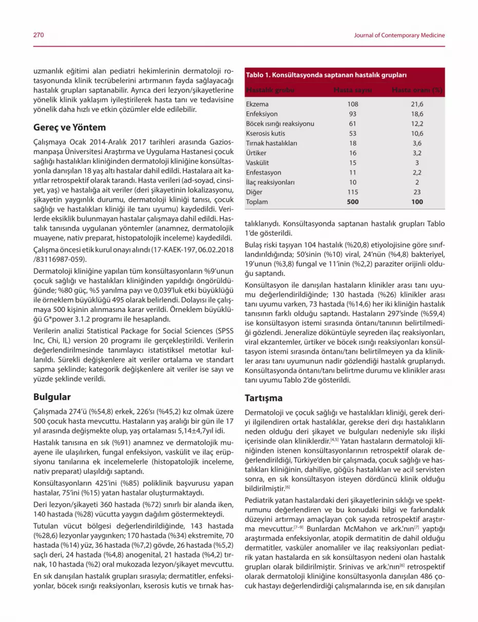
270 Journal of Contemporary Medicine
uzmanlık eğitimi alan pediatri hekimlerinin dermatoloji ro-tasyonunda klinik tecrübelerini artırmanın fayda sağlayacağı hastalık grupları saptanabilir. Ayrıca deri lezyon/şikayetlerine yönelik klinik yaklaşım iyileştirilerek hasta tanı ve tedavisine yönelik daha hızlı ve etkin çözümler elde edilebilir.
Gereç ve YöntemÇalışmaya Ocak 2014-Aralık 2017 tarihleri arasında Gazios-manpaşa Üniversitesi Araştırma ve Uygulama Hastanesi çocuk sağlığı hastalıkları kliniğinden dermatoloji kliniğine konsültas-yonla danışılan 18 yaş altı hastalar dahil edildi. Hastalara ait ka-yıtlar retrospektif olarak tarandı. Hasta verileri (ad-soyad, cinsi-yet, yaş) ve hastalığa ait veriler (deri şikayetinin lokalizasyonu, şikayetin yaygınlık durumu, dermatoloji kliniği tanısı, çocuk sağlığı ve hastalıkları kliniği ile tanı uyumu) kaydedildi. Veri-lerde eksiklik bulunmayan hastalar çalışmaya dahil edildi. Has-talık tanısında uygulanan yöntemler (anamnez, dermatolojik muayene, nativ preparat, histopatolojik inceleme) kaydedildi.
Çalışma öncesi etik kurul onayı alındı (17-KAEK-197, 06.02.2018 /83116987-059).
Dermatoloji kliniğine yapılan tüm konsültasyonların %9’unun çocuk sağlığı ve hastalıkları kliniğinden yapıldığı öngörüldü-ğünde; %80 güç, %5 yanılma payı ve 0,039'luk etki büyüklüğü ile örneklem büyüklüğü 495 olarak belirlendi. Dolayısı ile çalış-maya 500 kişinin alınmasına karar verildi. Örneklem büyüklü-ğü G*power 3.1.2 programı ile hesaplandı.
Verilerin analizi Statistical Package for Social Sciences (SPSS Inc, Chi, IL) version 20 programı ile gerçekleştirildi. Verilerin değerlendirilmesinde tanımlayıcı istatistiksel metotlar kul-lanıldı. Sürekli değişkenlere ait veriler ortalama ve standart sapma şeklinde; kategorik değişkenlere ait veriler ise sayı ve yüzde şeklinde verildi.
BulgularÇalışmada 274’ü (%54,8) erkek, 226’sı (%45,2) kız olmak üzere 500 çocuk hasta mevcuttu. Hastaların yaş aralığı bir gün ile 17 yıl arasında değişmekte olup, yaş ortalaması 5,14±4,7yıl idi.
Hastalık tanısına en sık (%91) anamnez ve dermatolojik mu-ayene ile ulaşılırken, fungal enfeksiyon, vaskülit ve ilaç erüp-siyonu tanılarına ek incelemelerle (histopatolojik inceleme, nativ preparat) ulaşıldığı saptandı.
Konsültasyonların 425’ini (%85) poliklinik başvurusu yapan hastalar, 75’ini (%15) yatan hastalar oluşturmaktaydı.
Deri lezyon/şikayeti 360 hastada (%72) sınırlı bir alanda iken, 140 hastada (%28) vücutta yaygın dağılım göstermekteydi.
Tutulan vücut bölgesi değerlendirildiğinde, 143 hastada (%28,6) lezyonlar yaygınken; 170 hastada (%34) ekstremite, 70 hastada (%14) yüz, 36 hastada (%7,2) gövde, 26 hastada (%5,2) saçlı deri, 24 hastada (%4,8) anogenital, 21 hastada (%4,2) tır-nak, 10 hastada (%2) oral mukozada lezyon/şikayet mevcuttu.
En sık danışılan hastalık grupları sırasıyla; dermatitler, enfeksi-yonlar, böcek ısırığı reaksiyonları, kserosis kutis ve tırnak has-
talıklarıydı. Konsültasyonda saptanan hastalık grupları Tablo 1’de gösterildi.Bulaş riski taşıyan 104 hastalık (%20,8) etiyolojisine göre sınıf-landırıldığında; 50’sinin (%10) viral, 24’nün (%4,8) bakteriyel, 19’unun (%3,8) fungal ve 11’inin (%2,2) paraziter orijinli oldu-ğu saptandı.Konsültasyon ile danışılan hastaların klinikler arası tanı uyu-mu değerlendirildiğinde; 130 hastada (%26) klinikler arası tanı uyumu varken, 73 hastada (%14,6) her iki kliniğin hastalık tanısının farklı olduğu saptandı. Hastaların 297’sinde (%59,4) ise konsültasyon istemi sırasında öntanı/tanının belirtilmedi-ği gözlendi. Jeneralize döküntüyle seyreden ilaç reaksiyonları, viral ekzantemler, ürtiker ve böcek ısırığı reaksiyonları konsül-tasyon istemi sırasında öntanı/tanı belirtilmeyen ya da klinik-ler arası tanı uyumunun nadir gözlendiği hastalık gruplarıydı. Konsültasyonda öntanı/tanı belirtme durumu ve klinikler arası tanı uyumu Tablo 2’de gösterildi.
TartışmaDermatoloji ve çocuk sağlığı ve hastalıkları kliniği, gerek deri-yi ilgilendiren ortak hastalıklar, gerekse deri dışı hastalıkların neden olduğu deri şikayet ve bulguları nedeniyle sıkı ilişki içerisinde olan kliniklerdir.[4,5] Yatan hastaların dermatoloji kli-niğinden istenen konsültasyonlarının retrospektif olarak de-ğerlendirildiği, Türkiye’den bir çalışmada, çocuk sağlığı ve has-talıkları kliniğinin, dahiliye, göğüs hastalıkları ve acil servisten sonra, en sık konsültasyon isteyen dördüncü klinik olduğu bildirilmiştir.[6]
Pediatrik yatan hastalardaki deri şikayetlerinin sıklığı ve spekt-rumunu değerlendiren ve bu konudaki bilgi ve farkındalık düzeyini artırmayı amaçlayan çok sayıda retrospektif araştır-ma mevcuttur.[7–9] Bunlardan McMahon ve ark.'nın[7] yaptığı araştırmada enfeksiyonlar, atopik dermatitin de dahil olduğu dermatitler, vasküler anomaliler ve ilaç reaksiyonları pediat-rik yatan hastalarda en sık konsültasyon nedeni olan hastalık grupları olarak bildirilmiştir. Srinivas ve ark.'nın[6] retrospektif olarak dermatoloji kliniğine konsültasyonla danışılan 486 ço-cuk hastayı değerlendirdiği çalışmalarında ise, en sık danışılan
Tablo 1. Konsültasyonda saptanan hastalık grupları
Hastalık grubu Hasta sayısı Hasta oranı (%)
Ekzema 108 21,6Enfeksiyon 93 18,6Böcek ısırığı reaksiyonu 61 12,2Kserosis kutis 53 10,6Tırnak hastalıkları 18 3,6Ürtiker 16 3,2Vaskülit 15 3Enfestasyon 11 2,2İlaç reaksiyonları 10 2Diğer 115 23Toplam 500 100

271Atiye Oğrum, Pediatristler için zor deri lezyonları
Tablo 2. Konsültasyonda tanı/öntanı belirtme durumu ve klinikler arası tanı uyumu
Hastalık grubu Hastalık Hastalık Tanı/ Uyumsuz† Uyumlu‡ Toplam alt grubu öntanı yok* hasta sayısı
n % n % n %
Ekzema Atopik dermatit 22 50 0 0 22 50 44 Kontakt dermatit 28 90,3 1 3,3 2 6,4 31 Seboreik dermatit 16 100 0 0 0 0 16 Diaper dermatit 2 25 0 0 6 75 8 Numuler dermatit 4 100 0 0 0 0 4 Perianal dermatit 4 100 0 0 0 0 4 Fitofotodermatit 0 0 1 100 0 0 1Enfeksiyon Viral Anogenital siğil 0 0 1 50 1 50 2 Verruka vulgaris 1 10 0 0 9 90 10 Herpes virus enfeksiyonu 3 42,8 1 14,4 3 42,8 7 Molluskum kontagiosum 4 80 1 20 0 0 5 El ayak ağız hastalığı 5 35,7 4 28,6 5 35,7 14 Eritema infeksiyosum 3 75 1 25 0 0 4 Varisella 0 0 2 50 2 50 4 Roseola infantum 2 50 1 25 1 25 4 Bakteriyel Folikülit 8 80 1 10 1 10 10 İmpetigo 8 88,9 0 0 1 11,1 9 Kızıl 0 0 0 0 1 100 1 Akut paronişi 2 100 0 0 0 0 2 Selülit 0 0 0 0 1 0 1 SSSS§ 0 0 0 0 1 0 1 Fungal İntertrijinöz kandidiasis 1 33,3 2 66,7 0 0 3 Tinea (kapitis/korporis/pedis) 6 75 0 0 2 25 8 Onikomikoz 1 50 0 0 1 50 2 Pitriasis versikolor 5 83,3 1 16,7 0 0 6Böcek ısırığı reaksiyonu Böcek ısırığı 38 62,3 19 31,1 4 6,6 61Kserosis kutis Kserosis kutis 37 69,8 4 7,6 12 22,6 53Tırnak hastalıkları Beau çizgileri 2 66,7 1 33,3 0 0 3 Onikodistrofi 3 42,8 3 42,8 1 14,4 7 Onikomadezis 5 83,3 1 16,7 0 0 6 Subungual hematom 1 100 0 0 0 0 1 Trakionişi 1 100 0 0 0 0 1Ürtiker Akut Ürtiker 5 31,3 4 25 7 43,7 16Vaskülit Akut infantil hemorajik ödem 2 66,7 1 33,3 0 0 3 Lökositoklastik vaskülit 3 30 0 0 7 70 10 Ürtikeryal vaskülit 1 50 1 50 0 0 2İlaç ilişkili durumlar İlaç erüpsiyonu 8 80 1 10 1 10 10Enfestasyon Skabiyez 4 40 5 50 1 10 10 Pedikülosis 0 0 0 0 1 100 1DiğerII 62 53,9 16 14 37 32,1 115Toplam 297 59,4 73 14,6 130 26 500
*: Konsültasyon istem notunda tanı/öntanı belirtilmemiş; †: Öntanı/tanıda klinikler arası uyum yok; ‡: Öntanı/tanıda klinikler arası uyum var; §: Stafilokoksik haşlanmış deri sendromu; II: Tekrarlayan oral aft, miliaria, pitriazis alba, pitriazis rosea, eritema nodozum, mastositoz, iktiyozis, akne, hemanjiom, travmatik bül, hidroa vaksiniforme, eritema toksikum neonatarum, palmar peeling, psorizis, alopesi areata, bölgesel hiperhidroz, prurigo, granuloma annulare, perleş, pernio, intertrigo, juvenil bahar erüpsiyonu, aplazia kutis konjenita, atrofik glossit, morfea, nevus depigmentozus, piezojenik pedal papül, pigmente purpurik dermatoz, pilomatrikoma, polimorf ışık erüpsiyonu, terra firma forme dermatozu, yanık, yenidoğanın subkutan yağ nekrozu, epidermolizis bülloza, eritema multiforme, akantozis nigrigans, albinizm.

272 Journal of Contemporary Medicine
hastalık grupları; enfeksiyonlar, acil dermatolojik durumlar ve genodermatozlar olarak bildirilmiştir. Türkiye’de ikinci basa-mak bir çocuk hastanesindeki 539 hastanın dermatoloji klini-ğine yapılan konsültasyonlarının, retrospektif olarak değerlen-dirildiği çalışmada; en sık konsültasyon nedeni olan hastalık atopik dermatit ve diğer dermatitler iken, bunu vaskülitler, ilaç erüpsiyonları, enfeksiyonlar ve ürtiker tanılarının izlediği bildirilmiştir.[8] Çalışmamızda, en sık saptanan hastalık grupla-rı; dermatitler, enfeksiyonlar ve böcek ısırığı reaksiyonlarıydı. Bazı çalışmalarda daha yüksek oranda bildirilen vaskülit, ilaç erüpsiyonu, acil dermatolojik durumlar gibi tabloların bu çalış-mada daha düşük oranda gözlenmesinin, konsültasyonla da-nışılan hastaların %85’inin ayaktan poliklinik başvurusu yapan ve olasılıkla klinik tablonun daha hafif olduğu hasta grubu ol-masıyla ilişkilendirilebileceğini düşünüyoruz. Bununla birlikte, poliklinik ağırlıklı hasta değerlendirmesinin, pediatri hekimle-rinin günlük pratikte daha sık karşılaşacakları deri şikayet/lez-yonlarının saptanması açısından daha faydalı bir değerlendir-me olacağı kanaatindeyiz.Konsültasyonla danışılan en sık hastalık grubu ekzemalar olup, ekzemalarda ilk üç sırayı, atopik, kontakt ve seboreik derma-tit almaktaydı. Atopik dermatitin etiyopatogenezi oldukça karmaşıktır ve günümüzde hâlen tamamıyla aydınlatılmış değildir.[10] Hastalığın patogenezinde etkili olan faktörler ge-netik yatkınlık, çevresel faktörler, epidermal bariyer fonksiyon bozukluğu ve immün disregülasyondur.[11] Hastalığın tedavisi çok yönlü yaklaşım gerektirmekte olup; bunlar tetikleyici fak-törlerin eliminasyonu, deri bariyerinin yenilenmesi ve derinin hidrasyonu, hasta eğitimi ve deri inflamasyonunun farmako-lojik tedavisini içerir.[12] Tetikleyici faktörlerin eliminasyonu aşamasında; sıcak ve düşük nemden kaçınılması, stafilokok-kus aureus ve herpes simpleks virüs gibi deri enfeksiyonları-nın tedavisi, sedasyon ve kaşıntı azaltma amaçlı sedatif oral antihistaminik kullanımı ile stres ve anksiyetenin giderilmesi yer alır.[12] Aeroallerjen ve gıda allerjilerinin atopik dermatitte atağı şiddetlendiren faktörler olduğu konusu ise tartışmalıdır.[13,14] Deri hidrasyonunu sağlamada emolyenler günde bir–üç kez uygulanmalıdır.[15] En iyi uygulama banyodan çıktıktan he-men sonraki beş dakika içinde, fazla su bir havluyla silindikten sonra, yapılan uygulamadır. Sedatif antihistaminikler kaşın-tıyı azaltmasının yanı sıra özellikle uyku probleminin aşılma-sını sağlayarak da tedaviye katkıda bulunur.[15] Farmakolojik tedavide, hafif vakalarda düşük potens topikal kortikostero-idler iki-dört hafta süresince; orta ve ileri şiddette vakalarda orta-yüksek potens topikal kortikosteroidler iki haftayı geç-meyecek süreyle kullanılabilir.[15] Akut şiddetli alevlenmelerde kısa süreli oral kortikosteroid tedavisi gerekebilir. İdame teda-visi aralıklı düşük potens topikal kortikosteroid ya da topikal immünmodülatörlerle sağlanabilir. Vücudun nemlendirilmesi, atopik dermatitin idame tedavisinde de en önemli yaklaşım-lardan biridir.[16]
Çalışmamızda sık danışılan egzemalardan olan kontakt der-matitler, pediatrik popülasyonda sık görülmekte olup, allerjik ve irritan olmak üzere iki grupta incelenir.[17] Kontakt allerjen-lere karşı gelişen sensitizasyon infant dönemde başlar ve adö-
lesan döneme doğru artarak devam eder.[18] Sıklıkla atlanan bir tanı olup, tanıya ulaşmada, hikaye ve fizik muayeneye ek olarak deri yama testi kullanılabilir.[17] Konsültasyonlarda sık gözlenen bir egzema çeşidi olan seboreik dermatit ise, saçlı deri, yüz ve intertrijinöz alanlar gibi sebase glandların yoğun olduğu alanlarda, eritemli zeminde yağlı görünümlü, sarımsı skuamla karakterizedir.[19] İnfant dönemde saçlı deri yerleşimi-ne ek olarak, diaper dermatit kliniğinde de görülebilir. Egze-malarda doğru tanıya ulaşmada, hikaye ve dermatolojik mua-yenenin önemi büyüktür.İkinci sıklıkta danışılan hastalık grubu enfeksiyonlar olup, konsültasyonda öntanı belirtilmeyen ya da klinikler arası uyum gözlenmeyen en sık enfeksiyon nedeni viral hastalık-lardı. Bu grup hastalıklarda konsültasyon isteminde belirtilen en sık ön tanılar ilaç erüpsiyonu ve ürtikerdi. Bu durumun, viral döküntülü hastalıklarda, döküntü öncesinde gözlenen ateşin nedeni olabilecek bir odak saptanamaması ve verilen ampirik antibiyoterapi sonrası döküntünün ortaya çıkmasıy-la klinik tablonun karmaşık hale gelmesi nedeniyle olabile-ceğini düşünüyoruz. Ayrıca, ilaç erüpsiyonlarının en sık for-mu olan makülopapüler döküntü, viral ekzantemler ile klinik olarak karışabilir. Viral enfeksiyonların sıklıkla ateşin eşlik et-tiği kaşıntısız ve soluk eritemli döküntüyle seyrederken; ilaç erüpsiyonlarında lezyonların sıklıkla kaşıntılı olacağı ve inf-lamatuvar doku reaksiyonunu işaret eden canlı kırmızı-mor renkte görülebileceğini ve bu klinik özelliklerin iki tabloyu ayırmada faydalı olabileceğini düşünüyoruz. Ayrıca hemog-ram tetkikinde lökositoz varlığı, enfeksiyon; eozinofil yüksek-liği, ilaç erüpsiyonunu destekler laboratuar bulguları olarak tanıya ulaşmada yardımcı olabilir.Viral hastalıkların klinik olarak karışabileceği bir diğer hasta-lık olan ürtiker, kaşıntılı papül ya da plaklarda karakterizedir.[20] Ayrıca dermal ödemle karakterize ürtikerde kaşıntı ovmayla rahatladığından deride ekskoriasyonun sık gözlenen bir du-rum olmadığını düşünüyoruz. Ürtikerde lezyonların 1–24 saat içinde kendiliğinden kaybolması tipiktir.[21] Bu nedenle kaybol-makta olan plakların viral döküntü kliniğini taklit edebileceği ve tanıya ulaşmada zorluğa neden olabileceğini düşünüyoruz. Bundan dolayı lezyonların gün içerisinde yer değiştirip değiş-tirmediğinin sorgulanması tanıya ulaşmada ipucu olarak de-ğerlendirilebilir.[20] Ayrıca bu bulgunun, ürtikeri, lezyonlarda yer değiştirmenin gözlenmediği ilaç erüpsiyonundan ayırma-da da faydalı bir bulgu olabileceğini düşünüyoruz. Hastadan net klinik bilginin alınamadığı durumlarda, birkaç adet ürtiker plağı sınırının çizilerek 24 saat sonraki kontrolde plakların kay-bolup kaybolmadığının değerlendirilmesi de ürtiker tanısını doğrulamada yararlı olabilir.El ayak ağız hastalığında tuttuğu lokalizasyonlar nedeniyle ti-pik vakaların tanısı kolay olmakla beraber, bu lokalizasyonlar dışında da lezyonların görüldüğü atipik vaka varlığı da akılda tutulmalı ve yaygın deri döküntüsünün görüldüğü hastalıkla-rın ayırıcı tanısında düşünülmelidir.[22]
Semptomatik olduğunda şiddetli tabloyla seyredebilen pri-mer herpes virüs enfeksiyonu, sıklıkla ilk 6 yaşta geçirilmek-

273Atiye Oğrum, Pediatristler için zor deri lezyonları
te olup, çoğunlukla ilk atak asemptomatiktir.[23] Semptomatik geçirildiğinde ise, yaygın oral aftı taklit eden akut gingivosto-matit tablosu şeklinde ortaya çıkar.[24] Bu çalışmada bir hastada primer herpes virüs enfeksiyonu saptanmış olup, oral aft teda-visiyle klinik düzelme saptanmayan hastada klinik tablo siste-mik antiviral tedaviyle hızla gerilemiştir. Çocukluk yaş grubun-da klinik olarak ateşin eşlik ettiği ve oral aft benzeri multiple yaygın erozyon ve ülserasyon varlığında primer herpes virüs enfeksiyonunun ayırıcı tanıda düşünülmesinin faydalı olabile-ceğini düşünüyoruz.Fungal enfeksiyonlardan olan intertrijinöz kandidiazis, sıklıkla diaper dermatit öntanı/tanısıyla danışılmıştı. Lezyon çevresin-de satellit papül ve püstüllerin varlığı, diaper dermatit üzerine eklenen kandidal enfeksiyon açısından destekleyici bir bulgu olarak akılda tutulmalıdır.[24]
Konsültasyon tanılarının %12’sini oluşturan böcek ısırığı reak-siyonlarında dermatoloji ile uyumlu tanı oranı oldukça düşük olup, 61 hastanın dördünde (%6,6) tanı uyumu gözlendi. Bu grubun konsültasyon notunda en sık belirtilen öntanı/tanıların ilaç erüpsiyonu, ürtiker ve selülit olduğu saptandı. Böcek ısırığı reaksiyonlarının sıklıkla yaz aylarında görülür ve ekstremiteler gibi vücudun distal ve açık alanlarına lokalize olma eğilimin-dedir.[25] Dikkatli muayene ile lezyon ortasında, böcek ısırığına bağlı gelişen pikür izinin görülmesi de tanıya ulaşmada faydalı olabilir. Klinik olarak kaşıntılı papül dışında ısırığın neden ol-duğu şiddetli inflamatuvar reaksiyona sekonder olarak eritemli nodül, vezikül ya da bül de gelişebilir.[25]
Enfestasyon grubunda yer alan hastalıklardan skabiyez 10 hastada saptanmış olup, bu hastaların birinde öntanıda skabi-yez vardı. Oldukça bulaşıcı olması nedeniyle skabiyezin erken teşhisinin, sağlık çalışanlarına bulaşın önlenmesi ve hastalı-ğın toplumda yayılımının önüne geçilmesi açısından oldukça önemli olduğunu düşünüyoruz. Özellikle gece şiddetlenen jeneralize vücut kaşıntısı ve aile bireylerinde de benzer şika-yetlerin varlığında ayırıcı tanıda skabiyez de düşünülmelidir.[26]
Tırnak hastalıkları grubunda danışılan 18 hastadan altısında görülen onikomadezis tablosu, tırnak büyümesinin geçici ola-rak durması nedeniyle tırnak plağı proksimalinin tırnak mat-riksinden ayrılmasıyla karakterizedir.[27] Onikomadezis gelişi-minden önceki bir-iki ayda geçirilen viral hastalık sorgusu olası etiyolojik nedeni bulmak açısından faydalı olabilir.[27] Özellikle el ayak ağız hastalığı sonrası gelişen onikomadezisle ilgili çok sayıda olgu bildirimi mevcuttur.[27] Bunun dışında ilaçlar ve tır-nağa lokal travma da onikomadezis nedeni olabilir.[27]
KısıtlılıklarÇalışma retrospektif dizaynda ve tek merkezlidir. Çalışmada, sadece konsültasyon ile danışılan hastalar değerlendirmeye alındığından, pediatri polikliniğine başvuran hastalardaki deri şikayeti/lezyon sıklığı, bu başvurulardaki her bir hastalığın ora-nı ve bu hastalık nedenli konsültasyon istenme oranı hesapla-namamıştır. Bununla birlikte, her iki kliniğin de değerlendirdiği hastaların çalışmaya alınmasının, tanı uyumunun değerlendi-rilebilmesi açısından daha faydalı olacağını düşünüyoruz.
SonuçSonuç olarak, atopik dermatitin de dahil olduğu dermatitler ile jeneralize deri döküntüsüyle giden hastalıkların tanı ve ayı-rıcı tanısına yönelik bilgi düzeyinin artırılması faydalı olabilir. Böcek ısırığı reaksiyonları çok farklı kliniklerle karşımıza çıka-bileceğinden hem lokalize hem de jeneralize deri döküntüleri-nin ayırıcı tanısında düşülmelidir. Yüksek oranda bulaşıcı olan skabiyez, jeneralize kaşıntıyla gelen hastaların ayırıcı tanısında düşünülmelidir. Deri lezyonlarında ayırıcı tanının karmaşıklığı nedeniyle, hastaların erken tanı ve etkin tedavisi için, gerekli görülen her durumda konsültasyon hizmetinden yararlanıl-masının faydalı olacağı kanaatindeyiz.
Çıkar çatışması: Bildirilmemiştir.
Kaynaklar1. Çiçek D, Kandi B, Oğuz S, ve ark. Diyabetes mellituslu hasta-
larda gözlenen deri bulguları. FÜ Sağlık Bilimleri Tıp Dergisi 2010;24(2):77-80.
2. Şahin EB, Hapa A, Elçin G, ve ark. Lökositoklastik vaskülit: 60 has-tanın geriye dönük analizi. Türk Dermatoloji Dergisi 2011;5(4):85-91.
3. Öztürkcan S, Özel F, Doğan S, Seyfikli Z, Hatipoğlu A. Akciğer kanserli hastalarda deri bulguları. Tüberküloz ve Toraks Dergisi 2003;51(1):23-6.
4. Adışen E, Ünal S, Gürer MA. Dermatoloji konsültasyonları. Turk-derm 2006;40(4):126-9.
5. Çiçek D. Dermatoloji konsültasyonlarının değerlendirilmesi. Fırat Tıp Dergisi 2007;12(3):181-3.
6. Srinivas SM, Hiremagalore R, Venkataramaiah LD, Premalatha R. Pediatric dermatology inpatient consultations: a retrospective study. Indian J Pediatr 2015;82:541-4.
7. McMahon P, Goddard D, Frieden IJ. Pediatric dermatology inpa-tient cosultations: a retrospective study of 427 cases. J Am Acad Dermatol 2013;68(6):926-31.
8. Afsar FS. Analysis of pediatric dermatology inpatient consul-tations in a pediatric teaching hospital. Arch Argent Pediatr 2017;115(6):e377-84.
9. Peñate Y, Borrego L, Hernández N, Islas D. Pediatric dermato-logy consultations:a retrospective analysis of inpatient consul-tations referred to the dermatology service. Pediatr Dermatol 2012;29(1):115-8.
10. Birtekocak F, Uysal P, Büyüköztürk, Karul A. Süt çocuklarında atopik dermatit ağırlığı ile serum TSLP ve TARC düzeyleri arasındaki ilişki.İzmir Doktor Behçet Uz Çocuk Hastalıkları Dergisi 2016;6(1):28-36.
11. Kulthanan K, Chularojanamontri L, Manapajon A, Nuchkull P. Pre-valence and clinical characteristics of adult-onset atopic derma-titis with positive skin prick testing to mites. Asian Pac J Allergy Immunol2011;29(4):318-26.
12. Tollefson MM, Bruckner AL. Atopic dermatitis: skin-directed ma-nagement. Pediatrics 2014;134(6):e1735-44.
13. Fu T, Keiser E, Linos E, et al. Eczema and sensitization to common allergens in the United States: a multiethnic, population-based study. Pediatr Dermatol 2014;31(1):21-6.
14. Garritsen FM, ter Haar NM, Spuls PI. House dust mite reduction in the management of atopic dermatitis. A critically appraised topic. Br J Dermatol 2013;168(4):688-91.

274 Journal of Contemporary Medicine
15. Ertam İ, Su Ö, Alper S, Sarıcaoğlu H, ve ark. Türkiye atopik dermatit tanı ve tedavi kılavuzu-2018. Turkderm 2018;52(1):6-23.
16. Uysal P, Uzuner N. Çocuklarda atopik dermatit tedavisi.İzmir Dok-tor Behçet Uz Çocuk Hastalıkları Dergisi 2013;3(2):77-86.
17. Pigatto P, Martelli A, Marsili C, Fiocchi A. Contact dermatitis in children. Ital J Pediatr 2010;36:2.
18. de Waard-van der Spek FB, Oranje, AP. Patch test in children with suspected allergic contact dermatitis: a prospective study and re-view of the literature. Dermatology 2009;218(2):119-25.
19. Victoire A, Magin P, Coughlan J, van Driel ML. Interventions for infantile seborrhoeic dermatitis (including cradle cap). Cochrane Database Syst Rev 2019;3:CD011380.
20. Uysal P, Avcil S, Erge D. Ürtikerli çocukluklarda yüksek doz an-tihistaminik kullanımı ve risk etmenleri. Turk Pediatri Arşivi 2016;51(4):198-203.
21. Bernstein JA, Lang DM, Khan DA, et al. The diagnosis and mana-gement of acute and chronic urticaria: 2014 update. Practice Pa-
rameter. J Allergy Clin Immunol 2014;133(5):1270-7.22. Harris PNA, Wang AD, Yin M, Lee CK, Archuleta S. Atypical hand,
foot, and mouth disease: eczema coxsackium can also occur in adults. Lancet Infec Dis 2014;14(11):1043.
23. Arduino PG, Porter SR. Herpes Simplex Virus Type 1 infection: overview on relevant clinico-pathological features. J Oral Pathol Med 2008;37(2):107-21.
24. Bahçetepe N, Kocatürk Göncü ÖE, Güngör Ş, Köklü N, Kuteyla Ül-kümen P.Obezite ve deri. Okmeydanı Tıp Dergisi 2014;30(1):34-8.
25. Kar S, Dongre A, Krishnan A, Godse S, Singh N. Epidemiological study of insect bite reactions from central India. Indian J Derma-tol 2013;58(5):337-41.
26. Chosidow O. Scabies and pediculosis. Lancet 2000;355(9206):819-26.
27. Salgado F, Handler MZ, Schwartz RA.Shedding light on onycho-madesis. Cutis 2017;99(1):33-6.

Corresponding (İletişim): Cennet Rağbetli, Van Eğitim ve Araştırma Hastanesi, Mikrobiyoloji Kliniği, Van, TurkeyE-mail (E-posta): [email protected]
Received (Geliş Tarihi): 22.08.2019 Accepted (Kabul Tarihi): 26.08.2019
DOI: 10.16899/jcm.609560J Contemp Med 2019;9(3):275-279
Orjinal Araştırma / Original Article
JOURNAL OF
CONTEMPORARY MEDICINEJournal ofContemporary MedicineYEAR: 2018 VOLUME: 8 ISSUE: 1
Yoğun bakım ünitelerinden izole edilen çok ilaca dirençli Acinetobacter ve Pseudomonas izolatlarında
karbapenem direncinin araştırılması
Investigation of the carbapenem resistance in multi-drug resistant Acinetobacter and Pseudomonas isolates in intensive care units
Amaç: Bu çalışmada hastanemiz yoğun bakım ünitelerinde (YBÜ) yatan hastalarda Pseudomonas ve Acinetobacter izolatlarında çoklu ilaç direnci (ÇİD) gelişmesine neden olan karbapenem direncinin genotipik analizlerle ortaya konulmasını amaçlanmıştır. Gereç ve Yöntem: Çalışmada, Nisan 2014–Aralık 2014 tarihleri ara-sında Yüzüncü Yıl Üniversitesi Dursun Odabaş Tıp Merkezi YBÜ'deki hastalardan izole edilen 51 Acinetobacter baumannii ve 51 Pseu-domonas aeruginosa izolatlarında karbapenem direncine neden olan genler araştırılmıştır. İzolatların antimikrobiyal duyarlılıkları, Kirby-Bauer disk difüzyon yöntemi ve BD Phoenix (Becton Dickin-son, ABD) otomatize sistemi ile belirlenmiştir. Tüm izolatlarda kar-bapenem direncine yol açan IMP, VIM, GES, GIM, SPM, OXA-10, OXA-23 ve OXA-51 gen bölgeleri Polimeraz Zincir Reaksiyonu (PZR) ile belirlenmiştir. Ayrıca tüm izolatlarda pulse field gel electrophorezi (PFGE) ile klonal ilişki ortaya konmuştur.Bulgular: Toplam 102 izolatın hiçbirinde IMP, VIM, GES, GIM gen bölgeleri tespit edilmemiştir. A. baumannii izolatının tamamında en az bir gen bölgesi pozitif olmak üzere %98'inde OXA-51, %77'sinde OXA-23 ve %4'ünde SPM geni pozitif bulunmuştur. Bu izolatlarda; %77 oranında OXA-23 ile OXA-51 ve %4 oranında OXA-23, OXA-51 ile SPM birlikteliği saptanmıştır. P. aeruginosa izolatında ise OXA-51 %18 ve OXA-10 ise %14 oranında tespit edilmiştir. PFGE yöntemi ile klonal ilişki analizi sonuçlarına göre A. baumannii izolatlarının kümeleşme oranı %80 olarak belirlenirken P. aeruginosa da bu oran %53 olarak belirlenmiştir.Sonuç: Çalışmamızda A. baumannii suşlarının karbapenem diren-cinden SPM, OXA 23 ve OXA 51 ve P. aeroginosa suşlarının karba-penem direncinden OXA 10 ve OXA 51 tipi karbapenemaz genleri sorumlu bulunmuştur.Anahtar Sözcükler: A. baumannii; karbapenemase; P. aeruginosa.
Introduction: The aim of this study was to determine the carbapenem resistance in Pseudomonas and Acinetobacter isolates in the intensive care units (ICU) of our hospital by genotypic analysis.Methods: Study strains included imipenem or meropenem resistant 51 Acinetobacter baumannii and 51 Pseudomonas aeruginosa iso-lates between April 2014–December 2014 at intensive care units in YYU, Dursun Odabas Medical Center, Turkey. Antimicrobial suscepti-bility testing of the isolates were performed by Kirby-Bauer disk dif-fusion method and BD Phoenix automated system in our hospital. All strains that cause carbapenemase encoding genes by IMP, VIM, GES, GIM, SPM, OXA10, OXA-23 and OX-51 gene regions were investigated with Polymerase Chain Reaction. Pulsed Field Gel Electrophoresis (PFGE) was performed to detect moleculer epidemiologic relation-ships among the isolates.Results: A total of 102 isolates evaluated for carbapenemase gene re-gions of IMP, VIM, GES, GIM were not observed in any of the isolates. The presence of the carbapenem resistant of OXA-51 (98%), OXA-51 (77%), OXA-23 (77%) and SPM (4%) genes positive were found A. baumannii isolates. In these strains, both OXA-23 and OXA-51 %77 and OXA-23&OXA-51&SPM %4 genes were observed. Detection of OXA genes in P. aeruginosa strains of OXA51 strains (18%) and OXA-10 strains (14%) showed multidrug-resistant. According to the clonal relationship between the isolates were identified by pulsed-field gel electroforesis, cluster rate was found to be 80% A. baumannii strains and 53% P. aeruginosa strains.Discussion and Conclusion: In our study, carbapenemase genes cod-ing for SPM, OXA 23 and OXA 51 were responsible for carbapenem resistance of A. baumannii isolates, while genes coding for OXA 10 and OXA 51 were identified in carbapenem-resistant P. aeruginosa isolates.Keywords: A. baumannii; carbapenemase; P. aeruginosa.
Özet Abstract
Cennet Rağbetli,1 Hüseyın Güdücüoğlu,2 Mehmet Parlak2
1Sağlık Bilimleri Üniversitesi, Van Eğitim ve Araştırma Hastanesi, Mikrobiyoloji Kliniği, Van Turkey2Yüzüncü Yıl Üniversitesi Tıp Fakültesi, Tıbbi Mikrobiyoloji Anabilim Dalı, Van, Turkey
Sözlü sunu olarak 15–19 March 2016 tarihinde Arusha, Tanzania'da "2nd Neuroscience Stereology and Scientific Writing Symposium"unda sunulmuştur.

276 Journal of Contemporary Medicine
Antibiyotik direnci yaygın ve kontrolsüz antibiyotik kulla-nımına bağlı olarak günümüzde büyük bir sorun haline
gelmiştir. Özellikle yoğun bakım ünitelerinde sıkça görülen hastane enfeksiyonlarında çoklu ilaç direnci (ÇİD) belirlenen mikroorganizmalar daha çok önem kazanmıştır. Bu üniteler-de hastalık etkeni olarak en sık izole ettiğimiz nonfermenta-tifler Pseudomonas ve Acinetobacter izolatlarıdır. Bu bakteri-lerin etken olduğu enfeksiyonlarda kullanılan antibiyotiklere karşı direncin artması tedavide güçlüklere sebep olmaktadır. Dirençli nonfermentatif mikroorganizmaların sebep olduğu enfeksiyonlar mortalitenin artmasına, hastane yatış süresinin uzamasına ve tedavi maliyetinde artışlar gibi sorunlara sebep olmaktadır.[1,2]
Karbapenemler, penisilin ve sefalosporin gibi beta laktamlara dirençli bakteri türlerinin çoğuna karşı etkilidir. Ama son yıl-larda özellikle nonfermantatif gram negatif bakteriler arasında karbapenemlere karşı artan bir direnç problemi gözlenmek-tedir.[3,4]
Bu çalışmada hastanemiz Yoğun Bakım Ünitelerinde (YBÜ) yatan hastalarda en sık rastlanan nonfermentatiflerden Pseu-domonas ve Acinetobacter izolatlarında ÇİD gelişimine sebep olan Metallo beta laktamaz (MBL) direnci fenotipik ve genoti-pik analizlerle ortaya konulmasını amaçlanmıştır.
Gereç ve YöntemNisan 2014-Aralık 2014 tarihleri arasında Yüzüncü Yıl Üniversi-tesi Dursun Odabaş Tıp Merkezi Mikrobiyoloji Laboratuvarın-da imipenem veya meropenem dirençli 51 Acinetobacter bau-mannii ve 51 Pseudomonas aeruginosa izolatı çalışmaya dâhil edilmiştir. izolatlarının antimikrobiyal duyarlılıkları, Kirby-Ba-uer disk difüzyon yöntemi ve BD Phoenix (Becton Dickinson, ABD) otomatize sistemi ile belirlenmiştir. MBL varlığı, çift disk sinerji testi (ÇDST), kombine disk testi (KDT), Modifiye Hodge testi (MHT) ile tespit edilmiştir. Tüm izolatlarda karbapenem direncine yol açan IMP, VIM, GES, GIM, SPM, OXA-10, OXA-23 ve OXA-51 gen bölgeleri Polimeraz Zincir Reaksiyonu (PZR) ile belirlenmiştir. Ayrıca tüm izolatlarda Pulsed-field jel elect-rophorezi (PFGE) ile klonal ilişki ortaya konmuştur. Moleküler bant analizleri için benzerlik hesaplarının yapılmasında Pear-son korelasyon katsayısı ve kümeleşme analizi için de UPG-MA (Unweighthed Pairvise Grouping Matematical Avenaging matematiksel ortalamayla ağırlıksız çiftlerin gruplandırılması) yöntemi kullanıldı.
SonuçlarÇalışmaya karbapenem dirençli 51 P. aeruginosa ve 51 A. ba-umannii olmak üzere toplam 102 adet izolat dahil edilmiştir. Çalışmaya alınan 102 izolatın 18'i (%18) çocuk yoğun bakım, 84'ü (%82) yetişkin yoğun bakım ünitelerinden izole edilmiş-tir. Hastaların yaşı 11 günlük ve 87 yaş arasında değişmektey-di (ortalama yaş: 35.6, SS: 23.6). Her biri farklı hastadan izole edien bu izolatlar izolasyon tarihlerine göre kronolojik ola-rak numaralanmıştır. Hastaların YBÜ'de kalış süresi ortalama 3.3 ay olduğu belirlenmiştir. Hastalar ağırlıklı olarak solunum
yolu enfeksiyonları nedeniyle (%53) takip edilmekteydi. Çocuk YBÜ'de 10 adet P. aeruginosa ve 8 adet A. baumannii izole edi-lirken, yetişkin YBÜ'de 43 adet A. baumannii ve 41 adet P. ae-ruginosa izole edilmiştir. İzolatların elde edildiği yer ve örnek türlerine göre dağılımı Tablo 1'de gösterilmiştir.
Çalışmada disk diffüzyon ve otomatize sistemle imipenem ve meropeneme dirençli bulunan 51'i A. baumannii, 51'i P. aerugi-nosa izolatlarının diğer antibiyotiklere duyarlılık test sonuçları Tablo 2'de verilmiştir.
Çalışılan kökenlerde IMP, VIM, GES, GIM, SPM, OXA-51, OXA-23, OXA-10 grup genlerinin varlığını araştırmak amacıyla PZR yapıldı. Toplam 102 izolatın hiç birinde IMP, VIM, GES, GIM gen bölgeleri tespit edilemedi. A. baumannii izolatları-nın tamamında SPM, OXA-51 ve OXA-23 gen bölgelerinden en az bir tanesi pozitif bulundu. P. aerugonosa'da izolatların 16'sında (%31); OXA-51 (%18) ve OXA-10 (14) gen bölgele-rinden en az bir tanesi pozitif bulundu. PFGE ile pozitiflik saptanan gen bölgelerinin izolatlara göre dağılımı Tablo 3, Şekil 1, 2'de verilmiştir.
Tiplendirilen 51 A. baumannii izolatında 38 farklı PFGE profili göstermiştir. Klonal yönden ilişkili izolatlar, 13 farklı küme içe-risinde yer almaktadırlar. Toplam 51 A. baumannii izolatunun 41'i herhangi bir küme içerisinde yer almaktadır. İzolatların kü-meleşme oranı %80'dir.
En büyük küme; 7 izolatın yer aldığı VI ile kodlanan kümedir. Bunu sırasıyla; I (6 izolat), VIII (6 izolat), IV (4 izolat), II (2 izolat), III (2 izolat), X (2 izolat), XI (2 izolat), XII (2 izolat), XIII (2 izolat), XIV (2 izolat), XV (2 izolat), XVII (2 izolat) kümeleri takip etmek-tedir (Şekil 1).
Tiplendirilen 51 P. aeruginosa izolatının 43 farklı PFGE profili göstermiştir. Klonal yönden ilişkili izolatlar, 11 farklı küme içe-risinde yer almaktadırlar. Toplam 51 P. aeruginosa izolatının 27'si herhangi bir küme içerisinde yer almaktadır. İzolatların
Tablo 1. Elde edildikleri birimlere göre izolatların ve örnek türlerinin dağılımı
Klinik Örnek A. baumannii P. aeruginosa
Çocuk YBÜ Kan 7 0(n=18) İdrar 0 5 Kulak 0 2 Trekeal aspirat 1 1 Yara 0 1 BOS 0 1Yetişkin YBÜ Trekeal aspirat 32 20(n=84) Yara 5 6 İdrar 1 9 Kan 3 2 Kulak 0 3 Plevra sıvısı 2 1 Toplam 51 51
YBÜ: Yoğun bakım ünitesi.

277Cennet Rağbetli, Yoğun bakım ünitelerinde karbapenem direncinin araştırılması
kümeleşme oranı %53'tür.
En büyük küme; 4 izolatın yer aldığı XII ile kodlanan kümedir. Bunu sırasıyla; I (3 izolat), V (3 izolat), XXI (3 izolat), IV (2 izolat), XIV (2 izolat), XIX (2 izolat), XXIII (2 izolat), XXVIII (2 izolat), XXIX (2 izolat), XXXIII (2 izolat) kümeleri takip etmektedir (Şekil 2).
TartışmaYoğun Bakım Üniteleri, invaziv girişimlerin sıklıkla yapıldığı birimler olup, uzun süreli yatış tedavi olma, yanık tedavileri, immün yetmezlik, malignensi gibi altta yatan risk faktörlerleri, büyük cerrahi girişimler ve uzun süreli geniş spektrumlu anti-biyotik kullanımı hastane enfeksiyonları açısından risk oluştur-maktadır.[5] A. baumannii ve P. aeruginosa gibi nonfermentatif bakteriler YBÜ'lerde sıklıkla karşımıza çıkan hastane enfeksi-yonu etkenidirler. Bu mikroorganizmalar ÇİD olarak karşımıza çıkmakta ve bu izolatlarla oluşan enfeksiyonlar tedavi güçlük-lerine neden olmaktadır. Ampirik tedavi de dahil olmak üzere bu etkenlerin neden olduğu enfeksiyonlarda karbapanemler
sıklıkla kullanılmakta ve bu durum karbapenem direncinde artış olarak kendini göstermektedir.[6] Özellikle nonfermenta-tif gram negatif basillerde karbapenem direnci daha yüksek oranlarda görülmekte ve hızla artarak dünya çapında yayıl-ma göstermektedir.[7] Türkiye'de yapılan çeşitli araştırmalar-da nonfermentatiflerde imipenem ve meropenem direnci %40'lardan %92'lere ulaşmıştır.[8]
Karbapenem direncini belirlemek amacıyla Hacettepe Üni-versitesinde gerçekleştirilen bir çalışmada 110 P. aeruginosa izolatının 11'inde blaVIM pozitif bulunmuştur.[9] Özgümüş ve ark. Karadeniz Teknik Üniversitesinden elde ettikleri izolatlar-da gerçekleştirdikleri çalışmada VIM ve SPM genlerini Pseudo-monas'larda saptamışlardır.[10] Trakya Üniversitesi Tıp Fakülte-si'nde gerçekleştirilen bir çalışmada P. aeruginosa kökenlerinin tümünde yapılan PZR ve multipleks PZR yöntemleri sonucun-da karbapenem direncinden sorumlu NDM-1, IMP, VIM, SPM-1, SIM-1, GIM-1 enzimleri negatif bulunmuştur.[11] Bizim çalışma-mızda IMP, GIM, GES ve VIM genleri çalışılan tüm izolatlarda negatif bulunmuştur.
Çin'de 2010 yılında OXA-23 üreten A. baumannii salgını rapor-lanırken Nijerya'da ise 2013 yılında %60 OXA-23 varlığı belirtil-miştir.[12,13] İran'da 2015 yılında gerçekleştirilen çalışmada bla-
OXA-23 ve blaVIM gen varlığı sırasıyla %83 ve %12.5 olarak ortaya konmuştur.[14] Turton ve ark. 2006 yılında İngiltere'de yaptıkları çalışmada, multipleks PZR yöntemi ile A. baumannii izolatların-da OXA-51, OXA-23 ve Class 1-integraz kodlayan genler araş-tırmış ve izolatların tamamında OXA-51 kodlayan gen belirle-mişlerdir. Bizim çalışmamızda da 51 A. baumannii izolatının 50 sinde OXA-51 geni pozitif olarak bulunmuştur.[15]
Gördebil yüksek lisans tez çalışmasında incelediği 55 izolatın tamamında (%100) OXA 51 ve ISAba1 geni tespit etmişlerdir.
Tablo 2. A. baumannii ve P. aeruginosa izolatlarının antibiyotik duyarlılık test sonuçlar
Antibiyotik A. baumannii P. aeruginosa
n S I R (%) n S I R (%)
Aztreonam 51 0 0 51 (100) 51 13 8 30 (59)Gentamisin 51 0 0 51 (100) 51 20 2 29 (57)Seftazidim 51 0 0 51 (100) 49 20 6 23 (45)Ciprofloksasin 51 0 0 51 (100) 51 28 3 20 (39)Sefoperazon 51 0 0 51 (100) 51 17 15 19 (37)TPZ 51 0 0 51 (100) 51 33 0 18 (35)Levofloksasin 51 0 0 51 (100) 37 20 1 16 (31)Sefepim 51 0 0 51 (100) 51 17 21 13 (26)SAM 51 0 0 51 (100) – – – –Seftriakson 51 0 0 51 (100) – – – –TIC 51 0 0 51 (100) – – – –Amikasin 51 2 0 49 (96) 51 37 4 10 (20)TMP-SXT 51 20 0 31 (61) - - - -Kolistin 51 51 0 0 (0) 5 5 0 0 (0)Norfloksasin – – – – 14 10 0 4 (8)
n: Test edilen izolat sayısı; TPZ: Piperasilin-tazobaktam; SAM: Ampisilin-sulbaktam; TIC: Tikarsilin-klavulonat; TMP-SXT: Trimetoprim-sulfametoksazol.
Tablo 3. Bakterilerde tespit edilen gen bölgeleri
Genler A. baumannii P. aeruginosa n=51 (%50) n=51(%50)
n % n %
SPM 2 4 – –OXA-51 50 98 9 18OXA-23 39 77 – –OXA-10 – – 7 14OXA-23 ve OXA-51 39 77 – –SPM, OXA-23 ve OXA-51 2 4 – –

278 Journal of Contemporary Medicine
İzolatların 4'ünde (%7.27) OXA 58, OXA 51 ve ISAba1, 3'ünde (%5.45) OXA-24 ve OXA-51 geni belirlenmişken OXA 23 geni hiç bir izolatta bulunamamıştır.[16] Ülkemizde 2008 yılında 7 yıl-lık periyod dahilinde yapılan çalışmada Ankara ve İstanbul'dan toplanan 321 A. baumannii izolatında OXA-23 ve OXA-58 var-lığı araştırılmış toplam 44 izolatın 26'sında OXA-23 (%59.1) ve 18'inde (%40.9) OXA-58 tespit edilmiştir.[17] Çeşitli illerdeki (Afyonkarahisar, Ankara, Bolu, Elazığ, Erzurum, Isparta, İstan-bul, Kahramanmaraş, Konya, Sakarya, Van) 13 üniversite ve devlet hastanesinin mikrobiyoloji laboratuvarlarında, 2008-2011 tarihleri arasında izole edilen toplam 834 A. baumannii klinik izolatının dahil edildiği çalışmada karbapeneme dirençli izolatların blaOXA-23-like ve blaOXA-58-like gen pozitiflikleri ise sıra-sıyla %74.4 ve %17.3 olarak tespit edilmiştir. Yirmi beş izolat hem blaOXA-23-like hem de blaOXA-58-like gen pozitifliği göstermiştir.[5] Bizim çalışmamıza dahil edilen izolatlardan A. baumannii'de OXA-51 (%98), OXA-23 (%77), OXA-23 ve OXA-51 birlikteliği
(%77), SPM, OXA-23 ve OXA-51 birlikteliği (%4) ve SPM (%4) olarak bulunmuştur. P. aeruginosa izolatlarında OXA-51 (%18) ve OXA-10 (%14) tespit edilmiştir.
Gördebil ve ark. karbapenem dirençli Acinetobacter izolat-larının PFGE ile klonal ilişkilerinin incelemesi sonucunda, 55 izolatın 29 (%52.7)'unun yakın ilişkili olduğu ve aynı klondan köken aldığını bildirmilerdir.[16] Bu çalışmada tiplendirilen 51 A. baumannii izolatı 38 farklı PFGE profili göstermiştir. Klonal yönden ilişkili izolatlar, 13 farklı küme içerisinde yer almak-tadırlar. Toplam 51 A. baumannii izolatının 41'i herhangi bir küme içerisinde yer almaktadır. İzolatların kümeleşme oranı %80 olarak bulunmuştur. PFGE tiplendirmesinin sonucunda hastanemizdeki Acinetobacter spp. izolatlarının oldukça yük-sek bir kısmının klonal yönden ilişkili oldukları saptanmıştır. Tüm izolatlar MDR olarak bulunmuştur. Çekin ve ark. 2012 yı-lın ilk dört ayında izole edilen salgın ilişkili endemik dönem Pseudomonas izolatlarında antibiyotipleri benzer (0-1 izolat/ay) saptanması çevresel kaynaklara yönlendirmiştir. İlk haf-tada görülen A pulsotipine ait izolatın 16. haftanın sonuna
Şekil 1. Acinetobacter baumannii PFGE sonuçları.
Şekil 2. Pseudomonas aerugenosa PFGE sonuçları.

279Cennet Rağbetli, Yoğun bakım ünitelerinde karbapenem direncinin araştırılması
kadar varlığını sürdürdüğü belirlenmiştir. B, C, D pulsotiplere ait izolatlara da hastanede de salgın boyunca hastanede rast-lananmıştır.[17] Yetkin ve ark. 105 P. aeroginusa izolatında 1 yıllık periyota 80 hasta örneğinde gerçekleştirdikleri çalışmada 28 izolatın klonal ilişki göstediği bu 26 izolatında 9'u tekrarlayan enfeksiyon olarak belirlenmiştir.[18] Nozokomiyal patojenlerin hastanede kolayca yayılabildiğini ve uygun korunma ve kont-rol önemlerinin alınmaması halinde yıllarca hastane ortamın-da kalabileceğini vurgulanmalıdır. Genel olarak ortak direnç ve PFGE profili gösteren izolatlarda direnç yayılımının klonal ya-yılıma paralel olduğu kabul edilmektedir.[19] Çalışmamızda tip-lendirilen 51 P. aeruginosa izolatı 43 farklı PFGE profili göster-miştir. Klonal yönden ilişkili izolatlar, 11 farklı küme içerisinde yer almaktadırlar. Toplam 51 P. aeruginosa izolatının 27'si her-hangi bir küme içerisinde yer almaktadır. Suşların kümeleşme oranı %53'tür. PFGE analizi için kronolojik olarak sıralanması yapılmış ve aynı klona ait izolatların hastanemizde uzun süre varlığını sürdürebildiği belirlenmiştir.Sonuç olarak, tüm izolatlarda ülkemizde daha önce bildirilen gen bölgeleri çalışmamızda da benzer olarak bulundu. Klonal ilişki oranlarına göre hastanemizdeki hastalar arasında izolat-ların bulaş derecesinin oldukça yüksek olduğunu, hastane-deki klonun uzun periyotlarda ortamda kalabildiğini ortaya koymuştur. Bu durumda moleküler tiplendirme çalışmalarının desteğini alarak hastanemizde daha etkili korunma ve kontrol önlemlerinin alınması gerekliliğini ortaya koymuştur.
Teşekkür: Prof. Dr. Barış Otlu ve ekibine moleküler analizlerdeki katkılarından dolayı teşekkür ederim.
Yüzüncü Yıl Üniversitesi Bilimsel Araştırma Projeleri Başkanlığı ta-rafından 2014-TF-U109 Tıpta uzmanlık tezi projesi olarak destek-lenmiştir.
Etik kurul onayı: Bu çalışma Cennet Rağbetli'nin Tıpta Uzmanlık Tezi olarak Yüzüncü Yıl Üniversitesi Tıp Fakültesi İlaç Dışı Klinik Araş-tırmalar Etik Kurulunun 04.04.2014 tarih B.30.2.YYU.0.01.00.00/29 nolu kararı ile çalışılmaya başlanmıştır.
Çıkar çatışması: Bildirilmemiştir.
Kaynaklar1. Siegel JD, Rhinehart RN, Jackson M et al. The Healthcare Infection
Control Practices Advisory Committee. Management of MDROs in healthcare settings. CDC; 1-74, 2006.
2. Guideline for prevention of nasocomial pneumonia. Centers for Disease Control and Prevention. Respir Care; 39: 1191, 1994.
3. Nordmann P, Poirel L, Carrer A, Toleman MA, Walsh TR. How to de-tect NDM-1 producers, J Clin Microbiol; 49(2): 718-21, 2011.
4. Sarı H. Karbapenemlere dirençli Gram negatif basil izolatlarında imipenem-EDTA/ meropenemEDTA disk yöntemi ve modifiye Ho-dge test ile metallo-beta-laktamaz varlığının araştırılması. Kartal Eğitim ve Araştırma Hastanesi İnfeksiyon Hastalıkları ve Klinik Mikrobiyoloji Servisi, Uzmanlık Tezi, İstanbul, 2005.
5. Çiftci İH, Aşık G, Karakeçe E, Öksüz L, Yağcı S et al. Distirbution of
blaOXA genes in Acinetobacter baumannii strains: a multicenter study. Microbiol Bul; 47(4): 592-602, 2013.
6. Monteiro J, Widen RH, Pignatari ACC, Kubasek C, Silbert S. Rapid detection of carbapenemase genes by multiplex real-time PCR. J Antimicrob Chemother; 67(4): 906-909, 2012.
7. Lye DC, Wijaya L, Chan J, Teng CP, Leo YS. Ertapenem for treatment sof extended-spectrum beta lactamase-producing and multidrug resistant. Gram-negative bacteremia Ann Acad Med Singapore; 37: 831-4, 2008.
8. Yıldırım I, Ceyhan M, Gur D, Mugnaioli, C, Rossolini, G. First detec-tion of VIM-1 type metallo beta lactamase in a multidrug-resistant Klebsiella pneumoniae clinical isolate from Turkey also producing the CTX-M-15 extended-spectrum beta lactamase. Int J Antimic-rob Agents; 29: 621, 2007.
9. Çakar A. Hacettepe Üniversitesi Hastanelerinde ayrıştırılan P. aeru-ginosa izolatlarında metallo beta laktamaz enziminin fenotipik ve genotipik yöntemlerle araştırılması, Hacettepe Üniversitesi sağlık Bilimleri Enstitüsü Mikrobiyoloji Programı Doktora Tezi, Ankara, 2000.
10. Özgümüş OB, Caylan R, Tosun I, Sandallı C, Aydın K, Köksal I. Mo-lecular epidemiology of clinical Pseudomonas aeruginosa isolates carrying IMP-1 metallo-beta lactamase gene in a university hospi-tal in Turkey. Microb Drug Resist; 13: 191-8, 2007.
11. Aksoy MD. Karbapenemlere Dirençli Pseudomonas aeruginosa Kö-kenlerinde Metallo Beta Laktamaz Enzimlerinin Fenotipik Ve Mo-leküler Yöntemlerle Araştırılması. Trakya Üniversitesi Tıp Fakültesi Tıbbi Mikrobiyoloji Anabilim Dalı, Uzmanlık Tezi, Edirne, 2013.
12. Liu S, Wang Y, Xu J, Li Y, Guo J, Ke Y, Yuan X, Wang L, Du X, Wang Z, Huang L, Zhang N, Chen Z. Genome sequence of an OXA23- pro-ducing, carbapenem-resistant Acinetobacter baumannii strain of sequence type ST75. J Bacteriol; 194(21): 6000-1, 2012.
13. Olaitan AO, Berrazeg M, Fagade OE, Adelowo OO, Alli JA, Rolain JM. Emergence of multidrug-resistant Acinetobacter baumannii producing OXA-23 carbapenemase, Nigeria. Int J Infect Dis; 17(6): 469-70, 2013.
14. Azimi L, Talebi M, Pourshafie MR, Owlia P, Lari AR. Characterization of Carbapenemases in Extensively Drug Resistance Acinetobacter baumannii in a Burn Care Center in Iran. International Journal of Molecular and Cellular Medicine;4:1,2015.
15. Turton JF, Woodford N, Glover J, Yarde S, Kaufmann ME, Pitt TL. Identification of Acinetobacter baumannii by detection of the bla-OXA-51-like carbapenemase gene intrinsic to this species. Journal of Clinical Microbiology; 2974-2976, 2006.
16. Gördebil S. Karbapenem dirençli Acinetobacter baumanii izolatla-rında direnç genlerinin PCR ile araştırılması ve PFGE yöntemiyle genotip tayini. Çukurova Üniversitesi Sağlık Bilimleri Enstitüsü Mikrobiyoloji Anabilim Dalı, Adana, 2011.
17. Çekin Y, Karagöz A, Kızılateş F, Çekin AH, Öztoprak-Halıcı N, Bül-büller N, Durmaz R. Karbapenem dirençli Pseudomonas aerogino-sa'ya bağlı bir hastane enfeksiyonu salgınının incelenmesi. Mikro-biyol Bul; 47(4): 619-627, 2013.
18. Yetkin G, Otlu B, Çiçek A, Kuzucu C, Durmaz R. Clinical, microbio-logic and epidemiologic charecteristic of Pseudomonas aerugino-sa infections in a University Hospital, Malatya, Turkey. American Journal of Infection Control; 5: 188-192, 2006.
19. Falagas ME, Kopterides P. Risk factors for the isolation of mul-ti-drug-resistant Acinetobacter baumannii and Pseudomonas ae-ruginosa: a systematic review of the literature J Hosp Infect; 64: 7-15, 2006.

Corresponding (İletişim): Ahmet Hamdi Kepekçi, İstanbul Yeni Yuzyil Üniversitesi, Sağlık Hizmetleri Meslek Yüksekokulu, Odyometri Bölümü, İstanbul, TurkeyE-mail (E-posta): [email protected]
Received (Geliş Tarihi): 26.06.2019 Accepted (Kabul Tarihi): 04.09.2019
DOI: 10.16899/jcm.582742J Contemp Med 2019;9(3):280-283
Orjinal Araştırma / Original Article
JOURNAL OF
CONTEMPORARY MEDICINEJournal ofContemporary MedicineYEAR: 2018 VOLUME: 8 ISSUE: 1
Hatalı radikal mastoidektomi boşluğundaki inatçı otore problemini çözmek için yapılan timpanoplasti uygulaması
Application of tympanoplasty to solve the persistent otorrhea problemin the faulty radical mastoidectomy cavity
Amaç: Geçirilmiş radikal mastoidektomi operasyonu sonrası inatçı otore problemini çözmek için timpanoplasti uygulamasının sonuç-larını değerlendirmek.Gereç ve Yöntem: Radikal Mastoidektomi (RM) sonrası devam eden inatçı otore nedeniyle, hastanemiz KBB kliniğinde Ekim 2010–Şubat 2015 tarihleri arasında timpanoplasti uygulanan 18 hasta retrospektif olarak değerlendirildi. Postoperatif olarak hastalar 9 ay boyunca 3 aylık kontrollere çağrıldı. Timpanoplasti uygulamasında retroauriküler yaklaşım kullanılarak kolesteatom ve kalıcı granülas-yon dokusu tarafından istila edilen sinüs timpani ve promontori-um’u döşeyen yassı epitel ve mukoza eleve edildi. Fascia temporalis grefti, facial ridge üzerine, canalis facialis üzerine, meatus'un alt du-varı üzerine ve tuba auditiva'nın lateral duvarı üzerine yerleştirildi. Timpanomeatal flep fascia greftinin lateraline yerleştirildikten son-ra kapalı bir kulak boşluğu “mesotympanum” oluşturmak için tim-panoplasti uygulandı.Bulgular: İnatçı otoresi olan bütün hastaların hepsinde sinüs tim-pani’de kolesteatom veya kalıcı granülasyon dokusu bulundu. Orta kulak kolestatomu ve postmastoidektomi kavitesinin rekurren ko-lestatomu değişkenleri ile facial ridge değerleri arasında istatistik-sel bir anlamlılık bulunmuştur (p<0,05). Takip süresince kolestea-tom ve kalıcı granülasyon dokuları nüks etmedi.Sonuç: Timpanoplasti yapılan tüm hastalarda otore başarılı bir şe-kilde tedavi edildi. Ameliyat sonrası hastalarımızın radikal boşlukla-rı iyileşti ve kapalı kavite elde edildi.Anahtar Sözcükler: Otore; radikal mastoidektomi boşluğu; sinüs timpani; timpanoplasti.
Introduction: To evaluate the results of tympanoplasty to resolve the stubborn otorrhea problem after a radical mastoidectomy.Methods: Between October 2010 and February 2015, 18 patients who underwent tympanoplasty that underwent persistent otor-rhea after radical mastoidectomy in our ENT clinic were retrospec-tively evaluated. Postoperatively, patients were called for a 3-month follow-up visit for 9 months. In the tympanoplasty procedure, the flat epithelium and mucosa, covering the sinus tympani and promontorium invaded by cholesteatoma and permanent granu-lation tissue, were elevated using the retroauricular approach. The temporalis fascia graft was placed on the facial ridge, on the facial canal, on the inferior meatal wall, and the lateral wall of the tuba auditiva. After the tympanomeatal flap was placed on the lateral side of the fascia graft, tympanoplasty was performed to create a mesotympanum.Results: Cholesteatoma or permanent granulation tissue was found in sinus tympani in all patients with stubborn otorrhea. A statistically significant difference was found between the variables of recurrent cholesteatoma of middle ear cholesteatoma and post-mastoidectomy cavity and facial ridge values (p<0.05). During fol-low-up, it did not recur in cholesteatoma and permanent granula-tion tissues.Discussion and Conclusion: Otorrhea was successfully treated in all patients who underwent tympanoplasty. Postoperative radical cavi-ties of our patients were healed, and the closed cavity was obtained.Keywords: Otorrhea; radical mastoidectomy cavity; sinus tympani; tympanoplasty.
Özet Abstract
Ahmet Hamdi Kepekçi,1,2 Cengiz Çelikyurt3
1İstanbul Yeni Yuzyil Üniversitesi, Sağlık Meslek Yüksek Okulu, Odyometri Bölümü, İstanbul, Turkey2Meltem Hastanesi, Kulak Burun Boğaz Kliniği, İstanbul, Turkey
3Özel Safa Hastanesi, Kulak Burun Boğaz Kliniği, İstanbul, Turkey

281Ahmet Hamdi Kepekçi, Radikal mastoidektomi sonrası inatçı otore için timpanoplasti
Gelişmekte olan ülkelerde kronik süpüratif otitis media ku-lak, burun boğaz (KBB) kliniklerinde hala sık görülen bir
problemdir.[1–3] Kolesteatom ve kalıcı granülasyon dokuları, temporal kemiği aşındıran ilerleyici bir hastalıktır ve tedavi edilmezse ciddi komplikasyonlara yol açar. Radikal mastoidek-tomi (RM), kolesteatom ve kalıcı granülasyon dokularının var-lığında kullanılan değerli bir invaziv cerrahi tekniktir. RM’nin temel amacı kolesteatoma kesesini tamamen çıkartmak, gra-nülasyon dokusunu temizlemek ve kendi kendini temizleyen güvenli ve kuru kulak elde etmektir.
RM operasyonundan sonra kavitenin epitelize olarak iyileşme-si birkaç ay sürer. Hal böyleyken RM den sonra mastoid kavite-de kolestetom ve granülasyon dokusu ve buna bağlı dirençli otore ortaya çıkabilir. Otore bazı hastalarda kabul edilemeye-cek kadar kötü kokulu olabilir.
Antibiyotik ve kortikosteroid içeren kulak damlaları ve verilen antibiyotik tedavisi kulak kavitesindeki otoreyi durdurmak için yetersiz kalabilir.
Bu çalışmada, RM sonrası radikal kavitesindeki kolesteatoma ve kalıcı granülasyon dokularının neden olduğu otorenin tim-panoplasti yapılarak tedavi edilebileceği değerlendirilmiştir.
Gereç ve YöntemRM sonrası devam eden inatçı otore nedeniyle, hastanemiz KBB kliniğinde, Ekim 2010 - Şubat 2015 tarihleri arasında timpanop-lasti uygulanan hastalar retrospektif olarak değerlendirildi.
Ameliyattan önce, KBB polikliniğinde hastaların her biri as-piratör yardımıyla ve topikal ilaçlarla tedavi edildi. Tedaviye rağmen kötü kokulu otoresi olan tedavi edilemeyen hastalar çalışmaya dahil edildi.
Her hastaya preoperatif olarak temporal kemik bilgisayarlı tomografisi yapıldı. Sinüs timpanisinde kolesteatom ve kalıcı granülasyon dokularını tespit edilen ve östaki tüpü açık olan hastalar çalışmaya dahil edildi.
Temporal kemik BT tetkikinde nervus facialis’in mastoid seg-mentinin skeletonize olmadığı her durumda facial ridge yük-sek olarak kabul edildi.
Postoperatif olarak hastalar 9 ay boyunca 3 aylık kontrollere çağrıldı.
Cerrahi prosedürTüm olgularda retroauriküler yaklaşım kullanıldı ve kolestea-tom ve granülasyon dokusu tarafından istila edilen sinüs tim-paninin girişinin görünürlüğünü artırmak için endaural yakla-şımla birleştirildi. Radikal mastoidektomi boşluğunu kapsayan epitel tabakasının insizyonu, posterior epitympanumda baş-lanarak (Şekil 1, 2), facial ridgenin arkasından geçerek, inferior sulcus tympanicus’a 1 cm mesafede ve anterior meatal duvar-da bitirildi.
Epitel tabakasından oluşan parsiyel timpanomeatal flep, alt meatusun cildinin medial yarısı, facial ridge ve orta kulağı kap-layan epitel tabakası eleve edilerek oluşturuldu.
Timpanomeatal flap oluşturulurken, nervus facialis tanım-lanmalıdır. Kronik otitis medianın revizyon ameliyatı, hastalık sürecinin kendisinden ya da önceki ameliyatın sonucundan kaynaklanabilen anomaliler sebebiyle ekstra dikkat gerektirir. Nervus facialis en sık timpanik segmentte açık haldedir.
Nervus facialis’in mastoid segmenti skeletonize oluncaya ka-dar facial ridge elmas tur ile alçaltılmıştır.
Nervus facialis’in mastoid segmentinin lateralinde ve anterio-runda yumurta kabuğu kalınlığında bir kemik kalır.
Kolesteatom ve kalıcı granülasyon dokuları, dik açılı künt mik-ro disektörler kullanılarak kademeli olarak disseke edilir.
Kolesteatom ve kalıcı granülasyon dokuları, alttan üste, üsten alta; önden arkaya doğru ve sinüs timpaninin lateral duvarın-da nervus facialis’in konturu dikkatlice süpürerek diseke edilir.
Elmas ile turlama stapedius tendon ve eminentia piramida-lis’in ortaya çıkmasına kadar devam edilir.
Orta kulağın medial duvarını kaplayan epitel tabakanın ele-vasyonu, ostium tympanicum tubae auditivae’ya kadar de-vam edilir.
Ossiküloplasti, mastoid korteksin matkap ve keski yardımıyla oluşturulan kemik grefti ile yapılır.
Fascia temporalis grafti, üstte nervus facialis ve kısmen epi-
Şekil 1. Preop görünümü.
Şekil 2. Timpanomeatal flep yüksekliği.

282 Journal of Contemporary Medicine
timpamun üstüne, arkada facial ridge üzerine, altta meatusun alt kemik duvarının üstüne ve önde tuba auditiva’nın lateral kemik duvarının üstüne yerleştirildi ve medialden spongostan tampon ile desteklendi. timpanomeatal flep, fascia grafti üze-rine serilerek medial teknikle timpanoplasti uygulanmış oldu.Orta kulağın medial duvarı ile greft arasına spongostan doldu-rularak mesotympanum oluşturuldu (Şekil 3).
Bulgular12 kadın ve 6 erkek toplam 18 hastanın 20 kulağına timpanop-lasti uygulandı. Hastalar ortalama olarak 13,4 yıl (aralık: 0,5 ile 40 yıl) önce RM olmuşlardı. Opere edilen 18 hastanın, ortalama yaşı 40,4 yıl (aralık: 19-59 yıl), birer kulağı 2 hastanın ise iki kulağı opere edildi. Hastalar bir gün hastanede takip edildiler.20 kulakta kolesteatoma ve inatçı granülasyon dokusu, sinüs timpaniden başarıyla diseke edildi ve timpanoplasti yapıldı. İnatçı otoresi olan bütün hastaların hepsinde sinüs timpani-de kolesteatom veya kalıcı granülasyon dokusu bulundu. Orta kulak kolestatomu ve postmastoidektomi kavitesinin rekurren kolestatomu değişkenleri ile fasiyal ridge değerleri arasında istatistiksel bir anlamlılık bulunmuştur (p<0,05).Kolesteatomu tamamen kaldırılamayacak kadar derin olan si-
nüs timpani vakası yoktu.
Ameliyattan 3 ay sonra yapılan kontrollerde tüm hastalarda otore kesildi ve mesotympanum oluştu (Şekil 4).
Takip süresince kolesteatom ve kalıcı granülasyon dokuları nüks etmedi.
İstatistikVeri IBM istatistiksel paket sosyal bilimler v21 kullanılarak ana-liz edilmiştir. (SPSS Inc. Chicago, IL, USA). İstatistiksel anlamlığı bulmak için tek yönlü ANOVA testi uygulandı. P<0.05 anlamlı olarak değerlendirildi.
TartışmaBirçok otolog geniş meatoplasti, düşük fasiyal ridge ve pürüz-süz mastoid kavite elde edilmesini mastoidektominin cerrahi prensipleri olarak kabul etmişlerdir.[4] Yüksek bırakılan facial ridge yetersiz cerrahi işlemin bir işaretidir. Bu hastaların sinüs timpanisinde kolesteatoma ve kalıcı granülasyon dokusu de-vam edebilir. Çalışmamızdaki olguların hepsinin sinüs timpa-nisinde kolesteatom ve kalıcı granülasyon dokusu vardı.
Radikal mastoidektomi uygulanmasında fasiyal ridge yüksek bırakılmamalıdır.
Literatürde revizyon mastoidektomi ile ilgili yapılan çalış-malarda otore nedenleri araştırıldı. Primer cerrahinin ba-şarısızlığına katkıda bulunan önemli faktörler, fasiyal ridge yüksekliği, stenotik meatus ve kötü şekilli kavite, kemik çı-kıntısı, kolesteatom ve kalıcı granülasyon dokularıdır.[5,6] Ka-senömm’un revizyon timpanomastoid ameliyatı geçirmiş 50 hasta üzerinde yapılan çalışmasında, önceki ameliyatın başa-rısız olmasının en yaygın nedenleri olarak, tegmen, sinodural açı, retrolabyrinthine, retro facial ve mastoid uç bölgelerinde kalmış mastoid hücreler (%100), mastoid boşluğunun kenar-larından sarkan kemik çıkıntılar ve yüksek facial ridge (%98), yetersiz dar meatus (%84). kolesteatom ve kalıcı granülasyon dokuları (%46), ve çıkarılmamış caput mallei (%40) bulundu.[7] Revizyon mastoid cerrahileri esnasında mastoid boşlukta-ki akıntıya katkıda bulunan, önceden kalmış veya yeniden oluşmuş hastalıkların başlıca alanları olarak, perisinus veya sinudural açıdaki alanlar ve mastoid apeks hücreleri bulun-muştur. Bu nedenle perisinus, perilabyrinthine ve mastoid apeks alanlarının yeterince dışa açılmalarına özel dikkat gös-terilmesi gerekir.[4–6,8]
Bizim olgularda, facial ridgenin yüksekliğinin düşürülmesi ve kolesteatom ve kalıcı granülasyon dokularının orta kulak ve sinüs timpaninden çıkarılması ve kapalı bir mesotympanum oluşturulması, mastoid kavite iyileşmesi için yeterli olmuştur.
Kolesteatom ve kalıcı granülasyon dokularının çıkarılmasın-dan sonra, tüm mastoid hücreleri araştırmaya gerek yoktur. Granülasyon dokusu ameliyattan sonra gelişir, mastoid boş-luğunun boyutunu azaltır. Orta kulakta hücrelerin açılma-sının nedeni, hypotympanum ve tuba auditiva’da bulunan hücrelerin mukozasını ilerleterek promontorium’un epiteli-zasyonu içindir.
Şekil 3. Operasyon sırasında kemik greftinin son aşaması ve greftli timpaneatal flep.
Şekil 4. Postop görüntü.
283Ahmet Hamdi Kepekçi, Radikal mastoidektomi sonrası inatçı otore için timpanoplasti
Hastalarımızın radikal boşlukları en geç üç ayda iyileşir ve kuru kavite elde edilir. Çok derin bir sinüs timpani durumunda kul-lanabileceğimiz teleskop vardı, ancak çalışmamızda otoen-doskopiye gerek kalmadı.
Arriaga, östaki tüpünün hala çalıştığını gösteren protympa-numda kapalı bir küçük hava boşluğu olan otore olmayan bazı hastaların radikal boşluklarını yeniden yapılandırdığını bildir-di.[9] Çalışmamızda, tarif ettiğimiz yöntemle sinüs timpanisin-den otoresi olan hastalarda mezotimpanı yeniden oluşturul-maya çalışıldı.
Ayrıca, ossiküloplasti, oluşan kulak zarı oluşumunu destekler-ken duymaya katkıda bulunur. Bu çalışmada bu cerrahi işlemin işitme düzeyine katkısı çalışma kapsamı dışında bırakılmıştır. Uygulanan timpanoplasti yöntemini iyileştirmek ve işitmeye olan katkılarını araştırmak için ileri çalışmalara ihtiyaç vardır.
Çıkar çatışması: Yazarlar çıkar çatışması bildirmemişlerdir.
Finansal destek: Yazarlar bu çalışmanın maddi destek almadığını beyan etmişlerdir.
Etik kurul: Bu çalışmanın protokolü, 05.06.2017, 2017/4 tarihin-de İstanbul Yeni Yüzyıl Üniversitesi Etik Kurulu tarafından onay-lanmıştır.
Kaynaklar1. Westerberg BD, Lee PK, Lukwago L, Zaramba S, Bubikere S,
Stewart I. Cross-sectional survey of hearing impairment and ear disease in Uganda. J Otolaryngol Head Neck Surg. 2008;37(6).
2. Akinpelu O, Amusa Y, Komolafe E, Adeolu A, Oladele A, Ameye S. Challenges in management of chronic suppurative otitis media in a developing country. J Laryngol Otol. 2008;122(1):16-20.
3. Maharjan M, Bhandari S, Singh I, Mishra S. Prevalence of otitis me-dia in school going children in Eastern Nepal. Kathmandu Univ Med J (KUMJ). 2006;4(4):479-82.
4. Shaan M, Landolfi M, Taibah A, Russo A, Szymanski M, Sanna M. Modified Bondy technique. Am J Otolaryngol. 1995;16(5):695-7.
5. Görür K, Ozcan C, Unal M, Vayisoğlu Y. Causes of failure in open ca-vity mastoidectomy. Kulak Burun Bogaz Ihtis Derg. 2002;9(3):179-83.
6. Wang H-M, Lin J-C, Lee K-W, Tai C-F, Wang L-F, Chang H-M, et al. Analysis of mastoid findings at surgery to treat middle ear choles-teatoma. Arch Otolaryngol Head Neck Surg. 2006;132(12):1307-10.
7. Kasenõmm P. Intraoperative findings of revision canal wall-down tympanomastoid surgery. Acta oto-laryngologica. 2013;133(8): 826-32.
8. Cho Y-S, Hong SD, Chung KW, Hong SH, Chung W-H, Park SH. Revision surgery for chronic otitis media: characteristics and out-comes in comparison with primary surgery. Auris Nasus Larynx. 2010;37(1):18-22.
9. Brackmann D, Shelton C, Arriaga MA. Otologic Surgery E-Book: Elsevier Health Sciences; 2015.

Corresponding (İletişim): Midhat Elmacı, Dr. Ali Kemal Belviranlı Doğum ve Çocuk Hastanesi, Çocuk Nefrolojisi Kliniği, Konya, TurkeyE-mail (E-posta): [email protected]
Received (Geliş Tarihi): 29.07.2019 Accepted (Kabul Tarihi): 15.08.2019
DOI: 10.16899/jcm.597912J Contemp Med 2019;9(3):284-287
Orjinal Araştırma / Original Article
JOURNAL OF
CONTEMPORARY MEDICINEJournal ofContemporary MedicineYEAR: 2018 VOLUME: 8 ISSUE: 1
Böbrek ve üriner sistemin doğumsal anomalileri:806 olgunun analizi
Congenital anomalies of the kidney and the urinary tract:Analysis of 806 cases
Amaç: Böbrek ve üriner sistemin doğumsal anomalileri (BÜSDA) ta-nımı, konjenital yapısal anomaliler kullanılan bir terimdir. Prenatal ultrasonografinin yaygın kullanıma girmesiyle BÜSDA, çocuk nefro-loji ve üroloji vizitlerinin önemli bir bölümünü oluşturmaktadır. Bu retrospektif çalışmada, BÜSDA tanısı ile takip edilen hastaların ana-lizi ve bu hastalardaki renal hasar varlığı araştırılmıştır.Gereç ve Yöntem: Kliniğimizde 2012-2018 yılları arasında BÜSDA tanısı ile takip edilen hastaların dosyaları retrospektif olarak ince-lendi. Birden fazla anomalisi olanlar (sendromik olgular vb.) çalış-ma dışı bırakıldı. Dosyalardan antenatal tanı varlığı, yaş, cinsiyet, görüntüleme ve sintigrafi sonuçları kaydedildi. Ultrasonografi ile takip edilen hastalarda gerektiğinde işeme sistoüretrografisi (İSUG) ve dimerkaptosüksinik asit (DMSA) sintigrafisi kullanıldı.Bulgular: Çalışmaya 572 erkek (%71) ve 234 kız (%29) olmak üze-re toplam 806 hasta dahil edildi. Ortanca yaş 2 ay ve ortanca takip süresi 16 ay olarak bulundu. Hastaların 503’ünde (%62.4) antenatal tanı mevcuttu ve 29 hastada (%3.6) prematür doğum öyküsü mev-cuttu. Üst üriner sistemde en sık görülen anomaliler sırasıyla izole hidronefroz (%58.2), ektopik böbrek (%7.8) ve at nalı böbrek (%6.7) idi. Alt üriner sistemde ise primer vezikoüreteral reflü (%10.9) ve megaüreterdi (%1.6). Hastalardan 75 olguda (%9.3) renal skar, 53 olguda ise (%6.6) renal hipodisplazi saptandı.Sonuç: BÜSDA’nın prenatal tanısı renal fonksiyon kaybına neden olabilecek problemlerin erken dönemde tanınması şanısını artır-maktadır. Buna rağmen hastaların ¼’ünden fazlası uzun dönemde tanı almadan kalmaktadır. BÜSDA’lı çocuklar için, uygun tanı ve te-davi ile renal hasarın ilerlemesini önlenmesi gereklidir.Anahtar Sözcükler: Anomali; antenatal; BÜSDA; konjenital; üriner sistem.
Introduction: Congenital anomalies of the kidney and the urinary tract (CAKUT) is a term used for a broad spectrum of congenital structural anomalies. After implementation of prenatal ultrasonog-raphy, CAKUT became an important part of pediatric nephrology and urology visits. The aim of this retrospective study is to analyze the outcomes as well as prevelance of renal damage in this group of patients.
Methods: Patients diagnosed with CAKUT between 2012 and 2018 were retrospectively reviewed. Patients with multiple anomalies (i.e.syndromes) were excluded. Presence of antenatal diagnosis, pa-tient age, gender, imaging studies and scintigraphy results were noted. Ultrasonography was used for follow-up imaging, in addition to VCUG and DMSA when needed.
Results: There were 806 patients in our cohort (572 males and 234 females, 71% vs. 29%). Median age was 2 and median follow-up period was 16 months. Only 3.6% of the patients were prematurely born where antenatal diagnosis was present in 503 patients (62.4%). Most common anomalies in the upper urinary tract were isolated hydronephrosis (58.2%), ectopic kidney (7.8%) and horseshoe kidney (6.7%), respectively. On the other hand,primary VUR (10.9%) was the most common abnormality of the lower urinary tract. Renal scarring was observed in 75 patients (9.3%) while renal hypodysplasia was present in 53 patients (6.6%).
Discussion and Conclusion: Prenatal diagnosis of CAKUT has in-creased early diagnosis of problems that may lead to renal function loss. For children with CAKUT, proper diagnosis and follow-up is nec-essary in order to prevent renal deterioration.
Keywords: Anomaly; antenatal; CAKUT; congenital; urinary system.
Özet Abstract
A. Midhat Elmacı,1 M. İrfan Dönmez2
1Dr. Ali Kemal Belviranlı Doğum ve Çocuk Hastanesi, Çocuk Nefrolojisi Kliniği, Konya, Turkey2Konya Eğitim Araştırma Hastanesi, Çocuk Ürolojisi Kliniği, Konya, Turkey

285Midhat Elmacı, BÜSDA: 806 olgunun analizi
Böbrek ve üriner sistemin doğumsal anomalileri (BÜSDA) ta-nımı böbrek, toplayıcı sistem, mesane ve üretranın yapısal
anormallikleri için kullanılmaktadır. BÜSDA, prenatal dönem-de saptanan anomalilerin %20–30’nu oluşturur ve 1000 do-ğumda 3–7 sıklıkla görülmektedir.[1] BÜSDA, hafif hidronefroz gibi asemptomatik seyredebilen bir anomaliden bilateral renal agenezi gibi hayatı tehdit eden ciddi anormalliklere kadar ge-niş bir yelpazededir.[2,3] Başlıca görülen anomaliler üreteropel-vik bileşke darlığı (UPD), vezikoüreteral reflü (VUR), konjenital megaüreter, duplike sistem, posterior uretral valv (PUV), üre-terovezikal darlık (UVD), multikistik displastik böbrek (MKDB), renal agenezi veya hipodisplazi, ektopik böbrek, at nalı böbrek olarak sıralanabilir.[2,4] Bir hastalık grubu olarak BÜSDA çocuk-larda son dönem böbrek hastalığının %40–50’sinin sebebidir.[5] BÜSDA vakalarının çoğunda etiyoloji belli değildir, genetik ve çevresel faktörlerin rol oynadığı düşünülmektedir.[5] Her ne kadar prenatal dönemdeki ultrasonografi (US) taramalarında BÜSDA tespit edilebilmekteyse de belli bir grup hasta tanı al-madan erişkin yaşa ulaşabilmektedir.[5]
Bu yazıda BÜSDA tanısı ile takip edilen hastaların klinik ve de-mografik özellikleri incelenmiş ve bu vakalardaki renal hasar varlığı araştırılmıştır.
Gereç ve YöntemHastanemiz çocuk nefroloji kliniğinde 2012–2018 yılları arasın-da BÜSDA tanısı ile takip edilen hastaların dosyaları retrospek-tif olarak tarandı. Böbrek dışı anomalisi olan sendromik olgular çalışma dışı bırakıldı. Dosyalardan hastaların tanı yaşı, cinsiye-ti, laboratuvar bulguları ve görüntüleme sonuçları kaydedildi. Hidronefroz tespiti üriner US’de renal pelvis antero-posterior (AP) çapının tüm hastalarda supin pozisyonunda transvers planda ölçümü ile yapıldı ve ≥7 mm AP çap izole hidronefroz olarak tanımlandı. Vezikoüreteral reflü için inceleme bilateral hidronefrozlu olgularda, üreteral dilatasyon varlığında, me-sane anomalileri, çift toplayıcı sistem varlığı, renal parankim problemi varlığında veya izlemde ateşli idrar yolu enfeksiyonu durumunda yapılmıştır. Olgularda VUR tanısı, işeme sistoü-retrografisi (İSUG) ile uluslararası reflü çalışma komitesinin kriterlerine göre konuldu. Renal agenezi, hipodisplazi, at nalı böbrek, MKDB ve ektopik böbrek tanılarında US ve dimerkap-tosüksinik asit (DMSA) sintigrafisi kullanıldı. Renal hipodispla-zi, üriner US’de böbrek boyutunun yaşa göre <2SD (standart deviasyon) olması ve DMSA’da skar olmadan diferansiyel renal fonksiyonun <%40 olması olarak kabul edildi. Edinsel renal ha-sar tanısı DMSA sintigrafisinde skar varlığı ile konuldu.Sayısal verilerin değerlendirmesinde normal dağılım gösteren parametreler için ortalama±SD, normal dağılım göstermeyen parametreler için ise median (minimum-maksimum) kullanıldı.
BulgularÇalışmaya 572 erkek (%71) ve 234 kız (%29) olmak üzere top-lam 806 hasta dahil edildi. Ortanca yaş 2 ay ve ortanca takip süresi 16 ay olarak bulundu. Hastaların 503’ünde (%62.4) an-tenatal tanı mevcuttu ve 29 hastada (%3.6) prematür doğum
öyküsü mevcuttu (Tablo 1). Üst üriner sistemde en sık görü-len anomaliler sırasıyla izole hidronefroz (HN) (%58.2), ektopik böbrek (%7.8) ve at nalı böbrek (%6.7) idi (Tablo 2). Alt üriner sistemde ise primer VUR (%10.9) ve megaüreterdi (%1.6) (Tab-lo 3). İzole HN grubunda 12 hastada (%2.6) cerrahi müdahele
Tablo 1. Hastaların klinik ve demografik özellikleri
n %
Cinsiyet Erkek 572 71 Kız 234 29Ortanca yaş, ay (aralık) 2 0.1–204Ortanca takip süresi, ay (aralık) 16 2–72Antenatal tanı 503 62.4Prematürite 29 3.6Renal skar 75 9.3Renal hipodisplazi 53 6.6
Tablo 2. Hastalardaki üst üriner sistem anomalileri
n %
İzole hidronefroz 469 58.2 Sağ 74 15.8 Sol 335 71.4 Bilateral 60 12.8 Cerrahi 12 2.6 Renal skar 18 3.8Ektopik böbrek 63 7.8 Sağ 29 46 Sol 34 54 Kros ektopi 4 6.3 Renal skar 8 12.7 Renal hipodisplazi 24 38.1At nalı böbrek 54 6.7 Renal skar 8 14.8 Renal hipodisplazi 10 18.5Renal agenezi 52 6.5 Sağ 21 40.4 Sol 31 59.6 Renal skar 1 1.9Renal hipodisplazi 27 3.3 Unilateral 26 96.3 Bilateral 1 3.7 Renal skar 4 14.8Multikistik displastik böbrek 21 2.6 Sağ 10 47.6 Sol 11 52.4 Renal skar – –Çift toplayıcı sistem 9 1.1 Sağ 4 44.4 Sol 5 55.6 Renal skar 7 77.8

286 Journal of Contemporary Medicine
gerekliliği olmuşken, 18 hastada (%3.8) renal skar tespit edil-miştir. VUR grubunda 48 hastada (%54.5) idrar yolu enfeksiyo-nu (İYE) ve 25 hastada (%28.4) renal skar saptanmıştır. Çalışma grubundaki 75 olguda (%9.3) renal skar, 53 olguda ise (%6.6) renal hipodisplazi saptandı. Düzenli takibi mevcut olan 415 hastanın 97’sinde (%23.4) İYE, 37’sinde (%8.9) ürolitiazis ve 12’sinde (%2.9) kronik böbrek hastalığı tespit edilmiştir.
TartışmaBöbrek ve üriner sistemin doğumsal anomalisi tanımı, üriner sistemin yapısal malformasyonlarını ifade eder ve çocuklarda son dönem böbrek hastalığına kadar ilerleyebilen bir hastalık grubunu oluşturmaktadır.[2,3] Prenatal US’nin son 20 yılda rutin kullanıma girmesiyle BÜSDA erken dönemde tanı alabilmekte ve hatta bir kısmına prenatal dönemde müdahale edilebilmek-tedir.[2] Ancak tüm bu gelişmelere rağmen BÜSDA’nın önemli bir kısmı tanı almadan erişkin yaşa ulaşabilmektedir.[3] Çalış-mamızda hastaların %62.4’ü antenatal dönemde tanı almıştır. Bunun en muhtemel sebebi hastanemizin bir kadın hastalık-ları doğum ve çocuk hastanesi olmasıdır. Antenatal takip ve postnatal yönlendirmenin önemi burada ortaya çıkmaktadır.Üriner sistem dilatasyonu en sık rastlanılan BÜSDA olup tüm gebeliklerin %1–2’sinde görülmektedir.[6,7] Prenatal dönemde saptanan HN’nin büyük bir kısmında (%50–88) postnatal dö-nemde önemli bir patoloji saptanmazken (geçici/fizyolojik HN), kalan grubun %10–40’lık kesiminde ürolojik anormalik-lerin erken ortaya çıkabilmektedir.[8] İzole HN, üreter dilatas-yonunun eşlik etmediği hidronefroz olarak tanımlanır ve pre-natal dönemde saptanan olguların çoğu bu şekildedir.[6] Bizim serimizde hastaların %52.8’inde izole HN mevcuttu. HN’de böbrek hasarı ve cerrahi müdahale HN’nin derecesi ile yakın-dan ilgilidir. Uzun dönem takip edilen hasta grubunda büyük bir kısmının spontan remisyona uğradığı veya stabil kaldığı gözlemlenmiştir.[9–12] Ancak başlangıçta ciddi HN olan olgu-
ların %7.5–28’inde cerrahi müdahale gerekebilmektedir.[13,14] Tüm hasta grubumuzda cerrahi müdahale gerekenlerin oranı %2.6, renal hasar oranı ise %3.8 olarak bulunmuştur.BÜSDA içinde VUR, tekrarlayan idrar yolu enfeksiyonu (İYE) ve böbrek hasarı gelişimi açısından en önemli anomaliler-den birisidir. Tekrarlayan ateşli İYE, renal parankim hasarı ve bunun sonucunda proteinüri, hipertansiyon ve kronik böb-rek hastalığının başlıca sebebidir.[15] Çalışmamızda hastaların %10.9’unda VUR saptanmış olup bunların %28.4’ünde renal skar ve %21.6’sında renal hipodisplazi vardı. Reflü nefropatisinin konjenital mi yoksa kazanılmış mı oldu-ğu konusu hala tartışmalıdır. Prenatal VUR tespit edilen ve İYE öyküsü olmayan hastaların sintigrafisinde, %30 oranında re-nal skar tespit edilmiştir ve bu da renal hasarın etiyolojisinde İYE’nin rolü hakkında şüpheleri artırmaktadır.[16] Son zamanlar-da kabul gören genel görüş, renal skarın anormal böbrek geli-şimi (displazik böbrek) ile konjenital yada piyelonefrit ile ilişkili renal hasar ile sonuçlanan kazanılmış olabileceğidir.[17] Hastala-rın takibinde İYE 48 hastada (%54.5) saptanmıştır. Ayrıca skarlı hastaların %52’sinde, renal hipodisplazi saptanan hastaların %73.7’sinde İYE tespit edilmemiştir. Bulgularımız VUR’a bağlı re-nal hasarın konjenital olabileceği görüşünü desteklemektedir.Renal hipodisplazi böbrek boyutu ve nefron sayısında azalma veya anormal renal parankim ile karakterizedir. Sıklığı 400 can-lı doğumda 1 olup unilateral veya bilateral olabilir. Özellikle bilateral görüldüğünde erken dönemde böbrek yetmezliği ile sonuçlanır.[4] VUR’a bağlı renal skar ve böbrekte küçülme ola-bileceği için bu durumun primer renal hipodisplaziden ayırımı yapılmalıdır. BÜSDA hastaların uzun dönem takibinde renal hipodisplazi, son dönem böbrek hastalığı için önemli bir risk faktörü olarak bildirilmiştir.[17] Çalışmamızda hastaların yalnız-ca %3.3’ünde renal hipodisplazi mevcuttu ve bunların sadece 1’inde bilateraldi. BÜSDA olgularının uzun dönem takibi ile ilgili literatürde sı-nırlı sayıda bilgi mevcuttur. Böbrek büyüklüğü ve sayı anoma-lilerini içeren 312 hastanın takibinde 30 yaşına kadar 58 hasta diyaliz tedavisine başlamıştır. Bu çalışmada diyaliz tedavisine başlamak için risk faktörü olarak soliter böbrek ve PUV ilişkili renal hipodisplazi varlığı rapor edilmiştir.[17] Avrupa Böbrek, Di-yaliz ve Transplantasyon derneğinin (ERA-EDTA) verilerinin de-ğerlendirildiği başka bir çalışmada BÜSDA ilişkili son dönem böbrek hastalığı (SDBH) ile kontrol grubunda renal replasman tedavisine başlama yaşı karşılaştırılmıştır. Bu çalışmada BÜS-DA’ya bağlı SDBH görülme sıklığı erişkinlerde çocuklara göre daha fazla bulunmuştur.[18] Çalışmamızda kronik böbrek has-talığı %2.9 gibi düşük oranda bulunmuştur. Bunun sebebinin kısa takip süresi olması muhtemeldir.Sonuç olarak, prenatal dönemdeki takip ve görüntüleme yön-temlerindeki gelişmelere rağmen BÜSDA tanısı gözden ka-çabilmektedir. Postnatal dönemde hastaların klinik takibinin düzenli yapılması ve ileri inceleme gereken olguların erken-den tespit edilmesi gerekir. Geniş bir yelpazede klinik bulgu verebilen BÜSDA’da, kişiye özgü takip ve tedavi ile kalıcı renal hasar riski en aza indirilebilir.
Tablo 3. Hastalardaki alt üriner sistem anomalileri
n %
Vezikoüreteral reflü 88 10.9 Unilateral 48 54.5 Bilateral 40 45.5 Renal Skar 25 28.4 Renal hipodisplazi 19 21.6Konjenital megaüreter 13 1.6 Unilateral 12 92.3 Bilateral 1 7.7 Renal Skar 1 7.7Üreterosel 5 0.6 Sol 5 100 Sağ – – Renal skar 2 40Posterior uretral valv 5 0.6 Renal skar 1 20

287Midhat Elmacı, BÜSDA: 806 olgunun analizi
Çıkar çatışması: Bildirilmemiştir.
Kaynaklar1. Queisser-Luft A, Stolz G, Wiesel A, et al. Malformations in new-
born: results based on 30,940 infants and fetuses from the Mainz congenital birth defect monitoring system (1990-1998). Archives of gynecology and obstetrics 2002;266:163-7.
2. Uy N, Reidy K. Developmental Genetics and Congenital Anoma-lies of the Kidney and Urinary Tract. J Pediatr Genet 2016;5:51-60.
3. Song R, Yosypiv IV. Genetics of congenital anomalies of the kid-ney and urinary tract. Pediatr Nephrol 2011;26:353-64.
4. Shapiro E TS. Anomalies of upper urinary tract. In: al. WAJe, editor. Campbell-Walsh Urology Book. 11th ed ed. Philadelphia: WB Sa-unders; 2016. p. 2975.
5. Capone VP, Morello W, Taroni F, et al. Genetics of Congenital Ano-malies of the Kidney and Urinary Tract: The Current State of Play. Int J Mol Sci 2017;18.
6. Yamacake KG, Nguyen HT. Current management of antenatal hydronephrosis. Pediatr Nephrol 2013;28:237-43.
7. Elmacı AM, Akın F. Konjenital böbrek ve üriner kanal anomalisi bulunan çocukların klinik ve demografik özellikleri. Dicle Tıp Derg 2014:309-12.
8. Nguyen HT, Benson CB, Bromley B, et al. Multidisciplinary consen-sus on the classification of prenatal and postnatal urinary tract di-lation (UTD classification system). J Pediatr Urol 2014;10:982-98.
9. Longpre M, Nguan A, Macneily AE, et al. Prediction of the out-come of antenatally diagnosed hydronephrosis: a multivariable analysis. J Pediatr Urol 2012;8:135-9.
10. Coelho GM, Bouzada MC, Pereira AK, et al. Outcome of isolated antenatal hydronephrosis: a prospective cohort study. Pediatr Nephrol 2007;22:1727-34.
11. Braga LH, McGrath M, Farrokhyar F, et al. Associations of Initial Society for Fetal Urology Grades and Urinary Tract Dilatation Risk Groups with Clinical Outcomes in Patients with Isolated Prenatal Hydronephrosis. The Journal of urology 2017;197:831-7.
12. Elmaci AM, Donmez MI. Time to resolution of isolated antenatal hydronephrosis with anteroposterior diameter </= 20 mm. Eur J Pediatr 2019;178:823-8.
13. Yang Y, Hou Y, Niu ZB, et al. Long-term follow-up and manage-ment of prenatally detected, isolated hydronephrosis. J Pediatr Surg 2010;45:1701-6.
14. Dias CS, Silva JM, Pereira AK, et al. Diagnostic accuracy of renal pelvic dilatation for detecting surgically managed ureteropelvic junction obstruction. The Journal of urology 2013;190:661-6.
15. Mattoo TK MR. Vesicoureteral reflux and renal scarring. In: Avner ED HW, Niaudet P, Yoshikawa N, editor. Pediatric nephrology. Ber-lin: Springer; 2009. p. 1311–28.
16. Mathew R MT. Vesicoureteral Reflux. In: Geary DF SF, editor. Com-prehensive pediatric nephrology. Philadelphia: Mosby Elsevier; 2008. p. 499–525.
17. Sanna-Cherchi S, Ravani P, Corbani V, et al. Renal outcome in pa-tients with congenital anomalies of the kidney and urinary tract. Kidney Int 2009;76:528-33.
18. Wuhl E, van Stralen KJ, Verrina E, et al. Timing and outcome of renal replacement therapy in patients with congenital malfor-mations of the kidney and urinary tract. Clin J Am Soc Nephrol 2013;8:67-74.

Corresponding (İletişim): Murat Konak, Selçuk Üniversitesi Selçuklu Tıp Fakültesi, Pediatri Anabilim Dalı, Neonatoloji Kliniği, Konya, TurkeyE-mail (E-posta): [email protected]
Received (Geliş Tarihi): 17.09.2019 Accepted (Kabul Tarihi): 25.09.2019
DOI: 10.16899/jcm.621423J Contemp Med 2019;9(3):288-294
Orjinal Araştırma / Original Article
JOURNAL OF
CONTEMPORARY MEDICINEJournal ofContemporary MedicineYEAR: 2018 VOLUME: 8 ISSUE: 1
Adölesanlarda kemik mineral dansite ölçüm sonuçlarının değerlendirilmesi
Evaluation of bone mineral density measurement results in adolescents
Amaç: Adölesan yaştaki çocuklarda hızlı büyümeye bağlı ihtiyacın artması, sosyoekonomik durum, iklim, yetersiz kalsiyum ve vitamin D alımı, yetersiz güneşlenme ve fiziksel aktivitenin etkisi ile vitamin D eksikliği oluşmakta, bu durum önemsenmediğinde çocukların erişkin yaştaki kemik sağlığını olumsuz etkilemekte, osteoporoz/osteomalaziye zemin hazırlamaktadır. Biz çalışmamızda adölesan çocuklarda kemik mineral yoğunluğunu ve etkileyebilecek faktör-leri değerlendirmeyi amaçladık.Gereç ve Yöntem: Mayıs 2008 ile Kasım 2008 tarihleri arasında şika-yeti olmayan, yaşları 13–17 arasında, farklı sosyoekonomik düzeyde öğrencilerinden rastgele örneklem yöntemi ile vakalar alındı. Kro-nik hastalığı olan veya kemik, kalsiyum, vitamin D metabolizmasını etkileyen ilaç kullananlar çalışma dışı bırakıldı. Demografik verileri ve kemik sağlığı ile ilişkili subklinik şikayetleri sorgulanıp kaydedil-di. Serum kalsiyum, fosfor, alkalen fosfataz, parathormon ve kalsidi-ol değerleri ve kemik mineral dansitesi ölçümü yapıldı.Bulgular: Çalışmaya alınan olguların 198’ı (%57.7) erkek, 145'i (%42.3) kızdı. Olguların yaş ortalaması 15,44±1,28 yıldı. Z-skoruna göre ilkbaharda olguların 39’unda (%11,4), sonbaharda 17'sinde (%7) osteoporoz tespit edildi. İlkbaharda olgularda %17.7 vitamin D <10 ng/ml, %72 oranında vitamin D <20 ng/ml idi. Yaz sonunda ise bu düzeyler sırasıyla %1.6 ve %41.1 idi. Vitamin D eksikliği kışın kızlarda erkeklerden 9 kat daha sık görülmekteydi. Kemik mineral dansitesi ile vitamin D düzeyleri arasında anlamlı ilişki saptanmadı. KMD normal olanların vücut kitle indeksleri daha yüksek, P ve ALP düzeyleri daha düşüktü (p<0.05). Osteoporotik Z-skoruna sahip grupta %17.9 oranıyla kemik ve eklem ağrısı en sık ve eşit oranda mevcuttu.Sonuç: Sağlıklı görünen ve belirgin yakınması olmayan adölesan-larda yaklaşık %30 oranında kemik mineral dansite içeriğinin azal-mış olarak tespit edilmesi ve beraberinde %72 oranında vitamin D yetersizliği tespit edilmesi bu problemin ciddiyetini göstermekte-
Introduction: In adolescents, vitamin D deficiency occurs due to the increase in need owing to rapid growth, socioeconomic status, climate, insufficient calcium and vitamin D intake, insufficient sunbathing and physical activity. This situation negatively affects the bone health of children in adulthood and paves the way for osteoporosis/osteomala-cia. In this study, we aimed to evaluate bone mineral density and the factors that may affect it in adolescent children.Methods: Study has been conducted on three different district schools in Erzurum which have different socioeconomic status. Sub-jects aged between 13 and 17 from different socioeconomic status were chosen randomly between April 2008 and October 2008. None of subjects had medical complaints. Students with chronic diseases or history of drug use which associated with vitamin D metabolism were excluded. 343 and 246 adolescents were admitted to study in spring and autumn respectively.Results: 198 of subjects (57.7%) were male and 145 (42.3) were fe-male. Average age was 15.44±1.28. In spring, vitamin D deficiency was detected in 17.7% and vitamin D insufficiency was detected in 72% of subjects. At the end of summer these rates were 1.6% and 41.1% respectively. According to the Z score, osteoporosis was detected in 39 (11.4%) of the cases at the end of winter and 17 (7%) at the end of the summer. In winter vitamin D insufficiency was 9 times more com-mon in girls than boys. The most important factors associated with vitamin D levels were physical exercise and sunlight exposure dura-tions. There was no significant relevance detected between vitamin D levels and bone mineral density (BMD). At BMD, in winter, 11.4% showed osteoporosis, 20.1% showed osteopenia and 68.5% were nor-mal. Most common complaint was joint and muscle pain in subjects with osteoporozis.Discussion and Conclusion: In adolescents who appear healthy and have no significant complaints, 30% decrease in BMD content and 72% vitamin D deficiency indicate the severity of this problem. The
Özet Abstract
Murat Konak,1 Handan Alp2
1Selçuk Üniversitesi Selçuklu Tıp Fakültesi, Çocuk Sağlığı ve Hastalıkları Anabilim Dalı, Neonatoloji Kliniği, Konya, Turkey2Atatürk Üniversitesi Tıp Fakültesi, Çocuk Sağlığı ve Hastalıkları Anabilim Dalı, Erzurum, Turkey
289Murat Konak, Adölesanlarda kemik sağlığı
Adölesan dönemi kemikte yeniden biçimlenme ve büyü-menin görüldüğü en kritik dönemdir. Bu dönemde kemik
mineral kitlesi hızlı ve sürekli artar. Büyüme ile erişilebilen en yüksek kemik kitlesi olarak tarif edilen “doruk” kemik kitlesi-nin (DKK) %57’si pubertal dönemde kazanılır.[1,2] Pubertenin bitimiyle giderek azalır ve 18 yaşında erişkin kemik kütlesinin %90'ına ulaşılır.[1] DKK, normal büyüme ve gelişme sırasında kazanılan en yüksek kemik mineral dansitesi (KMD)’dir. DKK’ne ulaşıncaya kadar KMD artar. DKK’ne ulaşıldıktan sonra kemik kaybı başlar ve hayatın sonuna dek devam eder.[1–3] Bu neden-le geç adölesan dönemde ulaşılan DKK’nin, erişkin dönemde-ki osteoporozun ana belirleyicisi olduğu bildirilmektedir.[1,4] KMD’nin düşüklüğü yaşamın her safhasında ciddi kırık riskiyle beraberdir.[1,5] KMD’yi belirleyen faktörler ise genetik, hormo-nal durum, fiziksel aktivite ile Ca ve vitamin D yeterliliğidir. Hızlı büyümeyle birlikte olan pubertede metabolik ihtiyacın artması, yetersiz vitamin D alımı, çeşitli sebeplerden fiziksel ak-tivite azlığı, hava kirliliği, televizyon ve bilgisayar başında ge-çirdikleri sürelerin artması, kapalı mekanlarda daha çok zaman geçirmeleri, deri kanseri riski nedeniyle yüksek faktörlü güneş kremlerinin kullanımının artması ve yeterli oranda güneş ışını alamama sonucu “adölesan riketsi” görülebilmektedir.[1,6,7]
Osteoporoz, kemik dokunun mikromatriksinde bozulma ve kemik kütlesinde azalma ile karakterize olan ve kırık oluşumu-na yatkınlık yarattığı bilinen multifaktöriyel, kompleks bir has-talıktır.[3] Osteoporozda kemik yıkımı, kemik yapımından daha hızlı ve fazla olmakta dolayısıyla kemik kütlesinin kaybı ile sonuçlanmaktadır.[8] Bu hastalıkta kemik mineralinin matrikse oranı normaldir ama daha az kemik vardır. Vitamin D eksikliği-ne bağlı rikets yada osteomalazide ise kemik hacmi normaldir, kemik matriksin mineralizasyonunda defekt vardır.[5]
Kemik kütlesi (kemik mineral içeriği) hem kemiğin boyutuna hem de yoğunluğuna bağlıdır. Dual energy X-ray absorpsio-metri (DEXA), kemik kütlesini yüksek doğrulukta ölçen, invaziv olmayan ve çok düşük radyasyon yayan bir tekniktir.[8] Çocuk-larda KMD, önerilen değerlere göre standart deviasyon skorla-rı şeklinde belirtilir ve Z-skorları kemik kütlesinin ölçülmesinde kullanılmaktadır.[5]
Bu çalışmada, bölgemizdeki adölesanlarda KMD ölçüm düzey-lerini ve ilişkili olabilecek faktörleri göstermeyi amaçladık.
Gereç ve YöntemÇalışma, Erzurum İl Merkezindeki okullarda, Nisan–Mayıs 2008 (kış sonu) ile Eylül–Ekim (yaz sonu) 2008 tarihleri arasında, yaş-ları 13–17 yıl arasında olan sağlıklı öğrencilerde yapıldı. Ata-
türk Üniversitesi Tıp Fakültesi Etik Kurulu’nun 14.03.2007 tarih ve 2/7 karar no ile onaylandı. Olgular bölgedeki okullardan yaş ve cinsiyete göre tabakalı örneklemle belirlendi. Öğrencilerin sosyodemografik bilgi-leri, beslenme alışkanlıkları, güneşe maruziyet, fizik aktivite durumlarını sorgulayan anket ile sorgulandı. Kemik meta-bolizmasını etkileyebilecek vitamin D, Ca, antikonvülsan, kortikosteroid, heparin ve östrojen türevi ilaç kullananlar ile kronik karaciğer, böbrek, endokrin ve gastrointestinal sistem hastalıkları olan olgular çalışmaya dahil edilmedi. Çalışmayı kabul eden 369 olgudan 26 tanesi kullandıkları ilaç, takip edildikleri hastalık, sevk problemleri ve eksik bilgileri nede-niyle çalışmadan çıkarıldı. Çalışma kış sonunda toplam 343 olgu (145 kız, 198 erkek) ile başlandı ancak yaz sonundaki aşamaya 97 olgu kendilerinden kaynaklanan nedenlerle de-vam etmedi ve 246 olguyla tamamlandı. Hastaların aileleri çalışma konusunda bilgilendirildi ve bilgilendirme formu im-zalatılarak onayları alındı.Olguların fizik muayeneleri yapıldı. Vücut ağırlıkları ve boyları uygun şekilde ölçülüp vücut kitle indeksleri hesaplandı. Olgu-ların puberteleri Tanner evrelemesine göre yapıldı. Tüm olgularda biyokimyasal olarak serumda Kalsiyum (Ca), Fosfor (P), Alkalen Fosfataz (ALP), Parathormon (PTH), 25 Hid-roksi vitamin D (25 (OH) D) ve düzeyi çalışıldı. Bu tetkikler kış ve yaz sonu dönem de tekrarlandı. Nükleer Tıp laboratuvarın-da KMD'leri ölçüldü. KMD'nin değişimini görmek için yaz sonu değerlendirmede tekrarlandı.
Serum 25(OH)D düzeyiDPC Gamma-C12 (DPC Cirrus Inc. A subsidiary of Diagnostic Products Corporation Los Angeles, CA 90045 USA) cihazı ile BioSource 25OH-Vit.D3-Ria-CT Kit (BioSource Europe S.A. Rue de I’Industrie, 8, B–1400 Nivelles, Belgium.) kitleri kullanılarak 2 saat oda ısısında inkübasyondan sonra ölçüldü. Sonuçlar ng/ml olarak verildi. Serum 25(OH)D3 vitamin düzeyi 10 ng/ml altında olanlar vitamin D eksikliği; 10 ile 20 ng/ml arası olan olgular ise vitamin D yetersizliği olarak kabul edildi.[9] Son dö-nemde yapılan çalışmalar ile tanımların sırası ile <20 ng/ml, 20–30 ng/ml olması ve vitamin D için normal değerin >30 ng/ml üzerindeki değer olduğu vurgulanmıştır.[8]
Serum parathormon düzeyiİmmulite 2000 marka (Diagnostic Products Corporation Los Angeles, CA 90045 USA) hormon analizöründe orijinal kitler kullanılarak solid faz iki yönlü chemiluminescent (immunoas-say) yöntemi kullanılarak 60 dk inkübasyon süresi ile ölçüldü.
dir. Halk sağlığı sorunu olan osteoporoz ve vitamin D yetersizliği gibi hastalıkların önlenmesi için de aileler ve adölesanların bilgilen-dirilmesi ve bu durumda korunmak için gerekli beslenme, güneş-lenme ve fiziksel aktivitenin önemi anlatılmalıdır.Anahtar Sözcükler: Adölesan riketsi; D vitamini yetersizliği; kemik dansitometrisi; osteoporoz; subklinik vitamin d eksikliği.
importance of nutrition, sunbathing and physical activity for families and adolescents should be explained to prevent diseases such as Os-teoporosis and vitamin D deficiency which is a serious public health problem.Keywords: Adolescent ricketts; vitamin d insufficiency; bone mineral density; osteoporosis; subclinic vitamin D deficiency.

290 Journal of Contemporary Medicine
Sonuçlar pg/ml olarak verildi. Serum Parathormon normal de-ğeri 12–65 pg/ml olarak kabul edildi.
3.3. Nükleer tıp görüntülemeleriKMD ölçümü için Atatürk Üniversitesi Tıp Fakültesi Nükleer Tıp Anabilim Dalında; DEXA yöntemi ile çalışılan Hologic QDR 4500 Elite marka cihazı kullanıldı. Trabeküler kemikler değer-lendirilmek istenildiğinden olguların tümünde ön-arka spinal kemik mineral dansitesi L1-L4 düzeyinden DEXA yöntemiyle ölçüldü. Her yaş ve cinsiyet için Türk çocuklarının normal de-ğerlerine[10] göre değerlendirilerek Z skorları belirlendi.
Z skoru: Hastanın KMD değeri – yaş ve cinsiyete göre olması gereken KMD
Standart Sapma
Verilerin değerlendirilmesiVeriler bilgisayar ortamında SPSS (Statistical Package for the Social Science) 11.5 paket programı kullanılarak istatistiksel analize tabi tutuldu. Parametreler ortalama ve standart sap-ma cinsinden ifade edildi. İstatistiksel analizler yapılırken Chi-square, t testi, varyans analizi (ANOVA), Kruskal Wallis H, Wilcoxon ve Mann-Whitney U testleri kullanıldı. Parametreler arası ilişki Spearman korelasyon testi kullanılarak araştırıldı.
Elde edilen p değerleri 0.05’ten büyük ve eşit olduğu durum-larda anlamsız, küçük olduğu durumlarda farklar istatistiksel olarak anlamlı kabul edildi.
BulgularÇalışmaya kış sonunda dahil edilen 343 vakanın 198'ı (%57.7) erkek, 145'ı (%42.3) kızdı. Erkek/kız oranı 1.3 idi. Vakaların yaş ortalaması 15,44±1,28 yıl idi. Olguların demografik verileri Tablo 1'de gösterildi.Çekilen kemik mineral dansitometrisinde Z-skoruna göre kış sonunda olguların 39’unda (%11.4), yaz sonunda 17’sinde (%7) osteoporoz tespit edildi (Tablo 1). Olguların ilkbaharda ortalama Z skoru KMD -0.44±1.3 gr/cm², sonbaharda -0.11±1.3 gr/cm² idi.
Vitamin D düzeyi ile KMD arasındaki ilişkiVakaların ilk değerlendirilmesinde 35‘inde (%10.2) vitamin D düzeyi normal olarak değerlendirilirken, 308’inde (%89.2) vita-min D düzeyi <20 ng/ml idi. Vitamin D eksikliğinin ve yetersiz-liğinin kızlarda erkeklerden daha fazla görüldüğü tespit edildi (p<0.05) (Tablo 2). Vitamin D normal düzeyi >30 ng/ml olarak alınması durumunda her iki dönemde de hiçbir olgunun nor-
Tablo 1. Değişkenlerin mevsimlere göre dağılımı
Değişkenler İlkbahar (n=343) Sonbahar (n=246) p Nisan Ekim
n % n %
Yaş (yıl) 15.4±1.3 15.4±1.3 >0.05Boy (cm) 161±10.3 161.8±10.7 >0.05Kilo (kg) 55.1±10.5 54.9±10.7 >0.05VKİ (kg/m2) 21±2.8 20.8±2.8 >0.05SES Düşük 112 32.7 82 33.3 Orta 129 37.6 95 38.7 >0.05 Yüksek 102 29.7 69 28.0 Fiziksel aktivite (dk/gün) <15 dk 163 47.5 25 10.2 <15–60 dk 115 33.5 80 32.5 0.00 >60 dk 65 19.0 141 57.3 Günde ortalama açık havada geçirilen süre (dk/gün) 66.5±30.6 100.7±29.4 0.00Güneşlenme zamanı (dk/gün) 55±27.8 96.3±34.8 0.00Hafta sonu açık havada geçirilen süre (dk/gün) 70.4±31.6 104.1±26.3 0.00Ca alımı (mg/gün) 368±120 388±125 >0.05Pubertal durum Taner 3–4 231 67.3 172 69.9 >0.05 Taner 5 112 32.7 74 30.1 KMD (Z-skoru g/cm2) Osteoporoz (<-2) 39 11.4 17 7.0 Osteopeni (>-2–<-1) 69 20.1 47 19.3 >0.05 Normal (>-1) 235 68.5 179 73.7
VKİ: Vücut kitle indeksi; SES: Sosyoekonomik seviye; KMD: Kemik mineral dansitesi.

291Murat Konak, Adölesanlarda kemik sağlığı
mal değere sahip olmadığı görüldü. Mevsimlere ve cinsiyete göre olgularda tespit edilen vitamin D düzeyleri Tablo 2'de gösterildi.
Kış sonunda KMD ölçümünde z-skoruna göre vakaların 39’unda (%11.4) osteoporotik düzey mevcuttu ve bunların 8 (%13.1) tanesinde vitamin D eksikliği, 25 (%10.1) tanesinde vitamin D yetersizliği, 6 (%17.1) tanesinde vitamin D düzeyi >20 ng/ml idi. 69 (%20.1) vakada z-skoruna göre osteopeni vardı ve bunların 12 (%19.7) tanesinde vitamin D eksikliği, 52 (%21.1) tanesinde vitamin D yetersizliği, 5 (%14.3) tanesinde vitamin D düzeyi normaldi.
İlkbaharda KMD normal olanların ortalama vitamin D sevi-yesi 14.7±4.7 ng/ml idi. Osteoporoz değerine sahip olanların 14.6±5.2 ng/ml olarak saptandı. Sonbaharda sırasıyla bu orta-lamalar 20.4±5.9 ng/ml ve 20.7±5.1 ng/ml olduğu görüldü. Her iki dönemde de z-skoruna göre olguların vitamin D düzeyleri arasında istatistiksel olarak anlamlı fark saptanmadı (p>0.05).
Yaz sonundaki değerlendirmede vitamin D eksikliği olanlarda osteoporoz ve osteopeni tespit edilmedi. Vitamin D değeri >20 ng/ml olanların %6.4’ünde osteoporoz, %16.4’ünde osteopeni tespit edildi. Her iki dönemde de vitamin D düzeylerine göre KMD’ler arasında istatiksel olarak anlamlı fark yoktu (p>0.05).
Cinsiyete göre kemik mineral dansitesi arasındaki ilişkiKızların ortalama KMD değeri kış ayında -0.38±1.24, yaz sonun-da -0.13±1.27 olarak ölçüldü. Erkeklerde ortalama KMD değeri
kış ayında -0.44±1.35, yaz sonunda -0.10±1.27 olarak ölçüldü. Aralarında istatistiki olarak anlamlı fark yoktu (p>0.05).
Kış sonunda kızlarda osteoporoz 12 (%8.3), osteopeni 35 (%24.1) ve 98 (%67.6) oranında görülmekteydi. Erkeklerde osteoporoz 27 (%13.6), osteopeni 34 (%17.6) ve 138 (%69.2) oranında görüldüğü tespit edildi. Yaz sonunda kızlarda oste-oporoz 6 (%6.4), osteopeni 19 (%20.2) ve 69 (%73.4) oranında görülmekteydi. Erkeklerde osteoporoz 11 (%7.4), osteopeni 28 (%18.8) ve 110 (%73.8) oranında görüldüğü tespit edildi.
Kemik dansitometrisi ile ilişkili parametrelerKMD normal olanların BMI'i daha yüksek, P ve ALP düzeyleri daha düşüktü (p<0.05) (Tablo 3). Kızlarda KMD değerine göre grupların değerlendirmesinde BMI, Ca, ALP, vitamin D ve PTH değerleri açısından anlamlı fark yokken, P değeri KMD normal olan grupta diğer iki gruba göre istatiksel olarak anlamlı daha düşüktü (p<0.05) (Tablo 3).
Şikayetlerle KMD düzeylerinin değerlendirmesinde; osteopo-rotik Z-skoruna sahip grupta %17.9 oranıyla kemik ve eklem ağrısı en sık ve eşit oranda mevcuttu. %12.8 kas ağrısı ve %2.6 kırık bulunuyordu. Osteopenik grupta %10.1 oranıyla en sık şikayet kas ağrısıydıGruplar arasında istatiksel olarak anlamlı fark yoktu (p>0.05) (Tablo 4).
Tartışma Adölesan dönemi erişkin kemik sağlığı bakımından kritik bir dönemdir. Bu dönemde hızlı kemik gelişimi nedeniyle vitamin
Tablo 2. Mevsimlere ve cinsiyete göre D vitamini düzeyleri
Cinsiyet İlkbahar Sonbahar D vitamini (ng/ml) D vitamini (ng/ml)
Eksiklik Yetersizlik Normal Toplam p Eksiklik Yetersizlik Normal Toplam p
n % n % n % n % n % n % n % n %
Kız 53 36.5 90 62.1 2 1.4 145 42.3 <0.05 4 4.2 70 73.6 21 22.2 95 38.6 <0.05Erkek 8 4.0 157 79.3 33 16.7 198 57.7 <0.05 0 0 31 20.5 120 79.5 151 61.4 <0.05Toplam 61 17.7 247 72.0 35 10.3 343 100 <0.05 4 1.6 101 41.0 141 57.4 246 100 <0.05
Tablo 3. Olguların kemik dansitometrisi ile ilişkili parametreleri
KMD (gr/cm²)
Değişkenler Kız Erkek
Oseoporoz Osteopeni Normal p Oseoporoz Osteopeni Normal p
VKİ (kg/m²) 20.1±2.8 20.4±3.1 21.6±3.1 0.06 19.9±2.4 19.7±2.4 21.3±2.6 0.00Ca (mg/dl) 9.7±0.4 9.8±0.5 9.9±0.4 0.25 9.7±0.3 9.8±0.3 9.8±0.4 0.20P (mg/dl) 4.2±0.7 4.2±0.6 3.9±0.5 0.02 4.6±0.5 4.4±0.6 4.2±0.6 0.00ALP (U/L) 180.9±9 179.6±178 135.8±7 0.08 338.4±103 258.3±112 219.7±104 0.00VIT D (ng/ml) 11.6±2.9 12.3±4.1 11.3±3.1 0.28 15.9±5.4 15.6±3.6 17.1±4.1 0.11PTH (pg/ml) 48.5±15.1 68.7±58.9 54±30.9 0.12 58.1±19.2 53.7±23.7 52.1±25.1 0.49
KMD: Kemik mineral dansitesi; VKİ: Vücut kitle indeksi; P: Fosfat; ALP: Alkalen fosfataz; PTH: Parathormon.

292 Journal of Contemporary Medicine
D eksikliği riski artmaktadır.[1,2,8,11] Pubertede büyüme atağı ile birlikte kemik kütlesinde görülen artış, beraberinde artan Ca ve vitamin D ihtiyacını da doğurur.[12] Adölesan dönemdeki kız çocuklarının ve genç kadınların vitamin D alımlarının, genç erişkinlik dönemlerinde ölçülen kemik mineral yoğunlukları ile pozitif ilişki gösterdiği bildirilmektedir.[13] Finlandiya’da 178 sağlıklı adölesanın %61.8’inde vitamin D düzeyi, PTH’u normal sınırlarda tutmak için sınır değer olan 16 ng/ml’nin altında sap-tanmış ve vitamin D düzeyi düşük olanların tamamında ön kol kemik mineral KMD düşük bulunmuştur.[2,8,14] Kristinsson ve ar-kadaşları[15] ise 16–20 yaşlarındaki sağlıklı kız çocuklarında se-rum kalsidiol düzeyleri ile KMD arasındaki ilişki saptadıklarını bildirmişlerdir.
Siddiqui ve arkadaşları[6] Suudi Arabistan’da %81, gelişmiş ülke olmalarına rağmen Sullivan ve arkadaşları[16] 2005'te Ameri-ka’da yaptıkları çalışmada %48 vitamin D eksikliği rapor etti-ler. Hatun ve arkadaşları ise 89 adölesan kızda %43 vitamin D yetersizliği, %21.3 vitamin D eksikliği bildirmişlerdir.[17] Bizim çalışmamızda Türkiye’nin doğusunda, karasal iklime ve 39o enleme sahip Erzurum’da adölesan çocuklarda vitamin D dü-zeyinin kış döneminde ciddi düşük seviyelere indiğini göste-ren çalışmalarla uyumlu olarak[18,19] kış sonu değerlendirmede hem eksiklik-yetersizlik oranı çok yüksekti hem de vitamin D ortalama değerleri yaz sonu değerlere kıyasla anlamlı oranda düşüktü (p<0.05). Her iki dönem de de şayet vitamin D normal değeri >30 ng/ml olarak kabul edilmesi durumunda hiçbir ol-guda normal değer tespit edilmeyecekti.[8]
Adölesan dönemde fiziksel aktivite ile femur boynu ve lomber bölge KMD’nin arttığı bilinmektedir.[20] Bu nedenle kemik tur-noverinin hızlandığı puberte döneminde yapılan fiziksel akti-
vite önem kazanmaktadır. Olgularımızda kışın fiziksel aktivite süresi açısından <15 dk olanlar %47.5, >60 dk olanlar %19 idi. Yaz sonunda sırasıyla %10.2 ve %57.3 idi. Kış sonu ile yaz sonu fiziksel aktivite süreleri açısından istatistiksel olarak anlam-lı fark vardı (p<0.05) ve yaz aylarında fiziksel aktivite süreleri artıyordu. Her iki mevsimde de erkeklerin fiziksel aktivite sü-releri kızlarınkinden istatistiksel olarak önemli oranda yüksekti (p<0.05).
Hatun ve arkadaşları[17] subklinik vitamin D eksikliği tespit et-tikleri vakaların boylarının daha kısa olduğunu belirtiler ve uzun süre vitamin D eksikliğine maruz kalmanın boyun daha kısa olmasına neden olabileceğini öne sürdüler. Olgularımızda bu çalışmaya benzer şekilde boy açısından; her iki dönemde de vitamin D düzeyi normal olanların boyu eksik ve yetersiz olan-lardan istatistiki olarak anlamlı bir şekilde fazlaydı (p<0.05).
Adölesanda normal kemik mineralizasyonu için gerekli olan vitamin D düzeyi tartışmalıdır.[21] Olgularımızda ölçülen kemik mineral dansitometrisinde Z-skoruna göre ilkbaharda olgula-rın %11.4’ünde osteoporoz, %20.1’inde osteopeni tespit edildi. Bizim olgularımızda her iki dönemde de z-skoruna göre olgu-ların vitamin D düzeyleri arasında istatistiksel olarak anlamlı fark saptanmadı (p>0.05). Outila ve arkadaşları yaptıkları ça-lışmada ise vitamin D’nin <16 ng/ml olduğu değerde KMD’nin azaldığını saptadılar.[14] Diğer bir çalışmada ise kemik minerali-zasyonunun korunması için vitamin D değerinin 20 ng/ml üze-rinde olması gerektiğini tespit edildi ve KMD’nin yaş ve puber-te arttıkça arttığı saptandı.[22] Çalışmamızda vitamin D ile KMD arasında korelasyon saptanmadığından KMD’ini etkileyen vi-tamin D değeri hakkında bilgi edinilemedi. Benzer olarak Tür-kiye, Amerika, Lübnan ve Hindistan da yapılan çalışmalarda da
Tablo 4. KMD ile şikayetler arasındaki ilişki
KMD
<-2 osteoporoz -2_-1=osteopeni >-1=normal p Toplam
n % n % n %
Kemik ağrısı Hayır 32 82.05 65 94.2 209 88.9 0.14 306 Evet 7 17.9 4 5.8 26 11.1 37Kas ağrısı Hayır 34 87.2 62 89.8 207 88.1 0.89 303 Evet 5 12.8 7 10.2 28 11.9 40Eklem ağrısı Hayır 32 82.1 63 91.3 211 89.8 0.29 306 Evet 7 17.9 6 8.7 24 10.2 37Kırık Hayır 38 97.4 69 100 232 98.7 0.47 339 Evet 1 2.6 0 0 3 1.3 4Ellerde titreme Hayır 39 100 68 98.52 235 100 0.13 342 Evet 0 0 1 1.4 0 0 1
KMD: Kemik mineral dansitesi.

293Murat Konak, Adölesanlarda kemik sağlığı
vitamin D ile KMD arasında korelasyon saptanmadığını rapor edildi.[17,23–25] Ayrıca direkt olarak vitamin D düzeyi ile kemik kütle ölçümünün doğru bir şekilde korole edilemeyeceği ileri sürülmüştür.[21] Farklı bir çalışmada ise çocuk ve genç erişkinle-rin KMD’ne vitamin D yetersizliği değil de Ca alınımının azlığı etkili olduğu gösterilmiştir.[26]
Erkeklerde kemik mineral dansitesi normal olanların PTH de-ğeri osteopeni ve osteoporotik düzeye sahip olanlarınkinden daha düşüktü ancak istatistiksel olarak anlamsızdı (p>0.05). KMD normal olanların BMI’i daha yüksek ve P ve ALP düzeyleri daha düşüktü (p<0.05). Kızlarda KMD değerine göre grupların değerlendirmesinde BMI, Ca, ALP, PTH ve DPD değerleri açısın-dan anlamlı fark yoktu. P değeri ise KMD normal olan grupta diğer iki gruba göre istatistiksel olarak anlamlı daha düşüktü (p<0.05) Yapılan birkaç çalışmada da düşük vitamin D değeri-nin düşük KMD, yüksek PTH ve yüksek kemik turnover markır-larıyla ilişkili olduğunu tespit edildi.[9,27]
Olgularımızda puberte evresine göre vitamin D ortalama dü-zeyleri açısından istatistiksel olarak anlamlı fark tespit edil-medi (p>0.05). Vitamin D eksikliği görülme oranı ise puberte evresi artıkça istatistiksel olarak anlamlı yükseliyordu ve evre 5'te diğer iki grubun toplamından daha fazla yüzdeye sahipti (p<0.05). Zeghoud ve arkadaşları[28] serum vitamin D düzeyi düşüklüğünün IV-V Tanner puberte evresine sahip olan adöle-sanlarda daha yüksek oranlarda (%38) olduğunu tespit etmiş-lerdir. Van’da yapılan çalışmada ise vitamin D yetersizliği olan olguların en sık (%70) puberte evre IV’te olduğunu, Narchi ve arkadaşları adölesan riketsli 21 vakanın tamamının Tanner II ile IV pubertal evrede olduklarını bildirmişlerdir.[29]
Rikets ve vitamin D eksikliği olan olgular semptomatik ve asemptomatik olabilirler. İran’da 2003 yılında yapılan çalışma-da herhangi bir şikayeti olmayan olgular değerlendirildiğinde %10.2 oranında vitamin D eksikliği %8.6 oranında hipokalse-mi tespit etmişlerdir.[30] Bizim olguların %37’sinin şikayeti var-dı. Bu şikayetleri içinde en sık görüleni nonspesifik şikayetlerdi (%35.2). Kas ağrısı %31.3, eklem ağrısı ise %29.1 oranında göz-lendi. Suudi Arabistan’dan Abdullah MA, rikets tespit edilen adölesanlarda FM bulgusu olmadığını ancak en sık şikayetin özellikle alt ekstremitede ağrı ve sızı olduğu belirtildi.[31] Çalış-mamızda vitamin D eksikliği çok sık görülmesine rağmen kırık şikayeti olan olgu yoktu. Benzer şekilde 2008’de Amerika’da 85 primer ve sekonder ostoporoz ve osteopenili çocuk da vitamin D düzeyi ile kırık görülme hızı arasında ilişki saptanmamıştır.[25]
Sonuç olarak, güneşli bir coğrafyaya sahip olmamıza rağmen kış mevsimleri uzun ve soğuk geçen bölgemizde sağlıklı gö-rünen ve yakınması olmayan adölesanlarda yaklaşık %30 ora-nında kemik mineral dansite içeriğinin azalmış olarak tespit edilmesi ve beraberinde %72 gibi büyük kısmında vitamin D yetersizliği, %17.7 oranında vitamin D eksikliği tespit edilmesi bu problemin ciddiyetini göstermektedir. Zirve kemik kitlesi-nin önemli oranının kazanıldığı bilinen adölesan döneminde kızlar başta olmak üzere tüm adölesanlara kemik mineral içe-riğini güçlendirici önlemler ve vitamin D profilaksisi gerekliliği görülmektedir. Ciddi bir halk sağlığı sorunu olan Osteoporoz
ve vitamin D yetersizliği gibi hastalıkların önlenmesi için de ai-leler ve adölesanların bilgilendirilmesi ve bu durumda korun-mak için gerekli beslenme, güneşlenme ve fiziksel aktivitenin önemi anlatılmalıdır. Bu durumla mücadele için bir sağlık po-litikası oluşturulmalı ve gelecek nesillerin sağlığı korunmaya çalışılmalıdır.
Çıkar çatışması: yazarların açıklanması gereken herhangi bir çı-kar çatışması yoktur.
Finansal destek: Bu çalışma için herhangi bir fon alınmamıştır.
Kaynaklar1. Winzenberg T, Jones G. Vitamin D and Bone Health in Childhood
and Adolescence. Calcif Tissue Int 2013;92:140–150.2. Mora S, Gilsanz V. Establishment of peak bone mass. Endocrinol
Metabol Clin N Am 2003; 32: 39-63. 3. Ahmadieh H, Arabi A. Vitamins And Bone Health: Beyond Calcium
And Vitamin D. Nutr Rev. 2011 Oct;69(10):584-98.4. Heaney RP, Abrams S, Hughes DB et al. Peak bone mass. Osteopo-
ros Int 2000;11:985-1009.5. Van der Sluis IM, de Muinck Keizer-Schrama SM. Osteoporosis in
Childhood: Bone density of children in health and disease. J Ped Endocrinol Metab 2001;14:817-32.
6. Siddiqui AM, Hayat ZK. Prevalence of vitamin D deficiency rickets in adolescent school girls in Western region, Saudi Arabia. Saudi Med J 2007; Vol. 28 (3): 441-444
7. Das G, Crocombe S, Mcgrath M, Berry JL, Mughal M Z. Hypovita-minosis D among healthy adolescent girls attending an iner city school. Arch Dis Child 2006;91:569-572.
8. Saggese G, Vierucci F, Boot AM et al. Vitamin D in childhood and adolescence: an expert position statement. Eur J Pediatr, 2015;174(5), 565-576.
9. Cashman KD, Hill TR, Cotter AA et all. Low vitamin D status ad-versely affects bone health parameters in adolescents. Am J Clin Nutr 2008;87:1039-44.
10. Goksen D, Darcan S, Coker M, Kose T. Bone mineral density of he-althy Turkish children and adolescents. J Clin Densitom 2006;9:84-90.
11. Richardson JP. Vitamin D deficiency – the once and present epi-demic. Am Fam Physician 2005;71:241–242.
12. Guillemant J, Le H-T, Maria A et al. Wintertime vitamin D deficiency in male adolescents: effects on paratyroid function and responce to vitamin D3 supplements. Osteoporos Int 2001;12:875-879.
13. Neville CE, Robson PJ, Murray LJ et al. The effect of nutritient inta-ke on bone minera status in young adults: The Northern Ireland Young Hearts Project. Calcif Tissue Int 2001; 70:89-98.
14. Outila TA, Karkkainen M, Lamberg-Allardt CJE. Vitamin D status effects serum parathyroid hormone concentrations winter in fe-male adolescnts: associations with forearm bone mineral density. AmJ Clin Nutr 2001;74:206-210.
15. Kristinsson JÖ, Valdimarsson Ö, Sigursson G et al. Serum 25 hyd-roxyvitamin D levels and bone mineral density in 16-20 years old girls: lack of association. J Intern Med 1998;243(5);381-388
16. Sullivan SS, Rosen CJ, Halteman WA, Chen TC, Holick MF. Adoles-cent girls in Maine are at risk for citamin D insufficiency. J. Am Diet Assos 2005; 105: 971-974.
17. Hatun S, Islam O, Cizmecioglu F et al. Subclinical vitamin D defi-ciency is increased in adolescant girls who wear concealing clot-

294 Journal of Contemporary Medicine
hing. Am Soc Nutr Sci 2005;135, 218-222.18. Öztürk A, Hasanoğlu A, Vurgun N. Kayseri ve çevresinde 0-3 yaş
grubu çocuklarda raşitizm görülme sıklığı, Erciyes Tıp Dergisi 1989; 11:212-216.
19. Semba RD, Garrett E, Johnson BA, Guralnik JM, Fried LP. Vitamin D deficiency among older women with and without disability. Am J Clin Nutr 2000;72:1529-1534.
20. Uusi –Rasi K, Haapasalo H, Kannus P, Pasanen M, Sievanen H. De-terminants of bone mineralization in 8 to 20 year old Finnish fe-males.Eur J Clin Nutr 1997; 51:54-59.
21. Leonard MB, Zemel BS. Current concepts in pediatric bone disea-se. Pediatr Clin North Am 2002; 49(1):143–73
22. Lehtonen-Veromaa M, Mottonen T, Irjala K et al. Vitamin D inta-ke is low and hypovitaminosis D common in healthy 9- to 15-ye-ar-old Finnish girls, Eur. J. Clin. Nutr. 1999; 53:746-751.
23. Smotkin-Tangorra M, Purushothaman R, Gupta A, Nejati G, Anhalt H, Ten S. Prevalence of vitamin D ınsufficiency in obese children and adolescents. J Pediatr Endocrinol Metab 2007;20 (7):817-823.
24. Budak N, Çiçek B, Şahin H, Tutuş A. Bone mineral density and se-rum 25-hydroxyvitamin D level: is there any difference according to the dressing style of female university students. Int Journal of Food Science and Nutritiın. 2004. 569-575.
25. Bowden SA, Robinson RF, Carr R, Mahan JD. Prevalence of vitamin
D deficiency and insufficiency in children with osteopenia or os-teoporosis reffered to a pediatric metabolic bone clicic. Pediatrics Vol 121, Number 6, June 2008.
26. Oliveri MB, Wittich A, Mautalen C, chaperon A, Kizlansky A. Perip-heral bone mass is not effacted by winter vitamin D deficiency in children and young adults from Ushuai. Calcif Tissue Int 2000; 67:220-224.
27. Cheng S, Tylavsky F, Kröger H, et all. Association of low 25-hyd-roxyvitamin D concertrations with elevated parathyroid hormo-ne concentrations and low cortical bone density in early pubertal and prepubertal Finnsh girls. Am J Clin Nutr 2003;78:485-92.
28. Zeghoud F, Delaveyne R, Rehel P, Chalas J, Garabedian M, Odievre M. D vitamini and pubertal maturation. Interest and tolerance of D vitamini supplementation: during the winter season, Arch. Pe-diatr. 1995; 2:221-226,
29. Narchi H, El Jamil M, Kulaylat N. Symptoamatic rickets in adoles-cence, Arch Dis Child 2001; 84:501-503.
30. Dahifar H, Faraji A, Yassobi S, Ghorbani A. Asymptomatic rickets in adolescent girls. Indian J of Pediatr, Vol 74-June, 2007.116:634 – 639.
31. Abdullah MA, Salhi HS, Barky LA et all. Adolescent rickets in Saudi Arabia: A rich and sunny country. J of Ped Endoc&Met, 2002;15, 1017-1025.

Corresponding (İletişim): Semih Erden, Necmettin Erbakan Üniversitesi Tıp Fakültesi, Çocuk ve Ergen Psikiyatrisi Anabilim Dalı, Konya, TurkeyE-mail (E-posta): [email protected]
Received (Geliş Tarihi): 20.08.2019 Accepted (Kabul Tarihi): 17.09.2019
DOI: 10.16899/jcm.607311J Contemp Med 2019;9(3):295-300
Original Article / Orjinal Araştırma
JOURNAL OF
CONTEMPORARY MEDICINEJournal ofContemporary MedicineYEAR: 2018 VOLUME: 8 ISSUE: 1
Autism and vaccinations: Does google side with science?
Otizm ve aşılar: Google bilimin yanında mı?
Semih Erden,1 Kevser Nalbant,2 Hurşit Ferahkaya1
1Department of Child and Adolescent Psychiatry, Necmettin Erbakan University Faculty of Medicine, Konya, Turkey2Department of Child and Adolescent Psychiatry, Hacettepe University Faculty of Medicine, Ankara, Turkey
Introduction: Genetic and environmental factors are thought to play a role in the etiology of autism spectrum disorder. Studies on vaccination, which is one of the environmental factors, suggest that there is no relationship between vaccines and autism devel-opment. However, a part of society still believes that vaccines lead to autism. Therefore, we aimed to investigate the distribution and reliability of the articles on the internet, which is the first place to which families refer for information about this subject.Methods: In this study, we searched the term autism vaccine in Google’s version for Turkey. The information on each website was grouped as accurate, incorrect, and contradictory. We used the JAMA score, a standard indicator of health information quality, to assess the basic reliability index of the article. The JAMA score takes into account whether a website declares an author, the date of writing, financial ownership, and whether the information is sup-ported by references.Results: A total of 124 websites were listed as a result of this search. When irrelevant and inaccessible sites were removed, 102 web pages were evaluated. The information obtained from 76 (74.5%) websites was accurate, 20 (19.6%) websites contained incorrect information, and 6 (5.9%) presented contradictory information. Although the JAMA score of the sites containing the accurate information was higher than the sites with incorrect information, the difference be-tween them was not statistically significant. In terms of the informa-tion given in the content of the internet sites, it was found that six of the top ten sites were accurate, three were incorrect, and one was contradictory. No statistically significant relationship was found be-tween the accuracy of the information and the ranking of the sites.Discussion and Conclusion: These results show that accurate in-formation is not listed primarily in the top positions, and that infor-mation about autism and vaccination has similar distribution. It is important to increase the number of articles and projects aimed at informing society about autism and vaccination through the internet by scientists and government officials and to ensure that families have access to accurate informationKeywords: Autism; google; internet; public health; vaccine.
Amaç: Otizm spektrum bozukluğunun etyolojisinde genetik ve çevre-sel faktörlerin rol oynadığı düşünülmektedir. Çevresel faktörlerden aşı ile ilgili yapılan çalışmalar, aşı ile otizm gelişimi arasında ilişki olmadığı-nı göstermiş olmasına karşın toplumun bir kesimi hala aşıların otizme yol açtığına inanmaktadır. Bu çalışmada, bu konu ile ilgili ailelerin bilgi sahibi olmak için başvurdukları ilk yer olan internetteki aşı ve otizm ile ilgili yazıların dağılımı ve güvenilirliği araştırılmıştır.Gereç ve Yöntem: Bu çalışmada, Google’un Türkiye sürümünde “otizm aşı” ifadesi aranmış, her sitedeki bilgiler doğru, yanlış ve çelişkili ola-rak gruplandırılmıştır. Yazıların temel güvenilirlik endeksini değerlen-dirmek için sağlık bilgi kalitesinin standart bir göstergesi olan JAMA puanını kullanılmıştır. JAMA puanı, bir web sitesinin yazar ilan edip et-mediğini, yazılma tarihini, finansal sahipliğini ve bilgilerin referanslarla desteklenip desteklenmediğini dikkate almaktadır.Bulgular: Yapılan arama sonucunda toplam 124 internet sitesi listelen-miş, alakasız ve ulaşılamayan siteler çıkarıldığında 102 site değerlendir-meye alınmıştır. Bu internet sitelerinin76’sının (%74.5) doğru, 20’sinin (%19.6) yanlış, 6’sının (%5.9) ise çelişkili bilgiler içerdiği saptanmıştır. Doğru bilgi içeren sitelerin JAMA puanının yanlış bilgi içeren sitelere göre daha yüksek olmasına rağmen aralarındaki fark istatistiksel olarak anlamlı bulunmamıştır.İnternet sitelerilerinin içeriğindeki bilgiler açı-sından ilk on sitenin altısının doğru, üçünün yanlış ve birinin çelişkili olduğu tespit edilmiştir. Bilgilerin doğruluğu ile sitelerin sıralaması ara-sında istatistiksel olarak anlamlı bir ilişki bulunmamıştır.Sonuç: Bu sonuçlar, doğru bilgilerin öncelikle üst sıralarda listelen-mediğini, otizm ve aşı konusunda bilgilerin benzer dağıldığını göster-mektedir. Bilim insanları ve devlet yetkilileri tarafından internet ortamı aracılığıyla otizm ve aşı ile ilgili toplumu bilgilendirme amaçlı yazıların ve projelerin arttırılarak ailelerin doğru bilgilere ulaşmalarının sağlan-ması önemlidir.Anahtar Sözcükler: Otizm; google; internet; halk sağlığı; aşı.
ÖzetAbstract

296 Journal of Contemporary Medicine
Autism spectrum disorder (ASD) is a neurodevelopmental disorder with symptoms beginning in the early child-
hood period characterized by limited social interaction, ar-eas of interest, and communication. The increasing preva-lence of ASD has been reported to be 1/68–1/59 in recent studies.[1,2] Many genetic and environmental factors have been studied in research exploring the etiopathogenesis of ASD. Studies have led to the consideration that reciprocal in-teractions of many factors play a role in the etiology of ASD, rather than a single factor.Childhood vaccinations are the most cost-effective method to provide protection against infectious diseases, which reduces death and disability worldwide.[3] Vaccinations ensure protec-tion against disease through individual immunity, but also ensure communal immunity by reducing contact between unvaccinated individuals and disease vectors due to the pres-ence of vaccinated individuals. Thus, the incidence rate of dis-ease is reduced through herd immunity. However, childhood vaccination is one of the environmental factors blamed for the etiology of ASD. As the prevalence of ASD and the increase in communal vaccination coincided with approximately the same period, vaccines were accused of causing ASD. This issue has lately become an essential general health issue with vac-cine-preventable diseases increasing in the community due to the fear of a claimed link between vaccinations and ASD.[4] In this context, an article supporting the link between measles, mumps, and rubella (MMR) vaccinations and autism was pub-lished by Andrew Wakefield in the Lancet in 1998.[5] When it was understood that the results were incorrect, the Lancet retracted the article in 2010.[6] Subsequently, other studies suggested a link between the MMR vaccine and ASD,[7] and concerns were raised that the ethyl mercury included in other vaccines as a preservative might increase the risk of ASD.[8,9] Conversely, well-designed studies in various countries have shown that there is no connection between vaccination and ASD.[10–13] One case-control study showed that there was no connection between ethyl mercury and ASD[14] Furthermore, according to two meta-analytic articles, there were no correla-tions between exposure to vaccine/ethyl mercury and ASD[4,15] Finally, a recent review reported strong evidence refuting the hypothetical role of vaccination in the etiology of ASD[16] In spite of these research results, there are still fears in the com-munity that childhood vaccines or vaccine compounds are risk factors for the development of ASD, and vaccinations have continued to be a source of concern for parents and increas-ing numbers of anti-vaccination groups have been observed in recent years.The internet is an important mass communication tool, which makes it easy to access information. Internet tech-nologies have brought innovation to many areas of our lives involving information sharing from public services to trade, from social relationships and entertainment to intercultural interaction, and in providing news. A study stated that one of the most important reasons for people using the internet was to search for information related to health.[17] Addition-
ally, some studies reported that information on vaccines on the internet adversely affected parents and caused vaccine rejection.[18,19] As a result, the importance of researching the accuracy of the information in the online network gains im-portance each day due to its effect on parental decisions re-garding childhood vaccinations. There are still very few stud-ies evaluating the accuracy of information about vaccination in online networks. To the best of our knowledge, no studies have evaluated online data about vaccination and ASD etiol-ogy in our country, Turkey. This study aimed to research the distribution and reliability of articles about “autism and vac-cinations” on internet websites, which families visit daily to obtain information.
Materials and MethodThe study was performed using the Turkish Google interface for the key terms otizm aşı (Turkish words for autism and vac-cination) in May of 2019. No other word was added to pre-vent the search engine from being affected by other words. Before completing the research, cookies and the search his-tory were deleted from the computer to prevent the effect of previous search results. The search was completed after log-ging out of the Google account. The internet address typed for the search was www.google.com.tr.
The search results accessed 124 websites. These websites were transferred to a table and visited separately according to the list in the table. Twenty-two websites unrelated to the search terms or which could not be accessed were excluded from the analysis. The typology of each website was recorded. The typologies were grouped as commercial, government, health portal, news, non-profit, professional, and scientific journals. Websites that did not comply with any category or which were difficult to classify were listed as “other.”
If the information on the website clearly stated that vacci-nations did not cause autism it was grouped as ‘accurate’; if the inverse was stated it was grouped as ‘incorrect,’ and if the website contained both accurate and incorrect information or did not give a clear message, it was grouped as ‘contradic-tory. ’To evaluate the basic reliability index for the articles, JAMA points (the standard marker for health information quality) were used. JAMA points note whether the author is credited on the website, the date of publishing, financial ownership, and whether the information is supported by ref-erences.
Statistical analysisThe Statistical Package for the Social Sciences (SPSS) version 15.0 was used for statistical analysis. For the assessment of data, the Pearson correlation test was used for correlations between data, the Mann-Whitney U test and Spearman cor-relation test were used for non-parametric data. For the eval-uation of categoric data, the Chi-square test was used. For all statistical procedures, the significance level was accepted as p<0.05, and all tests were two-tailed.

297Semih Erden, Autism and vaccinations
Results
Website typology and contentAccording to the search results, a total of 102 websites were taken into analysis. When the type of website was investigated, most (31.4%) were observed to be news sites (Table 1). The distribution of the information on the websites according to the search results was given in Table 2. We grouped websites as government and professional sites and others that took scientific resources into account (Table 3). As seen in Table 3, there were no significant differences in terms of the distribu-tion of accurate and incorrect information according to web-site groups. Additionally, it was found that most of the web-sites (74.5%) contained accurate information (Table 2). When the correlation between the accuracy of the information on the websites and their listing on Google was analyzed, no sta-tistically significant difference was found (Table 4). However, when the distribution of the authors and page listings were analyzed, articles with unnamed authors were found on the first two pages in a statistically significant manner (p<0.05). The distribution of the information on the websites accord-ing to the authors was given in Table 5. Authors in our study, divided into two groups as medical doctor and others. There was no statistically significant difference was found between
the groups of authors in terms of correct and incorrect infor-mation (Tablo 6).
JAMA pointsThere was no statistically significant difference found for JAMA points of websites containing accurate, incorrect/con-tradictory information (Table 7). However, JAMA points were higher for accurate information. The JAMA scores of scientific journals, professional, and government website groups were significantly higher than other sources (Table 8).
There was no statistically significant correlation between JAMA points and website listings. Additionally, of the 10 re-sults on the first page of the search, six contained accurate information, three contained incorrect information, and one was contradictory. Thirty-nine percent of the websites had references;43.4% of those with accurate information had ref-erences, and 34.8% of the websites with incorrect information had references. None of the pages with contradictory infor-mation contained references.
Table 1. Search results according to website typology
Typology Website
n %
Commercial 11 10.5Government 1 1Health portal 13 12.4News 33 31.4Non-profit 28 26.7Professional 11 10.5Scientific journals 5 4.8Other 3 2.9
Table 3. Search results according to the groups of website
Typology Content
Accurate Incorrect/ Statistic Contradictory
n % n %
Scientific journals, professional and government 15 88.2 2 11.8 χ2:2,023NS
Others 61 71.8 24 28.2
NS: Not significant.
Table 5. Distribution of content according to the author
Author Content
Accurate Incorrect Contradictory
n % n % n %
Medical doctor 25 83.3 5 16.7 –Expert in variety of areas 3 75 1 25 –Journalist 8 66.7 4 33.3 –Unknown 40 70.2 12 21.1 5 8.8No expertise – – 1 50 1 50
Table 4. Google listing of websites according to content
Content Website listing Mean±SD t-test
Accurate 54.11±31.62 F=2,784NS
Incorrect/Contradictory 60.04±38.60
SD: Standard deviation; NS: Not significant.
Table 2. Search results according to the distribution of website contents
Typology Content
Accurate Incorrect Contradictory
n % n % n %
Commercial 8 72.7 1 9.1 2 18.2News 21 63.6 9 27.3 3 9.1Health portal 10 76.9 3 23.1 – –Non-profit 22 78.6 6 21.4 – –Government 1 100 – – – –Professional 10 90.9 1 9.1 – –Scientific journals 4 80 – – 1 20Total 76 74.5 20 19.6 6 5.9

298 Journal of Contemporary Medicine
DiscussionThe aim of the study was to analyze the content and reliability of websites related to “autism and vaccination.” To date, stud-ies researching the information content on Google related to surgical and psychiatric diseases have been performed in dif-ferent countries. However, to the best of our knowledge, no studies have researched the content and reliability of websites related with autism and vaccinations in Turkey. In this study, according to the search results from Google’s Turkish inter-face, though there was no statistically significant difference in terms of the distribution of information related to autism and vaccinations on the websites, it appeared that there was more accurate information listed.
When the distribution of websites was examined in our study, news sites and non-profit sites about autism and vaccinations were found to be more common, with relatively fewer articles from government sources and scientific journals. This finding shows that the topic of autism and vaccination is a popular topic. A study reported that in the context of vaccination and autism, Twitter directed the news agenda, and the social in-formation and discussion platform ‘Reddit’ monitored this
agenda.[20] A study that researched website content related to autism and vaccination in many languages reported fewer articles from government sources.[21] However, there is also study reporting more government-supported website related to flu vaccination.[22] This may be explained by governments becoming more involved in strong vaccination campaigns considering the effect of the flu virus in terms of morbidity and mortality. Finally, according to our data, it is our belief that the authorities organizing the state's health care policy in Turkey need to be more involved in this process.When the information related to vaccinations was examined on the websites, it appeared that there were three times more websites containing accurate information than those con-taining incorrect information in our study. Similar studies of Google interfaces from different countries reported that ac-curate information was found at least two or three times more frequently compared with incorrect information.[21] A study comparing the information in 84 websites stated that 16.7% of websites contained at least one myth in information in the context of autism-vaccination.[23] Another study analyzing YouTube videos reported that information supporting anti-vaccination was found three times more frequently than those with accurate information. Venkatraman et al. compared in-formation found on YouTube with information obtained from Google, Wikipedia, and PubMed, and observed that informa-tion on YouTube clearly supported the presence of a connec-tion between vaccination and autism.[15] A study in America in-vestigated 100 popular videos about autism on YouTube and reported that 16% of videos contained incorrect information related to vaccinations.[16] In our study, search results from the Google search engine found that the first website with incor-rect information about autism and vaccinations was second in the results list. Another multicenter study of the American interface for Google found the first incorrect information in 43rd place in the search results.[21] However, in Turkey, negative information was reported in the first 10 search results, simi-lar to search results in England, Australia, France and Italy.[21] Furthermore, a study analyzing 10,380 online data given by websites from 144 countries about vaccination showed that 31% of data contained incorrect information about vaccina-tion.[24] Finally, it can be said that more websites containing accurate information in the context of vaccination-autism are found in general. In Turkey, more websites with misinforma-tion are found at the top of searches. We believe this may be due to the fact that the headlines of the websites containing the incorrect information are written in a worrying manner.In this study, though there were statistically significant dif-ferences in professional, scientific journals, and health web-sites about autism and vaccinations, more accurate informa-tion was found. Also, incorrect information was found more on other pages and news sites. A website belonging to the government was also found to have accurate information. A study by Arif et al. reported that there was more incorrect information related to vaccinations in Google interfaces on commercial and other pages.[7] Another study reported infor-
Table 6. Distribution of content according to author type
Author Content
Accurate Incorrect/ Statistic Contradictory
n % n %
Medical doctor 25 83.3 5 16.7 χ2:1,742NS
Others 51 70.8 21 29.2
NS: Not significant.
Table 7. JAMA points according to content
Content JAMA points Statistic
Median Min.–Max.
Accurate 2 0–4 χ2=781,000NS
Incorrect/Contradictory 2 0–4
Min.: Minimum; Max.: Maximum; NS: Not significant.
Table 8. JAMA points of websites
Typology JAMA points Statistic
Median Min.–Max.
Scientific journals, professional and, government 3 0–4 U: 382,000**Others 2 0–4
Min.: Minimum; Max.: Maximum; **p<0.001.

299Semih Erden, Autism and vaccinations
mation supporting anti-vaccination in half of the Twitter sam-ples collected from 2009 to 2015.[25] Finally, it seems necessary to increase the number informative articles about the lack of correlation between vaccination and autism on news websites and government sites. When the scientific journals investi-gated in our study were examined, one of the top five articles contained references about concerns related to vaccinations in families. However, it was not stated that vaccinations did not cause autism. It is reported that there are no anti-vaccina-tion articles among scientific journals on Google’s American interface, whereas English and Australian interfaces have sev-eral anti-vaccination scientific articles. In conclusion, it can be said that information related to vaccination is compatible with scientific articles listed on Google searches in Turkey.
When the listings of information in Google results were exam-ined in our study, there appeared to be no difference in the distribution of accurate and incorrect information related to vaccination. Additionally, it was observed that incorrect infor-mation was in the bottom half of the search results. Accurate information was listed at the top of the search results, although this situation was not statistically significant. It is known that websites that are visited more often are on the rise in Google’s search results, so it can be said that, though not significant, ac-curate information related to vaccination receives more visits. However, the lack of analysis of visitor numbers in our study is a significant limitation. Additionally, it was observed that ar-ticles without named authors were found more often in the first two pages of the search results. According to our findings, although there were no statistically significant differences in terms of the distribution of authors, anti-vaccination informa-tion appeared to be written by authors who were not medical doctors. Also, pages written by medical doctors were found listed lower than websites with unnamed authors, though not significant. In conclusion, although pages containing accurate information were at the top of the list in the context of autism-vaccination, the Google listing of the pages may be indepen-dent of the reliability of the author.
When the reliability marker of JAMA points was examined for pages in our study, although there was no statistically signifi-cant difference found in JAMA points between websites con-taining accurate, incorrect or contradictory information, the JAMA points were higher for pages with accurate information. Additionally, the JAMA points for news sites were statistically significantly lower. Another study evaluating JAMA points reported that the google.com and google.co.uk websites had higher points compared with the localized interfaces in English-Australia, French, and Italian.[21] According to our re-sults, the reliability of anti-vaccination articles on websites and news sites related to this topic in Turkey seems to belower.
Our study makes an important contribution to the literature because it is the first on this topic in Turkey. Additionally, we hope that the study may help to find a solution by ensuring a better understanding of the causes of anti-vaccination movements, which have increased in recent years. The most
significant limitation of our study is that the visitor numbers to the websites researched were not evaluated. Although it is known that Google lists websites based on click num-bers, the lack of evaluation of visitor numbers is an impor-tant limitation resulting in not knowing how often each page was visited. Another limitation of our study is that sites were not researched according to vaccination type. Future studies about this topic are needed to analyze vaccination types and to illuminate whether there is a rejection of any particular type of vaccination.When families learn that their children have autism, they expe-rience a difficult emotional process. They then enter a period of uncertainty and search the possible causes and treatment of autism in-depth on the internet. Incorrect information in-cludes risks for both families and their children’s health. As a result, it is important for families to access accurate informa-tion about the causes and treatment of autism such that they can take better care of their children. In conclusion, increasing the numbers of articles and projects by both government and scientific communities to inform the public is important to en-sure that families can access accurate information.
Conflict of interest: The author(s) declared no potential conflicts of interest with respect to the research, authorship, and/or publi-cation of this article.
Funding: The author(s) received no financial support for the re-search, authorship, and/or publication of this article.
References1. Prevalence of autism spectrum disorder among children aged
8 years - autism and developmental disabilities monitoring network, 11 sites, United States, 2010. Morbidity and mortality weekly report. Surveill Summ (Washington, D.C: 2002). Mar 28 2014;63(2):1-21.
2. Baio J, Wiggins L, Christensen DL, et al. Prevalence of autism spec-trum disorder among children aged 8 years—autism and devel-opmental disabilities monitoring network, 11 sites, United States, 2014. MMWR Surveill Summ. 2018;67(6):1.
3. Haverkate M, D'Ancona F, Giambi C, et al. Mandatory and recom-mended vaccination in the EU, Iceland and Norway: results of the VENICE 2010 survey on the ways of implementing national vacci-nation programmes. Euro Surveill. 2012.
4. Taylor LE, Swerdfeger AL, Eslick GD. Vaccines are not associated with autism: an evidence-based meta-analysis of case-control and cohort studies. Vaccine. 2014;32(29):3623-3629.
5. Wakefield AJ, Murch SH, Anthony A, et al. RETRACTED: Ileal-lym-phoid-nodular hyperplasia, non-specific colitis, and pervasive de-velopmental disorder in children: Lancet; 1998.
6. Murch SH, Anthony A, Casson DH, et al. Retraction of an interpre-tation. Lancet. 2004;363(9411):750.
7. Kawashima H, Mori T, Kashiwagi Y, Takekuma K, Hoshika A, Wake-field A. Detection and sequencing of measles virus from periph-eral mononuclear cells from patients with inflammatory bowel disease and autism. Dig Dis Sci. 2000;45(4):723-729.
8. Bernard S, Enayati A, Redwood L, Roger H, Binstock T. Autism: a novel form of mercury poisoning. Medical Hypotheses.

300 Journal of Contemporary Medicine
2001;56(4):462-471.9. Offit PA. Vaccines and autism revisited—the Hannah Poling case.
N Engl J Med. 2008;358(20):2089-2091.10. DeStefano F, Bhasin TK, Thompson WW, Yeargin-Allsopp M, Boyle
C. Age at first measles-mumps-rubella vaccination in children with autism and school-matched control subjects: a population-based study in metropolitan Atlanta. Pediatrics. 2004;113(2):259-266.
11. Smeeth L, Cook C, Fombonne E. Erratum: MMR vaccination and pervasive developmental disorders: A case-control study (Lancet (2004) 364 (963-969)). Lancet. 2004;364(9448):1856.
12. DeWilde S, Carey IM, Richards N, Hilton SR, Cook DG. Do children who become autistic consult more often after MMR vaccination? Br J Gen Pract. 2001;51(464):226-227.
13. Fombonne E, Zakarian R, Bennett A, Meng L, McLean-Heywood D. Pervasive developmental disorders in Montreal, Quebec, Canada: prevalence and links with immunizations. Pediatrics. 2006;118(1):e139-e150.
14. Price CS, Thompson WW, Goodson B, et al. Prenatal and infant exposure to thimerosal from vaccines and immunoglobulins and risk of autism. Pediatrics. 2010;126(4):656-664.
15. Yoshimasu K, Kiyohara C, Takemura S, Nakai K. A meta-analysis of the evidence on the impact of prenatal and early infancy ex-posures to mercury on autism and attention deficit/hyperactivity disorder in the childhood. Neurotoxicology. 2014;44:121-131.
16. Bölte S, Girdler S, Marschik PB. The contribution of environmental exposure to the etiology of autism spectrum disorder. Cell Mol Life Sci. 2019;76(7):1275-1297.
17. Fox S, Duggan M. Health online 2013. Health. 2013;2013:1-55.18. García-Basteiro AL, Álvarez-Pasquín M-J, Mena G, et al. A public-
professional web-bridge for vaccines and vaccination: User con-cerns about vaccine safety. Vaccine. 2012;30(25):3798-3805.
19. Betsch C, Brewer NT, Brocard P, et al. Opportunities and challenges of Web 2.0 for vaccination decisions. Vaccine. 2012;30(25):3727-3733.
20. Jang SM, Mckeever BW, Mckeever R, Kim JK. From social media to mainstream news: The information flow of the vaccine-autism controversy in the US, Canada, and the UK. Health Commun. 2019;34(1):110-117.
21. Arif N, Al-Jefri M, Bizzi IH, et al. Fake news or weak science? Vis-ibility and characterization of anti-vaccine webpages returned by Google in different languages and countries. Front Immunol. 2018;9:1215.
22. Maki A, Evans R, Ghezzi P. Bad news: analysis of the quality of in-formation on influenza prevention returned by Google in English and Italian. Front Immunol. 2015;6:616.
23. Ruiz JB, Bell RA. Understanding vaccination resistance: vaccine search term selection bias and the valence of retrieved informa-tion. Vaccine. 2014;32(44):5776-5780.
24. Larson HJ, Smith DM, Paterson P, et al. Measuring vaccine confi-dence: analysis of data obtained by a media surveillance system used to analyse public concerns about vaccines. Lancet Infect Dis. 2013;13(7):606-613.
25. Tomeny TS, Vargo CJ, El-Toukhy S. Geographic and demographic correlates of autism-related anti-vaccine beliefs on Twitter, 2009-15. Soc Sci Med. 2017;191:168-175.

Corresponding (İletişim): Fuat Buğrul, Selçuk Üniversitesi Tıp Fakültesi, Pediatri Anabilim Dalı, Pediatrik Endokrinoloji Bilim Dalı, Konya, TurkeyE-mail (E-posta): [email protected]
Received (Geliş Tarihi): 30.07.2019 Accepted (Kabul Tarihi): 20.09.2019
DOI: 10.16899/jcm.598719J Contemp Med 2019;9(3):301-304
Olgu Sunumu / Case Report
JOURNAL OF
CONTEMPORARY MEDICINEJournal ofContemporary MedicineYEAR: 2018 VOLUME: 8 ISSUE: 1
Süt çocukluğu döneminde tanı alan Williams-Beurensendromu vakası: Erken tanının önemi
The case of Williams-Beuren syndrome diagnosed in infancy:The importance of early diagnosis
Williams-Beuren sendromu (WBS) 7. kromozomun uzun kolunun 11.23 bölgesinin mikrodelesyonu sonucu oluşan 1/ 20.000 sıklığın-da görülen nadir bir hastalıktır. Karakteristik yüz özelliklerine zihin-sel yetersizlik, konjenital kalp anomalileri ve endokrin bozukluklar eşlik edebilir. Tanı klinik ve laboratuvar bulgularıyla şüphelenilen vakalarda floresan in situ hibridizasyon (FISH) yöntemi kullanılarak delesyonun gösterilmesiyle konur. Bu yazıda, WBS tanısı alan 8 aylık bir vakanın tipik ve atipik bulguları tanımlanmış ve erken tanının öneminin vurgulanması amaçlanmıştır.Anahtar Sözcükler: Konjenital kalp hastalığı; mikrodelesyon send-romları; süt çocuğu; Williams-Beuren sendromu.
Williams-Beuren syndrome (WBS) is a rare disease with a frequency of 1/20.000 caused by the microdeletion of 11.23 regions of the long arm of chromosome 7. The characteristic facial features of WBS may be accom-panied by the mental deficiency, congenital heart abnormalities and en-docrine disorders. The diagnosis is made by demonstrating the deletion using fluorescence in situ hybridization (FISH) in the cases suspected by the clinical and the laboratory findings. The typical and atypical findings of an 8-month-old patient diagnosed with WBS is described in this article and our aim is to emphasize the importance of early diagnosis of WBS.Keywords: Congenital heart disease; microdeletion syndromes; in-fancy; Williams-Beuren syndrome.
Özet Abstract
Williams-Beuren sendromu (WBS) 7. kromozomun uzun kolunun 11.23 bölgesinin mikrodelesyonu sonucu olu-
şan 1/20.000 sıklıkta görülen nadir bir hastalıktır. İlk kez Willi-ams tarafından 1961 yılında supravalvüler aort stenozu (SVAS), tipik yüz görünümü ve hafif zihinsel yetersizliği olan bir grupta tanımlanmıştır.[1,2]
Tanı, klinik ve laboratuvar bulgularıyla şüphelenilen vakalarda floresan in situ hibridizasyon (FISH) yöntemiyle konur. FISH ile delesyon tespit edilemeyen vakalarda MLPA, SNP array ya da mikroarray (CGH) yöntemleriyle de tanıya ulaşılabilir.[3,4]
Bu yazıda, WBS tanısı alan 8 aylık bir hasta tipik ve atipik bulgu-larıyla sunularak erken tanının önemi vurgulanmak istenmiştir.
Olgu SunumuSekiz aylık kız hasta büyüme ve nöromotor gelişim geriliği ya-
kınmalarıyla polikliniğimize getirildi. Öyküsünden intrauterin büyüme geriliği olduğu, 41 gebelik haftasında sezaryen ile 1800 g ağırlığında doğduğu, doğum sonrası asfiksi nedeniyle 25 gün yenidoğan yoğun bakım ünitesinde kaldığı öğrenildi. Anne baba arasında akrabalık yoktu. Fizik muayenesinde boyu 63 cm (-2.58 SDS), ağırlığı 5500 g (-3.2 SDS), boya göre ağırlığı %81, baş çevresi 41 cm (-2.3 SDS) idi. Belirgin geniş kulaklar, kaşların medialinde düzensiz dağılım, göz çevresinde dolgun-luk, epikantal katlantı, basık burun köprüsü, tombul burun ucu, ince üst dudak, uzun filtrum, geniş ağız mevcuttu (Şekil 1). Cilt gerginliğinde azalma, yumuşak ve esnek bir cilt yapı-sı vardı. Kardiyovasküler değerlendirmede kan basıncı 90/55 mmHg idi ve mezokardiyak odakta 3/6 sistolik üfürüm vardı. Nörolojik değerlendirmede etrafla ilgisiz olduğu, baş kontro-lünün olmadığı, gövde ve ekstremite tonusunun azaldığı, des-tekli oturamadığı saptandı.
Fuat Buğrul,1 Fahrettin Duymuş2
1Selçuk Üniversitesi Tıp Fakültesi, Pediatri Anabilim Dalı, Pediatrik Endokrinoloji Bilim Dalı, Konya, Turkey2Selçuk Üniversitesi Tıp Fakültesi, Tıbbi Genetik Anabilim Dalı, Konya, Turkey

302 Journal of Contemporary Medicine
Laboratuvar tetkiklerinde subklinik hipotiroidi dışında belir-gin bir patoloji yoktu (Tablo 1). Ekokardiyografide periferik pulmoner arter stenoz (PPAS), sekundum atrial septal defekt (ASD), perimembranöz ventriküler septal defekt (VSD) ve per-sistan sol superior vena cava saptandı. Karın ultrasonografisin-de patoloji yoktu, renal arterlerde akım normal olarak değer-lendirildi. Oftalmolojik değerlendirmede strabismus ve her iki gözde ileri hipermetropi saptandı. Odyolojik değerlendirme normaldi. Ağır nöromotor gelişim geriliği olan hastanın kra-niyal görüntülemesi planlandı; ancak, aile istemediği için ya-pılamadı. Fenotipik özellikleri ve kardiyak bulguları ile birlikte WBS düşünülen hastanın FISH analizinde 7q11.23 bölgesinde mikrodelesyon tespit edildi (Şekil 2).
TartışmaWilliams-Beuren sendromu otozomal dominant olarak kalıtı-lan 7q11.23’deki bir ardışık gen mikrodelesyon sendromudur. Bu bölgede yer alan elastin geni hastalığın patogenezinde önemli yer tutar. Karakteristik yüz özelliklerine zihinsel ye-tersizlik, konjenital kalp anomalileri ve endokrin bozukluklar eşlik edebilir. Olguların tipik fenotipik özellikleri “peri yüzü” diye de isimlendirilen geniş alın, periorbital dolgunluk, burun kökü basıklığı, geniş ağız, epikantal katlantılar, uzun filtrum ve dolgun yanaklardır. WBS’li çocuklar genelde arkadaş canlısı ve sosyal kişilik yapılarına sahiptirler.Süt çocukluğu döneminde bulgular belirgin olmasa da yaş
ilerledikçe yüz görünümü ve kişilik yapısı daha tipik hale gelir.[4,5] Hastamızda tipik fenotipik bulgulara ek olarak hastalığın özellikleri arasında literatürde daha nadir görüldüğü bildiri-len ince üst dudak ve kaşların medialde düzensiz dağılımı gibi bulguları da saptanmıştır.
WBS’de en sık kardiyovasküler sistem etkilenir, ayrıca morbidi-te ve mortalitenin en önemli nedenidir. Aort ve renal arterlerin düz kaslarının hipertrofisine bağlı olarak gelişen vasküler dar-lık ciddiyeti değişkenlik göstermekle birlikte hastaların yakla-şık %70'inde bulunur. PPAS süt çocukluğunda daha sık görülür ve genellikle zamanla düzelir. WBS’li vakalarda hipertansiyon saptanma oranı %40–50 civarında olduğu bildirilmektedir. Kli-nik olarak hipertansiyon süt çocukluğunda saptanmasa da za-man içerisinde belirginleşebilir. Ventriküler veya atriyal septal defektler ve aort veya mitral kapak dejenerasyonu gibi intra-kardiyak lezyonlar hastaların %20'sinde görülür.[6,7]
Şekil 1. Hastamızın fenotipik görünümü.
Tablo 1. Hastanın laboratuvar tetkikleri
Sonuçlar Normal
Kalsiyum (mg/dl) 10.6 8.8–10.9Fosfor (mg/dl) 5.8 3.2–6.3Alkalen Fosfataz (U/L) 198 130–250FT4 (ng/dl) 1.3 0.9–1.7TSH (mU/L) 9.6 0.2–5.1Parathormon (ng/L) 25.05 12–65Glukoz (mg/dl) 99 70–100İdrar kalsiyum/kreatin oranı 0.3 <0.6
Şekil 2. Williams Beuren sendromu için yapılan FISH analiz görüntüsü. Yeşil sinyal: Kromozom 7 belirteci; Kırmızı sinyal: ELN geni belirteci; Sarı oklar kromozom 7 yi göstermektedir.

303Fuat Buğrul, Süt çocukluğu döneminde tanı alan Williams-Beuren sendromu
Vakamızda WBS’de en sık saptanan kardiyovasküler bulgu olan SVAS saptanmezken daha nadir görülen persistan sol superior vena cava anomalisi ile birlikte PPAS saptanmış olup kan basıncı değerleri normal sınırlardaydı. Renal arter kan akı-mı normal olarak değerlendirildi.WBS ile ilişkili endokrin bozukluklar arasında hiperkalsemi, subklinik hipotiroidi, glukoz intoleransı/diyabet ve puberte prekoks en fazla dikkat çeken sorunlardır. Hiperkalsemi özel-likle erken dönemde genelde hafif seyirli (<11.5 mg/dl) olsa da bazen orta veya ağır şekilde de seyredebilmektedir. Hiper-kalsemi atakları asemptomatik olabilir veya normokalsemisi olan hastalarda bile yaygın olarak ortaya çıkan nonspesifik semptomlarla (kolik veya sinirlilik, hipotoni, azalmış iştah ve kabızlık) ilişkili olabilir. Hiperkalsemiye neden olan me-kanizma net olarak aydınlatılamasa da üzerinde durulan en önemli mekanizmalar; D vitamini duyarlılığının artması, 1,25 dihidroksi vitamin D seviyesinin artması ve kalsitonin sentez ve salınım defektidir.[4,8] Hastamızda hiperkalsemi saptanma-dı ve 25 OH D vitamini ile parathormon düzeyleri de normal saptandı. Hipotiroidi, WBS’li hastalarda sık görülür. Hastaların %15–30’unda subklinik hipotiroidi, sadece %2’inde aşikar hi-potiroidi bildirilmiştir. Otoimmun tiroid hastalık riskinde de artış olmadığı bildirilmiştir.[9,10] Bizim olgumuzda da subklinik hipotiroidi saptanmış olup levotiroksin tedavisi başlandı.WBS’li küçük çocuklar erken motor becerilerin ve konuşma becerilerinin kazanılmasında yaşıtlarına göre gecikme yaşa-maktadır. Bilişsel olarak IQ puanları genellikle düşüktür. Li-teratürde özellikle küçük yaşta tanı alanların rehabilitasyon programları ve eğitimsel müdahaleler sayesinde prognozu-nun ve sosyal uyumunun iyileştiği bildirilmiştir.[3,4] Hastamı-zın erken dönemde tanı alması özel eğitime erken başlaması açısından avantaj sağlarken doğumda ağır perinatal asfiksi yaşaması nöromotor gelişiminde prognoza olumsuz yönde etki etmiştir. WBS’li hastalarda yapısal böbrek anomalileri, enürezis ve nefrokalsinozis görülebilmektedir. Hiperkalsiüri genellikle hiperkalsemiye eşlik eder, ancak özellikle bebeklik-ten sonra izole hiperkalsiüri de saptanabilmekte ve hastaların %5 ila %10'unda nefrokalsinozis bulunur.[4,11] Hastamızın se-rum kalsiyum düzeyi ve idrar kalsiyum/kreatin oranı normal sınırlardaydı ve üriner sistem ultrasonografisinde yapısal bir anomali saptanmadı. Oftalmolojik anormallikler ve görsel motor bozukluları WBS'nin önemli özelliklerindendir. Yıldızsı iris paterni WBS hastalarının %70'inde görülür ve klinik tanı koymak için Amerikan Pediatri Akademisi (AAP) tarafından önerilen tanısal skorlama yöntemindeki yüz özelliklerden biridir. Şaşılık ve kırma kusurları WBS’li hastaların %50'sinde saptanmaktadır. Kırma kusurları arasında en sık görüleni hi-permetropik astigmatizmdir. Şaşılık bebeklik döneminde sık iken kırma kusuru çocukluk ve ergenlik döneminde sık görül-mektedir. WBS’de kırma kusurlarına bağlı ortaya çıkan ambli-yopinin önlenmesi için erken tanı ile birlikte tedavi hastanın yaşam kalitesinin arttırılmasına önemli katkı sağlamaktadır.[4,12] Bizim hastamızda da şaşılık ve ileri yaşlarda görülmesi beklenen hipermetropik kırma kusuru saptandı ve tedavisi planlanarak rutin göz muayenesi takibine başlandı.
İşitme problemlerine WBS’de sık rastlanmaktadır. Hastaların %90’ında hiperakuzi saptanırken %50’inde tekrarlayan kronik otitis media görülmektedir. WBS’li vakaların sadece %10’unda iletim tipi işitme kaybı saptanmaktadır.[4,13] Hastamıza yapılan işitme testinde patolojik bulgu saptanmasa da poliklinik takip-lerinde işitmenin tekrar değerlendirilmesi planlandı.Günümüzde WBS için kesin bir tedavi yoktur, ancak hastala-rın yaşam kalitelerini artırmak bilişsel ve sosyal yeterliliklerin gelişimini etkin bir biçimde sağlayabilmek, olgularda kar-diyovasküler, endokrin, nöromotor sistemleri de içine alan multisistemik yaklaşım benimsemek son derece önemlidir. Yenidoğan ve süt çocukluğu döneminde klinik bulgular de-ğişken olduğundan, hastaların tanı alma yaşları genellikle gecikir. Sendromun etkili yönetimi için olgulara erken tanı konulması son derece önemlidir. Takipte yıllık olarak genel sistemlerin muayenesi, görme taraması, işitme değerlen-dirmesi, her iki koldan kan basıncının ölçümü, kardiyolojik değerlendirme, tiroid fonksiyon testleri, serum glukozu ve kalsiyum düzeyi ile spot idrarda kalsiyum/kreatinin oranı ve tam idrar tetkiki yapılması önerilmektedir.[3,4] Olgumuzda ai-leye genetik danışmanlık verilmiş ve ilgili bölümlere yönlen-dirme yapılmıştır.Sonuç olarak süt çocuklarında, hastamızda olduğu gibi özgül fenotipik bulgular belirgin olmayabilir. Özellikle dismorfik bul-guları olan olgularda konjenital kalp hastalıkları (SVAS, PPAS gibi) tespit edildiğinde ayırıcı tanıda Williams-Beuren sendro-mu mutlaka düşünülmelidir. Hastaların erken tanısı ve multi-disipliner bir ekiple düzenli takibi hastanın yaşam kalitesinin artmasına önemli katkı sağlamaktadır.
Çıkar çatışması: Bildirilmemiştir.
Kaynaklar1. Williams JC, Barratt-Boyes BG, Lowe JB. Supravalvular aortic ste-
nosis. Circulation. 1961;24:1311-8.2. Beuren AJ, Schulze C, Eberle P, Harmjanz D, Apitz J. The syndrome
of supravalvular aortic stenosis, peripheral pulmonary stenosýs, mental retardation and similar facial appearance. Am J Cardiol 1964;13:471-483.
3 Committee on Genetics. American Academy of Pediatrics: Health care supervision for children with Williams Syndrome. Pediatr 2001;107:1192-1204.
4. Pober BR. Williams–Beuren Syndrome. N Engl J Med. 2010 Jan 21;362(3):239-52.
5. Morris CA. Introduction: Williams Syndrome. Am J Med Genet C Semin Med Genet. 2010 May 15; 154C(2): 203–208.
6. Pober BR, Johnson M, Urban Z. Mechanisms and treatment of car-diovascular disease in Williams-Beuren syndrome. J Clin Invest. 2008 May;118(5):1606-15.
7. Eronen M, Peippo M, Hiippala A, Raatikka M, Arvio M, Johansson R, Kähkönen M. Cardiovascular manifestations in 75 patients with Williams syndrome. J Med Genet. 2002 Aug;39(8):554-8.
8. Culler FL, Jones KL, Deftos LJ. Imparied calcitonin secretion in pa-tients with Williams syndrome. J Pediatr. 1985 Nov;107(5):720-3.
9. Cambiaso P, Orazi C, Digilio MC et al. Thyroid Morphology and Su-bclinical Hypothyroidism in Children and Adolescents with Willi-ams Syndrome. J Pediatr. 2007 Jan;150(1):62-5.

304 Journal of Contemporary Medicine
10. Kim YM, Cho JH, Kang E, Lee BH, Choi JH, Yoo HW. Endocrine dys-functions in children with Williams-Beuren syndrome. Ann Pedi-atr Endocrinol Metab. 2016 Mar;21(1):15-20.
11. Sforzini C, Milani D, Fossali E, Barbato A, Grumieri G, Bianchetti MG, Selicorni A. Renal tract ultrasonography and calcium ho-meostasis in Williams-Beuren syndrome. Pediatr Nephrol. 2002 Nov;17(11):899-902.
12. Viana MM, Frasson M, Galvão H, Leão LL, Stofanko M, Gonçal-ves- Dornelas H, da Silva Cunha P. Ocular Features in 16 Brazili-an Patients with Williams-Beuren Syndrome. Ophthalmic Genet. 2015;36(3):234-8.
13. Gothelf D, Farber N, Raveh E, Apter A, Attias J. Hyperacusis in Wil-liams syndrome: characteristics and associated neuroaudiologic abnormalites. Neurology. 2006 Feb 14;66(3):390-5.