JAES AÇEH - DergiPark
6
Journal of Anatolian Environmental and Animal Sciences (Anadolu Çevre ve Hayvancılık Bilimleri Dergisi) DOI: https://doi.org/10.35229/jaes.681972 JAES Year: 5, No: 1, 2020 (74-79) AÇEH Yıl: 5, Sayı: 1, 2020 (74-79) ARAŞTIRMA MAKALESİ RESEARCH PAPER 74 Detection of Hg 2+ in Aqueous Media by A New Xanthene Based Schiff Base Sensor Kaan KARAOĞLU Recep Tayyip Erdogan University, Vocational School of Technical Sciences, Department of Chemistry and Chemical Process Technology, 53100 Rize, Turkey Geliş/Received: 29.01.2020 Kabul/Accepted: 27.02.2020 How to cite: Karaoğlu, K. (2020). Detection of Hg 2+ in aqueous media by a new xanthene based schiff base sensor . J. Anatolian Env. and Anim. Sciences, 5(1), 74-79. Atıf yapmak için: Karaoğlu, K. (2020). Yeni Bir Ksenten Bazlı Schiff Bazı Sensörü ile Sulu Ortamda Hg 2+ Tespiti. Anadolu Çev. ve Hay. Dergisi, 5(1), 74- 79. Abstract: A xanthene-based colorimetric sensor, 2-((5-chloro-2-oxoindolin-3-ylidene)amino)- 3',6'-bis(diethylamino)spiro[isoindoline-1,9'-xanthen]-3-one, was designed and its metal sensing properties was evaluated in aqueous solutions. The sensor showed colorimetric response toward Hg 2+ from colorless solution to pink among various metal ions such as Na + , K + , Mg 2+ , Ca 2+ , Mn 2+ , Fe 3+ , Co 2+ , Ni 2+ , Cu 2+ , Zn 2+ , Al 3+ , Cd 2+ and Pb 2+ . The addition of Hg 2+ exhibits an absorption enhancement of the sensor based on a spirocycle ring-opening process and a subsequent hydrolysis reaction. The detection limit of the sensor for Hg 2+ was found to be 7.88×10 −8 M. Keywords: Isatin, Mercury sensor, Rhodamine B hydrazide. Yeni Bir Ksenten Bazlı Schiff Bazı Sensörü ile Sulu Ortamda Hg 2+ Tespiti Öz: Bir ksanten bazlı kolorimetrik sensör, 2-((5-kloro-2-oksoindolin-3-iliden)amino)-3',6'- bis(dietilamino)spiro[izoindolin-1,9'-ksanten]-3-on, bileşiği tasarlanmış ve sulu ortamda metal tespit özellikleri incelenmiştir. Sensör bileşiği Na + , K + , Mg 2+ , Ca 2+ , Mn 2+ , Fe 3+ , Co 2+ , Ni 2+ , Cu 2+ , Zn 2+ , Al 3+ , Cd 2+ ve Pb 2+ iyonları arasında Hg 2+ iyonuna renksizden pembeye değişen kolorimetrik karşılık vermiştir. Hg 2+ iyonlarının ilavesi ile sensör bileşiği spirohalka açılması ve ardından hidroliz tepkimesi vererek absorpsiyon artışı sergilemiştir. Sensörün Hg 2+ iyonu için tespit limitinin 7,88×10 −8 M olduğu belirlenmiştir. Anahtar kelimeler: Cıva sensör, izatin, rhodamine B hidrazit. INTRODUCTION Ion content of water is essentially crucial for humans because deficiency or excess intake of metal ions to body causes the metabolic disorder. Bioaccumulation of mercury, one of the neurotoxic metal, causes immune system failure (Voutsadaki et al., 2010, Verep et al., 2018). Because of its possible damage to the ecosystem, mercury should be monitored effectively in water sources. Some sophisticated systems such as inductively coupled plasma * : https://orcid.org/0000-0003-3265-8328 *Sorumlu yazar: Kaan KARAOGLU Recep Tayyip Erdoğan Üniversitesi, Teknik Bilimler Meslek Yüksekokulu, Kimya ve Kimyasal Proses Teknolojisi Bölümü, 53100 Rize, Türkiye : [email protected] Cep telefonu : +90 (553) 425 86 08 Telefon : +90 (464) 228 00 22 Faks : +90 (464) 228 00 25 *Corresponding author’s: Kaan KARAOGLU Recep Tayyip Erdogan University, Vocational School of Technical Sciences, Department of Chemistry and Chemical Process Technology, 53100 Rize, Turkey : [email protected] Mobile telephone : +90 (553) 425 86 08 Telephone : +90 (464) 228 00 22 Fax : +90 (464) 228 00 25
-
Upload
khangminh22 -
Category
Documents
-
view
1 -
download
0
Transcript of JAES AÇEH - DergiPark

Journal of Anatolian Environmental and Animal Sciences (Anadolu Çevre ve Hayvancılık Bilimleri Dergisi)
DOI: https://doi.org/10.35229/jaes.681972
JAES Year: 5, No: 1, 2020 (74-79)
AÇEH Yıl: 5, Sayı: 1, 2020 (74-79)
ARAŞTIRMA MAKALESİ RESEARCH PAPER
74
Detection of Hg2+ in Aqueous Media by A New Xanthene Based Schiff Base Sensor
Kaan KARAOĞLU
Recep Tayyip Erdogan University, Vocational School of Technical Sciences, Department of Chemistry and Chemical Process Technology, 53100 Rize,
Turkey
Geliş/Received: 29.01.2020 Kabul/Accepted: 27.02.2020
How to cite: Karaoğlu, K. (2020). Detection of Hg2+ in aqueous media by a new xanthene based schiff base sensor . J. Anatolian Env. and Anim. Sciences, 5(1), 74-79.
Atıf yapmak için: Karaoğlu, K. (2020). Yeni Bir Ksenten Bazlı Schiff Bazı Sensörü ile Sulu Ortamda Hg2+ Tespiti. Anadolu Çev. ve Hay. Dergisi, 5(1), 74-
79.
Abstract: A xanthene-based colorimetric sensor, 2-((5-chloro-2-oxoindolin-3-ylidene)amino)-
3',6'-bis(diethylamino)spiro[isoindoline-1,9'-xanthen]-3-one, was designed and its metal sensing
properties was evaluated in aqueous solutions. The sensor showed colorimetric response toward
Hg2+ from colorless solution to pink among various metal ions such as Na+, K+, Mg2+, Ca2+, Mn2+,
Fe3+, Co2+, Ni2+, Cu2+, Zn2+, Al3+, Cd2+ and Pb2+. The addition of Hg2+ exhibits an absorption
enhancement of the sensor based on a spirocycle ring-opening process and a subsequent
hydrolysis reaction. The detection limit of the sensor for Hg2+ was found to be 7.88×10⁻⁸ M.
Keywords: Isatin, Mercury sensor, Rhodamine B hydrazide.
Yeni Bir Ksenten Bazlı Schiff Bazı Sensörü ile Sulu Ortamda Hg2+ Tespiti
Öz: Bir ksanten bazlı kolorimetrik sensör, 2-((5-kloro-2-oksoindolin-3-iliden)amino)-3',6'-
bis(dietilamino)spiro[izoindolin-1,9'-ksanten]-3-on, bileşiği tasarlanmış ve sulu ortamda metal
tespit özellikleri incelenmiştir. Sensör bileşiği Na+, K+, Mg2+, Ca2+, Mn2+, Fe3+, Co2+, Ni2+, Cu2+,
Zn2+, Al3+, Cd2+ ve Pb2+ iyonları arasında Hg2+ iyonuna renksizden pembeye değişen kolorimetrik
karşılık vermiştir. Hg2+ iyonlarının ilavesi ile sensör bileşiği spirohalka açılması ve ardından
hidroliz tepkimesi vererek absorpsiyon artışı sergilemiştir. Sensörün Hg2+ iyonu için tespit
limitinin 7,88×10⁻⁸ M olduğu belirlenmiştir.
Anahtar kelimeler: Cıva sensör, izatin, rhodamine B hidrazit.
INTRODUCTION
Ion content of water is essentially crucial for
humans because deficiency or excess intake of metal ions
to body causes the metabolic disorder. Bioaccumulation of
mercury, one of the neurotoxic metal, causes immune
system failure (Voutsadaki et al., 2010, Verep et al., 2018).
Because of its possible damage to the ecosystem, mercury
should be monitored effectively in water sources. Some
sophisticated systems such as inductively coupled plasma
* : https://orcid.org/0000-0003-3265-8328
*Sorumlu yazar:
Kaan KARAOGLU
Recep Tayyip Erdoğan Üniversitesi,
Teknik Bilimler Meslek Yüksekokulu,
Kimya ve Kimyasal Proses Teknolojisi
Bölümü, 53100 Rize, Türkiye
Cep telefonu : +90 (553) 425 86 08
Telefon : +90 (464) 228 00 22
Faks : +90 (464) 228 00 25
*Corresponding author’s:
Kaan KARAOGLU
Recep Tayyip Erdogan University,
Vocational School of Technical Sciences,
Department of Chemistry and Chemical
Process Technology, 53100 Rize, Turkey
Mobile telephone : +90 (553) 425 86 08
Telephone : +90 (464) 228 00 22
Fax : +90 (464) 228 00 25

Karaoglu, 5(1), 74-79, (2020) J. Anatolian Env. and Anim. Sciences, Yıl:5, No:1, (74-79), 2020
75
mass/emission spectrometry (ICP-MS and ICP-AES),
atomic absorption spectrometry (AAS) which require
expensive instrumentation and pretreatments have been
applied to detect trace amount of mercury (Cope et al.,
1982; Jarzyńska & Falandysz, 2011; Yuan et al., 2014). On
the other hand, optical techniques enable real-time and
high precisely online analysis of metal ions (Yoon et al.,
2007). Therefore, design fluorescent chemosensors for the
detection of mercury ion at the nano-molar level is of great
current interest (Aksuer et al., 2011; Cammann et al., 1991;
Farruggia et al., 2006; Zhao et al., 2011).
Colorimetric sensors such as triazine, coumarin,
quinolone, rhodamine, and chromenylium-cyanine have
been designed for detection of the transition metal ions in
aqueous media (Aksuer et al., 2011 Du et al., 2017;
Voutsadaki et al., 2010;; Wei et al., 2016).
Hydroxybenzaldehyde Schiff bases derived from
Rhodamine B, one of the most used probe structure, was
enables Cu2+ sensing via coordination of transition metal
ion through phenolic oxygen and imine nitrogen atoms of
the sensor because of its large association constant (Li et
al., 2015; Tang et al.,2011; Xiang et al., 2006). Copper
sensing properties of the rhodamine B hydrazide and its
Schiff base derivatives have been investigated extensively,
while few reports have focused on mercury sensing
properties of this class of compounds (Jiao et al., 2016;
Wanichacheva et al., 2012).
In this work, a new xanthene based Schiff base
chemosensor was designed and characterized. Rhodamine
B based probe synthesized by condensation reaction
between rhodamine B hydrazide and 5-chloroisatin. The
molecular structure of the sensor was characterized by IR,
UV-vis., LC-MSMS, 1H NMR, 13C NMR and mass
analysis. Spectrophotometric Hg2+−sensing properties of
the sensor among various metal ions such as Na+, K+, Mg2+,
Ca2+, Mn2+, Fe3+, Co2+, Ni2+, Cu2+, Zn2+, Al3+, Cd2+ and
Pb2+ have been investigated at physiological pH.
MATERIAL AND METHOD
The aqueous solutions of Na+, K+, Mg2+, Ca2+,
Mn2+, Fe3+, Co2+, Ni2+, Cu2+, Zn2+, Al3+, Cd2+, Hg2+, and
Pb2+ ions were freshly prepared from their nitrate salts
except for Mn2+ was prepared acetate salt. Hydrazine
hydrate was obtained from Merck and rhodamine B was
obtained from Sigma Aldrich. All the solvents were of
reagent grade and used as received.
The infrared (FT-IR) spectra of the compounds
were recorded on a Perkin Elmer Spectrum 100
spectrometer equipped with an ATR apparatus. UV/vis
experiments were performed on a Spectrocan DV 60
spectrophotometer. Heated-electrospray mass spectra (H-
ESI) were recorded using Thermo Sci. TSQ Quantum
Access MAX Triple Stage Quadrupole mass spectrometer
and 1H and 13C NMR spectra were recorded on an Agilent
Technologies 400/54 spectrometer at the Central Research
Laboratory of Recep Tayyip Erdogan University.
Synthesis of sensor: Rhodamine B hydrazide (1)
was prepared according to the procedure described by
Zhang et al. (2017). A round bottom flask was charged with
an ethanolic solution of rhodamine B (2.0 g) under nitrogen
atmosphere. The solution was stirred at room temperature,
and then hydrazine hydrate (8 mL, 85%) was added drop-
wise to the solution. The reaction was carefully controlled
by TLC monitoring. The mixture was cooled to room
temperature, and the microcrystalline product was filtrated
under vacuum. Microcrystalline product was treated by 1
M hydrochloric acid then the pH of the solution was
adjusted to 8 by NaOH solution. The resulting precipitate
was filtered, washed three times with pure water, and then
purified by column chromatography (CH2Cl2:EtOH,
100:2.5, v/v). 5-Chloroisatin (0.182 g, 1.0 mmol) was
added to an ethanolic solution of 1 (0.503 g, 1.1 mmol)
under nitrogen atmosphere. The reaction mixture was
heated under reflux for 4 hours and then the solvent
removed by rotary under reduced pressure. The obtained
solid product was purified by column chromatography,
CH2Cl2/EtOH (100:2.5, v/v) (Figure 1).
Yield: 42%. Color: Pale yellow. C36H34ClN5O3
(M= 620.13), ESI-MS, m/z (%): 619.67 (91%) [M+H]+,
641.78 (44%) [M+Na]+. FT-IR (cm-1): 1748 ν(C=O), 1704
ν(C=O), 1613 ν(–C=N–), 1544–1449 ν(Ar-H), 1214 ν(C–
O).
Figure 1. Synthesis reaction of the sensor.
RESULTS AND DISCUSSION
Structural Characterization: The compounds
were characterized by spectroscopic techniques such as IR,
UV─Vis, 1H and 13C NMR, mass and elemental analysis.
Characteristic FT-IR peaks corresponding to C=O and NH
groups for 5-chloroisatin were observed at 1747 and 3179
cm-1, respectively (blue line in Figure 2). FT-IR spectra of
1 give a characteristic peak at 3326, 3198, and 1657 cm-1,
which are assignable to a N–H stretching band and a C=O
stretching bands. The disappearance of amine peaks at
3326 and 3198 cm-1 (red line in Figure 2) and appearance
of a new peak at 1704 cm-1 supports the condensation of 5-
Karaoglu, 5(1), 74-79, (2020) J. Anatolian Env. and Anim. Sciences, Yıl:5, No:1, (74-79), 2020
76
chloroisatin with 1. IR spectrum of the sensor shows two
absorption band at 2970 and 2929 cm-1 corresponding to
CH stretching. 1H and 13C NMR spectra of the compounds
have been recorded in d6-dimethylsulfoxide, and spectra
are in accordance with suggested structures (Figure 3 and
Figure 4) (Aires-de-Sousa et al., 2002; Banfi&Patiny,
2008; Binev & Aires-de-Souza, 2004; Binev et al., 2004;
Castillo et al., 2011). The characteristic proton signals
corresponding to NH, CH3 and CH2 signals were observed
at 8.670 (a broad singlet), 3.328 (a quartet) and 1.164 ppm
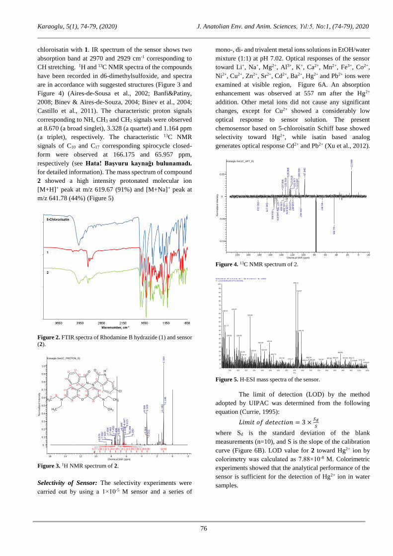
(a triplet), respectively. The characteristic 13C NMR
signals of C10 and C17 corresponding spirocycle closed-
form were observed at 166.175 and 65.957 ppm,
respectively (see Hata! Başvuru kaynağı bulunamadı.
for detailed information). The mass spectrum of compound
2 showed a high intensity protonated molecular ion
[M+H]+ peak at m/z 619.67 (91%) and [M+Na]+ peak at
m/z 641.78 (44%) (Figure 5)
Figure 2. FTIR spectra of Rhodamine B hydrazide (1) and sensor
(2).
Karaoglu-Sen1C_PROTON_01
16 14 12 10 8 6 4 2 0 -2
Chemical Shif t (ppm)
0
0.1
0.2
0.3
0.4
0.5
0.6
0.7
0.8
0.9
1.0
No
rma
lize
d I
nte
nsity
12.978.003.192.382.061.221.182.391.241.071.060.77
1.1
46
1.1
64
1.1
82
3.3
11
3.3
29
3.3
46
3.3
64
6.2
83
6.2
98
6.3
05
6.4
13
6.4
47
6.4
69
6.8
95
7.4
38
7.4
50
7.4
60
7.5
54
7.9
24
7.9
32
8.6
70
O
N
O
N
NCH3
CH3
N CH3
CH3
NH
Cl
O
10
1213
14
15
11
16 17
2122
23
24201928
2726
25
18
29
3031
2
3
1
7
4
5
6
8
9
Figure 3. 1H NMR spectrum of 2.
Selectivity of Sensor: The selectivity experiments were
carried out by using a 1×10⁻⁵ M sensor and a series of
mono-, di- and trivalent metal ions solutions in EtOH/water
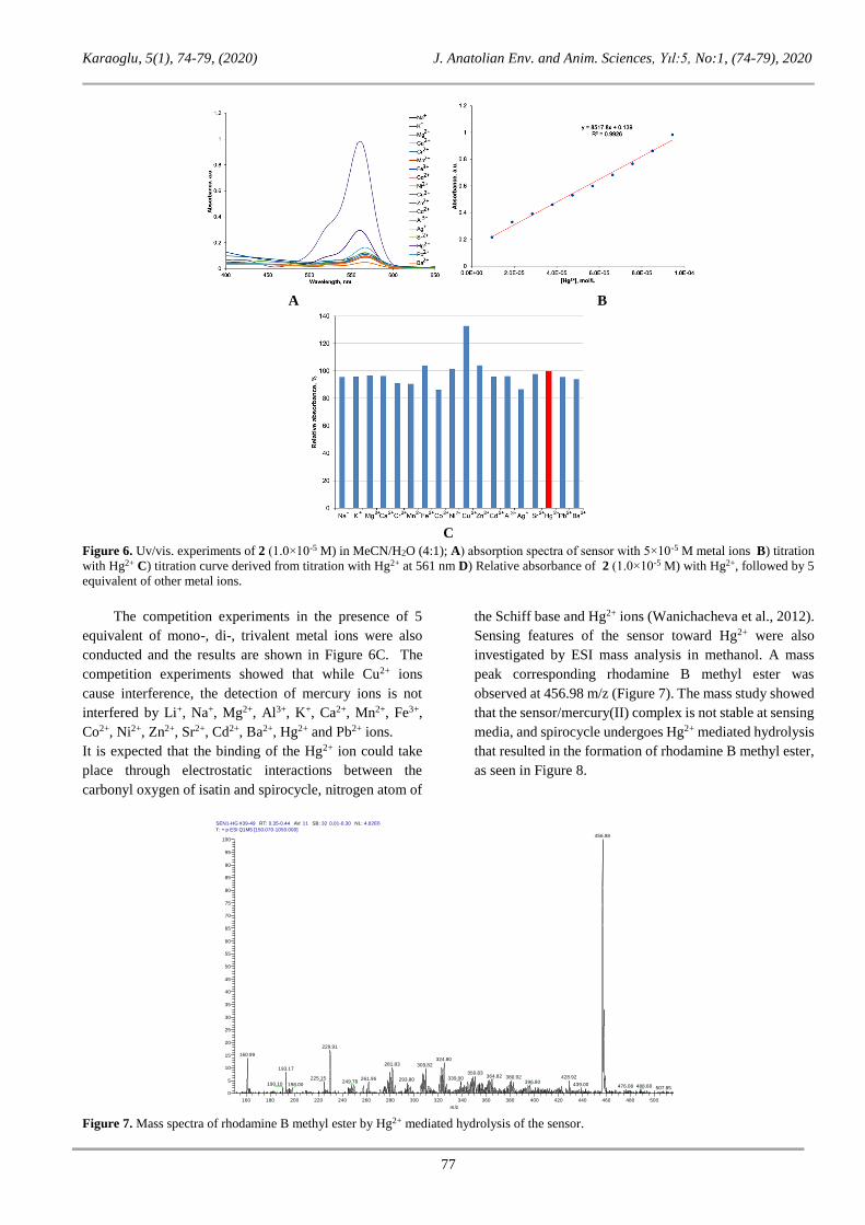
mixture (1:1) at pH 7.02. Optical responses of the sensor
toward Li+, Na+, Mg2+, Al3+, K+, Ca2+, Mn2+, Fe3+, Co2+,
Ni2+, Cu2+, Zn2+, Sr2+, Cd2+, Ba2+, Hg2+ and Pb2+ ions were
examined at visible region, Figure 6A. An absorption
enhancement was observed at 557 nm after the Hg2+
addition. Other metal ions did not cause any significant
changes, except for Cu2+ showed a considerably low
optical response to sensor solution. The present
chemosensor based on 5-chloroisatin Schiff base showed
selectivity toward Hg2+, while isatin based analog
generates optical response Cd2+ and Pb2+ (Xu et al., 2012).
Karaoglu-Sen1C_APT_01
220 200 180 160 140 120 100 80 60 40 20 0 -20
Chemical Shif t (ppm)
-0.10
-0.05
0
0.05
No
rma
lize
d I
nte
nsity
18
2.2
21
16
6.1
75 1
53.8
31
14
8.8
76
14
7.7
95
13
7.9
36
13
5.6
39
13
2.5
16
13
1.5
95
12
9.9
79
12
8.0
68
12
3.8
19
12
3.0
32
12
2.9
77
11
8.8
07
11
3.6
87
10
8.0
30
10
4.4
67
97
.945
65
.957
44
.355
12
.599
Figure 4. 13C NMR spectrum of 2.
Figure 5. H-ESI mass spectra of the sensor.
The limit of detection (LOD) by the method
adopted by UIPAC was determined from the following
equation (Currie, 1995):
𝐿𝑖𝑚𝑖𝑡 𝑜𝑓 𝑑𝑒𝑡𝑒𝑐𝑡𝑖𝑜𝑛 = 3 ×𝑆𝑑
𝑆
where Sd is the standard deviation of the blank
measurements (n=10), and S is the slope of the calibration
curve (Figure 6B). LOD value for 2 toward Hg2+ ion by
colorimetry was calculated as 7.88×10⁻⁸ M. Colorimetric
experiments showed that the analytical performance of the
sensor is sufficient for the detection of Hg2+ ion in water
samples.
SEN2 #34-40 RT: 0.31-0.36 AV: 7 SB: 30 0.00-0.27 NL: 4.80E5
T: + p ESI Q1MS [150.070-1050.000]
200 250 300 350 400 450 500 550 600 650 700 750 800 850 900 950 1000 1050
m/z
0
5
10
15
20
25
30
35
40
45
50
55
60
65
70
75
80
85
90
95
100
Re
lative
Ab
un
da
nce
609.11
619.67
226.97158.12
324.94
167.77
641.78
256.99194.99
445.01390.99
413.04333.19261.96 884.46
469.79276.72
689.30 854.30 914.55 950.72521.29 825.96575.17 764.87 1015.10695.88666.07 978.85
1045.82
Karaoglu, 5(1), 74-79, (2020) J. Anatolian Env. and Anim. Sciences, Yıl:5, No:1, (74-79), 2020
77
A B
C
Figure 6. Uv/vis. experiments of 2 (1.0×10⁻⁵ M) in MeCN/H2O (4:1); A) absorption spectra of sensor with 5×10⁻⁵ M metal ions B) titration
with Hg2+ C) titration curve derived from titration with Hg2+ at 561 nm D) Relative absorbance of 2 (1.0×10⁻⁵ M) with Hg2+, followed by 5
equivalent of other metal ions.
The competition experiments in the presence of 5
equivalent of mono-, di-, trivalent metal ions were also
conducted and the results are shown in Figure 6C. The
competition experiments showed that while Cu2+ ions
cause interference, the detection of mercury ions is not
interfered by Li+, Na+, Mg2+, Al3+, K+, Ca2+, Mn2+, Fe3+,
Co2+, Ni2+, Zn2+, Sr2+, Cd2+, Ba2+, Hg2+ and Pb2+ ions.
It is expected that the binding of the Hg2+ ion could take
place through electrostatic interactions between the
carbonyl oxygen of isatin and spirocycle, nitrogen atom of
the Schiff base and Hg2+ ions (Wanichacheva et al., 2012).
Sensing features of the sensor toward Hg2+ were also
investigated by ESI mass analysis in methanol. A mass
peak corresponding rhodamine B methyl ester was
observed at 456.98 m/z (Figure 7). The mass study showed
that the sensor/mercury(II) complex is not stable at sensing
media, and spirocycle undergoes Hg2+ mediated hydrolysis
that resulted in the formation of rhodamine B methyl ester,
as seen in Figure 8.
Figure 7. Mass spectra of rhodamine B methyl ester by Hg2+ mediated hydrolysis of the sensor.
SEN1-HG #39-49 RT: 0.35-0.44 AV: 11 SB: 32 0.01-0.30 NL: 4.02E6
T: + p ESI Q1MS [150.070-1050.000]
160 180 200 220 240 260 280 300 320 340 360 380 400 420 440 460 480 500
m/z
0
5
10
15
20
25
30
35
40
45
50
55
60
65
70
75
80
85
90
95
100
Re
lative
Ab
un
da
nce
456.98
229.91
160.99324.80
281.83 309.82193.17
350.83364.82 428.92380.92339.00225.15 261.96 293.80249.78 396.80190.10 198.00 439.00 476.08 488.68 507.85

Karaoglu, 5(1), 74-79, (2020) J. Anatolian Env. and Anim. Sciences, Yıl:5, No:1, (74-79), 2020
78
Figure 8. The proposed hydrolysis reaction of the sensor.
CONCLUSIONS
In summary, synthesis, characterization, and
metal sensing properties of a new rhodamine B-based
sensor have been reported. The metal ion detection ability
of the sensor was investigated by colorimetric assays. The
spectrophotometric experiments showed that the sensor
enables selective and sensitive recognition of Hg2+ ion over
competitive metal ions, such as Li+, Na+, Mg2+, Al3+, K+,
Ca2+, Mn2+, Fe3+, Co2+, Ni2+, Cu2+, Zn2+, Sr2+, Cd2+, Ba2+
and Pb2+. The sensor showed linear optical response to
Hg2+ ion between 1×10⁻⁵ to 1×10⁻⁴ M. LOD value for the
sensor toward Hg2+ were calculated as and 7.88×10⁻⁸ M.
According to mass data, optical signals could be produced
by two step mechanism. First, Hg2+ ion coordinates to
sensor through two carbonyl oxygen and nitrogen atom of
imine bond and then complex structure undergoes Hg2+ ion
mediated hydrolysis reaction.
REFERENCES R
Aires-de-Sousa, J., Hemmer, M.C. & Gasteiger, J.
(2002). Prediction of 1 H NMR chemical shifts
using neural networks. Analytical Chemistry,
74(1), 80-90. DOI: 10.1021/ac010737m.
Aksuner, N., Basaran, B., Henden, E., Yilmaz, I. &
Cukurovali, A. (2011). A sensitive and selective
fluorescent sensor for the determination of
mercury(II) based on a novel triazine-thione
derivative. Dyes and Pigments, 88(2), 143-148.
DOI: 10.1016/j.dyepig.2010.05.014.
Banfi, D. & Patiny, L. (2008). www.nmrdb.org:
Resurrecting and Processing NMR Spectra On-
line. CHIMIA International Journal for
Chemistry, 62(4), 280-281. DOI:
10.2533/chimia.2008.280.
Binev, Y. & Aires-de-Sousa, J. (2004). Structure-Based
predictions of 1 H NMR chemical shifts using
feed-forward neural networks. Journal of
Chemical Information and Computer Sciences,
44(3), 940–945. DOI: 10.1021/ci034228s.
Binev, Y., Corvo, M. & Aires-de-Sousa, J. (2004). The
impact of available experimental data on the
prediction of 1 H NMR chemical shifts by neural
networks. Journal of Chemical Information and
Computer Sciences, 44(3), 946-949. DOI:
10.1021/ci034229k.
Cammann, K., Lemke, U., Rohen, A., Sander, J.,
Wilken, H. & Winter, B. (1991). Chemical
sensors and biosensors-principles and
applications. Angewandte Chemie International
Edition in English, 30(5), 516-539. DOI:
10.1002/anie.199105161.
Castillo, A.M., Patiny, L. & Wist, J. (2011). Fast and
accurate algorithm for the simulation of NMR
spectra of large spin systems. Journal of Magnetic
Resonance, 209(2), 123-130. DOI:
10.1016/j.jmr.2010.12.008.
Cope, M.J., Kirkbright, G.F. & Burr, P.M. (1982). Use
of inductively coupled plasma optical emission
spectrometry (ICP–OES) for the analysis of doped
cadmium mercury telluride employing a graphite
rod electrothermal vaporisation device for sample
introduction. The Analyst, 107(1275), 611-616.
DOI: 10.1039/AN9820700611.
Currie, L. A. (1995). Nomenclature in evaluation of
analytical methods, including detect ion and
quantification capabilities (IUPAC
Recommendations). Pure & Applied Chemistry,
67(10), 1699-1723. DOI: 10.1016/S0003-
2670(99)00104-X.
Du, W., Cheng, Y., Shu, W. & Qi, Z. (2017). A novel
rhodamine-based fluorescence chemosensor
containing polyether for mercury (II) ions in
aqueous solution. Química Nova, 40(7), 733-738.
DOI: 10.21577/0100-4042.20170060.
Farruggia, G., Iotti, S., Prodi, L., Montalti, M.,
Zaccheroni, N., Savage, P. B. & Wolf, F. I.
(2006). 8-Hydroxyquinoline Derivatives as
Fluorescent Sensors for Magnesium in Living
Cells. Journal of the American Chemical Society,
128(1), 344–350. DOI: 10.1021/ja056523u
Jarzyńska, G. & Falandysz, J. (2011). The determination
of mercury in mushrooms by CV-AAS and ICP-
AES techniques. Journal of Environmental
Science and Health, 46(6), 569–573. DOI:
10.1080/10934529.2011.562816.
Jiao, Y., Zhang, L & Zhou, P. (2016). A rhodamine B-
based fluorescent sensor toward highly selective

Karaoglu, 5(1), 74-79, (2020) J. Anatolian Env. and Anim. Sciences, Yıl:5, No:1, (74-79), 2020
79
mercury (II) ions detection. Talanta, 150, 14-19.
DOI: 10.1016/j.talanta.2015.11.065.
Li, N., Yu, C., Ji, Y. & Zhang J. (2015). Characterization
of a Cu2+-selective fluorescent probe derived from
rhodamine B with 1,2,4-triazole as subunit and its
application in cell imaging. Turkish Journal of
Chemistry, 39, 660-666. DOI: 10.3906/kim-1410-
58.
Prodi, L., Bargossi, C., Montalti, M., Zaccheroni, N.,
Su, N., Bradshaw, J.S. & Savage, P.B. (2000).
An effective fluorescent chemosensor for mercury
ions. Journal of the American Chemical Society,
122(28), 6769-6770. DOI: 10.1021/ja0006292.
Tang, R., Lei, K., Chen, K., & Zhao, H. (2011). A
Rhodamine-Based Off–On fluorescent
chemosensor for selectively sensing Cu(II) in
aqueous solution. Journal of Fluorescence, 21,
141-148. DOI: 10.1007/s10895-010-0698-x.
Voutsadaki, S., Tsikalas, G.K., Klontzas, E., Froudakis,
G.E. & Katerinopoulos, H.E. (2010). A “turn-
on” coumarin-based fluorescent sensor with high
selectivity for mercury ions in aqueous media.
Chemical Communications, 46(19), 3292. DOI:
10.1039/b926384e.
Wanichacheva, N., Setthakarn, K., Prapawattanpol, N.,
Hanmenga, O., Sanghiran, V., Lee, S. &
Grudpan, K. (2012). Rhodamine B-based “turn-
on” fluorescent and colorimetric chemosensors
for highly sensitive and selective detection of
mercury (II) ions. Journal of Luminescence,
132(1), 35-40. DOI:
10.1016/j.jlumin.2011.07.015.
Wei, Y., Cheng, D., Ren, T., Li, Y., Zeng, Z. & Yuan, L.
(2016). Design of NIR chromenylium-cyanine
fluorophore library for “Switch-ON” and
ratiometric detection of bio-active species in vivo.
Analytical Chemistry, 88(3), 1842-1849. DOI:
10.1021/acs.analchem.5b04169.
Xu, L., Xu, Y., Zhu, W., Sun, X., Xu, Z. & Qian, X.
(2012). Modulating the selectivity by switching
sensing media: a bifunctional chemosensor
selectivity for Cd2+ and Pb2+ in different aqueous
solutions. RSC Advances, 2(15), 6323. DOI:
10.1039/c2ra20840g.
Verep, B., Mutlu, T., Yüksek, T. & Gürdal A.A. (2018).
Sert ve Yumuşak Su Koşullarında Karadeniz
Alabalığı (Salmo coruhensis) Dokularında Ağır
Metal (Civa: Hg) Birikiminin Belirlenmesi.
Journal of Anatolian Environmental&Animal
Sciences, 3(1), 19-26. DOI:
10.35229/jaes.387681.
Yoon, S., Miller, E.W., He, Q., Do, P.H. & Chang, C.J.
(2007). A bright and specific fluorescent sensor
for mercury in water, cells, and tissue.
Angewandte Chemie International Edition,
46(35), 6658-6661. DOI:
10.1002/anie.200701785.
Yuan, C., Liu, B., Liu, F., Han, M.-Y. & Zhang, Z.
(2014). Fluorescence “Turn On” detection of
mercuric ion based on bis (dithiocarbamato)
copper(II) complex functionalized carbon
nanodots. Analytical Chemistry, 86(2), 1123-
1130. DOI: 10.1021/ac402894z.
Zhang, Y.-S., Balamurugan, R., Lin, J.-C., Fitriyani, S.,
Liu, J.-H. & Emelyanenko, A. (2017). Pd2+
fluorescent sensors based on amino and imino
derivatives of rhodamine and improvement of
water solubility by the formation of inclusion
complexes with β-cyclodextrin. The Analyst,
142(9), 1536-1544. DOI: 10.1039/C6AN02594C.
Zhao, Y., Zheng, B., Du, J., Xiao, D. & Yang, L. (2011).
A fluorescent “turn-on” probe for the dual-
channel detection of Hg(II) and Mg(II) and its
application of imaging in living cells. Talanta,
85(4), 2194-2201. DOI:
10.1016/j.talanta.2011.07.070.