eu - eScholarship@McGill
162
Page 1 of 162 Examining the pro-tumorigenic functions of the mitogen- activated protein kinase interacting protein kinases 1 and 2 (MNK1/2)-eukaryotic initiation factor 4E (eIF4E) pathway in breast cancer by Qianyu Guo Division of Experimental Medicine Faculty of Medicine McGill University Montréal, Québec, Canada June 2019 A thesis submitted to McGill University in partial fulfillment of the requirements of the degree of Doctor of Philosophy (Ph.D) © Qianyu Guo, 2019
-
Upload
khangminh22 -
Category
Documents
-
view
1 -
download
0
Transcript of eu - eScholarship@McGill
Page 1 of 162
Examining the pro-tumorigenic functions of the mitogen-
activated protein kinase interacting protein kinases 1
and 2 (MNK1/2)-eukaryotic initiation factor 4E (eIF4E)
pathway in breast cancer
by Qianyu Guo
Division of Experimental Medicine
Faculty of Medicine
McGill University
Montréal, Québec, Canada
June 2019
A thesis submitted to McGill University in partial fulfillment of the requirements of the degree
of Doctor of Philosophy (Ph.D)
© Qianyu Guo, 2019
Page 2 of 162
Table of Contents
Title page………………………………………………………………………………………….1
Table of contents…………………………………………………………………………………..2
List of figures and tables…………………………………………………………………………..6
English abstract……………………………………………………………………………………8
French abstract…………………………………………………………………………………….9
Acknowledgements………………………………………………………………………………11
Contributions to knowledge and elements of original scholarship………………………………13
Contribution of authors…………………………………………………………………………..13
Abbreviations…………………………………………………………………………………….16
Chapter 1. Introduction and Literature Review………………………………………………….22
1.1. Breast anatomy and physiology……………………………………………………………..23
1.1.1 Mammary gland structure and cellular components…………………………………..23
1.1.2 Lactation and involution………………………………………………………………23
1.2. General introduction to breast cancer……………………………………………………….25
1.2.1 Epidemiology and breast cancer risk factors………………………………………….25
1.2.2 Classifications of breast cancer………………………………………………………..26
1.2.2.1 Ductal carcinoma in situ (DCIS) and invasive ductal carcinoma (IDC)……...26
1.2.2.2 Lobular carcinoma in situ (LCIS) and invasive lobular carcinoma (ILC)…….28
1.2.2.3 Other invasive breast carcinomas……………………………………………..28
1.2.2.4 Biological subtypes of breast cancer…………………………………………..29
1.3. Regulation of the DCIS-IDC transition……………………………………………………..31
1.3.1 Degradation of the basement membrane and ECM remodeling during the DCIS-IDC
conversion………………………………………………………………………………………..31
1.3.2 The functions of myoepithelial cells and CAFs in promoting the DCIS-IDC
transition…………………………………………………………………………………………32
1.3.3 Dysregulated signaling in the DCIS-IDC transition……………………..……………33
1.3.4 Tumor immunity in DCIS-IDC transition and IDC dissemination……………………34
1.4. Post-partum breast cancer (PPBC) ………………………………………………………….35
1.4.1 Fibroblasts……………………………………………………………………………..36
1.4.2. Lymphatic & endothelial cells………………………………………………………..37
Page 3 of 162
1.4.3. Macrophages………………………………………………………………………….37
1.4.4. Myeloid-derived suppressor cells (MDSCs) …………………………………………37
1.4.5. Innate lymphoid cells…………………………………………………………………38
1.4.6. Dendritic cells (DCs) ………………………………………………………………...39
1.4.7. T cells…………………………………………………………………………………40
1.5. Pro-tumorigenic functions of MNK1/2-eIF4E pathway in Breast Cancer………………….41
1.5.1 Overview of MNK1/2-eIF4E Pathway, eIF4F complex and regulation of mRNA
translation………………………………………………………………………………………...41
1.5.2 The functions of MNK1/2-eIF4E pathway in breast tumorigenesis………………...43
1.5.3 Other MNK1/2 substrates…………………………………………………………...45
1.5.4 Targeting MNK1/2 in cancer………………………………………………………..46
1.5.5 Crosstalks between MNK1/2 and PI3K/Akt/mTOR pathways……………………..46
1.6. General introduction to the thesis research………………………………………………….47
1.7 Reference…………………………………………………………………………………….48
Chapter 2. MNK1/NODAL signaling promotes invasive progression of breast ductal carcinoma
in situ………………………………………………………………………………......................61
2.1 Abstract………………………………………………………………………………………62
2.2 Introduction…………………………………………………………………………………..63
2.3 Materials and Methods……………………………………………………………………….64
2.3.1 Cells and Reagents………………………………………………………………….64
2.3.2 Growth Curves……………………………………………………………………...64
2.3.3 Mammosphere Formation Assay…………………………………………………...64
2.3.4 Western Blotting……………………………………………………………………64
2.3.5 Quantitative PCR…………………………………………………………………...65
2.3.6 Clonogenic Assay…………………………………………………………………..65
2.3.7 Aldefluor Assay…………………………………………………………………….65
2.3.8 Migration and Invasion Assay……………………………………………………...65
2.3.9 Immunohistochemistry……………………………………………………………..66
2.3.10 Orthotopic Mouse Model………………………………………………………….66
2.3.11 Statistical Analysis………………………………………………………………...67
2.4 Results………………………………………………………………………………………..67

Page 4 of 162
2.4.1 MNK1 activity is elevated in high grade and IDC, compared to low grade DCIS
clinical samples…………………………………………………………………………………..67
2.4.2 MNK1 knock-out impairs DCIS proliferation, and DCIS to IDC conversion……..67
2.4.3 MNK1 regulates NODAL morphogen to control DCIS progression………………72
2.4.4 MNK1 and NODAL regulate DCIS tumor invasion……………………………….75
2.4.5 MNK1 can be pharmacologically targeted to inhibit the DCIS to IDC transition….76
2.5 Discussion……………………………………………………………………………………79
2.6 Acknowledgments……………………………………………………………………………82
2.7 References……………………………………………………………………………………82
2.8 Supplementary materials……………………………………………………………………..86
Chapter 3. phospho-eIF4E/IL-33 drives immune evasion and lung metastasis in postpartum
breast cancer (PPBC) …………………………………………………………………………100
3.1 Abstract……………………………………………………………………………………101
3.2 Introduction…………………………………………………………………………………102
3.3 Methods……………………………………………………………………………………..104
3.3.1 Mouse Model……………………………………………………………………..104
3.3.2 Cells and Reagents………………………………………………………………..104
3.3.3 Western Blotting………………………………………………………………….104
3.3.4 Quantitative PCR…………………………………………………………………105
3.3.5 Migration and Invasion Assay……………………………………………………105
3.3.6 Co-culture Assay………………………………………………………………….105
3.3.7 Immunohistochemistry (IHC) ……………………………………………………105
3.3.8 Immunofluorescence (IF) ………………………………………………………...106
3.3.9 ILC2 isolation…………………………………………………………………….106
3.3.10 Statistical Analysis………………………………………………………………106
3.4 Results………………………………………………………………………………………106
3.4.1 Loss of eIF4E phosphorylation in the stroma protects against PPBC lung
metastasis……………………………………………………………………………………….106
3.4.2 Characterizing the changes in the lung immune microenvironment of PPBC mice
that are dependent on the phosphorylation of eIF4E…………………………………………...108
Page 5 of 162
3.4.3 eIF4E phosphorylation regulates IL33 expression in fibroblasts to support breast
cancer cell invasion……………………………………………………………………………..111
3.4.4 IL-33 activates the MNK1/2-eIF4E pathway downstream of activated ST2 in breast
tumor cells to build immunosuppressive TME…………………………………………………113
3.4.5 Increased therapeutic efficacy of blocking MNK1/2 combined with anti–PD1
immunotherapy…………………………………………………………………………………114
3.5 Discussion…………………………………………………………………………………..116
3.6 Acknowledgements…………………………………………………………………………118
3.7 References…………………………………………………………………………………..118
3.8 Supplementary materials……………………………………………………………………123
Chapter 4. Discussion and Future Directions…………………………………………………..131
4.1 Comprehensive discussion of findings.………………………………………………….131
4.2 Future directions……………………………………………………………………………132
4.2.1 Potential roles of MNK1 in the nucleus…………………………………………..132
4.2.2 Characterizing the functions of MNK1/2-eIF4E in the DCIS to IDC transition in an
immune competent model………………………………………………………………………133
4.2.3 Characterizing the immune landscape of PPBC patient samples………………...134
4.2.4 Characterizing the functions of MNK1/2-eIF4E in organ specific breast cancer
metastasis……………………………………………………………………………………….136
4.2.5 Targeting CAFs in metastatic breast cancer……………………………………...137
4.2.6 Targeting mRNA translation to increase sensitivity to checkpoint blockade…….138
4.3 Final conclusion and summary……………………………………………………………..139
4.4 References…………………………………………………………………………………..140
Chapter 5. References…………………………………………………………………………..143
Permission to use published work in thesis……………………………………………………157
Page 6 of 162
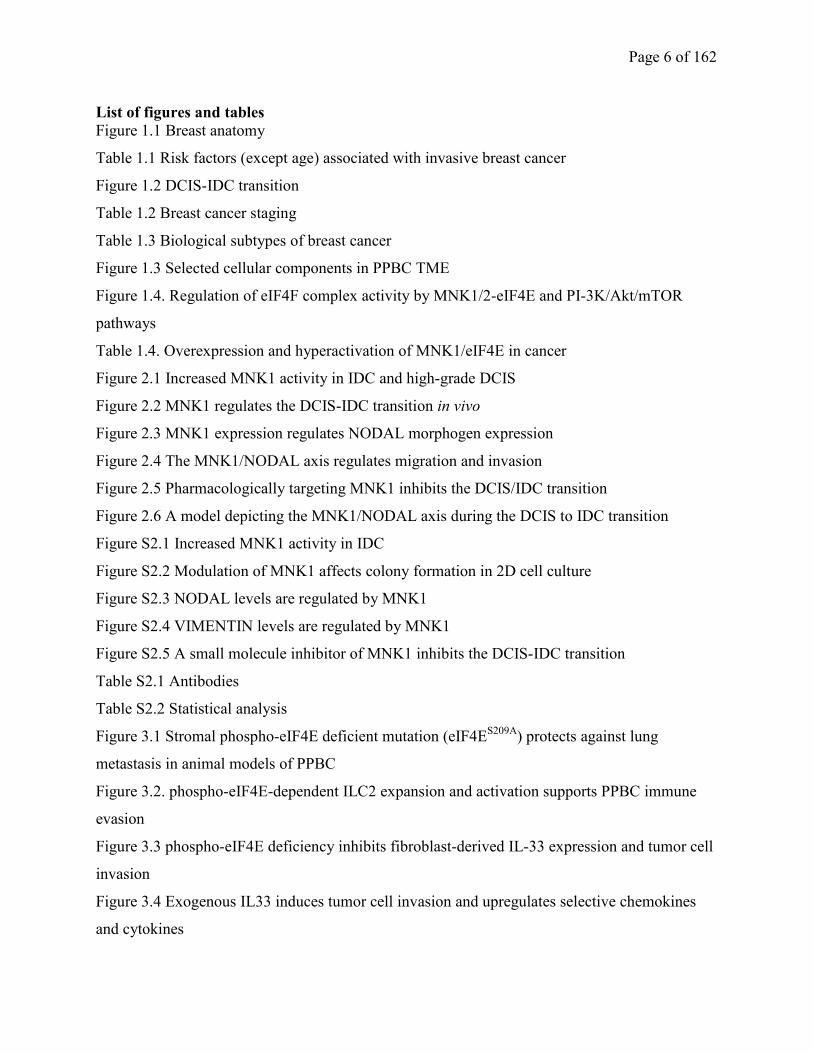
List of figures and tables
Figure 1.1 Breast anatomy
Table 1.1 Risk factors (except age) associated with invasive breast cancer
Figure 1.2 DCIS-IDC transition
Table 1.2 Breast cancer staging
Table 1.3 Biological subtypes of breast cancer
Figure 1.3 Selected cellular components in PPBC TME
Figure 1.4. Regulation of eIF4F complex activity by MNK1/2-eIF4E and PI-3K/Akt/mTOR
pathways
Table 1.4. Overexpression and hyperactivation of MNK1/eIF4E in cancer
Figure 2.1 Increased MNK1 activity in IDC and high-grade DCIS
Figure 2.2 MNK1 regulates the DCIS-IDC transition in vivo
Figure 2.3 MNK1 expression regulates NODAL morphogen expression
Figure 2.4 The MNK1/NODAL axis regulates migration and invasion
Figure 2.5 Pharmacologically targeting MNK1 inhibits the DCIS/IDC transition
Figure 2.6 A model depicting the MNK1/NODAL axis during the DCIS to IDC transition
Figure S2.1 Increased MNK1 activity in IDC
Figure S2.2 Modulation of MNK1 affects colony formation in 2D cell culture
Figure S2.3 NODAL levels are regulated by MNK1
Figure S2.4 VIMENTIN levels are regulated by MNK1
Figure S2.5 A small molecule inhibitor of MNK1 inhibits the DCIS-IDC transition
Table S2.1 Antibodies
Table S2.2 Statistical analysis
Figure 3.1 Stromal phospho-eIF4E deficient mutation (eIF4ES209A
) protects against lung
metastasis in animal models of PPBC
Figure 3.2. phospho-eIF4E-dependent ILC2 expansion and activation supports PPBC immune
evasion
Figure 3.3 phospho-eIF4E deficiency inhibits fibroblast-derived IL-33 expression and tumor cell
invasion
Figure 3.4 Exogenous IL33 induces tumor cell invasion and upregulates selective chemokines
and cytokines
Page 7 of 162
Figure 3.5 Combination of SEL201 and PD1-blockade decreased 66cl4 lung metastasis in PPBC
model
Figure 3.6 Graphic summary
Supplementary Figure 3.1
Supplementary Figure 3.3
Supplementary Figure 3.4
Supplementary Figure 3.5
Supplementary Table 3.1. Details of Antibodies
Supplementary Table 3.2. Details of Primers (Mouse)
Supplementary Table 3.3. Statistic Analysis (Figures)
Supplementary Table 3.4. Details of Statistic Analysis (Supplementary Figures)
Page 8 of 162
Abstract
Cancer metastasis is a multi-step process. This thesis focuses on two critical steps of the
metastatic process in breast cancer: (1) the transition from non-invasive to invasive disease, and
(2) immune evasion that supports metastasis. MAP kinase-interacting serine/threonine-protein
kinases 1 and 2 (MNK1/2) are ubiquitously expressed serine/threonine kinases downstream of
the ERK1/2 and p38 pathways. Hyper-activation of MNK1/2 due to external stimuli such as
growth factors or stress signaling can enhance tumor cell invasion and metastasis in multiple
solid malignancies including breast cancer, but the molecular mechanisms underpinning these
effects of MNK1/2 remain largely unknown. Using in vitro and in vivo models, we demonstrated
a novel function of MNK1, where the kinase drives the transition of breast ductal carcinoma in
situ (DCIS), a non-invasive “stage 0” disease, into invasive ductal carcinoma (IDC). At the
mechanistic level, MNK1 upregulates the expression of NODAL, a pro-tumorigenic morphogen,
to support a partial epithelial-mesenchymal transition (EMT), and to maintain cancer stemness
properties that increase the risk of tumor relapse and metastasis.
The best characterized function of MNK1/2 is to phosphorylate the eukaryotic translation
initiation factor 4E (eIF4E) at Ser209. Multiple pro-oncogenic pathways converge on the
MNK1/2-eIF4E axis, which serves as a critical regulator of the translation of mRNAs that
encode for proteins that promote cell invasion. The MNK1/2-eIF4E axis has been recently
reported to reinforce the survival of pro-metastatic neutrophils in breast cancer. However, our
knowledge of how aberrant mRNA translation regulates breast tumor immunity remains limited.
We chose to study post-partum breast cancer (PPBC), an aggressive subtype of breast cancer, as
it has been characterized by robust immune cell suppression, to examine how the MNK1/2-
eIF4E axis shapes pro-tumorigenic immunity during metastasis. We demonstrate that eIF4E
phosphorylation is important to support tumor immune evasion for PPBC metastasis. Using a
mouse model that is devoid of eIF4E phosphorylation, and inhibitors of MNK1/2, we show that
type 2 innate lymphoid cell (ILC2) function, myeloid-derived suppressor cells (MDSCs)
accumulation, and cytotoxic T cell exclusion, are dependent on the MNK1/2-eIF4E axis.
Immune targeted therapies have not shown great promise in breast cancer. We showed that the
inhibition of MNK1/2 using the inhibitor SEL201, can work in concert with anti-PD1 immune
targeted therapy to inhibit PPBC metastasis. Thus, we show the possibility of enhancing the
efficacy of immunotherapy by using a small molecule inhibitor that blocks mRNA translation.
Page 9 of 162
RÉSUMÉ
Le métastase du cancer est un processus a plusieurs étapes. Cette thèse est centrée sur deux
étapes critiques du processus de métastase dans le cancer du sein : (1) la transition du cancer
non-invasif en cancer invasif, et (2) une évasion du système immunitaire qui supporte la
progression vers un stade métastatique. MAP kinase-interacting serine/threonine-protein kinases
1 and 2 (MNK1/2) sont des kinases sérine/thréonine en aval du processus biologique de ERK1/2
et p38. L’hyper-activation de MNK1/2 causée par des stimuli externes comme des facteurs de
croissance ou des signaux de stress peuvent augmenter l’invasion cellulaire et les métastases
dans plusieurs cancers solides comme le cancer du sein. Malgré cela, le mécanisme moléculaire
derrière ces effets causés par MNK1/2 sont encore largement inconnues. En utilisant des modèles
in vitro et in vivo, nous avons démontré une nouvelle fonction de MNK1; la kinase facilite la
transition du cancer du sein canalaire in situ (DCIS), un « stage 0 », non-invasif de la maladie, en
cancer du sein canalaire invasif (IDC). Au niveau du mécanisme moléculaire, MNK1 augmente
l’expression de NODAL, un morphogène pro-tumorigène, pour supporter un transition
épithéliale-mesenchymal (EMT) partielle et maintenir le charactère souche du cancer qui
augmente le risque de rechute et metastase.
La fonction la plus étudiée de MNK1/2 est sa capacité à phosphoryler le facteur d’initiation
eucaryote de traduction de 4E (eIF4E) à Ser209. Plusieurs processus biologiques proto-
oncogènes converge sur l’axe MNK1/2-eIF4E, qui joue un rôle critique dans la translation de
mRNAs qui codent pour des protéines qui poussent l’invasion cellulaire. Il a récemment été
démontré que l’axe MNK1/2-eIF4E renforce la survie de neutrophiles pro-métastases dans le
cancer du sein. Cependant, notre connaissance de cette fonction est encore très limitée. Nous
avons choisi d’étudier le cancer du sein post-partum (PPBC), un sous-type très agressif du cancer
du sein, notamment parce que celui-ci est caractérisé par une diminution robuste de cellules
immunitaires. Nous voulons de ce fait, examiner comment l’axe MNK1/2-eIF4E agit sur
l’immunité pro-tumorigène durant l’étape métastatique. Nous démontrons que la phosphorylation
de eIF4E est importante dans le support de l’évasion immunitaire dans les métastases du PPBC.
En utilisant un modèle murin déficient de la phosphorylation d’eIF4E et en utilisant des
inhibiteurs de MNK1/2, nous montrons que l’activation des cellules lymphoïdes innées de type 2
(ILC2), l’accumulation des myeloid-derived suppressor cells (MDSCs) et l’exclusion des
cellules T cytotoxiques sont dépendantes de l’axe MNK1/2-eIF4E. La thérapie immunitaire

Page 10 of 162
ciblée n’a pas démontré beaucoup de résultats positifs dans le cancer du sein. Nous avons montré
que l’inhibition de MNK1/2 en utilisant l’inhibiteur SEL201, peut travailler en parallèle avec la
thérapie immunitaire ciblée anti-PD1 pour diminuer les métastases dans le PPBC. Ainsi nous
montrons la possibilité d’améliorer les effets de l’immunothérapie en utilisant un inhibiteur qui
agit sur le blocage de la translation des mRNAs.

Page 11 of 162
Acknowledgements
First and foremost, I would like to thank my supervisors, Drs. Wilson H. Miller Jr. and Sonia V.
del Rincon for providing an amazing platform to pursue my dream as a scientist. Specifically, I
am forever grateful for Dr. Miller for his acceptance of me as his PhD student and his belief that
I would grow into a successful scientist. I deeply appreciate his constructive critiques of my
work, funding my project and his drive to translate laboratory findings into the clinic. I have
enormous gratitude towards Dr. Sonia del Rincon, who has always been understanding and
supportive all throughout my candidature, especially when I was in difficulty. My successes are
deeply rooted in her guidance.
It is also a great honor and privilege to be a member of an amazing team where everybody
collaborates and has each other’s back. I would like to thank my best friends Fan Huang,
William Yang and Dr. Yao Zhan, who have always stood by my side, offered support, motivate
and inspire me during this wonderful journey as a PhD student. I also thank Sai Sakktee Krisna,
Samuel Preston and Dr. Margarita Bartish for their assistance with countless animal experiments
and their friendship. Much of the daily things that go unrecognized and the scientific support are
accredited to Christophe Goncalves, who is the guru of IHC/IF staining in our team. I appreciate
his help, humor and support for countless troubleshooting experiments.
My gratitude also goes to my thesis committee members and collaborators. Thanks to Dr. Lynne-
Marie Postovit, Mark Basik, Réjean Lepointe and Frédéric Amant who have kindly shared
clinical samples and provides me with important clinical perspectives. Dr. Luck McCaffrey has
always been open for scientific discussions and supportive for many fellowship applications.
Drs. Jörg Fritz and Claudia Duerr have provided guidance and help for various immune cell
profiling, and I have reinforced my knowledge of immunology by discussing project directions
with them.
Sharing a lab and meeting space with Dr. Koren Mann and her group has also been very fruitful.
I thank Dr. Koren Mann to be my academic advisor and for her amazing scientific input and her
assistance with manuscripts. Our animal technician Dany Plourde has also been a great help with
setting up countless animal experiments.
I am also grateful to Veronique Michaud and Kathy Ann Forner of animal quarter. Kathy has
been very patient and helpful to train me for various animal surgeries. Veronique has been

Page 12 of 162
instrumental in helping me with countless IVIS imaging and ultrasound, and has always been
patient and wise. We have shared many laughs.
I am also grateful for the financial support I have received in the form of grants and scholarships
from various agencies, Canadian Institutes for Health Research (CIHR), Cancer Research
Society (CRS), Rossy Cancer Network, Cole Foundation, McGill Integrated Cancer Research
Training Program (MICRTP) graduate studentship, and Ruth & Alex Dworkin fellowship of
McGill University.
My deepest gratitude goes to my mum and dad, aunt Jun and uncle Ray. You have always been
supporting me and encouraging me to do my best to fulfill my life goal. Dad, thank you for
inspiring my curiosity in science and leading me into the field that I am extremely interested in.
Your work ethic was definitely passed down. Mom, thank you for your dedication in both career
and family. Your wisdom has helped me reach amazing heights. Aunt Jun and uncle Ray, thank
you for your constant support and tremendous belief in me. I have been extremely lucky to have
you all as my family.
Page 13 of 162
Contributions to knowledge and elements of original scholarship
1. MNK1 promotes the transition of breast ductal carcinoma in situ (DCIS) into invasive ductal
carcinoma.
2. MNK1 regulates the expression of a pro-invasive morphogen named NODAL.
3. MNK1/NODAL axis is important to maintain the cancer stem cell features in DCIS cells.
4. MNK1/NODAL axis drives the DCIS to IDC transition through promoting a partial epithelial-
mesenchymal transition (EMT), mainly by regulating the expression of Vimentin.
5. Inhibition of MNK1/2 activity by SEL201 partially suppresses the DCIS to IDC transition.
6. Phospho-eIF4E in the tumor microenvironment (TME) promotes metastasis of post-partum
breast cancer (PPBC).
7. Phospho-eIF4E in the TME facilitates MDSCs accumulation and cytotoxic T cell exclusion
from the lung, the most prominent metastatic site in our PPBC model.
8. Phospho-eIF4E regulates fibroblasts-derived IL-33, to support PPBC immune evasion.
9. The combination of SEL201 and anti-PD1 antibody inhibits PPBC lung metastasis.
Contribution of authors
The present thesis consists in large part of manuscripts published or in preparation for
submission to peer reviewed journals, of which I am the first author. Their citations are included
here, with permission where required, and are distributed in the text as follows:
Chapter 1
Guo Q*, Huang F*, Goncalves C, Del Rincon SV, and Miller WH Jr. Translation of cancer
immunotherapy from the bench to the bedside. Advances in cancer research. 2019;143:1-62.
(*Equal contributions)
Chapter 2
Guo Q, Li VZ, Nichol JN, Huang F, Yang W, Preston SEJ, Talat Z, Lefrère H, Yu H, Zhang G,
Basik M, Goncalves C, Zhan Y, Plourde D, Su J, Torres J, Marques M, Al Habyan S, Bijian K,
Amant F, Witcher M, Behbod F, McCaffrey L, Alaoui-Jamali MA, Giannakopoulos NV,
Brackstone M, Postovit LM, del Rincón SV, Miller WH Jr. MNK1/NODAL signaling promotes
invasive progression of breast ductal carcinoma in situ. Cancer Research. 2019; 79(7):1646-57.
Page 14 of 162
Chapter 3
Guo Q, Bartish M, Krisna SS, Huang F, Li VZ, Preston SEJ, Emond A, Lefrère H, Duerr
C, Gui
Y, Goncalves C, Plourde D, Su J, Hewgill S, Yang W, Khoury E, Zhan Y, Narykina V, Basik M,
Amant F, Lapointe R, Fritz JH, del Rincón SV, Miller WH Jr. phospho-eIF4E/IL33 drives
immune evasion and lung metastasis in postpartum breast cancer (PPBC) (Manuscript in
preparation)
Publications that include work performed by Guo Q, but not included in the presentation of this
dissertation are listed below:
Yang W*, Khoury E*, Guo Q, Emond A, Huang F, Gonçalves C, Zhan Y, Plourde D, Nichol JN,
Dahabieh MS, del Rincón SV. Miller WH Jr. MNK1 signaling induces an ANGPTL4-mediated
gene signature to drive melanoma progression (Manuscript in review - Oncogene) (*Equal
contributions)
Zhan Y, Guo J, Yang W, Goncalves C, Rzymski T, Dreas A, Zylkiewicz E, Mikulski M,
Brzozka K, Golas A, Kong Y, Ma M, Huang F, Huor B, Guo Q, da Silva SD, Torres J, Cai Y,
Topisirovic I, Su J, Bijian K, Alaoui-Jamali MA, Huang S, Journe F, Ghanem GE, Miller WH
Jr., del Rincón SV. MNK1/2 inhibition limits oncogenicity and metastasis of KIT-mutant
melanoma. The Journal of clinical investigation. 2017;127(11):4179-92.
Robichaud N, Hsu BE, Istomine R, Alvarez F, Blagih J, Ma EH, Morales SV, Dai DL, Li G,
Souleimanova M, Guo Q, del Rincón SV, Miller WH Jr., Cajal SRY, Park M, Jones RG,
Piccirillo CA, Siegel PM, Sonenberg N. Translational control in the tumor microenvironment
promotes lung metastasis: Phosphorylation of eIF4E in neutrophils. Proceedings of the National
Academy of Sciences. 2018; 115(10): E2202-09.
The manuscripts forming the major part of this thesis included work from several co-authors
to whom I owe many thanks. My contributions and those of my co-authors are delineated
here.
Chapter 1 is a literature review consisting mainly of a manuscript entitled “Translation of cancer
immunotherapy from the bench to the bedside”, which will constitute a chapter of the upcoming
book “Immunotherapy of Cancer” for Advances in Cancer Research. The original draft were

Page 15 of 162
work of myself and Fan Huang. Christophe Goncalves designed the figures. All authors
contributed extensive editorial work on numerous subsequent drafts.
Chapter 2 was published in Cancer Research with the title “MNK1/NODAL signaling promotes
invasive progression of breast ductal carcinoma in situ”. I designed, performed and analyzed the
experiments presented in all figures. I drafted the manuscript and compiled the figures, with
editorial input from all authors. Some replication experiments, including migration and invasion
assays, western blots, IHC stainings were performed by Vivian Z. Li. Fan Huang and William
Yang assisted multiple in vivo experiments and performed in vitro assays such as qPCR and
western blots. Samuel E.J. Preston, Zahra Talat, Hanne Lefrère, Henry Yu, Christophe
Goncalves and Yao Zhan have participated in multiple in vitro and in vivo experiments. Dany
Ploudre, Jie Su and Sarah Al Habyan were involved in mouse colony maintenance. Mark Basik,
Muriel Brackstone, Nadia V. Giannakopoulos, Lynne-Marie Postovit, Luke McCaffrey and
Michael Witcher provided clinical samples and offered suggestions for manuscript writing.
Jessica N. Nichol modified the figures and manuscript. Wilson H. Miller Jr. and Sonia V. del
Rincón secured the funding secured funding, supervised the research and edited the manuscript.
Chapter 3 is a manuscript in preparation. I designed, performed and analyzed the experiments
presented in all figures. I drafted the manuscript and compiled the figures, with editorial input
from all authors. Some replication experiments were performed by Margarita Bartish, Sai
Sakktee Krisna, Fan Huang and Claudia Duerr. Additionally, Sai Sakktee Krisna, Claudia Duerr,
Shannon Hewgill, and Jörg H. Fritz have performed ILC2 flow cytometry, ILC2 sorting and ex
vivo ILC2 experiments, as well as offered suggestions and protocols for ILC2-related
experiments. Audrey Emond and Samuel E.J. Preston performed flow cytometry staining and
analysis. Other colleagues, including William Yang, Hanne Lefrère, Elie Khoury and Yao Zhan
have participated in multiple in vitro and in vivo experiments. Dany Ploudre and Jie Su were
involved in mouse colony maintainence and assisted multiple animal procedures including
gavaging and animal surgeries. Christophe Goncalves assisted troubleshooting of multiple IHC
and IF stainings, as well as performed the characterization of mammary glan involution. Mark
Basik, Frédéric Amant and Réjean Lapointe provided clinical samples and offered suggestions
for manuscript writing. Wilson H. Miller Jr. and Sonia V. del Rincón secured funding,
supervised the research and edited the manuscript.
Page 16 of 162
Abbreviations
eIF4E binding proteins (4EBPs)
1,4,5-trisphosphate (IP3)
Aldehyde dehydrogenase (ALDH)
All-trans retinoic acid (ATRA)
Antibody-dependent cellular cytotoxicity (ADCC)
Antigen presenting cells (APCs)
Atypical ductal hyperplasia (ADH)
B and T lymphocyte attenuator 4 (BTLA4)
Basement membrane (BM)
B-cell CLL/lymphoma 9 (Bcl-9)
BODIPY™-aminoacetaldehyde (BAAA)
Breast cancer type 1/2 susceptibility protein (BRCA1/2)
Cytotoxic T-lymphocyte-associated protein-4 (CTLA-4)
Cancer-associated fibroblasts (CAFs)
Cancer stem cell (CSC)
CHEK2
chronic myeloid leukemia (CML)
C-C motif receptor 8 (CCR-8)
Cluster of differentiation 10 (CD10)
Cluster of differentiation 11b (CD11b)
Cluster of differentiation 40 (CD40)
Cluster of differentiation 44 (CD44)
Cluster of differentiation 133 (CD133)
C-C motif chemokine ligand 2 (CCL2)
C-C motif chemokine ligand 5 (CCL5)
C-C motif chemokine ligand 17 (CCL17)
C-C motif chemokine ligand 21 (CCL21)
C-X-C motif chemokine ligand 1 (CXCL1)
C-X-C motif chemokine ligand 2 (CXCL2)
C-X-C motif chemokine ligand 2 (CXCL12)
Page 17 of 162
C-X-C motif chemokine ligand 14 (CXCL14)
C-C motif receptor 7 (CCR7)
C-X-C motif receptor 4 (CXCR4)
Cyclic GMP-AMP synthase (cGAS)
Cyclooxygenase-2 (COX2)
Cytokeratin (CK)
Cytosolic phospholipase A2 (cPLA2)
Damage-associated molecular patterns (DAMPs)
Dendritic cells (DCs)
Distal-less Homeobox 4 (DLX4)
Ductal carcinoma in situ (DCIS)
Endoplasmic reticulum (ER)
Engrailed Homeobox 1 (EN1)
Epidermal growth factor (EGF)
Epithelial-to-mesenchymal transition (EMT)
Estrogen receptor (ER)
Eukaryotic translation initiation factor 3C (eIF3C)
Eukaryotic translation initiation factor 4A (eIF4A)
Eukaryotic translation initiation factor 4E (eIF4E)
Eukaryotic translation initiation factor 4F (eIF4F)
Eukaryotic translation initiation factor 4G (eIF4G)
Extracellular signal–regulated kinases-1/2 (ERK-1/2)
Extracellular matrix (ECM)
Fibroblast-activation protein (FAP),
Forkhead Box M1 (FoxM1)
Glioma-Associated Oncogene Homolog 1 (Gli1)
Glucocorticoid-induced TNFR-related protein (GITR)
Glyceraldehyde 3-phosphate dehydrogenase (GAPDH)
Granulocyte-colony stimulating factor (G-CSF)
Granulocyte-macrophage colony-stimulating factor (GM-CSF)
Growth hormone (GH)
Page 18 of 162
Heparan sulfate (HS)
Hepatocellular carcinoma (HCC)
Hematoxylin and eosin (H&E)
Hypoxia-inducible factor 1- α (HIF-1α)
Homeobox B13 (HOXB13)
Hormone replacement therapy (HRT)
Hyaluronic acid (HA)
Human epidermal growth factor receptor 2 (HER2)
Imaging mass cytometry (IMC)
Immunohistochemistry (IHC)
Immunofluorescence (IF)
Indoleamine-2,3-dioxygenase (IDO)
Inducible nitric oxide synthase (iNOS)
Inducible T-cell co-stimulator (ICOS)
Innate lymphoid cells (ILCs)
Group 1 ILCs (ILC1)
Group 2 ILCs (ILC2)
Group 3 ILCs (ILC3)
Insulin-like growth factor-1 (IGF-1)
Interferon-γ (IFN-γ)
Interleukin-1β (IL-1β)
Interleukin-4 (IL-4)
Interleukin-5 (IL-5)
Interleukin-6 (IL-6)
Interleukin-7 (IL-7)
Interleukin-10 (IL-10)
Interleukin-13 (IL-13)
Interleukin-33 (IL-33)
Interleukin-4 receptor (IL-4R)
Interleukin-10 receptor (IL-10R)
Invasive ductal carcinoma (IDC)
Page 19 of 162
Invasive lobular carcinoma (ILC)
Knockout (KO)
Lobular carcinoma in situ (LCIS)
lymphoid tissue-inducer (LTi)
Lymphocyte antigen 6 complex locus G6D (Ly6G)
Lymphocyte-activation gene 3 (LAG3)
Major histocompatibility complex class I (MHC-I)
Mammary gland (MG)
Mitogen-activated protein kinase (MAPK)
Mitogen-activated protein kinase-interacting serine/threonine protein kinase 1/2 (MNK1/2),
Matrix metalloproteinases (MMPs)
Macrophage colony-stimulating factor (M-CSF)
Macrophage colony-stimulating factor receptor (M-CSFR)
Mechanistic target of rapamycin (mTOR)
Mechanistic target of rapamycin complex 1 (mTORC1)
Monocytic-MDSCs (M-MDSCs)
Mouse mammary tumor virus (MMTV)
Myeloid-derived suppressor cells (MDSCs)
Natural killer cells (NK cells)
Nitric oxide (NO)
Nuclear factor kappa-light-chain-enhancer of activated B cells (NF-κB)
Nuclear export signal (NES)
Nucleus localization signal (NLS)
Pancreatic satellite cells (PSCs)
Pancreatic ductal adenocarcinoma (PDAC)
Polyomavirus middle T-antigen (PyMT)
Phospholipase C-β (PLC-β)
Phosphatidylinositol-4,5-bisphosphate 3-kinase (PI-3K)
PTB (polypyrimidine tract-binding protein)-associated splicing factor (PSF)
Programmed cell death protein 1 (PD-1)
Programmed death-ligand 1 (PD-L1)
Page 20 of 162
Prostaglandin E2 (PGE2),
Protein kinase C-ζ (PKC-ζ)
Polymorphonuclear-MDSCs (PMN-MDSCs)
Poly (ADP-ribose) polymerase (PARP)
Post-partum breast cancer (PPBC)
Progesterone receptor (PR)
Type 2A serine/threonine protein phosphatase (PP2A)
Retinoic acid metabolism blocking agents (RAMBA)
Homologues of the Drosophila protein, mothers against decapentaplegic (Mad) and the
Caenorhabditis elegans protein Sma (Smad)
Severe combined immunodeficiency (SCID)
Serine/arginine-rich splicing factor (SRSF1)
α-Smooth muscle actin (α-SMA)
Sonic hedgehog (SHH)
Signal transducer and activator of transcription 1 (STAT-1)
Signal transducer and activator of transcription 3 (STAT-3)
Stimulator of interferon genes (STING)
SRY-Box 11 (SOX11)
Suppressor of tumorigenicity 2 (ST2)
SWI/SNF Related, Matrix Associated, Actin Dependent Regulator Of Chromatin, Subfamily E,
Member 1 (SMARCE1)
T-Box 15 (TBX15)
T-cell immunoglobulin and mucin-domain containing-3 (TIM-3)
Thymic stromal lymphopoietin (TSLP)
Folicular helper T cells (Tfh)
T helper cells (Th cells)
Type 1 T helper cells (Th1 cells)
Type 2 T helper cells (Th2 cells)
Type 9 T helper cells (Th9 cells)
Type 22 T helper cells (Th22 cells)
Page 21 of 162
Regulatory T cells (Treg cells)
T cell immunoreceptor with Ig and ITIM domains (TIGIT)
Thymic stromal lymphopoietin (TSLP)
TNF alpha-induced protein 8 like 3 (TNFAIP8L3)
Tissue inhibitor of metalloproteinases (TIMPs)
Transforming growth factor β (TGF-β)
Transported associated with antigen processing-1/2 (TAP-1/2)
Tumor microenvironment (TME)
Tumor infiltrating lymphocytes (TILs)
Tumor necrosis factor-α (TNFα)
Triple-negative breast cancer (TNBC)
Tryptophan 2,3- dioxygenase (TDO)
Vascular endothelial growth factor-C (VEGF-C)
Vascular endothelial growth factor-D (VEGF-D)
Vascular endothelial growth factor receptor-2/3 (VEGFR-2/3)
V-domain Ig suppressor of T cell activation (VISTA)
Whey acidic protein promoter (WAP)
Wild-type (WT)

Page 22 of 162
Chapter 1. A comprehensive review of the relevant literature
Part of this chapter is adapted from the following review article:
Chapter 1. Translation of cancer immunotherapy from bench to bedside. Volume 143. Cancer
Immunotherapy. Guo et al, Advances in Cancer Research 2019
Access this review at:
doi.org/10.1016/bs.acr.2019.03.001
Page 23 of 162
1.1. Breast anatomy and physiology
1.1.1 Mammary gland structure and cellular components
Mammary glands are highly specialized sweat glands that are responsible for milk production in
females, and are a part of the mucosal immune system (1). Each breast consists of 15-20
functional units of milk production called lobes. The lobes are connected by 6-8 ducts which
carry the milk to the nipple. Adipocytes are a major class of supporting cells in between the
lobes. Over 75% of the breast lymphatic drainage is through axillary lymph nodes, which contain
20-30 nodes in the axillary region (1).
The breast tissues are composed of multiple cell types that collaborate to maintain homeostasis
and function of the gland. The major cell types include epithelial and myoepithelial cells,
adipocytes, fibroblasts, lymphatic and blood vascular cells, as well as immune cells (2, 3).
Epithelial and myoepithelial cells form a structure called the mammary bilayer, in which
epithelial cells line the ducts and myoepithelial cells are found near the basement membrane
(BM) (4). Luminal epithelial cells express cytokeratins 8 and 18, while myoepithelial cells
express cytokeratins 5 and 14 as well as smooth muscle actin (SMA) that contribute to their
contractility (4). During pregnancy, the luminal epithelium rapidly proliferates to form alveoli
that produce milk.
Mammary adipocytes, apart from providing physical support of the breast structure, are also
considered endocrine cells that regulate mammary epithelial cell growth (4). Additionally,
adipocytes serve as a reservoir of energy for milk production, as lipid content reduction is
observed during lactation (summarized by Inman et al. (4)). Finally, mammary fibroblasts are a
population of multi-functional cells that synthesize the extracellular matrix (ECM) and provide
functional support to the mammary epithelium (summarized by Inman et al. (4)).
1.1.2 Lactation and involution
A mammary gland in the non-pregnant and non-lactating status contains a network of epithelial
cells that drain into the main lactiferous ducts (5) (Figure 1.1). To become a secretory organ for
milk production, the mammary gland passes through discrete stages of development. First, the
mammary gland undergoes extensive ductal growth and elongation into the mammary fat pad, as
well as moderate secondary and tertiary branching during puberty. This process is mainly driven
by growth hormone (GH), estrogen, progesterone, epidermal growth factor (EGF), insulin-like
growth factor-1 (IGF-1)(6).
Page 24 of 162
In non-pregnant individuals, the epithelium proliferates and undergoes apoptosis during each
menstrual cycle (4). During pregnancy, in contrast, secondary and tertiary branching is
augmented, and the alveolar epithelium rapidly proliferates in response to circulating hormones
(4, 6). Progesterone and prolactin are two important hormones that promote the formation of
lobular-alveolar units (6). Parturition-induced withdrawal of progesterone, together with
episodically produced prolactin, stimulate milk synthesis from the lobular-alveolar units (6).
Specifically, mammary epithelial cells produce milk in a finely orchestrated process of
endocytosis (uptake of blood-borne molecules at its basal side), intracellular trafficking, and
exocytosis (release of milk at its apical side)(6). Infantile suckling causes the release of oxytocin,
which is produced by the paraventricular nucleus of the hypothalamus and released by the
posterior pituitary. Oxytocin stimulates the contraction of myoepithelial cells and the subsequent
discharge of milk through the ductal tree to the nipple (6).
Weaning-induced involution of mammary glands is a normal physiological process, where milk
stasis induces massive mammary epithelial death and subsequent tissue remodeling back to a
non-lactational status. The progression of mammary gland involution occurs in two distinct
stages. In mice, the first reversible stage occurs during day 1 and 2 post-weaning, where lactation
can be re-induced by suckling (7). The second irreversible stage starts on approximately day 3
post-weaning. The involution process is complete around day 10, with few alveoli remaining
(7). The involution time frame is more heterogeneous in humans: on average, massive mammary
epithelial cell death is observed within 12 months postpartum, and by 18 months postpartum, the
lobular area and cellular components are indistinguishable from nulliparous cases (8).
Interestingly, although the most significant increase in immune cell infiltration peaks around 1-6
weeks postpartum and then decreases dramatically after 1-2 years, it takes up to 10 years for the
immune cell infiltrate of the breast to return to a basal level (8).
The tissue remodeling proceeds in a step-wise manner. First, the milk-producing mammary
epithelial cells are eliminated by programmed cell death, leading to the collapse of functional
alveolar structures. Second, the cell debris and remaining milk components are eliminated by
phagocytosis. In the meanwhile, the extracellular matrix (ECM) and lymphatic vasculature are
remodeled and the stroma is repopulated with adipocytes. A variety of stromal cells and immune
cells, such as fibroblasts, neutrophils and macrophages, participate in the process of mammary
Page 25 of 162
gland involution (9-12). This process will be further discussed in Section 4 in the context of how
the involuting mammary gland promotes breast cancer metastasis.
1.2. General introduction to breast cancer
1.2.1 Epidemiology and breast cancer risk factors
Breast cancer is the most common malignancy affecting women. It is a leading cause of cancer
death in women (13, 14). Recognized risk factors for breast cancer include age, ethnicity,
exposure to estrogen and radiation, family history and genetic predispositions, as well as life-
style related factors such as obesity, alcohol consumption and lack of exercise (15). Additionally,
the following endocrine and lifestyle-associated factors also contribute to breast tumorigenesis:
early menarche and late menopause, nulliparous or delayed first pregnancy, short breastfeeding
duration or lack of breastfeeding, consumption of oral contraceptives, and hormone replacement
therapy (HRT) with estrogens and progesterone (15). Such environmental and genetic risk
factors are summarized in Table 1.1.
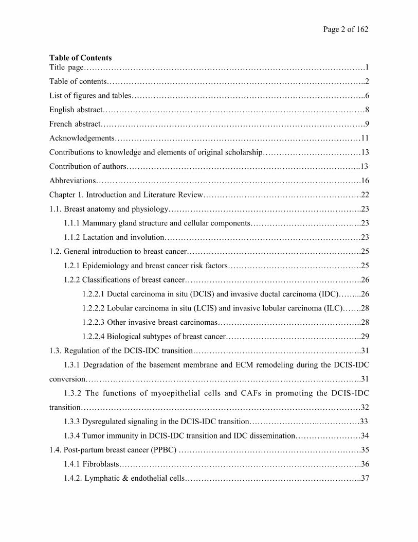
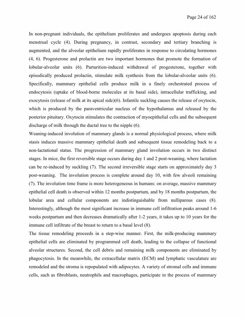
Figure 1.1 Breast anatomy image ©American Cancer Society 2017. Used with permission.
Page 26 of 162
Table 1.1 Risk factors (except age) associated with invasive breast cancer
(Adapted from Textbook of Medical Oncology, Chapter 5)
Major risk factors increasing risk >2 times (in descending order)
• BRCA1/2 mutations
• CHEK2 (also known as CHK2): (CHEK2(*)1100delC, a truncating variant that abrogates the
kinase activity) twofold increase of breast cancer risk in women and a tenfold
increase of risk in men
• Premenopausal breast cancer in mother and sister
• In situ cancer—ductal or lobular or atypical hyperplasia in breast biopsy
• Premenopausal breast cancer in mother or sister; bilateral breast cancer in first relative
• Hyperplasia without atypia in breast biopsy
Minor risk indicators or factors increasing risk ≤2 times
• Postmenopausal breast cancer in first-degree relative
• Obesity in women above 50 years
• Excess radiation to the chest wall or breast in history
• Alcohol consumption
• Nulliparous or delayed first pregnancy
• No breast feeding or short duration breast feeding of children
• Prolonged use of estrogens (contraceptive and/or hormone replacement)
1.2.2 Classifications of breast cancer
Breast cancer is a cluster of heterogeneous diseases classified by their histological characteristics
and genomic signatures. Histologically, breast cancer is sub-grouped into the following
categories:
1.2.2.1 Ductal carcinoma in situ (DCIS) and invasive ductal carcinoma (IDC)
DCIS, which accounts for approximately 20% of all newly diagnosed breast cancer cases, is
often described as “Stage 0” disease due to its non-invasive nature (15). DCIS is characterized by
the malignant proliferation of mammary ductal epithelial cells, which remain confined to the
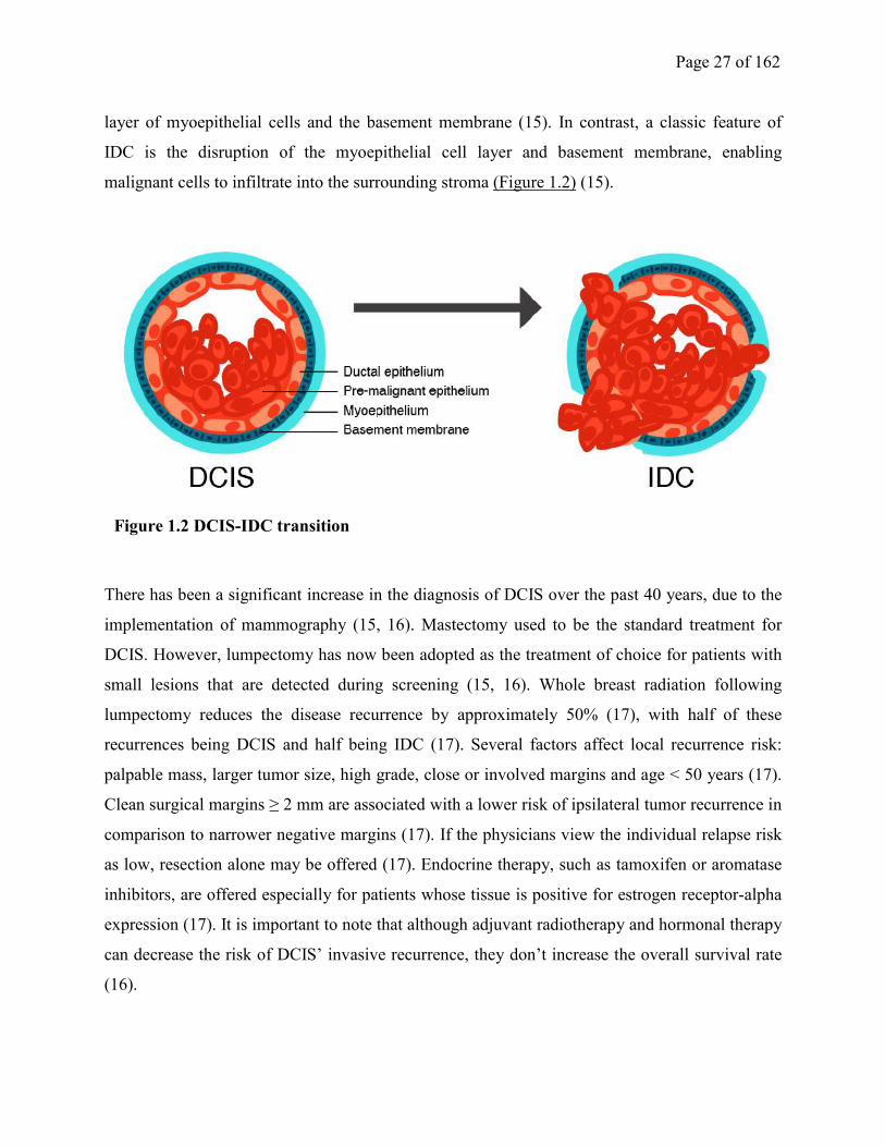
Page 27 of 162
layer of myoepithelial cells and the basement membrane (15). In contrast, a classic feature of
IDC is the disruption of the myoepithelial cell layer and basement membrane, enabling
malignant cells to infiltrate into the surrounding stroma (Figure 1.2) (15).
There has been a significant increase in the diagnosis of DCIS over the past 40 years, due to the
implementation of mammography (15, 16). Mastectomy used to be the standard treatment for
DCIS. However, lumpectomy has now been adopted as the treatment of choice for patients with
small lesions that are detected during screening (15, 16). Whole breast radiation following
lumpectomy reduces the disease recurrence by approximately 50% (17), with half of these
recurrences being DCIS and half being IDC (17). Several factors affect local recurrence risk:
palpable mass, larger tumor size, high grade, close or involved margins and age < 50 years (17).
Clean surgical margins ≥ 2 mm are associated with a lower risk of ipsilateral tumor recurrence in
comparison to narrower negative margins (17). If the physicians view the individual relapse risk
as low, resection alone may be offered (17). Endocrine therapy, such as tamoxifen or aromatase
inhibitors, are offered especially for patients whose tissue is positive for estrogen receptor-alpha
expression (17). It is important to note that although adjuvant radiotherapy and hormonal therapy
can decrease the risk of DCIS’ invasive recurrence, they don’t increase the overall survival rate
(16).
Figure 1.2 DCIS-IDC transition
Page 28 of 162
1.2.2.2 Lobular carcinoma in situ (LCIS) and invasive lobular carcinoma (ILC)
LCIS is defined as a type of cancer wherein pre-malignant breast epithelial cells are confined in
one or more lobules without breaching the basement membrane (15). It is considered benign and
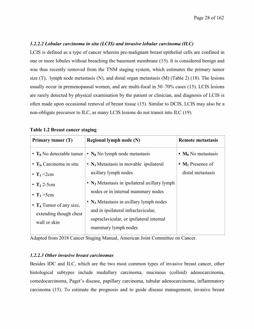
was thus recently removed from the TNM staging system, which estimates the primary tumor
size (T), lymph node metastasis (N), and distal organ metastasis (M) (Table 2) (18). The lesions
usually occur in premenopausal women, and are multi-focal in 50–70% cases (15). LCIS lesions
are rarely detected by physical examination by the patient or clinician, and diagnosis of LCIS is
often made upon occasional removal of breast tissue (15). Similar to DCIS, LCIS may also be a
non-obligate precursor to ILC, as many LCIS lesions do not transit into ILC (19).
Table 1.2 Breast cancer staging
Primary tumor (T) Regional lymph node (N) Remote metastasis
• T0 No detectable tumor
• Tis Carcinoma in situ
• T1 <2cm
• T2 2-5cm
• T3 >5cm
• T4 Tumor of any size,
extending though chest
wall or skin
• N0 No lymph node metastasis
• N1 Metastasis in movable ipsilateral
axillary lymph nodes
• N2 Metastasis in ipsilateral axillary lymph
nodes or in internal mammary nodes
• N3 Metastasis in axillary lymph nodes
and in ipsilateral infraclavicular,
supraclavicular, or ipsilateral internal
mammary lymph nodes
• M0 No metastasis
• M1 Presence of
distal metastasis
Adapted from 2018 Cancer Staging Manual, American Joint Committee on Cancer.
1.2.2.3 Other invasive breast carcinomas
Besides IDC and ILC, which are the two most common types of invasive breast cancer, other
histological subtypes include medullary carcinoma, mucinous (colloid) adenocarcinoma,
comedocarcinoma, Paget’s disease, papillary carcinoma, tubular adenocarcinoma, inflammatory
carcinoma (15). To estimate the prognosis and to guide disease management, invasive breast
Page 29 of 162
carcinomas are first assessed by TNM staging (Table 2) (17, 18). Subsequently, the ER/PR and
HER2 status are examined (17). Lumpectomy or mastectomy is performed, followed by
radiation, chemotherapy and/or hormone therapy depending on the staging and hormone receptor
status. The details of management have been extensively reviewed elsewhere (17, 18).
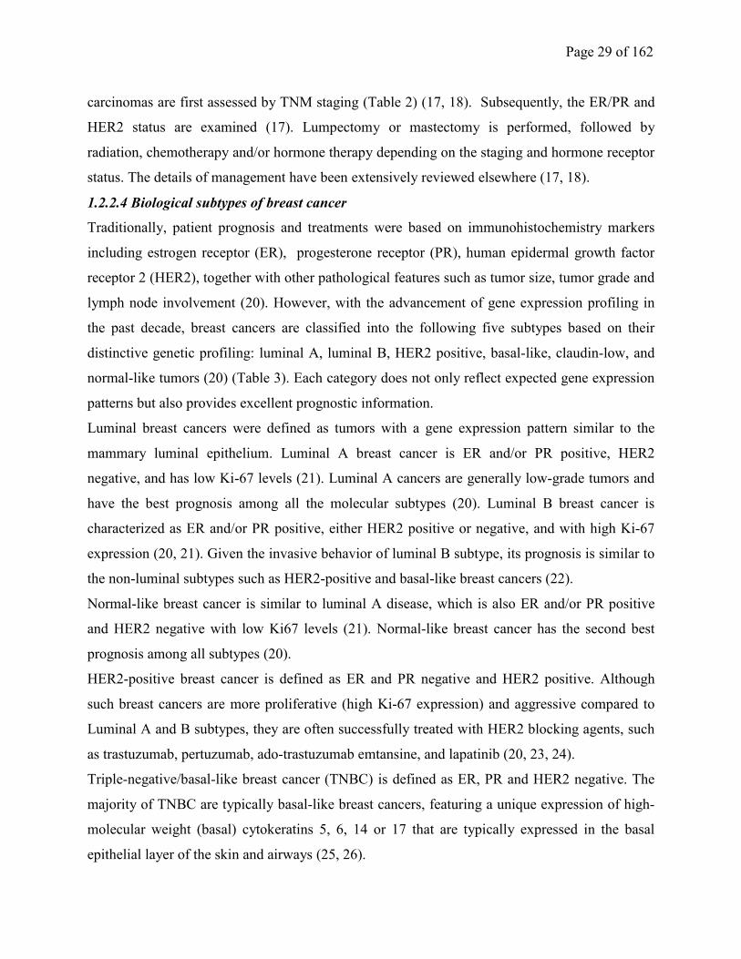
1.2.2.4 Biological subtypes of breast cancer
Traditionally, patient prognosis and treatments were based on immunohistochemistry markers
including estrogen receptor (ER), progesterone receptor (PR), human epidermal growth factor
receptor 2 (HER2), together with other pathological features such as tumor size, tumor grade and
lymph node involvement (20). However, with the advancement of gene expression profiling in
the past decade, breast cancers are classified into the following five subtypes based on their
distinctive genetic profiling: luminal A, luminal B, HER2 positive, basal-like, claudin-low, and
normal-like tumors (20) (Table 3). Each category does not only reflect expected gene expression
patterns but also provides excellent prognostic information.
Luminal breast cancers were defined as tumors with a gene expression pattern similar to the
mammary luminal epithelium. Luminal A breast cancer is ER and/or PR positive, HER2
negative, and has low Ki-67 levels (21). Luminal A cancers are generally low-grade tumors and
have the best prognosis among all the molecular subtypes (20). Luminal B breast cancer is
characterized as ER and/or PR positive, either HER2 positive or negative, and with high Ki-67
expression (20, 21). Given the invasive behavior of luminal B subtype, its prognosis is similar to
the non-luminal subtypes such as HER2-positive and basal-like breast cancers (22).
Normal-like breast cancer is similar to luminal A disease, which is also ER and/or PR positive
and HER2 negative with low Ki67 levels (21). Normal-like breast cancer has the second best
prognosis among all subtypes (20).
HER2-positive breast cancer is defined as ER and PR negative and HER2 positive. Although
such breast cancers are more proliferative (high Ki-67 expression) and aggressive compared to
Luminal A and B subtypes, they are often successfully treated with HER2 blocking agents, such
as trastuzumab, pertuzumab, ado-trastuzumab emtansine, and lapatinib (20, 23, 24).
Triple-negative/basal-like breast cancer (TNBC) is defined as ER, PR and HER2 negative. The
majority of TNBC are typically basal-like breast cancers, featuring a unique expression of high-
molecular weight (basal) cytokeratins 5, 6, 14 or 17 that are typically expressed in the basal
epithelial layer of the skin and airways (25, 26).
Page 30 of 162
Table 1.3 Biological subtypes of breast cancer
Molecular characteristics Histological
grade
Other features
ER PR HE
R2
Other markers
Luminal A + + - CK8/18+
Ki67Low
Low Best prognosis
Low recurrence
rates
Luminal B + + +/- CK12/18+
Ki67High
if HER2-
Intermediate/
High
HER2-
positive
- - + CK5/6+/-
CK8/18+/-
Ki67High
Intermediate/
High
Associated with
younger age of
diagnosis
Basal-like - - - CK5/6/14/17+, EGFR
+
Ki67High
High
Claudin-
low
- - - Claudin 3/4/7Low
E-cadherinLow
Ki67High
High Intense
lymphocytic
infiltration
Normal-like - - EGFR-
CK5-
Variable With adipocytes
gene signatures
Besides basal-like breast cancers, other categories of TNBC exist. One example is the claudin-
low breast cancer, which is a subtype of TNBC (26-28). Claudin-low tumors are not only ER, PR
and HER2 negative, but they also typically express low levels of claudins 3, 4, or 7 and E-
Cadherin, which are important cell-cell junction molecules which can limit cell motility (26).
Without these key junction proteins, claudin-low breast cancers are characterized by high levels
of mesenchymal markers, such as vimentin and high histological grade, indicating their highly
invasive nature (26, 28, 29). Additionally, claudin-low lesions almost always have a young age
Page 31 of 162
of tumor onset, intense immune cell infiltration, higher tumor grade, larger tumor size, and a
restricted tumor margin (26). Patients with claudin-low tumors have a worse overall survival
when compared to patients with luminal A type of breast cancer (26). Interestingly, however,
claudin-low tumors were associated with a low local recurrence rate following breast-conserving
therapy (26).
Currently, TNBC remains a clinical challenge, as its risk to metastasize is high and there are no
effective targeted therapies against metastatic TNBC (20, 30). Various experimental therapies
have been explored, and poly (ADP-ribose) polymerase (PARP) inhibitors have shown
promising effects against TNBC. Clinical investigations are ongoing to examine if PARP
inhibitors can sensitize TNBC to immune checkpoint blockade therapies (31).
1.3 Regulation of the DCIS-IDC transition
Previous studies have provided evidence that DCIS lesions are heterogeneous (32). Not all DCIS
lesions progress into IDC. Indeed, the progression from DCIS to IDC is a critical step in breast
tumorigenesis that remains poorly understood. In summary, DCIS-IDC conversion is
characterized by the following aspects:
1.3.1 Degradation of the basement membrane and ECM remodeling during the DCIS-IDC
conversion
The DCIS basement membrane (BM) is a structured ECM layer mainly composed of collagen IV
and laminins. The proteolytic degradation of DCIS BM is mediated by proteolytic enzymes
including matrix metalloproteinases (MMPs) and cathepsins. Multiple MMPs, such as MMP1,
MMP2, MMP9, MMP13, MMP14 (also known as MT1-MMP) and MMP26 (33-38), are all
involved in BM degradation during the transition from DCIS to IDC. Upregulation of MMPs
during the progression of DCIS to IDC is tightly controlled by several master regulators of
transcription, including SMARCE1 (38), p63 (37), SOX11 (39, 40), HOXB13(39), Engrailed
Homeobox 1 (EN1) (39), Distal-less Homeobox 4 (DLX4), TBX15 (39) and FoxM1 (35).
Additionally, a link between the upregulation of cathepsins and the DCIS-IDC transition has also
been reported. For example, cathepsin-D expression is increased in IDC compared to DCIS (41).
Several important polysaccharides in the ECM, such as hyaluronic acid (HA) and heparan sulfate
(HS), also serve as inhibitors to prevent tumor invasion. Increased expression of their
corresponding degrading enzymes, including hyaluronidase, heparanase-1 and heparan
Page 32 of 162
endosulfatase-2, are observed in IDC compared to DCIS, and may also contribute to the
transition from DCIS to invasive disease (34, 42, 43).
1.3.2 The functions of myoepithelial cells and CAFs in promoting the DCIS-IDC transition:
As described in section 1.1, myoepithelial cells separate the luminal cells from the surrounding
BM. In response to oxytocin, myoepithelial cells contract to expel the milk (44). It is
hypothesized that myoepithelial cells function as a physical barrier between the pre-malignant
epithelium and BM to prevent DCIS invasion through BM (45). Previous studies have provided
evidence to support its inhibitory effect during DCIS invasive transition, and have suggested that
the apoptosis of myoepithelial cells is a key event during the DCIS-IDC conversion. CD10, a
marker of myoepithelial cells, is highly expressed in normal breast tissues (45). Based on CD10
mRNA levels, DCIS patients with high CD10 mRNA expression showed no disease recurrence,
while those with low CD10 displayed a higher risk of local relapse (45). Myoepithelial cells also
prevent invasion by inhibition of tumor cell invasion and recruitment of lymphatic/blood vessel
endothelial cells. For example, myoepithelial cells express protease inhibitors such as tissue
inhibitor of metalloproteinases (TIMPs) to degrade MMPs (33), and directly downregulate the
expression of multiple MMPs. Moreover, myoepithelial cells produce soluble factors to prevent
the migration of tumor cells and endothelial cells. For example, Alpaugh et al. have reported that
myoepithelial cells can make use of chymotrypsin to shed off soluble CD44 molecules, which
block migration of breast cancer cells and endothelial cells (46). In contrast, pro-invasive
functions of myoepithelial cells are also reported. For example, tumor-associated myoepithelial
cells can secrete CXCL14 to support the invasion of DCIS cells (47). A subset of myoepithelial
cells expressing αvβ6 integrin induce MMP9 expression by secreting TGFβ to induce EMT and
DCIS-IDC transition (48), and high levels of αvβ6 integrin predicts disease recurrence (48).
In contrast to myoepithelial cells, fibroblasts in DCIS lesions seem to play a critical role to aid
ductal epithelial cells to gain invasive properties in a step-wise fashion. For example, fibroblasts
are activated in an NF-κB pathway-dependent manner, and contribute to the progression from
atypical ductal hyperplasia (ADH) to DCIS. More importantly, the expansion of fibroblasts
together with the loss of myoepithelial cells contribute to the disruption of BM, which further
supports DCIS-IDC transition (49). Additionally, fibroblasts also play an essential role in the
ECM remodeling during the transition to IDC. Specifically, fibroblast-derived CCL2 (50),
periostin (51), thrombin (52), IL6 (53), and certain isoforms of tenascin-C (54), have all been
Page 33 of 162
reported to be involved in promoting this invasive transition. Future studies of the myoepithelial
cells and fibroblasts isolated from DCIS lesions are needed to further understand the functions of
these two cell types with opposing functions in DCIS invasive transition.
1.3.3 Dysregulated signaling in the DCIS-IDC transition
A complex dysregulation of signaling networks has been proposed to underpin the invasive
transition of DCIS. Although it is not clear which external stimuli are critical for driving DCIS
invasion, several cytokines and chemokines, including TGFβ (55), NODAL (56), IL1β (57), IL6
(53, 58, 59), TNFα (57, 58), CCL2 (50) and CCL5 (57), have all been implicated in this process.
NODAL, a member of the TGF-beta family morphogen, is often re-expressed in cancer to
contribute to tumor cell plasticity for efficient invasion and metastasis. NODAL signaling is
regulated at multiple levels, including the control of NODAL gene expression, the cleavage of
NODAL precursor to generate mature NODAL, and the expression of NODAL inhibitory
proteins. Several transcriptional factors have been identified to regulate NODAL in embryonic
development , such as Notch intracellular domain (NICD), Tcf/Lef, SOX and Oct (60, 61). The
regulation of NODAL in cancer has started to be revealed. For example, hypoxia induces
NODAL expression in melanoma (62). BMP, another TGF-beta family member, can inhibit
NODAL expression in seminoma (63). However, it remains unclear how NODAL expression is
controlled in breast cancer. NODAL precursor protein, or pro-NODAL, is cleaved by the
subtilisin-like proprotein convertases Furin and PACE4 to generate mature NODAL. Both Furin
and PACE4 have been implicated in breast cancer invasion and metastasis (64, 65). Lefty1/2 can
serve as NODAL antagonists, and are induced by NODAL signaling to generate a negative
feedback loop for this pathway (66). Cancer cells lack Lefty1/2 expression, resulting in
uncontrolled NODAL signaling to support tumorigenesis (66).
Dysregulation of several well-known pro-tumorigenic signal transduction pathways can enhance
the invasiveness of DCIS. For example, elevated Akt and MAPK activation are observed in IDC
specimens (67), and increased PI-3K/Akt and MAPK activities are responsible for the disruption
of the normal breast epithelial architecture to prompt DCIS cells to acquire invasive
characteristics (68-72). Overexpression of PKC-ζ is observed in IDC samples compared to
DCIS. Hyperactivation of PKC in the DCIS-IDC conversion can be achieved by increased 1,4,5-
trisphosphate (IP3) levels, either via phospholipase C-β (PLC-β) overexpression that yields more
Page 34 of 162
IP3 production or through TNF alpha-induced protein 8 like 3 (TNFAIP8L3)-mediated IP3
accumulation (73, 74).
The sonic hedgehog (SHH) pathway also contributes to the DCIS-IDC transition. In IDC the
overexpression of SHH will lead to increased nuclear translocation of the transcriptional
regulator Gli1 to ultimately induce a pro-tumorigenic transcriptional program (75). Inductions of
Smads, TBX3, NF-κB, HIF-1α and Bcl-9 are also observed in DCIS-IDC transition (55, 70, 76-
78).
Such aberrantly activated signaling pathways described above might eventually lead to a partial
epithelial-to-mesenchymal transition (EMT). The process of a partial EMT allows DCIS cells to
gain plasticity for more effective invasion and metastasis. During a partial EMT, tumor cells gain
mesenchymal features to invade through basement membranes into the blood or lymphatic
vessels, and then undergo a mesenchymal-to-epithelial transition (MET) to colonize a secondary
organ. A partial EMT can be characterized by the elevated expression of mesenchymal markers
Snail (79), Slug (78), Twist1 (78), MMP2 (73) and Vimentin (56), while still expressing levels of
epithelial markers such as E-Cadherin. A partial EMT during the DCIS-IDC transition is also
associated with enhanced stemness of DCIS cells, which provides DCIS cells with self-renewal
capacity and plasticity to proliferate and invade. Overexpression of several cancer stem cell
markers, including CD133 and ALDH, are known to prompt DCIS cells to invade (72, 74, 80,
81).
1.3.4 Tumor immunity in DCIS-IDC transition and IDC dissemination
Similar to many other cancer types, the DCIS to invasive transition is a process driven by
immune evasion, and a strong T-helper 1 (Th1) and cytotoxic T lymphocytic response predicts
less invasion and metastasis, thus predicts good prognosis in DCIS patients (82). T cell
exhaustion, which is characterized by the expression of two important immune checkpoints
TIGIT and PD-L1, may also contribute to DCIS invasion (83). Although B cell infiltration is
reported to predict poor survival in a small DCIS cohort (84), future studies with larger cohorts
are needed to verify this finding. Additionally, neutrophil recruitment to the TME by IDC-
derived IL6 and IL8 serves as an important mechanism of immune evasion to augment IDC
dissemination (85). Interestingly, the levels of tumor-infiltrating lymphocytes (TILs) are
heterogenous in DCIS. For example, TILs are significantly higher in DCIS with ER- or HER2+
status, TP53 mutation, high copy number changes and comedo necrosis (86). However, the
Page 35 of 162
mechanisms regulating lymphocyte infiltration into DCIS tumor microenvironment (TME)
remain unclear.
1.4 Post-partum breast cancer (PPBC): PPBC, defined as breast cancer diagnosed up to 10
years following the latest pregnancy, has approximately a 3-fold increased risk of metastasis and
death (87-89), compared to breast cancer occuring outside this window of time. Such an
increased risk is restricted to patients receiving a breast cancer diagnosis post-partum, as women
with a cancer diagnosis during pregnancy have a favorable prognosis comparable to that of
nulliparous patients (89). Although it is unclear why PPBC is associated with a poor prognosis,
recent studies have suggested that the microenvironment of the involuting mammary gland
provides many favorable factors for any existing pre-malignant cells to survive and become
invasive (9, 11, 12, 90). Specifically, the process of mammary gland involution entails removing
cellular debris remaining after mammary epithelial apoptosis. Thus it is not surprising that during
involution, dendritic cells, macrophages and T cells infiltrate into the mammary gland. Indeed,
increased immune cell infiltration has been reported in PPBC, compared to nulliparous breast
cancer (11, 12), indicating an important role of dysregulated immunity in PPBC progression.
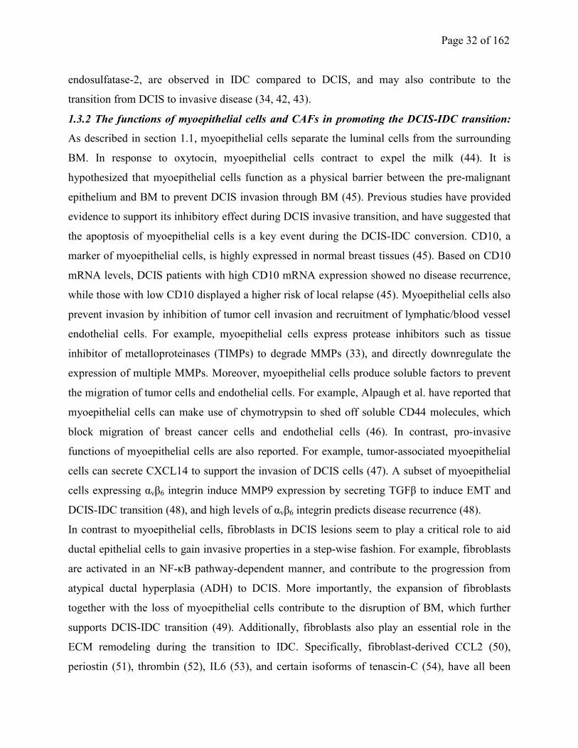
Summarized below are different cell types that contribute to the uniqueness of the PPBC TME
(Figure 1.3).
Figure 1.3 Selected cellular components in PPBC TME.

Page 36 of 162
1.4.1 Fibroblasts: Fibroblasts control extracellular matrix deposition during tissue remodeling
processes, such as mammary gland involution and breast cancer invasion. Fibroblasts that are
isolated during mammary gland involution (involution fibroblasts) have acquired a unique
activation state that is characterized by an immunosuppressive gene signature and enhanced
extracellular matrix remodeling (91). Although these physiologically activated fibroblasts do not
express α-smooth muscle actin (α-SMA), which is a marker for activated fibroblasts under
pathological conditions (92), they do contribute to tumor promotion during involution (91).
Cancer-associated fibroblasts (CAFs), on the other hand, are pathologically activated fibroblasts.
They are a highly proliferative and heterogeneous population of fibroblasts that support the
persistent growth and immune evasion of breast cancer (93). Contrary to activated fibroblasts
which can be deactivated and eventually disappear in the end stage of involution, CAFs gain the
capacity to sustain their proliferative potential and contribute to various aspects of breast cancer
metastatic cascade. This includes increasing cancer cell invasion into surrounding lymphatic and
blood vessels, protecting the survival of circulating tumor cells, and supporting the colonization
of tumor cells at distal organs (93, 94).
It remains unclear if the CAFs in PPBC TME are derived from involution fibroblasts (91). But
involution-fibroblasts and CAFs do make use of similar mechanisms to support pro-tumorigenic
immunity. Specifically, both types of fibroblasts express multiple pro-inflammatory enzymes and
soluble factors. For example, involution-fibroblasts produce a variety of chemokines to induce
M2-like macrophage polarization and IL10 production, which further dampens the anti-tumor
immunity. Such activated fibroblasts aid the tumor cells to recruit MDSCs at the tumor border,
and prevent cytotoxic T cell infiltration (91). Similarly, CAFs are also reported to produce
Chitinase 3-like 1, a secretory protein that drives M2 macrophage polarization and tumorigenesis
(95). Additionally, both types of fibroblasts express COX2 to produce prostaglandin E2 (PGE2),
which mediates the maintenance of breast cancer stem cells, promotes recruitment of MDSCs,
and confers resistance to natural killer (NK) cell and cytotoxic T cell-mediated anti-tumor
immunity (91, 96-100). As such, by enhancing M2-like macrophage expansion and MDSC
recruitment, involution-fibroblasts and CAFs impair cytotoxic T cell and NK cell-based tumor
eradication, thus creating favorable conditions for breast tumor progression.
1.4.2. Lymphatic endothelial cells: Involution is accompanied by increased lymphatic growth
and remodeling. Unfortunately, the lymphatic system is the most common route for breast cancer
Page 37 of 162
metastasis (1). Human PPBC features a high density of peritumor lymphatic vessels, which
partially explains its increased risk of metastasis (101). Increased lymphatic endothelial cell
proliferation and neo-lymphangiogenesis are driven by multiple lymphangiogenic factors that are
induced during involution, such as VEGF-C, VEGF-D, along with their receptors VEGFR2/3
(101). The COX2 inhibitor celecoxib is a potent inhibitor of lymphangiogenesis during
involution, and prevents PPBC metastasis (102).
1.4.3. Macrophages:
In response to pro-inflammatory signals such as IFNγ secreted by NK and T cells in the TME,
macrophages assume a functional phenotype referred to as M1-like, in analogy with the type 1 T
helper cells (Th1)-mediated immunity and serve to amplify the anti-tumoral response in the
TME. In contrast, upon receiving signals via the IL4R or IL10R, macrophages assume anti-
inflammatory tissue-remodeling roles aimed to dampen the immune response and clear cell
debris (103). Efferocytosis, which is defined as the clearance of dying cells by M2-like
macrophages or other phagocytes, is a critical process during involution (104). M2-like
macrophages produce several factors important for this process, such as arginase I, involved in
collagen biosynthesis. Unfortunately, M2-like macrophages are associated with pro-tumorigenic
immunity (103). By producing immunosuppressive cytokines, such as IL4, IL10, IL13 and TGF-
β (104, 105), M2-like macrophages are proposed to mediate PPBC immune evasion. Targeting
M2-like macrophages has been attempted in PPBC models; with the suggestion that ibuprofen
can diminish the population of M2-like macrophages by promoting M1-like macrophage
polarization (12).
1.4.4. Myeloid-derived suppressor cells (MDSCs): MDSCs are a heterogeneous population of
immature myeloid cells that are associated with immune suppression and tumor progression. The
heterogeneity of MDSCs is reflected in their genetic and morphological diversities, and are
subdivided into monocytic-MDSCs (M-MDSCs) and polymorphonuclear-MDSCs (PMN-
MDSCs) (106). MDSCs are important during pregnancy to avoid the maternal rejection of the
fetus (107), however the functions of MDSCs during mammary gland involution and PPBC
development are not well characterized. Recently, Pennock and colleagues reported that
granulocytes positive for immature myeloid markers Gr1 and CD11b remain at a high level
during involution (12), indicating the potential importance of MDSCs in contributing to the
immunosuppressive microenvironment during involution.
Page 38 of 162
Most research to date has focused on the importance of MDSCs in breast cancer progression.
The levels of MDSCs in both the TME and the circulation is correlated to impaired breast cancer
patient survival (108-110), as it supports type 2 pro-tumorigenic immunity (109).
Breast tumor cells produce abundant G-CSF and GM-CSF to stimulate MDSC intra-tumor
infiltration (111). Tumor-infiltrating MDSCs utilize several mechanisms to support breast
tumorigenesis directly. MDSC-derived IL6 and nitric oxide (NO) promote the maintenance of
breast cancer stem cell (CSC) via activation of the STAT3 and NOTCH pathways in breast
cancer cells (108). MDSCs also produce MMPs that degrade extracellular matrix, thus
facilitating breast tumor cell invasion (112). Additionally, MDSCs house several amino acid
catabolic enzymes to achieve immune suppression. For example, arginase I and inducible nitric
oxide synthase (iNOS) expression by MDSCs facilitates the catabolism of L-arginine into L-
ornithine (113, 114) and L-arginine to nitric oxide (NO) (115). While L-ornithine is reported to
block cytotoxic T cell differentiation (116), NO derived from MDSCs can inhibit NK cell-
mediated cytotoxicity against breast cancer cells (115). Two other catabolic enzymes,
indoleamine-2,3-dioxygenase (IDO) and tryptophan 2,3- dioxygenase (TDO), that degrade L-
tryptophan into kynurenine are also highly expressed in MDSCs (117). Depletion of L-arginine
and accumulation of kynurenine both contribute to the paralysis of CD8+ T cells and NK cells
(117, 118), and tryptophan starvation in the TME facilitates the polarization of CD4+ T cells into
regulatory T cells (Treg) (119). Lastly, NO production by MDSC-derived inducible nitric oxide
synthase (iNOS) impairs NK cell-dependent antibody-dependent cellular cytotoxicity (ADCC) in
breast cancer models (115). Thus, ablation, or exclusion, of MDSCs from the TME can be a
potential anti-cancer therapy, with the inhibition of the CSF1/CSF1R pathway and PI-3Kγ as just
two putative modes (120-122).
1.4.5. Innate lymphoid cells: Innate lymphoid cells (ILCs) are distinct populations of lymphoid
cells that lack variable T or B cell receptors. This group of innate immune cells are classified
based on their distinct expression of transcription factors and their unique cytokine production
profiles, and include NK cells, group 1 ILCs (ILC1), group 2 ILCs (ILC2), group 3 ILCs (ILC3),
and lymphoid tissue-inducer (LTi) cells (reviewed by Chiossone et al (123)). Although the anti-
tumor effect and the role of NK cells in immune surveillance have become more evident, the
functions of ILC1, 2, 3 and LTi cells in tumor biology are only just beginning to emerge (124).
Page 39 of 162
A few recent studies suggest that ILC2s may have pro-tumorigenic roles in cancer (125, 126),
possibly by inducing MDSC expansion and inhibiting cytotoxic T cell infiltration (125, 126).
ILC2 frequency is elevated in breast tumor samples, compared to benign breast tissues (127).
The polarization and expansion of ILC2s are regulated by a few essential cytokines, including
IL33, IL7, IL25 and thymic stromal lymphopoietin (TSLP) (128). Activated ILC2s produce
abundant IL5 and IL13, which are important cytokines for type 2 immunity (128). Given the
importance of type 2 immunity in breast cancer immune evasion, it is not surprising that
exogenous IL33 can induce ILC2 polarization and supports 4T1 breast cancer cell metastasis
(129).
However, it remains unclear how ILC2s contribute to the tissue remodeling during mammary
gland involution, and how ILC2s regulate PPBC immunity. A part of my work is dedicated to the
potential pro-tumorigenic roles of IL33, and potentially ILC2, in breast cancer metastasis.
1.4.6. Dendritic cells (DCs): DCs are bone marrow-derived professional antigen presenting cells
(APCs) of the myeloid lineage responsible for sampling environmental antigens and providing
this information to cells from the adaptive immune system (130). Although DCs are recruited to
mammary gland early during involution (11), the precise functions of DCs in this process are
poorly characterized. Milk stasis generates various stress signals that trigger involution. Cellular
stress, such as endoplasmic reticulum (ER) stress and autophagy, are induced during involution
(131). Such stress signals also drive the expression and release of damage-associated molecular
patterns (DAMPs) as “come and eat me” signals to attract DCs (132). Additionally, dying cell-
derived DNA can be uptaken by DCs to activate the cyclic GMP-AMP synthase
(cGAS)/stimulator of interferon genes (STING) pathway, which serves as a defense immune
mechanism (reviewed by Corrales and Gajewski (133)). These observations possibly indicate a
crucial role of DCs in immune surveillance. Escape from DC-mediated immune surveillance is
an important step for breast cancer metastasis. Breast cancer cells often escape from DC-
mediated immune surveillance by multiple mechanisms, including decreasing MHC-I
presentation on the cell surface, and by silencing the expressions of transported associated with
antigen processing (TAP)-1/2 (134, 135). However, it remains unclear how DCs regulate PPBC
progression and metastasis.
1.4.7. T cells: T cells mature in the thymus, and express either CD8 (cytotoxic T cells) or CD4
(T helper cells, Th cells). Th cells are further divided into various subsets, including Th1, Th2,
Page 40 of 162
Th9, Th17, Th22, Treg and follicular helper T cells (Tfh) (reviewed by Golubovskaya and Wu
(136)). Th1 cells, which are activated by IFNγ and IL12, can produce IFNγ to activate cytotoxic
T cells. In contrast, Th2 cells are stimulated by IL4 and secrete IL4, IL5, IL10 and IL13. Th1 and
Th2 cells oppose each other to maintain an immune balance (reviewed by Patel et al. (137)).
Similar to most malignancies, breast cancer progression is often characterized by the disruption
of Th1/Th2 balance and diminished cytotoxic T cell-dependent tumor eradication (reviewed by
Disis (138), Borst et al. (139)). Exhaustion of cytotoxic T cells is arguably the best-characterized
mechanism for immune evasion of tumor cells (140). It is broadly defined as diminished effector
function and sustained expression of inhibitory checkpoints on the cell surface of T cells (141). T
cell exhaustion can be regulated via the following mechanisms: (a) soluble factors such as IL10
and TGFβ, (b) expression of inhibitory checkpoint molecules on the cell surface, such as CTLA-
4 and PD-1, or (c) direct or indirect interactions with inhibitory stromal cells, such as Treg,
MDSCs and CAFs. In order to develop effective therapies against PPBC, strategies to restore the
Th1-mediated immunity have been explored in PPBC mouse models. For example, ibuprofen
effectively blocks breast tumor outgrowth in a PPBC model, partially due to ibuprofen-induced
Th1 and cytotoxic T cell infiltration into the TME (12).
Increased infiltration of Treg cells during involution is an important checkpoint to control
inflammation, and is suggestive of wound resolution (11). Unfortunately, Treg cells also play an
important role in breast cancer progression, as they tend to enrich in more aggressive breast
cancers (142). Intra-tumoral Tregs often express elevated levels of inhibitory checkpoint
molecules (PD-1, PD-L1 and CTLA-4) (142, 143), indicating that the tumor-infiltrating Treg cells
exert more of an immune suppressive function in TME than in the periphery. Tumor-infiltrating
Treg cells express high levels of CCR8 (142), and depletion of Treg cells by inhibiting CCR8
pathway may be a promising strategy against breast cancer.
In summary, multiple non-immune and immune cell types in the tumor stroma contribute to
PPBC immune evasion and metastasis (Figure 1.3). However, our understanding of PPBC tumor
immunology is still rudimentary. To find better targets for this aggressive disease, future studies
are needed to better understand how different stromal cells contribute to PPBC pathogenesis and
metastasis.
1.5. Pro-tumorigenic functions of MNK1/2-eIF4E pathway in Breast Cancer
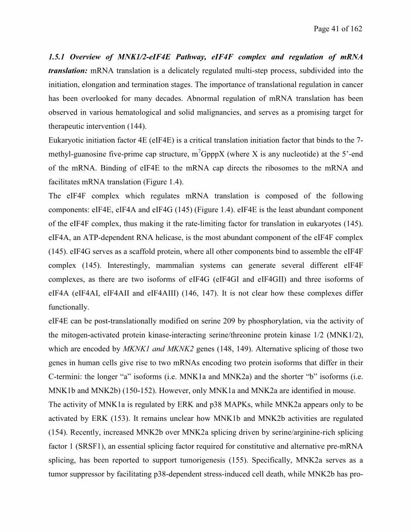
Page 41 of 162
1.5.1 Overview of MNK1/2-eIF4E Pathway, eIF4F complex and regulation of mRNA
translation: mRNA translation is a delicately regulated multi-step process, subdivided into the
initiation, elongation and termination stages. The importance of translational regulation in cancer
has been overlooked for many decades. Abnormal regulation of mRNA translation has been
observed in various hematological and solid malignancies, and serves as a promising target for
therapeutic intervention (144).
Eukaryotic initiation factor 4E (eIF4E) is a critical translation initiation factor that binds to the 7-
methyl-guanosine five-prime cap structure, m7GpppX (where X is any nucleotide) at the 5’-end
of the mRNA. Binding of eIF4E to the mRNA cap directs the ribosomes to the mRNA and
facilitates mRNA translation (Figure 1.4).
The eIF4F complex which regulates mRNA translation is composed of the following
components: eIF4E, eIF4A and eIF4G (145) (Figure 1.4). eIF4E is the least abundant component
of the eIF4F complex, thus making it the rate-limiting factor for translation in eukaryotes (145).
eIF4A, an ATP-dependent RNA helicase, is the most abundant component of the eIF4F complex
(145). eIF4G serves as a scaffold protein, where all other components bind to assemble the eIF4F
complex (145). Interestingly, mammalian systems can generate several different eIF4F
complexes, as there are two isoforms of eIF4G (eIF4GI and eIF4GII) and three isoforms of
eIF4A (eIF4AI, eIF4AII and eIF4AIII) (146, 147). It is not clear how these complexes differ
functionally.
eIF4E can be post-translationally modified on serine 209 by phosphorylation, via the activity of
the mitogen-activated protein kinase-interacting serine/threonine protein kinase 1/2 (MNK1/2),
which are encoded by MKNK1 and MKNK2 genes (148, 149). Alternative splicing of those two
genes in human cells give rise to two mRNAs encoding two protein isoforms that differ in their
C-termini: the longer “a” isoforms (i.e. MNK1a and MNK2a) and the shorter “b” isoforms (i.e.
MNK1b and MNK2b) (150-152). However, only MNK1a and MNK2a are identified in mouse.
The activity of MNK1a is regulated by ERK and p38 MAPKs, while MNK2a appears only to be
activated by ERK (153). It remains unclear how MNK1b and MNK2b activities are regulated
(154). Recently, increased MNK2b over MNK2a splicing driven by serine/arginine-rich splicing
factor 1 (SRSF1), an essential splicing factor required for constitutive and alternative pre-mRNA
splicing, has been reported to support tumorigenesis (155). Specifically, MNK2a serves as a
tumor suppressor by facilitating p38-dependent stress-induced cell death, while MNK2b has pro-
Page 42 of 162
oncogenic properties (155). It remains unclear how eIF4E S209 phosphorylation facilitates
mRNA translation. Past studies have suggested that eIF4E S209 phosphorylation may speed up
the dissociation of eIF4E from mRNAs to facilitate its recycling for further rounds of mRNA
translation (156). Thus, eIF4E phosphorylation may have led to punctual increases in the
availability of eIF4E, which may explain why 5’UTR structures that are highly sensitive to total
eIF4E levels also respond to elevated eIF4E phosphorylation (156). However, this hypothesis
still lacks direct supporting evidence. eIF4E phosphorylation does not affect global mRNA
translation, as shown by the experiments wherein serine 2019 is mutated to an alanine, or genetic
ablation of MNK1/2 (149, 157). Instead, the phosphorylation of eIF4E preferentially controls the
translation of a specific subset of mRNAs with complex 5’-UTRs, which will be further
discussed in section 6.2 (149, 157).
1.5.2 The functions of MNK1/2-eIF4E pathway in breast tumorigenesis:
The activation of MNK1/2-eIF4E pathway is controlled by two well-characterized MAPK
members, p38 and ERK1/2 (Figure 4). Various external stimuli, such as mitogenic and external
stress signals can activate MAPK signaling, which ultimately converge on the MNK1/2-eIF4E
pathway (reviewed by Bhat et al. (144)). MNK1/2-eIF4E pathway activation facilitates
tumorigenesis by inducing the translation of pro-tumorigenic and pro-invasive mRNAs without
significantly altering the expression of housekeeping genes such as GAPDH (144).
Figure 1.4. Regulation of eIF4F complex activity by MNK1/2-eIF4E and PI-
3K/Akt/mTOR pathways.
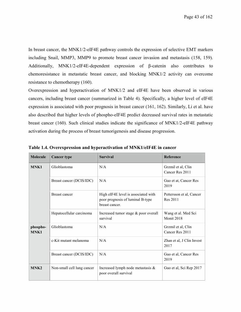
Page 43 of 162
In breast cancer, the MNK1/2-eIF4E pathway controls the expression of selective EMT markers
including Snail, MMP3, MMP9 to promote breast cancer invasion and metastasis (158, 159).
Additionally, MNK1/2-eIF4E-dependent expression of β-catenin also contributes to
chemoresistance in metastatic breast cancer, and blocking MNK1/2 activity can overcome
resistance to chemotherapy (160).
Overexpression and hyperactivation of MNK1/2 and eIF4E have been observed in various
cancers, including breast cancer (summarized in Table 4). Specifically, a higher level of eIF4E
expression is associated with poor prognosis in breast cancer (161, 162). Similarly, Li et al. have
also described that higher levels of phospho-eIF4E predict decreased survival rates in metastatic
breast cancer (160). Such clinical studies indicate the significance of MNK1/2-eIF4E pathway
activation during the process of breast tumorigenesis and disease progression.
Table 1.4. Overexpression and hyperactivation of MNK1/eIF4E in cancer
Molecule Cancer type Survival Reference
MNK1 Glioblastoma N/A Grzmil et al, Clin
Cancer Res 2011
Breast cancer (DCIS/IDC) N/A Guo et at, Cancer Res
2019
Breast cancer High eIF4E level is associated with
poor prognosis of luminal B-type
breast cancer.
Pettersson et al, Cancer
Res 2011
Hepatocellular carcinoma Increased tumor stage & poor overall
survival
Wang et al. Med Sci
Monit 2018
phospho-
MNK1
Glioblastoma N/A Grzmil et al, Clin
Cancer Res 2011
c-Kit mutant melanoma N/A Zhan et al, J Clin Invest
2017
Breast cancer (DCIS/IDC) N/A Guo et al, Cancer Res
2019
MNK2 Non-small cell lung cancer Increased lymph node metastasis &
poor overall survival
Guo et al, Sci Rep 2017
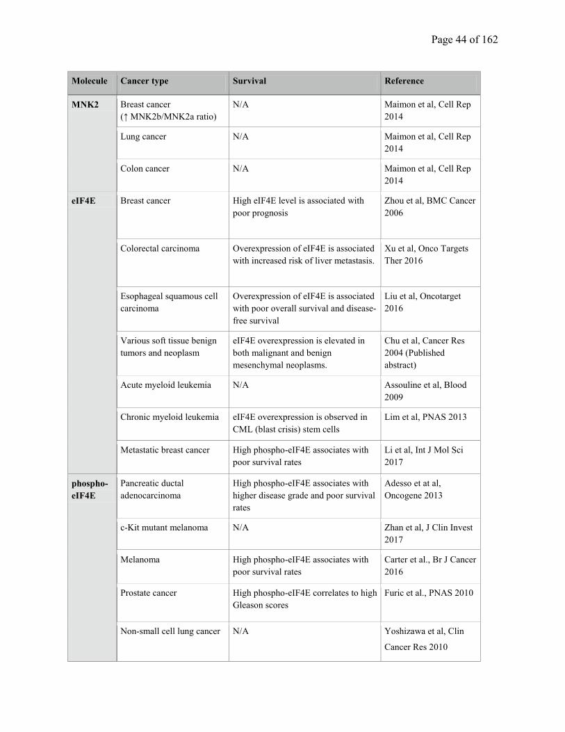
Page 44 of 162
Molecule Cancer type Survival Reference
MNK2 Breast cancer
(↑ MNK2b/MNK2a ratio)
N/A Maimon et al, Cell Rep
2014
Lung cancer N/A Maimon et al, Cell Rep
2014
Colon cancer N/A Maimon et al, Cell Rep
2014
eIF4E Breast cancer High eIF4E level is associated with
poor prognosis
Zhou et al, BMC Cancer
2006
Colorectal carcinoma Overexpression of eIF4E is associated
with increased risk of liver metastasis.
Xu et al, Onco Targets
Ther 2016
Esophageal squamous cell
carcinoma
Overexpression of eIF4E is associated
with poor overall survival and disease-
free survival
Liu et al, Oncotarget
2016
Various soft tissue benign
tumors and neoplasm
eIF4E overexpression is elevated in
both malignant and benign
mesenchymal neoplasms.
Chu et al, Cancer Res
2004 (Published
abstract)
Acute myeloid leukemia N/A Assouline et al, Blood
2009
Chronic myeloid leukemia eIF4E overexpression is observed in
CML (blast crisis) stem cells
Lim et al, PNAS 2013
Metastatic breast cancer High phospho-eIF4E associates with
poor survival rates
Li et al, Int J Mol Sci
2017
phospho-
eIF4E
Pancreatic ductal
adenocarcinoma
High phospho-eIF4E associates with
higher disease grade and poor survival
rates
Adesso et at al,
Oncogene 2013
c-Kit mutant melanoma N/A Zhan et al, J Clin Invest
2017
Melanoma High phospho-eIF4E associates with
poor survival rates
Carter et al., Br J Cancer
2016
Prostate cancer High phospho-eIF4E correlates to high
Gleason scores
Furic et al., PNAS 2010
Non-small cell lung cancer N/A Yoshizawa et al, Clin
Cancer Res 2010
Page 45 of 162
Our understanding of the functions of MNK1/2-eIF4E in tumor immunity is increasing.
Mehrotra et al. have described that MNK1/2 activity is essential for the production of anti-
neoplastic type I interferons (IFNs) in myeloproliferative neoplasms (163). Later, it was revealed
that phospho-eIF4E enhances IκB mRNA translation, and subsequently suppresses NF-κB
dependent transcription of the IFNβ gene (164), suggesting that phospho-eIF4E may inhibit
innate immunity by decreasing IFNβ production. Recently, MNK1/2-eIF4E pathway has also
been reported to support the expansion of pro-invasive neutrophils, and ablation of neutrophils
by an anti-Ly6G antibody can inhibit breast cancer metastasis in a pre-clinical model (165).
Finally, eIF4E has also been identified to serve as a key regulator of STAT1-dependent PD-L1
expression on tumor cells (166). The evidence thus far, suggests a possible role of the MNK1/2-
eIF4E axis in facilitating pro-tumorigenic immunity.
1.5.3 Other MNK1/2 substrates
MNK1/2 have substrates besides eIF4E, including hnRNP-A1, PTB (polypyrimidine tract-
binding protein)-associated splicing factor (PSF)-p54psf.nrb
, Sprouty2, and Cytosolic
phospholipase A2 (cPLA2) (167-169). Overexpression of hnRNP A1 has been reported in
various cancer types, including breast cancer (170, 171). This overexpression leads to an increase
in the levels of several EMT markers, thus facilitating the motility of cancer cells (171). The role
of MNK1 and Sprouty2 in cancer is less defined. DaSilva and colleagues reported that MNK-
dependent Sprouty 2 phosphorylation increases the stability of Sprouty2, which is a negative
feedback modulators of receptor tyrosine kinase pathways (167). However, Sprouty 2 was also
reported as a potential tumor suppressor in breast cancer, as loss of Sprouty 2 is associated with
increased tumor grade and poor prognosis (172). Further studies are needed to better define the
functions of these, and potentially other, MNK1/2 substrates in cancer, with an emphasis on how
MNK1/2 impacts their protein stability and activity.
1.5.4 Targeting MNK1/2 in cancer:
MNK1/2 activities are dispensable for normal development, but are potential therapeutic targets
in various malignancies, and there are now efforts to identify selective small molecule inhibitors
that can be used in the clinic (144). Cercosporamide and CGP57380 are two well-described
MNK1/2 inhibitors with anti-neoplastic efficacy. However, both compounds exhibit significant
Page 46 of 162
off-target effects (173). To address the need for more selective modes of repressing MNK1/2
activity, novel strategies to target MNK1/2 are being developed. For example, retinoic acid
metabolism blocking agents (RAMBA) which induce MNK1 degradation effectively decreases
eIF4E phosphorylation (174). Resorcylic acid lactone analogues are also developed to block
MNK1/2 activity, and, as a consequence, inhibit eIF4E phosphorylation and cancer cell
proliferation (175). Three novel orally bioavailable MNK1/2 inhibitors: SEL201, BAY1143269
and eFT508, have been reported by us and others to show promising anti-cancer effects (56, 176-
179).
1.5.5 Crosstalk between the MNK1/2 and PI3K/Akt/mTOR pathways:
The eIF4E is a nexus point on which the MNK1/2 and PI3K/Akt/mTOR pathways converge.
Crosstalk exists between these two pathways, with important implications for cancer cell
biology. The PI3K/AKT/mTOR signaling pathway is one of the most commonly dysregulated
pathways in cancer. Upon mTOR-mediated phosphorylation of the eIF4E binding proteins
(4EBPs) 1/2/3, their repressive binding to eIF4E is disrupted, freeing eIF4E to assemble into the
eIF4F complex, thus promoting mRNA translation (reviewed by Janku et al. (180)). PI-
3K/Akt/mTOR pathway has been extensively characterized with respect to tumor cell survival,
proliferation, invasion, metabolism and genomic instability (Reviewed by Janku et al. (180)).
Rapamycin and its analogs, including everolimus, temsirolimus and ridaforolimus, are used as
allosteric mTOR inhibitors. Recently, catalytic mTOR inhibitors, such as PP242 and OSI-027,
which block the activity of both mTORC1 and mTORC2, were developed in an attempt to
enhance the anti-cancer efficacy of the mTOR blockade. Unfortunately, complex feedback loops
involving activation of PI3K and MAPK signaling have limited the success of mTOR inhibitors
as anti-cancer agents, and MNK1/2-eIF4E activation contributes to the resistance of mTOR
inhibitors in cancer cells (181-183). Specifically, mTOR inhibitors, such as rapamycin and
everolimus, have been demonstrated to induce eIF4E phosphorylation (184, 185). The molecular
mechanisms underlying the phosphorylation of eIF4E as a consequence of mTOR inhibition are
not fully elucidated. Katsha et al. reported that everolimus resistance is caused by Aurora kinase
A-dependent inhibition of type 2A serine/threonine protein phosphatase (PP2A) activity, which
subsequently elevates eIF4E phosphorylation (186). This process is independent of MNK1/2
activity in upper gastrointestinal adenocarcinomas (186). Alternatively, rapamycin was also
reported to induce eIF4E phosphorylation via MNK2, but independent of p38 and ERK1/2
Page 47 of 162
activities (187). Additionally, MNK1/2 can also promote resistance to mTOR inhibitors by
stimulating the constitutive activation of mTORC1, and facilitating mTORC1 binding to its
substrates (185).
Multiple avenues to block aberrant mRNA translation in cancer, by co-inhibiting
PI3K/Akt/mTOR and MNK/eIF4E axis, have been explored. MNK1/2 inhibition has been shown
to be a potential strategy to overcome mTOR inhibitor resistance in malignant cells. For
example, CGP57380 synergizes with a few mTOR inhibitors (rapamycin, everolimus, or OSI-
027) to inhibit tumorigenesis in several solid and hematological malignancies, including
medulloblastoma, non-small cell lung cancer and acute lymphocytic leukemia (187-189). In
breast cancer models, MNK1/2 inhibitors cercosporamide and CGP57380 have shown
synergistic anti-breast cancer effects when combined with an in-house PI-3K inhibitor or an
mTOR inhibitor PP242 (183). This could partially be explained by proposing that the inhibition
of MNK1/2 and PI-3K/Akt/mTOR pathways together blocks the assembly of the eIF4F complex.
Additionally, co-treatment of CGP57380 plus everolimus overcomes tamoxifen resistance in
breast cancer (190). These data support that combination of mTOR and MNK1/2 inhibitors may
be a promising regimen against breast cancer and other malignancies.
1.6 Rationale, objective and general introduction to the thesis research
Although many pro-tumorigenic functions of MNK1 have been characterized, it remains unclear
how MNK1 regulates the early steps of the non-invasive to invasive breast carcinoma transition.
We hypothesized that MNK1 drives DCIS to IDC transition, and we aimed to test this hypothesis
by MNK1 overexpression and knock-out in a DCIS model. We performed a series of
experiments to demonstrate that MNK1 activity in DCIS is needed to support a partial EMT and
maintain cancer stem cell properties, as the DCIS cells transit to become invasive tumor cells
(Chapter 2). Additionally, our knowledge of how aberrant mRNA translation regulates breast
tumor immunity, and which pro-tumorigenic and pro-inflammatory factors are involved, remains
limited. Multiple pro-oncogenic pathways converge on the MNK1/2-eIF4E axis, thus it is
important to understand how this pathway affects breast cancer immune evasion. In Chapter 3, I
chose to study PPBC, an aggressive subtype of breast cancer characterized by robust immune
cell influx. We hypothezied that stromal MNK1/2-eIF4E axis shapes pro-tumorigenic immunity
Page 48 of 162
during metastasis of PPBC, and we performed a series of experiments to examine how MNK1/2-
eIF4E axis impacts the functions of different immune and non-immune cell types in PPBC TME.
The work presented in this thesis highlights the critical functions of MNK1/2-eIF4E pathway in
breast cancer. The objectives of our studies were as follows:
a) To determine if MNK1 regulates the transition from DCIS to invasive disease.
b) To understand how the MNK1/eIF4E axis, functioning in stromal cells, regulates PPBC pro-
tumorigenic immunity.
1.7 References
1. Pandya S, and Moore RG. Breast development and anatomy. Clin Obstet Gynecol.
2011;54(1):91-5.
2. Degnim AC, Brahmbhatt RD, Radisky DC, Hoskin TL, Stallings-Mann M,
Laudenschlager M, et al. Immune cell quantitation in normal breast tissue lobules with
and without lobulitis. Breast cancer research and treatment. 2014;144(3):539-49.
3. Degnim AC, Hoskin TL, Arshad M, Frost MH, Winham SJ, Brahmbhatt RA, et al.
Alterations in the Immune Cell Composition in Premalignant Breast Tissue that Precede
Breast Cancer Development. Clin Cancer Res. 2017;23(14):3945-52.
4. Inman JL, Robertson C, Mott JD, and Bissell MJ. Mammary gland development: cell fate
specification, stem cells and the microenvironment. Development. 2015;142(6):1028-42.
5. Masso-Welch PA, Darcy KM, Stangle-Castor NC, and Ip MM. A developmental atlas of
rat mammary gland histology. Journal of mammary gland biology and neoplasia.
2000;5(2):165-85.
6. Truchet S, and Honvo-Houeto E. Physiology of milk secretion. Best Pract Res Clin
Endocrinol Metab. 2017;31(4):367-84.
7. Ning Y, Hoang B, Schuller AG, Cominski TP, Hsu MS, Wood TL, et al. Delayed
mammary gland involution in mice with mutation of the insulin-like growth factor
binding protein 5 gene. Endocrinology. 2007;148(5):2138-47.
8. Jindal S, Gao D, Bell P, Albrektsen G, Edgerton SM, Ambrosone CB, et al. Postpartum
breast involution reveals regression of secretory lobules mediated by tissue-remodeling.
Breast Cancer Res. 2014;16(2):R31.
9. Schedin P. Pregnancy-associated breast cancer and metastasis. Nature reviews Cancer.
2006;6(4):281-91.
10. O'Brien J, Lyons T, Monks J, Lucia MS, Wilson RS, Hines L, et al. Alternatively
activated macrophages and collagen remodeling characterize the postpartum involuting
mammary gland across species. The American journal of pathology. 2010;176(3):1241-
55.
11. Martinson HA, Jindal S, Durand-Rougely C, Borges VF, and Schedin P. Wound healing-
like immune program facilitates postpartum mammary gland involution and tumor
progression. International journal of cancer Journal international du cancer.
2015;136(8):1803-13.
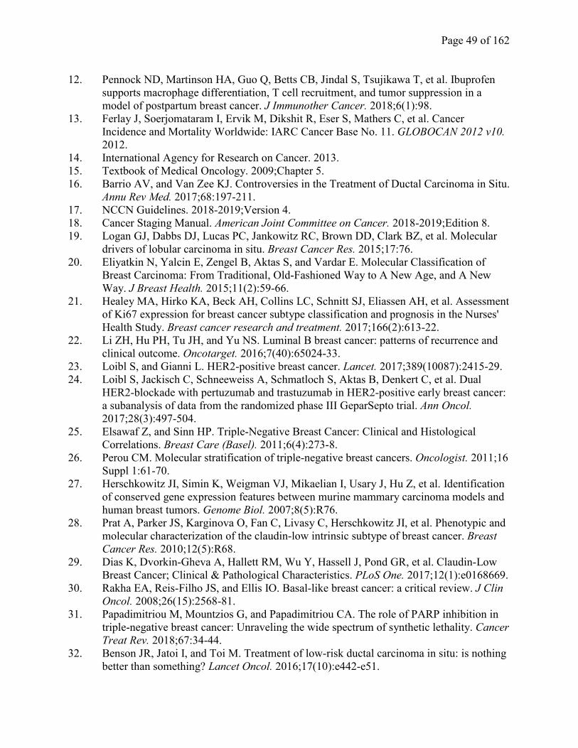
Page 49 of 162
12. Pennock ND, Martinson HA, Guo Q, Betts CB, Jindal S, Tsujikawa T, et al. Ibuprofen
supports macrophage differentiation, T cell recruitment, and tumor suppression in a
model of postpartum breast cancer. J Immunother Cancer. 2018;6(1):98.
13. Ferlay J, Soerjomataram I, Ervik M, Dikshit R, Eser S, Mathers C, et al. Cancer
Incidence and Mortality Worldwide: IARC Cancer Base No. 11. GLOBOCAN 2012 v10.
2012.
14. International Agency for Research on Cancer. 2013.
15. Textbook of Medical Oncology. 2009;Chapter 5.
16. Barrio AV, and Van Zee KJ. Controversies in the Treatment of Ductal Carcinoma in Situ.
Annu Rev Med. 2017;68:197-211.
17. NCCN Guidelines. 2018-2019;Version 4.
18. Cancer Staging Manual. American Joint Committee on Cancer. 2018-2019;Edition 8.
19. Logan GJ, Dabbs DJ, Lucas PC, Jankowitz RC, Brown DD, Clark BZ, et al. Molecular
drivers of lobular carcinoma in situ. Breast Cancer Res. 2015;17:76.
20. Eliyatkin N, Yalcin E, Zengel B, Aktas S, and Vardar E. Molecular Classification of
Breast Carcinoma: From Traditional, Old-Fashioned Way to A New Age, and A New
Way. J Breast Health. 2015;11(2):59-66.
21. Healey MA, Hirko KA, Beck AH, Collins LC, Schnitt SJ, Eliassen AH, et al. Assessment
of Ki67 expression for breast cancer subtype classification and prognosis in the Nurses'
Health Study. Breast cancer research and treatment. 2017;166(2):613-22.
22. Li ZH, Hu PH, Tu JH, and Yu NS. Luminal B breast cancer: patterns of recurrence and
clinical outcome. Oncotarget. 2016;7(40):65024-33.
23. Loibl S, and Gianni L. HER2-positive breast cancer. Lancet. 2017;389(10087):2415-29.
24. Loibl S, Jackisch C, Schneeweiss A, Schmatloch S, Aktas B, Denkert C, et al. Dual
HER2-blockade with pertuzumab and trastuzumab in HER2-positive early breast cancer:
a subanalysis of data from the randomized phase III GeparSepto trial. Ann Oncol.
2017;28(3):497-504.
25. Elsawaf Z, and Sinn HP. Triple-Negative Breast Cancer: Clinical and Histological
Correlations. Breast Care (Basel). 2011;6(4):273-8.
26. Perou CM. Molecular stratification of triple-negative breast cancers. Oncologist. 2011;16
Suppl 1:61-70.
27. Herschkowitz JI, Simin K, Weigman VJ, Mikaelian I, Usary J, Hu Z, et al. Identification
of conserved gene expression features between murine mammary carcinoma models and
human breast tumors. Genome Biol. 2007;8(5):R76.
28. Prat A, Parker JS, Karginova O, Fan C, Livasy C, Herschkowitz JI, et al. Phenotypic and
molecular characterization of the claudin-low intrinsic subtype of breast cancer. Breast
Cancer Res. 2010;12(5):R68.
29. Dias K, Dvorkin-Gheva A, Hallett RM, Wu Y, Hassell J, Pond GR, et al. Claudin-Low
Breast Cancer; Clinical & Pathological Characteristics. PLoS One. 2017;12(1):e0168669.
30. Rakha EA, Reis-Filho JS, and Ellis IO. Basal-like breast cancer: a critical review. J Clin
Oncol. 2008;26(15):2568-81.
31. Papadimitriou M, Mountzios G, and Papadimitriou CA. The role of PARP inhibition in
triple-negative breast cancer: Unraveling the wide spectrum of synthetic lethality. Cancer
Treat Rev. 2018;67:34-44.
32. Benson JR, Jatoi I, and Toi M. Treatment of low-risk ductal carcinoma in situ: is nothing
better than something? Lancet Oncol. 2016;17(10):e442-e51.
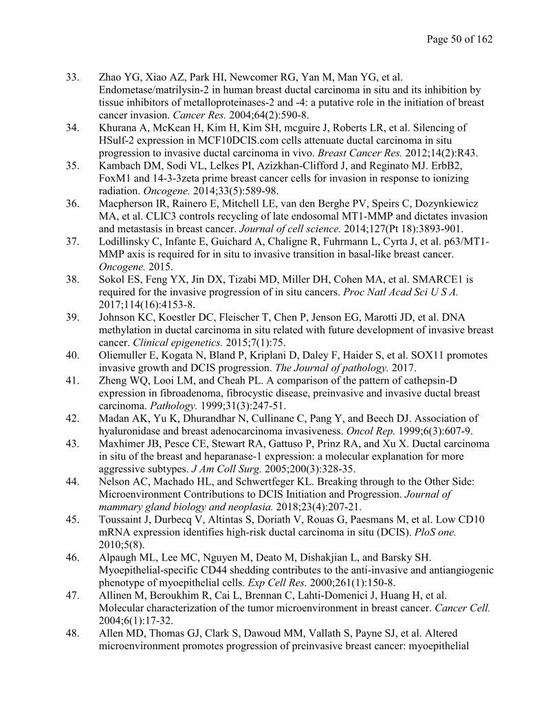
Page 50 of 162
33. Zhao YG, Xiao AZ, Park HI, Newcomer RG, Yan M, Man YG, et al.
Endometase/matrilysin-2 in human breast ductal carcinoma in situ and its inhibition by
tissue inhibitors of metalloproteinases-2 and -4: a putative role in the initiation of breast
cancer invasion. Cancer Res. 2004;64(2):590-8.
34. Khurana A, McKean H, Kim H, Kim SH, mcguire J, Roberts LR, et al. Silencing of
HSulf-2 expression in MCF10DCIS.com cells attenuate ductal carcinoma in situ
progression to invasive ductal carcinoma in vivo. Breast Cancer Res. 2012;14(2):R43.
35. Kambach DM, Sodi VL, Lelkes PI, Azizkhan-Clifford J, and Reginato MJ. ErbB2,
FoxM1 and 14-3-3zeta prime breast cancer cells for invasion in response to ionizing
radiation. Oncogene. 2014;33(5):589-98.
36. Macpherson IR, Rainero E, Mitchell LE, van den Berghe PV, Speirs C, Dozynkiewicz
MA, et al. CLIC3 controls recycling of late endosomal MT1-MMP and dictates invasion
and metastasis in breast cancer. Journal of cell science. 2014;127(Pt 18):3893-901.
37. Lodillinsky C, Infante E, Guichard A, Chaligne R, Fuhrmann L, Cyrta J, et al. p63/MT1-
MMP axis is required for in situ to invasive transition in basal-like breast cancer.
Oncogene. 2015.
38. Sokol ES, Feng YX, Jin DX, Tizabi MD, Miller DH, Cohen MA, et al. SMARCE1 is
required for the invasive progression of in situ cancers. Proc Natl Acad Sci U S A.
2017;114(16):4153-8.
39. Johnson KC, Koestler DC, Fleischer T, Chen P, Jenson EG, Marotti JD, et al. DNA
methylation in ductal carcinoma in situ related with future development of invasive breast
cancer. Clinical epigenetics. 2015;7(1):75.
40. Oliemuller E, Kogata N, Bland P, Kriplani D, Daley F, Haider S, et al. SOX11 promotes
invasive growth and DCIS progression. The Journal of pathology. 2017.
41. Zheng WQ, Looi LM, and Cheah PL. A comparison of the pattern of cathepsin-D
expression in fibroadenoma, fibrocystic disease, preinvasive and invasive ductal breast
carcinoma. Pathology. 1999;31(3):247-51.
42. Madan AK, Yu K, Dhurandhar N, Cullinane C, Pang Y, and Beech DJ. Association of
hyaluronidase and breast adenocarcinoma invasiveness. Oncol Rep. 1999;6(3):607-9.
43. Maxhimer JB, Pesce CE, Stewart RA, Gattuso P, Prinz RA, and Xu X. Ductal carcinoma
in situ of the breast and heparanase-1 expression: a molecular explanation for more
aggressive subtypes. J Am Coll Surg. 2005;200(3):328-35.
44. Nelson AC, Machado HL, and Schwertfeger KL. Breaking through to the Other Side:
Microenvironment Contributions to DCIS Initiation and Progression. Journal of
mammary gland biology and neoplasia. 2018;23(4):207-21.
45. Toussaint J, Durbecq V, Altintas S, Doriath V, Rouas G, Paesmans M, et al. Low CD10
mRNA expression identifies high-risk ductal carcinoma in situ (DCIS). PloS one.
2010;5(8).
46. Alpaugh ML, Lee MC, Nguyen M, Deato M, Dishakjian L, and Barsky SH.
Myoepithelial-specific CD44 shedding contributes to the anti-invasive and antiangiogenic
phenotype of myoepithelial cells. Exp Cell Res. 2000;261(1):150-8.
47. Allinen M, Beroukhim R, Cai L, Brennan C, Lahti-Domenici J, Huang H, et al.
Molecular characterization of the tumor microenvironment in breast cancer. Cancer Cell.
2004;6(1):17-32.
48. Allen MD, Thomas GJ, Clark S, Dawoud MM, Vallath S, Payne SJ, et al. Altered
microenvironment promotes progression of preinvasive breast cancer: myoepithelial
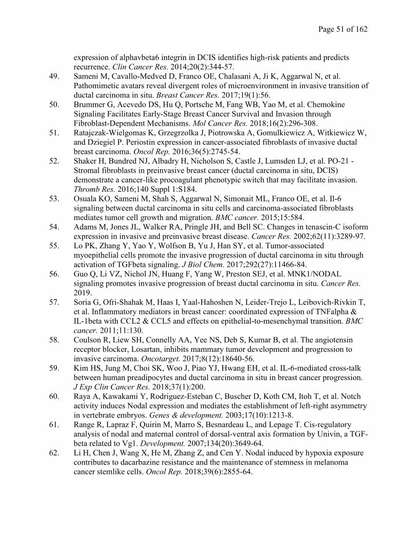
Page 51 of 162
expression of alphavbeta6 integrin in DCIS identifies high-risk patients and predicts
recurrence. Clin Cancer Res. 2014;20(2):344-57.
49. Sameni M, Cavallo-Medved D, Franco OE, Chalasani A, Ji K, Aggarwal N, et al.
Pathomimetic avatars reveal divergent roles of microenvironment in invasive transition of
ductal carcinoma in situ. Breast Cancer Res. 2017;19(1):56.
50. Brummer G, Acevedo DS, Hu Q, Portsche M, Fang WB, Yao M, et al. Chemokine
Signaling Facilitates Early-Stage Breast Cancer Survival and Invasion through
Fibroblast-Dependent Mechanisms. Mol Cancer Res. 2018;16(2):296-308.
51. Ratajczak-Wielgomas K, Grzegrzolka J, Piotrowska A, Gomulkiewicz A, Witkiewicz W,
and Dziegiel P. Periostin expression in cancer-associated fibroblasts of invasive ductal
breast carcinoma. Oncol Rep. 2016;36(5):2745-54.
52. Shaker H, Bundred NJ, Albadry H, Nicholson S, Castle J, Lumsden LJ, et al. PO-21 -
Stromal fibroblasts in preinvasive breast cancer (ductal carcinoma in situ, DCIS)
demonstrate a cancer-like procoagulant phenotypic switch that may facilitate invasion.
Thromb Res. 2016;140 Suppl 1:S184.
53. Osuala KO, Sameni M, Shah S, Aggarwal N, Simonait ML, Franco OE, et al. Il-6
signaling between ductal carcinoma in situ cells and carcinoma-associated fibroblasts
mediates tumor cell growth and migration. BMC cancer. 2015;15:584.
54. Adams M, Jones JL, Walker RA, Pringle JH, and Bell SC. Changes in tenascin-C isoform
expression in invasive and preinvasive breast disease. Cancer Res. 2002;62(11):3289-97.
55. Lo PK, Zhang Y, Yao Y, Wolfson B, Yu J, Han SY, et al. Tumor-associated
myoepithelial cells promote the invasive progression of ductal carcinoma in situ through
activation of TGFbeta signaling. J Biol Chem. 2017;292(27):11466-84.
56. Guo Q, Li VZ, Nichol JN, Huang F, Yang W, Preston SEJ, et al. MNK1/NODAL
signaling promotes invasive progression of breast ductal carcinoma in situ. Cancer Res.
2019.
57. Soria G, Ofri-Shahak M, Haas I, Yaal-Hahoshen N, Leider-Trejo L, Leibovich-Rivkin T,
et al. Inflammatory mediators in breast cancer: coordinated expression of TNFalpha &
IL-1beta with CCL2 & CCL5 and effects on epithelial-to-mesenchymal transition. BMC
cancer. 2011;11:130.
58. Coulson R, Liew SH, Connelly AA, Yee NS, Deb S, Kumar B, et al. The angiotensin
receptor blocker, Losartan, inhibits mammary tumor development and progression to
invasive carcinoma. Oncotarget. 2017;8(12):18640-56.
59. Kim HS, Jung M, Choi SK, Woo J, Piao YJ, Hwang EH, et al. IL-6-mediated cross-talk
between human preadipocytes and ductal carcinoma in situ in breast cancer progression.
J Exp Clin Cancer Res. 2018;37(1):200.
60. Raya A, Kawakami Y, Rodriguez-Esteban C, Buscher D, Koth CM, Itoh T, et al. Notch
activity induces Nodal expression and mediates the establishment of left-right asymmetry
in vertebrate embryos. Genes & development. 2003;17(10):1213-8.
61. Range R, Lapraz F, Quirin M, Marro S, Besnardeau L, and Lepage T. Cis-regulatory
analysis of nodal and maternal control of dorsal-ventral axis formation by Univin, a TGF-
beta related to Vg1. Development. 2007;134(20):3649-64.
62. Li H, Chen J, Wang X, He M, Zhang Z, and Cen Y. Nodal induced by hypoxia exposure
contributes to dacarbazine resistance and the maintenance of stemness in melanoma
cancer stemlike cells. Oncol Rep. 2018;39(6):2855-64.
Page 52 of 162
63. Nettersheim D, Jostes S, Sharma R, Schneider S, Hofmann A, Ferreira HJ, et al. BMP
Inhibition in Seminomas Initiates Acquisition of Pluripotency via NODAL Signaling
Resulting in Reprogramming to an Embryonal Carcinoma. PLoS genetics.
2015;11(7):e1005415.
64. Jaaks P, and Bernasconi M. The proprotein convertase furin in tumour progression.
International journal of cancer Journal international du cancer. 2017;141(4):654-63.
65. Wang F, Wang L, and Pan J. PACE4 regulates proliferation, migration and invasion in
human breast cancer MDA-MB-231 cells. Molecular medicine reports. 2015;11(1):698-
704.
66. Kalyan A, Carneiro BA, Chandra S, Kaplan J, Chae YK, Matsangou M, et al. Nodal
Signaling as a Developmental Therapeutics Target in Oncology. Mol Cancer Ther.
2017;16(5):787-92.
67. Kaya H, Erbarut I, Ozkan N, Bekiroglu N, Sen S, and Abaciglu U. Immunoexpression of
HER family, neuregulin, MAPK and AKT in invasive ductal carcinomas of the breast.
Eur J Gynaecol Oncol. 2008;29(4):350-6.
68. Pearson GW, and Hunter T. PI-3 kinase activity is necessary for ERK1/2-induced
disruption of mammary epithelial architecture. Breast Cancer Res. 2009;11(3):R29.
69. Novitskaya V, Romanska H, Dawoud M, Jones JL, and Berditchevski F. Tetraspanin
CD151 regulates growth of mammary epithelial cells in three-dimensional extracellular
matrix: implication for mammary ductal carcinoma in situ. Cancer Res.
2010;70(11):4698-708.
70. Nam JM, Ahmed KM, Costes S, Zhang H, Onodera Y, Olshen AB, et al. beta1-Integrin
via NF-kappaB signaling is essential for acquisition of invasiveness in a model of
radiation treated in situ breast cancer. Breast Cancer Res. 2013;15(4):R60.
71. Jacob T, Gray JW, Troxell M, and Vu TQ. Multiplexed imaging reveals heterogeneity of
PI3K/MAPK network signaling in breast lesions of known PIK3CA genotype. Breast
cancer research and treatment. 2016;159(3):575-83.
72. Kim HS, Jung M, Choi SK, Moon WK, and Kim SJ. Different Biological Action of Oleic
Acid in ALDHhigh and ALDHlow Subpopulations Separated from Ductal Carcinoma In
Situ of Breast Cancer. PloS one. 2016;11(9):e0160835.
73. Lian K, Ma C, Hao C, Li Y, Zhang N, Chen YH, et al. TIPE3 protein promotes breast
cancer metastasis through activating AKT and NF-kappaB signaling pathways.
Oncotarget. 2017;8(30):48889-904.
74. Al-Qassab Y, Grassilli S, Brugnoli F, Vezzali F, Capitani S, and Bertagnolo V. Protective
role of all-trans retinoic acid (ATRA) against hypoxia-induced malignant potential of
non-invasive breast tumor derived cells. BMC cancer. 2018;18(1):1194.
75. Souzaki M, Kubo M, Kai M, Kameda C, Tanaka H, Taguchi T, et al. Hedgehog signaling
pathway mediates the progression of non-invasive breast cancer to invasive breast cancer.
Cancer science. 2011;102(2):373-81.
76. Elsarraj HS, Hong Y, Valdez KE, Michaels W, Hook M, Smith WP, et al. Expression
profiling of in vivo ductal carcinoma in situ progression models identified B cell
lymphoma-9 as a molecular driver of breast cancer invasion. Breast Cancer Res.
2015;17:128.
77. Liu WS, Chan SH, Chang HT, Li GC, Tu YT, Tseng HH, et al. Isocitrate dehydrogenase
1-snail axis dysfunction significantly correlates with breast cancer prognosis and
regulates cell invasion ability. Breast Cancer Res. 2018;20(1):25.
Page 53 of 162
78. Krstic M, Kolendowski B, Cecchini MJ, Postenka CO, Hassan HM, Andrews J, et al.
TBX3 promotes progression of pre-invasive breast cancer cells by inducing EMT and
directly up-regulating SLUG. The Journal of pathology. 2019.
79. Abba MC, Gong T, Lu Y, Lee J, Zhong Y, Lacunza E, et al. A Molecular Portrait of
High-Grade Ductal Carcinoma In Situ. Cancer Res. 2015;75(18):3980-90.
80. Knudsen ES, Dervishaj O, Kleer CG, Pajak T, Schwartz GF, and Witkiewicz AK. EZH2
and ALDH1 expression in ductal carcinoma in situ: complex association with recurrence
and progression to invasive breast cancer. Cell Cycle. 2013;12(13):2042-50.
81. Li Q, Eades G, Yao Y, Zhang Y, and Zhou Q. Characterization of a stem-like
subpopulation in basal-like ductal carcinoma in situ (DCIS) lesions. J Biol Chem.
2014;289(3):1303-12.
82. Kristensen VN, Vaske CJ, Ursini-Siegel J, Van Loo P, Nordgard SH, Sachidanandam R,
et al. Integrated molecular profiles of invasive breast tumors and ductal carcinoma in situ
(DCIS) reveal differential vascular and interleukin signaling. Proc Natl Acad Sci U S A.
2012;109(8):2802-7.
83. Gil Del Alcazar CR, Huh SJ, Ekram MB, Trinh A, Liu LL, Beca F, et al. Immune Escape
in Breast Cancer During In Situ to Invasive Carcinoma Transition. Cancer discovery.
2017;7(10):1098-115.
84. Miligy I, Mohan P, Gaber A, Aleskandarany MA, Nolan CC, Diez-Rodriguez M, et al.
Prognostic significance of tumour infiltrating B lymphocytes in breast ductal carcinoma
in situ. Histopathology. 2017;71(2):258-68.
85. Thirkettle S, Decock J, Arnold H, Pennington CJ, Jaworski DM, and Edwards DR.
Matrix metalloproteinase 8 (collagenase 2) induces the expression of interleukins 6 and 8
in breast cancer cells. J Biol Chem. 2013;288(23):16282-94.
86. Hendry S, Pang JB, Byrne DJ, Lakhani SR, Cummings MC, Campbell IG, et al.
Relationship of the Breast Ductal Carcinoma In Situ Immune Microenvironment with
Clinicopathological and Genetic Features. Clin Cancer Res. 2017;23(17):5210-7.
87. Stensheim H, Moller B, van Dijk T, and Fossa SD. Cause-specific survival for women
diagnosed with cancer during pregnancy or lactation: a registry-based cohort study. J
Clin Oncol. 2009;27(1):45-51.
88. Johansson AL, Andersson TM, Hsieh CC, Cnattingius S, and Lambe M. Increased
mortality in women with breast cancer detected during pregnancy and different periods
postpartum. Cancer Epidemiol Biomarkers Prev. 2011;20(9):1865-72.
89. Callihan EB, Gao D, Jindal S, Lyons TR, Manthey E, Edgerton S, et al. Postpartum
diagnosis demonstrates a high risk for metastasis and merits an expanded definition of
pregnancy-associated breast cancer. Breast cancer research and treatment.
2013;138(2):549-59.
90. O'Brien J, Martinson H, Durand-Rougely C, and Schedin P. Macrophages are crucial for
epithelial cell death and adipocyte repopulation during mammary gland involution.
Development. 2012;139(2):269-75.
91. Guo Q, Minnier J, Burchard J, Chiotti K, Spellman P, and Schedin P. Physiologically
activated mammary fibroblasts promote postpartum mammary cancer. JCI Insight.
2017;2(6):e89206.
92. Bertolami C, and Donoff RB. The effect of full-thickness skin grafts on the actomyosin
content of contracting wounds. J Oral Surg. 1979;37(7):471-6.
Page 54 of 162
93. Kalluri R. The biology and function of fibroblasts in cancer. Nature reviews Cancer.
2016;16(9):582-98.
94. Li B, and Wang JH. Fibroblasts and myofibroblasts in wound healing: force generation
and measurement. J Tissue Viability. 2011;20(4):108-20.
95. Cohen N, Shani O, Raz Y, Sharon Y, Hoffman D, Abramovitz L, et al. Fibroblasts drive
an immunosuppressive and growth-promoting microenvironment in breast cancer via
secretion of Chitinase 3-like 1. Oncogene. 2017;36(31):4457-68.
96. Holt DM, Ma X, Kundu N, Collin PD, and Fulton AM. Modulation of host natural killer
cell functions in breast cancer via prostaglandin E2 receptors EP2 and EP4. J
Immunother. 2012;35(2):179-88.
97. Markosyan N, Chen EP, and Smyth EM. Targeting COX-2 abrogates mammary
tumorigenesis: Breaking cancer-associated suppression of immunosurveillance.
Oncoimmunology. 2014;3:e29287.
98. Basudhar D, Glynn SA, Greer M, Somasundaram V, No JH, Scheiblin DA, et al.
Coexpression of NOS2 and COX2 accelerates tumor growth and reduces survival in
estrogen receptor-negative breast cancer. Proc Natl Acad Sci U S A. 2017;114(49):13030-
5.
99. Tian J, Hachim MY, Hachim IY, Dai M, Lo C, Raffa FA, et al. Cyclooxygenase-2
regulates TGFbeta-induced cancer stemness in triple-negative breast cancer. Sci Rep.
2017;7:40258.
100. Li A, Chen P, Leng Y, and Kang J. Histone deacetylase 6 regulates the
immunosuppressive properties of cancer-associated fibroblasts in breast cancer through
the STAT3-COX2-dependent pathway. Oncogene. 2018;37(45):5952-66.
101. Borges VF, Elder AM, and Lyons TR. Deciphering Pro-Lymphangiogenic Programs
during Mammary Involution and Postpartum Breast Cancer. Front Oncol. 2016;6:227.
102. Lyons TR, Borges VF, Betts CB, Guo Q, Kapoor P, Martinson HA, et al.
Cyclooxygenase-2-dependent lymphangiogenesis promotes nodal metastasis of
postpartum breast cancer. The Journal of clinical investigation. 2014;124(9):3901-12.
103. Mantovani A, Sica A, Allavena P, Garlanda C, and Locati M. Tumor-associated
macrophages and the related myeloid-derived suppressor cells as a paradigm of the
diversity of macrophage activation. Human immunology. 2009;70(5):325-30.
104. Stanford JC, Young C, Hicks D, Owens P, Williams A, Vaught DB, et al. Efferocytosis
produces a prometastatic landscape during postpartum mammary gland involution. The
Journal of clinical investigation. 2014;124(11):4737-52.
105. Qian BZ, and Pollard JW. Macrophage diversity enhances tumor progression and
metastasis. Cell. 2010;141(1):39-51.
106. Talmadge JE, and Gabrilovich DI. History of myeloid-derived suppressor cells. Nature
reviews Cancer. 2013;13(10):739-52.
107. Gantt S, Gervassi A, Jaspan H, and Horton H. The role of myeloid-derived suppressor
cells in immune ontogeny. Front Immunol. 2014;5:387.
108. Peng D, Tanikawa T, Li W, Zhao L, Vatan L, Szeliga W, et al. Myeloid-Derived
Suppressor Cells Endow Stem-like Qualities to Breast Cancer Cells through IL6/STAT3
and NO/NOTCH Cross-talk Signaling. Cancer Res. 2016;76(11):3156-65.
109. Gonda K, Shibata M, Ohtake T, Matsumoto Y, Tachibana K, Abe N, et al. Myeloid-
derived suppressor cells are increased and correlated with type 2 immune responses,
Page 55 of 162
malnutrition, inflammation, and poor prognosis in patients with breast cancer. Oncol Lett.
2017;14(2):1766-74.
110. Safarzadeh E, Hashemzadeh S, Duijf PHG, Mansoori B, Khaze V, Mohammadi A, et al.
Circulating myeloid-derived suppressor cells: An independent prognostic factor in
patients with breast cancer. Journal of cellular physiology. 2018.
111. Li W, Tanikawa T, Kryczek I, Xia H, Li G, Wu K, et al. Aerobic Glycolysis Controls
Myeloid-Derived Suppressor Cells and Tumor Immunity via a Specific CEBPB Isoform
in Triple-Negative Breast Cancer. Cell metabolism. 2018;28(1):87-103 e6.
112. Yang L, Huang J, Ren X, Gorska AE, Chytil A, Aakre M, et al. Abrogation of TGF beta
signaling in mammary carcinomas recruits Gr-1+CD11b+ myeloid cells that promote
metastasis. Cancer Cell. 2008;13(1):23-35.
113. Molinier-Frenkel V, and Castellano F. Immunosuppressive enzymes in the tumor
microenvironment. FEBS Lett. 2017;591(19):3135-57.
114. Secondini C, Coquoz O, Spagnuolo L, Spinetti T, Peyvandi S, Ciarloni L, et al. Arginase
inhibition suppresses lung metastasis in the 4T1 breast cancer model independently of the
immunomodulatory and anti-metastatic effects of VEGFR-2 blockade. Oncoimmunology.
2017;6(6):e1316437.
115. Stiff A, Trikha P, Mundy-Bosse B, McMichael E, Mace TA, Benner B, et al. Nitric
Oxide Production by Myeloid-Derived Suppressor Cells Plays a Role in Impairing Fc
Receptor-Mediated Natural Killer Cell Function. Clin Cancer Res. 2018;24(8):1891-904.
116. Susskind BM, Sekar J, Tandon PM, Lind DS, and Bear HD. Differential sensitivity of
cytotoxic T lymphocytes and lymphokine-activated killer cells to inhibition by L-
ornithine. Cell Immunol. 1991;133(1):41-54.
117. Prendergast GC, Smith C, Thomas S, Mandik-Nayak L, Laury-Kleintop L, Metz R, et al.
Indoleamine 2,3-dioxygenase pathways of pathogenic inflammation and immune escape
in cancer. Cancer Immunol Immunother. 2014;63(7):721-35.
118. Li T, Yang Y, Hua X, Wang G, Liu W, Jia C, et al. Hepatocellular carcinoma-associated
fibroblasts trigger NK cell dysfunction via PGE2 and IDO. Cancer Lett.
2012;318(2):154-61.
119. Abdel-Wahab O, Patel J, and Levine RL. Clinical implications of novel mutations in
epigenetic modifiers in AML. Hematology/oncology clinics of North America.
2011;25(6):1119-33.
120. Zelante T, and Ricciardi-Castagnoli P. The yin-yang nature of CSF1R-binding cytokines.
Nature immunology. 2012;13(8):717-9.
121. De Henau O, Rausch M, Winkler D, Campesato LF, Liu C, Cymerman DH, et al.
Overcoming resistance to checkpoint blockade therapy by targeting PI3Kgamma in
myeloid cells. Nature. 2016;539(7629):443-7.
122. Kumar V, Donthireddy L, Marvel D, Condamine T, Wang F, Lavilla-Alonso S, et al.
Cancer-Associated Fibroblasts Neutralize the Anti-tumor Effect of CSF1 Receptor
Blockade by Inducing PMN-MDSC Infiltration of Tumors. Cancer Cell. 2017;32(5):654-
68 e5.
123. Chiossone L, and Vivier E. Immune checkpoints on innate lymphoid cells. The Journal of
experimental medicine. 2017;214(6):1561-3.
124. Mattner J, and Wirtz S. Friend or Foe? The Ambiguous Role of Innate Lymphoid Cells in
Cancer Development. Trends in immunology. 2017;38(1):29-38.
Page 56 of 162
125. Li J, Razumilava N, Gores GJ, Walters S, Mizuochi T, Mourya R, et al. Biliary repair and
carcinogenesis are mediated by IL-33-dependent cholangiocyte proliferation. The Journal
of clinical investigation. 2014;124(7):3241-51.
126. Chevalier MF, Trabanelli S, Racle J, Salome B, Cesson V, Gharbi D, et al. ILC2-
modulated T cell-to-MDSC balance is associated with bladder cancer recurrence. The
Journal of clinical investigation. 2017;127(8):2916-29.
127. Salimi M, Wang R, Yao X, Li X, Wang X, Hu Y, et al. Activated innate lymphoid cell
populations accumulate in human tumour tissues. BMC cancer. 2018;18(1):341.
128. von Moltke J, and Locksley RM. I-L-C-2 it: type 2 immunity and group 2 innate
lymphoid cells in homeostasis. Current opinion in immunology. 2014;31:58-65.
129. Jovanovic IP, Pejnovic NN, Radosavljevic GD, Pantic JM, Milovanovic MZ, Arsenijevic
NN, et al. Interleukin-33/ST2 axis promotes breast cancer growth and metastases by
facilitating intratumoral accumulation of immunosuppressive and innate lymphoid cells.
International journal of cancer Journal international du cancer. 2014;134(7):1669-82.
130. Gardner A, and Ruffell B. Dendritic Cells and Cancer Immunity. Trends in immunology.
2016;37(12):855-65.
131. Warri A, Cook KL, Hu R, Jin L, Zwart A, Soto-Pantoja DR, et al. Autophagy and
unfolded protein response (UPR) regulate mammary gland involution by restraining
apoptosis-driven irreversible changes. Cell Death Discov. 2018;4:40.
132. Pitt JM, Vetizou M, Daillere R, Roberti MP, Yamazaki T, Routy B, et al. Resistance
Mechanisms to Immune-Checkpoint Blockade in Cancer: Tumor-Intrinsic and -Extrinsic
Factors. Immunity. 2016;44(6):1255-69.
133. Corrales L, and Gajewski TF. Molecular Pathways: Targeting the Stimulator of
Interferon Genes (STING) in the Immunotherapy of Cancer. Clin Cancer Res.
2015;21(21):4774-9.
134. Henle AM, Nassar A, Puglisi-Knutson D, Youssef B, and Knutson KL. Downregulation
of TAP1 and TAP2 in early stage breast cancer. PloS one. 2017;12(11):e0187323.
135. Sultan M, Vidovic D, Paine AS, Huynh TT, Coyle KM, Thomas ML, et al. Epigenetic
Silencing of TAP1 in Aldefluor(+) Breast Cancer Stem Cells Contributes to Their
Enhanced Immune Evasion. Stem cells. 2018;36(5):641-54.
136. Golubovskaya V, and Wu L. Different Subsets of T Cells, Memory, Effector Functions,
and CAR-T Immunotherapy. Cancers. 2016;8(3).
137. Patel SA, and Minn AJ. Combination Cancer Therapy with Immune Checkpoint
Blockade: Mechanisms and Strategies. Immunity. 2018;48(3):417-33.
138. Disis ML. Immune regulation of cancer. J Clin Oncol. 2010;28(29):4531-8.
139. Borst J, Ahrends T, Babala N, Melief CJM, and Kastenmuller W. CD4(+) T cell help in
cancer immunology and immunotherapy. Nature reviews Immunology. 2018;18(10):635-
47.
140. Wherry EJ. T cell exhaustion. Nature immunology. 2011;12(6):492-9.
141. Sharma P, and Allison JP. The future of immune checkpoint therapy. Science.
2015;348(6230):56-61.
142. Plitas G, Konopacki C, Wu K, Bos PD, Morrow M, Putintseva EV, et al. Regulatory T
Cells Exhibit Distinct Features in Human Breast Cancer. Immunity. 2016;45(5):1122-34.
143. Li Z, Dong P, Ren M, Song Y, Qian X, Yang Y, et al. PD-L1 Expression Is Associated
with Tumor FOXP3(+) Regulatory T-Cell Infiltration of Breast Cancer and Poor
Prognosis of Patient. Journal of Cancer. 2016;7(7):784-93.
Page 57 of 162
144. Bhat M, Robichaud N, Hulea L, Sonenberg N, Pelletier J, and Topisirovic I. Targeting
the translation machinery in cancer. Nature reviews Drug discovery. 2015;14(4):261-78.
145. Pelletier J, Graff J, Ruggero D, and Sonenberg N. Targeting the eIF4F translation
initiation complex: a critical nexus for cancer development. Cancer Res. 2015;75(2):250-
63.
146. Li Q, Imataka H, Morino S, Rogers GW, Jr., Richter-Cook NJ, Merrick WC, et al.
Eukaryotic translation initiation factor 4AIII (eIF4AIII) is functionally distinct from
eIF4AI and eIF4AII. Molecular and cellular biology. 1999;19(11):7336-46.
147. Castello A, Alvarez E, and Carrasco L. Differential cleavage of eIF4GI and eIF4GII in
mammalian cells. Effects on translation. J Biol Chem. 2006;281(44):33206-16.
148. Mahalingam M, and Cooper JA. Phosphorylation of mammalian eIF4E by Mnk1 and
Mnk2: tantalizing prospects for a role in translation. Prog Mol Subcell Biol. 2001;27:132-
42.
149. Ueda T, Watanabe-Fukunaga R, Fukuyama H, Nagata S, and Fukunaga R. Mnk2 and
Mnk1 are essential for constitutive and inducible phosphorylation of eukaryotic initiation
factor 4E but not for cell growth or development. Molecular and cellular biology.
2004;24(15):6539-49.
150. O'Loghlen A, Gonzalez VM, Pineiro D, Perez-Morgado MI, Salinas M, and Martin ME.
Identification and molecular characterization of Mnk1b, a splice variant of human MAP
kinase-interacting kinase Mnk1. Exp Cell Res. 2004;299(2):343-55.
151. O'Loghlen A, Gonzalez VM, Jurado T, Salinas M, and Martin ME. Characterization of
the activity of human MAP kinase-interacting kinase Mnk1b. Biochimica et biophysica
acta. 2007;1773(9):1416-27.
152. Scheper GC, Parra JL, Wilson M, Van Kollenburg B, Vertegaal AC, Han ZG, et al. The
N and C termini of the splice variants of the human mitogen-activated protein kinase-
interacting kinase Mnk2 determine activity and localization. Molecular and cellular
biology. 2003;23(16):5692-705.
153. Waskiewicz AJ, Flynn A, Proud CG, and Cooper JA. Mitogen-activated protein kinases
activate the serine/threonine kinases Mnk1 and Mnk2. The EMBO journal.
1997;16(8):1909-20.
154. Proud CG. Mnks, eIF4E phosphorylation and cancer. Biochimica et biophysica acta.
2015;1849(7):766-73.
155. Maimon A, Mogilevsky M, Shilo A, Golan-Gerstl R, Obiedat A, Ben-Hur V, et al. Mnk2
Alternative Splicing Modulates the p38-MAPK Pathway and Impacts Ras-Induced
Transformation. Cell reports. 2014;7(2):501-13.
156. Scheper GC, and Proud CG. Does phosphorylation of the cap-binding protein eIF4E play
a role in translation initiation? Eur J Biochem. 2002;269(22):5350-9.
157. Furic L, Rong L, Larsson O, Koumakpayi IH, Yoshida K, Brueschke A, et al. eIF4E
phosphorylation promotes tumorigenesis and is associated with prostate cancer
progression. Proc Natl Acad Sci U S A. 2010;107(32):14134-9.
158. Robichaud N, Del Rincon SV, Huor B, Alain T, Petruccelli LA, Hearnden J, et al.
Phosphorylation of eIF4E promotes EMT and metastasis via translational control of
SNAIL and MMP-3. 2014.
159. Pettersson F, Del Rincon SV, Emond A, Huor B, Ngan E, Ng J, et al. Genetic and
pharmacologic inhibition of eIF4E reduces breast cancer cell migration, invasion, and
metastasis. Cancer Res. 2015;75(6):1102-12.
Page 58 of 162
160. Li Z, Sun Y, Qu M, Wan H, Cai F, and Zhang P. Inhibiting the MNK-eIF4E-beta-catenin
axis increases the responsiveness of aggressive breast cancer cells to chemotherapy.
Oncotarget. 2017;8(2):2906-15.
161. Zhou S, Wang GP, Liu C, and Zhou M. Eukaryotic initiation factor 4E (eIF4E) and
angiogenesis: prognostic markers for breast cancer. BMC cancer. 2006;6:231.
162. Pettersson F, Yau C, Dobocan MC, Culjkovic-Kraljacic B, Retrouvey H, Puckett R, et al.
Ribavirin treatment effects on breast cancers overexpressing eIF4E, a biomarker with
prognostic specificity for luminal B-type breast cancer. Clin Cancer Res.
2011;17(9):2874-84.
163. Mehrotra S, Sharma B, Joshi S, Kroczynska B, Majchrzak B, Stein BL, et al. Essential
role for the Mnk pathway in the inhibitory effects of type I interferons on
myeloproliferative neoplasm (MPN) precursors. J Biol Chem. 2013;288(33):23814-22.
164. Herdy B, Jaramillo M, Svitkin YV, Rosenfeld AB, Kobayashi M, Walsh D, et al.
Translational control of the activation of transcription factor NF-kappaB and production
of type I interferon by phosphorylation of the translation factor eIF4E. Nature
immunology. 2012;13(6):543-50.
165. Robichaud N, Hsu BE, Istomine R, Alvarez F, Blagih J, Ma EH, et al. Translational
control in the tumor microenvironment promotes lung metastasis: Phosphorylation of
eIF4E in neutrophils. Proc Natl Acad Sci U S A. 2018;115(10):E2202-E9.
166. Cerezo M, Guemiri R, Druillennec S, Girault I, Malka-Mahieu H, Shen S, et al.
Translational control of tumor immune escape via the eIF4F-STAT1-PD-L1 axis in
melanoma. Nature medicine. 2018;24(12):1877-86.
167. DaSilva J, Xu L, Kim HJ, Miller WT, and Bar-Sagi D. Regulation of sprouty stability by
Mnk1-dependent phosphorylation. Molecular and cellular biology. 2006;26(5):1898-907.
168. Guil S, Long JC, and Caceres JF. hnRNP A1 relocalization to the stress granules reflects
a role in the stress response. Molecular and cellular biology. 2006;26(15):5744-58.
169. Buxade M, Morrice N, Krebs DL, and Proud CG. The PSF.p54nrb complex is a novel
Mnk substrate that binds the mRNA for tumor necrosis factor alpha. J Biol Chem.
2008;283(1):57-65.
170. Loh TJ, Moon H, Cho S, Jang H, Liu YC, Tai H, et al. CD44 alternative splicing and
hnRNP A1 expression are associated with the metastasis of breast cancer. Oncol Rep.
2015;34(3):1231-8.
171. Roy R, Huang Y, Seckl MJ, and Pardo OE. Emerging roles of hnRNPA1 in modulating
malignant transformation. Wiley interdisciplinary reviews RNA. 2017;8(6).
172. Faratian D, Sims AH, Mullen P, Kay C, Um I, Langdon SP, et al. Sprouty 2 is an
independent prognostic factor in breast cancer and may be useful in stratifying patients
for trastuzumab therapy. PloS one. 2011;6(8):e23772.
173. Konicek BW, Stephens JR, McNulty AM, Robichaud N, Peery RB, Dumstorf CA, et al.
Therapeutic inhibition of MAP kinase interacting kinase blocks eukaryotic initiation
factor 4E phosphorylation and suppresses outgrowth of experimental lung metastases.
Cancer Res. 2011;71(5):1849-57.
174. Ramalingam S, Gediya L, Kwegyir-Afful AK, Ramamurthy VP, Purushottamachar P,
Mbatia H, et al. First MNKs degrading agents block phosphorylation of eIF4E, induce
apoptosis, inhibit cell growth, migration and invasion in triple negative and Her2-
overexpressing breast cancer cell lines. Oncotarget. 2014;5(2):530-43.
Page 59 of 162
175. Xu J, Chen A, Joy J, Xavier VJ, Ong EH, Hill J, et al. Rational design of resorcylic acid
lactone analogues as covalent MNK1/2 kinase inhibitors by tuning the reactivity of an
enamide Michael acceptor. ChemMedChem. 2013;8(9):1483-94.
176. Dreas A, Mikulski M, Milik M, Fabritius CH, Brzozka K, and Rzymski T. Mitogen-
activated Protein Kinase (MAPK) Interacting Kinases 1 and 2 (MNK1 and MNK2) as
Targets for Cancer Therapy: Recent Progress in the Development of MNK Inhibitors.
Curr Med Chem. 2017;24(28):3025-53.
177. Zhan Y, Guo J, Yang W, Goncalves C, Rzymski T, Dreas A, et al. MNK1/2 inhibition
limits oncogenicity and metastasis of KIT-mutant melanoma. The Journal of clinical
investigation. 2017.
178. Reich SH, Sprengeler PA, Chiang GG, Appleman JR, Chen J, Clarine J, et al. Structure-
based Design of Pyridone-Aminal eFT508 Targeting Dysregulated Translation by
Selective Mitogen-activated Protein Kinase Interacting Kinases 1 and 2 (MNK1/2)
Inhibition. J Med Chem. 2018;61(8):3516-40.
179. Xu Y, Poggio M, Jin HY, Shi Z, Forester CM, Wang Y, et al. Translation control of the
immune checkpoint in cancer and its therapeutic targeting. Nature medicine.
2019;25(2):301-11.
180. Janku F, Yap TA, and Meric-Bernstam F. Targeting the PI3K pathway in cancer: are we
making headway? Nature reviews Clinical oncology. 2018;15(5):273-91.
181. Bianchini A, Loiarro M, Bielli P, Busa R, Paronetto MP, Loreni F, et al. Phosphorylation
of eIF4E by MNKs supports protein synthesis, cell cycle progression and proliferation in
prostate cancer cells. Carcinogenesis. 2008;29(12):2279-88.
182. Grzmil M, Huber RM, Hess D, Frank S, Hynx D, Moncayo G, et al. MNK1 pathway
activity maintains protein synthesis in rapalog-treated gliomas. The Journal of clinical
investigation. 2014;124(2):742-54.
183. Lineham E, Tizzard GJ, Coles SJ, Spencer J, and Morley SJ. Synergistic effects of
inhibiting the MNK-eIF4E and PI3K/AKT/ mTOR pathways on cell migration in MDA-
MB-231 cells. Oncotarget. 2018;9(18):14148-59.
184. Wang X, Yue P, Chan CB, Ye K, Ueda T, Watanabe-Fukunaga R, et al. Inhibition of
mammalian target of rapamycin induces phosphatidylinositol 3-kinase-dependent and
Mnk-mediated eukaryotic translation initiation factor 4E phosphorylation. Molecular and
cellular biology. 2007;27(21):7405-13.
185. Brown MC, and Gromeier M. MNK Controls mTORC1:Substrate Association through
Regulation of TELO2 Binding with mTORC1. Cell reports. 2017;18(6):1444-57.
186. Katsha A, Wang L, Arras J, Omar OM, Ecsedy J, Belkhiri A, et al. Activation of EIF4E
by Aurora Kinase A Depicts a Novel Druggable Axis in Everolimus-Resistant Cancer
Cells. Clin Cancer Res. 2017;23(14):3756-68.
187. Eckerdt F, Beauchamp E, Bell J, Iqbal A, Su B, Fukunaga R, et al. Regulatory effects of a
Mnk2-eIF4E feedback loop during mTORC1 targeting of human medulloblastoma cells.
Oncotarget. 2014;5(18):8442-51.
188. Wen Q, Wang W, Luo J, Chu S, Chen L, Xu L, et al. CGP57380 enhances efficacy of
RAD001 in non-small cell lung cancer through abrogating mTOR inhibition-induced
phosphorylation of eIF4E and activating mitochondrial apoptotic pathway. Oncotarget.
2016;7(19):27787-801.
189. Huang XB, Yang CM, Han QM, Ye XJ, Lei W, and Qian WB. MNK1 inhibitor
CGP57380 overcomes mTOR inhibitor-induced activation of eIF4E: the mechanism of

Page 60 of 162
synergic killing of human T-ALL cells. Acta pharmacologica Sinica. 2018;39(12):1894-
901.
190. Geter PA, Ernlund AW, Bakogianni S, Alard A, Arju R, Giashuddin S, et al. Hyperactive
mTOR and MNK1 phosphorylation of eIF4E confer tamoxifen resistance and estrogen
independence through selective mRNA translation reprogramming. Genes &
development. 2017;31(22):2235-49.
Page 61 of 162
Chapter 2. MNK1/NODAL signaling promotes the invasive progression of breast
ductal carcinoma in situ.
This chapter has been published as: Guo et al, Cancer Research 2019
Access to this article at:
doi:10.1158/0008-5472.CAN-18-1602
Access to supplementary materials at:
http://cancerres.aacrjournals.org/content/suppl/2019/01/18/0008-5472.CAN-18-1602.DC1
Program of research: The mechanisms by which breast cancers progress from relatively
indolent DCIS to IDC are not well understood. In Chapter 2, our research interest was to
understand whether tumor-intrinsic MNK1 would facilitate the DCIS invasive transition, and to
investigate whether it would be possible to inhibit the invasive transition using a small molecule
inhibitor of MNK1.
Rationale & Hypothesis: It remains unclear how MNK1 regulates the early steps of the non-
invasive to invasive breast carcinoma transition. Given previous reports showing that MNK1
promotes invasion, we hypothesized that MNK1 might promote the DCIS to IDC transition. We
aimed to test this hypothesis by engineering DCIS cells to express a constitutively active MNK1
or to knock-out MNK1 using CRISPR/cas9 technology.
Page 62 of 162
2.1 Abstract
The mechanisms by which breast cancers progress from relatively indolent ductal carcinoma in
situ (DCIS) to invasive ductal carcinoma (IDC) are not well understood. However, this process is
critical to the acquisition of metastatic potential. MAP kinase-interacting serine/threonine-protein
kinase 1 (MNK1) signaling can promote cell invasion. NODAL, a morphogen essential for
embryogenic patterning, is often re-expressed in breast cancer. Herein, we describe a
MNK1/NODAL signaling axis that promotes DCIS progression to IDC. We generated MNK1
knockout (KO) or constitutively active MNK1 (caMNK1)-expressing human MCF-10A-derived
DCIS cell lines, which were orthotopically injected into the mammary glands of mice. Loss of
MNK1 represses NODAL expression, inhibits DCIS to IDC conversion, and decreases tumor
relapse and metastasis. Conversely, caMNK1 induces NODAL expression and promotes IDC.
Furthermore, the MNK1/NODAL axis promotes cancer stem cell properties and invasion in
vitro. Finally, the MNK1/2 inhibitor SEL201 blocks DCIS progression to invasive disease in
vivo. In clinical samples, IDC and DCIS with microinvasion express higher levels of phospho-
MNK1 and NODAL than low grade (invasion-free) DCIS. Cumulatively, our data support
further development of MNK1 inhibitors as therapeutics for preventing invasive disease.
Page 63 of 162
2.2 Introduction
Over the last 40 years, there has been a significant increase in the diagnosis of breast ductal
carcinoma in situ (DCIS) due to the implementation of mammography screening (1). DCIS has
the potential to, but does not always, progress into invasive ductal carcinoma (IDC). The
signaling pathways that mediate the transition from non-invasive to invasive disease remain
largely uncharacterized.
MAP kinase-interacting serine/threonine-protein kinases 1 and 2 (MNK1/2) are ubiquitously
expressed serine/threonine kinases downstream of the ERK1/2 and p38 (2) pathways. The best
characterized function of MNK1/2 is to phosphorylate eukaryotic initiation factor 4E (eIF4E) at
Ser209 (3). For a variety of reasons, there is clinical interest in characterizing the role of
MNK1/2 signaling in tumour initiation and progression. First, MNKs are often linked to pro-
survival signals in response to a wide variety of stimuli (4, 5). Second, loss of MNK function
impairs both cell migration and VIMENTIN expression (6). Third, previous work has described
the significance of the MNK/eIF4E pathway in regulating oncogenic mRNA translation in
various hematological and solid malignancies (7-10). Finally, lack of MNK1 decreases the
oncogenic potential of leukemia (11), gliomas (12), melanoma (13), ovarian cancer (14), and
malignant peripheral nerve sheath tumors (15).
In the present study, we take advantage of CRISPR-Cas9 technology and a new pharmacological
tool to provide insight into the role of MNK1 signaling as a regulator of the progression of DCIS
to invasive disease. Furthermore, we identify NODAL as an important downstream effector of
MNK1 signaling. NODAL is a transforming growth factor beta (TGFB) family morphogen, and
promotes invasiveness during primitive streak formation and mammary gland development (16).
Abnormal re-expression of NODAL has been observed in various malignancies (17), and
increased NODAL expression has been positively correlated with the transition from local (Stage
1) to invasive (Stage 2 and above) disease (18). NODAL maintains the self-renewal capacity of
cancer stem cells (CSCs), and promotes the invasiveness of several solid tumors, including
breast cancer (19). Importantly, while MNK1 has been shown to lie downstream of TGFB1
signaling (9), interactions between MNK1 and NODAL have not been considered.
MNKs are potential therapeutic targets in various malignancies, and there are now efforts to
identify selective small molecule inhibitors that can be used in the clinic (13). We were therefore
Page 64 of 162
prompted to interrogate whether MNK1 could serve as a target for therapeutic intervention in
pre-invasive disease.
2.3 Materials and Methods
2.3.1 Cells and Reagents
The MCF10DCIS.com (DCIS) cell line was purchased from Wake Forest University. DCIS cells
were first tagged with luciferase (DCIS-Luc). DCIS-Luc CTL and MNK1 knock-out (KO) cell
lines were generated using the CRISPR/Cas9 system (20). DCIS-Luc cells were transfected with
Cas9/sgRNA-GFP plasmids. GFP+ single cells were sorted 48 hours after transfection. Single
cell clones were expanded and western blotting was performed to verify if MNK1 expression
was successfully ablated in each clone. DCIS-Luc cells were also genetically modified to express
either MNK1T332D
(constitutively active MNK1, caMNK1) or the corresponding empty vector,
pBABE (21). The pBABE and caMNK1 plasmids were kind gifts from Dr. J.A. Cooper (Fred
Hutchinson Cancer Research Center, Seattle, WA, USA). All modified DCIS cell lines were
cultured in DMEM/F12 supplemented with 5% horse serum, 10mM HEPES, 1.05mM CaCl2, and
antibiotics. SUM225 cells were cultured in DMEM/F12 supplemented with 5% fetal bovine
serum, 10mM HEPES, 5μg/ml insulin, 1μg/ml hydrocortisone and antibiotics. MDA-MB-468
cells were cultured in DMEM supplemented with 10% fetal bovine serum and antibiotics. 66cl4
cells were cultured in RPMI supplemented with 10% fetal bovine serum and antibiotics.
2.3.2 Growth Curves
Growth and viability of cells was determined via trypan blue exclusion using a hemocytometer.
2.3.3 Mammosphere Formation Assay
Mammosphere formation assay was performed as previously described (22). Briefly, 60,000
cells were seeded per well in 6-well low-adherent plates. Bright field images were collected at
day 7 and colonies with a diameter greater than 50µm were recorded. Colonies were then sorted
based on size as described previously (23).
2.3.4 Western Blotting
Cells were lysed with RIPA buffer (150 mmol/L Tris-HCl, pH 7, 150 mmol/L NaCl, 1% NP-40,
1% sodium deoxycholate, 0.1% SDS) supplemented with protease and phosphatase inhibitors
(Roche) as previously described (24). Equal amount of protein samples were loaded and
separated on 10% SDS-PAGEs. p-eIF4E, p-MNK1, eIF4E, MNK1, NODAL, and VIMENTIN
Page 65 of 162
were probed with corresponding antibodies. GAPDH was probed to confirm equal protein
loading. Antibody information is listed in table S1.
2.3.5 Quantitative PCR
RNA was prepared using Trizol (Invitrogen). cDNA was prepared from 1 mg of total RNA,
using iScript cDNA Synthesis Kit (Bio-Rad). NODAL expression (Forward Primer: 5’-
AGGGCGAGTGTCCTAATCCT-3’, Reverse primer: 5’-CAAAGCTAGAGCCCTGTCCC-3’)
was quantified using the Applied Biosystems 7500 Fast Real-Time PCR System with SYBR
Green. 36B4 (Forward primer: 5’-GGCACCGAGGCAACAGTT-3’, Reverse primer: 5’-
TCATCCAGCAGGTGTTTGACA-3’) was used as the internal control.
2.3.6 Clonogenic Assay
All DCIS cells were seeded at 300 cells per well in 6-well plates and treated as indicated. After
14 days, cells were fixed and stained with 0.5% crystal violet in 70% ethanol. SUM225 cells
were seeded at 2000 cells per well in 6-well plates and treated as indicated. After 21 days, cells
were fixed and stained with 0.5% crystal violet in 70% ethanol. Visible colonies were counted
using a Gel Count colony counter (Oxford Optronix).
2.3.7 Aldefluor Assay
The enzymatic activity of ALDH was detected using the ALDEFLUOR staining kit (Stem Cell
Technologies). 1 x 10⁶ trypsinized single-cells were suspended in the ALDEFLUOR assay buffer
and incubated with 1.5µM BODIPY™-aminoacetaldehyde (BAAA) for 30 min at 37oC. As a
negative control to establish background fluorescence level, a separate sample was treated with
15µM diethylaminobenzaldehyde, a selective ALDH inhibitor. Necrotic cells were excluded by
incubating each sample with 2.5 μL of 7-Aminoactinomycin D for 15 minutes prior
to FACS analysis. Cell fluorescence was measured using BD Bioscience FACScalibur flow
cytometer, and data were analyzed using Flowjo Software Version.10 from LCC.
2.3.8 Migration and Invasion Assay
Cells were seeded at 1 million cells per 10 cm dish on day 1 in full media, then switched to
serum free media on day 2 and starved overnight. On day 3, the transwells were coated with
Collagen I (20 μg/ml) as previously reported (24). 200,000 cells were seeded into the transwells
(Corning) and were allowed to migrate and invade for 16 hrs. Migrated cells were fixed with 5%
glutaradehyde (Sigma) and stained with 0.5% crystal violet (Sigma) as previously reported (24).
Page 66 of 162
2.3.9 Immunohistochemistry
Human breast cancer tissue was obtained in collaboration with Dr. Muriel Brackstone and the
project was approved by the Research Ethics Board at the University of Western Ontario (REB
102254).
Immunohistochemistry, and hematoxylin and eosin (H&E) staining, was performed as previously
described (25). Briefly, tumor sections were stained for p63, MNK1, p-MNK1, NODAL,
VIMENTIN and counterstained with 20% Harris-modified hematoxylin (Fisher). Antibody
information is listed in table S1. Slides were scanned and assessed using Spectrum (Aperio
Technologies). All the animal and patient IHC samples were scored blindly by a pathologist (Dr.
Jose Torres). The specificity of phospho-MNK1 and MNK1 antibody staining was validated on
samples from DCIS xenografts and breast cancer tissue microarrays with adjacent breast tissue.
NODAL staining was scored by a pathologist (Dr. Nadia Giannakopoulos), to derive a score for
% positive cells and another for intensity as previously described (26). These values were then
multiplied to obtain a NODAL score. Three sections were analyzed for each case and the average
score was taken for both invasive and DCIS lesions.
2.3.10 Orthotopic Mouse Model
Athymic nude mice, severe combined immunodeficiency (SCID) mice and NOD/SCID mice
were purchased from Charles River Laboratory. 100,000 modified DCIS cells were resuspended
in 50% matrigel and were injected into the mammary fat pad of athymic nude mice. DCIS-Luc
CTL and MNK1-KO xenografts were allowed to grow for 5 weeks. DCIS-Luc pBABE and
caMNK1 tumors were allowed to grow for 1.5 weeks. The xenograft, mammary fat pad and
adjacent lymph nodes were then removed surgically. The mice were monitored for potential
tumor relapse and metastasis. In parallel, 100,000 modified DCIS-Luc pBABE and caMNK1
cells were resuspended in 1xPBS and were injected into the mammary glands of SCID mice.
Tumors were allowed to grow for 8 weeks.
For the limiting dilution experiment, 10,000, 1000 or 100 DCIS-Luc pBABE and caMNK1 cells
were resuspended in 1xPBS and were injected into the mammary glands of NOD/SCID mice.
Tumors were allowed to grow for 60 days.
50,000 SUM225 cells were resuspended in PBS and injected through the nipples into the
mammary ducts of NOD/SCID mice (kindly provided by Dr. Moulay Alaoui-Jamali) with a 33-
Page 67 of 162
gauge Hamilton syringe. Tumors were allowed to grow for 9 weeks. Animal experiments were
conducted following protocols approved by McGill University Animal Care and Use Committee.
2.3.11 Statistical Analysis
Prism software (GraphPad) was used to perform statistical analysis. Three independent
experiments were performed for all in vitro work. The number of samples for all in vitro and in
vivo work are listed in Supplementary Table S2. The significance of differences between groups
by applying either an unpaired Student’s t test, Wilcoxon-Mann-Whitney, one-way or two-way
ANOVA, Kruskal-Wallis test, or Chi-square test of independence as appropriate. The specific
statistical analysis for each figure is listed in table S2. P values < 0.05 were considered
significant.
2. 4 Results
2.4.1 MNK1 activity is elevated in high grade and IDC, compared to low grade DCIS
clinical samples
To determine whether the activity of MNK1 is tumor grade dependent in breast cancer patients,
we assessed the expression of phospho-MNK1 by immunohistochemistry (IHC) in a series of
human breast samples, comprising low grade DCIS (13 cases), high grade DCIS (12 cases) and
IDC (15 cases). High grade DCIS usually presents with significant variation in the size and shape
of nuclei, as well as comedo necrosis. Phospho-MNK1 levels were significantly elevated in high
grade DCIS/IDC lesions, compared to low grade DCIS (Fig. 2.1A, Supplementary Fig. 2.1A).
Phospho-MNK1 levels were scored from 0 to 5, with 0-2 corresponding to low levels, and 3-5
corresponding to high levels (Fig. 2.1B). The percentage of samples with high phospho-MNK1
levels increases from low grade DCIS to high grade DCIS/IDC. Specifically, there are only 8.3%
samples with high phospho-MNK1 levels in low grade DCIS, but it is elevated to 55% in high
grade DCIS/IDC samples (Fig. 2.1B). Moreover, our analysis of high grade DCIS samples
revealed a higher expression of MNK1 in 30.4% of analyzed samples, compared to 0% of low
grade DCIS samples analyzed (Supplementary Fig. 2.1C). Thus, our results show that increased
expression of phospho-MNK1 and MNK1 occurs in a larger percentage of high grade DCIS/IDC
samples, compared to low grade DCIS specimens.
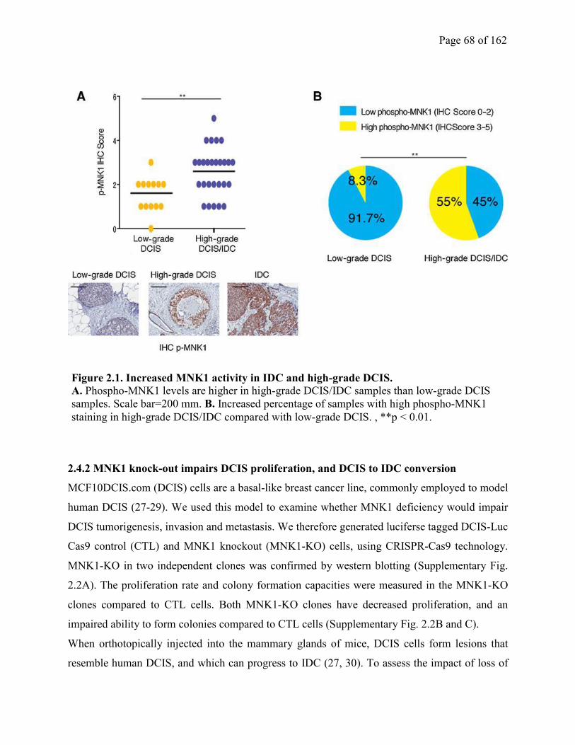
Page 68 of 162
2.4.2 MNK1 knock-out impairs DCIS proliferation, and DCIS to IDC conversion
MCF10DCIS.com (DCIS) cells are a basal-like breast cancer line, commonly employed to model
human DCIS (27-29). We used this model to examine whether MNK1 deficiency would impair
DCIS tumorigenesis, invasion and metastasis. We therefore generated luciferse tagged DCIS-Luc
Cas9 control (CTL) and MNK1 knockout (MNK1-KO) cells, using CRISPR-Cas9 technology.
MNK1-KO in two independent clones was confirmed by western blotting (Supplementary Fig.
2.2A). The proliferation rate and colony formation capacities were measured in the MNK1-KO
clones compared to CTL cells. Both MNK1-KO clones have decreased proliferation, and an
impaired ability to form colonies compared to CTL cells (Supplementary Fig. 2.2B and C).
When orthotopically injected into the mammary glands of mice, DCIS cells form lesions that
resemble human DCIS, and which can progress to IDC (27, 30). To assess the impact of loss of
Figure 2.1. Increased MNK1 activity in IDC and high-grade DCIS. A. Phospho-MNK1 levels are higher in high-grade DCIS/IDC samples than low-grade DCIS
samples. Scale bar=200 mm. B. Increased percentage of samples with high phospho-MNK1
staining in high-grade DCIS/IDC compared with low-grade DCIS. , **p < 0.01.
Page 69 of 162
MNK1 activity on the progression from DCIS to IDC in vivo, CTL or MNK1-KO cells were
mixed with 50% matrigel and injected into the mammary fat pad of athymic nude mice. A
previous study using this methodology reported that DCIS xenografts should remain DCIS after
4 weeks of injection, but progress to IDC after 5 weeks (27). Hypothesizing that MNK1 activity
would accelerate the conversion of DCIS to IDC, we resected the xenografts, including
surrounding mammary gland tissue, 5 weeks after DCIS tumor cell injection (Supplementary
Fig. 2.2D). Tumor outgrowth was monitored by luciferin injection and IVIS imaging. MNK1-
KO tumor initiation and proliferation was significantly slower than observed with CTL-derived
tumors (Fig. 2.2A). MNK1 deficiency in tumor xenografts was verified by IHC (Fig. 2.2B).
DCIS is characterized by tubular-like structures with an intact intact myoepithelial layer of cells
that stain positive for p63, while IDC is characterized by structures that lack intact myoepithelial
layers (27). A histological analysis demonstrated that 80% of MNK1-KO xenografts retained a
DCIS morphology, while all CTL tumors progressed into IDC (Fig. 2.2C). Additionally, we
observed tissue necrosis in 25% of the CTL tumors, while none of the MNK1-KO xenografts
presented with necrosis (Fig. 2.2D). This could be important clinically, as the presence of
necrosis in breast cancer is related to increased invasiveness and poor prognosis (31). Animals
were kept alive after primary tumor resection, to monitor for potential tumor recurrence,
metastasis and survival rates.
Although not statistically significant, mice that had received MNK1-KO cells showed a trend of
better overall survival than those animals that were implanted with CTL cells (Fig. 2.2E).
Furthermore, 80% of mice that received CTL cells had relapsed metastatic disease, while no
mice that received MNK1-KO cells had metastasis (Fig. 2.2F). The metastatic CTL tumors were
found in the chest area and abdominal cavity, next to the pancreas, small bowel, kidneys and rib
bones (Fig. 2.2F and G). Full necropsy and histological analysis also revealed metastatic cancer
in the lungs (Fig. 2.2G). The percentage of mice with metastasis to the lungs, chest cavity, or
abdomen is graphed in Fig. 2.2G.
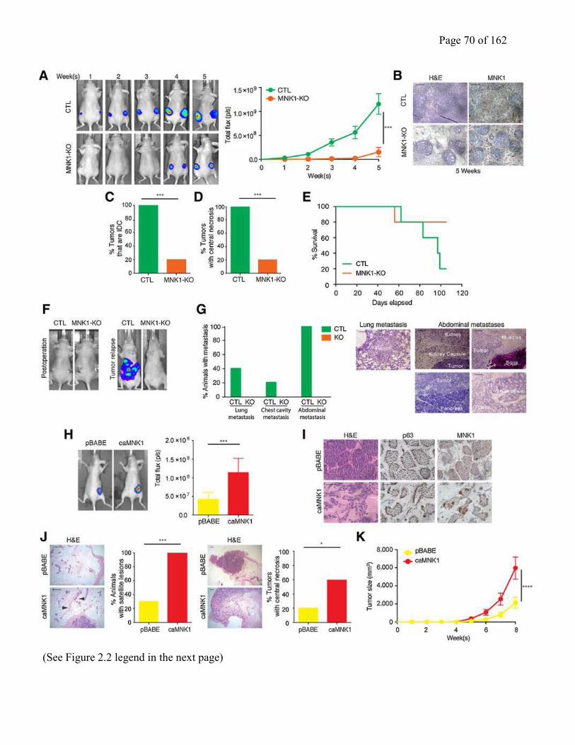
Page 70 of 162
(See Figure 2.2 legend in the next page)
Page 71 of 162
Next, we hypothesized that constitutive activation of MNK1 would promote the DCIS to IDC
transition. Thus, we generated DCIS-luciferase-tagged cells that stably express empty vector
pBABE (DCIS-luc pBABE) or MNK1T332D
(constitutively active MNK1, DCIS-Luc caMNK1)
(21). caMNK1 expression in DCIS was validated to be functional by detecting increased
phosphorylation of one of its best studied substrates, eIF4E (Supplementary Fig. 2.2E). pBABE-
and caMNK1-derived cell lines showed similar proliferation rates and clonogenic capacities
(Supplementary Fig. 2.2F and G).
We characterized the in vivo tumorigenic effects of increased MNK1 activation in DCIS cells.
DCIS-luc pBABE or caMNK1 cells were mixed with 50% matrigel and injected into the
mammary fat pad of athymic nude mice (Supplementary Fig. 2.2H). We resected the xenografts
at 1.5 weeks after DCIS tumor cell injection, expecting that caMNK1 xenografts might transition
faster to IDC, compared to vector control derived tumors. Tumors are not palpable after 1.5
weeks of implantation, thus tumor outgrowth was estimated by IVIS imaging. Unlike the lack of
proliferative advantage observed in 2D culture, caMNK1 expressing tumors were larger
compared to pBABE control expressing tumors (Fig. 2.2H). caMNK1 expression in the tumor
xenografts was confirmed by IHC for MNK1 (Fig. 2.2I). As expected, DCIS-luc pBABE cells
formed lesions that were characteristic of DCIS, with positive staining for p63 (Fig. 2.2I). DCIS-
luc caMNK1 cells on the other hand, formed lesions that consisted of a mixture of tubular-like
and irregular structures, resembling IDC (Fig. 2.2I). Consistent with the previously reported role
of MNK1 in promoting tumor invasion, we also found that all mice that had been injected with
Figure 2.2. MNK1 regulates the DCIS-IDC transition in vivo. A. Tumor outgrowth is measured
by IVIS imaging. B. MNK1-KO is retained in the xenografts as confirmed by IHC. Scale bar = 50
μm. C. All CTL xenografts have progressed into IDC, while only 20% MNK1-KO tumors have
progressed to an IDC-like morphology. D. All CTL xenografts have central necrosis, while only
20% MNK1-KO tumors have central necrosis. E. Survival curve of mice receiving CTL and MNK1-
KO cells. F. Representative IVIS imaging showing complete tumor removal post-operation and
tumor recurrence in animals receiving CTL cells. G. Percentage of animals presented with
metastasis at different sites and representative images of metastasis in various tissues of mice
receiving CTL and MNK1-KO cells. Scale bar = 200 μm. H. Tumor outgrowth is measured by IVIS
imaging. I. pBABE xenografts maintain DCIS morphology, while caMNK1 tumors have progressed
into a mixed morphology of DCIS/IDC. caMNK1 over-expression is maintained in the xenografts as
confirmed by IHC. Scale bar = 50 μm. J. 100% of mice with caMNK1 tumors have micrometastasis
in the mammary gland, while 30% of pBABE have micrometastasis. Micrometastases are indicated
by arrows. 60% caMNK1 and 20% pBABE tumors have central necrosis. Scale bar = 200 μm. K.
DCIS-Luc caMNK1 xenografts present with growth advantage over pBABE controls.
Page 72 of 162
caMNK1-expressing DCIS cells had satellite lesions throughout the mammary fat pad, compared
to only 3 out of 10 mice receiving pBABE cells (Fig. 2.2J). Additionally, 6 out of the 10 mice
injected with caMNK1 cells have tumors with central necrosis, while only 2 out of 10 mice
injected with pBABE cells present with tumors with central necrosis (Fig. 2.2J). Finally, we
sought to confirm whether the injection of caMNK1 expressing DCIS cells resulted in enhanced
tumor proliferation. To this end, we performed a second animal experiment, wherein DCIS-luc
pBABE and caMNK1 cells were injected into the mammary fat pads of SCID mice, and tumor
outgrowth was monitored, via IVIS imaging, over the course of 8 weeks. Tumor formation in the
orthotopic mammary fat pad was significantly increased in mice injected with caMNK1 cells
compared with those injected with the control pBABE cells (Fig. 2.2K, Supplementary Fig.
2.2I). We further interrogated whether the increase in tumor formation associated with caMNK1
expression was due to an increase in proliferation or a decrease in apoptosis. Using IHC, we
stained pBABE and caMNK1 expressing tumor xenografts for phospho-histone H3, a marker of
proliferation (32), and showed no difference between the pBABE and caMNK1 tumor groups
(Supplementary Fig. 2.2J). We also included in our analysis, IHC staining for the proliferative
marker Ki67, and this too showed no difference in pBABE versus caMNK1 expressing tumor
xenografts (Supplementary Fig. 2.2J). However, we did find that our IHC staining for the levels
of cleaved caspase-3, a marker of cell death, trended downward in caMNK1 expressing tumors
(Supplementary Fig. 2.2J). These data suggest that caMNK1-derived tumors are larger than their
pBABE counterparts, due to an evasion of apoptotic cell death. Cumulatively, the data presented
here demonstrate that modulation of MNK1 influences the DCIS to IDC transition in vivo.
2.4.3 MNK1 regulates NODAL morphogen to control DCIS progression
We next investigated the molecular mechanism underlying the regulation of the DCIS to IDC
transition by activated MNK1. Strizzi et al previously showed that NODAL protein levels are
increased in IDC relative to in DCIS (18). As NODAL has been shown to be downstream of p38
(33), a major upstream activator of MNK1, we hypothesized that NODAL may be regulated in
our model system. Consistent with the previous report from Strizzi et al (18), we observed that
NODAL levels are higher in the DCIS patient samples with micro-invasion, as compared to pure
DCIS lesions (Fig. 2.3A). MNK1-KO DCIS xenografts express reduced NODAL, as detected by
IHC staining (Fig. 2.3B, Supplementary Fig. 2.3A). Conversely, the expression of constitutively
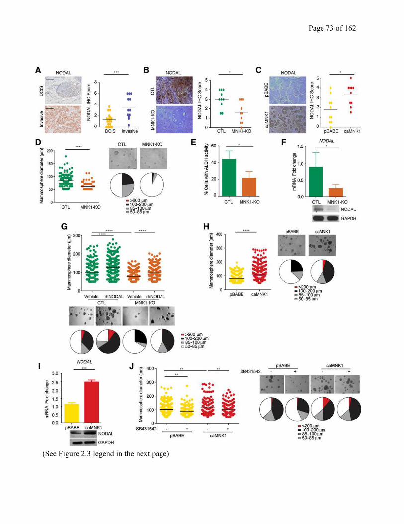
Page 73 of 162
(See Figure 2.3 legend in the next page)
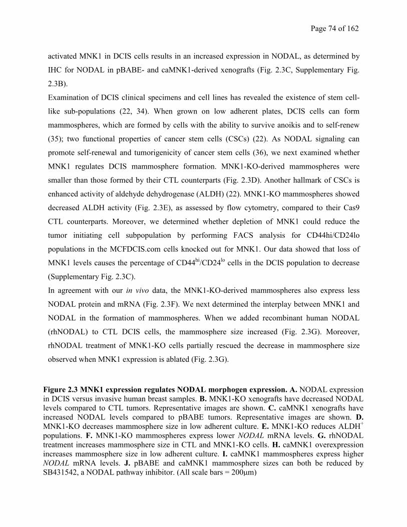
Page 74 of 162
activated MNK1 in DCIS cells results in an increased expression in NODAL, as determined by
IHC for NODAL in pBABE- and caMNK1-derived xenografts (Fig. 2.3C, Supplementary Fig.
2.3B).
Examination of DCIS clinical specimens and cell lines has revealed the existence of stem cell-
like sub-populations (22, 34). When grown on low adherent plates, DCIS cells can form
mammospheres, which are formed by cells with the ability to survive anoikis and to self-renew
(35); two functional properties of cancer stem cells (CSCs) (22). As NODAL signaling can
promote self-renewal and tumorigenicity of cancer stem cells (36), we next examined whether
MNK1 regulates DCIS mammosphere formation. MNK1-KO-derived mammospheres were
smaller than those formed by their CTL counterparts (Fig. 2.3D). Another hallmark of CSCs is
enhanced activity of aldehyde dehydrogenase (ALDH) (22). MNK1-KO mammospheres showed
decreased ALDH activity (Fig. 2.3E), as assessed by flow cytometry, compared to their Cas9
CTL counterparts. Moreover, we determined whether depletion of MNK1 could reduce the
tumor initiating cell subpopulation by performing FACS analysis for CD44hi/CD24lo
populations in the MCFDCIS.com cells knocked out for MNK1. Our data showed that loss of
MNK1 levels causes the percentage of CD44hi
/CD24lo
cells in the DCIS population to decrease
(Supplementary Fig. 2.3C).
In agreement with our in vivo data, the MNK1-KO-derived mammospheres also express less
NODAL protein and mRNA (Fig. 2.3F). We next determined the interplay between MNK1 and
NODAL in the formation of mammospheres. When we added recombinant human NODAL
(rhNODAL) to CTL DCIS cells, the mammosphere size increased (Fig. 2.3G). Moreover,
rhNODAL treatment of MNK1-KO cells partially rescued the decrease in mammosphere size
observed when MNK1 expression is ablated (Fig. 2.3G).
Figure 2.3 MNK1 expression regulates NODAL morphogen expression. A. NODAL expression
in DCIS versus invasive human breast samples. B. MNK1-KO xenografts have decreased NODAL
levels compared to CTL tumors. Representative images are shown. C. caMNK1 xenografts have
increased NODAL levels compared to pBABE tumors. Representative images are shown. D.
MNK1-KO decreases mammosphere size in low adherent culture. E. MNK1-KO reduces ALDH+
populations. F. MNK1-KO mammospheres express lower NODAL mRNA levels. G. rhNODAL
treatment increases mammosphere size in CTL and MNK1-KO cells. H. caMNK1 overexpression
increases mammosphere size in low adherent culture. I. caMNK1 mammospheres express higher
NODAL mRNA levels. J. pBABE and caMNK1 mammosphere sizes can both be reduced by
SB431542, a NODAL pathway inhibitor. (All scale bars = 200μm)
Page 75 of 162
Together, these results suggest that MNK1-dependent regulation of NODAL is needed to
regulate mammosphere size. To confirm and extend our results of MNK1-dependent regulation
of NODAL expression, we also determined the impact of MNK1 deficiency on NODAL
expression in two breast cancer cell lines, 66cl4 and MB-MDA-468. Similar to our results in
DCIS cells, depleting MNK1 in 66cl4 cells using CRISPR-Cas9 technology, and by siRNA in
MB-MDA-468 cells caused these cells to express less NODAL (Supplementary Fig. 2.3D and
E). Moreover, biological data in MNK1 null 66cl4 cells shows a decrease in mammosphere
growth, colony formation, and invasion (Supplementary Fig. 2.3F). Similarly, concomitant with
the reduced NODAL levels observed when we silence MNK1 in MDA-MB-468 cells, their
invasion is also impaired (Supplementary Fig. 2.3G).
caMNK1 expression increased both the size of DCIS mammospheres (Fig. 2.3H), and the
expression of NODAL mRNA and protein in these spheres (Fig. 2.3I). Consistent with our
observations in the caMNK1 tumor xenografts (Supplementary Fig. 2.2J), we see no change in
Ki67 levels, but a decrease in the expression of cleaved caspase-3 in the caMNK1-derived
mammospheres (Supplementary Fig. 2.3H). We also assessed tumor formation from limiting
dilutions of inoculated pBABE or caMNK1 cells, wherein we injected 10,000, 1,000, or 100
cells. As shown on the graph in Supplementary Fig. 2.3I, the caMNK1 expressing cells have an
increased tumor-initiating capability, compared to their pBABE control counterparts. 60 days
post-injection, the 100 cell injected cohort of mice, did not show any signal by IVIS imaging.
Finally, we determined whether the induction of NODAL expression by activated MNK1 was
responsible for increased DCIS mammosphere formation. We utilized SB431542, an inhibitor
routinely used to block NODAL signaling (37). Mammosphere size was significantly reduced in
pBABE and caMNK1 cells, indicating again the importance of NODAL signaling to the
increased and mammosphere size when MNK1 is constitutively activated (Fig. 2.3J).
2.4.4 MNK1 and NODAL regulate DCIS tumor invasion
MNK1 and NODAL can facilitate tumor invasion and metastasis, with the regulation of known
mediators of invasion such as VIMENTIN (3, 8, 9). Here we observed that MNK1-KO tumor
xenografts express less VIMENTIN, compared to the CTL-derived tumors (Fig. 2.4A,
Supplementary Fig. 2.4A). In contrast, caMNK1 tumors show stronger staining for VIMENTIN,
compared to the pBABE-derived tumors (Fig. 2.4B, Supplementary Fig. 2.4B). The changes in
VIMENTIN expression that we observed by modulating MNK1 levels, led us to investigate the
Page 76 of 162
invasive characteristics of our cell models. MNK1-KO cells express less VIMENTIN (Fig.
2.4C), and MNK1 deficiency restrained the invasion of DCIS cells (Fig. 2.4C). In comparison,
caMNK1 overexpression promoted the invasion of DCIS cells, and increased VIMENTIN
expression (Fig. 2.4D). Next, we tested the role that VIMENTIN might play in the increased
invasion seen in cells that express a constitutively activated MNK1 (caMNK1). When
VIMENTIN expression is repressed using siRNA, the caMNK1 cells lose their ability to invade
(Fig. 2.4E), suggesting a role for VIMENTIN in the enhanced invasion phenotype related to
constitutive MNK1 activity.
Finally, to assess whether MNK1-dependent regulation of NODAL contributes to cell invasion,
CTL and MNK1-KO cells were treated with rhNODAL. We observed that rhNODAL treatment
can enhance the invasion of DCIS CTL cells, and partially overcome the decrease in cell
invasion observed in MNK1 depleted cells (Fig. 2.4F). Additionally, we next looked at cell
invasion using pBABE and caMNK1 cells that had been transiently transfected with NODAL
siRNA. As expected, caMNK1 cells transfected with control siRNA (siCTL) are more invasive
than pBABE siCTL cells. NODAL knock-down reduced the invasion of both pBABE and
caMNK1 cells, but caMNK1 siNODAL cells are still more invasive than their pBABE
siNODAL counterparts (Fig. 2.4G).
2.4.5 MNK1 can be pharmacologically targeted to inhibit the DCIS to IDC transition
We next determined whether the effects we observed with ablation of MNK1 in DCIS cells
would be phenocopied using SEL201, an orally bioavailable small molecule inhibitor of
MNK1/2 activity (13). SEL201 induces a G2/M cell cycle arrest and inhibits proliferation of
both pBABE and caMNK cells in a dose dependent manner (Supplementary Fig. 2.5A and B).
Consistent with genetic ablation of MNK1 expression, SEL201 treatment efficiently reduced the
number of colonies (Fig. 2.5A), ALDH activity (Fig. 2.5B) and the size of mammospheres (Fig.
2.5C), derived from pBABE and caMNK1 cells. SEL201 treatment also suppressed invasion of
both pBABE and caMNK1 expressing DCIS cells, concomitant with reduced levels of NODAL
(Fig. 2.5D).
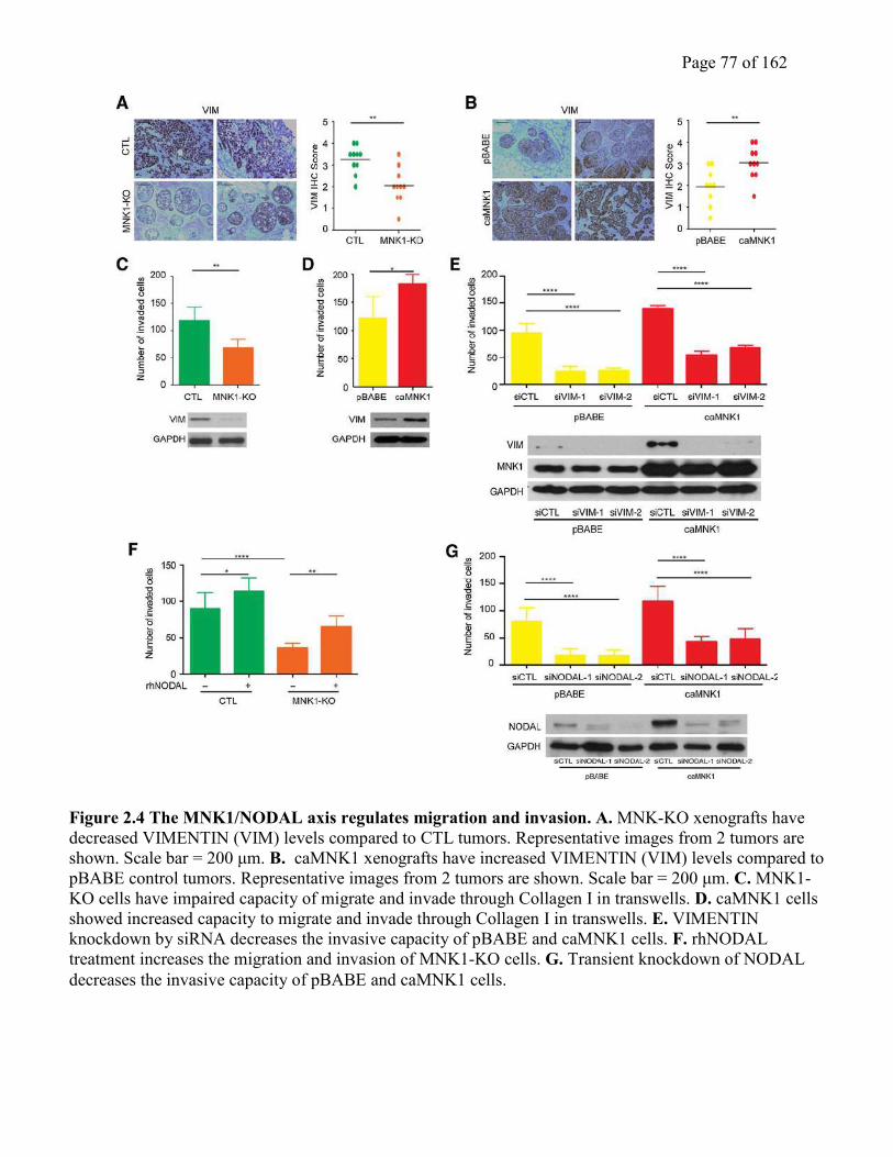
Page 77 of 162
Figure 2.4 The MNK1/NODAL axis regulates migration and invasion. A. MNK-KO xenografts have
decreased VIMENTIN (VIM) levels compared to CTL tumors. Representative images from 2 tumors are
shown. Scale bar = 200 μm. B. caMNK1 xenografts have increased VIMENTIN (VIM) levels compared to
pBABE control tumors. Representative images from 2 tumors are shown. Scale bar = 200 μm. C. MNK1-
KO cells have impaired capacity of migrate and invade through Collagen I in transwells. D. caMNK1 cells
showed increased capacity to migrate and invade through Collagen I in transwells. E. VIMENTIN
knockdown by siRNA decreases the invasive capacity of pBABE and caMNK1 cells. F. rhNODAL
treatment increases the migration and invasion of MNK1-KO cells. G. Transient knockdown of NODAL
decreases the invasive capacity of pBABE and caMNK1 cells.
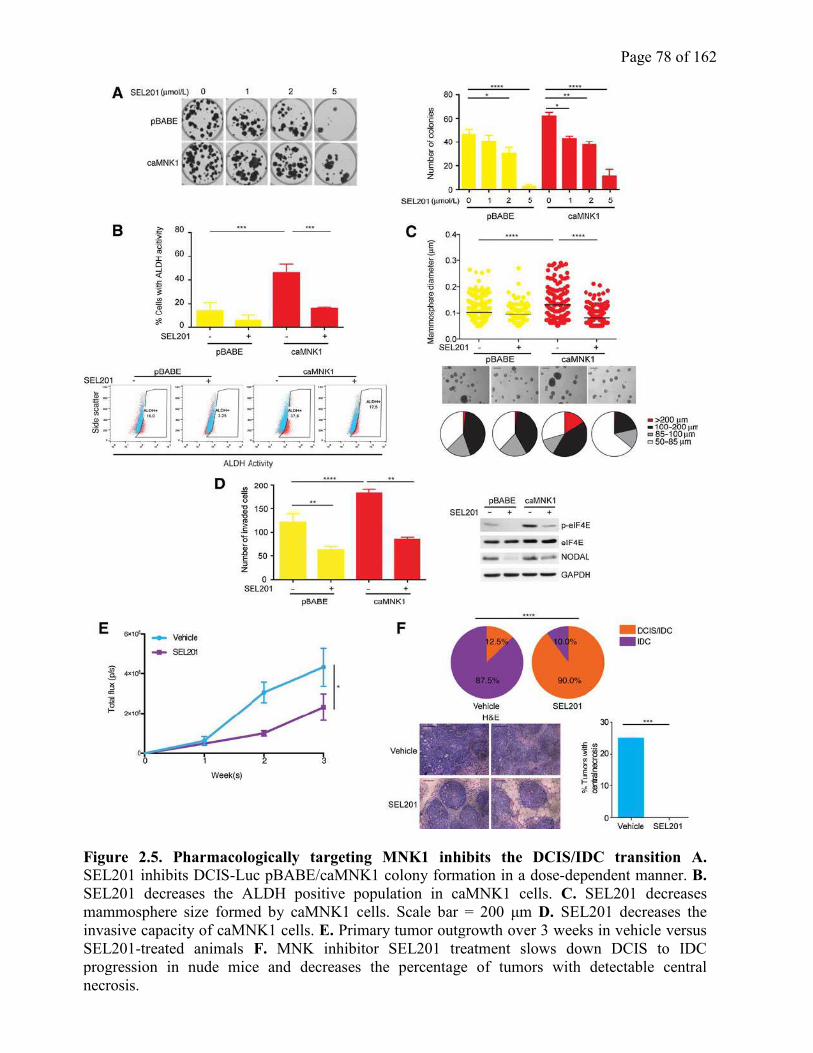
Page 78 of 162
Figure 2.5. Pharmacologically targeting MNK1 inhibits the DCIS/IDC transition A.
SEL201 inhibits DCIS-Luc pBABE/caMNK1 colony formation in a dose-dependent manner. B.
SEL201 decreases the ALDH positive population in caMNK1 cells. C. SEL201 decreases
mammosphere size formed by caMNK1 cells. Scale bar = 200 μm D. SEL201 decreases the
invasive capacity of caMNK1 cells. E. Primary tumor outgrowth over 3 weeks in vehicle versus
SEL201-treated animals F. MNK inhibitor SEL201 treatment slows down DCIS to IDC
progression in nude mice and decreases the percentage of tumors with detectable central
necrosis.
Page 79 of 162
Having shown that MNK1 regulates the DCIS to IDC conversion in animal models, we next
investigated whether pharmacologically blocking MNK1 would inhibit the progression of DCIS
to invasive disease. DCIS-Luc CTL cells were orthotopically injected into the mammary glands
of nude mice, and then the mice were randomized to either vehicle control or SEL201 treatment
groups (Supplementary Fig. 2.5C). SEL201 was delivered by gavage at a dose of 75mg/kg per
day, which we have previously shown concomitantly suppresses MNK1 activity and lung
metastasis (13). SEL201 significantly decreased the primary tumor size after a three week-
treatment (Fig. 2.5E). Furthermore, xenografts from the SEL201 group showed a reduced
percentage of IDC (10% of tumours) compared to the vehicle group (87.5% of tumors) (Fig.
2.5F). Consistent with our previous work (13), SEL201 showed no overt systemic toxicity, as
evidenced by body weight (Supplementary Fig. 2.5D) and tests for liver function (ALT and
AST) (Supplementary Fig. 2.5D). SUM225 cells are also commonly used to model DCIS, and
consistent with the data shown thus far, reducing MNK1 levels using siRNA in SUM225 induces
a loss of NODAL expression, while SUM225-expressing caMNK1 express increased levels of
NODAL (Supplementary Fig. 2.5E). Compared to MCFDCIS.com cells, SUM225 cells slowly
transition from DCIS to IDC following intraductal injections, a technique which minimizes
disturbance to the mammary gland microenvironment (38). SEL201 also inhibits colony
formation of SUM225 cells in vitro, and slows down the transition from DCIS to IDC in the
SUM225 intraductal model of DCIS (Supplementary Fig. 2.5F and 2.5G). In summary, our
results provide evidence to show the feasability of inhibiting MNK1 to slow the conversion of
DCIS to IDC.
2.5 Discussion
The mechanism underlying the transition of DCIS to IDC remains poorly understood. Previous
studies have identified several potential factors that facilitate the conversion to invasive breast
cancer, such as p63/MT1-MMP (39), SMARCE1 (40), and Singleminded-2s (SIM2s) (29).
Unfortunately, many of these proteins have proven difficult to therapeutically target as a means
to block disease progression in pre-clinical models. Herein, we identify the MNK1/NODAL
signaling axis as a key molecular pathway regulating the progression of DCIS to IDC and breast
cancer recurrence as metastatic disease (Fig. 2.6). Moreover, MNK1 inhibitors such as SEL201,
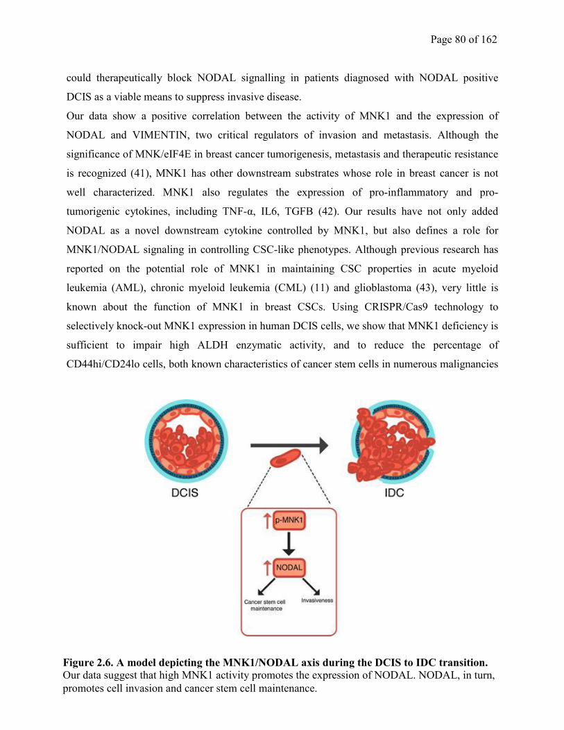
Page 80 of 162
could therapeutically block NODAL signalling in patients diagnosed with NODAL positive
DCIS as a viable means to suppress invasive disease.
Our data show a positive correlation between the activity of MNK1 and the expression of
NODAL and VIMENTIN, two critical regulators of invasion and metastasis. Although the
significance of MNK/eIF4E in breast cancer tumorigenesis, metastasis and therapeutic resistance
is recognized (41), MNK1 has other downstream substrates whose role in breast cancer is not
well characterized. MNK1 also regulates the expression of pro-inflammatory and pro-
tumorigenic cytokines, including TNF-α, IL6, TGFB (42). Our results have not only added
NODAL as a novel downstream cytokine controlled by MNK1, but also defines a role for
MNK1/NODAL signaling in controlling CSC-like phenotypes. Although previous research has
reported on the potential role of MNK1 in maintaining CSC properties in acute myeloid
leukemia (AML), chronic myeloid leukemia (CML) (11) and glioblastoma (43), very little is
known about the function of MNK1 in breast CSCs. Using CRISPR/Cas9 technology to
selectively knock-out MNK1 expression in human DCIS cells, we show that MNK1 deficiency is
sufficient to impair high ALDH enzymatic activity, and to reduce the percentage of
CD44hi/CD24lo cells, both known characteristics of cancer stem cells in numerous malignancies
Figure 2.6. A model depicting the MNK1/NODAL axis during the DCIS to IDC transition.
Our data suggest that high MNK1 activity promotes the expression of NODAL. NODAL, in turn,
promotes cell invasion and cancer stem cell maintenance.
Page 81 of 162
(44). This could be clinically important as DCIS CSC subpopulations are thought to be
responsible for resistance to radiotherapy and potential disease recurrence (45, 46). We thus
speculate that pharmacologically inhibiting MNK1 will limit the plasticity of DCIS cells via
downregulation of NODAL and forestall resistance to radiotherapy.
Our study has shown that NODAL mRNA levels are controlled by MNK1, and MNK1/2 have a
traditional role as regulators of oncogenic mRNA translation. Therefore, it is possible that
MNK1 is regulating NODAL on the level of mRNA translation. In fact, our work and that of
other labs, has shown that NODAL works with a positive feedback loop to activate its own
transcription (47, 48). We posit that MNK1 may inhibit NODAL protein synthesis, and this in
turn would lead to a corresponding suppression in NODAL mRNA. It is also possible that
MNK1 regulates NODAL by a yet to be ascribed role in the nucleus. MNK1/2 are also found in
the nucleus, suggesting activities beyond their role in protein synthesis (42, 49). Interestingly, we
have observed nuclear localization of MNK1 in our murine xenografts and clinical breast
samples, in this study (Fig. 1A), and in our melanoma work (13). This observation is line with
MNK1 containing both nuclear localization and export signals, and prior work showing that
MNK1 can shuttle between the nucleus and cytoplasm (50, 51). Access to CRISPR/Cas9
technology for selectively deleting MNK1 and MNK2 will no doubt lead to defining novel
biological functions of these kinases.
A question raised by our work is identifying the molecular mechanism by which MNK1
signaling is activated in invasive disease. Our data show that NODAL lies downstream of
MNK1, however, it is entirely possible that NODAL can itself feedback to activate MNK1.
TGFB super-family cytokines, such as NODAL and Activin, utilize non-canonical MAPK
cascades to regulate breast tumor progression (15, 24, 49), however it remains unclear whether
MNK1 cooperates with NODAL to drive DCIS towards invasive disease. Our results also
demonstrated that exogenous NODAL treatment can increase DCIS mammosphere size and it is
tempting to speculate that this cytokine can also induce MNK1 phosphorylation. Indeed,
NODAL has been shown to activate ERK1/2, immediately upstream of MNK1, in breast cancer
cells (37).
We have demonstrated the feasibility of pharmacologically blocking MNK1/2 activity in vivo,
resulting in blocking of the progression of DCIS to invasive disease. Together, our data
Page 82 of 162
contribute to the increasing discussion about the clinical applications of MNK1/2 inhibitors in
cancer.
2.6 Acknowledgments
This research is funded by grants from the Cancer Research Society (PIN20239 to W. Miller),
the Canadian Institutes for Health Research (MOP-142281 to W. Miller), the Canadian Institutes
for Health Research (PJT-156269 to W. Miller and S.V. del Rincon), the Canadian Cancer Societ
(703811 to W. Miller and S.V. del Rincon), the Rossy Cancer Network (to W. Miller), and the
Israel Cancer Research Fund (to W. Miller and S.V. del Rincon). Q. Guo was supported by a
Cole Foundation Ph.D fellowship, McGill Integrated Cancer Research Training Program
(MICRTP) graduate studentship, and a McGill Faculty of Medicine graduate studentship. V.Z. Li
was supported by a MICRTP undergraduate studentship. J. Nichol was supported by a fellowship
from the Rossy Cancer Network. W. Yang and F. Huang were supported by MICRTP graduate
studentships. Y. Zhan was supported by a Toronto-Dominion Bank/Lady Davis Institute (LDI)
Ph.D fellowship and the Israel Cancer Research Fund. We thank Selvita S.A. for supplying
SEL201 and the Flow Cytometry and Animal Core Facilities of the Lady Davis Institute for their
support.
2.7 References
1. Martinez-Perez C, Turnbull AK, Ekatah GE, Arthur LM, Sims AH, Thomas JS, et al.
Current treatment trends and the need for better predictive tools in the management of ductal
carcinoma in situ of the breast. Cancer Treat Rev. 2017;55:163-72.
2. Wang X, Flynn A, Waskiewicz AJ, Webb BL, Vries RG, Baines IA, et al. The
phosphorylation of eukaryotic initiation factor eIF4E in response to phorbol esters, cell stresses,
and cytokines is mediated by distinct MAP kinase pathways. J Biol Chem. 1998;273(16):9373-7.
3. Bhat M, Robichaud N, Hulea L, Sonenberg N, Pelletier J, Topisirovic I. Targeting the
translation machinery in cancer. Nat Rev Drug Discov. 2015;14(4):261-78.
4. Adesso L, Calabretta S, Barbagallo F, Capurso G, Pilozzi E, Geremia R, et al.
Gemcitabine triggers a pro-survival response in pancreatic cancer cells through activation of the
MNK2/eIF4E pathway. Oncogene. 2013;32(23):2848-57.
Page 83 of 162
5. Chrestensen CA, Shuman JK, Eschenroeder A, Worthington M, Gram H, Sturgill TW.
MNK1 and MNK2 regulation in HER2-overexpressing breast cancer lines. J Biol Chem.
2007;282(7):4243-52.
6. Beggs JE, Tian S, Jones GG, Xie J, Iadevaia V, Jenei V, et al. The MAP kinase-
interacting kinases regulate cell migration, vimentin expression and eIF4E/CYFIP1 binding.
Biochem J. 2015;467(1):63-76.
7. Furic L, Rong L, Larsson O, Koumakpayi IH, Yoshida K, Brueschke A, et al. eIF4E
phosphorylation promotes tumorigenesis and is associated with prostate cancer progression. Proc
Natl Acad Sci U S A. 2010;107(32):14134-9.
8. Konicek BW, Stephens JR, McNulty AM, Robichaud N, Peery RB, Dumstorf CA, et al.
Therapeutic inhibition of MAP kinase interacting kinase blocks eukaryotic initiation factor 4E
phosphorylation and suppresses outgrowth of experimental lung metastases. Cancer Res.
2011;71(5):1849-57.
9. Robichaud N, Del Rincon SV, Huor B, Alain T, Petruccelli LA, Hearnden J, et al.
Phosphorylation of eIF4E promotes EMT and metastasis via translational control of SNAIL and
MMP-3. Oncogene. 2015;34(16):2032-42.
10. Wendel HG, Silva RL, Malina A, Mills JR, Zhu H, Ueda T, et al. Dissecting eIF4E action
in tumorigenesis. Genes Dev. 2007;21(24):3232-7.
11. Lim S, Saw TY, Zhang M, Janes MR, Nacro K, Hill J, et al. Targeting of the MNK-
eIF4E axis in blast crisis chronic myeloid leukemia inhibits leukemia stem cell function. Proc
Natl Acad Sci U S A. 2013;110(25):E2298-307.
12. Ueda T, Sasaki M, Elia AJ, Chio, II, Hamada K, Fukunaga R, et al. Combined deficiency
for MAP kinase-interacting kinase 1 and 2 (Mnk1 and Mnk2) delays tumor development. Proc
Natl Acad Sci U S A. 2010;107(32):13984-90.
13. Zhan Y, Guo J, Yang W, Goncalves C, Rzymski T, Dreas A, et al. MNK1/2 inhibition
limits oncogenicity and metastasis of KIT-mutant melanoma. J Clin Invest. 2017.
14. Liu S, Zha J, Lei M. Inhibiting ERK/Mnk/eIF4E broadly sensitizes ovarian cancer
response to chemotherapy. Clin Transl Oncol. 2017.
15. Lock R, Ingraham R, Maertens O, Miller AL, Weledji N, Legius E, et al. Cotargeting
MNK and MEK kinases induces the regression of NF1-mutant cancers. J Clin Invest. 2016.
16. Hendrix MJ, Seftor EA, Seftor RE, Kasemeier-Kulesa J, Kulesa PM, Postovit LM.
Reprogramming metastatic tumour cells with embryonic microenvironments. Nat Rev Cancer.
2007;7(4):246-55.
17. Kirsammer G, Strizzi L, Margaryan NV, Gilgur A, Hyser M, Atkinson J, et al. Nodal
signaling promotes a tumorigenic phenotype in human breast cancer. Semin Cancer Biol.
2014;29:40-50.
18. Strizzi L, Hardy KM, Margaryan NV, Hillman DW, Seftor EA, Chen B, et al. Potential
for the embryonic morphogen Nodal as a prognostic and predictive biomarker in breast cancer.
Breast Cancer Res. 2012;14(3):R75.
19. Bodenstine TM, Chandler GS, Seftor RE, Seftor EA, Hendrix MJ. Plasticity underlies
tumor progression: role of Nodal signaling. Cancer Metastasis Rev. 2016;35(1):21-39.
20. Cong L, Ran FA, Cox D, Lin S, Barretto R, Habib N, et al. Multiplex genome
engineering using CRISPR/Cas systems. Science. 2013;339(6121):819-23.
21. Waskiewicz AJ, Johnson JC, Penn B, Mahalingam M, Kimball SR, Cooper JA.
Phosphorylation of the cap-binding protein eukaryotic translation initiation factor 4E by protein
kinase Mnk1 in vivo. Mol Cell Biol. 1999;19(3):1871-80.

Page 84 of 162
22. Li Q, Eades G, Yao Y, Zhang Y, Zhou Q. Characterization of a stem-like subpopulation
in basal-like ductal carcinoma in situ (DCIS) lesions. J Biol Chem. 2014;289(3):1303-12.
23. West NR, Murray JI, Watson PH. Oncostatin-M promotes phenotypic changes associated
with mesenchymal and stem cell-like differentiation in breast cancer. Oncogene.
2014;33(12):1485-94.
24. Pettersson F, Del Rincon SV, Emond A, Huor B, Ngan E, Ng J, et al. Genetic and
pharmacologic inhibition of eIF4E reduces breast cancer cell migration, invasion, and metastasis.
Cancer Res. 2015;75(6):1102-12.
25. Quail DF, Walsh LA, Zhang G, Findlay SD, Moreno J, Fung L, et al. Embryonic protein
nodal promotes breast cancer vascularization. Cancer Res. 2012;72(15):3851-63.
26. Yu L, Harms PW, Pouryazdanparast P, Kim DS, Ma L, Fullen DR. Expression of the
embryonic morphogen Nodal in cutaneous melanocytic lesions. Mod Pathol. 2010;23(9):1209-
14.
27. Hu M, Yao J, Carroll DK, Weremowicz S, Chen H, Carrasco D, et al. Regulation of in
situ to invasive breast carcinoma transition. Cancer Cell. 2008;13(5):394-406.
28. Rakha EA, Reis-Filho JS, Ellis IO. Basal-like breast cancer: a critical review. J Clin
Oncol. 2008;26(15):2568-81.
29. Scribner KC, Behbod F, Porter WW. Regulation of DCIS to invasive breast cancer
progression by Singleminded-2s (SIM2s). Oncogene. 2013;32(21):2631-9.
30. Miller FR, Santner SJ, Tait L, Dawson PJ. MCF10DCIS.com xenograft model of human
comedo ductal carcinoma in situ. J Natl Cancer Inst. 2000;92(14):1185-6.
31. Gilchrist KW, Gray R, Fowble B, Tormey DC, Taylor SGt. Tumor necrosis is a
prognostic predictor for early recurrence and death in lymph node-positive breast cancer: a 10-
year follow-up study of 728 Eastern Cooperative Oncology Group patients. J Clin Oncol.
1993;11(10):1929-35.
32. Gerring Z, Pearson JF, Morrin HR, Robinson BA, Harris GC, Walker LC.
Phosphohistone H3 outperforms Ki67 as a marker of outcome for breast cancer patients.
Histopathology. 2015;67(4):538-47.
33. Bradham CA, McClay DR. p38 MAPK is essential for secondary axis specification and
patterning in sea urchin embryos. Development. 2006;133(1):21-32.
34. Currie MJ, Beardsley BE, Harris GC, Gunningham SP, Dachs GU, Dijkstra B, et al.
Immunohistochemical analysis of cancer stem cell markers in invasive breast carcinoma and
associated ductal carcinoma in situ: relationships with markers of tumor hypoxia and
microvascularity. Hum Pathol. 2013;44(3):402-11.
35. Farnie G, Clarke RB, Spence K, Pinnock N, Brennan K, Anderson NG, et al. Novel cell
culture technique for primary ductal carcinoma in situ: role of Notch and epidermal growth
factor receptor signaling pathways. J Natl Cancer Inst. 2007;99(8):616-27.
36. Quail DF, Taylor MJ, Postovit LM. Microenvironmental regulation of cancer stem cell
phenotypes. Curr Stem Cell Res Ther. 2012;7(3):197-216.
37. Quail DF, Zhang G, Findlay SD, Hess DA, Postovit LM. Nodal promotes invasive
phenotypes via a mitogen-activated protein kinase-dependent pathway. Oncogene.
2014;33(4):461-73.
38. Behbod F, Kittrell FS, LaMarca H, Edwards D, Kerbawy S, Heestand JC, et al. An
intraductal human-in-mouse transplantation model mimics the subtypes of ductal carcinoma in
situ. Breast Cancer Res. 2009;11(5):R66.
Page 85 of 162
39. Lodillinsky C, Infante E, Guichard A, Chaligne R, Fuhrmann L, Cyrta J, et al. p63/MT1-
MMP axis is required for in situ to invasive transition in basal-like breast cancer. Oncogene.
2015.
40. Sokol ES, Feng YX, Jin DX, Tizabi MD, Miller DH, Cohen MA, et al. SMARCE1 is
required for the invasive progression of in situ cancers. Proc Natl Acad Sci U S A.
2017;114(16):4153-8.
41. Wheater MJ, Johnson PW, Blaydes JP. The role of MNK proteins and eIF4E
phosphorylation in breast cancer cell proliferation and survival. Cancer Biol Ther.
2010;10(7):728-35.
42. Joshi S, Platanias LC. Mnk kinases in cytokine signaling and regulation of cytokine
responses. Biomol Concepts. 2015;6(1):85.
43. Bell JB, Eckerdt FD, Alley K, Magnusson LP, Hussain H, Bi Y, et al. MNK Inhibition
Disrupts Mesenchymal Glioma Stem Cells and Prolongs Survival in a Mouse Model of
Glioblastoma. Mol Cancer Res. 2016;14(10):984-93.
44. Ginestier C, Hur MH, Charafe-Jauffret E, Monville F, Dutcher J, Brown M, et al.
ALDH1 is a marker of normal and malignant human mammary stem cells and a predictor of poor
clinical outcome. Cell Stem Cell. 2007;1(5):555-67.
45. Williams KE, Bundred NJ, Landberg G, Clarke RB, Farnie G. Focal adhesion kinase and
Wnt signaling regulate human ductal carcinoma in situ stem cell activity and response to
radiotherapy. Stem Cells. 2015;33(2):327-41.
46. Farnie G, Johnson RL, Williams KE, Clarke RB, Bundred NJ. Lapatinib inhibits
stem/progenitor proliferation in preclinical in vitro models of ductal carcinoma in situ (DCIS).
Cell Cycle. 2014;13(3):418-25.
47. Saijoh Y, Oki S, Tanaka C, Nakamura T, Adachi H, Yan YT, et al. Two nodal-responsive
enhancers control left-right asymmetric expression of Nodal. Dev Dyn. 2005;232(4):1031-6.
48. Topczewska JM, Postovit LM, Margaryan NV, Sam A, Hess AR, Wheaton WW, et al.
Embryonic and tumorigenic pathways converge via Nodal signaling: role in melanoma
aggressiveness. Nat Med. 2006;12(8):925-32.
49. Rannou Y, Salaun P, Benaud C, Khan J, Dutertre S, Giet R, et al. MNK1 kinase activity
is required for abscission. J Cell Sci. 2012;125(Pt 12):2844-52.
50. Parra-Palau JL, Scheper GC, Wilson ML, Proud CG. Features in the N and C termini of
the MAPK-interacting kinase Mnk1 mediate its nucleocytoplasmic shuttling. J Biol Chem.
2003;278(45):44197-204.
51. Scheper GC, Parra JL, Wilson M, Van Kollenburg B, Vertegaal AC, Han ZG, et al. The
N and C termini of the splice variants of the human mitogen-activated protein kinase-interacting
kinase Mnk2 determine activity and localization. Mol Cell Biol. 2003;23(16):5692-705.
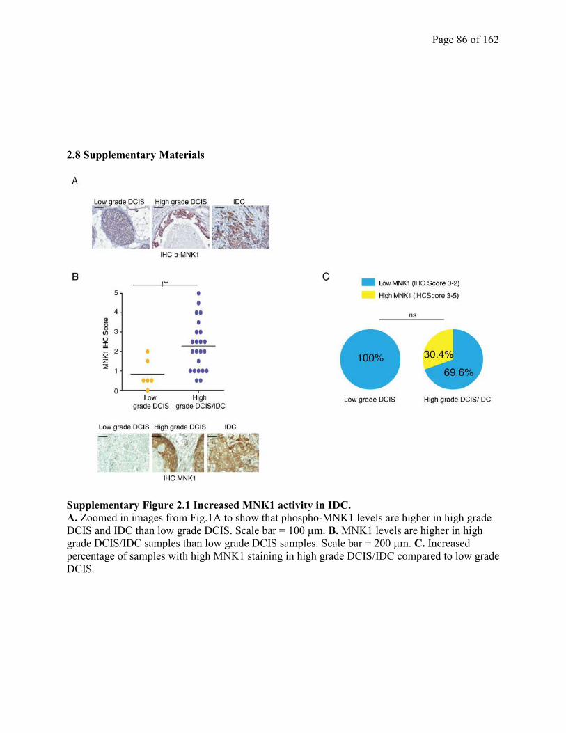
Page 86 of 162
2.8 Supplementary Materials
Supplementary Figure 2.1 Increased MNK1 activity in IDC.
A. Zoomed in images from Fig.1A to show that phospho-MNK1 levels are higher in high grade
DCIS and IDC than low grade DCIS. Scale bar = 100 µm. B. MNK1 levels are higher in high
grade DCIS/IDC samples than low grade DCIS samples. Scale bar = 200 µm. C. Increased
percentage of samples with high MNK1 staining in high grade DCIS/IDC compared to low grade
DCIS.
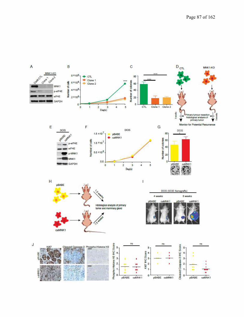
Page 87 of 162
Page 88 of 162
Supplementary Figure 2.2 Modulation of MNK1 affects colony formation in 2D cell
culture. A. MNK1 knock-out by CRISPR-Cas 9 technology in 2 independent clones is
confirmed by WB. B. MNK1-KO inhibits cell proliferation. C. MNK1-KO inhibits colony
formation. D. Schematic depicting timing and measured outcomes in CTL and MNK1-KO
xenografts. E. caMNK1 overexpression is confirmed by WB. F. caMNK1 overexpression does
not affect DCIS proliferation. G. caMNK1 overexpression increases DCIS colony formation. H.
Schematic depicting timing and measured outcomes in pBABE and caMNK1 xenografts. I. IVIS
imaging of pBABE and caMNK1 xenografts in SCID mice. J. pBABE and caMNK1 xenografts
express similar levels of Ki67 and phospho-histone H3. Cleaved caspase-3 levels in caMNK1
xeonografts trended downward compared to pBABE.
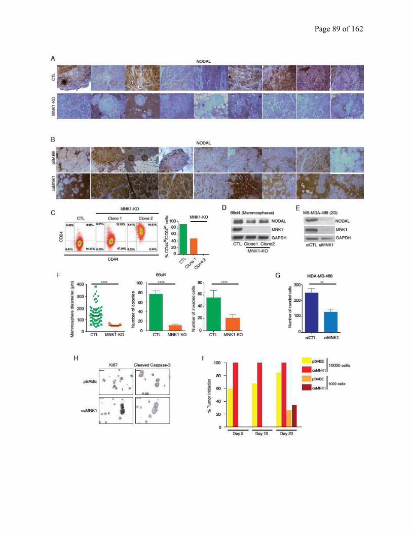
Page 89 of 162
Page 90 of 162
Supplementary Figure 2.3 NODAL levels are regulated by MNK1. A. MNK1-KO xenografts
have decreased NODAL levels compared to CTL tumors. Representative images from 18 tumors
are shown. Scale bar = 200 μm. B. caMNK1 xenografts have increased NODAL levels compared
to pBABE tumors. Representative images from 18 tumors are shown. Scale bar = 200 μm. C.
Loss of MNK1 causes decrease of CD44hi/CD24lo cells in the DCIS population to decrease. D.
66cl4 MNK1-KO mammospheres have decreased NODAL levels compared to CTL
mammospheres. E. MNK1 knock-down by siRNA decreases NODAL levels and cell invasion in
MB-MDA-468 cells. F. 66cl4 MNK1-KO cells form mammospheres with smaller diameters,
showed impaired colony formation ability and decreased invasion compared to CTL
mammospheres. G. MNK1 knockdown by siRNA decreases MB-MDA-468 invasion. H.
caMNK1 expressing DCIS mammospheres express decreased cleaved caspase 3 but similar Ki67
levels compared to pBABE mammospheres. I. Tumor initiation rate of DCIS-Luc pBABE and
caMNK1 in vivo limiting dilution experiment.
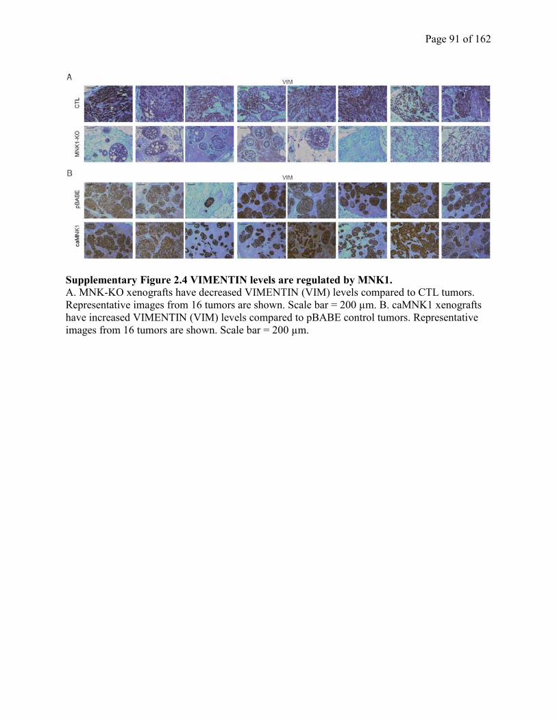
Page 91 of 162
Supplementary Figure 2.4 VIMENTIN levels are regulated by MNK1. A. MNK-KO xenografts have decreased VIMENTIN (VIM) levels compared to CTL tumors.
Representative images from 16 tumors are shown. Scale bar = 200 µm. B. caMNK1 xenografts
have increased VIMENTIN (VIM) levels compared to pBABE control tumors. Representative
images from 16 tumors are shown. Scale bar = 200 µm.
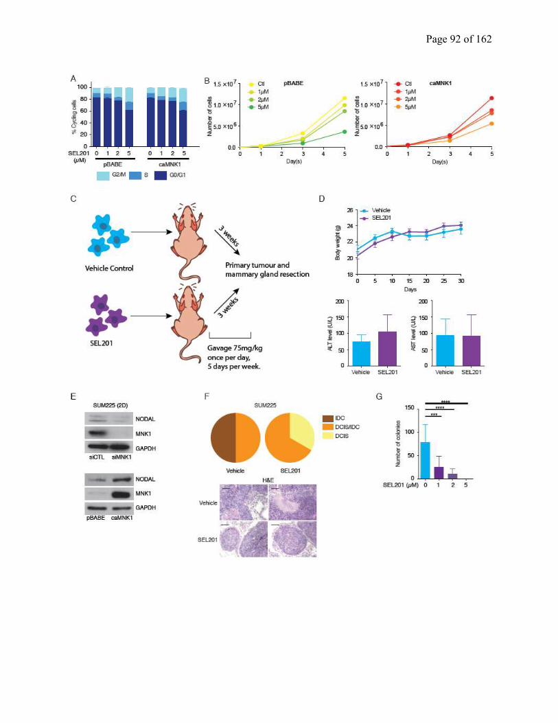
Page 92 of 162
Page 93 of 162
Supplementary Figure 2.5 A small molecule inhibitor of MNK1 inhibits the DCIS-IDC
transition. A. SEL201 induces a G2/M arrest in pBABE and caMNK1 cells in a dose dependent
manner. B. SEL201 inhibits pBABE and caMNK1 cell proliferation in a dose-dependent manner.
C. Schematic depicting the timing and measured outcomes of SEL201 xenograft experiment. D.
SEL201 has no effect on mouse body weight (upper panel), or on liver toxicity, as assessed by
ALT and AST (lower panels) levels. E. MNK1 knock-down by siRNA decreases NODAL levels
in SUM225 cells (upper panel). caMNK1 expressing SUM225 cells express increased NODAL
levels compared to pBABE controls (lower panel). F. SUM225 primary tumor outgrowth over 9
weeks in vehicle versus SEL201-treated animals. MNK inhibitor SEL201 treatment slows down
DCIS to IDC progression in NOD/SCID mice. G. SEL201 inhibits SUM225 colony formation.
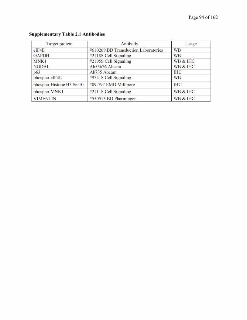
Page 94 of 162
Supplementary Table 2.1 Antibodies
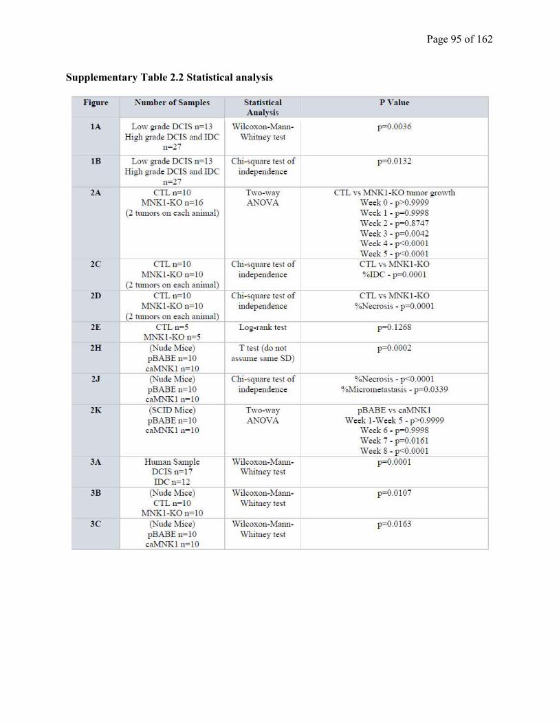
Page 95 of 162
Supplementary Table 2.2 Statistical analysis
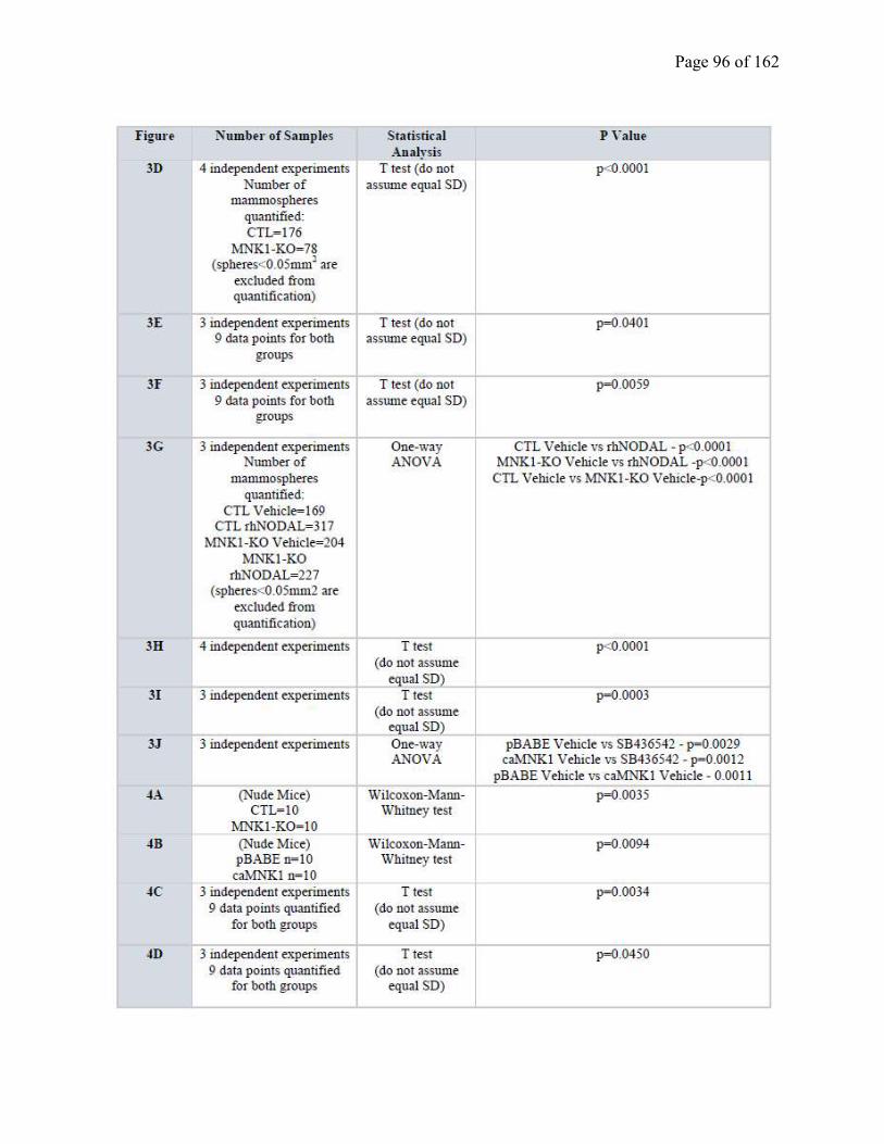
Page 96 of 162
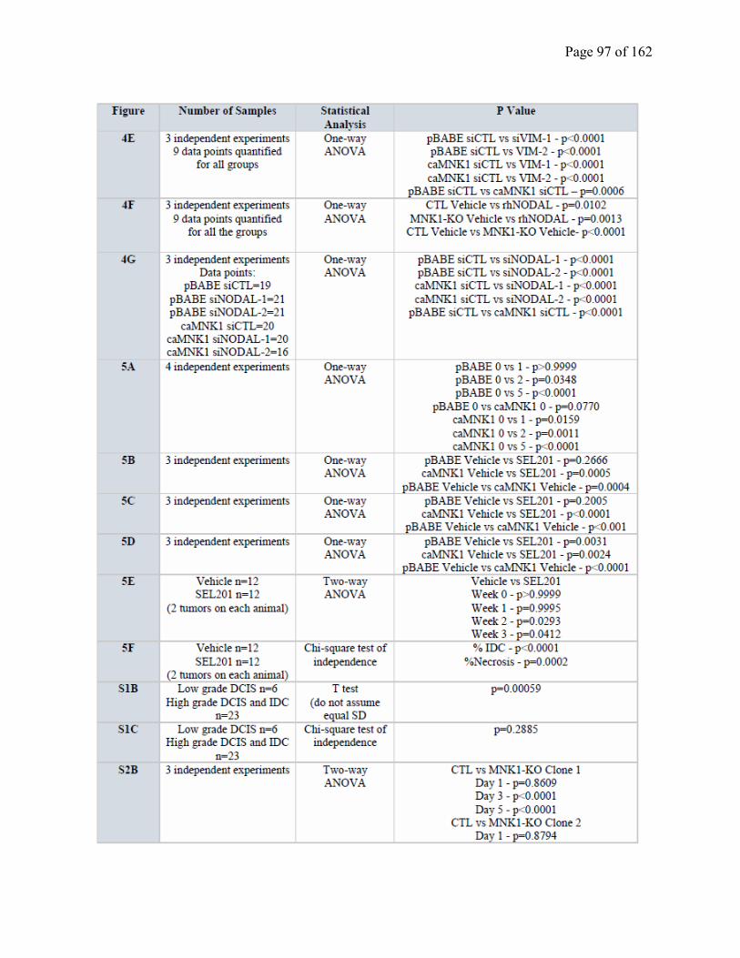
Page 97 of 162
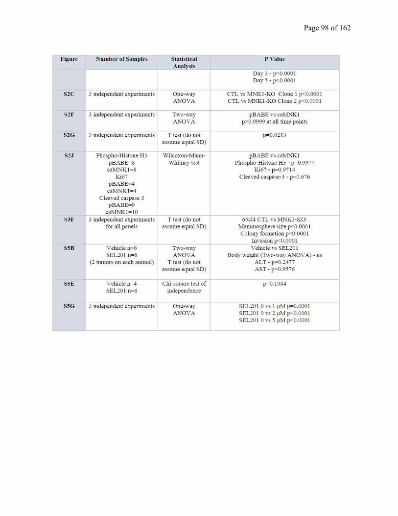
Page 98 of 162
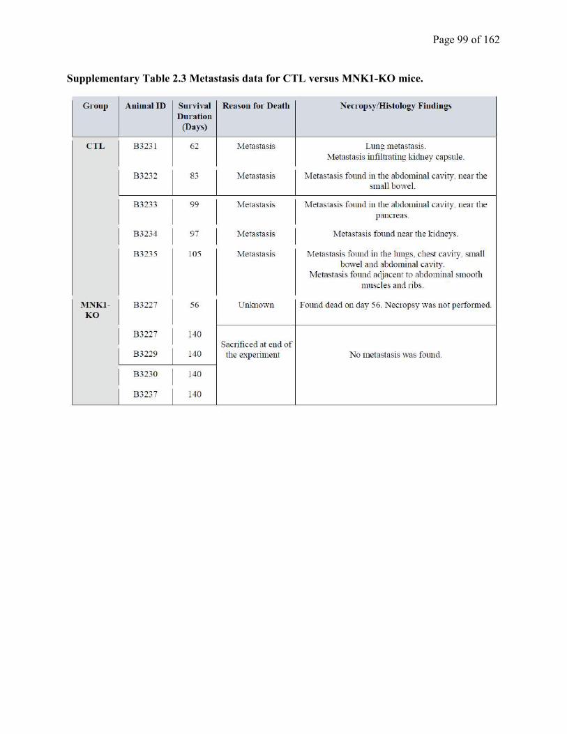
Page 99 of 162
Supplementary Table 2.3 Metastasis data for CTL versus MNK1-KO mice.
Page 100 of 162
Chapter 3. phospho-eIF4E/IL-33 drives immune evasion and lung metastasis in
postpartum breast cancer (PPBC)
(Manuscript in preparation)
Program of research: In chapter 2, we have demonstrated a central role of tumor intrinsic
MNK1/NODAL axis in the conversion of DCIS to IDC. However, our knowledge of how
aberrant mRNA translation regulates breast tumor immunity, and which pro-tumorigenic and
pro-inflammatory factors are involved, remains limited. PPBC is an aggressive subtype of breast
cancer characterized by robust immune cell influx. In this chapter, we plan to understand how
aberrant MNK1/2-eIF4E activity contributes to PPBC immune evasion, and to investigate
possible strategies to enhance the effectiveness of immune-checkpoint blockade in PPBC by
targeting this pathway.
Rationale & Hypothesis: It remains unclear how aberrant MNK1/2-eIF4E activity affects
different cellular components of the tumor microenvironment. We hypothesize that the
MNK1/eIF4E axis, functioning in stromal cells, promotes PPBC pro-tumorigenic immunity. It is
important to understand how the MNK1/2-eIF4E pathway contributes to breast cancer immune
evasion, and investigate if blocking this pathway can sensitize PPBC to anti-PD1 blockade.
Page 101 of 162
3.1 Abstract
Breast cancer diagnosed within 10 years of a last pregnancy is defined as post-partum breast
cancer (PPBC,) and is highly metastatic. Mammary gland (MG) involution is a physiologic
process whereby the breast tissue, upon cessation or in the absence of lactation, is remodeled
back to its pre-pregnant state. In pre-clinical murine models of PPBC (PPBC mice), breast tumor
cells implanted into the involuting mammary gland show increased outgrowth and metastasis to
the lungs, compared to the same cells injected into nulliparous hosts. The MNK1/2-eIF4E axis is
a critical regulator of the translation of mRNAs that encode for proteins that promote tumor cell
invasion and metastasis. However, it remains unclear whether this axis modulates non-tumor
cells that make up the fabric of the PPBC tumor microenvironment (TME). We report that
phospho-eIF4E deficient (eIF4ES209A
) PPBC mice are protected against lung metastasis.
Characterization of the lung immune microenvironment of PPBC mice showed infiltration of
type 2 innate lymphoid cells (ILC2) and granulocytic myeloid-derived suppressor cells (G-
MDSC). However, while the lungs from eIF4ES209A
PPBC mice showed less ILC2 infiltration,
and reduced G-MDSC, there was a significant increase in the presence of cytotoxic T cells.
We next wanted to investigate whether upstream regulators of ILC2 are controlled by phospho-
eIF4E. Herein, we have shown that the expression of IL-33, a known hallmark ILC2-activating
alarmin cytokine and inducer of breast tumor cell invasion, is repressed when the MNK1/2-
eIF4E axis is blocked. Thus far, immune checkpoint blockade therapy has shown limited clinical
benefit in breast cancer. Herein, we show that the combination of orally bioavailable MNK1/2
inhibitor SEL201 and anti-PD1 can inhibit PPBC metastasis to the lungs. Thus, our study offers
a promising immune modulatory route to enhance efficacy of immunotherapy by targeting
aberrant mRNA translation.
Page 102 of 162
3.2 Introduction
Postpartum breast cancer (PPBC) is defined as breast cancer diagnosed within the first 10 years
of the most recent pregnancy (1). Given its highly metastatic nature (1, 2), patient prognosis is
poor in women diagnosed with PPBC. Mammary gland (MG) involution, the remodeling of the
breast tissue back to its pre-pregnant state, has been hypothesized to cause premalignant
epithelial cells to adopt invasive properties (2). Involution, akin to the process of wound healing,
is accompanied by orchestrated immune cell infiltration to the mammary gland (3). The
interactions between innate and adaptive immune cells, alongside other cell types found in this
microenvironment, are fundamental in developing the metastatic phenotype that is characteristic
of PPBC (4-6). As metastasis is the main cause of cancer-related mortality, there is an urgent
clinical need for more effective therapies, founded on a better understanding of PPBC tumor
immunity.
The regulation of mRNA translation is becoming increasingly recognized in the field of onco-
immunology, as it accelerates cellular responses to external stimuli by circumventing the need of
de novo transcription. Indeed, dysregulation of translational control is a prominent feature of
many cancers (7). For example, elevated levels of the eukaryotic initiator factor 4E (eIF4E), a
translational regulator that binds to the 7-methylguanosine cap at the 5’ end of the mRNA to
mediate translation initiation (7, 8), are associated with malignancy and poor prognosis in several
cancer types (9-14). eIF4E can be phosphorylated at serine 209 (S209) by MAP kinase-
interacting serine/threonine-protein kinases 1 and 2 (MNK1/2), and this post-translational
modification is essential for the pro-oncogenic effects of eIF4E (11). Increased MNK1/2 activity
has been associated with therapeutic resistance, tumorigenesis, invasion and metastasis (15-20).
We and others have previously shown that phosphorylation of eIF4E leads to the translational
upregulation of mRNAs, such as c-myc, Mcl-1, MMP3 and Snail, that support tumor cell
survival and a pro-invasive phenotype (11, 18). Phospho-eIF4E has recently been reported to
reinforce the survival of pro-metastatic neutrophils in breast cancer (21), however there remain
large gaps in our understanding of how the regulation of eIF4E phosphorylation impacts the
behavior of other immune and non-immune stromal cells found within the breast tumor
microenvironment (TME).
The TME is a complex network of multiple cell types that crosstalk to influence disease
progression. The temporal influx of macrophages, dendritic cells, T helper cells and regulatory T
Page 103 of 162
cells has been documented to occur during involution (2, 6). Fibroblasts are another critical
component of the TME and play a supportive role in the PPBC metastatic cascade (4). During
the physiological process of MG involution, fibroblasts enter an activated state characterized by
an increased expression of several genes regulating collagen deposition and production of
immunosuppressive chemokines (4). Activated fibroblasts help to remodel the mammary gland
back to a non-lactational state, but in the context of PPBC, they may facilitate evasion from anti-
tumor immunity.
Sites of breast cancer metastasis, including lungs, bone and liver, are selectively educated by the
primary tumor prior to the establishment of metastatic colonies (22). However, it remains unclear
what type of immune cells arrive first at the secondary organ to prepare the pre-metastatic “soil”
for the cancer cells to seed. A prompt first responder to pulmonary insults are the type 2 innate
lymphoid cell (ILC2), which we have previously shown are important cells to further recruit
multiple other immune cell components to the lungs (23). The polarization and activation of
ILC2 are controlled by a few key cytokines, including IL-33, IL-7, IL-25 and thymic stromal
lymphopoietin (TSLP) (24). Recent reports have revealed that ILC2s support tumor immune
evasion in a number of malignancies, including breast cancer (25-28). ILC2s secrete type 2
cytokines (e.g. IL-4, IL-5, and IL-13), to recruit and activate myeloid-derived suppressor cells
(MDSCs) that block cytotoxic T cell infiltration and anti-tumor function (25, 28). Thus, we
reasoned that ILC2s may serve as first responders to prepare the pulmonary microenvironment
for PPBC metastasis.
While the importance of phospho-eIF4E-mediated translational control in cancer cells is well-
established, its impact within the cells of the TME as a whole remains comparatively under-
studied. Here, we demonstrate that host phospho-eIF4E regulates the function of multiple cell
types, including fibroblasts, ILC2s, MDSCs and cytotoxic T cells, to support the immune
evasion and metastasis of PPBC tumors. We also provide evidence for a potential therapeutic
intervention in PPBC, by showing that the combination of the MNK1/2 inhibitor SEL201 and
anti-PD1 blockade decreases PPBC lung metastasis.
Page 104 of 162
3.3 Methods
3.3.1 Mouse Model
Wild-type (WT) Balb/c and C57BL6 mice were purchased from Charles River Laboratory.
eIF4ES209A/S209A
Balb/c and eIF4ES209A/S209A
C57BL6 mice were gifts from Dr. Nahum Sonenberg
at McGill University, and have been previously described (11). PPBC models were set up as
previously reported (29, 30). Briefly, 6-week old WT or eIF4ES209A/S209A
female mice were mated
with male mice. Pregnant mice were monitored until new pups were born and allowed to lactate
for 11 to 14 days. Pups were removed from the dams, transferred to foster mothers, and the dams
are allowed to undergo forced weaning-induced mammary gland involution. On involution day
1, that is twenty-four hours post-forced weaning, 200,000 66cl4 cells were injected into the
inguinal mammary gland of Balb/c mice and tumors were allowed to grow for either 14 days or
33 days. 200,000 E0771 cells were injected into the mammary gland of C57BL6 mice for 26
days. Animal experiments were conducted following protocols approved by McGill University
Animal Care and Use Committee.
3.3.2 Cells and Reagents
The 66cl4 and MDA-MB-231 cell lines were kind gifts from Dr. Josie Ursini-Siegel at McGill
University. The E0771 cell line was purchased from CH3 BioSystems. 66cl4 was cultured in
RPMI with 10% FBS and antibiotics. E0771 was cultured in RPMI supplemented with 10mM
HEPES, 10% FBS and antibiotics. WT and eIF4ES209A
mouse embryonic fibroblasts (MEFs) (11,
18). Cancer-associated fibroblasts (CAFs) derived from patient breast cancer were obtained in
collaboration with Dr. Mark Basik at McGill University as previously described (31). The
project was approved by the Research Ethics Board at McGill University. CAFs and MDA-MB-
231 cells were cultured with DMEM supplemented with 10% FBS and antibiotics.
3.3.3 Western Blotting
Cells were lysed with RIPA buffer (150 mmol/L Tris-HCl, pH 7, 150 mmol/L NaCl, 1% NP-40,
1% sodium deoxycholate, 0.1% SDS) supplemented with protease and phosphatase inhibitors
(Roche) as previously described (17, 32). Equal amounts of protein (in µg) were loaded and
separated on 10% SDS-PAGE gels. Antibodies were used to detect: p-eIF4E, p-MNK1, eIF4E,
MNK1, p-ERK1/2, ERK1/2, p-p38, p38, IL33, Vimentin and PD-L1. GAPDH was probed to
confirm equal protein loading. Antibody information is listed in Supplementary Table 3.1.
Page 105 of 162
3.3.4 Quantitative PCR
RNA was prepared using E.Z.N.A. total RNA isolation kit (OMEGA Bio-Tek). cDNA was
prepared from 1 μg of total RNA, using iScript cDNA Synthesis Kit (Bio-Rad). Target genes
were quantified using the Applied Biosystems 7500 Fast Real-Time PCR System with SYBR
Green. Primers are listed in Supplementary Table 3.2.
3.3.5 Migration and Invasion Assay
66cl4 and E0771 cells were seeded at 1 million cells per 10 cm dish on day 1 in full media, then
starved overnight by switching them to serum-free media on day 2. On day 3, transwells were
coated with Collagen I (20 μg/ml) as previously reported (17). 200,000 cells were seeded into the
transwells (Corning) and were allowed to migrate and invade for 16 hrs. Migrated cells were
fixed with 5% glutaraldehyde (Sigma) and stained with 0.5% crystal violet (Sigma) as previously
reported (17). Stained cells were then counted and quantified.
3.3.6 Co-culture Assay
66cl4 and E0771 cells were seeded at 2 million cells per 10cm dish on day 1 in full media, then
switched to serum-free media on day 2 and starved overnight. 200,000 WT or eIF4ES209A/S209A
mouse embryonic fibroblasts were seeded into 12-well companion plates on day 2. On day 3,
transwells were coated with Collagen I (20 μg/ml) as previously reported. 50,000 66cl4 or E0771
were seeded into the transwells and were allowed to migrate and invade towards WT or
eIF4ES209A/S209A
fibroblasts for 48hrs. Migrated cells were fixed, stained and quantified as
described above. WT and eIF4ES209A/S209A
fibroblasts were harvested for WB or qPCR.
MDA-MB-231 cells were seeded at 3 million cells per 10cm dish on day 1 in full media, then
switched to serum-free media on day 2 and starved overnight. 50,000 patient-derived CAFs were
seeded into 6-well companion plates on day 2. On day 3, transwells were coated with Collagen I
(20 μg/ml) as previously reported. 200,000 MDA-MB-231 cells were seeded into the transwells
and were allowed to migrate and invade towards CAFs for 48hrs. Migrated cells were fixed,
stained and quantified as described above. CAFs were harvested for WB.
3.3.7 Immunohistochemistry (IHC)
Immunohistochemistry and hematoxylin and eosin (H&E) stainings were performed as
previously described (17). Briefly, tumor and lung sections were stained for IL33 and Ki67, and
counterstained with 20% Harris-modified hematoxylin (Fisher). Antibody information is listed in
Page 106 of 162
Supplementary Table 3.1. Slides were scanned and assessed using Spectrum (Aperio
Technologies). All animal and patient IHC samples were quantified by QuPath software.
3.3.8 Immunofluorescence (IF)
IF staining was performed as previously described (33). Briefly, primary tumor tissues were
stained for phospho-eIF4E, IL33 and α-smooth muscle actin (α-SMA), and nucleus were labeled
with DAPI. Primary and secondary antibodies were listed in Supplementary Table 1. Slides were
scanned, and the images were analyzed by Visiopharm software.
3.3.9 ILC2 isolation
ILC2s were isolated from bone marrow and expanded as previously reported (23). ILC2s were
stained with violet proliferation dye and subsequently monitored for cell division by flow
cytometry. IL5 and IL13 secretion by ILC2s was quantified by ELISA as previously reported
(23).
3.3.10 Statistical Analysis
Prism software (GraphPad) was used to determine statistical significance of differences.
Unpaired Student’s t test, one-way ANOVA or two-way ANOVA is used, as
appropriate. P values < 0.05 were considered significant. The details of statistical analysis for
each experiment are listed in Supplementary Table 3.3 and Supplementary Table 3.4.
3.4 Results
3.4.1 Loss of eIF4E phosphorylation in the stroma protects against PPBC lung metastasis
We have previously reported that the absence of phospho-eIF4E in both the tumor and stromal
cells is sufficient to reduce lung metastasis in the PyMT transgenic model of breast cancer (18).
In order to further dissect the importance of stromal phospho-eIF4E in PPBC, we investigated
whether stromal phospho-eIF4E deficiency is sufficient to block metastasis in an aggressive
mouse model of PPBC. Using the involuting mammary gland as an experimental platform to
model PPBC metastasis, 66cl4 murine breast cancer cells were injected into the inguinal
mammary glands of wild-type (WT) or eIF4ES209A
(phospho-eIF4E null) Balb/c mice one day
following weaning-induced involution (Figure 3.1a). Consistent with previously published data,
tumor cells injected into the involuting mammary gland are more metastatic, compared to the
same cells injected into the virgin mammary glands of age matched mice (Supplementary Figure
3.1a). We next harvested the lungs at 33 days post-tumor cell injection for the quantification of
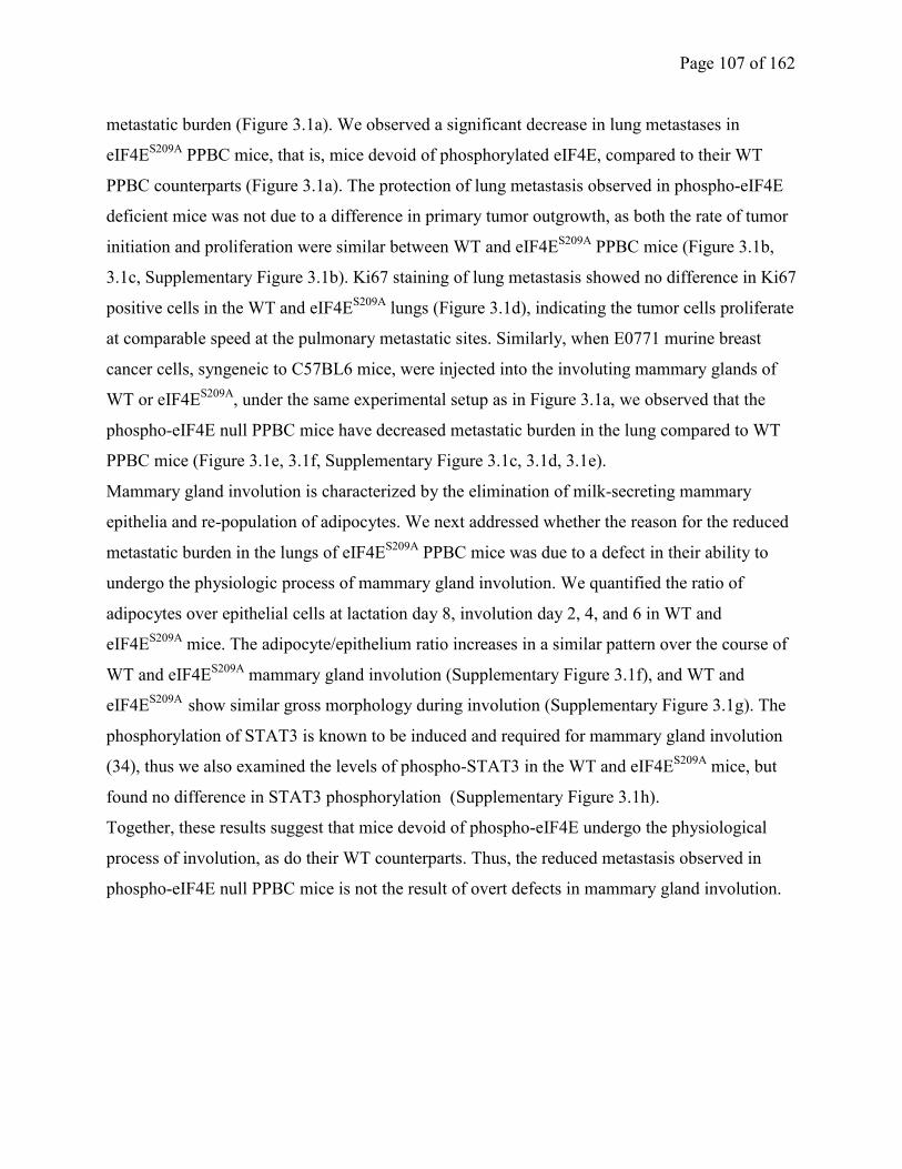
Page 107 of 162
metastatic burden (Figure 3.1a). We observed a significant decrease in lung metastases in
eIF4ES209A
PPBC mice, that is, mice devoid of phosphorylated eIF4E, compared to their WT
PPBC counterparts (Figure 3.1a). The protection of lung metastasis observed in phospho-eIF4E
deficient mice was not due to a difference in primary tumor outgrowth, as both the rate of tumor
initiation and proliferation were similar between WT and eIF4ES209A
PPBC mice (Figure 3.1b,
3.1c, Supplementary Figure 3.1b). Ki67 staining of lung metastasis showed no difference in Ki67
positive cells in the WT and eIF4ES209A
lungs (Figure 3.1d), indicating the tumor cells proliferate
at comparable speed at the pulmonary metastatic sites. Similarly, when E0771 murine breast
cancer cells, syngeneic to C57BL6 mice, were injected into the involuting mammary glands of
WT or eIF4ES209A
, under the same experimental setup as in Figure 3.1a, we observed that the
phospho-eIF4E null PPBC mice have decreased metastatic burden in the lung compared to WT
PPBC mice (Figure 3.1e, 3.1f, Supplementary Figure 3.1c, 3.1d, 3.1e).
Mammary gland involution is characterized by the elimination of milk-secreting mammary
epithelia and re-population of adipocytes. We next addressed whether the reason for the reduced
metastatic burden in the lungs of eIF4ES209A
PPBC mice was due to a defect in their ability to
undergo the physiologic process of mammary gland involution. We quantified the ratio of
adipocytes over epithelial cells at lactation day 8, involution day 2, 4, and 6 in WT and
eIF4ES209A
mice. The adipocyte/epithelium ratio increases in a similar pattern over the course of
WT and eIF4ES209A
mammary gland involution (Supplementary Figure 3.1f), and WT and
eIF4ES209A
show similar gross morphology during involution (Supplementary Figure 3.1g). The
phosphorylation of STAT3 is known to be induced and required for mammary gland involution
(34), thus we also examined the levels of phospho-STAT3 in the WT and eIF4ES209A
mice, but
found no difference in STAT3 phosphorylation (Supplementary Figure 3.1h).
Together, these results suggest that mice devoid of phospho-eIF4E undergo the physiological
process of involution, as do their WT counterparts. Thus, the reduced metastasis observed in
phospho-eIF4E null PPBC mice is not the result of overt defects in mammary gland involution.
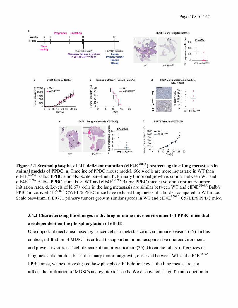
Page 108 of 162
3.4.2 Characterizing the changes in the lung immune microenvironment of PPBC mice that
are dependent on the phosphorylation of eIF4E
One important mechanism used by cancer cells to metastasize is via immune evasion (35). In this
context, infiltration of MDSCs is critical to support an immunosuppressive microenvironment,
and prevent cytotoxic T cell-dependent tumor eradication (35). Given the robust differences in
lung metastatic burden, but not primary tumor outgrowth, observed between WT and eIF4ES209A
PPBC mice, we next investigated how phospho-eIF4E deficiency at the lung metastatic site
affects the infiltration of MDSCs and cytotoxic T cells. We discovered a significant reduction in
Figure 3.1 Stromal phospho-eIF4E deficient mutation (eIF4ES209A
) protects against lung metastasis in
animal models of PPBC. a. Timeline of PPBC mouse model. 66cl4 cells are more metastatic in WT than
eIF4ES209A
Balb/c PPBC animals. Scale bar=4mm. b. Primary tumor outgrowth is similar between WT and
eIF4ES209A
Balb/c PPBC animals. c. WT and eIF4ES209A
Balb/c PPBC mice have similar primary tumor
initiation rates. d. Levels of Ki67+ cells in the lung metastasis are similar between WT and eIF4ES209A
Balb/c
PPBC mice. e. eIF4ES209A
C57BL/6 PPBC mice have reduced lung metastatic burden compared to WT mice.
Scale bar=4mm. f. E0771 primary tumors grow at similar speeds in WT and eIF4ES209A
C57BL/6 PPBC mice.
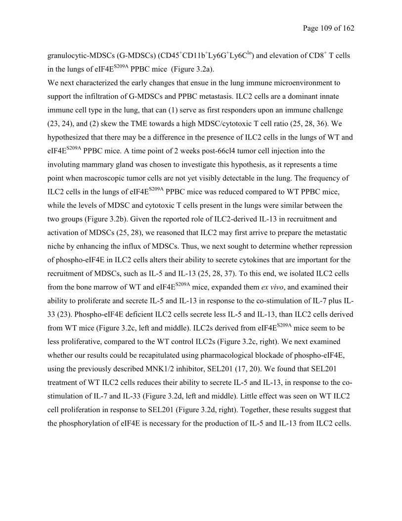
Page 109 of 162
granulocytic-MDSCs (G-MDSCs) (CD45+CD11b
+Ly6G
+Ly6C
lo) and elevation of CD8
+ T cells
in the lungs of eIF4ES209A
PPBC mice (Figure 3.2a).
We next characterized the early changes that ensue in the lung immune microenvironment to
support the infiltration of G-MDSCs and PPBC metastasis. ILC2 cells are a dominant innate
immune cell type in the lung, that can (1) serve as first responders upon an immune challenge
(23, 24), and (2) skew the TME towards a high MDSC/cytotoxic T cell ratio (25, 28, 36). We
hypothesized that there may be a difference in the presence of ILC2 cells in the lungs of WT and
eIF4ES209A
PPBC mice. A time point of 2 weeks post-66cl4 tumor cell injection into the
involuting mammary gland was chosen to investigate this hypothesis, as it represents a time
point when macroscopic tumor cells are not yet visibly detectable in the lung. The frequency of
ILC2 cells in the lungs of eIF4ES209A
PPBC mice was reduced compared to WT PPBC mice,
while the levels of MDSC and cytotoxic T cells present in the lungs were similar between the
two groups (Figure 3.2b). Given the reported role of ILC2-derived IL-13 in recruitment and
activation of MDSCs (25, 28), we reasoned that ILC2 may first arrive to prepare the metastatic
niche by enhancing the influx of MDSCs. Thus, we next sought to determine whether repression
of phospho-eIF4E in ILC2 cells alters their ability to secrete cytokines that are important for the
recruitment of MDSCs, such as IL-5 and IL-13 (25, 28, 37). To this end, we isolated ILC2 cells
from the bone marrow of WT and eIF4ES209A
mice, expanded them ex vivo, and examined their
ability to proliferate and secrete IL-5 and IL-13 in response to the co-stimulation of IL-7 plus IL-
33 (23). Phospho-eIF4E deficient ILC2 cells secrete less IL-5 and IL-13, than ILC2 cells derived
from WT mice (Figure 3.2c, left and middle). ILC2s derived from eIF4ES209A
mice seem to be
less proliferative, compared to the WT control ILC2s (Figure 3.2c, right). We next examined
whether our results could be recapitulated using pharmacological blockade of phospho-eIF4E,
using the previously described MNK1/2 inhibitor, SEL201 (17, 20). We found that SEL201
treatment of WT ILC2 cells reduces their ability to secrete IL-5 and IL-13, in response to the co-
stimulation of IL-7 and IL-33 (Figure 3.2d, left and middle). Little effect was seen on WT ILC2
cell proliferation in response to SEL201 (Figure 3.2d, right). Together, these results suggest that
the phosphorylation of eIF4E is necessary for the production of IL-5 and IL-13 from ILC2 cells.
Page 110 of 162
Figure 3.2. phospho-eIF4E-dependent ILC2 expansion and activation supports PPBC immune evasion. a. eIF4E
S209A Balb/c PPBC mice have increased total and cytotoxic T cells, as well as decreased G-MDSCs in the
lungs after 33 days post 66cl4 injection. b. eIF4ES209A
Balb/c PPBC lungs have decreased ILC2 cells than WT
PPBC lungs after 14 days post 66cl4 injection, but the levels of T cells and MDSCs are similar. c. eIF4ES209A
ILC2s proliferate slower and produces less IL-5 and IL-13 than WT ILC2s. d. SEL201 reduces IL-5 and IL-13
production from WT ILC2s.
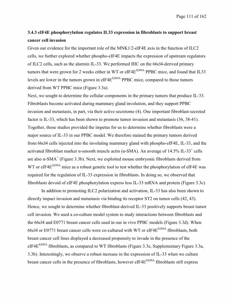
Page 111 of 162
3.4.3 eIF4E phosphorylation regulates IL33 expression in fibroblasts to support breast
cancer cell invasion
Given our evidence for the important role of the MNK1/2-eIF4E axis in the function of ILC2
cells, we further explored whether phospho-eIF4E impacts the expression of upstream regulators
of ILC2 cells, such as the alarmin IL-33. We performed IHC on the 66cl4-derived primary
tumors that were grown for 2 weeks either in WT or eIF4ES209A
PPBC mice, and found that IL33
levels are lower in the tumors grown in eIF4ES209A
PPBC mice, compared to those tumors
derived from WT PPBC mice (Figure 3.3a).
Next, we sought to determine the cellular components in the primary tumors that produce IL-33.
Fibroblasts become activated during mammary gland involution, and they support PPBC
invasion and metastasis, in part, via their active secretome (4). One important fibroblast-secreted
factor is IL-33, which has been shown to promote tumor invasion and metastasis (36, 38-41).
Together, those studies provided the impetus for us to determine whether fibroblasts were a
major source of IL-33 in our PPBC model. We therefore stained the primary tumors derived
from 66cl4 cells injected into the involuting mammary gland with phospho-eIF4E, IL-33, and the
activated fibroblast marker α-smooth muscle actin (α-SMA). An average of 14.5% IL-33+ cells
are also α-SMA+ (Figure 3.3b). Next, we exploited mouse embryonic fibroblasts derived from
WT or eIF4ES209A
mice as a robust genetic tool to test whether the phosphorylation of eIF4E was
required for the regulation of IL-33 expression in fibroblasts. In doing so, we observed that
fibroblasts devoid of eIF4E phosphorylation express less IL-33 mRNA and protein (Figure 3.3c).
In addition to promoting ILC2 polarization and activation, IL-33 has also been shown to
directly impact invasion and metastasis via binding its receptor ST2 on tumor cells (42, 43).
Hence, we sought to determine whether fibroblast-derived IL-33 positively supports breast tumor
cell invasion. We used a co-culture model system to study interactions between fibroblasts and
the 66cl4 and E0771 breast cancer cells used in our in vivo PPBC models (Figure 3.3d). When
66cl4 or E0771 breast cancer cells were co-cultured with WT or eIF4ES209A
fibroblasts, both
breast cancer cell lines displayed a decreased propensity to invade in the presence of the
eIF4ES209A
fibroblasts, as compared to WT fibroblasts (Figure 3.3e, Supplementary Figure 3.3a,
3.3b). Interestingly, we observe a robust increase in the expression of IL-33 when we culture
breast cancer cells in the presence of fibroblasts, however eIF4ES209A
fibroblasts still express
Page 112 of 162
Figure 3.3 phospho-eIF4E deficiency inhibits fibroblast-derived IL-33 expression and tumor cell invasion. a. WT
tumors express more IL-33 than eIF4ES209A
tumors. b. Co-localization of IL-33 with α-SMA+ cells in WT tumors.
c. eIF4ES209A
fibroblasts express less IL-33 than WT. d. Timeline of fibroblast-tumor cell co-culture. e. 66cl4 migrates less
towards eIF4ES209A
fibroblasts. f. eIF4ES209A
fibroblasts express less IL-33 compared to WT fibroblasts when co-cultured
with 66cl4 cells, and IL-33 expression is further induced in WT but not eIF4ES209A
fibroblasts by 66cl4 cells. g. ST2
knockdown diminishes 66cl4 invasion towards WT fibroblasts. h. i. SEL201 inhibits 66cl4 and E0771 invasion towards
fibroblasts and suppresses IL33 expression. j. SEL201 suppresses MDA-MB-231 invasion towards patient derived CAFs.
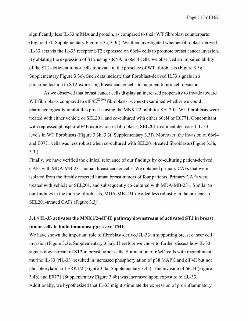
Page 113 of 162
significantly less IL-33 mRNA and protein, as compared to their WT fibroblast counterparts
(Figure 3.3f, Supplementary Figure 3.3c, 3.3d). We then investigated whether fibroblast-derived
IL-33 acts via the IL-33 receptor ST2 expressed on 66cl4 cells to promote breast cancer invasion.
By ablating the expression of ST2 using siRNA in 66cl4 cells, we observed an impaired ability
of the ST2-deficient tumor cells to invade in the presence of WT fibroblasts (Figure 3.3g,
Supplementary Figure 3.3e). Such data indicate that fibroblast-derived IL33 signals in a
paracrine fashion to ST2-expressing breast cancer cells to augment tumor cell invasion.
As we observed that breast cancer cells display an increased propensity to invade toward
WT fibroblasts compared to eIF4ES209A
fibroblasts, we next examined whether we could
pharmacologically inhibit this process using the MNK1/2 inhibitor SEL201. WT fibroblasts were
treated with either vehicle or SEL201, and co-cultured with either 66cl4 or E0771. Concomitant
with repressed phospho-eIF4E expression in fibroblasts, SEL201 treatment decreased IL-33
levels in WT fibroblasts (Figure 3.3h, 3.3i, Supplementary 3.3f). Moreover, the invasion of 66cl4
and E0771 cells was less robust when co-cultured with SEL201-treated fibroblasts (Figure 3.3h,
3.3i).
Finally, we have verified the clinical relevance of our findings by co-culturing patient-derived
CAFs with MDA-MB-231 human breast cancer cells. We obtained primary CAFs that were
isolated from the freshly resected human breast tumors of four patients. Primary CAFs were
treated with vehicle or SEL201, and subsequently co-cultured with MDA-MB-231. Similar to
our findings in the murine fibroblasts, MDA-MB-231 invaded less robustly in the presence of
SEL201-treated CAFs (Figure 3.3j).
3.4.4 IL-33 activates the MNK1/2-eIF4E pathway downstream of activated ST2 in breast
tumor cells to build immunosuppressive TME
We have shown the important role of fibroblast-derived IL-33 in supporting breast cancer cell
invasion (Figure 3.3e, Supplementary 3.3a). Therefore we chose to further dissect how IL-33
signals downstream of ST2 in breast tumor cells. Stimulation of 66cl4 cells with recombinant
murine IL-33 (rIL-33) resulted in increased phosphorylation of p38 MAPK and eIF4E but not
phosphorylation of ERK1/2 (Figure 3.4a, Supplementary 3.4a). The invasion of 66cl4 (Figure
3.4b) and E0771 (Supplementary Figure 3.4b) was increased upon exposure to rIL-33.
Additionally, we hypothesized that IL-33 might stimulate the expression of pro-inflammatory
Page 114 of 162
and pro-tumorigenic cytokines/chemokines in tumor cells. IL-33-stimulated the mRNA
expression of CXCL-1, CCL-17, GM-CSF and IL-6, without significantly affecting IL-4 and
CXCL-2 levels (Figure 3.4c, Supplementary Figure 3.4c). Given the reported functions of
CXCL-1, CCL-17, GM-CSF and IL-6 in cancer immune evasion (44-52), our data provides
evidence to show that IL-33 may serve as an upstream signal to initiate such immunosuppressive
signaling cascade in the PPBC TME to facilitate metastasis.
3.4.5 Increased therapeutic efficacy of blocking MNK1/2 combined with anti–PD1
immunotherapy
Accumulating evidence has implicated the central role of immunosuppressive cells including
MDSCs and CAFs in resistance to immune checkpoint blockade (53, 54). The efficacy of
immune checkpoint inhibitors in PPBC would likely be improved by overcoming this immune
suppression. Given the diverse role of phospho-eIF4E in contributing to PPBC immune evasion
and the increased presence of CD8+ T cells in the lungs of phospho-eIF4E deficient PPBC mice,
we hypothesized that blocking the phosphorylation of eIF4E using the MNK1/2 inhibitor
SEL201 might alleviate PPBC immunosuppression and sensitize tumor bearing mice to the anti-
tumor effects of PD1 blockade.
To test this hypothesis, WT PPBC mice were treated with vehicle, SEL201, anti-PD1 antibody,
or the combination of SEL201 plus anti-PD1 antibody (Figure 3.5a). Remarkably, SEL201 plus
anti-PD1 blockade decreased PPBC lung metastasis, while SEL201 or anti-PD1 alone did not
show any significant anti-metastatic effects (Figure 3.5b). The reduction in metastatic burden in
Num
ber
of 66cl4
cells
mig
rate
d
0
50
100
150p<0.0001
Vehicle rIL-33
a b
p38
eIF4E
GAPDH
p-eIF4E
p-p38
Vehicle rIL-33
c
0
20
40
60
80
CXCL-1
Vehicle rIL-33
0
5
10
15
20
CCL-17
Vehicle rIL-33
0
5
10
15
GM-CSF
Vehicle rIL-33
IL-6
0
1
2
3
4
Vehicle rIL-33
p<0.000125
p<0.0001p<0.0001 p=0.0002
mR
NA
Fold
change
Ve
hic
le
rIL
33
Figure 3.4 Exogenous IL33 induces tumor cell invasion and upregulates selective
chemokines and cytokines. a. IL33 induces p38/eIF4E phosphorylation. b. IL33 induces 66cl4
invasion. c. IL33 induces expressions of CXCL1, IL-6, CCL-17 and GM-CSF in 66cl4 cells.
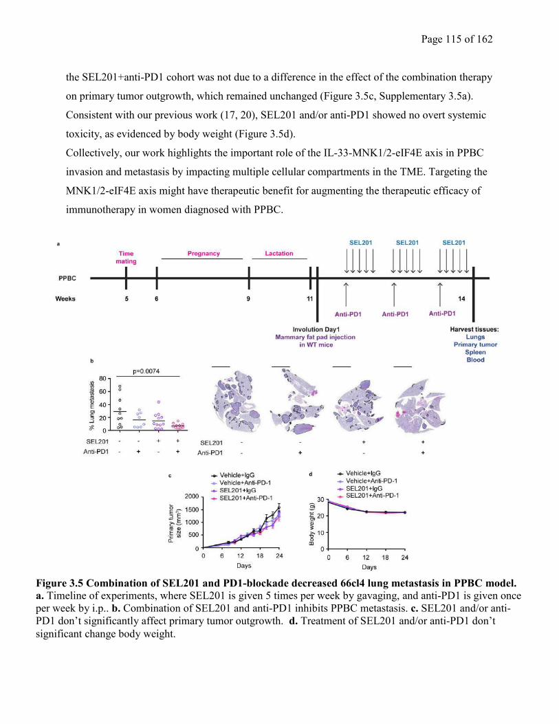
Page 115 of 162
the SEL201+anti-PD1 cohort was not due to a difference in the effect of the combination therapy
on primary tumor outgrowth, which remained unchanged (Figure 3.5c, Supplementary 3.5a).
Consistent with our previous work (17, 20), SEL201 and/or anti-PD1 showed no overt systemic
toxicity, as evidenced by body weight (Figure 3.5d).
Collectively, our work highlights the important role of the IL-33-MNK1/2-eIF4E axis in PPBC
invasion and metastasis by impacting multiple cellular compartments in the TME. Targeting the
MNK1/2-eIF4E axis might have therapeutic benefit for augmenting the therapeutic efficacy of
immunotherapy in women diagnosed with PPBC.
Figure 3.5 Combination of SEL201 and PD1-blockade decreased 66cl4 lung metastasis in PPBC model.
a. Timeline of experiments, where SEL201 is given 5 times per week by gavaging, and anti-PD1 is given once
per week by i.p.. b. Combination of SEL201 and anti-PD1 inhibits PPBC metastasis. c. SEL201 and/or anti-
PD1 don’t significantly affect primary tumor outgrowth. d. Treatment of SEL201 and/or anti-PD1 don’t
significant change body weight.
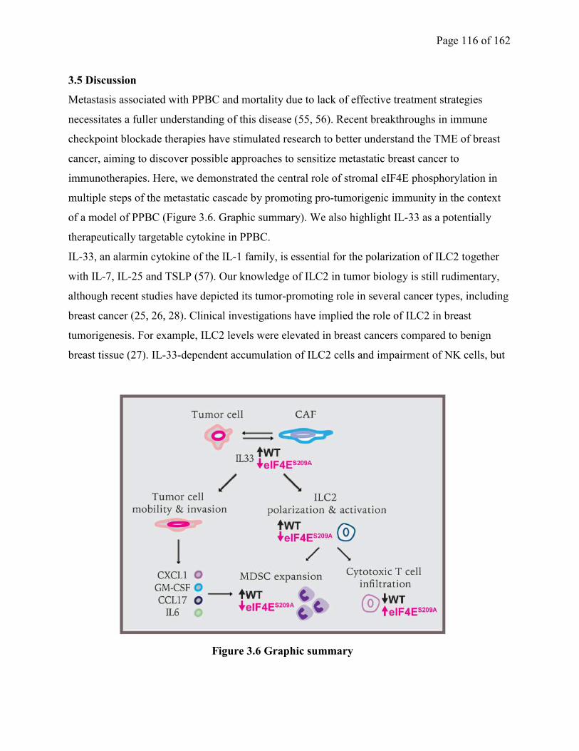
Page 116 of 162
3.5 Discussion
Metastasis associated with PPBC and mortality due to lack of effective treatment strategies
necessitates a fuller understanding of this disease (55, 56). Recent breakthroughs in immune
checkpoint blockade therapies have stimulated research to better understand the TME of breast
cancer, aiming to discover possible approaches to sensitize metastatic breast cancer to
immunotherapies. Here, we demonstrated the central role of stromal eIF4E phosphorylation in
multiple steps of the metastatic cascade by promoting pro-tumorigenic immunity in the context
of a model of PPBC (Figure 3.6. Graphic summary). We also highlight IL-33 as a potentially
therapeutically targetable cytokine in PPBC.
IL-33, an alarmin cytokine of the IL-1 family, is essential for the polarization of ILC2 together
with IL-7, IL-25 and TSLP (57). Our knowledge of ILC2 in tumor biology is still rudimentary,
although recent studies have depicted its tumor-promoting role in several cancer types, including
breast cancer (25, 26, 28). Clinical investigations have implied the role of ILC2 in breast
tumorigenesis. For example, ILC2 levels were elevated in breast cancers compared to benign
breast tissue (27). IL-33-dependent accumulation of ILC2 cells and impairment of NK cells, but
Figure 3.6 Graphic summary
Page 117 of 162
not cytotoxic T cell functions, has been shown to facilitate breast cancer metastasis (36). In our
pre-clinical mouse model of PPBC, we showed that hosts devoid of eIF4E phosphorylation
presented with an elevated influx of cytotoxic T cells in the lungs, possibly due to an overriding
of an immune suppressive ILC2-MDSC axis. Our results in PPBC are in line with the innate and
adaptive immuno-crosstalk that exist in other models (25, 28).
In addition to its impact on ILC2 cells, IL-33 has also been reported as a multi-functional pro-
tumorigenic cytokine that can maintain cancer stem cell properties, alter cancer cell metabolism,
and facilitate tumor cell proliferation (42, 58, 59). IL-33 executes such functions via signaling
through its ST2 receptor, and elevation of IL-33 was observed in the serum of breast cancer
patients (60, 61). High levels of IL-33 were observed to predict tamoxifen resistance in ER+
breast cancer (62). More importantly, levels of matrix metallopeptidase 11 (MMP11), a pro-
invasive enzyme responsible for tissue remodeling, are directly correlated to IL-33 levels in
breast cancer patients (61), supporting a possible pro-invasive function of IL-33. Additionally,
we have also demonstrated that phospho-eIF4E-mediated IL-33 production from fibroblasts is
further induced when co-cultured with breast tumor cells. Thus, our data provided a novel
mechanism where breast cancer cells educate fibroblasts to secrete more IL-33, thus allowing
breast cancer cells to gain more invasive properties.
Additionally, it is also acknowledged that IL-33 reinforces pro-tumorigenic inflammation by
inducing IL-6 (43). Our study has not only verified this finding in breast cancer but also
expanded the repertoire of IL-33-induced cytokines and chemokines produced by cancer cells, as
we showed that IL-33 induces the expression of CXCL-1, CCL-17, IL-6 and GM-CSF in breast
cancer cells. The significance of these four factors in tumor immune evasion has been supported
by multiple previous reports. For example, over-expression of CXCL-1 and its receptor CXCR-
2, as well as elevated circulating IL-6 levels, are all correlated to breast cancer metastasis and
poor survival rate (63, 64), and CXCL-1, IL-6 and GM-CSF are all potent mediators for the
recruitment and expansion of MDSCs and M2-like macrophages (45-48, 50-52). CCL-17, an
important ligand for CCR-4, has also been demonstrated to elicit Th2 and Treg-mediated cancer
immune evasion (44, 49). Taken together, we have provided novel evidence that IL-33 may act
directly on breast tumor cells to induce further expression of selected immunosuppressive
chemokines and cytokines.
Page 118 of 162
Finally, immune checkpoint blockades designed to release the brakes on exhausted cytotoxic T
cells have largely improved the patient prognosis in several cancers (65), but are less effective to
date in breast cancer. It is proposed that many breast cancers present with failed or suboptimal T
cell priming. Given our data supporting the notion that deficiency of eIF4E phosphorylation
enhances T cell infiltration, scheduling of MNK1/2 inhibitor administration will be important in
future clinical trials, given that blocking MNK1/2 may be more effective when used to prime the
TME by ameliorating overall immunosuppression and facilitating cytotoxic T cell infiltration.
Indeed, the anti-metastatic effect of a phospho-eIF4E deficient host was accompanied by
increased cytotoxic T cell infiltration in our PPBC animal model (Figure 3.6). We propose the
use of MNK1/2 inhibitors as a tool to convert “cold” breast tumors to “hot” tumors, thus offering
the opportunity for immune checkpoint blockade to become more effective in highly metastatic
cancers such as PPBC.
3.6 Acknowledgments
This research is funded by grants from the Cancer Research Society (PIN20239 to W. Miller),
the Canadian Institutes for Health Research (MOP-142281 to W. Miller), the Canadian Institutes
for Health Research (PJT-156269 to W. Miller and S.V. del Rincón), the Canadian Cancer
Society
(703811 to W. Miller and S.V. del Rincon), the Rossy Cancer Network (to W. Miller), and the
Israel Cancer Research Fund (to W. Miller and S.V. del Rincón). Q. Guo was supported by a
McGill Faculty of Medicine graduate studentship. F. Huang was supported by MICRTP graduate
studentships. V.Z. Li was supported by a McGill Integrated Cancer Research Training Program
(MICRTP) undergraduate studentship. We thank Selvita S.A. for supplying SEL201,the Flow
Cytometry Cores of the Lady Davis Institute and Goodman Cancer Centre, and the Animal Core
Facilities of the Lady Davis Institute for their support.
3.7 References
1. Goddard ET, Bassale S, Schedin T, Jindal S, Johnston J, Cabral E, et al. Association
Between Postpartum Breast Cancer Diagnosis and Metastasis and the Clinical Features
Underlying Risk. JAMA Netw Open. 2019;2(1):e186997.
2. Schedin P. Pregnancy-associated breast cancer and metastasis. Nature reviews Cancer.
2006;6(4):281-91.
Page 119 of 162
3. Martinson HA, Jindal S, Durand-Rougely C, Borges VF, and Schedin P. Wound healing-
like immune program facilitates postpartum mammary gland involution and tumor progression.
International journal of cancer Journal international du cancer. 2015;136(8):1803-13.
4. Guo Q, Minnier J, Burchard J, Chiotti K, Spellman P, and Schedin P. Physiologically
activated mammary fibroblasts promote postpartum mammary cancer. JCI Insight.
2017;2(6):e89206.
5. O'Brien J, Lyons T, Monks J, Lucia MS, Wilson RS, Hines L, et al. Alternatively
activated macrophages and collagen remodeling characterize the postpartum involuting
mammary gland across species. The American journal of pathology. 2010;176(3):1241-55.
6. Pennock ND, Martinson HA, Guo Q, Betts CB, Jindal S, Tsujikawa T, et al. Ibuprofen
supports macrophage differentiation, T cell recruitment, and tumor suppression in a model of
postpartum breast cancer. J Immunother Cancer. 2018;6(1):98.
7. Ruggero D. Translational control in cancer etiology. Cold Spring Harbor perspectives in
biology. 2013;5(2).
8. Bhat M, Robichaud N, Hulea L, Sonenberg N, Pelletier J, and Topisirovic I. Targeting
the translation machinery in cancer. Nature reviews Drug discovery. 2015;14(4):261-78.
9. Assouline S, Culjkovic B, Cocolakis E, Rousseau C, Beslu N, Amri A, et al. Molecular
targeting of the oncogene eIF4E in acute myeloid leukemia (AML): a proof-of-principle clinical
trial with ribavirin. Blood. 2009;114(2):257-60.
10. Carter JH, Deddens JA, Spaulding NI, Lucas D, Colligan BM, Lewis TG, et al.
Phosphorylation of eIF4E serine 209 is associated with tumour progression and reduced survival
in malignant melanoma. British journal of cancer. 2016;114(4):444-53.
11. Furic L, Rong L, Larsson O, Koumakpayi IH, Yoshida K, Brueschke A, et al. eIF4E
phosphorylation promotes tumorigenesis and is associated with prostate cancer progression. Proc
Natl Acad Sci U S A. 2010;107(32):14134-9.
12. Li BD, Liu L, Dawson M, and De Benedetti A. Overexpression of eukaryotic initiation
factor 4E (eIF4E) in breast carcinoma. Cancer. 1997;79(12):2385-90.
13. Lim S, Saw TY, Zhang M, Janes MR, Nacro K, Hill J, et al. Targeting of the MNK-
eIF4E axis in blast crisis chronic myeloid leukemia inhibits leukemia stem cell function. Proc
Natl Acad Sci U S A. 2013;110(25):E2298-307.
14. Zhou S, Wang GP, Liu C, and Zhou M. Eukaryotic initiation factor 4E (eIF4E) and
angiogenesis: prognostic markers for breast cancer. BMC cancer. 2006;6:231.
15. Bianchini A, Loiarro M, Bielli P, Busa R, Paronetto MP, Loreni F, et al. Phosphorylation
of eIF4E by MNKs supports protein synthesis, cell cycle progression and proliferation in prostate
cancer cells. Carcinogenesis. 2008;29(12):2279-88.
16. Geter PA, Ernlund AW, Bakogianni S, Alard A, Arju R, Giashuddin S, et al. Hyperactive
mTOR and MNK1 phosphorylation of eIF4E confer tamoxifen resistance and estrogen
independence through selective mRNA translation reprogramming. Genes & development.
2017;31(22):2235-49.
17. Guo Q, Li VZ, Nichol JN, Huang F, Yang W, Preston SEJ, et al. MNK1/NODAL
signaling promotes invasive progression of breast ductal carcinoma in situ. Cancer Res. 2019.
18. Robichaud N, Del Rincon SV, Huor B, Alain T, Petruccelli LA, Hearnden J, et al.
Phosphorylation of eIF4E promotes EMT and metastasis via translational control of SNAIL and
MMP-3. 2014.
Page 120 of 162
19. Wheater MJ, Johnson PW, and Blaydes JP. The role of MNK proteins and eIF4E
phosphorylation in breast cancer cell proliferation and survival. Cancer biology & therapy.
2010;10(7):728-35.
20. Zhan Y, Guo J, Yang W, Goncalves C, Rzymski T, Dreas A, et al. MNK1/2 inhibition
limits oncogenicity and metastasis of KIT-mutant melanoma. The Journal of clinical
investigation. 2017.
21. Robichaud N, Hsu BE, Istomine R, Alvarez F, Blagih J, Ma EH, et al. Translational
control in the tumor microenvironment promotes lung metastasis: Phosphorylation of eIF4E in
neutrophils. Proc Natl Acad Sci U S A. 2018;115(10):E2202-E9.
22. Peinado H, Zhang H, Matei IR, Costa-Silva B, Hoshino A, Rodrigues G, et al. Pre-
metastatic niches: organ-specific homes for metastases. Nature reviews Cancer. 2017;17(5):302-
17.
23. Duerr CU, McCarthy CD, Mindt BC, Rubio M, Meli AP, Pothlichet J, et al. Type I
interferon restricts type 2 immunopathology through the regulation of group 2 innate lymphoid
cells. Nature immunology. 2016;17(1):65-75.
24. Mindt BC, Fritz JH, and Duerr CU. Group 2 Innate Lymphoid Cells in Pulmonary
Immunity and Tissue Homeostasis. Front Immunol. 2018;9:840.
25. Chevalier MF, Trabanelli S, Racle J, Salome B, Cesson V, Gharbi D, et al. ILC2-
modulated T cell-to-MDSC balance is associated with bladder cancer recurrence. The Journal of
clinical investigation. 2017;127(8):2916-29.
26. Li J, Razumilava N, Gores GJ, Walters S, Mizuochi T, Mourya R, et al. Biliary repair and
carcinogenesis are mediated by IL-33-dependent cholangiocyte proliferation. The Journal of
clinical investigation. 2014;124(7):3241-51.
27. Salimi M, Wang R, Yao X, Li X, Wang X, Hu Y, et al. Activated innate lymphoid cell
populations accumulate in human tumour tissues. BMC cancer. 2018;18(1):341.
28. Trabanelli S, Chevalier MF, Martinez-Usatorre A, Gomez-Cadena A, Salome B, Lecciso
M, et al. Tumour-derived PGD2 and NKp30-B7H6 engagement drives an immunosuppressive
ILC2-MDSC axis. Nat Commun. 2017;8(1):593.
29. Lyons TR, O'Brien J, Borges VF, Conklin MW, Keely PJ, Eliceiri KW, et al. Postpartum
mammary gland involution drives progression of ductal carcinoma in situ through collagen and
COX-2. Nature medicine. 2011;17(9):1109-15.
30. Lyons TR, Borges VF, Betts CB, Guo Q, Kapoor P, Martinson HA, et al.
Cyclooxygenase-2-dependent lymphangiogenesis promotes nodal metastasis of postpartum
breast cancer. The Journal of clinical investigation. 2014;124(9):3901-12.
31. Hosein AN, Wu M, Arcand SL, Lavallee S, Hebert J, Tonin PN, et al. Breast carcinoma-
associated fibroblasts rarely contain p53 mutations or chromosomal aberrations. Cancer Res.
2010;70(14):5770-7.
32. Pettersson F, Del Rincon SV, Emond A, Huor B, Ngan E, Ng J, et al. Genetic and
pharmacologic inhibition of eIF4E reduces breast cancer cell migration, invasion, and metastasis.
Cancer Res. 2015;75(6):1102-12.
33. Dahabieh MS, Ha Z, Di Pietro E, Nichol JN, Bolt AM, Goncalves C, et al. Peroxisomes
protect lymphoma cells from HDAC inhibitor-mediated apoptosis. Cell death and differentiation.
2017;24(11):1912-24.
34. Chapman RS, Lourenco PC, Tonner E, Flint DJ, Selbert S, Takeda K, et al. Suppression
of epithelial apoptosis and delayed mammary gland involution in mice with a conditional
knockout of Stat3. Genes & development. 1999;13(19):2604-16.
Page 121 of 162
35. Chen DS, and Mellman I. Oncology meets immunology: the cancer-immunity cycle.
Immunity. 2013;39(1):1-10.
36. Jovanovic IP, Pejnovic NN, Radosavljevic GD, Pantic JM, Milovanovic MZ, Arsenijevic
NN, et al. Interleukin-33/ST2 axis promotes breast cancer growth and metastases by facilitating
intratumoral accumulation of immunosuppressive and innate lymphoid cells. International
journal of cancer Journal international du cancer. 2014;134(7):1669-82.
37. Quail DF, Olson OC, Bhardwaj P, Walsh LA, Akkari L, Quick ML, et al. Obesity alters
the lung myeloid cell landscape to enhance breast cancer metastasis through IL5 and GM-CSF.
Nature cell biology. 2017;19(8):974-87.
38. Chen SF, Nieh S, Jao SW, Wu MZ, Liu CL, Chang YC, et al. The paracrine effect of
cancer-associated fibroblast-induced interleukin-33 regulates the invasiveness of head and neck
squamous cell carcinoma. The Journal of pathology. 2013;231(2):180-9.
39. Dahlgren MW, Jones SW, Cautivo KM, Dubinin A, Ortiz-Carpena JF, Farhat S, et al.
Adventitial Stromal Cells Define Group 2 Innate Lymphoid Cell Tissue Niches. Immunity.
2019;50(3):707-22 e6.
40. Eckhardt BL, Parker BS, van Laar RK, Restall CM, Natoli AL, Tavaria MD, et al.
Genomic analysis of a spontaneous model of breast cancer metastasis to bone reveals a role for
the extracellular matrix. Mol Cancer Res. 2005;3(1):1-13.
41. Yang Y, Andersson P, Hosaka K, Zhang Y, Cao R, Iwamoto H, et al. The PDGF-BB-
SOX7 axis-modulated IL-33 in pericytes and stromal cells promotes metastasis through tumour-
associated macrophages. Nat Commun. 2016;7:11385.
42. Fang M, Li Y, Huang K, Qi S, Zhang J, Zgodzinski W, et al. IL33 Promotes Colon
Cancer Cell Stemness via JNK Activation and Macrophage Recruitment. Cancer Res.
2017;77(10):2735-45.
43. Yu XX, Hu Z, Shen X, Dong LY, Zhou WZ, and Hu WH. IL-33 Promotes Gastric
Cancer Cell Invasion and Migration Via ST2-ERK1/2 Pathway. Dig Dis Sci. 2015;60(5):1265-
72.
44. Berlato C, Khan MN, Schioppa T, Thompson R, Maniati E, Montfort A, et al. A CCR4
antagonist reverses the tumor-promoting microenvironment of renal cancer. The Journal of
clinical investigation. 2017;127(3):801-13.
45. Brenot A, Knolhoff BL, DeNardo DG, and Longmore GD. SNAIL1 action in tumor cells
influences macrophage polarization and metastasis in breast cancer through altered GM-CSF
secretion. Oncogenesis. 2018;7(3):32.
46. Chen L, Wang S, Wang Y, Zhang W, Ma K, Hu C, et al. IL-6 influences the polarization
of macrophages and the formation and growth of colorectal tumor. Oncotarget.
2018;9(25):17443-54.
47. Duluc D, Delneste Y, Tan F, Moles MP, Grimaud L, Lenoir J, et al. Tumor-associated
leukemia inhibitory factor and IL-6 skew monocyte differentiation into tumor-associated
macrophage-like cells. Blood. 2007;110(13):4319-30.
48. Miyake M, Hori S, Morizawa Y, Tatsumi Y, Nakai Y, Anai S, et al. CXCL1-Mediated
Interaction of Cancer Cells with Tumor-Associated Macrophages and Cancer-Associated
Fibroblasts Promotes Tumor Progression in Human Bladder Cancer. Neoplasia.
2016;18(10):636-46.
49. Mondini M, Loyher PL, Hamon P, Gerbe de Thore M, Laviron M, Berthelot K, et al.
CCR2-Dependent Recruitment of Tregs and Monocytes Following Radiotherapy Is Associated
with TNFalpha-Mediated Resistance. Cancer immunology research. 2019;7(3):376-87.
Page 122 of 162
50. Wang D, Sun H, Wei J, Cen B, and DuBois RN. CXCL1 Is Critical for Premetastatic
Niche Formation and Metastasis in Colorectal Cancer. Cancer Res. 2017;77(13):3655-65.
51. Weber R, Fleming V, Hu X, Nagibin V, Groth C, Altevogt P, et al. Myeloid-Derived
Suppressor Cells Hinder the Anti-Cancer Activity of Immune Checkpoint Inhibitors. Front
Immunol. 2018;9:1310.
52. Zhu H, Gu Y, Xue Y, Yuan M, Cao X, and Liu Q. CXCR2(+) MDSCs promote breast
cancer progression by inducing EMT and activated T cell exhaustion. Oncotarget.
2017;8(70):114554-67.
53. Kalluri R. The biology and function of fibroblasts in cancer. Nature reviews Cancer.
2016;16(9):582-98.
54. Veglia F, Perego M, and Gabrilovich D. Myeloid-derived suppressor cells coming of age.
Nature immunology. 2018;19(2):108-19.
55. Callihan EB, Gao D, Jindal S, Lyons TR, Manthey E, Edgerton S, et al. Postpartum
diagnosis demonstrates a high risk for metastasis and merits an expanded definition of
pregnancy-associated breast cancer. Breast cancer research and treatment. 2013;138(2):549-59.
56. Johansson AL, Andersson TM, Hsieh CC, Cnattingius S, and Lambe M. Increased
mortality in women with breast cancer detected during pregnancy and different periods
postpartum. Cancer Epidemiol Biomarkers Prev. 2011;20(9):1865-72.
57. Duerr CU, and Fritz JH. Regulation of group 2 innate lymphoid cells. Cytokine.
2016;87:1-8.
58. Li Y, Shi J, Qi S, Zhang J, Peng D, Chen Z, et al. IL-33 facilitates proliferation of
colorectal cancer dependent on COX2/PGE2. J Exp Clin Cancer Res. 2018;37(1):196.
59. Wang C, Chen Z, Bu X, Han Y, Shan S, Ren T, et al. IL-33 signaling fuels outgrowth and
metastasis of human lung cancer. Biochemical and biophysical research communications.
2016;479(3):461-8.
60. Lu DP, Zhou XY, Yao LT, Liu CG, Ma W, Jin F, et al. Serum soluble ST2 is associated
with ER-positive breast cancer. BMC cancer. 2014;14:198.
61. Yang ZP, Ling DY, Xie YH, Wu WX, Li JR, Jiang J, et al. The Association of Serum IL-
33 and sST2 with Breast Cancer. Dis Markers. 2015;2015:516895.
62. Hu H, Sun J, Wang C, Bu X, Liu X, Mao Y, et al. IL-33 facilitates endocrine resistance
of breast cancer by inducing cancer stem cell properties. Biochemical and biophysical research
communications. 2017;485(3):643-50.
63. Acharyya S, Oskarsson T, Vanharanta S, Malladi S, Kim J, Morris PG, et al. A CXCL1
paracrine network links cancer chemoresistance and metastasis. Cell. 2012;150(1):165-78.
64. Salgado R, Junius S, Benoy I, Van Dam P, Vermeulen P, Van Marck E, et al. Circulating
interleukin-6 predicts survival in patients with metastatic breast cancer. International journal of
cancer Journal international du cancer. 2003;103(5):642-6.
65. Sharma P, and Allison JP. The future of immune checkpoint therapy. Science.
2015;348(6230):56-61.
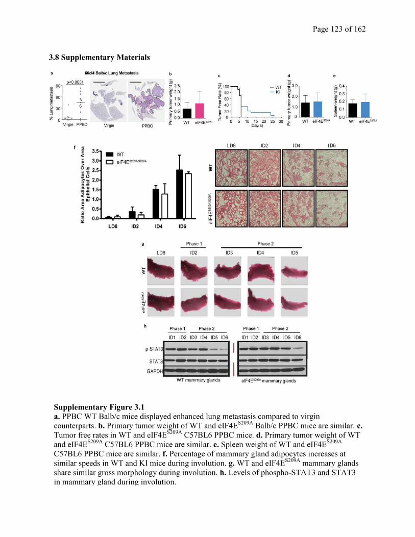
Page 123 of 162
3.8 Supplementary Materials
Supplementary Figure 3.1
a. PPBC WT Balb/c mice displayed enhanced lung metastasis compared to virgin
counterparts. b. Primary tumor weight of WT and eIF4ES209A
Balb/c PPBC mice are similar. c.
Tumor free rates in WT and eIF4ES209A
C57BL6 PPBC mice. d. Primary tumor weight of WT
and eIF4ES209A
C57BL6 PPBC mice are similar. e. Spleen weight of WT and eIF4ES209A
C57BL6 PPBC mice are similar. f. Percentage of mammary gland adipocytes increases at
similar speeds in WT and KI mice during involution. g. WT and eIF4ES209A
mammary glands
share similar gross morphology during involution. h. Levels of phospho-STAT3 and STAT3
in mammary gland during involution.
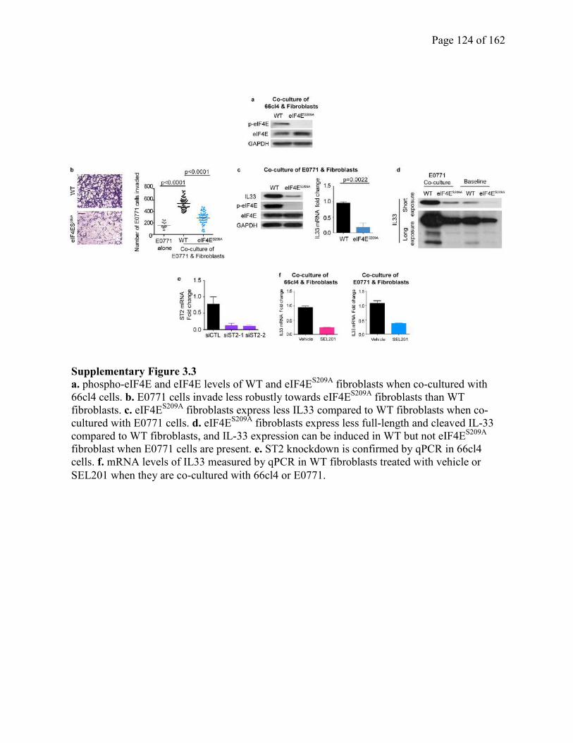
Page 124 of 162
Supplementary Figure 3.3
a. phospho-eIF4E and eIF4E levels of WT and eIF4ES209A
fibroblasts when co-cultured with
66cl4 cells. b. E0771 cells invade less robustly towards eIF4ES209A
fibroblasts than WT
fibroblasts. c. eIF4ES209A
fibroblasts express less IL33 compared to WT fibroblasts when co-
cultured with E0771 cells. d. eIF4ES209A
fibroblasts express less full-length and cleaved IL-33
compared to WT fibroblasts, and IL-33 expression can be induced in WT but not eIF4ES209A
fibroblast when E0771 cells are present. e. ST2 knockdown is confirmed by qPCR in 66cl4
cells. f. mRNA levels of IL33 measured by qPCR in WT fibroblasts treated with vehicle or
SEL201 when they are co-cultured with 66cl4 or E0771.
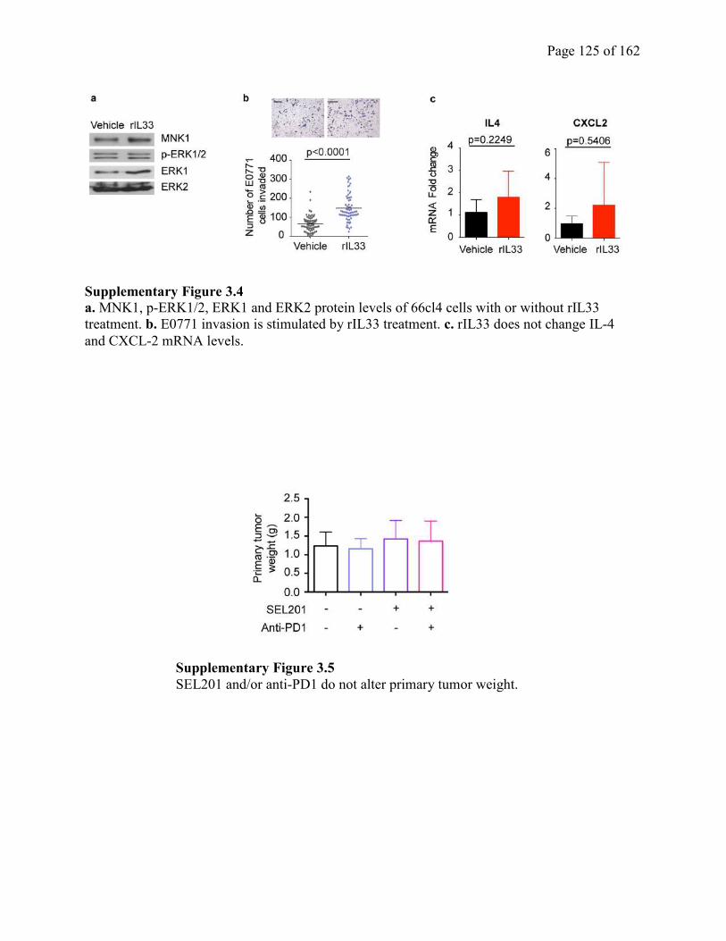
Page 125 of 162
Supplementary Figure 3.5
SEL201 and/or anti-PD1 do not alter primary tumor weight.
Supplementary Figure 3.4
a. MNK1, p-ERK1/2, ERK1 and ERK2 protein levels of 66cl4 cells with or without rIL33
treatment. b. E0771 invasion is stimulated by rIL33 treatment. c. rIL33 does not change IL-4
and CXCL-2 mRNA levels.
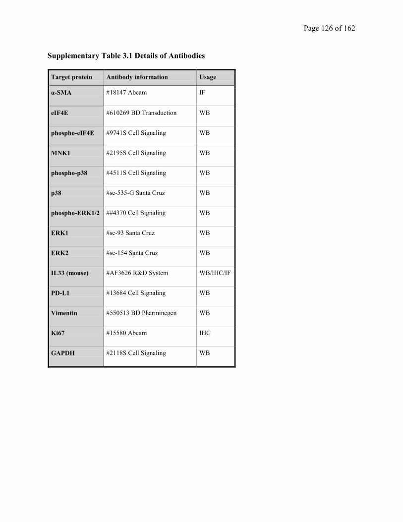
Page 126 of 162
Supplementary Table 3.1 Details of Antibodies
Target protein Antibody information Usage
α-SMA #18147 Abcam IF
eIF4E #610269 BD Transduction WB
phospho-eIF4E #9741S Cell Signaling WB
MNK1 #2195S Cell Signaling WB
phospho-p38 #4511S Cell Signaling WB
p38 #sc-535-G Santa Cruz WB
phospho-ERK1/2 ##4370 Cell Signaling WB
ERK1 #sc-93 Santa Cruz WB
ERK2 #sc-154 Santa Cruz WB
IL33 (mouse) #AF3626 R&D System WB/IHC/IF
PD-L1 #13684 Cell Signaling WB
Vimentin #550513 BD Pharminegen WB
Ki67 #15580 Abcam IHC
GAPDH #2118S Cell Signaling WB
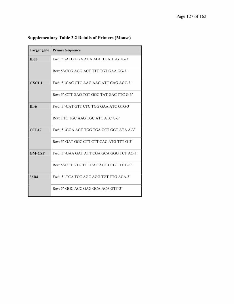
Page 127 of 162
Supplementary Table 3.2 Details of Primers (Mouse)
Target gene Primer Sequence
IL33 Fwd: 5’-ATG GGA AGA AGC TGA TGG TG-3’
Rev: 5’-CCG AGG ACT TTT TGT GAA GG-3’
CXCL1 Fwd: 5’-CAC CTC AAG AAC ATC CAG AGC-3’
Rev: 5’-CTT GAG TGT GGC TAT GAC TTC G-3’
IL-6 Fwd: 5’-CAT GTT CTC TGG GAA ATC GTG-3’
Rev: TTC TGC AAG TGC ATC ATC G-3’
CCL17 Fwd: 5’-GGA AGT TGG TGA GCT GGT ATA A-3’
Rev: 5’-GAT GGC CTT CTT CAC ATG TTT G-3’
GM-CSF Fwd: 5’-GAA GAT ATT CGA GCA GGG TCT AC-3’
Rev: 5’-CTT GTG TTT CAC AGT CCG TTT C-3’
36B4 Fwd: 5’-TCA TCC AGC AGG TGT TTG ACA-3’
Rev: 5’-GGC ACC GAG GCA ACA GTT-3’
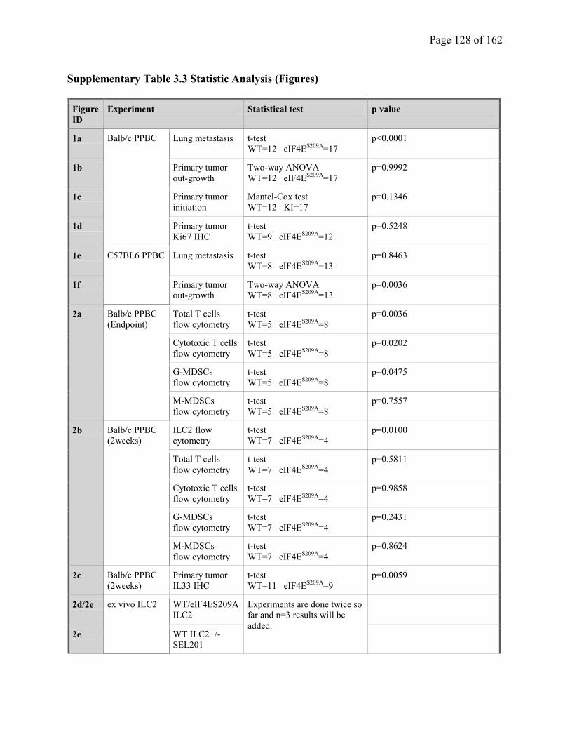
Page 128 of 162
Supplementary Table 3.3 Statistic Analysis (Figures)
Figure
ID Experiment Statistical test p value
1a Balb/c PPBC Lung metastasis t-test WT=12 eIF4E
S209A=17
p<0.0001
1b Primary tumor
out-growth Two-way ANOVA
WT=12 eIF4ES209A
=17 p=0.9992
1c Primary tumor
initiation Mantel-Cox test WT=12 KI=17
p=0.1346
1d Primary tumor
Ki67 IHC t-test WT=9 eIF4E
S209A=12
p=0.5248
1e C57BL6 PPBC Lung metastasis t-test WT=8 eIF4E
S209A=13
p=0.8463
1f Primary tumor
out-growth Two-way ANOVA
WT=8 eIF4ES209A
=13 p=0.0036
2a Balb/c PPBC (Endpoint)
Total T cells flow cytometry
t-test WT=5 eIF4E
S209A=8
p=0.0036
Cytotoxic T cells flow cytometry
t-test WT=5 eIF4E
S209A=8
p=0.0202
G-MDSCs flow cytometry
t-test WT=5 eIF4E
S209A=8
p=0.0475
M-MDSCs flow cytometry
t-test WT=5 eIF4E
S209A=8
p=0.7557
2b Balb/c PPBC (2weeks)
ILC2 flow
cytometry t-test WT=7 eIF4E
S209A=4
p=0.0100
Total T cells flow cytometry
t-test WT=7 eIF4E
S209A=4
p=0.5811
Cytotoxic T cells flow cytometry
t-test WT=7 eIF4E
S209A=4
p=0.9858
G-MDSCs flow cytometry
t-test WT=7 eIF4E
S209A=4
p=0.2431
M-MDSCs flow cytometry
t-test WT=7 eIF4E
S209A=4
p=0.8624
2c Balb/c PPBC (2weeks)
Primary tumor
IL33 IHC t-test WT=11 eIF4E
S209A=9
p=0.0059
2d/2e ex vivo ILC2 WT/eIF4ES209A
ILC2
Experiments are done twice so
far and n=3 results will be
added.
2e WT ILC2+/-
SEL201
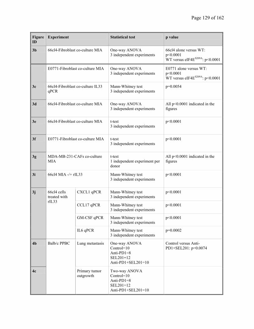
Page 129 of 162
Figure
ID Experiment Statistical test p value
3b 66cl4-Fibroblast co-culture MIA One-way ANOVA 3 independent experiments
66cl4 alone versus WT:
p<0.0001
WT versus eIF4ES209A
: p<0.0001
E0771-Fibroblast co-culture MIA One-way ANOVA 3 independent experiments
E0771 alone versus WT:
p<0.0001
WT versus eIF4ES209A
: p<0.0001
3c 66cl4-Fibroblast co-culture IL33
qPCR Mann-Whitney test 3 independent experiments
p=0.0054
3d 66cl4-Fibroblast co-culture MIA One-way ANOVA 3 independent experiments
All p<0.0001 indicated in the
figures
3e 66cl4-Fibroblast co-culture MIA t-test 3 independent experiments
p<0.0001
3f E0771-Fibroblast co-culture MIA t-test 3 independent experiments
p<0.0001
3g MDA-MB-231-CAFs co-culture
MIA t-test 1 independent experiment per
donor
All p<0.0001 indicated in the
figures
3i 66cl4 MIA -/+ rIL33 Mann-Whitney test 3 independent experiments
p<0.0001
3j 66cl4 cells
treated with
rIL33
CXCL1 qPCR Mann-Whitney test 3 independent experiments
p<0.0001
CCL17 qPCR Mann-Whitney test 3 independent experiments
p<0.0001
GM-CSF qPCR Mann-Whitney test 3 independent experiments
p<0.0001
IL6 qPCR Mann-Whitney test 3 independent experiments
p=0.0002
4b Balb/c PPBC Lung metastasis One-way ANOVA Control=10 Anti-PD1=8 SEL201=12 Anti-PD1+SEL201=10
Control versus Anti-
PD1+SEL201: p=0.0074
4c Primary tumor
outgrowth Two-way ANOVA Control=10 Anti-PD1=8 SEL201=12 Anti-PD1+SEL201=10
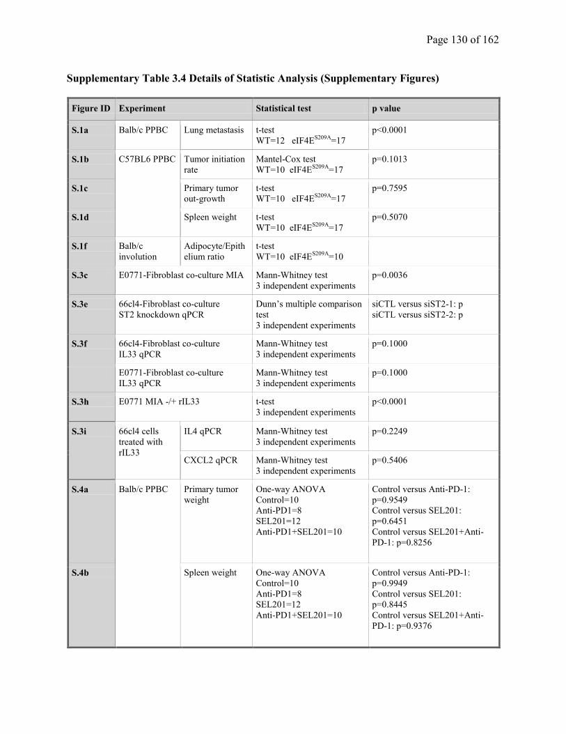
Page 130 of 162
Supplementary Table 3.4 Details of Statistic Analysis (Supplementary Figures)
Figure ID Experiment Statistical test p value
S.1a Balb/c PPBC Lung metastasis t-test WT=12 eIF4E
S209A=17
p<0.0001
S.1b C57BL6 PPBC Tumor initiation
rate Mantel-Cox test WT=10 eIF4E
S209A=17
p=0.1013
S.1c Primary tumor
out-growth t-test WT=10 eIF4E
S209A=17
p=0.7595
S.1d Spleen weight t-test WT=10 eIF4E
S209A=17
p=0.5070
S.1f Balb/c
involution Adipocyte/Epith
elium ratio t-test WT=10 eIF4E
S209A=10
S.3c E0771-Fibroblast co-culture MIA Mann-Whitney test 3 independent experiments
p=0.0036
S.3e 66cl4-Fibroblast co-culture ST2 knockdown qPCR
Dunn’s multiple comparison
test 3 independent experiments
siCTL versus siST2-1: p siCTL versus siST2-2: p
S.3f 66cl4-Fibroblast co-culture IL33 qPCR
Mann-Whitney test 3 independent experiments
p=0.1000
E0771-Fibroblast co-culture IL33 qPCR
Mann-Whitney test 3 independent experiments
p=0.1000
S.3h E0771 MIA -/+ rIL33 t-test 3 independent experiments
p<0.0001
S.3i 66cl4 cells
treated with
rIL33
IL4 qPCR Mann-Whitney test 3 independent experiments
p=0.2249
CXCL2 qPCR Mann-Whitney test 3 independent experiments
p=0.5406
S.4a Balb/c PPBC Primary tumor
weight One-way ANOVA Control=10 Anti-PD1=8 SEL201=12 Anti-PD1+SEL201=10
Control versus Anti-PD-1: p=0.9549 Control versus SEL201:
p=0.6451 Control versus SEL201+Anti-
PD-1: p=0.8256
S.4b Spleen weight One-way ANOVA Control=10 Anti-PD1=8 SEL201=12 Anti-PD1+SEL201=10
Control versus Anti-PD-1: p=0.9949 Control versus SEL201:
p=0.8445 Control versus SEL201+Anti-
PD-1: p=0.9376
Page 131 of 162
Chapter 4. Discussion and Future Directions
4.1 Comprehensive discussion of findings
In this body of work, we were mainly interested in investigating the pro-tumorigenic functions of
MNK1/2-eIF4E pathway in two important stages of the breast cancer metastatic cascade: (a) the
early stage of transition from benign disease to invasive cancer, and (b) the colonization of tumor
cells in a secondary organ. Our findings have provided a fundamental investigation of the
targeting of an aberrant protein synthesis pathway. We have also provided groundwork for
targeting of the aberrant MNK1/2-eIF4E pathway as a viable means of preventing breast cancer
invasion and treating metastatic disease.
Currently, DCIS patients are considered over-treated due to the lack of reliable biomarkers to
predict individuals with high risk of disease recurrence. Although our study has suggested the
potential role of MNK1/NODAL pathway in promoting the DCIS invasive transition, it remains
unclear how MNK1 and phospho-MNK1 levels correlate with disease recurrence and metastasis,
as well as patient survival, and whether the inhibition of MNK activity with small molecular
inhibitors like SEL201 would be useful to decrease the risk of disease recurrence. A larger cohort
of patients with pure DCIS, mixed DCIS/IDC and IDC with long-term follow-up would be
needed to examine if MNK1 and/or phospho-MNK1 might serve as biomarkers to predict
recurrence and patient outcome. More importantly, it is possible to use SEL201 in future clinical
trials to test its safety profile, and examine if SEL201 may lower the risk of disease relapse as
IDC and metastasis. Given SEL201 is an orally bioavailable MNK inhibitor, it is possible to
easily test whether patients with high MNK1 or phospho-MNK1 will benefit from using
SEL201.
Page 132 of 162
In the context of PPBC, a highly metastatic form of breast cancer, we add knowledge about the
novel functions of the MNK1/2-eIF4E pathway in the regulation of innate and adaptive
immunity during the process of PPBC metastasis. However, it is important to notice that a
limitation of our study is the difference between human PPBC and mouse PPBC model, although
we have used a gold standard mouse model where malignant cells were introduced on day 1
following forced weaning. Such differences should be recognized, as PPBC in humans is defined
as receiving breast cancer diagnosis within 10 years of having had a last child (1). Additionally,
we have offered a possible way to sensitize PPBC to PD1 blockade by adding a MNK1/2
inhibitor in our pre-clinical mouse model of this metastatic disease. This body of work provides
the possibility of increasing the response rate of metastatic breast cancer to immune checkpoint
blockade when combined with a MNK1/2 inhibitor. An ongoing phase II clinical trials
(NCT03616834) is currently being performed to test the anti-neoplastic efficacy of the MNK1/2
inhibitor eFT508 in combination with anti-PD1/anti-PD-L1 therapy.
4.2 Future directions
Several future directions have stemmed from this thesis research:
4.2.1 Potential roles of MNK1 in the nucleus
Although we have observed MNK1-dependent upregulation of NODAL mRNA and protein in
DCIS cells, the detailed molecular mechanism underlying the regulation of NODAL by MNK1
remains unclear. There are a few possible mechanisms for how MNK1 regulates NODAL gene
transcription. First, MNK1/eIF4E activation may drive the mRNA translation of a transcription
factor that can bind to the promoter of the NODAL gene, which in turn upregulates NODAL
mRNA levels. Second, MNK1 may indirectly increase NODAL mRNA stability. The first
possibility is supported by our observation that MNK1 and phospho-MNK1 can localize in both
Page 133 of 162
the cytoplasm and nucleus in mouse and human DCIS/IDC tissues and our melanoma work (2,
3). Although we did not specifically investigate the functions of MNK1a versus MNK1b in our
DCIS model, previous reports have suggested that MNK1b, which preferentially localizes in the
nucleus (4), is associated with poor patient outcome in triple-negative breast cancer (5). This
indicates that MNK1b may have a special role in the nucleus to regulate breast tumorigenesis
and metastasis. Thus, we propose that MNK1 may serve as an indirect transcriptional regulator
in the nucleus, aside from its canonical role in regulating mRNA translation. In order to test this
hypothesis, MNK1 plasmid constructs with a mutated nuclear export signal (NES) (R/26,27,28/A
mutation) and a MNK1 construct with a mutated nucleus localization signal (NLS) (L390S) need
to be expressed in the breast cancer cells, and MNK1-interacting proteins in the nucleus can be
pulled down. The identities of MNK1-interacting proteins can be identified by mass
spectrometry, and the identification of potential MNK1-interacting factors may shed light on
novel functions of MNK1 in the nucleus.
4.2.2 Characterizing the functions of MNK1/2-eIF4E in the DCIS to IDC transition in an
immune competent model
We have shown data suggesting that the MNK1/NODAL pathway is critical for maintaining
DCIS CSC populations, and MNK1 deficiency lowers the risk of relapse as metastatic disease.
However, all these studies were done with human DCIS cells in a murine immuno-deficient host.
Future studies are needed to examine how MNK1/2-eIF4E activity regulates the maintenance of
DCIS CSC populations, and how its activity shapes tumor immunity in an immune competent
host. Recently, the functions of immune cells have been described in DCIS and IDC patients. For
example, Hendry and colleagues have reported that high levels of tumor infiltrating lymphocytes
in DCIS patient samples are associated with a high-grade tumor, presence of necrosis, ER-
Page 134 of 162
negative lesions and HER2-positive tumors (6). More importantly, infiltration of total and
activated cytotoxic T cells is decreased in IDC compared to DCIS lesions (7). Such studies
suggest that the exclusion of cytotoxic T cells from the TME may contribute to DCIS to IDC
conversion.
Several transgenic breast cancer models, such as MMTV-Neu and WAP-T mice, are reported to
bear lesions that highly resemble human DCIS. MMTV-Neu is a transgenic mouse model with
mammary gland specific expression of proto-oncogene HER2/Neu driven by murine mammary
tumor virus (MMTV) promoter/enhancer, and WAP-T mouse model is a whey acidic protein
promoter (WAP)-driven SV40 breast cancer model. Both MMTV-Neu and WAP-T mice are
reported to bear lesions that highly resemble human DCIS, and such DCIS-like lesions can
invade through the basement membrane, and transit into invasive mammary carcinoma in a step-
wise fashion (8-10). These model systems can be used to study the impact of aberrant mRNA
translation in various innate and adaptive immune cells during the DCIS to IDC conversion.
More importantly, immuno-competent mouse models will be advantageous to test the possibility
of combinational targeted and immunotherapies to prevent the DCIS to invasive transition and
metastasis. For example, NODAL neutralizing antibody has shown promising anti-cancer effects
(11-13). Future studies could also be done to test whether the combination of SEL201 and an
existing anti-NODAL monoclonal antibody 3D1 (11) can elicit superior anti-tumor immunity to
impair DCIS invasive transition in immune-competent DCIS models.
4.2.3 Characterizing the immune landscape of PPBC patient samples
The importance of tumor-stroma interactions is increasingly recognized. Recent studies have
revealed that the presence of tumor infiltrating lymphocytes (TILs), especially CD8+ T cells,
predict good prognosis in breast cancer (14, 15). However, it remains unclear how various
Page 135 of 162
immune cells interact with breast tumor epithelium and other non-immune stromal components
such as CAFs. Further interrogation of the tumor microenvironment at subcellular resolution will
possibly uncover pathological features, new biomarker correlations and cell-cell interactions.
Imaging mass cytometry (IMC) is a powerful tool that can quantitatively measure more than 50
markers by using metal-conjugated antibodies directly on tissue sections. Therefore, by using
IMC, which has been widely used to study the TME, it is possible to further characterize the
PPBC TME and compare those tumors to breast cancers that are not diagnosed during the post-
partum period.
Given the elevated immune cell infiltration and fibroblast activation that occurs during mammary
gland involution in human studies and animal models (1, 16, 17), we hypothesize that PPBC
tissues will have a higher level of overall immune infiltration as well as altered spatial
localization and/or levels of stromal components. To test this hypothesis, future work in our lab
will be to map the detailed landscape of PPBC TME by using the IMC technology. Specifically,
we will characterize the levels of infiltration and distribution of key stromal cells, including
CAFs, cytotoxic T cells, Th1 and Th2 cells, regulatory T cells, MDSCs, and ILC2s. More
importantly, we will characterize the levels of MNK1, eIF4E and the phosphorylated forms of
MNK1 and eIF4E, in order to get a more detailed picture of which immune cells types have high
expressions and/or high activities of MNK1 and eIF4E. Additionally, we will examine the
expression of important immune-checkpoints such as PD-1 and PD-L1, as this hopefully will
reveal more information on the immune-checkpoints that are highly expressed in the context of
PPBC, thus helping to guide future research on targeting key checkpoints in this highly
metastatic disease. Overall, such a detailed characterization of the PPBC TME will shed more
light on how aberrant mRNA translation impacts different cellular components, and to provide
Page 136 of 162
novel insight on strategies that can be employed to re-shape tumor immunity to improve PPBC
patient outcomes.
4.2.4 Characterizing the functions of MNK1/2-eIF4E in organ specific breast cancer
metastasis
Breast cancers are prone to metastasize to a few secondary organs, including liver, bones, lungs
and brain. Although it remains unclear why some cancers metastasize to specific organs, some
earlier studies revealed that cancer cells may utilize specific receptors on their surface which
match the ligands that are highly expressed in secondary organs. For example, breast cancer cells
express specific chemokine receptors, such as C-X-C motif receptor 4 (CXCR4) and C-C motif
receptor 7 (CCR7), to recognize the chemokines that are preferentially expressed in the lymph
nodes (CCL21), liver, bone and lungs (CXCL12) (Summarized by Muller et al. (18)). More
recently, several studies have uncovered the functions of tumor cell-derived exosomes in the
preparation of pre-metastatic niche (19). Exosomes are heterogenous small membrane vesicles
(~30-100nm) containing various biomolecules such as RNA, DNA, lipids and protein. Tumor
cell-derived exosomes can fuse with stromal cells in secondary organs, and such fusion
subsequently prepares the “soil” for the tumor cell to seed. Exosomes, which contain many
mRNAs and non-coding mRNAs, may serve as an important mediator for genetic component
exchange between cells (20, 21). Recently, overexpression of eukaryotic translation initiation
factor 3 subunit C (eIF3C) is reported to promote exosome release from hepatocellular
carcinoma (HCC) (22). However, it remains largely unknown how exosome-dependent pre-
metastatic niche formation is affected by aberrant mRNA translational machinery. Aberrant
mRNA translation may offer the advantage of an accelerated cellular response to external stimuli
by circumventing the need of de novo transcription, thus it is possible that mRNA translational
Page 137 of 162
regulatory factors, such as the eukaryotic initiation factor (eIF) family members, may regulate
the dynamic process of exosome-dependent pre-metastatic niche formation. Future studies are
needed to explore whether hyper-activation of pro-oncogenic mRNA translational machineries
like the MNK1/2-eIF4E axis can affect the quantities and contents of tumor-derived exosomes,
and how they affect organ-specific metastasis.
Liver metastasis seems to be more common in patients diagnosed with PPBC than those with
nulliparous breast cancer (23). Recently, the concept of “liver involution” has been proposed, as
liver weight loss, hepatocyte apoptosis, and ECM remodeling have been observed after forced
weaning (23). The Schedin group proposes that the liver involuting microenvironment is
important for the establishment of PPBC liver metastasis in rodent models (23). Future studies
are needed to verify if this liver involution process also exists in postpartum women, and to
characterize whether the MNK1/2-eIF4E pathway contributes to the pro-tumorigenic immunity
in PPBC liver metastasis and to examine whether a MNK1/2 inhibitor plus checkpoint blockade
can be applied to inhibit liver metastasis. In order to investigate these questions, 4T1 breast
cancer cells can be injected into the spleens of WT and eIF4ES209A
PPBC mice. The splenic
injections will allow the colonization of tumor cells in the liver. We anticipate that the
eIF4ES209A
liver microenvironment will be less favorable for breast cancer colonization, similar
to our work in lung metastasis. Additionally, MNK1/2 inhibitor SEL201 and/or anti-PD1
antibody could be applied to 4T1-tumor bearing WT liver metastatic PPBC animals to test if
SEL201 can increase the anti-tumor effect of anti-PD1 antibody.
4.2.5 Targeting CAFs in metastatic breast cancer
We and others have observed CAFs as important stromal components that support breast cancer
immune evasion and metastasis. Recent studies have examined the possibility of depleting CAFs
Page 138 of 162
as an anti-cancer therapy. For example, fibroblast-activation protein (FAP), which is a surface
marker on activated fibroblasts, is a potential therapeutic target on CAFs. Depletion of FAP+
cells have led to inhibition of tumor outgrowth in several solid malignancies (summarized by
Chen & Song (24)). Alternatively, previous studies have also investigated the possibility of
inactivating CAFs by using all-trans retinoic acid (ATRA, a vitamin A derivative) and
calcipotriol (a vitamin D receptor agonist), as the functions of nuclear receptors such as retinoic
acid receptor and vitamin D receptor in CAF activation have been recently revealed.
Specifically, a high dose of ATRA impairs the collagen-deposition ability of pancreatic satellite
cells (PSCs)-derived fibroblasts and induces them to enter into a more quiescent status (25, 26).
Vitamin D receptor suppresses the activation of pancreatic satellite cells (PSCs)-derived
fibroblasts in pancreatic ductal adenocarcinoma (PDAC), and Calcipotriol can block activation
and proliferation of CAFs to render anti-neoplastic effects (27). However, the effects of ATRA
and calcipotriol in deactivating fibroblasts are only reported in PDAC. It remains unclear if these
two vitamin derivatives have similar impact on CAFs from other solid malignancies.
Despite the somewhat promising results in animal models, it remains challenging to target CAFs
in human cancer, mainly due to the heterogeneity of CAFs and the lack of specificity while
targeting these plastic cells. Future studies should further explore the biology of CAFs in primary
tumor sites, circulation and metastatic sites. This may allow us to uncover better strategies to
target CAFs in a direct or indirect fashion, so the TME might be re-shaped into a mode for anti-
tumor immunity.
4.2.6 Targeting mRNA translation to increase sensitivity to checkpoint blockade
Recently, two independent groups have reported that PD-L1 expression is regulated by eIF4E or
the eIF4F complex (28, 29). Here, we have shown that the MNK1/2 inhibitor SEL201 can
Page 139 of 162
increase the anti-metastatic effect of anti-PD1 blockade, possibly by elimination of the PD-
L1:PD1 interaction by two mechanisms. Although this provides a possible strategy to overcome
resistance to immune-checkpoint blockade, the PPBC mice treated with the combination of
SEL201 and anti-PD1 were not completely free of metastasis. This could be explained by the
potential importance of many other immune checkpoints, such as TIM3, LAG3, TIGIT, VISTA
and BTLA4, which also contribute to tumor immune evasion. Furthermore, blunted signaling
from co-stimulatory pathways, such as CD40, ICOS and GITR, may also serve as non-redundant
mechanisms for tumor immune evasion. Thus, blocking antibodies of other immune-checkpoints
and agonists of co-stimulatory molecules is being actively investigated in combination with anti-
CTLA4 or anti-PD1/anti-PD-L1 blockade. Recent results of a phase Ib clinical trial testing CD40
agonists in conjunction with nivolumab, a PD1 blocking antibody, in pancreatic ductal
carcinoma has provided a novel strategy for the treatment of highly-aggressive cancers (30). To
find a better therapy for PPBC and metastatic breast cancer in general, future studies are required
to thoroughly profile the expression status of immune-checkpoints and co-stimulatory molecules
in patient samples. Such studies may help to reveal non-redundant immuno-regulatory pathways
to PD1:PD-L1/2 and CTLA-4, thus to discover novel therapies to overcome metastatic breast
cancer.
4.3 Final conclusion and summary
The work presented in this thesis highlights the critical functions of MNK1/2-eIF4E pathway in
breast cancer. The objectives of our studies were as follows:
a) To determine if MNK1 regulates the transition from DCIS to invasive disease.
b) To understand how the MNK1/eIF4E axis, functioning in stromal cells, regulates PPBC pro-
tumorigenic immunity.
Page 140 of 162
The objectives of our studies were accomplished as follows:
a) We identified tumor intrinsic the MNK1/NODAL axis as a key event promoting the early
invasive progression in breast ductal carcinoma in situ. Of potential relevance to the clinic, we
show that ablation of MNK1, genetically or pharmacologically, partially impairs the DCIS to
IDC transition.
b) Our findings expand the importance of eIF4E phosphorylation to not only include cancer
cells, but also in stromal cells of the tumor microenvironment (i.e. fibroblasts and immune cells),
in permitting pro-tumorigenic immune evasion in PPBC. Our findings culminated in proposing
to block the MNK1/2-eIF4E pathway as a strategy to partially inhibit breast cancer invasion, and
to sensitize metastatic PPBC to anti-PD1 therapy.
4.5 References
1. Goddard ET, Bassale S, Schedin T, Jindal S, Johnston J, Cabral E, et al. Association
Between Postpartum Breast Cancer Diagnosis and Metastasis and the Clinical Features
Underlying Risk. JAMA Netw Open. 2019;2(1):e186997.
2. Guo Q, Li VZ, Nichol JN, Huang F, Yang W, Preston SEJ, et al. MNK1/NODAL
signaling promotes invasive progression of breast ductal carcinoma in situ. Cancer Res. 2019.
3. Zhan Y, Guo J, Yang W, Goncalves C, Rzymski T, Dreas A, et al. MNK1/2 inhibition
limits oncogenicity and metastasis of KIT-mutant melanoma. The Journal of clinical
investigation. 2017.
4. O'Loghlen A, Gonzalez VM, Pineiro D, Perez-Morgado MI, Salinas M, and Martin ME.
Identification and molecular characterization of Mnk1b, a splice variant of human MAP kinase-
interacting kinase Mnk1. Exp Cell Res. 2004;299(2):343-55.
5. Pinto-Diez C, Garcia-Recio EM, Perez-Morgado MI, Garcia-Hernandez M, Sanz-Criado
L, Sacristan S, et al. Increased expression of MNK1b, the spliced isoform of MNK1, predicts
poor prognosis and is associated with triple-negative breast cancer. Oncotarget.
2018;9(17):13501-16.
6. Hendry S, Pang JB, Byrne DJ, Lakhani SR, Cummings MC, Campbell IG, et al.
Relationship of the Breast Ductal Carcinoma In Situ Immune Microenvironment with
Clinicopathological and Genetic Features. Clin Cancer Res. 2017;23(17):5210-7.
7. Gil Del Alcazar CR, Huh SJ, Ekram MB, Trinh A, Liu LL, Beca F, et al. Immune Escape
in Breast Cancer During In Situ to Invasive Carcinoma Transition. Cancer discovery.
2017;7(10):1098-115.
Page 141 of 162
8. Cardiff RD, and Wellings SR. The comparative pathology of human and mouse
mammary glands. Journal of mammary gland biology and neoplasia. 1999;4(1):105-22.
9. Green JE, Shibata MA, Yoshidome K, Liu ML, Jorcyk C, Anver MR, et al. The
C3(1)/SV40 T-antigen transgenic mouse model of mammary cancer: ductal epithelial cell
targeting with multistage progression to carcinoma. Oncogene. 2000;19(8):1020-7.
10. Schulze-Garg C, Lohler J, Gocht A, and Deppert W. A transgenic mouse model for the
ductal carcinoma in situ (DCIS) of the mammary gland. Oncogene. 2000;19(8):1028-37.
11. Foca A, Sanguigno L, Foca G, Strizzi L, Iannitti R, Palumbo R, et al. New Anti-Nodal
Monoclonal Antibodies Targeting the Nodal Pre-Helix Loop Involved in Cripto-1 Binding.
International journal of molecular sciences. 2015;16(9):21342-62.
12. Hendrix MJ, Seftor EA, Seftor RE, Kasemeier-Kulesa J, Kulesa PM, and Postovit LM.
Reprogramming metastatic tumour cells with embryonic microenvironments. Nature reviews
Cancer. 2007;7(4):246-55.
13. Strizzi L, Sandomenico A, Margaryan NV, Foca A, Sanguigno L, Bodenstine TM, et al.
Effects of a novel Nodal-targeting monoclonal antibody in melanoma. Oncotarget.
2015;6(33):34071-86.
14. Ali HR, Provenzano E, Dawson SJ, Blows FM, Liu B, Shah M, et al. Association
between CD8+ T-cell infiltration and breast cancer survival in 12,439 patients. Ann Oncol.
2014;25(8):1536-43.
15. Salgado R, Denkert C, Demaria S, Sirtaine N, Klauschen F, Pruneri G, et al. The
evaluation of tumor-infiltrating lymphocytes (TILs) in breast cancer: recommendations by an
International TILs Working Group 2014. Ann Oncol. 2015;26(2):259-71.
16. Guo Q, Minnier J, Burchard J, Chiotti K, Spellman P, and Schedin P. Physiologically
activated mammary fibroblasts promote postpartum mammary cancer. JCI Insight.
2017;2(6):e89206.
17. Martinson HA, Jindal S, Durand-Rougely C, Borges VF, and Schedin P. Wound healing-
like immune program facilitates postpartum mammary gland involution and tumor progression.
International journal of cancer Journal international du cancer. 2015;136(8):1803-13.
18. Muller A, Homey B, Soto H, Ge N, Catron D, Buchanan ME, et al. Involvement of
chemokine receptors in breast cancer metastasis. Nature. 2001;410(6824):50-6.
19. Hoshino A, Costa-Silva B, Shen TL, Rodrigues G, Hashimoto A, Tesic Mark M, et al.
Tumour exosome integrins determine organotropic metastasis. Nature. 2015;527(7578):329-35.
20. Nabet BY, Qiu Y, Shabason JE, Wu TJ, Yoon T, Kim BC, et al. Exosome RNA
Unshielding Couples Stromal Activation to Pattern Recognition Receptor Signaling in Cancer.
Cell. 2017;170(2):352-66 e13.
21. Valadi H, Ekstrom K, Bossios A, Sjostrand M, Lee JJ, and Lotvall JO. Exosome-
mediated transfer of mRNAs and microRNAs is a novel mechanism of genetic exchange
between cells. Nature cell biology. 2007;9(6):654-9.
22. Lee HY, Chen CK, Ho CM, Lee SS, Chang CY, Chen KJ, et al. EIF3C-enhanced
exosome secretion promotes angiogenesis and tumorigenesis of human hepatocellular carcinoma.
Oncotarget. 2018;9(17):13193-205.
23. Goddard ET, Hill RC, Nemkov T, D'Alessandro A, Hansen KC, Maller O, et al. The
Rodent Liver Undergoes Weaning-Induced Involution and Supports Breast Cancer Metastasis.
Cancer discovery. 2017;7(2):177-87.
24. Chen X, and Song E. Turning foes to friends: targeting cancer-associated fibroblasts.
Nature reviews Drug discovery. 2019;18(2):99-115.
Page 142 of 162
25. Chronopoulos A, Robinson B, Sarper M, Cortes E, Auernheimer V, Lachowski D, et al.
ATRA mechanically reprograms pancreatic stellate cells to suppress matrix remodelling and
inhibit cancer cell invasion. Nat Commun. 2016;7:12630.
26. Dauer P, Zhao X, Gupta VK, Sharma N, Kesh K, Gnamlin P, et al. Inactivation of
Cancer-Associated-Fibroblasts Disrupts Oncogenic Signaling in Pancreatic Cancer Cells and
Promotes Its Regression. Cancer Res. 2018;78(5):1321-33.
27. Sherman MH, Yu RT, Engle DD, Ding N, Atkins AR, Tiriac H, et al. Vitamin D
receptor-mediated stromal reprogramming suppresses pancreatitis and enhances pancreatic
cancer therapy. Cell. 2014;159(1):80-93.
28. Cerezo M, Guemiri R, Druillennec S, Girault I, Malka-Mahieu H, Shen S, et al.
Translational control of tumor immune escape via the eIF4F-STAT1-PD-L1 axis in melanoma.
Nature medicine. 2018;24(12):1877-86.
29. Xu Y, Poggio M, Jin HY, Shi Z, Forester CM, Wang Y, et al. Translation control of the
immune checkpoint in cancer and its therapeutic targeting. Nature medicine. 2019;25(2):301-11.
30. Rech AJ, Dada H, Kotzin JJ, Henao-Mejia J, Minn AJ, Twyman-Saint Victor C, et al.
Radiotherapy and CD40 Activation Separately Augment Immunity to Checkpoint Blockade in
Cancer. Cancer Res. 2018;78(15):4282-91.
Page 143 of 162
Chapter 5. References
1. Pandya S, and Moore RG. Breast development and anatomy. Clin Obstet Gynecol.
2011;54(1):91-5.
2. Degnim AC, Brahmbhatt RD, Radisky DC, Hoskin TL, Stallings-Mann M,
Laudenschlager M, et al. Immune cell quantitation in normal breast tissue lobules with
and without lobulitis. Breast cancer research and treatment. 2014;144(3):539-49.
3. Degnim AC, Hoskin TL, Arshad M, Frost MH, Winham SJ, Brahmbhatt RA, et al.
Alterations in the Immune Cell Composition in Premalignant Breast Tissue that Precede
Breast Cancer Development. Clin Cancer Res. 2017;23(14):3945-52.
4. Inman JL, Robertson C, Mott JD, and Bissell MJ. Mammary gland development: cell fate
specification, stem cells and the microenvironment. Development. 2015;142(6):1028-42.
5. Masso-Welch PA, Darcy KM, Stangle-Castor NC, and Ip MM. A developmental atlas of
rat mammary gland histology. Journal of mammary gland biology and neoplasia.
2000;5(2):165-85.
6. Truchet S, and Honvo-Houeto E. Physiology of milk secretion. Best Pract Res Clin
Endocrinol Metab. 2017;31(4):367-84.
7. Ning Y, Hoang B, Schuller AG, Cominski TP, Hsu MS, Wood TL, et al. Delayed
mammary gland involution in mice with mutation of the insulin-like growth factor
binding protein 5 gene. Endocrinology. 2007;148(5):2138-47.
8. Jindal S, Gao D, Bell P, Albrektsen G, Edgerton SM, Ambrosone CB, et al. Postpartum
breast involution reveals regression of secretory lobules mediated by tissue-remodeling.
Breast Cancer Res. 2014;16(2):R31.
9. Schedin P. Pregnancy-associated breast cancer and metastasis. Nature reviews Cancer.
2006;6(4):281-91.
10. O'Brien J, Lyons T, Monks J, Lucia MS, Wilson RS, Hines L, et al. Alternatively
activated macrophages and collagen remodeling characterize the postpartum involuting
mammary gland across species. The American journal of pathology. 2010;176(3):1241-
55.
11. Martinson HA, Jindal S, Durand-Rougely C, Borges VF, and Schedin P. Wound healing-
like immune program facilitates postpartum mammary gland involution and tumor
progression. International journal of cancer Journal international du cancer.
2015;136(8):1803-13.
12. Pennock ND, Martinson HA, Guo Q, Betts CB, Jindal S, Tsujikawa T, et al. Ibuprofen
supports macrophage differentiation, T cell recruitment, and tumor suppression in a
model of postpartum breast cancer. J Immunother Cancer. 2018;6(1):98.
13. Ferlay J, Soerjomataram I, Ervik M, Dikshit R, Eser S, Mathers C, et al. Cancer
Incidence and Mortality Worldwide: IARC Cancer Base No. 11. GLOBOCAN 2012 v10.
2012.
14. International Agency for Research on Cancer. 2013.
15. Textbook of Medical Oncology. 2009;Chapter 5.
16. Barrio AV, and Van Zee KJ. Controversies in the Treatment of Ductal Carcinoma in Situ.
Annu Rev Med. 2017;68:197-211.
17. NCCN Guidelines. 2018-2019;Version 4.
18. Cancer Staging Manual. American Joint Committee on Cancer. 2018-2019;Edition 8.
Page 144 of 162
19. Logan GJ, Dabbs DJ, Lucas PC, Jankowitz RC, Brown DD, Clark BZ, et al. Molecular
drivers of lobular carcinoma in situ. Breast Cancer Res. 2015;17:76.
20. Eliyatkin N, Yalcin E, Zengel B, Aktas S, and Vardar E. Molecular Classification of
Breast Carcinoma: From Traditional, Old-Fashioned Way to A New Age, and A New
Way. J Breast Health. 2015;11(2):59-66.
21. Healey MA, Hirko KA, Beck AH, Collins LC, Schnitt SJ, Eliassen AH, et al. Assessment
of Ki67 expression for breast cancer subtype classification and prognosis in the Nurses'
Health Study. Breast cancer research and treatment. 2017;166(2):613-22.
22. Li ZH, Hu PH, Tu JH, and Yu NS. Luminal B breast cancer: patterns of recurrence and
clinical outcome. Oncotarget. 2016;7(40):65024-33.
23. Loibl S, and Gianni L. HER2-positive breast cancer. Lancet. 2017;389(10087):2415-29.
24. Loibl S, Jackisch C, Schneeweiss A, Schmatloch S, Aktas B, Denkert C, et al. Dual
HER2-blockade with pertuzumab and trastuzumab in HER2-positive early breast cancer:
a subanalysis of data from the randomized phase III GeparSepto trial. Ann Oncol.
2017;28(3):497-504.
25. Elsawaf Z, and Sinn HP. Triple-Negative Breast Cancer: Clinical and Histological
Correlations. Breast Care (Basel). 2011;6(4):273-8.
26. Perou CM. Molecular stratification of triple-negative breast cancers. Oncologist. 2011;16
Suppl 1:61-70.
27. Herschkowitz JI, Simin K, Weigman VJ, Mikaelian I, Usary J, Hu Z, et al. Identification
of conserved gene expression features between murine mammary carcinoma models and
human breast tumors. Genome Biol. 2007;8(5):R76.
28. Prat A, Parker JS, Karginova O, Fan C, Livasy C, Herschkowitz JI, et al. Phenotypic and
molecular characterization of the claudin-low intrinsic subtype of breast cancer. Breast
Cancer Res. 2010;12(5):R68.
29. Dias K, Dvorkin-Gheva A, Hallett RM, Wu Y, Hassell J, Pond GR, et al. Claudin-Low
Breast Cancer; Clinical & Pathological Characteristics. PLoS One. 2017;12(1):e0168669.
30. Rakha EA, Reis-Filho JS, and Ellis IO. Basal-like breast cancer: a critical review. J Clin
Oncol. 2008;26(15):2568-81.
31. Papadimitriou M, Mountzios G, and Papadimitriou CA. The role of PARP inhibition in
triple-negative breast cancer: Unraveling the wide spectrum of synthetic lethality. Cancer
Treat Rev. 2018;67:34-44.
32. Benson JR, Jatoi I, and Toi M. Treatment of low-risk ductal carcinoma in situ: is nothing
better than something? Lancet Oncol. 2016;17(10):e442-e51.
33. Zhao YG, Xiao AZ, Park HI, Newcomer RG, Yan M, Man YG, et al.
Endometase/matrilysin-2 in human breast ductal carcinoma in situ and its inhibition by
tissue inhibitors of metalloproteinases-2 and -4: a putative role in the initiation of breast
cancer invasion. Cancer Res. 2004;64(2):590-8.
34. Khurana A, McKean H, Kim H, Kim SH, mcguire J, Roberts LR, et al. Silencing of
HSulf-2 expression in MCF10DCIS.com cells attenuate ductal carcinoma in situ
progression to invasive ductal carcinoma in vivo. Breast Cancer Res. 2012;14(2):R43.
35. Kambach DM, Sodi VL, Lelkes PI, Azizkhan-Clifford J, and Reginato MJ. ErbB2,
FoxM1 and 14-3-3zeta prime breast cancer cells for invasion in response to ionizing
radiation. Oncogene. 2014;33(5):589-98.
Page 145 of 162
36. Macpherson IR, Rainero E, Mitchell LE, van den Berghe PV, Speirs C, Dozynkiewicz
MA, et al. CLIC3 controls recycling of late endosomal MT1-MMP and dictates invasion
and metastasis in breast cancer. Journal of cell science. 2014;127(Pt 18):3893-901.
37. Lodillinsky C, Infante E, Guichard A, Chaligne R, Fuhrmann L, Cyrta J, et al. p63/MT1-
MMP axis is required for in situ to invasive transition in basal-like breast cancer.
Oncogene. 2015.
38. Sokol ES, Feng YX, Jin DX, Tizabi MD, Miller DH, Cohen MA, et al. SMARCE1 is
required for the invasive progression of in situ cancers. Proc Natl Acad Sci U S A.
2017;114(16):4153-8.
39. Johnson KC, Koestler DC, Fleischer T, Chen P, Jenson EG, Marotti JD, et al. DNA
methylation in ductal carcinoma in situ related with future development of invasive breast
cancer. Clinical epigenetics. 2015;7(1):75.
40. Oliemuller E, Kogata N, Bland P, Kriplani D, Daley F, Haider S, et al. SOX11 promotes
invasive growth and DCIS progression. The Journal of pathology. 2017.
41. Zheng WQ, Looi LM, and Cheah PL. A comparison of the pattern of cathepsin-D
expression in fibroadenoma, fibrocystic disease, preinvasive and invasive ductal breast
carcinoma. Pathology. 1999;31(3):247-51.
42. Madan AK, Yu K, Dhurandhar N, Cullinane C, Pang Y, and Beech DJ. Association of
hyaluronidase and breast adenocarcinoma invasiveness. Oncol Rep. 1999;6(3):607-9.
43. Maxhimer JB, Pesce CE, Stewart RA, Gattuso P, Prinz RA, and Xu X. Ductal carcinoma
in situ of the breast and heparanase-1 expression: a molecular explanation for more
aggressive subtypes. J Am Coll Surg. 2005;200(3):328-35.
44. Nelson AC, Machado HL, and Schwertfeger KL. Breaking through to the Other Side:
Microenvironment Contributions to DCIS Initiation and Progression. Journal of
mammary gland biology and neoplasia. 2018;23(4):207-21.
45. Toussaint J, Durbecq V, Altintas S, Doriath V, Rouas G, Paesmans M, et al. Low CD10
mRNA expression identifies high-risk ductal carcinoma in situ (DCIS). PloS one.
2010;5(8).
46. Alpaugh ML, Lee MC, Nguyen M, Deato M, Dishakjian L, and Barsky SH.
Myoepithelial-specific CD44 shedding contributes to the anti-invasive and antiangiogenic
phenotype of myoepithelial cells. Exp Cell Res. 2000;261(1):150-8.
47. Allinen M, Beroukhim R, Cai L, Brennan C, Lahti-Domenici J, Huang H, et al.
Molecular characterization of the tumor microenvironment in breast cancer. Cancer Cell.
2004;6(1):17-32.
48. Allen MD, Thomas GJ, Clark S, Dawoud MM, Vallath S, Payne SJ, et al. Altered
microenvironment promotes progression of preinvasive breast cancer: myoepithelial
expression of alphavbeta6 integrin in DCIS identifies high-risk patients and predicts
recurrence. Clin Cancer Res. 2014;20(2):344-57.
49. Sameni M, Cavallo-Medved D, Franco OE, Chalasani A, Ji K, Aggarwal N, et al.
Pathomimetic avatars reveal divergent roles of microenvironment in invasive transition of
ductal carcinoma in situ. Breast Cancer Res. 2017;19(1):56.
50. Brummer G, Acevedo DS, Hu Q, Portsche M, Fang WB, Yao M, et al. Chemokine
Signaling Facilitates Early-Stage Breast Cancer Survival and Invasion through
Fibroblast-Dependent Mechanisms. Mol Cancer Res. 2018;16(2):296-308.
Page 146 of 162
51. Ratajczak-Wielgomas K, Grzegrzolka J, Piotrowska A, Gomulkiewicz A, Witkiewicz W,
and Dziegiel P. Periostin expression in cancer-associated fibroblasts of invasive ductal
breast carcinoma. Oncol Rep. 2016;36(5):2745-54.
52. Shaker H, Bundred NJ, Albadry H, Nicholson S, Castle J, Lumsden LJ, et al. PO-21 -
Stromal fibroblasts in preinvasive breast cancer (ductal carcinoma in situ, DCIS)
demonstrate a cancer-like procoagulant phenotypic switch that may facilitate invasion.
Thromb Res. 2016;140 Suppl 1:S184.
53. Osuala KO, Sameni M, Shah S, Aggarwal N, Simonait ML, Franco OE, et al. Il-6
signaling between ductal carcinoma in situ cells and carcinoma-associated fibroblasts
mediates tumor cell growth and migration. BMC cancer. 2015;15:584.
54. Adams M, Jones JL, Walker RA, Pringle JH, and Bell SC. Changes in tenascin-C isoform
expression in invasive and preinvasive breast disease. Cancer Res. 2002;62(11):3289-97.
55. Lo PK, Zhang Y, Yao Y, Wolfson B, Yu J, Han SY, et al. Tumor-associated
myoepithelial cells promote the invasive progression of ductal carcinoma in situ through
activation of TGFbeta signaling. J Biol Chem. 2017;292(27):11466-84.
56. Guo Q, Li VZ, Nichol JN, Huang F, Yang W, Preston SEJ, et al. MNK1/NODAL
signaling promotes invasive progression of breast ductal carcinoma in situ. Cancer Res.
2019.
57. Soria G, Ofri-Shahak M, Haas I, Yaal-Hahoshen N, Leider-Trejo L, Leibovich-Rivkin T,
et al. Inflammatory mediators in breast cancer: coordinated expression of TNFalpha &
IL-1beta with CCL2 & CCL5 and effects on epithelial-to-mesenchymal transition. BMC
cancer. 2011;11:130.
58. Coulson R, Liew SH, Connelly AA, Yee NS, Deb S, Kumar B, et al. The angiotensin
receptor blocker, Losartan, inhibits mammary tumor development and progression to
invasive carcinoma. Oncotarget. 2017;8(12):18640-56.
59. Kim HS, Jung M, Choi SK, Woo J, Piao YJ, Hwang EH, et al. IL-6-mediated cross-talk
between human preadipocytes and ductal carcinoma in situ in breast cancer progression.
J Exp Clin Cancer Res. 2018;37(1):200.
60. Raya A, Kawakami Y, Rodriguez-Esteban C, Buscher D, Koth CM, Itoh T, et al. Notch
activity induces Nodal expression and mediates the establishment of left-right asymmetry
in vertebrate embryos. Genes & development. 2003;17(10):1213-8.
61. Range R, Lapraz F, Quirin M, Marro S, Besnardeau L, and Lepage T. Cis-regulatory
analysis of nodal and maternal control of dorsal-ventral axis formation by Univin, a TGF-
beta related to Vg1. Development. 2007;134(20):3649-64.
62. Li H, Chen J, Wang X, He M, Zhang Z, and Cen Y. Nodal induced by hypoxia exposure
contributes to dacarbazine resistance and the maintenance of stemness in melanoma
cancer stemlike cells. Oncol Rep. 2018;39(6):2855-64.
63. Nettersheim D, Jostes S, Sharma R, Schneider S, Hofmann A, Ferreira HJ, et al. BMP
Inhibition in Seminomas Initiates Acquisition of Pluripotency via NODAL Signaling
Resulting in Reprogramming to an Embryonal Carcinoma. PLoS genetics.
2015;11(7):e1005415.
64. Jaaks P, and Bernasconi M. The proprotein convertase furin in tumour progression.
International journal of cancer Journal international du cancer. 2017;141(4):654-63.
65. Wang F, Wang L, and Pan J. PACE4 regulates proliferation, migration and invasion in
human breast cancer MDA-MB-231 cells. Molecular medicine reports. 2015;11(1):698-
704.
Page 147 of 162
66. Kalyan A, Carneiro BA, Chandra S, Kaplan J, Chae YK, Matsangou M, et al. Nodal
Signaling as a Developmental Therapeutics Target in Oncology. Mol Cancer Ther.
2017;16(5):787-92.
67. Kaya H, Erbarut I, Ozkan N, Bekiroglu N, Sen S, and Abaciglu U. Immunoexpression of
HER family, neuregulin, MAPK and AKT in invasive ductal carcinomas of the breast.
Eur J Gynaecol Oncol. 2008;29(4):350-6.
68. Pearson GW, and Hunter T. PI-3 kinase activity is necessary for ERK1/2-induced
disruption of mammary epithelial architecture. Breast Cancer Res. 2009;11(3):R29.
69. Novitskaya V, Romanska H, Dawoud M, Jones JL, and Berditchevski F. Tetraspanin
CD151 regulates growth of mammary epithelial cells in three-dimensional extracellular
matrix: implication for mammary ductal carcinoma in situ. Cancer Res.
2010;70(11):4698-708.
70. Nam JM, Ahmed KM, Costes S, Zhang H, Onodera Y, Olshen AB, et al. beta1-Integrin
via NF-kappaB signaling is essential for acquisition of invasiveness in a model of
radiation treated in situ breast cancer. Breast Cancer Res. 2013;15(4):R60.
71. Jacob T, Gray JW, Troxell M, and Vu TQ. Multiplexed imaging reveals heterogeneity of
PI3K/MAPK network signaling in breast lesions of known PIK3CA genotype. Breast
cancer research and treatment. 2016;159(3):575-83.
72. Kim HS, Jung M, Choi SK, Moon WK, and Kim SJ. Different Biological Action of Oleic
Acid in ALDHhigh and ALDHlow Subpopulations Separated from Ductal Carcinoma In
Situ of Breast Cancer. PloS one. 2016;11(9):e0160835.
73. Lian K, Ma C, Hao C, Li Y, Zhang N, Chen YH, et al. TIPE3 protein promotes breast
cancer metastasis through activating AKT and NF-kappaB signaling pathways.
Oncotarget. 2017;8(30):48889-904.
74. Al-Qassab Y, Grassilli S, Brugnoli F, Vezzali F, Capitani S, and Bertagnolo V. Protective
role of all-trans retinoic acid (ATRA) against hypoxia-induced malignant potential of
non-invasive breast tumor derived cells. BMC cancer. 2018;18(1):1194.
75. Souzaki M, Kubo M, Kai M, Kameda C, Tanaka H, Taguchi T, et al. Hedgehog signaling
pathway mediates the progression of non-invasive breast cancer to invasive breast cancer.
Cancer science. 2011;102(2):373-81.
76. Elsarraj HS, Hong Y, Valdez KE, Michaels W, Hook M, Smith WP, et al. Expression
profiling of in vivo ductal carcinoma in situ progression models identified B cell
lymphoma-9 as a molecular driver of breast cancer invasion. Breast Cancer Res.
2015;17:128.
77. Liu WS, Chan SH, Chang HT, Li GC, Tu YT, Tseng HH, et al. Isocitrate dehydrogenase
1-snail axis dysfunction significantly correlates with breast cancer prognosis and
regulates cell invasion ability. Breast Cancer Res. 2018;20(1):25.
78. Krstic M, Kolendowski B, Cecchini MJ, Postenka CO, Hassan HM, Andrews J, et al.
TBX3 promotes progression of pre-invasive breast cancer cells by inducing EMT and
directly up-regulating SLUG. The Journal of pathology. 2019.
79. Abba MC, Gong T, Lu Y, Lee J, Zhong Y, Lacunza E, et al. A Molecular Portrait of
High-Grade Ductal Carcinoma In Situ. Cancer Res. 2015;75(18):3980-90.
80. Knudsen ES, Dervishaj O, Kleer CG, Pajak T, Schwartz GF, and Witkiewicz AK. EZH2
and ALDH1 expression in ductal carcinoma in situ: complex association with recurrence
and progression to invasive breast cancer. Cell Cycle. 2013;12(13):2042-50.
Page 148 of 162
81. Li Q, Eades G, Yao Y, Zhang Y, and Zhou Q. Characterization of a stem-like
subpopulation in basal-like ductal carcinoma in situ (DCIS) lesions. J Biol Chem.
2014;289(3):1303-12.
82. Kristensen VN, Vaske CJ, Ursini-Siegel J, Van Loo P, Nordgard SH, Sachidanandam R,
et al. Integrated molecular profiles of invasive breast tumors and ductal carcinoma in situ
(DCIS) reveal differential vascular and interleukin signaling. Proc Natl Acad Sci U S A.
2012;109(8):2802-7.
83. Gil Del Alcazar CR, Huh SJ, Ekram MB, Trinh A, Liu LL, Beca F, et al. Immune Escape
in Breast Cancer During In Situ to Invasive Carcinoma Transition. Cancer discovery.
2017;7(10):1098-115.
84. Miligy I, Mohan P, Gaber A, Aleskandarany MA, Nolan CC, Diez-Rodriguez M, et al.
Prognostic significance of tumour infiltrating B lymphocytes in breast ductal carcinoma
in situ. Histopathology. 2017;71(2):258-68.
85. Thirkettle S, Decock J, Arnold H, Pennington CJ, Jaworski DM, and Edwards DR.
Matrix metalloproteinase 8 (collagenase 2) induces the expression of interleukins 6 and 8
in breast cancer cells. J Biol Chem. 2013;288(23):16282-94.
86. Hendry S, Pang JB, Byrne DJ, Lakhani SR, Cummings MC, Campbell IG, et al.
Relationship of the Breast Ductal Carcinoma In Situ Immune Microenvironment with
Clinicopathological and Genetic Features. Clin Cancer Res. 2017;23(17):5210-7.
87. Stensheim H, Moller B, van Dijk T, and Fossa SD. Cause-specific survival for women
diagnosed with cancer during pregnancy or lactation: a registry-based cohort study. J
Clin Oncol. 2009;27(1):45-51.
88. Johansson AL, Andersson TM, Hsieh CC, Cnattingius S, and Lambe M. Increased
mortality in women with breast cancer detected during pregnancy and different periods
postpartum. Cancer Epidemiol Biomarkers Prev. 2011;20(9):1865-72.
89. Callihan EB, Gao D, Jindal S, Lyons TR, Manthey E, Edgerton S, et al. Postpartum
diagnosis demonstrates a high risk for metastasis and merits an expanded definition of
pregnancy-associated breast cancer. Breast cancer research and treatment.
2013;138(2):549-59.
90. O'Brien J, Martinson H, Durand-Rougely C, and Schedin P. Macrophages are crucial for
epithelial cell death and adipocyte repopulation during mammary gland involution.
Development. 2012;139(2):269-75.
91. Guo Q, Minnier J, Burchard J, Chiotti K, Spellman P, and Schedin P. Physiologically
activated mammary fibroblasts promote postpartum mammary cancer. JCI Insight.
2017;2(6):e89206.
92. Bertolami C, and Donoff RB. The effect of full-thickness skin grafts on the actomyosin
content of contracting wounds. J Oral Surg. 1979;37(7):471-6.
93. Kalluri R. The biology and function of fibroblasts in cancer. Nature reviews Cancer.
2016;16(9):582-98.
94. Li B, and Wang JH. Fibroblasts and myofibroblasts in wound healing: force generation
and measurement. J Tissue Viability. 2011;20(4):108-20.
95. Cohen N, Shani O, Raz Y, Sharon Y, Hoffman D, Abramovitz L, et al. Fibroblasts drive
an immunosuppressive and growth-promoting microenvironment in breast cancer via
secretion of Chitinase 3-like 1. Oncogene. 2017;36(31):4457-68.
Page 149 of 162
96. Holt DM, Ma X, Kundu N, Collin PD, and Fulton AM. Modulation of host natural killer
cell functions in breast cancer via prostaglandin E2 receptors EP2 and EP4. J
Immunother. 2012;35(2):179-88.
97. Markosyan N, Chen EP, and Smyth EM. Targeting COX-2 abrogates mammary
tumorigenesis: Breaking cancer-associated suppression of immunosurveillance.
Oncoimmunology. 2014;3:e29287.
98. Basudhar D, Glynn SA, Greer M, Somasundaram V, No JH, Scheiblin DA, et al.
Coexpression of NOS2 and COX2 accelerates tumor growth and reduces survival in
estrogen receptor-negative breast cancer. Proc Natl Acad Sci U S A. 2017;114(49):13030-
5.
99. Tian J, Hachim MY, Hachim IY, Dai M, Lo C, Raffa FA, et al. Cyclooxygenase-2
regulates TGFbeta-induced cancer stemness in triple-negative breast cancer. Sci Rep.
2017;7:40258.
100. Li A, Chen P, Leng Y, and Kang J. Histone deacetylase 6 regulates the
immunosuppressive properties of cancer-associated fibroblasts in breast cancer through
the STAT3-COX2-dependent pathway. Oncogene. 2018;37(45):5952-66.
101. Borges VF, Elder AM, and Lyons TR. Deciphering Pro-Lymphangiogenic Programs
during Mammary Involution and Postpartum Breast Cancer. Front Oncol. 2016;6:227.
102. Lyons TR, Borges VF, Betts CB, Guo Q, Kapoor P, Martinson HA, et al.
Cyclooxygenase-2-dependent lymphangiogenesis promotes nodal metastasis of
postpartum breast cancer. The Journal of clinical investigation. 2014;124(9):3901-12.
103. Mantovani A, Sica A, Allavena P, Garlanda C, and Locati M. Tumor-associated
macrophages and the related myeloid-derived suppressor cells as a paradigm of the
diversity of macrophage activation. Human immunology. 2009;70(5):325-30.
104. Stanford JC, Young C, Hicks D, Owens P, Williams A, Vaught DB, et al. Efferocytosis
produces a prometastatic landscape during postpartum mammary gland involution. The
Journal of clinical investigation. 2014;124(11):4737-52.
105. Qian BZ, and Pollard JW. Macrophage diversity enhances tumor progression and
metastasis. Cell. 2010;141(1):39-51.
106. Talmadge JE, and Gabrilovich DI. History of myeloid-derived suppressor cells. Nature
reviews Cancer. 2013;13(10):739-52.
107. Gantt S, Gervassi A, Jaspan H, and Horton H. The role of myeloid-derived suppressor
cells in immune ontogeny. Front Immunol. 2014;5:387.
108. Peng D, Tanikawa T, Li W, Zhao L, Vatan L, Szeliga W, et al. Myeloid-Derived
Suppressor Cells Endow Stem-like Qualities to Breast Cancer Cells through IL6/STAT3
and NO/NOTCH Cross-talk Signaling. Cancer Res. 2016;76(11):3156-65.
109. Gonda K, Shibata M, Ohtake T, Matsumoto Y, Tachibana K, Abe N, et al. Myeloid-
derived suppressor cells are increased and correlated with type 2 immune responses,
malnutrition, inflammation, and poor prognosis in patients with breast cancer. Oncol Lett.
2017;14(2):1766-74.
110. Safarzadeh E, Hashemzadeh S, Duijf PHG, Mansoori B, Khaze V, Mohammadi A, et al.
Circulating myeloid-derived suppressor cells: An independent prognostic factor in
patients with breast cancer. Journal of cellular physiology. 2018.
111. Li W, Tanikawa T, Kryczek I, Xia H, Li G, Wu K, et al. Aerobic Glycolysis Controls
Myeloid-Derived Suppressor Cells and Tumor Immunity via a Specific CEBPB Isoform
in Triple-Negative Breast Cancer. Cell metabolism. 2018;28(1):87-103 e6.
Page 150 of 162
112. Yang L, Huang J, Ren X, Gorska AE, Chytil A, Aakre M, et al. Abrogation of TGF beta
signaling in mammary carcinomas recruits Gr-1+CD11b+ myeloid cells that promote
metastasis. Cancer Cell. 2008;13(1):23-35.
113. Molinier-Frenkel V, and Castellano F. Immunosuppressive enzymes in the tumor
microenvironment. FEBS Lett. 2017;591(19):3135-57.
114. Secondini C, Coquoz O, Spagnuolo L, Spinetti T, Peyvandi S, Ciarloni L, et al. Arginase
inhibition suppresses lung metastasis in the 4T1 breast cancer model independently of the
immunomodulatory and anti-metastatic effects of VEGFR-2 blockade. Oncoimmunology.
2017;6(6):e1316437.
115. Stiff A, Trikha P, Mundy-Bosse B, McMichael E, Mace TA, Benner B, et al. Nitric
Oxide Production by Myeloid-Derived Suppressor Cells Plays a Role in Impairing Fc
Receptor-Mediated Natural Killer Cell Function. Clin Cancer Res. 2018;24(8):1891-904.
116. Susskind BM, Sekar J, Tandon PM, Lind DS, and Bear HD. Differential sensitivity of
cytotoxic T lymphocytes and lymphokine-activated killer cells to inhibition by L-
ornithine. Cell Immunol. 1991;133(1):41-54.
117. Prendergast GC, Smith C, Thomas S, Mandik-Nayak L, Laury-Kleintop L, Metz R, et al.
Indoleamine 2,3-dioxygenase pathways of pathogenic inflammation and immune escape
in cancer. Cancer Immunol Immunother. 2014;63(7):721-35.
118. Li T, Yang Y, Hua X, Wang G, Liu W, Jia C, et al. Hepatocellular carcinoma-associated
fibroblasts trigger NK cell dysfunction via PGE2 and IDO. Cancer Lett.
2012;318(2):154-61.
119. Abdel-Wahab O, Patel J, and Levine RL. Clinical implications of novel mutations in
epigenetic modifiers in AML. Hematology/oncology clinics of North America.
2011;25(6):1119-33.
120. Zelante T, and Ricciardi-Castagnoli P. The yin-yang nature of CSF1R-binding cytokines.
Nature immunology. 2012;13(8):717-9.
121. De Henau O, Rausch M, Winkler D, Campesato LF, Liu C, Cymerman DH, et al.
Overcoming resistance to checkpoint blockade therapy by targeting PI3Kgamma in
myeloid cells. Nature. 2016;539(7629):443-7.
122. Kumar V, Donthireddy L, Marvel D, Condamine T, Wang F, Lavilla-Alonso S, et al.
Cancer-Associated Fibroblasts Neutralize the Anti-tumor Effect of CSF1 Receptor
Blockade by Inducing PMN-MDSC Infiltration of Tumors. Cancer Cell. 2017;32(5):654-
68 e5.
123. Chiossone L, and Vivier E. Immune checkpoints on innate lymphoid cells. The Journal of
experimental medicine. 2017;214(6):1561-3.
124. Mattner J, and Wirtz S. Friend or Foe? The Ambiguous Role of Innate Lymphoid Cells in
Cancer Development. Trends in immunology. 2017;38(1):29-38.
125. Li J, Razumilava N, Gores GJ, Walters S, Mizuochi T, Mourya R, et al. Biliary repair and
carcinogenesis are mediated by IL-33-dependent cholangiocyte proliferation. The Journal
of clinical investigation. 2014;124(7):3241-51.
126. Chevalier MF, Trabanelli S, Racle J, Salome B, Cesson V, Gharbi D, et al. ILC2-
modulated T cell-to-MDSC balance is associated with bladder cancer recurrence. The
Journal of clinical investigation. 2017;127(8):2916-29.
127. Salimi M, Wang R, Yao X, Li X, Wang X, Hu Y, et al. Activated innate lymphoid cell
populations accumulate in human tumour tissues. BMC cancer. 2018;18(1):341.
Page 151 of 162
128. von Moltke J, and Locksley RM. I-L-C-2 it: type 2 immunity and group 2 innate
lymphoid cells in homeostasis. Current opinion in immunology. 2014;31:58-65.
129. Jovanovic IP, Pejnovic NN, Radosavljevic GD, Pantic JM, Milovanovic MZ, Arsenijevic
NN, et al. Interleukin-33/ST2 axis promotes breast cancer growth and metastases by
facilitating intratumoral accumulation of immunosuppressive and innate lymphoid cells.
International journal of cancer Journal international du cancer. 2014;134(7):1669-82.
130. Gardner A, and Ruffell B. Dendritic Cells and Cancer Immunity. Trends in immunology.
2016;37(12):855-65.
131. Warri A, Cook KL, Hu R, Jin L, Zwart A, Soto-Pantoja DR, et al. Autophagy and
unfolded protein response (UPR) regulate mammary gland involution by restraining
apoptosis-driven irreversible changes. Cell Death Discov. 2018;4:40.
132. Pitt JM, Vetizou M, Daillere R, Roberti MP, Yamazaki T, Routy B, et al. Resistance
Mechanisms to Immune-Checkpoint Blockade in Cancer: Tumor-Intrinsic and -Extrinsic
Factors. Immunity. 2016;44(6):1255-69.
133. Corrales L, and Gajewski TF. Molecular Pathways: Targeting the Stimulator of
Interferon Genes (STING) in the Immunotherapy of Cancer. Clin Cancer Res.
2015;21(21):4774-9.
134. Henle AM, Nassar A, Puglisi-Knutson D, Youssef B, and Knutson KL. Downregulation
of TAP1 and TAP2 in early stage breast cancer. PloS one. 2017;12(11):e0187323.
135. Sultan M, Vidovic D, Paine AS, Huynh TT, Coyle KM, Thomas ML, et al. Epigenetic
Silencing of TAP1 in Aldefluor(+) Breast Cancer Stem Cells Contributes to Their
Enhanced Immune Evasion. Stem cells. 2018;36(5):641-54.
136. Golubovskaya V, and Wu L. Different Subsets of T Cells, Memory, Effector Functions,
and CAR-T Immunotherapy. Cancers. 2016;8(3).
137. Patel SA, and Minn AJ. Combination Cancer Therapy with Immune Checkpoint
Blockade: Mechanisms and Strategies. Immunity. 2018;48(3):417-33.
138. Disis ML. Immune regulation of cancer. J Clin Oncol. 2010;28(29):4531-8.
139. Borst J, Ahrends T, Babala N, Melief CJM, and Kastenmuller W. CD4(+) T cell help in
cancer immunology and immunotherapy. Nature reviews Immunology. 2018;18(10):635-
47.
140. Wherry EJ. T cell exhaustion. Nature immunology. 2011;12(6):492-9.
141. Sharma P, and Allison JP. The future of immune checkpoint therapy. Science.
2015;348(6230):56-61.
142. Plitas G, Konopacki C, Wu K, Bos PD, Morrow M, Putintseva EV, et al. Regulatory T
Cells Exhibit Distinct Features in Human Breast Cancer. Immunity. 2016;45(5):1122-34.
143. Li Z, Dong P, Ren M, Song Y, Qian X, Yang Y, et al. PD-L1 Expression Is Associated
with Tumor FOXP3(+) Regulatory T-Cell Infiltration of Breast Cancer and Poor
Prognosis of Patient. Journal of Cancer. 2016;7(7):784-93.
144. Bhat M, Robichaud N, Hulea L, Sonenberg N, Pelletier J, and Topisirovic I. Targeting
the translation machinery in cancer. Nature reviews Drug discovery. 2015;14(4):261-78.
145. Pelletier J, Graff J, Ruggero D, and Sonenberg N. Targeting the eIF4F translation
initiation complex: a critical nexus for cancer development. Cancer Res. 2015;75(2):250-
63.
146. Li Q, Imataka H, Morino S, Rogers GW, Jr., Richter-Cook NJ, Merrick WC, et al.
Eukaryotic translation initiation factor 4AIII (eIF4AIII) is functionally distinct from
eIF4AI and eIF4AII. Molecular and cellular biology. 1999;19(11):7336-46.
Page 152 of 162
147. Castello A, Alvarez E, and Carrasco L. Differential cleavage of eIF4GI and eIF4GII in
mammalian cells. Effects on translation. J Biol Chem. 2006;281(44):33206-16.
148. Mahalingam M, and Cooper JA. Phosphorylation of mammalian eIF4E by Mnk1 and
Mnk2: tantalizing prospects for a role in translation. Prog Mol Subcell Biol. 2001;27:132-
42.
149. Ueda T, Watanabe-Fukunaga R, Fukuyama H, Nagata S, and Fukunaga R. Mnk2 and
Mnk1 are essential for constitutive and inducible phosphorylation of eukaryotic initiation
factor 4E but not for cell growth or development. Molecular and cellular biology.
2004;24(15):6539-49.
150. O'Loghlen A, Gonzalez VM, Pineiro D, Perez-Morgado MI, Salinas M, and Martin ME.
Identification and molecular characterization of Mnk1b, a splice variant of human MAP
kinase-interacting kinase Mnk1. Exp Cell Res. 2004;299(2):343-55.
151. O'Loghlen A, Gonzalez VM, Jurado T, Salinas M, and Martin ME. Characterization of
the activity of human MAP kinase-interacting kinase Mnk1b. Biochimica et biophysica
acta. 2007;1773(9):1416-27.
152. Scheper GC, Parra JL, Wilson M, Van Kollenburg B, Vertegaal AC, Han ZG, et al. The
N and C termini of the splice variants of the human mitogen-activated protein kinase-
interacting kinase Mnk2 determine activity and localization. Molecular and cellular
biology. 2003;23(16):5692-705.
153. Waskiewicz AJ, Flynn A, Proud CG, and Cooper JA. Mitogen-activated protein kinases
activate the serine/threonine kinases Mnk1 and Mnk2. The EMBO journal.
1997;16(8):1909-20.
154. Proud CG. Mnks, eIF4E phosphorylation and cancer. Biochimica et biophysica acta.
2015;1849(7):766-73.
155. Maimon A, Mogilevsky M, Shilo A, Golan-Gerstl R, Obiedat A, Ben-Hur V, et al. Mnk2
Alternative Splicing Modulates the p38-MAPK Pathway and Impacts Ras-Induced
Transformation. Cell reports. 2014;7(2):501-13.
156. Scheper GC, and Proud CG. Does phosphorylation of the cap-binding protein eIF4E play
a role in translation initiation? Eur J Biochem. 2002;269(22):5350-9.
157. Furic L, Rong L, Larsson O, Koumakpayi IH, Yoshida K, Brueschke A, et al. eIF4E
phosphorylation promotes tumorigenesis and is associated with prostate cancer
progression. Proc Natl Acad Sci U S A. 2010;107(32):14134-9.
158. Robichaud N, Del Rincon SV, Huor B, Alain T, Petruccelli LA, Hearnden J, et al.
Phosphorylation of eIF4E promotes EMT and metastasis via translational control of
SNAIL and MMP-3. 2014.
159. Pettersson F, Del Rincon SV, Emond A, Huor B, Ngan E, Ng J, et al. Genetic and
pharmacologic inhibition of eIF4E reduces breast cancer cell migration, invasion, and
metastasis. Cancer Res. 2015;75(6):1102-12.
160. Li Z, Sun Y, Qu M, Wan H, Cai F, and Zhang P. Inhibiting the MNK-eIF4E-beta-catenin
axis increases the responsiveness of aggressive breast cancer cells to chemotherapy.
Oncotarget. 2017;8(2):2906-15.
161. Zhou S, Wang GP, Liu C, and Zhou M. Eukaryotic initiation factor 4E (eIF4E) and
angiogenesis: prognostic markers for breast cancer. BMC cancer. 2006;6:231.
162. Pettersson F, Yau C, Dobocan MC, Culjkovic-Kraljacic B, Retrouvey H, Puckett R, et al.
Ribavirin treatment effects on breast cancers overexpressing eIF4E, a biomarker with
Page 153 of 162
prognostic specificity for luminal B-type breast cancer. Clin Cancer Res.
2011;17(9):2874-84.
163. Mehrotra S, Sharma B, Joshi S, Kroczynska B, Majchrzak B, Stein BL, et al. Essential
role for the Mnk pathway in the inhibitory effects of type I interferons on
myeloproliferative neoplasm (MPN) precursors. J Biol Chem. 2013;288(33):23814-22.
164. Herdy B, Jaramillo M, Svitkin YV, Rosenfeld AB, Kobayashi M, Walsh D, et al.
Translational control of the activation of transcription factor NF-kappaB and production
of type I interferon by phosphorylation of the translation factor eIF4E. Nature
immunology. 2012;13(6):543-50.
165. Robichaud N, Hsu BE, Istomine R, Alvarez F, Blagih J, Ma EH, et al. Translational
control in the tumor microenvironment promotes lung metastasis: Phosphorylation of
eIF4E in neutrophils. Proc Natl Acad Sci U S A. 2018;115(10):E2202-E9.
166. Cerezo M, Guemiri R, Druillennec S, Girault I, Malka-Mahieu H, Shen S, et al.
Translational control of tumor immune escape via the eIF4F-STAT1-PD-L1 axis in
melanoma. Nature medicine. 2018;24(12):1877-86.
167. DaSilva J, Xu L, Kim HJ, Miller WT, and Bar-Sagi D. Regulation of sprouty stability by
Mnk1-dependent phosphorylation. Molecular and cellular biology. 2006;26(5):1898-907.
168. Guil S, Long JC, and Caceres JF. hnRNP A1 relocalization to the stress granules reflects
a role in the stress response. Molecular and cellular biology. 2006;26(15):5744-58.
169. Buxade M, Morrice N, Krebs DL, and Proud CG. The PSF.p54nrb complex is a novel
Mnk substrate that binds the mRNA for tumor necrosis factor alpha. J Biol Chem.
2008;283(1):57-65.
170. Loh TJ, Moon H, Cho S, Jang H, Liu YC, Tai H, et al. CD44 alternative splicing and
hnRNP A1 expression are associated with the metastasis of breast cancer. Oncol Rep.
2015;34(3):1231-8.
171. Roy R, Huang Y, Seckl MJ, and Pardo OE. Emerging roles of hnRNPA1 in modulating
malignant transformation. Wiley interdisciplinary reviews RNA. 2017;8(6).
172. Faratian D, Sims AH, Mullen P, Kay C, Um I, Langdon SP, et al. Sprouty 2 is an
independent prognostic factor in breast cancer and may be useful in stratifying patients
for trastuzumab therapy. PloS one. 2011;6(8):e23772.
173. Konicek BW, Stephens JR, McNulty AM, Robichaud N, Peery RB, Dumstorf CA, et al.
Therapeutic inhibition of MAP kinase interacting kinase blocks eukaryotic initiation
factor 4E phosphorylation and suppresses outgrowth of experimental lung metastases.
Cancer Res. 2011;71(5):1849-57.
174. Ramalingam S, Gediya L, Kwegyir-Afful AK, Ramamurthy VP, Purushottamachar P,
Mbatia H, et al. First MNKs degrading agents block phosphorylation of eIF4E, induce
apoptosis, inhibit cell growth, migration and invasion in triple negative and Her2-
overexpressing breast cancer cell lines. Oncotarget. 2014;5(2):530-43.
175. Xu J, Chen A, Joy J, Xavier VJ, Ong EH, Hill J, et al. Rational design of resorcylic acid
lactone analogues as covalent MNK1/2 kinase inhibitors by tuning the reactivity of an
enamide Michael acceptor. ChemMedChem. 2013;8(9):1483-94.
176. Dreas A, Mikulski M, Milik M, Fabritius CH, Brzozka K, and Rzymski T. Mitogen-
activated Protein Kinase (MAPK) Interacting Kinases 1 and 2 (MNK1 and MNK2) as
Targets for Cancer Therapy: Recent Progress in the Development of MNK Inhibitors.
Curr Med Chem. 2017;24(28):3025-53.
Page 154 of 162
177. Zhan Y, Guo J, Yang W, Goncalves C, Rzymski T, Dreas A, et al. MNK1/2 inhibition
limits oncogenicity and metastasis of KIT-mutant melanoma. The Journal of clinical
investigation. 2017.
178. Reich SH, Sprengeler PA, Chiang GG, Appleman JR, Chen J, Clarine J, et al. Structure-
based Design of Pyridone-Aminal eFT508 Targeting Dysregulated Translation by
Selective Mitogen-activated Protein Kinase Interacting Kinases 1 and 2 (MNK1/2)
Inhibition. J Med Chem. 2018;61(8):3516-40.
179. Xu Y, Poggio M, Jin HY, Shi Z, Forester CM, Wang Y, et al. Translation control of the
immune checkpoint in cancer and its therapeutic targeting. Nature medicine.
2019;25(2):301-11.
180. Janku F, Yap TA, and Meric-Bernstam F. Targeting the PI3K pathway in cancer: are we
making headway? Nature reviews Clinical oncology. 2018;15(5):273-91.
181. Bianchini A, Loiarro M, Bielli P, Busa R, Paronetto MP, Loreni F, et al. Phosphorylation
of eIF4E by MNKs supports protein synthesis, cell cycle progression and proliferation in
prostate cancer cells. Carcinogenesis. 2008;29(12):2279-88.
182. Grzmil M, Huber RM, Hess D, Frank S, Hynx D, Moncayo G, et al. MNK1 pathway
activity maintains protein synthesis in rapalog-treated gliomas. The Journal of clinical
investigation. 2014;124(2):742-54.
183. Lineham E, Tizzard GJ, Coles SJ, Spencer J, and Morley SJ. Synergistic effects of
inhibiting the MNK-eIF4E and PI3K/AKT/ mTOR pathways on cell migration in MDA-
MB-231 cells. Oncotarget. 2018;9(18):14148-59.
184. Wang X, Yue P, Chan CB, Ye K, Ueda T, Watanabe-Fukunaga R, et al. Inhibition of
mammalian target of rapamycin induces phosphatidylinositol 3-kinase-dependent and
Mnk-mediated eukaryotic translation initiation factor 4E phosphorylation. Molecular and
cellular biology. 2007;27(21):7405-13.
185. Brown MC, and Gromeier M. MNK Controls mTORC1:Substrate Association through
Regulation of TELO2 Binding with mTORC1. Cell reports. 2017;18(6):1444-57.
186. Katsha A, Wang L, Arras J, Omar OM, Ecsedy J, Belkhiri A, et al. Activation of EIF4E
by Aurora Kinase A Depicts a Novel Druggable Axis in Everolimus-Resistant Cancer
Cells. Clin Cancer Res. 2017;23(14):3756-68.
187. Eckerdt F, Beauchamp E, Bell J, Iqbal A, Su B, Fukunaga R, et al. Regulatory effects of a
Mnk2-eIF4E feedback loop during mTORC1 targeting of human medulloblastoma cells.
Oncotarget. 2014;5(18):8442-51.
188. Wen Q, Wang W, Luo J, Chu S, Chen L, Xu L, et al. CGP57380 enhances efficacy of
RAD001 in non-small cell lung cancer through abrogating mTOR inhibition-induced
phosphorylation of eIF4E and activating mitochondrial apoptotic pathway. Oncotarget.
2016;7(19):27787-801.
189. Huang XB, Yang CM, Han QM, Ye XJ, Lei W, and Qian WB. MNK1 inhibitor
CGP57380 overcomes mTOR inhibitor-induced activation of eIF4E: the mechanism of
synergic killing of human T-ALL cells. Acta pharmacologica Sinica. 2018;39(12):1894-
901.
190. Geter PA, Ernlund AW, Bakogianni S, Alard A, Arju R, Giashuddin S, et al. Hyperactive
mTOR and MNK1 phosphorylation of eIF4E confer tamoxifen resistance and estrogen
independence through selective mRNA translation reprogramming. Genes &
development. 2017;31(22):2235-49.
Page 155 of 162
191. Goddard ET, Bassale S, Schedin T, Jindal S, Johnston J, Cabral E, et al. Association
Between Postpartum Breast Cancer Diagnosis and Metastasis and the Clinical Features
Underlying Risk. JAMA Netw Open. 2019;2(1):e186997.
192. Pinto-Diez C, Garcia-Recio EM, Perez-Morgado MI, Garcia-Hernandez M, Sanz-Criado
L, Sacristan S, et al. Increased expression of MNK1b, the spliced isoform of MNK1,
predicts poor prognosis and is associated with triple-negative breast cancer. Oncotarget.
2018;9(17):13501-16.
193. Cardiff RD, and Wellings SR. The comparative pathology of human and mouse
mammary glands. Journal of mammary gland biology and neoplasia. 1999;4(1):105-22.
194. Green JE, Shibata MA, Yoshidome K, Liu ML, Jorcyk C, Anver MR, et al. The
C3(1)/SV40 T-antigen transgenic mouse model of mammary cancer: ductal epithelial cell
targeting with multistage progression to carcinoma. Oncogene. 2000;19(8):1020-7.
195. Schulze-Garg C, Lohler J, Gocht A, and Deppert W. A transgenic mouse model for the
ductal carcinoma in situ (DCIS) of the mammary gland. Oncogene. 2000;19(8):1028-37.
196. Foca A, Sanguigno L, Foca G, Strizzi L, Iannitti R, Palumbo R, et al. New Anti-Nodal
Monoclonal Antibodies Targeting the Nodal Pre-Helix Loop Involved in Cripto-1
Binding. International journal of molecular sciences. 2015;16(9):21342-62.
197. Hendrix MJ, Seftor EA, Seftor RE, Kasemeier-Kulesa J, Kulesa PM, and Postovit LM.
Reprogramming metastatic tumour cells with embryonic microenvironments. Nature
reviews Cancer. 2007;7(4):246-55.
198. Strizzi L, Sandomenico A, Margaryan NV, Foca A, Sanguigno L, Bodenstine TM, et al.
Effects of a novel Nodal-targeting monoclonal antibody in melanoma. Oncotarget.
2015;6(33):34071-86.
199. Ali HR, Provenzano E, Dawson SJ, Blows FM, Liu B, Shah M, et al. Association
between CD8+ T-cell infiltration and breast cancer survival in 12,439 patients. Ann
Oncol. 2014;25(8):1536-43.
200. Salgado R, Denkert C, Demaria S, Sirtaine N, Klauschen F, Pruneri G, et al. The
evaluation of tumor-infiltrating lymphocytes (TILs) in breast cancer: recommendations
by an International TILs Working Group 2014. Ann Oncol. 2015;26(2):259-71.
201. Muller A, Homey B, Soto H, Ge N, Catron D, Buchanan ME, et al. Involvement of
chemokine receptors in breast cancer metastasis. Nature. 2001;410(6824):50-6.
202. Hoshino A, Costa-Silva B, Shen TL, Rodrigues G, Hashimoto A, Tesic Mark M, et al.
Tumour exosome integrins determine organotropic metastasis. Nature.
2015;527(7578):329-35.
203. Nabet BY, Qiu Y, Shabason JE, Wu TJ, Yoon T, Kim BC, et al. Exosome RNA
Unshielding Couples Stromal Activation to Pattern Recognition Receptor Signaling in
Cancer. Cell. 2017;170(2):352-66 e13.
204. Valadi H, Ekstrom K, Bossios A, Sjostrand M, Lee JJ, and Lotvall JO. Exosome-
mediated transfer of mRNAs and microRNAs is a novel mechanism of genetic exchange
between cells. Nature cell biology. 2007;9(6):654-9.
205. Lee HY, Chen CK, Ho CM, Lee SS, Chang CY, Chen KJ, et al. EIF3C-enhanced
exosome secretion promotes angiogenesis and tumorigenesis of human hepatocellular
carcinoma. Oncotarget. 2018;9(17):13193-205.
206. Goddard ET, Hill RC, Nemkov T, D'Alessandro A, Hansen KC, Maller O, et al. The
Rodent Liver Undergoes Weaning-Induced Involution and Supports Breast Cancer
Metastasis. Cancer discovery. 2017;7(2):177-87.
Page 156 of 162
207. Chen X, and Song E. Turning foes to friends: targeting cancer-associated fibroblasts.
Nature reviews Drug discovery. 2019;18(2):99-115.
208. Chronopoulos A, Robinson B, Sarper M, Cortes E, Auernheimer V, Lachowski D, et al.
ATRA mechanically reprograms pancreatic stellate cells to suppress matrix remodelling
and inhibit cancer cell invasion. Nat Commun. 2016;7:12630.
209. Dauer P, Zhao X, Gupta VK, Sharma N, Kesh K, Gnamlin P, et al. Inactivation of
Cancer-Associated-Fibroblasts Disrupts Oncogenic Signaling in Pancreatic Cancer Cells
and Promotes Its Regression. Cancer Res. 2018;78(5):1321-33.
210. Sherman MH, Yu RT, Engle DD, Ding N, Atkins AR, Tiriac H, et al. Vitamin D
receptor-mediated stromal reprogramming suppresses pancreatitis and enhances
pancreatic cancer therapy. Cell. 2014;159(1):80-93.
211. Rech AJ, Dada H, Kotzin JJ, Henao-Mejia J, Minn AJ, Twyman-Saint Victor C, et al.
Radiotherapy and CD40 Activation Separately Augment Immunity to Checkpoint
Blockade in Cancer. Cancer Res. 2018;78(15):4282-91.
Page 157 of 162
Permission to use published work in thesis
Permission for chapter 1
ELSEVIER LICENSE
TERMS AND CONDITIONS Jul 02, 2019
This Agreement between Dr. Wilson Miller Lab ("You") and Elsevier ("Elsevier") consists of
your license details and the terms and conditions provided by Elsevier and Copyright Clearance
Center. License Number 4620841147971
License date Jul 02, 2019 Licensed Content Publisher Elsevier
Page 158 of 162
Licensed Content Publication Elsevier Books Licensed Content Title Advances in Cancer Research
Licensed Content Author Qianyu Guo,Fan Huang,Christophe Goncalves,Sonia V. del Rincón,Wilson H. Miller Licensed Content Date Jan 1, 2019 Licensed Content Pages 62 Start Page
1 End Page 62 Type of Use reuse in a thesis/dissertation
Portion
figures/tables/illustrations Number of figures/tables/illustrations 1 Format electronic Are you the author of this Elsevier chapter? Yes
Will you be translating? No Original figure numbers Figure 1 Title of your thesis/dissertation Examining the pro-tumorigenic functions of the mitogen-activated protein kinase interacting protein kinases 1 and 2 (MNK1/2)-eukaryotic initiation factor 4E(eIF4E) pathway in breast cancer
Expected completion date
Jun 2019 Estimated size (number of pages) 160 Requestor Location Dr. Wilson Miller Lab
E504, Segal Cancer Centre Jewish General Hospital 3755 Cote-Sainte-Catherine Rd Montreal, QC H3T 1E2 Canada Attn: Dr. Wilson Miller Lab Publisher Tax ID
GB 494 6272 12 Total 0.00 USD Terms and Conditions
INTRODUCTION 1. The publisher for this copyrighted material is Elsevier. By clicking "accept" in connection with completing this
licensing transaction, you agree that the following terms and conditions apply to this transaction (along with the
Billing and Payment terms and conditions established by Copyright Clearance Center, Inc. ("CCC"), at the time that
you opened your Rightslink account and that are available at any time at http://myaccount.copyright.com).
GENERAL TERMS 2. Elsevier hereby grants you permission to reproduce the aforementioned material subject to the terms and
conditions indicated.
3. Acknowledgement: If any part of the material to be used (for example, figures) has appeared in our publication
with credit or acknowledgement to another source, permission must also be sought from that source. If such
Page 159 of 162
permission is not obtained then that material may not be included in your publication/copies. Suitable
acknowledgement to the source must be made, either as a footnote or in a reference list at the end of your
publication, as follows:
"Reprinted from Publication title, Vol /edition number, Author(s), Title of article / title of chapter, Pages No.,
Copyright (Year), with permission from Elsevier [OR APPLICABLE SOCIETY COPYRIGHT OWNER]." Also
Lancet special credit - "Reprinted from The Lancet, Vol. number, Author(s), Title of article, Pages No., Copyright
(Year), with permission from Elsevier."
4. Reproduction of this material is confined to the purpose and/or media for which permission is hereby given.
5. Altering/Modifying Material: Not Permitted. However figures and illustrations may be altered/adapted minimally
to serve your work. Any other abbreviations, additions, deletions and/or any other alterations shall be made only
with prior written authorization of Elsevier Ltd. (Please contact Elsevier at [email protected]). No
modifications can be made to any Lancet figures/tables and they must be reproduced in full.
6. If the permission fee for the requested use of our material is waived in this instance, please be advised that your
future requests for Elsevier materials may attract a fee.
7. Reservation of Rights: Publisher reserves all rights not specifically granted in the combination of (i) the license
details provided by you and accepted in the course of this licensing transaction, (ii) these terms and conditions and
(iii) CCC's Billing and Payment terms and conditions.
8. License Contingent Upon Payment: While you may exercise the rights licensed immediately upon issuance of the
license at the end of the licensing process for the transaction, provided that you have disclosed complete and
accurate details of your proposed use, no license is finally effective unless and until full payment is received from
you (either by publisher or by CCC) as provided in CCC's Billing and Payment terms and conditions. If full
payment is not received on a timely basis, then any license preliminarily granted shall be deemed automatically
revoked and shall be void as if never granted. Further, in the event that you breach any of these terms and
conditions or any of CCC's Billing and Payment terms and conditions, the license is automatically revoked and shall
be void as if never granted. Use of materials as described in a revoked license, as well as any use of the materials
beyond the scope of an unrevoked license, may constitute copyright infringement and publisher reserves the right to
take any and all action to protect its copyright in the materials.
9. Warranties: Publisher makes no representations or warranties with respect to the licensed material.
10. Indemnity: You hereby indemnify and agree to hold harmless publisher and CCC, and their respective officers,
directors, employees and agents, from and against any and all claims arising out of your use of the licensed material
other than as specifically authorized pursuant to this license.
11. No Transfer of License: This license is personal to you and may not be sublicensed, assigned, or transferred by
you to any other person without publisher's written permission.
12. No Amendment Except in Writing: This license may not be amended except in a writing signed by both parties
(or, in the case of publisher, by CCC on publisher's behalf).
13. Objection to Contrary Terms: Publisher hereby objects to any terms contained in any purchase order,
acknowledgment, check endorsement or other writing prepared by you, which terms are inconsistent with these
terms and conditions or CCC's Billing and Payment terms and conditions. These terms and conditions, together
with CCC's Billing and Payment terms and conditions (which are incorporated herein), comprise the entire
agreement between you and publisher (and CCC) concerning this licensing transaction. In the event of any conflict
between your obligations established by these terms and conditions and those established by CCC's Billing and
Payment terms and conditions, these terms and conditions shall control.
14. Revocation: Elsevier or Copyright Clearance Center may deny the permissions described in this License at their
sole discretion, for any reason or no reason, with a full refund payable to you. Notice of such denial will be made
using the contact information provided by you. Failure to receive such notice will not alter or invalidate the
denial. In no event will Elsevier or Copyright Clearance Center be responsible or liable for any costs, expenses or
damage incurred by you as a result of a denial of your permission request, other than a refund of the amount(s) paid
by you to Elsevier and/or Copyright Clearance Center for denied permissions.
LIMITED LICENSE The following terms and conditions apply only to specific license types:
15. Translation: This permission is granted for non-exclusive world English rights only unless your license was
granted for translation rights. If you licensed translation rights you may only translate this content into the languages
you requested. A professional translator must perform all translations and reproduce the content word for word
preserving the integrity of the article.
Page 160 of 162
16. Posting licensed content on any Website: The following terms and conditions apply as follows: Licensing
material from an Elsevier journal: All content posted to the web site must maintain the copyright information line on
the bottom of each image; A hyper-text must be included to the Homepage of the journal from which you are
licensing at http://www.sciencedirect.com/science/journal/xxxxx or the Elsevier homepage for books
at http://www.elsevier.com; Central Storage: This license does not include permission for a scanned version of the
material to be stored in a central repository such as that provided by Heron/XanEdu.
Licensing material from an Elsevier book: A hyper-text link must be included to the Elsevier homepage
at http://www.elsevier.com . All content posted to the web site must maintain the copyright information line on the
bottom of each image.
Posting licensed content on Electronic reserve: In addition to the above the following clauses are applicable: The
web site must be password-protected and made available only to bona fide students registered on a relevant course.
This permission is granted for 1 year only. You may obtain a new license for future website posting.
17. For journal authors: the following clauses are applicable in addition to the above:
Preprints: A preprint is an author's own write-up of research results and analysis, it has not been peer-reviewed, nor has it had
any other value added to it by a publisher (such as formatting, copyright, technical enhancement etc.).
Authors can share their preprints anywhere at any time. Preprints should not be added to or enhanced in any way in
order to appear more like, or to substitute for, the final versions of articles however authors can update their
preprints on arXiv or RePEc with their Accepted Author Manuscript (see below).
If accepted for publication, we encourage authors to link from the preprint to their formal publication via its DOI.
Millions of researchers have access to the formal publications on ScienceDirect, and so links will help users to find,
access, cite and use the best available version. Please note that Cell Press, The Lancet and some society-owned have
different preprint policies. Information on these policies is available on the journal homepage.
Accepted Author Manuscripts: An accepted author manuscript is the manuscript of an article that has been
accepted for publication and which typically includes author-incorporated changes suggested during submission,
peer review and editor-author communications.
Authors can share their accepted author manuscript:
immediately
o via their non-commercial person homepage or blog
o by updating a preprint in arXiv or RePEc with the accepted manuscript
o via their research institute or institutional repository for internal institutional uses
or as part of an invitation-only research collaboration work-group
o directly by providing copies to their students or to research collaborators for their
personal use
o for private scholarly sharing as part of an invitation-only work group on
commercial sites with which Elsevier has an agreement
After the embargo period
o via non-commercial hosting platforms such as their institutional repository
o via commercial sites with which Elsevier has an agreement
In all cases accepted manuscripts should:
link to the formal publication via its DOI
bear a CC-BY-NC-ND license - this is easy to do
if aggregated with other manuscripts, for example in a repository or other site, be shared
in alignment with our hosting policy not be added to or enhanced in any way to appear
more like, or to substitute for, the published journal article.
Page 161 of 162
Published journal article (JPA): A published journal article (PJA) is the definitive final record of published
research that appears or will appear in the journal and embodies all value-adding publishing activities including peer
review co-ordination, copy-editing, formatting, (if relevant) pagination and online enrichment.
Policies for sharing publishing journal articles differ for subscription and gold open access articles:
Subscription Articles: If you are an author, please share a link to your article rather than the full-text. Millions of
researchers have access to the formal publications on ScienceDirect, and so links will help your users to find, access,
cite, and use the best available version.
Theses and dissertations which contain embedded PJAs as part of the formal submission can be posted publicly by
the awarding institution with DOI links back to the formal publications on ScienceDirect.
If you are affiliated with a library that subscribes to ScienceDirect you have additional private sharing rights for
others' research accessed under that agreement. This includes use for classroom teaching and internal training at the
institution (including use in course packs and courseware programs), and inclusion of the article for grant funding
purposes.
Gold Open Access Articles: May be shared according to the author-selected end-user license and should contain
a CrossMark logo, the end user license, and a DOI link to the formal publication on ScienceDirect.
Please refer to Elsevier's posting policy for further information.
18. For book authors the following clauses are applicable in addition to the above: Authors are permitted to place
a brief summary of their work online only. You are not allowed to download and post the published electronic
version of your chapter, nor may you scan the printed edition to create an electronic version. Posting to a
repository: Authors are permitted to post a summary of their chapter only in their institution's repository.
19. Thesis/Dissertation: If your license is for use in a thesis/dissertation your thesis may be submitted to your
institution in either print or electronic form. Should your thesis be published commercially, please reapply for
permission. These requirements include permission for the Library and Archives of Canada to supply single copies,
on demand, of the complete thesis and include permission for Proquest/UMI to supply single copies, on demand, of
the complete thesis. Should your thesis be published commercially, please reapply for permission. Theses and
dissertations which contain embedded PJAs as part of the formal submission can be posted publicly by the awarding
institution with DOI links back to the formal publications on ScienceDirect.
Elsevier Open Access Terms and Conditions You can publish open access with Elsevier in hundreds of open access journals or in nearly 2000 established
subscription journals that support open access publishing. Permitted third party re-use of these open access articles is
defined by the author's choice of Creative Commons user license. See our open access license policy for more
information.
Terms & Conditions applicable to all Open Access articles published with Elsevier: Any reuse of the article must not represent the author as endorsing the adaptation of the article nor should the article
be modified in such a way as to damage the author's honour or reputation. If any changes have been made, such
changes must be clearly indicated.
The author(s) must be appropriately credited and we ask that you include the end user license and a DOI link to the
formal publication on ScienceDirect.
If any part of the material to be used (for example, figures) has appeared in our publication with credit or
acknowledgement to another source it is the responsibility of the user to ensure their reuse complies with the terms
and conditions determined by the rights holder.
Additional Terms & Conditions applicable to each Creative Commons user license: CC BY: The CC-BY license allows users to copy, to create extracts, abstracts and new works from the Article, to
alter and revise the Article and to make commercial use of the Article (including reuse and/or resale of the Article
by commercial entities), provided the user gives appropriate credit (with a link to the formal publication through the
relevant DOI), provides a link to the license, indicates if changes were made and the licensor is not represented as
endorsing the use made of the work. The full details of the license are available
at http://creativecommons.org/licenses/by/4.0.
CC BY NC SA: The CC BY-NC-SA license allows users to copy, to create extracts, abstracts and new works from
the Article, to alter and revise the Article, provided this is not done for commercial purposes, and that the user gives
appropriate credit (with a link to the formal publication through the relevant DOI), provides a link to the license,
indicates if changes were made and the licensor is not represented as endorsing the use made of the work. Further,
any new works must be made available on the same conditions. The full details of the license are available
at http://creativecommons.org/licenses/by-nc-sa/4.0.
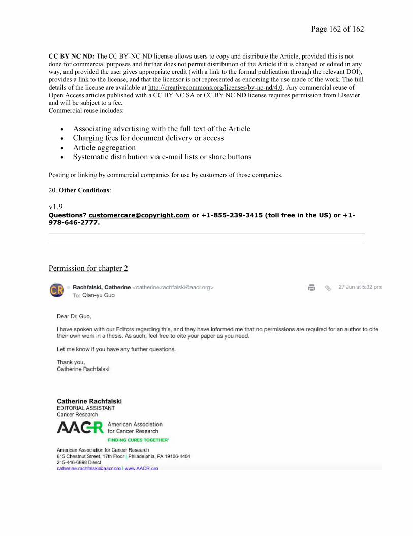
Page 162 of 162
CC BY NC ND: The CC BY-NC-ND license allows users to copy and distribute the Article, provided this is not
done for commercial purposes and further does not permit distribution of the Article if it is changed or edited in any
way, and provided the user gives appropriate credit (with a link to the formal publication through the relevant DOI),
provides a link to the license, and that the licensor is not represented as endorsing the use made of the work. The full
details of the license are available at http://creativecommons.org/licenses/by-nc-nd/4.0. Any commercial reuse of
Open Access articles published with a CC BY NC SA or CC BY NC ND license requires permission from Elsevier
and will be subject to a fee.
Commercial reuse includes:
Associating advertising with the full text of the Article
Charging fees for document delivery or access
Article aggregation
Systematic distribution via e-mail lists or share buttons
Posting or linking by commercial companies for use by customers of those companies.
20. Other Conditions:
v1.9 Questions? [email protected] or +1-855-239-3415 (toll free in the US) or +1-978-646-2777.
Permission for chapter 2