MEJFM_Vol6_Iss8.pdf - Middle East Journal of Family Medicine
34
Chief Editor: Abdulrazak Abyad MD, MPH, AGSF, AFCHSE Email: [email protected] Assistant to the Editor: Ms Rima Khatib Email: [email protected] Reporter and Photographer: Dr Manzoor Butt, Email: [email protected] Ethics Editor and Publisher: Ms Lesley Pocock medi+WORLD International 572 Burwood Road, Hawthorn, Vic Australia 3122 Phone: +61 (3) 9819 1224: Fax: +61 (3) 9819 3269 Email: lesleypocock@mediworld. com.au Editorial enquiries: [email protected] Advertising enquiries: [email protected] While all efforts have been made to ensure the accuracy of the infor- mation in this journal, opinions expressed are those of the authors and do not necessarily reflect the views of The Publishers, Editor or the Editorial Board. The publishers, Editor and Editorial Board cannot be held responsible for errors or any consequences arising from the use of information contained in this journal; or the views and opinions expressed. Publication of any advertisements does not constitute any endorse- ment by the Publishers and Editors of the product advertised. The contents of this journal are copyright. Apart from any fair deal- ing for purposes of private study, re- search, criticism or review, as permit- ted under the Australian Copyright Act, no part of this program may be reproduced without the permission of the publisher. 2 Editorial Abdul Abyad Original Contribution / Clinical Investigation 3 Efficacy of 3 Day Azithromycin Versus 10 Day Co-Amoxiclav in the Treatment of Children with Acute Otitis Media Khaled Amro, MD 6 Investigation of Demographic and Clinical Features in 131 Iranian Patients with Cluster Headache A.Ghorbani, MD, A.Chitsaz, MD, M.R.Savoj, MD, M. Etemadifar, MD 9 Nitroimidazoles in The Treatment of Intestinal Amoebiasis Dr Suleiman Muneizel 12 Usefulness of C-reactive Protein in Diagnosis of Intrapartum and Post- partum Neonatal Sepsis Khaled Amro, MD Medicine and Society 15 How Many People Have Cancer Patients (Alive or Deceased) in Their Homes, in Our City? Dilek Toprak, Nurhan Dogan, Serap Demir, Gülnihal Tufan 19 Women Knowledge Assessment about Self Care Behavior in Shiraz Health Care Center 2006 Vizeshfar, Fatemeh- Mehdizadeh, Kadege Education and Training 24 How to Write a Scientific Paper “Publish or perish” A Motivation to Learn More Ebtisam Elghblawi Case Report 28 Heterotopic Pregnancy in Natural Cycle - Probably Not Rare Dr.Ramadevi V Wani, Dr.Sami Al Taher Child-Watch section 32 Child-Watch Distribution of Eid Gifts to Blind Girls School Models and Systems of Primary Care 33 Advances in Surgical Education - for Surgical Trainees and Family Doc- tors Lesley Pocock ISSN 148-4196 October 2008 - Volume 6, Issue 8
-
Upload
khangminh22 -
Category
Documents
-
view
1 -
download
0
Transcript of MEJFM_Vol6_Iss8.pdf - Middle East Journal of Family Medicine
�
Chief Editor:Abdulrazak AbyadMD, MPH, AGSF, AFCHSEEmail: [email protected]
Assistant to the Editor:Ms Rima KhatibEmail: [email protected]
Reporter and Photographer:Dr Manzoor Butt,Email: [email protected]
Ethics Editor and Publisher:Ms Lesley Pocockmedi+WORLD International572 Burwood Road,Hawthorn, Vic Australia 3122Phone: +61 (3) 9819 1224:Fax: +61 (3) 9819 3269Email: [email protected]
Editorial enquiries:[email protected]
Advertising enquiries:[email protected]
While all efforts have been made toensure the accuracy of the infor-mation in this journal, opinions expressed are those of the authors and do not necessarily reflect the views of The Publishers, Editor or the Editorial Board. The publishers, Editor and Editorial Board cannot be held responsible for errors or any consequences arising from the use of information contained in this journal;or the views and opinions expressed.Publication of any advertisements does not constitute any endorse-ment by the Publishers and Editors of the productadvertised.
The contents of this journal arecopyright. Apart from any fair deal-ing for purposes of private study, re-search, criticism or review, as permit-ted under the Australian Copyright Act, no part of this program may be reproduced without the permission of the publisher.
2 Editorial Abdul Abyad
Original Contribution / Clinical Investigation3 Efficacy of 3 Day Azithromycin Versus 10 Day Co-Amoxiclav in the
Treatment of Children with Acute Otitis Media Khaled Amro, MD6 Investigation of Demographic and Clinical Features in 131 Iranian
Patients with Cluster Headache A.Ghorbani, MD, A.Chitsaz, MD, M.R.Savoj, MD, M. Etemadifar, MD9 Nitroimidazoles in The Treatment of Intestinal Amoebiasis Dr Suleiman Muneizel12 Usefulness of C-reactive Protein in Diagnosis of Intrapartum and Post-
partum Neonatal Sepsis Khaled Amro, MD
Medicine and Society15 How Many People Have Cancer Patients (Alive or Deceased) in Their
Homes, in Our City? Dilek Toprak, Nurhan Dogan, Serap Demir, Gülnihal Tufan19 Women Knowledge Assessment about Self Care Behavior in Shiraz
Health Care Center 2006 Vizeshfar, Fatemeh- Mehdizadeh, Kadege
Education and Training24 How to Write a Scientific Paper “Publish or perish” A Motivation to
Learn More Ebtisam Elghblawi
Case Report28 Heterotopic Pregnancy in Natural Cycle - Probably Not Rare Dr.Ramadevi V Wani, Dr.Sami Al Taher
Child-Watch section32 Child-Watch Distribution of Eid Gifts to Blind Girls School
Models and Systems of Primary Care33 Advances in Surgical Education - for Surgical Trainees and Family Doc-
tors Lesley Pocock
ISSN 148-4196 October 2008 - Volume 6, Issue 8
MIDDLE EAST JOURNAL OF FAMILY MEDICINE • VOLUME 6 , ISSUE 8�
This is the eighth issue this year and the current issue is rich with valuable papers from the region. Dr Elghiblawi E reviewed a topic of great importance in the middle east, that is the process of writing a Scientific Paper for publication. Dr Elghiblawi stressed that writing up is a daunting task; it involves a great deal of planning, preparation and time; it is simply a skill born from practice. In order to write, you need to read. The article supplies the researcher with a few simple guidelines on how to prepare, and write an organized scientific paper, which ranges from its early drafting in order to improve the manuscript, and then its final publication.
A paper from Turkey attempted to assess how many people have cancer patients (alive or dead) in their homes. The authors administered a questionnaire to 2035 people in 75 different parts of the city. There were 100 (4.9%) people who have been living with a patient diagnosed as cancer and 333 people (16.4%) who had a history of a patient dying from cancer in their home. The authors concluded that cancer is a part of our lives either with a patient in our house or a relative who has died from cancer. Public education and health services for home carers’
are needed for many people to care for these cancer patients.
Dr Amro K did a randomized study to compare the use of Azithromycin and co-Amoxiclav in the treatment of symptoms and signs of acute suppurative otitis media in children. He noted satisfactory clinical response was measured regarding symptoms and signs two weeks after the beginning of therapy. They were 84.6% for Azithromycin and 88% for Co-Amoxiclav. At day 28, 61 patients (82.4%) were cured on Azithromycin compared with 66 patients (83.5 %) on Co-Amoxiclav. The author concluded that Azithromycin given for three days and Co-Amoxiclav for ten days had similar efficacy; however, Azithromycin was better tolerated.
A paper from Iran looked at the demographic and clinical features in 131 Iranian patients with cluster headache. The study was performed in the Isfahan Medical University from June 2006 to June 2007. 131 patients with definite cluster headache were selected randomly. The authors concluded that on the basis of this study, may be there is a regional and race difference among different studies. According to treatability of this type of headache, and morbidity and costs that are produced by this disease, more extensive studies on the base of prevalence, predisposing factors, different aspects of treatment, and prophylactic treatments should be taken to provide patients with more suitable and effective help.
A cross sectional survey was conducted in Iran to evaluate knowledge and behavior related to “self care” among women who attended to Shiraz Health Center. A convenience sample of 607 women were selected by cluster and random sampling. 52.2% of women had good knowledge about self care behavior. Knowledge of blood cholesterol was highest rate among women (91.1%). Good practice about screening tests was only 3.1% for control of blood pressure as routine, was higher compared to other screening tests. The authors concluded that practice of women about screening tests especially as a
routine, is weak. With attention to the importance of self care behavior in promotion of life quality and life span, therefore nurses and health workers must teach and encourage women about self care behavior and attending screening tests.
Dr Muneizel S attempted to determine the efficacy and side effects of tinidazole compared with metronidazole in the treatment of amoebiasis in Jordanian patients. A randomized controlled clinical trial was carried out on 66 subjects with Entamoeba histolytica infestation. Infected patients were treated with either tinidazole or metronidazole (Tinidazole 2gm single dose orally for 3 days and metronidazole 2gm single dose orally for 3 days) . 27 of 32 patients (87.5%) treated with tinidazole and 23 of 34 patients (67.5%) treated with metronidazole had parasitological cure. Cure rates between two groups were significant statistically (P<0.01). The author concludes that Tinidazole was more effective than metronidazole, produced fewer and milder side effects, and is recommended with high efficacy in treating intestinal amoebiasis.
A cohort study from Jordan looked to determine effects of intrapartum risk factors for early onset sepsis (EOS) on CRP levels in neonates and to assess the suitability of this test in diagnosing EOS. A total of 200 neonates were studied. CRP levels in cord blood and neonatal blood at 24 hours were estimated using commercial kits. Elevated cord CRP levels was significantly associated with rupture of membranes for 24 hours labour more than 12 hours and maternal fever. Several intrapartum risk factors for EOS can cause elevation in CRP levels. However, this test may be useful in excluding infection.
From the Editor
Abdulrazak Abyad MD, MPH, AGSF, AFCHS(Chief Editor)
Editorial office:Abyad Medical Center & Middle East Longevity InstituteAzmi Street, Abdo CenterPO BOX 618Tripoli, LebanonP + (961) 6 443684F + (961) 6 443685E [email protected] www.amc-lb.com
FROM THE EDITOR
MIDDLE EAST JOURNAL OF FAMILY MEDICINE • VOLUME 6 , ISSUE 8 �
ORIGINAL CONTRIBUTION AND CLINICAL INVESTIGATION
Efficacy of 3 Day Azithromycin Versus 10 Day Co-Amoxiclav in the Treatment of Children with Acute Otitis Media
ABSTRACT Objective: To compare the use of Azithromycin and co-Amoxiclav in the treatment of symptoms and signs of acute suppurative otitis media in children.
Methods: Children of four months to 12 years of age, attended out-patient Pediatric and ENT clinics at Prince Hashim bin Al Hussain Hospi-tal in AZ-Zarqa (JORDAN) from June 2006 to June 2007; with signs and symptoms of acute suppurative oti-tis media, were enrolled in the study. Patients were randomized to receive either Azithromycin 10 mg/kg/day in a single dose for 3 days or co-Amoxi-clav 45 mg/kg/day in three divided doses for 10 days. Clinical improve-ment was evaluated on the 2nd and 4th weeks after therapy.
Results: Satisfactory clinical re-sponse was measured regarding symptoms and signs two weeks after the beginning of therapy. They were 84.6% for Azithromycin and 88% for Co-Amoxiclav. At day 28, 61 patients (82.4%) were cured on Azithromycin compared with 66 patients (83.5 %) on Co-Amoxiclav.
Conclusion: Azithromycin given for three days and Co-Amoxiclav for ten days had similar efficacy; however, Azithromycin was better tolerated.
IntroductionOtitis Media is an inflammation
in the middle ear. Subcategories include acute otitis media, otitis media with effusion (also known as “glue ear”), recurrent acute otitis media, and chronic suppurative otitis media. Acute otitis media presents with systemic and local signs and has a rapid onset. The persistence of an effusion beyond three months without signs of infection defines otitis media with effusion, whereas chronic suppurative otitis media is characterized by continuing inflammation in the middle ear giving rise to otorrhoea and a perforated tympanic membrane(1).
Our study is on Acute Suppurative Otitis Media (ASOM), which is a suppurative infection of the middle ear cavity and is most common in healthy children between 6 months and 2 years of age, it is more common in boys, in patients of lower socioeconomic status, in formula fed infants, and in the winter months(2). It is an important health problem in early childhood, and is the most frequent condition for which antibiotics are prescribed in the USA(3).
The most common pathogens are streptococcus pneumonic Hemophilus Influenza, and Branhamella catarrhal; half of these organisms are B-lactamase producers. Very young children will not complain of pain but will be irritable and may bang the head on the cot sides. On examination, the young child is febrile, restless and uncooperative with red, bulging tympanic membrane. If the ears discharge, it is usually blood stained
initially and this may worry parents.
The discharge then becomes mucopurulent(4). A combination of important factors contributes to pathogenesis of ASOM. The most two important factors in children are Eustachian tube dysfunction and the child susceptibility to recurrent upper respiratory tract infections. In the child, the Eustachian tube is shorter (less distance for organisms to travel), placed horizontal (inadequate drainage of middle ear) and has adenoids present at the opening, which can readily block the tube and serve as a reservoir of infection.
Bacteria are responsible for the majority of cases(5). Antibiotic treatment of Acute Suppurative Otitis Media hastens symptomatic relief and potentially prevents the development of more serious invasive disease(6). As there are a number of antibiotics used for this purpose we undertook this trial in children with ASOM to compare the use of two important antibiotics commonly used in this condition in Jordan.
Amoxicillin semi synthetic penicillin has broadened spectra against Gram-negatives and is effective orally. Amoxicillin plus Clavulanate is Clavamox or Augmentin. The Clavulanate is not an anti- microbial agent; it inhibits beta-lactamase enzymes and has given extended life to penicillinase(7). Generally ampicillin, amoxicillin, or co-Amoxiclav (amoxicillin-clavulanate) are preferred and most commonly used in ASOM in Jordan.
Azithromycin is an azolide antibiotic. It is active in vitro against
Key words: Acute Suppurative Otitis Media, Children, Antibiotics.
Khaled Amro, MD*
*Pediatrician from department of pediatricIn Royal Medical Services-Jordan
MIDDLE EAST JOURNAL OF FAMILY MEDICINE • VOLUME 6 , ISSUE 8�
ORIGINAL CONTRIBUTION AND CLINICAL INVESTIGATION
a variety of microorganisms and has a greater distribution in tissues, a longer elimination half-life and a lower incidence of adverse effects than Erythromycin(8). The purpose of this study was to compare the clinical use of Azithromycin with Amoxiclav.
Materials and MethodsThis consecutive study was carried out at PrinceHashim Hospital in AZ-Zarqa city in Jordan from June 2006 to June 2007, on children attending outpatient ENT and pediatric OPD clinics. Children of ages four months to twelve years were enrolled in the study, if they satisfied one or more of the following criteria:
Ear pain or fullness.Decreased hearing.Discharge from the external auditory canal.Bulging or marked injection of the tympanic membrane.Loss of the normal light reflex or tympanic membrane landmarks.As well as generalized symptoms; fever, general malaise, and irritability.
Exclusion criteria included: History of Macrolide or B-lactamase drug allergy, history of antibiotic treatment in the preceding four weeks, Symptoms persisting for more than four weeks, and children receiving antimicrobial prophylaxis.
Patients were randomized on alternative weeks and accordingly divided into two groups: First group received either Azithromycin (10 mg/kg/day) once daily for three days, and second group were given Co-Amoxiclav (45mg/kg/day) in three divided doses for ten days.
Assessment of these patients was carried out on the initial visits and follow up was done on days 14 and 28.
Patients were identified to be cured when there was a complete resolution of all signs and symptoms, improved by partial resolution of signs and symptoms, and failed, if there were no changes or worsening of symptoms and signs.
On follow up visits complete Ear, Nose and Throat examination was
•••
•
•
•
performed by the same physicians at all pre treatment and post treatment visits.
ResultsTwo hundred and three patients
were initially enrolled in the study. 17 patients were non-eligible as 5 had allergy to Amoxicillin, and 12 didn’t fulfill the inclusion criteria because they received antibiotics in the preceding four weeks. The total number of patients found eligible in our study was 186 children. All of these patients were randomized into two groups; the first group included 91 children (received Azithromycin 10mg/kg/day once daily for three days), while the second group included 95 children and were given Co-Amoxiclav 45 mg/kg/day in three divided doses for 10days). The mean age of patients enrolled was 3.4 years (range 4 months-12 years).
The most common symptom was ear pain (94%) while the most common sign was of injection tympanic membrane (93%). (Table I) Patients’ post treatment evaluation was done at two weeks; in the first group, 66 out of 78 children (84.6%) showed improvement or were cured, compared to 74 out of 84 children (88%) in the second group.
However, at four weeks post treatment, 61 out 74 children (82.4%) in the first group were completely cured and did not need any further antibiotic treatment, compared again to 66 out of 69 children (83.5%) in the second group. (Table II, Figure 1)
Regarding the adverse effects to the drugs used, these were mostly seen in children treated with Co-Amoxiclav compared with those who received Azithromycin and occurred in18% and 10% respectively. The most commonly observed side effect with both drugs was diarrhea. Rash and vomiting were also seen.
DiscussionOur study was looking for the pr
In Jordan, particularly the Royal Medical Services, Acute Suppurative Otitis Media is usually treated with
antibiotics, and generally amoxicillin, or Co-Amoxiclav (amoxicillin-clavulanate) are preferred and this depends on the availability and the cost of these medications. In his study, Dunne MW et al have provided evidence that Azithromycin for three days of treatment with a total dose of 30 mg/kg/day is as effective as Co-Amoxiclav given at 45 mg/kg/day. Similarly our study carried out on children of various age groups showed that the success rate of treatment at 2 and 4 weeks was nearly equivalent for both antibiotics and there was no significant difference (Table II).
Satisfactory clinical response regarding symptoms and signs evaluated at 2 weeks post treatment was 84.6% for Azithromycin and 88% for Co-Amoxiclav; this is compared to results seen in the Dunne et al study where the clinical success (cure and improvement) in all subjects was 83% for Azithromycin group of patients, and 88% for patients on Co-Amoxiclav on evaluation at 10 days post treatment. However regarding Co- Amoxiclav, it was noted that it led to a quicker resolution of tympanic membrane signs such as bulging and loss of landmarks at two weeks after initiation of treatment, whereas at four weeks of treatment both agents showed a similar outcome.
As for the mechanism of action, Co-Amoxiclav is a bactericidal agent whereas Azithromycin is a protein synthesis inhibitor (bacteristatic) agent; it is an azolide antibiotic, which has a greater distribution in tissues, a longer elimination half-life, and a lower incidence of adverse effects, than erythromycin. These pharmacokinetic features allow once-daily dosing and a shorter duration of therapy(9,10).
Our diagnoses were based on acute signs of infection and eardrum abnormalities, which is in keeping with the day-to-day practice in our hospital (The Royal Medical Services). There is a considerable controversy as to what antibiotic to use if at all, as some studies showed that up to 80% of cases with ASOM would resolve within one week without antibiotic treatment. The generalized use of antibiotics in this condition increases health care costs and creates
MIDDLE EAST JOURNAL OF FAMILY MEDICINE • VOLUME 6 , ISSUE 8 �
ORIGINAL CONTRIBUTION AND CLINICAL INVESTIGATION
numerous side effects(10). Watchful waiting at the first visit was justified by Damoiseaux et al for children aged 6-24 months with ASOM(11), and Froom et al state that the Netherlands is the only country where only a minority of the episodes of Otitis Media are treated with antibiotics.
The outcome of ASOM does not seem to be any worse than in other countries. In addition, doctors are often uncertain about the diagnosis of Suppurative Otitis Media. Therefore, we recommend that clinicians should immediately reconsider the routine use of antimicrobials for children with Suppurative Otitis Media and consider treating symptoms with analgesics and observation for lack of improvement(12). In conclusion, azithromycin given for three days and co-Amoxiclav for 10 days had similar efficacy; however, Azithromycin was better tolerated.
ReferencesO’Neill P.Clinical evidence: Acute otitis media. BMJ1999; 319:833-835.Prince A. Infections Diseases. In: Behrman RE, Kliegman RM, editors. Nelson essential of pedi-atrics. 4th edition, Saunders WB, USA. 2001; 10: 388-389.Arrieta A, Arguedas A, Fernandez P, et al. High-dose azithromycin versus high-dose amoxicillincla-vulanate for treatment of children with recurrent or persistent acute otitis media. Antimicrob Agents Chemother2003; 47(10): 3179-3186.Kerr AG. Acute otitis media. In: Adams DA, Cinna-mond MJ, editors. Scott-Brown’s Otolaryngology. 6th edition, Arnold. 1996; 8: 220-221.Jacob A, Rupa V, Job A, Joseph A. Hearing impair-ment and otitis media in a rural primary school in south India. Int J Pediar Otohinolaryngol1997; 39(2): 133-138.Dunne MW, Latiolais T, Lewis B, et al. Randomized, double-blind study of the clinical efficacy of 3 days of azithromycin compared with co-amoxiclav for the treatment of acute otitis media. J Antimicrob Chem-other 2003; 52(3): 469-472.Kennell Toda. University of Wisconsin-Madison, Department of Bacteriology. 2002Foulds G, Shepard RM, Johnson RB, et al. The pharmacokinetics of Azithromycin in human serum and tissues. J Antimicrob Chemother1990; 25:73-82.Nahata MC, Koranyi KI, Luke DR, Foulds G.Pharmacokinetics of azithromycin in pediatric pa-tients with acute otitis media. Antimicrobial Agents
1.
2.
3.
4.
5.
6.
7.
8.
9.
Chemotherapy1995; 39(8): 1875-1877.Parra A, Ponte C, Cenjor C, et al. Effect of antibiotic treatment delay on therapeutic outcome of experi-mental acute otitis media caused by Streptococcus pneumoniae strains with different susceptibilities to amoxicillin. Antimicrobial Agents Chemothera-py2004; 48(3): 860-866.Damoiseaux RAMJ, Van Balen FAM, Hoes AW, et al. Primary care based randomised, double blind trial of amoxicillin versus placebo for acute otitis media in children aged under 2 years.BMJ2000; 320: 350-354.Froom J, Culpepper L, Grob P, et al. Diagnosis and antibiotic treatment of acute otitis media: Re-port from International Primary Care Network. BMJ 1990; 300: 582-586.
10.
11.
12.
Table 1 Signs and symptoms found at presentation
Signs and symptoms Number %Ear pain or fullness 175 94Decrease hearing 23 12.3Discharge from external auditory canal 13 7Injection of tympanic membrane 172 93Bulging of tympanic membrane 98 47.8Perforated tympanic membrane 9 4.8Generalized symptoms, fever, general malaise and irritability 69 37
Table 2: Responseaftertwoandfourweeks.
Azithromycin % Co-Amoxiclav %Response at two weeks cured and/or improved 66/78 84.6 74/84 88Response at four weeks cured and/or improved 61/74 82.4 66/79 83.5
Figure 1: Total number of patients
MIDDLE EAST JOURNAL OF FAMILY MEDICINE • VOLUME 6 , ISSUE 8�
ORIGINAL CONTRIBUTION AND CLINICAL INVESTIGATION
IntroductionThe International Association for the
Study of Pain (IASP)1,3 defines cluster headache as ‘unilateral, excruciatingly severe attacks of pain principally in the ocular, frontal and temporal areas recurring in separate bouts with daily or almost daily attacks for weeks to months usually with ipsilateral lacrimation, conjunctival injection, photophobia and nasal stuffiness, and/or rhinorrhoea’1,20.
Prevalence rate of cluster headache was ranged from 56 per 100,000 (prevalence rate for men of 115.3 per 100,000) to 326 per 100,000 with an incidence rate of 2.5 to 9.8 per 100,000 per year in different studies2,22,27,31,32,33,35.
Cluster headache is predominantly a disease of men. Onset typically begins in the third decade of life. Periodicity is a cardinal feature of cluster headache. In most patients, the first cluster of attacks, the cluster period, persists on average 6-12 weeks and is followed by a remission lasting for months or even years. During a cluster, the patient may experience from one to three or more attacks in 24 hours, and the attacks commonly occur at similar times throughout the 24 hours for many days. Onset during the night or 1-2 hours after falling asleep is common. In some patients, perhaps as many as 10%, periods of
relief become less common, and the condition enters the chronic phase in which attacks may occur daily for months or years1,23,24,28.
The pain is strictly unilateral and almost always remains on the same side of the head from cluster to cluster. The pain is generally felt in the retro-orbital and temporal regions but may be maximal in the cheek or jaw (lower syndrome)21. It is usually described as steady or boring and of terrible intensity (so-called suicide headache).
The pain intensifies very rapidly, peaking in 5-10 minutes and usually persisting for 45 minutes to 2 hours. During the pain of cluster headache, the nostril on the side of the pain is generally blocked; this blockage in turn leads to ipsilateral overflow to tears caused by blockage of the nasolacrimal duct. The conjunctiva may be injected ipsilaterally, and the superficial temporal artery may be visibly distended. Profuse sweating and facial flushing on the side of the headache have been described but are rare. Nasal drainage usually signals the end of the attack1,7,8,9,30,34.
Different treatment strategies (acute, maintenance) have been mentioned in different references1,7,11,12,13,14,23,24,26,29.
In our study, we aimed to determine the demographic and symptomatologic
Investigation of Demographic and Clinical Features in 131 Iranian Patients with Cluster HeadacheA.Ghorbani, MD1, A.Chitsaz, MD1, M.R.Savoj, MD2, M. Etemadifar, MD3.
1. Associated professor, Department of Neurology, Isfahan Medical University, Iran.2. Resident of Neurology, Department of Neurology, Isfahan Medical University, Iran.3.: Professor, Department of Neurology, Isfahan Medical University, Iran.
Corresspondence to:Associated Prof. A. Ghorbani,Department of Neurology,Isfahan Medical University,IranFax: (+98) 311-6684510Tel: (+98) 311-6685555E-mail: [email protected]
ABSTRACTBackground: Cluster headache is defined as ‘unilateral, excruciatingly severe attacks of pain principally in the ocular, frontal, and temporal are-as recurring in separate bouts along with daily or almost daily attacks for weeks to months usually with ipsilat-eral lacrimation, conjunctival injec-tion, photophobia and nasal stuffi-ness and/or rhinorrhoea’.Methods: This descriptive study was performed in the Isfahan Medical University from June 2006 to June 2007. 131 patients with definite clus-ter headache were selected random-ly. Data was taken from past history and presenting features of patients.Results: Among with 131 Iranian patients investigated in our study (referred with possible diagnosis of cluster headache from other cent-ers), there were: 120 male, 11 fe-male, 68.7% 20 to 40 years old, 67% with abrupt onset headache, 90 with pulsatile pain, 30 with non-pulsatile pain, 2 with both types, 69.4% with less than 60 minute dura-tion, 38.8% with similar time occur-rence of headache, more prevalent autonomic sign, lacrimation, nostril block, vomiting, prominence of tem-poral artery, rinorrhoea, petosis, and profuse sweating, site of headache: 101 around the orbit, 19 far from the orbit, 96 with seasonal relationshop, 102 with episodic pattern, 29 with chronic form, Free period: 60%: 7 to 12 months. Related foods: dairy products, onions, vinegar, pickles, fatty foods, fast food, eggs, toasted foods, pungent foods, cucumbers and potatoes. Familial and childhood cluster headache, non alcohol con-sumption: 22 patients, smoking: 52 patients, history of head trauma: 15 patients.Conclusion: On the basis of this study, maybe there is a regional and race difference among different studies. According to treatability of this type of headache, and morbidity and costs that are produced by this disease, more extensive studies on the base of prevalence, predispos-ing factors, different aspects of treat-ment, and prophylactic treatments should be taken to provide patients with more suitable and effective helps.
Key words: Cluster headache, headache types, clinical features.
MIDDLE EAST JOURNAL OF FAMILY MEDICINE • VOLUME 6 , ISSUE 8 �
ORIGINAL CONTRIBUTION AND CLINICAL INVESTIGATION
presentation of cluster headaches in Iranian patients who visited our health care units.
Subjects and MethodsThis descriptive study was
performed in the Isfahan Medical University from June 2006 to June 2007. 131 patients with definite cluster headache were selected randomly from patients referred to Alzahra hospital, Noor hospital and other neurologist offices (that participated in this study). With MRI, CT scan, and blood sample studies other diagnoses were excluded and those selected had definite cluster headache criteria on the base of ICD10 criteria. Patients with non-definite diagnosis or another diagnosis were not included in our study. We designed special forms for systematic uptake of needed information from past history and presenting features of patients. These forms were completed by neurologists. The registration form of patients was attached to the end of this article.
ResultsAmong 131 Iranian patients
investigated in our study (referred with possible diagnosis of cluster headache from other centers), 120 patients were male and 11 female. 68.7% had 20 to 40 years (mean age: 35.55 years; range: 18-63). 67% of our patients presented with abrupt onset headache. Quality of pain was pulsatile in 90 patients, non-pulsatile in 30 patients and both these types in 2 patients. In 69.4% of patients duration of each attack was less than 60 minutes and in 4.8% was more than 180 minutes. Only in 38.8% of patients, attacks occurred in similar times (32 patients 1 hour after falling asleep and 19 patients between 2300 hours to 200 hours).
More prevalent autonomic signs presented with headache in order of prevalence were: lacrimation (102 patients), nostril block (90 patients), vomiting (70 patients), prominence of temporal artery (52 patients), rhinorrhea (50 patients), petosis (36 patients), and profuse sweating (30 patients).
Predisposing factors obtained in our study were: stress (106 patients), smoking (52 patients), special foods (40 patients), cold (35 patients), flashing lights (32 patients), alcohol (22 patients), heat (16 patients), history of head trauma (15 patients).
Among special foods to which 40 patients described a relation to their headaches there were: dairy products, onions, vinegar, pickles, fatty foods, fast food, eggs, toasted foods, pungent foods, cucumbers, and potatoes.
Among 22 patients with a history of alcohol consumption, 17 patients reported beginning or deterioration of headache with alcohol use and among 52 smokers, 17 patients did. All of 15 patients with history of head trauma had this event 10 years after.
Site of headache in 101 patients was around the orbit (70 in the right side and 31 in the left side) and in 19 patients far from the orbit. More common site of pain radiation was the ipsilateral forehead and cheek.
In 96 patients, environment had no effect on the pain relief. In 61 patients pain commencement was related to season (23: winter, 20: summer, 12: autumn, 6: spring). 102 patients had episodic pattern and 29 patients had chronic form. In almost all patients periodicity of attacks were one or two attacks daily. In 102 patients with episodic patterns, 58 patients described duration of each episode 4 to 8 weeks and 26 patients about 4 weeks.
Free period between each episode was from 1 month to 3 years that were 7 to 12 months in 60% of these patients.
In our study, we did not find familial or childhood cluster headache among our patients.
In our study, the follow up for treatment was not performed. Patients with non-cluster headache were not investigated and classified in our study.
DiscussionThere was no appropriate data
from Iranian patients about cluster
headache accessible in different investigations and therefore we could not compare our results with other Iranian data. We compared our results with data from developed countries.
In our investigation the male to female ratio was 11:1 that is significantly higher than other studies (6:1)15,25,32. Quality of pain in other studies was often non-pulsatile but in our study more than one-half of patients had pulsatile headache quality16,20,23,24. Often pain was unilateral and only 6.8% of patients had radiation of pain to other side (15% in other studies)17,20,23,24. 62% of patients had pain without predictable diurnal pattern but in other studies headache was beginning among 21 to 10 o’clock1,18,23,24. Only 38.8% of patients had special diurnal pattern for headache and these findings are against the theory of biologic clock effect on periodicity of cluster headache20,37,38. According to data from history of head trauma that at least was presented 10 years before beginning of cluster headache, it seems that there is no relation between head trauma and cluster headache. This finding is according to the Kudrow (1980)19 study and against the Manzoni (1983)17
study17,18,36.
In our study we did not find any patients with document of cluster headache occurrence in their family that is accordant with other studies3,4. Childhood cluster headache was not found in our study (in other studies the presentation of childhood cluster headache was rare)5,6,10.
In other aspects of our study, the results are similar to other studies that were done in other countries1,15,16,17,18,19,27,31.
ConclusionOn the basis of this study, we
found that maybe there is a regional and race difference among different studies. According to treatability of this type of headache, mortality, and costs that are produced by this disease, more extensive studies on the base of prevalence, predisposing factors, different aspects of treatment and prophylactic treatments should
MIDDLE EAST JOURNAL OF FAMILY MEDICINE • VOLUME 6 , ISSUE 8�
ORIGINAL CONTRIBUTION AND CLINICAL INVESTIGATION
be taken to provide patients with more suitable and effective help.
ReferencesZakrzewska J. M. Cluster headache: review of the lit-erature. British Journal of Oral and Maxillofacial Surgery .2001; 39: 103-113.Ekbom K, Svensson DA, Pedersen NL, Waldenlind E .Lifetime prevalence and concordance risk of cluster headache in the Swedish twin population. Neurology. 2006 Sep 12;67(5):798-803.El Amrani M, Ducros A, Boulan P, Aidi S, Crassard, Visy JM , Tournier-Lasserve E, Bousser MG . Familial cluster headache: a series of 186 index patients. Headache. 2002 Nov-Dec;42(10):974-7.Leone M, Rigamonti A, Russel MB, Mea E, D’Amico D, Grazzi L , Bussone G . Selective vs. complete family interview for detecting those affected by familial cluster headache. Cephalalgia. 2004 Nov; 24(11):938-9.Lampl C . Childhood-onset cluster headache. Pediatr Neurol. 2002 Aug; 27(2):138-40.McNabb S, Whitehouse W . Arch Dis Child. Clus-ter headache-like disorder in childhood. 1999 Dec; 81(6):511-2.Dodick DW and Campbell JK. Cluster headache diag-nosis, management and treatment. In: SD Silberstein, R Lipton and DJ Dalessio. Wolff’s headache and other head pain: Oxford university press; oxford .2001: 283-309.Hannerz J. Symptoms and diseases and smoking hab-its in female episodic cluster headache and migraine patients. Cephalalgia. 1997; 17: 499-500.Kudrow L. The pathogenesis of cluster headache. Curr Opin Neurol 1994; 7: 278-282.Ekbom K , Svensson DA , Traff H , Waldenlind E . Age
1.
2.
3.
4.
5.
6.
7.
8.
9.
10.
at onset and sex ratio in cluster headache: observations over three decades.Cephalalgia. 2002 Mar;22(2):94-100.Mathew NT. Cluster headache. Seminars in Neurology. 1997; 17:313-323.Monstad I, Krabbe A, Micieli G, Prusinski A, Cole J, Pilgrim A, Shevlin P. Preemptive oral treatment with sumatriptan during a cluster period. Headache. 1995; 35: 607-613.Mathew NT. Cluster headache. Neurology. 1992; 42 (Suppl 2): 22-31.Gabai IJ, Spierings EL. Prophylactic treatment of clus-ter headache with verapamil. Headache. 1989; 29: 167-168.Krabbe AA. Cluster headache: a review. Acta Neurol. Scand. 1986; 74: 1-9.Ekbom k. Some observation on pain in cluster head-ache. Headache. 1975; 14:219-225.Manzoni GC, Terzano MG and Bono G. et el. Cluster headache-clinical finding in 180 patients. Cephalgia. 1983; 3:21-30.Lance JW and Anthony M. Migranous neuralgia or clus-ter headache. J. Neurol. SCi. 1971; 13:401-414.Kudrow L. Cluster headache: Mechanism and manage-ment. Oxford university press, New York. 1980.May A; Leone M. Update on cluster headache. Curr Opin Neurol. 2003; 16(3):333-40.Gross SG. Dental presentations of cluster headaches. Curr Pain Headache Rep. 2006; 10(2):126-9.Russell MB. Epidemiology and genetics of cluster head-ache. Lancet Neurol. 2004; 3(5):279-83.Rozen TD. Cluster headache: diagnosis and treatment. Curr Pain Headache Rep. 2005; 9(2): 135-40.Capobianco DJ, Dodick DW. Diagnosis and treatment of cluster headache. Semin Neurol. 2006; 26(2):242-59.Lin KH, Wang PJ, Fuh JL, Lu SR, Chung CT, Tsou HK, Wang SJ. Cluster headache in the Taiwanese -- a clinic-based study. Cephalalgia. 2004; 24(8):631-8.Fabre N. Treatment of cluster headache.Rev Neurol (Paris). 2005; 161(6-7):696-9.Finkel AG. Epidemiology of cluster headache. Curr Pain
11.
12.
13.
14.
15.
16.
17.
18.
19.
20.
21.
22.
23.
24.
25.
26.
27.
Headache Rep. 2003; 7(2):144-9.Ducros A, Bousser MG. Cluster headache. Ann Med Interne (Paris). 2003; 154(7):468-74.Stallmach M. Prophylactic treatment of cluster head-ache with verapamil. Schweiz Rundsch Med Prax. 2003; 92(46):1951-3.Drummond PD. Mechanisms of autonomic disturbance in the face during and between attacks of cluster head-ache. Cephalalgia. 2006; 26(6):633-41.Torelli P, Beghi E, Manzoni GC. Cluster headache prevalence in the Italian general population.Neurology. 2005; 64(3):469-74.Ekbom K, Svensson DA, Pedersen NL, Waldenlind E. Lifetime prevalence and concordance risk of cluster headache in the Swedish twin population. Neurology. 2006; 67(5):798-803.Torelli P, Castellini P, Cucurachi L, Devetak M, Lambru G, Manzoni GC. Cluster headache prevalence: method-ological considerations. A review of the literature. Acta Biomed Ateneo Parmense. 2006; 77(1):4-9.Tanuri Fda C, Sanvito WL. Cluster headache: study of autonomic alterations and other associated mani-festations in 28 cases. Arq Neuropsiquiatr. 2004; 62(2A):297-9.Black DF, Swanson JW, Stang PE. Decreasing inci-dence of cluster headache: a population-based study in Olmsted County, Minnesota. Headache. 2005; 45(3):220-3.Manzoni GC, Lambru G, Torelli P. Head trauma and cluster headache. Curr Pain Headache Rep. 2006; 10(2):130-6.Pringsheim T. Cluster headache: evidence for a disor-der of circadian rhythm and hypothalamic function. Can J Neurol Sci. 2002; 29(1):33-40.Nappi G, Sandrini G, Alfonsi E, Cecchini AP, Micieli G, Moglia A. Impaired circadian rhythmicity of nociceptive reflex threshold in cluster headache. Headache. 2002; 42(2):125-31.ICD 10 Guide for Headaches. International Headache Classification Committee. Cephalgia.
28.
29.
30.
31.
32.
33.
34.
35.
36.
37.
38.
39.
MIDDLE EAST JOURNAL OF FAMILY MEDICINE • VOLUME 6 , ISSUE 8 �
ORIGINAL CONTRIBUTION AND CLINICAL INVESTIGATION
IntroductionEntamoeba histolytica is the
etiological agent of amoebic dysentery. Worldwide, 40-50 million symptomatic cases of amoebiasis occur annually and 70,000 to 100,000 deaths are due to this infection.[1]
Molecular phylogeny places entamoeba on one of the lowermost branches of the eukaryotic tree, closest to dictyostelium. Although the organism was originally thought to lack mitochondria, nuclear-encoded mitochondrial genes and a remnant organelle have now been identified.[2,3] Unusual features of entamoeba include polyploid chromosomes that vary in length; multiple origins of DNA replication; abundant, repetitive DNA; closely spaced genes that largely lack introns; a novel GAAC element controlling the expression of messenger RNA; and unique endocytic pathways.[4,5,6,7] There are two distinct, but morphologically identical species of Entamoeba: Entamoeba histolytica, which is pathogenic and Entamoeba dispar, which is non-pathogenic.[15]
Ingestion of the quadrinucleate cyst of E. histolytica from fecally contaminated food or water initiates infection. Infection with E. histolytica may be asymptomatic or may cause dysentery or extra intestinal disease. Asymptomatic infection should be treated because of its potential to progress to invasive disease. Patients with amebic colitis typically present with a several-week history of cramping abdominal pain, weight loss, and watery or bloody diarrhea. The insidious onset and variable signs and symptoms make diagnosis difficult, with fever and grossly bloody stool absent in most cases.[8,9,10]
Therapy for invasive infection differs from therapy for noninvasive infection.
Noninvasive infections may be treated with paromomycin. Nitroimidazoles, particularly metronidazole, are the mainstay of therapy for invasive amebiasis.[11] Nitroimidazoles with longer half-lives (namely, tinidazole, secnidazole, and ornidazole) are better tolerated and allow shorter periods of treatment.
Approximately 90 percent of patients who present with mild-to-moderate amebic dysentery have a response to nitroimidazole therapy. Parasites persist in the intestine in as many as 40 to 60 percent of patients who receive nitroimidazole. Therefore, nitroimidazole treatment should be followed with paromomycin or the second-line agent diloxanide furoate to cure luminal infection. Metronidazole and paromomycin should not be given at the same time, since the diarrhea that is a common side effect of paromomycin may make it difficult to assess the patient’s response to therapy.[12,13,14] In this study we assess the efficacy of the 2 nitroimidazoles available in Jordan, tinidazole and metronidazole.
Patients and MethodsThe efficacy and tolerability of
metronidazole and tinidazole were evaluated in a randomized clinical trial performed with 66 patients who attended the out-patient clinic and emergency room in QAMH. The study period was 12 months from July 2005 to July 2006. The subjects (24 females and 42 males) were randomly allocated to two groups: experiment group (n=32) were given tinidazole and control group (n=34) were given metronidazole [Table 1]. In group one, metronidazole 2gm as a single dose orally for 3 days), and in group two, tinidazole 2 gm single dose orally were
Nitroimidazoles in The Treatment of Intestinal AmoebiasisDr Suleiman Muneizel MD, JBDepartment of Internal Medicine, Royal Medical Services QAMH
Corresspondence to:Dr Suleiman MuneizelEmail: [email protected]
ABSTRACTObjective: Entamoeba histolytica is one of the common intestinal proto-zoans in the Middle East. Treatment of infection has some difficulties by metronidazole because of the long course of therapy and various side effects. The objective of this study was to determine efficacy and side effects of tinidazole compared with metronidazole in the treatment of amoebiasis in Jordanian patients.
Patients and Methods: Over an interval period of one year dura-tion, starting July 2005 through July 2006,a randomized controlled clini-cal trial was carried out on 66 sub-jects (42 males, 24females) with Entamoeba histolytica infestation who presented to out-patients clinic or emergency room in Queen Alia Military Hospital in Jordan ,infected patients were treated with either tini-dazole or metronidazole (Tinidazole 2gm single dose orally for 3 days and metronidazole 2gm single dose oral-ly for 3 days). Parasitological cure was documented when there were 3 successive negative stool examina-tions for entamoeba histolytica at 1-2 weeks after therapy.
Results: 27 of 32 patients (87.5%) treated with tinidazole and 23 of 34 patients (67.5%) treated with metro-nidazole had parasitological cure. Cure rates between two groups were significant statistically (P<0.01). No major side effects were observed except 13 cases in metronidazole group who had nausea, epigastric pain, mild headache and some had metallic taste. Three cases in tini-dazole group had nausea, dizziness and headache.
Conclusion: Tinidazole was more effective than metronidazole, pro-duced fewer and milder side effects, and is recommended with high effi-cacy in treating intestinal amoebia-sis.
Key-words: Amoebiasis ,Treatment, Nitroimidazo ,Metronidazole.
MIDDLE EAST JOURNAL OF FAMILY MEDICINE • VOLUME 6 , ISSUE 810
prescribed respectively.[16] Patients were followed for three weeks after the end of therapy for the presence of entamoeba histolytica in their stool. Clinical and parasitological follow-up was carried out before, and at 7, 14, and 21 days after treatment and the outcome of treatment was noted. Parasitological cure was documented when there were three consecutive negative stool examinations for entamoeba histolytica at 1-3 weeks after therapy termination.
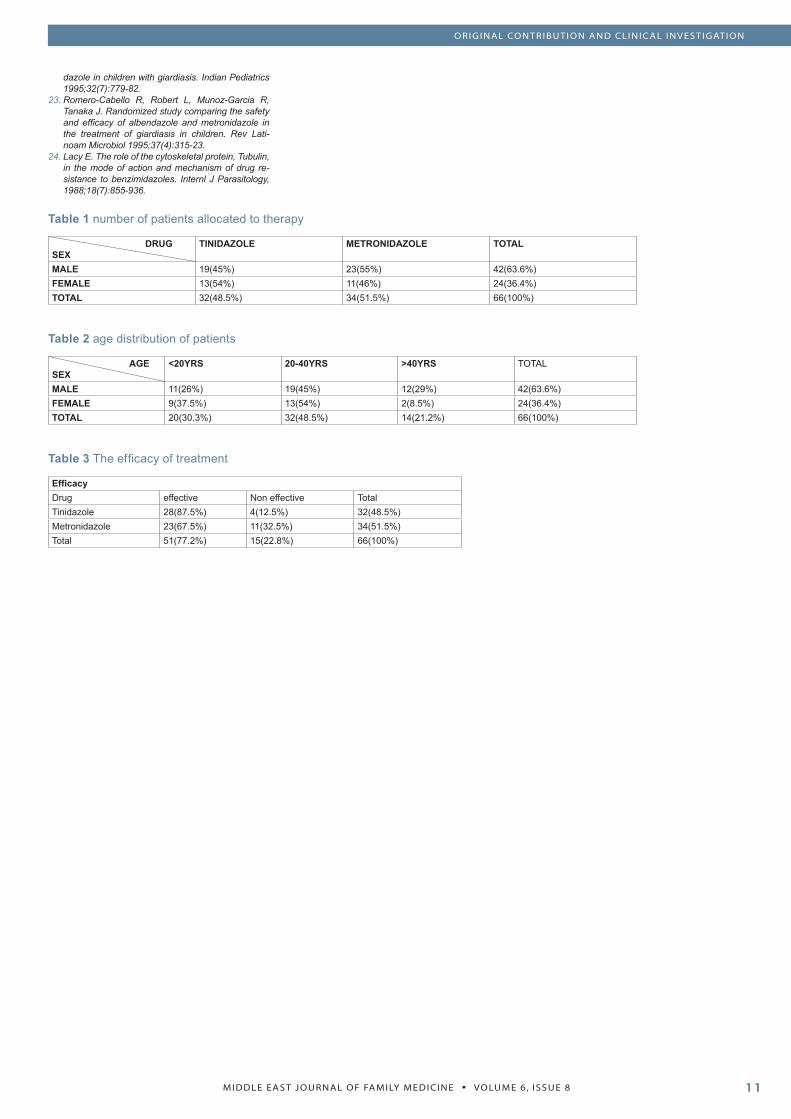
ResultsAs illustrated in Table 1 the sample
size of both groups was almost identical 32 (48.5%) and 34 (51.5%)of tinidazole and metronidazole respectively. The males constituted the majority of patients 42 (63.6%) while the females were 24 forming 36.4% of the patients, The male to female ratio was 1.75:1.
The age distribution of patients ranged from 16 years to 68 years, the commonest age group was among 20 years-40 years making up around half of all patients (48.5%) as shown in Table 2.
28 of 32 patients (87.5%) treated with tinidazole and 23 of 34 patients (67.5%) treated with metronidazole had parasitological cure. Cure rate between the two groups was statistically significant (P<0.01). No major side effects were observed except two cases in the metronidazole group who had mild headache and abdominal pain for two days and three cases in tinidazole group who reported nausea, dizziness and headache. Efficacy of two regimens in term of drug are presented in [Table 3]. Tinidazole appears to be safe having a few ignorable side effects and produced a significant cure rate, more effective than metronidazole.
DiscussionA 2 gm single dose for 3 days
regimen of tinidazole had excellent effectiveness in treatment of amoebiasis as compared with metronidazole. Introduction of nitroheterocyclic drugs in the late 1950s and the 1960s heralded a
new era in the treatment of infections caused by a range of pathogenic protozoan parasites.[17] Metronidazole is the drug now most widely used in the treatment of anaerobic protozoan parasitic infections caused by G. intestinalis, Trichomonas vaginalis and Entamoeba histolytica.[18,19] Although various drugs have been available for several decades to treat this infection, none of them is entirely satisfactory due to high incidence of undesirable side effects and a significant failure rate in clearing parasites from the gastrointestinal tract.[19,20] Some evidence suggests that drug resistance may be responsible for these failures.[21,22] Unfortunately, failures in treatment of amoebiasis with standard metronidazole therapy have been reported in five to 20% cases. In the event of overt clinical resistance to metronidazole in entamoeba histolytica strains, tinidazole could be an alternative treatment. A key issue should be keeping in mind the documented cross-resistance between currently used nitroimidazole drugs .As such the choice of drug will differ in each case depending on the local conditions and keeping in view the sensitivity of parasite strain. Moreover, perhaps treatment of all asymptomatic entamoeba histolytica infections in developing countries hyperendemic for the disease is doubtful because of rapid reinfection. Clinical metronidazole resistance in Trichomonas vaginalis has also been documented previously.[22] Single dose therapy with tinidazole is effective in the metronidazole-resistant strains of T. vaginalis which could be another advantage of this drug.
ConclusionsTinidazole was more effective than
metronidazole produced fewer and mild side effects. We recommend tinidazole as drug of choice for treatment of amoebiasis because of its efficacy, and desirable tolerance. This preparation is preferred to metronidazole in the treatment of entamoeba histolytica infection as a considerable advantage in low socio-economic communities. Moreover, this drug may be tried and used if
other agents failed in the treatment of clinical amoebiasis.
ReferencesWorld Health Organization. WHO/PAHO/UNESCO report: A consultation with experts on amebiasis. Mexico City, Mexico28-29 January, 1997.Epidemiol Bull PAHO. 1997; 18:13-14.Mai Z, Ghosh S, Frisardi M, Rosenthal B, Rogers R, Samuelson J. Hsp60 is targeted to a cryptic mitochondrion-derived organelle (“crypton”) in the microaerophilic protozoan parasite Entamoeba histolytica. Mol Cell Biol 1999; 19:2198-2205. [Free Full Text]Tovar J, Fischer A, Clark CG. The mitosome, a novel organelle related to mitochondria in the amitochon-driate parasite Entamoeba histolytica. Mol Microbial 1999; 32:1013-1021. [CrossRef][ISI][Medline]Willhoeft U, Tannich E. The electrophoretic karyo-type of Entamoeba histolytica. Mol Biochem Parasi-tol 1999;99:41-53. [CrossRef][Medline]Dhar SK, Choudhury NR, Mittal V, Bhattacharya A, Bhattacharya S. Replication initiates at multiple dispersed sites in the ribosomal DNA plasmid of the protozoan parasite Entamoeba histolytica. Mol Cell Biol 1996;16:2314-2324. [Abstract]Singh U, Rogers JB, Mann BJ, Petri WA Jr. Tran-scription initiation is controlled by three core pro-moter elements in the hgl5 gene of the protozoan parasite Entamoeba histolytica. Proc Natl Acad Sci U S A 1997;94:8812-8817. [Free Full Text]Saito-Nakano Y, Nakazawa M, Shigeta Y, Takeuchi T, Nozaki T. Identification and characterization of genes encoding novel Rab proteins from Entamoe-ba histolytica. Mol Biochem Parasitol 2001;116:219-222.Adams EB, MacLeod IN. Invasive amebiasis. I. Amebic dysentery and its complications. Medicine (Baltimore) 1977;56:315-323. [Medline]Aristizabal H, Acevedo J, Botero M. Fulminant amebic colitis. World J Surg 1991;15:216-221. [CrossRef][Medline]Ellyson JH, Bezmalinovic Z, Parks SN, Lewis FR Jr. Necrotizing amebic colitis: a frequently fatal compli-cation. Am J Surg 1986;152:21-26.Powell SJ, MacLeod I, Wilmot AL, Elsdon-Dew E. Metronidazole in amoebic dysentery and amoebic liver abscess. Lancet 1966;2:1329-1331. [Medline]Blessmann J, Tannich E. Treatment of asymptomat-ic intestinal Entamoeba histolytica infection. N Engl J Med 2002;347:1384-1384. [Free Full Text]McAuley JB, Herwaldt BL, Stokes SL, et al. Diloxa-nide furoate for treating asymptomatic Entamoeba histolytica cyst passers: 14 years’ experience in the United States. Clin Infect Dis 1992;15:464-468. [Medline]McAuley JB, Juranek DD. Paromomycin in the treatment of mild-to-moderate intestinal amebiasis. Clin Infect Dis 1992;15:551-552. [Medline]Diamond LS, Clark CG. A redescription of En-tamoeba histolytica Schaudinn 1903 (emended walker, 1911) separating it from Entamoeba dispar Brumpt, 1925. J Eukaryot Microbiol. 1993;40:340-4. [PubMed]A comparative study of tinidazole and metronida-zole as a single daily dose for three days in sympto-matic intestinal amoebiasis.Drugs. 1978; 15 Suppl 1:19-22. No abstract avail-able.PMID: 350563 [PubMed - indexed for MEDLINE]Campbell WC, Rew RS. Chemotherapy of Parasitic diseases, New York: Plenum Press 1986; 146-7.Upcroft JA, Campbell RW, Benkali K. Efficacy of new 5-nitroimidazoles against metronidazole-susceptible and resistant Giardia, Trichomonas & Entamoeba spp. Antimicrob Agents Chemother 1999;43:73-6.MacMillan JA, DeAngelis CD, Feigin RD, Warshaw JB. Oski’s Pediatrics: Principles and Practice. Phila-delphia, Lippincott Williams &Wilkins 1999; 1176-7.Misra PK, Kumar A, Agarwal V, Jagota SC: A com-parative clinical trial of albendazole versus metroni-
1.
2.
3.
4.
5.
6.
7.
8.
9.
10.
11.
12.
13.
14.
15.
16.
17.
18.19.
20.
21.
22.
ORIGINAL CONTRIBUTION AND CLINICAL INVESTIGATION
MIDDLE EAST JOURNAL OF FAMILY MEDICINE • VOLUME 6 , ISSUE 8 ��
ORIGINAL CONTRIBUTION AND CLINICAL INVESTIGATION
dazole in children with giardiasis. Indian Pediatrics 1995;32(7):779-82.Romero-Cabello R, Robert L, Munoz-Garcia R, Tanaka J. Randomized study comparing the safety and efficacy of albendazole and metronidazole in the treatment of giardiasis in children. Rev Lati-noam Microbiol 1995;37(4):315-23.Lacy E. The role of the cytoskeletal protein, Tubulin, in the mode of action and mechanism of drug re-sistance to benzimidazoles. Internl J Parasitology, 1988;18(7):855-936.
23.
24.
Table 1 number of patients allocated to therapy
DRUGSEX
TINIDAZOLE METRONIDAZOLE TOTAL
MALE 19(45%) 23(55%) 42(63.6%)FEMALE 13(54%) 11(46%) 24(36.4%)TOTAL 32(48.5%) 34(51.5%) 66(100%)
Table 2 age distribution of patients
AGESEX
<20YRS 20-40YRS >40YRS TOTAL
MALE 11(26%) 19(45%) 12(29%) 42(63.6%)FEMALE 9(37.5%) 13(54%) 2(8.5%) 24(36.4%)TOTAL 20(30.3%) 32(48.5%) 14(21.2%) 66(100%)
Table 3 The efficacy of treatment
EfficacyDrug effective Non effective TotalTinidazole 28(87.5%) 4(12.5%) 32(48.5%)Metronidazole 23(67.5%) 11(32.5%) 34(51.5%)Total 51(77.2%) 15(22.8%) 66(100%)
MIDDLE EAST JOURNAL OF FAMILY MEDICINE • VOLUME 6 , ISSUE 8��
Usefulness of C-reactive Protein in Diagnosis of Intrapartum and Postpartum Neonatal Sepsis
ABSTRACTTo determine effects of intrapartum risk factors for early onset sepsis (EOS) on CRP levels in neonates and to assess the suitability of this test in diagnosing EOS. Design: Cohort study. Setting: Labour and post natal wards in a pediatric and obstetric department at military hospital in Zarka. Subjects: 200 neonates at risk of developing infection. Methods: CRP levels in cord blood and neonatal blood at 24 hours were estimated using commer-cial kits. Babies were observed for signs of sepsis for at least 48 hours. Results:Seven (3.5%) neonates had elevated CRP levels in the cord blood. At 24 hours, 82 (41%) babies had elevated levels. El-evated cord CRP levels were significantly associated with rupture of membranes for 24 hours labour more than 12 hours and maternal fever. At 24 hours, elevated CRP levels were associated with primi-parity, more than three vaginal examina-tions after membrane rupture, meconium staining of amniotic fluid and amnioinfu-sion. Ten (4%) of babies developed EOS. The negative predictive value for elevated CRP levels at 24 hours was 99%. Conclu-sion: Several intrapartum risk factors for EOS can cause elevation in CRP levels. However, this test may be useful in ex-cluding infection.
IntroductionIt is estimated that about 5 million
neonates die every year in low-income countries. Infection contributes to approximately 30 to 40% of neonatal deaths in these countries(1). However, early diagnosis of neonatal sepsis has remained a frustrating experience even in high-income countries(2). This has prompted the evaluation of surrogate markers of inflammation as possible tools for early diagnosis of bacterial sepsis(3-7). Estimations of cytokine levels and CRP levels are potentially useful in this respect(3-8). Although several studies confirm that CRP levels are useful in the early diagnosis of sepsis, there are reports to the contrary(9-12). It is suggested that serial rather than single determinations of CRP levels may be more useful in diagnosis of sepsis(13). Such tests could be of special importance in a newborn that is asymptomatic or has only equivocal signs at birth but has risk factors for infection(2). The present study was designed to evaluate the effect of intrapartum risk factors for early onset sepsis (EOS) on neonatal CRP levels and the utility of CRP in the diagnosis of EOS.
Method This was a prospective cohort study conducted at a pediatric and obstetric department at military hospital in Zarka from March to October 2006.
Inclusion and exclusion criteria
Neonates were included if their mothers had at least one of the following risk factors for neonatal infection: prelabour rupture of
membranes (ROM), more than three vaginal examinations after ROM, intrapartum fever (oral temperature >38º C), foul-smelling odour, and untreated or partially treated urinary tract infection in the antenatal period. Newborn babies born at less than 28 weeks, weighing less than 1,000 g or with lethal congenital anomalies were excluded from the study.
Primary outcome
The primary outcome was EOS, defined as sepsis occurring within 48 hours of birth. The following were considered to be signs suggestive of sepsis: lethargy or poor feeding; axillary temperature <36ºC or >38º C for more than one hour; significant jaundice with serum bilirubin >15 mg% in the absence of blood group incompatibility; apnoea or respiratory distress; peripheral capillary refill time of >3 sec on the forehead or mid sternum; heart rate of >160/min corrected for elevation of body temperature (10 beats / ºC rise); vomiting, diarrhoea or ileus; petechiae or bleeding diathesis; omphalitis; seizures. Laboratory markers considered abnormal were: total leukocyte count <5,000/mm3, neutrophil count <1,500/mm3, and immature to total neutrophil ratio > 0.2.
Newborn babies developing signs suggestive of sepsis were categorised as having sepsis or probable sepsis. Sepsis was diagnosed if the newborn baby had signs suggestive of sepsis and a positive blood culture. Probable sepsis was diagnosed in a newborn baby with negative blood culture, if it had two or more signs suggestive of sepsis and one or more abnormal
Key words: C-reactive protein, neonatal sepsis.
Khaled Amro, MDDepartment of pediatric in Zarka military hospital
Corresspondence to:Dr. Khaled Amroe.mail: [email protected]
ORIGINAL CONTRIBUTION AND CLINICAL INVESTIGATION
MIDDLE EAST JOURNAL OF FAMILY MEDICINE • VOLUME 6 , ISSUE 8 ��
laboratory markers, or two or more abnormal laboratory markers with one or more signs suggestive of sepsis. Newborn babies with sepsis or probable sepsis received antibiotics for about 14 days. The remaining newborn babies were classified as at risk of infection and received antibiotics for an average of 5 days.
Sample size estimation
For an expected incidence of early onset sepsis among 4000 births of 3% and a worst acceptable incidence of 1.5%, the sample size required for 75% confidence is 184. For an expected incidence of 2% and a worst acceptable incidence of 1%, the sample size for 70% confidence is 182. Therefore, a sample of 200 was studied.
Laboratory techniques
Approximately 3 mL of blood was collected from the umbilical cord after clamping and cutting of the cord. About 24 hours later, approximately 2 mL of blood was collected by venepuncture from the newborn. Samples were transported without delay to the laboratory for total leukocyte count, absolute neutrophil count, immature to total leukocyte ratio and CRP estimation. CRP levels were determined on a daily basis using a latex agglutination test (Omega Diagnostics Ltd, Alloa, Scotland, UK). This is a semi-quantitative method with a detection limit of 6 mg/L. The investigator performing the CRP test was blinded to the clinical status of the newborn babies.
Data collection and analyses
Newborn babies were observed for signs of sepsis for at least 48 hours. Clinical data were collected using a questionnaire. Data were analysed using EpiInfo Version 6. Proportions were compared by Chi-square test. Relative risks were calculated for the risk factors for sepsis. The predictive values of CRP for diagnosing neonatal sepsis were also calculated.
ResultsThere were 200 newborn babies enrolled for the study. The mean (SD) gestational age was 38.5 (2.2)
week. Seven (3.5%) neonates had CRP levels of >6 mg/L in cord blood while 82 babies (41%) had elevated levels at 24 hours. CRP levels in cord blood of >6 mg/L was significantly associated with rupture of membranes for more than 24 hours, labour for more than 12 hours and maternal fever. At 24 hours, elevation in CRP levels was significantly associated with primiparity, more than three vaginal examinations after rupture of membranes, meconium staining of amniotic fluid and amnioinfusion. When the cut-off CRP level was increased to 12 mg/L, significant association was noted only with maternal fever. There was no association between Apgar score, birth weight and CRP levels.
Within 48 hours, 41 of the 200 babies with risk of infection developed at least one sign attributable to infection. Twenty seven had more than one sign. Of these, only two babies were diagnosed to have sepsis. Group B beta haemolytic streptococci were isolated from blood culture in one baby, while the other had coagulase-negative staphylococci. An additional eight babies were diagnosed to have probable sepsis. The sensitivity, specificity, positive and negative predictive values of CRP estimation at 24 hours for diagnosis of EOS using 6 mg/L as the cut off were 80%, 60%, 7.7% and 98.6% respectively. The corresponding values for a cut off level of 12 mg/L were 30%, 81.3%, 6.3% and 96.5% respectively.
Table 1 provides association between CRP levels and sepsis. CRP elevation was not significantly associated with the presence or number of signs. It was also noted that 10 of the 12 babies with CRP levels of 48 mg/L or more did not have evidence of infection. Only three of the 48 babies with CRP levels above 12 mg/L were diagnosed to have EOS.
Only one baby among those with sepsis or probable sepsis had abnormal total leukocyte and absolute neutrophil counts in the cord blood. Five (50%) had abnormal immature to total leukocyte ratio. Eighty nine of the 123 (72.3%) CRP negative babies and 38 of the 82 (46.34%) CRP positive babies received antibiotics for less than three days.
DiscussionThis study was done to evaluate the
association between intrapartum risk factors for infection with CRP levels and showed that several such risk factors can cause elevated CRP levels in the absence of infection. This is in agreement with previously published reports(7,13). Since CRP does not cross the placenta, the elevated levels are due to production of CRP in the neonate. Chorioamnionitis can result in elevation of IL 6 levels even in uninfected neonates(7). Stimuli other than infection, like hypoxia, trauma and metabolic changes can also induce production of proinflammatory mediators(7). Significant association is reported between birth asphyxia and elevated IL 6 levels. In prolonged labour, IL 6 levels rise in the neonate probably related to physical activity of labour. This cytokine stimulates CRP production.
There are few longitudinal studies examining CRP changes in healthy babies with intrapartum risk of infection. Cytokine elevation seen in the early neonatal period in such babies probably reflects physiological stress induced at birth(13). Since CRP levels rise during the initial 24 hours in many babies irrespective of infection or administration of antibiotics, serial determinations in this period may not be of much use in diagnosis but may help in identifying uninfected babies and restricting antibiotic use(14,15). Our data showed lower antibiotic use in babies who were CRP negative.
Various studies utilising varying protocols have suggested different values as upper limit of normal(8) In our study, at 24 h, CRP levels of 6mg/L had a negative predictive value of 99%. This level therefore could be used to guide antibiotic therapy when latex agglutination kits are used. Testing samples in further dilutions to establish the actual amount of CRP may not be necessary since increasing levels were not associated with increasing severity or prognosis.
Cord blood CRP levels estimated using a kit with 6 mg/L as detection limit, could not satisfactorily predict EOS. Recent studies show that cut off values may be different for cord and 24 hour samples(7). More sensitive
ORIGINAL CONTRIBUTION AND CLINICAL INVESTIGATION
MIDDLE EAST JOURNAL OF FAMILY MEDICINE • VOLUME 6 , ISSUE 8��
techniques like nephelometry may help set cut off levels for cord blood. In comparison to leukocyte counts and ratios, CRP levels at 24 hours proved to be the single best indicator for diagnosing EOS. However, the 80% sensitivity obtained is unacceptably low for making critical decisions. If utilised with caution, this test can help in reducing antimicrobial use in the new-born.
ConclusionIntrapartum risk factors for early onset sepsis can cause elevation of cord and neonatal CRP levels in the absence of infection. A CRP level of <6mg/L at 24 hour has a good negative predictive value for neonatal sepsis. Serial CRP levels are not useful in diagnosing early onset sepsis.
ReferencesThe WHO young infants study group, Bacterial eti-ology of serious infections in young infants in de-veloping countries: results of a multicentric study, Pediatr Infect Dis J 1999; 18: S17-22.Escobar GJ. Effect of systemic inflammatory re-sponse on biochemical markers of neonatal bacte-rial infection: A fresh look at old confounders. Clini Chem 2003; 49: 21-22.Ng PC, Cheng SH, Chui KM, Fok TF, Wong MY, Wong W et al. Diagnosis of late onset neonatal sepsis with cytokines, adhesion molecule, and C-reactive protein in preterm very low birthweight in-fants. Arch Dis Child Fetal Neonatal Ed 1997; 77: F221-F227.Chan DK, Ho LY. Usefulness of C-reactive protein in the diagnosis of neonatal sepsis. Singapore Med J 1997; 38: 252-255.Magudumana MO, Ballot DE, Cooper PA, Trusler J, Cory BJ, Viljoen E et al. Serial interleukin 6 meas-urements in the early diagnosis of neonatal sepsis. J Trop Pediatr 2000; 46: 267-271.Dollner H, Vatten L, Linnebo I, Zanussi GF, Laerdal A, Austgulen R. Inflammatory mediators in umbili-cal plasma from neonates who develop early-onset sepsis. Biol Neonate 2001; 80: 41-47.Chiesa C, Pellegrini G, Panero A, Osborn JF, Si-gnore F, Assumma, et al. C-reactive protein, inter-leukin 6 and procalcitonin in immediate post natal period: influence of illness severity, risk status, ante-natal and perinatal complications and infection. Clin Chem 2003; 49: 60 -68.Vesikari T. Cytokine determinations and rapid diag-nosis of early onset neonatal septicemia. Acta Pedi-
1.
2.
3.
4.
5.
6.
7.
8.
atr 1999; 88: 585-591.Suri M, Thirupuram S, Sharma VK. Diagnostic and prognostic utility of C-reactive protein, alpha-1-anti-trypsin and alpha-2-macroglobulin in neonatal sep-sis: a comparative account. Indian Pediatr 1991; 28: 1159-1164.Krediet T, Gerards L, Fleer A, van Stekelenburg G. The predictive value of CRP and I/T-ratio in neona-tal infection. J Perinat Med 1992; 20: 479-485.Anwer SK, Mustafa S. Rapid identification of neona-tal sepsis. J Pak Med Assoc 2000; 50: 94-98.Santana C, Guindeo MC, Gonzalez G, Garcia-Mu-noz F, Saavedra P, Domenech E. Cord blood levels of cytokines as predictors of early neonatal sepsis. Acta Pediatr 2001; 90: 1176-1181.Chiesa C, Signore F, Assumma M, Buffone E, Tra-montozzi P, Osborn JF, et al. Serial measurements of C-reactive protein and interleukin-6 in the im-mediate postnatal period: reference intervals and analysis of maternal and perinatal confounders. Clin Chem 2001; 47: 1016-1022.Philip AG, Mills PC. Use of C-reactive protein in minimizing antibiotic exposure: experience with in-fants initially admitted to a well-baby nursery. Pedi-atrics 2000; 106: E4.Bomela HN, Ballot DE, Cory BJ, Cooper PA. Use of C-reactive protein to guide duration of empiric anti-biotic therapy in suspected early neonatal sepsis. Pediatr Infect Dis J 2000;19: 531-535.
9.
10.
11.
12.
13.
14.
15.
Table 1 CRP levels and neonatal sepsis
CRP levels (mg/L) Sepsis Probable sepsis No sepsisCord blood <6 (n = 195)At 24 hrs<6 0 1 1226 1 3 34> 12 1 2 31
Cord blood <6 (n = 5)At 24 hrs<6 0 1 06 0 1 1> 12 0 0 2
ORIGINAL CONTRIBUTION AND CLINICAL INVESTIGATION
MIDDLE EAST JOURNAL OF FAMILY MEDICINE • VOLUME 6 , ISSUE 8 ��
MEDICINE AND SOCIET Y
How Many People Have Cancer Patients (Alive or Deceased) in Their Homes, in Our City?
ABSTRACTObjectives: Our aim was to assess how many people have cancer (alive or de-ceased) in their homes, in our city; the types of cancer, and their relationship with living places and economic status.
Methods: A questionnaire was adminis-tered to 2035 people in 75 different parts of our city. Only one person in each house was selected and asked if there had been any cancer patients living or deceased in the house.
Results: There were 100 (4.9%) people who have been living with a patient diag-nosed as having cancer and 333 people (16.4%) who had a history of a patient who had died from cancer in the home. The death rate among 333 patients was 34.2% for lung cancer, 9.0% for gastric cancer and 7.21% for colon cancer. Also people living in urban areas have more living (odds ratio=1.45) and deceased (odds ratio=1.28) cancer patients in their homes, than people living in villages.
Conclusion: Cancer is a part of our lives either with a patient in our house or a rel-ative who has died from cancer. So public education and health services for home-care need for many people to care for these cancer patients. According to our study lung cancer is the most prevalent cancer in our region.
IntroductionCancer is a serious growing
problem worldwide, especially a disease of the developing world. It is not only a biological processes it is also the outcome of lifestyle decisions and social conditions. So we need to understand what causes cancer at the biological and social levels but also what cancer causes to the families, to the people living with a person who has cancer.
Based on the World Cancer Report there were 10.1 million new cases, 6.2 million deaths and 22.4 million persons living with cancer in the year 2000. It is second to cardiovascular disease as a cause of death in developed countries and the number of new cases is expected to grow by 50% over the next 20 years, to reach 15 million by 2020(1). In 2006, in Europe, there were an estimated 3,191,600 cancer cases diagnosed (excluding non-melanoma skin cancers) and 1,703,000 deaths from cancer(2). Cancer is an important problem in both public health and political terms worldwide, irrespective of a country’s development.
Cancer arises out of conditions of life, which result in exposure to carcinogens.We can argue about two factors on this subject:
Changes people make in the worldWhere people live
We know that there are social factors in cancer etiology, which include socioeconomic status, occupation
•
•
(industrial hazards), radiation, medications, habits, food handling, air and water pollution.
These social dimensions of cancer have important implications for the design of cancer control programming. They stem from behavior patterns that people evolve to meet their biological, psychological and social needs. These patterns, in turn, create a lifestyle which influences cancer incidence. They include the development of addictions to tobacco, drugs and alcohol, the ways in which food is prepared, stored and eaten, and certain other risk patterns.
The disease is widely feared around the world over as synonymous with suffering and death. Patients may be stigmatized and experience social isolation and family tensions as well as the inability to get insurance or even job loss with economic dependence aggravated by high costs of medical care, if there is no health insurance. This condition also causes changes in lifestyle of the families involved.
As a result cancer impacts not only on the patient, but also his or her family and community. The aim of our study was to assess how many people have cancer patients (living or deceased) in their homes, in our city; the types of cancers, and their relationship with living places and economic status.
Methods
Key words: Cancer, prevalence, family, Turkey, epidemiology.
Dilek Toprak1, Nurhan Dogan2, Serap Demir3, Gülnihal Tufan3
1. Afyon Kocatepe University, Department of Family Medicine, Turkey,2. Afyon Kocatepe University, Department of Biostatistics, Turkey,3. Afyon Kocatepe University, Department of Internal Medicine, Turkey
Corresspondence to:Dilek Toprak, MDDepartment of Family MedicineAfyon Kocatepe University,Afyonkarahisar 03200, TURKEYTelephone number : 0090 532 3827836Fax number : 0090 272 2132907E-mail : [email protected]
MIDDLE EAST JOURNAL OF FAMILY MEDICINE • VOLUME 6 , ISSUE 8��
MEDICINE AND SOCIET Y
The study was conducted in Afyonkarahisar, a middle Anatolian city, between November 2005 and February 2006. The present study was approved by the Afyon Kocatepe University Faculty of Medicine Clinical Research Ethics Committee and written, informed consent was obtained from all participants.
A total of 2035 people, from 75 different screening regions (18 urban, 57 villages) of our city were detected according to the population records of the year 2000, which represent the population of the area appropriately. A total of 7000 km. roadway driven for the research by a team of 15 physicians, 1 nurse and a driver. The records of the regional health institutions were used in order to determine the subjects. People older than 18 years old were grouped as 19-40 years old, 41-64 years old, 65 and over. According to population distribution of year 2000, we determined the minimum number of people as 1990 (when d=0.02) and at the end of the study we reached a number of 2035 people. The study group selected randomly from the “Family Cards” of the primary health centers, regarding the gender and ages. Only one person was selected from every house.
The subjects were informed about the study by telephone interviews one night before, their approvals were obtained and their transport to the health institutions, where the study would be conducted, was provided. The data were collected by a questionnaire in which face to face survey method was performed by the physicians. As this study is a part of a big epidemiological research study only the question about cancer (Is there a person with cancer or who has died from cancer) was regarded and its relations with economic status and living place have been evaluated. Also type of cancer was asked about.
The data of the study was written in SPSS 12.0 version. Statistical evaluation of the study was performed using the chi-square test and p values lower than 0.05 were accepted as significant.
Results
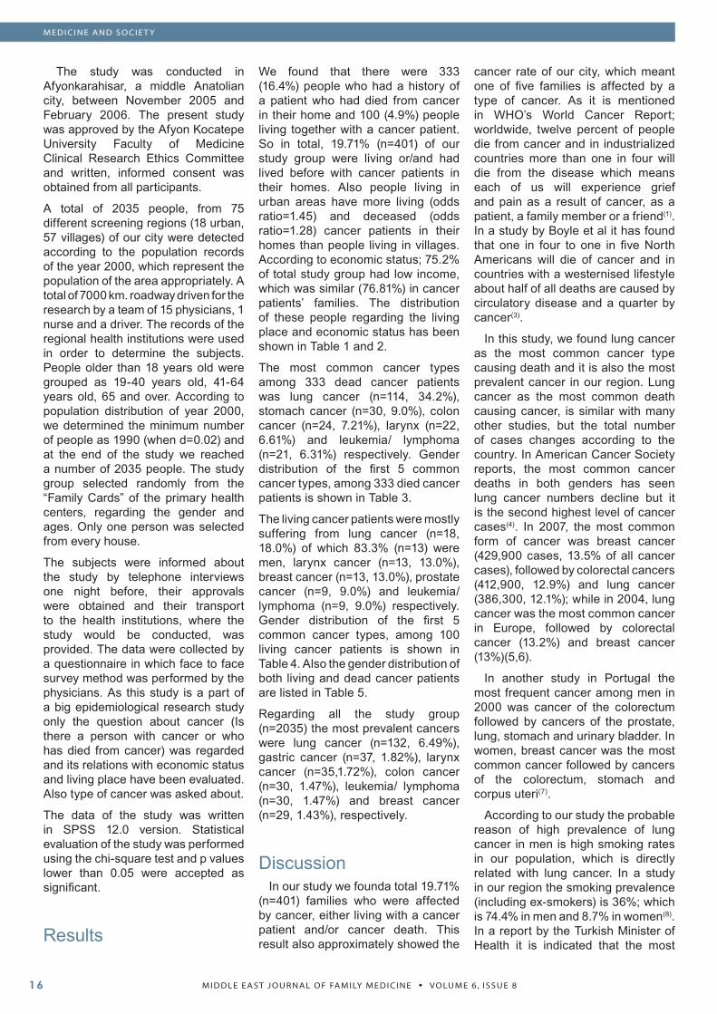
We found that there were 333 (16.4%) people who had a history of a patient who had died from cancer in their home and 100 (4.9%) people living together with a cancer patient. So in total, 19.71% (n=401) of our study group were living or/and had lived before with cancer patients in their homes. Also people living in urban areas have more living (odds ratio=1.45) and deceased (odds ratio=1.28) cancer patients in their homes than people living in villages. According to economic status; 75.2% of total study group had low income, which was similar (76.81%) in cancer patients’ families. The distribution of these people regarding the living place and economic status has been shown in Table 1 and 2.
The most common cancer types among 333 dead cancer patients was lung cancer (n=114, 34.2%), stomach cancer (n=30, 9.0%), colon cancer (n=24, 7.21%), larynx (n=22, 6.61%) and leukemia/ lymphoma (n=21, 6.31%) respectively. Gender distribution of the first 5 common cancer types, among 333 died cancer patients is shown in Table 3.
The living cancer patients were mostly suffering from lung cancer (n=18, 18.0%) of which 83.3% (n=13) were men, larynx cancer (n=13, 13.0%), breast cancer (n=13, 13.0%), prostate cancer (n=9, 9.0%) and leukemia/ lymphoma (n=9, 9.0%) respectively. Gender distribution of the first 5 common cancer types, among 100 living cancer patients is shown in Table 4. Also the gender distribution of both living and dead cancer patients are listed in Table 5.
Regarding all the study group (n=2035) the most prevalent cancers were lung cancer (n=132, 6.49%), gastric cancer (n=37, 1.82%), larynx cancer (n=35,1.72%), colon cancer (n=30, 1.47%), leukemia/ lymphoma (n=30, 1.47%) and breast cancer (n=29, 1.43%), respectively.
DiscussionIn our study we founda total 19.71%
(n=401) families who were affected by cancer, either living with a cancer patient and/or cancer death. This result also approximately showed the
cancer rate of our city, which meant one of five families is affected by a type of cancer. As it is mentioned in WHO’s World Cancer Report; worldwide, twelve percent of people die from cancer and in industrialized countries more than one in four will die from the disease which means each of us will experience grief and pain as a result of cancer, as a patient, a family member or a friend(1). In a study by Boyle et al it has found that one in four to one in five North Americans will die of cancer and in countries with a westernised lifestyle about half of all deaths are caused by circulatory disease and a quarter by cancer(3).
In this study, we found lung cancer as the most common cancer type causing death and it is also the most prevalent cancer in our region. Lung cancer as the most common death causing cancer, is similar with many other studies, but the total number of cases changes according to the country. In American Cancer Society reports, the most common cancer deaths in both genders has seen lung cancer numbers decline but it is the second highest level of cancer cases(4). In 2007, the most common form of cancer was breast cancer (429,900 cases, 13.5% of all cancer cases), followed by colorectal cancers (412,900, 12.9%) and lung cancer (386,300, 12.1%); while in 2004, lung cancer was the most common cancer in Europe, followed by colorectal cancer (13.2%) and breast cancer (13%)(5,6).
In another study in Portugal the most frequent cancer among men in 2000 was cancer of the colorectum followed by cancers of the prostate, lung, stomach and urinary bladder. In women, breast cancer was the most common cancer followed by cancers of the colorectum, stomach and corpus uteri(7).
According to our study the probable reason of high prevalence of lung cancer in men is high smoking rates in our population, which is directly related with lung cancer. In a study in our region the smoking prevalence (including ex-smokers) is 36%; which is 74.4% in men and 8.7% in women(8). In a report by the Turkish Minister of Health it is indicated that the most
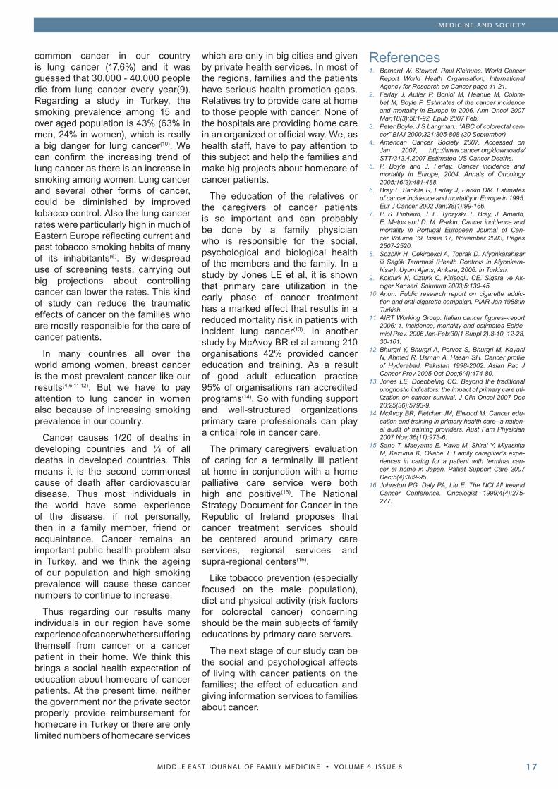
MIDDLE EAST JOURNAL OF FAMILY MEDICINE • VOLUME 6 , ISSUE 8 ��
MEDICINE AND SOCIET Y
common cancer in our country is lung cancer (17.6%) and it was guessed that 30,000 - 40,000 people die from lung cancer every year(9). Regarding a study in Turkey, the smoking prevalence among 15 and over aged population is 43% (63% in men, 24% in women), which is really a big danger for lung cancer(10). We can confirm the increasing trend of lung cancer as there is an increase in smoking among women. Lung cancer and several other forms of cancer, could be diminished by improved tobacco control. Also the lung cancer rates were particularly high in much of Eastern Europe reflecting current and past tobacco smoking habits of many of its inhabitants(6). By widespread use of screening tests, carrying out big projections about controlling cancer can lower the rates. This kind of study can reduce the traumatic effects of cancer on the families who are mostly responsible for the care of cancer patients.
In many countries all over the world among women, breast cancer is the most prevalent cancer like our results(4,6,11,12). But we have to pay attention to lung cancer in women also because of increasing smoking prevalence in our country.
Cancer causes 1/20 of deaths in developing countries and ¼ of all deaths in developed countries. This means it is the second commonest cause of death after cardiovascular disease. Thus most individuals in the world have some experience of the disease, if not personally, then in a family member, friend or acquaintance. Cancer remains an important public health problem also in Turkey, and we think the ageing of our population and high smoking prevalence will cause these cancer numbers to continue to increase.
Thus regarding our results many individuals in our region have some experience of cancer whether suffering themself from cancer or a cancer patient in their home. We think this brings a social health expectation of education about homecare of cancer patients. At the present time, neither the government nor the private sector properly provide reimbursement for homecare in Turkey or there are only limited numbers of homecare services
which are only in big cities and given by private health services. In most of the regions, families and the patients have serious health promotion gaps. Relatives try to provide care at home to those people with cancer. None of the hospitals are providing home care in an organized or official way. We, as health staff, have to pay attention to this subject and help the families and make big projects about homecare of cancer patients.
The education of the relatives or the caregivers of cancer patients is so important and can probably be done by a family physician who is responsible for the social, psychological and biological health of the members and the family. In a study by Jones LE et al, it is shown that primary care utilization in the early phase of cancer treatment has a marked effect that results in a reduced mortality risk in patients with incident lung cancer(13). In another study by McAvoy BR et al among 210 organisations 42% provided cancer education and training. As a result of good adult education practice 95% of organisations ran accredited programs(14). So with funding support and well-structured organizations primary care professionals can play a critical role in cancer care.
The primary caregivers’ evaluation of caring for a terminally ill patient at home in conjunction with a home palliative care service were both high and positive(15). The National Strategy Document for Cancer in the Republic of Ireland proposes that cancer treatment services should be centered around primary care services, regional services and supra-regional centers(16).
Like tobacco prevention (especially focused on the male population), diet and physical activity (risk factors for colorectal cancer) concerning should be the main subjects of family educations by primary care servers.
The next stage of our study can be the social and psychological affects of living with cancer patients on the families; the effect of education and giving information services to families about cancer.
ReferencesBernard W. Stewart, Paul Kleihues. World Cancer Report World Heath Organisation, International Agency for Research on Cancer page 11-21.Ferlay J, Autier P, Boniol M, Heanue M, Colom-bet M, Boyle P. Estimates of the cancer incidence and mortality in Europe in 2006. Ann Oncol 2007 Mar;18(3):581-92. Epub 2007 Feb.Peter Boyle, J S Langman., “ABC of colorectal can-cer” BMJ 2000;321:805-808 (30 September)American Cancer Society 2007. Accessed on Jan 2007, http://www.cancer.org/downloads/STT/313,4,2007 Estimated US Cancer Deaths.P. Boyle and J. Ferlay. Cancer incidence and mortality in Europe, 2004. Annals of Oncology 2005;16(3):481-488.Bray F, Sankila R, Ferlay J, Parkin DM. Estimates of cancer incidence and mortality in Europe in 1995. Eur J Cancer 2002 Jan;38(1):99-166.P. S. Pinheiro, J. E. Tyczyski, F. Bray, J. Amado, E. Matos and D. M. Parkin. Cancer incidence and mortality in Portugal European Journal of Can-cer Volume 39, Issue 17, November 2003, Pages 2507-2520.Sozbilir H, Cekirdekci A, Toprak D. Afyonkarahisar ili Saglik Taramasi (Health Controls in Afyonkara-hisar). Uyum Ajans, Ankara, 2006. In Turkish.Kokturk N, Ozturk C, Kirisoglu CE. Sigara ve Ak-ciger Kanseri. Solunum 2003;5:139-45.Anon. Public research report on cigarette addic-tion and anti-cigarette campaign. PIAR Jan 1988;In Turkish.AIRT Working Group. Italian cancer figures--report 2006: 1. Incidence, mortality and estimates Epide-miol Prev. 2006 Jan-Feb;30(1 Suppl 2):8-10, 12-28, 30-101.Bhurgri Y, Bhurgri A, Pervez S, Bhurgri M, Kayani N, Ahmed R, Usman A, Hasan SH. Cancer profile of Hyderabad, Pakistan 1998-2002. Asian Pac J Cancer Prev 2005 Oct-Dec;6(4):474-80.Jones LE, Doebbeling CC. Beyond the traditional prognostic indicators: the impact of primary care uti-lization on cancer survival. J Clin Oncol 2007 Dec 20;25(36):5793-9.McAvoy BR, Fletcher JM, Elwood M. Cancer edu-cation and training in primary health care--a nation-al audit of training providers. Aust Fam Physician 2007 Nov;36(11):973-6.Sano T, Maeyama E, Kawa M, Shirai Y, Miyashita M, Kazuma K, Okabe T. Family caregiver’s expe-riences in caring for a patient with terminal can-cer at home in Japan. Palliat Support Care 2007 Dec;5(4):389-95.Johnston PG, Daly PA, Liu E. The NCI All Ireland Cancer Conference. Oncologist 1999;4(4):275-277.
1.
2.
3.
4.
5.
6.
7.
8.
9.
10.
11.
12.
13.
14.
15.
16.
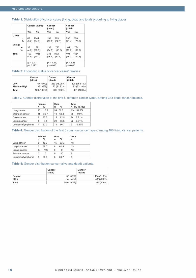
MIDDLE EAST JOURNAL OF FAMILY MEDICINE • VOLUME 6 , ISSUE 8��
MEDICINE AND SOCIET Y
Table 1: Distribution of cancer cases (living, dead and total) according to living places
Cancer (living) Cancer(dead)
Cancer(total)
Yes No Yes No Yes NoUrban n %
63 1044(5.7) (94.3)
198 909(17.9) (82.1)
237 870(21.4) (78.6)
Village n %
37 891(4.0) (96.0)
135 793(14.5) (85.5)
164 764(17.7) (82.3)
Total 100 1935(4.9) (95.1)
333 1702(16.4) (83.6)
401 1634(19.7) (80.3)
χ2 = 3.13p= 0.077
χ2 = 4.112p= 0.043
χ2 = 4.45p= 0.035
Table 2: Economic status of cancer cases’ families
Cancer(alive)
Cancer(dead)
Cancer(total)
LowMedium-High
67 (67%)33 (33%)
260 (78.08%)73 (21.92%)
308 (76.81%) 93 (23.19%)
Total 100 (100%) 333 (100%) 401 (100%)
Table 3: Gender distribution of the first 5 common cancer types, among 333 dead cancer patients.
Femalen %
Malen %
Totaln (% in 333)
Lung cancer 15 13.2 99 86.8 114 34.2%Stomach cancer 11 36.7 19 63.3 30 9.0%Colon cancer 9 37.5 15 62.5 24 7.21%Larynx cancer 1 4.5 21 95.5 22 6.61%Leukemia/lymphoma 7 33.3 14 66.7 21 6.31%
Table 4: Gender distribution of the first 5 common cancer types, among 100 living cancer patients.
Femalen %
Malen %
Totaln
Lung cancer 3 16.7 15 83.3 18Larynx cancer 5 38.5 8 61.5 13Breast cancer 13 100 0 0 13Prostate cancer 0 0 9 100 9Leukemia/lymphoma 3 33.3 6 66.7 9
Table 5: Gender distribution cancer (alive and dead) patients.
Cancer(alive)
Cancer(dead)
FemaleMale
48 (48%)52 (52%)
104 (31.2%)229 (68.8%)
Total 100 (100%) 333 (100%)
MIDDLE EAST JOURNAL OF FAMILY MEDICINE • VOLUME 6 , ISSUE 8 ��
MEDICINE AND SOCIET Y
Women Knowledge Assessment about Self Care Behavior in Shiraz Health Care Center 2006
ABSTRACT Background: People must accept ‘self care” responsibility. “Self care” include health promotion, prevention and limited diseases. Specific aims about women’s health are prevention and control cancer specially breast cancer and cervical cancer, AIDS, STD, anemia, violence, osteoprosis and chronic low back pain. Therefore women with attention of age groups must know self carebehavior and do it.
Method and Material: A cross sec-tional survey was conducted to eval-uate knowledge and behavior related to “self care” among women who attended to Shiraz Health Center. A convenience sample was 607 wom-en who selected by cluster and ran-domly sampling. Data were collected by questionnaire and then analysis with descriptive statistic, ANOVA and T-test.
Results: 52.2% of women had good knowledge about self care behavior. Knowledge of blood cholesterol was highest rate among women (91.1%). Good practice about screening test was only 3.1% control blood pressure as routine was higher in compare to other screening tests.
Conclusions: Practice of women about screening tests specially as routine is weak. With attention of importance self care behavior in promotion life quality and life span, therefore nurses and health workers must teach and encourage women about self care behavior and screen-ing tests.
IntroductionFor a long time during the twentieth
century, the women’s health care focused mainly on reproductive functions as periodic cycle, pregnancy, delivery and menopause; however, there was a basic change in the attitude by 1990. It should be noted that women’s health is actually associated with their lives’ cultural, social and biological dimensions. By 1990, regulations were developed in terms of women’s health issues throughout the world. Longevity among women is higher than men, and they should be supported to promote these extra years of lifetime from the point of view of health care. However success in each health planning program depends upon the cooperation of the subjects themselves (22, pp:562-565).
One is, individually, responsible for one’s health, though, it’s a basic right for human being. It means that is not the official or social health services which keep the people healthy; it is achieved just through the individual attempts.
People should accept a range of responsibilities, related to their health, called “self-care”. The concept is referred to as a range of health-maintaining proceedings done by the individual.
It mainly includes activities to prevent diseases and to promote health, such as, dietary regimen, sleep, exercise, body weight control, habits and style of life, immunity, screening, and preventing the diseases by consulting the physicians in time. Therefore it’s necessary for the women, regarding
their age, to do the self-care behaviors (1, pp: 55-56). The specific objective concerning women’s health and screening is to prevent and control various kinds of cancer such as breast cancer, reproductive tract cancers like genitalia, AIDS, and diseases such as Anemia, Steoprosis, chronic low back pains and violence (22, pp: 562-565).
The most common cancer among women is breast cancer, with which one out of nine people are affiliated.In IRAN It also forms 12.6 percent of all cancers and it is more common among the women aged 35-years old and higher.
The diagnosis of cancer is increasing because of its Mamography. At present, because of the restrictions of primary prevention, focusing on secondary prevention, namely, in time diagnosis is of significant importance. Several studies have revealed that screening through clinical examination of the breast and mammography are effective in diagnosing breast cancer(25). In addition, women who do the self-exam of their breasts have more chances to diagnosis the disease. 19 percent of the deaths due to breast cancer occur because ignoring mammography.
Cervical cancer is considered the second common cancer among women throughout the world. Life expectancy is 66 percent, and if it’s diagnosed in time, it would increase to 100 percent for 5 years of life expectancy. The manifestation of cervical cancer maximizes at the age of 45 through 55 years. Pap smears is the main screening test for cervical
Vizeshfar, Fatemeh- Mehdizadeh, Kadege
Corresspondence to:Vizeshfar, Fatemeh- Mehdizadeh, KadegeNursing Faculty of Shiraz Medical Science UniversityZeinab Nursing CollageTell:(098-781-2241251)Fax:(098-781-2247111)
Key words: Knowledge, Practice, Self care.
MIDDLE EAST JOURNAL OF FAMILY MEDICINE • VOLUME 6 , ISSUE 820
cancer. A decrease in mortality rate in cervical cancer during the last 50 years has been due to the use of pap smear (1, pp: 55-56).
In America, 20 percent of women suffer from during pregnancy. According to a study, in America women between 12-50 years and at risk receive just half of the necessary iron (2, pp: 201-222). Accordingly, international women health society suggests that screening tests should be continued while aging(26).
Hypertention is considered as one the most important risk factors in heart diseases and the most common factor of heart/kidney failure in CVA (cardio vascular accident). In most countries, consequently, international woman’s health community recommended that screening tests should be followed as women grow older.
Hypertension is one of the main factors in heart diseases and the current previlage factor in CHF, and CVA. In many countries one out of four is suffusing from hypertension. In American 25% of men and 21% of women are afflicted with hypertension.(2pp:201-222)There fore the people’s awareness of hypertension and planning to control if is of significant importance.
Considering the importance of blood cholesterol in developing croner vascular diseases and variety of studies such as Framingham in American it is suggested that the 20- year-old indicate and over do the Lippopratein test every five years or with less interval if there is any family background Auother screening test is Measuring density of bone(19).
Those who use steroids for a long time, women inmenopaue age, those who smoke and those with a background with fracture are prove to steoprosis and frequent fractures should have enough knowledge in this regard.
Since certain cultural and social factors can affect women’s health and Odditionally self care behavior varies in different communities, this study can help women to pramote.
Knowledge and abilities in self-care with attention to the age groups through planning for necessary
(helpful) education.
Methods This study is a cross sectional
survey conducted to evaluate knowledge and self-care behavior among women.
Through cluster and random sampling, 607 women referring to the medical centers in northan, Southern central, Vestbrn and eastern parts of Shiraz, were studied. The subjects were studied to determine their knowledge and self care behaviors related to preventing cervicalcancer AIDS , Breast cancer hypertension high cholesterol rate. anemia steoprosis and relationship between knowledge and self-care behavior with women Demographic character: stice.
The questianare consisting of two parts of demography features and questionnaire todetermine their knowledge and practice related to self care was distributed and based on their responses to the questions it was divided into 3 groups of good, average, and weak.
The data were analyzed using SPSS package and description analysis of variance one way anova and t-test.
FindingsThe results of this study revealed
that 60.8% (369 people) Of the subjects were among women of 20-30 years old, 7.1% lower than 20 years and 8.9% over 40 years old. 67.8 of the subjects (404) get married when they were than 20 years of age.
86.6% of the were housewives.the most of them 77.9% (472) of the degree Diplom and less were 7.6% (46) illiterate and 14.5% higher education.
45.3% (275) had only one child and just 6.4 (36) had no child. Regarding subjects’ knowledge of self-care behaviors in the table 1 depicts 91.1% (553) were aware of the ways to control chlostrol, 88.3 (536) aware of AIDS prevention.
73.1% (444) of anemia Symptom, 60.3% (366) of the ways to prevent
steoprosis 41.5% (355) were aware of the ways to diagnose steoporosis and just 23.2 (141) had knowledge of the age of testing steoporosis. 21.6% (131) were aware of the ways to prevent disease and 17.1 (104) knew the age to momography test.
As shown in table 2, 71%(431) knew how to measure hypertention 64.9,(394) blood chlostrol and 59.6(362) cheked pap smear and 51.9 (315) tested for anemia, pelvis examination was done by 32.1 (195), breast self exunination done by 27.8 according to the this tabl. Blood BP measure has been done more than other screening tests. Table 3 shows the fulfillment of screening test with proper intervals. According to the table, BP measure had the most frequency, then pap smear 34.1(207) blood cholestral16.6 (101).
Blood cholesterol 16.6. pelvis examination 15.8 breast self eam:natio11.6(10) and mammography, table 4 depicts the level of knowledge of subjects about self-care behaviors which statistically shows that among the subjects 52.2 (317) had good knowledge, 38.7 average, and 9.1 (55) had weak knowledge about self-care behavior.
Practically, 59.5 (361) of the subjects had weak practice, about self care 37.4 (227) were average and just 3.1% (19) had good practice. Table 5 shows the frequency of density of practice about self care.
Table 6 shows that the majority of subjects gain their information through several sources as mass-media (Radio/TV) health care centers, and relatives or friends.
One way variance analysis showed that there is a meaningful relationship between knowledge (f=15, p= 000) level of education (f=47, p= 000), number of children (f=3.2, p=0.2), and t-test shows a meaningful relationship between knowledge and the occupation of the subjects(p=0.000).
Analysis of variance also showed that there is meaningful relationship between practice and age (f=44.9, p=0.000) number of children (f=12.4, p=0.000) and level of education(p=0.000) from and t-test showed that this significant
MEDICINE AND SOCIET Y
MIDDLE EAST JOURNAL OF FAMILY MEDICINE • VOLUME 6 , ISSUE 8 ��
relationship exists between knowledge and occupation on the other hand.
DiscussionIn order to maintain health, self-
care is one of the basic needs in our community. Skill in self-care determines the quality of life and longevity. Self-care begins from family members and society to gain, maintain and promote health. Since human body is constantly changing physiologically and these natural changes lead to disorder, it’s necessary for women to have knowledge about health fullness and maintain it and fulfill screening tests as they grow older.
This study revealed 73.1% of the subjects were aware of AIDS and the ways of its prevention and its commune. In Enosolease’s study in Nigeria, 100% of women who illegal had experience abortion, and in Ekanem study in Nigeria, 89.9% of pregnant women, and in hesketh’ study in China the majority of health care staff were aware of AIDS.
The reason for this similarity indicate great international attempts to enhance people’s knowledge of the importance and dangers of AIDS specially women. Lee Chung et al in their study on the relation of Anti-body test of HIV and its acceptance by people from Hongkong cause to this conclusion that having knowledge about HIV can increase the fulfillment of screening test for AIDS.
The other finding of this study was that 91.1% of the subjects were aware of the ways to control blood cholesterol but only 16.6% of them did the test in proper interval. In order to control the level of blood cholesterol, a blood random sampling for screening the level of cholesterol and HDL amony the individuals above 20 year old and its should be repeated every 5 years if the normal ragne is present(3pp:70). Although the subjects were aware of the importance of blood cholesterol in cardio vascular diseases, a few of them had done the related test. Maybe it is because the subjects were mostly young. However, the individuals should be encourage to do the screening test.
Artineain (2004) in his study on American and Mexicans revealed that the majority of subjects used up more fried high fat foods and cookies and less fruits, vegetables and diaries. Inactivity, obesity and smoking are the main factors in cardio vascular disease development.
Researches emphasize the importance of nutrition in preventing cardio vascular diseases and doing screening test(6).
Artiniein in this study also revealed that 63.1% of the subjects under the study had knowledge about the dangers of hypertension and that was measurement by 71%, showing that the highest figures are related to screening tests.
It is recommended that women above 21 year check their BP every 2 years in case it’s done and with less interval if there is evidence of Border line hypertension or if there is High Bpond History of BP in the family(19). Though the frequency of checking BP in screening tests among the subjects was more than that in the other tests. Regarding the point that all subjects were expeecting pregnancy, and its routine that this BP is checked during this period may be the case.
Since people’s knowledge and practice plays a significant role in decreasing cardio vascular disease, it’s necessary to plan specific and widespread program.
A plan called Healty heart was conducted in Isfahan to decrease the cardio vascular diseases through controlling risk factors and enhancing health behaviors during which the subject’s FBS, cholesterol and thri glycride were measured(23) primary steoprosis May happen at either sex or at any age which secondary steoprosis happens because of using drugs or due to disease on the other hand, this is a widespread disease through out the world and may people are suffering this dangerous disease. Therefore in order to decrease any fracture during the middle aged years, the bones should be solidated since childhood another finding .of this study was knowledge about early detection of breast cancer
64.1% of the women had knowledge
of mammography, and 7.1% knew the age of the test 27.8% of them fulfilled breast self-examination but only 7.1% had done it regularly. It’s helpful for the women between 20-39 years old to do clinical examination of the breast every 3 years and 40 years old and above anualy and mamography for the women of 40 years and above once or twice a year(19).
Azizi suggests breast self-examination, montly, for the women above 20 Alwash (2001) in his study in UAI showed that women had not enough knowledge about breast cancer. The results indicated that 12.1% or the subjects had done Breast Self-exam and 10.3% of them had done mammography(17). Ahmad stewart (2004) in their study in canada showed that 8.5% of the subjects, mostly imigrants had done brest clinical exam and it also remaind that the older women had more knowledge about BC showing more interest for doing the tests(4).
Smokin’s (2004) on American couloured skin irimigrant women’s revealed that 54% of them hadn’t done Mamography during the last 15 months The simulatian between the results can be due to identically of the subjects from the point of view of social economical status.
Since Breast cancer is considered one of the main cancers of women and on the other hand, it will be with and treated if it is diagnosed of the early stage, the role of health staff to do Breast cancer screening test in considerable. They should incouraged and educating women to do.
A study in canada showed after of education about Breast cancer , 70.8% of the imigrant women had done cyclic checks and 59.7% of the breast cyclic exams. And also there was an increase in the average scores of their knowledge from 3.3% to 7%(5).
Of course, in addition to planning and interfereing in creating a positive attitude Breast self examinatia . screening tests and evaluating women’s knowledge of the tests in educational and cultural framework, the rate of facilities accessibility and Satisfaction in doing screening test are also crucial.
MEDICINE AND SOCIET Y
MIDDLE EAST JOURNAL OF FAMILY MEDICINE • VOLUME 6 , ISSUE 8��
In smokin’s study (2005), there was a positive between relationship and pap smears test, from one increasing in age private insurance agents having knowledge of the health center and satisfaction during the care the other hand(25). Of the 59.6% of the subjects of the study who had tested pap smear only 34.1% of them had done the test regularly. It’s recommended that the were with sexual activities should do pap smear test till 70 years old and if negative for 3 times, do it each 3 year.
Mandelblatt, et al (2002) revealed in their study that 80% of cervical cancer occur in developing countries, where there is no organized and programmed screening test. Pap smear is an effective way in decreasing mortality rate due to cervical cancer. Through pap smear in addition to HPV test during a five year period among the women between 20-70 years old has decreased 90% of mortality rate due to cervical cancer(17). Gitangi&et. al (2003) in a study in Kenia showed that 51% of the subjects participating in the study were aware of cervical cancer while 22% had done.
In another study conducted by McFarland& et .al (2003) having low knowledge of cervical cancer and pap smears test 40% of the never had done pap smear(18), and in this regard in the study done by Kim in American on Korea women, 26% of the subjects had not been aware of aware the name of the test (PS)pap smear and 34% of the had done the test(14). The reasons for identification of the results can be attributed to the similarity in socioeconomic class, level of education and race.
Some of the main problems for doing screnning test include economic and cultural factor,low levelof education and the existence of no insurance for women. Someother problems include lack of knowledge in women, health care staff’s negative attitude and time restructure of physicians for doing research.
Education and health care are effective in the increasing of the knowledge away women and improving their practice, and healthcare staff play an important role in screening based on culture
and ethical principles(14).
Breast cancer The most prevalent kind of cancer among the women and nearly one out of nine of women is suffering for breast cancer. Breast cancer in Iran is prevalent among the women aged 35 and higher 6.6 people in thousand and 12.6 percent of all the cancer.
Prognosis has been increased because of mamography during the recent years. Because of limitation in primary prevention, attention should be paid to secondary prevention which 13 immediate diagnosis.
A large number of studies have revealed that mamography and clinical examination of the breast are effective measures to discover breast cancer in time. 19% percent of the death due to breast cancer happen because of not doing mamography. In addition the women who test their breast have the chance to discover breast cancer in early stages.
Cervical cancer is the second main cancer prevailing among women through the world. Servival rate is 66% percent and can enhance to 100% percent it is discovered in time. Cervical cancer happens maximally till 45-55 years. Pap smears the main test for screening of Cervical cancer decrease in time. mortality rate due to Cervical cancer during the last 50 years because of in time diagnosis using pap smears.
20% of women in fertility ages are suffering from anemia.
According to a study in American women between 12-50 years to anemia receive just half of the necessary Fe.
The findings of this study showed that only 21.6% of the subjects knew the prevention ways of the diseases. Because of this important role in the family in different aspect of promoting health (individual/heath care right nutrition exercise stress control…), women should received enough and necessary information in these area, nurses and the health workers must teach and encourage women about self care behavior and Screening tests.
ReferencesPark. G.E & et al. Social and prevention medicine 5Ed, Smat Co, Tehran 2002 pp: 55-56.Azizi F, Gangorban, M. Epidemiology and control the current disease in Iran 2rd Ed Eshtag Co 2002 pp: 201-222.Al-Safi SA, Otoom SA. Disterbution of blood pres-sure and heart rate among adults in Jordan: A na-tional survey,clin.Exp.Hypertens,2005,Aug;27(6):467-75.Ahmad F, Stewart DE. Predictors of clinical breast cancer examination among south Asian immigrant women, File:// A:breast2.htm, 2005.Ahmad F, Cameron JI, Stewar DE. A tailored inter-vention to promote breast cancer screening among south Asian immigrant women. File://A\breast.htm, 2005.Artinian NT. Eating patterns and cardio vascular dis-ease risk in a Detroid Mexian American population. File://A//eat, htm 2005.Bener A, Alwash R, Miller CJ. Knowledge attitudes and practices related to breast cancer screening: a survey of Arabic women, File://A\\breast 4. htm, 2005.Behbakht K, Lynch A, Teal S. Social and cultural barriers to papanicolaou test screening in urban population, File://A\\pubmed 1, htm, 2005.Deborah J, Toober T, Lisa A, Strycker Russell E. En-hancing support for health behavior change among women at risk for hear disease. The Mediterranean lifesly trial, File://A:\\Enhanching support for health behavior change, 2005.Enosolease ME. Acceptance rate of HIV testing among women seeking induced abortion in Benin city Nigeria,Afr J Repord Health 2004 Aug;8(2)Ekman EE.Voluntary conselling and testing for hu-man immunodeficiency virus:study on acceptance by Nigeria women attending antenatal clinic, 2005.Gichangi P,Estambate,B,Bwayo J.Knowledge and practice about cervical cancer and pap smear test-ing among patient at Kenyatta national hospital Nairobi,Kenya, 2005.Grindel CG,Brown L,Caplan L,The effect of breast cancer screening message on knowledge,attitudes,perceived risk and mammography screening of Afri-can American women in the rural south, 2005.Kim K,Chen EH,Kim J,Cervical cancer screening knowledge and practices among Korean American women.Cancer pract 2001 Mar-Apr;9(2):81-97.Klug SJ,Hetzer M,Blettner M.Screening for breast cancer in a large German city:Participation,motivitation and knowledge of risk factors.Eur J public Health 2005 Feb;15(1):70-7.Lee K,Cheung WT,Kwong VS.Access to appropra-tive information on HIV is important in maximizing the acceptance of the antenatal HIV antibody test, 2005.Mendelblatt JS,Lawrence WF,Gaffikin L. costs and benefits of different strategies to screen for cervical cancer in less developed countries.J Natl cancer Ins 2002 Oct 2;94(19):1469-83.Mcfarland DM,Cervical cancer and pap smear screening in Bostwana:knowledge and perception-Ins Nurs Rev 2003 Sep;50(3):167-75.National womens health resourse center:self care handbook,World online-medical center.2005Osteoprosis screening,http://www.docto-relimer.com/osteoprosis-screening 2005.Orems self care modeles;self care theory in the ambulatory setting,portfolio professional nursing, 2005.Stanhope M,Lancaster J,Community public health nursing,Stlouis:Mosby Co,2000.pp:562-565.Sarraf Zadegan N,Sadri G,Malek Afzali H.Isfahan healty heart programme,a comprehensive integrat-ed community based programme for cardiovascular disease prevention and control.design method and initial experience,2005Slocum D Patient education resourses osteoporo-sis, www/sdgm.com/management/osteoporosis , 2004. Smoki C,Mcphee SJ,Nguyeu T.The effect of access and satisfaction on regular mammography and pa-panicolou test screening in a multiethnic population.Med care 2004Sep;(9):914-26.Womens health:Anemia,health world online.medi-
1.
2.
3.
4.
5.
6.
7.
8.
9.
10.
11.
12.
13.
14.
15.
16.
17.
18.
19.
20.
21.
22.
23.
24.
25.
26.
MEDICINE AND SOCIET Y
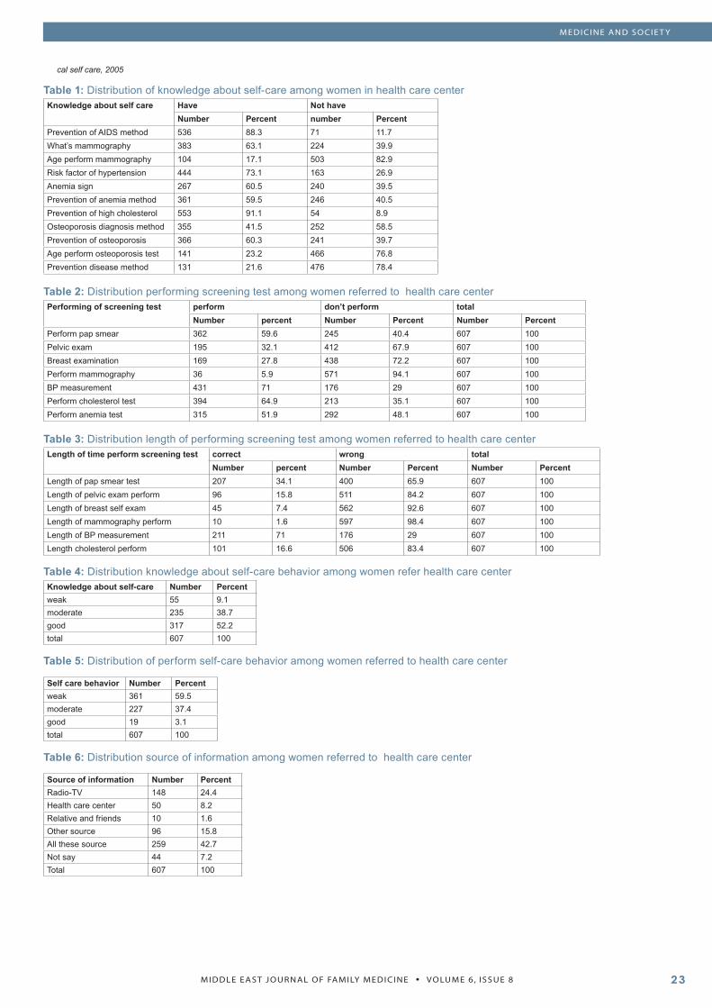
MIDDLE EAST JOURNAL OF FAMILY MEDICINE • VOLUME 6 , ISSUE 8 ��
Table 1: Distribution of knowledge about self-care among women in health care centerKnowledge about self care Have Not have
Number Percent number PercentPrevention of AIDS method 536 88.3 71 11.7What’s mammography 383 63.1 224 39.9Age perform mammography 104 17.1 503 82.9Risk factor of hypertension 444 73.1 163 26.9Anemia sign 267 60.5 240 39.5Prevention of anemia method 361 59.5 246 40.5Prevention of high cholesterol 553 91.1 54 8.9Osteoporosis diagnosis method 355 41.5 252 58.5Prevention of osteoporosis 366 60.3 241 39.7Age perform osteoporosis test 141 23.2 466 76.8Prevention disease method 131 21.6 476 78.4
Table 2: Distribution performing screening test among women referred to health care centerPerforming of screening test perform don’t perform total
Number percent Number Percent Number PercentPerform pap smear 362 59.6 245 40.4 607 100Pelvic exam 195 32.1 412 67.9 607 100Breast examination 169 27.8 438 72.2 607 100Perform mammography 36 5.9 571 94.1 607 100BP measurement 431 71 176 29 607 100Perform cholesterol test 394 64.9 213 35.1 607 100Perform anemia test 315 51.9 292 48.1 607 100
Table 3: Distribution length of performing screening test among women referred to health care centerLength of time perform screening test correct wrong total
Number percent Number Percent Number PercentLength of pap smear test 207 34.1 400 65.9 607 100Length of pelvic exam perform 96 15.8 511 84.2 607 100Length of breast self exam 45 7.4 562 92.6 607 100Length of mammography perform 10 1.6 597 98.4 607 100Length of BP measurement 211 71 176 29 607 100Length cholesterol perform 101 16.6 506 83.4 607 100
Table 4: Distribution knowledge about self-care behavior among women refer health care centerKnowledge about self-care Number Percentweak 55 9.1moderate 235 38.7good 317 52.2total 607 100
Table 5: Distribution of perform self-care behavior among women referred to health care center
Self care behavior Number Percentweak 361 59.5moderate 227 37.4good 19 3.1total 607 100
Table 6: Distribution source of information among women referred to health care center
Source of information Number PercentRadio-TV 148 24.4Health care center 50 8.2Relative and friends 10 1.6Other source 96 15.8All these source 259 42.7Not say 44 7.2Total 607 100
cal self care, 2005
MEDICINE AND SOCIET Y
MIDDLE EAST JOURNAL OF FAMILY MEDICINE • VOLUME 6 , ISSUE 8����
EDUCATION AND TRAINING
How to Write a Scientific Paper “Publish or perish” A Motivation to Learn More
ABSTRACTMigraine is a neurovascular disorder characterized by a unilateral mild or severe headache lasting from a few hours to as long as three days. It has been recently shown in many stud-ies that this disorder has a firm and complicated genetic background that exposes individuals to a higher sus-ceptibility to migraine attacks. Old theories used to focus on the vas-cular changes and the subsequent blood flow alterations in the brain to explain the different symptoms oc-curring during migraines. New theo-ries on the other hand are shedding more light on the involvement of the nervous system in the brain, prima-rily the trigeminal nerve in the brain stem, considering it the primary cause for the initiation of migraine attacks. Changes in blood vessels in the brain are believed to be an epi-phenomenon only.In this article, the pathophysiology of a migraine attack is explained on the basis of the unified theory that tries to integrate all the available scientific data about migraine.
Sources and selection criteria
Based on science direct and Pubmed search engine, Key words: scientific writing, component of scientific writing, planning writing, manuscript writing, writing skills and writing tips, writing guidelines, medical manuscript, and format of writing.
Some general hints in writing an article
Writing up is a daunting task; it involves a great deal of planning, preparation and time, it is simply a skill born from practice. In order to write, you need to read.
This article shall supply the researcher with a few simple guidelines on how to prepare, and write an organized scientific paper, which shall be ranging from its early drafting in order to improve the manuscript, and then its final publication (Andrei V.Alexandrov, 2004, Parati G, Valentini M, 2005, Nahas FX, Ferreira LM, 2005).
The approach in this article is the only way, nonetheless likely not the best way to write a scientific paper (Selma Cetin, and David J. Hackam, 2005).
Any manuscript consists generally speaking of 4 main sections; why you start? (Introduction, background and hypothesis), what you did (methods), what you gained (results), what does it mean (discussion) (David J Pierson, 2004). Before starting writing up, search the literature for similar resources dealing with your topic (Andrei V.Alexandrov, 2004). Consult the mentor when possible, because after all he/ she is the senior author (Andrei V.Alexandrov, 2004, J. Smyth, J. Verweij, M, et al, 2006). Good writing shall involve using of simple terms; in a direct order with its objectivity and then report it with a
good discussionn (Nahas FX, Ferreira LM, 2005).
Bear in mind to avoid a very excessive lengthily article, which makes them harder to be evaluated or read by the anticipated readers, if published (Szklo.M, 2006).
Show your draft to your colleagues for critique to refine and revise, and in order to improve it as well, and if English is not your mother language, show it to native who can improve its text and wording (Alexandrov AV, Hennerici MG, 2006, Amanda Tompsonm, 2006).
Research is an important tool for developing and innovations (Albaeean JW, Scholes J, 2005), because without it no advance can be achieved or discovering a new phenomenon (Nte AR, Awi DD, 2006), additionally, to advance the state of scientific knowledge plus advancing the career in the mean time (Rosenfeldt FL, Dowling JT, et al, 2000).
Furthermore, it is important to publish those results in order to be retrieved and duplicated/ replicated; otherwise the research will be incomplete and irrelevant. Therefore writing a scientific paper demands skill, and experience in order to present it to the specific audience in a well organized structure chronologically, with a clear purpose to answer the question which been investigated (Cetin S, Hackam DJ, 2005, Nte AR, Awi DD, 2006).
Any journal shall involve the main following three main factors; the authors, the reviewers and the editors. The last two are mandatory to present the scientific article to the well accepted presentation for publication (Branson RD, 2004, Zheng JW, 2005). Its been estimated that about 5% will be accepted for publication, whereas 50% will be rejected, and about 45% will be returned to the author with some suggested notes to be followed (Brian F McCabe, 2004). There are
Ebtisam ElghblawiDepartment of dermatology, BUM Hospital, Tripoli-Libya
MIDDLE EAST JOURNAL OF FAMILY MEDICINE • VOLUME 6 , ISSUE 8 ����
EDUCATION AND TRAINING
some suggested publication failures; either the author don’t know how to start writing or don’t know where to put and where (Kliewer MA, 2005 & 2006).
The objective of this article to present the main anatomy of a scientific paper, put forwards some common mistakes, presents some science-writing rules, and offers some science-writing tips. At the end, research results either can only confirm or reject a hypothesis.
It is a great deal to think of what kind of writing shall this paper involve, it can range from a research report, to many such as; original work, review paper, editorial, and case report, …etc).
Structure of a scientific paper at submission
Consider the word IMRaD in structuring your paper:
Title- subject been studiedAbstract- What did I do in a nutshell/ summary of the paper (main reason for the study, results and conclusions).Introduction- why the problem been investigateMaterials & Methods- how to solve that raised problemResults- what was gained?Discussion- what that result means, if its significantAcknowledgements- who helped meReferences- what work been looked throughTables- extra informationFigures- extra information
Title - should be catchy and informative.
The title is the most important part of your paper, it alerts the reader to topic of your paper, generally speaking it sells the article (Andrei V.Alexandrov, 2004), therefore it should be short, simple, specific and concise describing the work done in the presented article, so make the title dynamic and informative, rather than descriptive, with consideration for the proposed and intended audience. Also be specific, and mention if paper is a human, animal or bench study
1.2.
3.
4.
5.6.
7.
8.
9.10.
Include author name, highest academic degree, affiliation, email address, source of funding if any, phone, and fax of the correspondent.
Abstract
A good abstract should be accurate, self-contained, concise and specific, non-evaluative, coherent, and readable. The abstract wording differ from one journal to another, some requires 100-120 and others 200-300 words limit; it should start with an introductory sentence stating the raised question to be investigated, and what supposed to happen (hypothesis) (M. J. Kern, and H. N. Bonneau, 2003), then summarizing the goals, followed by 3-4 sentences addressing the main methods carried-out, and on whom , then 3-4 sentences about key finding of the study with analysis and lastly a statement of conclusions and recommendations. Abstract is the synopsis of the paper, so that the reader will decide to read your article or not. Generally speaking, writing an abstract means to extract and summarize, and be to the point quickly. However abstract is the first part, It must be written last as it will summarize the work done. This means simply the order of writing best will be as the following: Introduction, Methods & Materials, Results, Discussion, Statistics, Abstract, and, finally, Title. Avoid very long abstract, and use of abbreviations. Do not include any information, which is not covered in the manuscript, do not include tables, graphs, or references. Also, avoid any detailed descriptions of methods. Avoid mentioning any prior work in the abstract.
Introduction - Classic introduction 3 paragraphs:
Introduce the readers to the subject under study with its background, and state why you are interested in doing it (objectives), and what question you are going to address under study and what you are going to prove, and how you intend to answer and discuss them, how it relates to previous work in the area with careful reference selection findings (Brian F McCabe, 2004, W.P Naylor, A.Munoz-Vivevos, 2005, Selma Cetin, and David J. Hackam, 2005). That simply involves what you know about the subject
by a through complete literature review, and what you do not know by constructing a hypothesis (objective and purpose), and then what your paper aims to accomplish.
Describe the research strategy. Explain the theoretical implications of the study, and summarizes the relevant literature in that area.
Finally above all, it is important to include a concluding paragraph stressing the importance of your work in the article.
Materials & Methods
It is the easiest part for most authors (Brian F McCabe, 2004). Structure your method clearly and precisely with sub-heading, such as; subjects, selection criteria, data collection and procedure, statistical methods. Start by describing the study subject; whether they were normal volunteers, patients, or animals; with referring to subject selection criteria by defining age, sex, diagnosis…etc in a demographic table if it was a retrospective study, and how they are recruited; if randomly or not, exclusion and inclusion criteria if its prospective study, and indicate if there is a pilot study and control group, and describe the control subject as well, also get the approval of local medical ethics committee (animal/ human study) (M. J. Kern, and H. N. Bonneau, 2003), Signed informed consent form by subjects or their legal representatives (in critical conditions).
If the study involved using an equipment, describe it in details with referring to how it was calibrated and validated (R.D Branson, 2004), accepted standard measurement, product source, list of device used, Detailed descriptions of the devices; the name/model, manufacturer, and city and state of the manufacturer., timeline for procedures and measurements, any interventions were applied and when, and what data were collected, and timing of measurement with timing for interventions.
Outline how specimens were prepared if there is any. If animals were used, define their strains, with their age and weight. Describe any
MIDDLE EAST JOURNAL OF FAMILY MEDICINE • VOLUME 6 , ISSUE 8��
specimen prepared and how was made.
Statistical analysis
consult you mentor and the statisticians beforehand, state how data were collected and how handled, name the statistical test used to evaluate the data, state the sample size, and power calculations, and indicate the probability level, and why? Start by descriptive statistics, then used a specific one when needed for comparison. Identify clearly the independent variable (predictor), and the dependent variable (outcome), and see if there are any associations. If it is complicated protocol, include a diagram, table or flowchart to explain the methods you used.
Lastly indicate why this method was chosen, and when it was been carried. Therefore, it is mandatory to present detailed description on how study was performed, and what was done to answer the raised research question in the article, so another scientist can judge the appropriateness of the method applied, repeat it (Andrei V.Alexandrov, 2004).
Results
Its heart of the article, it is the main reason seeking for publication (Brian F McCabe, 2004). Results can either support or refute the proposed hypothesis.
Simply just state those important data achieved with numbers and statistics in a logical manner, and be to the point. Bear in mind the shorter you can present the results, the better (Alexandrov AV, Hennerici MG, 2004). Do not give opinions or speculations. Avoid abbreviations unless it’s been clearly defined and spelled out clearly. State the hypothesis you have applied in the study, with justification, put the results, and see if the results support any previous published findings to justify your conclusions (Alexandrov AV, Hennerici MG, 2006). Avoid giving details about irrelevant topics such as the programme been used for to enter the data (Streiner DL., 2007). Highlight the originality of your work if it is in a wording. Thereafter begin with the major positive findings; also give negative findings at the end of the results section as if it was
not anticipated, then review your hypothesis to see what went wrong, and consider if there should be a recommendation to repeat the further study in the future. Present statistical information using statistical terms appropriately, Acknowledge any problem with data (e.g. small sample size, limited follow-up time etc.), Use well-designed tables, graphs, flow charts, histograms and figures to present your results as its simple and self-explanatory; be sure to cite and summarize them in the text (Kallet RH, 2004). A well-designed table should stand on its own without further clarification.
Discussion
It’s the most challenging, and harder part of a paper (Rosenfeldt FL, Dowling JT, et al, 2000), you need to explain the meaning of the achieved results, and explore its significance to the reader (Hess DR, 2004), Indicate what they mean, and how its analyszed (Kallet RH, 2004), and if it was something new (novel). Start by evaluating and interpreting the results implications with referring to the hypothesis/ original question applied, any benefits and drawbacks, also if the gained data support the proposed hypothesis in your study, and if results were consistent with other previous investigators reports and if it does support it, and if the results were unexpected, explain what was the limitations, and why, and if the conditions were different, and if a further research is recommended to carry on to answer those unexplained results. Stay connected in your discussion between your study question and results, and see if there is any association between them. Let the results speak themselves; do not try to show how much your results are terrific (Andrei V.Alexandrov, 2004).
Look for Similarities and differences between your results and the work of others, which should clarify and confirm your conclusions. Negative results should be accepted as such without an attempt to explain them away. Try to ask yourself the following questions: Did I add something new in here?, Did my study resolve some of the original problem?, What conclusions and speculative propositions can I draw from my
study?
Conclusions
Conclusions have to be based on the present study findings (Alexandrov AV, Hennerici MG, 2006). It should be simple and clear to be noticed (Alexandrov AV, 2004). It has to be to what you believe that you have proven (Brian F McCabe, 2004), it’s actually conceptualization, and if you recommend any further work to be done in the future to define the problem.
Acknowledgments - as needed
A thank to those who helped.
References
Reference should be checked for completeness and accuracy before submission, Refer to articles from peer-reviewed journals (or those being “in press”), Limit list to key citations, Cite references throughout the paper, Refer to reference guidelines for targeted journal
Submitting the Paper for Publication
Before submitting, you need to consider the followings; Selecting a journal, read and follow the “instructions for Authors” in order to meet the proposed criteria of submission; because it varies from one journal to another (Brian F McCabe, 2004), Don’t forget to number the pages as been instructed in the intended journal to apply, Understand issues of copyright, Organize materials for submission, proof read several times, request internal review prior to submission, lastly remember every article has a home.
Common cause for rejection to publish:
Piles of papers are submitted to scientific journals annually. Rejection rates are escalating in most well-known journals. Apply the reviewer’s comments to improve your paper. 50% of initially rejected articles are eventually published somewhere else, (David J Pierson, 2004). The common reasons for rejections are; Insufficient and inaccurate data, poor study design, incomplete statistics, over interpretation of data, out dated information, difficult to follow
EDUCATION AND TRAINING
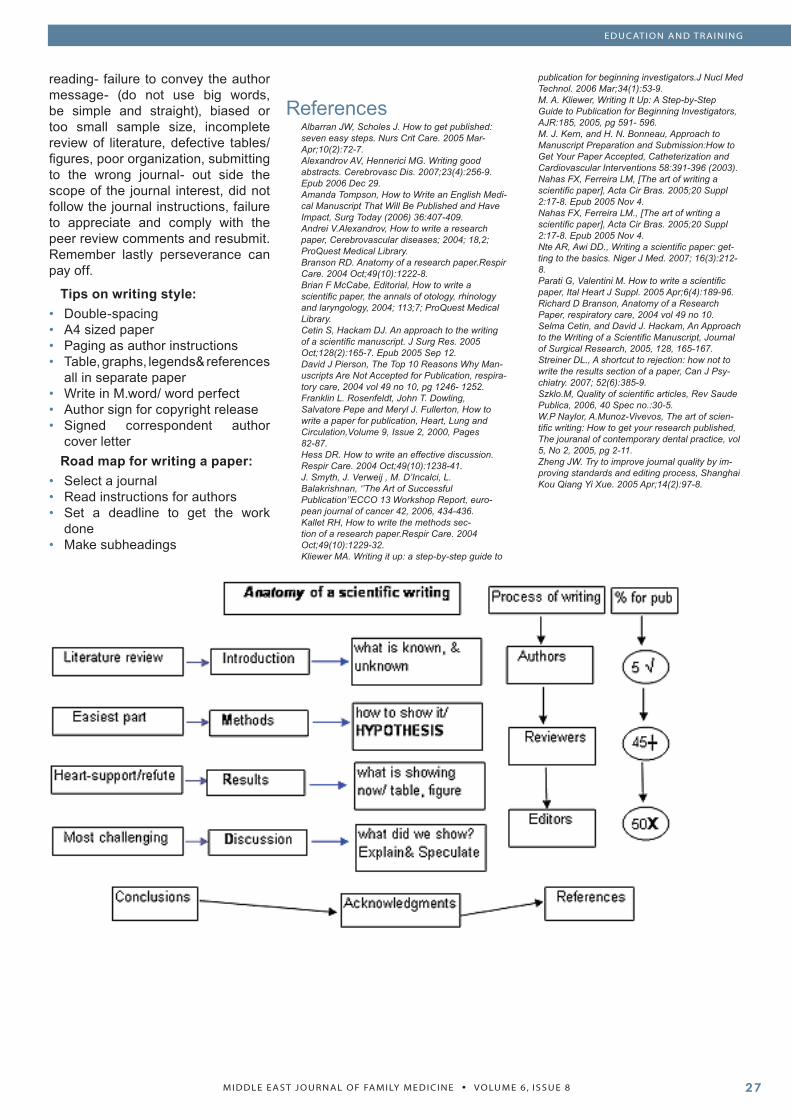
MIDDLE EAST JOURNAL OF FAMILY MEDICINE • VOLUME 6 , ISSUE 8 ��
EDUCATION AND TRAINING
reading- failure to convey the author message- (do not use big words, be simple and straight), biased or too small sample size, incomplete review of literature, defective tables/ figures, poor organization, submitting to the wrong journal- out side the scope of the journal interest, did not follow the journal instructions, failure to appreciate and comply with the peer review comments and resubmit. Remember lastly perseverance can pay off.
Tips on writing style:Double-spacingA4 sized paperPaging as author instructionsTable, graphs, legends& references all in separate paperWrite in M.word/ word perfectAuthor sign for copyright releaseSigned correspondent author cover letter
Road map for writing a paper:Select a journalRead instructions for authorsSet a deadline to get the work doneMake subheadings
••••
•••
•••
•
ReferencesAlbarran JW, Scholes J. How to get published: seven easy steps. Nurs Crit Care. 2005 Mar-Apr;10(2):72-7.Alexandrov AV, Hennerici MG. Writing good abstracts. Cerebrovasc Dis. 2007;23(4):256-9. Epub 2006 Dec 29.Amanda Tompson, How to Write an English Medi-cal Manuscript That Will Be Published and Have Impact, Surg Today (2006) 36:407-409.Andrei V.Alexandrov, How to write a research paper, Cerebrovascular diseases; 2004; 18,2; ProQuest Medical Library.Branson RD. Anatomy of a research paper.Respir Care. 2004 Oct;49(10):1222-8.Brian F McCabe, Editorial, How to write a scientific paper, the annals of otology, rhinology and laryngology, 2004; 113;7; ProQuest Medical Library.Cetin S, Hackam DJ. An approach to the writing of a scientific manuscript. J Surg Res. 2005 Oct;128(2):165-7. Epub 2005 Sep 12.David J Pierson, The Top 10 Reasons Why Man-uscripts Are Not Accepted for Publication, respira-tory care, 2004 vol 49 no 10, pg 1246- 1252.Franklin L. Rosenfeldt, John T. Dowling, Salvatore Pepe and Meryl J. Fullerton, How to write a paper for publication, Heart, Lung and Circulation,Volume 9, Issue 2, 2000, Pages 82-87.Hess DR. How to write an effective discussion. Respir Care. 2004 Oct;49(10):1238-41.J. Smyth, J. Verweij , M. D’Incalci, L. Balakrishnan, ‘’The Art of Successful Publication’’ECCO 13 Workshop Report, euro-pean journal of cancer 42, 2006, 434-436.Kallet RH, How to write the methods sec-tion of a research paper.Respir Care. 2004 Oct;49(10):1229-32.Kliewer MA. Writing it up: a step-by-step guide to
publication for beginning investigators.J Nucl Med Technol. 2006 Mar;34(1):53-9.M. A. Kliewer, Writing It Up: A Step-by-Step Guide to Publication for Beginning Investigators, AJR:185, 2005, pg 591- 596.M. J. Kern, and H. N. Bonneau, Approach to Manuscript Preparation and Submission:How to Get Your Paper Accepted, Catheterization and Cardiovascular Interventions 58:391-396 (2003).Nahas FX, Ferreira LM, [The art of writing a scientific paper], Acta Cir Bras. 2005;20 Suppl 2:17-8. Epub 2005 Nov 4.Nahas FX, Ferreira LM., [The art of writing a scientific paper], Acta Cir Bras. 2005;20 Suppl 2:17-8. Epub 2005 Nov 4.Nte AR, Awi DD., Writing a scientific paper: get-ting to the basics. Niger J Med. 2007; 16(3):212-8.Parati G, Valentini M. How to write a scientific paper, Ital Heart J Suppl. 2005 Apr;6(4):189-96.Richard D Branson, Anatomy of a Research Paper, respiratory care, 2004 vol 49 no 10.Selma Cetin, and David J. Hackam, An Approach to the Writing of a Scientific Manuscript, Journal of Surgical Research, 2005, 128, 165-167.Streiner DL., A shortcut to rejection: how not to write the results section of a paper, Can J Psy-chiatry. 2007; 52(6):385-9.Szklo.M, Quality of scientific articles, Rev Saude Publica, 2006, 40 Spec no.:30-5.W.P Naylor, A.Munoz-Vivevos, The art of scien-tific writing: How to get your research published, The jouranal of contemporary dental practice, vol 5, No 2, 2005, pg 2-11.Zheng JW. Try to improve journal quality by im-proving standards and editing process, Shanghai Kou Qiang Yi Xue. 2005 Apr;14(2):97-8.
MIDDLE EAST JOURNAL OF FAMILY MEDICINE • VOLUME 6 , ISSUE 8��
Heterotopic Pregnancy in Natural Cycle - Probably Not Rare
ABSTRACTWe present here a case of heterotop-ic pregnancy in a natural conception cycle. The woman presented with he-morrhagic shock at 7+ weeks preg-nancy. She had had an ultrasound three days prior to the admission, which showed a normal intrauterine pregnancy. An urgent ultrasound done in the department clinched the diagnosis of heterotopic pregnancy. The ectopic component was treated by laparotomy and the intrauterine pregnancy continued to term une-ventfully. The unique feature of this case is the patient’s lack of risk fac-tors for ectopic pregnancy. The aim of this case report is to emphasize the importance of sonographic ex-amination of the adnexa at booking ultrasound in all pregnant women. Problems associated with the diag-nosis and special considerations in-volved in the management of hetero-topic pregnancy are highlighted.
CASE REPORTA 28 year old nullipara who had
suffered 4 first trimester abortions attended our hospital emergency department with history of 8 weeks amenorrhoea and complaints of sudden onset of lower abdominal pain and dyschesia of one day duration. She said that she had had an ultrasound three days ago and was told she had a normal intrauterine pregnancy corresponding to LMP gestational age. There was no vaginal bleeding. She had no known risk factors for ectopic pregnancy. She had felt faint prior to the time of presentation. On examination she was in pain with pallor, tachycardia with low volume pulse and hypotension with blood pressure of 90/60. The abdomen was distended and there was marked generalized tenderness. A provisional diagnosis of 8 weeks pregnancy with ruptured hemorrhagic corpus luteal cyst was made. Resuscitative measures were commenced for the treatment of shock. Simultaneously an urgent ultrasound was done. It showed a single intrauterine gestational sac with a viable embryo corresponding to 7+ weeks pregnancy. Another gestational sac with a live embryo of approximately seven weeks gestational age was seen in the right adnexa (Fig 1 and 2). There was fluid with low level echoes in the pelvis and hepatorenal pouch, suggestive of hemoperitoneum. Thus, a diagnosis of heterotopic pregnancy with ruptured ectopic pregnancy and hemoperitoneum was
made. An emergency laparotomy was performed.
There was hemoperitoneum of 1.5 liters. Uterus was enlarged corresponding to 8 weeks size. The right adnexa showed ruptured right tubal ectopic pregnancy. The ectopic gestational sac with embryo was still seen within the ruptured tube. The left adnexa appeared normal. The tubal ectopic pregnancy contents were removed and salpingostomy was performed taking care to avoid undue manipulation of the uterus. She received three units of packed red blood cells in the intraoperative and immediate postoperative period. Ultrasound done on the third postoperative day affirmed the viability of intrauterine embryo. She was discharged home on the fourth postoperative day on folic acid and progesterone support. The intrauterine pregnancy continued uneventfully till term. Cesarean section was done at 38 weeks pregnancy as there was non-reassuring fetal trace during labor and she delivered a live girl, weight 3.070 kg, with Apgar score of 8 at 1 minute and 9 at five minutes.site of injury in about 69% followed by maxilla which represents 14% of the cases, then the other bones as
shown in Table 3.
DiscussionHeterotypic pregnancy defined
as the presence of intrauterine pregnancy coexisting with an
Dr.Ramadevi V Wani1 MRCOGDr.Sami Al Taher1 KFUF1 Department of Obstetrics and Gynecology, Al Jahra Hospital, Kuwait
Corresspondence to:Dr.Ramadevi V Wani MRCOGPO Box 17672Khaldiya, 72457KUWAITEmail: [email protected]: 00965-6779120Residence: 00965-4880924Fax: 00965-4577045
Key words: natural conception, ectopic pregnancy, ultrasound, adnexa.
CASE REPORTS
MIDDLE EAST JOURNAL OF FAMILY MEDICINE • VOLUME 6 , ISSUE 8 ��
ectopic pregnancy is a relatively underestimated clinical condition. It was first described by Duverney in 1708 during an autopsy. The incidence of heterotopic pregnancy in spontaneous cycles was quoted as 1: 30,000 about 50 years ago. However in the last three decades there has been an almost four-fold increase in the incidence of ectopic pregnancy and a corresponding increase in the incidence of heterotopic pregnancy. The current quoted risk of heterotopic pregnancy is 1 in 3889-6778 in the general population[1]. In a meta-analysis in 1996, previous ectopic pregnancy, previous tubal surgery, documented tubal pathology and in utero diethylstilbestrol (DES) exposure were found to be associated strongly with the occurrence of ectopic pregnancy[2]. In the same study previous genital infections (pelvic inflammatory disease, Chlamydia, gonorrhoea), infertility, and a lifetime number of sexual partners > 1 were associated with a mildly increased risk. Heterotopic pregnancy occurs even more frequently after pharmacologic ovulation stimulation. After ART the risk is greatest increasing to 1.2-2.9%[3,4].
Diagnosis of heterotopic pregnancy is often difficult. It is more likely to be thought of and diagnosed in pregnancies following assisted reproductive technology. Even then there are several diagnostic pitfalls. Clinical diagnosis of heterotopic pregnancy is unlikely as the symptoms of pain and bleeding are often attributed to be due to the complication of intrauterine pregnancy. Quantitative assessment of ß HCG and progesterone are falsely reassuring, as they are often in the normal range due to co-existing intrauterine pregnancy. Our patient had natural conception. Secondly the woman did not have any of the above-mentioned risk factors for ectopic pregnancy. Moreover the presence of a viable intrauterine pregnancy may have reassured the ultrasonographer and he may have overlooked the adnexa. These could probably be the reasons for the diagnosis not being made at the time of first ultrasound.
The improved resolution of the transvaginal ultrasound enables more
accurate diagnosis of heterotopic pregnancy[5]. It has resulted in the earlier diagnosis of ectopic pregnancy and has contributed to a decrease in the maternal mortality and morbidity associated with this condition. However Qusehal A et al reported transabdominal ultrasound to be more useful than transvaginal ultrasound as it can visualize those areas, which cannot be assessed by the latter. In effect both methods are complimentary[6]. Sonographic detection of an extra uterine gestation sac with or without fetal pole and /or cardiac activity, together with an intrauterine pregnancy confirms the diagnosis of heterotopic pregnancy. In our patient who presented with acute abdomen and shock, in view of the history of an ultrasound showing normal intrauterine pregnancy, ruptured hemorrhagic
corpus luteal cyst was the first diagnosis considered. However an urgent ultrasound done in the department clinched the diagnosis. In our patient the ectopic embryo located in the upper zone of the right iliac fossa was only picked up by the transabdominal ultrasound. It was inaccessible to transvaginal ultrasound.
In a woman with heterotopic pregnancy who is clinically stable laparoscopic management is both feasible and safe. All tubal pregnancies irrespective of their location - even in the technically most demanding situations with interstitial or cornual location can be successfully managed by laparoscopy with an uneventful course for the remaining intrauterine pregnancy[7]. Feasibility of laparoscopic surgery depends on the expertise of the surgical team. Non-surgical management is an efficient alternative with a good prognosis for intrauterine pregnancy. Transvaginal ultrasound guided aspiration of the gestational sac fluid and injection of potassium chloride, hyperosmolar glucose or hypertonic sodium chloride solution into it results in resorption of the ectopic trophoblastic tissue[8,9,10]. Unlike ectopic pregnancy, heterotopic pregnancy with viable intrauterine pregnancy cannot be treated with methotrexate for obvious reasons. However, when a patient presents
with acute abdomen and shock, laparotomy and salpingectomy/salpingostomy is the safest option. In our patient as she presented with shock we did laparotomy and salpingostomy. Prompt resuscitative measures for the treatment of shock and the careful introduction of cardiac sparing anesthetics are necessary for the survival of the intrauterine pregnancy.
In a literature review by Tal et al involving 139 cases of heterotopic pregnancy, where most of them were treated surgically the overall live intrauterine pregnancy rate was 66% irrespective of the presence of hemoperitoneum[11]. In a comparison study of assisted vs. spontaneous heterotopic conceptions, the assisted conception group had a higher live birth rate than the spontaneous group (47.8 vs. 20%)[12]. Our patient was delivered at term by cesarean section.
ConclusionHeterotopic pregnancy is a
potentially fatal condition. However when diagnosed early and treated appropriately it can result in survival of the precious intrauterine pregnancy. We recommend that all obstetricians and sonologists performing obstetric ultrasound should routinely evaluate the adnexa and document it. This practice in addition to diagnosing heterotopic pregnancy will also help in the diagnosis of other adnexal pathology. In pregnant women with acute abdomen clinicians should always consider heterotopic pregnancy in the differential diagnosis.
ReferencesRizk B. Outcome of assisted reproductive technol-ogy. In: Brinsdon PR editor. Textbook of in vitro fertilization and assisted reproduction. The Bourn hall guide to clinical and laboratory practice. 2nd ed. UK. Parthenon publishing; 1999. P 316-317.Ankum WM, Mol BW, Vander Veen F, Bossuyt PM. Risk factors for ectopic pregnancy: a meta- analy-sis. Fertil Steril. 1996; 65(6): 1093-1099.Goldman GA, Fisch B, Ovadia J, tadir Y. Heterotypic pregnancy after assisted reproductive technologies. Obstet Gynecol Surv 1992; 47(4): 217-221.Dimitry ES, Margara R, Subak-Sharpe R, Mills M, Winston R. nine cases of heterotopic pregnancy in four years of in vitro fertilization. Fertil Steril. 1990; 53(1): 107-110.Fa EM, Gersovich EO. High resolution ultrasound in the diagnosis of heterotopic pregnancy: combined
1.
2.
3.
4.
5.
CASE REPORTS
MIDDLE EAST JOURNAL OF FAMILY MEDICINE • VOLUME 6 , ISSUE 830
Figure 1: Transabdominal scan demonstrating intrauterine pregnancy and tubal ectopic pregnancy
transabdominal and transvaginal approach. Br J Obstet & Gynaecol. 1993; 100(9): 871-872.Qusehal A, Mamouchi H, Ghazli M, Kadiri R. Heter-otypic pregnancy: value of transabdominal sonog-raphy. J Radiol 2001; 82(7): 851-853Pschera H, Kandemir S. Laparoscopic Treatment of Heterotypic pregnancies: Benefits, complications, and Safety Aspects. J Turkish german Gynecol As-soc. 2005; 6(2): 90-94.Fernandez H, Fournet P, Lelaider C, Olivennes F. Non surgical treatment of heterotopic pregnancy:
6.
7.
8.
a report of six cases. Fertil steril. 1993; 60(3) 428-432.Goldberg JM, Bedaiwy MA. Transvaginal local in-jection of hyperosmolar glucose for the treatment of heterotopic pregnancies. Obstet Gynecol. 2006; 107: 509-510.Prorocic M, Vasiljevic M. Treatment of heterotopic cervical pregnancy after in vitro fertilization-embryo transfer by using transvaginal ultrasound guided aspiration and instillation of hypertonic solution of sodium chloride. Fertil Steril 2007; 88(4): 969.
9.
10.
Tal J, Haddad S, Gordon N, Timor- Tritsch I. het-erotopic pregnancy after ovulation induction and as-sisted reproductive technologies: a literature review from 1971to 1993. Fertil Steril. 1996; 66: 1-12.Han SH, Jee BC, Suh CS, Kim SH, Choi YM, Kim JG, Moon SY. Clinical outcomes of tubal heterotopic pregnancies: assisted vs. spontaneous concep-tions. Gynecol Obstet Invest. 2007; 64(1): 49-54..
11.
12.
CASE REPORTS
MIDDLE EAST JOURNAL OF FAMILY MEDICINE • VOLUME 6 , ISSUE 8 ��
CASE REPORTS
Figure 2: Transabdominal scan showing viable tubal ectopic pregnancy
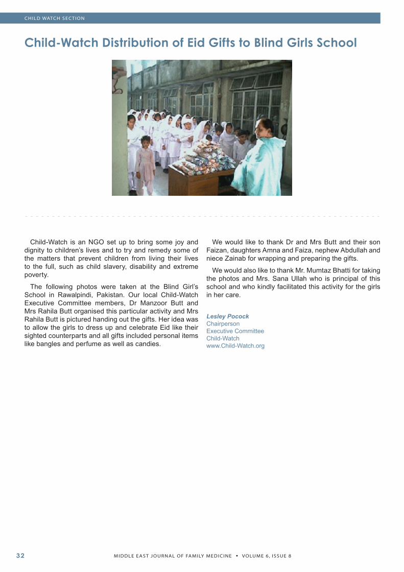
MIDDLE EAST JOURNAL OF FAMILY MEDICINE • VOLUME 6 , ISSUE 8��
CHILD WATCH SECTION
Child-Watch Distribution of Eid Gifts to Blind Girls School
Child-Watch is an NGO set up to bring some joy and dignity to children’s lives and to try and remedy some of the matters that prevent children from living their lives to the full, such as child slavery, disability and extreme poverty.
The following photos were taken at the Blind Girl’s School in Rawalpindi, Pakistan. Our local Child-Watch Executive Committee members, Dr Manzoor Butt and Mrs Rahila Butt organised this particular activity and Mrs Rahila Butt is pictured handing out the gifts. Her idea was to allow the girls to dress up and celebrate Eid like their sighted counterparts and all gifts included personal items like bangles and perfume as well as candies.
We would like to thank Dr and Mrs Butt and their son Faizan, daughters Amna and Faiza, nephew Abdullah and niece Zainab for wrapping and preparing the gifts.
We would also like to thank Mr. Mumtaz Bhatti for taking the photos and Mrs. Sana Ullah who is principal of this school and who kindly facilitated this activity for the girls in her care.
Lesley PocockChairpersonExecutive CommitteeChild-Watchwww.Child-Watch.org
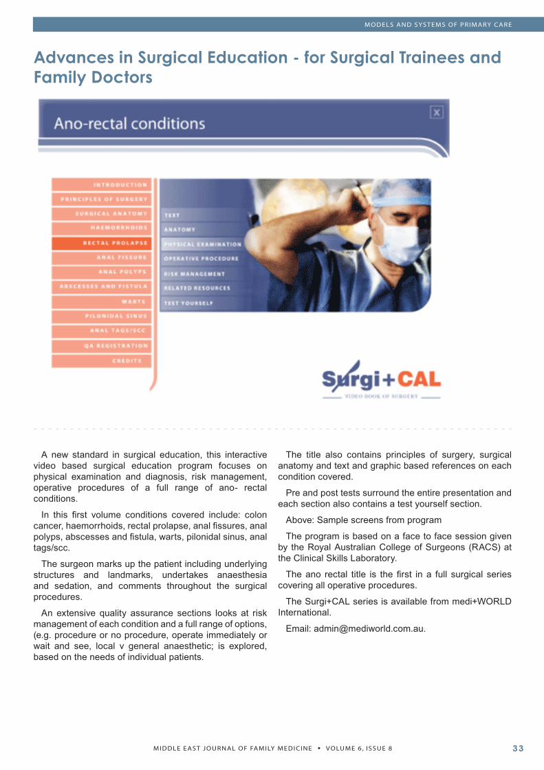
MIDDLE EAST JOURNAL OF FAMILY MEDICINE • VOLUME 6 , ISSUE 8 ��
MODELS AND SYSTEMS OF PRIMARY CARE
Advances in Surgical Education - for Surgical Trainees and Family Doctors
A new standard in surgical education, this interactive video based surgical education program focuses on physical examination and diagnosis, risk management, operative procedures of a full range of ano- rectal conditions.
In this first volume conditions covered include: colon cancer, haemorrhoids, rectal prolapse, anal fissures, anal polyps, abscesses and fistula, warts, pilonidal sinus, anal tags/scc.
The surgeon marks up the patient including underlying structures and landmarks, undertakes anaesthesia and sedation, and comments throughout the surgical procedures.
An extensive quality assurance sections looks at risk management of each condition and a full range of options, (e.g. procedure or no procedure, operate immediately or wait and see, local v general anaesthetic; is explored, based on the needs of individual patients.
The title also contains principles of surgery, surgical anatomy and text and graphic based references on each condition covered.
Pre and post tests surround the entire presentation and each section also contains a test yourself section.
Above: Sample screens from program
The program is based on a face to face session given by the Royal Australian College of Surgeons (RACS) at the Clinical Skills Laboratory.
The ano rectal title is the first in a full surgical series covering all operative procedures.
The Surgi+CAL series is available from medi+WORLD International.
Email: [email protected].

MIDDLE EAST JOURNAL OF FAMILY MEDICINE • VOLUME 6 , ISSUE 8��