Mesophase Separation and Probe Dynamics in Protein-Polyelectrolyte Coacervates
Upload
massgeneralCategory
view
5download
0
Pergamon
l Original Contribution
Magnetic Resonance Imaging, Vol. 12, No. 4, pp. 641-652, 1994 Copyright 0 1994 Elsevier Science Ltd Printed in the USA. All rights reserved
0730-725X/94 $6.00 + .OO
0730-725X(93)E0070-5
MITOCHONDRIAL LOCALIZATION AND CHARACTERIZATION OF 99Tc-SESTAMIBI IN HEART CELLS BY ELECTRON PROBE X-RAY
MICROANALYSIS AND 99Tc-NMR SPECTROSCOPY
DAVID PIWNICA-WORMS,* JAMES F. KRONAUGE,* ANN LEFURGEY,~ MARK BACKUS,?
DANIEL HOCKETT,~ PETER INGRAM,~ MELVYN LIEBERMAN,t B. LEONARD HOLMAN,*
ALUN G. JONES,* AND ALAN DAVISON~
*Department of Radiology, Harvard Medical School, Brigham and Women’s Hospital, Boston, MA, USA TDepartment of Cell Biology, Duke University Medical Center, Durham, NC, USA
SResearch Triangle Institute, Research Triangle Park, NC, USA §Department of Chemistry, Massachusetts Institute of Technology, Cambridge, MA, USA
As the development of targeted intracellular magnetic resonance contrast agents proceeds, techniques for the quan- titative analysis of the subcellular compartmentation and characterization of metallopharmaceuticals must also advance. To this end, the subcellular distribution and chemical state of hexakis (Zmethoxyisobutyl isonitrile) technetium-99 (WTc-SESTAMIBI), the ground state of the organotechnetium radiopharmaceutical used for the noninvasive evaluation of myocardial perfusion and viability by scintigraphy, has been determined by a novel application of electron probe X-ray microanalysis (EPXMA) and *Tc-NMR spectroscopy. In cryopreserved cultured chick heart cells equilibrated in 36 CM WTc-SESTAMIRI, EPXMA imaging of mitochondria yielded a respiratory uncoupler-sensitive characteristic WTc X-ray peak representing 32.0 f 2.9 nmoles Tc/mg dry weight, while EPXMA of cytoplasm or nucleus showed no peak significantly greater than the threshold detectability limit of -1 nmole/mg dry weight. Technetium-99 NMR spectroscopy of heart cells equilibrated with 99Tc-SESTAMIBI showed a single peak at -45.5 ppm with no evidence of significant line broadening or chemical shift compared to aqueous chemical standards, indicating that the majority of the complex exists unbound within the mitochon- drial matrix. These data quantitatively demonstrate the localization of this lipophilic cationic organometallic com- plex within mitochondria in situ, consistent with a sequestration mechanism dependent on membrane potentials. Furthermore, this study establishes the general feasibility of combined EPXMA and NMR spectroscopy for the direct subcellular localization and characterization of metallopharmaceuticals, techniques that are readily appli- cable to MR contrast agents.
Keywords Electron probe; Organotechnetium compounds: Isonitriles; Mitochondria; Membrane potential; WTc nuclear magnetic resonance spectroscopy.
INTRODUCTION
Hexakis (2-methoxyisobutylisonitrile) technetium-99m (I) (99mTc-SESTAMIBI) is a clinically proven myocar- dial perfusion imaging agent and possible viability probe useful for the noninvasive evaluation of coro- nary artery disease. l-3 Whole animal and intact per- fused organ models have shown that initial regional tissue distribution of this complex is proportional to
blood flo~.~ However, 99mTc-SESTAMIBI, a lipo- philic cation, is unique among perfusion tracers in that final myocardial accumulation and retention is depen- dent on intact mitochondrial (A$) and plasma mem- brane (E,) potentials. 5-8 In the steady state, evidence from cellular pharmacological and biophysical analy- sis suggests that 99mTc-SESTAMIBI is localized within mitochondria.5-7*9 In cultured chick heart cells, for example, conditions which depolarize A$ and E,,,,
RECEIVED 6/28/93; ACCEPTED 1 l/4/93. PhD, Department of Radiology, Brigham and Women’s Hos- Address correspondence to David Piwnica-Worms, MD, pital, 75 Francis St., Boston, MA 02115.
641
642 Magnetic Resonance Imaging 0 Volume 12, Number 4, 1994
such as incubating cells in high K buffer plus the po- tassium ionophore valinomycin or exposing them to the mitochondrial uncoupler carbonyl cyanide-m-chloro- phenylhydrazone (CCCP), inhibit >95% of 99mTc- SESTAMIBI net uptake? Conversely, hyperpolarizing A$ by exposing cells to nigericin (K+/H+ exchanger) or oligomycin (FIFO ATP synthase inhibitor) significantly enhances net myocellular accumulation of the agent by at least 5-fold.’ Similar techniques applied to NIH 3T3 fibroblasts, v-src transformed fibroblast@ and human carcinoma cell lines in vitrolo also point to mitochon- drial localization of the agent in the steady-state.
Whole organ techniques are also consistent with a localization mechanism that is metabolically sensitive. In an isolated rat heart preparation, irreversible cell injury produced by prolonged exposure to sodium cyanide or lytic membrane disruption produced by Tri- ton X-100 significantly reduce peak accumulation of 99mTc-SESTAMIBI.11 In rat and guinea pig hearts loaded with 99mTc-SESTAMIBI, the agent is found as- sociated with the mitochondrial fraction in a substrate- dependent manner. 9v’2 In addition, after 90 min of reperfusion preceded by 3 h of coronary occlusion in an open-chest dog model, a significant reduction in myocardial accumulation of 99mTc-SESTAMIBI is ob- served independent of flow, consistent with an energy- dependent mechanism of localization in vivo.13
Variable extraction efficiencies by heart tissue for different members of this class of technetium com- poundsi and the disparate data on actual mechanisms of retentionI have hindered further development and interpretation of the in vivo behavior of these metal- lopharmaceuticals. A better understanding of the steric requirements and other properties of these complexes to guide rational design of improved clinical agents re- quires quantitative information on the chemical identity and mechanism of localization. The exact nature of the subcellular or biomolecular interaction of the agent in vivo cannot be confirmed with physiological techniques alone, unless used in combination with physical chem- ical methodologies.
The purpose of this study was to exploit the unique NMR spectroscopic properties of 99Tc in a biological system to chemically characterize carrier-added 99Tc- SESTAMIBI in a physiologically relevant model of heart tissue. The structural identity in solution was combined, under the same conditions, with subcellu- lar localization of the complex in myocellular tissue using a novel application of electron probe X-ray microanalysis (EPXMA). Combined physico-chemical information from 99Tc-NMR and direct subcellular localization with EPXMA should firmly establish the final biological compartment and chemical status of 99Tc-SESTAMIBI within myocardial tissue.
MATERIALS AND METHODS
Heart Cell Isolation and Cell Culture Isolated chick ventricular myocytes prepared from
11 -day-old chick embryo hearts serially disaggregated with trypsin for use in 99Tc-NMR experiments were obtained by minor modification of previously pub- lished methods.5 Briefly, 120 hearts in two batches were trimmed of major blood vessels and atria, finely minced, and serially exposed to 0.028% (wt/vol) tryp- sin in Ca2+- and Mg 2+-free Earle’s salt solution for 7 min at 37” C. Disaggregation was aided by gentle trit- uration and agitation on an orbital shaker bath. Cells released from the first exposure were discarded, and cells aspirated from the next four exposures added to an equal volume of trypsin deactivating solution consisting of ice-cold Ca 2+-free, Earle’s solution plus 6% bovine calf serum. The cells were centrifuged at 400 g for 10 min, resuspended and combined in Ca2+- free Earle’s plus 1% serum before counting with a hemocytometer.
For EPXMA experiments, heart cells were cultured as spherical aggregates as described.16 Hearts were dis- sected from 11 -day-old chick embryos, minced in Sa- line G and serially trypsinized (O.OS@/‘o) at 37°C. Cells were filtered, centrifuged, resuspended in culture me- dia, then preplated for one hour to reduce the presence of nonmuscle cells.16 The culture media consisted of 60% medium 199 (GIBCO, Grand Island, NY), 33% Earle’s buffered salt solution (5.4 mM potassium and 1 mM calcium), 5% fetal bovine serum (Hyclone Lab- oratories, Logan, UT), and 2% chick embryo extract. Cells (l-l .5 x 106) were plated into IOO-mm culture dishes containing pinholes in an agar coating and in- cubated at 37°C for 3-4 days in a controlled environ- ment of 95% air and 5% C02. Spontaneously beating aggregates formed during this time (diameter = 50 to 100 pm).
Solutions Control solution for experiments was a modified
Earle’s balanced salt solution (MEBSS) containing (mM): 145 Na+, 5.4 K+, 1.2 Ca2+, 0.8 Mg2+, 152 Cl-, 0.8 H,PO;, 0.8 SO:-, 5.6 dextrose, 4.0 HEPES, and 1% bovine calf serum (vol/vol), pH 7.4 + 0.05. Exper- iments were performed at 4”C, 25’C, and 37“C as in- dicated in the text. All compounds were reagent grade. Carbonyl cyanide-m-chlorophenylhydrazone (CCCP) (Sigma Chemical Co., St. Louis, MO) was dissolved in dimethylsulfoxide (DMSO) before addition to solutions. Final DMSO concentration was -0.570, which has been found to have no effect on 99mTc-SESTAMIBI accumulation* or on the electrophysiological charac- teristics of cultured heart cells.”
“Tc-NMR and EPXMA of ggTc-SESTAMIBI in heart cells 0 D. PIWNICA-WORMS ET AL. 643
Carrier-added 99Tc-SESTAMIBI was prepared from 8-10 mg (lo-12 pmoles) of solid 99T~-(MIBI)6 chloride powder dissolved in 0.5 ml of 95% ethanol solution. l8 Synthesis of the radiolabeled compound 99mT~-SESTAMIBI was performed with a one-step kit formulation (kindly provided by T.R. Carroll, Cardi- olite, E.I. DuPont, Medical Products Division, Biller- ica, MA) containing solid stannous chloride (0.075 mg) as a reducing agent for the technetium and MIBI as the CU(MIBI)~BF~ salt .5 Radiochemical purity was found to be greater than 97% by thin-layer chromatography (aluminum oxide plates; J.T. Baker, Phillipsburg, N.J.) using ethanol (absolute) as the mobile phase.
99Tc-NMR Spectroscopy Cell suspensions (- 1 x lo* cells) were incubated at
25°C for 40 min in 50 cc Pyrex tubes containing 10 ml loading solution comprising MEBSS and 25-100 PM [99Tc-SESTAMIBI]. The cells were spun (5 min @ 400 x g), washed in ice-cold MEBSS, then resuspended in 1 ml ice-cold isotope-free MEBSS and transferred to a 5-mm NMR tube. 99Tc-NMR spectra were imme- diately obtained on a Varian VXR-500 spectrometer using 99Tc0; as the external reference (112.542 MHz) and hexakis (t-butylisonitrile) technetium(I) (99Tc- TBI), the prototypical Tc-isonitrile complex, as an ex- perimental standard (-1914 ppm relative to 99T~0;) before and after each cell spectra.” Typical spectra were recorded with the sample spinner on by applying a 14.5-ps pulse width (90’ tip) and 0.15 s acquisition time for 5 K to 10 K acquisitions for cells and 128 ac- quisitions for standards. Because the T, and T2 of 99Tc are <50 ms, no acquisition delay or additional relaxa- tion delay were used. Spectral width was set at 16 KHz using 30 K data points in the FID weighted with lo-Hz Gaussian line broadening. Spectra were obtained at 4°C 25°C and 37°C. For one series of experiments, CCCP was added directly to cells in the NMR tube (final concentration = 5 FM).
To document the energetic integrity of myocardial cells used in 99Tc-NMR experiments, aliquots of cell suspensions were equilibrated for 40-60 min in MEBSS containing tracer 99mTc-SESTAMIBI (lo-25 &i/ml; 5-9 pmol/mCi) and i4C-mannitol (1 &i/ml). One ml samples of the cell suspensions were spun in a centri- fuge (-400 x g; 5 mm), and the supernatants and pellets assayed for 99mT~ by gamma counting, 14C by liquid scintillation counting and protein by the method of Lowry.’ Calculation of trapped extracellular spaces with “C-mannitol allowed subtraction of extracellu- lar 99mTc-SESTAMIBI from the pellet to yield cell- associated 99mTc activity.
Chemical standards were obtained with solutions
of 5% ethanol/M% normal saline, 5% ethanol/95% octanol and 5% ethanol/95% normal saline plus 3.5 mM human serum albumin, each containing *Tc- SESTAMIBI (0.5 to 2 mM) and using the same acqui- sition parameters and temperatures as for the cell suspensions.
Electron Probe X-ray Microanalysis Aggregates were incubated at 37°C for 60 min in
HEPES (N-2-Hydroxyethyl-piperazine-N’-2-ethane- sulfonic acid)-buffered MEBSS containing either 0 or 36 PM [99Tc-SESTAMIBI]. Each culture dish with 50 to 100 aggregates also contained lo-25 &i/ml meta- stable 99mTc-SESTAMIBI (0.1-0.6 nM) in addition to the concentrations of ground state 99Tc-SESTAMIBI specified above. Some dishes also contained CCCP (5 PM) as indicated.
Aggregates were frozen, sectioned, and freeze-dried in preparation for EPXMA as follows20: each culture dish containing cells was positioned on a heated stage (37°C) of a dissecting microscope (Wild, MS, Heer- brugg, Switzerland), and individual aggregates micro- pipetted onto cryomicrotome specimen stubs covered with gelatin. The stub and aggregate were then plunged into liquid propane cooled by liquid nitrogen. Total time in transferring each aggregate from the culture dish to the propane was < 15 s. Frozen specimens from each experiment were then processed either by freeze- substitution to determine the quality of quick freezing with conventional transmission electron microscopy (CTEM) or by cryosectioning the frozen specimen fol- lowed by freeze-drying before EPXMA as described.”
Analyses were performed on the freeze-dried cryo- sections in a transmission electron microscope (12OOEX TEMSCAN, JEOL USA Inc., Peabody, MA) equipped with a scanning device, additional hard X-ray aperture, collimated 30 mm2 Si(Li) energy dispersive X-ray de- tector and multichannel analyzer (TN 5500, Noran Inc., Middleton, WI), and low-background cryotrans- fer stage (Model 626, Gatan Inc., Pittsburgh, PA). Techniques to acquire quantitative elemental X-ray images have been described.20T21 Quantitative spectral images were obtained at 12,400x magnification with a 128 x 128 pixel matrix for most physiological ele- ments of interest including Na and K, as well as for Tc. For image mapping, dwell time was 4 s per pixel, beam current was approximately 10 nA (beam spot diameter of roughly 20 nm), specimen tilt was 35”, and acceler- ating voltage was 80 KeV. All maps were acquired at room temperature without evidence of specimen con- tamination. Following initial mass loss under the elec- tron beam,22 mass changes were ~1% during data acquisition and were assumed to be the same for stan- dards and sample.
644 Magnetic Resonance Imaging 0 Volume 12, Number 4, 1994
Digital image acquisition and spectral processing, data storage, image display, and processing and retrieval of quantitative image information were performed with a microcomputer (Macintosh 11, Apple Computer, Cupertino, CA). 2o Spectral data were quantified by the Hall continuum normalization method’3 with peak centroid shift and broadening corrections of Kitazawa et al.24 Appropriate corrections were made for carbon support film thickness. Data were acquired in the for- mat of nmol element/mg dry weight.
Conventional Transmission Electron Microscopy (CTEM)
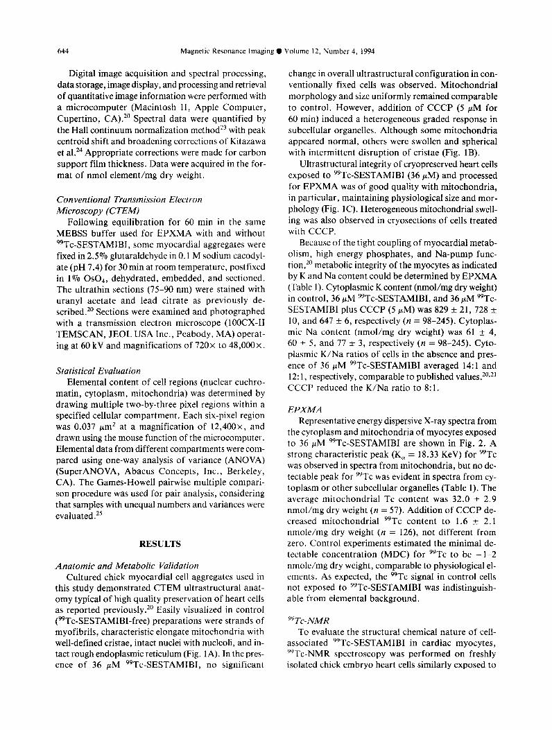
Following equilibration for 60 min in the same MEBSS buffer used for EPXMA with and without 99Tc-SESTAMIBI, some myocardial aggregates were fixed in 2.5% glutaraldehyde in 0.1 M sodium cacodyl- ate (pH 7.4) for 30 min at room temperature, postfixed in 1% 0s04, dehydrated, embedded, and sectioned. The ultrathin sections (75-90 nm) were stained with uranyl acetate and lead citrate as previously de- scribed.” Sections were examined and photographed with a transmission electron microscope (lOOCX-II TEMSCAN, JEOL USA Inc., Peabody, MA) operat- ing at 60 kV and magnifications of 720x to 48,000~.
Statistical Evaluation Elemental content of cell regions (nuclear euchro-
matin, cytoplasm, mitochondria) was determined by drawing multiple two-by-three pixel regions within a specified cellular compartment. Each six-pixel region was 0.037 pm2 at a magnification of 12,400x, and drawn using the mouse function of the microcomputer. Elemental data from different compartments were com- pared using one-way analysis of variance (ANOVA) (SuperANOVA, Abacus Concepts, Inc., Berkeley, CA). The Games-Howell pairwise multiple compari- son procedure was used for pair analysis, considering that samples with unequal numbers and variances were evaluated.25
RESULTS
Anatomic and Metabolic Validation Cultured chick myocardial cell aggregates used in
this study demonstrated CTEM ultrastructural anat- omy typical of high quality preservation of heart cells as reported previously. ” Easily visualized in control (99Tc-SESTAMIBI-free) preparations were strands of myofibrils, characteristic elongate mitochondria with well-defined cristae, intact nuclei with nucleoli, and in- tact rough endoplasmic reticulum (Fig. 1A). In the pres- ence of 36 PM 99Tc-SESTAMIBI, no significant
change in overall ultrastructural configuration in con- ventionally fixed cells was observed. Mitochondrial morphology and size uniformly remained comparable to control. However, addition of CCCP (5 PM for 60 min) induced a heterogeneous graded response in subcellular organelles. Although some mitochondria appeared normal, others were swollen and spherical with intermittent disruption of cristae (Fig. 1B).
Ultrastructural integrity of cryopreserved heart cells exposed to 99Tc-SESTAMIBI (36 PM) and processed for EPXMA was of good quality with mitochondria, in particular, maintaining physiological size and mor- phology (Fig. 1C). Heterogeneous mitochondrial swell- ing was also observed in cryosections of cells treated with CCCP.
Because of the tight coupiing of myocardial metab- olism, high energy phosphates, and Na-pump func- tion,” metabolic integrity of the myocytes as indicated by K and Na content could be determined by EPXMA (Table 1). Cytoplasmic K content (nmol/mg dry weight) in control, 36 PM 99Tc-SESTAMIBI, and 36 FM 99Tc- SESTAMIBI plus CCCP (5 PM) was 829 + 21, 728 f 10, and 647 f 6, respectively (n = 98-245). Cytoplas- mic Na content (nmol/mg dry weight) was 61 k 4, 60 k 5, and 77 f 3, respectively (n = 98-245). Cyto- plasmic K/Na ratios of cells in the absence and pres- ence of 36 PM 99Tc-SESTAMIBI averaged 14:l and 12: 1, respectively, comparable to published values.20,2’ CCCP reduced the K/Na ratio to 8: 1.
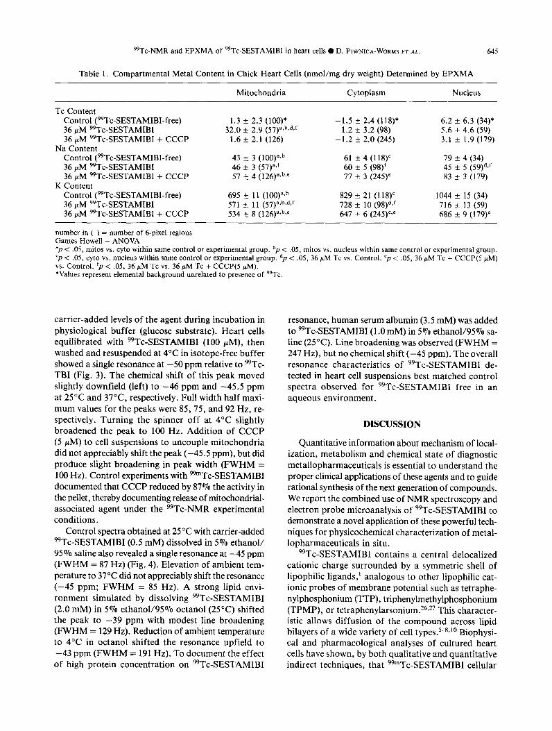
EPXMA Representative energy dispersive X-ray spectra from
the cytoplasm and mitochondria of myocytes exposed to 36 PM 99Tc-SESTAMIBI are shown in Fig. 2. A strong characteristic peak (K, = 18.33 KeV) for 99Tc was observed in spectra from mitochondria, but no de- tectable peak for 99Tc was evident in spectra from cy- toplasm or other subcellular organelles (Table 1). The average mitochondrial Tc content was 32.0 + 2.9 nmol/mg dry weight (n = 57). Addition of CCCP de- creased mitochondrial 99Tc content to 1.6 + 2.1 nmole/mg dry weight (n = 126), not different from zero. Control experiments estimated the minimal de- tectable concentration (MDC) for 99Tc to be -1-2 nmole/mg dry weight, comparable to physiological el- ements. As expected, the 99Tc signal in control cells not exposed to 99Tc-SESTAMIBI was indistinguish- able from elemental background.
yyTc-NMR To evaluate the structural chemical nature of ceII-
associated 99Tc-SESTAMIBI in cardiac myocytes, ‘“Tc-NMR spectroscopy was performed on freshly isolated chick embryo heart cells similarly exposed to
99Tc-NMR and EPXMA of 99Tc-SESTAMIBI in heart cells 0 D. PIWNICA-WORMS ET AL. 645
Table 1. Compartmental Metal Content in Chick Heart Cells (nmol/mg dry weight) Determined by EPXMA
Mitochondria Cytoplasm Nucleus
Tc Content Control (99Tc-SESTAMIBI-free) 36 ILM 99Tc-SESTAMIBI 36 PM 99Tc-SESTAMIBI + CCCP
Na Content
1.3 + 2.3 (lOO)* 32.0 f 2.9 (57)a,b,d,f
1.6 + 2.1 (126)
Control (99Tc-SESTAMIBI-free) 36 PM 99Tc-SESTAMIBI 36 FM 99Tc-SESTAMIBI + CCCP
K Content
43 + 3 (KKy~b 46 f 3 (57)“~~ 57 + 4 (126)a*b~’
Control (99Tc-SESTAMIBI-free) 695 + 11 (lOO)axb 36 PM 99Tc-SESTAMIBI 571 5 11 (57)avb,d,f 36 PM 99Tc-SESTAMIBI + CCCP 534 t 8 (126)a*b%’
-1.5 f 2.4 (118)* 1.2 f 3.2 (98)
- 1.2 + 2.0 (245)
61 f 4 (l18)c 60 + 5 (98)f 77 + 3 (245)’
829 + 21 (118)’ 728 + 10 (98)dvf 647 + 6 (245)c.”
6.2 + 6.3 (34)* 5.6 f 4.6 (59) 3.1 & 1.9 (179)
79 * 4 (34) 45 Z!? 5 (59)dJ 83 k 3 (179)
1044 * 15 (34) 716 -+ 13 (59) 686 + 9 (179)’
number in ( ) = number of 6-pixel regions Games-Howell - ANOVA “p < .05, mitos vs. cyto within same control or experimental group. “p < .05, mitos vs. nucleus within same control or experimental group. ‘p < .05, cyto vs. nucleus within same control or experimental group. dp < .05, 36 PM Tc vs. Control. “p < .05, 36 PM Tc + CCCP(5 PM) vs. Control. ‘p < .05, 36 pM Tc vs. 36 flLM Tc + CCCP(5 NM). *Values represent elemental background unrelated to presence of 99Tc.
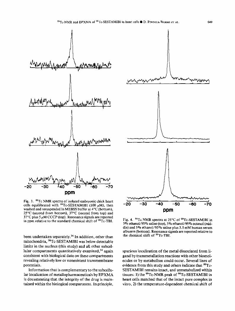
carrier-added levels of the agent during incubation in physiological buffer (glucose substrate). Heart cells equilibrated with 99Tc-SESTAMIBI (100 FM), then washed and resuspended at 4°C in isotope-free buffer showed a single resonance at -50 ppm relative to %Tc- TBI (Fig. 3). The chemical shift of this peak moved slightly downfield (left) to -46 ppm and -45.5 ppm at 25°C and 37”C, respectively. Full width half maxi- mum values for the peaks were 85,75, and 92 Hz, re- spectively. Turning the spinner off at 4°C slightly broadened the peak to 100 Hz. Addition of CCCP (5 PM) to cell suspensions to uncouple mitochondria did not appreciably shift the peak (-45.5 ppm), but did produce slight broadening in peak width (FWHM = 100 Hz). Control experiments with 99”Tc-SESTAMIBI documented that CCCP reduced by 87% the activity in the pellet, thereby documenting release of mitochondrial- associated agent under the 99Tc-NMR experimental conditions.
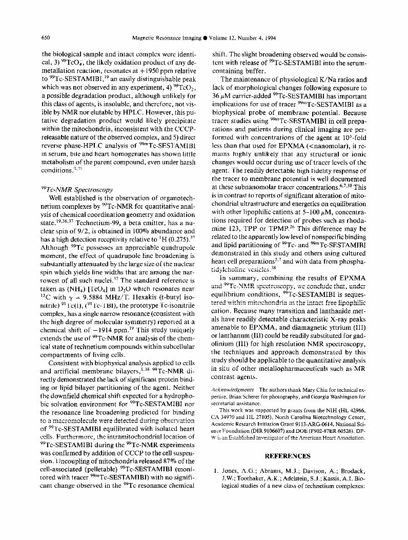
Control spectra obtained at 25°C with carrier-added *Tc-SESTAMI B I ( 0.5 mM) dissolved in 5% ethanol/ 95% saline also revealed a single resonance at -45 ppm (FWHM = 87 Hz) (Fig. 4). Elevation of ambient tem- perature to 37°C did not appreciably shift the resonance (-45 ppm; FWHM = 85 Hz). A strong lipid envi- ronment simulated by dissolving 99Tc-SESTAMIBI (2.0 mM) in 5% ethanol/95% octanol (25°C) shifted the peak to -39 ppm with modest line broadening (FWHM = 129 Hz). Reduction of ambient temperature to 4°C in octanol shifted the resonance upfield to -43 ppm (FWHM = 191 Hz). To document the effect of high protein concentration on 99Tc-SESTAMIBI
resonance, human serum albumin (3.5 mM) was added to 99Tc-SESTAMIBI (1 .O mM) in 5 % ethanol/95% sa- line (25°C). Line broadening was observed (FWHM = 247 Hz), but no chemical shift (-45 ppm). The overall resonance characteristics of 99Tc-SESTAMIBI de- tected in heart cell suspensions best matched control spectra observed for 99Tc-SESTAMIBI free in an aqueous environment.
DISCUSSION
Quantitative information about mechanism of local- ization, metabolism and chemical state of diagnostic metallopharmaceuticals is essential to understand the proper clinical applications of these agents and to guide rational synthesis of the next generation of compounds. We report the combined use of NMR spectroscopy and electron probe microanalysis of 99Tc-SESTAMIBI to demonstrate a novel application of these powerful tech- niques for physicochemical characterization of metal- lopharmaceuticals in situ.
99Tc-SESTAMIBI contains a central delocalized cationic charge surrounded by a symmetric shell of lipophilic ligands,’ analogous to other lipophilic cat- ionic probes of membrane potential such as tetraphe- nylphosphonium (TTP), triphenylmethylphosphonium (TPMP), or tetraphenylarsonium.26,27 This character- istic allows diffusion of the compound across lipid bilayers of a wide variety of cell types.5-sJo Biophysi- cal and pharmacological analyses of cultured heart cells have shown, by both qualitative and quantitative indirect techniques, that 99mTc-SESTAMIBI cellular
Magnetic Resonance Imaging 0 Volume 12, Number 4, 1994
Fig. lA,B. Conventional transmission electron micrographs of cultured embryonic chick heart cells chemically fixed in glu- taraldehyde following 60 min exposure to MEBSS (A) and to 36 PM 99Tc-SESTAMIBI plus 5 PM CCCP (B). M, mitochondria; N, nucleus; Ly, lysosome; My, myofibrils. Arrowhead: swollen mitochondria with disrupted cristae. Bar = 1 pm.
transport follows a three compartment series model in for mitochondrial sequestration of the agent.‘p7 Thus, which the plasma membrane and mitochondrial inner for the purpose of validating NMR and EPXMA with membrane are the primary transport barriers and that a metallopharmaceutical, the mitochondrial-specific transmembrane potentials provide the net driving force localization of 99Tc-SESTAMIBI was thought to en-

99Tc-NMR and EPXMA of 99Tc-SESTAMIBI in heart cells 0 D. PIWNICA-WORMS ET AL. 647
Fig. 1C. Scanning transmission electron micrograph of cryosection of cultured embryonic chick heart cells rapidly frozen in liquid propane following 60 min exposure to 36 PM “Tc-SESTAMIBI. Note that all contrast in the cryosection arises from mass density differences; no fixatives or stains are used in the preparation. M, mitochondria; Cy, cytoplasm; N, nucleus. (x9,500).
hance the probability of detecting a signal within the relevant compartment by these techniques.
EPXMA EPXMA can generally localize elements of atomic
number eleven or greater within cells with a spatial resolution approaching 200 A2* Used extensively for elemental analysis of physiological ions within sub- cellular compartments, this technology combined with ultrafast cyropreservation has contributed to under- standing the distribution of Na, K, Mg, Cl and Ca within tissue in situ under conditions of steady-state, hypoxia or metabolic injury.29m31 Characteristic ener- gies for Tc are 2.42 (L,) and 18.33 (K,) KeV.32 Thus, poised well away from other biological elements, the characteristic K, X-ray peak of 99Tc allowed facile direct microanalysis of the subcellular location of 99Tc-SESTAMIBI.
EPXMA analysis directly confirmed the mitochon- drial localization of 99Tc-SESTAMIBI predicted by the series model of lipophilic cation transport. The model predicts that E,,, should first concentrate 99Tc-SESTAMIBI within the cytoplasm in proportion
to the driving force of the time-averaged membrane potential (TAP) in these spontaneously beating prepa- rations of heart cells.* Electrophysiological measure- ment of TAP in similar preparations of cultured chick heart cells has determined a value of -41 mV.33 There- fore, at 36 PM extracellular [99Tc-SESTAMIBI], a TAP of -41 mV would theoretically produce a Nern- stian-dependent cytoplasmic concentration for 99Tc- SESTAMIBI of 175 PM. Assuming a cell water space of 6.9 pl/mg protein and 0.65 mg protein/mg dry weight as previously determined in cultured heart cell prep- arations, cytosolic 99Tc-SESTAMIBI would be 0.8 nmol/mg dry weight, below typical minimal detectable limits of elements by EPXMA.31 This points to a po- tential limitation of EPXMA for use with metallophar- maceuticals, that is, the agent must attain a content within the relevant compartment of - 1 nmole/mg dry weight. Nonetheless, the fact that 99Tc-SESTAMIBI was observed in mitochondria at - 1 ,OOO-fold excess over extracellular concentrations is consistent with bio- physical expectations for mitochondrial loading by a series configuration of Em and A$.5*7*26 Full analysis of A$ in situ by EPXMA and related ionic changes has
Magnetic Resonance Imaging 0 Volume 12, Number 4, 1994 648
(A) 8
7
6
5
0.8
0.6
0.4
0.2
0
K
I Ca
8 12 KeV
16 20
1, 1. 9. I(.”
I
Tc K a
0 4 8 12 16 20 KeV
Fig. 2. (A) Energy dispersive X-ray spectrum from cytoplasm of cells illustrated in Fig. 1C; vertical axis, X-ray counts in arbitrary units; horizontal axis, energy in KeV, 0 to 20. No peaks for technetium (Tc) are evident at the characteristic energies of 2.42 (L,) or 18.33 (K,). Inset shows Tc peak region at 18.33 KeV expanded 5 times for clarity. Other peaks do occur for physiologically relevant elements Na, Mg, P, S, Cl, K, Ca (labeled) and for specimen support materials, Cu, Ni (unlabeled). (B) Energy dispersive X-ray spectrum from mitochondria of cells illustrated in Fig. 1C. A peak for Tc occurs at the characteris- tic energy of 18.33 KeV (K,). This peak is representative of a Tc content of -50 nmol/mg dry weight. Inset shows Tc peak region x5 to illustrate resolution above background.
99Tc-NMR and EPXMA of 99Tc-SESTAMIBI in heart cells 0 D. PIWNICA-WORMS ET AL. 649
wm Fig. 3. 99Tc NMR spectra of isolated embryonic chick heart cells equilibrated with 99Tc-SESTAMIBI (100 PM), then washed and resuspended in MEBSS buffer at 4°C (bottom), 25°C (second from bottom), 37°C (second from top) and 37°C plus 5 PM CCCP (top). Resonance signals are reported in ppm relative to the standard chemical shift of *Tc-TBI.
been undertaken separately.34 In addition, other than mitochondria, WTc-SESTAMIBI was below detectable limits in the nucleus (this study) and all other subcel- lular compartments quantitatively examined,34 again consistent with biological data on these compartments revealing relatively low or nonexistent transmembrane potentials.
Information that is complementary to the subcellu- lar localization of metallopharmaceuticals by EPXMA is documenting that the integrity of the drug is main- tained within the biological compartment. In principle,
pm Fig 4. 99Tc NMR spectra at 25°C of 99Tc-SESTAMIBI in 5%‘ethano1/95% saline (top), 5% ethanol/M% octanol (mid- dle) and 5% ethanol/95% saline plus 3.5 mM human serum albumin (bottom). Resonance signals are reported relative to the chemical shift of 99Tc-TBI.
spurious localization of the metal dissociated from li- gand by transmetallation reactions with other biomol- ecules or by metabolism could occur. Several lines of evidence from this study and others indicate that 99Tc- SESTAMIBI remains intact, and unmetabolized within tissues: 1) the WTc-NMR peak of *Tc-SESTAMIBI in heart cells matched that of the intact pure complex in vitro, 2) the temperature-dependent chemical shift of
650 Magnetic Resonance Imaging 0 Volume 12, Number 4, 1994
the biological sample and intact complex were identi- cal, 3) 99T~0;, the likely oxidation product of any de- metallation reaction, resonates at + 1950 ppm relative to 99Tc-SESTAMIBl,‘9 an easily distinguishable peak which was not observed in any experiment, 4) 99Tc02, a possible degradation product, although unlikely for this class of agents, is insoluble, and therefore, not vis- ible by NMR nor elutable by HPLC. However, this pu- tative degradation product would likely precipitate within the mitochondria, inconsistent with the CCCP- releasable nature of the observed complex, and 5) direct reverse phase-HPLC analysis of 99mTc-SESTAMIBl in serum, bile and heart homogenates has shown little metabolism of the parent compound, even under harsh conditions.9,35
99Tc-NA4R Spectroscopy Well established is the observation of organotech-
netium complexes by %Tc-NMR for quantitative anal- ysis of chemical coordination geometry and oxidation state.19v36z37 Technetium-99, a beta emitter, has a nu- clear spin of 9/2, is obtained in 100% abundance and has a high detection receptivity relative to ] H (0.275).37 Although 99Tc possesses an appreciable quadrupole moment, the effect of quadrupole line broadening is substantially attenuated by the large size of the nuclear spin which yields line widths that are among the nar- rowest of all such nuclei. 37 The standard reference is taken as (NH,) [TcO,] in DzO which resonates near 13C with y = 9.5884 MHz/T. Hexakis (t-butyl iso- nitrile) 99Tc(I), (99Tc-TBI), the prototype Tc-isonitrile complex, has a single narrow resonance (consistent with the high degree of molecular symmetry) reported at a chemical shift of -1914 ppm.” This study uniquely extends the use of 99Tc-NMR for analysis of the chem- ical state of technetium compounds within subcellular compartments of living cells.
Consistent with biophysical analysis applied to cells and artificial membrane bilayers,‘s3* 99Tc-NMR di- rectly demonstrated the lack of significant protein bind- ing or lipid bilayer partitioning of the agent. Neither the downfield chemical shift expected for a hydropho- bic solvation environment for 99Tc-SESTAMIBI nor the resonance line broadening predicted for binding to a macromolecule were detected during observation of 99Tc-SESTAMIBI equilibrated with isolated heart cells. Furthermore, the intramitochondrial location of 99Tc-SESTAMIBI during the 99Tc-NMR experiments was confirmed by addition of CCCP to the cell suspen- sion. Uncoupling of mitochondria released 87% of the cell-associated (pelletable) 99Tc-SESTAMIBI (moni- tored with tracer 99mTc-SESTAMIBI) with no signifi- cant change observed in the 99Tc resonance chemical
shift. The slight broadening observed would be consis- tent with release of 99Tc-SESTAMIBI into the serum- containing buffer.
The maintenance of physiological K/Na ratios and lack of morphological changes following exposure to 36 PM carrier-added 99Tc-SESTAMIBI has important implications for use of tracer 99mTc-SESTAMIBI as a biophysical probe of membrane potential. Because tracer studies using 99mTc-SESTAMIBI in cell prepa- rations and patients during clinical imaging are per- formed with concentrations of the agent at 105-fold less than that used for EPXMA (cnanomolar), it re- mains highly unlikely that any structural or ionic changes would occur during use of tracer levels of the agent. The readily detectable high fidelity response of the tracer to membrane potential is well documented at these subnanomolar tracer concentrations.6,7,38 This is in contrast to reports of significant alteration of mito- chondrial ultrastructure and energetics on equilibration with other lipophilic cations at 5-100 PM, concentra- tions required for detection of probes such as rhoda- mine 123, TPP or TPMP.26 This difference may be related to the apparently low level of nonspecific binding and lipid partitioning of 99Tc- and 99mTc-SESTAMIBI demonstrated in this study and others using cultured heart cell preparations5,’ and with data from phospha- tidylcholine vesicles.“’
In summary, combining the results of EPXMA and ““Tc-NMR spectroscopy, we conclude that, under equilibrium conditions, 99Tc-SESTAMIBI is seques- tered within mitochondria as the intact free lipophilic cation. Because many transition and lanthanide met- als have readily detectable characteristic X-ray peaks amenable to EPXMA, and diamagnetic yttrium (III) or lanthanum (III) could be readily substituted for gad- olinium (III) for high resolution NMR spectroscopy, the techniques and approach demonstrated by this study should be applicable to the quantitative analysis in situ of other metallopharmaceuticals such as MR contrast agents.
Acknowledgments-The authors thank Mary Chiu for technical ex- pertise, Brian Scherer for photography, and Georgia Washington for
secretarial assistance.
This work was supported by grants from the NIH (HL 42966, CA 34970 and HL 27105), North Carolina Biotechnology Center, Academic Research Initiation Grant 9113-ARC-0614, National Sci- ence Foundation (DIR 9106607) and DOE (F902-87ER 60526). DP- W is an Established Investigator of the American Heart Association.
REFERENCES
1. Jones, A.G.; Abrams, M.J.; Davison, A.; Brodack, J.W.; Toothaker, A-K.; Adelstein, S.J.; Kassis, A.I. Bio- logica studies of a new class of technetium complexes:
99Tc-NMR and EPXMA of 99Tc-SESTAMIBI in heart cells 0 D. PIWNICA-WORMS ET AL. 651
the hexakis (alkylisonitrile) technetium (I) cations. Int. J. Nucl. Med. Biol. 11~225-234; 1984.
2. Wackers, F. J.; Berman, D.S.; Maddahi, J.; et al. Tc%m- hexakis 2-methoxy isobutylisonitrile: human biodistri- bution, dosimetry, safety, and preliminary comparison to thallium-201 for myocardial perfusion imaging. J. Nucl. Med. 30:301-311; 1989.
3. Rocco, T.P.; Dilsizian, V.; Strauss, H. W.; Boucher, C.A. Technetium-99m-isonitrile myocardial uptake at rest. II. Relation to clinical markers of potential viability. J. Am. CON. Cardiol. 14:1678-1684; 1989.
5. Piwnica-Worms, D.; Kronauge, J.F.; Chiu, M.L. Uptake and retention of hexakis (2-methoxy isobutyl isonitrile) technetium(I) in cultured chick myocardial cells: mito- chondrial and plasma membrane potential dependence. Circ. 82:1826-1838; 1990.
4. Leppo, J.A.; Meerdink, D.J. Comparison of the myo- cardial uptake of a technetium-labeled isonitrile analogue and thallium. Circ. Res. 65:632-639; 1989.
6. Chiu, M.L.; Kronauge, J.F.; Piwnica-Worms, D. Effect of mitochondrial and plasma membrane potentials on accumtdation of hexakis (Zmethoxyisobutyl isonitrile) technetium(I) in cultured mouse fibroblasts. J. Nucl. Med. 31:1646-1653; 1990.
7. Piwnica-Worms, D.; Kronauge, J.F.; Chiu, M.L. En- hancement by tetraphenylborate of 99mTc-MIBI uptake kinetics and accumulation in cultured chick heart cells. J. Nucl. Med. 32:1992-1999; 1991.
8. Piwnica-Worms, D.; Chiu, M.L.; Kronauge, J.F. Diver- gent kinetics of Tl-201 and Tc-99mSESTAMIBI in cul- tured chick ventricular myocytes during ATP depletion. Circ. 85:1531-1541; 1992.
9. Carvalho, P.A.; Chiu, M.L.; Kronauge, J.F.; Kawamura, M.; Jones, A.G.; Holman, B.L.; Piwnica-Worms, D. Subcellular distribution and analysis of technetium%m- MIBI in isolated perfused rat heart. J. Nucl. Med. 33: 1516-1521; 1992.
10. Delmon-Moingeon, L.I.; Piwnica-Worms, D.; Van den Abbeele, A.D.; Holman, B.L.; Davison, A.; Jones, A.G. Uptake of the cation hexakis (2-methoxy isobutyliso- nitrile) technetium-99m by human carcinoma cell lines in vitro. Cancer Res. 50:2198-2202; 1990.
11. Beanlands, R.; Dawood, F.; Wen, W.-H.; McLaughlin, P.R.; Butany, J.; Damati, G.; Liu, P.P. Are the kinetics of Tc-99m-methoxy isobutylisonitrile affected by cell metabolism and viability? Circulation 82: 1802-1814; 1990.
12. Crane, P.; Laliberte, R.; Heminway, S.; Thoolen, M.; Orlandi, C. Effect of mitochondrial viability and metab- olism on technetium-99m-sestamibi myocardial reten- tion. Europ. J. Nut. Med. 20~20-25; 1993.
13. Sinusas, A. J.; Trautman, K.A.; Bergin, J.D.; Watson, D.D.; Smith, W.H.; Beller, G. Quantification of area at risk during coronary occlusion and degree of myocardial salvage after reperfusion with Tc-99m methoxyisobutyl- isonitrile. Circtdation 82:1424-1437; 1990.
14. Piwnica-Worms, D.; Kronauge, J.F.; Holman, B.L.; Davison, A.; Jones, A.G. Comparative myocardial up-
take characteristics of hexakis (alkylisonitrile) techne- tium(1) complexes: effect of lipophilicity. Invest. Radiol. 24125-29; 1989.
15. Maublant, J.C.; Gachon, P.; Moins, N. Hexakis (2-meth- oxyisobutyl isonitrile) technetium99m and thallium-201 chloride: Uptake and release in cultured myocardial cells. J. Nucl. Med. 29:48-54; 1988.
16. Ebihara, L.; Shigeto, N.; Lieberman, M.; Johnson, E.A. The initial inward current in spherical clusters of chick heart cells. J. Gen. Physiol. 75:437-456; 1980.
17. Lieberman, M.; Manasek, F.J.; Swanobori, T.; Johnson, E.A. Cytochalasin B: its morphological and electrophys- iological actions on synthetic strands of cardiac muscle. Dev. Biol. 31:380-403; 1973.
18. Kronauge, J.F.; Davison, A.; Roseberry, A.M.; Costello, C.E.; Maleknia, S.; Jones, A.G. Synthesis and identi- fication of monocation Tc(CP1): in Tc(CNC(CH,)* COOCH&Cl and its hydrolysis products. Inorg. Chem. 30:4265-4271; 1991.
19. Davison, A.; Kronauge, J.F.; Jones, A.G.; Pearlstein, R.M.; Thornbuck, J.R. 99-Tc NMR spectroscopy of technetium(I) phosphine and phosphite complexes. In- org. Chem. 2713245-3246; 1988.
20. LeFurgey, A.; Ingram, P.; Lieberman, M. Quantitative microchemical imaging of calcium in Na-K pump inhib- ited heart cells. Cell Calcium 9:219-235; 1988.
21. LeFurgey, A.; Davilla, SD; Kopf, D.A.; Sommer, J.R. ; Ingram, P. Real-time quantitative elemental analysis and mapping: microchemical imaging in cell physiology. J. Microscopy 165:191-223; 1992.
22. Cantino, M.E.; Wilkinson, L.E.; Goddard, M.K.; John- son, D.E. Beam induced mass loss in high resolution bio- logical microscopy. J. Microscopy 144:317-327; 1986.
23. Hall, T.A. Biological x-ray microanalysis. J. Microscopy 117:145-163; 1979.
24. Kitazawa, T.; Shuman, H.; Somlyo, A.P. Quantitative electron probe analysis: problems and solutions. Ultra- microscopy 11:251-262; 1983.
25. Games, P.A.; Howell, J.F. Pairwise multiple comparison procedures with unequal N’s and/or variances: a Monte Carlo study. J. Educ. Stat. 1:113-125; 1976.
26. Ritchie, R.J. A critical assessment of the use of lipophilic cations as membrane potential probes. Prog. Biophys. Molec. Biol. 43:1-32; 1984.
27. Flewelling, R.F.; Hubbell W.L. The membrane dipole po- tential in a total membrane potential model: applications to hydrophobic ion interactions with membranes. Bio- phys. J. 49:541-552; 1986.
28. McDowall, A.W.; Chang, J.J.; Freeman, R.; LePault, J.; Walter, C.A.; Dubochet, J. Electron microscopy of frozen hydrated sections of vitreous ice and vitrified bio- logical samples. J. Microscopy 13 1: l-9; 1983.
29. Shuman, H.; Somlyo, A.V.; Somlyo, A.P. Quantitative electron probe microanalysis of biological thin sections: methods and validity. Utramicroscopy 1:3 17-339; 1976.
30. Morgan, A. J. X-ray microanalysis in electron microscopy for biologists. Oxford: Oxford University Press; 1985.
31. LeFurgey, A.; Bond, M.; Ingram, P. Frontiers in elec-
652 Magnetic Resonance Imaging 0 Volume 12, Number 4, 1994
tron probe microanalysis: application to cell physiology. Ultramicroscopy 24:185-220; 1988.
32. Larkins, F.P. Atomic Data and Nuclear Data Tables, 20, 1977.
33. Horres, C.R.; Aiton, J.F.; Lieberman, M. Potassium per-
meability of embryonic avian heart cells in tissue culture. Am. J. Physiol. (Cell) 236:Cl63-170; 1979.
34. Backus, M.; Piwnica-Worms, D.; Hackett, D.; Kro-
nauge, J.; Lieberman, M.; Ingram, P.; LeFurgey, A. Mi- croprobe analysis of Tc-MIBI in heart cells: calculation of mitochondrial potential. Am. J. Physiol. (Cell) 265: Cl78-C187; 1993.
35. Kronauge, J.F.; Kawamura, M.; Lepisto, E.; Holman,
B.L.; Davison, A.; Jones, A.G.; Costello, C.E.; Zeng,
C.-H. Metabolic studies of the myocardial perfusion agent Tc-(MIBI). In Nicolini, M., Bandoli, G., Mazzi,
U. (Eds). Technetium and Rhenium in Chemistry and Nuclear Medicine. Cortina Internatl; Verona; 1990: pp.
677-682. 36. Kidd, R.G. Technetium-99 NMR and rationalized quad-
rupole moment values for transition metal nuclei. J. Magn. Reson. 45:88-93; 1981.
37. Franklin, K.J.; Lock, C.J.L.; Sayer, B.G.; Schrobligen,
G. J. Chemical applications of 99-Tc NMR spectroscopy: preparation of novel Tc(VI1) species and their charac- terization by multinuclear NMR spectroscopy. J. Am. Chem. Sot. 104:5303-5306; 1982.
38. Chernoff, D.M.; Strichartz, G.R.; Piwnica-Worms, D.
Membrane potential determination in large unilamel-
lar vesicles with hexakis(2-methoxyisobutyl isonitrile) technetium(I). Biochim. Biophys. Acta 1147:262-266;
1993.
Copyright © 2022 FDOKUMEN