
miRNAs in Lymphocytic Leukaemias—The miRror of Drug ...
26
Citation: Sbirkov, Y.; Vergov, B.; Mehterov, N.; Sarafian, V. miRNAs in Lymphocytic Leukaemias—The miRror of Drug Resistance. Int. J. Mol. Sci. 2022, 23, 4657. https://doi.org/ 10.3390/ijms23094657 Academic Editor: Marta Coscia Received: 1 April 2022 Accepted: 21 April 2022 Published: 22 April 2022 Publisher’s Note: MDPI stays neutral with regard to jurisdictional claims in published maps and institutional affil- iations. Copyright: © 2022 by the authors. Licensee MDPI, Basel, Switzerland. This article is an open access article distributed under the terms and conditions of the Creative Commons Attribution (CC BY) license (https:// creativecommons.org/licenses/by/ 4.0/). International Journal of Molecular Sciences Review miRNAs in Lymphocytic Leukaemias—The miRror of Drug Resistance Yordan Sbirkov 1,2, *, Bozhidar Vergov 1 , Nikolay Mehterov 1,2 and Victoria Sarafian 1,2, * 1 Department of Medical Biology, Medical University of Plovdiv, 4002 Plovdiv, Bulgaria; [email protected] (B.V.); [email protected] (N.M.) 2 Division of Molecular and Regenerative Medicine, Research Institute at Medical University of Plovdiv, 4002 Plovdiv, Bulgaria * Correspondence: [email protected] (Y.S.); victoria.sarafi[email protected] (V.S.) Abstract: Refractory disease and relapse remain the main causes of cancer therapy failure. Refined risk stratification, treatment regimens and improved early diagnosis and detection of minimal residual disease have increased cure rates in malignancies like childhood acute lymphoblastic leukaemia (ALL) to 90%. Nevertheless, overall survival in the context of drug resistance remains poor. The regulatory role of micro RNAs (miRNAs) in cell differentiation, homeostasis and tumorigenesis has been under extensive investigation in different cancers. There is accumulating data demonstrating the significance of miRNAs for therapy outcomes in lymphoid malignancies and some direct demonstrations of the interplay between these small molecules and drug response. Here, we summarise miRNAs’ impact on chemotherapy resistance in adult and paediatric ALL and chronic lymphocytic leukaemia (CLL). The main focus of this review is on the modulation of particular signaling pathways like PI3K-AKT, transcription factors such as NF-κB, and apoptotic mediators, all of which are bona fide and pivotal elements orchestrating the survival of malignant lymphocytic cells. Finally, we discuss the attractive strategy of using mimics, antimiRs and other molecular approaches pointing at miRNAs as promising therapeutic targets. Such novel strategies to circumvent ALL and CLL resistance networks may potentially improve patients’ responses and survival rates. Keywords: miRNA; drug resistance; lymphocytic leukaemia 1. Introduction Cell function and fate are ultimately determined by the type and amount of proteins produced in the cell and their post-translational modifications. Control over protein syn- thesis is exerted at multiple levels through epigenetics, transcription and mRNA export regulation, stability, post-transcriptional modifications, fine-tuning of the translational machinery, including ribosomes, initiation factors, etc. Importantly, a complex system at the interface between transcription and translation further orchestrates the cell’s proteome– micro RNA (miRNAs) dynamics. Ranging from merely ~20 to 25 nucleotides in length, miRNAs can be transcribed from both intra- and inter-genic regions, after which they are processed in a tightly regulated step-wise manner to ultimately control mRNA degradation, decay or even to determine rates of translation [1]. The mechanisms of miRNA biogen- esis and control of translation (and even of transcription through promotor binding) are remarkably versatile and have been extensively reviewed [1,2]. Currently, there are more than 2500 confirmed miRNAs in the human genome, and it is estimated that they may regulate at least one-third of our mRNAs (or even up to 15,000 genes) [3–5]. The picture’s complexity is further rendered because tens of miRNAs may target one gene, and one miRNA could have tens to hundreds of mRNA clients [5,6]. Of note, miRNAs can be specifically targeted by cancer cells, as it has been shown that a large proportion of these short regulatory oligonucleotides are encoded in fragile sites in the Int. J. Mol. Sci. 2022, 23, 4657. https://doi.org/10.3390/ijms23094657 https://www.mdpi.com/journal/ijms
-
Upload
khangminh22 -
Category
Documents
-
view
4 -
download
0
Transcript of miRNAs in Lymphocytic Leukaemias—The miRror of Drug ...

Citation: Sbirkov, Y.; Vergov, B.;
Mehterov, N.; Sarafian, V. miRNAs in
Lymphocytic Leukaemias—The
miRror of Drug Resistance. Int. J. Mol.
Sci. 2022, 23, 4657. https://doi.org/
10.3390/ijms23094657
Academic Editor: Marta Coscia
Received: 1 April 2022
Accepted: 21 April 2022
Published: 22 April 2022
Publisher’s Note: MDPI stays neutral
with regard to jurisdictional claims in
published maps and institutional affil-
iations.
Copyright: © 2022 by the authors.
Licensee MDPI, Basel, Switzerland.
This article is an open access article
distributed under the terms and
conditions of the Creative Commons
Attribution (CC BY) license (https://
creativecommons.org/licenses/by/
4.0/).
International Journal of
Molecular Sciences
Review
miRNAs in Lymphocytic Leukaemias—The miRror ofDrug ResistanceYordan Sbirkov 1,2,*, Bozhidar Vergov 1, Nikolay Mehterov 1,2 and Victoria Sarafian 1,2,*
1 Department of Medical Biology, Medical University of Plovdiv, 4002 Plovdiv, Bulgaria;[email protected] (B.V.); [email protected] (N.M.)
2 Division of Molecular and Regenerative Medicine, Research Institute at Medical University of Plovdiv,4002 Plovdiv, Bulgaria
* Correspondence: [email protected] (Y.S.); [email protected] (V.S.)
Abstract: Refractory disease and relapse remain the main causes of cancer therapy failure. Refinedrisk stratification, treatment regimens and improved early diagnosis and detection of minimal residualdisease have increased cure rates in malignancies like childhood acute lymphoblastic leukaemia (ALL)to 90%. Nevertheless, overall survival in the context of drug resistance remains poor. The regulatoryrole of micro RNAs (miRNAs) in cell differentiation, homeostasis and tumorigenesis has been underextensive investigation in different cancers. There is accumulating data demonstrating the significanceof miRNAs for therapy outcomes in lymphoid malignancies and some direct demonstrations of theinterplay between these small molecules and drug response. Here, we summarise miRNAs’ impacton chemotherapy resistance in adult and paediatric ALL and chronic lymphocytic leukaemia (CLL).The main focus of this review is on the modulation of particular signaling pathways like PI3K-AKT,transcription factors such as NF-κB, and apoptotic mediators, all of which are bona fide and pivotalelements orchestrating the survival of malignant lymphocytic cells. Finally, we discuss the attractivestrategy of using mimics, antimiRs and other molecular approaches pointing at miRNAs as promisingtherapeutic targets. Such novel strategies to circumvent ALL and CLL resistance networks maypotentially improve patients’ responses and survival rates.
Keywords: miRNA; drug resistance; lymphocytic leukaemia
1. Introduction
Cell function and fate are ultimately determined by the type and amount of proteinsproduced in the cell and their post-translational modifications. Control over protein syn-thesis is exerted at multiple levels through epigenetics, transcription and mRNA exportregulation, stability, post-transcriptional modifications, fine-tuning of the translationalmachinery, including ribosomes, initiation factors, etc. Importantly, a complex system atthe interface between transcription and translation further orchestrates the cell’s proteome–micro RNA (miRNAs) dynamics. Ranging from merely ~20 to 25 nucleotides in length,miRNAs can be transcribed from both intra- and inter-genic regions, after which they areprocessed in a tightly regulated step-wise manner to ultimately control mRNA degradation,decay or even to determine rates of translation [1]. The mechanisms of miRNA biogen-esis and control of translation (and even of transcription through promotor binding) areremarkably versatile and have been extensively reviewed [1,2].
Currently, there are more than 2500 confirmed miRNAs in the human genome, andit is estimated that they may regulate at least one-third of our mRNAs (or even up to15,000 genes) [3–5]. The picture’s complexity is further rendered because tens of miRNAsmay target one gene, and one miRNA could have tens to hundreds of mRNA clients [5,6].Of note, miRNAs can be specifically targeted by cancer cells, as it has been shown that alarge proportion of these short regulatory oligonucleotides are encoded in fragile sites in the
Int. J. Mol. Sci. 2022, 23, 4657. https://doi.org/10.3390/ijms23094657 https://www.mdpi.com/journal/ijms

Int. J. Mol. Sci. 2022, 23, 4657 2 of 26
genome that are frequently deleted or silenced [7]. Examples of such genomic perturbationin leukaemias include miR-15a/16-1 deletion as part of chromosome 13q14, miR-125atranslocation to IGH, and others (discussed later) [7–9].
Acute and chronic lymphocytic leukaemias (ALL and CLL) are the most common haema-tological malignancies in children and adults. ALL is a heterogeneous disease arising fromhaematopoietic blast cells characterised by incomplete differentiation, loss of function, andmutations. Interestingly, ALL has a bimodal distribution, with one peak at age 5 (80% ofALL cases) and another at 50 years of age [10]. Childhood ALL (cALL) is treated successfully,leading to ~90% cure rates. In contrast, albeit rare (<1% of all cancers), the outcome in adultALL remains poor even if therapy is closely related in both groups [11,12]. Similarly, CLL is adisease of the elderly (median age of ~70) characterised by the expansion of slowly proliferat-ing CD5/CD19 positive B cell clones. There are a number of different cytogenetic modalitiesinfluencing the progression of the disease and the treatment protocols. Therefore, the 5-yearoverall survival (OS) can range from 90% down to merely ~10% in high-risk groups withdeletions of TP53 [13,14]. While these blood cancers have different aetiologies, cytogeneticsand dynamics, they share certain similarities, especially in the treatment strategies. Resistanceto the commonly used purine analogues, alkylating agents, anthracyclines and glucocorticoidsare the main reasons for drug failure. Even though the 5-year overall survival (OS) can reach90% for childhood ALL and CLL [15,16], this percentage is only around 60% for youngeradults with ALL and just ~10% for older patients with ALL [17]. Thus, the lack of therapyresponse can turn these well-manageable diseases into daunting battles to fight. Therefore, itis urgent to study the underlying reasons for drug resistance and the potential strategies toovercome them.
The role of miRNAs in regulating normal haematopoiesis, leukaemogenesis and asdiagnostic and prognostic markers in cancer has been under extensive investigation overthe last decade. Thus, several studies have elucidated a plethora of functions of thesesmall RNAs in blood cancers [18–22]. For instance, a specific miRNA signature correlateswith prognosis and disease progression in CLL [23], or subsets of miRNAs can be used todifferentiate between childhood B cell and T cell ALL [22]. Klein et al. have demonstratedthat miR-15a/16-1, frequently deleted (in ~60%) in CLL patients, acts as tumour suppressorsby controlling cell cycle progression. Furthermore, the deletion of these miRNAs can evendrive CLL in a mouse model, while their ectopic expression can reduce cell growth in bothmouse and human cells [24]. Similarly, in in vivo models, the overexpression of miR-125b(by retroviruses or by placing the miRNA after an IGH enhancer and promoter) can induce Bcell lymphoblastic leukaemia proving the direct role of miRNAs in leukaemogenesis [25,26].
The role of miRNAs in chemoresistance in solid tumours has been well-established [27,28].Therefore, here we summarise current knowledge of the link between certain miRNAs andmolecules regulating classical cell growth, renewal and survival signaling pathways or directlymodulating the cell cycle, apoptosis and other important processes related to chemoresis-tance in acute and chronic lymphocytic leukemias. We finally discuss the perspectives ofimplementing miRNAs as therapeutic targets in the clinic.
2. Implication of miRNAs in Drug Resistance in Adult ALL
ALL in adults accounts for ~0.3% of all malignancies [12]. About 75% of cases arisefrom B cells, while the remaining 25% of patients present with T cell ALL. Mortalityrates remain high, with merely 30–40% achieving complete remission. Around 50% ofpatients eventually relapse, and there is only 10% 5-year survival with refractory/relapsedisease [29]. Such poor treatment outcomes may be due to age-related intolerance tointensified therapy, the accumulation of mutations and higher in vitro drug resistancecompared to childhood ALL, where cure rates reach 90% [30]. Adult ALL shows great cyto-genetic heterogeneity, with subsets harbouring translocations like t(4;11), t(1;19), t(12;21)or t(9;22) resulting in MLL-AF4, PBX-E2A, ETV6-AML1, or BCR-ABL fusion proteins, re-spectively [31]. B cell ALL with BCR-ABL translocations or with Philadelphia chromosome(Ph)-like gene expression patterns are the two most common sub-types characterising

Int. J. Mol. Sci. 2022, 23, 4657 3 of 26
almost half of cases (15–25% and up to 30% respectively) and showing the worst out-come [10,32]. Standard chemotherapy in adult ALL, similarly to childhood ALL, relieson glucocorticoids (GC) (prednisolone or dexamethasone), anthracyclins (daunorubicin,doxorubicin, idarubicin), cyclophosphamide or cytarabine. Treatment could include L-Asparaginase and targeted therapies based on antibodies (monoclonal or bi-specific) andtyrosine kinase inhibitors for Ph+ patients [31]. Resistance to some of the abovementionedand other drugs and a decrease in apoptosis are events connected to the dysregulation ofcertain miRNAs (described below and summarised in Table 1).
Table 1. miRNAs, their targets and their effects in adult ALL.
miRNA Targets/Pathways Outcome/Correlation Reference
Signaling pathways
↑miR-19 PTEN (AKT) Promotes oncogenesis [33]
↑miR-20a PTEN (AKT) Promotes oncogenesis; potential role in resistance to GSIs [34,35]
↑miR-21 PTEN (AKT) Potential role in resistance to GSIs [35,36]
↑miR-26a PTEN (AKT) Promotes oncogenesis; potential role in resistance to GSIs [34,35]
↓miR-31 NIK (NF-κB) Increased cell survival; reduced apoptosis [35,37]
↑miR-92 PTEN (AKT) Promotes oncogenesis; potential role in resistance to GSIs [34,35]
↑miR-148/152 PTEN (AKT) Promotes oncogenesis; potential role in resistance to GSIs [34,35]
Apoptosis
↑miR-19 BIM Promotes oncogenesis [33]
↑miR-20a BIM Promotes oncogenesis; potential role in resistance to GSIs [34]
↑miR-27 BIM Promotes oncogenesis; potential role in resistance to GSIs [34]
↑miR-31 BCL-XL, XIAP, FLIP(through NF-κB) Resistance to apoptosis [37]
↑miR-92 BIM Promotes oncogenesis; potential in resistance to GSIs [34]
↑miR-93 p21 Uninvestigated [36]
↑miR-125b p53 Potential role in leukemogenesis [38]
↑miR-148/152 BIM Promotes oncogenesis; potential role in resistance to GSIs [34]
↑miR-155 TP53INP1 Uninvestigated [36]
Other targets
↓miR-128b MLL-AF4, CDKN1b Resistance to GCs and etoposide [39]
↑miR-142-3p GRα Resistance to GC/high-risk groups poorer overall survival [40]
↓miR-221 MLL-AF4, CDKN1b Resistance to GCs and etoposide [39]
Arrows (↓ and ↑) indicate miRNA downregulation or upregulation.
2.1. miRNA Modulation of Signaling Pathways—Notch and TCL-1-Driven Activation ofPI3K-AKT; NF-κB
AKT signaling has proved to be a key crossroad of several mutations and miRNAs inALL. Activation of this pathway can enhance cell survival and, importantly, drug resistance(mainly to glucocorticosteroids and other drugs like anthracyclins, as shown in acutemyeloid leukemia) [41,42]. Therefore, any modulation of AKT signaling can alter theresponse of lymphoblasts to these classes of chemotherapeutics. For example, both Notchsignaling, which is deregulated in more than 60% of patients with T-ALL (similarly to ~50%of childhood T-ALL, which have activating mutations), and PTEN inactivation (found inmore than 10% of patients) [43] can lead to AKT activation [44]. Certain translocationsinvolving the T cell receptor (TCR) α/δ genes can also enhance the activity of this kinase.TCL-1 (T cell leukaemia/lymphoma 1A oncogene) is frequently overexpressed due to

Int. J. Mol. Sci. 2022, 23, 4657 4 of 26
chromosomal rearrangements of the TCR locus. TCL-1 can drive leukaemogenesis ofT-ALL by interacting directly with AKT and enhancing its activity, which provides anothermeans of activating this pathway [45]. miR-19 is overexpressed in adult T-ALL and B-ALLsamples compared to normal tonsillar lymphocytes. In one case, this was shown to bedue to a translocation involving the TCR α/δ and the miR-17-92 locus (t(13;14)(q32;q11)).In a mouse model with transduced haemopoietic progenitor cells, Mavrakis et al. alsodemonstrated that Notch signaling in combination with miR-19 ectopic expression coulddrive T-ALL. The authors also showed that the oncogenic role is believed to be due to thedownregulation of PTEN and the pro-apoptotic BIM [33].
The set of miRNAs directly targeting PTEN in T-ALL cells has been expanded furtherto miR-20, miR-26a, miR-92 and miR-148/152 [34]. Of note, the crosstalk between differentoncogenic pathways is highlighted by the finding that loss of this tumour suppressor genecan make Notch-driven leukaemia cells resistant to gamma-secretase inhibitors (GSIs)(which block Notch activation) [35], suggesting a potential, yet uninvestigated, role ofAKT-modulating miRNAs in GSI drug resistance as well.
Interestingly, miR-181a is upregulated in primary T-ALL cells and correlates withAKT phosphorylation. Yan et al. also demonstrated that ectopic expression of this miRNAin HEK293 cells could activate the AKT signaling pathway and make cells less sensitive todoxorubicin. Furthermore, the upregulation of miR-181a was also shown upon treatmentof Jurkat (childhood ALL) cells with doxorubicin, cyclophosphamide, cytarabine andcisplatin. Finally, knock-down of miR-181 with antagomirs in a dox-resistant subclone ofJurkat cells could re-sensitize the lymphoblasts to the chemotherapeutics mentioned above,showing the role of this miRNA in drug resistance [46].
The central role of NF-κB activation in apoptosis, cell survival and multidrug resis-tance has been demonstrated in several different types of cancer, including haematologicalmalignancies like lymphomas, CLL and ALL [47–50]. For example, NF-κB can be activatedin cells resistant to vincristine and daunorubicin and inhibit this transcription factor withBAY 11-7082, which blocks IκB degradation and resensitises murine T-ALL cells to thesetwo chemotherapeutics [51]. The role of this family of transcription factors has been exten-sively discussed in ALL [52]. Therefore, miRNAs involved in the control of NF-κB may beimplicated in drug resistance.
The human T cell leukaemia virus type 1 (HTLV-1) is the first described human retrovirusand can induce adult T cell leukaemia (ATL) in 1–5% of the individuals it affects [53]. The roleof miRNAs in ATL has also been extensively studied [54]. One of the most interesting findingsis that the virally encoded proteins TAX and HBZ (which normally transactivate proviral RNAexpression and regulate 5′ LTR transcription) can deregulate some miRNAs. TAX’s ectopicexpression has been shown to immortalise cells and activate NF-κB constitutively via directinteraction with IKKγ. It can also lead to the upregulation of miR-21 (downregulates PTEN)and of the anti-apoptotic miR-93 (suppresses p21) and miR-155 (inhibits TP53INP1) [36].Furthermore, miR-31 is epigenetically silenced in primary ATL cells. The target of this miRNAis NIK, which promotes NF-κB signaling. Besides regulating this pathway, Yamigishi et al.found that miR-31 indirectly controls, through NF-κB, the levels of certain pro-apoptoticproteins BCL-XL, XIAP and FLIP [37].
2.2. miRNA Regulation of Apoptosis
While the activation of AKT and NF-κB by dysregulated miRNAs can tip the balanceof cell processes in lymphoblasts from cell death to survival, some miRNAs can directlymodulate apoptosis. Translocations of the immunoglobulin heavy chain (IGH) gene, lead-ing to overexpression of various targets like MYC, BCL-2, etc., are commonly seen in Bcell malignancies [38,55]. Interestingly, albeit rare in BCP-ALL, such a genetic perturbationinvolving IGH and miR-125b has been described [8,56]. This miRNA is known to down-regulate p53, providing a possible underlying oncogenic effect of IGH translocation [38].Of note, overexpression of miR-125b without IGH translocation has been described in adultALL [8], and a similar translocation was detected in childhood ALL [57]. Furthermore,
Int. J. Mol. Sci. 2022, 23, 4657 5 of 26
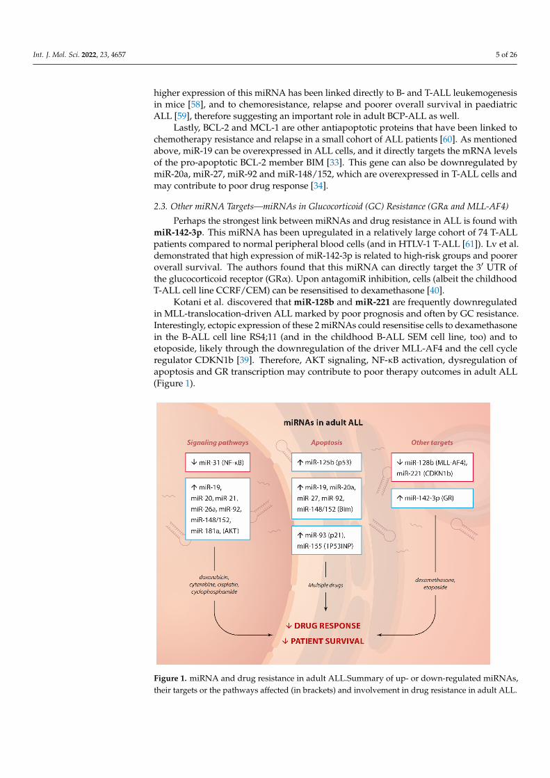
higher expression of this miRNA has been linked directly to B- and T-ALL leukemogenesisin mice [58], and to chemoresistance, relapse and poorer overall survival in paediatricALL [59], therefore suggesting an important role in adult BCP-ALL as well.
Lastly, BCL-2 and MCL-1 are other antiapoptotic proteins that have been linked tochemotherapy resistance and relapse in a small cohort of ALL patients [60]. As mentionedabove, miR-19 can be overexpressed in ALL cells, and it directly targets the mRNA levelsof the pro-apoptotic BCL-2 member BIM [33]. This gene can also be downregulated bymiR-20a, miR-27, miR-92 and miR-148/152, which are overexpressed in T-ALL cells andmay contribute to poor drug response [34].
2.3. Other miRNA Targets—miRNAs in Glucocorticoid (GC) Resistance (GRα and MLL-AF4)
Perhaps the strongest link between miRNAs and drug resistance in ALL is found withmiR-142-3p. This miRNA has been upregulated in a relatively large cohort of 74 T-ALLpatients compared to normal peripheral blood cells (and in HTLV-1 T-ALL [61]). Lv et al.demonstrated that high expression of miR-142-3p is related to high-risk groups and pooreroverall survival. The authors found that this miRNA can directly target the 3′ UTR ofthe glucocorticoid receptor (GRα). Upon antagomiR inhibition, cells (albeit the childhoodT-ALL cell line CCRF/CEM) can be resensitised to dexamethasone [40].
Kotani et al. discovered that miR-128b and miR-221 are frequently downregulatedin MLL-translocation-driven ALL marked by poor prognosis and often by GC resistance.Interestingly, ectopic expression of these 2 miRNAs could resensitise cells to dexamethasonein the B-ALL cell line RS4;11 (and in the childhood B-ALL SEM cell line, too) and toetoposide, likely through the downregulation of the driver MLL-AF4 and the cell cycleregulator CDKN1b [39]. Therefore, AKT signaling, NF-κB activation, dysregulation ofapoptosis and GR transcription may contribute to poor therapy outcomes in adult ALL(Figure 1).
Int. J. Mol. Sci. 2022, 23, 4657 6 of 26
Figure 1. miRNA and drug resistance in adult ALL.Summary of up- or down-regulated miRNAs,
their targets or the pathways affected (in brackets) and involvement in drug resistance in adult
ALL.
3. Implication of miRNAs in Drug Resistance in Childhood ALL
ALL is the most common pediatric cancer, with an incidence of 20 cases per 1,000,000.
It is manifested by increasing immature lymphoblast cells. There are two main subtypes
of cALL depending on the lineage: B cell and T cell leukemia, with B cell prevalence
around 75% of all cases. Other commonly used stratifications into risk groups are made
according to clinical features and cytogenetic markers. The onset is typically at the age of
3 to 5 years. With the development of modern drugs and chemotherapy regimens, the
treatment outcomes are substantially improved. Thus, the 5-year survival rates are signif-
icantly increased—from less than 60% in the 1980s to over 80% after 2004 and approaching
90% nowadays [12,62,63].
The standard treatment protocols include GCs, vincristine (vinca alkaloid), L-aspar-
aginase, anthracyclines and antimetabolites (cytarabine, methotrexate, mercaptopurine)
[64]. Despite the highly effective therapy and increased overall survival if relapse occurs,
only 50% of patients achieve a second remission. This percentage is even lower if the re-
lapse starts before the completion of primary treatment [65]. Thus, relapse and treatment
failure appear to be the factors that dramatically affect the overall survival of childhood
ALL patients. The intimate mechanisms behind these complications are not fully under-
stood. The reasons are hidden in mutations in signaling pathways that affect apoptosis,
proliferation, growth, and differentiation. In the last 15 years, the intense investigation of
the role of miRNA in the pathogenesis and prognosis of childhood ALL allowed the iden-
tification of miRNA signatures related to prognosis, treatment outcome and resistance to
some of the most commonly used drugs. In the light of the current review, we summarise
the data on miRNA regulation of genes that ensure proliferation and cell growth through
the activation of certain signaling pathways, and of genes that directly inhibit apoptosis
of leukemic blast cells, and thus provide drug resistance.
3.1. miRNA Modulation of Signaling Pathways—AKT, Notch—NF-κB
Figure 1. miRNA and drug resistance in adult ALL.Summary of up- or down-regulated miRNAs,their targets or the pathways affected (in brackets) and involvement in drug resistance in adult ALL.

Int. J. Mol. Sci. 2022, 23, 4657 6 of 26
3. Implication of miRNAs in Drug Resistance in Childhood ALL
ALL is the most common pediatric cancer, with an incidence of 20 cases per 1,000,000.It is manifested by increasing immature lymphoblast cells. There are two main subtypes ofcALL depending on the lineage: B cell and T cell leukemia, with B cell prevalence around75% of all cases. Other commonly used stratifications into risk groups are made accordingto clinical features and cytogenetic markers. The onset is typically at the age of 3 to5 years. With the development of modern drugs and chemotherapy regimens, the treatmentoutcomes are substantially improved. Thus, the 5-year survival rates are significantlyincreased—from less than 60% in the 1980s to over 80% after 2004 and approaching 90%nowadays [12,62,63].
The standard treatment protocols include GCs, vincristine (vinca alkaloid), L-asparaginase,anthracyclines and antimetabolites (cytarabine, methotrexate, mercaptopurine) [64]. Despitethe highly effective therapy and increased overall survival if relapse occurs, only 50% ofpatients achieve a second remission. This percentage is even lower if the relapse starts beforethe completion of primary treatment [65]. Thus, relapse and treatment failure appear tobe the factors that dramatically affect the overall survival of childhood ALL patients. Theintimate mechanisms behind these complications are not fully understood. The reasons arehidden in mutations in signaling pathways that affect apoptosis, proliferation, growth, anddifferentiation. In the last 15 years, the intense investigation of the role of miRNA in thepathogenesis and prognosis of childhood ALL allowed the identification of miRNA signaturesrelated to prognosis, treatment outcome and resistance to some of the most commonly useddrugs. In the light of the current review, we summarise the data on miRNA regulation of genesthat ensure proliferation and cell growth through the activation of certain signaling pathways,and of genes that directly inhibit apoptosis of leukemic blast cells, and thus provide drugresistance.
3.1. miRNA Modulation of Signaling Pathways—AKT, Notch—NF-κB
Signaling pathways that control cellular processes such as proliferation, differentiationand apoptosis enhance cell survival of malignant cells. Different mutations may alterproteins that act as receptors, enzymes, or inhibitors. Signaling pathways are crosslinked atdifferent levels, and their dysregulation may result in many unpredictable effects. miRNAsact as regulators of the translation of proteins involved in signaling pathways. Thus,alternations in miRNA expression are crucial factors that affect cellular processes. Moleculesacting as inhibitors of different signal proteins are tested in vitro and in vivo to find specificdrugs with less adverse effects and better treatment outcomes. In recent years miRNAshave been regarded as possible targets for developing new therapeutics [66,67].
In a cohort of 111 ALL patients, Li et al. found that the levels of miR-99a and miR-100are significantly downregulated compared to those in cells from healthy bone marrowdonors. Among other targets (discussed below), these two miRNAs were demonstratedto suppress mTOR and IGF1R. The AKT/mTOR axis is frequently activated in childhoodALL and has been strongly implicated in GC resistance through balancing proliferation,autophagy, apoptosis and cell metabolism [68]. Furthermore, in paediatric T-ALL celllines, AKT1 interacts directly with the GC receptor NR3C1 and phosphorylates it, makingit inactive and ineffective GC therapy [69]. Therefore, not surprisingly, when these twoAKT modulating miRNAs were overexpressed through mimics in 3 childhood ALL celllines, lymphoblasts exhibited decreased proliferation and enhanced apoptosis in responseto dexamethasone (likely due to the downregulation of the antiapoptotic BCL-2 familymember Mcl1) [70].
Interestingly, another group (Moqadam et al.) described that miR-99a or/and miR-100, when co-expressed with miR-125b, could lead to vincristine resistance. It was shownin vitro that neither one of these miRNAs could cause insensitivity to this drug. Only theco-expression of miR-125b with miR-99a or miR-100 could trigger resistance to therapy. Inthe absence of vincristine in vitro, there was no cell cycle arrest nor increased apoptosis ratein miR-125b-, miR-99a-, or miR-100-expressing cells, suggesting a time- and context-specific

Int. J. Mol. Sci. 2022, 23, 4657 7 of 26
role of these miRNA [71]. Further scientific data reported by Schotte et al. confirmedthat these three miRNAs can be upregulated (14-25-fold) in childhood ALL patients withTEL-AML1 fusion (in 12 of 31 and 12 of 29 samples) and that their overexpression confersvincristine and daunorubicin resistance [72]. However, the exact mechanisms and targetsof miR-99a, miR-100 and miR-125 remain uninvestigated.
Additionally, the same group reported that inhibited expression of miR-454 corre-lated with L-asparaginase resistance [72]. This miRNA has been validated as a tumoursuppressor in other malignancies like osteosarcoma, nasopharyngeal carcinoma, and mul-tiple myeloma [73–75]. MiR-454 can induce apoptosis and suppress cell survival andproliferation by inhibiting c-Met, consequently inactivating the AKT/mTOR pathway. Thec-Met/Akt/mTOR axis has been involved in drug resistance in vitro in other hematologicalmalignancies [73].
Lin et al. demonstrated that the overexpression of miR-454 could restore sensitivity tocisplatin in nasopharyngeal carcinoma cells, confirming the role of this miRNA chemother-apy response [75]. On the opposite, it was shown by other groups that overexpression ofmiR-454 in the SGC-7901 gastric carcinoma cell line and HCT-116 colorectal cancer cellscould mediate oxaliplatin resistance and promote proliferation through the CYLD gene.It regulates, in turn, the NF-κB and TGF-β signaling pathways and inhibits PTEN andactivates the AKT pathway [76,77]. The same miRNA could exhibit opposite regulatoryfunctions in certain cell types and tissues and use different pathways. Nevertheless, theexact involvement of miR-454 in L-asparaginase resistance in cALL is yet to be elucidated.
The same authors identified several dysregulated miRNAs in B-ALL samples, withmiR-708 overexpressed in high-risk patients. It was assumed that miR-708 upregulationmight contribute to the initiation of relapse (which is partially in line with another studyshowing that levels of this miRNA increase in relapse compared to complete remission [78]).Notably, two of the targets of miR-708, CNTFR and GNG12, could explain at least someof the mechanisms that may lead to therapy failure. Downregulation of the first gene wasshown to activate the Jak/STAT pathway, while the GNG12 gene product is involved inMAPK signaling [79,80].
Another miRNA that affects drug sensitivity in T-ALL cells through modulation of aspecific signaling pathway is the tumor suppressor miR-101. This miRNA was downregu-lated in T-ALL patients compared to control samples and directly targeted the 3‘UTR ofNotch1. Ectopic expression in Jurkat cells through a mimic sequence decreased prolifer-ation and enhanced apoptosis. Importantly, Qian et al. also proved that miR-101 couldsignificantly attenuate drug resistance to doxorubicin in vitro [81].
Lastly, the NF-κB transcription factor family members are activated in childhoodALL [82]. NF-kB-driven transcription has been shown to induce resistance to doxorubicinand etoposide in CEM T-ALL cells [83]. Overexpression of miR-125b has been demon-strated to activate NF-κB by directly targeting the inhibitory protein TNF-α–inducedprotein 3 (TNFAIP3/A20) in Jurkat cells [84]. Interestingly, this miRNA can be translocatedand overexpressed in BCP-ALL and correlates to chemotherapy resistance and poor sur-vival in childhood ALL. This may result from the NF-kB activation as described above [59].Another effect of miR-125b upregulation is the metabolic reprogramming of T-ALL in vitro.Liu et al. showed in Jurkat cells that high miRNA levels could lead to increased glucoseconsumption via upregulation of the glucose transporter GLUT1 and enhanced oxygenconsumption [84]—events strongly associated with glucocorticoid resistance [85]. Thereby,there is considerable diversity in the pathways that miRNAs may dysregulate in theircontribution towards drug insensitivity in childhood ALL (Table 2).

Int. J. Mol. Sci. 2022, 23, 4657 8 of 26
Table 2. miRNA, their targets and their effects in childhood ALL.
miRNA Targets/Pathways Outcome/Correlation Reference
Signaling pathways
↓miR-101 Notch1 Resistance to doxorubicin [81]
↑miR-125b TNFAIP3/A20 (NF-κB) Poor survival (doxorubicin, etoposide,glucocorticoids? in vitro) [59,84,85]
↓miR-204 IRAK1 (NF-κB) Resistance to vincristine [86]
↓miR-454 c-MET (AKT) Resistance to L-Asparaginase (cisplatinin nasopharyngeal sarcoma) [72,75]
↑miR-708 CNTFR (Jak/STAT), GNG12 (MAPK) High-risk group and relapse (multipledrug resistance) [78]
Apoptosis
↓miR-99a and -100 IGF1R, mTOR (MCL1) High risk; dexamethasone resistance [70]
↑miR-99a, miR-100 andmiR-125 (together) Uninvestigated Resistance to vincristine and
daunorubicin [71,72]
↑miR-223 E2F1 Complete remission (in B-ALL andAML) [78]
↓miR-652-3p Uninvestigated Relapse; resistance to vincristine andcytarabine [87]
↓miR-34a BCL-2 Resistance to doxorubicin [88]
↓miR-204 MCL1 (through IRAK1 and NF-κB) Resistance to vincristine [86]
Other targets
↑miR-27a BMI1 (epigenetic modifier)MDR1
Complete remissionSensitivity to doxorubicin (in K562 celllines) Low expression in relapsepatients
[78,89]
↓miR-99a and -100 FKBP51 (and GR expression andactivity)
Downregulated in high risk;Dexamethasone resistance
↑miR-223 FBXW7 (ubiquitination) Resistance to GSIs in T-ALL [90]
↑miR-331-5p MDR1Sensitivity to doxorubicin (in K562 celllines) Low expression in relapsepatients
[89]
↑miR-708 FOXO3 (self-renewal, AKT activation inmyeloid cells)
Good response to GCs; relapse-freesurvival [91]
↓miR-128b and miR-221MLL-rearranged fusion gene (therebydownregulating CDKN1B,and p27)
Resistance to GCs [39]
Arrows (↓ and ↑) indicate miRNA downregulation or upregulation.
3.2. miRNA Regulation of Apoptosis
The dysregulation of apoptosis is crucial in developing different pathological condi-tions, including malignancies [92]. By regulating the expression of pro- and anti-apoptoticproteins, miRNAs directly affect cell survival and drug sensitivity. Alternations in apoptosisare detected in childhood ALL as well. Higher expression of the anti-apoptotic proteinBCL-2 and downregulation of Bax were related to relapse and bad prognosis [93].
A direct link between miRNAs and apoptosis upon drug treatment in childhood ALLis provided by miR-34a (also discussed below in CLL). Najjary et al. demonstrated in Jurkatcells that expression of this miRNA alone or in combination with doxorubicin treatmentcould decrease BCL-2 and increase Caspase-3 and p53 levels, thus significantly enhancingcell death [88]. Another piece of evidence of the importance of miRNAs in apoptosis is

Int. J. Mol. Sci. 2022, 23, 4657 9 of 26
provided by Jiang et al., who reported that miR-652-3p was significantly downregulatedin pre-B pediatric ALL patients at diagnosis and relapse but upregulated at remissionand in healthy control samples. Atopic expression of an antagomiR sequence in REH andRS4;11 leukemic cells induced apoptosis and made cells more sensitive to vincristine andcytarabine, but the target genes have been identified neither in childhood ALL patientsnor in cell lines [87]. Lastly, miR-204 is silenced by promotor methylation in ALL. ThismiRNA regulates Irak1, which mediates NF-κB activation and consequent transcription.Importantly, these miR-204 targets also regulate the stability of the antiapoptotic proteinMCL1, which has been linked to response to vincristine [86]. Therefore, not surprisingly,ectopic expression of a miR-204 mimic in Jurkat cells could lead to decreased proliferationand apoptosis [94]. All these proofs of the miRNA-mediated control of apoptotic proteinsshow that the dysregulation of programmed cell death could provide survival advantagesand mediate drug resistance in childhood lymphoblastic leukemia.
3.3. Other miRNA Targets—MDR1, Proteasomal Degradation, GC Resistance (MLL-AF4 andGR Modulation)
A study comparing matched samples at diagnosis and in complete remission (CR)found that two miRNAs are highly expressed when therapy is successful—miR-223, whichtargets E2F1 (involved in DNA damage response, cell cycle arrest and apoptosis), and miR-27a, described to regulate the epigenetic modifier BMI1 [78]. Consistently, another groupdemonstrated that downregulation of the latter miRNA and miR-331–5p correlates with ahigher risk of relapse in a mixed cohort of ALL and AML patients. It was shown in vitro(in HEK-293 and AML cell lines) that both miRNAs control the levels of the oncoproteinMDR1 (multiple drug resistance protein 1). Importantly, ectopic expression of miR-27a ormiR-331-5p could restore sensitivity to doxorubicin in vitro [89].
Interestingly, Kumar et al. identified miR-223 as a Notch and NF-κB signaling pathwayin T-ALL. This miRNA appeared to downregulate the FBXW7 tumor suppressor geneinvolved in ubiquitination and proteasomal degradation of oncoproteins. The authorsshowed that miR-223 has implicated in GSI (γ-secretase inhibitor) resistance and thatantagomiRs could induce sensitivity in GSI-resistant T-ALL cells in vitro [90].
As mentioned previously, for adult ALL, miR-128b and miR-221 are strongly impli-cated in glucocorticoid resistance. They are found to be downregulated in MLL-rearrangedALL compared to other childhood ALL subtypes. These miRNAs directly target the mRNAsof the fusion gene and thereby downregulate CDKN1B, which generates the p27- cell-cyclecheckpoint regulator. Importantly, the restored expression of both miRNAs sensitised thechildhood ALL cell lines SEM to dexamethasone [39].
Li et al. reported that in high-risk childhood ALL patients (WBC > 5 × 10⁴, T-ALL,MLL-rearranged gene, BCR-ABL fusion gene), miR-99a and miR-100 are downregulated.Further, they described a novel target inhibited by both miRNAs—FKBP51. As a resultof the reduced expression of miR-99 and miR-100, FKBP51 levels are high and suppressthe activation of the glucocorticoid receptor (GR). Therefore, ectopic expression of thesemiRNAs would restore GR function upon dexamethasone treatment and lead to reducedproliferation and augmented apoptosis [70].
Lastly, even if the role of miR-708 in childhood ALL may seem to be controversial orperhaps cell-type or stage-specific, Han et al. demonstrated that high levels of this miRNA(as well as miR-223 and miR-27a) correlate with higher relapse-free survival rates andgood response to 7-day prednisolone monotherapy. Notably, miR-708 can downregulateFOXO3 levels both in vivo and in vitro. This transcription factor is a direct target of theGR [91] and is further known to be involved in GC target gene expression [95]. Therefore,not surprisingly, a positive correlation of this miRNA with GC sensitivity was observed—miR-708 was highly expressed in childhood ALL patients with a good response to 7-dayprednisolone monotherapy. It was downregulated in the prednisolone poor responsegroup [78].

Int. J. Mol. Sci. 2022, 23, 4657 10 of 26
In summary, miRNAs have been shown to control many signaling pathways connectedto proliferation and survival, several transcription factors, and specific key apoptosismediators, thus leading to drug resistance (Figure 2).
Int. J. Mol. Sci. 2022, 23, 4657 10 of 26
As mentioned previously, for adult ALL, miR-128b and miR-221 are strongly impli-
cated in glucocorticoid resistance. They are found to be downregulated in MLL-rear-
ranged ALL compared to other childhood ALL subtypes. These miRNAs directly target
the mRNAs of the fusion gene and thereby downregulate CDKN1B, which generates the
p27- cell-cycle checkpoint regulator. Importantly, the restored expression of both miRNAs
sensitised the childhood ALL cell lines SEM to dexamethasone [39].
Li et al. reported that in high-risk childhood ALL patients (WBC > 5 × 10⁴, T-ALL,
MLL-rearranged gene, BCR-ABL fusion gene), miR-99a and miR-100 are downregulated.
Further, they described a novel target inhibited by both miRNAs—FKBP51. As a result of
the reduced expression of miR-99 and miR-100, FKBP51 levels are high and suppress the
activation of the glucocorticoid receptor (GR). Therefore, ectopic expression of these miR-
NAs would restore GR function upon dexamethasone treatment and lead to reduced pro-
liferation and augmented apoptosis [70].
Lastly, even if the role of miR-708 in childhood ALL may seem to be controversial or
perhaps cell-type or stage-specific, Han et al. demonstrated that high levels of this miRNA
(as well as miR-223 and miR-27a) correlate with higher relapse-free survival rates and
good response to 7-day prednisolone monotherapy. Notably, miR-708 can downregulate
FOXO3 levels both in vivo and in vitro. This transcription factor is a direct target of the
GR [91] and is further known to be involved in GC target gene expression [95]. Therefore,
not surprisingly, a positive correlation of this miRNA with GC sensitivity was observed—
miR-708 was highly expressed in childhood ALL patients with a good response to 7-day
prednisolone monotherapy. It was downregulated in the prednisolone poor response
group [78].
In summary, miRNAs have been shown to control many signaling pathways con-
nected to proliferation and survival, several transcription factors, and specific key apop-
tosis mediators, thus leading to drug resistance (Figure 2).
Figure 2. miRNA and drug resistance in childhood ALL.Summary of up- or down-regulated miR-
NAs, their targets or pathways they affect (in brackets) and their involvement in drug resistance in
childhood ALL.
Figure 2. miRNA and drug resistance in childhood ALL.Summary of up- or down-regulated miRNAs,their targets or pathways they affect (in brackets) and their involvement in drug resistance inchildhood ALL.
4. Implication of miRNAs in Drug Resistance in CLL
CLL is the most common type of leukaemia, accounting for ~1% of all cancers andabout 10% of all haematological malignancies [12]. B cell CLL (>95% of cases) is charac-terised by clonal expansion and accumulation of immature CD5+/CD19+ B cells in thebone marrow and lymph nodes, where oncogenesis is believed to be driven by microenvi-ronment signals, and in the peripheral blood [96]. The most clinically significant markersfor poor disease outcomes are a lack of mutations in the immunoglobulin heavy chain vari-able region locus (IgHV), deletion of chromosome 13q (affecting the pro-apoptotic proteinBCL-2) or 17p (affecting the tumour suppressor p53), as well as high expression of ZAP-70and CD38 [13]. Standard chemotherapies, including fludarabine, cyclophosphamide andthe targeted anti-CD20 antibody rituximab, achieve 10-year overall survival of over 70%.However, the prognosis for relapse and high-risk patients can be poor—less than 25%5-year survival [97]. Differential expression between malignant and normal B cells andbetween CLL samples with different cytogenetics have linked hundreds of miRNAs to thepathogenesis, progression and prognosis of CLL [19,23]. Most of them implicated in drugresponse are shown in Table 3.

Int. J. Mol. Sci. 2022, 23, 4657 11 of 26
Table 3. miRNAs, their targets and their effects in CLL.
miRNA Targets/Pathways Outcome/Correlation Reference
Signaling pathways
↓miR-9 NF-κB Advanced Rai stage [98]
↓miR-15a and miR-16-1 IKKα (NF-κB) Resistance to fludarabine anddexamethasone [99]
↑miR-21 PTEN (AKT) Resistance to fludarabine [100]
↑miR-22 PTEN (AKT) B-CLL cell proliferation [101]
↑miR-26 PTEN (AKT) Advanced Binet stage; inferior time to firsttreatment; resistance to apoptosis in vitro [102,103]
↓miR-29 TCL-1, AKT (AKT) Indirect implication in resistance tofludarabine (through TCL-1) [104,105]
↓miR-34b/c ZAP70 (B cell receptor signaling) Poor overall survival [106]
↓miR-146a NF-κB Earlier onset of B cell malignancies (in mice) [107]
↓miR-150 ZAP70, FOX1, PI3K/GAB1 (B cell receptorsignaling) Poor overall survival [106,108]
↑miR-155 SHIP1 (B cell receptor signaling) Poor therapy outcome and overall survival [109]
↓miR-181a, b TCL-1 (AKT) Indirect implication in resistance tofludarabine (through TCL-1) [104]
↑miR-214 and PTEN (AKT) Resistance to apoptosis in vitro [103]
↓miR-708 IKKα (NF-κB) Poor treatment-free survival [78]
Apoptosis
↓miR-15a and miR-16-1 BCL-2 Overexpression induces apoptosis in vitro [102,110]
↓miR-17-5p BCL-2 Poor prognosis (correlates with p53inactivation) [111]
↓miR-29 MCL1 Poor prognosis (correlates with p53inactivation) [111,112]
↓miR-34a BCL-2, CDK4, CDK 6, E2F3, Cyclin E, c-Myc Poor prognosis (correlates with p53inactivation) [102,111]
↓miR-130a ATG2B Enhanced apoptosis following starvation [113]
↓miR-181a, b Bcl-2, Mcl-1, XIAP, PTEN Fludarabine sensitivity [102,114]
↑miR-221 p27 Fludarabine resistance [114]
Other targets
↑miR-125b CD20 Rituximab resistance [115]
↑miR-532-3p CD20 Rituximab resistance [115]
Arrows (↓ and ↑) indicate miRNA downregulation or upregulation.
4.1. miRNA Modulation of Signaling Pathways—BCR, NF-κB, TLR and TCL-1-AKT
B cell receptor (BCR) signaling has been validated as an essential factor in the patho-genesis of CLL (and other B cell malignancies like diffuse large B cell lymphoma), providingstimulation of critical pro-survival pathways like PI3K-AKT and NF-κB [116]. Under-standing the importance of such pathways has led to the development of small-moleculeinhibitors targeting signal transducers such as BCR-associated kinase (BTK), SYK andPI3Kδ. For example, ibrutinib (a BTK inhibitor) shows remarkable response rates of nearly80% [116]. Significantly, the expression of certain mediators of BCR signaling in CLL can beregulated by miRNAs. Thus, by investigating over 700 miRNAs in 168 patient samples,miR-150 was found to inversely correlate with unmutated IgHV and ZAP70 levels (BCRheavy chain and enhancer of BCR signaling, respectively). The frequently deleted clusterof miR-34b/c located on chromosome 11q can also target ZAP70 [106]. Furthemore, miR-150 has been shown to directly regulate the levels of two BCR signaling mediators—thetranscription factor FOXP1 and the PI3K adaptor molecule GAB1. Of note, lower levelsof miR-150 and higher expression of the abovementioned proteins correlate with poorer

Int. J. Mol. Sci. 2022, 23, 4657 12 of 26
overall survival [108]. Similarly, miR-155 targets SHIP1, which can inhibit BCR signaling.The high expression of this miRNA was found to correlate with poor treatment-free andOS in a cohort of over 260 CLL patients. This finding again suggests the importance of BCRsignaling in cell survival and therapy response [109]. BCR signaling and the downstreamactivation of PI3K and NF-κB pathways have not only been implicated in proliferation andsurvival, but also in the response to chemotherapy. For instance, the activation of AKT1 inparticular has been shown to lead to fludarabine resistance in CLL cells (through STAT3signaling) [117].
NF-κB is overactivated in CLL, leading to enhanced survival and decreased apoptosisdue to fludarabine and dexamethasone treatment [118,119]. Rearrangements resultingin BCR-ABL fusion and involving BCL-3, which stimulates the p50 and p52 subunits ofNF-κB, can also activate this transcription factor [120,121]. Strong DNA binding of the RelAsubunit of NF-κB has been shown to correlate to tumour burden (cell doubling time andcell counts) in patients, in vitro cell survival and resistance to fludarabine [122]. SeveralmiRNAs are known to regulate NF-κB activation in hematologic malignancies [99]. Of note,miR-15a and miR-16-1, deleted in more than 60% of CLL, have been shown to suppressIKKα. It phosphorylates the inhibitor of NF-κB (IkB), targeting it for ubiquitination andproteasomal degradation, which activates NF-κB signaling [99]. Members of the miR-9family (miR-9-3) can be silenced in CLL patients and cell lines by methylation and areimplicated in NF-κB activation [98], likely through direct targeting of the 3′ UTR of the p50subunit of NF-κB [99]. Similarly, miR-708 has been found to downregulate IKKβ, whichsuppresses NF-κB signaling directly. However, this miRNA can be epigenetically silenced inCLL cells. Baer et al. proved that methylation of the miR-708 enhancer correlates with poortreatment-free survival in a cohort of nearly 300 patients. The findings are similar to whathas been detected in childhood ALL and response to glucocorticoids [78], suggesting therole of this miRNA and NF-κB signaling in drug response and relapse [123]. Furthermore,NF-κB activation can be regulated by a negative feedback loop involving miR-146a [107].This miRNA’s downregulation (or knockout) can lead to earlier onset and more aggressiveB cell malignancies in c-Myc-driven in vivo models [124]. Its levels are deregulated in CLLpatients, as well [125].
Toll-like receptors (TLRs) have been established as essential co-activator moleculesin B cells. The importance of TLR signaling has been investigated in CLL, revealing thatstimulation of this receptor family can protect from apoptosis and correlates (throughactivation of NF-κB and STAT3) with poor prognosis [126,127]. Interestingly, TLR signalingin the microenvironment of CLL cells could activate NF-κB and has been implicated in CLLcell proliferation and survival [127] by activating members of the miR-17~92 family [128].Importantly, it has been shown that TLR signaling can rescue cells from apoptosis uponfludarabine treatment. miR-155-3p levels have risen concomitantly, implicating this miRNAin drug resistance [129].
The protooncogene TCL-1 (T cell leukemia/lymphoma 1) is frequently overexpressedin T-ALL [45] and CLL with chromosome 11q deletions [130]. Furthermore, transgenicmice expressing TCL-1 in B cells develop CLL, which demonstrates the leukemogenicproperties of this gene [131]. Interestingly, this mouse model has served to study drugefficacy. Johnson et al. proved that TCL-1 is connected with fludarabine resistance in ap53-independent manner [132]. Furthermore, TCL-1 has been shown to interact directlyand activate AKT in both T-ALL and CLL cells [133,134]. In particular, Hofbauer et al.have also demonstrated that targeting TCL-1 by siRNA can reduce AKT activation andresensitise resistant cells to fludarabine [134]. Therefore, it is noteworthy that miR-29 andmir-181 can directly regulate TCL-1 expression in CLL patients, implicating them (at leastindirectly) in drug resistance [104]. Of note, miR-29 has been confirmed to directly targetAKT (among a few other targets) [105]. miR-22 can also activate AKT signaling in CLL andenhance cell proliferation [101]. However, the role of this miRNA in fludarabine resistancehas not been studied yet.

Int. J. Mol. Sci. 2022, 23, 4657 13 of 26
Comparing expression in responders and non-responders to this drug, high levels ofthree other miRNAs have been strongly implicated in refractory disease—miR-21, miR-148a and miR-222. Significantly, anti-miRNA oligonucleotides against miR-21 and miR-222 enhanced fludarabine-driven apoptosis suggesting a therapeutic window [100]. Themechanism of how these miRNAs make CLL cells refractory to treatment has not beenelucidated yet, but Ferracin et al. suggest that it may be through regulation of PTEN and/orthe cell cycle [100]. The high expression of miR-21 in non-responders has been confirmed byanother study, which also highlighted that increased levels of miR-34 predict an excellentresponse to fludarabine [135]—proof in line with previous studies presenting this miRNAas downregulated in fludarabine-refractory cells [136,137].
Lastly, PI3K-AKT signaling can be controlled by the tumour suppressor PTEN, down-regulated in CLL and may also have prognostic value [138]. Zou et al. demonstratedthat miR-26a, which has previously appeared overexpressed in CLL [102], and miR-214could directly downregulate PTEN mRNA levels. High expression of miR-26a correlatesto inferior time to first treatment [103], which provides another 2 miRNAs that may beimplicated in PI3K-AKT-mediated leukemogenesis.
4.2. miRNA Regulation of Apoptosis and Autophagy
Besides the modulation of signaling pathways that augment cell proliferation and sur-vival, miRNAs can directly alter the levels of proteins involved in processes like apoptosisand autophagy, serving as rescue mechanisms for CLL cells upon chemotherapy treatment.Interestingly, miRNAs are frequently targeted by deletions or translocations, leading tolower expression, suggesting a tumour suppressor role of these miRNAs. This has beenshown for miR-15a and miR-16-1, which target the antiapoptotic protein BCL-2, in over60% of CLL patients [9]. Cimmino et al. found that both miRNAs are indeed inversely cor-related to Bcl-2 gene expression in a cohort of 26 CLL patients. Therefore, downregulationof these miRNAs protects from apoptosis, while overexpression of miR-15a or miR-16-1would directly diminish BCL-2 levels and induce apoptosis [110]. Such antisense strate-gies targeting BCL-2 have proven therapeutic potential in several malignancies, includingre-sensitizing cells to chemotherapy [139] (discussed later). Similarly, lower expression ofmiR-29, described in p53 mutant CLL samples [111], could result in higher levels of theanti-apoptotic protein MCL-1 and again in evasion of cell death [112].
A microarray covering over 800 miRNAs detected the downregulation of a numberof these regulatory molecules compared to normal peripheral blood B cells in a cohort of156 CLL patients. Interestingly, mimics of some of these hits were able to induce apoptosisin primary CLL cells. Zhu et al. also demonstrated that miR-181a and b directly down-regulate the mRNA levels of the antiapoptotic proteins BCL-2, MCL-1 and XIAP. Lastly,the authors showed that forced expression of miR-181 (as well as miR-15, 16 and 34) couldenhance fludarabine-induced apoptosis in primary CLL cells (from 40 samples) likely in ap53-dependent manner [102]. In contrast (and in another context), when looking at a smallercohort of 39 patients after their first 6 fludarabine cycles, Moussay et al. found that miR-181ais upregulated in refractory cells (making the role of this miRNA in drug response in CLLcontroversial). In addition, miR-221 was also upregulated in resistant patients, while miR-29was downregulated (in accordance with previous studies) [114].
TP53 (p53) is a master regulator of DNA damage response, apoptosis, the cell cycle andgene expression. Deletions and/or mutations of this gene are well-established to correlateto worse overall survival and poor response to chemotherapeutics (like chlorambucil,Cytoxan, prednisolone, vincristine and fludarabine) in CLL [140,141]. Interestingly, theroles of miRNAs and p53 in apoptosis and drug response may be reversed—p53 can controlthe expression of miRNAs, most likely through transactivation of their expression bybinding to predicted upstream DNA regulatory elements., This, in turn, would determinecell fate upon chemotherapy. The first study looking at the interplay between p53 andmiRNAs demonstrated that miR-17-5p, miR-29 and miR-34a are downregulated in patientswith p53 mutations, who have a poorer prognosis. These three regulatory transcripts have

Int. J. Mol. Sci. 2022, 23, 4657 14 of 26
been connected with the control of apoptosis (through BCL-2 and MCL-1) and the cellcycle (through CKD4, CDK6, E2F3, cyclin E and c-Myc) [111]. The already mentionedmiR15a/16-1 cluster is regulated by p53. These findings demonstrate that deletion and/ormutation of the “guardian of the genome” affect a number of miRNAs, which in turnregulate apoptosis, BCR and NF-κB pathways—all linked to drug resistance as describedabove [106], in particular to fludarabine [136,137].
Autophagy has been well-established as a survival mechanism in malignant cellsunder different forms of stress, such as starvation and chemotherapy. Interestingly, CLLcells treated with fludarabine or a PI3Kδ inhibitor (CAL-101) undergo autophagy [142].Indeed, inhibition of specific mediators of this process (autophagosome-lysosome fusionand the AMPK/ULK1 pathway) could enhance the cell-killing effect of inhibitors suchas of CDKs (flavopiridol) [142] or BCL-2 (venetoclax) [143]. miR-130a is epigeneticallysilenced in CLL. Kovaleva et al. have confirmed its role in autophagy regulation [144,145].Overexpression of this miRNA could downregulate the autophagy-related gene ATG2B,which enhanced apoptosis following starvation. Therefore, even if the role of this miRNAin drug resistance has not been exploited in CLL, its deregulation may provide lymphocyticcells with a survival mechanism upon chemotherapy treatment. This hypothesis could besupported by evidence from ovarian cancer where miR-130a is related to paclitaxel andcisplatin resistance [113]. Thereby, AKT and BCR signaling, NF-κB activation, modulationof apoptosis and the expression of CD20 (see below) constitute the main targets of miRNAs,by which CLL cells achieve drug resistance (Figure 3).
Int. J. Mol. Sci. 2022, 23, 4657 15 of 26
Figure 3. miRNA and drug resistance in CLL. Summary of up- or down-regulated miRNAs, their
targets or pathways they affect (in brackets) and their involvement in drug resistance.
4.3. Other miRNA Targets—CD20 and Resistance to Rituximab
Interestingly, miR-125b and miR-532-3p have been described to correlate to lympho-
cytopenia following rituximab treatment inversely. A bioinformatics approach with puta-
tive targets demonstrated that these miRNAs might lead to therapy resistance through
downregulation of the direct target of this antibody-based treatment—CD20 (MS4A
genes) [115].
5. Perspectives of miRroring Strategies in Targeting of Drug Resistance in Lympho-
cytic Leukaemias
The importance of miRNAs for the pathogenesis and prognosis of lymphocytic leu-
kaemias has been established previously, but their involvement in drug response has not
been discussed thoroughly. Here we summarise the scientific evidence which demon-
strates the role of these post-transcriptional mRNA modulators in drug resistance. Acti-
vation of AKT or NF-κB (Figure 4) can provide pro-survival signals. Direct control of
apoptosis appears as the major escape mechanism employed by dysregulated miRNAs in
relation to chemotherapy response in ALL and CLL. These data are not unexpected given
the germane part these pathways play in relapses and poor survival (described above).
Interestingly, we also found that certain miRNAs are implicated in several other dysfunc-
tional molecular axes contributing to drug resistance, such as glucocorticoid uptake and
receptor activation, transduction through TLRs, transcription by MLL-AF4, and regula-
tion of CD20 levels.
Figure 3. miRNA and drug resistance in CLL. Summary of up- or down-regulated miRNAs, theirtargets or pathways they affect (in brackets) and their involvement in drug resistance.

Int. J. Mol. Sci. 2022, 23, 4657 15 of 26
4.3. Other miRNA Targets—CD20 and Resistance to Rituximab
Interestingly, miR-125b and miR-532-3p have been described to correlate to lymphocy-topenia following rituximab treatment inversely. A bioinformatics approach with putativetargets demonstrated that these miRNAs might lead to therapy resistance through down-regulation of the direct target of this antibody-based treatment—CD20 (MS4A genes) [115].
5. Perspectives of miRroring Strategies in Targeting of Drug Resistance inLymphocytic Leukaemias
The importance of miRNAs for the pathogenesis and prognosis of lymphocyticleukaemias has been established previously, but their involvement in drug response has notbeen discussed thoroughly. Here we summarise the scientific evidence which demonstratesthe role of these post-transcriptional mRNA modulators in drug resistance. Activationof AKT or NF-κB (Figure 4) can provide pro-survival signals. Direct control of apoptosisappears as the major escape mechanism employed by dysregulated miRNAs in relation tochemotherapy response in ALL and CLL. These data are not unexpected given the germanepart these pathways play in relapses and poor survival (described above). Interestingly, wealso found that certain miRNAs are implicated in several other dysfunctional molecularaxes contributing to drug resistance, such as glucocorticoid uptake and receptor activation,transduction through TLRs, transcription by MLL-AF4, and regulation of CD20 levels.
Int. J. Mol. Sci. 2022, 23, 4657 16 of 26
Figure 4. Recurrent miRNA-driven activation of AKT signaling and NF-κB in lymphocytic leukae-
mias.
5.1. Silencing of miRNAs
There is a great amount of ongoing work (including in clinical trials) related to the
implementation of various approaches aiming to silence certain miRNAs (when they are
oncogenic—“oncomiRs”) or to express them ectopically (when they act as tumour sup-
pressors). Such therapeutic strategies have been under development for ~20 years. They
include inhibiting oncomiRs through complementary binding of various chemically-mod-
ified oligonucleotides like antagomiRs and anti-miR oligonucleotides (AMOs) [146,147].
Furthermore, microRNA sponges, which can simultaneously block a family of miRNAs
(or several different ones), can also be engineered and have shown therapeutic potential
in preclinical studies [148]. Small-molecule inhibitors of miRNAs (SMIRs) impeding dif-
ferent steps in the biosynthesis or target-binding of miRNAs have also proven a valid
strategy [147]. Lastly, there are also “double-edged sword” approaches—either using a
combination of small interfering RNAs (siRNAs) and miRNAs aiming at a single target
[149] or employing several miRNAs covering different players in the same pathway (e.g.,
PI3K-RAS-RAF) [150].
In ALL and CLL, we have highlighted that high expression of miRNAs such as miR-
17~92, miR-155-3p, miR-21, miR-221, miR-222, and others is strongly associated with drug
resistance to, e.g., fludarabine, making them potential therapeutic targets for inactivation.
Several studies provide encouraging examples of the beneficial effect of miRNA inhibition
on chemotherapy treatment in vitro. Yan et al. demonstrated that miR-181a is overex-
pressed in T-ALL samples, and its silencing by antagomiRs can resensitise Jurkat cells to
doxorubicin cyclophosphamide, cytarabine and cisplatin [46]. Harada et al. used a locked-
nucleic acid (LNA) antisense approach targeting miR-17 in cALL cells. Silencing this
miRNA could enhance the response to dexamethasone by increasing BIM levels and in-
ducing apoptosis [151]. Similarly, LNA anti-miR-21 and -222 augment cell death induc-
tion by fludarabine in a CLL model cell line [100]. Notably, the therapeutic power of an-
tagomiRs has been demonstrated in vivo as well. In an animal model using MEC-1 CLL
Figure 4. Recurrent miRNA-driven activation of AKT signaling and NF-κB in lymphocyticleukaemias.
5.1. Silencing of miRNAs
There is a great amount of ongoing work (including in clinical trials) related to theimplementation of various approaches aiming to silence certain miRNAs (when they areoncogenic—“oncomiRs”) or to express them ectopically (when they act as tumour suppres-sors). Such therapeutic strategies have been under development for ~20 years. They in-clude inhibiting oncomiRs through complementary binding of various chemically-modifiedoligonucleotides like antagomiRs and anti-miR oligonucleotides (AMOs) [146,147]. Fur-

Int. J. Mol. Sci. 2022, 23, 4657 16 of 26
thermore, microRNA sponges, which can simultaneously block a family of miRNAs (orseveral different ones), can also be engineered and have shown therapeutic potential inpreclinical studies [148]. Small-molecule inhibitors of miRNAs (SMIRs) impeding dif-ferent steps in the biosynthesis or target-binding of miRNAs have also proven a validstrategy [147]. Lastly, there are also “double-edged sword” approaches—either usinga combination of small interfering RNAs (siRNAs) and miRNAs aiming at a single tar-get [149] or employing several miRNAs covering different players in the same pathway(e.g., PI3K-RAS-RAF) [150].
In ALL and CLL, we have highlighted that high expression of miRNAs such asmiR-17~92, miR-155-3p, miR-21, miR-221, miR-222, and others is strongly associatedwith drug resistance to, e.g., fludarabine, making them potential therapeutic targets forinactivation. Several studies provide encouraging examples of the beneficial effect ofmiRNA inhibition on chemotherapy treatment in vitro. Yan et al. demonstrated that miR-181a is overexpressed in T-ALL samples, and its silencing by antagomiRs can resensitiseJurkat cells to doxorubicin cyclophosphamide, cytarabine and cisplatin [46]. Harada et al.used a locked-nucleic acid (LNA) antisense approach targeting miR-17 in cALL cells.Silencing this miRNA could enhance the response to dexamethasone by increasing BIMlevels and inducing apoptosis [151]. Similarly, LNA anti-miR-21 and -222 augment celldeath induction by fludarabine in a CLL model cell line [100]. Notably, the therapeuticpower of antagomiRs has been demonstrated in vivo as well. In an animal model usingMEC-1 CLL cells, injecting an anti-miR-17 oligonucleotide leads to reduced tumour burdenand increased survival in mice [152].
5.2. Ectopic Expression of miRNAs
Another therapeutic approach is to re-express tumour suppressor miRNAs. Thisstrategy is readily achievable in preclinical models through the intracellular introduction ofexogenous synthetic double-stranded RNA mimics (via lipid or polymeric-based carriers),viral vectors expressing the desired oligonucleotide, and other approaches [146,147,153].Here, we pinpoint the involvement in the therapy failure of several miRNAs. Thus, miR-15a/16-1 and miR-34b/c can be absent in CLL due to deletions in chromosomes 13q and 11q,respectively, while miR-29, miR-181a and b, and others have reduced expression in CLL.Again, several in vitro studies can serve as proof-of-concept research for the significant ther-apeutic advantage of miRNA modulation combined with standard chemotherapy. Ectopicexpression of miR-128b and miR-221 in RS4;11 and SEM ALL cells can be downregulatedin MLL-AF4-driven ALL. This event resensitises the lymphoblasts to etoposide and dexam-ethasone [39]. Similarly, miR-101 is downregulated in T-ALL samples compared to controlcells, and its levels decrease upon doxorubicin treatment in vitro. Interestingly, Qian et al.demonstrated that this miRNA directly targets Notch1, and overexpressing a mimic inJurkat cells enhance chemotherapy-induced sensitivity and apoptosis [81]. As mentionedabove, miR-181 is downregulated in CLL, most likely because it can decrease the levels ofseveral anti-apoptotic BCL-2 members. Ectopic expression of a mimic in primary cells canenhance fludarabine-driven apoptosis [102].
Lastly, a number of miRNAs are epigenetically silenced in lymphocytic leukaemias,including miR-130a, miR-708, miR-143 and miR-31. Therefore, it is possible to re-expresssuch miRNAs through epigenetic modifiers. A successful example is the use of HDAC in-hibitors to activate the transcription of DICER, which HTLV-1 suppresses in ATL. This couldlead to restored processing of several miRNAs and enhanced sensitivity to doxorubicinand etoposide, even if the approach is non-specific [154]. Therefore, several therapeuticstrategies could target miRNAs and drug resistance in lymphocytic leukaemias (Figure 5).

Int. J. Mol. Sci. 2022, 23, 4657 17 of 26
Int. J. Mol. Sci. 2022, 23, 4657 17 of 26
cells, injecting an anti-miR-17 oligonucleotide leads to reduced tumour burden and in-
creased survival in mice [152].
5.2. Ectopic Expression of miRNAs
Another therapeutic approach is to re-express tumour suppressor miRNAs. This
strategy is readily achievable in preclinical models through the intracellular introduction
of exogenous synthetic double-stranded RNA mimics (via lipid or polymeric-based car-
riers), viral vectors expressing the desired oligonucleotide, and other approaches
[146,147,153]. Here, we pinpoint the involvement in the therapy failure of several miR-
NAs. Thus, miR-15a/16-1 and miR-34b/c can be absent in CLL due to deletions in chro-
mosomes 13q and 11q, respectively, while miR-29, miR-181a and b, and others have re-
duced expression in CLL. Again, several in vitro studies can serve as proof-of-concept
research for the significant therapeutic advantage of miRNA modulation combined with
standard chemotherapy. Ectopic expression of miR-128b and miR-221 in RS4;11 and SEM
ALL cells can be downregulated in MLL-AF4-driven ALL. This event resensitises the lym-
phoblasts to etoposide and dexamethasone [39]. Similarly, miR-101 is downregulated in
T-ALL samples compared to control cells ,and its levels decrease upon doxorubicin treat-
ment in vitro. Interestingly, Qian et al. demonstrated that this miRNA directly targets
Notch1, and overexpressing a mimic in Jurkat cells enhance chemotherapy-induced sen-
sitivity and apoptosis [81]. As mentioned above, miR-181 is downregulated in CLL, most
likely because it can decrease the levels of several anti-apoptotic BCL-2 members. Ectopic
expression of a mimic in primary cells can enhance fludarabine-driven apoptosis [102].
Lastly, a number of miRNAs are epigenetically silenced in lymphocytic leukaemias,
including miR-130a, miR-708, miR-143 and miR-31. Therefore, it is possible to re-express
such miRNAs through epigenetic modifiers. A successful example is the use of HDAC
inhibitors to activate the transcription of DICER, which HTLV-1 suppresses in ATL. This
could lead to restored processing of several miRNAs and enhanced sensitivity to doxoru-
bicin and etoposide, even if the approach is non-specific [154]. Therefore, several thera-
peutic strategies could target miRNAs and drug resistance in lymphocytic leukaemias
(Figure 5).
Figure 5. Summary of therapeutic approaches to targeting the miRror in adult and paediatric ALL
and CLL.
Dysregulated pathways in ALL and CLL (left-hand side) and a list of therapeutic
strategies targeting miRNAs that can resensitise malignant lymphocytes to chemotherapy
and induce apoptosis (right-hand side).
Figure 5. Summary of therapeutic approaches to targeting the miRror in adult and paediatric ALLand CLL.
Dysregulated pathways in ALL and CLL (left-hand side) and a list of therapeuticstrategies targeting miRNAs that can resensitise malignant lymphocytes to chemotherapyand induce apoptosis (right-hand side).
5.3. Promises and Drawbacks
Undoubtedly, miRNAs cover most of the requirements for ideal biomarkers [155].Although the studies showing their therapeutic potential have increased in the last fewyears, the number of miRNAs included in trials that eventually reached the clinical benchremains limited. Mostly, RNA-based drugs are designed to bind to a specific target or groupof targets. However, miRNAs can regulate numerous tissue or cell-specific transcriptsthat belong to the same or different biological pathways [156,157]. Conversely, the limitedlength of eight nucleotides through which miRNA drugs interact with the seed sequenceis not strongly unique for the specific targets. Consequently, unspecific binding and reg-ulation of undiscovered transcripts are quite possible, leading to off-target effects withunanticipated and adverse therapeutic consequences [158,159]. In addition, miRNA:mRNAassociations are incompletely understood, and high seed sequence similarity does notnecessarily guarantee physiological regulation of the mRNA target [160,161]. Functionalexperiments in both in vitro and in vivo models are required to assess miRNAs’ suitabilityas therapeutic molecules. The number of bioinformatically predicted miRNA:mRNA inter-actions is currently poorly validated in experimental systems [162,163]. Even though manyhigh-throughput techniques facilitate in vitro validation, studies in animal models havesignificant throwbacks, such as the impossibility of completely mimicking the pathologyof interest and interference with endogenous miRNAs. Another obstacle is that somemiRNAs, such as Let-7, could modulate the immune response in vivo by interacting withtranscription factors and other regulators of intercellular communication pathways [164].One of the reasons for chemotherapy failure is the resistance that some tumors developagainst particular drugs. The primary treatment with let-7g-containing lentivirus sup-pressed tumor number and area in a murine model bearing non-small cell lung cancerxenografts [165]. However, the prolonged treatment appeared insufficient, as shown by the

Int. J. Mol. Sci. 2022, 23, 4657 18 of 26
development of tumor relapse [165] due to the loss of the let-7-binding site in the 3′UTRsof some oncogenes such as HMGA2 [166].
Another drawback of miRNA-based therapies is illustrated by problems connected tothe fact that RNA drugs are modified by the selective incorporation of 2′-O-methyl (2′OMe)in uridine or guanosine nucleosides to avoid the degradation caused by nucleases, as wellas to improve efficiency and target specificity [167,168]. It has been found that even thoughsuch a modification avoids cytokine production and off-target effects, it causes toxicity orrenders the molecule less efficient [159].
One requirement that stays at the front of miRNA replacement therapy is effectiveand safe drug delivery. The excessive application of the drug might cause hepatotoxicity,followed by organ failure and death [169]. This ultimately limits the amount of the requireddrug as it is frequently above the levels a cell can tolerate [169–174]. Moreover, the blood-brain barrier significantly restricts RNA-based drug delivery to tumors in the centralnervous system. This scenario can be overcome with physical activities such as intrathecaldelivery [175]. These examples illustrate that the application of miRNA-based therapyin clinical practice is a challenge demanding further efforts and improvements until itsroutine use is launched.
In summary, miRNAs are strongly implicated in drug resistance in lymphocyticmalignancies. Increasing evidence demonstrates that targeting these small regulatoryoligonucleotides in cell lines or mouse models can induce cell death and enhance drugresponse. With the advancement and required refinement of novel therapeutic strategiesand delivery systems like those in mRNA-based vaccines, more studies will likely beinvestigating the potential benefits of systemic administration of antagomiRs or miRNAmimics as single agents and in combination with immune-chemotherapeutics. Therefore,despite all the challenges mentioned above, looking at the miRror of drug resistance in adultand childhood ALL and CLL reveals an upcoming period of a promising multidisciplinaryeffort to overcome refractory disease and relapses and improve patient survival.
Author Contributions: Conceptualisation, writing, reviewing, editing and visualisation—Y.S. andV.S., draft preparation—Y.S., B.V., N.M. and V.S. All authors have read and agreed to the publishedversion of the manuscript.
Funding: This research was funded by the Bulgarian National Science Fund (BNSF) project numberKP-06-N23/12/19.12.2018. All authors have read and agreed to the published version of the manuscript.
Institutional Review Board Statement: Not applicable.
Informed Consent Statement: Not applicable.
Data Availability Statement: Not applicable.
Acknowledgments: The authors express their gratitude to Kristin Ozanian for her artwork.
Conflicts of Interest: The authors declare no conflict of interest.
References1. Valencia-Sanchez, M.A.; Liu, J.; Hannon, G.J.; Parker, R. Control of translation and mRNA degradation by miRNAs and siRNAs.
Genes Dev. 2006, 20, 515–524. [CrossRef] [PubMed]2. O’Brien, J.; Hayder, H.; Zayed, Y.; Peng, C. Overview of MicroRNA biogenesis, mechanisms of actions, and circulation. Front.
Endocrinol. 2018, 9, 402. [CrossRef] [PubMed]3. Hammond, S.M. An overview of microRNAs. Adv. Drug Deliv. Rev. 2015, 87, 3–14. [CrossRef] [PubMed]4. Alles, J.; Fehlmann, T.; Fischer, U.; Backes, C.; Galata, V.; Minet, M.; Hart, M.; Abu-Halima, M.; Grasser, F.A.; Lenhof, H.P.; et al.
An estimate of the total number of true human miRNAs. Nucleic Acids Res. 2019, 47, 3353–3364. [CrossRef]5. Plotnikova, O.; Baranova, A.; Skoblov, M. Comprehensive analysis of human microRNA-mRNA interactome. Front. Genet. 2019,
10, 933. [CrossRef]6. Liu, W.; Wang, X. Prediction of functional microRNA targets by integrative modeling of microRNA binding and target expression
data. Genome Biol. 2019, 20, 18. [CrossRef]

Int. J. Mol. Sci. 2022, 23, 4657 19 of 26
7. Calin, G.A.; Sevignani, C.; Dumitru, C.D.; Hyslop, T.; Noch, E.; Yendamuri, S.; Shimizu, M.; Rattan, S.; Bullrich, F.; Negrini,M.; et al. Human microRNA genes are frequently located at fragile sites and genomic regions involved in cancers. Proc. Natl.Acad. Sci. USA 2004, 101, 2999–3004. [CrossRef]
8. Chapiro, E.; Russell, L.J.; Struski, S.; Cave, H.; Radford-Weiss, I.; Valle, V.D.; Lachenaud, J.; Brousset, P.; Bernard, O.A.; Harrison,C.J.; et al. A new recurrent translocation t(11;14)(q24;q32) involving IGH@ and miR-125b-1 in B-cell progenitor acute lymphoblasticleukemia. Leukemia 2010, 24, 1362–1364. [CrossRef]
9. Calin, G.A.; Dumitru, C.D.; Shimizu, M.; Bichi, R.; Zupo, S.; Noch, E.; Aldler, H.; Rattan, S.; Keating, M.; Rai, K.; et al. Frequentdeletions and down-regulation of micro- RNA genes miR15 and miR16 at 13q14 in chronic lymphocytic leukemia. Proc. Natl.Acad. Sci. USA 2002, 99, 15524–15529. [CrossRef]
10. Paul, S.; Kantarjian, H.; Jabbour, E.J. Adult acute lymphoblastic leukemia. Mayo Clin. Proc. 2016, 91, 1645–1666. [CrossRef]11. Sant, M.; Allemani, C.; Tereanu, C.; De Angelis, R.; Capocaccia, R.; Visser, O.; Marcos-Gragera, R.; Maynadie, M.; Simonetti, A.;
Lutz, J.M.; et al. Incidence of hematologic malignancies in Europe by morphologic subtype: Results of the HAEMACARE project.Blood 2010, 116, 3724–3734. [CrossRef] [PubMed]
12. Siegel, R.L.; Miller, K.D.; Fuchs, H.E.; Jemal, A. Cancer statistics, 2022. CA Cancer J. Clin. 2022, 72, 7–33. [CrossRef]13. Chiorazzi, N.; Rai, K.R.; Ferrarini, M. Chronic lymphocytic leukemia. N. Engl. J. Med. 2005, 352, 804–815. [CrossRef] [PubMed]14. Oscier, D.; Wade, R.; Davis, Z.; Morilla, A.; Best, G.; Richards, S.; Else, M.; Matutes, E.; Catovsky, D.; The UK National Cancer
Research Institute (NCRI) Chronic Lymphocytic Leukaemia Working Group. Prognostic factors identified three risk groups in theLRF CLL4 trial, independent of treatment allocation. Haematologica 2010, 95, 1705–1712. [CrossRef] [PubMed]
15. Reedijk, A.M.J.; Coebergh, J.W.W.; de Groot-Kruseman, H.A.; van der Sluis, I.M.; Kremer, L.C.; Karim-Kos, H.E.; Pieters, R.Progress against childhood and adolescent acute lymphoblastic leukaemia in The Netherlands, 1990–2015. Leukemia 2021, 35,1001–1011. [CrossRef]
16. Kajuter, H.; Wellmann, I.; Khil, L.; Jockel, K.H.; Zhang, C.; Fink, A.M.; Hallek, M.; Stang, A. Survival of patients with chroniclymphocytic leukemia before and after the introduction of chemoimmunotherapy in Germany. Blood Cancer J. 2021, 11, 174.[CrossRef]
17. Lennmyr, E.; Karlsson, K.; Ahlberg, L.; Garelius, H.; Hulegardh, E.; Izarra, A.S.; Joelsson, J.; Kozlowski, P.; Moicean, A.;Tomaszewska-Toporska, B.; et al. Survival in adult acute lymphoblastic leukaemia (ALL): A report from the Swedish ALLRegistry. Eur. J. Haematol. 2019, 103, 88–98. [CrossRef]
18. Gebarowska, K.; Mroczek, A.; Kowalczyk, J.R.; Lejman, M. MicroRNA as a prognostic and diagnostic marker in T-cell acutelymphoblastic leukemia. Int. J. Mol. Sci. 2021, 22, 5317. [CrossRef]
19. Calin, G.A.; Liu, C.G.; Sevignani, C.; Ferracin, M.; Felli, N.; Dumitru, C.D.; Shimizu, M.; Cimmino, A.; Zupo, S.; Dono, M.; et al.MicroRNA profiling reveals distinct signatures in B cell chronic lymphocytic leukemias. Proc. Natl. Acad. Sci. USA 2004, 101,11755–11760. [CrossRef]
20. Sabarimurugan, S.; Kumarasamy, C.; Royam Madhav, M.; Samiappan, S.; Jayaraj, R. The significance of miRNAs as a prognosticbiomarker for survival outcome in T cell—Acute lymphoblastic leukemia patients: A systematic review and meta-analysis. CancerManag. Res. 2020, 12, 819–839. [CrossRef]
21. Luan, C.; Yang, Z.; Chen, B. The functional role of microRNA in acute lymphoblastic leukemia: Relevance for diagnosis,differential diagnosis, prognosis, and therapy. Onco Targets Ther. 2015, 8, 2903–2914. [CrossRef] [PubMed]
22. Nair, R.A.; Verma, V.K.; Beevi, S.S.; Rawoof, A.; Alexander, L.E.; Prasad, E.R.; Kumari, P.K.; Kumar, P.; Dinesh Kumar, L.MicroRNA signatures in blood or bone marrow distinguish subtypes of pediatric acute lymphoblastic leukemia. Transl. Oncol.2020, 13, 100800. [CrossRef] [PubMed]
23. Calin, G.A.; Ferracin, M.; Cimmino, A.; Di Leva, G.; Shimizu, M.; Wojcik, S.E.; Iorio, M.V.; Visone, R.; Sever, N.I.; Fabbri, M.; et al.A MicroRNA signature associated with prognosis and progression in chronic lymphocytic leukemia. N. Engl. J. Med. 2005, 353,1793–1801. [CrossRef] [PubMed]
24. Klein, U.; Lia, M.; Crespo, M.; Siegel, R.; Shen, Q.; Mo, T.; Ambesi-Impiombato, A.; Califano, A.; Migliazza, A.; Bhagat, G.; et al.The DLEU2/miR-15a/16-1 cluster controls B cell proliferation and its deletion leads to chronic lymphocytic leukemia. Cancer Cell2010, 17, 28–40. [CrossRef] [PubMed]
25. Enomoto, Y.; Kitaura, J.; Hatakeyama, K.; Watanuki, J.; Akasaka, T.; Kato, N.; Shimanuki, M.; Nishimura, K.; Takahashi, M.;Taniwaki, M.; et al. Emu/miR-125b transgenic mice develop lethal B-cell malignancies. Leukemia 2011, 25, 1849–1856. [CrossRef][PubMed]
26. So, A.Y.; Sookram, R.; Chaudhuri, A.A.; Minisandram, A.; Cheng, D.; Xie, C.; Lim, E.L.; Flores, Y.G.; Jiang, S.; Kim, J.T.; et al. Dualmechanisms by which miR-125b represses IRF4 to induce myeloid and B-cell leukemias. Blood 2014, 124, 1502–1512. [CrossRef]
27. Garofalo, M.; Croce, C.M. MicroRNAs as therapeutic targets in chemoresistance. Drug Resist. Updat. 2013, 16, 47–59. [CrossRef]28. Allen, K.E.; Weiss, G.J. Resistance may not be futile: microRNA biomarkers for chemoresistance and potential therapeutics. Mol.
Cancer Ther. 2010, 9, 3126–3136. [CrossRef]29. Ronson, A.; Tvito, A.; Rowe, J.M. Treatment of Relapsed/Refractory Acute Lymphoblastic Leukemia in Adults. Curr. Oncol. Rep.
2016, 18, 39. [CrossRef]

Int. J. Mol. Sci. 2022, 23, 4657 20 of 26
30. Styczynski, J.; Wysocki, M. In vitro drug resistance profiles of adult acute lymphoblastic leukemia: Possible explanation fordifference in outcome to similar therapeutic regimens. Leuk. Lymphoma 2002, 43, 301–307. [CrossRef]
31. Hoelzer, D.; Bassan, R.; Dombret, H.; Fielding, A.; Ribera, J.M.; Buske, C.; Committee, E.G. Acute lymphoblastic leukaemiain adult patients: ESMO Clinical Practice Guidelines for diagnosis, treatment and follow-up. Ann. Oncol. 2016, 27, v69–v82.[CrossRef] [PubMed]
32. Faderl, S.; O’Brien, S.; Pui, C.H.; Stock, W.; Wetzler, M.; Hoelzer, D.; Kantarjian, H.M. Adult acute lymphoblastic leukemia:Concepts and strategies. Cancer 2010, 116, 1165–1176. [CrossRef] [PubMed]
33. Mavrakis, K.J.; Wolfe, A.L.; Oricchio, E.; Palomero, T.; de Keersmaecker, K.; McJunkin, K.; Zuber, J.; James, T.; Khan, A.A.; Leslie,C.S.; et al. Genome-wide RNA-mediated interference screen identifies miR-19 targets in Notch-induced T-cell acute lymphoblasticleukaemia. Nat. Cell Biol. 2010, 12, 372–379. [CrossRef] [PubMed]
34. Mavrakis, K.J.; Van Der Meulen, J.; Wolfe, A.L.; Liu, X.; Mets, E.; Taghon, T.; Khan, A.A.; Setty, M.; Rondou, P.; Vandenberghe,P.; et al. A cooperative microRNA-tumor suppressor gene network in acute T-cell lymphoblastic leukemia (T-ALL). Nat. Genet.2011, 43, 673–678. [CrossRef] [PubMed]
35. Palomero, T.; Sulis, M.L.; Cortina, M.; Real, P.J.; Barnes, K.; Ciofani, M.; Caparros, E.; Buteau, J.; Brown, K.; Perkins, S.L.; et al.Mutational loss of PTEN induces resistance to NOTCH1 inhibition in T-cell leukemia. Nat. Med. 2007, 13, 1203–1210. [CrossRef][PubMed]
36. Sampey, G.C.; Van Duyne, R.; Currer, R.; Das, R.; Narayanan, A.; Kashanchi, F. Complex role of microRNAs in HTLV-1 infections.Front. Genet. 2012, 3, 295. [CrossRef]
37. Yamagishi, M.; Nakano, K.; Miyake, A.; Yamochi, T.; Kagami, Y.; Tsutsumi, A.; Matsuda, Y.; Sato-Otsubo, A.; Muto, S.; Utsunomiya,A.; et al. Polycomb-mediated loss of miR-31 activates NIK-dependent NF-kappaB pathway in adult T cell leukemia and othercancers. Cancer Cell 2012, 21, 121–135. [CrossRef]
38. Dyer, M.J.; Akasaka, T.; Capasso, M.; Dusanjh, P.; Lee, Y.F.; Karran, E.L.; Nagel, I.; Vater, I.; Cario, G.; Siebert, R. Immunoglobulinheavy chain locus chromosomal translocations in B-cell precursor acute lymphoblastic leukemia: Rare clinical curios or potentgenetic drivers? Blood 2010, 115, 1490–1499. [CrossRef]
39. Kotani, A.; Ha, D.; Hsieh, J.; Rao, P.K.; Schotte, D.; den Boer, M.L.; Armstrong, S.A.; Lodish, H.F. miR-128b is a potentglucocorticoid sensitizer in MLL-AF4 acute lymphocytic leukemia cells and exerts cooperative effects with miR-221. Blood 2009,114, 4169–4178. [CrossRef]
40. Lv, M.; Zhang, X.; Jia, H.; Li, D.; Zhang, B.; Zhang, H.; Hong, M.; Jiang, T.; Jiang, Q.; Lu, J.; et al. An oncogenic role of miR-142-3pin human T-cell acute lymphoblastic leukemia (T-ALL) by targeting glucocorticoid receptor-alpha and cAMP/PKA pathways.Leukemia 2012, 26, 769–777. [CrossRef]
41. Ryu, M.J.; Han, J.; Kim, S.J.; Lee, M.J.; Ju, X.; Lee, Y.L.; Son, J.H.; Cui, J.; Jang, Y.; Chung, W.; et al. PTEN/AKT signaling mediateschemoresistance in refractory acute myeloid leukemia through enhanced glycolysis. Oncol. Rep. 2019, 42, 2149–2158. [CrossRef][PubMed]
42. Follini, E.; Marchesini, M.; Roti, G. Strategies to overcome resistance mechanisms in T-cell acute lymphoblastic leukemia. Int. J.Mol. Sci. 2019, 20, 3021. [CrossRef] [PubMed]
43. Trinquand, A.; Tanguy-Schmidt, A.; Ben Abdelali, R.; Lambert, J.; Beldjord, K.; Lengline, E.; De Gunzburg, N.; Payet-Bornet, D.;Lhermitte, L.; Mossafa, H.; et al. Toward a NOTCH1/FBXW7/RAS/PTEN-based oncogenetic risk classification of adult T-cellacute lymphoblastic leukemia: A Group for Research in Adult Acute Lymphoblastic Leukemia study. J. Clin. Oncol. 2013, 31,4333–4342. [CrossRef] [PubMed]
44. Bongiovanni, D.; Saccomani, V.; Piovan, E. Aberrant signaling pathways in T-cell acute lymphoblastic leukemia. Int. J. Mol. Sci.2017, 18, 1904. [CrossRef]
45. Pekarsky, Y.; Hallas, C.; Croce, C.M. The role of TCL1 in human T-cell leukemia. Oncogene 2001, 20, 5638–5643. [CrossRef]46. Yan, Z.X.; Zheng, Z.; Xue, W.; Zhao, M.Z.; Fei, X.C.; Wu, L.L.; Huang, L.M.; Leboeuf, C.; Janin, A.; Wang, L.; et al. MicroRNA181a
is overexpressed in T-cell leukemia/lymphoma and related to chemoresistance. Biomed. Res. Int. 2015, 2015, 197241. [CrossRef]47. Vilimas, T.; Mascarenhas, J.; Palomero, T.; Mandal, M.; Buonamici, S.; Meng, F.; Thompson, B.; Spaulding, C.; Macaroun, S.;
Alegre, M.L.; et al. Targeting the NF-kappaB signaling pathway in Notch1-induced T-cell leukemia. Nat. Med. 2007, 13, 70–77.[CrossRef]
48. Turturro, F. Constitutive NF- kappa B activation underlines major mechanism of drug resistance in relapsed refractory diffuselarge B cell lymphoma. Biomed. Res. Int. 2015, 2015, 484537. [CrossRef]
49. Tromp, J.M.; Tonino, S.H.; Elias, J.A.; Jaspers, A.; Luijks, D.M.; Kater, A.P.; van Lier, R.A.; van Oers, M.H.; Eldering, E. Dichotomyin NF-kappaB signaling and chemoresistance in immunoglobulin variable heavy-chain-mutated versus unmutated CLL cellsupon CD40/TLR9 triggering. Oncogene 2010, 29, 5071–5082. [CrossRef]
50. Godwin, P.; Baird, A.M.; Heavey, S.; Barr, M.P.; O’Byrne, K.J.; Gately, K. Targeting nuclear factor-kappa B to overcome resistanceto chemotherapy. Front. Oncol. 2013, 3, 120. [CrossRef]
51. Garcia, M.G.; Alaniz, L.; Lopes, E.C.; Blanco, G.; Hajos, S.E.; Alvarez, E. Inhibition of NF-kappaB activity by BAY 11-7082 increasesapoptosis in multidrug resistant leukemic T-cell lines. Leuk. Res. 2005, 29, 1425–1434. [CrossRef] [PubMed]
52. Dos Santos, N.R.; Ghezzo, M.N.; da Silva, R.C.; Fernandes, M.T. NF-kappaB in T-cell Acute Lymphoblastic Leukemia: OncogenicFunctions in Leukemic and in Microenvironmental Cells. Cancers 2010, 2, 1838–1860. [CrossRef] [PubMed]

Int. J. Mol. Sci. 2022, 23, 4657 21 of 26
53. Durer, C.; Babiker, H.M. Adult T Cell Leukemia; StatPearls: Treasure Island, FL, USA, 2022.54. Moles, R.; Nicot, C. The emerging role of miRNAs in HTLV-1 infection and ATLL pathogenesis. Viruses 2015, 7, 4047–4074.
[CrossRef] [PubMed]55. Willis, T.G.; Dyer, M.J. The role of immunoglobulin translocations in the pathogenesis of B-cell malignancies. Blood 2000, 96,
808–822. [CrossRef]56. Sonoki, T.; Iwanaga, E.; Mitsuya, H.; Asou, N. Insertion of microRNA-125b-1, a human homologue of lin-4, into a rearranged
immunoglobulin heavy chain gene locus in a patient with precursor B-cell acute lymphoblastic leukemia. Leukemia 2005, 19,2009–2010. [CrossRef]
57. Tassano, E.; Acquila, M.; Tavella, E.; Micalizzi, C.; Panarello, C.; Morerio, C. MicroRNA-125b-1 and BLID upregulation resultingfrom a novel IGH translocation in childhood B-Cell precursor acute lymphoblastic leukemia. Genes Chromosomes Cancer 2010, 49,682–687. [CrossRef]
58. Bousquet, M.; Harris, M.H.; Zhou, B.; Lodish, H.F. MicroRNA miR-125b causes leukemia. Proc. Natl. Acad. Sci. USA 2010, 107,21558–21563. [CrossRef]
59. Piatopoulou, D.; Avgeris, M.; Marmarinos, A.; Xagorari, M.; Baka, M.; Doganis, D.; Kossiva, L.; Scorilas, A.; Gourgiotis, D.miR-125b predicts childhood acute lymphoblastic leukaemia poor response to BFM chemotherapy treatment. Br. J. Cancer 2017,117, 801–812. [CrossRef]
60. Kaufmann, S.H.; Karp, J.E.; Svingen, P.A.; Krajewski, S.; Burke, P.J.; Gore, S.D.; Reed, J.C. Elevated expression of the apoptoticregulator Mcl-1 at the time of leukemic relapse. Blood 1998, 91, 991–1000. [CrossRef]
61. Bellon, M.; Lepelletier, Y.; Hermine, O.; Nicot, C. Deregulation of microRNA involved in hematopoiesis and the immune responsein HTLV-I adult T-cell leukemia. Blood 2009, 113, 4914–4917. [CrossRef]
62. Statistics, S.P.C. Surveillance, Epidemiology, and End Results Program; National Cancer Institute at the National Instituteof Health. Available online: https://seer.cancer.gov/archive/csr/1975_2010/results_merged/sect_28_childhood_cancer.pdf(accessed on 25 March 2022).
63. Ozturk, A.P.; Koc, B.; Zulfikar, B. Acute complications and survival analysis of childhood acute lymphoblastic leukemia: A15-year experience. Clin. Lymphoma Myeloma Leuk. 2021, 21, e39–e47. [CrossRef] [PubMed]
64. Reiter, A.; Schrappe, M.; Ludwig, W.D.; Hiddemann, W.; Sauter, S.; Henze, G.; Zimmermann, M.; Lampert, F.; Havers, W.;Niethammer, D.; et al. Chemotherapy in 998 unselected childhood acute lymphoblastic leukemia patients. Results and conclusionsof the multicenter trial ALL-BFM 86. Blood 1994, 84, 3122–3133. [CrossRef] [PubMed]
65. Hunger, S.P.; Mullighan, C.G. Acute lymphoblastic leukemia in children. N. Engl. J. Med. 2015, 373, 1541–1552. [CrossRef][PubMed]
66. Lee, S.W.L.; Paoletti, C.; Campisi, M.; Osaki, T.; Adriani, G.; Kamm, R.D.; Mattu, C.; Chiono, V. MicroRNA delivery throughnanoparticles. J. Control. Release 2019, 313, 80–95. [CrossRef]
67. Lai, X.; Eberhardt, M.; Schmitz, U.; Vera, J. Systems biology-based investigation of cooperating microRNAs as monotherapy oradjuvant therapy in cancer. Nucleic Acids Res. 2019, 47, 7753–7766. [CrossRef]
68. Olivas-Aguirre, M.; Torres-Lopez, L.; Pottosin, I.; Dobrovinskaya, O. Overcoming glucocorticoid resistance in acute lymphoblasticleukemia: Repurposed drugs can improve the protocol. Front. Oncol. 2021, 11, 617937. [CrossRef]
69. Piovan, E.; Yu, J.; Tosello, V.; Herranz, D.; Ambesi-Impiombato, A.; Da Silva, A.C.; Sanchez-Martin, M.; Perez-Garcia, A.; Rigo, I.;Castillo, M.; et al. Direct reversal of glucocorticoid resistance by AKT inhibition in acute lymphoblastic leukemia. Cancer Cell2013, 24, 766–776. [CrossRef]
70. Li, X.J.; Luo, X.Q.; Han, B.W.; Duan, F.T.; Wei, P.P.; Chen, Y.Q. MicroRNA-100/99a, deregulated in acute lymphoblastic leukaemia,suppress proliferation and promote apoptosis by regulating the FKBP51 and IGF1R/mTOR signalling pathways. Br. J. Cancer2013, 109, 2189–2198. [CrossRef]
71. Akbari Moqadam, F.; Lange-Turenhout, E.A.; Aries, I.M.; Pieters, R.; den Boer, M.L. MiR-125b, miR-100 and miR-99a co-regulatevincristine resistance in childhood acute lymphoblastic leukemia. Leuk. Res. 2013, 37, 1315–1321. [CrossRef]
72. Schotte, D.; De Menezes, R.X.; Akbari Moqadam, F.; Khankahdani, L.M.; Lange-Turenhout, E.; Chen, C.; Pieters, R.; Den Boer,M.L. MicroRNA characterize genetic diversity and drug resistance in pediatric acute lymphoblastic leukemia. Haematologica 2011,96, 703–711. [CrossRef]
73. Que, W.; Chen, J.; Chuang, M.; Jiang, D. Knockdown of c-Met enhances sensitivity to bortezomib in human multiple myelomaU266 cells via inhibiting Akt/mTOR activity. APMIS 2012, 120, 195–203. [CrossRef] [PubMed]
74. Niu, G.; Li, B.; Sun, J.; Sun, L. miR-454 is down-regulated in osteosarcomas and suppresses cell proliferation and invasion bydirectly targeting c-Met. Cell Prolif. 2015, 48, 348–355. [CrossRef] [PubMed]
75. Lin, F.J.; Lin, X.D.; Xu, L.Y.; Zhu, S.Q. Long Noncoding RNA HOXA11-AS Modulates the Resistance of NasopharyngealCarcinoma Cells to Cisplatin via miR-454-3p/c-Met. Mol. Cells 2020, 43, 856–869. [CrossRef] [PubMed]
76. Qian, X.L.; Zhou, F.; Xu, S.; Jiang, J.; Chen, Z.P.; Wang, S.K.; Zuo, Y.; Ni, C. MiR-454-3p Promotes Oxaliplatin Resistance byTargeting PTEN in Colorectal Cancer. Front. Oncol. 2021, 11, 638537. [CrossRef]
77. Huang, C.; Liu, J.; Pan, X.; Peng, C.; Xiong, B.; Feng, M.; Yang, X. miR-454 promotes survival and induces oxaliplatin resistance ingastric carcinoma cells by targeting CYLD. Exp. Ther. Med. 2020, 19, 3604–3610. [CrossRef]

Int. J. Mol. Sci. 2022, 23, 4657 22 of 26
78. Han, B.W.; Feng, D.D.; Li, Z.G.; Luo, X.Q.; Zhang, H.; Li, X.J.; Zhang, X.J.; Zheng, L.L.; Zeng, C.W.; Lin, K.Y.; et al. A set ofmiRNAs that involve in the pathways of drug resistance and leukemic stem-cell differentiation is associated with the risk ofrelapse and glucocorticoid response in childhood ALL. Hum. Mol. Genet. 2011, 20, 4903–4915. [CrossRef]
79. Li, X.; Li, D.; Zhuang, Y.; Shi, Q.; Wei, W.; Zhang, H.; Ju, X.L. The expression and regulatory mechanism of microRNA-708 inpediatric common B-cell acute lymphoblastic leukemia. Zhonghua Xue Ye Xue Za Zhi 2013, 34, 138–143.
80. Li, X.; Li, D.; Zhuang, Y.; Shi, Q.; Wei, W.; Ju, X. Overexpression of miR-708 and its targets in the childhood common precursorB-cell ALL. Pediatr. Blood Cancer 2013, 60, 2060–2067. [CrossRef]
81. Qian, L.; Zhang, W.; Lei, B.; He, A.; Ye, L.; Li, X.; Dong, X. MicroRNA-101 regulates T-cell acute lymphoblastic leukemiaprogression and chemotherapeutic sensitivity by targeting Notch1. Oncol. Rep. 2016, 36, 2511–2516. [CrossRef]
82. Kordes, U.; Krappmann, D.; Heissmeyer, V.; Ludwig, W.D.; Scheidereit, C. Transcription factor NF-kappaB is constitutivelyactivated in acute lymphoblastic leukemia cells. Leukemia 2000, 14, 399–402. [CrossRef]
83. Wuerzberger-Davis, S.M.; Chang, P.Y.; Berchtold, C.; Miyamoto, S. Enhanced G2-M arrest by nuclear factor-{kappa}B-dependentp21waf1/cip1 induction. Mol. Cancer Res. 2005, 3, 345–353. [CrossRef]
84. Liu, Z.; Smith, K.R.; Khong, H.T.; Huang, J.; Ahn, E.E.; Zhou, M.; Tan, M. miR-125b regulates differentiation and metabolicreprogramming of T cell acute lymphoblastic leukemia by directly targeting A20. Oncotarget 2016, 7, 78667–78679. [CrossRef][PubMed]
85. Hulleman, E.; Kazemier, K.M.; Holleman, A.; VanderWeele, D.J.; Rudin, C.M.; Broekhuis, M.J.; Evans, W.E.; Pieters, R.; Den Boer,M.L. Inhibition of glycolysis modulates prednisolone resistance in acute lymphoblastic leukemia cells. Blood 2009, 113, 2014–2021.[CrossRef] [PubMed]
86. Li, Z.; Younger, K.; Gartenhaus, R.; Joseph, A.M.; Hu, F.; Baer, M.R.; Brown, P.; Davila, E. Inhibition of IRAK1/4 sensitizes T cellacute lymphoblastic leukemia to chemotherapies. J. Clin. Investig. 2015, 125, 1081–1097. [CrossRef] [PubMed]
87. Jiang, Q.; Lu, X.; Huang, P.; Gao, C.; Zhao, X.; Xing, T.; Li, G.; Bao, S.; Zheng, H. Expression of miR-652-3p and Effect on Apoptosisand Drug Sensitivity in Pediatric Acute Lymphoblastic Leukemia. Biomed. Res. Int. 2018, 2018, 5724686. [CrossRef]
88. Najjary, S.; Mohammadzadeh, R.; Mansoori, B.; Vahidian, F.; Mohammadi, A.; Doustvandi, M.A.; Khaze, V.; Hajiasgharzadeh, K.;Baradaran, B. Combination therapy with miR-34a and doxorubicin synergistically induced apoptosis in T-cell acute lymphoblasticleukemia cell line. Med. Oncol. 2021, 38, 142. [CrossRef] [PubMed]
89. Feng, D.D.; Zhang, H.; Zhang, P.; Zheng, Y.S.; Zhang, X.J.; Han, B.W.; Luo, X.Q.; Xu, L.; Zhou, H.; Qu, L.H.; et al. Down-regulatedmiR-331-5p and miR-27a are associated with chemotherapy resistance and relapse in leukaemia. J. Cell. Mol. Med. 2011, 15,2164–2175. [CrossRef]
90. Kumar, V.; Palermo, R.; Talora, C.; Campese, A.F.; Checquolo, S.; Bellavia, D.; Tottone, L.; Testa, G.; Miele, E.; Indraccolo, S.; et al.Notch and NF-kB signaling pathways regulate miR-223/FBXW7 axis in T-cell acute lymphoblastic leukemia. Leukemia 2014, 28,2324–2335. [CrossRef]
91. Lutzner, N.; Kalbacher, H.; Krones-Herzig, A.; Rosl, F. FOXO3 is a glucocorticoid receptor target and regulates LKB1 and its ownexpression based on cellular AMP levels via a positive autoregulatory loop. PLoS ONE 2012, 7, e42166. [CrossRef] [PubMed]
92. Chiang, M.Y.; Radojcic, V.; Maillard, I. Oncogenic Notch signaling in T-cell and B-cell lymphoproliferative disorders. Curr. Opin.Hematol. 2016, 23, 362–370. [CrossRef]
93. Wojcik, I.; Szybka, M.; Golanska, E.; Rieske, P.; Blonski, J.Z.; Robak, T.; Bartkowiak, J. Abnormalities of the P53, MDM2, BCL2 andBAX genes in acute leukemias. Neoplasma 2005, 52, 318–324.
94. Lin, C.; Chen, D.; Xiao, T.; Lin, D.; Lin, D.; Lin, L.; Zhu, H.; Xu, J.; Huang, W.; Yang, T. DNA methylation-mediated silencing ofmicroRNA-204 enhances T cell acute lymphoblastic leukemia by up-regulating MMP-2 and MMP-9 via NF-kappaB. J. Cell. Mol.Med. 2021, 25, 2365–2376. [CrossRef]
95. Asselin-Labat, M.L.; Biola-Vidamment, A.; Kerbrat, S.; Lombes, M.; Bertoglio, J.; Pallardy, M. FoxO3 mediates antagonistic effectsof glucocorticoids and interleukin-2 on glucocorticoid-induced leucine zipper expression. Mol. Endocrinol. 2005, 19, 1752–1764.[CrossRef] [PubMed]
96. Herishanu, Y.; Perez-Galan, P.; Liu, D.; Biancotto, A.; Pittaluga, S.; Vire, B.; Gibellini, F.; Njuguna, N.; Lee, E.; Stennett, L.; et al.The lymph node microenvironment promotes B-cell receptor signaling, NF-kappaB activation, and tumor proliferation in chroniclymphocytic leukemia. Blood 2011, 117, 563–574. [CrossRef] [PubMed]
97. Hallek, M. Chronic lymphocytic leukemia: 2020 update on diagnosis, risk stratification and treatment. Am. J. Hematol. 2019, 94,1266–1287. [CrossRef] [PubMed]
98. Wang, L.Q.; Kwong, Y.L.; Kho, C.S.; Wong, K.F.; Wong, K.Y.; Ferracin, M.; Calin, G.A.; Chim, C.S. Epigenetic inactivation of miR-9family microRNAs in chronic lymphocytic leukemia–implications on constitutive activation of NFkappaB pathway. Mol. Cancer2013, 12, 173. [CrossRef] [PubMed]
99. Rushworth, S.A.; Murray, M.Y.; Barrera, L.N.; Heasman, S.A.; Zaitseva, L.; Macewan, D.J. Understanding the role of miRNA inregulating NF-kappaB in blood cancer. Am. J. Cancer Res. 2012, 2, 65–74.
100. Ferracin, M.; Zagatti, B.; Rizzotto, L.; Cavazzini, F.; Veronese, A.; Ciccone, M.; Saccenti, E.; Lupini, L.; Grilli, A.; De Angeli, C.; et al.MicroRNAs involvement in fludarabine refractory chronic lymphocytic leukemia. Mol. Cancer 2010, 9, 123. [CrossRef] [PubMed]

Int. J. Mol. Sci. 2022, 23, 4657 23 of 26
101. Palacios, F.; Abreu, C.; Prieto, D.; Morande, P.; Ruiz, S.; Fernandez-Calero, T.; Naya, H.; Libisch, G.; Robello, C.; Landoni, A.I.; et al.Activation of the PI3K/AKT pathway by microRNA-22 results in CLL B-cell proliferation. Leukemia 2015, 29, 115–125. [CrossRef][PubMed]
102. Zhu, D.X.; Zhu, W.; Fang, C.; Fan, L.; Zou, Z.J.; Wang, Y.H.; Liu, P.; Hong, M.; Miao, K.R.; Liu, P.; et al. miR-181a/b significantlyenhances drug sensitivity in chronic lymphocytic leukemia cells via targeting multiple anti-apoptosis genes. Carcinogenesis 2012,33, 1294–1301. [CrossRef]
103. Zou, Z.J.; Fan, L.; Wang, L.; Xu, J.; Zhang, R.; Tian, T.; Li, J.Y.; Xu, W. miR-26a and miR-214 down-regulate expression of thePTEN gene in chronic lymphocytic leukemia, but not PTEN mutation or promoter methylation. Oncotarget 2015, 6, 1276–1285.[CrossRef]
104. Pekarsky, Y.; Santanam, U.; Cimmino, A.; Palamarchuk, A.; Efanov, A.; Maximov, V.; Volinia, S.; Alder, H.; Liu, C.G.; Rassenti,L.; et al. Tcl1 expression in chronic lymphocytic leukemia is regulated by miR-29 and miR-181. Cancer Res. 2006, 66, 11590–11593.[CrossRef] [PubMed]
105. Kwon, J.J.; Factora, T.D.; Dey, S.; Kota, J. A systematic review of miR-29 in cancer. Mol. Ther. Oncolytics 2019, 12, 173–194.[CrossRef] [PubMed]
106. Fabbri, M.; Bottoni, A.; Shimizu, M.; Spizzo, R.; Nicoloso, M.S.; Rossi, S.; Barbarotto, E.; Cimmino, A.; Adair, B.; Wojcik, S.E.; et al.Association of a microRNA/TP53 feedback circuitry with pathogenesis and outcome of B-cell chronic lymphocytic leukemia.JAMA 2011, 305, 59–67. [CrossRef] [PubMed]
107. Bhaumik, D.; Scott, G.K.; Schokrpur, S.; Patil, C.K.; Campisi, J.; Benz, C.C. Expression of microRNA-146 suppresses NF-kappaBactivity with reduction of metastatic potential in breast cancer cells. Oncogene 2008, 27, 5643–5647. [CrossRef] [PubMed]
108. Mraz, M.; Chen, L.; Rassenti, L.Z.; Ghia, E.M.; Li, H.; Jepsen, K.; Smith, E.N.; Messer, K.; Frazer, K.A.; Kipps, T.J. miR-150influences B-cell receptor signaling in chronic lymphocytic leukemia by regulating expression of GAB1 and FOXP1. Blood 2014,124, 84–95. [CrossRef] [PubMed]
109. Cui, B.; Chen, L.; Zhang, S.; Mraz, M.; Fecteau, J.F.; Yu, J.; Ghia, E.M.; Zhang, L.; Bao, L.; Rassenti, L.Z.; et al. MicroRNA-155influences B-cell receptor signaling and associates with aggressive disease in chronic lymphocytic leukemia. Blood 2014, 124,546–554. [CrossRef] [PubMed]
110. Cimmino, A.; Calin, G.A.; Fabbri, M.; Iorio, M.V.; Ferracin, M.; Shimizu, M.; Wojcik, S.E.; Aqeilan, R.I.; Zupo, S.; Dono, M.; et al.miR-15 and miR-16 induce apoptosis by targeting BCL2. Proc. Natl. Acad. Sci. USA 2005, 102, 13944–13949. [CrossRef] [PubMed]
111. Mraz, M.; Malinova, K.; Kotaskova, J.; Pavlova, S.; Tichy, B.; Malcikova, J.; Stano Kozubik, K.; Smardova, J.; Brychtova, Y.; Doubek,M.; et al. miR-34a, miR-29c and miR-17-5p are downregulated in CLL patients with TP53 abnormalities. Leukemia 2009, 23,1159–1163. [CrossRef] [PubMed]
112. Mott, J.L.; Kobayashi, S.; Bronk, S.F.; Gores, G.J. mir-29 regulates Mcl-1 protein expression and apoptosis. Oncogene 2007, 26,6133–6140. [CrossRef] [PubMed]
113. Sorrentino, A.; Liu, C.G.; Addario, A.; Peschle, C.; Scambia, G.; Ferlini, C. Role of microRNAs in drug-resistant ovarian cancercells. Gynecol. Oncol. 2008, 111, 478–486. [CrossRef] [PubMed]
114. Moussay, E.; Palissot, V.; Vallar, L.; Poirel, H.A.; Wenner, T.; El Khoury, V.; Aouali, N.; Van Moer, K.; Leners, B.; Bernardin, F.; et al.Determination of genes and microRNAs involved in the resistance to fludarabine in vivo in chronic lymphocytic leukemia. Mol.Cancer 2010, 9, 115. [CrossRef] [PubMed]
115. Gagez, A.L.; Duroux-Richard, I.; Lepretre, S.; Orsini-Piocelle, F.; Letestu, R.; De Guibert, S.; Tuaillon, E.; Leblond, V.; Khalifa, O.;Gouilleux-Gruart, V.; et al. miR-125b and miR-532-3p predict the efficiency of rituximab-mediated lymphodepletion in chroniclymphocytic leukemia patients. A French Innovative Leukemia Organization study. Haematologica 2017, 102, 746–754. [CrossRef][PubMed]
116. Burger, J.A.; Chiorazzi, N. B cell receptor signaling in chronic lymphocytic leukemia. Trends Immunol. 2013, 34, 592–601. [CrossRef][PubMed]
117. Hofbauer, S.W.; Krenn, P.W.; Pinomicronn Hofbauer, J.; Pucher, S.; Asslaber, D.; Egle, A.; Hartmann, T.N.; Greil, R. The AKT1isoform plays a dominant role in the survival and chemoresistance of chronic lymphocytic leukaemia cells. Br. J. Haematol. 2016,172, 815–819. [CrossRef] [PubMed]
118. Cuni, S.; Perez-Aciego, P.; Perez-Chacon, G.; Vargas, J.A.; Sanchez, A.; Martin-Saavedra, F.M.; Ballester, S.; Garcia-Marco, J.; Jorda,J.; Durantez, A. A sustained activation of PI3K/NF-kappaB pathway is critical for the survival of chronic lymphocytic leukemia Bcells. Leukemia 2004, 18, 1391–1400. [CrossRef] [PubMed]
119. Furman, R.R.; Asgary, Z.; Mascarenhas, J.O.; Liou, H.C.; Schattner, E.J. Modulation of NF-kappa B activity and apoptosis inchronic lymphocytic leukemia B cells. J. Immunol. 2000, 164, 2200–2206. [CrossRef] [PubMed]
120. McKeithan, T.W.; Takimoto, G.S.; Ohno, H.; Bjorling, V.S.; Morgan, R.; Hecht, B.K.; Dube, I.; Sandberg, A.A.; Rowley, J.D. BCL3rearrangements and t(14;19) in chronic lymphocytic leukemia and other B-cell malignancies: A molecular and cytogenetic study.Genes Chromosomes Cancer 1997, 20, 64–72. [CrossRef]
121. Reuther, J.Y.; Reuther, G.W.; Cortez, D.; Pendergast, A.M.; Baldwin, A.S., Jr. A requirement for NF-kappaB activation inBcr-Abl-mediated transformation. Genes Dev. 1998, 12, 968–981. [CrossRef] [PubMed]
122. Hewamana, S.; Alghazal, S.; Lin, T.T.; Clement, M.; Jenkins, C.; Guzman, M.L.; Jordan, C.T.; Neelakantan, S.; Crooks, P.A.;Burnett, A.K.; et al. The NF-kappaB subunit Rel A is associated with in vitro survival and clinical disease progression in chroniclymphocytic leukemia and represents a promising therapeutic target. Blood 2008, 111, 4681–4689. [CrossRef]

Int. J. Mol. Sci. 2022, 23, 4657 24 of 26
123. Baer, C.; Oakes, C.C.; Ruppert, A.S.; Claus, R.; Kim-Wanner, S.Z.; Mertens, D.; Zenz, T.; Stilgenbauer, S.; Byrd, J.C.; Plass,C. Epigenetic silencing of miR-708 enhances NF-kappaB signaling in chronic lymphocytic leukemia. Int. J. Cancer 2015, 137,1352–1361. [CrossRef] [PubMed]
124. Contreras, J.R.; Palanichamy, J.K.; Tran, T.M.; Fernando, T.R.; Rodriguez-Malave, N.I.; Goswami, N.; Arboleda, V.A.; Casero,D.; Rao, D.S. MicroRNA-146a modulates B-cell oncogenesis by regulating Egr1. Oncotarget 2015, 6, 11023–11037. [CrossRef][PubMed]
125. Visone, R.; Rassenti, L.Z.; Veronese, A.; Taccioli, C.; Costinean, S.; Aguda, B.D.; Volinia, S.; Ferracin, M.; Palatini, J.; Balatti, V.; et al.Karyotype-specific microRNA signature in chronic lymphocytic leukemia. Blood 2009, 114, 3872–3879. [CrossRef] [PubMed]
126. Muzio, M.; Scielzo, C.; Bertilaccio, M.T.; Frenquelli, M.; Ghia, P.; Caligaris-Cappio, F. Expression and function of toll like receptorsin chronic lymphocytic leukaemia cells. Br. J. Haematol. 2009, 144, 507–516. [CrossRef] [PubMed]
127. Kennedy, E.; Coulter, E.; Halliwell, E.; Profitos-Peleja, N.; Walsby, E.; Clark, B.; Phillips, E.H.; Burley, T.A.; Mitchell, S.; Devereux,S.; et al. TLR9 expression in chronic lymphocytic leukemia identifies a promigratory subpopulation and novel therapeutic target.Blood 2021, 137, 3064–3078. [CrossRef]
128. Bomben, R.; Gobessi, S.; Dal Bo, M.; Volinia, S.; Marconi, D.; Tissino, E.; Benedetti, D.; Zucchetto, A.; Rossi, D.; Gaidano, G.; et al.The miR-17 approximately 92 family regulates the response to Toll-like receptor 9 triggering of CLL cells with unmutated IGHVgenes. Leukemia 2012, 26, 1584–1593. [CrossRef] [PubMed]
129. Fonte, E.; Apollonio, B.; Scarfo, L.; Ranghetti, P.; Fazi, C.; Ghia, P.; Caligaris-Cappio, F.; Muzio, M. In vitro sensitivity of CLL cellsto fludarabine may be modulated by the stimulation of Toll-like receptors. Clin. Cancer Res 2013, 19, 367–379. [CrossRef]
130. Herling, M.; Patel, K.A.; Khalili, J.; Schlette, E.; Kobayashi, R.; Medeiros, L.J.; Jones, D. TCL1 shows a regulated expression patternin chronic lymphocytic leukemia that correlates with molecular subtypes and proliferative state. Leukemia 2006, 20, 280–285.[CrossRef]
131. Bichi, R.; Shinton, S.A.; Martin, E.S.; Koval, A.; Calin, G.A.; Cesari, R.; Russo, G.; Hardy, R.R.; Croce, C.M. Human chroniclymphocytic leukemia modeled in mouse by targeted TCL1 expression. Proc. Natl. Acad. Sci. USA 2002, 99, 6955–6960. [CrossRef]
132. Johnson, A.J.; Lucas, D.M.; Muthusamy, N.; Smith, L.L.; Edwards, R.B.; De Lay, M.D.; Croce, C.M.; Grever, M.R.; Byrd, J.C.Characterization of the TCL-1 transgenic mouse as a preclinical drug development tool for human chronic lymphocytic leukemia.Blood 2006, 108, 1334–1338. [CrossRef] [PubMed]
133. Pekarsky, Y.; Koval, A.; Hallas, C.; Bichi, R.; Tresini, M.; Malstrom, S.; Russo, G.; Tsichlis, P.; Croce, C.M. Tcl1 enhances Akt kinaseactivity and mediates its nuclear translocation. Proc. Natl. Acad. Sci. USA 2000, 97, 3028–3033. [CrossRef] [PubMed]
134. Hofbauer, S.W.; Pinon, J.D.; Brachtl, G.; Haginger, L.; Wang, W.; Johrer, K.; Tinhofer, I.; Hartmann, T.N.; Greil, R. Modifying aktsignaling in B-cell chronic lymphocytic leukemia cells. Cancer Res. 2010, 70, 7336–7344. [CrossRef]
135. Szymczyk, A.; Chocholska, S.; Macheta, A.; Szczepanek, D.; Hus, M.; Podhorecka, M. Assessment of microRNA expressionin leukemic cells as predictors of sensitivity to purine nucleoside analogs, fludarabine and cladribine, in chronic lymphocyticleukemia patients. Cancer Manag. Res. 2019, 11, 5021–5031. [CrossRef]
136. Zenz, T.; Habe, S.; Denzel, T.; Mohr, J.; Winkler, D.; Buhler, A.; Sarno, A.; Groner, S.; Mertens, D.; Busch, R.; et al. Detailed analysisof p53 pathway defects in fludarabine-refractory chronic lymphocytic leukemia (CLL): Dissecting the contribution of 17p deletion,TP53 mutation, p53-p21 dysfunction, and miR34a in a prospective clinical trial. Blood 2009, 114, 2589–2597. [CrossRef] [PubMed]
137. Zenz, T.; Mohr, J.; Eldering, E.; Kater, A.P.; Buhler, A.; Kienle, D.; Winkler, D.; Durig, J.; van Oers, M.H.; Mertens, D.; et al. miR-34aas part of the resistance network in chronic lymphocytic leukemia. Blood 2009, 113, 3801–3808. [CrossRef] [PubMed]
138. Zou, Z.J.; Zhang, R.; Fan, L.; Wang, L.; Fang, C.; Zhang, L.N.; Yang, S.; Li, Y.Y.; Li, J.Y.; Xu, W. Low expression level of phosphataseand tensin homolog deleted on chromosome ten predicts poor prognosis in chronic lymphocytic leukemia. Leuk. Lymphoma 2013,54, 1159–1164. [CrossRef] [PubMed]
139. Kim, R.; Emi, M.; Tanabe, K.; Toge, T. Therapeutic potential of antisense Bcl-2 as a chemosensitizer for cancer therapy. Cancer2004, 101, 2491–2502. [CrossRef] [PubMed]
140. el Rouby, S.; Thomas, A.; Costin, D.; Rosenberg, C.R.; Potmesil, M.; Silber, R.; Newcomb, E.W. p53 gene mutation in B-cell chroniclymphocytic leukemia is associated with drug resistance and is independent of MDR1/MDR3 gene expression. Blood 1993, 82,3452–3459. [CrossRef] [PubMed]
141. Zenz, T.; Mohr, J.; Edelmann, J.; Sarno, A.; Hoth, P.; Heuberger, M.; Helfrich, H.; Mertens, D.; Dohner, H.; Stilgenbauer, S.Treatment resistance in chronic lymphocytic leukemia: The role of the p53 pathway. Leuk. Lymphoma 2009, 50, 510–513. [CrossRef]
142. Mahoney, E.; Lucas, D.M.; Gupta, S.V.; Wagner, A.J.; Herman, S.E.; Smith, L.L.; Yeh, Y.Y.; Andritsos, L.; Jones, J.A.; Flynn, J.M.;et al. ER stress and autophagy: New discoveries in the mechanism of action and drug resistance of the cyclin-dependent kinaseinhibitor flavopiridol. Blood 2012, 120, 1262–1273. [CrossRef]
143. Avsec, D.; Jakos Djordjevic, A.T.; Kanduser, M.; Podgornik, H.; Skerget, M.; Mlinaric-Rascan, I. Targeting autophagy triggersapoptosis and complements the action of venetoclax in chronic lymphocytic leukemia cells. Cancers 2021, 13, 4557. [CrossRef][PubMed]
144. Kovaleva, V.; Mora, R.; Park, Y.J.; Plass, C.; Chiramel, A.I.; Bartenschlager, R.; Dohner, H.; Stilgenbauer, S.; Pscherer, A.; Lichter, P.;et al. miRNA-130a targets ATG2B and DICER1 to inhibit autophagy and trigger killing of chronic lymphocytic leukemia cells.Cancer Res. 2012, 72, 1763–1772. [CrossRef] [PubMed]

Int. J. Mol. Sci. 2022, 23, 4657 25 of 26
145. Pallasch, C.P.; Patz, M.; Park, Y.J.; Hagist, S.; Eggle, D.; Claus, R.; Debey-Pascher, S.; Schulz, A.; Frenzel, L.P.; Claasen, J.; et al.miRNA deregulation by epigenetic silencing disrupts suppression of the oncogene PLAG1 in chronic lymphocytic leukemia.Blood 2009, 114, 3255–3264. [CrossRef] [PubMed]
146. Abd-Aziz, N.; Kamaruzman, N.I.; Poh, C.L. Development of MicroRNAs as Potential Therapeutics against Cancer. J. Oncol. 2020,2020, 8029721. [CrossRef]
147. Shah, M.Y.; Ferrajoli, A.; Sood, A.K.; Lopez-Berestein, G.; Calin, G.A. microRNA therapeutics in cancer—An emerging concept.EBioMedicine 2016, 12, 34–42. [CrossRef] [PubMed]
148. Ebert, M.S.; Sharp, P.A. MicroRNA sponges: Progress and possibilities. RNA 2010, 16, 2043–2050. [CrossRef] [PubMed]149. Nishimura, M.; Jung, E.J.; Shah, M.Y.; Lu, C.; Spizzo, R.; Shimizu, M.; Han, H.D.; Ivan, C.; Rossi, S.; Zhang, X.; et al. Therapeutic
synergy between microRNA and siRNA in ovarian cancer treatment. Cancer Discov 2013, 3, 1302–1315. [CrossRef] [PubMed]150. Yuan, T.L.; Fellmann, C.; Lee, C.S.; Ritchie, C.D.; Thapar, V.; Lee, L.C.; Hsu, D.J.; Grace, D.; Carver, J.O.; Zuber, J.; et al.
Development of siRNA payloads to target KRAS-mutant cancer. Cancer Discov. 2014, 4, 1182–1197. [CrossRef] [PubMed]151. Harada, M.; Pokrovskaja-Tamm, K.; Soderhall, S.; Heyman, M.; Grander, D.; Corcoran, M. Involvement of miR17 pathway in
glucocorticoid-induced cell death in pediatric acute lymphoblastic leukemia. Leuk. Lymphoma 2012, 53, 2041–2050. [CrossRef][PubMed]
152. Dereani, S.; Macor, P.; D’Agaro, T.; Mezzaroba, N.; Dal-Bo, M.; Capolla, S.; Zucchetto, A.; Tissino, E.; Del Poeta, G.; Zorzet, S.;et al. Potential therapeutic role of antagomiR17 for the treatment of chronic lymphocytic leukemia. J. Hematol. Oncol. 2014, 7, 79.[CrossRef]
153. Fu, Y.; Chen, J.; Huang, Z. Recent progress in microRNA-based delivery systems for the treatment of human disease. ExRNA2019, 1, 24. [CrossRef]
154. Gazon, H.; Belrose, G.; Terol, M.; Meniane, J.C.; Mesnard, J.M.; Cesaire, R.; Peloponese, J.M., Jr. Impaired expression of DICER andsome microRNAs in HBZ expressing cells from acute adult T-cell leukemia patients. Oncotarget 2016, 7, 30258–30275. [CrossRef][PubMed]
155. Condrat, C.E.; Thompson, D.C.; Barbu, M.G.; Bugnar, O.L.; Boboc, A.; Cretoiu, D.; Suciu, N.; Cretoiu, S.M.; Voinea, S.C. miRNAsas biomarkers in disease: Latest findings regarding their role in diagnosis and prognosis. Cells 2020, 9, 276. [CrossRef] [PubMed]
156. Lim, L.P.; Lau, N.C.; Garrett-Engele, P.; Grimson, A.; Schelter, J.M.; Castle, J.; Bartel, D.P.; Linsley, P.S.; Johnson, J.M. Microarrayanalysis shows that some microRNAs downregulate large numbers of target mRNAs. Nature 2005, 433, 769–773. [CrossRef][PubMed]
157. Bonneau, E.; Neveu, B.; Kostantin, E.; Tsongalis, G.J.; De Guire, V. How close are miRNAs from clinical practice? A perspectiveon the diagnostic and therapeutic market. EJIFCC 2019, 30, 114–127. [PubMed]
158. Chen, Y.; Zhao, H.; Tan, Z.; Zhang, C.; Fu, X. Bottleneck limitations for microRNA-based therapeutics from bench to the bedside.Pharmazie 2015, 70, 147–154.
159. Jackson, A.L.; Burchard, J.; Schelter, J.; Chau, B.N.; Cleary, M.; Lim, L.; Linsley, P.S. Widespread siRNA “off-target” transcriptsilencing mediated by seed region sequence complementarity. RNA 2006, 12, 1179–1187. [CrossRef] [PubMed]
160. Seitz, H. Redefining microRNA targets. Curr. Biol. 2009, 19, 870–873. [CrossRef]161. Lal, A.; Navarro, F.; Maher, C.A.; Maliszewski, L.E.; Yan, N.; O’Day, E.; Chowdhury, D.; Dykxhoorn, D.M.; Tsai, P.; Hofmann,
O.; et al. miR-24 Inhibits cell proliferation by targeting E2F2, MYC, and other cell-cycle genes via binding to “seedless” 3′UTRmicroRNA recognition elements. Mol. Cell 2009, 35, 610–625. [CrossRef]
162. Leclercq, M.; Diallo, A.B.; Blanchette, M. Prediction of human miRNA target genes using computationally reconstructed ancestralmammalian sequences. Nucleic Acids Res. 2017, 45, 556–566. [CrossRef]
163. Lee, Y.J.; Kim, V.; Muth, D.C.; Witwer, K.W. Validated MicroRNA target databases: An evaluation. Drug Dev. Res. 2015, 76,389–396. [CrossRef] [PubMed]
164. Chakraborty, C.; Sharma, A.R.; Sharma, G.; Lee, S.S. The interplay among miRNAs, major cytokines, and cancer-relatedinflammation. Mol. Ther. Nucleic Acids 2020, 20, 606–620. [CrossRef] [PubMed]
165. Kumar, M.S.; Erkeland, S.J.; Pester, R.E.; Chen, C.Y.; Ebert, M.S.; Sharp, P.A.; Jacks, T. Suppression of non-small cell lung tumordevelopment by the let-7 microRNA family. Proc. Natl. Acad. Sci. USA 2008, 105, 3903–3908. [CrossRef] [PubMed]
166. Bussing, I.; Slack, F.J.; Grosshans, H. let-7 microRNAs in development, stem cells and cancer. Trends Mol. Med. 2008, 14, 400–409.[CrossRef]
167. Egli, M.; Manoharan, M. Re-Engineering RNA Molecules into Therapeutic Agents. Acc. Chem. Res. 2019, 52, 1036–1047. [CrossRef]168. Judge, A.D.; Bola, G.; Lee, A.C.; MacLachlan, I. Design of noninflammatory synthetic siRNA mediating potent gene silencing
in vivo. Mol. Ther. 2006, 13, 494–505. [CrossRef] [PubMed]169. Grimm, D.; Wang, L.; Lee, J.S.; Schurmann, N.; Gu, S.; Borner, K.; Storm, T.A.; Kay, M.A. Argonaute proteins are key determinants
of RNAi efficacy, toxicity, and persistence in the adult mouse liver. J. Clin. Investig. 2010, 120, 3106–3119. [CrossRef] [PubMed]170. Beer, S.; Bellovin, D.I.; Lee, J.S.; Komatsubara, K.; Wang, L.S.; Koh, H.; Borner, K.; Storm, T.A.; Davis, C.R.; Kay, M.A.; et al.
Low-level shRNA cytotoxicity can contribute to MYC-induced hepatocellular carcinoma in adult mice. Mol. Ther. 2010, 18,161–170. [CrossRef]
171. Boudreau, R.L.; Martins, I.; Davidson, B.L. Artificial microRNAs as siRNA shuttles: Improved safety as compared to shRNAsin vitro and in vivo. Mol. Ther. 2009, 17, 169–175. [CrossRef]

Int. J. Mol. Sci. 2022, 23, 4657 26 of 26
172. McBride, J.L.; Boudreau, R.L.; Harper, S.Q.; Staber, P.D.; Monteys, A.M.; Martins, I.; Gilmore, B.L.; Burstein, H.; Peluso, R.W.;Polisky, B.; et al. Artificial miRNAs mitigate shRNA-mediated toxicity in the brain: Implications for the therapeutic developmentof RNAi. Proc. Natl. Acad. Sci. USA 2008, 105, 5868–5873. [CrossRef]
173. Diederichs, S.; Jung, S.; Rothenberg, S.M.; Smolen, G.A.; Mlody, B.G.; Haber, D.A. Coexpression of Argonaute-2 enhances RNAinterference toward perfect match binding sites. Proc. Natl. Acad. Sci. USA 2008, 105, 9284–9289. [CrossRef] [PubMed]
174. Boudreau, R.L.; Monteys, A.M.; Davidson, B.L. Minimizing variables among hairpin-based RNAi vectors reveals the potency ofshRNAs. RNA 2008, 14, 1834–1844. [CrossRef] [PubMed]
175. Chiriboga, C.A. Nusinersen for the treatment of spinal muscular atrophy. Expert Rev. NeuroTher. 2017, 17, 955–962. [CrossRef][PubMed]