Efficient Identification of miRNAs for Classification of Tumor Origin
10
Efficient Identification of miRNAs for Classification of Tumor Origin Rolf Søkilde,* Martin Vincent,* Anne K. Møller, y Alastair Hansen, z Poul E. Høiby,* Thorarinn Blondal,* Boye S. Nielsen,* Gedske Daugaard, y Søren Møller,* and Thomas Litman* From Exiqon A/S,* Vedbæk; the Department of Oncology, y State University Hospital, Copenhagen; and the Department of Pathology, z Herlev University Hospital, Herlev, Denmark Accepted for publication October 1, 2013. Address correspondence to Thomas Litman, Ph.D., Molec- ular Biomedicine, Leo Pharma A/S, Ballerup, Denmark DK-2750. E-mail: tlitman@ hotmail.com. Carcinomas of unknown primary origin constitute 3% to 5% of all newly diagnosed metastatic cancers, with the primary source difficult to classify with current histological methods. Effective cancer treat- ment depends on early and accurate identification of the tumor; patients with metastases of unknown origin have poor prognosis and short survival. Because miRNA expression is highly tissue specific, the miRNA profile of a metastasis may be used to identify its origin. We therefore evaluated the potential of miRNA profiling to identify the primary tumor of known metastases. Two hundred eight formalin-fixed, paraffin-embedded samples, representing 15 different histologies, were profiled on a locked nucleic acideenhanced microarray platform, which allows for highly sensitive and specific detection of miRNA. On the basis of these data, we developed and cross-validated a novel classification algorithm, least absolute shrinkage and selection operator, which had an overall accuracy of 85% (CI, 79%e89%). When the classifier was applied on an independent test set of 48 metastases, the primary site was correctly identified in 42 cases (88% accuracy; CI, 75%e94%). Our findings suggest that miRNA expression profiling on paraffin tissue can efficiently predict the primary origin of a tumor and may provide pathologists with a molecular diagnostic tool that can improve their capability to correctly identify the origin of hitherto unidentifiable metastatic tumors and, eventually, enable tailored therapy. (J Mol Diagn 2014, 16: 106e115; http://dx.doi.org/10.1016/j.jmoldx.2013.10.001) Although most patients with cancer present with a primary tumor (at its site of origin), 10% to 15% of all cancers are diagnosed as metastases, and one-third of these may have a site of origin, which remains elusive, even after thorough physical and radiological examination, blood tests, and histological evaluation. 1 Thus, metastatic cancer of unknown primary (CUP) origin accounts for 3% to 6% of all cancer diagnoses and represents the seventh most frequent type of cancer, ranking below cancers of the lung, prostate, breast, cervix, colon, and stomach. Because effective cancer treatment depends on early identification of the primary tumor, patients with CUP origin have a poor prognosis with a median survival of 3 to 6 months and a 1-year survival rate of <25%. In addition, many patients with CUP origin are diagnosed with poorly differentiated adenocarcinomas, which make morphological and immuno- histochemical interpretation difficult. Thus, an unrecognized number of patients may be misclassified for tumor origin, and these patients could benefit from improved molecular classi- fication. 2 Cancer classification that is based on gene expression profiling by DNA microarrays was reported in 1999 for leu- kemia by Golub et al 3 and, subsequently, has been extended to include categorization of solid tumors. 4e8 miRNAs constitute a recently discovered class of tissue- specific, small, noncoding RNAs, which regulate the expression Supported by a grant from the Danish National Advanced Technology Foundation (048-2008-1). Disclosure: At the time of this study, R.S., M.V., T.B., P.E.H., B.S.N., S.M., and T.L. were employed at Exiqon A/S, which has a direct financial interest in the subject matter discussed. Current address of R.S., Institute of Clinical Sciences, Department of Oncology, Lund University, Lund, Sweden; of M.V., Department of Mathematical Sciences, University of Copenhagen, Copenhagen, Denmark; of P.E.H., Dako Denmark A/S, Glostrup, Denmark; of B.S.N., Bioneer, Hørsholm, Denmark; of S.M., Novo A/S, Hellerup, Denmark; and of T.L., Molecular Medicine, Leo Pharma A/S, Ballerup, Denmark. Copyright ª 2014 American Society for Investigative Pathology and the Association for Molecular Pathology. Published by Elsevier Inc. All rights reserved. http://dx.doi.org/10.1016/j.jmoldx.2013.10.001 jmd.amjpathol.org The Journal of Molecular Diagnostics, Vol. 16, No. 1, January 2014
-
Upload
independent -
Category
Documents
-
view
3 -
download
0
Transcript of Efficient Identification of miRNAs for Classification of Tumor Origin

The Journal of Molecular Diagnostics, Vol. 16, No. 1, January 2014
jmd.amjpathol.org
Efficient Identification of miRNAs for Classification ofTumor OriginRolf Søkilde,* Martin Vincent,* Anne K. Møller,y Alastair Hansen,z Poul E. Høiby,* Thorarinn Blondal,* Boye S. Nielsen,*Gedske Daugaard,y Søren Møller,* and Thomas Litman*
From Exiqon A/S,* Vedbæk; the Department of Oncology,y State University Hospital, Copenhagen; and the Department of Pathology,z Herlev UniversityHospital, Herlev, Denmark
Accepted for publication
C
a
P
h
October 1, 2013.
Address correspondence toThomas Litman, Ph.D., Molec-ular Biomedicine, Leo PharmaA/S, Ballerup, DenmarkDK-2750. E-mail: [email protected].
opyright ª 2014 American Society for Inve
nd the Association for Molecular Pathology.
ublished by Elsevier Inc. All rights reserved
ttp://dx.doi.org/10.1016/j.jmoldx.2013.10.001
Carcinomas of unknown primary origin constitute 3% to 5% of all newly diagnosed metastatic cancers,with the primary source difficult to classify with current histological methods. Effective cancer treat-ment depends on early and accurate identification of the tumor; patients with metastases of unknownorigin have poor prognosis and short survival. Because miRNA expression is highly tissue specific, themiRNA profile of a metastasis may be used to identify its origin. We therefore evaluated the potential ofmiRNA profiling to identify the primary tumor of known metastases. Two hundred eight formalin-fixed,paraffin-embedded samples, representing 15 different histologies, were profiled on a locked nucleicacideenhanced microarray platform, which allows for highly sensitive and specific detection of miRNA.On the basis of these data, we developed and cross-validated a novel classification algorithm, leastabsolute shrinkage and selection operator, which had an overall accuracy of 85% (CI, 79%e89%). Whenthe classifier was applied on an independent test set of 48 metastases, the primary site was correctlyidentified in 42 cases (88% accuracy; CI, 75%e94%). Our findings suggest that miRNA expressionprofiling on paraffin tissue can efficiently predict the primary origin of a tumor and may providepathologists with a molecular diagnostic tool that can improve their capability to correctly identify theorigin of hitherto unidentifiable metastatic tumors and, eventually, enable tailored therapy.(J Mol Diagn 2014, 16: 106e115; http://dx.doi.org/10.1016/j.jmoldx.2013.10.001)
Supported by a grant from the Danish National Advanced TechnologyFoundation (048-2008-1).Disclosure: At the time of this study, R.S., M.V., T.B., P.E.H., B.S.N.,
S.M., and T.L. were employed at Exiqon A/S, which has a direct financialinterest in the subject matter discussed.Current address of R.S., Institute of Clinical Sciences, Department of
Oncology, Lund University, Lund, Sweden; of M.V., Department ofMathematical Sciences, University of Copenhagen, Copenhagen, Denmark;of P.E.H., Dako Denmark A/S, Glostrup, Denmark; of B.S.N., Bioneer,Hørsholm, Denmark; of S.M., Novo A/S, Hellerup, Denmark; and of T.L.,Molecular Medicine, Leo Pharma A/S, Ballerup, Denmark.
Although most patients with cancer present with a primarytumor (at its site of origin), 10% to 15% of all cancers arediagnosed asmetastases, and one-third of thesemay have a siteof origin, which remains elusive, even after thorough physicaland radiological examination, blood tests, and histologicalevaluation.1 Thus, metastatic cancer of unknown primary(CUP) origin accounts for 3% to6%of all cancer diagnoses andrepresents the seventh most frequent type of cancer, rankingbelow cancers of the lung, prostate, breast, cervix, colon, andstomach. Because effective cancer treatment depends on earlyidentification of the primary tumor, patients with CUP originhave a poor prognosis with a median survival of 3 to 6 monthsand a 1-year survival rate of<25%. In addition, many patientswith CUP origin are diagnosed with poorly differentiatedadenocarcinomas, which make morphological and immuno-histochemical interpretation difficult. Thus, an unrecognizednumber of patients may be misclassified for tumor origin, and
stigative Pathology
.
these patients could benefit from improved molecular classi-fication.2 Cancer classification that is based on gene expressionprofiling by DNA microarrays was reported in 1999 for leu-kemia by Golub et al3 and, subsequently, has been extended toinclude categorization of solid tumors.4e8
miRNAs constitute a recently discovered class of tissue-specific, small, noncodingRNAs,which regulate theexpression
miRNA-Based Identification of Metastases
of genes involved in many biological processes, includingdevelopment, differentiation, apoptosis, and carcinogenesis.9,10
That miRNAs are promising molecular biomarkers for classi-fication of cancer has previously been suggested by Lu et al,11
Volinia et al,12 and work from Rosetta Genomics13e16 andwas recently reviewed by Di Leva and Croce.17
Besides their tissue specificity, a main advantage ofmiRNAs as biomarkers is their short size, which rendersthem more stable in formalin-fixed, paraffin-embedded(FFPE) material compared with mRNA.18,19 By applying amicroarray platform based on locked nucleic acid (LNA)-modified detection probes,20 which enable highly sensitiveand specific detection of >2000 miRNAs, we identifiedtissue-specific miRNA signatures for 35 tumors and histol-ogies, of which 15 were selected for classification.
In this study, we evaluate the potential of miRNA ex-pression profiling to identify the primary tumor in patientswith cancer. To this end, we have developed a multiclassclassification algorithm, which can identify the site of tumororigin with high specificity on the basis of the miRNAprofile of the metastasis. We here describe the developmentof this classifier, which is based on a comprehensivemiRNA expression data set.
Materials and Methods
Tumor Samples
More than 1100 FFPE tumor (both primary and metastases)and normal adjacent tissue samples were procured from the
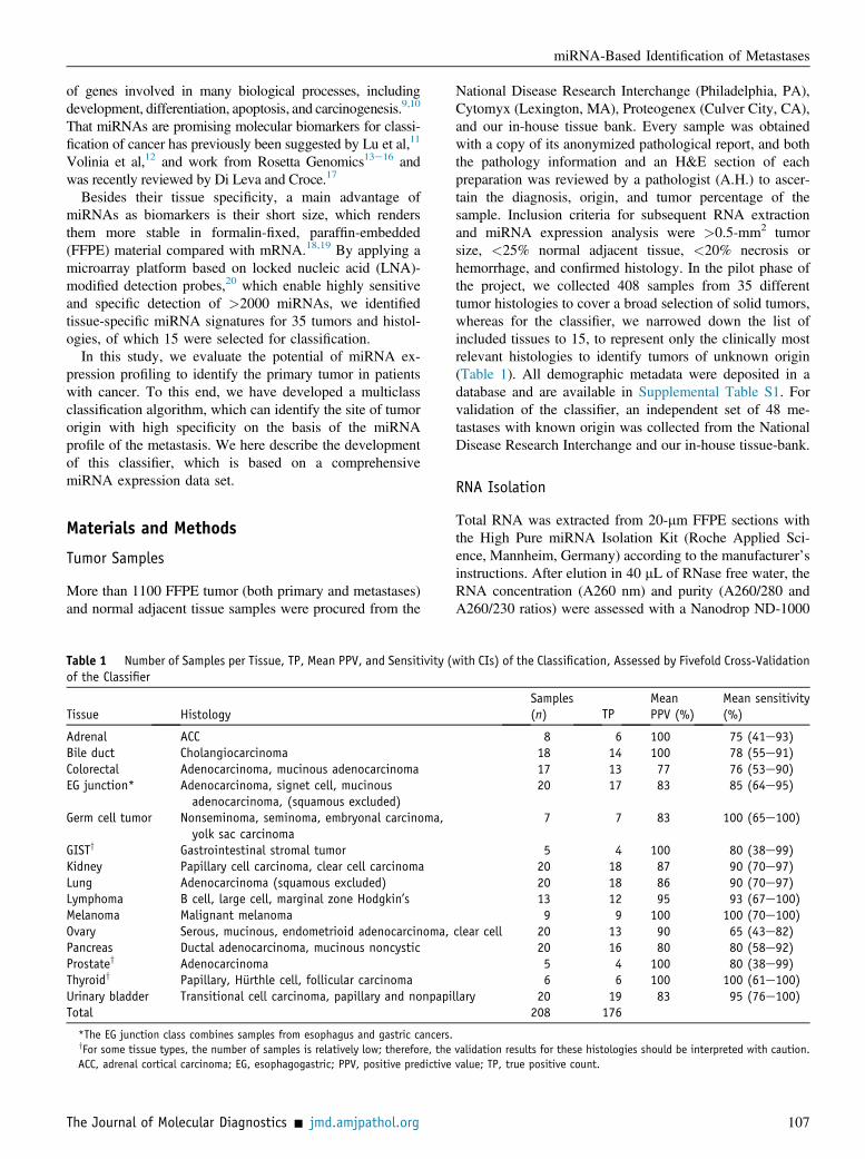
Table 1 Number of Samples per Tissue, TP, Mean PPV, and Sensitivity (of the Classifier
Tissue Histology
Adrenal ACCBile duct CholangiocarcinomaColorectal Adenocarcinoma, mucinous adenocarcinomaEG junction* Adenocarcinoma, signet cell, mucinous
adenocarcinoma, (squamous excluded)Germ cell tumor Nonseminoma, seminoma, embryonal carcinoma,
yolk sac carcinomaGISTy Gastrointestinal stromal tumorKidney Papillary cell carcinoma, clear cell carcinomaLung Adenocarcinoma (squamous excluded)Lymphoma B cell, large cell, marginal zone Hodgkin’sMelanoma Malignant melanomaOvary Serous, mucinous, endometrioid adenocarcinoma,Pancreas Ductal adenocarcinoma, mucinous noncysticProstatey AdenocarcinomaThyroidy Papillary, Hurthle cell, follicular carcinomaUrinary bladder Transitional cell carcinoma, papillary and nonpapiTotal
*The EG junction class combines samples from esophagus and gastric cancers.yFor some tissue types, the number of samples is relatively low; therefore, theACC, adrenal cortical carcinoma; EG, esophagogastric; PPV, positive predictive
The Journal of Molecular Diagnostics - jmd.amjpathol.org
National Disease Research Interchange (Philadelphia, PA),Cytomyx (Lexington, MA), Proteogenex (Culver City, CA),and our in-house tissue bank. Every sample was obtainedwith a copy of its anonymized pathological report, and boththe pathology information and an H&E section of eachpreparation was reviewed by a pathologist (A.H.) to ascer-tain the diagnosis, origin, and tumor percentage of thesample. Inclusion criteria for subsequent RNA extractionand miRNA expression analysis were >0.5-mm2 tumorsize, <25% normal adjacent tissue, <20% necrosis orhemorrhage, and confirmed histology. In the pilot phase ofthe project, we collected 408 samples from 35 differenttumor histologies to cover a broad selection of solid tumors,whereas for the classifier, we narrowed down the list ofincluded tissues to 15, to represent only the clinically mostrelevant histologies to identify tumors of unknown origin(Table 1). All demographic metadata were deposited in adatabase and are available in Supplemental Table S1. Forvalidation of the classifier, an independent set of 48 me-tastases with known origin was collected from the NationalDisease Research Interchange and our in-house tissue-bank.
RNA Isolation
Total RNA was extracted from 20-mm FFPE sections withthe High Pure miRNA Isolation Kit (Roche Applied Sci-ence, Mannheim, Germany) according to the manufacturer’sinstructions. After elution in 40 mL of RNase free water, theRNA concentration (A260 nm) and purity (A260/280 andA260/230 ratios) were assessed with a Nanodrop ND-1000
with CIs) of the Classification, Assessed by Fivefold Cross-Validation
Samples(n) TP
MeanPPV (%)
Mean sensitivity(%)
8 6 100 75 (41e93)18 14 100 78 (55e91)17 13 77 76 (53e90)20 17 83 85 (64e95)
7 7 83 100 (65e100)
5 4 100 80 (38e99)20 18 87 90 (70e97)20 18 86 90 (70e97)13 12 95 93 (67e100)9 9 100 100 (70e100)
clear cell 20 13 90 65 (43e82)20 16 80 80 (58e92)5 4 100 80 (38e99)6 6 100 100 (61e100)
llary 20 19 83 95 (76e100)208 176
validation results for these histologies should be interpreted with caution.value; TP, true positive count.
107
Søkilde et al
spectrophotometer (Thermo Scientific, Wilmington DE).The RNA was stored at �80�C until further analysis.
Microarray Profiling
For microarray analysis, we applied a common referencedesign in which the reference sample contains a mixture oftotal RNA to represent all tissue types in the study. This allowsfor both one- and two-channel data analysis, as described indetail by Søkilde et al.21 In the present study, we applied thetwo-channel ratio analysis, because this permits comparisonacross different array versions. One microgram of total RNAfrom each sample was labeled by using the miRCURY LNAmicroRNA Power labeling Kit (Exiqon, Vedbæk, Denmark),according to a two-step protocol as follows: calf intestinalalkaline phosphatase was applied to remove terminal 50
phosphates, and fluorescent labels were attached enzymati-cally to the 30 end of the miRNAs. Sample-specific RNA waslabeled with Hy3 (green) fluorophore, whereas the commonreference RNA pool was labeled with the Hy5 (red).
The Hy3- and Hy5-labeled RNA samples were mixed andco-hybridized to miRCURY LNA Arrays version Dx10 andversion 11 (Exiqon), which contain Tm-normalized captureprobes that target miRNAs from human, mouse, and rat, asregistered in miRBase version 19.0 at the Sanger Institute.22
Hybridization was performed overnight for 16 hours at 65�Cin a Tecan HS4800 hybridization station (Tecan, Männedorf,Switzerland). After washing and drying, the microarrayslides were scanned under ozone-free conditions (ozone level< 2.0 ppb to minimize bleaching of the fluorescent dyes) in aG2565BAMicroarray Scanner System (Agilent, Santa Clara,CA). The resulting images were quantified with Imagenesoftware version 8.0 (BioDiscovery, El Segundo, CA), andboth automatic quality control (flagging of poor spots by thesoftware) and manual, visual inspection were performed toensure the highest possible data quality.
Quantitative Real-Time PCR
The expression levels of 39 selected miRNAs were validatedby quantitative real-time PCR to apply the miRCURY LNAUniversal RT microRNA PCR system and SYBR Greenmaster mix according to the manufacturer’s instructions(Exiqon). The results are shown in Supplemental Figure S1.
Data Preprocessing and Normalization
All low-level analyses were performed in the R environment,including importing and preprocessing of the data with theuse of the LIMMA package (http://www.bioconductor.org/packages/2.13/bioc/html/limma.html, last accessed August29, 2013). Mean pixel intensities were used to calculatesignal (foreground) spot intensities, and median pixel in-tensities were applied to estimate background intensity. Afterexcluding flagged spots from the analysis, the normexpbackground correction method, with offset equal to 10, was
108
applied.23 For intraslide normalization, the global Lowess(Locally Weighted Scatterplot Smoothing) regression algo-rithm was applied, and log2 ratios of four intraslide replicateswere averaged. All expression data were deposited in theRosetta Resolver (Rosetta Biosoftware, Hoddesdon, UK)data management and analysis system.
Feature Selection and Classification
A miRNA expression database was built to identify miRNAswith high discriminatory power between tumor histologies.Three approaches for feature selection (that is,filters,wrappers,and embedded methods) are commonly used.24 Here, we haveapplied both filtering and a wrapper; differentially expressedmiRNAswere identifiedby running a one versus one, aswell asa one versus all t-tests for each histology, followed by rankingof the most significant candidate miRNAs. In addition, thefeature selection embedded in the least absolute shrinkage andselection operator (LASSO) classification algorithm wasapplied. The LASSO classifier was originally described byTibshirani25 and is based on a multinomial logistic model,which is fitted by using L1 regularization.26 The regularizationparameter is chosen by evaluating the results of a cross-validation along the entire regularization path. To solve theL1 regularized optimization problem we used the glmnet al-gorithm.27 The classifier was built on log2 ratio data from the208 samples and 15 cancer classes listed in Table 1.We tested and fivefold cross-validated the LASSO algo-
rithm and have listed its model coefficient, a measure ofdiscriminatory potential, in Supplemental Table S2. For thepresent multiclass classification task, we found that LASSOperformed on par with or even better than other classifica-tion algorithms, such as K nearest neighbor and lineardiscriminant analysis (data not shown).
Statistical Analysis
All calculations and statistical tests were done in the freesoftware environment for statistical computing and graphics Rversion 2.9.2 (http://www.r-project.org, last accessed August29, 2013). Formicroarray analysis, the open sourcepackage forR, Bioconductor, was used (http://www.bioconductor.org).Confidence intervals were calculated with the Wilson methodby using the R binom library, and the following script:binom.confint(x, n, conf.level Z 0.95, methods Z wilson),where x Z number of successes and n Z number of inde-pendent trials.
Results
Sample Selection
To obtain as comprehensive a data set as possible for con-structing the microarray tumor database, we initially profiled1129 samples that spanned most tumor sites and covered 35major histological subtypes. When considering which tissue
jmd.amjpathol.org - The Journal of Molecular Diagnostics

miRNA-Based Identification of Metastases
classes to include in the final classifier, we focused on thosemetastatic cancers that are most frequently found, that is, atautopsy, in CUP origin. Greater than 75% of all CUP cases areadenocarcinomas and poorly differentiated carcinomas, ofwhich the most common primary sites (when determined) arepancreas (25%), lung (20%), stomach, colorectum, and hep-atobiliary tract (8% to 12% each), and kidney (5%). Squamouscell carcinomas account for 10% to 15%, most of which arisefrom head and neck tumors, whereas melanoma represents 4%of all CUP cases. These relative frequencies, however, shouldbe interpreted with caution, because the epidemiology of CUPis changing due to both improved medical imaging technologyand lifestyle habits; therefore, different studies report dissimilarfrequencies of primary sites.28 On the basis of the above con-siderations, our selection of tissues includes the major carci-noma (12 of 15 histologies), as well as melanoma, germ celltumors (clear cell tumors), and lymphoma (small cell neo-plasms), because these can be difficult to distinguish frompoorly differentiated carcinoma. Finally, taking into accountthat in the clinical setting FFPE material is readily availableand, thus, represents an important resource for molecularprofiling, and that miRNAs are stable in FFPE blocks andstraightforward to extract,29 we decided to develop the classi-fier on FFPE material. Table 1 lists the 15 tissues and histol-ogies (columns 1 and 2), whichwere included in the training setthat consisted of 208 FFPE samples (199 primary tumors and 9metastases). A detailed summary of all patient demographicdata can be found in Supplemental Table S1, and the expres-sion data are deposited in Gene Expression Omnibus (http://www.ncbi.nlm.nih.gov/geo; accession number GSE50894).
Tissue-Specific miRNA Expression
The distribution of tissue-specific miRNAs (ie, thosemiRNAs that were preferentially expressed in samplesoriginating from one tissue compared with all other tissues)is summarized in the heatmap (Figure 1).
Figure 1 Expression of cancer-tissue specific miRNAs (rows) across 208 samheatmap shows median normalized log2 data for the top 5 to 10 miRNAs, identifieview of individual miRNAs and their classification score, please see Supplementa
The Journal of Molecular Diagnostics - jmd.amjpathol.org
From the heatmap it is evident that some histologies areeasy to distinguish from the rest because of a strong and ho-mogeneous tissue-specific miRNA signature [adrenal, lym-phoma, germ cell, prostate, gastrointestinal stromal tumor(GIST), and melanoma], whereas other tissue origins aremore difficult to classify accurately, mainly because of het-erogeneity within the group (ovary, lung) or because of highsimilarity to related tissue types [colorectal and esophago-gastric (EG) junction].
Feature Selection
Because selection of the candidate biomarkers is crucialfor performance of the classifier, we took several differentapproaches to identify the best possible tissue-specificmarkers. The first and simplest approach was to run one-against-one and one-against-all comparisons for each tissue,identifying differentially expressed miRNAs by t-tests.However, because running multiple two-sample t-tests canresult in an increased risk of committing a type I error (falsepositive), we also applied analysis of variance to compare all15 means (of the different histologies) in one test. Yet,because filtering-based methods, such as t-test and analysis ofvariance, do not provide a cross-validation option for opti-mization of the set of discriminatory features, we decided foran embedded approach, namely the LASSO method, whichintegrates feature selection within the classifier construction.With this method 132 miRNAs with high tissue discrimina-tory potential were identified; these are listed in SupplementalTable S2, which is a data matrix showing each feature’sLASSO model coefficient for the particular tissue of interest.
Finally, we made a literature search for tissue-specificmiRNAs and compared these with our top candidate dis-criminatory miRNAs. There was, not surprisingly, a highdegree of overlap between the miRNAs identified in ourstudy and those reported previously as having high predic-tive ability for cancer classification.12,15,16 The overlapping
ples (columns) that represent the 15 histologies in the training set. Thed by LASSO’s embedded feature selection algorithm, per class. For a detailedl Table S2.
109

Table 2 The miRNAs That Can Be Used for Identification of Tumor Origin
Tissue miRNA
ACC hsa-miR-129*, hsa-miR-136, hsa-miR-202*, hsa-miR-218, hsa-miR-376c,hsa-miR-488
Bile duct; cholangiocarcinoma hsa-miR-23a, hsa-miR-122, hsa-miR-214, hsa-miR-452, hsa-miR-616Colorectal; adenocarcinoma, mucinous adenocarcinoma hsa-miR-26b*, hsa-miR-95, hsa-miR-99b* hsa-miR-134, hsa-miR-192*,
hsa-miR-194 hsa-miR-196b, hsa-miR-220b, hsa-miR-224 hsa-miR-433,hsa-miR-491-5p, hsa-miR-516a-3p hsa-miR-629*, hsa-miR-767-3p,hsa-miR-890
EG junction; adenocarcinoma, signet cell, mucinousadenocarcinoma (squamous excluded)y
hsa-miR-7, hsa-miR-16-1*, hsa-miR-96* hsa-miR-124, hsa-miR-133b,hsa-miR-143 hsa-miR-145*, hsa-miR-147b, hsa-miR-450b-3pz
hsa-miR-323, hsa-miR-504, hsa-miR-548a-3p hsa-miR-548b-5p,hsa-miR-647, hsa-miR-892b
Germ cell tumor; nonseminoma, seminoma, embryonalcarcinoma, yolk sac carcinoma
hsa-miR-154*, hsa-miR-367, hsa-miR-372 hsa-miR-423-3p,hsa-miR-769-3p
GIST hsa-miR-132, hsa-miR-574-3p, hsa-miR-603Kidney; papillary cell carcinoma, clear cell carcinoma hsa-miR-10b, hsa-miR-30a*, hsa-miR-92a-1* hsa-miR-105,
hsa-miR-148a*, hsa-miR-196a hsa-miR-199b-5p, hsa-miR-204,hsa-miR-210 hsa-miR-340, hsa-miR-491-3p, hsa-miR-557
Lung; adenocarcinoma (squamous excluded) hsa-miR-23a*, hsa-miR-34b*, hsa-miR-34c-5p hsa-miR-96,hsa-miR-126*, hsa-miR-129-3p hsa-miR-185, hsa-miR-193b,hsa-miR-212 hsa-miR-217, hsa-miR-219-5p, hsa-miR-601
Lymphoma; B cell, large cell, marginal zone Hodgkin’s hsa-miR-10a, hsa-miR-27b, hsa-miR-142-5p hsa-miR-153,hsa-miR-155, hsa-miR-155* hsa-miR-451, hsa-miR-541*,hsa-miR-615-5p hsa-miR-641
Melanoma hsa-miR-146a, hsa-miR-150*, hsa-miR-211 hsa-miR-541*Ovary; serous, mucinous, endometrioid adenocarcinoma,clear cell
hsa-miR-92b, hsa-miR-130a, hsa-miR-130a* hsa-miR-135a,hsa-miR-141, hsa-miR-142-3p hsa-miR-330-5p, hsa-miR-499-5p,hsa-miR-514 hsa-miR-519c-3p, hsa-miR-522, hsa-miR-572hsa-miR-592, hsa-miR-708, hsa-miR-923
Pancreas; ductal adenocarcinoma, mucinous noncystic hsa-miR-199a-3p, hsa-miR-221*, hsa-miR-335 hsa-miR-431*,hsa-miR-454*, hsa-miR-582-3p hsa-miR-801, hsa-miR-892a
Prostate; adenocarcinoma hsa-miR-99a*, hsa-miR-133a, hsa-miR-363 hsa-miR-375, hsa-miR-924Thyroid; papillary, Hürthle cell, follicular carcinoma hsa-miR-138Urinary bladder; transitional cell carcinoma, papillaryand nonpapillary
hsa-miR-148a, hsa-miR-149, hsa-miR-203 hsa-miR-205, hsa-miR-934
yThe EG junction class combines samples from esophagus and gastric cancers.zmiR-323 was previously named miR-453.ACC, adrenal cortical carcinoma.
Søkilde et al
miRNAs are also indicated in Supplemental Table S2. ThemiRNAs that can be used for classification of tumor originare listed in Table 2.
Classifier Performance
Many different algorithms are available for multiclass can-cer classification and feature selection, such as K nearestneighbor,30 genetic algorithm,6 linear discriminant anal-ysis,31 support vector machine,32 recursive feature elimina-tion,4 nearest shrunken centroids,12 decision trees,15,33 andartificial neural networks.34,35
One of the main objectives of this study was to combinefeature selection and multiclass classification into onepipeline. The pipeline should be able to integrate identifi-cation of highly informative features useful for classificationwith cross-validation of the results. This dual function is notoffered by most other commonly used algorithms, which is
110
why we decided to remodel the LASSO algorithm for thispurpose.27 Specifically, we wished to optimize the model toobtain as high sensitivity (and accuracy) on all 15 tumorclasses as possible. This is illustrated in SupplementalFigure S2, which shows the performance of the LASSOclassifier as a function of the regularization parameter. Theoptimal value of this parameter was determined to be 4.1,because more complex models would entail more miRNAswithout a corresponding gain in performance.The results of the fivefold cross-validation of the LASSO
classifier are given in Table 3, which is a confusion matrix,showing the number of correct classifications along the di-agonal. The correct tissue of origin was predicted in themajority of cases (176 of 208 samples tested) with anoverall accuracy of 85% (CI, 79%e89%). Typically, thefalse-positive calls were because of similarities in histologywhich caused cross-reactivity; for example, three gastro-esophageal (EG junction) samples were wrongly predicted
jmd.amjpathol.org - The Journal of Molecular Diagnostics

Table 3 Confusion Matrix of Classification Results That Show the Number of Correct Classifications Along the Diagonal and the Number ofMis-Classifications Off the Diagonal (Based on Fivefold Cross-Validation of the LASSO Classifier)
Predicted class
True class
Adrenalgland
Cholangio-carcinoma
Colorectal EGjunction
Germ celltumor GIST Kidney Lung Lymphoma Melanoma Ovary Pancreas Prostate Thyroid Urothelial
Adrenal gland 6* 0 0 0 0 0
Cholangiocarcinoma 0 14* 0 0 0 0
Colorectal 0 0 13* 3 0 0
EG junction 0 1 2 17* 0 0
Germ cell tumor 0 0 0 0 7* 0 0 0 0 0 1 0 0 0 1
GIST 0 0 0 0 0 4* 0 0 0 0 0 0 0 0 0
Kidney 0 1 0 0 0 1 18* 0 0 0 1 1 0 0 0
Lung 0 2 0 0 0 0 0 18* 0 0 1 0 0 0 0
Lymphoma 0 0 0 0 0 0 0 1 12* 0 0 0 0 0 0
Melanoma 0 0 0 0 0 0 0 0 0 9* 0 0 0 0 0
Ovary 1 0 0 0 0 0 1 0 0 0 13* 0 0 0 0
Pancreas 1 0 2 0 0 0 1 0 0 0 1 16* 0 0 0
Prostate 0 0 0 0 0 0 0 0 0 0 0 0 4* 0 0
Thyroid 0 0 0 0 0 0 0 0 0 0 0 0 0 6* 0
Urothelial 0 0 0 0 0 0 0 1 1 0 1 0 1 0 19*
*The number of correct classifications.
miRNA-Based Identification of Metastases
as colorectal. We were not able to separate stomach cancersfrom esophageal adenocarcinomas, based on their miRNAprofile; which is why we decided to pool these two, rathersimilar histologies, which is consistent with other, recentmiRNA profiling studies.15,16
Validation on Metastatic Samples
Except for melanoma, the LASSO classifier was built onprimary tumors. Therefore, it was important to validate itsperformance in an independent test set, consisting of me-tastases (n Z 48) to different sites, including liver, lymphnodes, and omentum, to ensure that overfitting to the orig-inal training data were not an issue. The results of thevalidation are summarized in Table 4. During the optimi-zation of the classifier, we discovered that even though thevalidation samples all contained <25% normal surroundingtissue, the signal from especially the liver, classified mostmetastases to the liver as cholangiocarcinoma. Therefore, itwas necessary to add the rule to the classifier that the site ofmetastasis cannot be classified as the primary tumor (ie,metastasis to the liver is excluded from being identified as aprimary liver tumor). The prediction of the LASSO classi-fier was correct in 42 of 48 cases (accuracy, 88%; CI, 75%e94%), in either the first (33 cases) or the second (nine cases)classification attempt. Thus, the classification of the inde-pendent test set that consisted of metastatic samples onlyshowed that the performance of the LASSO classifier wascomparable with the estimates from the fivefold cross-validation. The same trend of misclassification of thedigestive system is seen for the metastatic samples, as forthe primary tumors. Unfortunately, it has not been possibleto test metastases from all of the histological classes and toall metastatic locations because of limited availability ofmetastatic samples.
The Journal of Molecular Diagnostics - jmd.amjpathol.org
Discussion
CUP represents a well-recognized and important clinicalproblem, because optimal treatment selection depends on acorrect identification of the site of origin, which is per defi-nition occult in a patient presenting with CUP. Therefore,many attempts have been made to improve diagnostic pa-thology workup of CUP, ranging from purely immunohisto-chemical schemes for subtyping the tumor,36 over combinedclassification approaches,35 to proteomic analysis37 and ma-chine learning algorithms that are based on large-scalemRNAmicroarray profiling4,7,32,38,39 or on reverse transcription-PCR data.6,31,40 Recently, miRNAs, which are characterizedby their highly tissue-specific expression, have also been re-ported as useful for classification of tumor types11,12 and forcarcinoma of unknown primary origin.13,15,16
In this study, we have applied an LNA-enhancedmicroarrayplatform to generate miRNA expression profiles from 208FFPE samples that represent 15 different tumor histologies.The miRNA data were used to successfully develop and vali-date a novel classification scheme, based on the LASSO al-gorithm, which integrates feature selection within the classifierconstruction.27 The accuracy of the LASSO algorithm was85% (CI, 79%e89%) when assessed by fivefold cross-validation on the initial training set, and 88% (CI, 75%e94%)when applied on an independent test set of 48metastases.Thus, the present approach has approximately the samesensitivity as other multiclass cancer classification methods.6,15
Where the LASSO method shows its strength, is its approxi-mately equal sensitivity to all of the classes in the classifier.Othermethodsmayhavepoorperformanceona few classes; forexample, the combined tree and K nearest neighborebasedmiRNA classifier reported by Rosenfeld et al15 has zero sensi-tivity to bladder cancer, whereas our LASSO algorithm detectsthis histologywith amean sensitivity of 95%(CI, 76%e100%).
111

Table 4 Validation of the LASSO Classifier on an Independent Test Set of 48 Metastatic Samples
True class Correct Metastasis site First prediction Percent (%) Second prediction Percent (%)
Colorectal Second Pelvis EG junction* 31 Colorectal 22Colorectal First Adrenal gland Colorectal 52 Ovary 16Colorectal First Liver Colorectal 74 Ovary 11Colorectal First Liver Colorectal 51 EG junction 20Colorectal First Liver Colorectal 81 Ovary 7Colorectal First Liver Colorectal 70 Ovary 9Colorectal No Liver Pancreas 30 Kidney 17Colorectal First Lung Colorectal 54 EG junction 14Colorectal First Liver Colorectal 61 EG junction 11Colorectal Second Omentum Pancreas 35 Colorectal 22Colorectal Second Liver EG junction 30 Colorectal 19Colorectal Second Omentum EG junction 40 Colorectal 37Colorectal First Lung Colorectal 53 EG junction 17Colorectal First Pending Colorectal 56 EG junction 23EG junction First Lymph node EG junction 17 Pancreas 15EG junction First Lymph node EG junction 54 Lymphoma 19EG junction First Lymph node EG junction 46 Colorectal 14Pancreas First Lymph node Pancreas 81 Lung 3Pancreas Second Omentum EG junction 19 Pancreas 17Pancreas Second Abdominal wall Lung 17 Pancreas 16Pancreas No Liver Colorectal 51 EG junction 22Pancreas No Omentum EG junction 40 Colorectal 31Ovary First Bowel Ovary 37 Urothelial carcinoma 23Ovary First Colon Ovary 31 Lung 22Ovary Second Colon Pancreas 60 Ovary 13Ovary First Colon Ovary 94 Thyroid 4Ovary No Colon EG junction 46 Pancreas 9Ovary First Gastric wall Ovary 38 Thyroid 38Ovary First Omentum Ovary 33 Lung 18Ovary First Omentum Ovary 37 Pancreas 26Ovary First Omentum Ovary 45 Thyroid 12Ovary First Omentum Ovary 70 Kidney 19Ovary No Omentum Urothelial 49 Lung 32Ovary First Omentum Ovary 83 Pancreas 6Ovary Second Omentum Cholangiocarcinoma 19 Ovary 16Ovary First Omentum Ovary 55 Thyroid 17Ovary First Omentum Ovary 47 Thyroid 18Ovary Second Pelvis Lung 39 Ovary 16Ovary First Pending Ovary 31 Lung 13Ovary First Pending Ovary 54 Thyroid 16Kidney First Lung Kidney 51 Cholangiocarcinoma 14Kidney First Lymph node Kidney 61 Cholangiocarcinoma 10Kidney First Adrenal gland Kidney 76 Melanoma 4Kidney First Pancreas Kidney 96 EG junction 1Kidney First Pancreas Kidney 42 Ovary 29Lung First Lymph node Lung 44 Kidney 12Lung No Lymph node Urothelial 48 Cholangiocarcinoma 30Urothelial First Colon Urothelial 75 Pancreas 13
Correct indicates whether the classifier was correct in either its first or second prediction. The percentages are calculated by the LASSO algorithm andindicate the likelihood of a correct classification of the particular tissue.
*The EG junction class combines samples from esophagus and gastric cancers.
Søkilde et al
Identifying the algorithm that is best suited for clinical useis an ongoing and controversial discussion. It has been arguedthat black box machine learning classifiers, such as supportvector machine and artificial neural networks, are not astransparent as, for example, decision trees for practical use by
112
pathologists.35 However, despite their intuitive and visualappeal, decision trees are not without limitations. If theybecome over-complex, they do not generalize the data well,and there is no backtracking option, meaning that a local(erroneous) optimal solution will prevent one from reaching
jmd.amjpathol.org - The Journal of Molecular Diagnostics

miRNA-Based Identification of Metastases
the global optimal solution (eg, the correct classification willbe missed once a wrong path is followed down a branch).41 Inthis respect, it is interesting that the binary decision treeoriginally proposed by Rosenfeld et al15 for miRNA classi-fication of cancer tissue has undergone substantial structuralchanges in the follow-up study by Rosenwald et al16 andMeiri et al,13 resulting in a more complex tree (with 12 branchpoints for some class labels) and more than half of the 48miRNAs reported in the original study replaced by other,tissue-specific miRNAs. This adjustment of tree structureprobably reflects both the altered tissue selection and thatseveral different tree designs may co-exist.
A recent article by Centeno et al35 suggests a hybrid, de-cision tree model, which incorporates both immunohisto-chemistry (IHC) and expression data for optimal separationof four types of carcinoma. However, one should bear inmind that interpretation of IHC staining is subjective;therefore, it can be difficult to determine a positive from anegative. As Gown’s fourth law of immunohistochemistrylaconically states, “All that turns brown is not positive.”42,p30
A meta-analysis performed by Anderson and Weiss43
showed that IHC only provides correct tissue identificationin 65.6% of metastatic cancers, and recent studies by Weisset al44 and by Oien et al45 both conclude molecular profilingoutperforms classification by IHC, in particular in cases withpoorly differentiated tumors. This underscores the need forimproved identification of the origin of metastases, which areinherently more difficult to classify than their corresponding,and often more well differentiated, primary tumor.
We believe that the LASSO algorithm offers the best ofboth worlds, that is, the performance of the complex ma-chine learning algorithm together with the intuitive under-standing of the simpler classifiers, because it is powerful andeasy to train, the model complexity (number and type offeatures) can be easily controlled, over-fitting is restricted bya penalty term, and data interpretation is simple; the readoutis the likelihood of a correct classification. Other conven-tional methods, such as linear discriminant analysis and Knearest neighbor, resulted in less accurate classification(data not shown) of this data compared with LASSO.
Because we were able to identify the origin of metastatictumors by their miRNA profile is consistent with the para-digm that the geneticmakeup of a primary tumor is retained inthe distant metastases.5,13,31 Several of the identified tissue-specific miRNAs are involved in differentiation, so if themiRNA signature is retained in the metastases, it should bepossible to identify its tissue of origin, unless the cancer is sodedifferentiated that all molecular marks of its primary originare lost. This brings up the question whether a real CUPrepresents an entity of its own, with a CUP-specific ratherthan a primary tissue-specific molecular signature.4,46,47
Some tissues are inherently difficult to classify correctly,for example, pancreas cancer, which is often poorly differ-entiated or dedifferentiated, and lung cancer with manypossible histologies. In our validation study, the classifier wasable to correctly label pancreas as the primary site in three of
The Journal of Molecular Diagnostics - jmd.amjpathol.org
five cases, which is not impressive but still better than whatcould be achieved in the commercial CupPrint follow-upstudy,39 in which none of the three pancreas cancers could beidentified. In addition, Park et al48 found that with IHCmarkers the sensitivity toward the combined group of pancreascancer and cholangiocarcinoma was quite low (28%).
We discovered that a main limitation to this type of studyis the identification of the superimposed host tissue (the site ofthemetastasis) signature, typically liver or lymph node, ratherthan the metastasis signature; in particular, when the amountof host tissue is large compared with the metastasis. Specif-ically, a primary liver cancer could not readily be distin-guished from a livermetastasis because of the overlaid, strongliver-associated miRNA signature. Therefore, when opti-mizing the classifier, we had to make the assumption that ametastasis to the liver cannot be primary liver cancer and thata lymph node metastasis is not a lymphoma. A likely solutionto the problem of contaminating surrounding tissue is to applylaser capture microdissection as suggested by Chen et al49 formiRNA analysis in intrahepatic cholangiocarcinoma.
In conclusion, our study suggests that miRNA expressionprofiling on FFPE tissue, followed by an efficient multiclassclassification algorithm, in this case LASSO, can efficientlypredict the primary origin of a tumor. Thus, it may providepathologists with an adjunct molecular diagnostic tool thateither alone or in combinationwith other relevant biomarkers,such as mRNA and proteins, for example, automated IHC,can improve their capability to correctly identify the origin ofmetastatic tumors, and eventually, to advance and expediterational, specific therapy of patients with metastatic disease.
Finally, we do acknowledge that even in the light of theseencouraging results, some caution is required; to translatethe present discovery work and proof of concept into clin-ical utility requires a reduction in test complexity, migrationof the microarray analysis to a quantitative reverse tran-scription PCR platform, further trimming of the numberof discriminatory miRNAs, and prospective clinical trials.Such trials will determine the significance of miRNA pro-filing in molecular cancer diagnostics.
Acknowledgments
We thank Drs. Bogumil Kaczkowski and Peder Worning forinitial support vector machine classification of the data andGitte Friis and Stine Jørgensen for excellent technicalassistance.
Supplemental Data
Supplemental material for this article can be found athttp://dx.doi.org/10.1016/j.jmoldx.2013.10.001.
References
1. Greco FA: Cancer of unknown primary site. Am Soc Clin Oncol EducBook 2013, 2013:175e181
113

Søkilde et al
2. Daugaard D, Møller A, Petersen B: Tumors of unknown origin. In:Edited by Cavalli F, Kaye S, Hansen H, Armitage J, Piccart-Gebhart M.Textbook of Medical Oncology. London, Informa, 2009, pp 313e322
3. GolubTR, SlonimDK,TamayoP,HuardC,GaasenbeekM,Mesirov JP,Coller H, Loh ML, Downing JR, Caligiuri MA, Bloomfield CD,Lander ES: Molecular classification of cancer: class discovery and classprediction by gene expression monitoring. Science 1999, 286:531e537
4. Ramaswamy S, Tamayo P, Rifkin R, Mukherjee S, Yeang CH,Angelo M, Ladd C, Reich M, Latulippe E, Mesirov JP, Poggio T,Gerald W, Loda M, Lander ES, Golub TR: Multiclass cancer diagnosisusing tumor gene expression signatures. Proc Natl Acad Sci U S A2001, 98:15149e15154
5. Buckhaults P, Zhang Z, Chen YC, Wang TL, St Croix B, Saha S,Bardelli A, Morin PJ, Polyak K, Hruban RH, Velculescu VE,Shih IeM: Identifying tumor origin using a gene expression-basedclassification map. Cancer Res 2003, 63:4144e4149
6. Ma XJ, Patel R, Wang X, Salunga R, Murage J, Desai R, Tuggle JT,Wang W, Chu S, Stecker K, Raja R, Robin H, Moore M, Baunoch D,Sgroi D, Erlander M: Molecular classification of human cancers usinga 92-gene real-time quantitative polymerase chain reaction assay. ArchPathol Lab Med 2006, 130:465e473
7. Kurahashi I, Fujita Y, Arao T, Kurata T, Koh Y, Sakai K,Matsumoto K, Tanioka M, Takeda K, Takiguchi Y, Yamamoto N,Tsuya A, Matsubara N, Mukai H, Minami H, Chayahara N,Yamanaka Y, Miwa K, Takahashi S, Takahashi S, Nakagawa K,Nishio K: A microarray-based gene expression analysis to identifydiagnostic biomarkers for unknown primary cancer. PLoS One 2013,8:e63249
8. Greco FA, Lennington WJ, Spigel DR, Hainsworth JD: Molecularprofiling diagnosis in unknown primary cancer: accuracy and ability tocomplement standard pathology. J Natl Cancer Inst 2013, 105:782e790
9. Esquela-Kerscher A, Slack FJ: Oncomirs - microRNAs with a role incancer. Nat Rev Cancer 2006, 6:259e269
10. Iorio MV, Croce CM: microRNA involvement in human cancer.Carcinogenesis 2012, 33:1126e1133
11. Lu J, Getz G, Miska EA, varez-Saavedra E, Lamb J, Peck D, Sweet-Cordero A, Ebert BL, Mak RH, Ferrando AA, Downing JR, Jacks T,Horvitz HR, Golub TR: MicroRNA expression profiles classify humancancers. Nature 2005, 435:834e838
12. Volinia S, Calin GA, Liu CG, Ambs S, Cimmino A, Petrocca F,Visone R, Iorio M, Roldo C, Ferracin M, Prueitt RL, Yanaihara N,Lanza G, Scarpa A, Vecchione A, Negrini M, Harris CC, Croce CM: AmicroRNA expression signature of human solid tumors defines cancergene targets. Proc Natl Acad Sci U S A 2006, 103:2257e2261
13. Meiri E, Mueller WC, Rosenwald S, Zepeniuk M, Klinke E,Edmonston TB, Werner M, Lass U, Barshack I, Feinmesser M,Huszar M, Fogt F, Ashkenazi K, Sanden M, Goren E, Dromi N,Zion O, Burnstein I, Chajut A, Spector Y, Aharonov R: A second-generation microRNA-based assay for diagnosing tumor tissueorigin. Oncologist 2012, 17:801e812
14. Pentheroudakis G, Pavlidis N, Fountzilas G, Krikelis D, Goussia A,Stoyianni A, Sanden M, St Croix B, Yerushalmi N, Benjamin H,Meiri E, Chajut A, Rosenwald S, Aharonov R, Spector Y: NovelmicroRNA-based assay demonstrates 92% agreement with diagnosisbased on clinicopathologic and management data in a cohort of pa-tients with carcinoma of unknown primary. Mol Cancer 2013, 12:57
15. Rosenfeld N, Aharonov R, Meiri E, Rosenwald S, Spector Y,Zepeniuk M, Benjamin H, Shabes N, Tabak S, Levy A, Lebanony D,Goren Y, Silberschein E, Targan N, Ben-Ari A, Gilad S, Sion-Vardy N, Tobar A, Feinmesser M, Kharenko O, Nativ O, Nass D,Perelman M, Yosepovich A, Shalmon B, Polak-Charcon S, Fridman E,Avniel A, Bentwich I, Bentwich Z, Cohen D, Chajut A, Barshack I:MicroRNAs accurately identify cancer tissue origin. Nature Biotechnol2008, 26:462e469
16. Rosenwald S, Gilad S, Benjamin S, Lebanony D, Dromi N,Faerman A, Benjamin H, Tamir R, Ezagouri M, Goren E, Barshack I,
114
Nass D, Tobar A, Feinmesser M, Rosenfeld N, Leizerman I,Ashkenazi K, Spector Y, Chajut A, Aharonov R: Validation of amicroRNA-based qRT-PCR test for accurate identification of tumortissue origin. Mod Pathol 2010, 23:814e823
17. Di Leva G, Croce CM: miRNA profiling of cancer. Curr Opin GenetDev 2013, 23:3e11
18. Liu A, Tetzlaff MT, Vanbelle P, Elder D, Feldman M, Tobias JW,Sepulveda AR, Xu X: MicroRNA expression profiling outperformsmRNA expression profiling in formalin-fixed paraffin-embedded tis-sues. Int J Clin Exp Pathol 2009, 2:519e527
19. Siebolts U, Varnholt H, Drebber U, Dienes HP, Wickenhauser C,Odenthal M: Tissues from routine pathology archives are suitable formicroRNA analyses by quantitative PCR. J Clin Pathol 2009, 62:84e88
20. Castoldi M, Benes V, Hentze MW, Muckenthaler MU: miChip: amicroarray platform for expression profiling of microRNAs based onlocked nucleic acid (LNA) oligonucleotide capture probes. Methods2007, 43:146e152
21. Søkilde R, Kaczkowski B, Barken K, Mouritzen P, Møller S,Litman T: MicroRNA expression analysis by LNA enhanced micro-arrays. Edited by Gusev Y. MicroRNA Profiling in Cancer: A Bioin-formatics Perspective. Singapore, Pan Stanford Publishing, 2009, pp23e46
22. Kozomara A, Griffiths-Jones S: miRBase: integrating microRNAannotation and deep-sequencing data. Nucleic Acids Res 2011, 39:D152eD157
23. Ritchie ME, Silver J, Oshlack A, Holmes M, Diyagama D,Holloway A, Smyth GK: A comparison of background correctionmethods for two-colour microarrays. Bioinformatics 2007, 23:2700e2707
24. Ma S, Huang J: Penalized feature selection and classification in bio-informatics. Brief Bioinform 2008, 9:392e403
25. Tibshirani R: Regression shrinkage and selection via the lasso. J RoyalStat Soc 1996, 58:267e288
26. Efron B, Hastie T, Johnstone I, Tibshirani R: Least angle regression.Ann Statist 2004, 32:409e499
27. Friedman J, Hastie T, Tibshirani R: Regularization Paths for Gener-alized Linear Models via Coordinate Descent. J Stat Softw 2010, 33:1e22
28. Pentheroudakis G, Golfinopoulos V, Pavlidis N: Switching bench-marks in cancer of unknown primary: from autopsy to microarray. EurJ Cancer 2007, 43:2026e2036
29. Li J, Smyth P, Flavin R, Cahill S, Denning K, Aherne S, Guenther SM,O’Leary JJ, Sheils O: Comparison of miRNA expression patterns usingtotal RNA extracted from matched samples of formalin-fixed paraffin-embedded (FFPE) cells and snap frozen cells. BMC Biotechnol 2007,7:36
30. van Laar RK, Ma XJ, de JD, Wehkamp D, Floore AN, Warmoes MO,Simon I, Wang W, Erlander M, van’t Veer LJ, Glas AM: Imple-mentation of a novel microarray-based diagnostic test for cancer ofunknown primary. Int J Cancer 2009, 125:1390e1397
31. Talantov D, Baden J, Jatkoe T, Hahn K, Yu J, Rajpurohit Y, Jiang Y,Choi C, Ross JS, Atkins D, Wang Y, Mazumder A: A quantitativereverse transcriptase-polymerase chain reaction assay to identify met-astatic carcinoma tissue of origin. J Mol Diagn 2006, 8:320e329
32. Tothill RW, Kowalczyk A, Rischin D, Bousioutas A, Haviv I, vanLaar RK, Waring PM, Zalcberg J, Ward R, Biankin AV,Sutherland RL, Henshall SM, Fong K, Pollack JR, Bowtell DD,Holloway AJ: An expression-based site of origin diagnostic methoddesigned for clinical application to cancer of unknown origin. CancerRes 2005, 65:4031e4040
33. Shedden KA, Taylor JM, Giordano TJ, Kuick R, Misek DE,Rennert G, Schwartz DR, Gruber SB, Logsdon C, Simeone D,Kardia SL, Greenson JK, Cho KR, Beer DG, Fearon ER, Hanash S:Accurate molecular classification of human cancers based on geneexpression using a simple classifier with a pathological tree-basedframework. Am J Pathol 2003, 163:1985e1995
jmd.amjpathol.org - The Journal of Molecular Diagnostics
miRNA-Based Identification of Metastases
34. Dennis JL, Oien KA: Hunting the primary: novel strategies for definingthe origin of tumours. J Pathol 2005, 205:236e247
35. Centeno BA, Bloom G, Chen DT, Chen Z, Gruidl M, Nasir A,Yeatman TY: Hybrid model integrating immunohistochemistry andexpression profiling for the classification of carcinomas of unknownprimary site. J Mol Diagn 2010, 12:476e486
36. Oien KA: Pathologic evaluation of unknown primary cancer. SeminOncol 2009, 36:8e37
37. Bloom GC, Eschrich S, Zhou JX, Coppola D, Yeatman TJ: Elucidationof a protein signature discriminating six common types of adenocar-cinoma. Int J Cancer 2007, 120:769e775
38. Su AI, Welsh JB, Sapinoso LM, Kern SG, Dimitrov P, Lapp H,Schultz PG, Powell SM, Moskaluk CA, Frierson HF Jr., Hampton GM:Molecular classification of human carcinomas by use of gene expres-sion signatures. Cancer Res 2001, 61:7388e7393
39. Horlings HM, van Laar RK, Kerst JM, Helgason HH, Wesseling J, vander Hoeven JJ, Warmoes MO, Floore A, Witteveen A, Lahti-Domenici J, Glas AM, van’t Veer LJ, de JD: Gene expression profilingto identify the histogenetic origin of metastatic adenocarcinomas ofunknown primary. J Clin Oncol 2008, 26:4435e4441
40. Varadhachary GR, Talantov D, RaberMN,Meng C, Hess KR, Jatkoe T,LenziR, SpigelDR,WangY,Greco FA,Abbruzzese JL,Hainsworth JD:Molecular profiling of carcinoma of unknown primary and correlationwith clinical evaluation. J Clin Oncol 2008, 26:4442e4448
41. Geurts P, Irrthum A, Wehenkel L: Supervised learning with decisiontree-based methods in computational and systems biology. Mol Biosyst2009, 5:1593e1605
The Journal of Molecular Diagnostics - jmd.amjpathol.org
42. Voigt J, Mathieu M, Bibeau F: The advent of immunohistochemistry incarcinoma of unknown primary site: a major progress. In: Edited byFizazi K. Carcinoma of an Unknown Primary Site. New York, Taylor& Francis, 2006, pp 25e33
43. Anderson GG, Weiss LM: Determining tissue of origin for metastaticcancers: meta-analysis and literature review of immunohistochemistryperformance. Appl Immunohistochem Mol Morphol 2010, 18:3e8
44. Weiss LM, Chu P, Schroeder BE, Singh V, Zhang Y, Erlander MG,Schnabel CA: Blinded comparator study of immunohistochemicalanalysis versus a 92-gene cancer classifier in the diagnosis of theprimary site in metastatic tumors. J Mol Diagn 2013, 15:263e269
45. Oien KA, Dennis JL: Diagnostic work-up of carcinoma of unknownprimary: from immunohistochemistry to molecular profiling. AnnOncol 2012, 23(suppl 10):x271ex277
46. Pentheroudakis G, Briasoulis E, Pavlidis N: Cancer of unknown pri-mary site: missing primary or missing biology? Oncologist 2007, 12:418e425
47. Varadhachary G: New strategies for carcinoma of unknown primary:the role of tissue of origin molecular profiling. Clin Cancer Res 2013,19:4027e4033
48. Park SY, Kim BH, Kim JH, Lee S, Kang GH: Panels of immunohis-tochemical markers help determine primary sites of metastatic adeno-carcinoma. Arch Pathol Lab Med 2007, 131:1561e1567
49. Chen L, Yan HX, Yang W, Hu L, Yu LX, Liu Q, Li L, Huang DD,Ding J, Shen F, Zhou WP, Wu MC, Wang HY: The role of microRNAexpression pattern in human intrahepatic cholangiocarcinoma. J Hep-atol 2009, 50:358e369
115