Miniaturized pre-clinical cancer models as research and diagnostic tools
15
Miniaturized pre-clinical cancer models as research and diagnostic tools ☆ Maria Håkanson a , Edna Cukierman b, ⁎, Mirren Charnley c, ⁎⁎ a CSEM SA, Section for Micro-Diagnostics, 7302 Landquart, Switzerland b Cancer Biology Program, Fox Chase Cancer Center, Philadelphia, PA 19111, USA c Centre for Micro-Photonics and Industrial Research Institute Swinburne, Swinburne University of Technology, Victoria 3122, Australia abstract article info Article history: Accepted 24 November 2013 Available online xxxx Keywords: Microfluidics Cancer models Pre-clinical drug assessment Cell culturing devices Extracellular matrix Microfabricated model systems Tumor microenvironment Cell adhesion-mediated drug resistance Combinatorial screening platforms Body-on-a-chip Cancer is one of the most common causes of death worldwide. Consequently, important resources are directed towards bettering treatments and outcomes. Cancer is difficult to treat due to its heterogeneity, plasticity and fre- quent drug resistance. New treatment strategies should strive for personalized approaches. These should target neoplastic and/or activated microenvironmental heterogeneity and plasticity without triggering resistance and spare host cells. In this review, the putative use of increasingly physiologically relevant microfabricated cell- culturing systems intended for drug development is discussed. There are two main reasons for the use of minia- turized systems. First, scaling down model size allows for high control of microenvironmental cues enabling more predictive outcomes. Second, miniaturization reduces reagent consumption, thus facilitating combinatorial approaches with little effort and enables the application of scarce materials, such as patient-derived samples. This review aims to give an overview of the state-of-the-art of such systems while predicting their application in cancer drug development. © 2013 Elsevier B.V. All rights reserved. Contents 1. Introduction . . . . . . . . . . . . . . . . . . . . . . . . . . . . . . . . . . . . . . . . . . . . . . . . . . . . . . . . . . . . . . 0 2. The importance of the tumor microenvironment in disease progression and treatment response . . . . . . . . . . . . . . . . . . . . . . . . 0 2.1. Therapeutic strategies targeting the cancer microenvironment . . . . . . . . . . . . . . . . . . . . . . . . . . . . . . . . . . . . 0 3. Microfabricated model systems for cancer research—possibilities and challenges . . . . . . . . . . . . . . . . . . . . . . . . . . . . . . . . 0 3.1. Advantages and disadvantages of microfabricated model system for cancer research . . . . . . . . . . . . . . . . . . . . . . . . . . 0 3.1.1. Open systems . . . . . . . . . . . . . . . . . . . . . . . . . . . . . . . . . . . . . . . . . . . . . . . . . . . . . . 0 3.1.2. Closed systems . . . . . . . . . . . . . . . . . . . . . . . . . . . . . . . . . . . . . . . . . . . . . . . . . . . . . . 0 3.2. Technical challenges and opportunities . . . . . . . . . . . . . . . . . . . . . . . . . . . . . . . . . . . . . . . . . . . . . . . 0 3.2.1. Adaptation to automatic handling systems and standard read-outs . . . . . . . . . . . . . . . . . . . . . . . . . . . . . . 0 3.2.2. Microfabrication can improve conventional assays and lead to innovation of analytical techniques . . . . . . . . . . . . . . . . 0 4. The application of microfabricated model systems for cell-based cancer drug discovery . . . . . . . . . . . . . . . . . . . . . . . . . . . . . 0 4.1. 2D and 3D cell arrays to determine the effect of specific environmental parameters on cancer behavior and drug response . . . . . . . . . 0 4.2. Controlled co-culture to separate heterogeneous cell interaction from environmental factors in cancer invasion scenarios . . . . . . . . . . 0 4.3. Integrated miniaturized multi-cell type systems to study cancer drug uptake and metabolism . . . . . . . . . . . . . . . . . . . . . . 0 4.4. Combinatorial screening platforms for the tight regulation of environmental factors . . . . . . . . . . . . . . . . . . . . . . . . . . . 0 4.5. Enabling the analysis of patient-derived cells by miniaturization . . . . . . . . . . . . . . . . . . . . . . . . . . . . . . . . . . . . 0 5. Conclusions and outlook . . . . . . . . . . . . . . . . . . . . . . . . . . . . . . . . . . . . . . . . . . . . . . . . . . . . . . . . . 0 Acknowledgments . . . . . . . . . . . . . . . . . . . . . . . . . . . . . . . . . . . . . . . . . . . . . . . . . . . . . . . . . . . . . . 0 References . . . . . . . . . . . . . . . . . . . . . . . . . . . . . . . . . . . . . . . . . . . . . . . . . . . . . . . . . . . . . . . . . . 0 Advanced Drug Delivery Reviews xxx (2013) xxx–xxx Abbreviations: μCCA, microscale cell culture analog; CAM-DR, cell-adhesion mediated drug resistance; CTCs, circulating tumor cells; ECM, extracellular matrix; HGF, hepatocyte growth factor; Hh, Hedgehog; MSCs, mesenchymal stem cells; VEGF, vascular endothelial cells growth factor. ☆ This review is part of the Advanced Drug Delivery Reviews theme issue on “Innovative tissue models for in vitro drug development”. ⁎ Corresponding author. Tel.: +1 215 214 4218. ⁎⁎ Corresponding author. Tel.: +61 3 9214 8008. E-mail addresses: [email protected] (E. Cukierman), [email protected] (M. Charnley). ADR-12546; No of Pages 15 0169-409X/$ – see front matter © 2013 Elsevier B.V. All rights reserved. http://dx.doi.org/10.1016/j.addr.2013.11.010 Contents lists available at ScienceDirect Advanced Drug Delivery Reviews journal homepage: www.elsevier.com/locate/addr Please cite this article as: M. Håkanson, et al., Miniaturized pre-clinical cancer models as research and diagnostic tools, Adv. Drug Deliv. Rev. (2013), http://dx.doi.org/10.1016/j.addr.2013.11.010
Transcript of Miniaturized pre-clinical cancer models as research and diagnostic tools

Advanced Drug Delivery Reviews xxx (2013) xxx–xxx
ADR-12546; No of Pages 15
Contents lists available at ScienceDirect
Advanced Drug Delivery Reviews
j ourna l homepage: www.e lsev ie r .com/ locate /addr
Miniaturized pre-clinical cancer models as research anddiagnostic tools☆
Maria Håkanson a, Edna Cukierman b,⁎, Mirren Charnley c,⁎⁎a CSEM SA, Section for Micro-Diagnostics, 7302 Landquart, Switzerlandb Cancer Biology Program, Fox Chase Cancer Center, Philadelphia, PA 19111, USAc Centre for Micro-Photonics and Industrial Research Institute Swinburne, Swinburne University of Technology, Victoria 3122, Australia
Abbreviations: μCCA,microscale cell culture analog; CAfactor; Hh, Hedgehog; MSCs, mesenchymal stem cells; VE☆ This review is part of the Advanced Drug Delivery Revi⁎ Corresponding author. Tel.: +1 215 214 4218.⁎⁎ Corresponding author. Tel.: +61 3 9214 8008.
E-mail addresses: [email protected] (E. Cuki
0169-409X/$ – see front matter © 2013 Elsevier B.V. All rhttp://dx.doi.org/10.1016/j.addr.2013.11.010
Please cite this article as: M. Håkanson, et a(2013), http://dx.doi.org/10.1016/j.addr.201
a b s t r a c t
a r t i c l e i n f oArticle history:Accepted 24 November 2013Available online xxxx
Keywords:MicrofluidicsCancer modelsPre-clinical drug assessmentCell culturing devicesExtracellular matrixMicrofabricated model systemsTumor microenvironmentCell adhesion-mediated drug resistanceCombinatorial screening platformsBody-on-a-chip
Cancer is one of the most common causes of death worldwide. Consequently, important resources are directedtowards bettering treatments and outcomes. Cancer is difficult to treat due to its heterogeneity, plasticity and fre-quent drug resistance. New treatment strategies should strive for personalized approaches. These should targetneoplastic and/or activated microenvironmental heterogeneity and plasticity without triggering resistance andspare host cells. In this review, the putative use of increasingly physiologically relevant microfabricated cell-culturing systems intended for drug development is discussed. There are two main reasons for the use of minia-turized systems. First, scaling down model size allows for high control of microenvironmental cues enablingmore predictive outcomes. Second,miniaturization reduces reagent consumption, thus facilitating combinatorialapproacheswith little effort and enables the application of scarcematerials, such as patient-derived samples. Thisreview aims to give an overview of the state-of-the-art of such systems while predicting their application incancer drug development.
© 2013 Elsevier B.V. All rights reserved.
Contents
1. Introduction . . . . . . . . . . . . . . . . . . . . . . . . . . . . . . . . . . . . . . . . . . . . . . . . . . . . . . . . . . . . . . 02. The importance of the tumor microenvironment in disease progression and treatment response . . . . . . . . . . . . . . . . . . . . . . . . 0
2.1. Therapeutic strategies targeting the cancer microenvironment . . . . . . . . . . . . . . . . . . . . . . . . . . . . . . . . . . . . 03. Microfabricated model systems for cancer research—possibilities and challenges . . . . . . . . . . . . . . . . . . . . . . . . . . . . . . . . 0
3.1. Advantages and disadvantages of microfabricated model system for cancer research . . . . . . . . . . . . . . . . . . . . . . . . . . 03.1.1. Open systems . . . . . . . . . . . . . . . . . . . . . . . . . . . . . . . . . . . . . . . . . . . . . . . . . . . . . . 03.1.2. Closed systems . . . . . . . . . . . . . . . . . . . . . . . . . . . . . . . . . . . . . . . . . . . . . . . . . . . . . . 0
3.2. Technical challenges and opportunities . . . . . . . . . . . . . . . . . . . . . . . . . . . . . . . . . . . . . . . . . . . . . . . 03.2.1. Adaptation to automatic handling systems and standard read-outs . . . . . . . . . . . . . . . . . . . . . . . . . . . . . . 03.2.2. Microfabrication can improve conventional assays and lead to innovation of analytical techniques . . . . . . . . . . . . . . . . 0
4. The application of microfabricated model systems for cell-based cancer drug discovery . . . . . . . . . . . . . . . . . . . . . . . . . . . . . 04.1. 2D and 3D cell arrays to determine the effect of specific environmental parameters on cancer behavior and drug response . . . . . . . . . 04.2. Controlled co-culture to separate heterogeneous cell interaction from environmental factors in cancer invasion scenarios . . . . . . . . . . 04.3. Integrated miniaturized multi-cell type systems to study cancer drug uptake and metabolism . . . . . . . . . . . . . . . . . . . . . . 04.4. Combinatorial screening platforms for the tight regulation of environmental factors . . . . . . . . . . . . . . . . . . . . . . . . . . . 04.5. Enabling the analysis of patient-derived cells by miniaturization . . . . . . . . . . . . . . . . . . . . . . . . . . . . . . . . . . . . 0
5. Conclusions and outlook . . . . . . . . . . . . . . . . . . . . . . . . . . . . . . . . . . . . . . . . . . . . . . . . . . . . . . . . . 0Acknowledgments . . . . . . . . . . . . . . . . . . . . . . . . . . . . . . . . . . . . . . . . . . . . . . . . . . . . . . . . . . . . . . 0References . . . . . . . . . . . . . . . . . . . . . . . . . . . . . . . . . . . . . . . . . . . . . . . . . . . . . . . . . . . . . . . . . . 0
M-DR, cell-adhesionmediateddrug resistance; CTCs, circulating tumor cells; ECM, extracellularmatrix; HGF, hepatocyte growthGF, vascular endothelial cells growth factor.ews theme issue on “Innovative tissue models for in vitro drug development”.
erman), [email protected] (M. Charnley).
ights reserved.
l., Miniaturized pre-clinical cancer models as research and diagnostic tools, Adv. Drug Deliv. Rev.3.11.010

2 M. Håkanson et al. / Advanced Drug Delivery Reviews xxx (2013) xxx–xxx
1. Introduction
Cancer remains a leading cause of death, for example, it is predictedto be responsible for one of every four deaths in the United States for2013 [1]. Thus, it is not surprising that the majority of drug candidatesin development today are aimed at stalling or eliminating cancer [2].However, cancer drug development is costly, as it suffers from a verylow success rate. For new compounds in clinical development, the suc-cess rate is three times lower for cancer drugs than for drugs treatingcardiovascular diseases [3]. Several factors complicate the developmentof drugs that can effectively treat cancer. The high heterogeneity andplasticity of an ever-changing dynamic disease [4] make it difficultto develop drugs that are widely applicable and that have low side-effects. Resistance to treatment ensues due to both repetitive treatment[5] and the unique cues derived from the tumor-specific microenviron-ment. Development of more specific drugs, which are less likely toinduce resistance, requires not only smart drug design but also morepredictive pre-clinical tests [6,7]. Together with the development ofpersonalized approaches, treatments specifically targeting the interac-tion between cancer cells and tumor-associated microenvironmentmay enable us to overcome the hurdle posed by the heterogeneity incancer [8–10].
There is extensive evidence demonstrating the importance of thetumormicroenvironment on cancer progression [11–13] and treatmentoutcome [14,15]. In fact, the tumor-associated stroma can play a role asa pro-tumorigenic environment [16], a drug desensitization setting[6,17–19] or a drug penetration barrier [20–22], which complicatesany therapeutic approach. To this end, the specific cross talk betweena given cancer and its stroma will have to be defined for each cancertype (and perhaps for each patient) in order to achieve effective thera-peutic targeting [23]. Consequently, in vitro models that better reflect
Fig. 1. Stages in the progression towards more relevant models in cell-based assays. The predicrelevant models are used. This graph outlines some directions taken in cell culture developmeplotted in an x–y graph. The linear curve thereby explains the hypothetical increase in predicsome examples of cell-types that have a higher relevance than cell lines in monoculture are pl[41], which is envisioned to be themost relevantmodel system. Systems that enable the culturefor the improvement of cancer therapy. On the y-axis, the culture platforms of different relevancincrease the relevance compared to standard 2D culture platforms [28,244]. For example, long 2in 2D [26]. Platforms that enable 3D culture and recapitulate the organization of cells in vivo a
Please cite this article as: M. Håkanson, et al., Miniaturized pre-clinical c(2013), http://dx.doi.org/10.1016/j.addr.2013.11.010
the in vivo environment may provide a more accurate indication of pa-tient outcome [24–27]. The parameters that are critical for a functionalmodel have been studied in-depth (Fig. 1). For example, culture of cellsin a 3D environment is crucial for several aspects of cell behavior[28–30] including the regulation of growth in cancerous [31–33] and mi-gratory cells [13,34,35] as well as for cell–cell interaction-dependent pro-cesses such as morphogenesis [32,36]. More relevant culture systems notonly include adapting the culture environment but also require advancesin the types of cells that are used. Established and immortalized cell linesare typically applied due to their ease of use, reproducibility, and availabil-ity. However, many of these cell lines are often altered in comparison tothe corresponding primary cells or original tumors on both phenotypicand genotypic levels [37]. Therefore, moving to the use of primary cells(although often not very practical) is one way of increasing predictivityof in vitro assays [38,39]. However, due to the high level of heterogeneityin neoplasias resulting in differing drug responses even between patientswith the same diagnosis, it may sometimes be necessary to use patient-derived cells to ensure a higher level of in vivo mimicry and hence in-crease the predictive value of personalized assays [40,41]. As heterotypiccell interactions are very fundamental for the function of certain tis-sues [42], co-culture methods including multiple cell types permodel system are another means of increasing relevance [43–45].
Today there are a vast number of approaches, usingmicrofabricationand novel scaffold materials, to develop new (i.e., 3D) cell culture plat-forms that recapitulate the characteristics of the in vivo environment[13,44,46–52]. These models have been crucial for the understandingof the role of the in vivo environment on the behavior of normal andmalignant cells [53] and are currently making the first steps into drugdevelopment [54]. Microfabricated culture systems are advantageousas they offer control of the culture environmentwith high reproducibil-ity at the level of single cells [55]. Thus a high control of the cell culture
tive value of drug development should theoretically improve when increasingly (in vivo)nt to improve these predictive values. Two parameters, cell type and culture system, aretability of a cell culture system with the alteration of these parameters. Along the x-axis,otted. This ranges from primary cells [87] to co-culture [243] to patient-derived materialsand analysis of patient cells can be used for tailored treatment,which holds a high promisee are plotted. Depending on themodel, a systemwith controlled extrinsic parametersmayDpatternsmake it possible to control cell growth of endothelial cells as capillary structuresre probably among the most relevant [13,33,57,87].
ancer models as research and diagnostic tools, Adv. Drug Deliv. Rev.

3M. Håkanson et al. / Advanced Drug Delivery Reviews xxx (2013) xxx–xxx
environment can be obtained by tightly regulating cell shape, dimen-sionality, adhesive surfaces/ligands, amount of cell–cell contacts andthe level and nature of provided soluble factors [47,51,56–58].
Since the early exploration of microfabricated and/or microfluidicsystems for cell studies in the 1990s [59], it has been predicted thatthis research area will contribute to improved systems in drug develop-ment [60,61]. Microtechnological approaches have highlighted the im-portance of the cell organization on a single-cell level [26,58,62], aswell as of solute gradients and flow [63–65] for cell behavior and drugresponse [66]. In spite of a slow translation from the bioengineeringlabs to the application among biologists and clinical researchers, themotivation to improve the in vitro tools in pre-clinical development isnow high, providing a greater impetus for new models to be evaluated.More predictive models could cut the costs in drug development, asmore compounds could be ruled in or out before conducting expensiveanimal and patient studies [67]. Clinical trials alone constitute the larg-est single cost in the drug development process. For the same reason,high-fidelity cell-based assays have been increasingly used in the lastdecade [68,69] both in target-validation and pre-clinical screening[70]. The advantage of cell-based over molecular assays is that theybetter represent the site of action of a drug including more of thein vivo complexity. Thereby, unpredicted targets and evidence of possi-ble negative side effects may be discovered at an early stage.
We now stand at a pointwhere the general improvement offered byorganotypic cell culture models is widely accepted. However, thesemodels still need to be more extensively evaluated to understandtheir power in drug development. This is not a trivial task. For example,we need to understand the model complexity needed for a certaindisease area. In some instances, increased predictivity in a model maybe achieved by simply switching from 2D culture to 3D culture or byreplacing established cell lines with primary cells [54]. In other cases,the parallel adaptation of the microenvironment may be crucial forthe relevance of other models [32]. To establish this knowledge base, ajoint interdisciplinary effort is required involving basic and clinical re-searchers, bioengineers, pharmacological developers and automationengineers, among others. For example, a recent systematic evaluationof different culture techniques at Roche was proven to be efficient atdetermining the optimal functional models for toxicological studies inhepatocytes [25].
This review aims to highlight the current clinically relevant informa-tion that could be obtained using miniaturized (i.e., microfabricated/microfluidics systems) cell models. For example, we will discuss thepossibility of mimicking the microenvironment to allow, e.g., stromal-dependent and other heterotypic cell contact-dependent drug re-sponses to be discovered. In general, these systems also offer a uniqueopportunity for an in-depth mechanistic understanding of specific cel-lular behaviors crucial in cancer, such as cell transformation andmetas-tasis. In addition, miniaturized models also have great potential in thefield of personalized treatment as diagnostic tools, given the minimalcell numbers needed per sample. Finally, some models will also allowfor combinatorial studies of drugs while altering microenvironmentalfactors, which should be advantageous in the development of targetedpersonalized strategies [6].
2. The importance of the tumor microenvironment in diseaseprogression and treatment response
Most eukaryotic cells exist in a complex environment in which theyare in contact with surrounding cells, soluble factors and/or extracellu-lar matrix (ECM). These interactions are important for the continuousregulation of homeostatic cell dynamics, which is clearly altered duringtumor development and progression. There are currently several linesof evidence demonstrating the crucial role of the microenvironment incancer progression [71], dissemination and metastasis [13,72]. Hence,even though cancer may originate from genetic mutations, its progres-sion may also be promoted by certain physiological conditions in the
Please cite this article as: M. Håkanson, et al., Miniaturized pre-clinical c(2013), http://dx.doi.org/10.1016/j.addr.2013.11.010
cancer microenvironment [73]. In fact, some investigators go as far asto suggest that looking for differences between normal and cancerouscells is unproductive [74], while others suggest treating the activatedtumor-microenvironment as ameans for cancer erosion [75] or evaluat-ing the levels of stromal activation and using these as predictive factorsanalogous to classic cancer staging or grading [23,50,76]. Although orig-inally proposed more than a century ago [77,78], the importance of thetumor microenvironment has only recently become well accepted[79–83]. The interaction between cancer cells and their microenviron-ment reportedly affects the outcome of treatment, [15] and we haveonly just started to apply this knowledge in diagnosis and treatment[84]. For an improved translation from pre-clinical testing to patientoutcome, test models that better reflect the complexity of the cancermicroenvironment may become crucial [6,20,24].
The vast majority (about 90%) of human cancers are carcinomas,malignant tumors of epithelial origin. For example, the first transformed(cancerous) epithelial cells at the primary site of breast cancer, a com-mon type of carcinoma, are initially surrounded by healthy epithelialcells and by an epithelial cell-derived and laminin rich ECM known asbasementmembrane. As the tumor progresses, this “normal” basementmembrane, that physically separates the epithelial from the connective(mesenchymal) tissue, is degraded thus facilitating a direct interaction(as opposed to indirectly paracrine signaling) between cancer cellsand the tumor-associated mesenchymal stroma [85]. With this, thetumor cells are exposed to a changing microenvironment. Initially, theenvironment contains epithelial cells and predominantly epithelialECM-associated proteins, such as collagen IV and laminin. Subsequently,the cancer cells directly interact with a completely different niche, thetumor-associated stroma,which is composedof activatedmesenchymalcells (e.g., myofibroblasts), which produce and alter collagen I rich ECMwith a peculiar topography [13,80,86,87]. Changes also include the stiff-ening of the tumor stroma by increased collagen deposition andcrosslinking [88]. Hence, tumorigenic process itself is thought to beresponsible for the activation of the pro-tumorigenic stroma [80,85]. Re-ciprocally, it is well accepted that such a tumor-activated mesenchymalmicroenvironment effectively promotes tumorigenesis in a positivefeed-back loop [89,90]. The activated myofibroblast-rich stroma hasbeen shown to support cancer progression including cell proliferationandmetastasis [91–97]. Parameters that modulate cancer progression in-clude, but are not limited to, integrin signaling [8], matrix rigidity [88],hypoxia [98] and signaling from stromal cells [99]. Interestingly, there isnot a clear-cut division in cause and consequence in these tumor–stromarelationships. Moreover, in vitro experimentation has shown that, on theone hand, normal fibroblasts (and recruited mesenchymal stem cells aswell as others) undergo conversion to myofibroblasts in response totumor cells [100], but these changes can also be induced as a result of apositive stromal feedback stimulated by the tumor-alteredmesenchymalECMs [87].
In addition to its effects on cancer progression, themicroenvironmentalso alters the overall response to cancer drug treatment [6,101], for ex-ample, by limiting drug penetration through the tumor-associated stro-ma [20,102] or by reducing drug effectiveness due to increased celldensity and consequently reduced metabolism and proliferation, whichlowers the susceptibility of the tumor cells to the drug in question[62,103]. A more complex factor is the biochemical signaling impartedby the tumor microenvironment known as cell-adhesion mediateddrug resistance (CAM-DR) [15,104]. This drug treatment cancer protec-tive effect originates in the adhesion of cells to both other cells andECMcomponents at the tumor site (Fig. 2A). The crucial effects of themi-croenvironment on drug response have been demonstrated in varioussystems [105]. These include (among others) using cell-derived ECMmatrices in vitro while validating the observations in vivo using animalmodels and human samples [12,13,87,106]. The interesting observationslinking cancer progression with increasing drug resistance were ob-served in several neoplasias such as ovarian cancer and melanoma. Inthe first case, a tumor-ECM cooperative effect was found as an over-
ancer models as research and diagnostic tools, Adv. Drug Deliv. Rev.

Fig. 2. The role of the microenvironment in drug response and new treatment strategies. Factors present in the tumor microenvironment induce environment-mediated drug resistance(EMDR) by two primary mechanisms: soluble factor-mediated drug resistance (SFM-DR) and cell adhesion-mediated drug resistance (CAM-DR) (A). When the cancer is treated for thefirst time, most tumor cells respond to the drugs. However, the interaction with microenvironmental factors can give enough protective signaling for some of the cells to survive therapyand eventually to repopulate the tumor with resistant cells. Therefore, therapeutic strategies that disrupt EMDR pathwayswould reduce the level of surviving cells and prevent the emer-gence of acquired resistance [6,15]. Cell adhesion via integrins is fundamental for cell survival, behavior and cellmigration. Therefore, these receptors are central in thedevelopment of newtumor–stromal interaction disrupting drugs (B). Inhibiting specific integrins can reduce the effect of CAM-DR and block additional stromal processes such as angiogenesis [128–131] (left).On the other hand, the specific tumor microenvironment can also be used for targeted delivery of drugs by, for example, recognition of a cancer-specific fibronectin [245] (right).
4 M. Håkanson et al. / Advanced Drug Delivery Reviews xxx (2013) xxx–xxx
expression of collagen VI correlatedwith ovarian cancer grade, while ad-hesionof tumor cells to this substrate in vitromediated CAM-DR [107]. Inthe second case, stromal secretion of hepatocyte growth factor (HGF)triggers activation of its receptor MET in melanoma thus reactivatingMAPK and PI3K/Akt, which in turn results in an acquired resistance toRAF inhibition [19]. Interestingly, the signaling mechanism responsiblefor these types of effects comprises a microenvironmental regulatedRAF-inhibitor resistance in BRAFV600E mutation. This Ras-independentmonomermutant is insensitive to feedback resulting in a low but consti-tutive Ras activity. The study suggested to overcome drug resistancethrough a drug combinational approach using specific RAF and MEKinhibitors resulting in a hindrance of the observed stromal regulatedresistance [108].
2.1. Therapeutic strategies targeting the cancer microenvironment
Numerous complex preclinical engineeringmodels [109] and strate-gies have been developed for the treatment of cancer. The impaired,leaky, vascular system and poor lymphatic drainage of cancerous tissuesculminates in an enhanced permeation and retention effect known asthe EPR effect [110]. Consequently, cancer drugs coupled to moleculeswithin a certain size range, including macromolecular drugs, liposomesand nanoparticles, are passively targeted to the cancerous tissues[20,111–113]. Alternatively, markers on the cancerous cells and sur-rounding vascular tissues are actively targeted through the exploitationof antigen–antibody or ligand–receptor interactions [112–116]. To thisend, investigators are actively developing in vitro biomimetic plat-forms/systems [109,117,118]. The growing knowledge of the signalingbetween the microenvironment and cancer cells has resulted in a newtherapeutic modality: combinatorial treatments of standard drugswith drugs targeting the microenvironment should be more specificand efficient [6,15]. Different entities of the cancer microenvironmentmay be targeted including signalingmolecules, such as Hedgehog factor(Hh) [119] and vascular endothelial cells growth factor (VEGF), as wellas specific extracellular matrix proteins and integrins. Cell adhesion-mediating integrins and their associated proteins are involved in keysignal transduction pathways related to proliferation and survival andmay therefore be promising drug targets [120] (Fig. 2B). Integrinshave been shown to regulate CAM-DR [105]. In studies of blood cancer,it seemed that leukemic CAM-DR is the result of an integrin-regulatedpost-transcriptional change, which is caused by degradation of
Please cite this article as: M. Håkanson, et al., Miniaturized pre-clinical c(2013), http://dx.doi.org/10.1016/j.addr.2013.11.010
activators of apoptosis such as Bim [121]. Other studies have suggestedthat hematologic CAM-DR is the result of an increased stability of sup-pressors of apoptosis and cell cycle regulators [122] and can thereforebe directly manipulated.
Studies correlating integrin expression levels in human tumors withpathological outcomes, such as patient survival and metastasis, haveidentified several integrins that might have an important role in cancerprogression. Members of the integrin β1 family are up-regulated in in-vasive breast cancer cells where high levels of β1-integrin in patient tis-sue have been correlated to decreased survival [123]. In vitro studiesrevealed the importance of β1-integrin for the growth of cancer cellsin a 3D Matrigel™ culture while conversely not affecting normal cells[124]. Another report highlighted the importance of α3β1-integrin inpulmonary metastasis [125], while, in a multiple myeloma animalmodel, treatment with a α4β1-integrin blocking antibody effectivelydisrupted tumor–stromal interactions decreasing tumor burden and in-creasing apoptosis [126]. In anothermore recent example, investigatorsobserved similar responseswhen inhibiting tumor–stromal interactionswith cilengitide (EMD 121974), a αVβ3/αVβ5 dual integrin inhibitor,compared to docetaxel as the second line treatment in non-small celllung cancer [127]. This trial concluded that there are fewer adverseresponses to cilengitide suggesting that these types of drugs could beused in combinatorial drug trials [127]. Furthermore, the combinatorialaction of a β1-inhibiting antibody with ionizing radiation was muchmore effective than either of these treatments alone [14]. After yearsof preclinical studies, there are now several integrin targeting drugs inclinical trials [6,8,127]. A particularly promising strategy is based onthe interference of integrins expressed during neovascularization withthe aim of suppressing tumor-induced stromal angiogenesis. Some tri-als have been concluded using etaracizumab (also known as Vitaxin,Abegrin or MEDI-522), an antibody against the vitronectin receptorαvβ3-integrin [128–131].
Alternatively, the cancer-specific environment (e.g., vascularsystem) has also been exploited for the targeted delivery of drug car-riers [20,112,116,132] (Fig. 2B), enhancing the efficiency and reducingside effects of drugs. For example, one strategy for the delivery to thetumor tissue is to use a specific gene coupled to a cationic nanoparticlewhich in turn is coupled to an αvβ3-integrin targeting ligand [133]. Apre-clinical study in mice showed a 15-fold increase in drug efficiencywhen this method was used to deliver doxorubicin-loaded nanoparti-cles to integrin ανβ3 positive tumor vasculature [9].
ancer models as research and diagnostic tools, Adv. Drug Deliv. Rev.

5M. Håkanson et al. / Advanced Drug Delivery Reviews xxx (2013) xxx–xxx
The ultimate goal of microenvironment-targeted treatment wouldbe to enable personalized therapy adapted both to the molecular char-acteristics of the specific cancer and its microenvironment [84]. Person-alized treatment, considering both the patient genotype and the stromalphenotype [23,50,76,134–136], is believed to be a solution for improvedtreatment of this heterogeneous disease. To reach this goal, predictivemarkers that can be used to determine the optimal treatment for a pa-tient are currently being verified [137,138]. An important buildingblock in personalized treatment will be relevant biological models thatenable the testing of the predictive markers such as tumor grafts inanimal models [139]. With the development of miniaturized systemswith high physiological relevance, a more pragmatic means of testingpatient samples will be made available.
3. Microfabricated model systems for cancer research—possibilitiesand challenges
3.1. Advantages and disadvantages of microfabricated model system forcancer research
This section describes how microfabricated model systems can beused for cell assays in cancer research. The general advantages anddisadvantages of replacing common experimental platforms withmicrofabricated counterparts are discussed for both open and closedsystems. Open systems are defined as microfabricated substrates, forexample, 2D adhesive patterns (e.g., stamps), immersed in cell culturemedia (Fig. 3A), while closed systems consist of microfluidics systemsthat enable tight control of the culture environment in three dimensionsas the liquid volume is controlled and exchanged through inlet and out-let holes (Fig. 3B) [140].
3.1.1. Open systemsInitial research on controlled cell culture platforms focused on the
growth of single cells, or cell colonies, on protein patterns of controlledshape and biochemical composition [55]. Thereby the role of cell shape,cell–cell interactions, cell–matrix interaction and even mechanical acti-vation by stretching of the substrates can be specifically assessed.Throughout the years, a large set of technologies to pattern cell culturesubstrates with adhesive and inert areas in two dimensions have beendeveloped. These technologies typically either rely directly on photo-lithographic methods or on the cheaper and versatile techniquesof soft lithography [141,142]. The spotting of protein arrays is a
Fig. 3.Controllingmicroenvironmental parameters bymicrofabrication. Engineering on the singThe controllable parameters include the ECM coating, cell spreading (cell shape), dimensionalvolume is controlled, and thereby the concentration of solutes and flow are additional parame
Please cite this article as: M. Håkanson, et al., Miniaturized pre-clinical c(2013), http://dx.doi.org/10.1016/j.addr.2013.11.010
complementary technique that can be applied when larger islands areacceptable, i.e., for the patterning of cell colonies. Methods for pattern-ingwith high reproducibility and stability over large areas have enabledthe industrial production of such platforms [143].
In the booming area of 3D cell culture, microfabricated systems canprovide advantages over other types of scaffolds by enabling greatercontrol of, e.g., cell positioning and cluster size. Originally discoveredin 1986, 3D Matrigel™ is a commonly used scaffold in organotypic cellculture [144]. This natural material can be tuned for higher control ofthe culture parameters by structuring the gel into 100 μm wide pillarsby, for example, the use of soft lithography tools [145]. Seeding singlecells onto these pillars reduced size polydispersity of the resultant cellcolonies and eased imaging. An alternative approach molded arrays ofmicrowells into the surface of collagen I enabling the creation ofmicrotissues with controlled shape within a day of cell seeding [146].A drawback of using ECM-based scaffold materials is that cells maymigrate out of cavities. Undesired cell migration can be mitigated by, forexample, placing a collagen lid on top of themicrowell array. A successfulstrategy to spatially control the position ofmicrotissues in such an array isto culture cells in microwells surrounded by non-adhesive (i.e., inert)areas. Platforms based on this principle have been extensively investigat-ed in our research group using PDMS microwell arrays [47,51,57,58,147]and by Lütolf, et al. using amicrostructured PEG-hydrogel [148], aswell asby others. Such platforms provide greater flexibility in the biochemical in-terface presented to the cells, as the substrate used to fabricate themicrowells can be coated with many different proteins. A creative exam-ple of how microfabricated 3D model system can enable new studies istheuse ofmagnetic actuatedmicrocantilevers to inducemechanical stressin the microenvironment of hydrogels. The groups of Chen and Reichrecently published an elegant study in which they used hydrogelssuspended in between an array of flexible columns to assess tissue me-chanics. The study successfully separated the mechanical contributionsof cell vs. matrices in either static or dynamic loading conditions [149].
One disadvantage of the open format is that it is not always possibleto address the individual units in an array. Hence, even though theseopen cell arrays contain a high number of experimental units with con-trolled environmental conditions, the full multiplexing potential of thearrays cannot always be fulfilled. To overcome this problem, Lee, et al.[150] combined the spotting of drugs on a chip with subsequentstamping onto the cell array. While this approach promises to increasethe multiplexing of open systems, there may be issues with the controlof the final drug dose and the avoidance of cross-contamination in
le cell levelmakes it possible to control the cellmicroenvironment onmany different levels.ity and rigidity for an open microfabricated system (A). In a closed system (B), the liquidters that can be controlled in such a system.
ancer models as research and diagnostic tools, Adv. Drug Deliv. Rev.

6 M. Håkanson et al. / Advanced Drug Delivery Reviews xxx (2013) xxx–xxx
rinsing steps. Addressability can also be added by directly spottingsamples into slightly larger microwells, for example, by using roboticdispensing to spot a large range of plasmid-DNA in a microwell array[151]. After cell seeding, the whole chip is immersed in culture mediato ensure enough nutrients and inhibit drying of the sample. Hence,this strategy is only feasible because the DNA was trapped in a dena-tured protein matrix after spotting; otherwise cross-contaminationwould occur. In conclusion, multiplexing on a 2D array can be per-formed when molecules, such as DNA or proteins, can be reliably fixedin the spots. However, the multiplexing of cells or small molecules onthese platforms requires further engineering, which could, for example,include the embedding of small drugs into disks of a biodegradablepolymer for slow and local diffusion into the immediately adjacentcell culture [152].
3.1.2. Closed systemsClosed systems have the advantage of enabling complete control of
the culture environment by means of microfluidic engineering (Fig. 3B).Such systems can be used for cell capture and growth, as well as to per-form cell-based assays. A clear advantage is the control of the liquidflow at in vivo relevant (capillary) dimensions, which makes it possibleto regulate nutrient and drug concentrations at the levels of single cellsor small cell clusters [153]. In particular, it is possible to investigate phar-macological responseswith veryhigh spatial and temporal control of localdrug concentration [154] enabling experiments not possible in bulk. Lam-inar flow regimes can easily be created in amicrochannel and are charac-terized by low turbulence and mixing of parallel streams by diffusiononly. This effect can be utilized both for the loading of cells into wells[155] and for the creation of well-defined concentration gradients withina given channel [156]. In contrast to open systems, the closed channelsmake it possible to individually address cells positioned in an array with-out cross-contamination. This can be obtained byplacing several channelsin parallel over the substrate or by using laminar flow. The sequential de-livery of soluble factors to cells in a complex network of microchannelscan be automatically controlled using electric or flow-controlled valves[157].
There is a growing awareness of the difference in cell behavior in 2Dvs. 3D microenvironments; for example, cell migration differs depend-ing on the dimensionality of the model system [28,158,159]. Thus, it isimportant to develop 3D environments within channels, for example,by filling microchannels with different gel forming materials such asMatrigel™, collagen or a synthetic peptide-based materials [160,161].Thereby the advantages of 3D culture can be combined with the closedsystem characteristics that allow tight management of soluble factorconcentration (i.e., cytokines, chemokines, growth factors, oxygen,levels of acidity and others) as well as controlled flow rates.
In the initial characterization of such systems, Kim, et al. demon-strated that cancer cells within a protein 3D gel inside a microchannelhad a higher viability compared to cells grown in the gel under staticconditions [63]. This discrepancy may be explained by improved invivo-like conditions such as increased mass transfer and/or physiologi-cal solute concentrations in the channel for which the flow and metab-olite concentration characteristics can be closely modeled [160]. This isimportant, as even very low flow rates can dramatically influence cellu-lar behaviors (e.g., migration) [162,163], which can make it difficult todecouple the influence of solute gradients and flow. This is disadvanta-geous, for example, in the study of the effect of interstitial flow, which istypically reduced in cancer. Using a smart gel-based approach to enabledecoupling, Haessler, et al. showed that an agarose gel could serve as aphysical barrier for convective flow between channels and thereby pro-vide a model for the study of flow-independent 3D chemotaxis [164].
The 3D microfluidic approach also makes it possible to mimic thefluid exchange between capillaries and the interstitial space, whichcomprises an important determinant of drug delivery and uptake in tis-sue [20]. Using a hydrogel-filled system, the effect of reduced interstitialflow on particle uptake with high time resolution could be studied
Please cite this article as: M. Håkanson, et al., Miniaturized pre-clinical c(2013), http://dx.doi.org/10.1016/j.addr.2013.11.010
[165]. The same principle can be used, in a simplistic manner, tomimic oxygen and nutrient uptake in transformed tissue [166].
The combination of an encapsulated compliant material with flowcomes with a number of practical difficulties including how to performcell loading, mass transfer limitations and added operational complexi-ty. To circumvent the issue of cell loading, cancer cells may be culturedin the channel on top of a 3D Matrigel™ coating [65]. Another alterna-tive is to produce 3D multi-cellular structures produced in a channelwhere the gel is replaced by a rigid pillar array [167]. Pillars and gelswith higher flow resistance can also be used to stabilize more fragilegels like collagen I [162]. 3D culture in channels can also be achievedby capturing multiple cells within cell traps to form cell clusters orspheroids.Wu, et al. used an array of U-shaped traps to enable drug dis-covery screens on cancer cell clusters with a very narrow size distribu-tion [155], which is difficult to achieve by other methods.
3.2. Technical challenges and opportunities
Introducing new technologies in biosciences is a difficult task,primarily because experimentation in this field usually relies on well-establishedmethods. Even though a technologymay seemhighly prom-ising, the hurdle of calibration and comparability to the huge databaseof results from standard screens can prevent the transition of the prod-uct from the lab tomorewide usage. It is therefore important to involveinstitutions focusing on drug discovery at an early stage of the designprocess to ensure the development of useful tools and enable validationof novel platforms by comparison with current methods. In this sectionwe review challenges, which must be overcome before miniaturizedmethods may find widespread use. Firstly, the use of microfabricatedsystems must be simplified [168]. Secondly, depending on the functionof the system, itmay need to be compatiblewith automated experimen-tation. Conversely, miniaturization has the power to not only improvestandard analytical technologies by reducing the signal to noise ratioand by integration of several steps to reduce the time needed for an ex-periment but also minimize experimental errors such as sample dilu-tion. In this section, we also discuss the possibility for improvementand innovation of analytical techniques based on the unique physicalprinciples in miniaturized systems.
3.2.1. Adaptation to automatic handling systems and standard read-outsNew cell analysis methods can find wider use, provided they can be
integrated into already established experimental infrastructure. Thisincludes the adaptation of the new methods to standard well plate for-mats that can then be handled in automated systems for liquid ex-change, cell seeding and assay analysis. For example, Meyvantsson,et al. elegantly integrated their passive pumpingmicrochannels for con-trolled cell culture into a 96-well plate format [169]. The same grouplater showed that this technology could be used to performhigh contentchemotaxis experiments with a 50-fold increase in throughput com-pared to conventional assays [170]. In general, open microfabricatedsystems aremore easily adapted for automation setups than closed sys-tems. Closed systems still suffer from a difficult loading process, as wellas the dependence on bulky pumping units. On the other hand, all typesof 2D and 3D cell arrays can be easily monitored by robotic systems[140,143,171,172], thereby enabling at least medium-throughputscreens.
Adapting standard assay read-outs tomicrofabricatedmodel systemsis another means of increasing the use of these systems in cell biologyand drug development research. Today, solution-based extracellularassays measuring, e.g., cell metabolites are still standard in high-throughput screening [173]. However, in the last decade image-basedread-outs have been increasingly used [174] because they provide datarelating to the sub-cellular level and thereby enable more detailed infor-mation about the treatment response in a given cell population. In highcontent screening, several signals related to cell behavior are collectedto produce a matrix of information about each sample [175]. For
ancer models as research and diagnostic tools, Adv. Drug Deliv. Rev.

7M. Håkanson et al. / Advanced Drug Delivery Reviews xxx (2013) xxx–xxx
example, measuring a number of different markers of apoptosis, e.g., nu-clear fragmentation, caspase-3 activity and mitochondrial activity [176],yields a more robust output than any single parameter would. Lately,high content screeningmethods have been adapted to real time imagingof living cells recording effects of drugs upon cell cycle or migration[177,178].
In contrast to animal models, as well as many standard in vitro sys-tems, the cellular events in a microfabricated system can be studied byhigh-resolution imaging with high temporal accuracy. Even when 3Dcell culture is performed within a microfabricated system, the smalldimensions allow the whole sample depth to fit within the focusdepth of a normal objective. There are several transparent materialsthat can be microfabricated, such as polystyrene [179], PDMS [180]and some hydrogels [148]. Even though PDMS is known to be relativelyaffordable, inert and ideal for rapid prototyping, it is a non-standardma-terial in biological research. Therefore, rapid prototyping of thermoplas-tics such as polystyrene [181] and cyclic olefin polymer [182] maybecome very important. These methods require some additional equip-ment compared to soft-lithography, such as a hot press andmore stablemolds, but will enable the researcher to develop processes that can betransferred directly to industrialization.
Standard solution-based assays [183] can easily be adapted to theminiaturized format. The smaller volumes require fewer cells per sam-ple. However, it is important to consider artifacts on the signal comingfrom surface adsorption of molecules, errors in dilutions and cell popu-lations below a critical cell number. If such factors are taken care of bychoosing appropriate material and coating, using a well-designed de-vice and by calibrating the read-out, it is possible to assure a high signalto noise ratio. On the other hand, intracellular staining assays, such asAnnexin V-PE [184] can be cell number-independent and used to mea-sure cell response also in single cell samples. This type of Cytometrygives information of the distribution of cell response within a popula-tion. High content screening procedures require high-resolution imag-ing, but can be adapted to microfluidic systems if the material in theimaging area is clear and suitably thin [185].
The quantitative analysis of cells present in a 3D environment is ingeneral challenging. Currently, typically two different approaches areutilized with satisfying results; microarray scanners to measure the in-tegrated intensity in a 3D cell sample [150] or high-resolution confocalmicroscopy to determine, e.g., cell death in a high-content analysis fash-ion on the single cell level [62].
3.2.2. Microfabrication can improve conventional assays and lead toinnovation of analytical techniques
There are numerous examples of how microfabrication can be usedto improve existing assays, e.g., in morphology-based readouts. Usingetched 3D microstructures, transformed cells could be more easily dis-tinguished from control cells [186]. Another group showed that, as aconsequence of growing cells on single-cell patterns, the signal tonoise ratio is improved due to a reduced variability in intracellular orga-nization [187]. A recent report shows howmicrofabrication can be usedto improve the standard comet-assay, which determines the electricfield-promoted travel distance of DNA fragments in an agarose gel as ameasure of DNA fragmentation due to apoptosis. The new platformshowed that, by microfabrication of the agarose gel, single or multiple3D cell culture, drug exposure and DNA analysis could be combinedon one substrate [188].
With a focus on the advantages of thesemethods andwith the broad-ening interfacing between engineers and biologists, we could expect thatmany more standard methods may be simplified by microfabrication.Clearly, the miniaturization efforts will also contribute to the develop-ment of completely new solutions for the analysis of cell behavior. Adirect consequence of miniaturization is the possibility of performingsingle cell analysis on both adherent cells [147] and cells in suspension[189]. Single cell analyses of cells in suspension assessing individualcell mechanics, such as stretchability, have been used in a high
Please cite this article as: M. Håkanson, et al., Miniaturized pre-clinical c(2013), http://dx.doi.org/10.1016/j.addr.2013.11.010
throughput manner to differentiate between invasive and non-invasivecells [190]. By studying the cell migration through narrow constrictionsin a microfabricated chip [191], the distinction between different celltypes in a heterogeneous cell population is made possible [192]. Such asystem could be developed towards a diagnostic tool or used as a func-tional read-out when assaying invasion-inhibiting drugs.
Hence, it will not be long before researchers develop methodologiesfor single cell drug responses using these methods (e.g., for circulatingtumor cell assessments and predictions). Moreover, for adherent cells,single cell analysis on chip, with organized cell positioning in x–y–z,enables serial and time-dependent analyses of, e.g., different steps ofapoptosis within a population [193]. Microfabrication also offers alter-natives tomicroscopy-based single cell analysis by integration of chem-ical [194] or electrical current [195] sensor arrays placed on detectionchips for fast and accurate analysis of drug response.
4. The application of microfabricated model systems for cell-basedcancer drug discovery
The promise of microscale systems for improvement of cell-basedassays in cancer research has led to intensive research within thisfield. Recent studies of novel systems usually show a relevant experi-mental design, in which the choice of cell type and drug or microenvi-ronment is strongly based on the current state-of-the-art in biologicalin vitro and in vivo research. However, since most studies are stillused to show feasibility of a method, cancer cell lines are often usedfor simplicity. This may be the correct choice and follow the state-of-the art for some models, while a primary cell would have generatedmore relevant results in other systems. Therefore, a more thoroughevaluation of many of the cited studies in this section would be neededto really show their respective predictive strengths for a certain diseasetypes. Frequently studied cancers in these systems are the breast, pros-tate and colon.
4.1. 2D and 3D cell arrays to determine the effect of specific environmentalparameters on cancer behavior and drug response
Unique aspects of cancer cell behavior and especially its dependenceon the microenvironment have been discovered by the application of2D cell arrays. In a pioneering study, the spreading area of cells on adhe-sivemicropatternswas used to study cell growth vs. apoptosis [59]. Lat-ter work also determined that cell–cell adhesion promoted cell divisionin normal cells but only when cells were sufficiently spread [196]. Thisresult may explain some of the diverging responses to anti-mitotic-drugs in tissues with different cell densities. Interestingly, using amicrowell array of cancer cell clusters, Håkanson, et al., recently showedthat this mechanismwas highly conserved in early stages of breast can-cer and was directly related to E-cadherin expression [62]. In fact, theformation of cell–cell contacts via E-cadherin also affects the secretionof vascular endothelial growth factor and interleukin-8, indicating theimportance of cell interactions in regulating the angiogenic potentialof tumor cells [197]. Adhesive micropatterns have also been used todetermine the importance of cell size and tissue geometry for the epi-thelial to mesenchymal transition [198,199], also known as EMT,which is critical in metastasis. Furthermore, the cell shape and the dis-tribution of the adhesion environment impacted on intracellular con-tractility and mitotic spindle orientation [200,201], which has alsobeen postulated to contribute to cancer metastasis [202]. This workhas now been extended into a 3D environment to reveal that cellshape is critical in controlling mitosis, regardless of the dimensionalityof the microenvironment [57]. The comparison between cells culturedin square microwells vs. 2D square patterns demonstrated that dimen-sionality alone also affected the progression of the cell through mitosisand spindle orientation.
3D cell arrays are favorable because of their relevance to cell behav-ior in vivo. Consequently, there has been a shift in recent years towards
ancer models as research and diagnostic tools, Adv. Drug Deliv. Rev.

8 M. Håkanson et al. / Advanced Drug Delivery Reviews xxx (2013) xxx–xxx
3Dmodels that simulate physiological settings. Controlling extrinsic pa-rameters in an in vivo relevant environment can be used to give insightinto the microenvironmental control of cell behavior. To this end, ourgroup has proposed to use multi-well configurations of cell-derivedECMs taking advantage of the different types of ECMs produced bydiverse stromal cells (e.g., normal vs. tumor-associated fibroblasts) tostudymatrix induced drug effects aswell as cell responses to the specif-ic ECMs in a variety of cancers [12,13,33,203]. On the other hand, othershave shown that microwell arrays produced in collagen I enable thestudy of the role of the 3D microenvironment on various cell behaviorssuch as branching [204], which is relevant for the control of organogen-esis, angiogenesis, invasion, and metastasis. It was shown that cellbranching was affected by the local geometry of the tissue and by thebiochemical signaling imparted by the surrounding ECM [43]. In fact,it has been recently proposed that local changes in tissue morphogene-sis are regulated by the ECM [205]. Alternatively, by producing amicrowell array using inert materials, we were able to independentlyvary extrinsic parameters, such as ECM composition, cell–cell adhesion,stiffness as well as the dimensionality of the culture environment[47,51,57,206]. In fact, we used a PEG-hydrogel microwell platform[15] to dissect the effect of the above-mentioned parameters on drug
Fig. 4. The application of miniaturized cell systems in cancer drug development. Different types oparameters on drug response. (A) A PEG-hydrogelmicrowell array platformwith protein only at thinteraction in 3D cultured breast cancer cells. On this platform, the extrinsic parameters could be cinto the role of cell positioning within the cluster and cell-density, comparing the images from difcell–cell interactions are important in 3D drug response but also matrix-interaction, although itsplatforms (B), which represent an in vitro alternative to the animal or even patient customized huferent organs. In one approach, a systemwas developed to integrate the effect of the intestinal absoadhesion arrays (C), withmixtures of different proteins, enables the combinatorial effect of differenmatrix proteins, it was found that the adhesion of one cell line was increased on fibronectin combbehavior [171].Miniaturization enables the immediate capture and analysis of small cell samples fin diagnosis and treatment prediction. However, the direct analysis of patient cells is an important aanalyzedwith a signal-to-noise ratio, which is similar to that in standardmacro scale analysis of lawith antibodies, cells could not only be captured within a microsystem but also further cultured t
Please cite this article as: M. Håkanson, et al., Miniaturized pre-clinical c(2013), http://dx.doi.org/10.1016/j.addr.2013.11.010
responses of multilayer cancer cell clusters [62] (Fig. 4A). In this recentstudy, we demonstrated that the two ECM proteins, laminin and colla-gen I, had discrete effects on breast cancer clusters treated with Taxoleven under control of other factors such as cluster geometry and celldensity. Optical sectioning of the clusters by confocal imaging revealedthat Taxol response also depends on the prevalence of cell–cell vs.cell–matrix interactions [62].
4.2. Controlled co-culture to separate heterogeneous cell interaction fromenvironmental factors in cancer invasion scenarios
Homogeneous cell–cell interactions between cancer cells as well asheterogeneous interactions between cancer cells and other cell typesin the microenvironment are known to be crucial for the progressionof cancer and metastasis [72]. Understanding the mechanisms behindcancer cell migration (and invasion) and the role of the microenviron-ment is critical for the development of metastasis-inhibiting cancertherapy. The importance of homo- and heterogeneous cellular interac-tions in normal and cancerous cells has been studied in vitro using(among other strategies) engineered cell–cell adherent receptor(e.g., cadherin) surface structures [51,207–209], as well as in co-
f cell arrays can be useful to determine the effect and the interdependence of environmentale bottomofmicrowells is exploited to study the effect of cell–matrix interaction and cell–cellontrolled tomimic the niche of a breast cancer. Further, confocal microscopy allowed insightferent positions within the clusters (z1, z2 and z3). One result of this work was that not onlyeffect is spatially limited [62]. Multi-organ interaction can be studied on “body-on-a-chip”manmodel, which can test the uptake, distribution and metabolic digestion of a drug in dif-rption and the hepaticmetabolism of drugs against breast cancer [221].Miniaturized spottedtmatrix proteins to be studied. By screening different combinations offibronectinwith otherined with collagen IV. The same study used the array to correlate ECM ligands to metastaticrompatients (D). The limited number of cells in patient samples has so far restricted their usespect in enabling personalized treatment. Inminiaturized systems, small cell numbers can berger cell populations. In a versatile approach using self-assembly ofmagnetic beads decoratedherein and analyzed [242].
ancer models as research and diagnostic tools, Adv. Drug Deliv. Rev.
9M. Håkanson et al. / Advanced Drug Delivery Reviews xxx (2013) xxx–xxx
culture systems [210]. Tightly controlled co-cultures, in terms of thedimensions of colony islands and cell contacts, can be produced bymicrofabrication [211]. The possibility to mimic complex in vivo envi-ronments makes microfabrication ideally suited to study essentialstages of cancer progression that rely on a complex communicationwith other cells, such as capillarymorphogenesis and cancer cell metas-tasis [212]. For example, Sarkar, et al. developed a simple breast cancersystem with cells present as single cells and colonies in an arraysurrounded by fibroblasts to study the role of homo- and heterotypiccell-adhesion in cell migration [213]. Chung, et al. used a system inwhich cells can migrate within a collagen I scaffold to investigate in de-tail the effect of cancer cells on the migration of endothelial cells, thefirst stage of capillarymorphogenesis [214]. Rizvi, et al. recently showedhow a Matrigel™-based microfluidic platform could be used to studythe role of flow on themetastatic transformation of a human ovary car-cinoma cell line [65].
Importantly, these platforms enable the separation of effects of,e.g., direct physical cell–cell contacts and paracrine signaling. This canprovide valuable information for the assertive targeting of tumor–stromal interactions. It was, for example, shown that soluble signalsfrom hepatocellular cancer cells increased the migratory activity offibroblasts in a concentration- and time-dependent manner [215]. Inanother system, the microfluidic compartmentalization of a collagen Igel enabled the separation of factors, thereby unraveling two differentmechanisms for the role of fibroblastic cells in the transition of breastcancer cells to migrative (invasive) cellular behaviors [216]. Both solu-ble factors and direct cancer cell to fibroblast cell contacts were shownto be important.
Amultitude of parameters play a role in themechanisms controllingcancer cellmovementwithin the vascular system, including shear force.Using a device with a thin membrane separating two channels, Song,et al. presented a detailed study of the heterogeneous cell interactions[217]. Using an endothelial cell layer on themembrane tomimic the lin-ing of the vessel wall, while metastatic tumor cells were perfused in theupper channel, the authors showed that the basal stimulation of the en-dothelium with the cytokine CXCL12 (also known as SDF-1) to mimicinflammation, significantly increased the adhesion of the cancer cellsthrough their receptor CXCR4 [217]. In a recent report, a microfluidicdevice was used to study the role of CXCL12 together with the hetero-typic interaction between salivary gland cancer and mesenchymalstem cells (MSCs) [218]. By 2D and 3D assays, precise cell patterning,stable chemokine gradient formation, and real-time evaluation of cellmigration, some details of the heterotypic cell–cell interactions wereaccurately and quantitatively investigated. MSCs were observed to berecruited by neoplastic cells. Particularly, MSCs exhibited the ability toenhance the invasion of cancer cells under a chemokine CXCL12 gradi-ent indicating the involvement of CXCL12–CXCR4 pathway and therole of MSCs in cancer progression [218].
The possibility to mimic the complex environment in tumor cellintravasation was also explored by Kamm and colleagues [219]. Theydeveloped a microfluidic-based system that recreates the tumor-vascular interface in three-dimensions allowing for high resolution,real-time imaging, and precise quantification of endothelial barrierfunction. In their device, the combined effect of biochemical factorsfrom the participating cells and cellular interactions with macrophageswere studied in three dimensions. With this method, they elegantlyshowed that signaling with macrophages via secretion of tumor necro-sis factor alpha results in endothelial barrier impairment and increasedintravasation rates. Their results provide evidence that the endotheliumposes a barrier to tumor cell intravasation that can be regulated by fac-tors present in the tumor microenvironment.
In summary, these examples showhowmicrofabricated systems canbe used to recreate and control discrete and in vivo-relevant conditions.Using such platforms to study key cell behavior in cancer may guide theway we think about tumor–stromal interactions with the goal of devel-oping novel therapeutic approaches.
Please cite this article as: M. Håkanson, et al., Miniaturized pre-clinical c(2013), http://dx.doi.org/10.1016/j.addr.2013.11.010
4.3. Integrated miniaturized multi-cell type systems to study cancer druguptake and metabolism
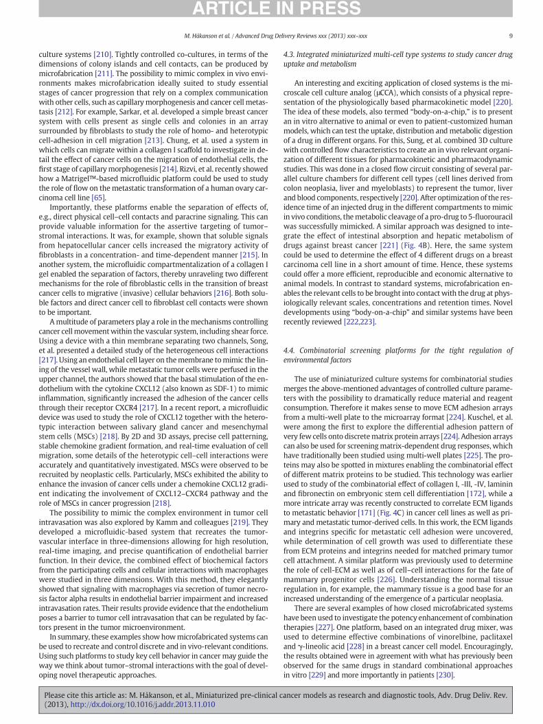
An interesting and exciting application of closed systems is the mi-croscale cell culture analog (μCCA), which consists of a physical repre-sentation of the physiologically based pharmacokinetic model [220].The idea of these models, also termed “body-on-a-chip,” is to presentan in vitro alternative to animal or even to patient-customized humanmodels, which can test the uptake, distribution andmetabolic digestionof a drug in different organs. For this, Sung, et al. combined 3D culturewith controlled flow characteristics to create an in vivo relevant organi-zation of different tissues for pharmacokinetic and pharmacodynamicstudies. This was done in a closed flow circuit consisting of several par-allel culture chambers for different cell types (cell lines derived fromcolon neoplasia, liver and myeloblasts) to represent the tumor, liverand blood components, respectively [220]. After optimization of the res-idence time of an injected drug in the different compartments to mimicin vivo conditions, themetabolic cleavage of a pro-drug to 5-fluorouracilwas successfully mimicked. A similar approach was designed to inte-grate the effect of intestinal absorption and hepatic metabolism ofdrugs against breast cancer [221] (Fig. 4B). Here, the same systemcould be used to determine the effect of 4 different drugs on a breastcarcinoma cell line in a short amount of time. Hence, these systemscould offer a more efficient, reproducible and economic alternative toanimal models. In contrast to standard systems, microfabrication en-ables the relevant cells to be brought into contactwith the drug at phys-iologically relevant scales, concentrations and retention times. Noveldevelopments using “body-on-a-chip” and similar systems have beenrecently reviewed [222,223].
4.4. Combinatorial screening platforms for the tight regulation ofenvironmental factors
The use of miniaturized culture systems for combinatorial studiesmerges the above-mentioned advantages of controlled culture parame-ters with the possibility to dramatically reduce material and reagentconsumption. Therefore it makes sense to move ECM adhesion arraysfrom a multi-well plate to the microarray format [224]. Kuschel, et al.were among the first to explore the differential adhesion pattern ofvery few cells onto discretematrix protein arrays [224]. Adhesion arrayscan also be used for screeningmatrix-dependent drug responses, whichhave traditionally been studied using multi-well plates [225]. The pro-teins may also be spotted in mixtures enabling the combinatorial effectof different matrix proteins to be studied. This technology was earlierused to study of the combinatorial effect of collagen I, -III, -IV, lamininand fibronectin on embryonic stem cell differentiation [172], while amore intricate array was recently constructed to correlate ECM ligandsto metastatic behavior [171] (Fig. 4C) in cancer cell lines as well as pri-mary and metastatic tumor-derived cells. In this work, the ECM ligandsand integrins specific for metastatic cell adhesion were uncovered,while determination of cell growth was used to differentiate thesefrom ECM proteins and integrins needed for matched primary tumorcell attachment. A similar platform was previously used to determinethe role of cell-ECM as well as of cell–cell interactions for the fate ofmammary progenitor cells [226]. Understanding the normal tissueregulation in, for example, the mammary tissue is a good base for anincreased understanding of the emergence of a particular neoplasia.
There are several examples of how closed microfabricated systemshave been used to investigate the potency enhancement of combinationtherapies [227]. One platform, based on an integrated drug mixer, wasused to determine effective combinations of vinorelbine, paclitaxeland γ-lineolic acid [228] in a breast cancer cell model. Encouragingly,the results obtained were in agreement with what has previously beenobserved for the same drugs in standard combinational approachesin vitro [229] and more importantly in patients [230].
ancer models as research and diagnostic tools, Adv. Drug Deliv. Rev.

10 M. Håkanson et al. / Advanced Drug Delivery Reviews xxx (2013) xxx–xxx
An interesting application is the combinatorial study of matrix pro-teins and solutes, considering the drastic effect of cellular and ECM in-teractions on drug response [33,101,225]. The NF-κB pathway is oneof the more frequently targeted pathways in drug development. Usinga diffusion-based device combined with a protein patterned substrateand studying the effects of different ECM mixtures in combinationwith diverse concentrations of IGF1 and TNF-α, Ying, et al. confirmedthe interaction of soluble factors with matrix proteins in the activationthe NF-κB pathway [231]. This initial workwas performedwith immor-talized endothelial cells but it would be expected that the samemethodcould be applied for the study of complex cancer cell signaling.
The epithelial tomesenchymal transition, or EMT, is important inme-tastasis as well as in cell plasticity and therefore highly relevant for drugdevelopment. As this transition is influencedbymicroenvironmental fac-tors [232], the study of this transition would profit from the controlledenvironment within a miniaturized system. In a feasibility experimentusing alveolar epithelial type II cells, Park, et al. demonstrated how therole of both immobilized ECM proteins (laminin and fibronectin) andsoluble factors (TGF-β1) could be investigated in one single step [233].The integrative analysis together with a high control of the culture envi-ronment may be particularly fruitful in the development of combinationtherapies targeting the cancer and its microenvironment.
4.5. Enabling the analysis of patient-derived cells by miniaturization
Miniaturized systems are particularly beneficial for the analysis ofsmall patient cell samples. As a complementary platform to cell lines,primary cells can increase predictivity of the pre-clinical screen andease target validation (Fig. 1) [39]. In addition, analyzing patient cellsfor predictive markers and dose-response curves are important toolsin personalized treatment [138]. However, the culture of primary cellsremains difficult since it is often hard to obtain a sufficiently large sam-ple size [39] and a homogeneous cell population (although heterogene-ity may be more representative of the human onset). In addition,primary cells aremore sensitive to tissue-culture passaging than normalcells and can dedifferentiate or acquire new genetic and/or epigeneticchanges after only a short time in culture. Miniaturized systems canhelp in removing some of these hurdles, since the reduced sample vol-ume makes it possible to obtain the required cell number directlyfrom a biopsy or with only a few culturing passages. In addition, thespecific-cell capture in miniaturized channels can help to purify asmall cell sample from in vivo sources (e.g. blood).
Initial progress in this field is exemplified, for example, by the studyof Beske, et al. inwhich sample consumptionwas reduced 2-fold bypro-ducing inserts for standard well plates that enabled analysis of 10 cellsamples per well [234]. Moving from standard to miniaturized ECMarrays for cell adhesion assays enabled applying this platform to thecharacterization of cancer cells from patients [224] and, more recently,to discriminate between local primary tumor cells and cells with meta-static potential [171]. In two examples of closed microfabricatedsystems determining drug concentration-response curves [41] and di-agnosing oncogenic kinase activity [235] in primary human leukemiacells, a much reduced number of cells were needed to produce resultswith signal-to-noise ratios comparable to standard assays. In fact, thecell number reduction in comparison to the conventional methodswas in themost extreme case 2000-fold [235], as shown in the assay de-velopment using a model cell line. In another example, an image-basedread-out facilitated single cell analysis in a microfluidic device, therebyreducing the cell numbers needed per assay evenmore [193]. In this de-vice, 300 hematopoietic cells were sufficient to obtain a drug-responsewith a similar statistical spread to that obtained using 15,000 to30,000 cells using conventional methods. However, while the afore-mentioned approachesmay be powerful in cancer diagnostic and devis-ing patient-specific treatment, reducing the experiments to a limitedamount of cells may not always give a good predictivity for highlyheterogenic tumors, such as renal cell carcinomas [236].
Please cite this article as: M. Håkanson, et al., Miniaturized pre-clinical c(2013), http://dx.doi.org/10.1016/j.addr.2013.11.010
An interesting application of microfabricated systems is the analysisof circulating cancer cells (CTSs) in blood and cells from fine needleaspirates. The microfluidic platform, with its high surface area, is supe-rior to standard methods for the capture of cancer cells present in solu-tion [237]. This is especially important for these samples, since theconcentration of CTCs may be only 1–100 per ml of blood [238]. Cellsmay be captured inside the chip by cell size- or cell mechanicalproperties-dependent and/or biochemical affinity-based methods[237–239]. This technology has been refined to create a platform thatcan bemanufactured on a commercial scale with improved cell capturerates [240]. However, despite the promising results, no microscale sys-tem for CTC-capture is currently FDA approved [238]. Therefore, it isstill important to validate the new systems, for example showing corre-lation to standard methods such as flow cytometry. Weigum, et al.developed a device for the early detection of oral cancer based on amicro-sieve for capture of the cells and subsequent analysis by immu-nochemistry [241]. It was shown that the levels of EGFR expression de-tected in the microscale device correlated well with flow cytometryresults. Recent work combined the immunophenotypic isolation withpost-culturing manipulations of patient cells [242] (Fig. 4D), therebyenabling further analysis of the cells after capture, e.g., determiningtheir drug response characteristics in a single microfluidics device.These novel tools thus open the path for multiplex analysis of patientsamples, e.g., characterization of cancer cell abundance and identifica-tion of predictive markers to help in the design of personalized therapy.
5. Conclusions and outlook
Current interdisciplinary activities in the field of miniaturizationhave resulted in a large number of novel in vitromodels for the capture,analysis and drug response evaluation of cancer cells. Much effort hasbeen expended on technological advancement of these systems, suchas novel microfluidic devices with better control of the culture environ-ment, improving cell capture and positioning techniques, as well as inthe development of innovative methods for assay read-out. What hasbeen missing for many years is a pool of experimental data obtainedin such systems, which could be compared to drug response dataobtained in conventional platforms and in clinical studies. Without alarge pool of examples showing the advantages of the new systems interms of predictivity, analysis of small cell samples, etc., the overall im-pact of microfabricated systems on modern biology continues to bemostly theoretical and thusmarginal. It is encouraging to see that the in-creasing collaboration between engineers and biologists has resulted inan impressive amount of data on the comparison of the new systemsto the conventional multi-well plate setup. However, the very importantstep of comparison to clinical data, proving the improved predictivity, isstill largely missing.
Here we listed several examples of how these systems can be ap-plied to cancer research. The possibility to manipulate and control thecell environment on the microscale makes these systems advantageousin comparison to standard methods. In particular, it is possible to studythe role of extrinsic parameters, which are often not easy to controlusing other methods. Application of these platforms has increased ourunderstanding of the interactions between cancer cells and theirmicroenvironment.
Many of the cell systems presented in this review were developedwith themotivation to find application in the clinic and pharmaceuticalindustry. Still, there are only a few researchers that focus on the realiza-tion of this translation into practice. The problem is especially true forclosed systems, which are typically more difficult in their handlingthan open ones. There is an overall consent that, in order to find wideuse of these systems, adaptation to automatic handling and read-outmethods is needed. Identifyingwhich are themost promising technolo-gies that should be adapted is a demanding step, which requires bridg-ing the specialized fields of basic biology, bioengineering, microfluidics,
ancer models as research and diagnostic tools, Adv. Drug Deliv. Rev.

11M. Håkanson et al. / Advanced Drug Delivery Reviews xxx (2013) xxx–xxx
clinical research and engineering. A joint decisionmust be taken on keyrequirements of the device-producing industry and clinical practice.
Cancer drug development faces many challenges, however, the pro-gression towards targeted and personalized treatment promises a brightfuture. Nonetheless, this direction requires more organotypic in vitromodels and new methods for patient sample analysis. This could be aunique chance for the bioengineering community for more extensivecollaborations with basic scientists and clinicians to explore the possibil-ities ofminiaturization for cancer cell analysis. In addition to aiding in thedevelopment in environment-targeted drugs, microfabricated systemshold a great promise in the analysis of patient cell samples to detect pre-dictive markers and determine dose-response profiles. With successfuladaptation to automated handling system, microfabricated model sys-tems may find wide applications in developing personalized diagnosisand patient-specific therapeutic modalities.
Acknowledgments
We would like to acknowledge comments and discussions made byDr. P. Lelkes (Temple University and Fox Chase Cancer Center) aswell aseffective proofreadingbyMrs. E. Ragan.We are also grateful to the SwissCompetence Center for Materials Science and Technology (CCMX) andthe Swiss National Science foundation (SNF) grant PA00P3_142120/1for providing funds to MH and MC thus making this work possible. Inaddition, funds to EC from the Commonwealth of Pennsylvania, FoxChase Cancer Center's Keystone Program in Personalized Kidney CancerTherapy, The Nodal Temple-FCCC Award, The Bucks County Board ofAssociates (in support of pancreatic cancer research) as well as fromNCI/NIH CA113451 and CA06927 were used for the completion of thisdocument.
References
[1] R. Siegel, D. Naishadham, A. Jemal, Cancer statistics, 2013, CA Cancer J. Clin. 63(2013) 11–30.
[2] T. Maziasz, V.J. Kadambi, L. Silverman, E. Fedyk, C.L. Alden, Predictive toxicologyapproaches for smallmolecule oncology drugs, Toxicol. Pathol. 38 (2010) 148–164.
[3] I. Kola, J. Landis, Can the pharmaceutical industry reduce attrition rates? Nat. Rev.Drug Discov. 3 (2004) 711–715.
[4] A. Kamb, S. Wee, C. Lengauer, Why is cancer drug discovery so difficult? Nat. Rev.Drug Discov. 6 (2007) 115–120.
[5] B.A. Teicher, Acute and chronic in vivo therapeutic resistance, Biochem. Pharmacol.77 (2009) 1665–1673.
[6] E. Cukierman, D.E. Bassi, The mesenchymal tumor microenvironment Adrug-resistant niche, Cell Adhes. Migr. 6 (2012) 285–296.
[7] J.-P. Gillet, S. Varma, M.M. Gottesman, The clinical relevance of cancer cell lines,J. Natl. Cancer Inst. 105 (2013) 452–458.
[8] J.S. Desgrosellier, D.A. Cheresh, Integrins in cancer: biological implications andtherapeutic opportunities, Nat. Rev. Cancer 10 (2010) 9–22.
[9] E. Murphy, B. Majeti, L. Barnes, M. Makale, S. Weis, K. Lutu-Fuga, W. Wrasidlo, D.A.Cheresh, Nanoparticle-mediated drug delivery to tumor vasculature suppressesmetastasis, Proc. Natl. Acad. Sci. U. S. A. 105 (2008) 9343–9348.
[10] T. Onishi, N. Hayashi, R.L. Theriault, G.N. Hortobagyi, N.T. Ueno, Future directions ofbone-targeted therapy for metastatic breast cancer, Nat. Rev. Clin. Oncol. 7 (2010)641–651.
[11] M. Paszek, N. Zahir, K. Johnson, J. Lakins, G. Rozenberg, A. Gefen, C. Reinhart-King, S.Margulies,M.Dembo,D. Boettiger, D.A.Hammer, V.M.Weaver, Tensional homeostasisand the malignant phenotype, Cancer Cell 8 (2005) 241–254.
[12] R. Castelló-Cros, D.R. Khan, J. Simons, M. Valianou, E. Cukierman, Staged stromalextracellular 3D matrices differentially regulate breast cancer cell responsesthrough PI3K and beta1-integrins, BMC Cancer 9 (2009) 94.
[13] J.G. Goetz, S. Minguet, I. Navarro-Lerida, J.J.L. R, R. Samaniego, E. Calvo, M. Tello, T.Osteso-Ibanez, T. Pellinen, A. Echarri, A. Cerezo, A.J. Klein-Szanto, R. Garcia, P.J.Keely, P. Sánchez-Mateos, E. Cukierman, M.A.D. Pozo, Biomechanical remodelingof themicroenvironment by stromal caveolin-1 favors tumor invasion andmetastasis,Cell 146 (2011) 148–163.
[14] C.C. Park, H.J. Zhang, E.S. Yao, C.J. Park, M.J. Bissell, β1 integrin inhibition dramaticallyenhances radiotherapy efficacy in human breast cancer xenografts, Cancer Res. 68(2008) 4398–4405.
[15] M. Meads, R. Gatenby, W. Dalton, Environment-mediated drug resistance: a majorcontributor to minimal residual disease, Nat. Rev. Cancer 9 (2009) 665–674.
[16] O.E. Franco, A.K. Shaw, D.W. Strand, S.W. Hayward, Cancer associated fibroblasts incancer pathogenesis, Semin. Cell Dev. Biol. 21 (2010) 33–39.
[17] K.E. Eberle, H.A. Sansing, P. Szaniszlo, V.A. Resto, A.L. Berrier, Carcinoma matrixcontrols resistance to cisplatin through talin regulation of NF-kB, PLoS ONE 6(2011) e21496.
Please cite this article as: M. Håkanson, et al., Miniaturized pre-clinical c(2013), http://dx.doi.org/10.1016/j.addr.2013.11.010
[18] E.H. Flach, V.W. Rebecca, M. Herlyn, K.S. Smalley, A.R. Anderson, Fibroblastscontribute to melanoma tumor growth and drug resistance, Mol. Pharm. 8(2011) 2039–2049.
[19] R. Straussman, T. Morikawa, K. Shee, M. Barzily-Rokni, Z.R. Qian, J. Du, A. Davis,M.M. Mongare, J. Gould, D.T. Frederick, Z.A. Cooper, P.B. Chapman, D.B. Solit, A.Ribas, R.S. Lo, K.T. Flaherty, S. Ogino, J.A. Wargo, T.R. Golub, Tumour micro-environment elicits innate resistance to RAF inhibitors through HGF secretion,Nature 487 (2012) 500–504.
[20] E. Cukierman, D.R. Khan, The benefits and challenges associated with the use ofdrug delivery systems in cancer therapy, Biochem. Pharmacol. 80 (2010) 762–770.
[21] J.K. Tunggal, D.S. Cowan, H. Shaikh, I.F. Tannock, Penetration of anticancer drugsthrough solid tissue: a factor that limits the effectiveness of chemotherapy forsolid tumors, Clin. Cancer Res. 5 (1999) 1583–1586.
[22] C. Feig, A. Gopinathan, A. Neesse, D.S. Chan, N. Cook, D.A. Tuveson, The pancreascancer microenvironment, Clin. Cancer Res. 18 (2012) 4266–4276.
[23] R. Kadaba, H. Birke, J. Wang, S. Hooper, C.D. Andl, F.D. Maggio, E. Soylu, M. Ghallab,D. Bor, F.E. Froeling, S. Bhattacharya, A.K. Rustgi, E. Sahai, C. Chelala, P. Sasieni, H.M.Kocher, Imbalance of desmoplastic stromal cell numbers drives aggressive cancerprocesses, J. Pathol. 230 (2013) 107–117.
[24] J. Friedrich, C. Seidel, R. Ebner, L. Kunz-Schughart, Spheroid-based drug screen:considerations and practical approach, Nat. Protoc. 4 (2009) 309–324.
[25] R. Kostadinova, F. Boess, D. Applegate, L. Suter, T. Weiser, T. Singer, B. Naughton, A.Roth, A long-term three dimensional liver co-culture system for improved predic-tion of clinically relevant drug-induced hepatotoxicity, Toxicol. Appl. Pharmacol.268 (2013) 1–16.
[26] K. Bhadriraju, C.S. Chen, Engineering cellular microenvironments to improvecell-based drug testing, Drug Discov. Today 7 (2002) 612–620.
[27] J. Friedrich, R. Ebner, L. Kunz-Schughart, Experimental anti-tumor therapy in 3-D:spheroids—old hat or new challenge? Int. J. Radiat. Biol. 83 (2007) 849–871.
[28] E. Cukierman, R. Pankov, D.R. Stevens, K.M. Yamada, Taking cell–matrix adhesionsto the third dimension, Science 294 (2001) 1708–1712.
[29] B. Trappmann, J.E. Gautrot, J.T. Connelly, D.G.T. Strange, Y. Li, M.L. Oyen, M.A. CohenStuart, H. Boehm, B. Li, V. Vogel, J.P. Spatz, F.M. Watt, W.T.S. Huck, Extracellular-matrix tethering regulates stem-cell fate, Nat. Mater. 11 (2012) 642–649.
[30] D.E. Discher, D.J. Mooney, P.W. Zandstra, Growth factors, matrices, and forcescombine and control stem cells, Science 324 (2009) 1673–1677.
[31] J. Debnath, S. Muthuswamy, J. Brugge, Morphogenesis and oncogenesis ofMCF-10A mammary epithelial acini grown in three-dimensional basement mem-brane cultures, Methods 30 (2003) 256–268.
[32] K.M. Yamada, E. Cukierman, Modeling tissue morphogenesis and cancer in 3D, Cell130 (2007) 601–610.
[33] I. Serebriiskii, R. Castelló-Cros, A. Lamb, E. Golemis, E. Cukierman, Fibroblast-derived 3D matrix differentially regulates the growth and drug-responsivenessof human cancer cells, Matrix Biol. 27 (2008) 573–585.
[34] H.-O. Lee, S.R. Mullins, J. Franco-Barraza, M. Valianou, E. Cukierman, J.D. Cheng,FAP-overexpressing fibroblasts produce an extracellular matrix that enhancesinvasive velocity and directionality of pancreatic cancer cells, BMC Cancer 11(2011) 245.
[35] R.J. Petrie, K.M. Yamada, At the leading edge of three-dimensional cell migration,J. Cell Sci. 125 (2012) 5917–5926.
[36] C. Nelson, M. Bissell, Modeling dynamic reciprocity: engineering three-dimensional culture models of breast architecture, function, and neoplastictransformation, Semin. Cancer Biol. 15 (2005) 342–352.
[37] W. Stein, Mining Databases to find the Genes that Determine the Intractability ofHuman Tumors, in: CHI's Tissue Models for Therapeutics, Boston, MA, USA, 2005.
[38] I. Cree, S. Glaysher, A. Harvey, Efficacy of anti-cancer agents in cell lines versushuman primary tumour tissue, Curr. Opin. Pharmacol. 4 (2010) 375–379.
[39] S. Goldbard, Bringing primary cells to mainstream drug development and drugtesting, Curr. Opin. Drug Discovery Dev. 9 (2006) 110–116.
[40] M.R. Trusheim, E.R. Berndt, F.L. Douglas, Stratifiedmedicine: strategic and economicimplications of combining drugs and clinical biomarkers, Nat. Rev. Drug Discov. 6(2007) 287–293.
[41] A. Ganser, G. Roth, J.C. van Galen, J. Hilderink, J.J.G. Wammes, I. Müller, F.N. vanLeeuwen, K.-H. Wiesmüller, R. Brock, Diffusion-driven device for a high-resolutiondose-response profiling of combination chemotherapy, Anal. Chem. 81 (2009)5233–5240.
[42] S.N. Bhatia, U.J. Balis, M.L. Yarmush, M. Toner, Effect of cell–cell interactions inpreservation of cellular phenotype: cocultivation of hepatocytes and nonparenchymalcells, FASEB J. 13 (1999) 1883–1900.
[43] A.L. Pavlovich, S. Manivannan, C.M. Nelson, Adipose stroma induces branchingmorphogenesis of engineered epithelial tubules, Tissue Eng. Part A 16 (2010)3719–3726.
[44] F. Xu, J. Celli, I. Rizvi, S. Moon, T. Hasan, U. Demirci, A three-dimensional in vitroovarian cancer coculture model using a high-throughput cell patterning platform,Biotechnol. J. 6 (2011) 204–212.
[45] J. Kalabis, G.S. Wong, M.E. Vega, M. Natsuizaka, E.S. Robertson, M. Herlyn, H.Nakagawa, A.K. Rustgi, Isolation and characterization of mouse and humanesophageal epithelial cells in 3D organotypic culture, Nat. Protoc. 7 (2012)235–246.
[46] K.Y. Lee, D.J. Mooney, Alginate: properties and biomedical applications,Prog. Polym. Sci. 37 (2012) 106–126.
[47] M. Håkanson, M. Textor, M. Charnley, Engineered 3D environments to elucidatethe effect of environmental parameters on drug response in cancer, Integr. Biol.(2011) 1–8.
[48] L. Lin, A. Perets, Y.-e. Har-el, D. Varma, M. Li, P. Lazarovici, D.L. Woerdeman, P.I.Lelkes, Alimentary ‘green’ Proteins as Electrospun Scaffolds for Skin Regenerative
ancer models as research and diagnostic tools, Adv. Drug Deliv. Rev.

12 M. Håkanson et al. / Advanced Drug Delivery Reviews xxx (2013) xxx–xxx
Engineering, J. Tissue Eng. Regen. Med. (2013), http://dx.doi.org/10.1002/term.1493 (In press).
[49] A. Lo, H. Mori, J. Mott, M. Bissell, Constructing three-dimensional models to studymammary gland branching morphogenesis and functional differentiation,J. Mammary Gland Biol. Neoplasia 17 (2012) 103–110.
[50] V. Gupta, D.E. Bassi, J.D. Simons, K. Devarajan, T. Al-Saleem, R.G. Uzzo, E.Cukierman, Elevated expression of stromal palladin predicts poor clinical outcomein renal cell carcinoma, PLoS ONE 6 (2011) e21494.
[51] M. Charnley, R. Kroschewski, M. Textor, The study of polarisation in single cellsusing model cell membranes, Integr. Biol. 4 (2012) 1059–1071.
[52] L. Li, M. Fukunaga-Kalabis, M. Herlyn, The three-dimensional human skinreconstruct model: a tool to study normal skin and melanoma progression, J. Vis.Exp. 3 (2011)e2937.
[53] G.Y. Lee, P.A. Kenny, E.H. Lee, M.J. Bissell, Three-dimensional culture models ofnormal and malignant breast epithelial cells, Nat. Methods 4 (2007) 359–365.
[54] F. Hirschhaeuser, H. Menne, C. Dittfeld, J. West, W. Mueller-Klieser, L.A.Kunz-Schughart, Multicellular tumor spheroids: an underestimated tool is catchingup again, J. Biotechnol. 148 (2010) 3–15.
[55] W.F. Liu, C.S. Chen, Cellular and multicellular form and function, Adv. Drug Deliv.Rev. 59 (2007) 1319–1328.
[56] M. Dusseiller, M.L. Smith, V. Vogel, M. Textor, Microfabricated three-dimensionalenvironments for single cell studies, Biointerphases 1 (2006) 1–4.
[57] M. Charnley, F. Anderegg, R. Holtackers, M. Textor, P. Meraldi, Effect of cell shapeand dimensionality on spindle orientation and mitotic timing, PLoS ONE 8(2013) e66918.
[58] M. Ochsner, M. Textor, V. Vogel, M.L. Smith, Dimensionality controls cytoskeletonassembly and metabolism of fibroblast cells in response to rigidity and shape,PLoS ONE 5 (2010) e9445.
[59] C. Chen, M. Mrksich, S. Huang, G. Whitesides, D.E. Ingber, Geometric control of celllife and death, Science 276 (1997) 1425–1428.
[60] D. Wlodkovic, J.M. Cooper, Tumors on chips: oncology meets microfluidics,Curr. Opin. Chem. Biol. 14 (2010) 556–557.
[61] X. Gidrol, B. Fouqué, L. Ghenim, V. Haguet, N. Picollet-D'hahan, B. Schaack, 2D and3D cell microarrays in pharmacology, Curr. Opin. Pharmacol. 9 (2009) 664–668.
[62] M. Håkanson, S. Kobel, M.P. Lutolf, M. Textor, E. Cukierman, M. Charnley,Controlled breast cancer microarrays for the deconvolution of cellularmultilayering and density effects upon drug responses, PLoS ONE 7 (2012) e40141.
[63] M.S. Kim, J.H. Yeon, J.-K. Park, A microfluidic platform for 3-dimensional cellculture and cell-based assays, Biomed. Microdevices 9 (2007) 25–34.
[64] Y. Torisawa, B. Mosadegh, T. Bersano-Begey, J.M. Steele, K.E. Luker, G.D. Luker, S.Takayama, Microfluidic platform for chemotaxis in gradients formed by CXCL12source-sink cells, Integr. Biol. 2 (2010) 680–686.
[65] I. Rizvi, U.A. Gurkan, S. Tasoglu, N. Alagic, J.P. Celli, L.B. Mensah, Z. Mai, U. Demirci, T.Hasan, Flow induces epithelial–mesenchymal transition, cellular heterogeneityand biomarker modulation in 3D ovarian cancer nodules, Proc. Natl. Acad. Sci.U. S. A. 110 (2013) E1974–E1983.
[66] S.I. Montanez-Sauri, K.E. Sung, E. Berthier, D.J. Beebe, Enabling screening in 3Dmicroenvironments: probing matrix and stromal effects on the morphology andproliferation of T47D breast carcinoma cells, Integr. Biol. 5 (2013) 631–640.
[67] Bleicher, H. Bohm, K. Muller, A. Alanine, Hit and lead generation: Beyondhigh-throughput screening, Nat. Rev. Drug Discov. 2 (2003) 369–378.
[68] J.A. Landro, HTS in the new millennium: the role of pharmacology and flexibility,J. Pharmacol. Toxicol. Methods 44 (2000) 273–289.
[69] M. Roberge, Cell-based screen for antimitotic agents and identification ofanalogues of rhizoxin, eleutherobin and paclitaxel in natural extracts, Cancer Res.60 (2000) 5052–5058.
[70] F. Balis, Evolution of anticancer drug discovery and the role of cell-based screeningbox, J. Natl. Cancer Inst. (2002) 85.
[71] H. Denys, G. Braems, K. Lambein, P. Pauwels, A. Hendrix, A. De Boeck, V. Mathieu,M. Bracke, O. De Wever, The extracellular matrix regulates cancer progressionand therapy response: implications for prognosis and treatment, Curr. Pharm.Des. 15 (2009) 1373–1384.
[72] J. Joyce, J. Pollard, Microenvironmental regulation of metastasis, Nat. Rev. Cancer 9(2009) 239–252.
[73] M.J. Bissell, D.C. Radisky, A. Rizki, V.M. Weaver, O.W. Petersen, The organizingprinciple: microenvironmental influences in the normal and malignant breast,Differentiation 70 (2002) 537–546.
[74] C. Sonnenschein, A.M. Soto, The death of the cancer cell, Cancer Res. 71 (2011)4334–4337.
[75] V. Hofmeister, D. Schrama, J.C. Becker, Anti-cancer therapies targeting the tumorstroma, Cancer Immunol. Immunother. 57 (2008) 1–17.
[76] M. Erkan, C.W. Michalski, S. Rieder, C. Reiser-Erkan, I. Abiatari, A. Kolb, N.A. Giese, I.Esposito, H. Friess, J. Kleeff, The activated stroma index is a novel and independentprognostic marker in pancreatic ductal adenocarcinoma, Clin. Gastroenterol.Hepatol. 6 (2008) 1155–1161.
[77] S. Paget, The distribution of secondary growths in cancer of the breast, Lancet 133(1889) 571–573.
[78] T. Langhans, Pulsirende cavernoese Geschwulst der Miltz mit metastatischenKnoten in der Leber, Virchows Arch. 75 (1879) 273–291.
[79] S. Kumar, V.M. Weaver, Mechanics, malignancy, and metastasis: the force journeyof a tumor cell, Cancer Metastasis Rev. 28 (2009) 113–127.
[80] D.A. Beacham, E. Cukierman, Stromagenesis: the changing face of fibroblasticmicroenvironments during tumor progression, Semin. Cancer Biol. 15 (2005)329–341.
[81] L.M. Cook, D.R. Hurst, D.R. Welch, Metastasis suppressors and the tumormicroenvironment, Semin. Cancer Biol. 21 (2011) 113–122.
Please cite this article as: M. Håkanson, et al., Miniaturized pre-clinical c(2013), http://dx.doi.org/10.1016/j.addr.2013.11.010
[82] I.P. Witz, O. Levy-Nissenbaum, The tumor microenvironment in the post-PAGETera, Cancer Lett. 242 (2006) 1–10.
[83] D. Tarin, Clinical and biological implications of the tumor microenvironment,Cancer Microenviron. 5 (2012) 95–112.
[84] L. Rønnov-Jessen, M. Bissell, Breast cancer by proxy: can the microenvironment beboth the cause and consequence? Trends Mol. Med. 15 (2008) 5–13.
[85] L.A. Kunz-Schughart, R. Knuechel, Tumor associated fibroblasts (Part II): functionalimpact on tumor tissue, Histol. Histopathol. 2 (2002) 623–637.
[86] M.W. Conklin, J.C. Eickhoff, K.M. Riching, C.A. Pehlke, K.W. Eliceiri, P.P. Provenzano,A. Friedl, P.J. Keely, Aligned collagen is a prognostic signature for survival in humanbreast carcinoma, Am. J. Pathol. 178 (2011) 1221–1232.
[87] M.D. Amatangelo, D.E. Bassi, A.J. Klein-Szanto, E. Cukierman, Stroma-derivedthree-dimensional matrices are necessary and sufficient to promote desmoplasticdifferentiation of normal fibroblasts, Am. J. Pathol. 167 (2005) 475–488.
[88] K.R. Levental, H. Yu, L. Kass, J.N. Lakins,M. Egeblad, J.T. Erler, S.F.T. Fong, K. Csiszar, A.Giaccia, W. Weninger, M. Yamauchi, D.L. Gasser, V.M. Weaver, Matrix crosslinkingforces tumor progression by enhancing integrin signaling, Cell 139 (2009)891–906.
[89] M. Bissell, M. LaBarge, Context, tissue plasticity, and cancer: are tumor stem cellsalso regulated by the microenvironment? Cancer Cell 7 (2005) 17–23.
[90] U.M. Polanska, A. Orimo, Carcinoma-associated fibroblasts: non-neoplastictumour-promoting mesenchymal cells, J. Cell. Physiol. 228 (2013) 1651–1657.
[91] R. Castelló-Cros, E. Cukierman, Stromagenesis during tumorigenesis: characterizationof tumor-associated fibroblasts and stroma-derived 3D matrices, Methods Mol. Biol.522 (2009) 275–305.
[92] C. Mehner, D.C. Radisky, Triggering the landslide: the tumor-promotional effects ofmyofibroblasts, Exp. Cell Res. 319 (2013) 1657–1662.
[93] P. Lu, V.M. Weaver, Z. Werb, The extracellular matrix: a dynamic niche in cancerprogression, J. Cell Biol. 196 (2012) 395–406.
[94] T. Oskarsson, J. Massague, Extracellular matrix players in metastatic niches, EMBOJ. 31 (2012) 254–256.
[95] M. Otranto, V. Sarrazy, F. Bonté, B. Hinz, G. Gabbiani, A. Desmouliere, The role of themyofibroblast in tumor stroma remodeling, Cell Adhes. Migr. 6 (2012) 203–219.
[96] M.A. Shields, S. Dangi-Garimella, A.J. Redig, H.G. Munshi, Biochemical role of thecollagen-rich tumour microenvironment in pancreatic cancer progression,Biochem. J. 441 (2012) 541–552.
[97] C. Strell, H. Rundqvist, A. Östman, Fibroblasts—a key host cell type in tumorinitiation, progression, and metastasis, Ups. J. Med. Sci. 117 (2012) 187–195.
[98] C. Wittekind, M. Neid, Cancer invasion and metastasis, Oncology 69 (2005) 14–16.[99] S. Jodele, L. Blavier, J.M. Yoon, Y.A. DeClerck, Modifying the soil to affect the seed:
role of stromal-derived matrix metalloproteinases in cancer progression, CancerMetastasis Rev. 25 (2006) 35–43.
[100] L. Ronnov-Jessen, O. Petersen, M.J. Bissell, Cellular changes involved in conversionof normal to malignant breast: importance of the stromal reaction, Physiol. Rev. 76(1996) 69–125.
[101] K.H.T. Paraiso, K.S.M. Smalley, Fibroblast-mediated drug resistance in cancer,Biochem. Pharmacol. 85 (2013) 1033–1041.
[102] A. Minchinton, I.F. Tannock, Drug penetration in solid tumors, Nat. Rev. Cancer 6(2006) 583–592.
[103] B. St Croix, C. Sheehan, J.W. Rak, V.A. Flørenes, J.M. Slingerland, R.S. Kerbel,E-Cadherin-dependent growth suppression is mediated by the cyclin-dependentkinase inhibitor p27(KIP1), J. Cell Biol. 142 (1998) 557–571.
[104] L.A. Hazlehurst, T.H. Landowski, W.S. Dalton, Role of the tumor microenvironmentinmediating de novo resistance to drugs and physiological mediators of cell death,Oncogene 22 (2003) 7396–7402.
[105] F. Aoudjit, K. Vuori, Integrin signaling in cancer cell survival and chemoresistance,Chemother. Res. Pract. 2012 (2012) 16.
[106] R.M. Quiros, M. Valianou, Y. Kwon, K.M. Brown, A.K. Godwin, E. Cukierman, Ovariannormal and tumor-associated fibroblasts retain in vivo stromal characteristics in a3-D matrix-dependent manner, Gynecol. Oncol. 110 (2008) 99–109.
[107] C. Sherman-Baust, A. Weeraratna, L. Rangel, E. Pizer, K. Cho, D. Schwartz, T. Schock,P. Morin, Remodeling of the extracellular matrix through overexpression ofcollagen VI contributes to cisplatin resistance in ovarian cancer cells, Cancer Cell3 (2003) 377–386.
[108] P. Lito, Christine A. Pratilas, Eric W. Joseph, M. Tadi, E. Halilovic, M. Zubrowski, A.Huang, Wai L. Wong, Margaret K. Callahan, T. Merghoub, Jedd D. Wolchok, E. deStanchina, S. Chandarlapaty, Poulikos I. Poulikakos, James A. Fagin, N. Rosen, Reliefof profound feedback inhibition ofmitogenic signaling by RAF inhibitors attenuatestheir activity in BRAFV600E melanomas, Cancer Cell 22 (2012) 668–682.
[109] B.R. Seo, P. DelNero, C. Fischbach, In vitro models of tumor vessels and matrix: En-gineering approaches to investigate transport limitations and drug delivery in can-cer, Adv Drug Deliv Rev, 2014. (this issue).
[110] J. Fang, H. Nakamura, H. Maeda, The EPR effect: Unique features of tumor bloodvessels for drug delivery, factors involved, and limitations and augmentation ofthe effect, Adv. Drug Deliv. Rev. 63 (2011) 136–151.
[111] L. Brannon-Peppas, J.O. Blanchette, Nanoparticle and targeted systems for cancertherapy, Adv. Drug Deliv. Rev. 64 (2012) 206–212(Supplement).
[112] D. Polyak, A. Eldar-Boock, H. Baabur-Cohen, R. Satchi-Fainaro, Polymer conjugatesfor focal and targeted delivery of drugs, Polym. Adv. Technol. 24 (2013) 777–790.
[113] S.D. Steichen, M. Caldorera-Moore, N.A. Peppas, A review of current nanoparticleand targeting moieties for the delivery of cancer therapeutics, Eur. J. Pharm. Sci.48 (2013) 416–427.
[114] G.P. Adams, L.M. Weiner, Monoclonal antibody therapy of cancer, Nat. Biotechnol.23 (2005) 1147–1157.
[115] E.T. Boder, W. Jiang, Engineering antibodies for cancer therapy, in: J.M. Prausnitz(Ed.), Annu Rev Chem Biomol Eng, vol. 2, 2011, pp. 53–75.
ancer models as research and diagnostic tools, Adv. Drug Deliv. Rev.

13M. Håkanson et al. / Advanced Drug Delivery Reviews xxx (2013) xxx–xxx
[116] J.D. Byrne, T. Betancourt, L. Brannon-Peppas, Active targeting schemes for nanoparticlesystems in cancer therapeutics, Adv. Drug Deliv. Rev. 60 (2008) 1615–1626.
[117] J.P. Morgan, P.F. Delnero, Y. Zheng, S.S. Verbridge, J. Chen, M. Craven, N.W. Choi, A.Diaz-Santana, P. Kermani, B. Hempstead, J.A. Lopez, T.N. Corso, C. Fischbach, A.D.Stroock, Formation of microvascular networks in vitro, Nat. Protoc. 8 (2013)1820–1836.
[118] S.S. Verbridge, A. Chakrabarti, P. Delnero, B. Kwee, J.D. Varner, A.D. Stroock, C.Fischbach, Physicochemical regulation of endothelial sprouting in a 3Dmicrofluidicangiogenesis model, J. Biomed. Mater. Res. A 101 (2013) 2948–2956.
[119] E. Heller, M.A. Hurchla, J. Xiang, X. Su, S. Chen, J. Schneider, K.-S. Joeng, M. Vidal, L.Goldberg, H. Deng, M.C. Hornick, J.L. Prior, D. Piwnica-Worms, F. Long, R. Cagan,K.N. Weilbaecher, Hedgehog signaling inhibition blocks growth of resistant tumorsthrough effects on tumor microenvironment, Cancer Res. 72 (2012) 897–907.
[120] S. Hehlgans, M. Haase, N. Cordes, Signalling via integrins: implications for cellsurvival and anticancer strategies, BBA Rev. Cancer 1775 (2007) 163–180.
[121] L.A. Hazlehurst, R.F. Argilagos, W.S. Dalton, β1 integrin mediated adhesionincreases Bim protein degradation and contributes to drug resistance in leukaemiacells, Br. J. Haematol. 136 (2007) 269–275.
[122] T. Lwin, L. Hazlehurst, S. Dessureault, R. Lai, W. Bai, E. Sotomayor, L.C. Moscinski,W.S. Dalton, J.G. Tao, Cell adhesion induces p27Kip1-associated cell-cycle arrestthrough down-regulation of the SCFSkp2 ubiquitin ligase pathway in mantle-celland other non-Hodgkin B-cell lymphomas, Blood 110 (2007) 1631–1638.
[123] E. Yao, H. Zhang, Y. Chen, B. Lee, K. Chew, Cancer Res, Increased beta1 integrin isassociated with decreased survival in invasive breast cancer, Cancer Res. 67(2007) 659–664.
[124] C.C. Park, H. Zhang, M. Pallavicini, J.W. Gray, F. Baehner, C.J. Park, M.J. Bisell, β1integrin inhibitory antibody induces apoptosis of breast cancer cells, inhibitsgrowth, and distinguishesmalignant from normal phenotype in three dimensionalcultures and in vivo, Cancer Res. 66 (2006) 1526–1535.
[125] H. Wang, W.L. Fu, J.H. Im, Z.Y. Zhou, S.A. Santoro, V. Iyer, C.M. DiPersio, Q.C. Yu, V.Quaranta, A. Al-Medhi, R.J. Muschel, Tumor cell α3β1 integrin and vascularlaminin-5 mediate pulmonary arrest and metastasis, J. Cell Biol. 164 (2004)935–941.
[126] Y. Mori, N. Shimizu, M. Dallas, M. Niewolna, B. Story, P. Williams, G. Mundy, T.Yoneda, Anti-alpha 4 integrin antibody suppresses the development of multiplemyeloma and associated osteoclastic osteolysis, Blood 104 (2004) 2149–2154.
[127] C. Manegold, J. Vansteenkiste, F. Cardenal, W. Schuette, P.J. Woll, E. Ulsperger, A.Kerber, J. Eckmayr, J. von Pawel, Randomized phase II study of three doses of theintegrin inhibitor cilengitide versus docetaxel as second-line treatment for patientswith advanced non-small-cell lung cancer, Invest New Drugs 31 (2013) 175–182.
[128] S. Patel, J. Jenkins, N. Papadopolous, M. Burgess, C. Plager, J. Gutterman, R.Benjamin, Pilot study of vitaxin – an angiogenesis inhibitor – in patients withadvanced leiomyosarcomas, Cancer 92 (2001) 1347–1348.
[129] C. Delbaldo, E. Raymond, K. Vera, L. Hammershaimb, K. Kaucic, S. Lozahic, M.Marty,S. Faivre, Phase I and pharmacokinetic study of etaracizumab (Abegrin), a humanizedmonoclonal antibody against alphavbeta3 integrin receptor, in patients withadvanced solid tumors, Invest New Drugs 26 (2008) 35–43.
[130] P. Hersey, J. Sosman, S. O'Day, J. Richards, A. Bedikian, R. Gonzalez, W. Sharfman, R.Weber, T. Logan, M. Buzoianu, L. Hammershaimb, J.M. Kirkwood, G. Etaracizumab,Melanoma study, a randomized phase 2 study of etaracizumab, a monoclonalantibody against integrin alpha(v)beta(3), + or—dacarbazine in patients withstage IV metastatic melanoma, Cancer 116 (2010) 1526–1534.
[131] S.J. Moschos, C.A. Sander, W. Wang, S.L. Reppert, L.M. Drogowski, D.M. Jukic, U.N.Rao, C. Athanassiou, M. Buzoianu, M. Mandic, L. Richman, L. McKinney, J.Leininger, D.A. Tice, L. Hammershaimb, J.M. Kirkwood, Pharmacodynamic (phase0) study using etaracizumab in advanced melanoma, J. Immunother. 33 (2010)316–325.
[132] J. Marlind, I. Pisoni, D. Neri, Tumor vascular targeting, Drugs Future 31 (2006)901–913.
[133] J. Hood, M. Bednarski, R. Frausto, S. Guccione, R. Reisfeld, R. Xiang, D.A. Cheresh,Tumor regression by targeted gene delivery to neovasculature, Science 296(2002) 2404–2407.
[134] S. Ono, G. Ishii, K. Nagai, T. Takuwa, J. Yoshida, M. Nishimura, T. Hishida, K. Aokage,S. Fujii, N. Ikeda, A. Ochiai, Podoplanin-positive cancer-associated fibroblasts couldhave prognostic value independent of cancer cell phenotype in stage I lung squa-mous cell carcinoma: usefulness of combining analysis of both cancer cell pheno-type and cancer-associated fibroblast phenotype, CHEST J. 143 (2013) 963–970.
[135] Z. Jia, Y. Wang, A. Sawyers, H. Yao, F. Rahmatpanah, X.-Q. Xia, Q. Xu, R. Pio, T. Turan,J.A. Koziol, S. Goodison, P. Carpenter, J. Wang-Rodriguez, A. Simoneau, F. Meyskens,M. Sutton,W. Lernhardt, T. Beach, J. Monforte, M.McClelland, D.Mercola, Diagnosisof prostate cancer using differentially expressed genes in stroma, Cancer Res. 71(2011) 2476–2487.
[136] H. Niu, H. Jiang, B. Cheng, X. Li, Q. Dong, L. Shao, S. Liu, X. Wang, Stromal proteomeexpression profile and muscle-invasive bladder cancer research, Cancer Cell Int. 12(2012) 39.
[137] N. Harbeck, M. Salem, U. Nitz, O. Gluz, C. Liedtke, Personalized treatment ofearly-stage breast cancer: present concepts and future directions, Cancer Treat.Rev. 36 (2010) 584–594.
[138] M.J. Bissell, Novel pathways and biomarkers unraveled by complex biologicalmodels, Adv Drug Deliv Rev, 2014. (this issue).
[139] M. Hidalgo, E. Bruckheimer, N.V. Rajeshkumar, I. Garrido-Laguna, E. De Oliveira, B.Rubio-Viqueira, S. Strawn, M.J. Wick, J. Martell, D. Sidransky, A pilot clinical studyof treatment guided by personalized tumorgrafts in patients with advanced cancer,Mol. Cancer Ther. 10 (2011) 1311–1316.
[140] G.H. Underhill, P. Galie, C.S. Chen, S.N. Bhatia, Bioengineering methods for analysisof cells in vitro, Annu. Rev. Cell Dev. Biol. 28 (2012) 385–410.
Please cite this article as: M. Håkanson, et al., Miniaturized pre-clinical c(2013), http://dx.doi.org/10.1016/j.addr.2013.11.010
[141] A. Faia-Torres, T. Goren, M. Textor, M. Pla-Roca, Patterned Biointerfaces, in:Comprehensive Biomaterials, Elsevier, 2011.
[142] D. Falconnet, G. Csucs, H.M. Grandin, M. Textor, Surface engineering approaches tomicropattern surfaces for cell-based assays, Biomaterials 27 (2006) 3044–3063.
[143] A. Azioune, M. Storch, M. Bornens, M. Théry, M. Piel, Simple and rapid process forsingle cell micro-patterning, Lab Chip 9 (2009) 1640–1642.
[144] H.K. Kleinman, M.L. McGarvey, J.R. Hassell, V.L. Star, F.B. Cannon, G.W. Laurie, G.R.Martin, Basement membrane complexes with biological activity, Biochemistry 25(1986) 312–318.
[145] T.R. Sodunke, K.K. Turner, S.A. Caldwell, K.W. McBride, M.J. Reginato, H.M. Noh,Micropatterns of Matrigel for three-dimensional epithelial cultures, Biomaterials28 (2007) 4006–4016.
[146] C. Nelson, J. Inman, M. Bissell, Three-dimensional lithographically definedorganotypic tissue arrays for quantitative analysis ofmorphogenesis andneoplasticprogression, Nat. Protoc. 3 (2008) 674–678.
[147] M. Ochsner, M.R. Dusseiller, H.M. Grandin, S. Luna-Morris, M. Textor, V. Vogel, M.L.Smith,Micro-well arrays for 3D shape control and high resolution analysis of singlecells, Lab Chip 7 (2007) 1074–1077.
[148] M. Lutolf, R. Doyonnas, K. Havenstrite, K. Koleckar, H.M. Blau, Perturbation of singlehematopoietic stem cell fates in artificial niches, Integr. Biol. 1 (2009) 59–69.
[149] R. Zhao, T. Boudou, W.-G. Wang, C.S. Chen, D.H. Reich, Decoupling cell and matrixmechanics in engineeredmicrotissues usingmagnetically actuatedmicrocantilevers,Adv. Mater. 25 (2013) 1699–1705.
[150] M. Lee, R. Kumar, S. Sukumaran, M. Hogg, D. Clark, J. Dordick, Three-dimensionalcellular microarray for high-throughput toxicology assays, Proc. Natl. Acad. Sci.U. S. A. 105 (2008) 59.
[151] M. Pla-Roca, R.F. Leulmi, H. Djambazian, S. Sundararajan, D. Juncker, Addressablenanowell arrays formed using reversibly sealable hybrid elastomer-metal stencils,Anal. Chem. 82 (2010) 3848–3855.
[152] S.N. Bailey, D.M. Sabatini, R.N. Stockwell,Microarrays of smallmolecules embeddedin biodegradable polymers for use in mammalian cell-based screens, Proc. Natl.Acad. Sci. U. S. A. 101 (2004) 16144–16149.
[153] E.W.K. Young, D.J. Beebe, Fundamentals of microfluidic cell culture in controlledmicroenvironments, Chem. Soc. Rev. 39 (2010) 1036.
[154] J. Pihl, J. Sinclair, E. Sahlin, M. Karlsson, F. Petterson, J. Olofsson, O. Orwar,Microfluidic gradient-generating device for pharmacological profiling, Anal.Chem. 77 (2005) 3897–3903.
[155] L.Y. Wu, D. Di Carlo, L.P. Lee, Microfluidic self-assembly of tumor spheroids foranticancer drug discovery, Biomed. Microdevices 10 (2008) 197–202.
[156] S. Takayama, E. Ostuni, P. LeDuc, K. Naruse, D.E. Ingber, G.M.Whitesides, Subcellularpositioning of small molecules, Nature 411 (2001) 1016.
[157] R. Gómez-Sjöberg, A.A. Leyrat, D.M. Pirone, C.S. Chen, S.R. Quake, Versatile, fullyautomated, microfluidic cell culture system, Anal. Chem. 79 (2007) 8557–8563.
[158] A.S. Meyer, S.K. Hughes-Alford, J.E. Kay, A. Castillo, A. Wells, F.B. Gertler, D.A.Lauffenburger, 2D protrusion but not motility predicts growth factor-inducedcancer cell migration in 3D collagen, J. Cell Biol. 197 (2012) 721–729.
[159] A.D. Doyle, F.W. Wang, K. Matsumoto, K.M. Yamada, One-dimensional topographyunderlies three-dimensional fibrillar cell migration, J. Cell Biol. 184 (2009)481–490.
[160] N.W. Choi, M. Cabodi, B. Held, J.P. Gleghorn, L.J. Bonassar, A.D. Stroock, Microfluidicscaffolds for tissue engineering, Nat. Mater. 6 (2007) 908–915.
[161] J. Lii, W.-J. Hsu, H. Parsa, A. Das, R. Rouse, S.K. Sia, Real-time microfluidic system forstudying mammalian cells in 3D microenvironments, Anal. Chem. 80 (2008)3640–3647.
[162] U. Haessler, J.C.M. Teo, D. Foretay, P. Renaud, M.A. Swartz, Migration dynamics ofbreast cancer cells in a tunable 3D interstitial flow chamber, Integr. Biol. 4 (2012)401–409.
[163] D. Irimia, G. Charras, N. Agrawal, T. Mitchison, M. Toner, Polar stimulation andconstrained cell migration in microfluidic channels, Lab Chip 7 (2007)1783–1790.
[164] U. Haessler, Y. Kalinin,M.A. Swartz,M.Wu, An agarose-basedmicrofluidic platformwith a gradient buffer for 3D chemotaxis studies, Biomed. Microdevices 11 (2009)827–835.
[165] C.P. Ng, S.H. Pun, A perfusable 3D cell-matrix tissue culture chamber for in situevaluation of nanoparticle vehicle penetration and transport, Biotechnol. Bioeng.99 (2008) 1490–1501.
[166] C.L. Walsh, B.M. Babin, R.W. Kasinskas, J.A. Foster, M.J. McGarry, N.S. Forbes, Amultipurpose microfluidic device designed to mimic microenvironment gradientsand develop targeted cancer therapeutics, Lab Chip 9 (2009) 545–554.
[167] S.-M. Ong, C. Zhang, Y.-C. Toh, S.H. Kim, H.L. Foo, C.H. Tan, D. van Noort, S. Park, H.Yu, A gel-free 3D microfluidic cell culture system, Biomaterials 29 (2008)3237–3244.
[168] I. Meyvantsson, D.J. Beebe, Cell culture models in microfluidic systems, Annu RevAnal Chem (Palo Alto, Calif) 1 (2008) 423–449.
[169] I. Meyvantsson, J. Warrick, S. Hayes, A. Skoien, D. Beebe, Automated cell culture inhigh density tubeless microfluidic device arrays, Lab Chip 8 (2008) 717–724.
[170] E. Berthier, J. Surfus, J. Verbsky, A. Huttenlocher, D.J. Beebe, A high-content chemotaxisassay for patient diagnosis, Integr. Biol. 2 (2010) 630–638.
[171] N.E. Reticker-Flynn, D.F.B. Malta, M.M.Winslow, J.M. Lamar, M.J. Xu, G.H. Underhill,R.O. Hynes, T.E. Jacks, S.N. Bhatia, A combinatorial extracellular matrix platformidentifies cell-extracellular matrix interactions that correlate with metastasis,Nat. Commun. 3 (2012) 1122.
[172] C.J. Flaim, S. Chien, S.N. Bhatia, An extracellular matrix microarray for probingcellular differentiation, Nat. Methods 2 (2005) 119–125.
[173] A. Niles, R.A. Moravec, T.L. Riss, Update on in vitro cytotoxicity assays for drugdevelopment, Expert Opin. Drug Discovery 3 (2008) 655–669.
ancer models as research and diagnostic tools, Adv. Drug Deliv. Rev.

14 M. Håkanson et al. / Advanced Drug Delivery Reviews xxx (2013) xxx–xxx
[174] M. Tanaka, R. Bateman, D. Rauh, E. Vaisberg, S. Ramachandani, C. Zhang, K.C.Hansen, A.L. Burlingame, J.K. Trautman, K.M. Shokat, C.L. Adams, An unbiasedcell morphology—based screen for new, biologically active small molecules, PLoSBiol. 3 (2005).
[175] P. Lang, K. Yeow, A. Nichols, A. Scheer, Cellular imaging in drug discovery, Nat. Rev.Drug Discov. 5 (2006) 343–356.
[176] H. Lövborg, P. Nygren, R. Larsson, Multiparametric evaluation of apoptosis: effects ofstandard cytotoxic agents and the cyanoguanidine CHS 828, Mol. Cancer Ther. 3(2004) 521–526.
[177] R.J. Errington, S.C. Chappell, I.A. Khan, N. Marquez, M. Wiltshire, V.D. Griesdoorn,P.J. Smith, et al., Time-Lapse Microscopy Approaches to Track Cell Cycle andLineage Progression at the Single-Cell Level, in: J. Paul Robinson (Ed.), Currentprotocols in cytometry/editorial board, 2013, (Chapter 12, Unit12 14).
[178] W. van Roosmalen, S.E. LeDevedec, S. Zovko, H. deBont, B. vandeWater, Functionalscreening with a live cell imaging-based random cell migration assay, Methods inmolecular biology (Clifton, N.J.) 769 (2011) 435–448.
[179] M. Dusseiller, D. Schlaepfer, M. Koch, R. Kroschewski, M. Textor, An invertedmicrocontact printing method on topographically structured polystyrene chipsfor arrayed micro-3-D culturing of single cells, Biomaterials 26 (2005) 5917–5925.
[180] X. Younan, G. Whitesides, Soft lithography, Annu. Rev. Mater. Sci. (1998) 33.[181] E.W.K. Young, E. Berthier, D.J. Guckenberger, E. Sackmann, C. Lamers, I.Mayvantsson,
A. Huttenlocher, D.J. Beebe, Rapid prototyping of arrayed microfluidic systems inpolystyrene for cell-based assays, Anal. Chem. 83 (2011) 1408–1417.
[182] J.S. Jeon, S. Chung, R.D. Kamm, J.L. Charest, Hot embossing for fabrication of amicrofluidic 3D cell culture platform, Biomed. Microdevices 13 (2011)325–333.
[183] Z. Wang, M.-C. Kim, M. Marquez, T. Thorsen, High-density microfluidic arrays forcell cytotoxicity analysis, Lab Chip 7 (2007) 740.
[184] R.D. Reif, M.M. Martinez, K. Wang, D. Pappas, Simultaneous cell capture andinduction of apoptosis using and anti-CD95 affinity microdevice, Anal. Bioanal.Chem. 395 (2009) 787–795.
[185] N. Ye, J. Qin, W. Shi, X. Liu, B. Lin, Cell-based high content screening using anintegrated microfluidic device, Lab Chip 7 (2007) 1696–1704.
[186] J.S. Strobl, M. Nikkhah, M. Agah, Actions of the anti-cancer drug suberoylanilidehydroxamic acid (SAHA) on human breast cancer cytoarchitecture in siliconmicrostructures, Biomaterials 31 (2010) 7043–7050.
[187] K. Schauer, T. Duong, K. Bleakley, S. Bardin, M. Bornens, B. Goud, Probabilisticdensity maps to study global endomembrane organization, Nat. Methods 7(2010) 560–566.
[188] E. Mercey, P. Obeid, D. Glaise, M.-L. Calvo-Munoz, C. Guguen-Guillouzo, B. Fouque,The application of 3D micropatterning of agarose substrate for cell culture and insitu comet assays, Biomaterials 31 (2010) 3156–3165.
[189] S. Lindstrom, H. Andersson-Svahn, Overview of single-cell analyses: microdevicesand applications, Lab Chip 10 (2010) 3363–3372.
[190] F. Lautenschläger, S. Paschke, S. Schinkinger, A. Bruel,M. Beil, J. Guck, The regulatoryrole of cellmechanics formigration of differentiatingmyeloid cells, Proc. Natl. Acad.Sci. U. S. A. 106 (2009) 15696–15701.
[191] C.G. Rolli, T. Seufferlein, R. Kemkemer, J. Spatz, Impact of tumor cell cytoskeletonorganization on invasiveness and migration: a microchannel-based approach,PLoS ONE 5 (2010) e8726.
[192] D. Irimia, M. Toner, Spontaneous migration of cancer cells under conditions ofmechanical confinement, Integr. Biol. 1 (2009) 506–512.
[193] D.Wlodkovic, S. Faley, M. Zagnoni, J.P. Wikswo, J.M. Cooper, Microfluidic single-cellarray cytometry for the analysis of tumor apoptosis, Anal. Chem. 81 (2009)5517–5523.
[194] S. Generelli, R. Jacquemart, N.F. de Rooij, M. Jolicoeur, M. Koudelka-Hep, O.T.Guenat, Potentiometric platform for the quantification of cellular potassium efflux,Lab Chip 8 (2008) 1210–1215.
[195] P. Wolf, A. Rothermel, A.G. Beck-Sickinger, A.A. Robitzki, Microelectrode chip basedreal time monitoring of vital MCF-7 mamma carcinoma cells by impedancespectroscopy, Biosens. Bioelectron. 24 (2008) 253–259.
[196] C. Nelson, C. Chen, Cell–cell signaling by direct contact increases cell proliferationvia a PI3K-dependent signal, FEBS Lett. 514 (2002) 238–242.
[197] C. Tan, B. Seo, D. Brooks, E. Chandler, H. Craighead, C. Fischbach, Parylene peel-offarrays to probe the role of cell–cell interactions in tumour angiogenesis, Integr.Biol. 1 (2009) 587–594.
[198] C.M. Nelson, D. Khauv, M.J. Bissell, D.C. Radisky, Change in cell shape is required formatrix metalloproteinase-induced epithelial–mesenchymal transition of mammaryepithelial cells, J. Cell. Biochem. 105 (2008) 25–33.
[199] E.W.Gomez,Q.K. Chen,N.Gjorevski, C.M.Nelson, Tissue geometry patterns epithelial–mesenchymal transition via intercellular mechanotransduction, J. Cell. Biochem. 110(2010) 44–51.
[200] M. Théry, V. Racine, A. Pepin, M. Piel, Y. Chen, J.-B. Sibarta, M. Bornens, Theextracellular matrix guides the orientation of the cell division axis, Nat. Cell Biol.7 (2005) 947–953.
[201] Q. Tseng, I. Wang, E. Duchemin-Pelletier, A. Azioune, N. Carpi, J. Gao, O. Filhol, M.Piel, M. Thery, M. Balland, A newmicropatterningmethod of soft substrates revealsthat different tumorigenic signals can promote or reduce cell contraction levels,Lab Chip 11 (2011) 2231–2240.
[202] J.M. Vasiliev, T. Omelchenko, I.M.Gelfand,H.H. Feder, E.M. Bonder, Rhooverexpressionleads to mitosis-associated detachment of cells from epithelial sheets: a link to themechanism of cancer dissemination, Proc. Natl. Acad. Sci. U. S. A. 101 (2004)12526–12530.
[203] Y. Kwon, E. Cukierman, A.K. Godwin, Differential expression of adhesive moleculesand proteases definemechanisms of ovarian tumor cellmatrix penetration/invasion,PLoS ONE 6 (2011) e18872.
Please cite this article as: M. Håkanson, et al., Miniaturized pre-clinical c(2013), http://dx.doi.org/10.1016/j.addr.2013.11.010
[204] C.M. Nelson, M.M. Vanduijn, J.L. Inman, D.A. Fletcher, M.J. Bissell, Tissue geometrydetermines sites of mammary branching morphogenesis in organotypic cultures,Science 314 (2006) 298–300.
[205] W.P. Daley, K.M. Yamada, ECM-modulated cellular dynamics as a driving force fortissue morphogenesis, Curr. Opin. Genet. Dev. 23 (2013) 408–414.
[206] M. Charnley, M. Textor, A. Khademohosseini, M. Lutolf, Integration column:microwell arrays for mammalian cell culture, Integr. Biol. 1 (2009) 625–634.
[207] N. den Elzen, C.V. Buttery, M.P. Maddugoda, G. Ren, A.S. Yap, Cadherin adhesionreceptors orient the mitotic spindle during symmetric cell division in mammalianepithelia, Mol. Biol. Cell 20 (2009) 3740–3750.
[208] J. Tsai, L.C. Kam, Lateral mobility of E-Cadherin enhances rac1 response in epithelialcells, Cell. Mol. Bioeng. 3 (2010) 84–90.
[209] M. Andreasson-Ochsner, G. Romano, M. Hakanson, M.L. Smith, D.E. Leckband, M.Textor, E. Reimhult, Single cell 3-D platform to study ligand mobility in cell–cellcontact, Lab Chip 11 (2011) 2876–2883.
[210] X. Dong-Le Bourhis, Y. Berthois, G. Millot, A. Degeorges, M. Sylvi, P.-M. Martin, F.Calvo, Effect of stromal and epithelial cells derived from normal and tumorousbreast tissue on the proliferation of human breast cancer cell lines in co-culture,Int. J. Cancer 71 (1997) 42–48.
[211] D.R. Albrecht, G.H. Underhill, T.B. Wassermann, R.L. Sah, S.N. Bhatia, Probing therole of multicellular organization in three-dimensional microenvironments,Nat. Methods 3 (2006) 369–375.
[212] Y. Huang, B. Agrawal, D. Sun, J.S. Kuo, J.C. Williams, Microfluidics-based devices:new tools for studying cancer and cancer stem cell migration, Biomicrofluidics 5(2011) 013412.
[213] S. Sarkar, B.L. Bustard, J.F. Welter, H. Baskaran, Combined experimental andmathematical approach for development of microfabrication-based cancer migrationassay, Ann. Biomed. Eng. 39 (2011) 2346–2359.
[214] S. Chung, R. Sudo, P.J. Mack, C.-R. Wan, V. Vickerman, R.D. Kamm, Cell migrationinto scaffolds under co-culture conditions in a microfluidic platform, Lab Chip 9(2009) 269–275.
[215] W. Liu, L. Li, X. Wang, L. Ren, X. Wang, J. Wang, Q. Tu, X. Huang, J. Wang, Anintegrated microfluidic system for studying cell-microenvironmental interactionsversatilely and dynamically, Lab Chip 10 (2010) 1717–1724.
[216] K.E. Sung, N. Yang, C. Pehlke, P.J. Keely, K.W. Eliceiri, A. Friedl, D.J. Beebe, Transitionto invasion in breast cancer: a microfluidic in vitro model enables examination ofspatial and temporal effects, Integr. Biol. 3 (2011) 439–450.
[217] J.W. Song, S.P. Cavnar, A.C. Walker, K.E. Luker, M. Gupta, Y.-C. Tung, G.D. Luker, S.Takayama, Microfluidic endothelium for studying the intravascular adhesion ofmetastatic breast cancer cells, PLoS ONE 4 (2009) e5756.
[218] H. Ma, M. Zhang, J. Qin, Probing the role of mesenchymal stem cells in salivarygland cancer on biomimetic microdevices, Integr. Biol. 4 (2012) 522–530.
[219] I.K. Zervantonakis, S.K. Hughes-Alford, J.L. Charest, J.S. Condeelis, F.B. Gertler, R.D.Kamm, Three-dimensional microfluidic model for tumor cell intravasation andendothelial barrier function, Proc. Natl. Acad. Sci. U. S. A. 109 (2012) 13515–13520.
[220] J.H. Sung, M.L. Shuler, A micro cell culture analog (mu CCA) with 3-D hydrogelculture of multiple cell lines to assess metabolism-dependent cytotoxicity ofanti-cancer drugs, Lab Chip 9 (2009) 1385–1394.
[221] Y. Imura, K. Sato, E. Yoshimura, Micro total bioassay system for ingestedsubstances: assessment of intestinal absorption, hepaticmetabolism and bioactivity,Anal. Chem. 82 (2010) 9983–9988.
[222] J.H. Sung, M.B. Esch, J.-M. Prot, C.J. Long, A. Smith, J.J. Hickman, M.L. Shuler,Microfabricated mammalian organ systems and their integration into models ofwhole animals and humans, Lab Chip 13 (2013) 1201–1212.
[223] J.H. Sung, M.L. Shuler, Microtechnology for mimicking in vivo tissue environment,Ann. Biomed. Eng. 40 (2012) 1289–1300.
[224] C. Kuschel, H. Steuer, A.N. Maurer, B. Kanzok, R. Stoop, B. Angres, Cell adhesionprofiling using extracellular matrix protein microarrays, BioTechniques 40(2006) 523–531.
[225] F. Aoudjit, K. Vuori, Integrin signaling inhibits paclitaxel-induced apoptosis inbreast cancer cells, Oncogene 20 (2001) 4995–5004.
[226] M.A. Labarge, C.M. Nelson, R. Villadsen, A. Fridriksdottir, J.R. Ruth, M.R. Stampfer,O.W. Petersen, M.J. Bissell, Human mammary progenitor cell fate decisions areproducts of interactions with combinatorial microenvironments, Integr. Biol. 1(2009) 70.
[227] J.E. Dancey, H.X. Chen, Strategies for optimizing combinations of molecularlytargeted anticancer agents, Nat. Rev. Drug Discov. 5 (2006) 649–659.
[228] M.C. Liu, Y.-C. Tai, A 3-D microfluidic combinatorial cell array, Biomed.Microdevices 13 (2011) 191–201.
[229] J. Menendez, S. Ropero, M. del Mar Barbacid, S. Montero, M. Solanas, E. Escrich, H.Cortes-Funes, R. Colomer, Synergistic interaction between vinorelbine andgamma-linolenic acid in breast cancer cells, Breast Cancer Res. Treat. 72 (2002)203–219.
[230] A. Chang, G. Garrow, Pilot-study of vinorelbine (navelbine) and paclitaxel (taxol)in patients with refractory breast-cancer and lung-cancer, Semin. Oncol. 22(1995) 66–71.
[231] Z. Yin, S.-C. Tao, R. Cheong, H. Zhu, A. Levchenko, An integrated micro-electro-fluidic and protein arraying system for parallel analysis of cell responsesto controlled microenvironments, Integr. Biol. 2 (2010) 416–423.
[232] K.K. Kim, M.C. Kugler, P.J. Wolters, L. Robillard, M.G. Galvez, A.N. Brumwell, D.Sheppard, H.A. Chapman, Alveolar epithelial cell mesenchymal transition developsin vivo during pulmonary fibrosis and is regulated by the extracellular matrix,Proc. Natl. Acad. Sci. U. S. A. 103 (2006) 13180–13185.
[233] E.S. Park, A.C. Brown, M.A. Difeo, T.H. Barker, H. Lu, Continuously perfused,non-cross-contaminating microfluidic chamber array for studying cellular responsesto orthogonal combinations of matrix and soluble signals, Lab Chip 10 (2010) 571.
ancer models as research and diagnostic tools, Adv. Drug Deliv. Rev.

15M. Håkanson et al. / Advanced Drug Delivery Reviews xxx (2013) xxx–xxx
[234] O. Beske, S. Goldbard, P. Turpin, The cell card system: a novel approach to assessingcompound selectivity for lead prioritization of G-protein-coupled receptors, Comb.Chem. High Throughput Screen. 8 (2005) 293–299.
[235] C. Fang, Y. Wang, N.T. Vu, W.-Y. Lin, Y.-T. Hsieh, L. Rubbi, M.E. Phelps, M. Mueschen,Y.-M. Kim, A.F. Chatziioannou, H.-R. Tseng, T.G. Graeber, Integrated microfluidicand imaging platform for a kinase activity radioassay to analyze minute patientcancer samples, Cancer Res. 70 (2010) 8299–8308.
[236] M. Gerlinger, A.J. Rowan, S. Horswell, J. Larkin, D. Endesfelder, E. Gronroos, P.Martinez,N.Matthews, A. Stewart, P. Tarpey, I. Varela, B. Phillimore, S. Begum,N.Q.McDonald, A.Butler, D. Jones, K. Raine, C. Latimer, C.R. Santos, M. Nohadani, A.C. Eklund, B.Spencer-Dene, G. Clark, L. Pickering, G. Stamp, M. Gore, Z. Szallasi, J. Downward, P.A.Futreal, C. Swanton, Intratumor heterogeneity and branched evolution revealed bymultiregion sequencing, N. Engl. J. Med. 366 (2012) 883–892.
[237] J.P. Gleghorn, E.D. Pratt, D. Denning, H. Liu, N.H. Bander, S.T. Tagawa, D.M. Nanus, P.A.Giannakakou, B.J. Kirby, Capture of circulating tumor cells fromwhole blood of pros-tate cancer patients using geometrically enhanced differential immunocapture(GEDI) and a prostate-specific antibody, Lab Chip 10 (2010) 27–29.
[238] P. Li, Z.S. Stratton, M. Dao, J. Ritz, T.J. Huang, Probing circulating tumor cells inmicrofluidics, Lab Chip 13 (2013) 602–609.
[239] S. Nagrath, L. Sequist, S. Maheswaran, D. Bell, D. Irimia, L. Ulkus, M. Smith, E. Kwak,S. Digumarthy, A. Muzikansky, Isolation of rare circulating tumour cells in cancerpatients by microchip technology, Nature 450 (2007) 1235–1239.
Please cite this article as: M. Håkanson, et al., Miniaturized pre-clinical c(2013), http://dx.doi.org/10.1016/j.addr.2013.11.010
[240] S.L. Stott, C.-H. Hsu, D.I. Tsukrov, M. Yu, D.T. Miyamoto, B.A. Waltman, S.M.Rothenberg, A.M. Shah, M.E. Smas, G.K. Korir, F.P. Floyd, A.J. Gilman, J.B. Lord, D.Winokur, S. Springer, D. Irimia, S. Nagrath, L.V. Sequist, R.J. Lee, K.J. Isselbacher, S.Maheswaran, D.A. Haber, M. Toner, Isolation of circulating tumor cells using amicrovortex-generating herringbone-chip, Proc. Natl. Acad. Sci. U. S. A. 107(2010) 18392–18397.
[241] S.E. Weigum, P.N. Floriano, N. Christodoulides, J.T. McDevitt, Cell-based sensor foranalysis of EGFR biomarker expression in oral cancer, Lab Chip 7 (2007) 995–1003.
[242] A.E. Saliba, L. Saias, E. Psychari, N. Minc, D. Simon, F.C. Bidard, C. Mathiot, J.Y. Pierga,V. Fraisier, J. Salamero, V. Saada, F. Farace, P. Vielh, L. Malaquin, J.L. Viovy,Microfluidic sorting and multimodal typing of cancer cells in self-assembledmagnetic arrays, Proc. Natl. Acad. Sci. U. S. A. 107 (2010) 14524–14529.
[243] B. Spink, R. Cole, B. Katz, J. Gierthy, L. Bradley, D. Spink, Inhibition of MCF-7 breastcancer cell proliferation by MCF-10A breast epithelial cells in coculture, Cell Biol.Int. 30 (2006) 227–238.
[244] E. Cukierman, R. Pankov, K.M. Yamada, Cell interactions with three-dimensionalmatrices, Curr. Opin. Cell Biol. 14 (2002) 633–640.
[245] M. Moschetta, F. Pretto, A. Berndt, K. Galler, P. Richter, A. Bassi, P. Oliva, E. Micotti,G. Valbusa, K. Schwager, M. Kaspar, E. Trachsel, H. Kosmehl, M.R. Bani, D. Neri, R.Giavazzi, Paclitaxel enhances therapeutic efficacy of the F8-IL2 immunocytokineto eda-fibronectin-positive metastatic human melanoma xenografts, Cancer Res.72 (2012) 1814–1824.
ancer models as research and diagnostic tools, Adv. Drug Deliv. Rev.