Trypanosome infection in dromedary camels in Eastern Ethiopia: prevalence, relative performance of...
34
Accepted Manuscript Title: Trypanosome infection in dromedary camels in Eastern Ethiopia: prevalence, relative performance of diagnostic tools and host related risk factors Author: Regassa Fikru Yimer Andualem Terefe Getachew Joris Menten Epco Hasker Bekana Merga Bruno Maria Goddeeris Philippe B ¨ uscher PII: S0304-4017(15)00194-6 DOI: http://dx.doi.org/doi:10.1016/j.vetpar.2015.04.008 Reference: VETPAR 7600 To appear in: Veterinary Parasitology Received date: 29-3-2014 Revised date: 27-3-2015 Accepted date: 2-4-2015 Please cite this article as: Fikru, R., Andualem, Y., Getachew, T., Menten, J., Hasker, E., Merga, B., Goddeeris, B.M., B¨ uscher, P.,Trypanosome infection in dromedary camels in Eastern Ethiopia: prevalence, relative performance of diagnostic tools and host related risk factors, Veterinary Parasitology (2015), http://dx.doi.org/10.1016/j.vetpar.2015.04.008 This is a PDF file of an unedited manuscript that has been accepted for publication. As a service to our customers we are providing this early version of the manuscript. The manuscript will undergo copyediting, typesetting, and review of the resulting proof before it is published in its final form. Please note that during the production process errors may be discovered which could affect the content, and all legal disclaimers that apply to the journal pertain.
Transcript of Trypanosome infection in dromedary camels in Eastern Ethiopia: prevalence, relative performance of...

Accepted Manuscript
Title: Trypanosome infection in dromedary camels in EasternEthiopia: prevalence, relative performance of diagnostic toolsand host related risk factors
Author: Regassa Fikru Yimer Andualem Terefe GetachewJoris Menten Epco Hasker Bekana Merga Bruno MariaGoddeeris Philippe Buscher
PII: S0304-4017(15)00194-6DOI: http://dx.doi.org/doi:10.1016/j.vetpar.2015.04.008Reference: VETPAR 7600
To appear in: Veterinary Parasitology
Received date: 29-3-2014Revised date: 27-3-2015Accepted date: 2-4-2015
Please cite this article as: Fikru, R., Andualem, Y., Getachew, T., Menten,J., Hasker, E., Merga, B., Goddeeris, B.M., Buscher, P.,Trypanosome infectionin dromedary camels in Eastern Ethiopia: prevalence, relative performance ofdiagnostic tools and host related risk factors, Veterinary Parasitology (2015),http://dx.doi.org/10.1016/j.vetpar.2015.04.008
This is a PDF file of an unedited manuscript that has been accepted for publication.As a service to our customers we are providing this early version of the manuscript.The manuscript will undergo copyediting, typesetting, and review of the resulting proofbefore it is published in its final form. Please note that during the production processerrors may be discovered which could affect the content, and all legal disclaimers thatapply to the journal pertain.

Page 1 of 33
Accep
ted
Man
uscr
ipt
1
Trypanosome infection in dromedary camels in Eastern Ethiopia: prevalence, relative 1
performance of diagnostic tools and host related risk factors2
3
Regassa Fikrua,b,c*, Yimer Andualemd, Terefe Getachewa, Joris Mentene, Epco Haskerf, Bekana 4
Mergaa, Bruno Maria Goddeerisc, Philippe Büscherb5
6
aCollege of Veterinary Medicine and Agriculture, Addis Ababa University, P.O. Box 34, Debre7
Zeit, Ethiopia.8
bInstitute of Tropical Medicine, Department of Biomedical Sciences, Nationalestraat 155, B-9
2000 Antwerp, Belgium.10
cKU Leuven, Faculty of Bioscience Engineering, Department Biosystems, Kasteelpark Arenberg 11
30, B-3001 Leuven, Belgium.12
dSchool of Veterinary Medicine, Wollo University, P.O. Box 1145, Dessie, Ethiopia13
eInstitute of Tropical Medicine, Department of Clinical Sciences, Nationalestraat 155, B-2000 14
Antwerp, Belgium.15
fInstitute of Tropical Medicine, Department of Public , Nationalestraat 155, B-2000 Antwerp, 16
Belgium.17
* Corresponding author. Tel: +25191190705618
E-mail address: [email protected]
20

Page 2 of 33
Accep
ted
Man
uscr
ipt
2
20
Abstract21
A cross-sectional study was conducted in Chifra and Dewe districts of Afar region, Eastern 22
Ethiopia, to determine the prevalence, agreement between diagnostic tests and host related risk 23
factors of trypanosome infection in camel. An overall prevalence of 2 %, 24.1 %, 21.3 %, 9.5 %24
and 7.8 % was recorded with respectively Giemsa stained thin blood smear, CATT/T. evansi, 25
RoTat1.2 PCR, 18S PCR and ITS-1PCR in a cohort of 399 animals. Only one T. vivax infection 26
was confirmed by TvPRAC PCR indicating T. evansi as the predominant species affecting 27
camels in the study area. No single animal was positive when tested with T. evansi type B 28
specific EVAB PCR. There was slight agreement between the CATT/T. evansi and the molecular 29
tests. Among the PCR methods, RoTat 1.2 PCR yielded a significantly higher positivity rate 30
compared to 18S PCR and ITS-1 PCR. There was no significant difference in the positivity rate 31
observed in each gender of camels (p > 0.05). The positivity rate was significantly higher in 32
camels with poor body condition and in older animals when tested using the CATT/T.evansi or 33
RoTat 1.2 PCR (p > 0.05). Camels that tested positive with all tests had significantly lower 34
PCV’s (p < 0.05). This study provides further evidence that T. evansi is endemic in the Afar 35
region of Ethiopia.. The latent class analysis indicated an estimate overall prevalence of 19% 36
(95% CI: 13 - 28). Moreover, the model indicated low sensitivity of CATT/T. evansi (43%) and 37
the PCR tests (39%-53%) but higher specificity of the PCR tests (86% -99%) and low specificity 38
of CATT/T. evansi (80%). This study suggests that improved sensitivity and reliability of the 39
tests would help diagnosis of trypanosomosis. Further studies are required to determine the 40
prevalence of clinical disease and losses due to trypanosomosis.41
42

Page 3 of 33
Accep
ted
Man
uscr
ipt
3
Keywords: Dromedary camel, Ethiopia, prevalence, risk factor, trypanosomosis, Trypanosoma43
evansi, Trypanosoma vivax44
Introduction45
Trypanosomosis is one of the major health problems with high morbidity and mortality in camels46
in Ethiopia (Demeke, 1998; Tekle and Abebe, 2001). The disease is caused by infection with 47
hemoflagellated parasites belonging to the genus Trypanosoma. Trypanosoma evansi belongs to 48
Trypanozoon subgenus and is the most commonly reported cause of camel trypanosomosis called 49
surra (Röttcher et al., 1987). T. evansi is mechanically transmitted by blood sucking flies, such as 50
Tabanidae and Stomoxys sp (Hoare, 1972). A number of researchers have investigated the 51
epidemiology of camel trypanosome infection in different parts of Ethiopia. These studies were 52
mainly based on parasitological and serological tests (Richard, 1979; Elias, 2003; Hailemariam 53
et al., 2008; Hagos et al., 2009; Kassa et al., 2011; Tadesse et al., 2012). Parasitological 54
examination suffers from limited sensitivity even when using the haematocrit centrifugation 55
technique. Serological tests are unable to distinguish past and current infections as the antibodies 56
persist in the circulation (Büscher, 2014). The serological tests also are not fully specific due to57
the possibility of cross reactions with antibodies produced against other infections. For example 58
Desquesnes et al. described cross reactivity of T. evansi antigen with anti T. cruzi antibodies59
(Desquesnes et al., 2007). Control of camel trypanosomosis depends mainly on the use of 60
curative and prophylactic drugs and their application ideally should take into account actual 61
information of the disease. However to properly understand the epidemiology of a disease, up-to-62
date and high-quality data are required through application of accurate diagnostic tools (Ali et 63
al., 2009). In Ethiopia, a number of studies indicated that T. evansi is the sole pathogenic 64
trypanosome infecting camels (Hailemariam et al., 2008; Kassa et al., 2011; Tadesse et al., 65

Page 4 of 33
Accep
ted
Man
uscr
ipt
4
2012). These findings need also to be verified with more accurate diagnostic techniques like 66
PCR to assess the possibility of infection with other mechanically transmitted trypanosomes like67
T. vivax (Birhanu et al., 2013). Therefore, this study was undertaken to assess the prevalence of 68
camel trypanosome infection in the Afar region using different diagnostic techniques, to assess 69
the relative performance of different diagnostic PCRs and to identify host related risk factors 70
associated with trypanosome infection.71
Materials and methods 72
Study area73
The present study was conducted in Chifra and Dewe districts of Afar Region, located in the 74
North-Eastern part of Ethiopia between 39°34' and 42°28'E longitude and 8°49' and 14°30'N 75
latitude in the rift valley (Figure 1). The districts have similar arid and semi-arid agro-ecology, 76
where livestock production is the main occupation of the community. The average elevation of 77
the districts is 802-825 meters above sea level with annual temperature ranging from 25ºC to 78
33ºC. The rainfall is bimodal with erratic distribution, the long rainy season (kerma) taking place 79
between Mid-June to Mid-September and the short rainy season (sugum) occurring between 80
March and April. The average annual rainfall is between 400 and 600 mm.81
Study animals and design82
Dromedary camels ≥ 6 months old were considered for blood sampling. Age, gender, and 83
previous treatment history were recorded. Body condition was assessed according to Faye et al. 84
(Faye et al., 2001). Animal age groups were defined as follows: 6 months to 1 year, >1 to 5 85
years, >5 to 10 years and >10 years.86
A cross-sectional study was conducted on 399 camels (199 camels from Chifra and 200 camels 87
from Dewe) from November 2011 to April 2012. A combination of convenience, purposive and 88

Page 5 of 33
Accep
ted
Man
uscr
ipt
5
multistage stratified sampling methods was applied according to Toma et al. (Toma et al., 1999). 89
First, the two study districts were selected based on their camel population and reported cases of 90
camel trypanosomosis. A complete list (sampling frame) of the pastoral villages was obtained 91
from which five pastoral associations (PAs) within each district were selected based on their 92
accessibility. From these PAs, three to five herds were selected from volunteer herders.93
Pastoralists keep their camel herd in groups based on age, gender and reproductive status 94
(lactating) in separate stables which were considered as stratum. However, some dominant 95
lactating females could not be caught. An average of 10 camels (8–12) were randomly selected 96
from each herd depending on the number of camels available from each stratum.. The samples 97
size required to estimate the prevalence of infection in the camel population was 384 based on 98
the formula given by Thrusfield with a design prevalence of 50%, a precision of 5% and a 95% 99
confidence interval (Thrusfield, 2005). The sample size was increased to 399 to provide an 100
allowance for sample loss..101
Specimen collection and diagnostic test102
Blood samples were collected from the jugular vein into 10 ml heparinised vacutainer tubes. 103
From the collected blood, thin smears were prepared, air dried, fixed with methanol and stained 104
with Giemsa for parasitological examination. Packed cell volume (PCV), expressed in 105
percentage, was estimated after 5 minutes centrifugation at 12000 rpm of blood in two capillary 106
tubes.107
About 250 µl of blood were preserved in an equal volume of AS-1 buffer (QIAgen) and stored at 108
ambient temperature for DNA extraction. The remaining blood was centrifuged and plasma was 109
collected and preserved at -20°C until the antibody detection was performed with the Card 110
Agglutination Test for Trypanosomiasis (CATT/T. evansi). CATT/T. evansi was performed as 111

Page 6 of 33
Accep
ted
Man
uscr
ipt
6
per the manufacturer‘s instructions (Institute of Tropical Medicine, Antwerp, Belgium). Briefly, 112
25μl of camel plasma, diluted 1:4 in CATT-buffer, were dispensed onto a reaction zone of a 113
plastic test card. After adding one drop (about 45 μl) of CATT reagent, the reaction mixture was 114
spread by a stirring rod and allowed to react on a CATT rotator for 5 min at 70 rpm. A specimen115
was considered positive when blue agglutinates were visible (Bajyana Songa and Hamers, 1988; 116
Verloo et al., 2000). 117
Molecular analysis118
For the molecular analysis, DNA was extracted with the QIAamp mini blood kit (Qiagen) from 119
200 µL blood/AS1 buffer into 200 µL elution buffer according to the manufacturer's instructions120
as described elsewhere (Fikru et al., 2012). Extracted DNA was stored at -20°C until tested by 121
the respective PCRs. The PCRs with their target sequence, primer sequences and amplicon 122
lengths are represented in Table 1. Interpretation of the results after electrophoresis in 2% 123
agarose gels and staining with ethidium bromide (EtBr), is based on the characteristic amplicon 124
lengths. The different PCRs were conducted on all the samples to assess their performance to 125
detect and identify the different trypanosome species affecting camels. 126
Furthermore, comparison of the analytical sensitivity of the RoTat 1.2 PCR, 18S PCR and ITS-1 127
PCR was carried out on purified DNA of two T. evansi isolates, from Kazakhstan and Morocco. 128
The DNA was tested as fivefold dilution series in water, ranging from 1 ng/μl down to 0.064 129
pg/μl.130
Data analysis131
Data were analysed using SPSS (Statistical Package for Social Sciences, version 20) and STATA 132
(Stata Statistical Software: Release 12. StataCorp LP). McNemar chi-square (χ2) test and odds 133
ratios (OR) were calculated to compare the prevalence of trypanosome infection in the two study 134

Page 7 of 33
Accep
ted
Man
uscr
ipt
7
areas. The level of agreement between diagnostic tests was determined using Cohen's kappa 135
coefficient interpreted following Landis and Koch (Cohen, 1960; Landis and Koch, 1977).136
ANOVA was used to assess the difference in mean PCV of parasitemic and aparasitemic camels. 137
95% binomial confidence interval and p < 0.05 was set to decide on statistical significance.138
For an exploratory assessment of the diagnostic accuracy of the different tests, allowing for the 139
imperfect nature of all diagnostic methods used, we performed latent class analysis (LCA). This 140
model-based approach allows an unbiased estimation of the sensitivity and specificity of 141
diagnostic methods when a gold standard test that is 100% sensitive and 100% specific is 142
unavailable. We performed LCA using WinBUGS (Stata Statistical Software: Release 12. 143
StataCorp LP) and R, assessing several models allowing for dependence between test results 144
within infected and non-infected subjects, as well as the standard conditional independence 145
model (Menten et al., 2008; R Core Team, 2014). The best fitting models presumed 100% 146
specificity of the thin blood smear Giemsa staining technique and allowed false negative test 147
results on the 3 PCR tests to be correlated. This model showed a good fit to the data (Bayesian 148
lack-of-fit p-value = 0.238). 149
150
Results151
The observed prevalences of trypanosome infection in camel in both districts, estimated using 152
the diagnostic tests are given in table 2.153
The overall prevalence of camel trypanosome infection was significantly (p < 0.05) lower in 154
Giemsa stained thin smear (Table 2) than in the other tests. The serological test, CATT/T. evansi155
and the molecular test, RoTat1.2 PCR (T. evansi type A-specific), revealed a significantly higher 156
prevalence than ITS-1 and 18S PCRs (p < 0.05). A number of specimens were shown to contain 157

Page 8 of 33
Accep
ted
Man
uscr
ipt
8
T. vivax based on the results of the ITS-1 PCR. The parasites observed by thin blood smear 158
examination had morphology characteristic of T. evansi with a small subterminal kinetoplast at 159
the posterior end and a flagellum (Hoare, 1972). With the T. vivax proline racemase PCR, only 160
one T. vivax infection could be confirmed. No single T. evansi type B infection was detected 161
with EVAB PCR. No statistically significant difference in prevalence of trypanosome infection 162
was recorded between the study districts by any of the diagnostic tools.163
Out of 96 CATT/T. evansi positives samples, respectively 26, 13 and 10 samples were also 164
positive in RoTat 1.2 PCR, 18S PCR and ITS-1 PCR (Table 3 ). On the other hand, out of 303 165
CATT/T. evansi negative samples, respectively 59, 25 and 21 samples were positive for 166
RoTat1.2 PCR, 18S PCR and ITS-1 PCR. There was slight agreement between CATT/T. evansi167
and 18S PCR (K = 0.07), CATT/T. evansi and RoTat1.2 PCR (K = 0.08), and CATT/T. evansi168
and ITS-1 PCR (K = 0.05). Out of the 85 RoTat1.2 PCR positives, 52 were negative in 18S PCR 169
and 58 were negative in ITS1 PCR. 18S PCR and ITS-1 PCR picked respectively 5 and 4 170
positive specimens out of 314 RoTat 1.2 PCR negative specimens (Table 3 ). There was slight171
agreement between RoTat1.2 PCR and Giemsa stained thin blood smear (K = 0.03) but there was 172
a moderate agreement between RoTat 1.2 PCR and 18s PCR (K = 0.47) and a fair agreement 173
between RoTat1.2 PCR and ITS-1 PCR (K = 0.39). There was a substantial agreement between 174
18S PCR and ITS-1 PCR (K = 0.79) though out of 38 positives for 18S PCR, 10 specimens were 175
negative for ITS-1 PCR and out of 31 ITS-1 PCR positives, 3 were negative for 18S PCR (Table 176
3). There was slight agreement between Giemsa stained thin blood smear and 18S PCR (K = 177
0.05).178
The frequency distribution of tests results (N) and probability of being infected for each outcome 179
pattern as estimated with LCA is presented in Table 3. Further results from LCA are summarised 180

Page 9 of 33
Accep
ted
Man
uscr
ipt
9
in Table 4. The estimated prevalence from the model was 19% (95% CI: 13 - 28). The model 181
indicated for CATT/T. evansi a low sensitivity of 43% with relatively low specificity of 80%. 182
The PCR tests showed higher specificity (86% for RoTat 1.2, >99% for 18S and ITS-1) with 183
sensitivity similar to CATT/T. evansi in the 40 to 50% range (53% for RoTat 1.2 PCR, 48% for 184
18S PCR, and 39% for ITS-1 PCR). The LCA results confirmed the very low sensitivity (11%) 185
of Giemsa stained thin blood smear in this setting.186
The latent class model classified all those with Giemsa stained thin blood smear positive as 187
infected, as well as all those with at least 2 PCRs positive, or CATT/T. evansi and at least one of 188
18S PCR or ITS-1 PCR positive. Those with only CATT/ T. evansi or at most a single PCR 189
positive were classified as not infected, as were those with only CATT/T. evansi and RoTat 1.2 190
PCR positive.191
When tested on purified DNA of a Kazakhstan and a Moroccan T. evansi strain, the analytical 192
sensitivity of the ITS-1 PCR was 0.064 pg/µl with both T. evansi strains and lower than the 193
RoTat1.2 PCR (0.32 pg/µl) and 18S PCR (0.32 pg/µl). However, at the lower DNA 194
concentrations, aspecific amplicons start to appear besides the Trypanozoon-specific 450 bp in 195
the ITS-1PCR (Figure 2). 196
None of the PCR tests showed a significant (p > 0.05) difference in positivity rate between 197
gender groups and between animals with poor and moderate body condition (Table 5). However 198
a significantly higher prevalence in female animals (χ2 = 5.4, p < 0.05; OR = 0.6, CI 0.3-0.9 by 199
CATT/T. evansi) and in camels with poor body condition (χ2 = 20.3, p < 0.05; OR = 26.8, CI 200
3.3-220.9 by thin smear examination, χ2 = 31.4, p < 0.05; OR = 4.1, CI 2.4-6.8 by RoTat1.2 201
PCR) was recorded. No significant difference in positivity rate was recorded between age groups 202
with ITS-1 PCR (χ2 = 0.7, p = 0.3; OR = 1.7, CI 0.5-6.0) and 18S PCR (χ2 = 0.2, p = 0.4; OR = 203

Page 10 of 33
Accep
ted
Man
uscr
ipt
10
1.3, CI 0.4-4.6) but in CATT/T. evansi (χ2 = 3.8, p < 0.05;, OR = 2.3, CI 1.0-5.2) and RoTat1.2 204
PCR (χ2 = 5.6, p < 0.05;, OR = 2.7, CI 1.2-6.2) there were significantly more positives in older205
camels. History of trypanocidal treatment had no significant (p > 0.05) impact on positivity rate206
by all the diagnostic tests. 207
The overall mean PCV in the studied camels was low (23.6%) indicating that the animals were 208
generally in poor condition. It is noteworthy that in our study, no camels were observed with 209
good body condition. In all diagnostic tests PCV was significantly (p < 0.05) lower in test 210
positive animals compared to test negative animals (table 6). 211
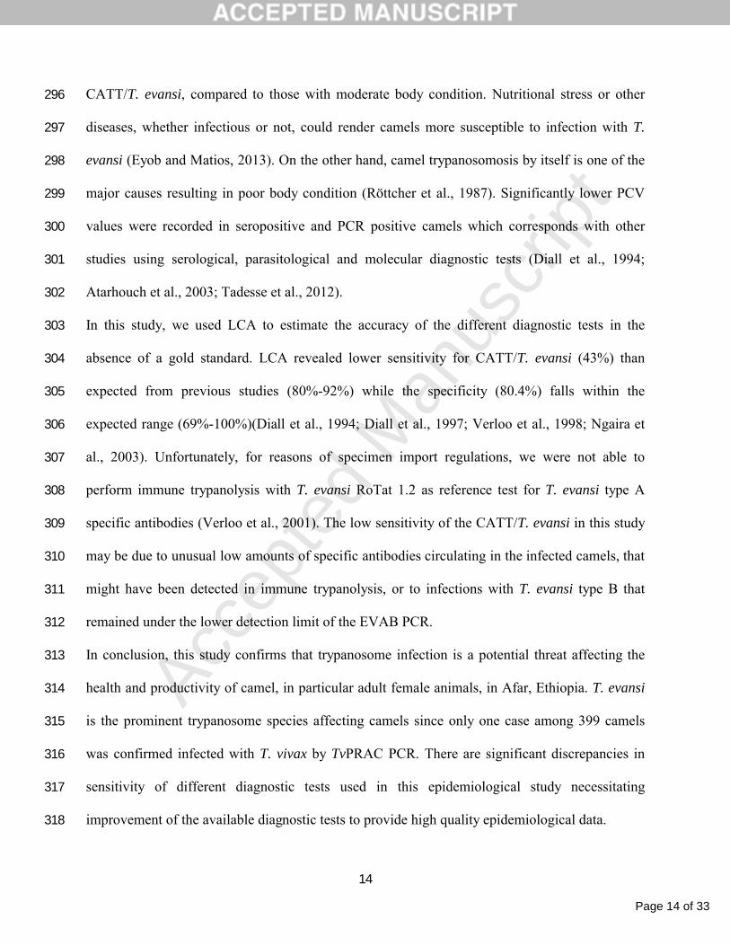
Discussion212
In this study, Giemsa stained thin blood smear examination, antibody detection by CATT/T.213
evansi and molecular tests by RoTat 1.2, 18S and ITS1-PCRs were used to assess prevalence and 214
host related risk factors of trypanosome infection in camels in Afar region, Eastern Ethiopia. 215
Only 2% of the examined camels were found infected by T. evansi by microscopy. This216
corresponds with previous findings in camels in different parts of Ethiopia, including two other 217
districts in Afar Region, Gowani and Awash Fentale (Getahun, 1998; Hailemariam et al., 2008; 218
Kassa et al., 2011; Tadesse et al., 2012) and in other African countries, for instance 2.3% in 219
Kenya (Ngaira et al., 2004) and 2.5% in Nigeria (Egbe-Nwiyi and Chaudry, 1994). However, 220
some other studies conducted in Ethiopia reported higher prevalences 6.5 %-12.1% (Richard, 221
1979; Abebe, 1991; Hailu, 2000; Hagos et al., 2009). This could be due to differences in the 222
management and husbandry regimens of the camels under study, or to seasonal effects as well as 223
to study design. Previous studies have shown that the prevalence of surra in camels is higher 224
during the rainy season and could be explained by higher densities of Tabanid and Stomoxys flies 225
during that season (Löhr et al., 1985; Luckins, 1988; Hailemariam et al., 2008).226

Page 11 of 33
Accep
ted
Man
uscr
ipt
11
In the absence of a golden standard test, the LCA revealed an overall estimate prevalence of 227
19.4%, with low sensitivity of CATT/T. evansi and the PCR tests (43% - 53%) but better than 228
the sensitivity of Giemsa stained thin blood smear examination (11%). When considering 229
microscopy, one should keep in mind that the observed prevalence reflects only a fraction of the 230
real prevalence since most microscopic techniques are poorly sensitive (OIE, 2012). In case of 231
Giemsa stained thin smear examination, the lower detection limit is greater than 500,000 232
trypanosomes/ml of blood (OIE, 2012). Therefore, indirect diagnosis, e.g. by detection of T. 233
evansi specific antibodies, may give a better estimation of the infection burden as has been 234
shown in previous studies (Diall et al., 1994; Gutierrez et al., 2000). An overall seroprevalence 235
of 24.1% was recorded with CATT/T. evansi and corresponds with the CATT seroprevalence 236
observed in Bale zone (24.9%) (Hagos et al., 2009).237
The observed seroprevalences may be overestimates of the actual prevalence since antibody 238
detection tests have the inherent shortcoming that past infections cannot be distinguished from 239
current infections (Luckins, 1988; Ngaira et al., 2003; Zeyed et al., 2010). Molecular diagnostics 240
targeting parasite DNA are considered good surrogates for parasite detection (Büscher, 2014). 241
An overall positivity rate of 21.1%, 9.5% and 7.8% was observed respectively by RoTat 1.2 242
PCR, 18S PCR and ITS-1 PCR, which is significantly higher than recorded by thin smear 243
examination (2%). CATT/T evansi and RoTat1.2 PCR yielded comparable positivity rates that 244
were significantly higher than in 18S and ITS-1 PCRs. Similar observations were made in 245
Uganda where very low molecular prevalence of Trypanozoon was recorded with the ITS-1 PCR 246
as compared to a PCR for the single copy gene phospholipase C (GPI-PLC) (Ahmed et al., 247
2013). The higher positivity rate recorded with RoTat1.2 PCR needs special attention but could 248
partly be explained by the low RoTat1.2 PCR specificity estimate by LCA. Our data show that 249

Page 12 of 33
Accep
ted
Man
uscr
ipt
12
the analytical sensitivity of RoTat 1.2 PCR based on the single copy gene is lower than the ITS-1 250
and 18S PCR based on multi copy sequences. Yet, RoTat 1.2 PCR unexpectedly yielded a higher 251
positivity rate than ITS-1 and 18S PCR. The lowest positivity rate obtained by ITS-1 PCR252
remains unexplained but it could be related to limited specificity of the ITS-1 primers. We 253
observed that when the concentration of target DNA decreases in the presence of host DNA, the 254
primers become less specific and tend to bind to the host DNA. On the other hand, we cannot 255
rule out that the RoTat1.2 PCR is less specific in the presence of host DNA as it is supported by 256
the LCA where RoTat1.2 PCR is less specific than ITS-1 PCR and 18S PCR. Noteworthy, only 257
one out of 8 thin blood smear positive animals was negative for CATT/T. evansi whereas out of 258
303 CATT/T. evansi negative samples 19.5%, 8.3% and 6.9% respectively, were positive for 259
RoTat1.2, 18S and ITS-1 PCRs. It is possible but unlikely that all these seronegative animals 260
carried early infections and had not yet formed detectable antibody levels. The number of RoTat 261
1.2 PCR positives that could not be detected by CATT/T. evansi is significantly higher than with262
18S and ITS-1 PCRs. This could be explained by the presence but not yet the expression of the 263
RoTat1.2 gene in the circulating trypanosomes (Verloo et al., 2001). On the other hand there are 264
a number of RoTat1.2 PCR and CATT/T. evansi negatives that are positive in 18S and ITS-1 265
PCR that could be explained by the absence of the RoTat1.2 gene like it has been described for 266
the T. evansi type B strains isolated in Kenya and suspected to circulate also in Ethiopia but not 267
confirmed in our study (Elsaid et al., 1998; Davison et al., 1999; Ngaira et al., 2003; Hagos et al., 268
2009; Zeyed et al., 2010; Salim et al., 2011).269
Even though 18S PCR and ITS-1 PCR cannot differentiate between the taxa of the Trypanozoon270
group, the positive results reported in this study are most likely due to T. evansi, since most of 271
them are positive in the T. evansi specific RoTat1.2 PCR and there are no records of tsetse flies272

Page 13 of 33
Accep
ted
Man
uscr
ipt
13
in the Afar region excluding the presence of T. brucei but not of non-RoTat 1.2 T. evansi (Njiru 273
et al., 2006). In Ethiopia, tsetse flies and tsetse-transmitted trypanosomes prevail in the 274
Southwestern and Northwestern parts of the country following major river basins between 275
longitude 33o and 38o E and latitude 5o and 12o N while the rest of the country, the North, 276
Southeast and East are considered tsetse free (National Tsetse and Trypanosomosis Investigation 277
and Control Center (NTTICC), 2004; Sinshaw et al., 2006). Our study revealed, for the first time 278
in Ethiopia, a T. vivax infection in camels. That there are no previous reports of camels infected 279
with T. vivax in Ethiopia is probably due to the fact that the commonly used serological and 280
parasitological tests fail to detect T. vivax or to distinguish it from T. evansi. Mixed infections of 281
T. congolense, T. vivax and Trypanozoon have been reported in camels in Nigeria but were based 282
on microscopic examination while in Sudan, using the ITS-1 PCR, only T. evansi (Trypanozoon) 283
has been detected in camel (Mbaya et al., 2010; Salim et al., 2011). Pathogenicity of T. vivax to284
camels is not well described but infected animals could act as reservoir and spread the parasite to 285
the more susceptible livestock species (Enwezor and Sackey, 2005).286
Whatever diagnostic technique used, the overall observed prevalence of surra was similar in both 287
study sites which can be explained by comparable camel husbandry practices and ecological 288
biotopes in both districts. On the other hand, a significantly higher prevalence was recorded in 289
female animals by CATT/T. evansi and Giemsa stained thin smear. Similar findings were 290
reported in Kenya and in Israel (Singh et al., 2004; Berlin et al., 2010). The fact that 291
seropositivity in CATT increases with age is in agreement with previous studies and probably 292
reflects time of exposure to biting flies and persistence of circulating antibodies, even after 293
successful treatment (Dia et al., 1997; Gutierrez et al., 2000; Atarhouch et al., 2003). Camels 294
with poor body conditions were more likely to be positive in Giemsa stained thin smear and 295
Page 14 of 33
Accep
ted
Man
uscr
ipt
14
CATT/T. evansi, compared to those with moderate body condition. Nutritional stress or other 296
diseases, whether infectious or not, could render camels more susceptible to infection with T. 297
evansi (Eyob and Matios, 2013). On the other hand, camel trypanosomosis by itself is one of the 298
major causes resulting in poor body condition (Röttcher et al., 1987). Significantly lower PCV 299
values were recorded in seropositive and PCR positive camels which corresponds with other 300
studies using serological, parasitological and molecular diagnostic tests (Diall et al., 1994; 301
Atarhouch et al., 2003; Tadesse et al., 2012).302
In this study, we used LCA to estimate the accuracy of the different diagnostic tests in the 303
absence of a gold standard. LCA revealed lower sensitivity for CATT/T. evansi (43%) than 304
expected from previous studies (80%-92%) while the specificity (80.4%) falls within the 305
expected range (69%-100%)(Diall et al., 1994; Diall et al., 1997; Verloo et al., 1998; Ngaira et 306
al., 2003). Unfortunately, for reasons of specimen import regulations, we were not able to 307
perform immune trypanolysis with T. evansi RoTat 1.2 as reference test for T. evansi type A 308
specific antibodies (Verloo et al., 2001). The low sensitivity of the CATT/T. evansi in this study 309
may be due to unusual low amounts of specific antibodies circulating in the infected camels, that 310
might have been detected in immune trypanolysis, or to infections with T. evansi type B that 311
remained under the lower detection limit of the EVAB PCR.312
In conclusion, this study confirms that trypanosome infection is a potential threat affecting the 313
health and productivity of camel, in particular adult female animals, in Afar, Ethiopia. T. evansi314
is the prominent trypanosome species affecting camels since only one case among 399 camels 315
was confirmed infected with T. vivax by TvPRAC PCR. There are significant discrepancies in 316
sensitivity of different diagnostic tests used in this epidemiological study necessitating 317
improvement of the available diagnostic tests to provide high quality epidemiological data.318

Page 15 of 33
Accep
ted
Man
uscr
ipt
15
Acknowledgements319
The PhD fellowship of Fikru Regassa was financed by the Belgian Directorate General for Development 320
Cooperation.321
References322
323
Abebe, W., 1991. Practices and major health problems of cames in the Ogaden, Ethiopia. Eurasian 324
Journal of Agriculture and Environmental Science 8, 633-642.325
Ali, I., Shafi, M., Chaudhry, Q., Faroo, Q., 2009. Camel rearing in Cholistan desert of Pakistan. Pakistan 326
Vet. J. 29, 85-92.327
Atarhouch, T., Rami, M., Bendahman, M.N., Dakkak, A., 2003. Camel trypanosomosis in Morocco 1: 328
results of a first epidemiological survey. Vet. Parasitol. 111, 277-286.329
Bajyana Songa, E., Hamers, R., 1988. A card agglutination test (CATT) for veterinary use based on an 330
early VAT RoTat 1/2 of Trypanosoma evansi. Ann. Soc. Belg. Méd. Trop. 68, 233-240.331
Berlin, D., Nasereddin, A., Azmi, K., Ereqat, S., Abdeen, Z., Baneth, G., 2010. Longitudinal study of an 332
outbreak of Trypanosoma evansi infection in equids and dromedary camels in Israel. Vet. 333
Parasitol. 174, 317-322.334
Birhanu, H., Fikru, R., Kidane, W., Gebrehiwot, T., Tola, A., Goddeeris, B.M., Büscher, P., 2013. 335
Epidemiology of Trypanosoma evansi in domestic animals in Tigray and Afar regions, Northern 336
Ethiopia. 32nd Meeting of the International Scientific Council for Trypanomiasis Research and 337
Control (ISCTRC), 8-12 September 2013, Khartoum, Sudan338

Page 16 of 33
Accep
ted
Man
uscr
ipt
16
Büscher, P., 2014. Diagnosis of African trypanosomiasis. In: Magez, S., Radwanska, M. (Eds.), 339
Trypanosomes and trypanosomiasis. Springer-Verlag, Wien, pp. 189-216.340
Claes, F., Radwanska, M., Urakawa, T., Majiwa, P., Goddeeris, B., Büscher, P., 2004. Variable surface 341
glycoprotein RoTat 1.2 PCR as a specific diagnostic tool for the detection of Trypanosoma evansi342
infections. Kinetoplastid Biol. Dis. 3, 1-6.343
Cohen, J., 1960. A coefficient of agreement for nominal scales. Educ. Psychol. Meas. 20, 37-46.344
Davison, H.C., Thrusfield, M.V., Muharsini, S., Husein, A., Partoutomo, S., Rae, P.F., Masake, R., Luckins, 345
A.G., 1999. Evaluation of antigen detection and antibody detection tests for Trypanosoma 346
evansi infections of buffaloes in Indonesia. Epidemiol. Infect. 123, 149-155.347
Deborggraeve, S., Claes, F., Laurent, T., Mertens, P., Leclipteux, T., Dujardin, J.C., Herdewijn, P., Büscher, 348
P., 2006. Molecular dipstick test for diagnosis of sleeping sickness. J. Clin. Microbiol. 44, 2884-349
2889.350
Demeke, G., 1998. Prevalence of camel trypanosomes and factors associated with the disease 351
occurrence in Leben district, Borena zone, Oromia region, Ethiopia. MSc Thesis. Addis Ababa 352
University and Free University of Berlin.353
Desquesnes, M., McLaughlin, G., Zoungrana, A., Dávila, A.M., 2001. Detection and identification of 354
Trypanosoma of African livestock through a single PCR based on internal transcribed spacer 1 of 355
rDNA. Int. J. Parasitol. 31, 610-614.356
Dia, M.L., Diop, C., Aminetou, M., Jacquiet, P., Thiam, A., 1997. Some factors affecting the prevalence of 357
Trypanosoma evansi in camels in Mauritania. Vet. Parasitol. 72, 111-120.358

Page 17 of 33
Accep
ted
Man
uscr
ipt
17
Diall, O., Bajyana Songa, E., Magnus, E., Kouyate, B., Diallo, B., Van Meirvenne, N., Hamers, R., 1994. 359
Evaluation d'un test sérologique d'agglutination directe sur carte dans le diagnostic de la 360
trypanosome caméline à Trypanosoma evansi. Rev. Sci. Tech. Off. Int. Epizoot. 13, 793-800.361
Diall, O., Diarra, B., Sanogo, Y., 1997. Evaluation of the antigen ELISA as a tool for assessing the impact of 362
tsetse control programmes on the incidence of trypanosome infections in livestock. In: IAEA 363
(Ed.). Joint FAO/IAEA Division of Nuclear Techniques in Food and Agriculture and the IAEA 364
Department of Technical Co-operation held in Addis Ababa, Ethiopia, 17-28 April 1995, Ethiopia, 365
pp. 57-64.366
Egbe-Nwiyi, T., Chaudry, S., 1994. Trypanosomosis: prevalence and pathology of camel of arid zone of 367
north eastern Nigeria. Trop. Vet. 20, 30-34.368
Elias, G., 2003. Study of the prevalence of camel trypanosomosis in selected sites of East Shoa, Arsi and369
Bale lowlands of Ethiopia. In. Addis Ababa University, Faculty of Veterinary Medicine, Debre Zeit, 370
Ethiopia.371
Elsaid, H.M., Nantulya, V.M., Hilali, M., 1998. Diagnosis of Trypanosoma evansi infection among 372
sudanese camels imported to Egypt using card agglutination test (CATT) and antigen detection 373
latex agglutination test (SURATEX). J. Protozool. Res. 8, 194-200.374
Enwezor, F.N.C., Sackey, A.K.B., 2005. Camel trypanosomosis - a review. Vet. Arh. 75, 439-425a.375
Eyob, E., Matios, L., 2013. Review on camel trypanosomosis (surra) due to Trypanosoma evansi: 376
Epidemiology and host response. Journal of Verterinary Medicine and Animal Health 5, 334-343.377
Faye, B., Bengoumi, M., Cleradin, A., Tabaran, A., Chilliard, Y., 2001. Body condition score in dromedary 378
camel: A tool for management of reproduction. Em. J. Food Agr. 13, 1-6.379

Page 18 of 33
Accep
ted
Man
uscr
ipt
18
Fikru, R., Goddeeris, B.M., Delespaux, V., Moti, Y., Tadesse, A., Bekana, M., Claes, F., De Deken, R., 380
Büscher, P., 2012. Widespread occurrence of Trypanosoma vivax in bovines of tsetse- as well as 381
non-tsetse-infested regions of Ethiopia: a reason for concern? Vet. Parasitol. 190, 355-361.382
Fikru, R., Hagos, A., Rogé, S., Reyna-Bello, A., Gonzatti, M.I., Merga, B., Goddeeris, B., Büscher, P., 2014. 383
A proline racemase based PCR for indentification of Trypanosoma vivax in cattle blood. PLoS 384
ONE 9, e84819.385
Getahun, D., 1998. The prevalence of camel trypanosomosis and factors associated with the disease 386
occurrence in Leben district, Borena zone, Oromiya region, Etiopia. MSc Thesis. Addis Ababa 387
University, Faculty of Veterinary Medicine and Freie Universität Berlin, Faculty of Veterinary 388
Medicine.389
Gutierrez, C., Juste, M.C., Corbera, J.A., Magnus, E., Verloo, D., Montoya, J.A., 2000. Camel 390
trypanosomosis in the Canary Islands: assessment of seroprevalence and infection rates using 391
the card agglutination test (CATT/T.evansi) and parasite detection tests. Vet. Parasitol. 90, 155-392
159.393
Hagos, A., Yilkal, A., Esayass, T., Alemu, T., Fikru, R., Feseha, G., Goddeeris, B.M., Claes, F., 2009. 394
Parasitological and serological survey on trypanosomosis (surra) in camels in dry and wet areas 395
of Bale Zone, Oromyia Region, Ethiopia. Rev. Méd. Vét. 12, 569-573.396
Hailemariam, L., Desalegn, L., Ibrahim, H., 2008. Prevalence and distribution of camel trypanosomosis in 397
the semi-arid and arid Awash Valley of Ethiopia. Ethiop. J. Anim. Prod. 8, 1-9.398
Hailu, D., 2000. The prevalence of camel trypanosomosis in the salt convey routes of Afar-Tigray. DVM 399
Thesis. Addis Ababa University, Faculty of Veterinary Medicine, Debre Zeit, Ethiopia.400

Page 19 of 33
Accep
ted
Man
uscr
ipt
19
Hoare, C.A., 1972. The trypanosomes of mammals. Blackwell Scientific Publications, Oxford. 749 pp.401
Kassa, T., Eguale, T., Chaka, H., 2011. Prevalence of camel trypanosomosis and its vectors in Fentale 402
district, South East Shoa zone, Ethiopia. Vet. Arh. 81, 611-621.403
Landis, J.R., Koch, G.G., 1977. The measurement of observer agreement for categorical data. Biometrics 404
33, 159-174.405
Löhr, K.F., Pholpark, S., Srikitjakarn, L., Thaboran, P., Bettermann, G., Staak, C., 1985. Trypanosoma 406
evansi infection in buffaloes in north-east Thailand. I. Field investigations. Trop. Anim. Health 407
Prod. 17, 121-125.408
Luckins, A.G., 1988. Trypanosoma evansi in Asia. Parasitol. Today 4, 137-142.409
Mbaya, A., Ibrahim, U., Apagu, S., 2010. Trypanosomosis of the dromedary camel (Camelus 410
dromedarius) and its vectors in the tsetse-free arid zone of North Eastern Maiduguri, Nigeria. 411
Vet. Parasitol. 124, 187-199.412
Menten, J., Boelaert, M., Lesaffre, E., 2008. Bayesian latent class models with conditionally dependent 413
diagnostic tests: a case study. Stat. Med. 27, 4469-4488.414
National Tsetse and Trypanosomosis Investigation and Control Center (NTTICC), 2004. Annural report on 415
tsetse and trypanosomosis survey. Bedele, Ethiopia.416
Ngaira, J.M., Bett, B., Karanja, S.M., Njagi, E.N.M., 2003. Evaluation of antigen and antibody rapid 417
detection tests for Trypanosoma evansi infection in camels in Kenya. Vet. Parasitol. 114, 131-418
141.419

Page 20 of 33
Accep
ted
Man
uscr
ipt
20
Ngaira, J.M., Njagi, E.N.M., Ngeranwa, J.J.N., Olembo, N.K., 2004. PCR amplification of RoTat 1.2 VSG 420
gene in Trypanosoma evansi isolates in Kenya. Vet. Parasitol. 120, 23-33.421
Njiru, Z.K., Constantine, C.C., Masiga, D.K., Reid, S.A., Thompson, R.C., Gibson, W.C., 2006. 422
Characterization of Trypanosoma evansi type B. Infect. Genet. Evol. 6, 292-300.423
OIE, 2012. Manual of diagnostic tests and vaccines for terrestrial animals.424
http://www.oie.int/fileadmin/Home/eng/Health_standards/tahm/2.04.18_TRYPANOSOMOSIS.p425
df426
Richard, D., 1979. The disease of the dromadary (Camelus dromedarius) in Ethiopia. Ethiop. Vet. Bull. 2, 427
46-67.428
Röttcher, D., Schillinger, D., Zweygarth, E., 1987. Trypanosomiasis in the camel (Camelus dromedarius). 429
Rev. Sci. Tech. Off. Int. Epizoot. 6, 463-470.430
Salim, B., Bakheit, M.A., Kamau, J., Nakamura, I., Sugimoto, C., 2011. Molecular epidemiology of camel 431
trypanosomiasis based on ITS1 rDNA and RoTat 1.2 VSG gene in the Sudan. Parasit. Vectors 4, 432
31.433
Singh, N., Pathak, K.M.L., Kumar, R., 2004. A comparative evaluation of parasitological, serological and 434
DNA amplification methods for diagnosis of natural Trypanosoma evansi infection in camels. 435
Vet. Parasitol. 126, 365-373.436
Sinshaw, A., Abebe, G., Desquesnes, M., Yoni, W., 2006. Biting flies and Trypanosoma vivax infection in 437
three highland districts bordering lake Tana, Ethiopia. Vet. Parasitol. 142, 35-46.438
Tadesse, A., Omar, A., Aragaw, K., Mekbib, B., Sheferaw, D., 2012. A study on camel trypanosomosis in 439
Jijiga zone, eastern Ethiopia. J. Vet. Adv. 2, 216-219.440

Page 21 of 33
Accep
ted
Man
uscr
ipt
21
Tekle, T., Abebe, G., 2001. Trypanosomosis and helminthoses: major health problems of camels 441
(Camelus dromedarius) in the southern rangelands of Borena, Ethiopia. J. Camel Pract. Res. 8, 442
39-42.443
Thrusfield, M.V., 2005. Veterinary epidemiology. Blackwell Publishing Professional, Ames, Iowa, USA.444
610 pp.445
Toma, B., Dufour, B., Sanaa, M., Benet, J.-J., Ellis, P., Moutou, F., Louza, A., 1999. Applied veterinary 446
epidemiology and control of disease in populations. Maison Alfort, Paris, France. 576 pp.447
Verloo, D., Holland, W., My, L.N., Thanh, N.G., Tam, P.T., Goddeeris, B., Vercruysse, J., 2000. Comparison 448
of serological tests for Trypanosoma evansi natural infections in water buffaloes from north 449
Vietnam. Vet. Parasitol. 92, 87-96.450
Verloo, D., Magnus, E., Büscher, P., 2001. General expression of RoTat 1.2 variable antigen type in 451
Trypanosoma evansi isolates from different origin. Vet. Parasitol. 97, 183-189.452
Verloo, D., Tibayrenc, R., Magnus, E., Büscher, P., Van Meirvenne, N., 1998. Performance of serological 453
tests for Trypanosoma evansi infections in camels from Niger. J. Protozool. Res. 8, 190-193.454
Zeyed, A., Habeeb, S., Allam, N., Ashry, H., Mohamed, A., Ashour, A., Taha, H., 2010. A critical 455
comparative study of parasitological and serological differential diagnostic methods of 456
Trypanosoma evansi infection in some farm animal in Egypt. Vet. World 1, 325-328.457
458
459

Page 22 of 33
Accep
ted
Man
uscr
ipt
22
Table 1: Primer sequences, target sequences and expected amplicon size of the different PCRs used to test blood from 399 camels
from Eastern Ethiopia.
Target group Target
sequence
Primers Primer sequence Amplicon (bp) Reference
18s F 5′-CGCCAAGCTAATACATGAACCAA-3′Trypanosomatidae 18S
rRNA 18S R 5′-TAATTTCATTCATTCGCTGGACG-3′
110 (Deborggraeve et
al., 2006)
ITS-1 F 5’-TGTAGGTGAACCTGCAGCTGGATC-3’Trypanozoon ITS-1
rRNA ITS-1 R 5-CCAAGTCATCCATCGCGACACGTT-3’
450 (Desquesnes et al.,
2001)
RoTat1.2F 5′-GCGGGGTGTTTAAAGCAATA-3′T. evansi type A RoTat1.2
RoTat1.2R 5′-ATTAGTGCTGCGTGTGTTCG-3′
205 (Claes et al., 2004)
T. evansi type B minicircle EVAB-1 5’-ACAGTCCGAGAGATAGAG-3' 436 (Njiru et al., 2006)
EVAB-2 5'-CTGTACTCTACATCTACCTC-3’
TvPRAC F 5’-CGCAAGTGGACCGTTCGCCT-3’T. vivax Proline
racemase TvPRAC-R 5’-ACGCGGGGCGAACAGAAGTG-3’
239 (Fikru et al., 2014)

Page 23 of 33
Accep
ted
Man
uscr
ipt
23
Table 2: Observed prevalences in % and with 95% confidence interval of T. evansi infection in camels in two districts of Eastern
Ethiopia using different diagnostic tests.
TotalDistrict Thin blood
smear
CATT/T. evansi RoTat1.2 PCR 18S PCR ITS-1 PCR
Chifra 2 (0.06-3.94) 20.6 (15-26.2) 21.1(15.3-26.8) 8 (4.2-11.8) 8 (4.2-11.8) 199
Dewe 2(0.05-3.95) 27.5(21.3-33.7) 21.5 (15.8-27.2) 11 (6.7-15.3) 7.5 (3.9-11.2) 200
Total 2(0.6-3.4) 24.1(19.9-28.3) 21.3 (17.3-25.3) 9.5 (6.6-12.4) 7.8 (5.2-10.4) 399

Page 24 of 33
Accep
ted
Man
uscr
ipt
24
Table 3: Frequency distribution (N) and probability of being infected for each outcome pattern observed in five diagnostic tests for T.
evansi infection performed on 399 camels from Eastern Ethiopia, as estimated with latent class analysis.
PCR
CATT/T. evansi RoTat 1.2 18S ITS1 Thin blood smear N Probability infected
+ + + + + 2 100
+ + + + - 6 100
+ + - - + 1 100
+ - - - + 4 100
- + + + - 18 100
- - - - + 1 100
+ - + + - 1 96.8
+ + + - - 3 94.1
- - + + - 1 92.2
- + + - - 4 85.3

Page 25 of 33
Accep
ted
Man
uscr
ipt
25
+ + - + - 1 84.7
+ - + - - 1 60.5
- - + - - 2 38.2
- - - + - 2 19.6
+ + - - - 13 18.4
+ - - - - 64 17.8
- + - - - 37 7.2
- - - - - 238 6.7

Page 26 of 33
Accep
ted
Man
uscr
ipt
26
Table 4: An exploratory assessment of the diagnostic accuracy of the different diagnostic tests performed on 399 camels from Eastern
Ethiopia, estimated by latent class analysis with a prevalence of 19.4 % (95% CI: 12.8 – 28.0)
Tests Sensitivity (95% CI) Specificity (95% CI)
CATT 42.5 (32.4-52.9) 80.4 (77.8-84.6)
PCR RoTat1.2 52.9 (39.3-67.9) 85.9 (84.1-88.1)
PCR 18S 47.7 (33.0-64.2) 99.2 (98.0-100)
PCR ITS-1 39.1 (26.4-52.8) 99.4 (98.8-100)
Giemsa stained thin smear 10.8 (7.3-14.6) 100 (fixed)

Page 27 of 33
Accep
ted
Man
uscr
ipt
27
Table 5: Host related variables and positivity rate obtained with different tests for T. evansi infection diagnosis, performed on 399
camels from Eastern Ethiopia.
Thin blood smear CATT/ T. evansi RoTat1.2 PCR 18S PCR ITS-1 PCR
Number Positive Positive Positive Positive Positive
Female 251 2.8* (0.8-4.8) 27.9 (22.6-33.5)* 23.1 (17.9-28.3) 8.8 (5.3-12.3) 6.8 (3.7-9.9)Gender
Male 148 0.7 17.6 (11.5-23.7) 18.2 (12.0-24.4) 10.8 (5.8-
15.8)
9.5 (4.8-14.2)
Moderate 311 0.3 17.7 (13.5-21.9) 22.5 (17.9-27.1) 10 (6.7-13.3) 7.4 (4.5-10.3)Body
condition
score
Poor 88 8* (2.3-10.7) 46.6* (36.4-56.8) 17 (9.1-24.9) 7 (1.7-12.3) 9.1 (3.1-15.1)
No 200 2.0 (0.1-3.9) 27.5 (21.3-33.7) 21.5 (15.8-27.2) 11 (6.7-15.3) 7.5 (3.8-11.2)Treatment
history Yes 199 2.0 (0.0-4.0) 20.6 (15.0-26.2) 21.1 (15.4-26.8) 8 (4.2-11.8) 8 (4.2-11.8)
Age category ≤ 1yr 50 0 20 (8.9-31.1) 22.0 (10.5-33.5) 12.0 (13.0-
31.0)
8.0 (0.5-15.5)

Page 28 of 33
Accep
ted
Man
uscr
ipt
28
1 < x ≤ 5 yr 72 0 15.3 (7.0-23.6) 25.0 (15.0-35.0) 12.5 (4.9-
20.1)
11.1 (3.8-
18.4)
5 < x ≤ 10 yr 252 2.4 (0.5-4.3) 25.8 (20.4-31.2) 18.3 (13.5-23.1) 7.9 (4.6-11.2) 6.3 (3.3-9.3)
> 10 yr 25 8 (0.0-18.3) 40.0* (20.8-59.2) 40.0* (20.8-59.2) 12.0 (0.0-
24.7)
12.0 (0.0-24-
7)
* significant difference

Page 29 of 33
Accep
ted
Man
uscr
ipt
29
Table 6: Mean PCV (%) with standard deviation (SD) and 95% confidence interval of 399 camels from Eastern Ethiopia tested using
different tests to detect T. evansi infection.
Test PCV SD 95% confidence interval
Negative 23.7 2.7 23.4-24.0Thin blood smear
Positive 20.7* 2,1 19.0-22.5
Negative 24.3 2.6 24.0-24.6CATT/T. evansi
Positive 21.5* 1.8 21.1-21.9
Negative 23.8 2.7 23.5-24.1RoTat1.2 PCR
Positive 23.0* 2.7 22.4-23.5
Negative 23.7 2.7 23.5-24.018S PCR
Positive 22.6* 2.7 21.8-23.5
Negative 23.7 2.7 23.4-24.0ITS-1 PCR
Positive 22.6* 2.7 21.7-23.6
* significant difference

Page 30 of 33
Accep
ted
Man
uscr
ipt
30
Figure legends
Figure 1: Map of Ethiopia with the two districts (green) Chifra and Dewe in the Afar Region (orange border).
Figure 2: Analytical sensitivity of the three PCRs: Panel A = RoTat1.2 PCR; Panel B = 18S PCR, Panel C = ITS-1PCR. Lanes 1:
Gene Ruler 100 bp DNA ladder (Fermentas). Lanes 2 -8: DNA of the T. evansi isolate from Kazakhstan at decreasing concentrations
(1000, 200, 40, 8, 0.16, 0.032, 0.0064 pg/µl). Lanes 9-15: DNA of the T. evansi isolate from Morocco at decreasing concentrations
(1000, 200, 40, 8, 0.16, 0.032, 0.0064 pg/µl).

Page 31 of 33
Accep
ted
Man
uscr
ipt
31
We conducted a cross-sectional study on camel trypanosome infection in Eastern Ethiopia
Depending on the diagnostic test, positivity rates ranged from 2% to 24%
Latent class analysis estimated an overall prevalence of 19%
All infections were due to Trypanosoma evansi except one with Trypanosoma vivax
Risk factors for infection were poor body condition and age

Page 32 of 33
Accep
ted
Man
uscr
ipt
Figure 1

Page 33 of 33
Accep
ted
Man
uscr
ipt
Figure 2