microRNA-383 impairs phosphorylation of H2AX by targeting PNUTS and inducing cell cycle arrest in...
9
microRNA-383 impairs phosphorylation of H2AX by targeting PNUTS and inducing cell cycle arrest in testicular embryonal carcinoma cells ☆ Helong Huang a , Hui Tian a , Zhengzheng Duan a , Yunxia Cao b , Xian-Sheng Zhang c , Fei Sun a,d, ⁎ a Department of Cell and Developmental Biology, School of Life Sciences, University of Science and Technology of China, Hefei, Anhui 230027, China b Reproduction Medical Center, Department of Obstetrics and Gynecology, The First Affiliated Hospital of Anhui Medical University, Hefei, Anhui 230032, China c Department of Urology, The First Affiliated Hospital of Anhui Medical University, Hefei, Anhui 230032, China d Hefei National Laboratory for Physical Sciences at Microscale and School of Life Sciences, University of Science and Technology of China, Hefei, Anhui 230026, China abstract article info Article history: Received 8 November 2013 Received in revised form 24 December 2013 Accepted 14 January 2014 Available online 22 January 2014 Keywords: microRNA-383 Protein phosphatase 1 Regulatory subunit 10 Spermatogenesis DNA damage Male germ cells with aberrant DNA damage are the weighted factor contributing to male infertility. Mounting evidence shows that DNA damage in male germ cells impairs spermatogenesis and lowers fecundity. MicroRNAs (miRNAs) regulating expression of multiple genes play a significant role in spermatogenesis. Our previous results have shown that microRNA-383 (miR-383) is one of the notable down-regulated microRNAs in the testes of sterile males with maturation arrest (MA) and is located predominantly in spermatogonia and primary spermatocytes. However, the role that miR-383 plays in DNA damage during spermatogenesis remains unknown. In this study, we found that miR-383 inhibited the focal formation and abundance of γH2AX, which is the major marker of sites of DNA damage, with or without ultraviolet irradiation and cisplatin in testicular embryonal carcinoma (NT-2) cells. In addition, NT-2 cells were remarkably sensitized to DNA damage reagent (cisplatin) by forcing expression of miR-383 and silencing expression of protein phosphatase 1, regulatory subunit 10 (PNUTS). By constructing Renilla luciferase reporters and co-transfecting miR-383 and reporters in NT-2 cells, we identified that PNUTS was a valid target of miR-383. Further results demonstrated that the repression of the phosphorylated form of H2AX by miR-383 was due to independent depletion of PNUTS and cell cycle arrest. In conclusion, we found a novel function of miR-383 in the DNA damage pathway. miR-383 impairs the phosphorylation of H2AX by targeting PNUTS and inducing cell cycle arrest independently, as well as sensitizing NT-2 cells to cisplatin. © 2014 Elsevier Inc. All rights reserved. 1. Introduction Ten to fifteen percent of couples of childbearing age are infertile, and half of the cases are due to male factors, which affect one out of 20 men in the general population [1,2]. Furthermore, human patients with non-obstructive azoospermia (NOA), which accounts for a considerable proportion of male factor infertility, possess dramatically lower sperm retrieval rate and clinical pregnancy rate and higher rates of DNA fragmentation [3,4]. Many studies have shown that DNA damage in sperm is associated with infertility and low fecundity [5–7]. However, the molecular mechanism of DNA damage occurring during spermato- genesis, especially in the early stage of spermatogenesis, remains obscure. Spermatogenesis is a well-tuned and dynamic process involving the transformation of spermatogonial stem cells to mature spermatozoa, including the stages of mitotic spermatogonia and meiotic spermato- cytes. Five most common types of DNA damage occur during spermato- genesis due to the remodeling of chromatin [6]. Normally, these sites of DNA damage are associated with phosphorylation of H2AX(H2A his- tone family, member X) at serine 139, referred to as γH2AX, and are re- solved by DNA damage repair regulation before spermatozoa production during spermatogenesis [8]. The mechanism underlying the aberrant DNA damage observed during spermatogenesis of testes of sterile patients is little known. MicroRNAs (miRNAs), a family of small noncoding RNAs, are generat- ed from processing of stem-loop structures of 70 nucleotides by several specific endonucleases and are ultimately delivered as mature single- stranded small RNAs of 19–23 nt that post-transcriptionally regulate gene expression through base pairing with the 3′-untranslated region (3′UTR) of target mRNAs [9]. Numerous studies have suggested that miRNAs play a crucial role in regulating the DNA damage pathway activated by radiation and DNA damage reagents. Inhibition of miR-34-c impairs DNA damage-induced S-phase arrest, whereas forced expression of miR-34 represses expression of c-Myc, leading to genomic instability [10]. miR-138 [11] and miR-24 [12] modulate the DNA damage response by targeting H2AX directly in cancer cell lines and terminally differentiat- ed blood cells, respectively. However, the role miRNAs play in DNA damage during spermatogenesis is poorly understood. Cellular Signalling 26 (2014) 903–911 ☆ Supported by the following grants: the National Natural Science Foundation of China (81125005 and 81370749), the National Basic Research Program of China (2014CB943100); the Chinese Academy of Sciences Knowledge Creative Program (KSCX2-EW-R-07). ⁎ Corresponding author at: School of Life Sciences, University of Science and Technology of China, Hefei, Anhui 230026, China. Tel.: +86 551 63600847; fax: +86 551 63602703. E-mail address: [email protected] (F. Sun). 0898-6568/$ – see front matter © 2014 Elsevier Inc. All rights reserved. http://dx.doi.org/10.1016/j.cellsig.2014.01.016 Contents lists available at ScienceDirect Cellular Signalling journal homepage: www.elsevier.com/locate/cellsig
Transcript of microRNA-383 impairs phosphorylation of H2AX by targeting PNUTS and inducing cell cycle arrest in...
Cellular Signalling 26 (2014) 903–911
Contents lists available at ScienceDirect
Cellular Signalling
j ourna l homepage: www.e lsev ie r .com/ locate /ce l l s ig
microRNA-383 impairs phosphorylation of H2AXby targeting PNUTS andinducing cell cycle arrest in testicular embryonal carcinoma cells☆
Helong Huang a, Hui Tian a, Zhengzheng Duan a, Yunxia Cao b, Xian-Sheng Zhang c, Fei Sun a,d,⁎a Department of Cell and Developmental Biology, School of Life Sciences, University of Science and Technology of China, Hefei, Anhui 230027, Chinab Reproduction Medical Center, Department of Obstetrics and Gynecology, The First Affiliated Hospital of Anhui Medical University, Hefei, Anhui 230032, Chinac Department of Urology, The First Affiliated Hospital of Anhui Medical University, Hefei, Anhui 230032, Chinad Hefei National Laboratory for Physical Sciences at Microscale and School of Life Sciences, University of Science and Technology of China, Hefei, Anhui 230026, China
☆ Supported by the following grants: the National NChina (81125005 and 81370749), the National Basi(2014CB943100); the Chinese Academy of Sciences(KSCX2-EW-R-07).⁎ Corresponding author at: School of Life Sciences, Unive
of China, Hefei, Anhui 230026, China. Tel.: +86 551 6360E-mail address: [email protected] (F. Sun).
0898-6568/$ – see front matter © 2014 Elsevier Inc. All rihttp://dx.doi.org/10.1016/j.cellsig.2014.01.016
a b s t r a c t
a r t i c l e i n f oArticle history:Received 8 November 2013Received in revised form 24 December 2013Accepted 14 January 2014Available online 22 January 2014
Keywords:microRNA-383Protein phosphatase 1Regulatory subunit 10SpermatogenesisDNA damage
Male germ cells with aberrant DNA damage are the weighted factor contributing to male infertility. Mountingevidence shows that DNA damage in male germ cells impairs spermatogenesis and lowers fecundity. MicroRNAs(miRNAs) regulating expression of multiple genes play a significant role in spermatogenesis. Our previous resultshave shown thatmicroRNA-383 (miR-383) is one of the notable down-regulatedmicroRNAs in the testes of sterilemales with maturation arrest (MA) and is located predominantly in spermatogonia and primary spermatocytes.However, the role that miR-383 plays in DNA damage during spermatogenesis remains unknown. In this study,we found that miR-383 inhibited the focal formation and abundance of γH2AX, which is the major marker ofsites of DNA damage, with or without ultraviolet irradiation and cisplatin in testicular embryonal carcinoma(NT-2) cells. In addition, NT-2 cells were remarkably sensitized to DNA damage reagent (cisplatin) by forcingexpression of miR-383 and silencing expression of protein phosphatase 1, regulatory subunit 10 (PNUTS). Byconstructing Renilla luciferase reporters and co-transfecting miR-383 and reporters in NT-2 cells, we identifiedthat PNUTSwas a valid target of miR-383. Further results demonstrated that the repression of the phosphorylatedform of H2AX by miR-383 was due to independent depletion of PNUTS and cell cycle arrest. In conclusion, wefound a novel function of miR-383 in the DNA damage pathway. miR-383 impairs the phosphorylation of H2AXby targeting PNUTS and inducing cell cycle arrest independently, as well as sensitizing NT-2 cells to cisplatin.
© 2014 Elsevier Inc. All rights reserved.
1. Introduction
Ten to fifteen percent of couples of childbearing age are infertile, andhalf of the cases are due to male factors, which affect one out of 20 menin the general population [1,2]. Furthermore, human patients withnon-obstructive azoospermia (NOA), which accounts for a considerableproportion of male factor infertility, possess dramatically lower spermretrieval rate and clinical pregnancy rate and higher rates of DNAfragmentation [3,4]. Many studies have shown that DNA damage insperm is associated with infertility and low fecundity [5–7]. However,the molecular mechanism of DNA damage occurring during spermato-genesis, especially in the early stage of spermatogenesis, remainsobscure.
Spermatogenesis is a well-tuned and dynamic process involving thetransformation of spermatogonial stem cells to mature spermatozoa,
atural Science Foundation ofc Research Program of ChinaKnowledge Creative Program
rsity of Science and Technology0847; fax: +86 551 63602703.
ghts reserved.
including the stages of mitotic spermatogonia and meiotic spermato-cytes. Five most common types of DNA damage occur during spermato-genesis due to the remodeling of chromatin [6]. Normally, these sites ofDNA damage are associated with phosphorylation of H2AX(H2A his-tone family, member X) at serine 139, referred to as γH2AX, and are re-solved by DNA damage repair regulation before spermatozoaproduction during spermatogenesis [8]. The mechanism underlyingthe aberrant DNA damage observed during spermatogenesis of testesof sterile patients is little known.
MicroRNAs (miRNAs), a family of small noncoding RNAs, are generat-ed from processing of stem-loop structures of 70 nucleotides by severalspecific endonucleases and are ultimately delivered as mature single-stranded small RNAs of 19–23 nt that post-transcriptionally regulategene expression through base pairing with the 3′-untranslated region(3′UTR) of target mRNAs [9]. Numerous studies have suggested thatmiRNAs play a crucial role in regulating the DNA damage pathwayactivated by radiation and DNA damage reagents. Inhibition of miR-34-cimpairs DNA damage-induced S-phase arrest, whereas forced expressionof miR-34 represses expression of c-Myc, leading to genomic instability[10]. miR-138 [11] and miR-24 [12] modulate the DNA damage responseby targeting H2AX directly in cancer cell lines and terminally differentiat-ed blood cells, respectively. However, the role miRNAs play in DNAdamage during spermatogenesis is poorly understood.
904 H. Huang et al. / Cellular Signalling 26 (2014) 903–911
Our previous study has identified the differential expression ofmiRNAs in the testes of NOA patients with maturation arrest (MA)[13]. In particular, miR-383 was notably down-regulated in spermato-gonia and spermatocytes of MA patients. These findings indicate thatmiR-383may be involved in events occurring in the early stage of sper-matogenesis where the DNA damage response is activated duringmeiosis. To investigate the possible role of miR-383 in DNA damagesignaling in spermatogenesis, we studied the correlation betweenmiR-383 and γH2AX in the testicular embryonal carcinoma cell lineNTERA-2 (NT-2) and explored the potential targets of miR-383 thatmay play roles in the DNA damage pathway.
2. Material and methods
2.1. Human testicular samples
Human testicular tissue samples from nine patients (aged21–38 years) with MA and from three control patients (aged31–37 years) were obtained from the First Affiliated Hospital ofAnhui Medical University (Hefei, China). Samples were dissected intoseveral portions for different experiments. The volumes of testiculartissues from patients varied greatly. As a result, some specimens werenot enough to be used in all experiments. Ideal normal controls wouldbe obtained from volunteers of known fertility; however, difficultiesin acquiring testicular material from this demographic group madethis impractical. Therefore, we obtained testicular tissues from patientsundergoing orchiectomy for prostate carcinoma before chemotherapy,who had a history of normal spermatogenesis and fertility. All speci-mens were taken with patient informed consent and with ethicalapproval from the Institutional Review Boards of the University ofScience and Technology of China and the Anhui Medical University.
2.2. Cell culture and transfection
NT-2 cells are derived from human embryonal carcinomas. Cells(NT-2, 293T, MCF-7, and H1299) were cultured in Dulbecco′s modifiedEagle′s medium (DMEM) supplemented with 10% (v/v) fetal bovineserum (Life Technology Inc., Grand Island, NY, USA) and 1% antibiotics(100 U/ml penicillin and 100 mg/ml streptomycin, Life TechnologyInc.). Cells were incubated at 37 °C in a humidified incubator with 5%carbon dioxide. Different transfection reagents were used for differentcell lines and nucleotides. Lipofectamine 2000 Reagent (Invitrogen,Carlsbad, CA, USA)was used for transfection of 293T cells and Lipofecta-mine RNAiMAX (Invitrogen) and Fugene HD (Roche) were used intransfection of oligonucleotides and plasmids into NT-2 cells, respec-tively. All transfection procedures were performed according to theprotocols supplied by manufacturers.
2.3. Oligonucleotides and vectors
siRNA duplex homologs in sequences with miR-383 mimics andmiR-383 inhibitor, as well as negative control (NC) and inhibitor NCwere chemically synthesized and optimized by Shanghai Gene-PharmaCo. (Shanghai, China). The inhibitor of miR-383 is single-stranded,sequence-specific, and chemically modified to specifically target andknock downmiR-383molecules. PNUTS siRNA (si.PNUTS)was designedand chemically synthesized by ShanghaiGene-PharmaCo. The sequenceof si.PNUTS was: sense, GACCCGUUCACCAGAAAUATT; antisense, UAUUUCUGGUGAACGGGUCTT.
The psiCHECK-2 dual luciferase reporter plasmidwas kindly provid-ed by Biliang Zhang (Guangzhou Institute of Biomedicine and Health,Chinese Academy of Sciences, China), and the pcDNA3.1+ vector waskindly provided by Mian Wu (USTC, China). Wild-type (WT) plasmidsof PNUTS 3′ UTR and H2AX 5′ UTR plus Coding DNA Sequence (CDS)were constructed by amplifying a 848-bp 3′ UTR fragment of PNUTSmRNA and 505-bp 5′ UTR plus CDS fragment of H2AXmRNA harboring
themiR-383 binding site predicted bymiRanda (http://www.microrna.org) and DNAman, respectively, whereas mutated (MT) PNUTS 3′ UTRwas generated by PCR-based site-directed mutagenesis. The fragmentsof WT and MT PNUTS 3′ UTR were fused with the psiCHECK-2 reportervector at the XhoI and NotI sites. The primers were as follows:
WT PNUTS 3′ UTR
Forward primer: 5′-CTACTCGAGCTGTGGACTGCAGCCTCGCTCTT-3′Reverse primer: 5′-CTAGCGGCCGCTTCACGCTGGCAAGGTTC-3′MT PNUTS 3′ UTRForward primer: 5′-CTACTCGAGTTCTGTAGACTGGTGCGCATTG-3′Reverse primer: 5′-CTAGCGGCCGC CAATGCGCACCAGTCTACAG AA-3′WT H2AX 5′ UTR plus CDSForward primer: 5′-CGCCTCGAGACAGCAGTTACACTGCGGCG-3′Reverse primer: 5′-CGC GCGGCCGCTTAGTACTCC TGGGAGGCCTGGGTG-3′
2.4. Luciferase reporter assay
Either 40 nM miR-383 mimics or miR-383 inhibitor were co-transfected with 200 ng psiCHECK-2 vector into 293 T cells in 24-wellplates, with three replicate wells for each condition. Cells were harvest-ed 30 h after transfection using the Dual Luciferase Reporter AssaySystem (Promega).
2.5. Western blotting
Western blotting experiments were performed as previouslydescribed [14]. The following antibodieswere used forWestern blottinganalysis: anti-PNUTS (BD Transduction Laboratories™, USA); anti-γH2AX (Abcam, Cambridge, MA, USA); anti-cyclin D1 (Santa CruzBiotechnology, Inc., CA, USA); anti-CDK4 (Santa Cruz Biotechnology,Inc.); and anti-actin (Abcam). Protein levels were normalized to actinand quantified using ImageJ system (USA).
2.6. Cisplatin sensitivity assay
cis-Diamminedichloroplatinum(II) (cisplatin or CDDP) was pur-chased from Sigma-Aldrich (Dorset, UK). It was dissolved in 0.9% (w/v)saline before each experiment at a stock concentration of 1 mM, andthen further diluted into the final concentrations used in experiments.
NT-2 cells were collected at the phase of exponential growth andthen seeded into 12-well plates at 70–80% confluence. They were cul-tured overnight in an incubator. Cells were transfected with miR-383mimics/negative control or si.PNUTS/siRNA negative control the nextday. After 24 h, cells were collected and re-seeded into 96-well platesat a density of 4000 cells per well within 100 μl of growth medium.Cells were allowed to adhere overnight and then treated with differentconcentrations of cisplatin, ranging from0 μM to 15 μM for a continuous72 h. Cisplatin sensitivity was detected with the Cell Counting Kit-8(CCK-8) (Dojindo Laboratories, Kumamoto, Japan) as previously de-scribed [15]. The numbers of cells were determined by measuring theabsorbance at 450 nmusing a 96-well format plate reader (ELX 800 uni-versal microplate Reader; BioTek, Inc., Highland Park, IL, USA).
2.7. Immunocytochemistry (ICC)
After NT-2 cells were exposed to ultraviolet (UV) irradiation for 12 hand transfectedwithmiR-383mimics/negative control, cells were fixedwith 4%paraformaldehyde (PFA) for 20min, and thenpermeabilized for10 min with 0.2% Triton/PBS, and blocked with 1% BSA/TBS for 1 h. Thecells were then incubated with anti-γH2AX monoclonal antibody(Abcam, Cambridge, MA, USA) for 1 h at room temperature. Then theywere incubated with donkey anti-mouse IgG (H + L) secondaryantibody (Alexa Fluor 555, Invitrogen) for 30 min in the dark at
905H. Huang et al. / Cellular Signalling 26 (2014) 903–911
room temperature. The nuclei were stain with Hoechst 33342 (Sigma-Aldrich, St. Louis, Missouri,USA), which is a blue fluorescent dye usedto stain DNA. The fluorescent signals were examined under a Nikonepifluorescence microscope (Nikon Eclipse 80i; Nikon, Tokyo, Japan).
2.8. Histological analysis and immunohistochemistry (IHC)
Immunohistochemistry (IHC) was conducted to evaluate the loca-tion of PNUTS in human testicular tissues. Human testes were dissectedinto pieces, fixedwith 4% PFA, embedded in paraffinwax, and sectionedat 4mM. To further confirm the specific syndrome of infertility, sectionswere stained with hematoxylin and eosin with a standard protocol.Initially, slides with testicular tissues were heated in 10 mM sodiumcitrate buffer, pH 6.0 for 10 min after deparaffinization in a microwaveoven. Then, sections were dipped into PBS containing 3% H2O2 and0.1% Triton X-100 to rule out endogenous peroxidase. After treatmentwith 10% normal donkey serum (Jackson ImmunoResearch Labs Inc.,West Grove, PA, USA) to block non-specific binding signals, slides wereincubated with PNUTS antibody (BD Transduction Laboratories™)overnight at 4 °C and then incubated with mouse biotinylated second-ary antibody (Abcam) for 2 h at room temperature. Immunosignals ofPNUTS were visualized using streptavidin-peroxidase and 3, 3′-diaminobenzidine (Maixin Bio, Fuzhou, China).
2.9. Human testicular tissue spreads
Testicular specimens fromnormal controls and human patientswithMA were dissected and placed quickly in PBS, pH7.4 at room tempera-ture. The de-capsulated testicular tissueswere placed in a hypotonic ex-traction buffer (HEB) containing 30 mM Tris, 50 mM sucrose, 17 mMcitric acid, 5 mM EDTA, 2.5 mM DTT, and 1 mM phenylmethylsulfonylfluoride (PMSF), pH 8.2 for 60 min. Subsequently, tubules were torninto pieces using two fine watchmaker forceps in 20 μl of 0.1 M sucroseand added to slides coated with 1% PFA. The slides were dried in ahumid chamber overnight at room temperature and washed in PBScontaining Photoflo (Kodak, EMS). After air drying for 10 min at roomtemperature, the slides were incubated with ADB for 30 min at roomtemperature to block non-specific signals and with anti-γH2AX mono-clonal antibody overnight at 37 °C. The slides were rinsed with TBSthree times for 10 min each time. They were incubated with ADBagain for 48 h at 4 °C in a humid chamber. The slides were rinsed as de-scribed above. After incubation with donkey anti-mouse IgG (H + L)secondary antibody (Alexa Fluor 555, Invitrogen) for 90 min at 37 °C,slides were secured by coverslips with the addition of vectashield(Vector Laboratories Inc., Burlingame, CA, USA). Fluorescent signalswere examined under a Nikon epifluorescence microscope (NikonEclipse 80i; Nikon).
2.10. RNA extraction and real-time PCR
RNA was extracted from cells and subjected to real-time PCR asdescribed as previously [16]. Briefly, RNA extraction was performedfollowing a standard Trizol protocol and real-time PCR was performedwith the ABI Step One System (Applied Biosystems, Foster City, CA,USA) using the SYBR Premix Ex Taq II kit (TaKaRa Bio, Inc.). To detectthe expression of PNUTS mRNA and mature miR-383, expression levelswere normalized to β-actin and U6, respectively.
Primers for Q-RT were as follows:
EYA1Forward primer: 5′-GTCACCCTGAGGAAGGTTCATTG-3′Reverse primer: 5′-CCCAGATTGATGCCTGTCATGTAG-3′WIP1Forward primer: 5′-GCCAGAACTTCCCAAGGAAAG-3′Reverse primer: 5′-GGTTCAGGTGACACCACAAATTC-3′Protein phosphatase 2A activator, regulatory subunit 4 (PPP2R4):
Forward primer: 5′-AGCCGGCAGCCAGGGAGTGTG-3′Reverse primer: 5′-AGGAAGGGACGGCGCTGATGTT-3′Protein phosphatase 4, catalytic subunit (PPP4C):Forward primer: 5′-GCTACCGCTGTGGGAATGTG-3′Reverse primer: 5′-GGGAGCAGCCTCAAAGATGA-3′Protein phosphatase 6, catalytic subunit (PPP6C):Forward primer: 5′-GCATTAAAGGCTAAATGGCCTGAT-3′Reverse primer: 5′-GTACAGTATC TCCAGGCATT AGCA-3′β-actinForward primer: 5′-TGGCACCCAGCACAATGAA-3′Reverse primer: 5′-CTAAGTCATAGTCCGCCTAGAAGCA-3′
2.11. Statistical analysis
All the experiments in this studywere performed at least three inde-pendent times. Data are shown as themeans plus standard errors of themean (SEMs). p-Value b 0.05 was considered statistically significant.
3. Results
3.1. Aberrant γH2AX foci exist in pachytene spermatocytes of patients withNOA
To investigate the DNA damage occurring in spermatocytes, we per-formed germ cell spreads of testicular tissues of normal controls andhuman patients with MA in their 20s to 30s. Testicular tissues wereimmunostained for synaptonemal complex protein 3 (SCP3) and thephosphorylated form of H2AX (γH2AX) that is confined to withinunsynapsed DNA regions and double-strand break (DSB) regions [17].Consistent with previously published results [18,19], intense and sys-tematic immunopositive signals of γH2AX were observed in the sexbody-containing asynaptic regions of X and Y chromosomes (Fig. 1).As shown in Fig. 1A and B, pachytene spermatocytes in the testes of nor-mal controls had low or none immunopositive foci of γH2AX on oraround the autosomes. However, the percentage of pachytene sper-matocytes with immunopositive foci of γH2AX in both sex chromo-somes and autosomes in human patients with MA was almost tripledcompared with that of normal controls (Fig. 1C through E; statisticalsummary is shown in Fig. 1F). Collectively, these data suggest that thetestes of human patients with MA have more severe DNA damage dur-ing spermatogenesis than those of normal controls. Though we cannotdraw the conclusion that DNA damage occurring during spermatogene-sis alone leads to infertility, DNA damage occurring in the early stages ofspermatogenesis at least partially contributes to the poor quality ofspermatozoa and lower fecundity.
3.2. miR-383 impairs focal formation of γH2AX induced by DNA damagereagents
Our previous work [13] suggested that miR-383 facilitated apoptosisof the NT-2 cell line andwas predominantly expressed in spermatogoniaand primary spermatocytes where meiosis was occurring. In order tohave a better understanding of the function ofmiR-383 in the DNA dam-age pathway, the effect of miR-383 on NT-2 cells treated with UV radia-tion and a DNA damage reagent (cisplatin) was evaluated. Initially, NT-2cells were exposed to an acute dose of 10 J/m2 UV and were transfectedwith negative control and four microRNA (miR-17, miR-106b, miR-373,andmiR-383)mimics that are differentially expressed in humanpatientswithNOA comparedwith normal controls [20]. As shown in Fig. 2A, focalformation of γH2AX was impaired drastically in miR-383-transfectedcells after 12 h of UV irradiation. miR-383 decreased γH2AX foci withor without UV exposure. After massive DNA damage induced by UVirradiation had occurred, the intensity of γH2AX foci was weakenedsignificantly, as the percentages of cells with more than 10 foci of
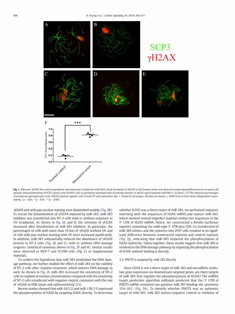
Fig. 1. Aberrant γH2AX foci exist in pachytene spermatocytes of patients with NOA. Focal formation of γH2AX in the human testes was detected using immunofluorescence on germ cellspreads. Immunodetection of SCP3 (green) and γH2AX (red) in pachytene spermatocytes of normal controls (A and B) and of patients withMA (C, D and E). (F) The statistical percentagesof pachytene spermatocytes with γH2AX-positive signals (red) in both XY and autosomes. Bar= 50 μm for all images. All data are means± SEMs from at least three independent exper-iments. *p b 0.05, **p b 0.01, ***p b 0.001.
906 H. Huang et al. / Cellular Signalling 26 (2014) 903–911
γH2AXandwith pan-nuclear stainingwere diminished notably (Fig. 2B).To rescue the diminishment of γH2AX induced by miR-383, miR-383inhibitor was transfected into NT-2 cells with or without exposure toUV irradiation. As shown in Fig. 2C and D, the intensity of γH2AXincreased after introduction of miR-383 inhibitor. In particular, thepercentages of cells with more than 10 foci of γH2AX without UV andof cells with pan-nuclear staining with UV were increased significantly.In addition, miR-383 substantially reduced the abundance of γH2AXprotein in NT-2 cells (Fig. 2E and G) with or without DNA damagereagents (statistical summary shown in Fig. 2F and H). Similar resultswere observed in MCF-7 and H1299 cells (Fig. S1 in Supplementalmaterial).
To confirm the hypothesis that miR-383 modulated the DNA dam-age pathway, we further studied the effect of miR-383 on the viabilityof NT-2 cells after cisplatin treatment, which induces DNA damage aswell. As shown in Fig. 2I, miR-383 increased the sensitivity of NT-2cells to cisplatin at various concentrations comparedwith the sensitivityof NT-2 cells transfected with negative control, consistent with the roleof γH2AX in DSB repair and radiosensitivity [21].
Recent studies showed thatmiR-24 [12] andmiR-138 [11] repressedthe phosphorylation of H2AX by targeting H2AX directly. To determine
whether H2AX was a direct target of miR-383, we performed sequencematching with the sequences of H2AX mRNA and mature miR-383,which showed several imperfect matches within two sequences in the5′ UTR of H2AX mRNA. Hence, we constructed a Renilla luciferasereporter containing the wild-type 5′ UTR plus CDS. Co-transfection ofmiR-383 mimics and the reporters into 293T cells resulted in no signif-icant difference between constructed reporter and control reporter(Fig. 2J), indicating that miR-383 impaired the phosphorylation ofH2AX indirectly. Taken together, these results suggest that miR-383 isinvolved in theDNAdamage pathwayby impairing thephosphorylationof H2AX without binding it directly.
3.3. PNUTS is targeted by miR-383 directly
Since H2AX is not a direct target of miR-383 and microRNAs modu-late gene expression via downstream targeted genes, are there targetsof miR-383 that regulate the phosphorylation of H2AX? The miRNAtarget prediction algorithm miRanda predicted that the 3′ UTR ofPNUTS mRNA contained one putative miR-383 binding site (positions154–161) (Fig. 3A). To identify whether PNUTS was an authentictarget of miR-383, miR-383 mimics/negative control or inhibitor of
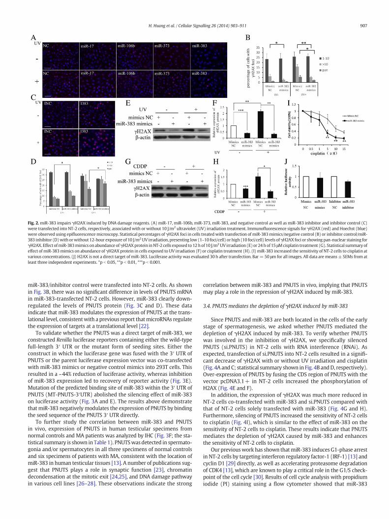
Fig. 2.miR-383 impairs γH2AX induced by DNA damage reagents. (A) miR-17, miR-106b, miR-373, miR-383, and negative control as well as miR-383 inhibitor and inhibitor control (C)were transfected into NT-2 cells, respectively, associatedwith or without 10 J/m2 ultraviolet (UV) irradiation treatment. Immunofluorescence signals for γH2AX (red) and Hoechst (blue)were observed using epifluorescencemicroscopy. Statistical percentages of γH2AX foci in cells treatedwith transfection of miR-383mimics/negative control (B) or inhibitor control/miR-383 inhibitor (D)with orwithout 12-hour exposure of 10 J/m2UV irradiation, presenting low (1–10 foci/cell) or high (10 foci/cell) levels ofγH2AX foci or showing pan-nuclear staining forγH2AX. Effect ofmiR-383mimics onabundance ofγH2AXprotein inNT-2 cells exposed to 12 hof 10 J/m2UV irradiation (E) or 24 h of 15 μMcisplatin treatment (G). Statistical summary ofeffect of miR-383mimics on abundance of γH2AX protein in cells exposed to UV irradiation (F) or cisplatin treatment (H). (I) miR-383 increased the sensitivity of NT-2 cells to cisplatin atvarious concentrations. (J) H2AX is not a direct target of miR-383. Luciferase activity was evaluated 30 h after transfection. Bar= 50 μm for all images. All data are means± SEMs from atleast three independent experiments. *p b 0.05, **p b 0.01, ***p b 0.001.
907H. Huang et al. / Cellular Signalling 26 (2014) 903–911
miR-383/inhibitor control were transfected into NT-2 cells. As shownin Fig. 3B, there was no significant difference in levels of PNUTS mRNAin miR-383-transfected NT-2 cells. However, miR-383 clearly down-regulated the levels of PNUTS protein (Fig. 3C and D). These dataindicate that miR-383 modulates the expression of PNUTS at the trans-lational level, consistentwith a previous report thatmicroRNAs regulatethe expression of targets at a translational level [22].
To validate whether the PNUTS was a direct target of miR-383, weconstructed Renilla luciferase reporters containing either the wild-typefull-length 3′ UTR or the mutant form of seeding sites. Either theconstruct in which the luciferase gene was fused with the 3′ UTR ofPNUTS or the parent luciferase expression vector was co-transfectedwith miR-383 mimics or negative control mimics into 293T cells. Thisresulted in a ~44% reduction of luciferase activity, whereas inhibitionof miR-383 expression led to recovery of reporter activity (Fig. 3E).Mutation of the predicted binding site of miR-383 within the 3′ UTR ofPNUTS (MT-PNUTS-3′UTR) abolished the silencing effect of miR-383on luciferase activity (Fig. 3A and E). The results above demonstratethat miR-383 negatively modulates the expression of PNUTS by bindingthe seed sequence of the PNUTS 3′ UTR directly.
To further study the correlation between miR-383 and PNUTSin vivo, expression of PNUTS in human testicular specimens fromnormal controls and MA patients was analyzed by IHC (Fig. 3F; the sta-tistical summary is shown in Table 1). PNUTSwas detected in spermato-gonia and/or spermatocytes in all three specimens of normal controlsand six specimens of patients with MA, consistent with the location ofmiR-383 in human testicular tissues [13]. A number of publications sug-gest that PNUTS plays a role in synaptic function [23], chromatindecondensation at the mitotic exit [24,25], and DNA damage pathwayin various cell lines [26–28]. These observations indicate the strong
correlation between miR-383 and PNUTS in vivo, implying that PNUTSmay play a role in the repression of γH2AX induced by miR-383.
3.4. PNUTS mediates the depletion of γH2AX induced by miR-383
Since PNUTS and miR-383 are both located in the cells of the earlystage of spermatogenesis, we asked whether PNUTS mediated thedepletion of γH2AX induced by miR-383. To verify whether PNUTSwas involved in the inhibition of γH2AX, we specifically silencedPNUTS (si.PNUTS) in NT-2 cells with RNA interference (RNAi). Asexpected, transfection of si.PNUTS into NT-2 cells resulted in a signifi-cant decrease of γH2AX with or without UV irradiation and cisplatin(Fig. 4A and C; statistical summary shown in Fig. 4B andD, respectively).Over-expression of PNUTS by fusing the CDS region of PNUTS with thevector pcDNA3.1+ in NT-2 cells increased the phosphorylation ofH2AX (Fig. 4E and F).
In addition, the expression of γH2AX was much more reduced inNT-2 cells co-transfected with miR-383 and si.PNUTS compared withthat of NT-2 cells solely transfected with miR-383 (Fig. 4G and H).Furthermore, silencing of PNUTS increased the sensitivity of NT-2 cellsto cisplatin (Fig. 4I), which is similar to the effect of miR-383 on thesensitivity of NT-2 cells to cisplatin. These results indicate that PNUTSmediates the depletion of γH2AX caused by miR-383 and enhancesthe sensitivity of NT-2 cells to cisplatin.
Our previouswork has shown thatmiR-383 induces G1-phase arrestin NT-2 cells by targeting interferon regulatory factor-1 (IRF-1) [13] andcyclin D1 [29] directly, as well as accelerating proteasome degradationof CDK4 [13], which are known to play a critical role in the G1/S check-point of the cell cycle [30]. Results of cell cycle analysis with propidiumiodide (PI) staining using a flow cytometer showed that miR-383

Fig. 3.PNUTS is targeted bymiR-383 directly. (A) Putative binding sites for humanmiR-383were predicted in the 3′UTR of PNUTSmRNA. Themutated bases of predictedmiR-383 bindingsites are underlined, namelyMT-PNUTS 3′UTR. (B) PNUTSmRNA expressionwas evaluated by real-time polymerase chain reaction (Q-RT). Total RNAwas extracted 48 h after transfectionusing a standard Trizol protocol. (C) Abundance of PNUTS proteinwas evaluated byWestern blotting; statistical summary is shown in (D). NT-2 cellswere harvested 48h after transfectionwith RIPA lysis buffer. (E)miR-383 targeted the3′UTRof PNUTS. Luciferase reporters containingeithermiR-383putativebinding sites fromwild-type PNUTS 3′UTR (WTPNUTS-3′UTR) ormutated PNUTS 3′UTR (MT PNUTS-3′UTR) were co-transfected with the indicated microRNA mimics into 293T cells. Luciferase activity was measured 30 h after transfection. (F) Immu-nohistochemistry analysis for PNUTS protein in human testicular tissues from normal controls and patients withmaturation arrest. SPG, spermatogonia; PS, primary spermatocyte; Bar=50 μm for all images. All data are means ± SEMs from at least three independent experiments. *p b 0.05, **p b 0.01, ***p b 0.001.
908 H. Huang et al. / Cellular Signalling 26 (2014) 903–911
induced G1 cell cycle arrest with or without exposure to UV irradiation(Fig. 5A), as consistent with our previous study. We further confirmedthat the G1 arrest caused by silencing of CDK4 in NT-2 cells led to asignificant decrease in the phosphorylation of H2AX by the Westernblotting method (Fig. 5B). In addition, phosphorylation of H2AX wasnotably impaired by silencing CDK4, together with silencing of cyclinD1 under UV irradiation treatment (Fig. 5C). In order to demonstratewhether PNUTS was involved in the cell cycle arrest induced bymiR-383, we transfected si.PNUTS into NT-2 cells. There was no obviousdifference in cell cycle between cells transfected with si.PNUTS andsiRNA negative control (Fig. 5D). Since phosphorylation of H2AXobserved throughout the S phase reflects the formation of DSB due tothe collision of replication forks with primary DNA lesions induced byUV irradiation [31], miR-383 may have a role in surveillance of DNAdamage generated in the early stage of mitosis. Taken together,miR-383 inhibits the phosphorylation of H2AX via two independent
Table 1The immunohistochemical expression pattern of PNUTS was summarized in Table 1. “+” mea
Gene Cell type Testicular tissue (ages)
N1 (33) N2 (31) N3 (37) A1
PNUTS Spermatogonia + + + +Spermatocyte + + + +
pathways, by targeting PNUTS directly as well as inducing cell cyclearrest.
4. Discussion
During the last decades, there are several studies [5–7,32–36] focus-ing on DNA damage in spermatozoa that have concentrated on theside-effects on the off-springs from in vitro fertilization (IVF) andintra-cytoplasmic sperm injection (ICSI) [37], namely assisted repro-ductive technology (ART). However, the origin of the DNA damageand the mechanism underlying DNA damage during the early stage ofspermatogenesis remain unclear. In this study, we observed that aber-rant increases in DNA damage signal indicated by the DNA damagemarker γH2AXwere found in pachytene spermatocytes in the testiculartissues of human patients with MA. This indicates that aberrant DNAdamage in the early stage of spermatogenesis is associated with MA.
ns mostly certain types of cells were detected, “−”means no positive signal of PNUTS.
(37) A2 (27) A3 (24) A4 (30) A5 (21) A6 (27)
− − + + ++ + + + +
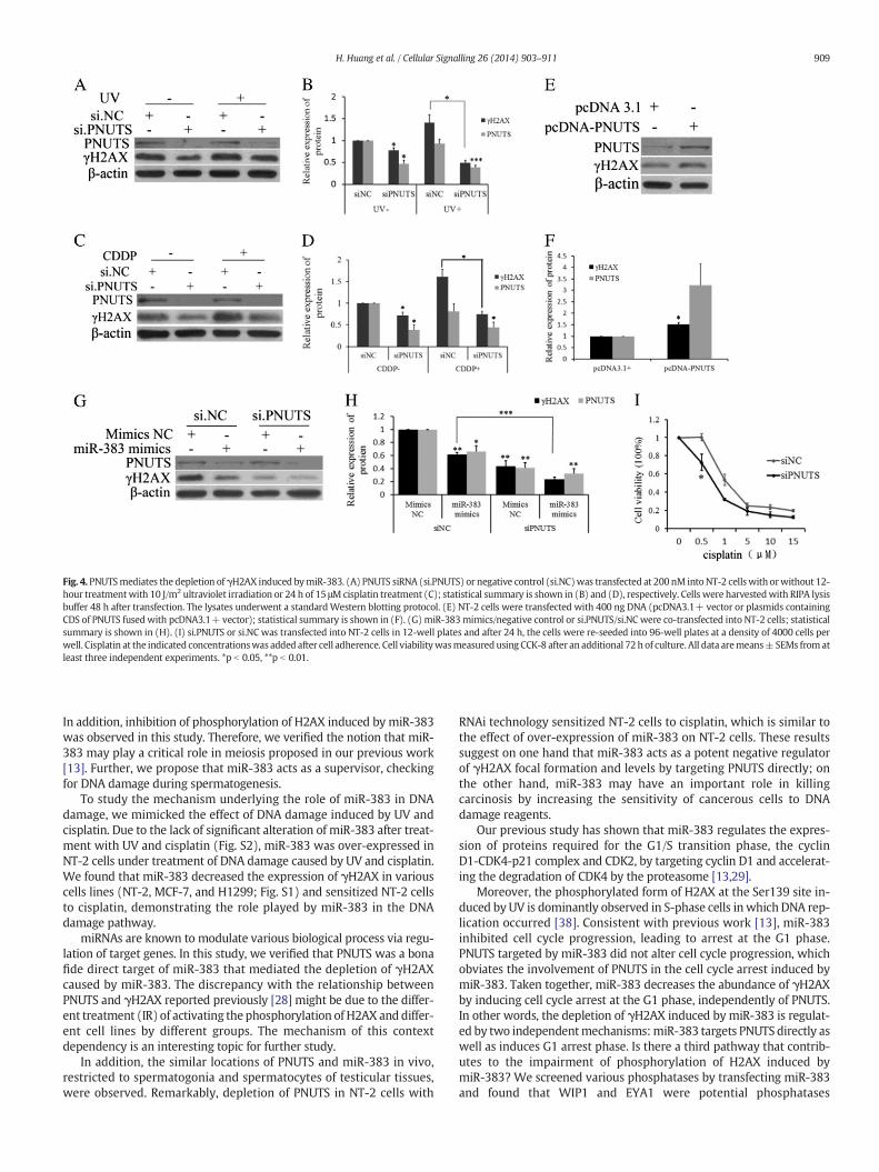
Fig. 4. PNUTSmediates the depletion of γH2AX induced bymiR-383. (A) PNUTS siRNA (si.PNUTS) or negative control (si.NC)was transfected at 200 nM intoNT-2 cellswith orwithout 12-hour treatmentwith 10 J/m2 ultraviolet irradiation or 24 h of 15 μMcisplatin treatment (C); statistical summary is shown in (B) and (D), respectively. Cells were harvestedwith RIPA lysisbuffer 48 h after transfection. The lysates underwent a standardWestern blotting protocol. (E) NT-2 cells were transfected with 400 ng DNA (pcDNA3.1+ vector or plasmids containingCDS of PNUTS fusedwith pcDNA3.1+ vector); statistical summary is shown in (F). (G) miR-383mimics/negative control or si.PNUTS/si.NC were co-transfected into NT-2 cells; statisticalsummary is shown in (H). (I) si.PNUTS or si.NC was transfected into NT-2 cells in 12-well plates and after 24 h, the cells were re-seeded into 96-well plates at a density of 4000 cells perwell. Cisplatin at the indicated concentrationswas added after cell adherence. Cell viabilitywasmeasuredusing CCK-8 after an additional 72h of culture. All data aremeans±SEMs fromatleast three independent experiments. *p b 0.05, **p b 0.01.
909H. Huang et al. / Cellular Signalling 26 (2014) 903–911
In addition, inhibition of phosphorylation of H2AX induced by miR-383was observed in this study. Therefore, we verified the notion that miR-383 may play a critical role in meiosis proposed in our previous work[13]. Further, we propose that miR-383 acts as a supervisor, checkingfor DNA damage during spermatogenesis.
To study the mechanism underlying the role of miR-383 in DNAdamage, we mimicked the effect of DNA damage induced by UV andcisplatin. Due to the lack of significant alteration of miR-383 after treat-ment with UV and cisplatin (Fig. S2), miR-383 was over-expressed inNT-2 cells under treatment of DNA damage caused by UV and cisplatin.We found that miR-383 decreased the expression of γH2AX in variouscells lines (NT-2, MCF-7, and H1299; Fig. S1) and sensitized NT-2 cellsto cisplatin, demonstrating the role played by miR-383 in the DNAdamage pathway.
miRNAs are known to modulate various biological process via regu-lation of target genes. In this study, we verified that PNUTS was a bonafide direct target of miR-383 that mediated the depletion of γH2AXcaused by miR-383. The discrepancy with the relationship betweenPNUTS and γH2AX reported previously [28] might be due to the differ-ent treatment (IR) of activating the phosphorylation of H2AX and differ-ent cell lines by different groups. The mechanism of this contextdependency is an interesting topic for further study.
In addition, the similar locations of PNUTS and miR-383 in vivo,restricted to spermatogonia and spermatocytes of testicular tissues,were observed. Remarkably, depletion of PNUTS in NT-2 cells with
RNAi technology sensitized NT-2 cells to cisplatin, which is similar tothe effect of over-expression of miR-383 on NT-2 cells. These resultssuggest on one hand that miR-383 acts as a potent negative regulatorof γH2AX focal formation and levels by targeting PNUTS directly; onthe other hand, miR-383 may have an important role in killingcarcinosis by increasing the sensitivity of cancerous cells to DNAdamage reagents.
Our previous study has shown that miR-383 regulates the expres-sion of proteins required for the G1/S transition phase, the cyclinD1-CDK4-p21 complex and CDK2, by targeting cyclin D1 and accelerat-ing the degradation of CDK4 by the proteasome [13,29].
Moreover, the phosphorylated form of H2AX at the Ser139 site in-duced by UV is dominantly observed in S-phase cells in which DNA rep-lication occurred [38]. Consistent with previous work [13], miR-383inhibited cell cycle progression, leading to arrest at the G1 phase.PNUTS targeted by miR-383 did not alter cell cycle progression, whichobviates the involvement of PNUTS in the cell cycle arrest induced bymiR-383. Taken together, miR-383 decreases the abundance of γH2AXby inducing cell cycle arrest at the G1 phase, independently of PNUTS.In other words, the depletion of γH2AX induced by miR-383 is regulat-ed by two independentmechanisms:miR-383 targets PNUTS directly aswell as induces G1 arrest phase. Is there a third pathway that contrib-utes to the impairment of phosphorylation of H2AX induced bymiR-383? We screened various phosphatases by transfecting miR-383and found that WIP1 and EYA1 were potential phosphatases

Fig. 5.miR-383 decreases γH2AX by inducing cell cycle arrest. (A) miR-383mimics or negative control was transfected into NT-2 cells at 150 nMwith or without 10 J/m2 ultraviolet (UV)irradiation for 12 h. Cells were harvested 48 h after transfection andwere subjected to cell cycle analysis by flow cytometry. (B)miR-383mimics or negative control were transfectedwithsi.CDK4 or si.NC into NT-2 cells. Cells were harvested with RIPA lysis buffer 48 h after transfection. The lysates underwent a standard Western blotting protocol. (C) Either si.CDK4 orsi.Cyclin D1 was transfected into NT-2 cells in association with 10 J/m2 UV irradiation treatment for 12 h. (D) si.NC or si.PNUTS was transfected into NT-2 cells at 200 nM. Cellswere harvested 48 h after transfection and were subjected to cell cycle analysis by flow cytometry. All data are means ± SEMs from at least three independent experiments. *p b 0.05,**p b 0.01, ***p b 0.001.
910 H. Huang et al. / Cellular Signalling 26 (2014) 903–911
responsible for regulating the phosphorylation of H2AX by miR-383(Figs. S3A and B). The specific mechanisms throughwhichmiR-383 de-creases γH2AX phosphorylation viaWIP1 and EYA1 need further study.At least we have shown that miR-383 plays a role in the DNA damagepathway via various pathways.
We found a novel function of miR-383 in the DNA damage pathwayin this study. In summary, we have demonstrated that miR-383decreases the levels and focal formation of γH2AX under treatmentwith UV and cisplatin by targeting PNUTS directly and inducing cellcycle arrest at the G1 phase. Mechanistic studies pertaining to the roleof miR383 in spermatogenesis will provide novel insights into treat-ment of male infertility. In addition, over-expression of miR-383 anddepletion of PNUTS sensitize NT-2 cells to a DNA damage reagent, indi-cating that miR-383 and PNUTS are also potential therapeutic agents forcancer treatment.
5. Conclusions
We found a novel function of miR-383 in the DNA damage pathway.miR-383 impairs the phosphorylation of H2AX by targeting PNUTS andinducing cell cycle arrest, and sensitizes NT-2 cells to cisplatin.
Conflict of interest statement
The authors declare no conflict of interest.
Acknowledgment
We are grateful to Dr. MianWu for providing the HEK293T cell line.
Appendix A. Supplementary data
Supplementary data to this article can be found online at http://dx.doi.org/10.1016/j.cellsig.2014.01.016.
References
[1] M.M. Matzuk, D.J. Lamb, Nat. Cell Biol. 4 Suppl. (2002) s41–s49.[2] R.I. McLachlan, D.M. de Kretser, Med. J. Aust. 174 (2001) 116–117.[3] A.J. Hung, P. King, P.N. Schlegel, J. Urol. 178 (2007) 608–612(discussion 612).[4] A. Jurisicova, Mol. Hum. Reprod. 5 (1999) 323–330.[5] A. Agarwal, T.M. Said, Hum. Reprod. Update 9 (2003) 331–345.[6] R.J. Aitken, G.N. De Iuliis, R.I. McLachlan, Int. J. Androl. 32 (2009) 46–56.[7] R.A. Saleh, A. Agarwal, E.A. Nada, M.H. El-Tonsy, R.K. Sharma, A. Meyer, D.R. Nelson,
A.J. Thomas, Fertil. Steril. 79 (Suppl. 3) (2003) 1597–1605.[8] F. Leduc, V. Maquennehan, G.B. Nkoma, G. Boissonneault, Biol. Reprod. 78 (2008)
324–332.[9] D.P. Bartel, Cell 136 (2009) 215–233.
[10] I.G. Cannell, Y.W. Kong, S.J. Johnston, M.L. Chen, H.M. Collins, H.C. Dobbyn, A. Elia,T.R. Kress, M. Dickens, M.J. Clemens, D.M. Heery, M. Gaestel, M. Eilers, A.E. Willis,M. Bushell, Proc. Natl. Acad. Sci. U. S. A. 107 (2010) 5375–5380.
[11] Y. Wang, J.W. Huang, M. Li, W.K. Cavenee, P.S. Mitchell, X. Zhou, M. Tewari, F.B.Furnari, T. Taniguchi, Mol. Cancer Res. 9 (2011) 1100–1111.
[12] A. Lal, Y. Pan, F. Navarro, D.M. Dykxhoorn, L. Moreau, E. Meire, Z. Bentwich, J.Lieberman, D. Chowdhury, Nat. Struct. Mol. Biol. 16 (2009) 492–498.
[13] J. Lian, H. Tian, L. Liu, X. Zhang, W. Li, Y. Deng, G. Yao, M. Yin, F. Sun, Cell Death Dis. 1(2010) e94.
[14] N. Liang, Y. Xu, Y. Yin, G. Yao, H. Tian, G.Wang, J. Lian, Y.Wang, F. Sun, Endocrinology152 (2011) 3213–3225.
[15] G. Yao,M. Yin, J. Lian, H. Tian, L. Liu, X. Li, F. Sun,Mol. Endocrinol. 24 (2010) 540–551.[16] M. Yin, M. Lü, G. Yao, H. Tian, J. Lian, L. Liu, M. Liang, Y.Wang, F. Sun, Mol. Endocrinol.
26 (2012) 1129–1143.[17] T.T. Paull, E.P. Rogakou, V. Yamazaki, C.U. Kirchgessner, M. Gellert, W.M. Bonner,
Curr. Biol. 10 (2000) 886–895.[18] G. Hamer, H.L. Roepers-Gajadien, A. van Duyn-Goedhart, I.S. Gademan, H.B. Kal, P.P.
van Buul, D.G. de Rooij, Biol. Reprod. 68 (2003) 628–634.[19] M Vries, L Ramos, P. Boer, Andrology 1 (2) (2013) 262–273.[20] J. Lian, X. Zhang, H. Tian, N. Liang, Y. Wang, C. Liang, X. Li, F. Sun, Reprod. Biol.
Endocrinol. 7 (2009) 13.[21] C.H. Bassing, K.F. Chua, J. Sekiguchi, H. Suh, S.R. Whitlow, J.C. Fleming, B.C.
Monroe, D.N. Ciccone, C. Yan, K. Vlasakova, Proc. Natl. Acad. Sci. 99 (2002)8173–8178.
[22] J. Gäken, A.M. Mohamedali, J. Jiang, F. Malik, D. Stangl, A.E. Smith, C. Chronis,A.G. Kulasekararaj, N.S.B. Thomas, F. Farzaneh, Nucleic Acids Res. 40 (2012)e75-e75.
[23] M. Rose, E. Dütting, N. Schröder, H. Sticht, J.H. Brandstätter, R. Enz, Mol. Cell.Neurosci. 37 (2008) 808–819.
[24] Landsverk Hxab, M. Kirkhus, M. Bollen, T. Kuntziger, P. Collas, Biochem. J. 390(2005) 709–717.

911H. Huang et al. / Cellular Signalling 26 (2014) 903–911
[25] J.-H. Lee, J. You, E. Dobrota, D.G. Skalnik, J. Biol. Chem. 285 (2010) 24466–24476.[26] R.A. Boon, K. Iekushi, S. Lechner, T. Seeger, A. Fischer, S. Heydt, D. Kaluza, K. Tréguer,
G. Carmona, A. Bonauer, Nature 495 (7439) (2013) 107–110.[27] H. Kim, O.-H. Lee, H. Xin, L.-Y. Chen, J. Qin, H.K. Chae, S.-Y. Lin, A. Safari, D. Liu, Z.
Songyang, Nat. Struct. Mol. Biol. 16 (2009) 372–379.[28] H.B. Landsverk, F. Mora-Bermúdez, O.J. Landsverk, G. Hasvold, S. Naderi, O. Bakke, J.
Ellenberg, P. Collas, R.G. Syljuåsen, T. Küntziger, EMBO Rep. 11 (2010) 868–875.[29] H. Tian, Y. Cao, X. Zhang, W. Liao, Y. Yi, J. Lian, L. Liu, H. Huang, W. Liu, M. Yin, Cell
Death Dis. 4 (2013) e617.[30] Y. Dong, L. Sui, K. Sugimoto, Y. Tai, M. Tokuda, Int. J. Cancer 95 (2001) 209–215.[31] A. Cerqueira, D. Santamaría, B. Martínez-Pastor, M. Cuadrado, O. Fernández-Capetillo,
M. Barbacid, J. Cell Biol. 187 (2009) 773–780.
[32] C.G. Fraga, P.A. Motchnik, M.K. Shigenaga, H.J. Helbock, R.A. Jacob, B.N. Ames, Proc.Natl. Acad. Sci. 88 (1991) 11003–11006.
[33] A. Agarwal, S.S. Allamaneni, Fertil. Steril. 84 (2005) 850–853.[34] A. Zini, J.M. Boman, E. Belzile, A. Ciampi, Hum. Reprod. 23 (2008)
2663–2668.[35] L. Simon, D.T. Carrell, Spermatogenesis, Springer, 2013. 137–146.[36] Y. Zhang, J. Trussell, K.R. Chohan, Seminars in reproductive medicine, 2013,
pp. 267–273.[37] V. Vloeberghs, G. Verheyen, H. Tournaye, Clinics (Sao Paulo) 68 (Suppl. 1) (2013)
151–156.[38] H.D. Halicka, X. Huang, F. Traganos, M.A. King, W. Dai, Z. Darzynkiewicz, Cell Cycle 4
(2005) 339–345.