Functional interaction of H2AX, NBS1, and p53 in ATM-dependent DNA damage responses and tumor...
10
MOLECULAR AND CELLULAR BIOLOGY, Jan. 2005, p. 661–670 Vol. 25, No. 2 0270-7306/05/$08.000 doi:10.1128/MCB.25.2.661–670.2005 Copyright © 2005, American Society for Microbiology. All Rights Reserved. Functional Interaction of H2AX, NBS1, and p53 in ATM-Dependent DNA Damage Responses and Tumor Suppression Jian Kang, 1 David Ferguson, 2 Hoseok Song, 1 Craig Bassing, 2 Mark Eckersdorff, 2 Frederick W. Alt, 2 and Yang Xu 1 * Section of Molecular Biology, Division of Biological Sciences, University of California, San Diego, La Jolla, California, 1 and The Center for Blood Research, The Children’s Hospital, Howard Hughes Medical Institute, Harvard Medical School, Boston, Massachusetts 2 Received 19 May 2004/Returned for modification 29 June 2004/Accepted 12 October 2004 Ataxia-telangiectasia (A-T) mutated (ATM) kinase signals all three cell cycle checkpoints after DNA double- stranded break (DSB) damage. H2AX, NBS1, and p53 are substrates of ATM kinase and are involved in ATM-dependent DNA damage responses. We show here that H2AX is dispensable for the activation of ATM and p53 responses after DNA DSB damage. Therefore, H2AX functions primarily as a downstream mediator of ATM functions in the parallel pathway of p53. NBS1 appears to function both as an activator of ATM and as an adapter to mediate ATM activities after DNA DSB damage. Phosphorylation of ATM and H2AX induced by DNA DSB damage is normal in NBS1 mutant/mutant (NBS1 m/m ) mice that express an N-terminally truncated NBS1 at lower levels. Therefore, the pleiotropic A-T-related systemic and cellular defects observed in NBS1 m/m mice are due to the disruption of the adapter function of NBS1 in mediating ATM activities. While H2AX is required for the irradiation-induced focus formation of NBS1, our findings indicate that NBS1 and H2AX have distinct roles in DNA damage responses. ATM-dependent phosphorylation of p53 and p53 re- sponses are largely normal in NBS1 m/m mice after DNA DSB damage, and p53 deficiency greatly facilitates tumorigenesis in NBS1 m/m mice. Therefore, NBS1, H2AX, and p53 play synergistic roles in ATM-dependent DNA damage responses and tumor suppression. Proper cellular responses to DNA double-strand breaks (DSBs) are critical for maintaining genetic stability and for tumor suppression (38). A gene consistently mutated in genetic instability syndrome ataxia-telangiectasia (A-T), called ATM (for ataxia-telangiectasia mutated), encodes a large protein kinase that is responsible for activating cellular responses to DNA damage (44). In this context, ATM is required for ho- mologous recombination and all three cell cycle checkpoints after DNA DSB damage. In addition, ATM deficiency leads to hypersensitivity to -irradiation and multisystemic defects, in- cluding growth retardation, abolished germ cell development, immunodeficiency, and greatly increased cancer risk (43). As a protein kinase, ATM functions by phosphorylating and acti- vating a number of DNA repair and checkpoint proteins, in- cluding p53, NBS1, H2AX, 53BP1, Brca1, Smc1, and Chk2 (43). Immediately after DNA DSB damage, histone H2AX is phosphorylated at the C-terminal Ser residues (Ser136 and Ser139) (40). This phosphorylation, called -H2AX, can be de- tected within minutes after the introduction of DSBs and is in- volved in the recruitment of other known components of DNA repair, including Brca1, NBS1/Mre11/Rad50, and 53BP1, to the sites of the DNA damage (11, 41, 48). -H2AX is mainly medi- ated by ATM after DNA DSB damage, suggesting that H2AX is a downstream mediator of ATM function (6, 20). H2AX is also required for the recruitment of 53BP1 to sites of DNA DSB damage, and 53BP1 appears to be an upstream activator of ATM (11, 39, 48). Therefore, it remains unclear whether H2AX plays any role in activating ATM after DNA DSB damage. NBS1, the gene product mutated in Nijmegen breakage syn- drome (NBS) patients, is the p95 component of the Mre11 complex that forms foci at the sites of DNA DSBs (8, 36, 47). The importance of NBS1 in DNA repair is indicated by the multisystemic defects observed in NBS patients, including growth retardation, immunodeficiency, and increased lym- phoid malignancies (46). In addition, NBS cells are hypersen- sitive to ionizing radiation and defective in intra-S and G 2 /M checkpoints. Targeted disruption of the N terminus of NBS1 in mice recapitulates most of the systemic and cellular defects observed in NBS patients (25, 50). The functional link between ATM and NBS1 is suggested by the large panel of defects shared by NBS1 and ATM mutations and is further demon- strated by the findings that ATM can phosphorylate and acti- vate NBS1 at a consensus site, Ser343 (21, 30, 51, 55). There- fore, NBS1 appears to function as a downstream mediator of ATM function. Recent studies show that mutation of the Mre11 complex or NBS1 leads to impaired ATM activation in human cell lines, indicating a role for NBS1 in activating ATM after DNA DSB damage (9, 23, 39, 45). Since the disruption of this NBS1 function could account for the A-T-related defects observed in NBS patients, the contribution of the impaired adapter function of NBS1 in mediating ATM activities to the A-T-related defects in NBS1 mutant mice and human patients remains to be established. The functional interaction between NBS1 and H2AX is complex. H2AX is required for the irradiation-induced focus formation of NBS1 after DNA DSB damage (11). In addition, * Corresponding author. Mailing address: Division of Biological Sci- ences, University of California, San Diego, 9500 Gilman Dr., La Jolla, CA 92093-0322. Phone: (858) 822-1084. Fax: (858) 534-0053. E-mail: [email protected]. 661 on February 27, 2016 by guest http://mcb.asm.org/ Downloaded from
-
Upload
independent -
Category
Documents
-
view
5 -
download
0
Transcript of Functional interaction of H2AX, NBS1, and p53 in ATM-dependent DNA damage responses and tumor...
MOLECULAR AND CELLULAR BIOLOGY, Jan. 2005, p. 661–670 Vol. 25, No. 20270-7306/05/$08.00�0 doi:10.1128/MCB.25.2.661–670.2005Copyright © 2005, American Society for Microbiology. All Rights Reserved.
Functional Interaction of H2AX, NBS1, and p53 in ATM-DependentDNA Damage Responses and Tumor Suppression
Jian Kang,1 David Ferguson,2 Hoseok Song,1 Craig Bassing,2 Mark Eckersdorff,2Frederick W. Alt,2 and Yang Xu1*
Section of Molecular Biology, Division of Biological Sciences, University of California, San Diego, La Jolla, California,1
and The Center for Blood Research, The Children’s Hospital, Howard Hughes Medical Institute,Harvard Medical School, Boston, Massachusetts2
Received 19 May 2004/Returned for modification 29 June 2004/Accepted 12 October 2004
Ataxia-telangiectasia (A-T) mutated (ATM) kinase signals all three cell cycle checkpoints after DNA double-stranded break (DSB) damage. H2AX, NBS1, and p53 are substrates of ATM kinase and are involved inATM-dependent DNA damage responses. We show here that H2AX is dispensable for the activation of ATMand p53 responses after DNA DSB damage. Therefore, H2AX functions primarily as a downstream mediatorof ATM functions in the parallel pathway of p53. NBS1 appears to function both as an activator of ATM andas an adapter to mediate ATM activities after DNA DSB damage. Phosphorylation of ATM and H2AX inducedby DNA DSB damage is normal in NBS1 mutant/mutant (NBS1m/m) mice that express an N-terminallytruncated NBS1 at lower levels. Therefore, the pleiotropic A-T-related systemic and cellular defects observedin NBS1m/m mice are due to the disruption of the adapter function of NBS1 in mediating ATM activities. WhileH2AX is required for the irradiation-induced focus formation of NBS1, our findings indicate that NBS1 andH2AX have distinct roles in DNA damage responses. ATM-dependent phosphorylation of p53 and p53 re-sponses are largely normal in NBS1m/m mice after DNA DSB damage, and p53 deficiency greatly facilitatestumorigenesis in NBS1m/m mice. Therefore, NBS1, H2AX, and p53 play synergistic roles in ATM-dependentDNA damage responses and tumor suppression.
Proper cellular responses to DNA double-strand breaks(DSBs) are critical for maintaining genetic stability and fortumor suppression (38). A gene consistently mutated in geneticinstability syndrome ataxia-telangiectasia (A-T), called ATM(for ataxia-telangiectasia mutated), encodes a large proteinkinase that is responsible for activating cellular responses toDNA damage (44). In this context, ATM is required for ho-mologous recombination and all three cell cycle checkpointsafter DNA DSB damage. In addition, ATM deficiency leads tohypersensitivity to �-irradiation and multisystemic defects, in-cluding growth retardation, abolished germ cell development,immunodeficiency, and greatly increased cancer risk (43). As aprotein kinase, ATM functions by phosphorylating and acti-vating a number of DNA repair and checkpoint proteins, in-cluding p53, NBS1, H2AX, 53BP1, Brca1, Smc1, and Chk2(43).
Immediately after DNA DSB damage, histone H2AX isphosphorylated at the C-terminal Ser residues (Ser136 andSer139) (40). This phosphorylation, called �-H2AX, can be de-tected within minutes after the introduction of DSBs and is in-volved in the recruitment of other known components of DNArepair, including Brca1, NBS1/Mre11/Rad50, and 53BP1, to thesites of the DNA damage (11, 41, 48). �-H2AX is mainly medi-ated by ATM after DNA DSB damage, suggesting that H2AX isa downstream mediator of ATM function (6, 20). H2AX is alsorequired for the recruitment of 53BP1 to sites of DNA DSB
damage, and 53BP1 appears to be an upstream activator of ATM(11, 39, 48). Therefore, it remains unclear whether H2AX playsany role in activating ATM after DNA DSB damage.
NBS1, the gene product mutated in Nijmegen breakage syn-drome (NBS) patients, is the p95 component of the Mre11complex that forms foci at the sites of DNA DSBs (8, 36, 47).The importance of NBS1 in DNA repair is indicated by themultisystemic defects observed in NBS patients, includinggrowth retardation, immunodeficiency, and increased lym-phoid malignancies (46). In addition, NBS cells are hypersen-sitive to ionizing radiation and defective in intra-S and G2/Mcheckpoints. Targeted disruption of the N terminus of NBS1 inmice recapitulates most of the systemic and cellular defectsobserved in NBS patients (25, 50). The functional link betweenATM and NBS1 is suggested by the large panel of defectsshared by NBS1 and ATM mutations and is further demon-strated by the findings that ATM can phosphorylate and acti-vate NBS1 at a consensus site, Ser343 (21, 30, 51, 55). There-fore, NBS1 appears to function as a downstream mediator ofATM function. Recent studies show that mutation of theMre11 complex or NBS1 leads to impaired ATM activation inhuman cell lines, indicating a role for NBS1 in activating ATMafter DNA DSB damage (9, 23, 39, 45). Since the disruption ofthis NBS1 function could account for the A-T-related defectsobserved in NBS patients, the contribution of the impairedadapter function of NBS1 in mediating ATM activities to theA-T-related defects in NBS1 mutant mice and human patientsremains to be established.
The functional interaction between NBS1 and H2AX iscomplex. H2AX is required for the irradiation-induced focusformation of NBS1 after DNA DSB damage (11). In addition,
* Corresponding author. Mailing address: Division of Biological Sci-ences, University of California, San Diego, 9500 Gilman Dr., La Jolla,CA 92093-0322. Phone: (858) 822-1084. Fax: (858) 534-0053. E-mail:[email protected].
661
on February 27, 2016 by guest
http://mcb.asm
.org/D
ownloaded from
direct interaction between �-H2AX and NBS1 suggests amechanism to recruit NBS1 to the sites of DNA DSB damage(27). These findings suggest that H2AX functions upstream ofNBS1 in DNA damage response. However, while the defectsobserved in either H2AX or NBS1 mutant mice are shared byATM mutant mice, H2AX and NBS1 mutant mice exhibitcertain distinct phenotypes (4, 12, 25, 50, 52). For example,while spermatogenesis is abolished in H2AX-deficient mice,oogenesis failure is only observed in NBS1 mutant mice.Therefore, NBS1 and H2AX might have distinct roles in DNAdamage responses.
It has been well established that ATM is required for theactivation of p53 responses to DNA DSB damage in humanand mouse cells (26, 49, 53, 54). ATM can phosphorylate p53at Ser15 (corresponding to Ser18 of mouse p53), and thisphosphorylation activates p53 responses to DNA DSB damage
(13, 14, 17, 28). While it has been well established that ATM isrequired to activate p53-dependent cell cycle G1 arrest uponDNA DSB damage, it remains unclear whether H2AX is in-volved in mediating this ATM function after DNA DSB dam-age. We employed NBS1, H2AX, and p53 mutant mice toinvestigate the functional interactions among ATM, NBS1,H2AX, and p53 in DNA damage responses, maintenance ofgenetic stability, and tumor suppression.
MATERIALS AND METHODS
Generation and culture of MEFs. Murine embryonic fibroblasts (MEFs) werederived from day 14 embryos as described previously (53). The MEFs werecultured in Dulbecco’s modified Eagle’s medium supplemented with 10% fetalbovine serum, 5 mM glutamine, 50 �M �-mercaptoethanol, and 50 U (each) ofpenicillin and streptomycin/ml at 37°C with 5% CO2. Cellular proliferationassays were performed as previously described (53). Briefly, 105 MEFs were
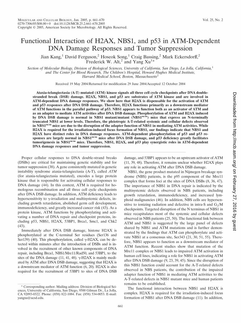
FIG. 1. Phosphorylation of ATM at Ser1981 and p53 at Ser18 after DNA DSB damage in H2AX�/� cells. (A) Phosphorylation of ATM inATM�/� and ATM�/� MEFs before (�) and 1 h after (�) IR (1 Gy). Phosphorylation of ATM in H2AX�/� and H2AX�/� thymocytes (B) andMEFs (C) 1 h after 1 Gy of IR or in H2AX�/� and H2AX�/� thymocytes 10 min after 1 Gy of IR (D). The genotypes and the treatment areindicated at the top. Phosphorylated ATM and total ATM are indicated on the right. p53 stabilization and phosphorylation at Ser18 in H2AX�/�
and H2AX�/� thymocytes after IR (E) and in MEFs after IR (F) or doxorubicin (G). The genotypes and time points after treatments are indicatedat the top. Phosphorylated p53, total p53, and actin are indicated on the right.
662 KANG ET AL. MOL. CELL. BIOL.
on February 27, 2016 by guest
http://mcb.asm
.org/D
ownloaded from
seeded onto each 35-mm-diameter plate, and each day after being plated, MEFsfrom three plates of each genotype were trypsinized and counted.
Ionizing radiation (IR) treatment of MEFs and cell cycle analysis. MEFs weresynchronized at the cell cycle G0 phase by serum starvation in medium containing0.1% fetal bovine serum for 96 h as described previously (54). The G0-synchro-nized MEFs were trypsinized and irradiated in suspension with a 137Cs �-raysource. The irradiated and mock-treated MEFs were plated in 10-cm-diameterplates at a density of 1 million cells/plate in normal MEF growth mediumsupplemented with 10 �M bromodeoxyuridine. After 24 h of bromodeoxyuridinelabeling, the cells were harvested and fixed in 70% ethanol, and the cell cycleprofile was analyzed as described previously (53).
Analysis of p53-dependent apoptosis in mouse thymocytes. Thymuses wererecovered from 1- to 2-month-old mice of various genotypes, and single-cellthymocyte suspensions were treated with increasing doses of IR as describedpreviously (15). The percentage of apoptotic cells 10 h after IR was analyzed bystaining with annexin V as described previously (15).
Western blot analysis of levels of proteins and protein phosphorylation. Pro-tein extracts from 4 � 105 embryonic stem cells or MEFs or 5 � 106 thymocyteswere separated by sodium dodecyl sulfate-polyacrylamide gel electrophoresis on6 to 15% polyacrylamide gels and transferred to nitrocellulose membranes. Themembranes were probed with various primary antibodies and subsequently in-cubated with horseradish peroxidase-conjugated secondary antibody and devel-oped with Enhanced Chemiluminescence PLUS (Amersham Pharmacia Bio-tech) or a Pierce SuperSignal kit. To determine the amount of protein in eachlane, the filter was stripped and probed with a rabbit polyclonal antibody againstactin (Santa Cruz Biotechnology, Inc.). Phosphospecific antibody against ATMwas purchased from Rockland. Phosphospecific antibody against p53 was pur-chased from Cell Signaling Technology.
Real-time PCR analysis. Total RNA was isolated from MEFs or thymocyteswith the combination of Trizol (Invitrogen) and RNAeasy RNA Cleanup (QIA-GEN). RNA was treated with RNase-free DNase (Roche Diagnostics) for 20min at room temperature before being reverse transcribed using Superscript IIreverse transcriptase (Invitrogen). Real-time PCR was performed on an ABI7000 machine with SYBR Green PCR Master Mix (Applied Biotechnology).PCR conditions consisted of a 10-min hot start at 95°C, followed by 40 cycles of30 s at 95°C and 1 min at 61°C. The average threshold cycle for each gene wasdetermined from triplicate reactions, and the levels of gene expression relative to
GAPDH (glyceraldehyde-3-phosphate dehydrogenase) were determined as pre-viously described (5). The primers used were described previously (13).
Generation and analysis of NBS1m/m) H2AX�/� and NBS1m/m p53�/� mice.The ATM�/�, p53�/�, NBS1 mutant/mutant (NBS1m/m), and H2AX�/� micewere described previously (4, 24, 25, 52). Because NBS1m/m females are sterile,p53�/� mice were bred with NBS1m/m males to generate p53�/� NBS1�/m mice,which were intercrossed to generate p53�/� NBS1m/m mice. The health status ofvarious mutant mice was monitored once every 2 days. The dead or dying micewere dissected and examined for tumors. The H2AX�/� males were sterile (4),and H2AX�/� NBS1�/m female and H2AX�/� NBS1�/� males were inter-crossed to screen for homozygous mutant embryos.
Analysis of chromosomes. Pretumor thymocytes were cultured in RPM-1medium supplemented with 12 ng of phorbol myristate acetate/ml and 0.25 �g ofionomycin/ml, and metaphases were prepared after 2 to 3 days of culture whenthe thymocytes reached the log phase of proliferation. Tumor cells were pre-pared for spectral karyotyping (SKY) analysis by culturing them in RPM-1medium supplemented with 10% fetal calf serum, antibiotics, 100 �M �-mer-captoethanol, and 100 U of interleukin-2/ml for 2 to 3 days until they reachedlog-phase growth. Thymocytes and tumor cells were treating with 0.05 �g ofColcemid/ml for 2.5 h prior to standard metaphase dropping. Metaphases frompretumor thymocytes were stained with DAPI (4�,6�-diamidino-2-phenylindole)and examined. SKY analysis of chromosomes was performed as described pre-viously (19). Chromosomes were analyzed using a Nikon Eclipse microscopeequipped with an Applied Spectral Imaging charge-coupled device camera and40 and 63� objectives.
RESULTS
H2AX is not required for ATM activation after DNA DSBdamage. Phosphorylation of ATM at Ser1981 is required toactivate ATM after DNA DSB damage (2). To control for thespecificity of the commercial monoclonal antibody that recognizesthe mouse ATM phosphorylated at Ser1981, we used the anti-body to probe the blot of the protein extracts from ATM�/� andATM�/� MEFs before and 1 h after 1 Gy of IR. While similar
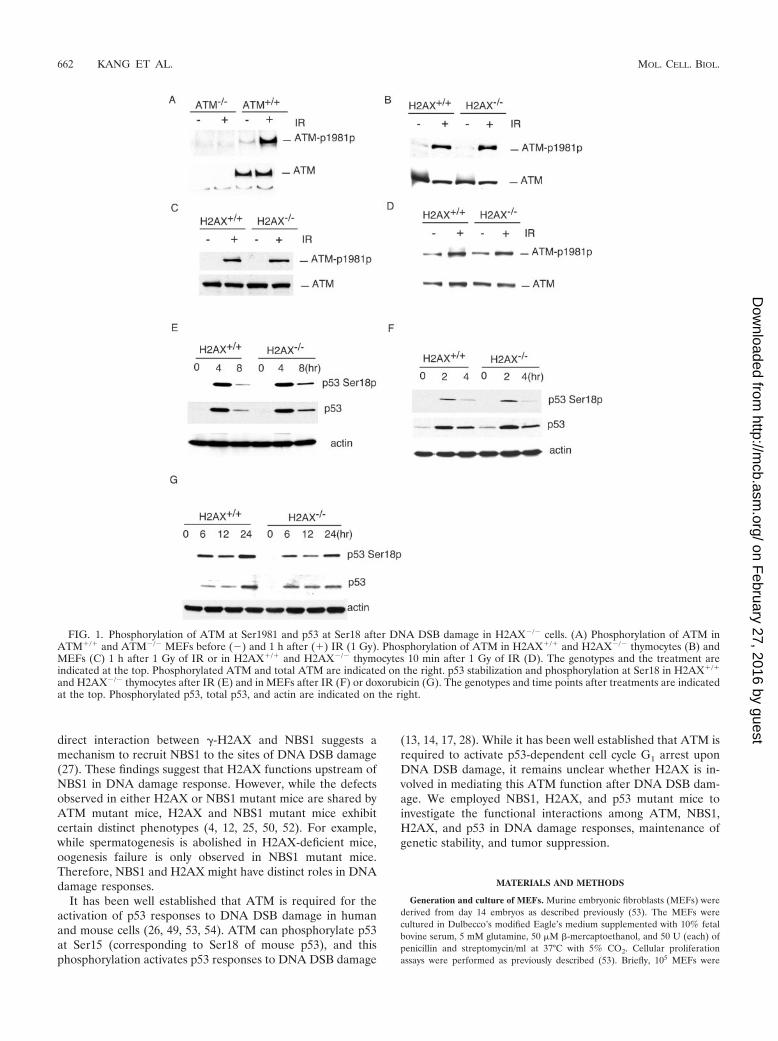
FIG. 2. p53-dependent transcription in H2AX�/� and H2AX�/� MEFs (A) and thymocytes (B) after DNA DSB damage. The mRNA levelsof each gene were determined by quantitative real-time PCR and standardized by the mRNA levels of GAPDH as described previously (13). ThemRNA levels in the untreated H2AX�/� cells are arbitrarily set to 1. The MEFs were harvested 12 h after doxorubicin (Dox) treatment (0.5 �M),and the thymocytes were harvested 4 h after treatment with 5 Gy of IR. Mean values from three independent experiments are presented, with errorbars indicating standard deviations.
VOL. 25, 2005 FUNCTIONAL INTERACTION OF H2AX, NBS1, AND p53 663
on February 27, 2016 by guest
http://mcb.asm
.org/D
ownloaded from
levels of ATM protein were detected in ATM�/� MEFs beforeand after IR, no ATM protein could be detected in ATM�/�
samples (Fig. 1A). In addition, while no phosphorylated ATMwas detected in ATM�/� MEFs before or after IR, phosphory-lation of ATM was significantly increased in ATM�/� MEFs 1 hafter 1 Gy of IR (Fig. 1A). Therefore, we concluded that themonoclonal antibody specifically recognizes mouse ATM phos-phorylated at Ser1981. To determine whether H2AX is requiredfor ATM activation after DNA damage, we analyzed the Ser1981phosphorylation of ATM in H2AX�/� and H2AX�/� thymocytesand MEFs after IR. Normal phosphorylation of ATM at Ser1981was observed in H2AX�/� thymocytes and MEFs 1 h after 1Gy ofIR, indicating that H2AX is not required for ATM activationafter DNA DSB damage (Fig. 1B and C). Consistent with thisconclusion, ATM phosphorylation at Ser1981 is normal inH2AX�/� thymocytes 10 min after 1 Gy of IR (Fig. 1D). Phos-phorylation of p53 at Ser18 and p53 stabilization were both nor-
mal in H2AX�/� MEFs and thymocytes after DNA DSB damageinduced by IR or doxorubicin (Fig. 1E, F, and G).
p53-dependent responses to DNA DSB damage in H2AX�/�
cells. While H2AX is not required for p53 stabilization afterDNA DSB damage, we analyzed p53-dependent transcriptionafter DNA DSB damage to determine whether H2AX is re-quired for p53 activities. Analysis of p53-dependent gene ex-pression in H2AX�/� MEFs before and after doxorubicintreatment indicated that expression of a number of p53-depen-dent genes was normal or slightly increased in H2AX�/�
MEFs after DNA DSB damage (Fig. 2A). In addition, thebasal levels of some p53 target genes, such as p21, were in-creased in H2AX�/� MEFs without doxorubicin treatment(Fig. 2A). We also found normal p53-dependent apoptosis inH2AX�/� thymocytes after IR (data not shown). In addition,p53-dependent expression of apoptotic genes, including Noxa,Puma, and Killer 5 genes, was normal or higher in H2AX�/�
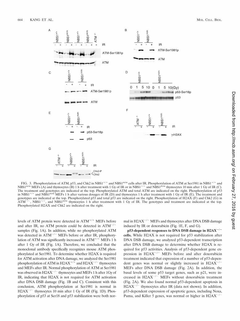
FIG. 3. Phosphorylation of ATM, p53, and Chk2 in NBS1�/� and NBS1m/m cells after IR. Phosphorylation of ATM at Ser1981 in NBS1�/� andNBS1m/m MEFs (A) and thymocytes (B) 1 h after treatment with 1 Gy of IR or in NBS1�/� and NBS1m/m thymocytes 10 min after 1 Gy of IR (C).The treatment and genotypes are indicated at the top. Phosphorylated ATM and total ATM are indicated on the right. Phosphorylation of p53in NBS1�/� and NBS1m/m MEFs 1 h after various dosages of IR (D) and thymocytes 1 h after treatment with 1 Gy of IR (E). The treatment andgenotypes are indicated at the top. Phosphorylated p53 and total p53 are indicated on the right. Phosphorylation of H2AX (F) and Chk2 (G) inATM�/�, NBS1�/�, and NBS1m/m thymocytes 1 h after treatment with 1 Gy of IR. The genotypes and treatment are indicated at the top.Phosphorylated H2AX and Chk2 are indicated on the right.
664 KANG ET AL. MOL. CELL. BIOL.
on February 27, 2016 by guest
http://mcb.asm
.org/D
ownloaded from
thymocytes after IR (Fig. 2B). Similar to what was observed inH2AX�/� MEFs, the basal levels of some p53-dependent apo-ptotic genes were increased in H2AX�/� thymocytes in theabsence of exogenous DNA damage.
Various ATM-dependent phosphorylation events in NBS1m/m
cells. Recent studies have suggested that the Mre11/Rad50/NBS1 complex functions upstream of ATM and is required forATM activation after DNA DSB damage (9, 23, 39, 45). Totest whether the large panel of A-T-related cellular and sys-temic defects observed in NBS1m/m mice are due to impairedATM activation, we analyzed the phosphorylation of ATM atSer1981 in NBS1m/m cells after IR. Normal ATM phosphory-lation was observed in NBS1m/m MEFs and thymocytes 1 hafter 1 Gy of IR (Fig. 3A and B). In addition, the phosphor-ylation of ATM at Ser1981 is also normal in NBS1m/m thymo-cytes 10 min after 1 Gy of IR (Fig. 3C). ATM-dependentphosphorylation of p53 at Ser18 and �-H2AX is normal inNBS1m/m cells after IR (Fig. 3D, E, and F). Since NBS1m/m
cells expressed an N-terminally truncated NBS1 protein at lowlevels (25), these findings indicated that the N-terminal domainof NBS1 is not required for the activation of ATM and ATM-dependent phosphorylation of p53 and H2AX after DNA DSBdamage.
ATM-dependent phosphorylation of Chk2 might activateChk2 activities after IR (1, 35, 37). To test whether Chk2 isphosphorylated in NBS1m/m thymocytes after IR, we analyzedthe phosphorylation status of Chk2 in NBS1�/� and NBS1m/m
thymocytes after IR. Phosphorylated Chk2 migrates moreslowly than unphosphorylated Chk2 in sodium dodecyl sulfate-polyacrylamide gel electrophoresis, and this allowed us to ac-curately determine the Chk2 phosphorylation status in thecells. While Chk2 was mostly phosphorylated in NBS1�/� thy-mocytes after IR, little phosphorylation of Chk2 could be de-tected in either ATM�/� or NBS1m/m thymocytes (Fig. 3G).Therefore, a complete NBS1 is required for ATM-dependentphosphorylation of Chk2 after DNA DSB damage.
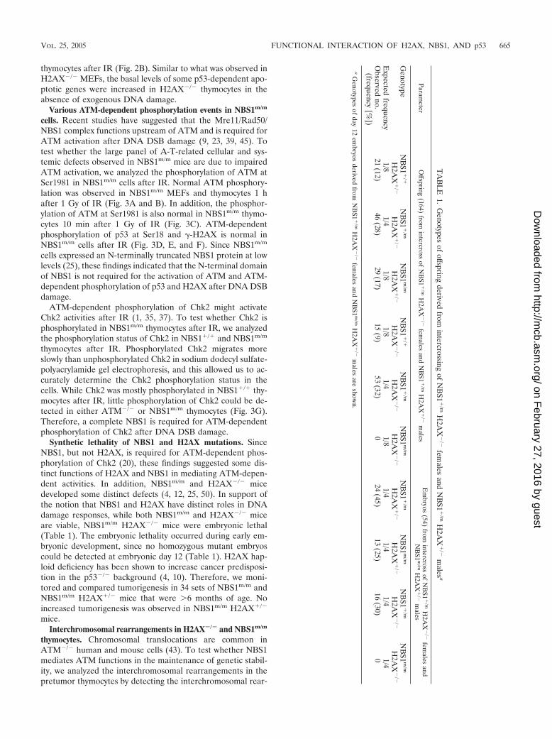
Synthetic lethality of NBS1 and H2AX mutations. SinceNBS1, but not H2AX, is required for ATM-dependent phos-phorylation of Chk2 (20), these findings suggested some dis-tinct functions of H2AX and NBS1 in mediating ATM-depen-dent activities. In addition, NBS1m/m and H2AX�/� micedeveloped some distinct defects (4, 12, 25, 50). In support ofthe notion that NBS1 and H2AX have distinct roles in DNAdamage responses, while both NBS1m/m and H2AX�/� miceare viable, NBS1m/m H2AX�/� mice were embryonic lethal(Table 1). The embryonic lethality occurred during early em-bryonic development, since no homozygous mutant embryoscould be detected at embryonic day 12 (Table 1). H2AX hap-loid deficiency has been shown to increase cancer predisposi-tion in the p53�/� background (4, 10). Therefore, we moni-tored and compared tumorigenesis in 34 sets of NBS1m/m andNBS1m/m H2AX�/� mice that were �6 months of age. Noincreased tumorigenesis was observed in NBS1m/m H2AX�/�
mice.Interchromosomal rearrangements in H2AX�/� and NBS1m/m
thymocytes. Chromosomal translocations are common inATM�/� human and mouse cells (43). To test whether NBS1mediates ATM functions in the maintenance of genetic stabil-ity, we analyzed the interchromosomal rearrangements in thepretumor thymocytes by detecting the interchromosomal rear-
TA
BL
E1.
Genotypes
ofoffspring
derivedfrom
intercrossingof
NB
S1�
/mH
2AX
�/�
females
andN
BS1
�/m
H2A
X�
/�m
alesa
Parameter
Offspring
(164)from
intercrossof
NB
S1�
/mH
2AX
�/�
females
andN
BS1
�/m
H2A
X�
/�m
alesE
mbryos
(54)from
intercrossof
NB
S1�
/mH
2AX
�/�
females
andN
BS1
m/m
H2A
X�
/�m
ales
Genotype
NB
S1�
/�
H2A
X�
/�N
BS1
�/m
H2A
X�
/�N
BS1
m/m
H2A
X�
/�N
BS1
�/�
H2A
X�
/�N
BS1
�/m
H2A
X�
/�N
BS1
m/m
H2A
X�
/�N
BS1
�/m
H2A
X�
/�N
BS1
m/m
H2A
X�
/�N
BS1
�/m
H2A
X�
/�N
BS1
m/m
H2A
X�
/�
Expected
frequency1/8
1/41/8
1/81/4
1/81/4
1/41/4
1/4O
bservedno.
(frequency[%
])21
(12)46
(28)29
(17)15
(9)53
(32)0
24(45)
13(25)
16(30)
0
aG
enotypesof
day12
embryos
derivedfrom
NB
S1�
/mH
2AX
�/�
females
andN
BS1
m/m
H2A
X�
/�m
alesare
shown.
VOL. 25, 2005 FUNCTIONAL INTERACTION OF H2AX, NBS1, AND p53 665
on February 27, 2016 by guest
http://mcb.asm
.org/D
ownloaded from
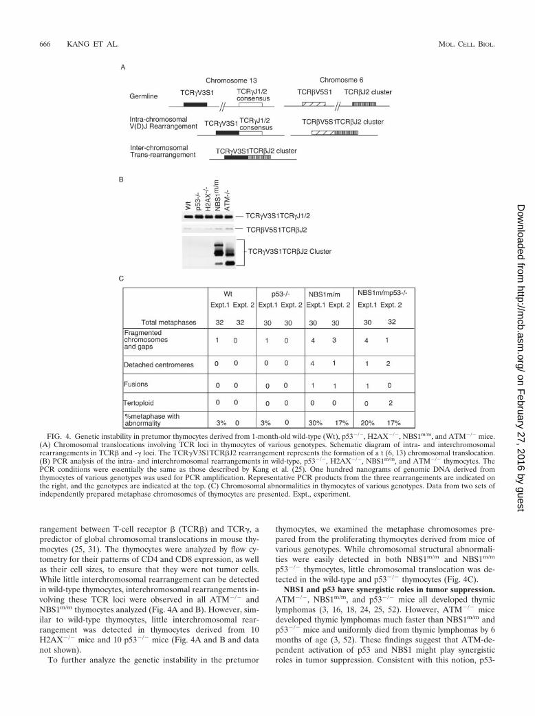
rangement between T-cell receptor � (TCR�) and TCR�, apredictor of global chromosomal translocations in mouse thy-mocytes (25, 31). The thymocytes were analyzed by flow cy-tometry for their patterns of CD4 and CD8 expression, as wellas their cell sizes, to ensure that they were not tumor cells.While little interchromosomal rearrangement can be detectedin wild-type thymocytes, interchromosomal rearrangements in-volving these TCR loci were observed in all ATM�/� andNBS1m/m thymocytes analyzed (Fig. 4A and B). However, sim-ilar to wild-type thymocytes, little interchromosomal rear-rangement was detected in thymocytes derived from 10H2AX�/� mice and 10 p53�/� mice (Fig. 4A and B and datanot shown).
To further analyze the genetic instability in the pretumor
thymocytes, we examined the metaphase chromosomes pre-pared from the proliferating thymocytes derived from mice ofvarious genotypes. While chromosomal structural abnormali-ties were easily detected in both NBS1m/m and NBS1m/m
p53�/� thymocytes, little chromosomal translocation was de-tected in the wild-type and p53�/� thymocytes (Fig. 4C).
NBS1 and p53 have synergistic roles in tumor suppression.ATM�/�, NBS1m/m, and p53�/� mice all developed thymiclymphomas (3, 16, 18, 24, 25, 52). However, ATM�/� micedeveloped thymic lymphomas much faster than NBS1m/m andp53�/� mice and uniformly died from thymic lymphomas by 6months of age (3, 52). These findings suggest that ATM-de-pendent activation of p53 and NBS1 might play synergisticroles in tumor suppression. Consistent with this notion, p53-
FIG. 4. Genetic instability in pretumor thymocytes derived from 1-month-old wild-type (Wt), p53�/�, H2AX�/�, NBS1m/m, and ATM�/� mice.(A) Chromosomal translocations involving TCR loci in thymocytes of various genotypes. Schematic diagram of intra- and interchromosomalrearrangements in TCR� and -� loci. The TCR�V3S1TCR�J2 rearrangement represents the formation of a t (6, 13) chromosomal translocation.(B) PCR analysis of the intra- and interchromosomal rearrangements in wild-type, p53�/�, H2AX�/�, NBS1m/m, and ATM�/� thymocytes. ThePCR conditions were essentially the same as those described by Kang et al. (25). One hundred nanograms of genomic DNA derived fromthymocytes of various genotypes was used for PCR amplification. Representative PCR products from the three rearrangements are indicated onthe right, and the genotypes are indicated at the top. (C) Chromosomal abnormalities in thymocytes of various genotypes. Data from two sets ofindependently prepared metaphase chromosomes of thymocytes are presented. Expt., experiment.
666 KANG ET AL. MOL. CELL. BIOL.
on February 27, 2016 by guest
http://mcb.asm
.org/D
ownloaded from

dependent p21 induction and cell cycle G1 arrest were bothnormal in NBS1m/m MEFs after various doses of IR (Fig. 5Aand data not shown). In addition, p53-dependent apoptosiswas modestly reduced in NBS1m/m thymocytes after IR com-pared with that in wild-type thymocytes (Fig. 5B). To furthertest the functional interactions of NBS1 and p53 in tumorsuppression, NBS1m/m p53�/� mice were generated and mon-itored for tumorigenesis. Similar to ATM�/� mice, NBS1m/m
p53�/� mice developed thymic lymphomas much faster thanNBS1m/m and p53�/� mice, and all NBS1m/m p53�/� mice diedfrom thymic lymphomas by 5 months of age (Fig. 5C). There-fore, NBS1 and p53 function synergistically in the suppressionof thymic lymphomas.
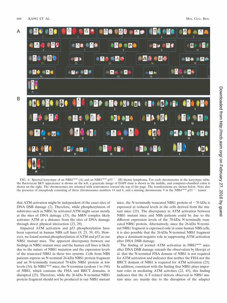
Chromosomal translocations in tumors derived from NBS1m/m
mice. To understand the rapid onset of tumorigenesis inNBS1m/m p53�/� mice and to determine the similarity to tu-mors arising in ATM�/� mice, SKY was performed to analyzethe chromosomal abnormalities in tumors derived fromNBS1m/m and NBS1m/m p53�/� mice. Two NBS1m/m tumorsand three NBS1m/m p53�/� tumors were analyzed. Chromo-somal translocations involving chromosome 12, including T(12;2), T(12;1), and T(12;11), were commonly detected inNBS1m/m tumors, with a clonal T(12;2) translocation observedin one tumor (Fig. 6A and data not shown). A subclonal T(14;12) translocation was also observed in both NBS1m/m tumors(Fig. 6A; data not shown). Similar chromosomal translocationswere observed in NBS1m/m p53�/� tumors. The first tumorcontained a clonal T(12;2) translocation in 12 of 13 meta-phases, comprised of the centromere and most of the long armof 12 attached to most of the distal long arm of 2 (Fig. 6B).This tumor also contained a subclonal T(3;14) translocation (3of 13 metaphases), as well as three or four intact copies ofchromosome 14 in every metaphase. The second tumor con-tained a T(14;2) translocation in 4 of 10 metaphases and extrachromosomes 14 and 15 in 7 metaphases (data not shown).The third tumor did not contain a detectable clonal aberrationbut was highly unstable, containing five separate transloca-tions, each seen in only one metaphase. Supernumerary chro-mosomes were observed only in NBS1m/m p53�/� tumors (av-erage chromosomal number, 45) but not in NBS1m/m tumors,both of which contained 40 chromosomes. These types of chro-mosomal translocations are also frequently observed in thymictumors derived from ATM�/� mice, as characterized in previ-ous publications (3, 32). These findings further support thenotion that NBS1 plays an important role in mediating ATM-dependent suppression of chromosomal translocations inmouse thymocytes.
DISCUSSION
The kinase activity of ATM is rapidly activated after DNADSB damage, leading to the phosphorylation and activation ofa number of DNA repair proteins, including NBS1 and 53BP1(2, 42). Recent studies have suggested that the Mre11/Rad50/NBS1 complex and 53BP1 function both upstream and down-stream of ATM after DNA DSB damage (9, 20, 21, 23, 30, 39,45, 48, 51, 55). H2AX, which is phosphorylated by ATM im-mediately after DNA DSB damage, is required for irradiation-induced focus formation at the site of DNA damage by a largenumber of DNA repair and checkpoint proteins, including53BP1 and NBS1, suggesting that H2AX might be involved inthe activation of ATM after DNA DSB damage (12, 48). How-ever, we demonstrate here that H2AX is not required for ATMactivation after DNA DSB damage. In addition, H2AX is notrequired for ATM-dependent phosphorylation of p53 andChk2 after DNA DSB damage (Fig. 1) (20). Therefore, H2AXfunctions as a downstream mediator of ATM functions afterDNA DSB damage. These findings also support the notion
FIG. 5. p53 and NBS1 play synergistic roles in tumor suppression.(A) Normal p53-dependent cell cycle G1 arrest in NBS1�/� andNBS1m/m MEFs after treatment with various doses of IR. The meanvalue from three independent experiments is presented with error barsindicating standard deviations. (B) Quantitative analysis of the per-centages of apoptotic thymocytes in NBS1�/� and NBS1m/m thymo-cytes 10 h after IR. The mean value from four independent experi-ments is presented with error bars indicating standard deviations. Wt,wild type. (C) Survival analysis of ATM�/�, NBS1m/m, p53�/�, andNBS1m/m p53�/� mice. Most of the data on NBS1m/m, p53�/�, andNBS1m/m p53�/� mice were derived from littermates. n represents thenumber of mice monitored for death caused by tumorigenesis. AllNBS1m/m p53�/� mice died from thymic lymphomas within 6 months.
VOL. 25, 2005 FUNCTIONAL INTERACTION OF H2AX, NBS1, AND p53 667
on February 27, 2016 by guest
http://mcb.asm
.org/D
ownloaded from
that ATM activation might be independent of the exact sites ofDNA DSB damage (2). Therefore, while phosphorylation ofsubstrates such as NBS1 by activated ATM might occur mostlyat the sites of DNA damage (33), the MRN complex likelyactivates ATM at a distance from the sites of DNA damagethrough direct physical interaction (23, 29).
Impaired ATM activation and p53 phosphorylation havebeen reported in human NBS cell lines (9, 23, 39, 45). How-ever, we found normal phosphorylation of ATM and p53 in ourNBS1 mutant mice. The apparent discrepancy between ourfindings in NBS1 mutant mice and the human cell lines is likelydue to the nature of NBS1 mutation and the expression levelsof the truncated NBS1 in these two systems. Cells from NBSpatients express an N-terminal 26-kDa NBS1 protein fragmentand an N-terminally truncated 70-kDa NBS1 protein at lowlevels (34). In NBS1m/m mice, the N terminus (codons 1 to 194)of NBS1, which contains the FHA and BRCT domains, isdisrupted (25). Therefore, while the 26-kDa N-terminal NBS1protein fragment should not be produced in our NBS1 mutant
mice, the N-terminally truncated NBS1 protein of 70 kDa isexpressed at reduced levels in the cells derived from the mu-tant mice (25). The discrepancy in ATM activation betweenNBS1 mutant mice and NBS patients could be due to thedifferent expression levels of the 70-kDa N-terminally trun-cated NBS1 protein. Alternatively, since the 26-kDa N-termi-nal NBS1 fragment is expressed only in some human NBS cells,it is also possible that the 26-kDa N-terminal NBS1 fragmentplays a dominant-negative role in suppressing ATM activationafter DNA DSB damage.
The finding of normal ATM activation in NBS1m/m miceafter DNA DSB damage extends the observation by Horejsi etal. that the N-terminal FHA domain of NBS1 is not requiredfor ATM activation and indicates that neither the FHA nor theBRCT domain of NBS1 is required for ATM activation (23).In addition, consistent with the finding that NBS1 plays impor-tant roles in mediating ATM activities (22, 45), this findingindicates that the A-T-related defects observed in NBS1 mu-tant mice are mainly due to the disruption of the adapter
FIG. 6. Spectral karyotype of an NBS1m/m (A) and an NBS1m/m p53�/� (B) thymic lymphoma. For each chromosome in the karyotype table,the fluorescent SKY appearance is shown on the left, a grayscale image of DAPI stain is shown in the middle, and computer-classified colon isshown on the right. The chromosomes are oriented with centromeres toward the top of the page. The translocations are shown below. Note alsothe presence of aneuploidy consisting of three chromosomes numbers 14 and 6, and a missing chromosome 9 in the NBS1m/m p53�/� tumor.
668 KANG ET AL. MOL. CELL. BIOL.
on February 27, 2016 by guest
http://mcb.asm
.org/D
ownloaded from
function of NBS1 in mediating ATM activities, such as thephosphorylation of Chk2.
Our studies also provide an improved understanding of thefunctional interactions among H2AX, NBS1, and p53. An ear-lier report showed that H2AX is required for the irradiation-induced focus formation of NBS1 after DNA DSB damage,suggesting that H2AX and NBS1 function in overlapping path-ways in the cellular responses to DNA DSB damage (11).However, several lines of evidence presented here and else-where indicate that NBS1 and H2AX also function in distinctpathways in DNA damage responses. In this context, NBS1,but not H2AX, is required for ATM-dependent Chk2 phosphor-ylation after DNA DSB damage (Fig. 3) (7, 20). In addition,NBS1, but not H2AX, is required for the ATM-dependent sup-pression of interchromosomal rearrangement, indicating thatNBS1 and H2AX have distinct roles in DNA damage responses.In support of this notion, the chromosomal abnormalities ob-served in the thymic tumors derived from NBS1m/m and NBS1m/m
p53�/� mice are different from those derived from H2AX�/�
p53�/� mice (4). The distinct roles of H2AX and NBS1 in DNArepair could also account for the synthetic lethality of NBS1mutation and H2AX mutation.
ATM is required to activate p53 responses to DNA DSBdamage (42). Our finding that H2AX and NBS1 are not im-portant for p53 responses to DNA DSB damage indicates thatH2AX and NBS1 function largely in distinct pathways of p53 inDNA DSB damage responses. In the context of H2AX�/�
cells, increased basal levels of p53 activities, possibly due to thepresence of endogenous genetic instability, might account forthe very modest cancer risk in these mutant mice. Consistentwith these notions, inactivation of p53 greatly facilitates tumor-igenesis in H2AX�/� mice and our NBS1 mutant mice, as wellas independently generated NBS1 mutant mice (4, 10, 50).Considering the greatly increased genetic instability in H2AXand NBS1 mutant mice, these findings indicate that p53 canefficiently suppress tumorigenesis in the presence of significantendogenous chromosomal abnormalities. Therefore, cancerpredisposition in ATM�/� mice, in which both p53 inactivationand genetic instability are present, is greatly increased com-pared with that in NBS1 mutant mice.
ACKNOWLEDGMENT
This work was supported by an NIH grant to Y.X. (CA77563).
REFERENCES
1. Ahn, J. Y., J. K. Schwarz, H. Piwnica-Worms, and C. E. Canman. 2000.Threonine 68 phosphorylation by ataxia telangiectasia mutated is requiredfor efficient activation of Chk2 in response to ionizing radiation. Cancer Res.60:5934–5936.
2. Bakkenist, C. J., and M. B. Kastan. 2003. DNA damage activates ATMthrough intermolecular autophosphorylation and dimer dissociation. Nature421:499–506.
3. Barlow, C., S. Hirotsune, R. Paylor, M. Liyanage, M. Eckhaus, F. Collins, Y.Shiloh, J. N. Crawley, T. Ried, D. Tagle, and A. Wynshaw-Boris. 1996.Atm-deficient mice: a paradigm of ataxia telangiectasia. Cell 86:159–171.
4. Bassing, C. H., H. Suh, D. O. Ferguson, K. F. Chua, J. Manis, M. Eckers-dorff, M. Gleason, R. Bronson, C. Lee, and F. W. Alt. 2003. Histone H2AX:a dosage-dependent suppressor of oncogenic translocations and tumors. Cell114:359–370.
5. Boley, S. E., V. A. Wong, J. E. French, and L. Recio. 2002. p53 heterozygosityalters the mRNA expression of p53 target genes in the bone marrow inresponse to inhaled benzene. Toxicol. Sci. 66:209–215.
6. Burma, S., B. P. Chen, M. Murphy, A. Kurimasa, and D. J. Chen. 2001.ATM phosphorylates histone H2AX in response to DNA double-strandbreaks. J. Biol. Chem. 276:42462–42467.
7. Buscemi, G., C. Savio, L. Zannini, F. Micciche, D. Masnada, M. Nakanishi,H. Tauchi, K. Komatsu, S. Mizutani, K. Khanna, P. Chen, P. Concannon, L.Chessa, and D. Delia. 2001. Chk2 activation dependence on Nbs1 after DNAdamage. Mol. Cell. Biol. 21:5214–5222.
8. Carney, J. P., R. S. Maser, H. Olivares, E. M. Davis, M. Le Beau, J. R. YatesIII, L. Hays, W. F. Morgan, and J. H. Petrini. 1998. The hMre11/hRad50protein complex and Nijmegen breakage syndrome: linkage of double-strandbreak repair to the cellular DNA damage response. Cell 93:477–486.
9. Carson, C. T., R. A. Schwartz, T. H. Stracker, C. E. Lilley, D. V. Lee, andM. D. Weitzman. 2003. The Mre11 complex is required for ATM activationand the G2/M checkpoint. EMBO J. 22:6610–6620.
10. Celeste, A., S. Difilippantonio, M. J. Difilippantonio, O. Fernandez-Ca-petillo, D. R. Pilch, O. A. Sedelnikova, M. Eckhaus, T. Ried, W. M. Bonner,and A. Nussenzweig. 2003. H2AX haploinsufficiency modifies genomic sta-bility and tumor susceptibility. Cell 114:371–383.
11. Celeste, A., O. Fernandez-Capetillo, M. J. Kruhlak, D. R. Pilch, D. W.Staudt, A. Lee, R. F. Bonner, W. M. Bonner, and A. Nussenzweig. 2003.Histone H2AX phosphorylation is dispensable for the initial recognition ofDNA breaks. Nat. Cell Biol. 5:675–679.
12. Celeste, A., S. Petersen, P. J. Romanienko, O. Fernandez-Capetillo, H. T.Chen, O. A. Sedelnikova, B. Reina-San-Martin, V. Coppola, E. Meffre, M. J.Difilippantonio, C. Redon, D. R. Pilch, A. Olaru, M. Eckhaus, R. D. Cam-erini-Otero, L. Tessarollo, F. Livak, K. Manova, W. M. Bonner, M. C.Nussenzweig, and A. Nussenzweig. 2002. Genomic instability in mice lackinghistone H2AX. Science 296:922–927.
13. Chao, C., M. Hergenhahn, M. D. Kaeser, Z. Wu, S. Saito, R. Iggo, M.Hollstein, E. Appella, and Y. Xu. 2003. Cell type- and promoter-specific rolesof Ser18 phosphorylation in regulating p53 responses. J. Biol. Chem. 8:8.
14. Chao, C., S. Saito, C. W. Anderson, E. Appella, and Y. Xu. 2000. Phosphor-ylation of murine p53 at ser-18 regulates the p53 responses to DNA damage.Proc. Natl. Acad. Sci. USA 97:11936–11941.
15. Chao, C., S. Saito, J. Kang, C. W. Anderson, E. Appella, and Y. Xu. 2000. p53transcriptional activity is essential for p53-dependent apoptosis followingDNA damage. EMBO J. 19:4967–4975.
16. Donehower, L. A., M. Harvey, B. L. Slagle, M. J. McArthur, C. A. Mont-gomery, Jr., J. S. Butel, and A. Bradley. 1992. Mice deficient for p53 aredevelopmentally normal but susceptible to spontaneous tumours. Nature356:215–221.
17. Dumaz, N., D. M. Milne, and D. W. Meek. 1999. Protein kinase CK1 is ap53-threonine 18 kinase which requires prior phosphorylation of serine 15.FEBS Lett. 463:312–316.
18. Elson, A., Y. Wang, C. J. Daugherty, C. C. Morton, F. Zhou, J. Campos-Torres, and P. Leder. 1996. Pleiotropic defects in ataxia-telangiectasia pro-tein-deficient mice. Proc. Natl. Acad. Sci. USA 93:13084–13089.
19. Ferguson, D. O., J. M. Sekiguchi, S. Chang, K. M. Frank, Y. Gao, R. A.DePinho, and F. W. Alt. 2000. The nonhomologous end-joining pathway ofDNA repair is required for genomic stability and the suppression of trans-locations. Proc. Natl. Acad. Sci. USA 97:6630–6633.
20. Fernandez-Capetillo, O., H. T. Chen, A. Celeste, I. Ward, P. J. Romanienko,J. C. Morales, K. Naka, Z. Xia, R. D. Camerini-Otero, N. Motoyama, P. B.Carpenter, W. M. Bonner, J. Chen, and A. Nussenzweig. 2002. DNA dam-age-induced G2-M checkpoint activation by histone H2AX and 53BP1. Nat.Cell Biol. 4:993–997.
21. Gatei, M., D. Young, K. M. Cerosaletti, A. Desai-Mehta, K. Spring, S.Kozlov, M. F. Lavin, R. A. Gatti, P. Concannon, and K. Khanna. 2000.ATM-dependent phosphorylation of nibrin in response to radiation expo-sure. Nat. Genet. 25:115–119.
22. Girard, P. M., E. Riballo, A. C. Begg, A. Waugh, and P. A. Jeggo. 2002. Nbs1promotes ATM dependent phosphorylation events including those requiredfor G1/S arrest. Oncogene 21:4191–4199.
23. Horejsi, Z., J. Falck, C. J. Bakkenist, M. B. Kastan, J. Lukas, and J. Bartek.2004. Distinct functional domains of Nbs1 modulate the timing and magni-tude of ATM activation after low doses of ionizing radiation. Oncogene23:3122–3127.
24. Jacks, T., L. Remington, B. O. Williams, E. M. Schmitt, S. Halachmi, R. T.Bronson, and R. A. Weinberg. 1994. Tumor spectrum analysis in p53-mutantmice. Curr. Biol. 4:1–7.
25. Kang, J., R. T. Bronson, and Y. Xu. 2002. Targeted disruption of NBS1reveals its roles in mouse development and DNA repair. EMBO J. 21:1447–1455.
26. Kastan, M. B., Q. Zhan, W. S. el-Deiry, F. Carrier, T. Jacks, W. V. Walsh,B. S. Plunkett, B. Vogelstein, and A. J. Fornace, Jr. 1992. A mammalian cellcycle checkpoint pathway utilizing p53 and GADD45 is defective in ataxia-telangiectasia. Cell 71:587–597.
27. Kobayashi, J., H. Tauchi, S. Sakamoto, A. Nakamura, K. Morishima, S.Matsuura, T. Kobayashi, K. Tamai, K. Tanimoto, and K. Komatsu. 2002.NBS1 localizes to gamma-H2AX foci through interaction with the FHA/BRCT domain. Curr. Biol. 12:1846–1851.
28. Lambert, P. F., F. Kashanchi, M. F. Radonovich, R. Shiekhattar, and J. N.Brady. 1998. Phosphorylation of p53 serine 15 increases interaction withCBP. J. Biol. Chem. 273:33048–33053.
VOL. 25, 2005 FUNCTIONAL INTERACTION OF H2AX, NBS1, AND p53 669
on February 27, 2016 by guest
http://mcb.asm
.org/D
ownloaded from
29. Lee, J. H., and T. T. Paull. 2004. Direct activation of the ATM protein kinaseby the Mre11/Rad50/Nbs1 complex. Science 304:93–96.
30. Lim, D. S., S. T. Kim, B. Xu, R. S. Maser, J. Lin, J. H. Petrini, and M. B.Kastan. 2000. ATM phosphorylates p95/nbs1 in an S-phase checkpoint path-way. Nature 404:613–617.
31. Lista, F., V. Bertness, C. J. Guidos, J. S. Danska, and I. R. Kirsch. 1997. Theabsolute number of trans-rearrangements between the TCRG and TCRBloci is predictive of lymphoma risk: a severe combined immune deficiency(SCID) murine model. Cancer Res. 57:4408–4413.
32. Liyanage, M., A. Coleman, S. du Manoir, T. Veldman, S. McCormack, R. B.Dickson, C. Barlow, A. Wynshaw-Boris, S. Janz, J. Wienberg, M. A. Fergu-son-Smith, E. Schrock, and T. Ried. 1996. Multicolour spectral karyotypingof mouse chromosomes. Nat. Genet. 14:312–315.
33. Lukas, C., J. Falck, J. Bartkova, J. Bartek, and J. Lukas. 2003. Distinctspatiotemporal dynamics of mammalian checkpoint regulators induced byDNA damage. Nat. Cell Biol. 5:255–260.
34. Maser, R. S., R. Zinkel, and J. H. Petrini. 2001. An alternative mode oftranslation permits production of a variant NBS1 protein from the commonNijmegen breakage syndrome allele. Nat. Genet. 27:417–421.
35. Matsuoka, S., G. Rotman, A. Ogawa, Y. Shiloh, K. Tamai, and S. J. Elledge.2000. Ataxia telangiectasia-mutated phosphorylates Chk2 in vivo and invitro. Proc. Natl. Acad. Sci. USA 97:10389–10394.
36. Matsuura, S., H. Tauchi, A. Nakamura, N. Kondo, S. Sakamoto, S. Endo, D.Smeets, B. Solder, B. H. Belohradsky, V. M. Der Kaloustian, M. Oshimura,M. Isomura, Y. Nakamura, and K. Komatsu. 1998. Positional cloning of thegene for Nijmegen breakage syndrome. Nat. Genet. 19:179–181.
37. Melchionna, R., X. B. Chen, A. Blasina, and C. H. McGowan. 2000. Thre-onine 68 is required for radiation-induced phosphorylation and activation ofCds1. Nat. Cell Biol. 2:762–765.
38. Mills, K. D., D. O. Ferguson, and F. W. Alt. 2003. The role of DNA breaksin genomic instability and tumorigenesis. Immunol. Rev. 194:77–95.
39. Mochan, T. A., M. Venere, R. A. DiTullio, Jr., and T. D. Halazonetis. 2003.53BP1 and NFBD1/MDC1-Nbs1 function in parallel interacting pathwaysactivating ataxia-telangiectasia mutated (ATM) in response to DNA dam-age. Cancer Res. 63:8586–8591.
40. Rogakou, E. P., D. R. Pilch, A. H. Orr, V. S. Ivanova, and W. M. Bonner.1998. DNA double-stranded breaks induce histone H2AX phosphorylationon serine 139. J. Biol. Chem. 273:5858–5868.
41. Sedelnikova, O. A., D. R. Pilch, C. Redon, and W. M. Bonner. 2003. HistoneH2AX in DNA damage and repair. Cancer Biol. Ther. 2:233–235.
42. Shiloh, Y. 2003. ATM and related protein kinases: safeguarding genomeintegrity. Nat. Rev. Cancer 3:155–168.
43. Shiloh, Y., and M. B. Kastan. 2001. ATM: genome stability, neuronal de-velopment, and cancer cross paths. Adv. Cancer Res. 83:209–254.
44. Shiloh, Y., and G. Rotman. 1996. Ataxia-telangiectasia and the ATM gene:linking neurodegeneration, immunodeficiency, and cancer to cell cycle check-points. J. Clin. Immunol. 16:254–260.
45. Uziel, T., Y. Lerenthal, L. Moyal, Y. Andegeko, L. Mittelman, and Y. Shiloh.2003. Requirement of the MRN complex for ATM activation by DNAdamage. EMBO J. 22:5612–5621.
46. van der Burgt, I., K. H. Chrzanowska, D. Smeets, and C. Weemaes. 1996.Nijmegen breakage syndrome. J. Med. Genet. 33:153–156.
47. Varon, R., C. Vissinga, M. Platzer, K. M. Cerosaletti, K. H. Chrzanowska, K.Saar, G. Beckmann, E. Seemanova, P. R. Cooper, N. J. Nowak, M. Stumm,C. M. Weemaes, R. A. Gatti, R. K. Wilson, M. Digweed, A. Rosenthal, K.Sperling, P. Concannon, and A. Reis. 1998. Nibrin, a novel DNA double-strand break repair protein, is mutated in Nijmegen breakage syndrome. Cell93:467–476.
48. Ward, I. M., K. Minn, K. G. Jorda, and J. Chen. 2003. Accumulation ofcheckpoint protein 53BP1 at DNA breaks involves its binding to phosphor-ylated histone H2AX. J. Biol. Chem. 278:19579–19582.
49. Westphal, C. H., S. Rowan, C. Schmaltz, A. Elson, D. E. Fisher, and P.Leder. 1997. atm and p53 cooperate in apoptosis and suppression of tumor-igenesis, but not in resistance to acute radiation toxicity. Nat. Genet. 16:397–401.
50. Williams, B. R., O. K. Mirzoeva, W. F. Morgan, J. Lin, W. Dunnick, and J. H.Petrini. 2002. A murine model of Nijmegen breakage syndrome. Curr. Biol.12:648–653.
51. Wu, X., V. Ranganathan, D. S. Weisman, W. F. Heine, D. N. Ciccone, T. B.O’Neill, K. E. Crick, K. A. Pierce, W. S. Lane, G. Rathbun, D. M. Livingston,and D. T. Weaver. 2000. ATM phosphorylation of Nijmegen breakage syn-drome protein is required in a DNA damage response. Nature 405:477–482.
52. Xu, Y., T. Ashley, E. E. Brainerd, R. T. Bronson, M. S. Meyn, and D.Baltimore. 1996. Targeted disruption of ATM leads to growth retardation,chromosomal fragmentation during meiosis, immune defects, and thymiclymphoma. Genes Dev. 10:2411–2422.
53. Xu, Y., and D. Baltimore. 1996. Dual roles of ATM in the cellular responseto radiation and in cell growth control. Genes Dev. 10:2401–2410.
54. Xu, Y., E. M. Yang, J. Brugarolas, T. Jacks, and D. Baltimore. 1998. In-volvement of p53 and p21 in cellular defects and tumorigenesis in atm�/�
mice. Mol. Cell. Biol. 18:4385–4390.55. Zhao, S., Y. C. Weng, S. S. Yuan, Y. T. Lin, H. C. Hsu, S. C. Lin, E. Gerbino,
M. H. Song, M. Z. Zdzienicka, R. A. Gatti, J. W. Shay, Y. Ziv, Y. Shiloh, andE. Y. Lee. 2000. Functional link between ataxia-telangiectasia and Nijmegenbreakage syndrome gene products. Nature 405:473–477.
670 KANG ET AL. MOL. CELL. BIOL.
on February 27, 2016 by guest
http://mcb.asm
.org/D
ownloaded from