A chaotic system based fragile watermarking scheme for image tamper detection
Upload
chu-sainte-justineCategory
view
0download
0
Systematic identification of fragile sites via genome-widelocation analysis of γ-H2AX
Rachel K. Szilard1,2, Pierre-Étienne Jacques1,3, Louise Laramée3, Benjamin Cheng2,Sarah Galicia2, Alain R. Bataille3, ManTek Yeung2,4, Megan Mendez2, Maxime Bergeron3,François Robert3,5,6, and Daniel Durocher2,4,62 Samuel Lunenfeld Research Institute, Mount Sinai Hospital, 600 University Avenue, Toronto,Ontario, M5G 1X5, Canada3 Laboratoire de chromatine et expression du génome, Institut de recherches cliniques deMontréal, 110 Avenue des Pins Ouest, Montréal, Québec, H2W 1R7, Canada4 Department of Molecular Genetics, University of Toronto, Toronto, Ontario, M5S 1A8, Canada5 Département de Médecine, Université de Montréal, Montréal, Québec, Canada
AbstractPhosphorylation of histone H2AX is an early response to DNA damage in eukaryotes. InSaccharomyces cerevisiae, DNA damage or replication fork stalling results in histone H2Aphosphorylation to yield γ-H2A (yeast γ-H2AX) in a Mec1 (ATR)- and Tel1 (ATM)- dependentmanner. Here, we describe the genome-wide location analysis of γ-H2A as a strategy to identifyloci prone to engage the Mec1 and Tel1 pathways. Remarkably, γ-H2A enrichment overlaps withloci prone to replication fork stalling and is caused by the action of Mec1 and Tel1, indicating thatthese loci are prone to breakage. Moreover, about half the sites enriched for γ-H2A map torepressed protein-coding genes, and histone deacetylases are necessary for formation of γ-H2A atthese loci. Finally, our work indicates that high resolution mapping of γ-H2AX is a fruitful routeto map fragile sites in eukaryotic genomes.
IntroductionDNA replication poses a major challenge to genome integrity1. Chromosomal fragile sites inthe human genome are associated with defects in DNA replication progression and can leadto genome rearreangements2. Replisomes encounter a number of obstacles that must beovercome in order to complete DNA replication in a timely yet accurate manner. Inadequatenucleotide or histone supplies, DNA damage, protein-DNA complexes, gene transcription,chromatin organization and topological strain can all potentially block replication forkprogression1.
6Correspondence to: Daniel Durocher ( [email protected]) and François Robert ([email protected]).1These authors contributed equally to this work.ACCESSION NUMBERSThe ChIP-chip data in this paper have been deposited in the NCBI Gene Expression Omnibus (GEO)(http://www.ncbi.nlm.nih.gov/geo/) and are accessible through GEO series accession number GSE18191.AUTHOR CONTRIBUTIONSRKS generated most yeast strains, and performed most ChIP-chip experiments as well as the TEL06R qPCR (assisted by MM) andGCR (assisted by SG) experiments. P-ÉJ analyzed all ChIP-chip data. LL performed the mutant tRNA qPCR experiment. AdditionalChIP-chip experiments were performed by BC (wild-type and h2a-S129Aγ-H2A), ARB (MCMs) and MB (RNAPII). MY performedfocus formation assays. DD and FR, with help from P-ÉJ and RKS, conceived the experiments and wrote the manuscript.
PubMed Central CANADAAuthor Manuscript / Manuscrit d'auteurNat Struct Mol Biol. Author manuscript; available in PMC 2011 April 22.
Published in final edited form as:Nat Struct Mol Biol. 2010 March ; 17(3): 299–305. doi:10.1038/nsmb.1754.
PMC
Canada Author M
anuscriptPM
C C
anada Author Manuscript
PMC
Canada Author M
anuscript
In budding yeast, up to 1,400 sites have been proposed as potential impediments toreplication forks. These sites include tRNA genes, Ty long-terminal repeats (LTRs),centromeres, DNA replication origins, the HMR and HML heterochromatic loci and therepeated rDNA units1,3–6. This list is an extrapolation based on the characterization of a fewsites, rather than the result of high-resolution mapping of replication fork pausing. Otherclasses of replisome progression (or stability) obstacles have been identified7,8 indicatingthat the list described above might not be exhaustive. Importantly, these natural replicationfork barriers have been linked to genome rearrangements, suggesting that some paused forkscollapse or are processed at these sites3,9–12.
Generally, unscheduled replisome stalling elicits the activation of protein kinases of thePI(3) kinase-like kinase (PIKK) family, particularly the Mec1/ATR ortholog13–17.Activation of Mec1/ATR signaling stabilizes replication forks to prevent their collapse18,19
although the critical Mec1/ATR targets that promote fork stability remain unknown.Nevertheless, several phosphorylation events have been characterized in response toreplication fork blocks. In particular, phosphorylation of histone H2AX is a near-universalfeature of the eukaryotic response to genotoxic stress20–23. This phosphorylation event,yielding γ-H2AX (γ-H2A in Saccharomyces cerevisiae), can occur as a consequence ofDNA replication fork stalling in an ATR/Mec1-dependent manner24,25. The link between γ-H2AX and replication fork stability is better established in S. cerevisiae where abrogation ofγ-H2A by mutation of the HTA genes produces sensitivity to camptothecin, a topoisomeraseI inhibitor that provokes replication-associated DNA double-strand breaks (DSBs)26. Thesame hta mutants are only mildly sensitive to other genotoxins, indicating that γ-H2A isimportant for the response to replication-associated DSBs.
In this study, we hypothesized that mapping γ-H2A in cycling cells might reveal fragilegenomic loci. We employed a genome-wide location assay (chromatin immunoprecipitationon tiled microarray, or ChIP-chip) to map γ-H2A-rich loci (referred to herein as γ-sites). Ourdata shows that γ-sites are distributed non-randomly and are concentrated at telomeres, therDNA locus, DNA replication origins, LTRs, tRNA genes and, surprisingly, activelyrepressed protein-coding genes. Using a combination of genetic studies and carbon sourcemanipulation, we found that the chromatin structure promoted by histone deacetylation canlead to PIKK activation. We conclude that mapping sites of γ-H2AX enrichment will be afruitful route to map at-risk genomic elements in eukaryotes.
RESULTSGenome-wide location analysis of γ-H2A
To identify loci enriched in γ-H2A, we carried out ChIP with a phosphospecific antibodythat recognizes yeast γ-H2A26 and hybridized the associated DNA to high-density genomictiling arrays. We typically performed competitive hybridization of DNA precipitated fromHTA1 HTA2 cells with DNA precipitated from the γ-H2A-deficient hta1-S129A hta2-S129Acells (referred to hereafter as h2a-S129A). We also tested other experimental designscontrolling for nucleosome density that yielded similar results (Supplementary Fig. 1a). Allexperiments (listed in Supplementary Table 1) were done at least in duplicate and combinedusing a weighted average method27. The combined datasets are available in SupplementaryFile 1.
We first examined γ-H2A enrichment in asynchronously dividing cell cultures (Fig. 1a).Statistical analysis of the enrichment profile identified 697 unambiguous loci enriched in γ-H2A using the criterion of a peak with a p value <0.1 (Supplementary File 1). We refer tothese loci asγ-sites. Fluorescence-activated cell sorting (FACS) analyses revealed noobvious cell cycle profile differences between wild-type and h2a-S129A cultures, thus
Szilard et al. Page 2
Nat Struct Mol Biol. Author manuscript; available in PMC 2011 April 22.
PMC
Canada Author M
anuscriptPM
C C
anada Author Manuscript
PMC
Canada Author M
anuscript
excluding the possibility that differences in γ-H2A enrichment are simply due to differencesin the cell cycle profiles (Supplementary Fig. 1b).
Statistically significant γ-sites vary in length but average 1255 bp. The average γ-site istherefore considerably shorter than 50 kb, the size of the γ-H2A domain caused by anunrepairable DSB delivered by the HO endonuclease28. Moreover, the shape of the γ-sites inour location analyses is strikingly different from the bimodal distribution of γ-H2Asurrounding HO-induced DSBs28. The enrichments we observed generally displayed asingle maximum intensity peak (see e.g. Fig. 1a). These differences suggest that the eventsmonitored by γ-H2A ChIP in cycling cells are likely not irreparable DSBs.
In addition to mapping γ-sites in asynchronously dividing cells, we also analyzed cellssynchronized in G1 by α-factor and cells synchronized at mid-S phase, obtained by releasingcells from a G1 block. The γ-H2A accumulation profiles in G1 and mid-S cells were highlysimilar to those obtained in asynchronous cultures (Supplementary Fig. 2). Since the γ-H2Aprofiles appeared highly similar whether cultures were synchronized or not, we pursued ouranalyses with datasets obtained from asynchronous cultures.
Visual and computational analysis of γ-sites identify 7 classes of genomic loci clearlyenriched with γ-H2A in cycling cells: (i) telomeric regions, (ii) DNA replication origins, (iii)tRNA genes, (iv) LTRs, (v) the rDNA locus, (vi) the silent mating type cassettes HMR andHML, and (vii) a group of protein-coding genes (Supplementary Table 2). Apart fromprotein-coding genes, all of the above loci are known to impede replisome progression,strongly suggesting that γ-H2A detected in cycling cells is caused by replication forkpausing or collapse. Interestingly, we also observed γ-H2A enrichment at centromeres,another obstacle for replisomes, but the low number of centromeres in the yeast genomeprecluded this class of γ-site from being identified as statistically significant at the 0.1 pvalue.
Telomeres show striking γ-H2A enrichment at every chromosome end despite the paucity ofprobes covering the repetitive telomeric and subtelomeric regions. γ-H2A accumulation attelomeres, which was also observed in another study29, was also detected by ChIP followedby quantitative real-time PCR (qPCR; Supplementary Fig. 3a). There is a strong correlationbetween the γ-H2A signal intensity and the proximity to the chromosome end(Supplementary Fig. 3b) whether or not the chromosome end contains a subtelomeric Y′element (e.g. see TEL06R, which does not contain a Y′ element; Fig. 1a). This resultindicates that Y′ subtelomeric elements are not responsible for the observed γ-H2A signal,suggesting that it originates either from the X element (common to all chromosome ends) orthe TG repeats themselves. However, the significance of this telomeric enrichment is stillunknown as h2a-S129A cells do not display any striking telomeric phenotype in all assayswe tested so far (telomere length, telomeric silencing, senescence assays, telomere cappingand chromosome healing, data not shown).
Visual inspection of the γ-H2A enrichment profiles identifies strong signals on ChrXII, at aregion encompassing the rDNA locus (Supplementary Fig. 3c) and on ChrIII, which harborsthe MAT locus and the silent mating type cassettes HMR and HML (Supplementary Fig.3d,e). Observing γ-H2A enrichment at rDNA is not surprising since this locus experiencesDNA strand breaks and recombination resulting from collisions between DNA replicationforks and RNA polymerase I or via abortive decatenation reactions1. The HMR and HMLloci are also known to impede replisomes, which might cause the observed signal30.However, these results are somewhat at odds with a recent report indicating that theseheterochromatic regions are refractory to DSB-induced γ-H2A accumulation29, although we
Szilard et al. Page 3
Nat Struct Mol Biol. Author manuscript; available in PMC 2011 April 22.
PMC
Canada Author M
anuscriptPM
C C
anada Author Manuscript
PMC
Canada Author M
anuscript
note that high levels of γ-H2A before DSB induction might have skewed the enrichmentratios calculated by Kim et al29.
γ-H2A accumulation correlates with replication fork blockageNext, we mapped the γ-H2A enrichment ratios on all tRNA genes, DNA replication originsand LTR elements to identify sub-regions that might be responsible for the γ-H2A signal(Fig. 1b-d, blue traces). The γ-H2A enrichment profiles at tRNA genes are strikinglyasymmetrical (Fig. 1b), peaking just before the RNA polymerase III transcription initiationsite, and followed by a sharp fall in signal intensity. This enrichment is independent of anyother nearby γ-H2A-enriched genomic loci (Supplementary Fig. 4). This asymmetry isconsistent with tRNA genes being polar replication fork barriers5 where the obstacle toreplisomes is the initiation complex rather than transcription itself3. TFIIIB binds the regionimmediately upstream of the tRNA gene transcription site, suggesting that DNA-boundTFIIIB elicits the γ-H2A signal observed at tRNA genes. In contrast, the averaged γ-H2Aenrichment ratios at DNA replication origins (defined as peaks of Mcm7 and Mcm4occupancy by ChIP-chip, see Methods) are largely symmetrical, peaking at the center of theorigin where the pre-replication complex assembles. Likewise, the averaged γ-H2Aenrichment ratios at LTRs are symmetrical, peaking at the 5′ end of the LTR.
Next we sought to map replication fork pausing at tRNA genes, DNA replication origins andLTRs in order to establish if the replisome accumulates or pauses at sites of γ-H2Aenrichment. We therefore performed ChIP-chip analysis of myc-tagged Pol2, the catalyticsubunit of DNA polymerase ε (DNAP). At first glance, we observed little similarity betweenDNAP and γ-H2A accumulation (Fig. 1a). However, averaging the DNAP signal at γ-sitesrevealed a striking accumulation of the replisome at these loci (Fig. 1b-d; gold traces). Sincereplication fork stalling or pausing can be defined by the accumulation of DNAP, theseresults suggest that the γ-sites are the consequence of impeded replisome progression.
To directly test this possibility, we used the fact that mutation of the TFIIIB-binding site ontRNA genes abolishes the impediment to replication fork progression and greatly reducesreplication fork stalling3. We therefore compared γ-H2A and DNAP enrichment by ChIP-qPCR at a tRNA gene (tS(GCU)L) that was either wild-type or contained a mutation in itsTFIIIB-binding site (tS(GCU)Lmut). As expected, deletion of the TFIIIB-binding siteabolished DNAP enrichment at this tRNA gene, indicating that replication forks no longerstall at the mutated tRNA gene (Fig. 2; gold curves). Satisfyingly, we observed that the γ-H2A signal was also abrogated at tS(GCU)Lmut (Fig. 2; blue curves). We therefore concludethat γ-H2A accumulates following unscheduled replication pausing, stalling or collapse.
γ-H2A accumulation is dependent on both Mec1 and Tel1In response to replisome pausing or stalling, H2A is solely phosphorylated by Mec124
whereas Tel1 contributes to γ-H2A formation, redundantly with Mec1, only when DSBs arepresent21. These properties allowed us to determine whether γ-sites resulted from replicationfork stalling, or whether DSBs also contributed to their formation. We therefore performedγ-H2A ChIP-chip in sml1Δ, mec1Δ sml1Δ, tel1Δ and mec1Δ sml1Δ tel1Δ cells. SML1 wasdeleted to circumvent the lethality of the MEC1 deletion and therefore sml1Δ acted as acontrol for strains with the MEC1 deletion. Not surprisingly, comparison of the total signalintensity ratios in the wild-type and sml1Δ strain revealed a Pearson correlation coefficientof 0.82 (Supplementary Table 3) and no major differences in the profiles, indicating thatsml1Δ did not affect H2A phosphorylation. Likewise, deletion of TEL1 or MEC1 did nothave a major impact on the γ-H2A signals, with Pearson correlations of 0.82 and 0.85 whencompared to their respective controls (Fig. 3a,b and Supplementary Table 3). However,deletion of either kinase led to a small decrease in the averaged signal intensities at tRNA
Szilard et al. Page 4
Nat Struct Mol Biol. Author manuscript; available in PMC 2011 April 22.
PMC
Canada Author M
anuscriptPM
C C
anada Author Manuscript
PMC
Canada Author M
anuscript
genes, DNA replication origins and LTRs, indicating that both kinases contributeindependently to γ-H2A accumulation at these sites (Fig. 3a,b). Upon closer inspection ofthe γ-sites in the mec1Δ sml1Δ strain, we observed that sites overlapping with mostcentromeres were strikingly absent in that strain (Fig. 3c). Moreover, the centromere-associated γ-H2A signal was also absent in G1 phase cells, indicating that these are labile γ-sites (Supplementary Fig. 2c). These results suggest that pericentromeric H2Aphosphorylation primarily occurs following replication fork pausing.
When the tel1Δ and mec1Δ mutations were combined together (in the sml1Δ background),all γ-sites were lost, leading to a Pearson correlation of 0.16 when compared to the sml1Δcontrol strain (Fig. 3d and Supplementary Table 3). Together, these results indicate that in acell population, γ-sites are caused by a combination of replication fork stalling and collapse,consistent with the idea that the great majority of the γ-sites mapped represent fragile sites.
RRM3 regulates γ-sitesThe Rrm3 helicase travels with the replication fork to facilitate fork progression throughnon-histone protein-DNA complexes3,31,32. RRM3 deletion increases the probability ofreplication fork stalling at ~1,400 potential sites in the genome3,31,32 that overlapremarkably with the γ-sites mapped in this study. We therefore mapped γ-H2A in rrm3Δcells. The γ-H2A profile of rrm3Δ cells is qualitatively similar to that of wild-type (Fig. 1a,red) although in some regions, Rrm3 clearly dampens the γ-H2A signal. Figure 1b-d showsthe profile of the averaged γ-H2A enrichment ratios at tRNA genes, DNA replication originsand LTRs in rrm3Δ cells overlaid against those obtained from wild-type cells. At these loci,the γ-H2A signal is on average more pronounced in the rrm3Δ strain than in wild-type, thusestablishing a positive correlation between the extent of replication fork pausing and H2Aphosphorylation at these sites4. This result strengthens the possibility that the γ-H2Aenrichment is caused by replisome stalling and collapse.
Regulation of genome stability by γ-H2AThe majority of γ-sites mapped in this study are candidate fragile sites. Indeed, Tyretrotransposons, tRNA genes, replication origins and telomeres can promote geneticrecombination or chromosome breakage10,12,33. To examine the relationship between γ-H2A and chromosome fragility in budding yeast, we investigated the relocation of Ddc2-GFP (green fluorescent protein) and Rad52-YFP (yellow fluorescent protein) intosubnuclear foci34,35 in wild-type and h2a-S129A strains to assess whether γ-H2A preventschromosome breakage. Mutation of H2A Ser129 elevated significantly the number of cellswith Ddc2 or Rad52 foci (Fig. 4a,b). The increase in Rad52-YFP foci seen in h2a-S129Acells is comparable to that seen in esc2Δ cells (Fig. 4c) which have a moderate increase ingenome instability36. Esc2 has recently been shown to contribute to genome integrity via themanagement of replication forks37,38. Since spontaneous Rad52 foci most likely arisefollowing replication fork collapse35, these results suggest that γ-H2A not only marks sitesof replication fork pausing but also plays a role in promoting replication fork integrity.
These latter results are particularly intriguing since data linking H2A phosphorylation togenome stability in yeast are only starting to emerge. γ-H2A does not appear to impact grosschromosomal rearrangements (GCRs) using an assay employing ChrV-L39. Since GCRassays are often biased towards certain classes of genome rearrangements, we sought toidentify an experimental context where γ-H2A promotes genome integrity. We thereforetested the effect of γ-H2A in a GCR assay that primarily examines break-induced replication(BIR) events on ChrXV-L36,40 (Fig. 4d), since BIR can restart replication forks aftercollapse41. We measured the frequency of GCR events in cells following treatment withmethylmethane sulfonate (MMS), an agent that impairs replication fork progression42 and
Szilard et al. Page 5
Nat Struct Mol Biol. Author manuscript; available in PMC 2011 April 22.
PMC
Canada Author M
anuscriptPM
C C
anada Author Manuscript
PMC
Canada Author M
anuscript
stimulates GCR formation43. As in the ChrV-L assay39, in the absence of exogenous DNAdamage, the h2a-S129A mutations did not greatly impact GCRs at ChrXV-L (Fig. 4e).However, when replisome progression is impaired by MMS, the frequency of GCRsincreases 14.2–fold in the h2a-S129A mutant versus 5.0–fold in the wild-type strain (Fig.4e). Together, these data suggest that γ-H2A promotes genome integrity when replisomeprogression is impaired, but that this function is only apparent when large amounts ofreplication fork stalling occurs. In support of a role of γ-H2A in promoting genomeintegrity, recent work has shown that Tel1-induced H2A phosphorylation opposes theformation of break-induced translocations44. Therefore H2A phosphorylation, either byMec1 or Tel1, can promote genome stability.
γ-H2A is enriched at actively repressed genesThe γ-sites described above correspond to known replisome barriers but accounted for only~30% of the total number of loci identified in the ChIP-chip experiments. Surprisingly, mostof the remaining γ-sites lie within or around protein-coding genes transcribed by RNApolymerase II (RNAPII). Using the same p value cutoff of 0.1, a total of 340 genes coincidepartly or entirely with γ-sites (Supplementary Table 2). Analysis of the relationship betweentranscription and γ-H2A enrichment shows a clear anti-correlation between transcription(measured by RNAPII occupancy) and γ-H2A enrichment, calculated on the completelength of the genes (Fig. 5a). The anti-correlation is especially marked for the leasttranscribed genes. Poorly transcribed genes are therefore associated with higher levels of γ-H2A. Mapping of the γ-H2A signal on genes enriched for γ-H2A and on a random group ofgenes not displaying γ-H2A enrichment shows that γ-H2A accumulates over the entirelength of the first group of genes (Fig. 5b). Similar to tRNA genes, DNA replication originsand LTRs, the averaged γ-H2A signal on these protein-coding genes is also more intense inrrm3Δ cells. Moreover, this γ-H2A enrichment is also Mec1- and Tel1-dependent (Fig. 5b)indicating that H2A phosphorylation over protein-coding genes arises from DNA damagesignaling. Intriguingly, in asynchronously dividing cells, DNAP does not preferentiallyaccumulate at repressed genes but instead is found at active genes (Supplementary Fig. 6), afinding recently corroborated by Azvolinsky and colleagues45.
A clear example of a gene displaying γ-H2A enrichment is GAL7 (Fig. 5c, blue trace). GAL7is repressed when cells are grown in media containing glucose as the carbon source and isactively transcribed when cells are grown in galactose46. This enrichment at GAL7 allowedus to test the causal relationship between transcription and the presence of γ-H2A. Wetherefore performed a γ-H2A ChIP-chip experiment on galactose-grown cells and observedthat the γ-H2A signal at GAL7 is completely abolished (Fig. 5c, green trace). These resultswere also confirmed by qPCR (data not shown) and support a model whereby activerepression results in H2A phosphorylation.
To test whether this observation is a general phenomenon or unique to GAL7 we binnedgenes in four groups according to their galactose inducibility27 and graphed the difference intheir averaged γ-H2A enrichment ratios following galactose induction (Fig. 5d).Importantly, this analysis revealed that galactose-inducible genes generally lose their γ-H2Asignal following galactose addition (Fig. 5d, red trace). Inversely, genes that are repressed ingalactose tend to gain γ-H2A signal when grown in this carbon source (Fig. 5d, blue trace).Collectively, this data demonstrates that repressed genes are more prone to H2Aphosphorylation than are active genes.
Accumulation of γ-H2A at inactive genes is HDAC-dependentNot all inactive genes show evidence of γ-H2A accumulation. To identify a common themebetween the RNAPII-transcribed genes associated with γ-H2A, we compared γ-H2A-
Szilard et al. Page 6
Nat Struct Mol Biol. Author manuscript; available in PMC 2011 April 22.
PMC
Canada Author M
anuscriptPM
C C
anada Author Manuscript
PMC
Canada Author M
anuscript
enriched genes with previously published transcription factor binding sites based on ChIP-chip data47. Interestingly, the genes with high γ-H2A levels are enriched in those whosepromoter is bound by the transcription factors Sum1 and Ume6 (Supplementary Table 4),which recruit histone deacetylases (HDACs)48–50. This analysis therefore suggested thatsome actively repressed genes may pose a special obstacle to DNA replication, likely due toa specialized chromatin structure involving HDACs. To investigate the role of HDACs in γ-site formation, we remapped γ-sites by ChIP-chip in cells lacking either Hst1 or Rpd3, twoHDACs likely to play a role since they are recruited by Sum1 (Hst1) or Ume6 (Rpd3).Figure 6a,b show the γ-H2A signal at SPR3 and TOS4, two genes regulated by Hst1 andRpd3, respectively. Consistent with this differential regulation, deletion of HST1 abolishedthe γ-signal at SPR3 but not at TOS4 (Fig. 6a). The inverse was seen for deletion of RPD3(Fig. 6b). To confirm that the γ-H2A signal observed on Rpd3-regulated genes was due to itsdeacetylase activity, we mapped γ-sites in the catalytically inactive rpd3-H188A strain andfound that its profile closely matched that of the rpd3Δ mutant (Pearson correlationcoefficient of 0.9, Supplementary Table 3). These results therefore suggest that a chromatinstructure dependent on HDAC activity promotes Mec1- and Tel1-dependent H2Aphosphorylation.
Finally, we analyzed the gene ontology (GO)51 terms for genes that have a γ-site moststrongly dependent on Hst1 and Rpd3 (Supplementary Table 5). Genes having a γ-H2Asignal dependent on HST1 are enriched for GO terms related to sporulation, a process knownto be regulated by Hst148. Similarly, genes whose γ-site is RPD3-dependent are enrichedwith GO categories related to cell cycle regulation and meiosis, two processes regulated byRpd349,50,52,53. This data supports the idea that Hst1 and Rpd3 influence γ-H2A formationdirectly. Additionally, we separated genes containing a γ-site in two groups: those that wereaffected by the deletion of HST1 and those that were affected by the deletion of RPD3.Genes that harbor Hst1-dependent γsites tend to be direct targets of Hst1 but not of Rpd3(Fig. 6c), while the Rpd3-dependent γ-sites are enriched in genes directly bound by Rpd3but not Hst1 (Fig. 6d). Together, this data indicates that a HDAC-mediated chromatinstructure elicits PIKK-dependent H2A phosphorylation and suggests that this chromatinstructure might pose a problem for replisome progression or stability.
DISCUSSIONIn this study, we surveyed the genome to identify loci displaying γ-H2A accumulation.Since Mec1 and Tel1 are responsible for γ-H2A formation in S. cerevisiae21, this studyprovides a high-resolution map of sites prone to PIKK activation in eukaryotes. Weconclude that the great majority of γ-sites observed are the consequence of replication forkpausing, stalling or collapse based on a number of observations. Firstly, ~30% of the γ-sitesmap to known natural replication fork barriers such as tRNA genes, DNA replicationorigins, telomeres and LTRs. Secondly, γsites overlap with DNAP accumulation at thesesites. Thirdly, deletion of RRM3 leads to an increase in γ-H2A accumulation at most γ-sites.Fourthly, our GCR and focus formation data support the idea that γ-H2A promotesreplication fork stability (or restart). Finally, we showed that γ-sites, with one notableexception discussed below, are dependent on the combined action of Mec1 and Tel1,indicating that the loci mapped are prone to breakage. We therefore contend that high-resolution mapping of γ-H2AX provides a novel tool to analyze genome architecture.
Recent studies have identified fragile sites in yeast that are particularly sensitive to lowlevels of DNA polymerases12 or DNA damage checkpoint signaling8,10. Satisfyingly, thesestudies point to Ty elements, tRNA genes or DNA replication origins as the source ofchromosome breakage. For example, we see at least two γ-sites at the 403 locus mapped byAdmire et al. (Supplementary Fig. 5). Surprisingly, the peaks at the 403 locus are not more
Szilard et al. Page 7
Nat Struct Mol Biol. Author manuscript; available in PMC 2011 April 22.
PMC
Canada Author M
anuscriptPM
C C
anada Author Manuscript
PMC
Canada Author M
anuscript
pronounced than the hundreds of other γ-sites we mapped. Our observation suggests that γ-sites (and replication fork pausing) are not the only determinants of the genomerearrangements observed in these studies.
Centromeric chromatin displays a unique property in that H2A phosphorylation at this locusis entirely Mec1-dependent and is also particularly labile, being totally absent from G1-synchronized cells. Why this is the case is currently unknown but we can speculate on twopossible mechanisms that would impart Mec1 specificity to this class of γ-sites. Firstly,centromeric chromatin might be a replisome barrier that promotes Mec1 activity but doesnot make replication forks prone to collapse; possibly the resulting centromeric γ-site plays arole in centromere biology. A second possibility, albeit harder to explain, is that centromericchromatin may be specifically refractory to Tel1 activity.
The link identified between histone deacetylation and H2A phosphorylation is perhaps themost unexpected finding of this study. Since H2A phosphorylation at silent genes arisesfrom Mec1/Tel1-dependent signaling, it indicates that the chromatin structure promoted byHDACs may impede replisome progression. However, we and a recent study45 do notobserve on average a major accumulation of DNAP on repressed genes. In contrast, DNAPclearly accumulates over active genes (Supplementary Fig. 6). This latter result indicatesthat replisome pausing at active genes must be managed effectively since it does not triggerMec1/Tel1-dependent signaling. It will be interesting to determine what mechanismstabilizes replication forks at actively transcribed genes since work by Azvolinsky et al.have already excluded Rrm3 from having such a function45. We also note that histoneacetylation has been reported to positively regulate the timing of late-origin firing54,55.Although it is not clear whether replication timing and the γ-sites seen at the HDAC-regulated genes are functionally linked, it may suggest that a relationship exists betweenreplisome progression and origin firing. Additionally, our results suggest that mechanismsmay exist that specifically promote replication through HDAC-repressed chromatin, and thata balance must be struck between the need for stable gene repression and replisome stability.
METHODSYeast strains
The strains used in this study are described in Supplementary Table 6. We constructed allstrains using standard genetic techniques.
AntibodiesWe obtained antibodies from the following sources: rabbit anti-yeast phospho-Ser129 H2A(γ-H2A), Millipore; mouse anti-myc (9E10), Santa Cruz or Covance; mouse anti-RNApolymerase II (8WG16), Covance; mouse anti-HA (F7), Santa Cruz; rabbit polyclonal yeasthistone H4 antibody is a kind gift of Alain Verreault; normal rabbit serum was obtainedfrom an unimmunized rabbit.
Chromatin immunoprecipitations and qPCRWe performed ChIP experiments essentially as described previously56. Briefly, we grew 50mL of cells in XY + 2% (w/v) glucose36 until the culture reached mid-log phase. Forgalactose induction experiments we precultured cells in XY + glucose, washed them twicein water, and subcultured them into XY + 2% (w/v) galactose for 7 hours. Growthconditions for cell synchronization experiments are described below. Next, we incubated800 μL of formaldehyde-fixed whole-cell extract with the appropriate antibody coupled tomagnetic beads (Dynal Biotech, Brown Deer, WI). We used immunoprecipitated DNAeither for genome-wide location analysis (see below) or qPCR analysis. For analysis of
Szilard et al. Page 8
Nat Struct Mol Biol. Author manuscript; available in PMC 2011 April 22.
PMC
Canada Author M
anuscriptPM
C C
anada Author Manuscript
PMC
Canada Author M
anuscript
TEL06R, we performed qPCR with SYBR Green PCR Master Mix (Applied Biosystems) onan ABI 7500 Fast Real-Time PCR System and analyzed the results as previouslydescribed57 using chromatin immunoprecipitated with normal rabbit serum as the controlcondition and TSC11 as the reference gene. For analysis of tS(GCU)L, we performed qPCRwith the Quantitect SYBR Green PCR kit (QIAGEN) on a Stratagene Mx3005P. Wedetermined enrichment at the tS(GCU)L locus after normalization against values obtainedfrom input samples using the ARN1 locus, as well as normalizing for nucleosome density byhistone H4 immunoprecipitation. The sequences of the oligonucleotides used for the qPCRexperiments are available in Supplementary Table 7.
DNA labeling and hybridizationWe amplified and labeled immunoprecipitated DNA as described previously56. Weperformed hybridization as described in Pokholok et al.58, using salmon sperm in place ofherring sperm DNA. We purchased the microarrays used for location analysis from AgilentTechnologies (Palo Alto, California, United States). The arrays contain a total of 44,290 Tm-adjusted 60-mer probes (including 2,306 controls), covering the entire genome (except forrepetitive regions) for an average density of one probe every 275 bp (±100 bp) within theprobed regions (catalog # G4486A and G4493A). Detailed scanning protocols are availablefrom the GEO submission described above.
Data analysisWe normalized the data and combined the replicates using a weighted average method asdescribed previously58. The procedure used to identify γ-sites listed in Supplementary Table2 is based on statistically enriched regions and is described in the Supplementary Methods.We performed visual inspection of the enrichment ratios on the UCSC Genome Browser(http://genome.ucsc.edu/). To interpolate between probes, we applied a standard Gaussianfilter (SD = 200 bp) to the data twice as described previously56. This will be referred to as“smoothed data”. We performed most of the mapping and correlation analyses as inRufiange et al.59. Additional details are available in the Supplementary Methods. Thelocation of DNA replication origins are defined as loci described as origins in oriDB60
(http://www.oridb.org, June 12 2007) and we refined their exact positions by ChIP-chip ofthe Mcm4 and Mcm7 components of the MCM complex (Supplementary File 1).
Cell synchronization and cell cycle profilingFor cell synchronization experiments, we precultured cells overnight in standard XY + 2%(w/v) glucose, subcultured them into 200 mL of XY (pH adjusted to 3.9) + 2% (w/v)glucose and grew them to an OD600 of 0.5–0.6. We enriched for G1 cells by exposure to α-factor (5 μg mL−1) for 2 h at 30°C prior to fixation for ChIP or FACS. To enrich for mid-S-phase cells, we washed α-factor-arrested cells twice in sterile water, released them into 200mL standard XY + 2% (w/v) glucose, and fixed them for ChIP or FACS after 32 minutes ofgrowth at 30°C. We determined cell cycle profiles by FACS as described36.
Ddc2-GFP and Rad52-YFP Focus Formation AssaysWe examined cells expressing either Ddc2-GFP or Rad52-YFP for focus formationessentially as described by Kanellis et al36. We took micrographs in 8–10 z-stacks spanning1–2 μm through the nucleus. For each strain, we examined 2–3 independent isolates. Foreach independent isolate, we examined a minimum of 100 cells. We counted foci by visuallyexamining each focal plane. Further details are available in the Supplementary Methods.
Szilard et al. Page 9
Nat Struct Mol Biol. Author manuscript; available in PMC 2011 April 22.
PMC
Canada Author M
anuscriptPM
C C
anada Author Manuscript
PMC
Canada Author M
anuscript
Gross chromosomal rearrangement analysisWe performed MMS-induced GCR measurements essentially as described in Myung andKolodner43. Briefly, we grew yeast cells from single colonies in media lacking uracil toselect for the URA3-CAN1 ChrXV-L arm. We washed mid-log phase cells twice in water andtreated them with 0.1% (v/v) MMS or dimethyl sulfoxide for 2h at 30 C. We washed thecells 3 times in water and resuspended them in 10 culture volumes of non-selective richmedia (XY + 2% (w/v) glucose). After growth to saturation, cells were plated onto a 10 cmplate containing 1 g L−1 5-FOA and 60 mg L−1 canavanine and the frequency of resultingcolony forming units was calculated.
Supplementary MaterialRefer to Web version on PubMed Central for supplementary material.
AcknowledgmentsWe thank the members of the Durocher and Robert laboratories for their help and discussions. We also thank NoriSugimoto (University of Medicine and Dentistry of New Jersey), Rodney Rothstein (Columbia University), DavidLydall (Newcastle University), Kim Nasmyth (University of Oxford), Angelika Amon (Massachussetts Insitute ofTechnoligy), William Bonner (National Cancer Institute), Karim Labib (Univeristy of Manchester), John Wyrick(Washington State University), Richard Young (Whitehead Institute for Biomedical Research), Grant Brown(University of Toronto) and Alain Verreault (Université de Montréal) for the gift of strains, plasmids and antibodiesand to Philippe Pasero (Institute of Human Genetics, CNRS) for kindly communicating unpublished results. DD isthe Thomas Kierans Chair in Mechanisms of Cancer Development and holds a Canada Research Chair (Tier II) inProteomics, Bioinformatics and Functional Genomics. FR holds a New Investigator Award from the CanadianInstitutes of Health Research. P-ÉJ holds a post-doctoral award from the IRCM training program in cancer researchfunded by the CIHR. This work was funded by grants from the Canadian Cancer Society Research Institute to DDand from the Canadian Institutes of Health Research to FR (MOP82891).
References1. Labib K, Hodgson B. Replication fork barriers: pausing for a break or stalling for time? EMBO Rep.
2007; 8:346–53. [PubMed: 17401409]2. Durkin SG, Glover TW. Chromosome fragile sites. Annu Rev Genet. 2007; 41:169–92. [PubMed:
17608616]3. Ivessa AS, et al. The Saccharomyces cerevisiae helicase Rrm3p facilitates replication past
nonhistone protein-DNA complexes. Mol Cell. 2003; 12:1525–36. [PubMed: 14690605]4. Ivessa AS, Zhou JQ, Schulz VP, Monson EK, Zakian VA. Saccharomyces Rrm3p, a 5′ to 3′ DNA
helicase that promotes replication fork progression through telomeric and subtelomeric DNA. GenesDev. 2002; 16:1383–96. [PubMed: 12050116]
5. Deshpande AM, Newlon CS. DNA replication fork pause sites dependent on transcription. Science.1996; 272:1030–3. [PubMed: 8638128]
6. Greenfeder SA, Newlon CS. Replication forks pause at yeast centromeres. Mol Cell Biol. 1992;12:4056–66. [PubMed: 1508202]
7. Cha RS, Kleckner N. ATR homolog Mec1 promotes fork progression, thus averting breaks inreplication slow zones. Science. 2002; 297:602–6. [PubMed: 12142538]
8. Raveendranathan M, et al. Genome-wide replication profiles of S-phase checkpoint mutants revealfragile sites in yeast. EMBO J. 2006; 25:3627–39. [PubMed: 16888628]
9. Rothstein R. Deletions of a tyrosine tRNA gene in S. cerevisiae Cell. 1979; 17:185–90.10. Admire A, et al. Cycles of chromosome instability are associated with a fragile site and are
increased by defects in DNA replication and checkpoint controls in yeast. Genes Dev. 2006;20:159–73. [PubMed: 16384935]
11. Roeder GS, Fink GR. DNA rearrangements associated with a transposable element in yeast. Cell.1980; 21:239–49. [PubMed: 6250713]
Szilard et al. Page 10
Nat Struct Mol Biol. Author manuscript; available in PMC 2011 April 22.
PMC
Canada Author M
anuscriptPM
C C
anada Author Manuscript
PMC
Canada Author M
anuscript
12. Lemoine FJ, Degtyareva NP, Lobachev K, Petes TD. Chromosomal translocations in yeast inducedby low levels of DNA polymerase a model for chromosome fragile sites. Cell. 2005; 120:587–98.[PubMed: 15766523]
13. Cliby WA, et al. Overexpression of a kinase-inactive ATR protein causes sensitivity to DNA-damaging agents and defects in cell cycle checkpoints. EMBO J. 1998; 17:159–69. [PubMed:9427750]
14. Brown EJ, Baltimore D. ATR disruption leads to chromosomal fragmentation and early embryoniclethality. Genes Dev. 2000; 14:397–402. [PubMed: 10691732]
15. Weinert TA, Kiser GL, Hartwell LH. Mitotic checkpoint genes in budding yeast and thedependence of mitosis on DNA replication and repair. Genes Dev. 1994; 8:652–65. [PubMed:7926756]
16. Paulovich AG, Hartwell LH. A checkpoint regulates the rate of progression through S phase in S.cerevisiae in response to DNA damage. Cell. 1995; 82:841–7. [PubMed: 7671311]
17. Sun Z, Fay DS, Marini F, Foiani M, Stern DF. Spk1/Rad53 is regulated by Mec1-dependentprotein phosphorylation in DNA replication and damage checkpoint pathways. Genes Dev. 1996;10:395–406. [PubMed: 8600024]
18. Desany BA, Alcasabas AA, Bachant JB, Elledge SJ. Recovery from DNA replicational stress is theessential function of the S-phase checkpoint pathway. Genes Dev. 1998; 12:2956–70. [PubMed:9744871]
19. Lopes M, et al. The DNA replication checkpoint response stabilizes stalled replication forks.Nature. 2001; 412:557–61. [PubMed: 11484058]
20. Rogakou EP, Pilch DR, Orr AH, Ivanova VS, Bonner WM. DNA double-stranded breaks inducehistone H2AX phosphorylation on serine 139. J Biol Chem. 1998; 273:5858–68. [PubMed:9488723]
21. Downs JA, Lowndes NF, Jackson SP. A role for Saccharomyces cerevisiae histone H2A in DNArepair. Nature. 2000; 408:1001–4. [PubMed: 11140636]
22. Foster ER, Downs JA. Histone H2A phosphorylation in DNA double-strand break repair. FEBS J.2005; 272:3231–40. [PubMed: 15978030]
23. Fernandez-Capetillo O, Lee A, Nussenzweig M, Nussenzweig A. H2AX: the histone guardian ofthe genome. DNA Repair (Amst). 2004; 3:959–67. [PubMed: 15279782]
24. Cobb JA, et al. Replisome instability, fork collapse, and gross chromosomal rearrangements arisesynergistically from Mec1 kinase and RecQ helicase mutations. Genes Dev. 2005; 19:3055–69.[PubMed: 16357221]
25. Ward IM, Chen J. Histone H2AX is phosphorylated in an ATR-dependent manner in response toreplicational stress. J Biol Chem. 2001; 276:47759–62. [PubMed: 11673449]
26. Redon C, et al. Yeast histone 2A serine 129 is essential for the efficient repair of checkpoint-blindDNA damage. EMBO Rep. 2003; 4:678–84. [PubMed: 12792653]
27. Ren B, et al. Genome-wide location and function of DNA binding proteins. Science. 2000;290:2306–9. [PubMed: 11125145]
28. Shroff R, et al. Distribution and dynamics of chromatin modification induced by a defined DNAdouble-strand break. Curr Biol. 2004; 14:1703–11. [PubMed: 15458641]
29. Kim JA, Kruhlak M, Dotiwala F, Nussenzweig A, Haber JE. Heterochromatin is refractory togamma-H2AX modification in yeast and mammals. J Cell Biol. 2007; 178:209–18. [PubMed:17635934]
30. Wang Y, Vujcic M, Kowalski D. DNA replication forks pause at silent origins near the HML locusin budding yeast. Mol Cell Biol. 2001; 21:4938–48. [PubMed: 11438651]
31. Azvolinsky A, Dunaway S, Torres JZ, Bessler JB, Zakian VA. The S. cerevisiae Rrm3p DNAhelicase moves with the replication fork and affects replication of all yeast chromosomes. GenesDev. 2006; 20:3104–16. [PubMed: 17114583]
32. Torres JZ, Schnakenberg SL, Zakian VA. Saccharomyces cerevisiae Rrm3p DNA helicasepromotes genome integrity by preventing replication fork stalling: viability of rrm3 cells requiresthe intra-S-phase checkpoint and fork restart activities. Mol Cell Biol. 2004; 24:3198–212.[PubMed: 15060144]
Szilard et al. Page 11
Nat Struct Mol Biol. Author manuscript; available in PMC 2011 April 22.
PMC
Canada Author M
anuscriptPM
C C
anada Author Manuscript
PMC
Canada Author M
anuscript
33. Rothstein R, Helms C, Rosenberg N. Concerted deletions and inversions are caused by mitoticrecombination between delta sequences in Saccharomyces cerevisiae. Mol Cell Biol. 1987;7:1198–207. [PubMed: 3550432]
34. Melo JA, Cohen J, Toczyski DP. Two checkpoint complexes are independently recruited to sites ofDNA damage in vivo. Genes Dev. 2001; 15:2809–21. [PubMed: 11691833]
35. Lisby M, Rothstein R, Mortensen UH. Rad52 forms DNA repair and recombination centers duringS phase. Proc Natl Acad Sci U S A. 2001; 98:8276–82. [PubMed: 11459964]
36. Kanellis P, et al. A screen for suppressors of gross chromosomal rearrangements identifies aconserved role for PLP in preventing DNA lesions. PLoS Genet. 2007; 3:e134. [PubMed:17696614]
37. Sollier J, et al. The Saccharomyces cerevisiae Esc2 and Smc5–6 proteins promote sister chromatidjunction-mediated intra-S repair. Mol Biol Cell. 2009; 20:1671–82. [PubMed: 19158389]
38. Mankouri HW, Ngo HP, Hickson ID. Esc2 and Sgs1 act in functionally distinct branches of thehomologous recombination repair pathway in Saccharomyces cerevisiae. Mol Biol Cell. 2009;20:1683–94. [PubMed: 19158388]
39. Myung K, Pennaneach V, Kats ES, Kolodner RD. Saccharomyces cerevisiae chromatin-assemblyfactors that act during DNA replication function in the maintenance of genome stability. Proc NatlAcad Sci U S A. 2003; 100:6640–5. [PubMed: 12750463]
40. Hackett JA, Feldser DM, Greider CW. Telomere dysfunction increases mutation rate and genomicinstability. Cell. 2001; 106:275–86. [PubMed: 11509177]
41. Kraus E, Leung WY, Haber JE. Break-induced replication: a review and an example in buddingyeast. Proc Natl Acad Sci U S A. 2001; 98:8255–62. [PubMed: 11459961]
42. Tercero JA, Longhese MP, Diffley JF. A central role for DNA replication forks in checkpointactivation and response. Mol Cell. 2003; 11:1323–36. [PubMed: 12769855]
43. Myung K, Kolodner RD. Induction of genome instability by DNA damage in Saccharomycescerevisiae. DNA Repair (Amst). 2003; 2:243–58. [PubMed: 12547388]
44. Lee K, Zhang Y, Lee SE. Saccharomyces cerevisiae ATM orthologue suppresses break-inducedchromosome translocations. Nature. 2008; 454:543–6. [PubMed: 18650924]
45. Azvolinsky A, Giresi PG, Lieb JD, Zakian VA. Highly transcribed RNA polymerase II genes areimpediments to replication fork progression in Saccharomyces cerevisiae. Mol Cell. 2009;34:722–734. [PubMed: 19560424]
46. Broach JR. Galactose regulation in Saccharomyces cerevisiae. The enzymes encoded by theGAL7, 10, 1 cluster are co-ordinately controlled and separately translated. J Mol Biol. 1979;131:41–53. [PubMed: 385888]
47. MacIsaac KD, et al. An improved map of conserved regulatory sites for Saccharomyces cerevisiae.BMC Bioinformatics. 2006; 7:113. [PubMed: 16522208]
48. Xie J, et al. Sum1 and Hst1 repress middle sporulation-specific gene expression during mitosis inSaccharomyces cerevisiae. EMBO J. 1999; 18:6448–54. [PubMed: 10562556]
49. Kadosh D, Struhl K. Repression by Ume6 involves recruitment of a complex containing Sin3corepressor and Rpd3 histone deacetylase to target promoters. Cell. 1997; 89:365–71. [PubMed:9150136]
50. Robert F, et al. Global position and recruitment of HATs and HDACs in the yeast genome. MolCell. 2004; 16:199–209. [PubMed: 15494307]
51. Ashburner M, et al. Gene ontology: tool for the unification of biology. The Gene OntologyConsortium. Nat Genet. 2000; 25:25–9. [PubMed: 10802651]
52. Inai T, Yukawa M, Tsuchiya E. Interplay between chromatin and trans-acting factors on the IME2promoter upon induction of the gene at the onset of meiosis. Mol Cell Biol. 2007; 27:1254–63.[PubMed: 17158929]
53. Veis J, Klug H, Koranda M, Ammerer G. Activation of the G2/M-specific gene CLB2 requiresmultiple cell cycle signals. Mol Cell Biol. 2007; 27:8364–73. [PubMed: 17908798]
54. Aparicio JG, Viggiani CJ, Gibson DG, Aparicio OM. The Rpd3-Sin3 histone deacetylase regulatesreplication timing and enables intra-S origin control in Saccharomyces cerevisiae. Mol Cell Biol.2004; 24:4769–80. [PubMed: 15143171]
Szilard et al. Page 12
Nat Struct Mol Biol. Author manuscript; available in PMC 2011 April 22.
PMC
Canada Author M
anuscriptPM
C C
anada Author Manuscript
PMC
Canada Author M
anuscript
55. Vogelauer M, Rubbi L, Lucas I, Brewer BJ, Grunstein M. Histone acetylation regulates the time ofreplication origin firing. Mol Cell. 2002; 10:1223–33. [PubMed: 12453428]
56. Guillemette B, et al. Variant histone H2A.Z is globally localized to the promoters of inactive yeastgenes and regulates nucleosome positioning. PLoS Biol. 2005; 3:e384. [PubMed: 16248679]
57. Pfaffl MW. A new mathematical model for relative quantification in real-time RT-PCR. NucleicAcids Res. 2001; 29:e45. [PubMed: 11328886]
58. Pokholok DK, et al. Genome-wide map of nucleosome acetylation and methylation in yeast. Cell.2005; 122:517–27. [PubMed: 16122420]
59. Rufiange A, Jacques PE, Bhat W, Robert F, Nourani A. Genome-wide replication-independenthistone H3 exchange occurs predominantly at promoters and implicates H3 K56 acetylation andAsf1. Mol Cell. 2007; 27:393–405. [PubMed: 17679090]
60. Nieduszynski CA, Hiraga S, Ak P, Benham CJ, Donaldson AD. OriDB: a DNA replication origindatabase. Nucleic Acids Res. 2007; 35:D40–6. [PubMed: 17065467]
Szilard et al. Page 13
Nat Struct Mol Biol. Author manuscript; available in PMC 2011 April 22.
PMC
Canada Author M
anuscriptPM
C C
anada Author Manuscript
PMC
Canada Author M
anuscript
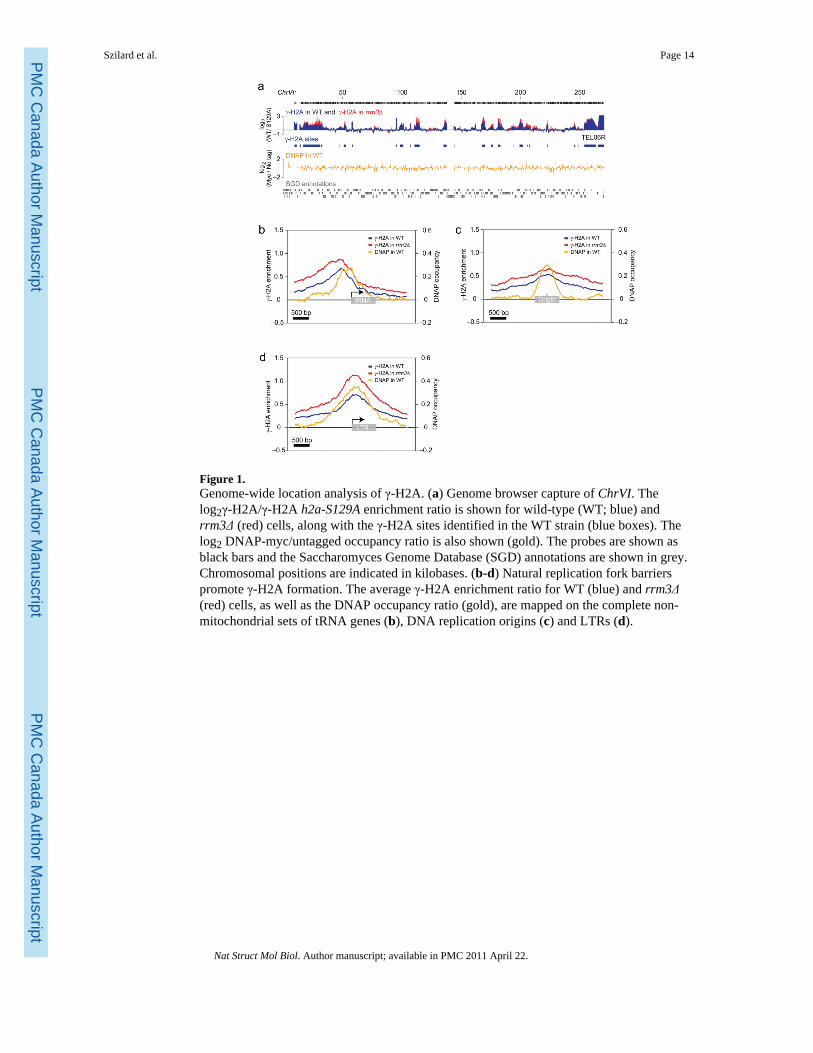
Figure 1.Genome-wide location analysis of γ-H2A. (a) Genome browser capture of ChrVI. Thelog2γ-H2A/γ-H2A h2a-S129A enrichment ratio is shown for wild-type (WT; blue) andrrm3Δ (red) cells, along with the γ-H2A sites identified in the WT strain (blue boxes). Thelog2 DNAP-myc/untagged occupancy ratio is also shown (gold). The probes are shown asblack bars and the Saccharomyces Genome Database (SGD) annotations are shown in grey.Chromosomal positions are indicated in kilobases. (b-d) Natural replication fork barrierspromote γ-H2A formation. The average γ-H2A enrichment ratio for WT (blue) and rrm3Δ(red) cells, as well as the DNAP occupancy ratio (gold), are mapped on the complete non-mitochondrial sets of tRNA genes (b), DNA replication origins (c) and LTRs (d).
Szilard et al. Page 14
Nat Struct Mol Biol. Author manuscript; available in PMC 2011 April 22.
PMC
Canada Author M
anuscriptPM
C C
anada Author Manuscript
PMC
Canada Author M
anuscript
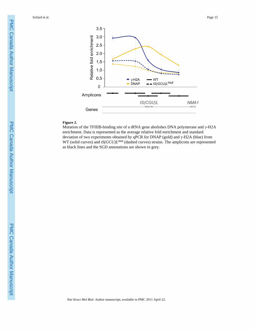
Figure 2.Mutation of the TFIIIB-binding site of a tRNA gene abolishes DNA polymerase and γ-H2Aenrichment. Data is represented as the average relative fold enrichment and standarddeviation of two experiments obtained by qPCR for DNAP (gold) and γ-H2A (blue) fromWT (solid curves) and tS(GCU)Lmut (dashed curves) strains. The amplicons are representedas black lines and the SGD annotations are shown in grey.
Szilard et al. Page 15
Nat Struct Mol Biol. Author manuscript; available in PMC 2011 April 22.
PMC
Canada Author M
anuscriptPM
C C
anada Author Manuscript
PMC
Canada Author M
anuscript
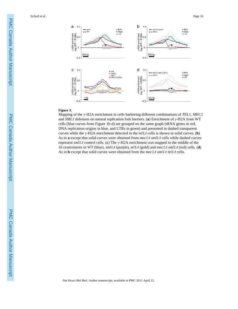
Figure 3.Mapping of the γ-H2A enrichment in cells harboring different combinations of TEL1, MEC1and SML1 deletions on natural replication fork barriers. (a) Enrichment of γ-H2A from WTcells (blue curves from Figure 1b-d) are grouped on the same graph (tRNA genes in red,DNA replication origins in blue, and LTRs in green) and presented in dashed transparentcurves while the γ-H2A enrichment detected in the tel1Δ cells is shown in solid curves. (b)As in a except that solid curves were obtained from mec1Δ sml1Δ cells while dashed curvesrepresent sml1Δ control cells. (c) The γ-H2A enrichment was mapped in the middle of the16 centromeres in WT (blue), sml1Δ (purple), tel1Δ (gold) and mec1Δ sml1Δ (red) cells. (d)As in b except that solid curves were obtained from the mec1Δ sml1Δ tel1Δ cells.
Szilard et al. Page 16
Nat Struct Mol Biol. Author manuscript; available in PMC 2011 April 22.
PMC
Canada Author M
anuscriptPM
C C
anada Author Manuscript
PMC
Canada Author M
anuscript
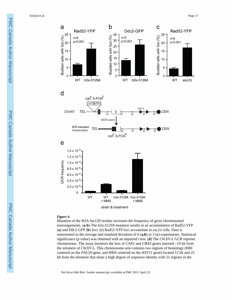
Figure 4.Mutation of the H2A Ser129 residue increases the frequency of gross chromosomalrearrangements. (a-b) The h2a-S129A mutation results in an accumulation of Rad52-YFP(a) and Ddc2-GFP (b) foci. (c) Rad52-YFP foci accumulate in esc2Δ cells. Data isrepresented as the average and standard deviation of 6 (a,b) or 5 (c) experiments. Statisticalsignificance (p value) was obtained with an unpaired t test. (d) The ChrXV-L GCR reporterchromosome. The assay monitors the loss of CAN1 and URA3 genes inserted ~10 kb fromthe telomere of ChrXV-L. This chromosome arm contains two regions of homology (HRIcentered on the PAU20 gene, and HRII centered on the HXT11 gene) located 12 kb and 25kb from the telomere that share a high degree of sequence identity with 21 regions in the
Szilard et al. Page 17
Nat Struct Mol Biol. Author manuscript; available in PMC 2011 April 22.
PMC
Canada Author M
anuscriptPM
C C
anada Author Manuscript
PMC
Canada Author M
anuscript
genome40. Consequently, DNA lesions formed at loci telomeric to HRI or HRII arepredominantly repaired by BIR, thereby converting a canavanine- and 5-fluoroorotic acid(5-FOA)-sensitive strain (canS FOAS) into a canavanine and 5-FOA-resistant strain (canR 5-FOAR). (e) The h2a-S129A mutation results in an increased frequency of drug-induced GCRevents. Cells carrying the ChrXV-L GCR reporter chromosome were grown in the presenceor absence of MMS and assayed for survival on media containing 5-FOA and canavanine.Data is represented as the average frequency of GCR events and standard error of the meanof 6 experiments.
Szilard et al. Page 18
Nat Struct Mol Biol. Author manuscript; available in PMC 2011 April 22.
PMC
Canada Author M
anuscriptPM
C C
anada Author Manuscript
PMC
Canada Author M
anuscript
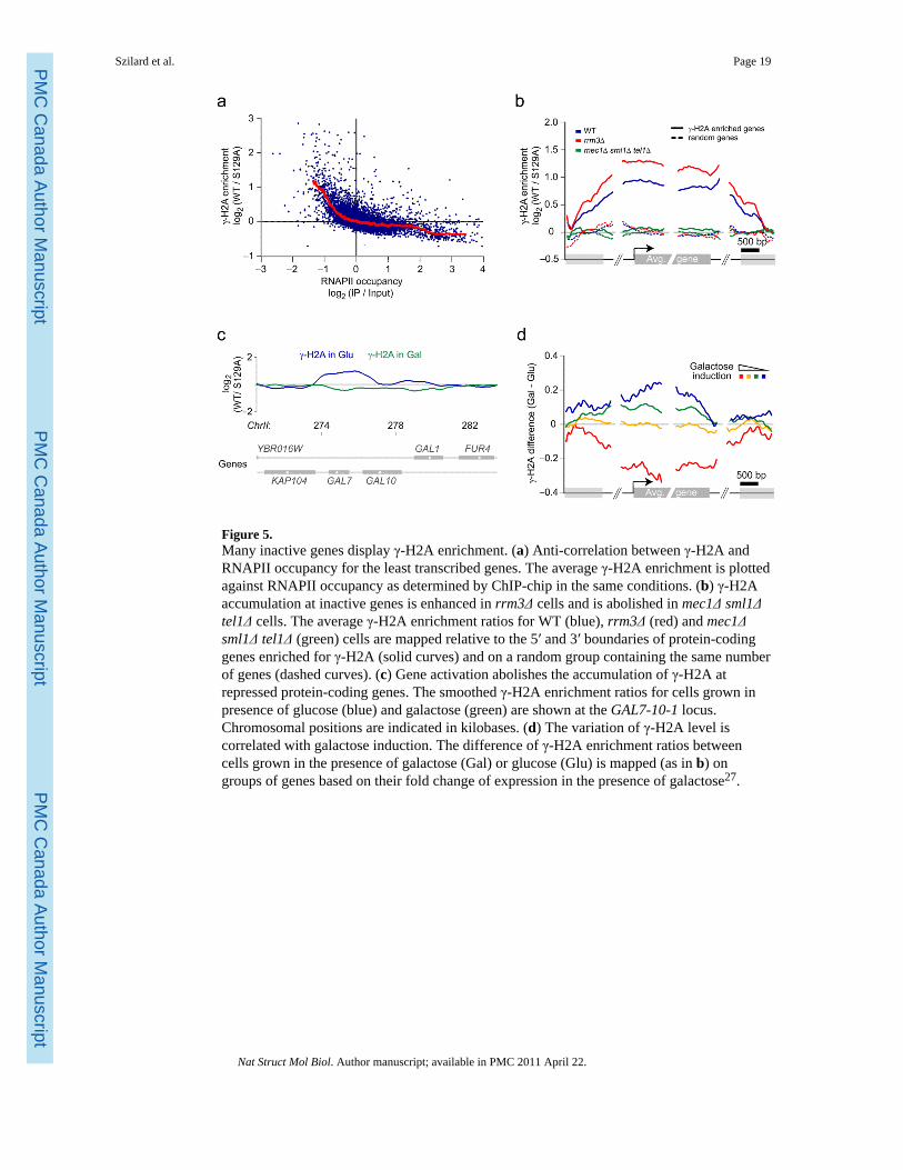
Figure 5.Many inactive genes display γ-H2A enrichment. (a) Anti-correlation between γ-H2A andRNAPII occupancy for the least transcribed genes. The average γ-H2A enrichment is plottedagainst RNAPII occupancy as determined by ChIP-chip in the same conditions. (b) γ-H2Aaccumulation at inactive genes is enhanced in rrm3Δ cells and is abolished in mec1Δ sml1Δtel1Δ cells. The average γ-H2A enrichment ratios for WT (blue), rrm3Δ (red) and mec1Δsml1Δ tel1Δ (green) cells are mapped relative to the 5′ and 3′ boundaries of protein-codinggenes enriched for γ-H2A (solid curves) and on a random group containing the same numberof genes (dashed curves). (c) Gene activation abolishes the accumulation of γ-H2A atrepressed protein-coding genes. The smoothed γ-H2A enrichment ratios for cells grown inpresence of glucose (blue) and galactose (green) are shown at the GAL7-10-1 locus.Chromosomal positions are indicated in kilobases. (d) The variation of γ-H2A level iscorrelated with galactose induction. The difference of γ-H2A enrichment ratios betweencells grown in the presence of galactose (Gal) or glucose (Glu) is mapped (as in b) ongroups of genes based on their fold change of expression in the presence of galactose27.
Szilard et al. Page 19
Nat Struct Mol Biol. Author manuscript; available in PMC 2011 April 22.
PMC
Canada Author M
anuscriptPM
C C
anada Author Manuscript
PMC
Canada Author M
anuscript
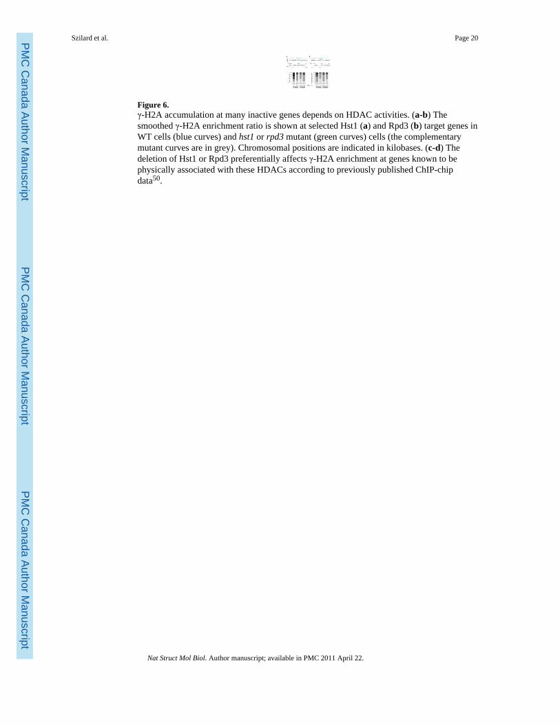
Figure 6.γ-H2A accumulation at many inactive genes depends on HDAC activities. (a-b) Thesmoothed γ-H2A enrichment ratio is shown at selected Hst1 (a) and Rpd3 (b) target genes inWT cells (blue curves) and hst1 or rpd3 mutant (green curves) cells (the complementarymutant curves are in grey). Chromosomal positions are indicated in kilobases. (c-d) Thedeletion of Hst1 or Rpd3 preferentially affects γ-H2A enrichment at genes known to bephysically associated with these HDACs according to previously published ChIP-chipdata50.
Szilard et al. Page 20
Nat Struct Mol Biol. Author manuscript; available in PMC 2011 April 22.
PMC
Canada Author M
anuscriptPM
C C
anada Author Manuscript
PMC
Canada Author M
anuscript
Copyright © 2022 FDOKUMEN