Methyl jasmonate decreases membrane fluidity and induces apoptosis through tumor necrosis factor...
20
Methyl jasmonate decreases membrane fluidity and induces apoptosis via tumor necrosis factor receptor 1 in breast cancer cells Laxmi Yeruva a,b , J. Abiodun Elegbede a , and Stephen W. Carper a a Chemistry Department, University of Nevada, Las Vegas 4505 Maryland Parkway, Las Vegas, NV 89154-4003 Abstract In recent years, studies with plant compounds have shown both chemotherapeutic and chemopreventive properties. The current study with plant stress hormones (jasmonates) showed growth inhibitory effects in breast cancer cells. Cis-jasmone (CJ) and methyl jasmonate (MJ) inhibited the long-term proliferation of MDA-MB-435 and MCF-7 cells. Cell cycle analysis showed G0/G1 and S-phase arrest with increasing apoptotic population. Cellular signaling studies with MJ showed decreased membrane fluidity and activation of extrinsic and intrinsic apoptotic pathways. Specifically in extrinsic apoptotic pathway increased expression of TNFR1, activation of MAPK and caspase-8 was observed. MJ also decreased the mitochondrial membrane potential and activated caspase-3 in breast cancer cells. In conclusion our results revealed novel signaling mechanism of MJ in breast cancer cells, indicating that MJ could have potential applications for chemotherapeutic purposes. Keywords Plant compounds; caspase-8; MAPK; Membrane fluidity; Breast Cancer Introduction Breast cancer is the second leading cause of cancer deaths among women in the US [1]. An estimated 40,910 breast cancer deaths and 180, 510 new cases are anticipated among women in 2007 [1]. Chemotherapy, radiation, surgery and immunotherapy are among the current treatment options for breast cancer. Chemotherapy using synthetic compounds, although shown to be effective in cancer treatment, also induces severe side effects due to their toxicity in noncancerous tissues. In recent years, studies have shown that plant compounds have chemotherapeutic and pharmacological activities against many types of cancers and could be utilized as alternative chemotherapeutic agents with low toxicity [2-7]. Jasmonates consists of cyclopentenone moiety, are synthesized in plants in response to injury, insect attack and wounding [8]. Several reports have demonstrated that cyclopentenone moiety containing molecules such as prostaglandins and clavulones induce apoptosis in different signaling mechanisms [9]. Cis-jasmone (CJ) and methyljasmonate (MJ) belong to the jasmonte family of compounds. Jasmonates have been reported to inhibit the growth of leukemia cells, lung, breast and prostate cancer cells [10]. To date, three separate mechanisms have been bFor Correspondence: Venkat Laxmi Yeruva, Biochemistry Department University of Arkansas for Medical Sciences Little Rock AR 72205, Telephone:501-686-7254 FAX: 501-686-8169, Email: [email protected], [email protected]. NIH Public Access Author Manuscript Anticancer Drugs. Author manuscript; available in PMC 2009 September 17. Published in final edited form as: Anticancer Drugs. 2008 September ; 19(8): 766–776. doi:10.1097/CAD.0b013e32830b5894. NIH-PA Author Manuscript NIH-PA Author Manuscript NIH-PA Author Manuscript
Transcript of Methyl jasmonate decreases membrane fluidity and induces apoptosis through tumor necrosis factor...

Methyl jasmonate decreases membrane fluidity and inducesapoptosis via tumor necrosis factor receptor 1 in breast cancercells
Laxmi Yeruvaa,b, J. Abiodun Elegbedea, and Stephen W. CarperaaChemistry Department, University of Nevada, Las Vegas 4505 Maryland Parkway, Las Vegas, NV89154-4003
AbstractIn recent years, studies with plant compounds have shown both chemotherapeutic andchemopreventive properties. The current study with plant stress hormones (jasmonates) showedgrowth inhibitory effects in breast cancer cells. Cis-jasmone (CJ) and methyl jasmonate (MJ)inhibited the long-term proliferation of MDA-MB-435 and MCF-7 cells. Cell cycle analysis showedG0/G1 and S-phase arrest with increasing apoptotic population. Cellular signaling studies with MJshowed decreased membrane fluidity and activation of extrinsic and intrinsic apoptotic pathways.Specifically in extrinsic apoptotic pathway increased expression of TNFR1, activation of MAPK andcaspase-8 was observed. MJ also decreased the mitochondrial membrane potential and activatedcaspase-3 in breast cancer cells. In conclusion our results revealed novel signaling mechanism of MJin breast cancer cells, indicating that MJ could have potential applications for chemotherapeuticpurposes.
KeywordsPlant compounds; caspase-8; MAPK; Membrane fluidity; Breast Cancer
IntroductionBreast cancer is the second leading cause of cancer deaths among women in the US [1]. Anestimated 40,910 breast cancer deaths and 180, 510 new cases are anticipated among womenin 2007 [1]. Chemotherapy, radiation, surgery and immunotherapy are among the currenttreatment options for breast cancer. Chemotherapy using synthetic compounds, althoughshown to be effective in cancer treatment, also induces severe side effects due to their toxicityin noncancerous tissues. In recent years, studies have shown that plant compounds havechemotherapeutic and pharmacological activities against many types of cancers and could beutilized as alternative chemotherapeutic agents with low toxicity [2-7].
Jasmonates consists of cyclopentenone moiety, are synthesized in plants in response to injury,insect attack and wounding [8]. Several reports have demonstrated that cyclopentenone moietycontaining molecules such as prostaglandins and clavulones induce apoptosis in differentsignaling mechanisms [9]. Cis-jasmone (CJ) and methyljasmonate (MJ) belong to the jasmontefamily of compounds. Jasmonates have been reported to inhibit the growth of leukemia cells,lung, breast and prostate cancer cells [10]. To date, three separate mechanisms have been
bFor Correspondence: Venkat Laxmi Yeruva, Biochemistry Department University of Arkansas for Medical Sciences Little Rock AR72205, Telephone:501-686-7254 FAX: 501-686-8169, Email: [email protected], [email protected].
NIH Public AccessAuthor ManuscriptAnticancer Drugs. Author manuscript; available in PMC 2009 September 17.
Published in final edited form as:Anticancer Drugs. 2008 September ; 19(8): 766–776. doi:10.1097/CAD.0b013e32830b5894.
NIH
-PA Author Manuscript
NIH
-PA Author Manuscript
NIH
-PA Author Manuscript
proposed to explain the anticancer effects of jasmonates. These include: (i) the ability of MJto open the mitochondrial permeability transition pore complex, resulting in the release ofcytochrome c, and induction of apoptosis; (ii) the ability of MJ to induce the re-differentiationof leukemia cells; and finally (iii) the ability of MJ to induce the expression of reactive oxygenspecies [10-12].
Apoptosis is a cellular suicide mechanism that occurs by extrinsic or intrinsic mechanisms[13]. The extrinsic apoptotic pathway involves a superfamily of death receptor ligands such astumor necrosis factor alpha (TNF- α, TNF-related apoptosis inducing ligand (TRAIL) and FAS(CD95/APO-1) (14). In breast cancer cells, TNF-α plays an important role in cellular responses,including inflammation and apoptosis [15-17]. TNF–α exerts its biological functionality bybinding two membrane receptors, tumor necrosis factor receptor 1 (TNFR1) and tumor necrosisfactor receptor 2 (TNFR2) [16]. The majority of TNF signaling pathways are attributable toTNFR1, which can bind both membrane bound and soluble TNF whereas TNFR2 can only beactivated by membrane bound TNF [16]. Activation of these receptors recruits FADD (Fasassociated death domain) and TRADD (TNF receptor-associated death domain) which resultsin the activation of caspase-8, mitogen activated protein kinase (MAPK) and ultimately celldeath [16]. Several studies have reported that both caspase-8 and MAPK can either directlyactivate caspase-3 via Bid or activate the intrinsic apoptotic pathway [16,18].
The MAPK pathway is part of the extrinsic apoptotic mechanism and plays an important rolein regulating a number of downstream molecules including kinases and scaffold proteins. Thebalance between these molecules exerts cellular responses including cell proliferation, cellcycle arrest, migration, differentiation and apoptosis. Studies conducted by Takenaka et al.indicated that the p38-MAPK pathway was activated during mitotic arrest in mammaliancultured cells [19-27]. Bulavin et al. also reported that p38α-MAPK was activated in responseto UV radiation and induced G2/M cell cycle arrest by decreasing CDC2 activity [22]. CDC2can phosphorylate BAD and induce neuronal apoptosis [23]. Additionally, it has been reportedthat ERK1 and ERK2 kinases are activated by phoshorylation in reponse to cell death inducedby external stressors [24-27]. Activation of MAPK results in signal transduction to intrinsicapoptotic proteins which ultimately results in cell death.
Activation of the intrinsic apoptotic pathway can occur either by caspase-8/caspase-2 mediatedBid cleavage or by direct stress to the mitochondria. Once the intrinsic apoptotic pathway hasbeen activated the mitochondrial membrane potential decreases and cytochrome c is released[28-30]. Cytochrome c can form an apoptosome complex with apaf-1 (apoptotic proteaseactivating factor-1) and procaspase-9. The apoptosome complex then activates caspase-3,caspase-6 and caspase-7 which results cell death [31].
The effects of CJ and MJ on breast cancer cells were studied using cytotoxicity, proliferation,DNA content analyses, apoptotic assays, ELISA and western blotting techniques. This studyshowed that both CJ and MJ inhibited the growth of breast cancer cells. Cell cycle analysisand apoptotic assays showed that CJ and MJ induced cell cycle arrest and apoptosis in breastcancer cell lines. Studies on membrane fluidity demonstrated that MJ decreased membranefluidity, probably activates apoptotic pathways. Specifically, our study revealed that MJincreased the expression of TNFR1 and further activated extrinsic apoptotic proteins,caspase-8, and MAPK. Intrinsic apoptotic mechanism showed that MJ decreasedmitochondrial membrane potential and activated caspase-3 in breast cancer cells. The resultsindicated that MJ induced apoptosis was associated with a decrease in membrane fluidity andintracellular signaling via TNFR1, MAPK, caspases-8, and caspase-3.
Yeruva et al. Page 2
Anticancer Drugs. Author manuscript; available in PMC 2009 September 17.
NIH
-PA Author Manuscript
NIH
-PA Author Manuscript
NIH
-PA Author Manuscript
Materials and methodsChemicals and reagents
Cell culture media (MEM), fetal bovine serum (FBS) and penicillin (1000 units/ml) andtreptomycin (1000 μg/ml) (P/S) were purchased from GIBCO (Grand Island, NY). Cis-jasmone(CJ), methyl jasmonate (MJ), protease inhibitors, propidium iodide (PI), deoxycholic acid(DCA), 1, 6-biphenyl-1, 2, 6-trihydrazine (DPH) and ribonuclease were obtained from SigmaChemical Company (St. Louis, MO). Phosphate buffered saline (PBS, lacking Ca2+ andMg2+) was purchased from Invitrogen Corporation (Grand Island, NY). Sodiumdodecylsulfate, bisacrylamide, N, N, N, N1 – tetra-methylethylenediamine (TEMED) andammonium persulfate were obtained from Bio-Rad Laboratories (Hercules, CA). Primaryantibody to caspase-3 was obtained from Pharmingen (Franklin Lakes, NJ) and caspase-8 wasfrom EMD Biosciences (San Diego, CA). Enzyme conjugated horse raddish peroxidase (HRP)secondary antibody and standards were obtained from Santa Cruz Biotechnology, Inc. (SantaCruz, CA). ECL+ western blotting detection system was obtained from Amersham Biosciences(Piscataway, NJ). All other reagents and chemicals were of the purest grade and were obtainedfrom reputable vendors.
Cell cultureBreast cancer cell lines (MDA-MB-435 and MCF7) were obtained from ATCC. All cultureswere maintained in Minimum Essential Media (MEM) supplemented with 10% FBS, 1%pencillin and streptomycin (1000units/ml) and HEPES buffer. All the cultures were maintainedat 37°C in humidified 95% O2 /5% CO2 atmosphere. During exposure to the jasmonatecompounds, the medium was replaced with treatment medium and control cells receivedmedium containing DMSO (0.1%-0.3%).
Cytotoxicity assayCells (~5000) were plated in 96-well plate and incubated overnight to allow adherence to theplate surface. Cells were exposed to varying concentrations of CJ or MJ for 24 hr. Attermination, the medium was replaced with 100μl of fresh medium; 10μl of alamarBlue wasadded and incubated for 4 hr. After 4 hr fluorescence was measured at 530 nm of excitationand 590 nm of emission wavelengths with Genios fluorescence plate reader (PHENIX ResearchProducts, Hayward, CA). Cell viability was calculated as percentage of control (100%).
Colony formation assayThe long-term (delayed) cytotoxic effects of agents on the breast cancer cells were determinedusing the colony formation assay (CFA). The cells were exposed to 0.5, 1.0 and 2.0 mMconcentrations of the compounds for 24 hr. The treatment medium was poured off and the cellswere washed with 1X PBS, harvested with trypsin EDTA, counted and replated into 60mmdishes. Cells were incubated in humidified 95% O2 /5% CO2 atmosphere at 37°C for 11 days.The medium was decanted and cells were stained with crystal violet (0.5g/100ml in 95%ethanol). Colonies (>50 cells/colony) were counted and the percentage survival (% viability)was calculated relative to the control group (100%).
Cell cycle analysis with flow cytometryCells (~0.25×10⁶) were plated in 100mm tissue culture plates, incubated overnight to adhereto the plate. Cells were exposed to 0.5mM and 3mM CJ or MJ for 24hr and washed with 1XPBS. Fresh and complete medium was added and the cells were incubated for 24 hr. MDA-MB-435 cells were also exposed to 500 μM of DCA for 4 hr, 8 hr and 24 hr. Cells were harvestedwith trypsin EDTA, washed with PBS, counted, fixed with 100μl of PBS and 900μl of absoluteethanol and stored at 4°C prior to DNA content analyses. Samples were stained with DNA
Yeruva et al. Page 3
Anticancer Drugs. Author manuscript; available in PMC 2009 September 17.
NIH
-PA Author Manuscript
NIH
-PA Author Manuscript
NIH
-PA Author Manuscript
staining solution containing 150μg/ml of propidium iodide (PI), 0.1% Triton x-100 and 1mg/ml of RNase-A (DNase free) (1:1:1 by volume) and incubated in the absence of light for onehour. Acquisition of the data was carried out with FACSCalibur flow cytometer (BectonDickinson, Sanjose, CA) and analyzed with ModfitLT 3.0 software (Verity Software House,Topsham, ME).
Detection of apoptosis by Hoechst and DNA fragmentationCells (~0.01×10⁶) were plated in 4-well slides and incubated for 24 hr prior to treatment. Cellswere exposed to 3.0mM CJ or MJ for 24 hr, at termination the medium was removed byaspiration and the cells were then stained with Hoechst (2μg/ml Hoechst) for 15 min.Morphological changes indicative of apoptosis were observed and the images acquired withfluorescence microscopy (Nikon Instruments Inc. Melville, NY). The number of cells in thebright field and Hoechst stained cells were counted and the percentage of apoptotic cells wascalculated as:
Cells (~1×10⁶) were seeded in 75mm tissue culture flasks and treated with 1, 2 or 3mM MJfor 24 hr. Cells treated with DCA (500 μM) for 4 hr and 8 hr were used as positive controls.At termination cells were harvested with trypsin EDTA, washed with 1X PBS and the DNAwas extracted as described in an earlier study (32). Briefly, cells (~1× 10⁶) were suspended in100μl of lysis buffer containing 10 mM Tris-HCl (pH 7.4), 10 mM EDTA, and 0.5% TritonX- 100 for 10 min at 4° C. Cells were centrifuged at 13,000 rpm for 5 min and RNase A(20μg/ml) was added to the supernatant and incubated for 1 hr at 37° C. Following incubation,Proteinase K was added to a final concentration of 20μg/ml and incubated for 30 min at 50°C. DNA was precipitated with 42mM NaCl and 50% isoproponal overnight at -20° C. TheDNA pellet was obtained by centrifugation for 15 min at 13, 000 rpm. The DNA pellet wasresuspended in 20μl TE buffer (10mM Tris-HCl and 1mM EDTA pH 8.0) and 10μl of the DNAsolution was electrophoresed on a 2% agarose gel in TBE buffer (89 mM Tris base, 89mMboric acid and 2.6 mM EDTA). The gel was stained with 1μg/ml ethidium bromide andphotographed.
Membrane fluidity studiesCells (~0.5 × 10⁶) were plated in 75mm tissue culture flasks and incubated for two days. Cellswere exposed to 3mM MJ, 10μM cholesterol, 500uM DCA for 4 hr. At termination cells wereharvested and washed with 1X PBS. The fluorescent dye, DPH (2μM), was added andpolarization was measured with LS 55 Luminescence Spectrometer (Perkin Elmer). The Pvalue for the study was calculated as follows:
TNFR1 ExpressionTNFR1 expression was measured by flow cytometry as described previously [33]. MDA-MB-435 and MCF-7 cells were plated in 75mm tissue culture flasks, incubated for two daysthen exposed to 0, 1mM, 2mM or 3mM MJ for 24 hr. Following exposure, the cells wereharvested with trypsin-EDTA (1mM), counted and ~1× 10⁶ cells were washed in 50% FBS at4°C for 15 min. Cells were washed with PBS-FBS (PBS with 1% FBS added) and incubated
Yeruva et al. Page 4
Anticancer Drugs. Author manuscript; available in PMC 2009 September 17.
NIH
-PA Author Manuscript
NIH
-PA Author Manuscript
NIH
-PA Author Manuscript
with 25μg/ml anti-human TNFR1 antibodies (R&D Systems) at 4°C for 60 min. Subsequently,cells were washed three times in PBS-FBS and incubated with FITC- conjugated goat anti-ratIgG (1:40 dilution) at 4°C for 2 hr. Cells were washed in PBS-FBS three times and analyzedwith FACS Calibur flow cytometer. Fluorescence data was collected and backgroundfluorescence was determined using unstained cells.
TNFR1 activation was confirmed by treating the cells with MJ in the presence of antibody.MDA-MB-435 cells (~5000/well) were plated in each well of a 96-well plate. After overnightincubation, the cells were exposed to 0, 3mM MJ, 3mM MJ with TNFR1 antibody, TNFR1antibody alone, or a nonspecific antibody for 24 hr. At termination the medium was removedby aspiration. Fresh medium and alamarBlue were then added and the cells were incubated for4 hr. Fluorescence was measured using Genios plate reader and the percent viability wascalculated via comparison to the control (100%).
MAPK assay by ELISAThe p38 and ERK1/2 activity was determined by an ELISA assay kit (Super array, FrederickMD). Cells (~1.5 × 10⁴) were plated in a 96-well plate and incubated overnight in humidified95% O2 / 5% CO2 atmosphere at 37°C. The following day, cells were serum starved for 18-24hr, exposed to MJ and assayed. Briefly, adherent cells were fixed to the plate, washed, blockedand incubated with a primary antibody followed by a secondary antibody. Developing solutionwas then added and the samples were allowed to incubate for 10 min at room temperaturebefore the stop solution was added. Absorbance was measured at 450 nm. Relativephosphorylation was normalized to cell number according to manufacturer’s instructions.
Caspase-8, 3, and membrane potential studiesThe expression of caspase-3 and caspase-8 was determined using western blot analysis. Controlor treated cells (~1.0×10⁶) were harvested with trypsin EDTA, pelleted in microfuge tubes at1000 rpm for 10 min and stored at -80°C and protein extraction was carried out according tothe manfacturer’s instructions (Biosource Intl. Inc., Camarillo, CA). The cell pellet was thawedon ice for 2 hr prior to protein extraction. PMSF (1mM) and 100μl of extraction buffercontaining protienase inhibitor (250μl/5ml) was added to ~1×10⁶ cells, while mixing every 10min for 30 min. The cell lysate was centrifuged at 13,000 rpm at 4°C for 10 min; supernatantwas collected, aliquoted into tubes and stored at -80°C. Total protein concentration wasdetermined using BCA protein assay kit (Pierce, Rockford, IL). Equal amount of protein wasloaded, subjected to SDS-PAGE and transferred to PVDF membrane. The membrane wasblocked for 1 hr at room temperature with blocking solution (10% nonfat dry milk solution +0.05% tween 20%) and washed twice with 1X western wash (12.4 mM trizma, 37.4 mM NaCl,0.5% TritonX-100, pH 7.4),. The blot was then incubated with primary antibodies (1μg/ml) tocaspase-3 or caspase-8 for 1 hr at room temperature followed by washing three times with 1Xwestern wash. Primary antibody was detected with 1:1000 diluted horse radish peroxidase(HRP) or fluorescenct secondary antibodies. HRP antibody signal was developed with ECL+detection kit (Amersham Biosciences, Piscataway, NJ) and visualized with TYPHOON 9410(Amersham Biosciences, Piscataway, NJ).
Caspase-3 activity was measured by flow cytometry as described in manufacturer’s protocol(Biovision, CA). Briefly, cells were exposed to 3mM MJ for 24 hr, harvested, counted andwashed with PBS. Caspase-3 substrate was added and washed with wash buffer (provided bythe manufacturer) and activity was measured by flow cytometry. Caspase-8 activity wasmeasured by fluoremetric assay as described in manfacturer’s protocol (Sigma ChemicalCompany St. Louis, MO). Cells were exposed to 3mM MJ for 0, 2 or 4 hr, harvested, and thecell pellet was collected. Caspase-8 substrate was added and fluorescence was measured atexcitation wavelength of 360 nm and emission wavelength of 440 nm.
Yeruva et al. Page 5
Anticancer Drugs. Author manuscript; available in PMC 2009 September 17.
NIH
-PA Author Manuscript
NIH
-PA Author Manuscript
NIH
-PA Author Manuscript

Mitochondrial membrane potential was measured as described in the manufacturer’s protocol(Cell technology, CA). Mitochondrial membrane potential detection kit, uses a fluorescentcationic dye, JC-1 (5,5’, 6,6’-tetrachloro-1,1’,3,3’ tetraethylbenzimidazolylcarbo-cyanineiodide) to signal the loss of mitochondrial membrane potential. In non-apoptotic cells, the dyeaccumulates in the mitochondrial matrix stains the mitochondria bright red and in apoptoticcells, due to mitochondrial membrane potential collapse JC-1 cannot accumulate with in themitochondria and hence it remains in the cytoplasm in a green fluorescent monomeric form.MDA-MB-435 and MCF-7 cells were plated in 25mm flasks and incubated overnight. Cellswere exposed to 0, IC50MJ (concentration that inhibits growth of 50% of cells) or 3mM MJfor 24 hr. At termination cells were harvested, counted, and washed with PBS. Cells (0.5 ×10⁶) were stained with JC-1 reagent and incubated for 15 min at 37° C. Cells were washedtwice with assay buffer (provided by manufacturer) and membrane potential was measured byflow cytometry.
Statistical analysesThe results were presented as Mean ± SEM of replicate analyses accompanied by the numberof independent experiments. Statistical analyses were performed using one or two tailed t-test(Graphpad Software Inc., San Diego CA and Microsoft Excel). Differences at p<0.05 or betterwere considered statistically significant.
ResultsCJ and MJ inhibited the growth of breast cancer cells
Proliferation inhibition of breast cancer cells was determined by cytotoxicity assays usingalmarBlue dye. Plant compounds CJ and MJ inhibited the growth of breast cancer cell linesin-vitro. In MDA-MB-435 cells, the concentration of CJ at which 50% of growth (IC50) wasinhibited after a 24 hr treatment was 1.7 mM. The IC50 concentration was 2.5 mM for MCF-7cells (Figure 1A). The IC50 concentration of MJ for MDA-MB-435 cells was 1.9 mM and 2.0mM for MCF-7 cells (Figure 1B). CJ and MJ were not cytotoxic to CDC-1070-SK andMCF-10A normal cells (Figure 1C)
Long-term proliferation inhibition of breast cancer cells by CJ or MJ was evaluated byclonogenic survival assays. The concentration of CJ at which 50% of long-term proliferationof MDA-MB-435 and MCF-7 cells was inhibited (IC50) after 24 hr treatment was 1.2mM(Figure 2A). The IC50 of MJ for MDA-MB-435 cells was 1.2mM and 1.45mM for MCF-7cells (Figure 2B). A time course with MDA-MB-435 and MCF-7 cells at 3mM CJ or MJrevealed a time dependent decrease in survival (Figure 2C and 2D). At 2 hr, CJ inhibited thelong-term proliferation of 70% of MDA-MB-435 cells and 60% of MCF-7 cells (Figure 2C).MJ inhibited the long-term proliferation of 70% of both MDA-MB-435 and MCF-7 cells in-vitro (Figure 2D).
Cell cycle analysisTo investigate the ability of the jasmonate compounds to induce cell cycle arrest, DNA contentanalyses was carried out by propidium iodide staining using flow cytometry. Representativehistograms of MDA-MB-435 and MCF-7 cells treated with CJ and MJ showed an increase inthe apoptotic peak compared to the control (Figure 3A and 3C). CJ and MJ induced cell cyclearrest and apoptosis (Figure 3A-3D) as well as G0/G1 arrest in MDA-MB-435 and MCF-7 celllines (Figure 3B and 3D). At 3mM MJ, MDA-MB-435 cells showed a block at S-phase while3mM CJ showed an S-phase block in MCF-7 cells (Figure 3B and 3D). Apoptosis was observedonly with 3mM MJ in both MDA-MB-435 and MCF-7 cells (Figure 3B and 3D).
Yeruva et al. Page 6
Anticancer Drugs. Author manuscript; available in PMC 2009 September 17.
NIH
-PA Author Manuscript
NIH
-PA Author Manuscript
NIH
-PA Author Manuscript

Apoptotic studies have demonstrated that CJ and MJ induced apoptosis in breast cancer cells(Figure 4A and 4B). To confirm apoptosis induction, cells were stained with Hoechst andimages were acquired with a fluorescenct microscope. The percentage of apoptotic cells werecalculated as explained previously in the materials and methods section (Figure 4A). Cellstreated with CJ or MJ revealed morphological changes in the cell shape from a full circle to anelongated oval, a change that is consistent with cells undergoing apoptosis , as well as asignificant increase in the amount of apoptosis in breast cancer cells (Supplemental Figure 1).MDA-MB-435 and MCF-7 cells showed 35.0% and 37.2% apoptosis upon treatment with MJand CJ induced 6.6% and 43.5% apoptosis in MDA-MB-435 and MCF-7 cells, respectively(Figure 4A). DCA induced G2/M arrest and apoptosis in MDA-MB-435 cells (Figure 4B).
To further characterize the levels of apoptosis induced in breast cancer cells by MJ and CJ,DNA fragmentation studies were conducted. After a 24 hr treatment with MJ, DNA ladderingwas not observed in MCF-7 cells, but a pronounced DNA smear was observed in MDA-MB-435 cells (Figure 4C). The DNA smear was observed with 3mM MJ, but not with the 1or 2mM concentrations of MJ (Figure 4C). DNA laddering was also observed in MDA-MB-435cells treated with DCA for 4 and 8 hr which is an indicative of apoptosis (Figure 4C).
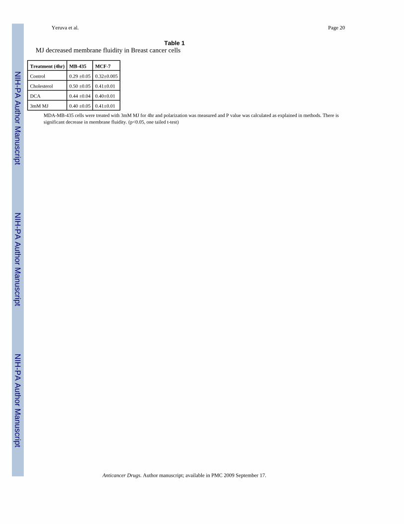
Membrane fluidityMJ is a hydrophobic molecule and therefore expected to decrease the membrane fluidity ofcells. Cells treated with cholesterol and DCA were used as positive controls. MDA-MB-435and MCF-7 cells were treated with 3mM MJ, DCA and cholesterol for 4 hr and fluidity decreasewas determined statistically where an increase in P value was indicative of decrease inmembrane fluidity. In both the cell lines decrease in membrane fluidity was observed comparedto the control (Table 1).
Cellular signalingStudies on the death receptors revealed that TNFR1 expression was increased and death signalswere initiated in breast cancer cells. MJ Induced TNFR1 expression was dose dependent inboth MD-MB-435 and MCF-7 cells (Figure 5A and 5B). Exposure of MDA-MB-435 cells toMJ in the presence of TNFR1 antibody showed increased cell viability which confirmedTNFR1 activation (Figure 5B). To confirm that MJ induced cell signaling through TNFR1binding, caspase-8 and MAPK activity were measured. Increased p38 and ERK1,2 activitywas observed after exposure to MJ treatment in MDA-MB-435 cells but not in MCF-7 cells(Figure 6). Activation of p38 was observed 2.0 hr after treatment and ERK1,2 activity wasobserved after 1.0 hr treatment with 3mM MJ in MDA-MB-435 but not in MCF-7 cells (Figure6A, 6B, 6C and 6D). Caspase-8 was activated following 2 hr treatment in MDA-MB-435 cellstreated with 3mM MJ (Figure 7A). Procaspase-8 was cleaved and activated until 8 hr in MDA-MB-435 cells but not in MCF-7 cells (Figure 7A, MCF-7 data not shown). Further confirmatorystudies indicated 5 fold increase in caspase-8 activity in MDA-MB-435 cells compared to thecontrol (Figure 7B)
To elucidate the involvement of mitochondria in response to MJ induced apoptosis,mitochondrial membrane potential and caspase-3 activity was measured. Cells treated with MJshowed a decrease in membrane potential and procaspase-3 activity compared to the controlexcept in caspase-3 mutant cell line MCF-7 (Figure 8A, 8B, and 8C). Flow cytometry analysisdemonstrated that caspase-3 activity was increased in MDA-MB-435 cell line, however, nochange in MCF-7 cells was observed (Figure 8B).
Yeruva et al. Page 7
Anticancer Drugs. Author manuscript; available in PMC 2009 September 17.
NIH
-PA Author Manuscript
NIH
-PA Author Manuscript
NIH
-PA Author Manuscript
Discussion and conclusionBreast cancer is the second leading cause of cancer related deaths among women in the US.Alternative strategies are required to combat the deaths caused by this disease. Jasmonates areplant compounds, which have been shown to inhibit growth and promote apoptosis in lung andprostate cancer and breast cancer cells. However, apoptotic mechanism of these compoundshas not been investigated in breast cancer cells. Our study demonstrated that jasmonates inhibitgrowth, induce cell cycle arrest and apoptosis in breast cancer cells. Apoptotic signalingmechanisms revealed that MJ increased the expression of TNFR1, activated caspase-8, p38,ERK1/2, and caspase-3 as well as decreased mitochondrial membrane potential in breast cancercells. To the best of our knowledge, this is the first report to demonstrate death receptor TNFR1expression, membrane fluidity decrease, caspase-8 activation in breast cancer cells.
Recent reports with MJ showed growth inhibition of lung cancer, breast cancer and leukemiacells [10]. It was reported that MJ induced apoptosis in human breast cancer cell line, MCF-7,and this study showed that CJ and MJ induced apoptosis in MDA-MB-435 and MCF-7 celllines. Cell cycle analysis revealed that CJ and MJ induced a block at G0/G1 in MDA-MB-435at all concentrations studied. In MCF-7 cells, CJ and MJ also induced a block at G0/G1 at allconcentrations studied above 0.5mM. At 0.5 mM, MJ and CJ induced a block in S-phase. InMCF-7 cells though DNA smear was not observed with 3mM MJ treatment, apoptosis wasobserved. These results indicate that the observed cell death was due to cell cycle arrest andapoptosis.
Earlier studies reported that the hydrophobic molecule DCA changes membrane fluidity. DCAdecreases membrane fluidity by rearranging cholesterol in the membrane which reducedmembrane permeability [34]. It has been reported that DCA induced apoptosis involvesactivation of caspase-2, caspase-3, caspase-7 and caspase-8 in the HT-29 colon cancer cell line[35,36]. In HCT-116 colon cancer cells, DCA activated p38 and ERK1/2. Studies usingpharmacological inhibitors specific to ERK1/2 revealed that elevated ERK1/2 activitysuppressed DCA induced apoptosis. Taken together, these data suggest that DCA can activatedifferent apoptotic proteins within different cell lines [35,36]. The mechanism for activationof apoptosis has not been proposed, however, activation of these molecules are likely the resultof the activation of receptor molecules (i. e., TNFR1) due to changes in membrane fluidity inthe presence of DCA. Given that MJ is a hydrophobic molecule like DCA, the plant compoundmay work similarly, by decreasing membrane fluidity which assembles receptors and leads toautophosphorylation and activation of death receptors. This study determined that MJdecreased membrane fluidity might have resulted in apoptotic signaling in cancer cells.
Previous report demonstrated that jasmonates affect the mitochondria, release cytochrome-cvia permeability transition pore complex in leukemia cells with out effecting normal cells[10,11]. Compared to normal cells, several cancer cells have shown high mitochondrialmembrane potential contributing to apoptosis resistance. Mitochondrial membrane potentialdecrease by dichloroacetate treatment induced apoptosis in cancer cells [37]. Similarly,jasmonate treatment showed a decrease in mitochondrial membrane potential and inducedapoptosis in breast cancer cells. In non cancerous cells jasmonates treatment showed nocytotoxic effect (mitochondrial assay) on normal cells (Figure 1C) and does not increaseTNFR1 expression (unpublished data). This indicates that jasmonates specifically act oncancerous cells and the observation is consistent with earlier report [10,11]. Our findings onbreast cancer cells extend the apoptotic mechanism of jasmonates observed.
Previous studies have demonstrated that jasmonates induce growth arrest and re-differentiationof leukemia cells in mitogen-activated kinase dependent pathway [11]. The results presentedhere showed activation of p38 and ERK1/2 in MDA-MB-435 cells but not in MCF-7 cells. MJ
Yeruva et al. Page 8
Anticancer Drugs. Author manuscript; available in PMC 2009 September 17.
NIH
-PA Author Manuscript
NIH
-PA Author Manuscript
NIH
-PA Author Manuscript

induced DNA fragmentation, hallmark of apoptosis in callus plant cells and we observed DNAfragmentation in MDA-MB-435 cells but not in MCF-7 cells. In MCF-7 cells lack of DNAfragmentation is possibly due to mutant caspase-3 and a different research group alsodemonstrated caspase-3 requirement in DNA fragmentation [38]. Hence in conjunction withthe data provided from previous studies suggest that jasmonates have a chemotherapeutic effectby inducing multiple apoptotic signaling pathways in cancer cells which involveTNFR1,caspase-8, capase-3, MAPK, bax/bclx/s, and cytochrome-c [9-12].
In conclusion, plant compounds induced apoptosis is probably associated with TNFR1expression. This study lends further insight into the apoptotic signaling mechanisms that areactivated by MJ which can be exploited for therapeutic purposes. MJ has shown to act on threedifferent types of cancer cells; hence it could be used for a wide variety of cancer treatments.To reduce the toxicity of conventional chemotherapy experienced by patients and to improvethe efficacy of treatments, future studies using a combination of MJ with otherchemotherapeutic agents need to be conducted.
AcknowledgmentsThe authors thank Shirly Shen and Casey Hall for their technical assistance and Becky Hess for her editing skills. Thisresearch was supported in part by ACS grant #IRG-103719 (to JAE) and in part by NIH Grant Number 2 P20 RR016464 from the INBRE Program of the National Center for Research Resources.
References1. American Cancer Society, Cancer Facts & Figures. American Cancer Society, Inc.; Atlanta, GA: 2007.2. Haag JD, Gould MN. Mammary carcinoma regression induced by perillyl alcohol, a hydroxylated
analog of limonene. Cancer Chemother Pharmacol 1994;34:477–483. [PubMed: 7923558]3. Yuri T, Danbara N, Tsujita-Kyutoku M, Kiyozuka Y, Senzaki H, Shikata N, et al. Perillyl alcohol
inhibits human breast cancer cell growth. Breast Cancer Res Treatment 2004;84:251–260.4. Bardon S, Foussard V, Fournel S, Loubat A. Monoterpenes inhibit proliferation of human colon cancer
cells by modulating cell cycle-related protein expression. Cancer Lett 2002;181:187–194. [PubMed:12175534]
5. Kallio A, Zheng A, Dahllund J, Heiskanen KM, Harkonen P. Role of mitochondria in tamoxifen-induced rapid death of MCF-7 breast cancer cells. Apoptosis 2005;10:1395–1410. [PubMed:16215679]
6. Yang HL, Chen CS, Chang WH, Lu FJ, Lai YC, Chen CC, et al. Growth inhibition and induction ofapoptosis in MCF-7 breast cancer cells by Antrodia camphorate. Cancer Lett 2006;231:215–227.[PubMed: 16399223]
7. Wu CC, Chan ML, Chen WY, Tsai CY, Chang FR, Wu YC. Pristimerin induces caspase-dependentapoptosis in MDA-MB-231 cells via direct effects on mitochondria. Mol Cancer Ther 2005;4:1277–1285. [PubMed: 16093444]
8. Farmer EE, Ryan CA. Interplant communication: Air-borne methyl jasmonate induces synthesis ofproteinases inhibitors in plant leaves. Proc Natl Acad Sci USA 1990;87:7713–7716. [PubMed:11607107]
9. Conti M. Cyclopentenone: a special moiety for anticancer drug design. Anticancer drugs2006;17:1017–1022. [PubMed: 17001173]
10. Flescher E. Jasmonates-a new family of anti-cancer agents. Anticancer drugs 2005;16:911–916.[PubMed: 16162967]
11. Rotem R, Heyfets A, Fingrut O, Blickstein D, Shaklai M, Flescher E. Jasmonates: novel anticanceragents acting directly and selectively on human cancer cell mitochondria. Cancer Res 2005;65:1984–1993. [PubMed: 15753398]
12. Kim JH, Lee SY, Oh SY, Han SI, Park HG, Yoo MA, et al. Methyl jasmonate induces apoptosisthrough induction of Bax/Bcl-XS and activation of caspase-3 via ROS production in A549 cells.Oncol Rep 2004;12(6):1233–1238. [PubMed: 15547743]
Yeruva et al. Page 9
Anticancer Drugs. Author manuscript; available in PMC 2009 September 17.
NIH
-PA Author Manuscript
NIH
-PA Author Manuscript
NIH
-PA Author Manuscript

13. Pinton P, Ferrari D, Di Virgilio F, Pozzan T, Rizzuto R. Molecular machinery and signaling eventsin apoptosis. Drug Dev Res 2001;52:558–570.
14. Ashkenazi A, Dixit VM. Death receptors: Signaling and modulation. Science 1998;281:1305–1308.[PubMed: 9721089]
15. Baud V, Karin M. Signal transduction by tumor necrosis factor and its relatives. Trends Cell Biol2001;11:372–377. [PubMed: 11514191]
16. Mathiasen IS, Hansen CM, Foghsgaard L, Jaatiela M. Sensitization to TNF-induced apoptosis by 1,25-dihydroxy vitamin D3 involves up-regulation of the TNF receptor 1 cathepsin. B Int J Cancer2001;93:224–231.
17. Jin Z, EI-diery WS. Overview of cell death signaling pathways. Cancer Biol Ther 2005;4:139–163.[PubMed: 15725726]
18. Lowe LC, Senaratne SG, Colston KW. Induction of apoptosis in breast cancer cells by apomine ismediated by caspase and p38 mitogen activated protein kinase activation. Biochem Biophys ResCommun 2005;329:772–779. [PubMed: 15737653]
19. Chang L, Karin M. Mammalian MAP kinase signaling cascades. Nature 2001;410:37–40. [PubMed:11242034]
20. Caffrey DR, O’Neill LA, Shields DC. The evolution of the MAP kinase pathways: coduplication ofinteracting proteins leads to new signaling cascades. J Mol Evol 1999;49:567–582. [PubMed:10552038]
21. Takenaka K, Moriguchi T, Nishida E. Activation of the protein kinase p38 in the spindle assemblycheckpoint and mitotic arrest. Science 1998;280:599–602. [PubMed: 9554853]
22. Bulavin DV, Higashimoto Y, Popoff IJ, Gaarde WA, Basrur V, Potapova O, et al. Initiation of a G2/M checkpoint after ultraviolet radiation requires p38 kinase. Nature 2001;411:102–107. [PubMed:11333986]
23. Konishi Y, Lehtinen M, Donovan N, Bonni A. Cdc2 phosphorylation of BAD links the cell cycle tothe cell death machinery. Mol Cell 2002;9:1005–1016. [PubMed: 12049737]
24. Seger R, Krebs EG. The MAPK signaling cascade. FASEB 1995;9:726–735.25. Johnson GL, Lapadat R. Mitogen-activated protein kinase pathways mediated by ERK, JNK, and p38
protein kinases. Science 2002;298:1911–1912. [PubMed: 12471242]26. Gille H, Kortenjann M, Thomae O, Moomaw C, Slaughter C, Cobb MH, et al. ERK phosphorylation
potentiates Elk-1-mediated ternary complex formation and transactivation. EMBO J 1995;14:951–962. [PubMed: 7889942]
27. Persons DL, Yazlovitskaya EM, Pelling JC. Effect of extracellular signal-regulated kinase on p53accumulation in response to cisplatin. J Biol Chem 2000;275:35778–35785. [PubMed: 10958792]
28. Desagher S, Osen-Sand A, Nichols A, Eskes R, Montessuit S, Lauper S, et al. Bid-inducedconformational change of bax is responsible for mitochondrial cytochrome c release during apoptosis.J Cell Biol 1999;144:891–901. [PubMed: 10085289]
29. Degli Esposti M, Dive C. Mitochondrial membrane permeabilisation by Bax/Bak. Biochem. BiophysRes Commun 2003;304:455–461.
30. Gross A, Jockel J, Wei MC, Korsmeyer SJ. Enforced dimerization of BAX results in its translocation,mitochondrial dysfunction and apoptosis. EMBO J 1998;17:3878–3885. [PubMed: 9670005]
31. Cain K, Bratton SB, Cohen GM. The Apaf-1 apoptosome: a large caspase-activating complex.Biochimie 2002;84:203–214. [PubMed: 12022951]
32. Yui S, Saeki T, Kanamoto R, Iwami K. Charesteristics of apoptosis in HCT116 colon cancer cellsinduced by deoxycholic acid. J Biochem 2005;28:151–157. [PubMed: 16091589]
33. Smolnikar K, Loffek S, Schulz T, Michna H, Diel P. Treatment with the pure antiestrogen faslodex(ICI182780) induces tumor necrosis factor receptor 1 (TNFR1) expression in MCF-7 breast cancercells. Breast Cancer Res Treat 2000;63:249–259. [PubMed: 11110059]
34. Jean-Louis S, Akare S, Ali MA, Mash EA Jr, Meuillet E, Martinez JD. Deoxycholic acid inducesintracellular signaling through membrane perturbations. J Biol Chem 2006;281:14948–14960.[PubMed: 16547009]
35. Qiao D, Stratagouleas ED, Martinez JD. Activation and role of mitogen-activated protein kinases indeoxycholic acid-induced apoptosis. Carcinogenesis 2001;22:35–41. [PubMed: 11159738]
Yeruva et al. Page 10
Anticancer Drugs. Author manuscript; available in PMC 2009 September 17.
NIH
-PA Author Manuscript
NIH
-PA Author Manuscript
NIH
-PA Author Manuscript

36. Schlottmann K, Wachs FP, Krieg RC, Kullmann F, Scholmerich J, Rogler G. Characterization of bilesalt-induced apoptosis in colon cancer cell lines. Cancer Res 2000;60:4270–4276. [PubMed:10945641]
37. Bonnet S, Archer SL, Allalunis-Turner J, Haromy A, Beaulieu C, Thompson R, et al. A mitochondria-K+ channel axis is suppressed in cancer and its normalization promotes apoptosis and inhibits cancergrowth. Cancer cell 2007;11:37–51. [PubMed: 17222789]
38. Jänicke RU, Engels IH, Dunkern T, Kaina B, Schulze-Osthoff K, Porter AG. Ionizing radiation butnot anticancer drugs causes cell cycle arrest and failure to activate the mitochondrial death pathwayin MCF-7 breast carcinoma cells. Oncogene 2001;20:5043–5053. [PubMed: 11526489]
Yeruva et al. Page 11
Anticancer Drugs. Author manuscript; available in PMC 2009 September 17.
NIH
-PA Author Manuscript
NIH
-PA Author Manuscript
NIH
-PA Author Manuscript
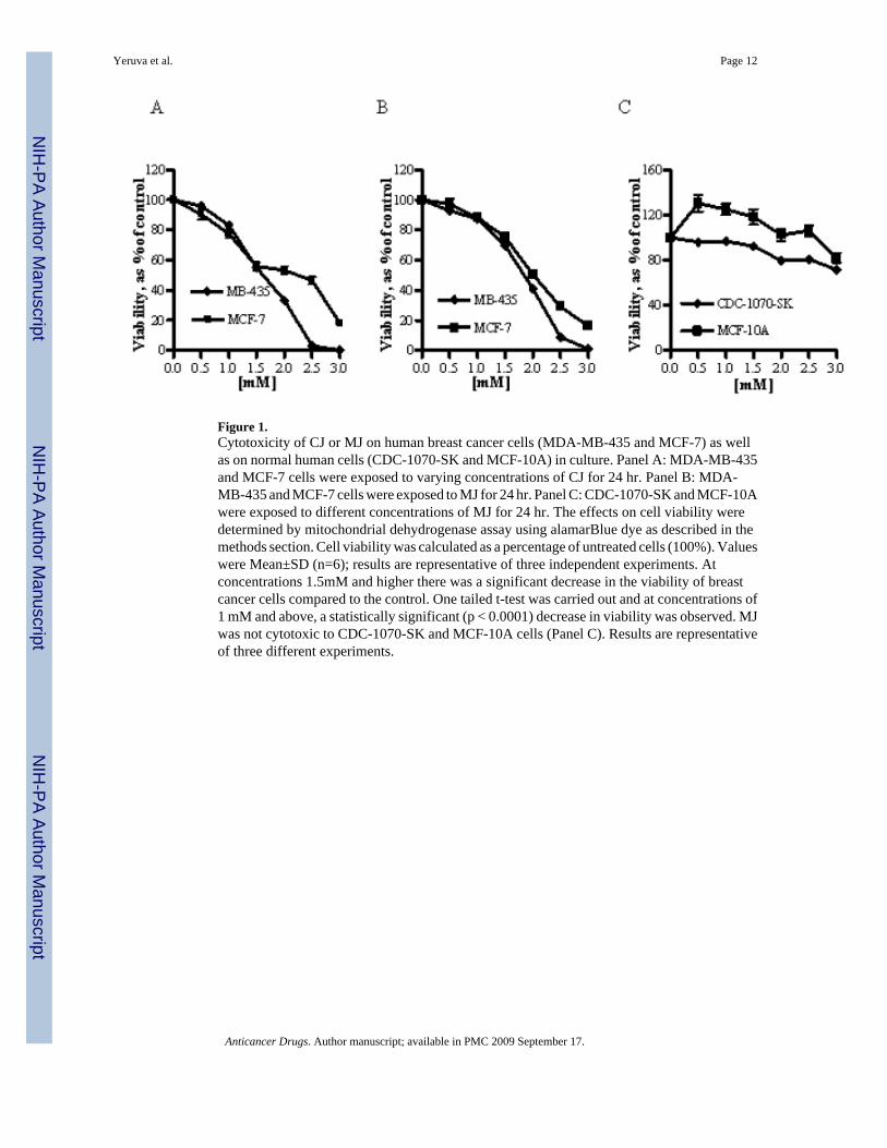
Figure 1.Cytotoxicity of CJ or MJ on human breast cancer cells (MDA-MB-435 and MCF-7) as wellas on normal human cells (CDC-1070-SK and MCF-10A) in culture. Panel A: MDA-MB-435and MCF-7 cells were exposed to varying concentrations of CJ for 24 hr. Panel B: MDA-MB-435 and MCF-7 cells were exposed to MJ for 24 hr. Panel C: CDC-1070-SK and MCF-10Awere exposed to different concentrations of MJ for 24 hr. The effects on cell viability weredetermined by mitochondrial dehydrogenase assay using alamarBlue dye as described in themethods section. Cell viability was calculated as a percentage of untreated cells (100%). Valueswere Mean±SD (n=6); results are representative of three independent experiments. Atconcentrations 1.5mM and higher there was a significant decrease in the viability of breastcancer cells compared to the control. One tailed t-test was carried out and at concentrations of1 mM and above, a statistically significant (p < 0.0001) decrease in viability was observed. MJwas not cytotoxic to CDC-1070-SK and MCF-10A cells (Panel C). Results are representativeof three different experiments.
Yeruva et al. Page 12
Anticancer Drugs. Author manuscript; available in PMC 2009 September 17.
NIH
-PA Author Manuscript
NIH
-PA Author Manuscript
NIH
-PA Author Manuscript
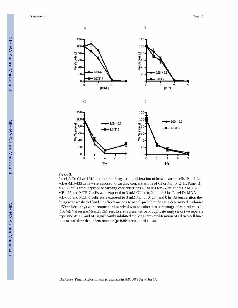
Figure 2.Panel A-D: CJ and MJ inhibited the long-term proliferation of breast cancer cells. Panel A:MDA-MB-435 cells were exposed to varying concentrations of CJ or MJ for 24hr. Panel B:MCF-7 cells were exposed to varying concentrations CJ or MJ for 24 hr. Panel C: MDA-MB-435 and MCF-7 cells were exposed to 3 mM CJ for 0, 2, 4 and 8 hr. Panel D: MDA-MB-435 and MCF-7 cells were exposed to 3 mM MJ for 0, 2, 4 and 8 hr. At termination thedrugs were washed off and the effects on long term cell proliferation were determined. Colonies(≥50 cells/colony) were counted and survival was calculated as percentage of control cells(100%). Values are Mean±SEM; results are representative of duplicate analyses of two separateexperiments. CJ and MJ significantly inhibited the long-term proliferation of all two cell linesin dose and time dependent manner (p<0.001; one tailed t-test).
Yeruva et al. Page 13
Anticancer Drugs. Author manuscript; available in PMC 2009 September 17.
NIH
-PA Author Manuscript
NIH
-PA Author Manuscript
NIH
-PA Author Manuscript
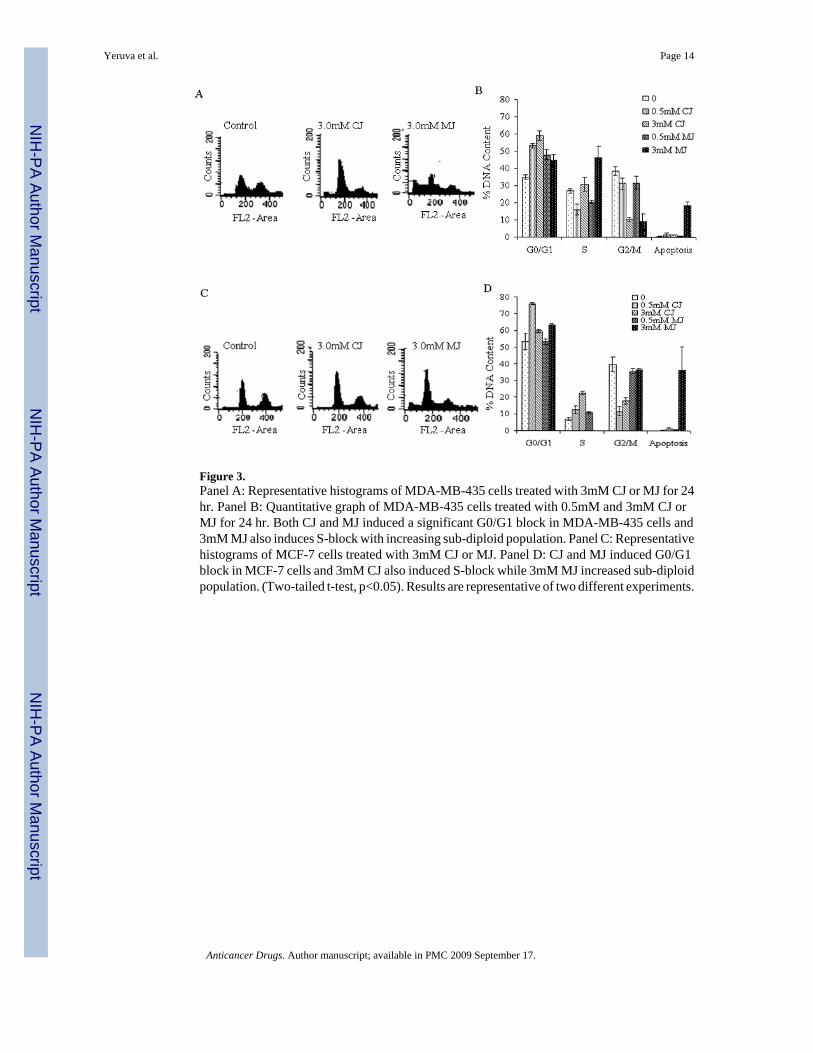
Figure 3.Panel A: Representative histograms of MDA-MB-435 cells treated with 3mM CJ or MJ for 24hr. Panel B: Quantitative graph of MDA-MB-435 cells treated with 0.5mM and 3mM CJ orMJ for 24 hr. Both CJ and MJ induced a significant G0/G1 block in MDA-MB-435 cells and3mM MJ also induces S-block with increasing sub-diploid population. Panel C: Representativehistograms of MCF-7 cells treated with 3mM CJ or MJ. Panel D: CJ and MJ induced G0/G1block in MCF-7 cells and 3mM CJ also induced S-block while 3mM MJ increased sub-diploidpopulation. (Two-tailed t-test, p<0.05). Results are representative of two different experiments.
Yeruva et al. Page 14
Anticancer Drugs. Author manuscript; available in PMC 2009 September 17.
NIH
-PA Author Manuscript
NIH
-PA Author Manuscript
NIH
-PA Author Manuscript

Figure 4.Panel A: To confirm apoptosis induction breast cancer cells were stained cells with Hoechstand fluorescence microscopy images were acquired. Percent apoptosis was calculated asexplained in the methods section. Both CJ and MJ induced apoptosis in breast cancer cells.Results are representative of two different experiments (p<0.01). Panel B: MDA-MB-435 cellswere treated with DCA (500μM) for 4 hr, 8 hr and 24 hr. DCA induced G2/M arrest at all threetime points measured and apoptosis at 24 hr. Panel C: DNA gel photograph of MDA-MB-435and MCF-7 cells treated with MJ for 24 hr. Lane 1: High DNA Mass™ Ladder shows 1000,2000, 3000, 4000, 6000, 10000 bp bands. Lane 2: MDA-MB-435 cells treated with 0.03%DMSO (Control). Lane 3: MDA-MB-435 cells treated with 1mM MJ. Lane 4: MDA-MB-435cells treated with 2mM MJ. Lane 5: MDA-MB-435 cells treated with 3mM MJ. Lane 6: MDA-MB-435 cells were treated with DCA for 4hr. Lane 7: MDA-MB-435 cells were treated withDCA for 8 hr. Lane 8: MCF-7 cells treated with 0.03% DMSO (Control). Lane 9: MCF-7 cellstreated with 1mM MJ. Lane 10: MCF-7 cells treated with 2mM MJ. Lane 11: MCF-7 cellstreated with 3mM MJ. DNA smear was observed only in MDA-MB-435 cells treated with3mM MJ and DCA treated cells were used as positive controls. Results are representative ofthree different experiments.
Yeruva et al. Page 15
Anticancer Drugs. Author manuscript; available in PMC 2009 September 17.
NIH
-PA Author Manuscript
NIH
-PA Author Manuscript
NIH
-PA Author Manuscript
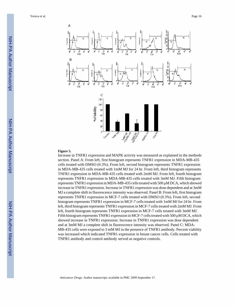
Figure 5.Increase in TNFR1 expression and MAPK activity was measured as explained in the methodssection. Panel A: From left, first histogram represents TNFR1 expression in MDA-MB-435cells treated with DMSO (0.3%). From left, second histogram represents TNFR1 expressionin MDA-MB-435 cells treated with 1mM MJ for 24 hr. From left, third histogram representsTNFR1 expression in MDA-MB-435 cells treated with 2mM MJ. From left, fourth histogramrepresents TNFR1 expression in MDA-MB-435 cells treated with 3mM MJ. Fifth histogramrepresents TNFR1 expression in MDA-MB-435 cells treated with 500 μM DCA, which showedincrease in TNFR1 expression. Increase in TNFR1 expression was dose dependent and at 3mMMJ a complete shift in fluorescence intensity was observed. Panel B: From left, first histogramrepresents TNFR1 expression in MCF-7 cells treated with DMSO (0.3%). From left, secondhistogram represents TNFR1 expression in MCF-7 cells treated with 1mM MJ for 24 hr. Fromleft, third histogram represents TNFR1 expression in MCF-7 cells treated with 2mM MJ. Fromleft, fourth histogram represents TNFR1 expression in MCF-7 cells treated with 3mM MJ.Fifth histogram represents TNFR1 expression in MCF-7 cells treated with 500 μM DCA, whichshowed increase in TNFR1 expression. Increase in TNFR1 expression was dose dependentand at 3mM MJ a complete shift in fluorescence intensity was observed. Panel C: MDA-MB-435 cells were exposed to 3 mM MJ in the presence of TNFR1 antibody. Percent viabilitywas increased which indicated TNFR1 expression in breast cancer cells. Cells treated withTNFR1 antibody and control antibody served as negative controls.
Yeruva et al. Page 16
Anticancer Drugs. Author manuscript; available in PMC 2009 September 17.
NIH
-PA Author Manuscript
NIH
-PA Author Manuscript
NIH
-PA Author Manuscript
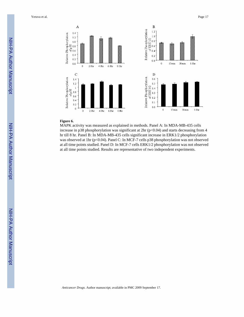
Figure 6.MAPK activity was measured as explained in methods. Panel A: In MDA-MB-435 cellsincrease in p38 phosphorylation was significant at 2hr (p<0.04) and starts decreasing from 4hr till 8 hr. Panel B: In MDA-MB-435 cells significant increase in ERK1/2 phosphorylationwas observed at 1hr (p<0.04). Panel C: In MCF-7 cells p38 phosphorylation was not observedat all time points studied. Panel D: In MCF-7 cells ERK1/2 phosphorylation was not observedat all time points studied. Results are representative of two independent experiments.
Yeruva et al. Page 17
Anticancer Drugs. Author manuscript; available in PMC 2009 September 17.
NIH
-PA Author Manuscript
NIH
-PA Author Manuscript
NIH
-PA Author Manuscript

Figure 7.Panel A: Western blot of caspase-8 in MDA-MB-435 cells treated with 3mM MJ for 2 hr, 4hr and 8 hr. Procaspase-8 was processed and cleaved to active caspase-8 at all the time pointsstudied. Caspase-8 activity was increased with 3mM MJ treatment in MDA-MB-435 cellscompared to the control. β-actin was a loading control. Panel B: Fluoremetric assay showedincreased caspase-8 activity with MJ treatment at 2 and 4 hr time points (p<0.01).
Yeruva et al. Page 18
Anticancer Drugs. Author manuscript; available in PMC 2009 September 17.
NIH
-PA Author Manuscript
NIH
-PA Author Manuscript
NIH
-PA Author Manuscript
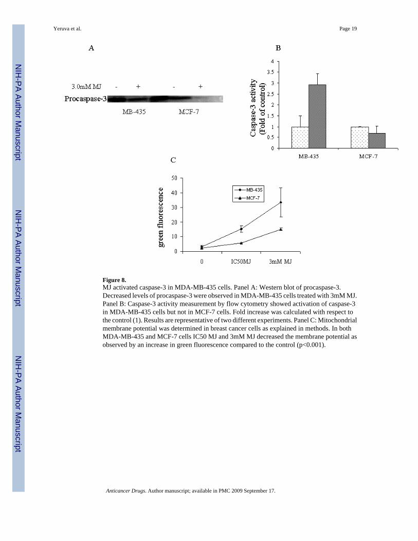
Figure 8.MJ activated caspase-3 in MDA-MB-435 cells. Panel A: Western blot of procaspase-3.Decreased levels of procaspase-3 were observed in MDA-MB-435 cells treated with 3mM MJ.Panel B: Caspase-3 activity measurement by flow cytometry showed activation of caspase-3in MDA-MB-435 cells but not in MCF-7 cells. Fold increase was calculated with respect tothe control (1). Results are representative of two different experiments. Panel C: Mitochondrialmembrane potential was determined in breast cancer cells as explained in methods. In bothMDA-MB-435 and MCF-7 cells IC50 MJ and 3mM MJ decreased the membrane potential asobserved by an increase in green fluorescence compared to the control (p<0.001).
Yeruva et al. Page 19
Anticancer Drugs. Author manuscript; available in PMC 2009 September 17.
NIH
-PA Author Manuscript
NIH
-PA Author Manuscript
NIH
-PA Author Manuscript
NIH
-PA Author Manuscript
NIH
-PA Author Manuscript
NIH
-PA Author Manuscript
Yeruva et al. Page 20
Table 1MJ decreased membrane fluidity in Breast cancer cells
Treatment (4hr) MB-435 MCF-7
Control 0.29 ±0.05 0.32±0.005
Cholesterol 0.50 ±0.05 0.41±0.01
DCA 0.44 ±0.04 0.40±0.01
3mM MJ 0.40 ±0.05 0.41±0.01
MDA-MB-435 cells were treated with 3mM MJ for 4hr and polarization was measured and P value was calculated as explained in methods. There issignificant decrease in membrane fluidity. (p<0.05, one tailed t-test)
Anticancer Drugs. Author manuscript; available in PMC 2009 September 17.