Sleep and sleep disorders in rare hereditary diseases - CORE

B978-0-12-417188-6.00004-9, 00004
Babson, 978-0-12-417188-6
Comp. by: BSubbulakshmi Stage: Proof Chapter No.: 4 Title Name: BabsonPage Number: 1 Date: 12/09/2014 Time: 07:14:09
P A R T 2
METHODS
c0020
To protect the rights of the author(s) and publisher we inform you that this PDF is an uncorrected proof for internal business use onlyby the author(s), editor(s), reviewer(s), Elsevier and typesetter SPi. It is not allowed to publish this proof online or in print. This proofcopy is the copyright property of the publisher and is confidential until formal publication.

B978-0-12-417188-6.00004-9, 00004
Comp. by: BSubbulakshmi Stage: Proof Chapter No.: 4 Title Name: BabsonPage Number: 2 Date: 12/09/2014 Time: 07:14:09
Babson, 978-0-12-417188-6
To protect the rights of the author(s) and publisher we inform you that this PDF is an uncorrected proof for internal business use onlyby the author(s), editor(s), reviewer(s), Elsevier and typesetter SPi. It is not allowed to publish this proof online or in print. This proofcopy is the copyright property of the publisher and is confidential until formal publication.

Sleep and Affect 3 © 2015 Elsevier Inc. All rights reserved.http://dx.doi.org/10.1016/B978-0-12-417188-6.00004-9
B978-0-12-417188-6.00004-9, 00004
Comp. by: BSubbulakshmi Stage: Proof Chapter No.: 4 Title Name: BabsonPage Number: 3 Date: 12/09/2014 Time: 07:14:09
Babson, 978-0-12-417188-6
c0020
C H A P T E R
4Methodology for the Assessment
of SleepChristopher B. Miller,*,† Simon D. Kyle,‡ Kerri L. Melehan,*,§ Delwyn J. Bartlett,*
*Centre for Integrated Research and Understanding of Sleep (CIRUS), Woolcock Institute of Medical Research, Sydney Medical School, University
of Sydney, Sydney, NSW, Australia†Institute of Neuroscience & Psychology, University of Glasgow, Glasgow,
United Kingdom‡School of Psychological Sciences, University of Manchester, Manchester,
United Kingdom§Sleep Investigation Unit, Sydney, Australia
AbbreviationsAASM American Academy of Sleep MedicineCPAP continuous positive airway pressureDISS daytime insomnia symptom scaleDLMO dim light melatonin onsetEEG electroencephalographyEMG electromyogramEOG electrooculogramESS Epworth sleepiness scalefMRI functional magnetic resonance imagingFOSQ functional outcomes of sleep questionnaireMRI magnetic resonance imagingMRS magnetic resonance spectroscopyMSLT multiple sleep latency testMWT maintenance of wakefulness testN1 NREM sleep stage 1N2 NREM sleep stage 2N3 NREM sleep stage 3 or slow-wave sleepNREM nonrapid eye movementOSA obstructive sleep apnea
dt0010p0010
dt0020p0020
dt0025p0025
dt0030p0030
dt0035p0035
dt0040p0040
dt0045p0045
dt0050p0050
dt0060p0060
dt0065p0065
dt0075p0075
dt0080p0080
dt0085p0085
dt0090p0090
dt0095p0095
dt0100p0100
dt0105p0105
dt0115p0115
dt0120p0120
To protect the rights of the author(s) and publisher we inform you that this PDF is an uncorrected proof for internal business use onlyby the author(s), editor(s), reviewer(s), Elsevier and typesetter SPi. It is not allowed to publish this proof online or in print. This proofcopy is the copyright property of the publisher and is confidential until formal publication.
B978-0-12-417188-6.00004-9, 00004
Comp. by: BSubbulakshmi Stage: Proof Chapter No.: 4 Title Name: BabsonPage Number: 4 Date: 12/09/2014 Time: 07:14:09
Babson, 978-0-12-417188-6
4 4. ASSESSMENT OF SLEEP
PET positron emission tomographyPSA power spectral analysisPSG polysomnographyPSQI Pittsburgh sleep quality indexREM rapid eye movementSCN suprachiasmatic nucleusSE sleep efficiencySOL sleep onset latencySWS slow-wave sleep
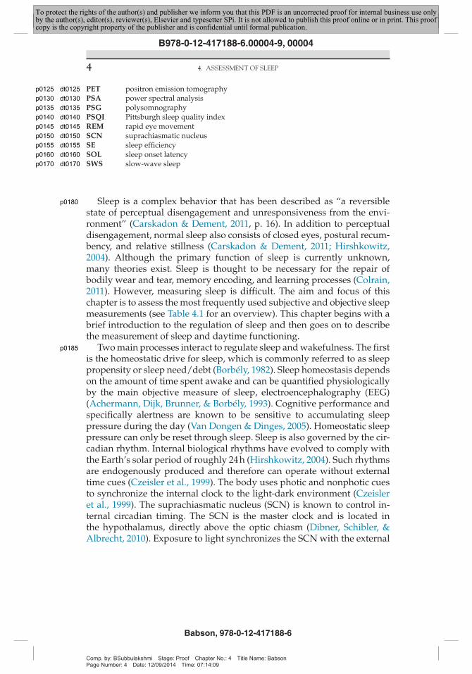
Sleep is a complex behavior that has been described as “a reversible state of perceptual disengagement and unresponsiveness from the envi-ronment” (Carskadon & Dement, 2011, p. 16). In addition to perceptual disengagement, normal sleep also consists of closed eyes, postural recum-bency, and relative stillness (Carskadon & Dement, 2011; Hirshkowitz, 2004). Although the primary function of sleep is currently unknown, many theories exist. Sleep is thought to be necessary for the repair of bodily wear and tear, memory encoding, and learning processes (Colrain, 2011). However, measuring sleep is difficult. The aim and focus of this chapter is to assess the most frequently used subjective and objective sleep measurements (see Table 4.1 for an overview). This chapter begins with a brief introduction to the regulation of sleep and then goes on to describe the measurement of sleep and daytime functioning.
Two main processes interact to regulate sleep and wakefulness. The first is the homeostatic drive for sleep, which is commonly referred to as sleep propensity or sleep need/debt (Borbély, 1982). Sleep homeostasis depends on the amount of time spent awake and can be quantified physiologically by the main objective measure of sleep, electroencephalography (EEG) (Achermann, Dijk, Brunner, & Borbély, 1993). Cognitive performance and specifically alertness are known to be sensitive to accumulating sleep pressure during the day (Van Dongen & Dinges, 2005). Homeostatic sleep pressure can only be reset through sleep. Sleep is also governed by the cir-cadian rhythm. Internal biological rhythms have evolved to comply with the Earth’s solar period of roughly 24 h (Hirshkowitz, 2004). Such rhythms are endogenously produced and therefore can operate without external time cues (Czeisler et al., 1999). The body uses photic and nonphotic cues to synchronize the internal clock to the light-dark environment (Czeisler et al., 1999). The suprachiasmatic nucleus (SCN) is known to control in-ternal circadian timing. The SCN is the master clock and is located in the hypothalamus, directly above the optic chiasm (Dibner, Schibler, & Albrecht, 2010). Exposure to light synchronizes the SCN with the external
dt0125p0125
dt0130p0130
dt0135p0135
dt0140p0140
dt0145p0145
dt0150p0150
dt0155p0155
dt0160p0160
dt0170p0170
p0180
p0185
To protect the rights of the author(s) and publisher we inform you that this PDF is an uncorrected proof for internal business use onlyby the author(s), editor(s), reviewer(s), Elsevier and typesetter SPi. It is not allowed to publish this proof online or in print. This proofcopy is the copyright property of the publisher and is confidential until formal publication.

B978-0-12-417188-6.00004-9, 00004
Comp. by: BSubbulakshmi Stage: Proof Chapter No.: 4 Title Name: BabsonPage Number: 5 Date: 12/09/2014 Time: 07:14:09
5
Babson, 978-0-12-417188-6
TA
BL
E 4
.1
Ove
rvie
w o
f the
Met
hods
use
d fo
r the
Ass
essm
ent o
f Sle
ep
Met
hod
Des
crip
tion
Pro
sC
ons
Poly
som
nogr
aphy
(PSG
)O
bjec
tive
mea
sure
of s
leep
an
d d
isor
der
s of
sle
ep1.
Gol
d s
tand
ard
mea
sure
of s
leep
and
th
e d
isor
der
s of
sle
ep1.
Exp
ensi
ve2.
Dif
ficu
lt to
acc
ess
3. D
isco
mfo
rt4.
Fir
st-n
ight
eff
ect
5. R
equi
res
anal
ysis
and
inte
rpre
tati
on
Mul
tipl
e sl
eep
late
ncy
test
(M
SLT
)O
bjec
tive
mea
sure
of
day
tim
e sl
eepi
ness
1. G
old
sta
ndar
d m
easu
re o
f sev
erit
y of
day
tim
e sl
eepi
ness
2. U
sed
in th
e d
iagn
osis
of n
arco
leps
y
1. R
equi
res
PSG
pri
or2.
In
lab
asse
ssm
ent w
ith
skill
ed s
taff
m
embe
r3.
Dif
ficu
lt to
acc
ess
4. C
eilin
g ef
fect
5. L
ack
of e
colo
gica
l val
idit
y6.
Req
uire
s an
alys
is a
nd in
terp
reta
tion
Mai
nten
ance
of w
akef
ulne
ss
test
(MW
T)
Obj
ecti
ve e
valu
atio
n of
w
akef
ulne
ss a
nd a
lert
ness
un
der
sop
orif
ic s
etti
ngs
1. N
o ce
iling
eff
ect a
nd d
oes
not
requ
ire
PSG
2. I
mpr
oved
eco
logi
cal v
alid
ity
over
M
SLT
1. I
n la
b as
sess
men
t wit
h sk
illed
sta
ff
mem
ber
2. D
iffi
cult
to a
cces
s3.
Req
uire
s an
alys
is a
nd in
terp
reta
tion
Act
igra
phy
Obj
ecti
ve m
easu
re o
f m
ovem
ent u
sed
to in
fer
slee
p-w
ake
beha
vior
1. C
orre
late
s w
ith
PSG
2. C
an b
e us
ed lo
ng-t
erm
3. E
colo
gica
lly v
alid
4. T
arge
t for
trea
tmen
t
1. L
imit
ed b
y ba
tter
y le
ngth
2. R
equi
res
anal
ysis
and
inte
rpre
tati
on
Clin
ical
inte
rvie
wO
bjec
tive
mea
sure
of
mov
emen
t use
d to
infe
r sl
eep-
wak
e be
havi
or
1. A
cces
sibl
e2.
Clin
ical
ly v
alid
3. U
sefu
l pri
or to
trea
tmen
t or
rese
arch
1. L
ack
of s
tand
ard
izat
ion
2. L
imit
ed w
itho
ut fu
rthe
r m
easu
res
of
slee
p
(Con
tinu
ed)
t0010
To protect the rights of the author(s) and publisher we inform you that this PDF is an uncorrected proof for internal business use onlyby the author(s), editor(s), reviewer(s), Elsevier and typesetter SPi. It is not allowed to publish this proof online or in print. This proofcopy is the copyright property of the publisher and is confidential until formal publication.

B978-0-12-417188-6.00004-9, 00004
Comp. by: BSubbulakshmi Stage: Proof Chapter No.: 4 Title Name: BabsonPage Number: 6 Date: 12/09/2014 Time: 07:14:09
6 4. ASSESSMENT OF SLEEP
Babson, 978-0-12-417188-6
Met
hod
Des
crip
tion
Pro
sC
ons
Slee
p d
iary
Subj
ecti
ve m
easu
re o
f sle
ep-
wak
e be
havi
or1.
Acc
essi
ble
2. T
arge
t for
trea
tmen
t3.
Eas
y to
use
and
inex
pens
ive
1. P
oor
corr
elat
ion
wit
h PS
G2.
Mus
t rem
embe
r to
fill
in3.
Sub
ject
ive
mea
sure
of s
leep
4. R
equi
res
anal
ysis
and
inte
rpre
tati
on
Que
stio
nnai
res
Subj
ecti
ve m
easu
res
of
slee
p, w
ake,
hea
lth,
and
co
gnit
ion
1. A
cces
sibl
e2.
Tar
get f
or tr
eatm
ent
3. E
asy
to u
se a
nd in
expe
nsiv
e
1. O
pen
to b
ias
2. R
equi
res
anal
ysis
and
inte
rpre
tati
on
Bra
in im
agin
gO
bjec
tive
mea
sure
of t
he
brai
n an
d it
s fu
ncti
onin
g1.
Non
inva
sive
met
hod
for
inve
stig
atin
g th
e br
ain
2. C
an b
e us
ed d
urin
g sl
eep
3. C
an m
easu
re b
rain
act
ivat
ion
and
m
etab
olis
m
1. E
xpen
sive
2. D
iffi
cult
to a
cces
s3.
Poo
r te
mpo
ral r
esol
utio
n4.
Lac
k of
val
idit
y5.
Req
uire
s an
alys
is a
nd in
terp
reta
tion
Cor
tiso
lH
orm
one
used
to in
fer
stre
ss1.
Obj
ecti
ve m
easu
re2.
Clo
sely
link
ed to
the
slee
p cy
cle
1. R
equi
res
anal
ysis
2. D
iffi
cult
to a
cces
s3.
Tes
t req
uire
s fu
rthe
r va
lidat
ion
Mel
aton
inH
orm
one
used
to in
fer
circ
adia
n al
ignm
ent
1. O
bjec
tive
mea
sure
2. M
ay b
e us
ed to
ind
icat
e ch
ange
wit
h tr
eatm
ent
3. C
lose
ly li
nked
to th
e sl
eep
cycl
e
1. R
equi
res
anal
ysis
2. D
iffi
cult
to a
cces
s3.
Tes
t req
uire
s fu
rthe
r va
lidat
ion
TA
BL
E 4
.1
Ove
rvie
w o
f the
Met
hods
use
d fo
r the
Ass
essm
ent o
f Sle
ep—
Con
t’d
To protect the rights of the author(s) and publisher we inform you that this PDF is an uncorrected proof for internal business use onlyby the author(s), editor(s), reviewer(s), Elsevier and typesetter SPi. It is not allowed to publish this proof online or in print. This proofcopy is the copyright property of the publisher and is confidential until formal publication.
B978-0-12-417188-6.00004-9, 00004
Comp. by: BSubbulakshmi Stage: Proof Chapter No.: 4 Title Name: BabsonPage Number: 7 Date: 12/09/2014 Time: 07:14:09
PART 1: ObjECTIvE DIAgNOSTIC MEASuRES OF SLEEP 7
Babson, 978-0-12-417188-6
light-dark cycle and serves as the primary circadian time giver for mam-mals (Czeisler et al., 1999; Stephan & Nunez, 1977).
PART 1: OBJECTIVE DIAGNOSTIC MEASURES OF SLEEP
Electroencephalography and polysomnography. Sleep and wake states are measured by EEG whereby electrodes on the scalp record electrical brain activity (Rechtschaffen & Kales, 1968). Electrical brain activity is the gold standard objective measurement of sleep (Kushida et al., 2005), and elec-trodes are placed according to standardized international criteria called the 10-20 placement system (Jasper, 1958), which aims to ensure reproduc-ibility of EEG studies. In the 10-20 system, each electrode site is mapped with letters and numbers. The letters F, T, C, P, and O refer to scalp loca-tions and stand for frontal, temporal, central, parietal, and occipital areas, respectively. Even numbers denote the right hemisphere and odd num-bers denote the left. “Z” means 0 and stands for the midline of the head (Oostenveld & Praamstra, 2001).
Clinicians use a diagnostic sleep study to diagnose sleep disorders, including sleep-related breathing disorders, parasomnias, sleep-related seizure disorders, and periodic limb movement disorders (Kushida et al., 2005). The diagnostic sleep study is extensive and provides a significant amount of useful information, but it is also expensive, difficult to access, and often uncomfortable for the patient (Ancoli-Israel et al., 2003). The recording of brain activity by EEG is only one aspect of the overall di-agnostic sleep study. The study can gather other information about the body during sleep, using polysomnography (PSG), which simply means “many sleep recordings” in addition to EEG, and the following measures can also determine sleep: eye movements measured via the electrooculo-gram (EOG) and muscle tone measured via the electromyogram (EMG) on the chin. Any sleep disturbance due to sleep disorders such as obstructive sleep apnea (OSA), periodic limb movement disorder, and parasomnias can be detected as part of the overall PSG assessment. Such disorders can be distinguished using a number of measures, including respiratory effort and airflow, snoring, body position, heart rate, oxygen saturation levels, and limb and jaw movements over the course of the night.
Sleep behavior can be scored according to standardized PSG criteria (Iber, Ancoli-Israel, Chesson, & Quan, 2007), through an evaluation of electrical wave-forms produced by the brain, eye movements, and muscle activity. Primarily, a sleep scientist evaluates brain electrical wave-form patterns for amplitude and frequency, as well as bursts of brain activity. Busts of brain activity include K-complexes (a single large amplitude wave-form <2 Hz in frequency with a brief positive electrical peak followed by
s0010
p0190
p0195
p0200
To protect the rights of the author(s) and publisher we inform you that this PDF is an uncorrected proof for internal business use onlyby the author(s), editor(s), reviewer(s), Elsevier and typesetter SPi. It is not allowed to publish this proof online or in print. This proofcopy is the copyright property of the publisher and is confidential until formal publication.

B978-0-12-417188-6.00004-9, 00004
Comp. by: BSubbulakshmi Stage: Proof Chapter No.: 4 Title Name: BabsonPage Number: 8 Date: 12/09/2014 Time: 07:14:09
8 4. ASSESSMENT OF SLEEP
Babson, 978-0-12-417188-6
a slower negative component; Loomis, Harvey, & Hobart, 1938) and sleep spindles (a short burst of oscillatory electrical activity of sigma frequency waves at 12-16 Hz that may follow from a K-complex; Loomis et al., 1938), and they can be used to categorize sleep stages (see Figure 4.1). A trained and certified sleep polysomnographic technician normally divides sleep into distinct sleep stages that are then summarized and reported on by a specialized sleep physician. A hypnogram is used to give an overview of the staging of sleep for the entire night (see Figure 4.2).
In adults, sleep consists of two main phases: non-rapid eye movement (NREM) sleep and rapid eye movement (REM) sleep. In healthy sleepers, the brain transitions through these stages of sleep in approximately 90-min cycles. On average, an individual experiences four to five sleep cycles during the night. NREM sleep comprises three distinct sleep stages. At sleep onset, an initial short period (5-10 min) of stage 1 sleep (N1) occurs, characterized by theta waves (4-7 Hz) and an absence of alpha waves (8-12 Hz) on the EEG recording, followed by a longer period (~20 min) of stage 2 sleep (N2) characterized by a mixed frequency background featur-ing sleep spindles and K-complexes. Stage 3 sleep (N3), otherwise known as slow-wave sleep (SWS), is characterized by delta frequency wave for-mations (0.5-4.5 Hz) of at least 75 μV amplitude, and it lasts for approxi-mately 30-40 min (Iber et al., 2007).
REM sleep is distinct and contains mixed frequency theta and beta EEG activity, with characteristic sawtooth-shaped waves, rapid eye move-ments, and muscle atonia. REM sleep usually occurs for the first time at
N3 REM
Wake N2N1 1 2
FIGURE 4.1 Stages of sleep and scoring information. Examples of periods of wake-fulness and sleep objectively defined through electroencephalography (EEG) used on one healthy 22-year-old male as part of an overnight sleep study (polysomnography). Each ex-ample presents 30 s of data from the following seven electrodes (top to bottom): left eye, right eye (EOG: electrooculogram); C3-A2, C4-A1, O2-A1, O1-A1; and a single channel of chin EMG (electromyogram). The top left image displays the period of wakefulness. The top middle image displays stage 1 sleep (N1), and the top right image displays stage 2 sleep (N2). The bottom left image displays stage 3 sleep (N3 or SWS). Rapid eye movement (REM) sleep is displayed in the bottom right image. 1, a K-complex and 2, a sleep spindle.
p0205
p0210
f0010
To protect the rights of the author(s) and publisher we inform you that this PDF is an uncorrected proof for internal business use onlyby the author(s), editor(s), reviewer(s), Elsevier and typesetter SPi. It is not allowed to publish this proof online or in print. This proofcopy is the copyright property of the publisher and is confidential until formal publication.

B978-0-12-417188-6.00004-9, 00004
Comp. by: BSubbulakshmi Stage: Proof Chapter No.: 4 Title Name: BabsonPage Number: 9 Date: 12/09/2014 Time: 07:14:09
PART 1: ObjECTIvE DIAgNOSTIC MEASuRES OF SLEEP 9
Babson, 978-0-12-417188-6
the end of each sleep cycle, and the first REM period is generally short, lasting 5-10 min or less. REM sleep episodes then become increasingly lon-ger throughout the sleep period (Aserinsky & Kleitman, 1953; Dement & Kleitman, 1957). The sleep cycles tend to alter their alignment as the sleep period progresses, with SWS dominant in the first third of the night and REM sleep more dominant toward the end of the sleep period (Carskadon & Dement, 2011). The proportion relative to total sleep time (TST) is as follows for the following sleep stages: N1 is approximately 2-5% of TST, N2 is 45-55%, N3 (SWS) is 20%, and REM sleep is 20-25% (Williams, Agnew, & Webb, 1964).
Overall, SWS is associated with homeostatic sleep pressure or the amount of time spent awake, and it can be quantified physiologically through EEG-derived slow-wave activity (SWA; spectral power in the 0.75-4.5 Hz bandwidth) (Achermann et al., 1993). Importantly, the majority of SWS is obtained during the first third of the night, and SWS is normally preserved by the brain under sleep-restricted schedules (Spiegel, Leproult, & Van Cauter, 1999). In addition, the onset of “deep” SWS sleep occurs quickly in healthy individuals (normally after about 20 min of lighter N1 and N2 sleep), indicating the importance of this stage of sleep. Studies disrupt-ing SWS have shown impairment in next day performance and endocrine secretion in healthy individuals (Spiegel et al., 1999). One of the primary functions of SWS may be to restore and repair both the brain and the body. On the other hand, REM sleep is more adaptable around SWS sleep needs. REM sleep (named after the discovery of rapid eye movements during this sleep phase; Aserinsky & Kleitman, 1953) appears to be important for the consolidation of memory traces through increases in synaptic plasticity in
3 4
Hours
N3
N2
N1
REM
Awake
5 6 7 821
FIGURE 4.2 Hypnogram of scored human sleep staging. A normal hypnogram used to provide an overview of the progression of sleep stages during the night for an individual’s sleep. The hypnogram is a continuous recording of electroencephalogram (EEG), electroocu-logram (EOG), and electromyogram (EMG) measures for scoring sleep. The y-axis represents sleep stages (evaluated in 30-s epochs), including REM, N1, N2, and N3 or SWS The x-axis represents time during the night in hours.
p0215
f0015
To protect the rights of the author(s) and publisher we inform you that this PDF is an uncorrected proof for internal business use onlyby the author(s), editor(s), reviewer(s), Elsevier and typesetter SPi. It is not allowed to publish this proof online or in print. This proofcopy is the copyright property of the publisher and is confidential until formal publication.

B978-0-12-417188-6.00004-9, 00004
Comp. by: BSubbulakshmi Stage: Proof Chapter No.: 4 Title Name: BabsonPage Number: 10 Date: 12/09/2014 Time: 07:14:09
10 4. ASSESSMENT OF SLEEP
Babson, 978-0-12-417188-6
the cortex of the brain (Diekelmann & Born, 2010). REM sleep is related to dream content, with people who awake during REM being more likely to report experiencing a dream relative to those who wake during other stages of sleep (Foulkes, 1962). Crucially, muscle paralysis or atonia takes place during REM sleep and prevents dream enactment. REM behavior disorder is characterized by a lack of muscle atonia and leads to patients acting out their dreams (Schenck, Hurwitz, & Mahowald, 1993). REM be-havior disorder may also be a marker for the subsequent development of a neurodegenerative disorder such as Parkinson’s disease (see Chapter 8 for more information; Schenck, Bundlie, & Mahowald, 1996).
Power spectral analysis of sleep. The standardized practice and scoring of sleep by PSG does not reflect all of the cortical processes that take place during the sleep period. EEG-defined sleep can also be analyzed using power spectral analysis (PSA), which reveals the microstructure of sleep. PSA is a mathematical quantification process for detecting periodicities in time series data (Perlis et al., 2001). PSA uses the fast Fourier transfor-mation to group EEG rhythms into the following frequency bands: delta (0.5-4 Hz), theta (4-7 Hz), alpha (7-14 Hz), beta (15-30 Hz), and gamma (30-100 + Hz) (Borbély, Baumann, Brandeis, Strauch, & Lehmann, 1981). The first Fourier analysis of EEG recordings was carried out in 1932 by Dietsch at the suggestion of Berger, the innovator of EEG recordings in humans (Achermann, 2009). PSA can be used to analyze the complex EEG signal, which is normally scored by hand and as an average of a 30-s pe-riod. The scorer may miss important aspects of this EEG signal if scoring over an 8-h recording. Quantitative methods such as PSA are essential for investigating the EEG signal in more detail. For example, slow-wave activation is a sensitive measure of sleep homeostasis or sleep pressure (Achermann, 2009), and NREM sleep is distinguished by both delta ac-tivity (0.5-4.5 Hz) and spindle frequency activity (12-14 Hz). REM sleep does not exhibit delta and spindle frequency activity. Further, alpha ac-tivity (~10 Hz) is present during the waking period before sleep onset (Achermann, 2009). Such quantification of EEG into frequency bands can allow PSA to give an overview of the structure of sleep, even when not scored visually by a technician. Alterations in the microstructure of sleep may not be detected via conventional visual scoring of sleep stages but are detected by PSA.
Multiple sleep latency test. The multiple sleep latency test (MSLT) was developed in the 1970s to measure the effects of sleep deprivation on sleepiness levels in young healthy individuals (Carskadon & Dement, 1982). Correlations between subjective measures of sleepiness and the MSLT suggested usage as an objective measure of daytime sleepiness, and these correlations led to the development of standardized MSLT testing protocols for both clinical and research purposes (Arand et al., 2005). The American Academy of Sleep Medicine (AASM) considerers the MSLT the
p0220
p0225
To protect the rights of the author(s) and publisher we inform you that this PDF is an uncorrected proof for internal business use onlyby the author(s), editor(s), reviewer(s), Elsevier and typesetter SPi. It is not allowed to publish this proof online or in print. This proofcopy is the copyright property of the publisher and is confidential until formal publication.

B978-0-12-417188-6.00004-9, 00004
Comp. by: BSubbulakshmi Stage: Proof Chapter No.: 4 Title Name: BabsonPage Number: 11 Date: 12/09/2014 Time: 07:14:09
PART 1: ObjECTIvE DIAgNOSTIC MEASuRES OF SLEEP 11
Babson, 978-0-12-417188-6
de facto gold standard objective measure of excessive daytime sleepiness (Littner et al., 2005), and the test is primarily used to diagnose narcolepsy. Daytime sleepiness may be caused by an increased sleep propensity accu-mulated through a lack of adequate sleep or sleep disorders. In the MSLT, participants are given four or five daytime nap opportunities (tests), each lasting 20 min every 2 h, following an overnight PSG evaluation. Muscle activity, eye movements, heart rate, and EEG are monitored during the testing procedure. In a dark room, patients must lie in bed and go to sleep. If the patient remains awake for the full 20 min, then the test ends. If sleep occurs during the test, the clinician wakes the patient up after 15 min of sleep (Littner et al., 2005). The two main outcomes are the average time to fall asleep, which ranges from 0 to 20 min, and the presence or absence of REM sleep. Patients with narcolepsy reportedly have a mean of 3.1 min, while healthy controls have a mean of 11.6 min (Littner et al., 2005). The time to fall asleep is sensitive to a number of factors, including sleep depri-vation, use of a stimulant or sedative medications, and the presence of sleep disorders such as narcolepsy and OSA (Kushida et al., 2005; Sunwoo et al., 2012). Although the MSLT is most often used to indicate narcolepsy compared with reported sleep propensity from fatigue and tiredness, it can assess REM-onset latency in those with moderate-to-severe OSA or excessive daytime sleepiness (Littner et al., 2005), in addition to profiling treatment effects (Thorpy, Westbrook, Ferber, & Fredrickson, 1992). Critics of the MSLT point out that the test exhibits a floor effect, meaning that the MSLT may not be sensitive enough to detect clinically significant changes due to treatment in severe populations (even when sleep latencies are doubled), as well as a ceiling effect imposed by the 20-min nap limit (limit-ing the usefulness in more alert individuals), and they assert that the sleep laboratory setting is not representative of the work place where sleep ten-dencies may be different (Arand et al., 2005).
Maintenance of wakefulness test. The maintenance of wakefulness test (MWT) is used to examine wakefulness and alertness in soporific settings (Arand et al., 2005) where high levels of vigilance and alertness are re-quired, as might be required for “fitness for duty” (Sullivan & Kushida, 2008). The MWT was developed to profile treatment changes in patients with severe levels of sleepiness and their ability to remain awake. By de-sign, the MWT overcomes the three previously mentioned limitations with the MSLT concerning floor or ceiling effects and the lack of ecological va-lidity (Arand et al., 2005). The ability to maintain wakefulness is the pri-mary outcome calculated indirectly by measuring sleep latency (Mitler, Gujavarty, & Browman, 1982). Unlike the MSLT, the MWT does not neces-sarily require an overnight sleep study prior to the test. In a dimly lit room, the participants’ EEG, EOG, and chin EMG are monitored, and he or she is asked to stay awake while seated in bed, with back and head supported by a bedrest for 40 min over four trials performed every 2 h. Physical activity
p0230
To protect the rights of the author(s) and publisher we inform you that this PDF is an uncorrected proof for internal business use onlyby the author(s), editor(s), reviewer(s), Elsevier and typesetter SPi. It is not allowed to publish this proof online or in print. This proofcopy is the copyright property of the publisher and is confidential until formal publication.

B978-0-12-417188-6.00004-9, 00004
Comp. by: BSubbulakshmi Stage: Proof Chapter No.: 4 Title Name: BabsonPage Number: 12 Date: 12/09/2014 Time: 07:14:09
12 4. ASSESSMENT OF SLEEP
Babson, 978-0-12-417188-6
and vocalizations are not allowed during the test (Littner et al., 2005). The reported outcome is the average length of time the patient is able to re-main awake, with the first occurrence of 15 s of sleep within a 30-s window being deemed the onset of sleep. The study is terminated after 40 min or after three 30-s windows classified as stage N1 sleep or any 30-s window of any other stage of sleep. Studies have demonstrated mean decreased sleep latencies in those with excessive daytime sleepiness and increases in sleep latencies after treatment for OSA (Sullivan & Kushida, 2008).
Actigraphy. Sleep can also be profiled through the use of actigraphy, which allows patterns of light, sleep, and wake behavior to be assessed over days or weeks. Actigraphy is cost effective and more convenient than a full PSG (Ancoli-Israel et al., 2003), and it can be repeated across many nights to build an ecologically valid assessment of sleep without the first-night effect of PSG (Ancoli-Israel et al., 2003). The first-night ef-fect is common with a PSG evaluation and is known to impair sleep in healthy good sleepers (Agnew, Webb, & Williams, 1966). Actigraphs are typically watch-like devices worn on the nondominant hand, and they use an accelerometer to record movement over a given threshold (see Figure 4.3). The wearer can also use an event marker to denote time in bed and awakenings during the night. Collected data is downloaded to a com-puter for the observation of rest and activity patterns across both night and day. Through analysis software, validated algorithms are applied for
FIGURE 4.3 Picture of an actigraph. An actigraph worn on the wrist of the nondominant hand. 1, the event marker button; 2, the light sensor.
p0235
f0020
To protect the rights of the author(s) and publisher we inform you that this PDF is an uncorrected proof for internal business use onlyby the author(s), editor(s), reviewer(s), Elsevier and typesetter SPi. It is not allowed to publish this proof online or in print. This proofcopy is the copyright property of the publisher and is confidential until formal publication.
B978-0-12-417188-6.00004-9, 00004
Comp. by: BSubbulakshmi Stage: Proof Chapter No.: 4 Title Name: BabsonPage Number: 13 Date: 12/09/2014 Time: 07:14:09
PART 1: ObjECTIvE DIAgNOSTIC MEASuRES OF SLEEP 13
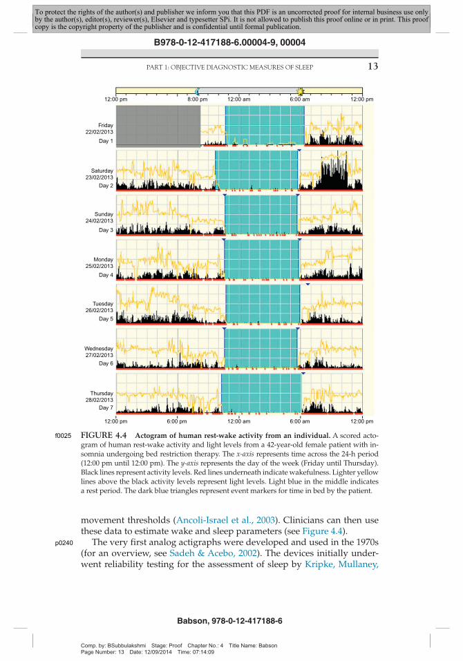
Babson, 978-0-12-417188-6
movement thresholds (Ancoli-Israel et al., 2003). Clinicians can then use these data to estimate wake and sleep parameters (see Figure 4.4).
The very first analog actigraphs were developed and used in the 1970s (for an overview, see Sadeh & Acebo, 2002). The devices initially under-went reliability testing for the assessment of sleep by Kripke, Mullaney,
Tuesday26/02/2013
Monday25/02/2013
Sunday24/02/2013
Saturday23/02/2013
Friday22/02/2013
12:00 pm 8:00 pm 12:00 am 6:00 am 12:00 pm
12:00 pm 6:00 pm 12:00 am 6:00 am 12:00 pm
Wednesday27/02/2013
Thursday28/02/2013
Day 5
Day 4
Day 3
Day 2
Day 1
Day 6
Day 7
FIGURE 4.4 Actogram of human rest-wake activity from an individual. A scored acto-gram of human rest-wake activity and light levels from a 42-year-old female patient with in-somnia undergoing bed restriction therapy. The x-axis represents time across the 24-h period (12:00 pm until 12:00 pm). The y-axis represents the day of the week (Friday until Thursday). Black lines represent activity levels. Red lines underneath indicate wakefulness. Lighter yellow lines above the black activity levels represent light levels. Light blue in the middle indicates a rest period. The dark blue triangles represent event markers for time in bed by the patient.
p0240
f0025
To protect the rights of the author(s) and publisher we inform you that this PDF is an uncorrected proof for internal business use onlyby the author(s), editor(s), reviewer(s), Elsevier and typesetter SPi. It is not allowed to publish this proof online or in print. This proofcopy is the copyright property of the publisher and is confidential until formal publication.

B978-0-12-417188-6.00004-9, 00004
Comp. by: BSubbulakshmi Stage: Proof Chapter No.: 4 Title Name: BabsonPage Number: 14 Date: 12/09/2014 Time: 07:14:09
14 4. ASSESSMENT OF SLEEP
Babson, 978-0-12-417188-6
Messin, and Wyborney (1978). The digital actigraphs currently in use have sufficient memory storage for long-term operation. The sampling rate (ep-och) is normally standardized to once per minute, although the user can change the epoch through computer software (Sadeh & Acebo, 2002). A longer sampling rate can increase the battery length, but longer intervals are not generally used for research purposes because intervals of greater than 1 min may compromise data validity. Previously, actigraphy has demonstrated high agreement rates with PSG data for TST and sleep effi-ciency variables in healthy subjects (Kushida et al., 2001). However, these variables have been found to have lower agreement rates in patients with OSA and insomnia. Therefore, practice guidelines for the use of actigra-phy suggest it should be used in combination with a sleep diary (Ancoli-Israel et al., 2003).
Actigraphy can provide an inexpensive way to follow up with patients experiencing varied sleep disorders. It is also very useful for patients with insomnia symptomology and treatment (see Figure 4.3) and in the diagnosis of circadian rhythm disorders when the patient’s internal cir-cadian timing does not significantly differ from the external environment (Morgenthaler et al., 2007). In advanced or delayed sleep phase disor-ders, the internal circadian clock of the patient may be ticking either too fast (advanced) or too slow (delayed). The strength of actigraphy is that it provides a noninvasive and long-term measure of activity and sleep. However, unlike PSG, actigraphy does not provide an assessment of sleep architecture and cannot provide a specific diagnosis of a sleep disorder, such as OSA.
PART 2: SUBJECTIVE DIAGNOSTIC MEASURES OF SLEEP
Clinical interview. A clinician can initially acquire an overview of the pa-tient’s sleep through a common unstructured clinical interview (Buysse, Ancoli-lsrael, Edinger, Lichstein, & Morin, 2006). Such clinical interviews are extremely useful for providing a baseline screening assessment and patient history prior to the start of treatment or research study. When tak-ing a patient’s history, a clinician normally asks questions to probe the following areas: family history, physical health history, previous medica-tion and alcohol use, history of mental illness, and the timing and onset of sleep (Schramm et al., 1993). The clinical history can also include obser-vations of the patient’s sleep-wake behavior during the night reported by the partner of the patient, family, caregivers, or travelling companions. For example, reports of parasomnias, including the acting out of dreams and nightmares, can be described by the patient or others and documented in sleep logs or diaries (Blagrove, Farmer, & Williams, 2004).
p0245
s0015
p0250
To protect the rights of the author(s) and publisher we inform you that this PDF is an uncorrected proof for internal business use onlyby the author(s), editor(s), reviewer(s), Elsevier and typesetter SPi. It is not allowed to publish this proof online or in print. This proofcopy is the copyright property of the publisher and is confidential until formal publication.

B978-0-12-417188-6.00004-9, 00004
Comp. by: BSubbulakshmi Stage: Proof Chapter No.: 4 Title Name: BabsonPage Number: 15 Date: 12/09/2014 Time: 07:14:09
PART 2: SubjECTIvE DIAgNOSTIC MEASuRES OF SLEEP 15
Babson, 978-0-12-417188-6
A lack of common standardization reduces the reliability of these clin-ical assessments, however (Buysse et al., 2006). As a result, specifically designed structured interviews for sleep disorders have been used to probe symptoms of insomnia, idiopathic hypersomnia, sleep-wake sched-ule disorders, sleep-induced respiratory impairment, narcolepsy, restless leg syndrome, and periodic movement disorders, among other condi-tions (Ohayon, Guilleminault, Zulley, Palombini, & Raab, 1999; Schramm et al., 1993). One structured interview was initially developed and tested for reliability and validity according to DSM-III-R and DSM-IV criteria (Schramm et al., 1993). The interview can be used to evaluate sleep-wake disorders, and the average time for administration is approximately 20-30 min. The interview consists of the following: (1) A brief semistructured overview of physical and mental health and questions regarding OSA and narcolepsy; (2) A specific and structured inquiry of sleep disorder symp-toms; and (3) A summary score sheet filled in at the end of the interview. The interviewer may omit irrelevant sections, depending on the response of the patient (Schramm et al., 1993), and physicians or health care pro-fessionals with no prior knowledge of sleep disorders can successfully administer it.
Sleep diary measures. Sleep can also be profiled through daily self-report measures of sleep. Sleep diaries are widely used in sleep science, and they are fundamental to understanding the subjective complaints of patients. Self-monitoring of sleep through a sleep diary enables nightly perceived metrics to be identified, and it normally includes the following estimated measures: sleep onset latency (SOL), wake-time after initial sleep onset (WASO), TST, total time spent in bed, sleep efficiency (SE: percentage of time spent asleep relative to the amount of time spent in bed), and a nu-merical estimation of overall sleep quality. Normally, patients are asked to complete the diary before they commence their day and refer back to the previous night’s sleep with approximations to the nearest 5 min. Some patients may complete daily entries in the morning or in both the morning and evening. The morning assessment is both sufficient and pre-ferred (Carney et al., 2012). Patients (especially those with insomnia) are advised to avoid filling in the sleep diary during the night, because this may further disrupt their sleep. Likewise, the use of a clock during the night to quantify periods of wakefulness should also be avoided. Thus, estimations of time are necessarily generalized to the nearest 5 min. When compared to gold standard PSG and actigraphy, sleep diaries tend to be less accurate because healthy participants tend to overestimate their TST, whereas patients with insomnia underestimate their sleep time (Lichstein et al., 2006; Maes et al., 2014).
Nevertheless, the sleep diary is the gold standard subjective assessment of sleep and an extremely important assessment of insomnia (Carney et al., 2012). The subjective complaint is considered primary, and clinicians
p0255
p0260
p0265
To protect the rights of the author(s) and publisher we inform you that this PDF is an uncorrected proof for internal business use onlyby the author(s), editor(s), reviewer(s), Elsevier and typesetter SPi. It is not allowed to publish this proof online or in print. This proofcopy is the copyright property of the publisher and is confidential until formal publication.

B978-0-12-417188-6.00004-9, 00004
Comp. by: BSubbulakshmi Stage: Proof Chapter No.: 4 Title Name: BabsonPage Number: 16 Date: 12/09/2014 Time: 07:14:09
16 4. ASSESSMENT OF SLEEP
Babson, 978-0-12-417188-6
typically quantify the progress of cognitive behavioral therapy for insom-nia using primary sleep diary outcome metrics (SOL, TST, WASO, and %SE). However, a lack of sleep diary standardization has hampered re-search methodologies in measuring sleep (Carney et al., 2012), resulting in inconsistent results between studies and a difficulty in translating lab find-ings into clinical practice. To address this problem, the AASM established an expert consensus of sleep medicine experts in order to standardize a self-report sleep diary. The proposed consensus sleep diary is currently a live document available for use, but it still requires validation, testing, and refinement (Carney et al., 2012).
Questionnaire assessments. Sleep can be profiled subjectively through self-report questionnaire measures. Many questionnaires allow patients to report on sleep, quality of life, health-related quality of life, and also daytime functioning (Shahid, Wilkinson, & Marcu, 2012). This section fo-cuses on a number of the most widely used and applicable questionnaires for the assessment of sleep and its disorders (Buysse et al., 2006).
Sleep. The Pittsburgh sleep quality index (PSQI; Buysse, Reynolds, Monk, Berman, & Kupfer, 1989) is one of the most widely used self- report measures for the assessment of sleep quality, having been cited well over 5000 times in the literature. This is a useful and general retrospective as-sessment of sleep quality and sleep disturbance over a 1-month period. Patients score 19 individual items that generate 7 component scores, including subjective sleep quality, sleep latency, sleep duration, sleep efficiency, sleep disturbance, use of sleep medication, and daytime dys-function (Buysse et al., 1989). The first 4 items are open questions, while items 5-19 are rated on a 4-point Likert scale. Seven components are de-rived from the individual item scores, and a total score of overall sleep quality, ranging from 0 to 21, is obtained by adding the 7 component scores. A global PSQI score is used to differentiate between those with good and poor sleep (>5 = poor sleeper). Clinicians can also use it with many patient and research populations to distinguish between good and poor sleepers. However, a disadvantage of the PSQI is the complexity of scoring the questionnaire.
Sleepiness. The Epworth sleepiness scale (ESS) profiles subjective esti-mations of excessive daytime sleepiness (Johns, 1992). The overall total score (out of 24) comprises a single factor, the propensity to fall asleep over the previous 2 weeks. The scale asks people to rate the likelihood that they would doze or fall asleep in eight different settings (sitting and reading for example). Patients are asked to rate on a 0-3 scale (0 = “would never doze,” 3 = “high chance”) their chance of dozing within those eight situations. Scores above 16 are regarded as evidence of extreme sleepiness, as indicated in patients with idiopathic hypersomnolence or narcolepsy (Johns, 1992). In patients with moderate-to-severe OSA, the ESS is sensi-tive to change with continuous positive airway pressure (CPAP) treatment
p0270
p0275
p0280
To protect the rights of the author(s) and publisher we inform you that this PDF is an uncorrected proof for internal business use onlyby the author(s), editor(s), reviewer(s), Elsevier and typesetter SPi. It is not allowed to publish this proof online or in print. This proofcopy is the copyright property of the publisher and is confidential until formal publication.

B978-0-12-417188-6.00004-9, 00004
Comp. by: BSubbulakshmi Stage: Proof Chapter No.: 4 Title Name: BabsonPage Number: 17 Date: 12/09/2014 Time: 07:14:09
PART 2: SubjECTIvE DIAgNOSTIC MEASuRES OF SLEEP 17
Babson, 978-0-12-417188-6
(Antic et al., 2011). The ESS can also be used to profile difficulties for peo-ple who have restricted sleep opportunities or those who undertake shift work (Garbarino et al., 2002; Van Dongen, Baynard, Maislin, & Dinges, 2004). Generally, patients with insomnia score low on this questionnaire, but, clinically, if a score >10 is identified, the clinician should look for an-other sleep disorder or depression. Compared to objective tests of day-time sleepiness, the ESS shows weak correlation with other measures of sleep propensity with the MWT in those with narcolepsy (r = −0.29; Sangal, Mitler, & Sangal, 1999) and the MSLT in those with OSA (r = −0.03, r = −0.15; Chervin & Aldrich, 1999; Olson, Cole, & Ambrogetti, 1998).
The functional outcomes of sleep questionnaire (FOSQ; Weaver et al., 1997) can be used to profile functional status due to sleep loss or excessive daytime sleepiness and, in particular, OSA. The questionnaire probes the extent to which sleepiness or sleep disruption impairs five aspects of daily activities: general productivity, social outcomes, activity levels, vigilance, and sexual relationships. Respondents rate the difficulty of these activities on a 4-point or 6-point scale (no difficulty to extreme difficulty). The FOSQ has good reliability and internal consistency, and it can be used to map improvements due to CPAP treatment for OSA (Montserrat et al., 2001).
Fatigue. The Flinders fatigue scale (Gradisar et al., 2007) is a brief as-sessment for measuring daytime fatigue. The questionnaire uses seven items to evaluate fatigue over the previous 2 weeks. Six of the seven items are presented in Likert format, and responses range from 0 (not at all) to 4 (extremely). Using a multiple-item checklist to indicate more than one response, the fifth item measures the time of day when fatigue is experi-enced, and the sum of this response is reported. One further item focuses on whether the respondent attributes the fatigue specifically to sleep. All items are summed and a total overall fatigue score is calculated. Total scores range from 0 to 31, and higher scores indicate greater fatigue. The scale can also discriminate between good and poor sleepers, with poor sleepers scoring significantly higher (Gradisar et al., 2007).
Insomnia. The daytime insomnia symptom scale (DISS; Levitt et al., 2004) has been used previously to probe differences in sleep, rhythms, and affect in groups of patients with insomnia, as compared to healthy good-sleeping controls (Buysse et al., 2007; Levitt et al., 2004). The DISS can map time point changes during the acute phase (first 3 weeks) of a successful behavioral intervention for insomnia (Miller, Kyle, Marshall, & Espie, 2013). Specifically, the DISS uses 20 visual analogue scales (rang-ing from 0 to 10 on a 10-cm line) and asks the participant to mark where he or she agrees or disagrees with each of the statements at the specified moment in time. For example, question one asks, “How alert do you feel” with “Very little” at the extreme left of the 10cm line and “Very much” at the far right. These items form four factors (alert cognition, sleepiness/fatigue, positive mood, and negative mood). Previously, participants have
p0285
p0290
p0295
To protect the rights of the author(s) and publisher we inform you that this PDF is an uncorrected proof for internal business use onlyby the author(s), editor(s), reviewer(s), Elsevier and typesetter SPi. It is not allowed to publish this proof online or in print. This proofcopy is the copyright property of the publisher and is confidential until formal publication.

B978-0-12-417188-6.00004-9, 00004
Comp. by: BSubbulakshmi Stage: Proof Chapter No.: 4 Title Name: BabsonPage Number: 18 Date: 12/09/2014 Time: 07:14:09
18 4. ASSESSMENT OF SLEEP
Babson, 978-0-12-417188-6
completed the DISS at four assessment time points per day (at awakening, noon, early evening, and bedtime) for at least 1 week (Buysse et al., 2007; Levitt et al., 2004; Miller et al., 2013).
The insomnia severity index (Morin, 1993) is a short seven-item ques-tionnaire that attempts to define the severity of both the nighttime and daytime impacts of insomnia over the past month (Morin, 1993). It has pre-viously been validated as a useful clinical tool for diagnosis and measur-ing treatment outcome (Bastien, Vallières, & Morin, 2001). The items of the questionnaire evaluate the following: duration of sleep onset, sleep main-tenance, early morning awakening, sleep dissatisfaction, daytime function-ing, noticeability of sleep problems by others, and the distress caused by the sleep problem. Each item uses a 5-point Likert scale to capture a rat-ing (0 = no problem, 4 = very severe problem). Each item score is summed and yields a total score from 0 (no insomnia) to 28 (severe clinical insomnia). The total score is interpreted as follows: no insomnia (0-7), subthreshold insomnia (8-14), moderate clinical insomnia (15-21), and severe clinical in-somnia (22-28). The questionnaire has also been used in population-based studies to quantify and evaluate insomnia symptomology in community samples. The instrument has shown high internal consistency for both clinical and community-based samples (Cronbach α ≥ 0.90).
PART 3: RESEARCH-FOCUSED MEASURES OF SLEEP
Neuroimaging of sleep. Magnetic resonance imaging (MRI) can be used to investigate in vivo anatomical changes and differences in the brain. Further brain-imaging techniques can also be used to evaluate cerebral blood flow with both functional magnetic resonance imaging (fMRI) and positron emission tomography (PET). In addition, PET and magnetic res-onance spectroscopy (MRS) can help evaluate in vivo brain metabolism. Such techniques allow insights into human sleep, building on our under-standing of sleep physiology and the brain. Within sleep disorders, brain imaging has been used to document disease progression and brain differ-ences between healthy controls and those with insomnia, OSA, restless leg syndrome, periodic limb movement disorder, and parasomnias. However, the use of brain imaging in sleep research and sleep pathology is still in its infancy. In this section, we introduce the role of neuroimaging of the brain during periods of sleep and wakfulness.
Magnetic resonance imaging. MRI is used to build an anatomical image of the brain, and it is the basis for any subsequent functional imagining study. Specifically, MRI can be used to map structural brain-related changes (grey and white matter volumes) through voxel-based morphometry. Images of the brain are obtained using voxels, which are three-dimensional blocks that represent an area of the brain containing over a million brain cells
p0300
s0020
p0305
p0310
To protect the rights of the author(s) and publisher we inform you that this PDF is an uncorrected proof for internal business use onlyby the author(s), editor(s), reviewer(s), Elsevier and typesetter SPi. It is not allowed to publish this proof online or in print. This proofcopy is the copyright property of the publisher and is confidential until formal publication.

B978-0-12-417188-6.00004-9, 00004
Comp. by: BSubbulakshmi Stage: Proof Chapter No.: 4 Title Name: BabsonPage Number: 19 Date: 12/09/2014 Time: 07:14:09
PART 3: RESEARCH-FOCuSED MEASuRES OF SLEEP 19
Babson, 978-0-12-417188-6
(Webb & Henkelman, 2008). Previously, researchers documented differ-ences in brain structures using MRI; they found reduced grey matter vol-umes in the brains of those with sleep disorders (OSA, insomnia, REM behavior Disorder) as compared to healthy matched controls (Altena, Vrenken, Van Der Werf, vandenHeuvel, & VanSomeren, 2010; Hanyu et al., 2012; Joo et al., 2010). Sleep loss may therefore be responsible for such reductions in brain matter. Diagnostically, MRI during sleep can also be used to localize upper airway collapse in patients with OSA (Kim et al., 2014).
Functional magnetic resonance imaging. fMRI is a neuroimaging procedure that detects brain activation through changes in blood flow (Buonocore & Hecht, 1995). Primarily, fMRI uses the blood-oxygen-level-dependent (BOLD) contrast. This technique enables the identification of neural activ-ity by detecting magnetic changes between oxygen-rich and oxygen-poor blood. The use of BOLD assumes that blood flow is coupled with the en-ergy use of the brain region receiving the blood. For example, when an area of the cortex is activated, the blood flow to that area increases (Buonocore & Hecht, 1995). fMRI can thus be used to profile changes in the BOLD signals from the brain during both rest and sleep, although sleep in the fMRI environment is difficult due to noise (Lovblad et al., 1999). Based on the sleeping brain activity recorded by the fMRI, networks that undergo changes are identified, (Picchioni, Duyn, & Horovitz, 2013) including the thalamocortical networks active during REM sleep (Wehrle et al., 2007), regions differentially activated in early and late N1 sleep (Picchioni et al., 2008), and areas of fluctuating activity during rest and sleep (Fukunaga et al., 2006). fMRI has also been used to profile the impact of sleep dis-orders, such as insomnia, during wakefulness (Spiegelhalder, Regen, Baglioni, Riemann, & Winkelman, 2013). Previous studies found reduced activation in the brains of patients with insomnia compared with controls during cognitive testing while awake in the scanner (Altena et al., 2008; Drummond et al., 2013; Stoffers et al., 2014), supporting reports of day-time impairments in individuals with insomnia. fMRI has good spatial resolution, but poor temporal resolution compared to EEG (Hall, Robson, Morris, & Brookes, 2013). This is because electrical signals detected by EEG are faster than the changes in blood flow detected by fMRI.
Positron-emission tomography. PET is an imaging technique that identi-fies chemical function in an organ. A radioactive tracer is used (oxygen-15; H2 15O) and accumulates in areas with higher levels of chemical activity, which are then displayed as brighter areas on a scan. During sleep, cere-bral blood flow of the brain can serve as a neuronal marker of synaptic ac-tivity for the PET (Maquet, 2000). Levels of glucose metabolism may also be estimated within the brain with PET, but glucose estimation requires a different compound [18F]fluorodeoxyglucose, which has a longer half-life (110 min) (Maquet, 2000).
p0315
p0320
To protect the rights of the author(s) and publisher we inform you that this PDF is an uncorrected proof for internal business use onlyby the author(s), editor(s), reviewer(s), Elsevier and typesetter SPi. It is not allowed to publish this proof online or in print. This proofcopy is the copyright property of the publisher and is confidential until formal publication.

B978-0-12-417188-6.00004-9, 00004
Comp. by: BSubbulakshmi Stage: Proof Chapter No.: 4 Title Name: BabsonPage Number: 20 Date: 12/09/2014 Time: 07:14:09
20 4. ASSESSMENT OF SLEEP
Babson, 978-0-12-417188-6
Clinicians have predominantly used PET to study aspects of SWS and REM sleep, specifically in healthy individuals (Braun et al., 1997). PET can compare differences in cortical processing during both sleep and wake, with localized decreases found in the sleeping brain during NREM sleep (Dang-Vu, 2012). For example, one study by Nofzinger et al. (2004) inves-tigated the differences in patients with insomnia and healthy controls, and the authors found a smaller decrease in global glucose consumption in the people with insomnia during both sleep and wake when compared to the healthy controls, suggesting a higher arousal level during both of these states for those with insomnia.
Magnetic resonance spectroscopy. Brain metabolism can be measured through in vivo proton MRS, which is useful for understanding subtle changes due to disease and/or response to treatment. A number of metab-olites are examined through this technique, including N-acetylaspartate (2.05 relative-unit parts per million (ppm)), creatine (3.02 ppm), cho-line (3.22 ppm), glutamate (2.35 ppm), glucose (3.43 ppm), myo-inositol (3.56 ppm), and lactate (1.33 ppm), among other measures (Rae, 2014). The sleeping brain and the consequences of sleep disorders have also been studied using MRS. Moderate oxygen desaturation can affect brain bio-energetic status during sleep (Rae et al., 2009). Brain neurochemistry also tends to be altered in those with OSA compared to healthy individuals (Bartlett et al., 2004; Kamba et al., 2001), with reduced creatine levels in the left hippocampal area correlating with increased OSA severity and deteri-orating cognitive performance (Bartlett et al., 2004).
Combined fMRI/PET-EEG studies. Recent research has moved toward the concurrent use of EEG and fMRI because this combination allows high an-atomical spatial resolution and high temporal resolution of neuronal activ-ity. However, data collection can be troublesome due to artifacts from the magnet that can interfere with the EEG trace. Resulting statistical methods for data interpretation are also difficult. When combined, fMRI-EEG en-ables greater sensitivity and precision when defining brain activity during sleep (Picchioni et al., 2013). By using EEG to define the sleep stage at the time of the fMRI analysis, correlations between the sleep stage and func-tional activity of the brain are possible (Spoormaker et al., 2010). The PSA of EEG sleep frequency bands and the sleep microarchitecture (K-complexes or sleep spindles) may then be correlated with fMRI activation levels (Schabus et al., 2007), such as examining potential fMRI activation between two different types of sleep spindles (slow vs. fast; Schabus et al., 2007).
Hormones involved in sleep. The endocrine system uses hormones to signal changes related to sleep-wake timing and sleep stages. Many hor-mones can be measured as part of sleep research, and this section high-lights two such hormones: cortisol and Melatonin. Although many other hormones can be measured in sleep research, we have focused on the two most extensively studied in association with sleep and its disorders.
p0325
p0330
p0335
p0340
To protect the rights of the author(s) and publisher we inform you that this PDF is an uncorrected proof for internal business use onlyby the author(s), editor(s), reviewer(s), Elsevier and typesetter SPi. It is not allowed to publish this proof online or in print. This proofcopy is the copyright property of the publisher and is confidential until formal publication.

B978-0-12-417188-6.00004-9, 00004
Comp. by: BSubbulakshmi Stage: Proof Chapter No.: 4 Title Name: BabsonPage Number: 21 Date: 12/09/2014 Time: 07:14:09
PART 3: RESEARCH-FOCuSED MEASuRES OF SLEEP 21
Babson, 978-0-12-417188-6
Cortisol. Sleep affects the alerting/stress hormone cortisol, and cortisol levels follow a strong circadian rhythm. Cortisol is the output from the hypothalamic-pituitary-adrenal axis, which is an endocrine system mal-leable to both bodily and external influence (Elder, Wetherell, Barclay, & Ellis, 2014). Cortisol secretion can be measured in saliva, urine, blood, or hair. Specifically, it has a 24-h circadian rhythm, with secretion increasing after awakening and peaking in the first 30 min of wakefulness. Cortisol then gradually declines over the course of the day and reaches its low-est point at around midnight. Levels remain low and begin to rise again in time for awakening (Follenius, Brandenberger, Bandesapt, Libert, & Ehrhart, 1992). During sleep, diminished cortisol secretion has been asso-ciated with SWS, suggesting cortisol may be involved in regulating sleep (Follenius et al., 1992). Sleep researchers have extensively studied the first cortisol sample on awakening and the resulting morning peak, referred to as the cortisol awakening response, and these levels are often disrupted in sleep and psychiatric disorders. For example, plasma and urinary cor-tisol concentrations are increased in patients with insomnia compared to healthy good-sleeping controls (Rodenbeck, Huether, Ruther, & Hajak, 2002; Vgontzas et al., 2001, 1998).
Melatonin. Melatonin is produced by the pineal gland and is controlled by the master circadian clock, the SCN (Dawson & Encel, 1993). Secretion of melatonin begins during the late evening (2 h before habitual bedtime), in time for sleep with the peak occurring during the middle of the night (Dijk & Cajochen, 1997). The onset of melatonin increase prior to sleep can be measured in saliva through the use of a dim light melatonin onset (DLMO) testing procedure, which is a marker of the patient’s individual circadian timing (Zisapel, 2007). DLMO is a valuable aid in the diagnosis of a circadian rhythm disorder, and it is crucial for determining the time of melatonin administration during treatment (Keijzer, Smits, Duffy, & Curfs, 2014).
The onset of melatonin increase is highly coupled with subjective levels of sleepiness and is known to “prepare the body for sleep” (Cajochen, Krauchi, & Wirz-Justice, 2003). The onset of melatonin secretion is also known to be disrupted in sleep phase disorders. This produces a mis-match between the internal circadian rhythm of the body clock and the external environment by either having an early (advanced) or late (de-layed) sleep period. The resulting effects can cause a degree of “social jet lag” and perhaps insomnia symptomology, specifically manifested by dif-ficulty getting to sleep or waking too early (Cajochen et al., 2003). These disorders may be managed with melatonin supplements (although the timing, dose, and release rate must be carefully considered beforehand). The incorrect use of melatonin has the potential to exacerbate the sleep complaint. Melatonin supplements have been touted as a way to increase TST, but further evidence regarding effectiveness is required (Wade et al.,
p0345
p0350
p0355
To protect the rights of the author(s) and publisher we inform you that this PDF is an uncorrected proof for internal business use onlyby the author(s), editor(s), reviewer(s), Elsevier and typesetter SPi. It is not allowed to publish this proof online or in print. This proofcopy is the copyright property of the publisher and is confidential until formal publication.

B978-0-12-417188-6.00004-9, 00004
Comp. by: BSubbulakshmi Stage: Proof Chapter No.: 4 Title Name: BabsonPage Number: 22 Date: 12/09/2014 Time: 07:14:09
22 4. ASSESSMENT OF SLEEP
Babson, 978-0-12-417188-6
2007). Behavioral interventions involving the correct timing and exposure to daylight (the primary resetting mechanism of the internal body clock) may be equally or more effective than melatonin (Rosenthal et al., 1990).
SUMMARY
Sleep and its impact on daytime functioning can be measured in a va-riety of ways through subjective self-report measures including sleep dairies, questionnaire assessments, and interviews. Although PSG is the gold standard, other more complex objective measures are being used and can range from the relatively simple and inexpensive actigraphy to brain- imaging methods. When considering which measure of sleep to imple-ment, the clinician should consider inherent data collection difficulties and cost. Defining the research goals prior to the implementation of a study enables the researcher to use the most effective methods to answer the pri-mary question, while opening pathways for further research in associated areas. In this chapter, we summarize the well-used measures of sleep as-sessment to highlight and increase knowledge of these measures and make them more available for future research into both sleep and affect.
AcknowledgmentsThis work was supported by the National Health and Medical Research Council (NHMRC, Australia) Centre for Integrated Research and Understanding of Sleep (CIRUS), Grant No. 571421.
ReferencesAchermann, P. (2009). EEG analysis applied to sleep. Epileptologie, 26, 28–33.Achermann, P., Dijk, D.-J., Brunner, D. P., & Borbély, A. A. (1993). A model of hu-
man sleep homeostasis based on EEG slow-wave activity: Quantitative compari-son of data and simulations. Brain Research Bulletin, 31(1–2), 97–113. http://dx.doi.org/10.1016/0361-9230(93)90016-5.
Agnew, H. W., Webb, W. B., & Williams, R. L. (1966). The first night effect: An EEG study of sleep. Psychophysiology, 2(3), 263–266. http://dx.doi.org/10.1111/j.1469-8986.1966.tb02650.x.
Altena, E., Van Der Werf, Y. D., Sanz-Arigita, E. J., Voorn, T. A., Rombouts, S.A.R.B., Kuijer, J. P. A., et al. (2008). Prefrontal hypoactivation and recovery in insomnia. Sleep, 31(9), 1271–1276.
Altena, E., Vrenken, H., Van Der Werf, Y. D., vandenHeuvel, O. A., & VanSomeren, E. J. W. (2010). Reduced orbitofrontal and parietal gray matter in chronic insomnia: A voxel-based morphometric study. Biological Psychiatry, 67(2), 182–185. http://dx.doi.org/10.1016/j.biopsych.2009.08.003.
Ancoli-Israel, S., Cole, R., Alessi, C., Chambers, M., Moorcroft, W., & Pollak, C. P. (2003). The role of actigraphy in the study of sleep and circadian rhythms. Sleep, 26(3), 342–392.
p0175
s0025
p0360
To protect the rights of the author(s) and publisher we inform you that this PDF is an uncorrected proof for internal business use onlyby the author(s), editor(s), reviewer(s), Elsevier and typesetter SPi. It is not allowed to publish this proof online or in print. This proofcopy is the copyright property of the publisher and is confidential until formal publication.

B978-0-12-417188-6.00004-9, 00004
Comp. by: BSubbulakshmi Stage: Proof Chapter No.: 4 Title Name: BabsonPage Number: 23 Date: 12/09/2014 Time: 07:14:09
SuMMARy 23
Babson, 978-0-12-417188-6
Antic, N. A., Catcheside, P., Buchan, C., Hensley, M., Naughton, M. T., Rowland, S., et al. (2011). The effect of CPAP in normalizing daytime sleepiness, quality of life, and neurocognitive function in patients with moderate to severe OSA. Sleep, 34(1), 111–119.
Arand, D., Bonnet, M., Hurwitz, T., Mitler, M., Rosa, R., & Sangal, R. B. (2005). The clinical use of the MSLT and MWT. Sleep, 28(1), 123–144.
Aserinsky, E., & Kleitman, N. (1953). Regularly occurring periods of eye motility, and con-comitant phenomena, during sleep. Science, 118(3062), 273–274.
Bartlett, D. J., Rae, C., Thompson, C. H., Byth, K., Joffe, D. A., Enright, T., et al. (2004). Hippocampal area metabolites relate to severity and cognitive function in obstructive sleep apnea. Sleep Medicine, 5(6), 593–596. http://dx.doi.org/10.1016/j.sleep.2004.08.004.
Bastien, C. H., Vallières, A., & Morin, C. M. (2001). Validation of the insomnia severity index as an outcome measure for insomnia research. Sleep Medicine, 2(4), 297–307. http://dx.doi.org/10.1016/S1389-9457(00)00065-4.
Blagrove, M., Farmer, L., & Williams, E. (2004). The relationship of nightmare frequency and nightmare distress to well-being. Journal of Sleep Research, 13(2), 129–136. http://dx.doi.org/10.1111/j.1365-2869.2004.00394.x.
Borbély, A. A. (1982). A two process model of sleep regulation. Human Neurobiology, 1(3), 195–204.
Borbély, A. A., Baumann, F., Brandeis, D., Strauch, I., & Lehmann, D. (1981). Sleep deprivation: Effect on sleep stages and EEG power density in man. Electroencephalography and Clinical Neurophysiology, 51(5), 483–493. http://dx.doi.org/10.1016/0013-4694(81)90225-X.
Braun, A. R., Balkin, T. J., Wesensten, N. J., Carson, R. E., Varga, M., Baldwin, P., et al. (1997). Regional cerebral blood flow throughout the sleep-wake cycle—An (H2O)-O-15 PET study. Brain, 120, 1173–1197. http://dx.doi.org/10.1093/brain/120.7.1173.
Buonocore, M. H., & Hecht, S. T. (1995). Functional magnetic-resonance-imaging depict the brain in action. Nature Medicine, 1(4), 379–381.
Buysse, D. J., Ancoli-lsrael, S., Edinger, J. D., Lichstein, K. L., & Morin, C. M. (2006). Recommendations for a standard research assessment of insomnia. Sleep: Journal of Sleep and Sleep Disorders Research, 29(9), 1155–1173.
Buysse, D. J., Reynolds, C. F., Monk, T. H., Berman, S. R., & Kupfer, D. J. (1989). The Pittsburgh sleep quality index—A new instrument for psychiatric practice and research. Psychiatry Research, 28(2), 193–213. http://dx.doi.org/10.1016/0165-1781(89)90047-4.
Buysse, D. J., Thompson, W., Scott, J., Franzen, P. L., Germain, A., Hall, M., et al. (2007). Daytime symptoms in primary insomnia: A prospective analysis using ecological momentary assessment. Sleep Medicine, 8(3), 198–208. http://dx.doi.org/10.1016/j.sleep.2006.10.006.
Cajochen, C., Krauchi, K., & Wirz-Justice, A. (2003). Role of melatonin in the regulation of human circadian rhythms and sleep. Journal of Neuroendocrinology, 15(4), 432–437. http://dx.doi.org/10.1046/j.1365-2826.2003.00989.x.
Carney, C. E., Buysse, D. J., Ancoli-Israel, S., Edinger, J. D., Krystal, A. D., Lichstein, K. L., et al. (2012). The consensus sleep diary: Standardizing prospective sleep self-monitoring. Sleep, 35(2), 287–302. http://dx.doi.org/10.5665/sleep.1642.
Carskadon, M. A., & Dement, W. C. (1982). The multiple sleep latency test—What does it measure. Sleep, 5, S67–S72.
Carskadon, M. A., & Dement, W. C. (2011). Chapter 2—Normal human sleep: An overview. In M. H. Kryger, T. Roth, & W. C. Dement (Eds.), Principles and practice of sleep medi-cine. (5th ed., Elsevier Saunders, Philadelphia, USA pp. 16–26).
Chervin, R. D., & Aldrich, M. S. (1999). The Epworth sleepiness scale may not reflect objective measures of sleepiness or sleep apnea. Neurology, 52(1), 125. http://dx.doi.org/10.1212/wnl.52.1.125.
Colrain, I. M. (2011). Sleep and the brain. Neuropsychology Review, 21(1), 1–4. http://dx.doi.org/10.1007/s11065-011-9156-z.
To protect the rights of the author(s) and publisher we inform you that this PDF is an uncorrected proof for internal business use onlyby the author(s), editor(s), reviewer(s), Elsevier and typesetter SPi. It is not allowed to publish this proof online or in print. This proofcopy is the copyright property of the publisher and is confidential until formal publication.

B978-0-12-417188-6.00004-9, 00004
Comp. by: BSubbulakshmi Stage: Proof Chapter No.: 4 Title Name: BabsonPage Number: 24 Date: 12/09/2014 Time: 07:14:09
24 4. ASSESSMENT OF SLEEP
Babson, 978-0-12-417188-6
Czeisler, C. A., Duffy, J. F., Shanahan, T. L., Brown, E. N., Mitchell, J. F., Rimmer, D. W., et al. (1999). Stability, precision, and near-24-hour period of the human circadian pacemaker. Science, 284(5423), 2177–2181. http://dx.doi.org/10.1126/science.284.5423.2177.
Dang-Vu, T. T. (2012). Neuronal oscillations in sleep: Insights from functional neuroimaging. Neuromolecular Medicine, 14(3), 154–167. http://dx.doi.org/10.1007/s12017-012-8166-1.
Dawson, D., & Encel, N. (1993). Melatonin and sleep in humans. Journal of Pineal Research, 15(1), 1–12. http://dx.doi.org/10.1111/j.1600-079X.1993.tb00503.x.
Dement, W., & Kleitman, N. (1957). Cyclic variations in EEG during sleep and their rela-tion to eye movements, body motility, and dreaming. Electroencephalography and Clinical Neurophysiology, 9(4), 673–690. http://dx.doi.org/10.1016/0013-4694(57)90088-3.
Dibner, C., Schibler, U., & Albrecht, U. (2010). The mammalian circadian timing sys-tem: Organization and coordination of central and peripheral clocks. Annual Review of Physiology, 72, 517–549. http://dx.doi.org/10.1146/annurev-physiol-021909-135821.
Diekelmann, S., & Born, J. (2010). The memory function of sleep. Nature Reviews Neuroscience, 11(2), 114–126. http://dx.doi.org/10.1038/nrn2762.
Dijk, D. J., & Cajochen, C. (1997). Melatonin and the circadian regulation of sleep initiation, consolidation, structure, and the sleep EEG. Journal of Biological Rhythms, 12(6), 627–635. http://dx.doi.org/10.1177/074873049701200618.
Drummond, S. P. A., Walker, M., Almklov, E., Campos, M., Anderson, D. E., & Straus, L. D. (2013). Neural correlates of working memory performance in primary insomnia. Sleep, 36(9), 1307–1316. http://dx.doi.org/10.5665/Sleep.2952.
Elder, G. J., Wetherell, M. A., Barclay, N. L., & Ellis, J. G. (2014). The cortisol awakening re-sponse—Applications and implications for sleep medicine. Sleep Medicine Reviews, 18(3), 215–224.
Follenius, M., Brandenberger, G., Bandesapt, J. J., Libert, J. P., & Ehrhart, J. (1992). Nocturnal cortisol release in relation to sleep structure. Sleep, 15(1), 21–27.
Foulkes, W. D. (1962). Dream reports from different stages of sleep. The Journal of Abnormal and Social Psychology, 65(1), 14. http://dx.doi.org/10.1037/h0040431.
Fukunaga, M., Horovitz, S. G., van Gelderen, P., de Zwart, J. A., Jansma, J. M., Ikonomidou, V. N., et al. (2006). Large-amplitude, spatially correlated fluctuations in BOLD fMRI signals during extended rest and early sleep stages. Magnetic Resonance Imaging, 24(8), 979–992. http://dx.doi.org/10.1016/j.mri.2006.04.018.
Garbarino, S., De Carli, F., Nobili, L., Mascialino, B., Squarcia, S., Peneo, M. A., et al. (2002). Sleepiness and sleep disorders in shift workers: A study on a group of Italian police offi-cers. Sleep, 25(6), 648–653.
Gradisar, M., Lack, L., Richards, H., Harris, J., Gallasch, J., Boundy, M., et al. (2007). The flinders fatigue scale: Preliminary psychometric properties and clinical sensitivity of a new scale for measuring daytime fatigue associated with insomnia. Journal of Clinical Sleep Medicine: JCSM: Official Publication of the American Academy of Sleep Medicine, 3(7), 722–728.
Hall, Emma L., Robson, Siân E., Morris, Peter G., Brookes, Matthew J. (2013). The relationship be-tween MEG and fMRI. NeuroImage(0). http://dx.doi.org/10.1016/j.neuroimage.2013.11.005.
Hanyu, H., Inoue, Y., Sakurai, H., Kanetaka, H., Nakamura, M., Miyamoto, T., et al. (2012). Voxel-based magnetic resonance imaging study of structural brain changes in patients with idiopathic REM sleep behavior disorder. Parkinsonism & Related Disorders, 18(2), 136–139. http://dx.doi.org/10.1016/j.parkreldis.2011.08.023.
Hirshkowitz, M. (2004). Normal human sleep: An overview. Medical Clinics of North America, 88(3), 551–565. http://dx.doi.org/10.1016/j.mcna.2004.01.001.
Iber, C., Ancoli-Israel, S., Chesson, A., Quan SF. (2007). The AASM manual for the scoring of sleep and associated events: Rules, terminology, and technical specification. For the American Academy of Sleep Medicine 1st ed.
Jasper, H. H. (1958). The ten twenty electrode system of the international federation. Electroencephalography and Clinical Neurophysiology, 10, 371–375.
To protect the rights of the author(s) and publisher we inform you that this PDF is an uncorrected proof for internal business use onlyby the author(s), editor(s), reviewer(s), Elsevier and typesetter SPi. It is not allowed to publish this proof online or in print. This proofcopy is the copyright property of the publisher and is confidential until formal publication.

B978-0-12-417188-6.00004-9, 00004
Comp. by: BSubbulakshmi Stage: Proof Chapter No.: 4 Title Name: BabsonPage Number: 25 Date: 12/09/2014 Time: 07:14:09
SuMMARy 25
Babson, 978-0-12-417188-6
Johns, M. W. (1992). Reliability and factor-analysis of the Epworth sleepiness scale. Sleep, 15(4), 376–381.
Joo, E. Y., Tae, W. S., Lee, M. J., Kang, J. W., Park, H. S., Lee, J. Y., et al. (2010). Reduced brain gray matter concentration in patients with obstructive sleep apnea syndrome. Sleep, 33(2), 235–241.
Kamba, M., Inoue, Y., Higami, S., Suto, Y., Ogawa, T., & Chen, W. (2001). Cerebral metabolic impairment in patients with obstructive sleep apnoea: An independent association of ob-structive sleep apnoea with white matter change. Journal of Neurology, Neurosurgery, and Psychiatry, 71(3), 334–339. http://dx.doi.org/10.1136/jnnp.71.3.334.
Keijzer, H., Smits, M. G., Duffy, J. F., & Curfs, L. M. G. (2014). Why the dim light melatonin onset (DLMO) should be measured before treatment of patients with circadian rhythm sleep disorders. Sleep Medicine Reviews, 18(4), 333–339.
Kim, Y.-C., Lebel, R. M., Wu, Z., Ward, S. L., Davidson, K., Michael, C. K., et al. (2014). Real-time 3D magnetic resonance imaging of the pharyngeal airway in sleep apnea. Magnetic Resonance in Medicine, 71(4), 1501–1510.
Kripke, D. F., Mullaney, D. J., Messin, S., & Wyborney, V. G. (1978). Wrist actigraphic mea-sures of sleep and rhythms. Electroencephalography and Clinical Neurophysiology, 44(5), 674–676. http://dx.doi.org/10.1016/0013-4694(78)90133-5.
Kushida, C. A., Chang, A., Gadkary, C., Guilleminault, C., Carrillo, O., & Dement, W. C. (2001). Comparison of actigraphic, polysomnographic, and subjective assessment of sleep parameters in sleep-disordered patients. Sleep Medicine, 2(5), 389–396. http://dx.doi.org/10.1016/S1389-9457(00)00098-8.
Kushida, C. A., Littner, M. R., Morgenthaler, T., Alessi, C. A., Bailey, D., Coleman, J., Jr., et al. (2005). Practice parameters for the indications for polysomnography and related proce-dures: An update for 2005. Sleep, 28(4), 499–521.
Levitt, H., Wood, A., Moul, D. E., Hall, M., Germain, A., Kupfer, D. J., et al. (2004). A pilot study of subjective daytime alertness and mood in primary insomnia participants using ecological momentary assessment. Behavioral Sleep Medicine, 2(2), 113–131. http://dx.doi.org/10.1207/s15402010bsm0202_3.
Lichstein, K. L., Stone, K. C., Donaldson, J., Nau, S. D., Soeffing, J. P., Murray, D., et al. (2006). Actigraphy validation with insomnia. Sleep, 29(2), 232–239.
Littner, M. R., Kushida, C., Wise, M., Davila, D. G., Morgenthaler, T., Lee-Chiong, T., et al. (2005). Practice parameters for clinical use of the multiple sleep latency test and the mainte-nance of wakefulness test—An American Academy of Sleep Medicine Report—Standards of practice committee of the American Academy of Sleep Medicine. Sleep, 28(1), 113–121.
Loomis, A. L., Harvey, E. N., & Hobart, G. A. (1938). Distribution of disturbance- patterns in the human electroencephalogram with special reference to sleep. Journal of Neurophysiology, 1, 413–430.
Lovblad, K. O., Thomas, R., Jakob, P. M., Scammell, T., Bassetti, C., Griswold, M., et al. (1999). Silent functional magnetic resonance imaging demonstrates focal activation in rapid eye movement sleep. Neurology, 53(9), 2193–2195.
Maes, J., Verbraecken, J., Willemen, M., De Volder, I., van Gastel, A., Michiels, N., et al. (2014). Sleep misperception, EEG characteristics and autonomic nervous system activity in pri-mary insomnia: A retrospective study on polysomnographic data. International Journal of Psychophysiology, 91(3), 163–171. http://dx.doi.org/10.1016/j.ijpsycho.2013.10.012.
Maquet, P. (2000). Functional neuroimaging of normal human sleep by positron emission tomography. Journal of Sleep Research, 9(3), 207–231. http://dx.doi.org/10.1046/j.1365-2869.2000.00214.x.
Miller, C. B., Kyle, S. D., Marshall, N. S., & Espie, C. A. (2013). Ecological momentary assess-ment of daytime symptoms during sleep restriction therapy for insomnia. Journal of Sleep Research, 22(3), 266–272. http://dx.doi.org/10.1111/Jsr.12024.
Mitler, M. M., Gujavarty, K. S., & Browman, C. P. (1982). Maintenance of wakefulness test—A polysomnographic technique for evaluating treatment efficacy in patients with excessive
To protect the rights of the author(s) and publisher we inform you that this PDF is an uncorrected proof for internal business use onlyby the author(s), editor(s), reviewer(s), Elsevier and typesetter SPi. It is not allowed to publish this proof online or in print. This proofcopy is the copyright property of the publisher and is confidential until formal publication.

B978-0-12-417188-6.00004-9, 00004
Comp. by: BSubbulakshmi Stage: Proof Chapter No.: 4 Title Name: BabsonPage Number: 26 Date: 12/09/2014 Time: 07:14:09
26 4. ASSESSMENT OF SLEEP
Babson, 978-0-12-417188-6
somnolence. Electroencephalography and Clinical Neurophysiology, 53(6), 658–661. http://dx.doi.org/10.1016/0013-4694(82)90142-0.
Montserrat, J. M., Ferrer, M., Hernandez, L., Farre, R., Vilagut, G., Navajas, D., et al. (2001). Effectiveness of CPAP treatment in daytime function in sleep apnea syndrome—A ran-domized controlled study with an optimized placebo. American Journal of Respiratory and Critical Care Medicine, 164(4), 608–613.
Morgenthaler, T., Alessi, C., Friedman, L., Owens, J., Kapur, V., Boehlecke, B., et al. (2007). Practice parameters for the use of actigraphy in the assessment of sleep and sleep dis-orders: An update for 2007. Sleep: Journal of Sleep and Sleep Disorders Research, 30(4), 519–529.
Morin, C. M. (1993). Insomnia: Psychological assessment and management. New York, NY: Guilford Press.
Nofzinger, E. A., Buysse, D. J., Germain, A., Price, J. C., Miewald, J. M., & Kupfer, D. J. (2004). Functional neuroimaging evidence for hyperarousal in insomnia. American Journal of Psychiatry, 161(11), 2126–2129. http://dx.doi.org/10.1176/appi.ajp.161.11.2126.
Ohayon, M. M., Guilleminault, C., Zulley, J., Palombini, L., & Raab, H. (1999). Validation of the sleep-EVAL system against clinical assessments of sleep disorders and polysomno-graphic data. Sleep, 22(7), 925–930.
Olson, L., Cole, M., & Ambrogetti, A. (1998). Correlations among Epworth sleepiness scale scores, multiple sleep latency tests and psychological symptoms. Journal of Sleep Research, 7(4), 248–253. http://dx.doi.org/10.1046/j.1365-2869.1998.00123.x.
Oostenveld, R., & Praamstra, P. (2001). The five percent electrode system for high-resolution EEG and ERP measurements. Clinical Neurophysiology, 112(4), 713–719. http://dx.doi.org/10.1016/S1388-2457(00)00527-7.
Perlis, M. L., Kehr, E. L., Smith, M. T., Andrews, P. J., Orff, H., & Giles, D. E. (2001). Temporal and stagewise distribution of high frequency EEG activity in patients with primary and secondary insomnia and in good sleeper controls. Journal of Sleep Research, 10(2), 93–104. http://dx.doi.org/10.1046/j.1365-2869.2001.00247.x.
Picchioni, D., Duyn, J. H., & Horovitz, S. G. (2013). Sleep and the functional connectome. NeuroImage, 80, 387–396. http://dx.doi.org/10.1016/j.neuroimage.2013.05.067.
Picchioni, D., Fukunaga, M., Carr, W. S., Braun, A. R., Balkin, T. J., Duyn, J. H., et al. (2008). fMRI differences between early and late stage-1 sleep. Neuroscience Letters, 441(1), 81–85. http://dx.doi.org/10.1016/j.neulet.2008.06.010.
Rae, C. D. (2014). A guide to the metabolic pathways and function of metabolites observed in human brain H-1 magnetic resonance spectra. Neurochemical Research, 39(1), 1–36. http://dx.doi.org/10.1007/s11064-013-1199-5.
Rae, C., Bartlett, D. J., Yang, Q., Walton, D., Denotti, A., Sachinwalla, T., et al. (2009). Dynamic changes in brain bioenergetics during obstructive sleep apnea. Journal of Cerebral Blood Flow and Metabolism, 29(8), 1421–1428. http://dx.doi.org/10.1038/jcbfm.2009.57.
Rechtschaffen, A., & Kales, A. (1968). A manual of standardized terminology, techniques and scoring system for sleep stages of human subjects. Bethesda, USA: National Institute of Neurological Diseases and Blindness, Neurological Information Network http://dx.doi.org/ 10.1234/12345678.
Rodenbeck, A., Huether, G., Ruther, E., & Hajak, G. (2002). Interactions between evening and nocturnal cortisol secretion and sleep parameters in patients with severe chronic primary insomnia. Neuroscience Letters, 324(2), 159–163. http://dx.doi.org/10.1016/S0304-3940(02)00192-1.
Rosenthal, N. E., Josephvanderpool, J. R., Levendosky, A. A., Johnston, S. H., Allen, R., Kelly, K. A., et al. (1990). Phase-shifting effects of bright morning light as treatment for delayed sleep phase syndrome. Sleep, 13(4), 354–361.
Sadeh, A., & Acebo, C. (2002). The role of actigraphy in sleep medicine. Sleep Medicine Reviews, 6(2), 113–124. http://dx.doi.org/10.1053/smrv.2001.0182.
To protect the rights of the author(s) and publisher we inform you that this PDF is an uncorrected proof for internal business use onlyby the author(s), editor(s), reviewer(s), Elsevier and typesetter SPi. It is not allowed to publish this proof online or in print. This proofcopy is the copyright property of the publisher and is confidential until formal publication.
B978-0-12-417188-6.00004-9, 00004
Comp. by: BSubbulakshmi Stage: Proof Chapter No.: 4 Title Name: BabsonPage Number: 27 Date: 12/09/2014 Time: 07:14:09
SuMMARy 27
Babson, 978-0-12-417188-6
Sangal, R. B., Mitler, M. M., & Sangal, J. M. (1999). Subjective sleepiness ratings (Epworth sleepiness scale) do not reflect the same parameter of sleepiness as objective sleepiness (maintenance of wakefulness test) in patients with narcolepsy. Clinical Neurophysiology, 110(12), 2131–2135. http://dx.doi.org/10.1016/S1388-2457(99)00167-4.
Schabus, M., Dang-Vu, T. T., Albouy, G., Balteau, E., Boly, M., Carrier, J., et al. (2007). Hemodynamic cerebral correlates of sleep spindles during human non-rapid eye move-ment sleep. Proceedings of the National Academy of Sciences of the United States of America, 104(32), 13164–13169. http://dx.doi.org/10.1073/pnas.0703084104.
Schenck, C. H., Bundlie, S. R., & Mahowald, M. W. (1996). Delayed emergence of a parkinso-nian disorder in 38% of 29 older men initially diagnosed with idiopathic rapid eye move-ment sleep behaviour disorder. Neurology, 46(2), 388–393. http://dx.doi.org/10.1212/WNL.46.2.388.
Schenck, C. H., Hurwitz, T. D., & Mahowald, M. W. (1993). REM sleep behaviour disorder: An update on a series of 96 patients and a review of the world literature. Journal of Sleep Research, 2(4), 224–231. http://dx.doi.org/10.1111/j.1365-2869.1993.tb00093.x.
Schramm, E., Hohagen, F., Grasshoff, U., Riemann, D., Hajak, G., Weess, H. G., et al. (1993). Test-retest reliability and validity of the structured interview for sleep disorders accord-ing to DSM-III—R. The American Journal of Psychiatry, 150(6), 867–872.
Shahid, A., Wilkinson, K., & Marcu, S. (2012). STOP, THAT and one hundred other sleep scales. New York: Springer.
Spiegel, K., Leproult, R., & Van Cauter, E. (1999). Impact of sleep debt on metabolic and endocrine function. The Lancet, 354(9188), 1435–1439. http://dx.doi.org/10.1016/S0140-6736(99)01376-8.
Spiegelhalder, K., Regen, W., Baglioni, C., Riemann, D., & Winkelman, J. W. (2013). Neuroimaging studies in insomnia. Current Psychiatry Reports, 15(11), 405.
Spoormaker, V. I., Schroter, M. S., Gleiser, P. M., Andrade, K. C., Dresler, M., Wehrle, R., et al. (2010). Development of a large-scale functional brain network during human non-rapid eye movement sleep. Journal of Neuroscience, 30(34), 11379–11387. http://dx.doi.org/10.1523/Jneurosci.2015-10.2010.
Stephan, F. K., & Nunez, A. A. (1977). Elimination of circadian rhythms in drinking, activ-ity, sleep, and temperature by isolation of the suprachiasmatic nuclei. Behavioral Biology, 20(1), 1–16. http://dx.doi.org/10.1016/S0091-6773(77)90397-2.
Stoffers, D., Altena, E., van der Werf, Y. D., Sanz-Arigita, E. J., Voorn, T. A., Astill, R. G., et al. (2014). The caudate: A key node in the neuronal network imbalance of insomnia? Brain, 137(Pt. 2), 610–620. http://dx.doi.org/10.1093/brain/awt329.
Sullivan, S. S., & Kushida, C. A. (2008). MUltiple sleep latency test and maintenance of wakefulness test. CHEST Journal, 134(4), 854–861. http://dx.doi.org/10.1378/chest.08-0822.
Sunwoo, B. Y., Jackson, N., Maislin, G., Gurubhagavatula, I., George, C. F., & Pack, A. I. (2012). Reliability of a single objective measure in assessing sleepiness. Sleep, 35(1), 149–158. http://dx.doi.org/10.5665/sleep.1606.
Thorpy, M. J., Westbrook, P., Ferber, R., & Fredrickson, P. (1992). The clinical use of the multi-ple sleep latency test. Sleep: Journal of Sleep Research & Sleep Medicine, 15(3), 268–276.
Van Dongen, H. P. A., Baynard, M. D., Maislin, G., & Dinges, D. F. (2004). Systematic inter-individual differences in neurobehavioral impairment from sleep loss: Evidence of trait-like differential vulnerability. Sleep, 27(3), 423–433.
Van Dongen, H. P. A., & Dinges, D. F. (2005). Sleep, circadian rhythms, and psychomo-tor vigilance. Clinics in Sports Medicine, 24(2), 237–249. http://dx.doi.org/10.1016/j.csm.2004.12.007.
Vgontzas, A. N., Bixler, E. O., Lin, H. M., Prolo, P., Mastorakos, G., Vela-Bueno, A., et al. (2001). Chronic insomnia is associated with nyctohemeral activation of the hypothalamic-pituitary-adrenal axis: Clinical implications. Journal of Clinical Endocrinology & Metabolism, 86(8), 3787–3794. http://dx.doi.org/10.1210/Jc.86.8.3787.
To protect the rights of the author(s) and publisher we inform you that this PDF is an uncorrected proof for internal business use onlyby the author(s), editor(s), reviewer(s), Elsevier and typesetter SPi. It is not allowed to publish this proof online or in print. This proofcopy is the copyright property of the publisher and is confidential until formal publication.

B978-0-12-417188-6.00004-9, 00004
Comp. by: BSubbulakshmi Stage: Proof Chapter No.: 4 Title Name: BabsonPage Number: 28 Date: 12/09/2014 Time: 07:14:09
28 4. ASSESSMENT OF SLEEP
Babson, 978-0-12-417188-6
Vgontzas, A. N., Tsigos, C., Bixler, E. O., Stratakis, C. A., Zachman, K., Kales, A., et al. (1998). Chronic insomnia and activity of the stress system: A preliminary study. Journal of Psychosomatic Research, 45(1), 21–31. http://dx.doi.org/10.1016/S0022-3999(97)00302-4.
Wade, A. G., Ford, I., Crawford, G., McMahon, A. D., Nir, T., Laudon, M., et al. (2007). Efficacy of prolonged release melatonin in insomnia patients aged 55-80 years: Quality of age sleep and next-day alertness outcomes. Current Medical Research and Opinion, 23(10), 2597–2605. http://dx.doi.org/10.1185/030079907x233098.
Weaver, T. E., Laizner, A. M., Evans, L. K., Maislin, G., Chugh, D. K., Lyon, K., et al. (1997). An instrument to measure functional status outcomes for disorders of excessive sleepiness. Sleep, 20(10), 835–843.
Webb, S. W., & Henkelman, R. M. (2008). Physics of medical imaging. Physics Today, 43(4), 77–78. http://dx.doi.org/10.1063/1.2810532.
Wehrle, R., Kaufmann, C., Wetter, T. C., Holsboer, F., Auer, D. P., Pollmacher, T., et al. (2007). Functional microstates within human REM sleep: First evidence from fMRI of a thalam-ocortical network specific for phasic REM periods. European Journal of Neuroscience, 25(3), 863–871. http://dx.doi.org/10.1111/j.1460-9568.2007.05314.x.
Williams, R. L., Agnew, H. W., Jr., & Webb, W. B. (1964). Sleep patterns in young adults: An EEG study. Electroencephalography and Clinical Neurophysiology, 17(4), 376–381. http://dx.doi.org/10.1016/0013-4694(64)90160-9.
Zisapel, N. (2007). Sleep and sleep disturbances: Biological basis and clinical implica-tions. Cellular and Molecular Life Sciences, 64(10), 1174–1186. http://dx.doi.org/10.1007/s00018-007-6529-9.
To protect the rights of the author(s) and publisher we inform you that this PDF is an uncorrected proof for internal business use onlyby the author(s), editor(s), reviewer(s), Elsevier and typesetter SPi. It is not allowed to publish this proof online or in print. This proofcopy is the copyright property of the publisher and is confidential until formal publication.

AbstractThis chapter provides an overview of the assessment methods used to examine sleep. We focus on techniques that are commonly employed to diagnose sleep disorders, evaluate treatment response, and summarize investigative methods adopted by sleep scientists. This chapter covers the following practices: polysomnography, actigraphy, brain imaging, hor-mone assessment, and subjective measures including self-report questionnaires and sleep diaries. These measures enable sleep scientists to further understand the role of sleep in human behavior.
Non-Print Items
B978-0-12-417188-6.00004-9, 00004
Comp. by: BSubbulakshmi Stage: Proof Chapter No.: 4 Title Name: BabsonPage Number: 1 Date: 12/09/2014 Time: 07:14:09
Babson, 978-0-12-417188-6
Keywords: Sleep, Assessment, Measures, Homeostatic, Circadian, Electroencephalography, Brain, Questionnaires
To protect the rights of the author(s) and publisher we inform you that this PDF is an uncorrected proof for internal business use onlyby the author(s), editor(s), reviewer(s), Elsevier and typesetter SPi. It is not allowed to publish this proof online or in print. This proofcopy is the copyright property of the publisher and is confidential until formal publication.
Copyright © 2022 FDOKUMEN