METHOD 33:0 - Gafta
29
33:0/1 METHOD 33:0 Printed with effect from 1st January 2003 MICROBIOLOGY - GENERAL GUIDANCE ON METHODS FOR THE DETECTION OF SALMONELLA IN VEGETABLE FEEDINGSTUFFS 1: Scope The International Standard gives general guidance on methods for the detection of Salmonella in vegetable feedingstuffs. Subject to the limitations discussed in the introduction, this International Standard is applicable to products intended for human consumption or feeding of animals. The incubation temperature (35 o C or 37 o C) shall be agreed by the parties concerned and shall be specified in the test report. 2: Normative references The following standards contain provisions, which through reference in this text constitute provisions of this International Standard. At the time of publication, the editions indicated were valid. All standards are subject to revision, and parties to agreements based on this International Standard are encouraged to investigate the possibility of applying the most recent editions of the standards indicated below. Members of IEC and ISO maintain registers of currently valid international Standards. ISO 6887:1983, Microbiology - General guidance for the preparation of dilutions for microbiological examinations. ISO 7218: 1985 Microbiology - General guidance for microbiological examinations. 3: Definitions For the purposes of this international Standard, the following definitions apply. 3:1 Salmonella: Microorganisms which form typical colonies on solid selective media and which display the niochemical and serological characteristics described when tests are carried out in accordance with this International Standard. 3:2 Detection of Salmonella: Determination of the presence of absence of these microorganisms, in a particular mass of product, when tests are carried out in accordance with this International Standard. 4: Principle The detection of Salmonella necessitates four successive stages (see also Annex A). NOTE 1 Salmonella may be present in small numbers and are often accompanied by considerably larger numbers of other members of Enterobacteriaceæ or of other families. Therefore, selective enrichment is necessary; furthermore, pre-enrichment is
-
Upload
khangminh22 -
Category
Documents
-
view
0 -
download
0
Transcript of METHOD 33:0 - Gafta

33:0/1
METHOD 33:0Printed with effect from 1st January 2003
MICROBIOLOGY - GENERAL GUIDANCE ON METHODS FOR THE DETECTION OFSALMONELLA IN VEGETABLE FEEDINGSTUFFS
1: ScopeThe International Standard gives general guidance on methods for the detection ofSalmonella in vegetable feedingstuffs.
Subject to the limitations discussed in the introduction, this International Standard isapplicable to products intended for human consumption or feeding of animals.
The incubation temperature (35oC or 37oC) shall be agreed by the parties concernedand shall be specified in the test report.
2: Normative referencesThe following standards contain provisions, which through reference in this textconstitute provisions of this International Standard. At the time of publication, theeditions indicated were valid. All standards are subject to revision, and parties toagreements based on this International Standard are encouraged to investigate thepossibility of applying the most recent editions of the standards indicated below.Members of IEC and ISO maintain registers of currently valid international Standards.
ISO 6887:1983, Microbiology - General guidance for the preparation of dilutions formicrobiological examinations.
ISO 7218: 1985 Microbiology - General guidance for microbiological examinations.
3: DefinitionsFor the purposes of this international Standard, the following definitions apply.
3:1 Salmonella: Microorganisms which form typical colonies on solid selective media andwhich display the niochemical and serological characteristics described when tests arecarried out in accordance with this International Standard.
3:2 Detection of Salmonella: Determination of the presence of absence of thesemicroorganisms, in a particular mass of product, when tests are carried out inaccordance with this International Standard.
4: PrincipleThe detection of Salmonella necessitates four successive stages (see also Annex A).
NOTE 1 Salmonella may be present in small numbers and are often accompanied byconsiderably larger numbers of other members of Enterobacteriaceæ or of otherfamilies. Therefore, selective enrichment is necessary; furthermore, pre-enrichment is

33:0/2
often necessary to permit detection of injured Salmonella.
4:1 Pre-enrichment in non-selective liquid mediumInoculation of buffered peptone water (also used as diluent) with the test portion, andincubation at 35oC or 37oC (as agreed) for 16 h to 20 h.
4:2 Enrichment in selective liquid mediaInoculation of magnesium chloride/malachite green medium and of a selenite/cystinemedium with the culture obtained in 4:1.
Incubation of the magnesium chloride/malachite green medium at 42oC for 24 h andincubation of the selenite/cystine medium at 35oC or 37oC (as agreed) for 24 h and afurther 24 h.
4:3 Plating out and recognitionFrom the cultures obtained in 4:2, inoculation of two selective solid media:
• phenol red/brilliant green agar, unless the International Standard appropriate to theproduct to be examined, or other specific considerations (for example the isolationof lactose-positive Salmonella), require substitution of some other medium as theone for obligatory use:
• any other solid selective medium (see 5:2 4:2).
Incubation at 35oC or 37oC (as agreed), and examination after 24 h and, if necessary,after 48 h to check for the presence of colonies which, from their characteristics, areconsidered to be presumptive Salmonella.
4:4 ConfirmationSubculturing of colonies of presumptive Salmonella, plated out as described in 4:3, andconfirmation by means of appropriate biochemical and serological tests.
5: Culture Media, Reagents and Sera
5:1 GeneralFor current laboratory practice, see ISO 7218.
5:2 Culture Media and ReagentsNOTE 2 Because of the large number of culture media and reagents, it has beenconsidered preferable, for the clarity of the text, to give their composition andpreparation in annex B.
5:2:1 Non-selective Pre-enrichment Medium:Buffered Peptone Water
See clause B:1
5:2:2 First Selective Enrichment Medium:Rappaport-Vassiliadis Magnesium

33:0/3
Chloride/Malachite Green Medium (RV medium)
See Clause B:2
5:2:3 Second Selective Enrichment Medium:Selenite/Cystine medium
See Clause B:3
5:2:4 Solid Selective Plating-out Media
5:2:4:1 First Medium: Phenol red/brilliant Green Agar(Edel and Kampelmachier)
See Clause B:4
This first medium is compulsory unless otherwise stated (see 4:3).
5:2:4:2 Second MediumThe choice of the second medium is left to the discretion of the testing laboratory,unless there is a specific International Standard relating to the product to be examined,which specifies the composition of this second medium.
5:2:5 Nutrient Agar
See Clause B:5
5:2:6 Triple Sugar/Iron Agar (TSI Agar)
See Clause B:6
5:2:7 Urea Agar (Christensen)
See Clause B:7
5:2:8 L-Lysine Decarboxylation Medium
See Clause B:8
5:2:9 Reagents for Detection of β-galactosidase (or prepared paper discs, used inaccordance with the manufacturer's instructions)
See Clause B:10
5:2:10:1 VP Medium
5:2:10:2 Creatine Solution (N-amidinosarcosine)
5:2:10:3 1-Naphthol, ethanolic solution

33:0/4
5:2:10:4 Potassium Hydroxide Solution
5:2:11 Reagents for Indole Reaction
See Clause B:11
5:2:11:1 Tryptone-tryptophan Medium
5:2:11:2 Kovacs Reagent (N,N-dicyclohexyl-carbodiimide pentachlorophenol complex)
5:2:12 Semi-solid Nutrient Agar
See Clause B:12
5:2:13 Saline Solution
See Clause B:13
5:3 SeraSeveral types of agglutinant sera containing antibodies for one or several O-antigensare available commercially, ie: anti-sera containing one or more "O" groups (calledmonovalent or polyvalent anti-O sera), anti-Vi sera, and anti-sera containing antibodiesfor one or several H-factors (called monovalent or polyvalent anti-H sera).
Every attempt should be made to ensure that the anti-sera used are adequate to providefor the detection of all Salmonella serotypes. Assistance towards this objective may beobtained by using anti-sera prepared by a supplier recognised as competent (forexample, by an appropriate government agency).
6: Apparatus and GlasswareNOTE 3 Disposable apparatus is an acceptable alternative to reusable glassware if ithas suitable specifications.
Usual microbiological laboratory equipment and, in particular, the following:
6:1 Apparatus for dry sterilisation (oven) or wet sterilisation (autoclave)
See ISO 7218
6:2 Drying cabinet or oven, ventilated by convection, capable of operating between 37oC± 1oC and 55oC ± 1oC.
6:3 Incubator, capable of operating at 35oC ± 1oC or 37oC ± 1oC, depending on thetemperature agreed.
6:4 Water Bath, capable of operating at 42,0 ± 1oC or incubator, capable of operating at42,0 ± 0,5oC.
6:5 Water Baths, capable of operating at 45oC ± 1oC, 55oC ± 1oC and 70oC ± 1oC.

33:0/5
6:6 Water Bath, capable of operating at 35oC ± 1oC or 37oC ± 1oC, depending on thetemperature agreed.
6:7 Loops, made of platinum/iridium or nickel/chromium, of diameter approximately 3mm.
6:8 pH-meter, having an accuracy of calibration of ± 0,1 pH unit at 25oC.
6:9 Culture Bottles or Flasks
NOTE 4 Bottles or flasks with non-toxic metallic or plastic screw-caps may be used.
6:10 Culture Tubes, 8 mm in diameter and 160 mm in length.
6:11 Measuring Cylinders
6:12 Graduated Pipettes, of nominal capacities 10 ml and 1 ml, graduated respectively in0,5 ml and 0.1 ml divisions.
6:13 Petri Dishes, of small size (diameter 90 mm to 100 mm) and/or large size (diameter140 mm).
7: SamplingIt is important that the laboratory receive a sample, which is truly representative andhas not been damaged or changed during transport or storage.
Sampling is not part of the method specified in this International Standard. See thespecific International Standard dealing with the product concerned. If there is nospecific International Standard, it is recommended that the parties concerned come toan agreement on this subject.
8: Preparation of the Test SamplePrepare the test sample in accordance with the specific International Standard dealingwith the product concerned. If there is no specific International Standard, it isrecommended that the parties concerned come to an agreement on this subject.
9: Procedure
(See diagram in Annex A.)
9:1 Test Portion and Initial Suspension
9:1:1 See ISO 6887 and the specific International Standard dealing with the productconcerned.
For preparation of the initial suspension, use as dilution fluid the pre-enrichmentmedium specified in 5:2:1.
9:1:2 In general, to prepare the initial suspension, add a 25 g test portion to 225 ml of pre-enrichment medium (5:2:1), which is the ratio of test portion to pre-enrichment medium

33:0/6
specified in this method.
If the prescribed test portion is other than 25 g, use the necessary quantity of pre-enrichment medium to yield approximately a 1/10 dilution (mass to volume).
NOTES
5 To reduce the examination workload when more than one 25 g test portion from aspecified lot of food has to be examined, and when evidence is available thatcompositing (pooling the test portions) does not affect the result for that particularfood, the test portions may be composited. For example, if 10 test portions of 25 gare to be examined, combine the 10 units to form a composite test portion of 250 gand add 2,25 litres of pre-enrichment broth. Alternatively, the 0,1 ml (RV medium)and 10 ml (selenite/cystine medium) portions of the pre-enrichment broths from the10 separate test portions (9:3:1) may be composited for enrichment in 0,1 litre and 1litre respectively of selective enrichment medium.
6 Dried or powdered food products may need a special rehydration procedure toenhance the recovery of Salmonella. Two techniques may be used for this purpose,that of immersion and that of agitation. Refer for this purpose to the specificInternational Standard dealing with the product under examination. If such astandard is not available, it is recommended that the parties concerned come to anagreement on this subject.
9:2 Non-Selective Pre-EnrichmentIncubate the initial suspension at 35oC or 37oC (as agreed) for not less than 16 h andnot more than 20 h.
9:3 Selective Enrichment
9:3:1 Transfer 0,1 ml of the culture obtained in 9:2 to a tube containing 10 ml of the RVmedium (5:2:2) transfer 10 ml of the culture obtained in 9:2 to a flask containing 100ml of selenite/cystine medium (5:2:3).
9:3:2 Incubate the two inoculated medium (9:3:1) for 18 h to 24 h as follows:
a) the inoculated RV medium at 42oC for 24 h;
b) the inoculated selenite/cystine medium at 35oC or 37oC (as agreed) for 24 h and afurther 24 h.
NOTE 7 For the selenite/cystine medium, it may, in some cases, be advantageous toraise the incubation temperature to 42oC. This modification should be indicated in thetest report.
9:4 Plating Out and Identification
9:4:1 Using the culture obtained in the RV medium, after incubation for 24 h, inoculate, bymeans of a loop (6:7), the surface of one large-size Petri dish (6:13) containing the firstselective plating-out medium (generally the phenol red/brilliant green agar, see

33:0/7
5:2:4:1), so that well-isolated colonies will be obtained.
In the absence of large dishes, use two small dishes, one after the other, using the sameloop (see note 8).
Proceed in the same way with the second selective plating-out medium (5:2:4:2) usinga new loop and Petri dishes of appropriate size.
NOTE 8 The following method of streaking is recommended when phenol red/brilliantgreen agar is used. Use one loop (6:7) for two dishes. Take a droplet from the edge ofthe surface of the fluid. Inoculate both dishes according to the two diagrams in AnnexD. Use the whole dish; loop streaks should be spaced about 0,5 cm apart. (Do notflame the loop or recharge it after making the first streak, nor when passing to thesecond dish.) When only one large dish is used, the method of streaking should be asindicated for the first dish Annex D.
9:4:2 Using the culture obtained in the selenite/cystine medium after incubation for 24 h,repeat the procedure described in 9:4:1 with the two selective plating-out media.
9:4:3 Invert the dishes (9:4:1) and (9:4:2) so that the bottom is uppermost, and place them inthe incubator (6:3) set at 35oC or 37oC (as agreed).
9:4:4 After a total incubation period of 48 h of the selenite/cystine medium (see 9:3:2 and9:4:3), repeat the procedure described in 9:4:2 and 9:4:3.
9:4:5 After incubation for 20 h to 24 h, examine the dishes (9:4:3 and 9:4:4) for the presenceof typical colonies of Salmonella. Typical colonies of Salmonella grown on phenolred/brilliant green agar cause the colour of the medium to change from pink to red.
9:4:6 If growth is slight or if no typical colonies of Salmonella are present, reincubate at 35oCor at 37oC (as agreed) for a further 18 h to 24 h.
Re-examine the plates for the presence of typical colonies of Salmonella.
NOTE 9 Any typical or suspect colony should be subjected to a confirmation (9:5); therecognition of colonies of Salmonella is to a large extent a matter of experience, andtheir appearance may vary somewhat, not only from species to species, but also frombatch to batch of medium. In this respect, agglutination, at this stage, of colonies withpolyvalent Salmonella anti-serium may facilitate recognition of suspected colonies.
9:4:7 Identification kits currently available commercially and permitting the identification ofSalmonella may be used.
9:5 Confirmation
9:5:1 Selection of Colonies for ConfirmationFor confirmation, take from each dish of each selective medium (see 9:4:5 and 9:4:6),five colonies considered to be typical or suspect.
If on one dish there are fewer than five typical or suspect colonies, take for

33:0/8
confirmation all the typical or suspect colonies.
Streak the selected colonies onto the surface of pre-dried nutrient agar plates (5:2:5), ina manner which will allow well-isolated colonies to develop.
Incubate the inoculated plates at 35oC or 37oC (as agreed) for 18 h to 24 h.
Use pure cultures for biochemical and serological confirmation.
9:5:2 Biochemical ConfirmationBy means of an inoculating wire, inoculate the media specified in 9:5:2:1 to 9:5:2:6with each of the cultures obtained from the colonies selected ion 9:5:1.
9:5:2:1 TSI Agar (5:2:6)Streak the agar slope surface and stab the butt.
Incubate at 35oC or 37oC (as agreed) for 24 h.
Interpret the changes in the medium as follows:
Butt
Yellow: glucose positive (fermentation of glucose).
red or unchanged: glucose negative (no fermentation of glucose).
black: formation of hydrogen sulfide.
bubbles or cracks: gas formation from glucose.
Slant Surface
Yellow: lactose and/or sucrose positive (lactose and/orsucrose used).
red or unchanged: lactose and sucrose negative (neither lactose norsucrose used).
Typical Salmonella cultures show alkaline (red) slants with gas formation and acid(yellow) butts, with (in about 90% of the cases) formation of hydrogen sulfide(blackening of the agar).
When a lactose-positive Salmonella is isolated (see 4:3), the TSI slant is yellow. Thus,preliminary confirmation of Salmonella cultures shall not be based on the results of theTSI agar test only (see 9:5:3).
9:5:2:2 Urea Agar (5:2:7)Streak the agar slope surface.
Incubate at 35oC or 37oC (as agreed) for 24 h and examine at intervals.

33:0/9
If the reaction is positive, splitting of urea liberates ammonia, which changes the colourof phenol red to rose-pink and later to deep cerise. The reaction is often apparent after2 h to 4 h.
9:5:2:3 L-Lysine Decarboxylation Medium (5:2:8)Inoculate just below the surface of the liquid medium.
Incubate at 35oC or 37oC (as agreed) for 24 h.
A purple colour after incubation indicates a positive reaction.
A yellow colour indicates a negative reaction.
9:5:2:4 Detection of β-galactosidase (5:2:9)Suspend a loopful of the suspected colony in a tube containing 0,25 ml of the salinesolution (5:2:13).
Add 1 drop of toluene and shake the tube.
Put the tube in a water bath (6:6) set at 35oC or 37oC (as agreed) and leave for 24 h,examining the tube at intervals.
A yellow colour indicates a positive reaction. The reaction is often apparent after 20min.
If prepared paper discs (5:2:9) are used, follow the manufacturer's instructions.
9:5:2:5 Medium for Voges-Proskauer (VP) Reaction (5:2:10)Suspend a loopful of the suspected colony in a sterile tube containing 0.2 ml of the VPmedium (5:2:10:1).
Incubate at 35oC or 37oC (as agreed) for 24 h.
After incubation, add two drops of the creatine solution (5:2:10:2), three drops of theethanolic solution of 1-naphthol (5:210:3) and then two drops of the potassiumhydroxide solution (5:2:10:4); shake after the addition of each reagent.
The formation of a pink to bright red colour within 15 min. indicates a positivereaction.
9:5:2:6 Medium for Indole Reaction (5:2:11)Inoculate a tube containing 5 ml of the tryptone/tryptophan medium (5:2:11:1) with thesuspected colony.
Incubate at 35oC or 37oC (as agreed) for 24 h.
After incubation, add 1 ml of the Kovacs reagent (5:2:11:2).
The formation of a red ring indicates a positive reaction.

33:0/10
A yellow-brown ring indicates a negative reaction.
9:5:2:7 Interpretation of the Biochemical TestsSalmonella generally show the reactions given in table 1.
9:5:3 Serological ConfirmationThe detection of the presence of Salmonella O-, Vi- and H-antigens is tested by slideagglutination with the appropriate sera, from pure colonies (9:5:1) and after auto-agglutinable strains have been eliminated.
9:5:3:1 Elimination of Auto-Agglutinable StrainsPlace one drop of the saline solution (5:2:13) onto a carefully cleaned glass slide.
Disperse in this drop part of the colony to be tested, so as to obtain a homogeneous andturbid suspension.
Rock the slide gently for 30 s to 60 s.
Observe the result against a dark background, preferably with the aid of a magnifyingglass.
If the bacteria have clumped into more or less distinct units, the strain is consideredauto-agglutinable, and shall not be submitted to the following tests as the detection ofthe antigens is impossible.

33:0/11
Table 1
Test 1)
Positiveor
negativereaction
Percentageof
Salmonellainoculationsshowing the
reaction 2)
TSI glucose (acid formation) (9:5:2:1)TSI glucose (gas formation) (9:5:2:1)TSI lactose (9:5:2:1)TSI sucrose (9:5:2:1)TSI hydrogen sulfide (9:5:2:1)Urea splitting (9:5:2:2)Lysine decarboxylation (9:5:2:3)β-Galactosidase reaction (9:5:2:5)Voges-Proskauer reaction (9:5:2:5)Indole reaction (9:5:2:6)
++--+-+---
10091,93)
99,24)
99,591,699
94,65)
98,54)
10098,9
1) Ewing W.H. and Ball M.M., The biochemical reactions of members of the genusSalmonella. National Communicable Disease Center, Atlanta, Georgia, USA(1966).
2) These percentages indicate only that not all strains of Salmonella show thereactions marked + or -. These percentages may vary from country to country andfrom food product to food product.
3) Salmonella typhi is anaerogenic.
4) The Salmonella subgenus III (Arizona) gives positive or negative lactose reactionsbut is always β-galactosidase positive. The Salmonella subgenus II gives a negativelactose reaction, but gives a positive β-galactosidase reaction. For the study ofstrains, it may be useful to carry out complementary biochemical tests.
5) S. paratyphi A is negative.
9:5:3:2 Examination for O-antigensUsing one pure colony recognized as non-auto-agglutinable, proceed according to9:5:3:1, using one drop of the anti-O serum (5:3) instead of the saline solution.
It agglutination occurs, the reaction is considered positive.
Use the poly- and monovalent sera one after the other.
9:5:3:3 Examination for Vi-antigensProceed according to 9:5:3:1, but using one drop of the anti-Vi serum (5:3) instead ofthe saline solution.

33:0/12
If agglutination occurs, the reaction is considered positive.
9:5:3:4 Examination for H-antigensInoculate the semi-solid nutrient agar (5:2:12) with a pure non-auto-agglutinablecolony.
Incubate the medium at 35oC or 37oC (as agreed) for 18 h to 24 h.
Use this culture for examination for the H-antigens, proceeding according to 9:5:3:1,but using one drop of the anti-H serum (5:3) instead of the saline solution.
If agglutination occurs, the reaction is considered positive.
9:5:4 Interpretation of Biochemical and Serological Reactions
Table 2 gives the interpretation of the confirmatory tests (9:5:2 and 9:5:3) carried outon the colonies used (9:5:1).
Table 2Biochemical
reactionsAuto-
agglutinationSerologicalreactions
Interpretation
Typical No O-, Vi- or H-antigen positive
Strainsconsidered to beSalmonella
Typical No All reactionsnegative
Typical Yes Not tested(see 9:5:3:1)
May beSalmonella
No typicalreactions
No O-, Vi- or H-antigen positive
No typicalreactions
No All reactionsnegative
Not consideredto be Salmonella
9:5:5 Definitive ConfirmationStrains which are considered to be Salmonella, or which may be Salmonella (see Table2), shall be sent to a recognised Salmonella reference centre for definitive typing.
This dispatch shall be accompanied by all possible information concerning the strain(s).
10: Expression of ResultsIn accordance with the results of the interpretation, indicate the presence or absence ofSalmonella in a test portion of x g of product.
11: Test ReportThe test report shall specify the method used and the results obtained. It shall alsomention all operating conditions not specified in this International Standard, orregarded as optional, together with details of any incidents, which may have influencedthe results.

33:0/13
It shall specify, in particular, the incubation temperature used, i.e.: 35oC or 37oC, and,in the case of the selenite/cystine medium, whether the temperature was raised to 42oC.
The test report shall also state whether a positive result was obtained only when using aplating-out medium (5:2:4) not specified in this International Standard.
The test report shall include all information necessary for the complete identification ofthe sample.
12: Quality AssuranceTo check the ability of the laboratory to detect Salmonella with the methods and mediadescribed in this International Standard, introduce reference samples into control flasksof the pre-enrichment medium (see 5:2:1). Proceed with the control flasks as for thetest cultures.

33:0/14
33:0/15
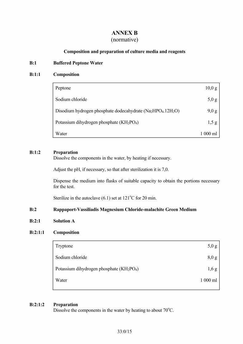
ANNEX B(normative)
Composition and preparation of culture media and reagents
B:1 Buffered Peptone Water
B:1:1 Composition
Peptone
Sodium chloride
Disodium hydrogen phosphate dodecahydrate (Na2HPO4.12H2O)
Potassium dihydrogen phosphate (KH2PO4)
Water
10,0 g
5,0 g
9,0 g
1,5 g
1 000 ml
B:1:2 PreparationDissolve the components in the water, by heating if necessary.
Adjust the pH, if necessary, so that after sterilization it is 7,0.
Dispense the medium into flasks of suitable capacity to obtain the portions necessaryfor the test.
Sterilize in the autoclave (6.1) set at 121oC for 20 min.
B:2 Rappaport-Vassiliadis Magnesium Chloride-malachite Green Medium
B:2:1 Solution A
B:2:1:1 Composition
Tryptone
Sodium chloride
Potassium dihydrogen phosphate (KH2PO4)
Water
5,0 g
8,0 g
1,6 g
1 000 ml
B:2:1:2 PreparationDissolve the components in the water by heating to about 70oC.

33:0/16
The solution shall be prepared on the day of preparation of the RV medium.
B:2:2 Solution B
B:2:2:1 Composition
Magnesium chloride hexahydrate (MgCl2.6H2O)
Water
400,0 g
1 000 ml
B:2:2:2 PreparationDissolve the magnesium chloride in the water.
As this salt is very hygroscopic, it is advisable to dissolve the entire contents of(MgCl2.6H2O) from a newly opened container, according to the formula. For instance,250 g of (MgCl2.6H2O) is added to 625 ml of water, giving a solution of total volumeof 795 ml and a concentration of about 31,5 g percent of (MgCl2.6H2O).
This solution can be kept in a brown glass bottle at room temperature.
B:2:3 Solution C
B:2:3:1 Composition
Malachite green oxalate
Water
0,4 g
100 ml
B:2:3:2 PreparationDissolve the malachite green oxalate in the water.
The solution can be kept in a brown glass bottle at room temperature.
B:2:4 Complete Medium
B:2:4:1 Composition
Solution A (B:2:1)
Solution B (B:2:2)
Solution C (B:2:3)
1 000 ml
100 ml
10 ml
B:2:4:2 Preparation

33:0/17
Add to 1 000 ml of solution A, 100 ml of solution B and 10 ml of solution C.
Adjust the pH, if necessary, so that after sterilization it is 5,2.
Distribute before use into test tubes in 10 ml quantities.
Sterilize in the autoclave (6:1) set at 115oC for 15 min.
Store the prepared medium in the refrigerator.
B:3 Selenite/cystine Medium
B:3:1 Base
B:3:1:1 Composition
Tryptone
Lactose
Disodium hydrogen phosphate dodecahydrate (Na2HPO4.12H2O)
Sodium hydrogen selenite
Water
5,0 g
4,0 g
10,0 g
4,0 g
1 000 ml
B:3:1:2 PreparationDissolve the first three basic components in the water by boiling for 5 min. Aftercooling, add the sodium hydrogen selenite.
Adjust the pH, if necessary, so that it is 7,0.
B:3:2 L-Cystine Solution
B:3:2:1 Composition
L-Cystine
Sodium hydroxide solution, c(NaOH) = 1 mol/l
Sterile water, to a final volume of
0,1 g
15 ml
100 ml
B:3:2:2 PreparationPlace the components in a sterile volumetric flask.
Dilute to 100 ml with sterile water.

33:0/18
Do not sterilize.
B:3:3 Complete Medium
B:3:3:1 Composition
Base (B:3:1)
L-Cystine solution (B:3:2)
1 000 ml
10 ml
B:3:3:2 Preparation
Cool the base and add the L-cystine solution aseptically. Adjust the pH, if necessary, sothat it is 7,0.
Dispense the medium aseptically into sterile flasks of suitable capacity to obtain theportions necessary for the test.
Use the medium on the day of preparation.
B:4:1 Base
B:4:1:1 Composition
Meat extract powder
Peptone
Yeast extract powder
Disodium hydrogen phosphate (Na2HPO4)
Sodium dihydrogen phosphate (NaH2PO4)
Agar in powder or in flake form
Water
5,0 g
10,0 g
3,0 g
1,0 g
0,6 g
12 g to 18 g 1)
900 ml
1) Depending on the gel strength of the agar.
B:4:1:2 Preparation
33:0/19
Dissolve the dehydrated base components or the dehydrated complete base in the waterby heating if necessary.
Adjust the pH, if necessary, so that after sterilization it is 7,0.
Transfer the base to tubes or flasks of appropriate capacity.
Sterilize in the autoclave (6:1) set at 121oC for 15 min.
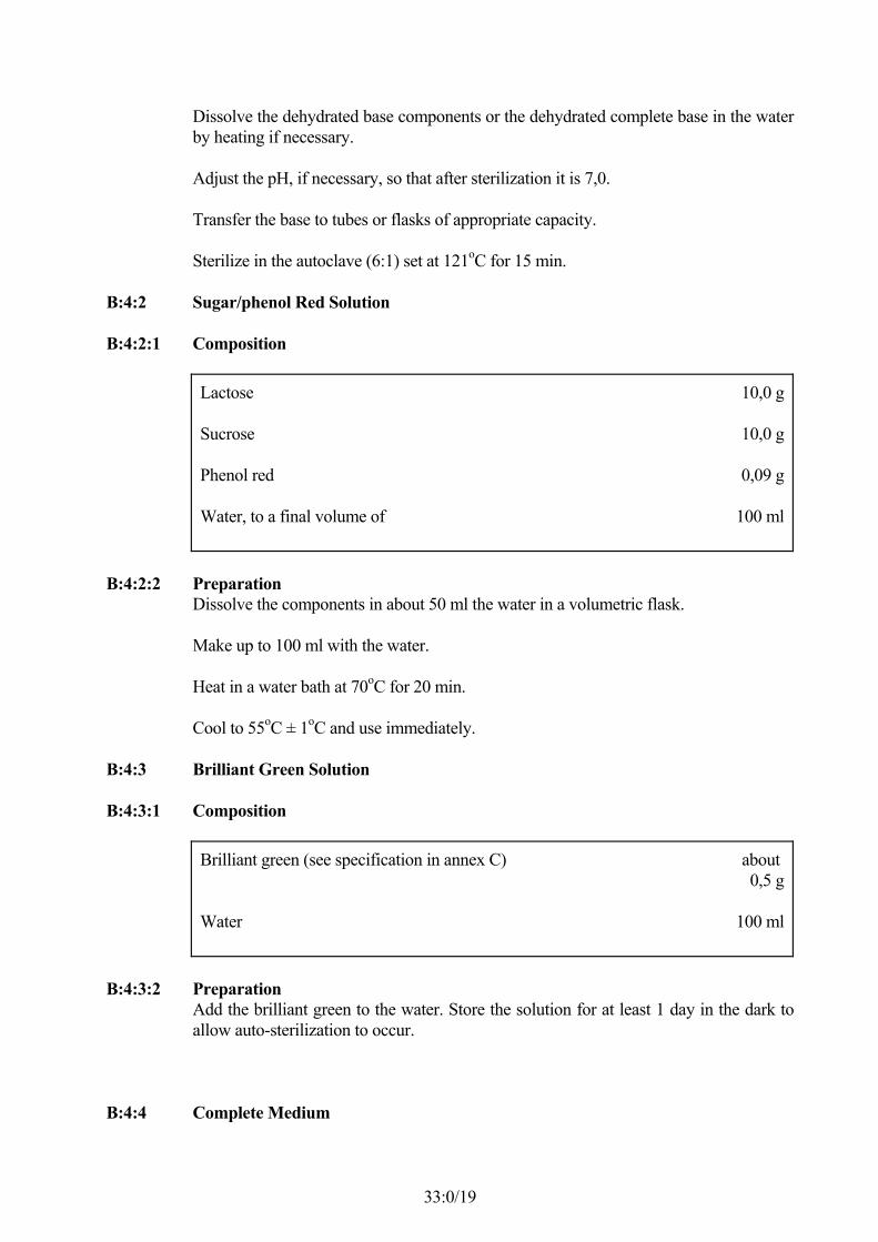
B:4:2 Sugar/phenol Red Solution
B:4:2:1 Composition
Lactose
Sucrose
Phenol red
Water, to a final volume of
10,0 g
10,0 g
0,09 g
100 ml
B:4:2:2 PreparationDissolve the components in about 50 ml the water in a volumetric flask.
Make up to 100 ml with the water.
Heat in a water bath at 70oC for 20 min.
Cool to 55oC ± 1oC and use immediately.
B:4:3 Brilliant Green Solution
B:4:3:1 Composition
Brilliant green (see specification in annex C)
Water
about 0,5 g
100 ml
B:4:3:2 PreparationAdd the brilliant green to the water. Store the solution for at least 1 day in the dark toallow auto-sterilization to occur.
B:4:4 Complete Medium

33:0/20
B:4:4:1 Composition
Base (B:4:1)
Sugar/phenol red solution (B:4:2)
Brilliant green solution (B:4:3)
900 ml
100 ml
1 ml
B:4:4:2 Preparation
Add under aseptic conditions, the brilliant green solution to the sugar/phenol redsolution cooled to 55oC ± 1oC. Add it all to the base at 50oC to 55oC and mix.
B:4:5 Preparation of the Agar PlatesPlace in each of an appropriate number of large Petri dishes (6:13) about 40 ml of thefreshly prepared complete medium (B:4:4). (If large dishes are not available, placeabout 15 ml of the medium in small Petri dishes.) Allow to solidify.
Immediately before use, dry the agar plates carefully (preferably with the lids off andthe agar surface downwards) in the oven (6:2) set between 37oC and 55oC until thesurface of the agar is dry.
If prepared in advance, the undried agar plates shall be kept for no longer than 4 h atroom temperature or one day in the refrigerator.
B:5 Nutrient Agar
B:5:1 Composition
Meat extract
Peptone
Agar
Water
3,0 g
5,0 g
12 g to 18 g 1)
1 000 ml
1) Depending on the gel strength of the agar.
B:5:2 Preparation
Dissolve the components or the dehydrated complete medium in the water, by heatingif necessary.

33:0/21
Adjust the pH, if necessary, so that after sterilization it is 7,0.
Transfer the culture medium into tubes or bottles of appropriate capacity.
Sterilize in the autoclave (6:1) set at 121oC for 20 min.
B:5:3 Preparation of nutrient agar platesTransfer about 15 ml of the melted medium to sterile small Petri dishes (6:13) andproceed as in B:4:5.
B:6 Triple Sugar/iron Agar (TSl agar)
B:6:1 Composition
Meat extract
Yeast extract
Peptone
Sodium chloride
Lactose
Sucrose
Glucose
Iron(III) citrate
Sodium thiosulfate
Phenol red
Agar
Water
3,0 g
3,0 g
20,0 g
5,0 g
10,0 g
10,0 g
1,0 g
0,3 g
0,3 g
0,024 g
12 g to18 g 1)
1 000 ml
1) Depending on the gel strength of the agar.
B:6:2 PreparationDissolve the components or the dehydrated complete medium in the water, by heatingif necessary.

33:0/22
Adjust the pH, if necessary, so that after sterilization it is 7,4.
Dispense the medium in quantities of 10 ml into test tubes.
Sterilize in the autoclave (6:1) set at 121oC for 10 min.
Allow to set in a slopng position to give a butt of depth 2,5 cm.
B:7 Urea Agar (Christensen)
B:7:1 Base
B:7:1:1 Composition
Peptone
Glucose
Sodium chloride
Potassium dihydrogen phosphate (KH 2PO4)
Phenol red
Agar
Water
1,0 g
1,0 g
5,0 g
2,0 g
0,012 g
12 g to18 g 1)
1 000 ml
1) Depending on the gel strength of the agar.
B:7:1:2 PreparationDissolve the components or the dehydrated complete base in the water, by heating ifnecessary.
Adjust the pH, if necessary, so that after sterilization it is 6,8.
Sterilize in the autoclave (6.1) set at 121oC for 20 min.
B:7:2 Urea Solution
B:7:2:1 Composition
Urea
Water, to a final volume of
400 g
1 000 ml
33:0/23
B:7:2:2 PreparationDissolve the urea in the water.
Sterilize by filtration and check the sterility.
(For details of the technique of sterilization by filtration, refer to any appropriatetextbook on microbilogy).
B:7:3 Complete Medium
B:7:3:1 Composition
Base (B:7:1)
Urea solution (B:7:2)
950 ml
50 ml
B:7:3:2 PreparationAdd, under aseptic conditions, the urea solution to the base, previously melted and thencooled to 45 C ± 1oC. Dispense the complete medium in quantities of 10 ml into steriletubes. Allow to set in a sloping position.
B:8 L-Lysine Decarboxylation Medium
B:8:1 Composition
L-Lysine monohydrochloride
Yeast extract
Glucose
Bromocresol purple
Water
5,0 g
3,0 g
1,0 g
0,015 g
1 000 ml
B:8:2 PreparationDissolve the components in the water, by heating if necessary.
Adjust the pH, if necessary, so that after sterilization it is 6,8.
Transfer the medium in quantities of 5 ml to narrow culture tubes (6:10).
Sterilize in the autoclave (6:1) set at 121oC for 10 min.
B:9 β-Galactosidase Reagent

33:0/24
B:9:1 Buffer Solution
B:9:1:1 Composition
Sodium dihydrogen phosphate (NaH2PO4)
Sodium hydroxide, 10 mol/l solution
Water, to a final volume of
6,9 g
about 3 ml
50 ml
B:9:1:2 PreparationDissolve the sodium dihydrogen jphosphate in approximately 45 ml of water in avolumetric flask.
Adjust the pH to 7,0 with the sodium hydroxide solution.
B:9:2 ONPG Solution
B:9:2:1 Composition
o-Nitrophenyl β-galactosidase (ONPG)
Water
0,08 g
15 ml
B:9:2:2 PreparationDissolve the ONPG in the water at 50oC ± 1oC.
Cool the solution.
B:9:3 Complete Reagent
B:9:3:1 Composition
Buffer solution (B:9:1)
ONPG solution (B:9:2)
5 ml
15 ml
B:9:3:2 PreparationAdd the buffer solution to the ONPG solution.
B:10 Reagents for Voges-Proskauer (VP) Reaction
B:10:1 VP Medium

33:0/25
B:10:1:1 Composition
Peptone
Glucose
Dipotassium hydrogen phosphate (K2HPO4)
Water
7,0 g
5,0 g
5,0 g
1 000 ml
B:10:1:2 PreparationDissolve the components in the water, by heating if necessary.
Adjust the pH, if necessary, so that after sterilization it is 6,9.
Transfer 3 ml of the medium into each of several tubes.
Sterilize in the autoclave (6:1) set at 115oC for 20 min.
B:10:2 Creatine Solution (N-amidinosarcosine)
B:10:2:1 Composition
Creatine monohydrate
Water
0,5 g
100 ml
B:10:2:2 PreparationDissolve the creatine monohydrate in the water.
B:10:3 1-Naphthol, ethanolic solution
B:10:3:1 Composition
1-Naphthol
Ethanol, 96 % (V/V)
6 g
100 ml
B:10:3:2 PreparationDissolve the 1-naphthol in the ethanol.
B:10:4 Potassium Hydroxide Solution
B:10:4:1 Composition
Potassium hydroxide
Water
40 g
100 ml
33:0/26
B:10:4:2 Preparation
Dissolve the potassium hydroxide in the water.
B:11 Reagents for Indole Reaction
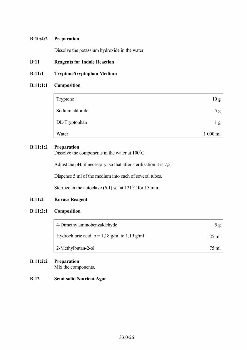
B:11:1 Tryptone/tryptophan Medium
B:11:1:1 Composition
Tryptone
Sodium chloride
DL-Tryptophan
Water
10 g
5 g
1 g
1 000 ml
B:11:1:2 PreparationDissolve the components in the water at 100oC.
Adjust the pH, if necessary, so that after sterilization it is 7,5.
Dispense 5 ml of the medium into each of several tubes.
Sterilize in the autoclave (6.1) set at 121oC for 15 min.
B:11:2 Kovacs Reagent
B:11:2:1 Composition
4-Dimethylaminobenzaldehyde
Hydrochloric acid ρ = 1,18 g/ml to 1,19 g/ml
2-Methylbutan-2-ol
5 g
25 ml
75 ml
B:11:2:2 PreparationMix the components.
B:12 Semi-solid Nutrient Agar

33:0/27
B:12:1 Composition
Meat extract
Peptone
Agar
Water
3,0 g
5,0 g
4 g to9 g 1)
1 000 ml
1) Depending on the gel strength of the agar.
B:12:2 Preparation
Dissolve the components in the water, by heating if necessary.
Adjust the pH, if necessary, so that after sterilization it is 7,0.
Transfer the medium to flasks of appropriate capacity.
Sterilize at 121oC for 20 min.
B:13 Saline Solution
B:13:1 Composition
Sodium Chloride
Water
8,5 g
1000 ml
B:13:2 Preparation
Dissolve the sodium chloride in the water, by heating if necessary.
Adjust the pH, if necessary, so that after sterilization it is 7,0.
Dispense quantities of the solution into flasks or tubes so that they will contain 90 ml to100 ml after sterilization.
Sterilize in the autoclave (6.1) set at 121oC for 20 min.

33:0/28
ANNEX C(normative)
Specification for Brilliant GreenC:1 Bacteriological Performance
Suppression of spreading of Proteus on phenol red/brilliant green agar (5:2:4:1) whilethe growth of Salmonella is not inhibited.
C:2 Method of Test
C:2:1 Medium
Prepare the phenol red/brilliant green agar plates according to clause B:4 but withvarious concentrations of brilliant green within the range 4:5 mg/l to 6 mg/l.
C:2:2 Procedure
Inoculate one set of agar plates having different concentrations of brilliant green with apure culture of a swarming Proteus, and another similar set with a pure culture ofSalmonella and incubate these plates at 35oC or 37cC (as agreed) for no longer than 24h.
A satisfactory concentration of brilliant green should allow growth of Salmonella withtypical pink colonies, 1 mm to 2 mm in diameter, and limited growth of Proteus; i.e. nospreading.
The concentration of brilliant green which shows this pattern should be used for thepreparation of the brilliant green solution (see B:4:3).

33:0/29


![(2Z,N 0 0 0 E)-N 0 0 0 -[(2-Hydroxy-1-naphthyl)- methylidene]furan-2-carbohydrazonic acid](https://static.fdokumen.com/doc/165x107/631360d4b033aaa8b2100e91/2zn-0-0-0-e-n-0-0-0-2-hydroxy-1-naphthyl-methylidenefuran-2-carbohydrazonic.jpg)