Metabotropic glutamate receptor-dependent long-term depression is impaired due to elevated ERK...
23
Metabotropic Glutamate Receptor-dependent Long-term Depression is Impaired Due to Elevated ERK Signaling in the ΔRG Mouse Model of Tuberous Sclerosis Complex Itzamarie Chévere-Torres 1 , Hanoch Kaphzan 1 , Aditi Bhattacharya 1 , Areum Kang 1 , Jordan M. Maki 1 , Michael J. Gambello 2 , Jack L. Arbiser 3 , Emanuela Santini 1 , and Eric Klann 1,† 1 Center for Neural Science, New York University, New York, NY 10003 2 Division of Medical Genetics, Department of Pediatrics, University of Texas Health Science Center at Houston, Houston, TX 77030 3 Department of Dermatology, Emory University School of Medicine, Atlanta, GA 30322 Abstract Tuberous sclerosis complex (TSC) and fragile X syndrome (FXS) are caused by mutations in negative regulators of translation. FXS model mice exhibit enhanced metabotropic glutamate receptor-dependent long-term depression (mGluR-LTD). Therefore, we hypothesized that a mouse model of TSC, ΔRG transgenic mice, also would exhibit enhanced mGluR-LTD. We measured the impact of TSC2-GAP mutations on the mTORC1 and ERK signaling pathways and protein synthesis-dependent hippocampal synaptic plasticity in ΔRG transgenic mice. These mice express a dominant/negative TSC2 that binds to TSC1, but has a deletion and substitution mutation in its GAP-domain, resulting in inactivation of the complex. Consistent with previous studies of several other lines of TSC model mice, we observed elevated S6 phosphorylation in the brains of ΔRG mice, suggesting upregulated translation. Surprisingly, mGluR-LTD was not enhanced, but rather was impaired in the ΔRG transgenic mice, indicating that TSC and FXS have divergent synaptic plasticity phenotypes. Similar to patients with TSC, the ΔRG transgenic mice exhibit elevated ERK signaling. Moreover, the mGluR-LTD impairment displayed by the ΔRG transgenic mice was rescued with the MEK-ERK inhibitor U0126. Our results suggest that the mGluR-LTD impairment observed in ΔRG mice involves aberrant TSC1/2-ERK signaling. Keywords tuberous sclerosis complex; TSC2; TSC1; mTORC1; ERK; fragile x syndrome; mGluR-LTD; NMDAR-LTD Introduction Tuberous sclerosis complex (TSC) is an autosomal dominant disorder that can cause hamartomas, benign and malignant neoplasms, seizures, mental impairment and autism, © 2011 Elsevier Inc. All rights reserved. † To whom correspondence should be addressed: Eric Klann, Ph.D., Center for Neural Science, New York University, 4 Washington Place, Room 809, New York, NY 10003, [email protected], phone: (212) 992-9769, fax: (212) 995-4011. Publisher's Disclaimer: This is a PDF file of an unedited manuscript that has been accepted for publication. As a service to our customers we are providing this early version of the manuscript. The manuscript will undergo copyediting, typesetting, and review of the resulting proof before it is published in its final citable form. Please note that during the production process errors may be discovered which could affect the content, and all legal disclaimers that apply to the journal pertain. NIH Public Access Author Manuscript Neurobiol Dis. Author manuscript; available in PMC 2013 March 1. Published in final edited form as: Neurobiol Dis. 2012 March ; 45(3): 1101–1110. doi:10.1016/j.nbd.2011.12.028. NIH-PA Author Manuscript NIH-PA Author Manuscript NIH-PA Author Manuscript
Transcript of Metabotropic glutamate receptor-dependent long-term depression is impaired due to elevated ERK...
Metabotropic Glutamate Receptor-dependent Long-termDepression is Impaired Due to Elevated ERK Signaling in theΔRG Mouse Model of Tuberous Sclerosis Complex
Itzamarie Chévere-Torres1, Hanoch Kaphzan1, Aditi Bhattacharya1, Areum Kang1, JordanM. Maki1, Michael J. Gambello2, Jack L. Arbiser3, Emanuela Santini1, and Eric Klann1,†
1Center for Neural Science, New York University, New York, NY 100032Division of Medical Genetics, Department of Pediatrics, University of Texas Health ScienceCenter at Houston, Houston, TX 770303Department of Dermatology, Emory University School of Medicine, Atlanta, GA 30322
AbstractTuberous sclerosis complex (TSC) and fragile X syndrome (FXS) are caused by mutations innegative regulators of translation. FXS model mice exhibit enhanced metabotropic glutamatereceptor-dependent long-term depression (mGluR-LTD). Therefore, we hypothesized that a mousemodel of TSC, ΔRG transgenic mice, also would exhibit enhanced mGluR-LTD. We measured theimpact of TSC2-GAP mutations on the mTORC1 and ERK signaling pathways and proteinsynthesis-dependent hippocampal synaptic plasticity in ΔRG transgenic mice. These mice expressa dominant/negative TSC2 that binds to TSC1, but has a deletion and substitution mutation in itsGAP-domain, resulting in inactivation of the complex. Consistent with previous studies of severalother lines of TSC model mice, we observed elevated S6 phosphorylation in the brains of ΔRGmice, suggesting upregulated translation. Surprisingly, mGluR-LTD was not enhanced, but ratherwas impaired in the ΔRG transgenic mice, indicating that TSC and FXS have divergent synapticplasticity phenotypes. Similar to patients with TSC, the ΔRG transgenic mice exhibit elevatedERK signaling. Moreover, the mGluR-LTD impairment displayed by the ΔRG transgenic micewas rescued with the MEK-ERK inhibitor U0126. Our results suggest that the mGluR-LTDimpairment observed in ΔRG mice involves aberrant TSC1/2-ERK signaling.
Keywordstuberous sclerosis complex; TSC2; TSC1; mTORC1; ERK; fragile x syndrome; mGluR-LTD;NMDAR-LTD
IntroductionTuberous sclerosis complex (TSC) is an autosomal dominant disorder that can causehamartomas, benign and malignant neoplasms, seizures, mental impairment and autism,
© 2011 Elsevier Inc. All rights reserved.†To whom correspondence should be addressed: Eric Klann, Ph.D., Center for Neural Science, New York University, 4 WashingtonPlace, Room 809, New York, NY 10003, [email protected], phone: (212) 992-9769, fax: (212) 995-4011.Publisher's Disclaimer: This is a PDF file of an unedited manuscript that has been accepted for publication. As a service to ourcustomers we are providing this early version of the manuscript. The manuscript will undergo copyediting, typesetting, and review ofthe resulting proof before it is published in its final citable form. Please note that during the production process errors may bediscovered which could affect the content, and all legal disclaimers that apply to the journal pertain.
NIH Public AccessAuthor ManuscriptNeurobiol Dis. Author manuscript; available in PMC 2013 March 1.
Published in final edited form as:Neurobiol Dis. 2012 March ; 45(3): 1101–1110. doi:10.1016/j.nbd.2011.12.028.
NIH
-PA Author Manuscript
NIH
-PA Author Manuscript
NIH
-PA Author Manuscript
(DiMario, 2004). At the molecular level, TSC is caused by either the loss or malfunction ofeither hamartin (TSC1) or tuberin (TSC2), which interact in a heterodimer known as theTSC1/TSC2 complex, to negatively regulate mammalian target of rapamycin complex 1(mTORC1) (Cheadle et al., 2000). mTORC1 functions as a molecular gatekeeper for cap-dependent translation initiation in neurons. Activation of the phosphoinositide 3-kinase(PI3K)/Akt and extracellular signal-regulated kinase (ERK) signaling pathways results inthe phosphorylation of TSC2 and inhibition of the GTPase-activating protein (GAP) activityof TSC2, which leads to increased levels of Rheb-GTP. This type of signaling activates themTOR complex 1 (mTORC1) and subsequent phosphorylation ribosomal S6 kinase 1(S6K1) and eukaryote initiation factor 4E-binding protein (4E-BP), key translation initiationregulators (Cai et al., 2006; Jozwiak, 2006; Jozwiak et al., 2005; Orlova and Crino, 2010;Yang et al., 2006).
It has been estimated that sporadic cases of TSC range from 60 to 70% of the cases reported,and that TSC1 mutations are significantly underrepresented compared to TSC2 (Jones et al.,1997). TSC2 gene mutations are more frequent and result in a more severe phenotype inTSC patients (i.e. seizures and learning disability), with the exception of reported cases ofpatients with no mutation identified, as well as one TSC2 mutation that causes a more mildphenotype (Camposano et al., 2009; Dabora et al., 2001; Jansen et al., 2006; Kwiatkowski,2003). In addition, the TSC2 gene is more prone than the TSC1 gene to large deletions,rearrangements, and missense mutations. Of particular interest is the finding that missensemutations are clustered within TSC2 exons 34–38, which encode for either Rap1GAP orGAP3 (Maheshwar et al., 1997). The TSC2-GAP domain is an essential structural domainfor the hydrolysis of GTP-bound Rheb to its inactive GDP-bound form (Tee et al., 2003).
Studies have shown that either loss or malfunction of TSC1 and TSC2 usually results inactivation of S6K1 and enhanced ribosomal protein S6 phosphorylation, resulting indefective regulation of cell size and proliferation (Krymskaya, 2003; Uhlmann et al., 2004).Moreover, studies in hippocampal pyramidal neurons have shown that the TSC pathwayregulates soma size, the density and size of dendritic spines, and the properties of excitatorysynapses, particularly AMPA receptor-mediated currents (Tavazoie et al., 2005). Additionalstudies have shown that loss of TSC1 function in the brain leads to neocorticalhyperexcitability associated with increased glutamate-mediated excitation in both humantissue and mouse brain (Wang et al., 2007). Finally, TSC2 heterozygous knockout micewere shown to exhibit elevated hippocampal mTORC1 signaling, which led to abnormallong-term potentiation (LTP) and deficits in hippocampus-dependent memory (Ehninger etal., 2008).
The ΔRG transgenic mouse has been developed, carrying a deletion in TSC2 of amino acidresidues 1617–1655 and a substitution of amino acid residues 1679–1742, which interfereswith both the GAP domain and rabaptin-5 binding motif of TSC2, respectively(Govindarajan et al., 2005; Pasumarthi et al., 2000). As a result, this dominant/negativeTSC2 protein is not able to hydrolyze GTP-bound small G-proteins, such as Rap1 and Rheb(Govindarajan et al., 2005; Pasumarthi et al., 2000; Zhang et al., 2003). Previous studieshave shown that ΔRG transgenic mice have increased expression of the dominant/negativeTSC2 driven by the cytomegalovirus (CMV) promoter and develop skin and brainabnormalities consistent with those observed in TSC patients (Bhatia et al., 2009;Govindarajan et al., 2005; Sambucetti et al., 1989). In addition, behavioral studies on ΔRGmice have revealed increased anxiety levels and mild deficits in hippocampus-dependentlearning and memory, consistent with TSC-related neuropsychiatric symptoms (Ehningerand Silva, 2010; Chévere-Torres et al., 2012).
Chévere-Torres et al. Page 2
Neurobiol Dis. Author manuscript; available in PMC 2013 March 1.
NIH
-PA Author Manuscript
NIH
-PA Author Manuscript
NIH
-PA Author Manuscript
Fragile X syndrome (FXS) is caused by loss of function mutations in the RNA-bindingprotein, fragile X mental retardation protein (FMRP), whose normal function is to suppresstranslation (Ronesi and Huber, 2008). Consistent with this notion, mouse models of FXSdisplay increased protein synthesis, enhanced mTORC1 signaling, and exaggeratedmetabotropic glutamate receptor-dependent long-term depression (mGluR-LTD) (Hou et al.,2006; Huber et al., 2002; Osterweil et al., 2010; Sharma et al., 2010). Based on evidence thatboth TSC1/2 and FMRP proteins act as negative regulators of protein synthesis andmTORC1 signaling, and the evidence that patients with TSC and FXS can both displayautism spectrum disorder, we hypothesized that the mutations in TSC2-GAP domain inΔRG mice would result in similar synaptic plasticity alterations and mTORC1 dysregulationas observed in other mouse models of TSC and FXS model mice. Herein we describeexperiments with the ΔRG transgenic mice that were conducted to determine whether theyexhibit hippocampal synaptic plasticity phenotypes consistent with other mouse models ofTSC and FXS.
Materials and MethodsAnimals
ΔRG Transgenic Mice—Generation of ΔRG mice has been described previously(Govindarajan et al., 2005). Mouse genotyping was performed by PCR using transgene- andwild-type-specific primer sets.
Tsc1 Floxed Mice—Mice with floxed Tsc1 (mixed genetic background composed ofC57Bl/6J, 129/SvJae, BALB/cJ) were generated as described previously (Kwiatkowski etal., 2002). For the generation of experimental mice, we crossed heterozygous floxed Tsc1(TSC1+/−) male mice and heterozygous floxed Tsc1 (TSC1+/−)-αCaMKII-Cre females. Micewere genotyped using Cre-specific primers and primers that identify floxed alleles of theTsc1 locus. The wild-type mice used in this study were TSC1+/+-αCaMKII-Cre and theexperimental conditional heterozygous mice used in this study were TSC1+/cko-αCaMKII-Cre (referred to as TSC1+/− cKO) mice.
Tsc2 Floxed Mice—Mice with floxed Tsc2 (mixed genetic background composed ofC57Bl/6J, 129/SvJae) were generated as described previously (Hernandez et al., 2007). Forthe generation of experimental mice, we crossed heterozygous floxed Tsc2 (TSC2+/−) malemice and heterozygous floxed Tsc2 (TSC2+/−)-αCaMKII-Cre females. Mice were genotypedusing Cre-specific primers and primers that identify floxed alleles of the Tsc2 locus. Thewild-type mice used in this study were TSC2++-αCaMKII-Cre and the experimentalconditional homozygous mice used in this study were TSC2cko/cko-αCaMKII-Cre (referredto as TSC2−/− cKO) mice.
For all experiments, male age-matched littermates were used. For biochemical experiments,4–12 week-old mice were used, for LTP experiments 8–12 week-old mice were used, and 4–6 week-old mice were used for mGluR-LTD experiments. Mice were kept on a 12 h on-offlight/dark cycle. Food and water were available at all times. All procedures were approvedby the New York University Animal Care and Use Committee and followed the NIHGuidelines for the use of animals in research.
Western Blot AnalysisProtein extracts were prepared by homogenizing hippocampal tissue in ice-cold hypotoniclysis buffer (HLB) containing phosphatase and protease inhibitor mixtures. Homogenates(20 µg) were resolved via 4–12% SDS-PAGE and transferred to polyvinylidene difluoride(PVDF) membranes. Immunobloting was performed using standard techniques. Rabbit
Chévere-Torres et al. Page 3
Neurobiol Dis. Author manuscript; available in PMC 2013 March 1.
NIH
-PA Author Manuscript
NIH
-PA Author Manuscript
NIH
-PA Author Manuscript
polyclonal antibodies were used to detect phospho-S6 (S235/236), phospho-S6 (S240/244),total S6, phospho-S6K1 (T389), and total S6K1 at a 1:1000 dilution (Cell SignalingTechnologies, Beverly, MA). 1:1000 rabbit anti-phospho-S6K1 (T389, Millipore Corp.,Billerica, MA). A monoclonal antibody was used to detect TSC1 levels at a 1:1000 dilution(Cell Signaling, Beverly, MA) in 5% BSA blocking solution. TSC2 levels were detectedusing a monoclonal antibody (CellSignaling, Beverly, MA) at a 1:1000 dilution in 5% BSAblocking solution. The dilution used for phospho-4E-BP (T37/46) and total 4E-BPantibodies was 1:500 (Cell Signaling Technologies, Beverly, MA) in 5% BSA/I-Block.Phospho-ERK (T202/Y404) and total ERK antibodies were diluted at 1:2000. β–actin and α-tubulin antibodies (Sigma-Aldrich, St. Louis, MO) were diluted 1:10,000 in 5% milk/T-TBS. Anti-rabbit and anti-mouse HRP-tagged antibodies (Promega, Madison, WI) werediluted 1:5000 in 0.2% I-Block (Tropix, Foster City, CA) and 1:10,000 in 5%Milk/T-TBS,respectively. Blots were developed using enhanced chemiluminescence detection (GEHealthcare, Fairfield, CT).
Immunohistochemistry and HistologySaggital sections (40 µm thick) were blocked with 1% BSA and incubated overnight at 4°Cwith TSC2 rabbit monoclonal antibody (1:200; Epitomics, Burlingame, CA) in 1% BSA.Sections were washed with PBS and incubated with secondary antibody for 30 min. Sectionsthen were incubated with ABC reagent (Pierce, Rockford, IL) and immunostaining wasvisualized using the VIP substrate kit for peroxidase (Vector Laboratories, Burlingame,CA). Brains from a different set of mice were used for histological studies. Saggital sections(40 µm thick) were mounted onto subbed slides. Nissl staining was performed according tostandard procedures. Sections from immunohistochemistry and Nissl staining were viewedand photographed using a BX51 Olympus microscope (Olympus, UK) and Neurolucida 7software (MBF Bioscience, Willinston, VT).
ElectrophysiologyBrains from age-matched littermate mice were removed and transverse hippocampal slices(400 µm) were prepared. Hippocampal slices were placed in cold-cutting solution (in mM:110 sucrose, 60 NaCl, 3 KCl, 1.25 NaH2PO4, 28 NaHCO3, 5 D-glucose, 0.6 Ascorbic Acid,0.5 CaCl2, and 7 MgCl2, gassed with 95% O2/5% CO2). The slices were incubated with a50:50 cutting/artificial CSF (ACSF) solution (in mM: 125 NaCl, 2.5 KCl, 1.25 NaH2PO4, 25NaHCO3, 25 D-glucose, 2 CaCl2, and 1MgCl2) for 15 min, followed by equilibration for 1hr in a humid, oxygenated interface chamber continuously perfused with 32°C ACSF at arate of 2ml/min. Field excitatory postsynaptic potentials (fEPSPs) were evoked bystimulation of the Schaeffer collateral pathway and recorded in stratum radiatum of areaCA1 using a glass recording pipette (1–4 MΩ). Stimulus intensity was adjusted to elicit afEPSP that was 50% of the maximum response in each slice. Baseline measurements weretaken for 30 min prior to delivery of a single train of high-frequency stimulation (HFS; 100Hz for 1 sec) to induce early phase LTP (E-LTP), prior to delivery of four, spaced trains ofHFS (100 Hz for 1 sec, 5 min intertrain interval) to induce late phase LTP (L-LTP), for 20min prior to treatment with DHPG (50µM, 10min; Ascent, Princeton, NJ) to induce mGluR-LTD, or for 20 min prior to deliver 900 pulses of low-frequency stimulation (LFS; 1Hz, 15min) to induce N-methyl D-aspartate NMDA-LTD. When indicated, ACSF wassupplemented with U0126 (1mM, Promega, Madison, WI). For LTD rescue experiments,baseline measurements were taken for 30 min prior to application of DHPG (50µM, 10min)in the presence of U0126 (1µM). Afterward, fEPSPs were evoked and recorded in thestratum radiatum of area CA1 in the presence of the inhibitor for 1 hour.
Chévere-Torres et al. Page 4
Neurobiol Dis. Author manuscript; available in PMC 2013 March 1.
NIH
-PA Author Manuscript
NIH
-PA Author Manuscript
NIH
-PA Author Manuscript
Statistical AnalysisGraph Pad Prism data analysis software (San Diego, CA) was used for graph production andstatistical analysis was used to assess the data. Student’s t-tests and two-way ANOVAs wereused for biochemical and electrophysiological data analysis, respectively. Data representmean±SEM, with p<0.05 used as significance criteria.
ResultsΔRG mice exhibit normal hippocampal LTP but impaired mGluR-dependent LTD
First, we confirmed the presence of the ΔRG transgene (Fig. 1A) by PCR techniques,detecting a band of 280bp (Fig. 1B). We determined whether the loss of TSC2-GAPfunction in ΔRG mice resulted in aberrant hippocampal morphological changes. Nisslstaining of sections from ΔRG mice indicated they have normal gross hippocampalmorphology (Fig. 1C). To determine whether TSC2 levels were increased, we examinedhippocampal tissue from ΔRG mice and their wild-type littermates withimmunocytochemistry and observed increased TSC2 levels in ΔRG mice compared to theirwild-type controls (Fig. 1D), consistent with previous studies (Bhatia et al., 2009).
Because mice with mutations in negative modulators of translation exhibit altered synapticfunction (Richter and Klann, 2009), we examined several forms of hippocampal synapticplasticity in slices from ΔRG mice. Analysis of synaptic output in response to increasingstimulatory input indicated that the basal synaptic transmission was normal in hippocampalarea CA1 of the ΔRG mice (Fig. 2A). Fiber volley amplitude measured with increasingstimulation also was unaltered in ΔRG mice (Fig. 2B). We next examined paired-pulsefacilitation (PPF), a short-lasting form of presynaptic plasticity evoked by temporally linkedstimuli, and found that PPF was unaltered in ΔRG mice using both short and long interpulseintervals (Fig. 2C). We proceeded to determine whether the ΔRG mice exhibited alteredlong-term potentiation (LTP). We used a single train of high-frequency stimulation (HFS) toinduce early phase LTP (E-LTP) and four, spaced trains of HFS to induce protein synthesis-dependent late phase LTP (L-LTP) in hippocampal slices from wild-type and ΔRG mice.We observed that both E-LTP and L-LTP in ΔRG mice were indistinguishable from that intheir wild-type littermates (Fig. 3A and 3B). Thus, basal synaptic transmission, PPF, E-LTP,and L-LTP are unaltered in ΔRG mice.
Previous studies have shown that a mouse model of fragile X syndrome (FXS) exhibitsenhanced hippocampal mGluR-LTD due to improper regulation of translation and itsuncoupling from mGluR signaling (Ronesi and Huber, 2008; Sharma et al., 2010; Osterweilet al., 2010). In addition, clinical studies have shown increased immunoreactivity for group ImGluRs (mGluR1/5) and phosphorylated ribosomal protein S6 within cortical tubers andsubependymal giant-cell tumors of TSC specimens (Boer et al., 2008), suggestingupregulation of mGluR-mTORC1 signaling similar to that observed in FXS model mice(Sharma et al., 2010). Therefore, we hypothesized that hippocampal mGluR-dependent LTDwould be enhanced in the ΔRG mice. Hippocampal slices from ΔRG mice and their wild-type littermates were treated with DHPG (50 µM, 10 min), a selective agonist of group ImGluRs, which resulted in a pronounced initial depression of synaptic transmission in bothΔRG and wild-type mice (Fig. 3C). This depression rapidly returned to near baseline levelsin ΔRG mice after drug washout, whereas in wild-type mice it was followed by a robustlong-lasting depression (Fig. 3C). This finding indicates that mutations affecting the TSC2-GAP domain result in an impairment of mGluR-LTD in the hippocampus. To determine thereceptor specificity of the LTD deficit in ΔRG mice, low-frequency stimulation (LFS) wasutilized to induce N-methyl D-aspartate receptor-dependent LTD (NMDAR-LTD) inhippocampal slices from ΔRG mice and their wild-type littermates. We observed that
Chévere-Torres et al. Page 5
Neurobiol Dis. Author manuscript; available in PMC 2013 March 1.
NIH
-PA Author Manuscript
NIH
-PA Author Manuscript
NIH
-PA Author Manuscript
NMDAR-LTD in ΔRG mice was identical to that of their wild-type littermates (Fig. 3D).These findings indicate that the impaired LTD is specific to altered mGluR-dependentsignaling in the ΔRG mice.
We next tested whether mGluR-LTD also was impaired in mice with a heterozygousconditional deletion of Tsc1 (TSC1+/− cKO) and homozygous conditional deletion of Tsc2(TSC2−/− cKO) in postnatal forebrain neurons under the control of the αCaMKII promoter(αCamKII-Cre) (Supplementary Fig. 1). We found that both TSC1+/− cKO and TSC2−/−
cKO mice exhibited significant impairments in mGluR-LTD compared to their wild-typecontrols (Fig. 4A and 4B). Further analysis and comparison of the average fEPSP slopesfrom ΔRG, TSC1+/− cKO and TSC2−/− cKO hippocampal slices during the time period of20 to 50 min after DHPG application revealed that all three TSC mouse models have asimilar level of LTD impairment (Fig. 4C). These findings indicate that disruption of theTSC1/2 complex either by conditional deletion of Tsc1/2 genes or by mutations affecting theTSC2-GAP domain results in deficient mGluR-LTD expression in the hippocampus.
Hippocampal mTORC1 signaling in ΔRG miceThe impairment in mGluR-LTD led us to examine mTORC1 signaling in hippocampalhomogenates from the ΔRG mice. Western blot analysis indicated that althoughphosphorylation of serine 235/236 on ribosomal protein S6 was elevated in the ΔRG mice,phosphorylation of serine 240/244, a rapamycin-sensitive/mTORC1-dependent site, wasunaffected (Fig. 5A). Moreover, phosphorylation of eukaryotic initiation factor 4E-bindingprotein (4E-BP) and p70 S6 kinase 1 (S6K1), direct substrates of mTORC1, were not alteredin the ΔRG mice (Fig. 5B). Because phosphorylation of serine 235/236 on S6 can beregulated by both mTORC1 and ERK signaling (Pende et al., 2004; Roux et al., 2004), weexamined ERK1/2 phosphorylation in the hippocampus of ΔRG mice. We observed that theΔRG mice exhibited a robust increase in ERK1/2 phosphorylation compared to their wild-type littermates (Fig. 5C). These findings suggest that the impaired mGluR-LTD observed inΔRG mice is not a direct consequence of elevated mTORC1 signaling as initially postulated,but could be related to aberrant TSC1/2-ERK signaling.
We also examined ERK1/2 phosphorylation in the hippocampus of TSC1+/− cKO andTSC2−/− cKO mice to determine whether enhanced ERK signaling also is a feature ofTSC1/2 deletion in the brain. We found that ERK phosphorylation was not affected in thehippocampus of either TSC1+/− cKO or TSC2−/− cKO mice (Fig. 6A and 6B). These datasuggest that a distinct molecular mechanism may be responsible for the deficit observed inmGluR-LTD in TSC1+/− cKO and TSC2−/− cKO mice, while the mGluR-LTD impairmentand aberrant ERK1/2 activation observed in ΔRG mice is because of TSC2-GAP mutationsin ΔRG mice.
Inhibition of MEK-ERK signaling rescues mGluR dependent-LTD in ΔRG miceOur findings of enhanced ERK phosphorylation led us to examine the contribution of MEK-ERK signaling to the impaired mGluR-LTD exhibited by the ΔRG mice. In wild-type mice,mGluR-LTD requires activation of both mTORC1 and MEK-ERK signaling (Banko et al.,2006; Gallagher et al., 2004; Hou and Klann, 2004; Sharma et al., 2010). We utilized theMEK inhibitor U0126 (Favata et al., 1998) and performed dose/response experiments inhippocampal slices from wild-type mice in an attempt to find an effective dose at whichlevels of ERK phosphorylation were reduced but not completely blocked and did notcompromise the integrity of mGluR-LTD induction in wild-type mice (Fig. 7A). Westernblot analysis indicated that ERK phosphorylation was significantly reduced when sliceswere incubated with U0126 at concentrations of either 1 µM (approximately 30% inhibition)or 5µM (approximately 80% inhibition), but not at 100 nM (Fig. 7A). To determine whether
Chévere-Torres et al. Page 6
Neurobiol Dis. Author manuscript; available in PMC 2013 March 1.
NIH
-PA Author Manuscript
NIH
-PA Author Manuscript
NIH
-PA Author Manuscript
the elevated MEK-ERK signaling was involved in the impaired mGluR-LTD, hippocampalslices from ΔRG mice and their wild-type littermates were treated with DHPG in thepresence of U0126 (1 µM). Treatment with U0126 rescued the mGluR-LTD deficitobserved in hippocampal slices from ΔRG mice (Fig. 7B, right panel). The effect of U0126was specific to mGluR-LTD in ΔRG mice, because this concentration of the drug did notsignificantly impact mGluR-LTD in wild-type littermates (Fig. 7B, left panel). To determinewhether treatment of ΔRG hippocampal slices with U0126 rescued mGluR-LTD back to thesame level of control groups (WT + vehicle; WT + U0126) we analyzed the average fEPSPslopes from WT and ΔRG hippocampal slices in the presence of the vehicle or U0126during the time period of 20 to 50 min after the washout of DHPG. We found that treatmentwith U0126 returned mGluR-LTD in ΔRG mice back to levels observed in WT mice (Fig.7C). We then determined whether treatment with DHPG resulted in a further increase inERK phosphorylation in ΔRG mice. Hippocampal slices were treated with DHPG and areaCA1 was isolated. Western blot data showed a significant increase of ERK phosphorylationin slices from ΔRG mice treated with DHPG (Fig. 8A). In addition, Western Blot analysisfrom area CA1 of hippocampal slices collected after the rescue experiment confirmed thereduction of ERK phosphorylation by U0126 (1µM) (Fig. 8B). Taken together, thesefindings are consistent with the idea that the impaired hippocampal mGluR-LTD exhibitedby ΔRG mice is due to enhanced MEK-ERK signaling.
DiscussionOur examination of hippocampal synaptic plasticity in the ΔRG mouse model of TSCresulted in several unexpected findings. First, we observed that both E-LTP and L-LTP wereunaltered in ΔRG mice (Fig. 3A and 3B). In contrast, E-LTP-inducing stimulation wasshown to produce stable, rapamycin-sensitive, L-LTP in TSC2 heterozygous knockout mice(Ehninger et al., 2008). It appears that in the complete absence of one allele, as in the case ofTSC2 heterozygous knockout mice, the impact on mTORC1 signaling is more robust andbroader than in the case of having specific mutations in the TSC2-GAP domain as is presentin the ΔRG mice. Indeed, we observed that mTORC1 signaling was not significantly alteredin the ΔRG mice (Fig. 4), whereas mTORC1 signaling in both TSC1+/− cKO and TSC2−/−
cKO mice were enhanced as shown by increase phosphorylation of S6K1 (SupplementaryFig. 1). The latter finding is consistent with recent studies where the hippocampal deletionof the Tsc1 gene resulted in enhanced mTORC1 signaling (Bateup et al., 2011). The secondunexpected finding was that mGluR-LTD was impaired in the ΔRG mice (Fig. 3D).Moreover, the ΔRG mice exhibited normal NMDAR-LTD in (Fig. 3C), similar to recentfindings in mice with virally-mediated deletion of the Tsc1 gene in the hippocampus (Bateupet al., 2011). Thus, disruption of TSC2-GAP function, either by GAP mutations ordecreasing TSC1/2 heterodimer stability compromising TSC2-GAP activity, interferes withdistinct molecular mechanisms required for the expression of mGluR-LTD but notNMDAR-LTD.
mGluR-LTD in both TSC1+/− cKO and TSC2−/− cKO mice was impaired at the same levelas it was in ΔRG mice (Fig. 4C). Our findings with TSC1+/− cKO mice (Fig. 4A) confirmrecent studies showing that the loss of Tsc1 impairs hippocampal mGluR-LTD (Bateup etal., 2011). The impaired mGluR-LTD in ΔRG mice was unexpected because mGluR-LTD,ERK phosphorylation, and mTORC1 signaling are enhanced in FXS model mice that lackFMRP, a translation suppressor (Hou et al., 2006; Sharma et al., 2010). It is possible thatthere are different subcellular pools of ERK that are activated in ΔRG mice and FXS micethat results in the phosphorylation of dissimilar ERK substrates that impact mGluR-LTDdifferently. Interestingly, the mGluR-LTD phenotype in the ΔRG mouse model of TSCdiffers from FXS model mice lacking FMRP, but more closely approximates that observedin transgenic mice overexpressing FMRP, which exhibit completely abolished mGluR-LTD
Chévere-Torres et al. Page 7
Neurobiol Dis. Author manuscript; available in PMC 2013 March 1.
NIH
-PA Author Manuscript
NIH
-PA Author Manuscript
NIH
-PA Author Manuscript
(Hou et al., 2006). Thus, ΔRG mice and FXS mice are examples of two types of improperactivation of ERK and mTORC1 signaling in the brain. In ΔRG mice, robust enhancementof ERK signaling results in impaired mGluR-LTD, whereas modest activation of ERK andmTORC1 signaling in FXS model mice results in enhanced mGluR-LTD (Hou et al., 2006;Sharma et al., 2010).
Although upregulated mTORC1 signaling has been reported in other mouse models of TSCand FXS (Bateup et al., 2011; Ehninger et al., 2008; Sharma et al., 2010), the mutation in theTSC2-GAP domain in the ΔRG mice appears to recruit an mTORC1-independentmechanism in the hippocampus, the third unexpected finding in our studies. Normally,TSC2 negatively regulates Rheb, which is immediately upstream of mTORC1 (Tee et al.,2003). Active, GTP-bound Rheb also has been shown to negatively regulate Ras/B-Raf/C-Raf/MEK signaling by disrupting B-Raf/C-Raf heterodimerization (Im et al., 2002;Karbowniczek et al., 2006). B-Raf has a high basal kinase activity toward MEK1/2, and isnow available to participate in the activation Rap1/Epac/B-Raf signaling pathway (Moodieet al., 1994; Papin et al., 1998; Papin et al., 1996; Reuter et al., 1995; Wang et al., 2006),which is then a potential mechanism for the upregulated ERK phosphorylation and S6235/236 phoshporylation observed in the ΔRG mice (Fig. 5A and 5C). In fact, the impairedmGluR-LTD observed in ΔRG mice was rescued with the MEK inhibitor U0126, supportingthe idea that elevated MEK-ERK signaling contributes to synaptic abnormalities in thesemice (Fig. 7B). Moreover, the enhanced basal phosphorylation of ERK in ΔRG mice isfurther increased by DHPG treatment (Fig. 8A). In contrast, conditional deletion of Tsc1 andTsc2 genes in the forebrain did not result in upregulated ERK phosphorylation (Fig. 6) Thus,it is possible that synaptic and behavioral phenotypes in mice with conditional deletions ofTsc1 and Tsc2 might be due to exaggerated mTORC1 signaling (Supplementary Fig. 1B)(Bateup et al., 2011; Ehninger et al., 2008) in contrast to the ΔRG mice where thephenotypes are due to exaggerated ERK signaling.
An additional potential mechanism for the elevated ERK phosphorylation we observed inthe ΔRG mice is altered activity of the striatal-enriched protein tyrosine phosphatase(STEP). STEP is a phosphatase shown to regulate synaptic function by modulating NMDAand AMPA receptor trafficking, as well as ERK phosphorylation and nuclear translocation(Braithwaite et al., 2006; Paul et al., 2003; Snyder et al., 2005). In addition, studies haveshown that STEP translation is triggered by activation of group I mGluRs and subsequentactivation of the ERK, PI3K and mTORC1 pathways (Zhang et al., 2008). It remains to bedetermined whether dysregulation of STEP is involved in the elevated ERK phosphorylationobserved in the ΔRG mice.
Importantly, in accordance with our findings, analyses of brain lesions and tumorsassociated with TSC have shown aberrantly activated ERK that is correlated with increasedphosphorylation of ribosomal protein S6 (Jozwiak et al., 2008; Ma et al., 2005; Ma et al.,2007). Thus, simultaneous treatment with inhibitors of both mTORC1 and ERK signalingpathways could be an effective treatment for TSC patients. Future studies will be required todelineate the molecular mechanism resulting in altered TSC1/2-ERK signaling and whetherit is involved in any cognitive and behavioral abnormalities displayed by the ΔRG mice.
ConclusionsOur studies herein show that disruptive mutations of the GAP domain of TSC2 impairs theexpression of mGluR-LTD in ΔRG mice, without affecting NMDAR-dependent LTD.Moreover, rescue experiments with the MEK inhibitor U0126 supports the idea thatmutations in the TSC2-GAP domain in the ΔRG mice recruits an ERK-dependent andmTORC1-independent mechanism, resulting in impaired mGluR-LTD. In contrast, MEK-ERK signaling appears to be normal but mTORC1 signaling is elevated in mice with
Chévere-Torres et al. Page 8
Neurobiol Dis. Author manuscript; available in PMC 2013 March 1.
NIH
-PA Author Manuscript
NIH
-PA Author Manuscript
NIH
-PA Author Manuscript
conditional deletion of Tsc1 and Tsc2 genes, suggesting that impaired mGluR-LTD in thesemice is due to exaggerated mTORC1 signaling. Consequently, ΔRG mice is a mouse modelof TSC that can be used to study the role of both TSC2-GAP domain and TSC1/2-ERKsignaling in abnormal synaptic plasticity associated with TSC.
Research Highlights
• Mutations in TSC2-GAP domain disrupt hippocampal mGluR-LTD in ΔRGmice.
• ΔRG mice exhibit normal mTORC1 signaling, but enhanced ERK signaling.
• Impaired hippocampal mGluR-LTD is due to aberrant MEK/ERK singnaling inΔRG mice.
• Mice with conditional deletions of the Tsc1 and Tsc2 genes exhibit deficits inmGluR-LTD, but normal MEK/ERK signaling
Supplementary MaterialRefer to Web version on PubMed Central for supplementary material.
Abbreviations
4E-BP eIF4E binding protein
ACSF artificial cerebrospinal fluid
CMV cytomegalovirus
FXS fragile X syndrome
GAP GTPase-activating protein
GDP guanosine diphosphate
GTP guanosine triphosphate
mTORC1 mammalian trget of rapamycin complex 1
Rheb Ras homologenrinched in brain
STEP striatal-enrinched tyrosine phosphatase
TSC tuberous sclerosis complex
AcknowledgmentsThis work was supported by National Institutes of Health grants NS034007 and NS047384 (E.K.), and MH085489(I.C.T.).
ReferencesBanko JL, et al. Regulation of eukaryotic initiation factor 4E by converging signaling pathways during
metabotropic glutamate receptor-dependent long-term depression. J Neurosci. 2006; 26:2167–2173.[PubMed: 16495443]
Bateup HS, et al. Loss of Tsc1 In Vivo Impairs Hippocampal mGluR-LTD and Increases ExcitatorySynaptic Function. J Neurosci. 2011; 31:8862–8869. [PubMed: 21677170]
Bhatia B, et al. Tuberous sclerosis complex suppression in cerebellar development andmedulloblastoma: separate regulation of mammalian target of rapamycin activity and p27 Kip1localization. Cancer Res. 2009; 69:7224–7234. [PubMed: 19738049]
Chévere-Torres et al. Page 9
Neurobiol Dis. Author manuscript; available in PMC 2013 March 1.
NIH
-PA Author Manuscript
NIH
-PA Author Manuscript
NIH
-PA Author Manuscript
Boer K, et al. Cellular localization of metabotropic glutamate receptors in cortical tubers andsubependymal giant cell tumorFs of tuberous sclerosis complex. Neuroscience. 2008; 156:203–215.[PubMed: 18706978]
Braithwaite SP, et al. Synaptic plasticity: one STEP at a time. Trends Neurosci. 2006; 29:452–458.[PubMed: 16806510]
Cai SL, et al. Activity of TSC2 is inhibited by AKT-mediated phosphorylation and membranepartitioning. J Cell Biol. 2006; 173:279–289. [PubMed: 16636147]
Camposano SE, et al. Distinct clinical characteristics of tuberous sclerosis complex patients with nomutation identified. Ann Hum Genet. 2009; 73:141–146. [PubMed: 19133941]
Cheadle JP, et al. Molecular genetic advances in tuberous sclerosis. Hum Genet. 2000; 107:97–114.[PubMed: 11030407]
Chévere-Torres I, et al. Impaired social interactions and motor learning in tuberous sclerosis complexmodel mice expressing a dominant/negative form of tuberin. Neurobiol. Dis. 2012 in press.
Dabora SL, et al. Mutational analysis in a cohort of 224 tuberous sclerosis patients indicates increasedseverity of TSC2, compared with TSC1, disease in multiple organs. Am J Hum Genet. 2001;68:64–80. [PubMed: 11112665]
DiMario FJ Jr. Brain abnormalities in tuberous sclerosis complex. J Child Neurol. 2004; 19:650–657.[PubMed: 15563010]
Ehninger D, et al. Reversal of learning deficits in a Tsc2+/− mouse model of tuberous sclerosis. NatMed. 2008; 14:843–848. [PubMed: 18568033]
Ehninger D, Silva AJ. Increased Levels of Anxiety-related Behaviors in a Tsc2 Dominant NegativeTransgenic Mouse Model of Tuberous Sclerosis. Behav Genet. 2010
Favata MF, et al. Identification of a novel inhibitor of mitogen-activated protein kinase kinase. J BiolChem. 1998; 273:18623–18632. [PubMed: 9660836]
Gallagher SM, et al. Extracellular signal-regulated protein kinase activation is required formetabotropic glutamate receptor-dependent long-term depression in hippocampal area CA1. JNeurosci. 2004; 24:4859–4864. [PubMed: 15152046]
Govindarajan B, et al. Transgenic expression of dominant negative tuberin through a strongconstitutive promoter results in a tissue-specific tuberous sclerosis phenotype in the skin and brain.J Biol Chem. 2005; 280:5870–5874. [PubMed: 15576369]
Hernandez O, et al. Generation of a conditional disruption of the Tsc2 gene. Genesis. 2007; 45:101–106. [PubMed: 17245776]
Hou L, et al. Dynamic translational and proteasomal regulation of fragile X mental retardation proteincontrols mGluR-dependent long-term depression. Neuron. 2006; 51:441–454. [PubMed:16908410]
Hou L, Klann E. Activation of the phosphoinositide 3-kinase-Akt-mammalian target of rapamycinsignaling pathway is required for metabotropic glutamate receptor-dependent long-termdepression. J Neurosci. 2004; 24:6352–6361. [PubMed: 15254091]
Huber KM, et al. Altered synaptic plasticity in a mouse model of fragile X mental retardation. ProcNatl Acad Sci U S A. 2002; 99:7746–7750. [PubMed: 12032354]
Im E, et al. Rheb is in a high activation state and inhibits B-Raf kinase in mammalian cells. Oncogene.2002; 21:6356–6365. [PubMed: 12214276]
Jansen AC, et al. Unusually mild tuberous sclerosis phenotype is associated with TSC2 R905Qmutation. Ann Neurol. 2006; 60:528–539. [PubMed: 17120248]
Jones AC, et al. Molecular genetic and phenotypic analysis reveals differences between TSC1 andTSC2 associated familial and sporadic tuberous sclerosis. Hum Mol Genet. 1997; 6:2155–2161.[PubMed: 9328481]
Jozwiak J. Hamartin and tuberin: working together for tumour suppression. Int J Cancer. 2006; 118:1–5. [PubMed: 16206276]
Jozwiak J, et al. Positive and negative regulation of TSC2 activity and its effects on downstreameffectors of the mTOR pathway. Neuromolecular Med. 2005; 7:287–296. [PubMed: 16391386]
Jozwiak J, et al. Possible mechanisms of disease development in tuberous sclerosis. Lancet Oncol.2008; 9:73–79. [PubMed: 18177819]
Chévere-Torres et al. Page 10
Neurobiol Dis. Author manuscript; available in PMC 2013 March 1.
NIH
-PA Author Manuscript
NIH
-PA Author Manuscript
NIH
-PA Author Manuscript
Karbowniczek M, et al. Rheb inhibits C-raf activity and B-raf/C-raf heterodimerization. J Biol Chem.2006; 281:25447–25456. [PubMed: 16803888]
Krymskaya VP. Tumour suppressors hamartin and tuberin: intracellular signalling. Cell Signal. 2003;15:729–739. [PubMed: 12781866]
Kwiatkowski DJ. Tuberous sclerosis: from tubers to mTOR. Ann Hum Genet. 2003; 67:87–96.[PubMed: 12556239]
Kwiatkowski DJ, et al. A mouse model of TSC1 reveals sex-dependent lethality from liverhemangiomas, and up-regulation of p70S6 kinase activity in Tsc1 null cells. Hum Mol Genet.2002; 11:525–534. [PubMed: 11875047]
Ma L, et al. Phosphorylation and functional inactivation of TSC2 by Erk implications for tuberoussclerosis and cancer pathogenesis. Cell. 2005; 121:179–193. [PubMed: 15851026]
Ma L, et al. Identification of S664 TSC2 phosphorylation as a marker for extracellular signal-regulatedkinase mediated mTOR activation in tuberous sclerosis and human cancer. Cancer Res. 2007;67:7106–7112. [PubMed: 17671177]
Maheshwar MM, et al. The GAP-related domain of tuberin, the product of the TSC2 gene, is a targetfor missense mutations in tuberous sclerosis. Hum Mol Genet. 1997; 6:1991–1996. [PubMed:9302281]
Moodie SA, et al. Association of MEK1 with p21ras.GMPPNP is dependent on B-Raf. Mol Cell Biol.1994; 14:7153–7162. [PubMed: 7935430]
Orlova KA, Crino PB. The tuberous sclerosis complex. Ann N Y Acad Sci. 2010; 1184:87–105.[PubMed: 20146692]
Osterweil EK, et al. Hypersensitivity to mGluR5 and ERK1/2 leads to excessive protein synthesis inthe hippocampus of a mouse model of fragile X syndrome. J Neurosci. 2010; 30:15616–15627.[PubMed: 21084617]
Papin C, et al. Modulation of kinase activity and oncogenic properties by alternative splicing reveals anovel regulatory mechanism for B-Raf. J Biol Chem. 1998; 273:24939–24947. [PubMed:9733801]
Papin C, et al. Identification of signalling proteins interacting with B-Raf in the yeast two-hybridsystem. Oncogene. 1996; 12:2213–2221. [PubMed: 8668348]
Pasumarthi KB, et al. Enhanced cardiomyocyte DNA synthesis during myocardial hypertrophy in miceexpressing a modified TSC2 transgene. Circ Res. 2000; 86:1069–1077. [PubMed: 10827137]
Paul S, et al. NMDA-mediated activation of the tyrosine phosphatase STEP regulates the duration ofERK signaling. Nat Neurosci. 2003; 6:34–42. [PubMed: 12483215]
Pende M, et al. S6K1(−/−)/S6K2(−/−) mice exhibit perinatal lethality and rapamycin-sensitive 5'-terminal oligopyrimidine mRNA translation and reveal a mitogen-activated protein kinase-dependent S6 kinase pathway. Mol Cell Biol. 2004; 24:3112–3124. [PubMed: 15060135]
Reuter CW, et al. Biochemical analysis of MEK activation in NIH3T3 fibroblasts. Identification of B-Raf and other activators. J Biol Chem. 1995; 270:7644–7655. [PubMed: 7706312]
Richter JD, Klann E. Making synaptic plasticity and memory last: mechanisms of translationalregulation. Genes Dev. 2009; 23:1–11. [PubMed: 19136621]
Ronesi JA, Huber KM. Metabotropic glutamate receptors and fragile x mental retardation protein:partners in translational regulation at the synapse. Sci Signal. 2008; 1:pe6. [PubMed: 18272470]
Roux PP, et al. Tumor-promoting phorbol esters and activated Ras inactivate the tuberous sclerosistumor suppressor complex via p90 ribosomal S6 kinase. Proc Natl Acad Sci U S A. 2004;101:13489–13494. [PubMed: 15342917]
Sambucetti LC, et al. NF-kappa B activation of the cytomegalovirus enhancer is mediated by a viraltransactivator and by T cell stimulation. EMBO J. 1989; 8:4251–4258. [PubMed: 2556267]
Sharma A, et al. Dysregulation of mTOR signaling in fragile X syndrome. J Neurosci. 2010; 30:694–702. [PubMed: 20071534]
Snyder EM, et al. Regulation of NMDA receptor trafficking by amyloid-beta. Nat Neurosci. 2005;8:1051–1058. [PubMed: 16025111]
Tavazoie SF, et al. Regulation of neuronal morphology and function by the tumor suppressors Tsc1and Tsc2. Nat Neurosci. 2005; 8:1727–1734. [PubMed: 16286931]
Chévere-Torres et al. Page 11
Neurobiol Dis. Author manuscript; available in PMC 2013 March 1.
NIH
-PA Author Manuscript
NIH
-PA Author Manuscript
NIH
-PA Author Manuscript
Tee AR, et al. Tuberous sclerosis complex gene products, Tuberin and Hamartin, control mTORsignaling by acting as a GTPase-activating protein complex toward Rheb. Curr Biol. 2003;13:1259–1268. [PubMed: 12906785]
Uhlmann EJ, et al. Loss of tuberous sclerosis complex 1 (Tsc1) expression results in increased Rheb/S6K pathway signaling important for astrocyte cell size regulation. Glia. 2004; 47:180–188.[PubMed: 15185396]
Wang Y, et al. Neocortical hyperexcitability in a human case of tuberous sclerosis complex and micelacking neuronal expression of TSC1. Ann Neurol. 2007; 61:139–152. [PubMed: 17279540]
Wang Z, et al. Rap1-mediated activation of extracellular signal-regulated kinases by cyclic AMP isdependent on the mode of Rap1 activation. Mol Cell Biol. 2006; 26:2130–2145. [PubMed:16507992]
Yang Q, et al. TSC1/TSC2 and Rheb have different effects on TORC1 and TORC2 activity. Proc NatlAcad Sci U S A. 2006; 103:6811–6816. [PubMed: 16627617]
Zhang Y, et al. Rheb is a direct target of the tuberous sclerosis tumour suppressor proteins. Nat CellBiol. 2003; 5:578–581. [PubMed: 12771962]
Zhang Y, et al. The tyrosine phosphatase STEP mediates AMPA receptor endocytosis aftermetabotropic glutamate receptor stimulation. J Neurosci. 2008; 28:10561–10566. [PubMed:18923032]
Chévere-Torres et al. Page 12
Neurobiol Dis. Author manuscript; available in PMC 2013 March 1.
NIH
-PA Author Manuscript
NIH
-PA Author Manuscript
NIH
-PA Author Manuscript
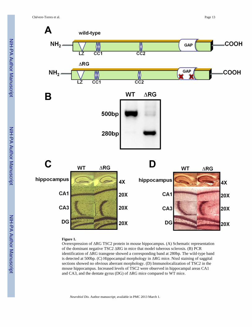
Figure 1.Overexpression of ΔRG TSC2 protein in mouse hippocampus. (A) Schematic representationof the dominant negative TSC2 ΔRG in mice that model tuberous sclerosis. (B) PCRidentification of ΔRG transgene showed a corresponding band at 280bp. The wild-type bandis detected at 500bp. (C) Hippocampal morphology in ΔRG mice. Nissl staining of saggitalsections showed no obvious aberrant morphology. (D) Immunolocalization of TSC2 in themouse hippocampus. Increased levels of TSC2 were observed in hippocampal areas CA1and CA3, and the dentate gyrus (DG) of ΔRG mice compared to WT mice.
Chévere-Torres et al. Page 13
Neurobiol Dis. Author manuscript; available in PMC 2013 March 1.
NIH
-PA Author Manuscript
NIH
-PA Author Manuscript
NIH
-PA Author Manuscript
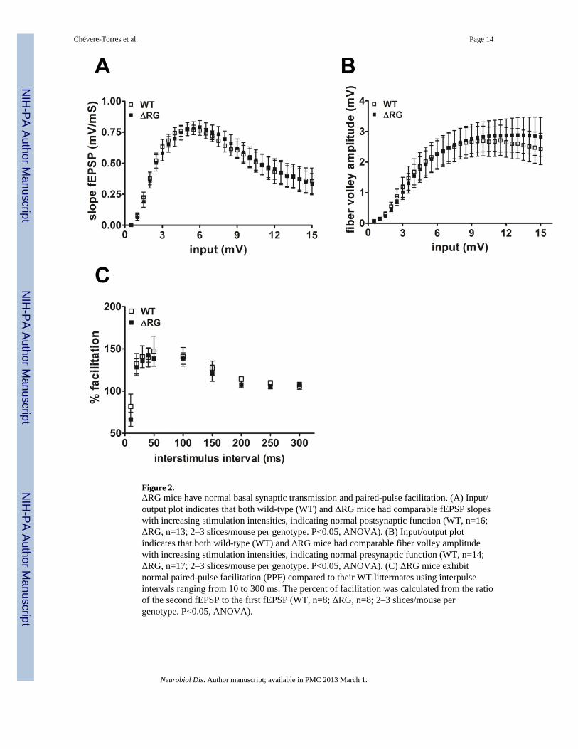
Figure 2.ΔRG mice have normal basal synaptic transmission and paired-pulse facilitation. (A) Input/output plot indicates that both wild-type (WT) and ΔRG mice had comparable fEPSP slopeswith increasing stimulation intensities, indicating normal postsynaptic function (WT, n=16;ΔRG, n=13; 2–3 slices/mouse per genotype. P<0.05, ANOVA). (B) Input/output plotindicates that both wild-type (WT) and ΔRG mice had comparable fiber volley amplitudewith increasing stimulation intensities, indicating normal presynaptic function (WT, n=14;ΔRG, n=17; 2–3 slices/mouse per genotype. P<0.05, ANOVA). (C) ΔRG mice exhibitnormal paired-pulse facilitation (PPF) compared to their WT littermates using interpulseintervals ranging from 10 to 300 ms. The percent of facilitation was calculated from the ratioof the second fEPSP to the first fEPSP (WT, n=8; ΔRG, n=8; 2–3 slices/mouse pergenotype. P<0.05, ANOVA).
Chévere-Torres et al. Page 14
Neurobiol Dis. Author manuscript; available in PMC 2013 March 1.
NIH
-PA Author Manuscript
NIH
-PA Author Manuscript
NIH
-PA Author Manuscript
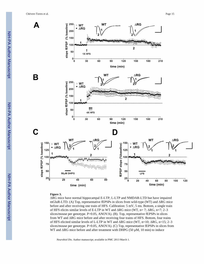
Figure 3.ΔRG mice have normal hippocampal E-LTP, L-LTP and NMDAR-LTD but have impairedmGluR-LTD. (A) Top, representative fEPSPs in slices from wild-type (WT) and ΔRG micebefore and after receiving one train of HFS. Calibration: 5 mV, 5 ms. Bottom, a single trainof HFS elicits similar levels of E-LTP in WT and ΔRG mice (WT, n= 7; ΔRG, n=7; 2–3slices/mouse per genotype. P>0.05, ANOVA). (B). Top, representative fEPSPs in slicesfrom WT and ΔRG mice before and after receiving four trains of HFS. Bottom, four trainsof HFS elicited similar levels of L-LTP in WT and ΔRG mice (WT, n=10; ΔRG, n=15; 2–3slices/mouse per genotype. P>0.05, ANOVA). (C) Top, representative fEPSPs in slices fromWT and ΔRG mice before and after treatment with DHPG (50 µM, 10 min) to induce
Chévere-Torres et al. Page 15
Neurobiol Dis. Author manuscript; available in PMC 2013 March 1.
NIH
-PA Author Manuscript
NIH
-PA Author Manuscript
NIH
-PA Author Manuscript
mGLUR-LTD. Bottom, DHPG application induced LTD in WT mice, but not in ΔRG mice(WT, n=7; ΔRG, n=8; 2–3 slices/mouse per genotype. **p<0.01, ANOVA). (D) Top,representative fEPSPs in slices from wild-type (WT) and ΔRG mice before and afterreceiving 900 pulses of LFS for 15 min. Bottom, LFS elicits similar levels of NMDR-LTDin WT and ΔRG mice (WT, n= 12; ΔRG, n=12; 3 slices/mouse per genotype. P>0.05,ANOVA).
Chévere-Torres et al. Page 16
Neurobiol Dis. Author manuscript; available in PMC 2013 March 1.
NIH
-PA Author Manuscript
NIH
-PA Author Manuscript
NIH
-PA Author Manuscript
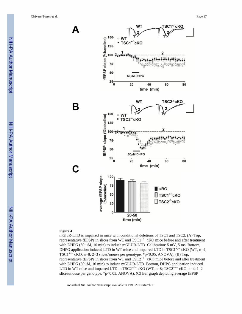
Figure 4.mGluR-LTD is impaired in mice with conditional deletions of TSC1 and TSC2. (A) Top,representative fEPSPs in slices from WT and TSC1+/− cKO mice before and after treatmentwith DHPG (50 µM, 10 min) to induce mGLUR-LTD. Calibration: 5 mV, 5 ms. Bottom,DHPG application induced LTD in WT mice and impaired LTD in TSC1+/− cKO (WT, n=4;TSC1+/− cKO, n=8; 2–3 slices/mouse per genotype. *p<0.05, ANOVA). (B) Top,representative fEPSPs in slices from WT and TSC2−/− cKO mice before and after treatmentwith DHPG (50µM, 10 min) to induce mGLUR-LTD. Bottom, DHPG application inducedLTD in WT mice and impaired LTD in TSC2−/− cKO (WT, n=8; TSC2−/− cKO, n=4; 1–2slices/mouse per genotype. *p<0.05, ANOVA). (C) Bar graph depicting average fEPSP
Chévere-Torres et al. Page 17
Neurobiol Dis. Author manuscript; available in PMC 2013 March 1.
NIH
-PA Author Manuscript
NIH
-PA Author Manuscript
NIH
-PA Author Manuscript

slope 20 to 50 min after the washout of DHPG. DHPG application induced same level ofmGluR-LTD in ΔRG, TSC1+/− cKO, and TSC2−/− cKO mice (ΔRG, n=8; TSC1+/− cKO,n=8; TSC2−/− cKO, n=4. p>0.05, Student’s t-test compared with ΔRG for the given timeperiod).
Chévere-Torres et al. Page 18
Neurobiol Dis. Author manuscript; available in PMC 2013 March 1.
NIH
-PA Author Manuscript
NIH
-PA Author Manuscript
NIH
-PA Author Manuscript
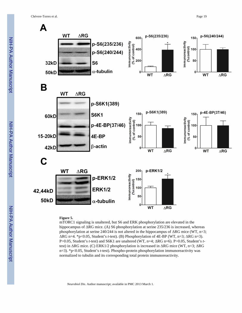
Figure 5.mTORC1 signaling is unaltered, but S6 and ERK phosphorylation are elevated in thehippocampus of ΔRG mice. (A) S6 phosphorylation at serine 235/236 is increased, whereasphosphorylation at serine 240/244 is not altered in the hippocampus of ΔRG mice (WT, n=3;ΔRG n=4. *p<0.05, Student’s t-test). (B) Phosphorylation of 4E-BP (WT, n=3; ΔRG n=3).P>0.05, Student’s t-test) and S6K1 are unaltered (WT, n=4; ΔRG n=6). P>0.05, Student’s t-test) in ΔRG mice. (C) ERK1/2 phosphorylation is increased in ΔRG mice (WT, n=3; ΔRGn=3). *p<0.05, Student’s t-test). Phospho-protein phosphorylation immunoreactivity wasnormalized to tubulin and its corresponding total protein immunoreactivity.
Chévere-Torres et al. Page 19
Neurobiol Dis. Author manuscript; available in PMC 2013 March 1.
NIH
-PA Author Manuscript
NIH
-PA Author Manuscript
NIH
-PA Author Manuscript
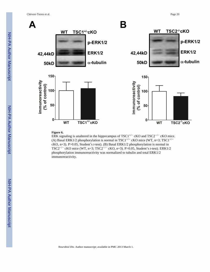
Figure 6.ERK signaling is unaltered in the hippocampus of TSC1+/− cKO and TSC2−/− cKO mice.(A) Basal ERK1/2 phosphorylation is normal in TSC1+/− cKO mice (WT, n=3; TSC1+/−
cKO, n=3). P>0.05, Student’s t-test). (B) Basal ERK1/2 phosphorylation is normal inTSC2−/− cKO mice (WT, n=3; TSC2−/− cKO, n=3). P>0.05, Student’s t-test). ERK1/2phosphorylation immunoreactivity was normalized to tubulin and total ERK1/2immunoreactivity.
Chévere-Torres et al. Page 20
Neurobiol Dis. Author manuscript; available in PMC 2013 March 1.
NIH
-PA Author Manuscript
NIH
-PA Author Manuscript
NIH
-PA Author Manuscript
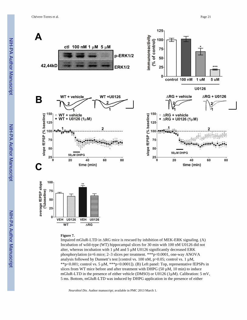
Figure 7.Impaired mGluR-LTD in ΔRG mice is rescued by inhibition of MEK-ERK signaling. (A)Incubation of wild-type (WT) hippocampal slices for 30 min with 100 nM U0126 did notalter, whereas incubation with 1 µM and 5 µM U0126 significantly decreased ERKphosphorylation (n=6 mice; 2–3 slices per treatment. ***p<0.0001, one-way ANOVAanalysis followed by Dunnett’s test [control vs. 100 nM, p>0.05; control vs. 1 µM,**p<0.001; control vs. 5 µM, ***p<0.0001]). (B) Left panel: Top, representative fEPSPs inslices from WT mice before and after treatment with DHPG (50 µM, 10 min) to inducemGluR-LTD in the presence of either vehicle (DMSO) or U0126 (1µM). Calibration: 5 mV,5 ms. Bottom, mGluR-LTD was induced by DHPG application in the presence of either
Chévere-Torres et al. Page 21
Neurobiol Dis. Author manuscript; available in PMC 2013 March 1.
NIH
-PA Author Manuscript
NIH
-PA Author Manuscript
NIH
-PA Author Manuscript
vehicle or 1 µM U0126 in WT mice (WT + vehicle, n=14; WT + U0126, n=14; 1–2 slices/mouse per drug treatment. P>0.05, ANOVA). Right panel: top, representative fEPSPs inslices from ΔRG mice before and after treatment with DHPG (50 µM, 10 min) to inducemGluR-LTD in the presence of either vehicle or U0126 (1µM). Bottom, mGluR-LTD wasinduced by DHPG application in the presence of 1 µM U0126, but not vehicle, in ΔRG mice(ΔRG + vehicle, n=12; ΔRG + U0126, n=13; 1–2 slices/mouse per drug treatment.***p<0.0001, ANOVA). The vehicle and U0126 was present before, during and afterDHPG application. (C) Left panel: bar graph depicting average fEPSP slope 20 to 50 minafter the washout of DHPG in the presence of either vehicle or U0126 (WT + vehicle, n=14;WT + U0126, n=14; ΔRG + vehicle, n=12; ΔRG + U0126, n=13; **p<0.01, one-wayANOVA; [**p<0.05, Student’s t-test compared with ΔRG + vehicle for the given timeperiod]).
Chévere-Torres et al. Page 22
Neurobiol Dis. Author manuscript; available in PMC 2013 March 1.
NIH
-PA Author Manuscript
NIH
-PA Author Manuscript
NIH
-PA Author Manuscript
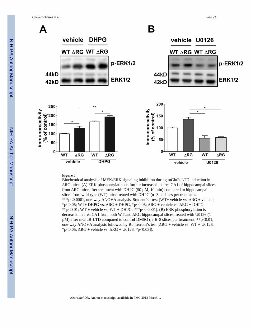
Figure 8.Biochemical analysis of MEK/ERK signaling inhibition during mGluR-LTD induction inΔRG mice. (A) ERK phosphorylation is further increased in area CA1 of hippocampal slicesfrom ΔRG mice after treatment with DHPG (50 µM, 10 min) compared to hippocampalslices from wild-type (WT) mice treated with DHPG (n=3–4 slices per treatment.***p<0.0001, one-way ANOVA analysis. Student’s t-test [WT+ vehicle vs. ΔRG + vehicle,*p<0.05; WT+ DHPG vs. ΔRG + DHPG, *p<0.05; ΔRG + vehicle vs. ΔRG + DHPG,**p<0.01; WT + vehicle vs. WT + DHPG, ***p<0.0001]. (B) ERK phosphorylation isdecreased in area CA1 from both WT and ΔRG hippocampal slices treated with U0126 (1µM) after mGluR-LTD compared to control DMSO (n=6–8 slices per treatment. **p<0.01,one-way ANOVA analysis followed by Bonferroni’s test [ΔRG + vehicle vs. WT + U0126,*p<0.05; ΔRG + vehicle vs. ΔRG + U0126, *p<0.05]).
Chévere-Torres et al. Page 23
Neurobiol Dis. Author manuscript; available in PMC 2013 March 1.
NIH
-PA Author Manuscript
NIH
-PA Author Manuscript
NIH
-PA Author Manuscript