Medicinal Herbs for the Biosynthesis of Nano particles to Enhance Cancer Therapeutics
15
UGC SPONSORED NATIONAL CONFERENCE ON CURRENT APPROACHES AND CHALLENGES IN NANOMATERIAL AND NANOMEDICINE (NACNAN-13)29-30 th August 2013- ISBN 978-81-9278-804-3 Medicinal Herbs for the Biosynthesis of Nano particles to Enhance Cancer Therapeutics S.T.Gopukumar 1* , P.K.Praseetha 1 , V.M.Priya kumari 2 and R.Shabi Ruskin 2 1 Department of Nanotechnology, Noorul Islam Centre for Higher Education, Noorul Islam University, Kumaracoil, Tamilnadu, India 2 Department of Biotechnology, Noorul Islam College of Arts and Science, Kumaracoil, Tamilnadu, India Abstract Medicinal Herbs are in use for thousands of years and are renowned for their effectiveness in cancer. Medicinal herbs are having their own anti-cancer properties. Bio- synthesized nano particles are very effective in cancer therapeutics as they have a controlled size and modified properties. The medicinally usable herbs were identified and extracted for biochemical profile and formulated for nano-medical applications with iron nanoparticles. Compared with other therapeutic drugs, this nanoparticles enables a highly integrated design that incorporates multiple functions, such as cell targeting, ultra-sensitive imaging and therapy. This study reveals the detailed study of medicinal herbs and cancer, which enhances the cancer therapeutics by green nano synthesis. Iron nano particles were synthesized and characterized by using Ultra Violet Visible Spectroscopy (UV-vis), Transmission Electron Microscopy (TEM) and Fourier Transform Infra-Red Spectroscopy (FTIR). Anti tumour effects were clinically experimented via In-vitro techniques. Keywords: Medicinal Herbs, nanomedicine, iron nano particle, UV-vis, TEM, FTIR, * Author for correspondence: [email protected]
Transcript of Medicinal Herbs for the Biosynthesis of Nano particles to Enhance Cancer Therapeutics
UGC SPONSORED NATIONAL CONFERENCE ON CURRENT APPROACHES AND CHALLENGES IN NANOMATERIAL AND NANOMEDICINE (NACNAN-13)29-30th August 2013- ISBN 978-81-9278-804-3
Medicinal Herbs for the Biosynthesis of Nano particles to Enhance Cancer
Therapeutics
S.T.Gopukumar1*, P.K.Praseetha1, V.M.Priya kumari2 and R.Shabi Ruskin2
1Department of Nanotechnology, Noorul Islam Centre for Higher Education,
Noorul Islam University, Kumaracoil, Tamilnadu, India 2Department of Biotechnology, Noorul Islam College of Arts and Science,
Kumaracoil, Tamilnadu, India
Abstract
Medicinal Herbs are in use for thousands of years and are renowned for their
effectiveness in cancer. Medicinal herbs are having their own anti-cancer properties. Bio-
synthesized nano particles are very effective in cancer therapeutics as they have a controlled
size and modified properties. The medicinally usable herbs were identified and extracted for
biochemical profile and formulated for nano-medical applications with iron nanoparticles.
Compared with other therapeutic drugs, this nanoparticles enables a highly integrated design
that incorporates multiple functions, such as cell targeting, ultra-sensitive imaging and
therapy. This study reveals the detailed study of medicinal herbs and cancer, which enhances
the cancer therapeutics by green nano synthesis. Iron nano particles were synthesized and
characterized by using Ultra Violet Visible Spectroscopy (UV-vis), Transmission Electron
Microscopy (TEM) and Fourier Transform Infra-Red Spectroscopy (FTIR). Anti tumour
effects were clinically experimented via In-vitro techniques.
Keywords: Medicinal Herbs, nanomedicine, iron nano particle, UV-vis, TEM, FTIR,
* Author for correspondence: [email protected]
UGC SPONSORED NATIONAL CONFERENCE ON CURRENT APPROACHES AND CHALLENGES IN NANOMATERIAL AND NANOMEDICINE (NACNAN-13)29-30th August 2013- ISBN 978-81-9278-804-3
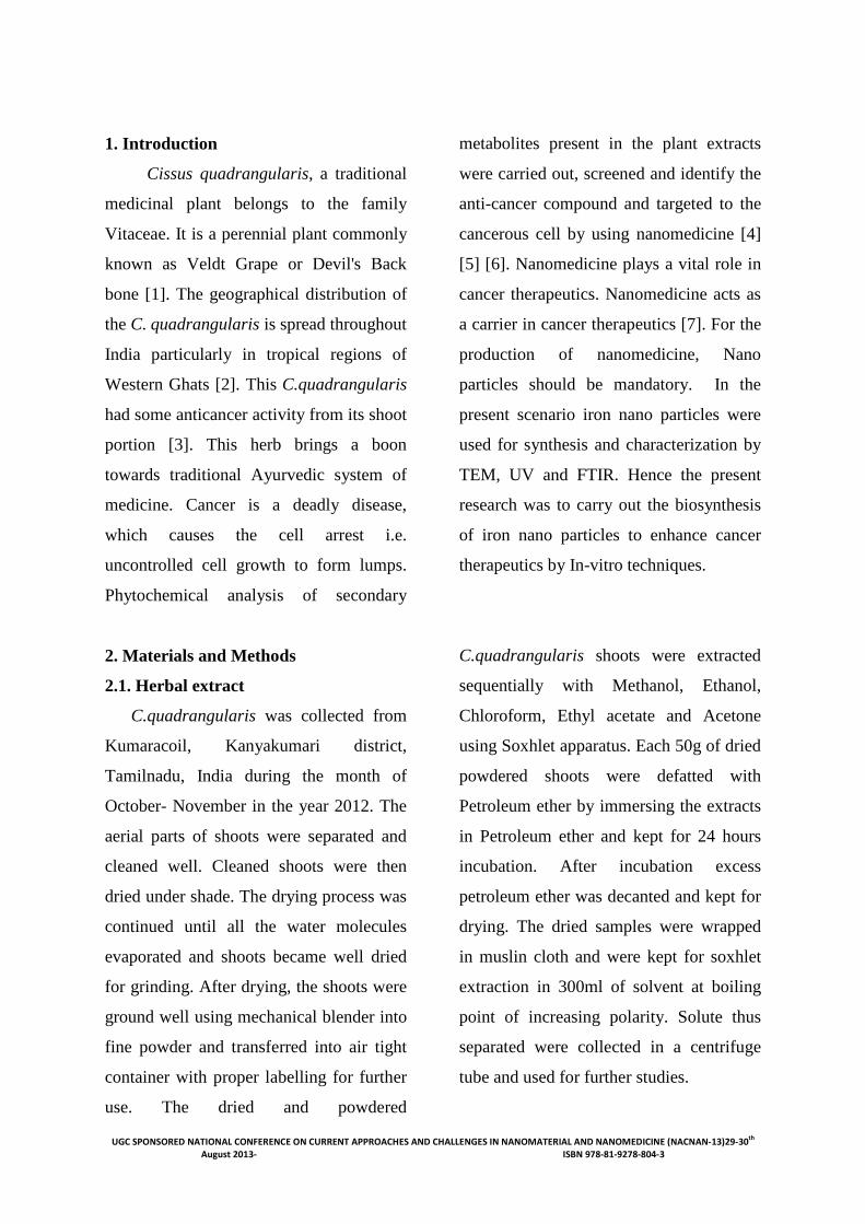
1. Introduction
Cissus quadrangularis, a traditional
medicinal plant belongs to the family
Vitaceae. It is a perennial plant commonly
known as Veldt Grape or Devil's Back
bone [1]. The geographical distribution of
the C. quadrangularis is spread throughout
India particularly in tropical regions of
Western Ghats [2]. This C.quadrangularis
had some anticancer activity from its shoot
portion [3]. This herb brings a boon
towards traditional Ayurvedic system of
medicine. Cancer is a deadly disease,
which causes the cell arrest i.e.
uncontrolled cell growth to form lumps.
Phytochemical analysis of secondary
metabolites present in the plant extracts
were carried out, screened and identify the
anti-cancer compound and targeted to the
cancerous cell by using nanomedicine [4]
[5] [6]. Nanomedicine plays a vital role in
cancer therapeutics. Nanomedicine acts as
a carrier in cancer therapeutics [7]. For the
production of nanomedicine, Nano
particles should be mandatory. In the
present scenario iron nano particles were
used for synthesis and characterization by
TEM, UV and FTIR. Hence the present
research was to carry out the biosynthesis
of iron nano particles to enhance cancer
therapeutics by In-vitro techniques.
2. Materials and Methods
2.1. Herbal extract
C.quadrangularis was collected from
Kumaracoil, Kanyakumari district,
Tamilnadu, India during the month of
October- November in the year 2012. The
aerial parts of shoots were separated and
cleaned well. Cleaned shoots were then
dried under shade. The drying process was
continued until all the water molecules
evaporated and shoots became well dried
for grinding. After drying, the shoots were
ground well using mechanical blender into
fine powder and transferred into air tight
container with proper labelling for further
use. The dried and powdered
C.quadrangularis shoots were extracted
sequentially with Methanol, Ethanol,
Chloroform, Ethyl acetate and Acetone
using Soxhlet apparatus. Each 50g of dried
powdered shoots were defatted with
Petroleum ether by immersing the extracts
in Petroleum ether and kept for 24 hours
incubation. After incubation excess
petroleum ether was decanted and kept for
drying. The dried samples were wrapped
in muslin cloth and were kept for soxhlet
extraction in 300ml of solvent at boiling
point of increasing polarity. Solute thus
separated were collected in a centrifuge
tube and used for further studies.
UGC SPONSORED NATIONAL CONFERENCE ON CURRENT APPROACHES AND CHALLENGES IN NANOMATERIAL AND NANOMEDICINE (NACNAN-13)29-30th August 2013- ISBN 978-81-9278-804-3
2.2. Green Synthesis of Iron Nano particles
with Herb extracts
For the green synthesis of the herbs
around 8 ml of 0.1 M FeCl3 and 2 ml of
0.2 M FeCl2 was added in a 100 ml beaker.
In addition to that, 10 ml of
C.quadrangularis (Veldt Grape) extract
was added drop by drop. Initially, a brown
precipitate was formed. The reaction
continued for 20 minutes. Then the beaker
was removed from the magnetic stirrer.
The pH was adjusted to 8. After synthesis,
the black precipitate formed was washed
3-4 times with distilled water to avoid
errors due to high optical density of the
solution. The nanoparticles were removed
from the solution by magnetic separation.
The experiment was done in duplicates to
produce reproducibility.
3. Characterization of green synthesis of
Iron nano particles
3.1. UV-vis spectroscopy of iron
nanoparticles
Iron nanoparticles synthesized by
green method were analyzed for UV-Vis
spectroscopy. The UV-Vis spectroscopy
measurements of iron nanoparticles were
recorded on Systronic double beam
spectrophotometer: 2202. The progress of
the reaction between metal ions and the
C.quadrangularis extracts were monitored
by UV-Vis spectra of iron nanoparticles.
Green synthesized iron nanoparticles were
measured in a wavelength ranging from
200-1100 nm.
3.2. Transmission Electron Microscopy
(TEM) of iron nanoparticles
The prepared iron nanoparticles
were analyzed using TEM. The iron
nanoparticles were taken for TEM analysis
on carbon-coated copper TEM grids. The
films on the TEM grids were allowed to
stand for 2 min following which the extra
solution was removed using a blotting
paper and the grid was allowed to dry prior
to measurement.
3.3. Fourier transform infrared
spectrophotometer (FTIR) of iron
nanoparticles
Dried powder of ethanolic extract
of C.quadrangularis was considered for
instrumental analysis. For the FTIR study,
dried powder of ethanolic extract (10mg)
of C.quadrangularis was encapsulated in
100mg of KBr pellet, in order to prepare
translucent sample discs. The powdered
samples were treated for FTIR
spectroscopy.
4. Anti tumour effect – In-vitro
Cancer cell lines were procured
from NCCS Pune and was maintained in
10% heat inactivated FBS (Foetal Bovine
Serum) in carbon dioxide incubator. The
cells may be in confluent stage. So the
cells were trypsinised using 0.025%
trypsin (cell culture grade HIMEDIA)
upon reaching confluency. Then the cells
UGC SPONSORED NATIONAL CONFERENCE ON CURRENT APPROACHES AND CHALLENGES IN NANOMATERIAL AND NANOMEDICINE (NACNAN-13)29-30th August 2013- ISBN 978-81-9278-804-3
were subcultured on to microculture plates
and used for further studies. Anticancer
effect of iron nanosynthesised
C.quadrangularis plant extracts such as
acetone, chloroform, Ethanol, Ethyl
acetate and Methanol was determined on
MCF cell lines. A standard concentration
of 500µg/ml was added and incubated.
The anti-proliferative effect was
determined by standard MTT assay. The
cell culture suspension was washed with
1X PBS (Phosphate Buffered Saline) and
then added 200µl MTT [3-(4, 5-Dimethyl
thiazole-2yl)-2, 5-diphyhyl tetra-zolium
Bromide] solution to the culture flask
(MTT 5mg/volume dissolved in PBS).
Filtered through a 0.2µm filter before use.
Then incubated at 37ºC for 3 hours,
removed all MTT solution, washed with 1
x PBS and added 300µl DMSO to each
culture flask and incubated at room
temperature for 30 minutes until all cells
get lysed and homogenous colour was
obtained. The solution was then
transferred to centrifuge tube and
centrifuged at top speed for 2 minutes to
precipitate cell debris. Debris was
dissolved using DMSO. OD was measured
at 540 nm using DMSO blank. Then the
percentage viability was calculated
Calculation .u䍀.ොFᄄC. oǴ 猀yᄄ椒y謃yF轿⮀뾐.ᄄො ᄄ椒㨐ou椒ᄄො䍀. oǴ 㨐ᄄᴄ錃謃.뾐.ᄄො ᄄ椒㨐ou椒ᄄො䍀. oǴ 䍀oොFuo謃 时↸迷迷
3. Results and Discussion
Qualitative Phytochemical Screening
of Ethanolic Extract of C.quadrangularis
Qualitative phytochemical screening was
used to determine the presence of some of
the secondary metabolites. The results of
screening test revealed the presence of
medically active compounds. From the
Table: (1), it could be seen that Phenol,
Alkaloids, Tannins and Flavanoids were
present in the ethanol extract of
C.quadrangularis, while Saponin and
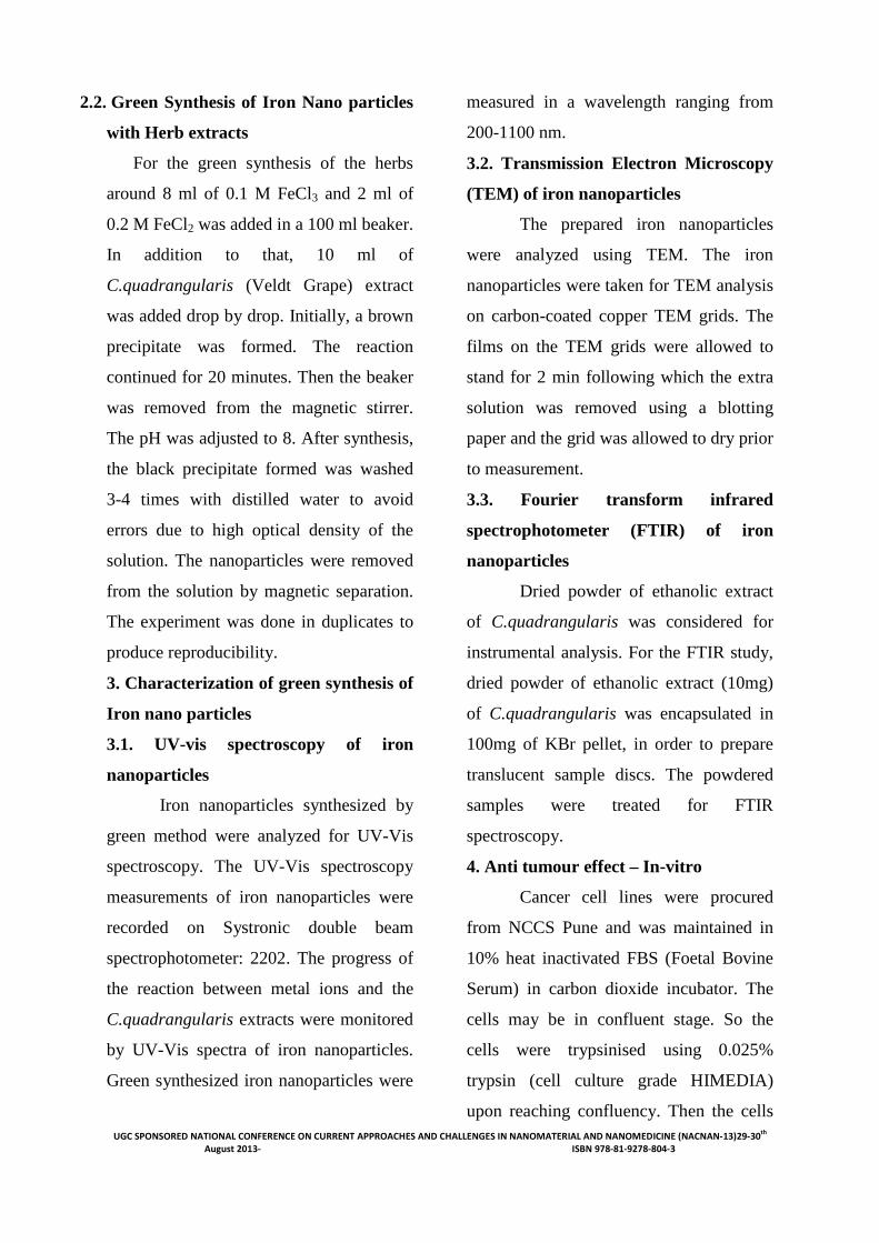
Carbohydrates were absent. Green
synthesized ethanolic iron nano particles
were attracted towards the magnet is
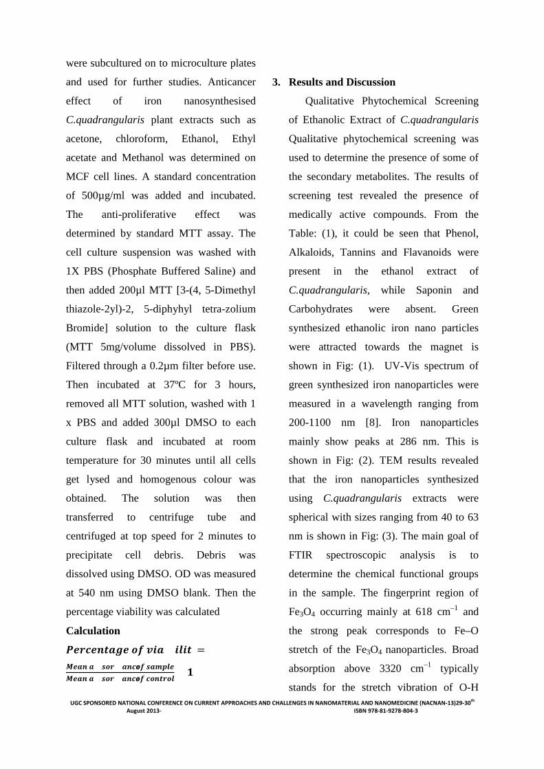
shown in Fig: (1). UV-Vis spectrum of
green synthesized iron nanoparticles were
measured in a wavelength ranging from
200-1100 nm [8]. Iron nanoparticles
mainly show peaks at 286 nm. This is
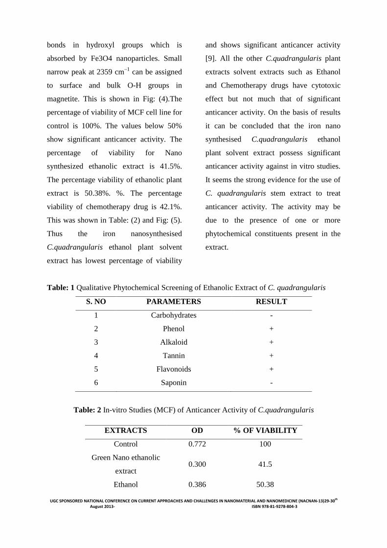
shown in Fig: (2). TEM results revealed
that the iron nanoparticles synthesized
using C.quadrangularis extracts were
spherical with sizes ranging from 40 to 63
nm is shown in Fig: (3). The main goal of
FTIR spectroscopic analysis is to
determine the chemical functional groups
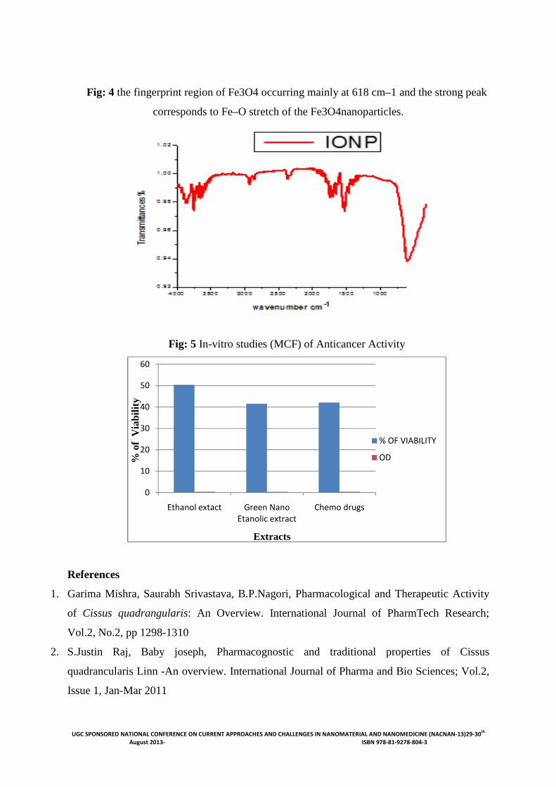
in the sample. The fingerprint region of
Fe3O4 occurring mainly at 618 cm–1 and
the strong peak corresponds to Fe–O
stretch of the Fe3O4 nanoparticles. Broad
absorption above 3320 cm–1 typically
stands for the stretch vibration of O-H
UGC SPONSORED NATIONAL CONFERENCE ON CURRENT APPROACHES AND CHALLENGES IN NANOMATERIAL AND NANOMEDICINE (NACNAN-13)29-30th August 2013- ISBN 978-81-9278-804-3
bonds in hydroxyl groups which is
absorbed by Fe3O4 nanoparticles. Small
narrow peak at 2359 cm–1 can be assigned
to surface and bulk O-H groups in
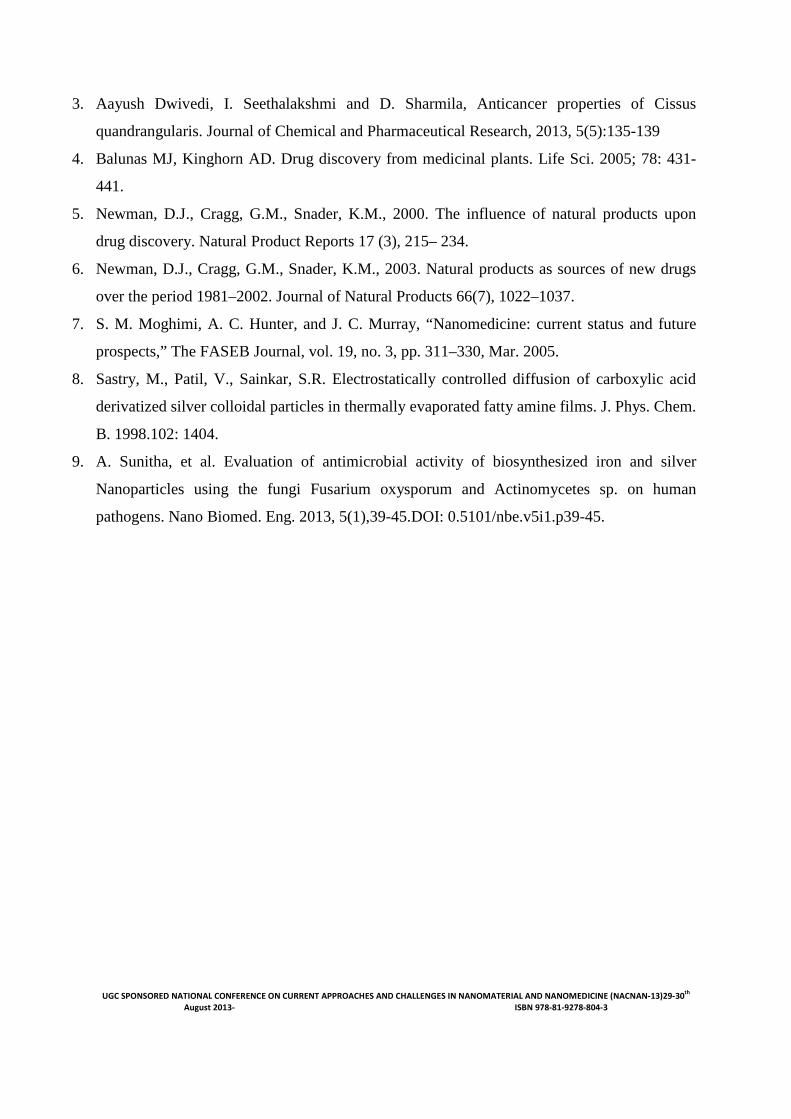
magnetite. This is shown in Fig: (4).The
percentage of viability of MCF cell line for
control is 100%. The values below 50%
show significant anticancer activity. The
percentage of viability for Nano
synthesized ethanolic extract is 41.5%.
The percentage viability of ethanolic plant
extract is 50.38%. %. The percentage
viability of chemotherapy drug is 42.1%.
This was shown in Table: (2) and Fig: (5).
Thus the iron nanosynthesised
C.quadrangularis ethanol plant solvent
extract has lowest percentage of viability
and shows significant anticancer activity
[9]. All the other C.quadrangularis plant
extracts solvent extracts such as Ethanol
and Chemotherapy drugs have cytotoxic
effect but not much that of significant
anticancer activity. On the basis of results
it can be concluded that the iron nano
synthesised C.quadrangularis ethanol
plant solvent extract possess significant
anticancer activity against in vitro studies.
It seems the strong evidence for the use of
C. quadrangularis stem extract to treat
anticancer activity. The activity may be
due to the presence of one or more
phytochemical constituents present in the
extract.
Table: 1 Qualitative Phytochemical Screening of Ethanolic Extract of C. quadrangularis
S. NO PARAMETERS RESULT
1 Carbohydrates -
2 Phenol +
3 Alkaloid +
4 Tannin +
5 Flavonoids +
6 Saponin -
Table: 2 In-vitro Studies (MCF) of Anticancer Activity of C.quadrangularis
EXTRACTS OD % OF VIABILITY
Control 0.772 100
Green Nano ethanolic
extract 0.300 41.5
Ethanol 0.386 50.38
UGC SPONSORED NATIONAL CONFERENCE ON CURRENT APPROACHES AND CHALLENGES IN August 2013-
Chemotherapy drugs
Fig: 1 Green synthesized ethanolic iron nanoparticles attracted towards magnet.
Fig: 2 UV absorption spectroscopy of microbially synthesized iron Nanoparticles.
Fig: 3 TEM of green synthesized iron nanoparticles at a magnification of 40,000 x
UGC SPONSORED NATIONAL CONFERENCE ON CURRENT APPROACHES AND CHALLENGES IN NANOMATERIAL AND NANOMEDICINE (NACNAN ISBN 978-81-9278-804-3
Chemotherapy drugs 0.367 42.1
Green synthesized ethanolic iron nanoparticles attracted towards magnet.
UV absorption spectroscopy of microbially synthesized iron Nanoparticles.
TEM of green synthesized iron nanoparticles at a magnification of 40,000 x
NANOMATERIAL AND NANOMEDICINE (NACNAN-13)29-30th
Green synthesized ethanolic iron nanoparticles attracted towards magnet.
UV absorption spectroscopy of microbially synthesized iron Nanoparticles.
TEM of green synthesized iron nanoparticles at a magnification of 40,000 x
UGC SPONSORED NATIONAL CONFERENCE ON CURRENT APPROACHES AND CHALLENGES IN NANOMATERIAL AND NANOMEDICINE (NACNAN-13)29-30th August 2013- ISBN 978-81-9278-804-3
Fig: 4 the fingerprint region of Fe3O4 occurring mainly at 618 cm–1 and the strong peak
corresponds to Fe–O stretch of the Fe3O4nanoparticles.
Fig: 5 In-vitro studies (MCF) of Anticancer Activity
References
1. Garima Mishra, Saurabh Srivastava, B.P.Nagori, Pharmacological and Therapeutic Activity
of Cissus quadrangularis: An Overview. International Journal of PharmTech Research;
Vol.2, No.2, pp 1298-1310
2. S.Justin Raj, Baby joseph, Pharmacognostic and traditional properties of Cissus
quadrancularis Linn -An overview. International Journal of Pharma and Bio Sciences; Vol.2,
Issue 1, Jan-Mar 2011
0
10
20
30
40
50
60
Ethanol extact Green Nano Etanolic extract
Chemo drugs
% OF VIABILITY
OD%of
Via
bilit
y
Extracts
UGC SPONSORED NATIONAL CONFERENCE ON CURRENT APPROACHES AND CHALLENGES IN NANOMATERIAL AND NANOMEDICINE (NACNAN-13)29-30th August 2013- ISBN 978-81-9278-804-3
3. Aayush Dwivedi, I. Seethalakshmi and D. Sharmila, Anticancer properties of Cissus
quandrangularis. Journal of Chemical and Pharmaceutical Research, 2013, 5(5):135-139
4. Balunas MJ, Kinghorn AD. Drug discovery from medicinal plants. Life Sci. 2005; 78: 431-
441.
5. Newman, D.J., Cragg, G.M., Snader, K.M., 2000. The influence of natural products upon
drug discovery. Natural Product Reports 17 (3), 215– 234.
6. Newman, D.J., Cragg, G.M., Snader, K.M., 2003. Natural products as sources of new drugs
over the period 1981–2002. Journal of Natural Products 66(7), 1022–1037.
7. S. M. Moghimi, A. C. Hunter, and J. C. Murray, “Nanomedicine: current status and future
prospects,” The FASEB Journal, vol. 19, no. 3, pp. 311–330, Mar. 2005.
8. Sastry, M., Patil, V., Sainkar, S.R. Electrostatically controlled diffusion of carboxylic acid
derivatized silver colloidal particles in thermally evaporated fatty amine films. J. Phys. Chem.
B. 1998.102: 1404.
9. A. Sunitha, et al. Evaluation of antimicrobial activity of biosynthesized iron and silver
Nanoparticles using the fungi Fusarium oxysporum and Actinomycetes sp. on human
pathogens. Nano Biomed. Eng. 2013, 5(1),39-45.DOI: 0.5101/nbe.v5i1.p39-45.
UGC SPONSORED NATIONAL CONFERENCE ON CURRENT APPROACHES AND CHALLENGES IN NANOMATERIAL AND NANOMEDICINE (NACNAN-13)29-30th August 2013- ISBN 978-81-9278-804-3
POLYMER MEDIATED SYNTHESIS OF CALCIUM CARBONATE
NANOPARTICLES AND THEIR INFLUENCES ON NR/EPDM RUBBER BLENDS
P G Prajith1, K Kurien Thomas1 and T Muraleedharan Nair2 1Research Department of Chemistry, Bishop Moore College, Mavelikara-690110,
Alappuzha, Kerala, India. 2Common Facility Service Centre, Department of Industries and Commerce, Payyanad,
Manjeri, Kerala, India.
Contact email [email protected]
Introduction
Currently, nanostructure materials have
become an interesting field for researchers.
The nanoparticles can be prepared by
several synthetic routes like, sol gel
processing, in situ polymerization,
hydrothermal process, matrix-mediated
control of growth, forced hydrolysis
approach, etc The matrix-mediated control
of growth and morphology has drawn
considerable attention among various
groups of researchers since it offers a new
route to material synthesis. It is well
known that the nanoparticles play an
important role in their application to
polymer nanocomposites. Recent and
ongoing research on polymer/inorganic
nanocomposites has shown significant
improvements in mechanical, physical and
thermal properties of polymers. These
improvements are obtained at a very low
loading of the nanofillers (1-10%) as
compared to the conventional fillers,
which require a high loading (25-40%)1. It
is well known that the particle size,
structure, and surface characteristics are
important parameters that determine the
reinforcing ability of filler; the particle
size is especially important because a
reduction in size provides a greater surface
area.
In the present study we have prepared the
nanoparticles of calcium carbonate by
polymer mediated growth method2 and
were characterized using X-ray diffraction
and microscopic techniques. Further, these
particles were incorporated into
NR/EPDM blends by two roller mill
mixing technique. Various mechanical
properties like tensile strength, Young’s
Modulus and tear strength of the
composites were characterized by standard
methods. The effects of addition of nano
CaCO3 on the various mechanical and
physical properties of the rubber blend
were investigated.
UGC SPONSORED NATIONAL CONFERENCE ON CURRENT APPROACHES AND CHALLENGES IN NANOMATERIAL AND NANOMEDICINE (NACNAN-13)29-30th August 2013- ISBN 978-81-9278-804-3
2. Experimental
2.1. Materials
Indian Standard Natural Rubber (ISNR-5
by Rubber Research Institute of India) and
Ethylene propylene diene monomer rubber
(EPDM by Goodyear rubber company,
USA) was used.. Analytical grades of
Calcium chloride, Ammonium carbonate,
and Polyethylene glycol (PEG-6000) were
procured from Merck and were used for
the synthesis of nanoparticles of calcium
carbonate. The rubber additives, namely
Stearic acid, Zinc Oxide (ZnO), Sulphur,
and F (Accelerator), were of commercial
grade.
2.2. Synthesis and Characterization of
Nanoparticles
Nano calcium carbonate was synthesized
by polymer mediated technique. CaCl2
(110 g by weight) was placed in 100 mL of
water; 248 g of PEG was diluted in 100
mL of water by mild heating. A complex
of calcium chloride and PEG was prepared
in molar ratio of 1: 20. Another solution of
(NH4)2CO3 was prepared, with 96 g placed
in 100 mL of distilled water. The first
complex was digested for 12 h, and then
the solution of(NH4)2CO3 was added
slowly; the mixture was again kept for
digestion for 12 h ,the precipitate was
filtered, washed with water, The
precipitate was sonicated using a bath
type ultrasonic disintegrator for 45
minutes and dried in a vacuum drier3.
These nanoparticles were characterized
using X-ray diffraction (XRD) Scanning
electron microscopy (SEM), and
transmission electron microscopy (TEM).
2.3. Preparation and Analysis of
NR/EPDM Nanocomposites
Formulation for rubber compounds was as
follows in phr: NR (75 g); EPDM (25g);
ZnO (5 g); Stearic acid (2 g); F (1.5 g);
Sulfur (2 g); and Nano CaCO3 (variable).
NR was initially masticated in the mixer
for 3 minutes and blended with EPDM,
followed by the vulcanization ingredients.
The cure characteristics were analyzed
using Rheometer and was subjected to
compression molding at 150oC4. The cured
sheets were subjected to conditioning for
24 h and the mechanical properties, such
as the tensile strength and tear strength
were measured per ASTM Standards. The
compression-molded specimens were
tested to report hardness data with a Shore
A hardness tester per ASTM D 2240
3. Results and Discussion
3.1. Nanoparticle Characterization
The TEM, SEM and XRD images of the
nanoparticles are shown in Fig.1, Fig.2
and Fig.3 respectively. Shape of nano
CaCO3 is observed to be spherical in
TEM, while the particle size is 5-10 nm.

UGC SPONSORED NATIONAL CONFERENCE ON CURRENT APPROACHES AND CHALLENGES IN August 2013-
The average particle size of nano CaCO
were calculated from XRD using
Scherrer’s Formula and was recorded to be
Å, θ is the diffraction angle and Δ2θ
Fig.1. TEM image of NanoCaCO3
Fig.3
3.2. Analysis of Mechanical and
Physical properties of Composites
The addition of nano CaCO3 appreciably
increases the mechanical properties of the
composites. Table-1 Shows that the tensile
properties of the nanocomposites increase
upon increasing the nano filler amount to
reach a maximum value of 18.67 MPa at 2
wt % filler loading. Fig-4 indica
NR/EPDM nano composites exhibit a
maximum tensile strength at low filler
Operations: Smooth 0.150 | Background 1.445,1.000 | ImportFile: SAIFXR101026B-03(P3).raw - Step: 0.020 ° - Step time: 29.5 s - WL1: 1.5406 - kA2 Ratio: 0.5 - Generator kV: 40 kV - Generator mA: 35 mA -
Lin
(C
ounts
)
0
1000
2000
3000
4000
5000
3 10 20
UGC SPONSORED NATIONAL CONFERENCE ON CURRENT APPROACHES AND CHALLENGES IN NANOMATERIAL AND NANOMEDICINE (NACNAN ISBN 978-81-9278-804-3
The average particle size of nano CaCO3
were calculated from XRD using
Scherrer’s Formula and was recorded to be
below 20 nm. Scherrer’s Formula,
=kλ ⁄Δ2θ cos θ. Where‘d’ is the particle
size, ‘k’ is the order of reflection, λ=1.542
θ is the full width at half maximum5.
3 Fig.2. SEM image of Nano CaCO3
Fig.3. XRD pattern for NanoCaCO3
3.2. Analysis of Mechanical and
Physical properties of Composites
appreciably
increases the mechanical properties of the
1 Shows that the tensile
properties of the nanocomposites increase
upon increasing the nano filler amount to
reach a maximum value of 18.67 MPa at 2
4 indicate that
NR/EPDM nano composites exhibit a
maximum tensile strength at low filler
loading (2phr) followed by a decrease.
Generally, filler particle size has
significant effect on the tensile strength of
the composites owing to the interfacial
area per unit volume. The increase in
tensile strength is attributed to the nano
reinforcement of the fillers used
facilitates efficient stress transfer.
However, particle agglomeration tends to
reduce the strength of the material.
Agglomerates may act as strong
O3BMC-P3
Operations: Smooth 0.150 | Background 1.445,1.000 | ImportFile: SAIFXR101026B-03(P3).raw - Step: 0.020 ° - Step time: 29.5 s - WL1: 1.5406 - kA2 Ratio: 0.5 - Generator kV: 40 kV - Generator mA: 35 mA -
2-Theta - Scale
30 40 50 60 70 80
NANOMATERIAL AND NANOMEDICINE (NACNAN-13)29-30th
Scherrer’s Formula, d (Å)
Where‘d’ is the particle
size, ‘k’ is the order of reflection, λ=1.542
3
loading (2phr) followed by a decrease.
Generally, filler particle size has
significant effect on the tensile strength of
the composites owing to the interfacial
volume. The increase in
tensile strength is attributed to the nano
reinforcement of the fillers used6, which
facilitates efficient stress transfer.
However, particle agglomeration tends to
reduce the strength of the material.
Agglomerates may act as strong stress
UGC SPONSORED NATIONAL CONFERENCE ON CURRENT APPROACHES AND CHALLENGES IN NANOMATERIAL AND NANOMEDICINE (NACNAN-13)29-30th August 2013- ISBN 978-81-9278-804-3
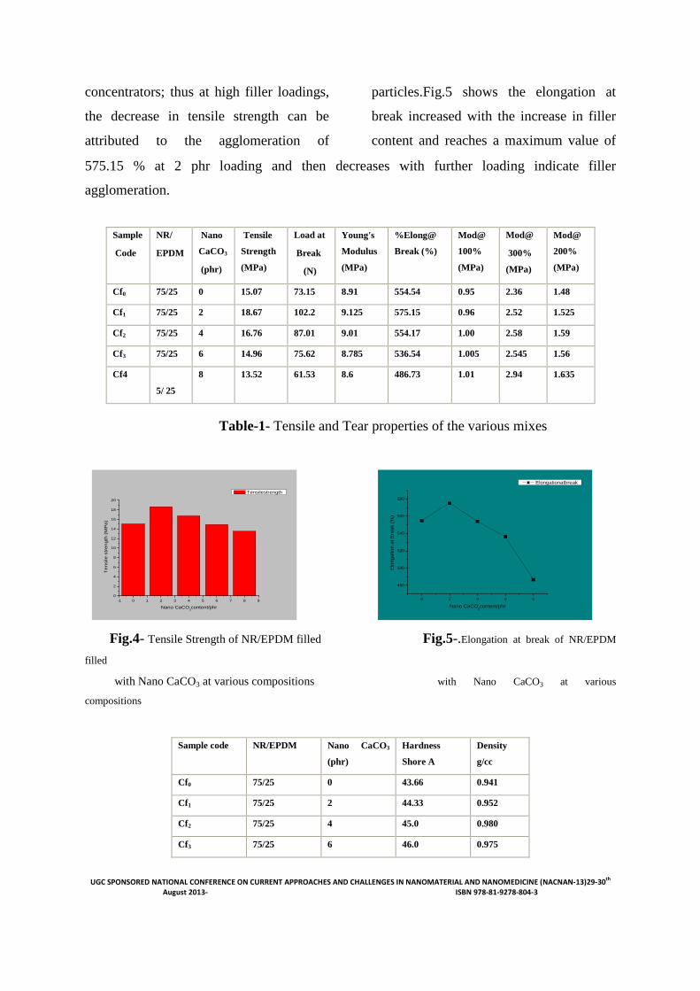
concentrators; thus at high filler loadings,
the decrease in tensile strength can be
attributed to the agglomeration of
particles.Fig.5 shows the elongation at
break increased with the increase in filler
content and reaches a maximum value of
575.15 % at 2 phr loading and then decreases with further loading indicate filler
agglomeration.
Table-1- Tensile and Tear properties of the various mixes
Fig.4- Tensile Strength of NR/EPDM filled Fig.5-.Elongation at break of NR/EPDM
filled
with Nano CaCO3 at various compositions with Nano CaCO3 at various
compositions
-1 0 1 2 3 4 5 6 7 8 90
2
4
6
8
10
12
14
16
18
20
Te
nsile
str
en
gth
(M
Pa
)
Nano CaCO3content/phr
Tensilestrength
0 2 4 6 8
480
500
520
540
560
580
Elo
ng
atio
n a
t B
rea
k (%
)
Nano CaCO3content/phr
Elongationatbreak
Sample
Code
NR/
EPDM
Nano
CaCO3
(phr)
Tensile
Strength
(MPa)
Load at
Break
(N)
Young's
Modulus
(MPa)
%Elong@
Break (%)
Mod@
100%
(MPa)
Mod@
300%
(MPa)
Mod@
200%
(MPa)
Cf0 75/25 0 15.07 73.15 8.91 554.54 0.95 2.36 1.48
Cf1 75/25 2 18.67 102.2 9.125 575.15 0.96 2.52 1.525
Cf2 75/25 4 16.76 87.01 9.01 554.17 1.00 2.58 1.59
Cf3 75/25 6 14.96 75.62 8.785 536.54 1.005 2.545 1.56
Cf4
5/ 25
8 13.52 61.53 8.6 486.73 1.01 2.94 1.635
Sample code NR/EPDM Nano CaCO3
(phr)
Hardness
Shore A
Density
g/cc
Cf0 75/25 0 43.66 0.941
Cf1 75/25 2 44.33 0.952
Cf2 75/25 4 45.0 0.980
Cf3 75/25 6 46.0 0.975
UGC SPONSORED NATIONAL CONFERENCE ON CURRENT APPROACHES AND CHALLENGES IN NANOMATERIAL AND NANOMEDICINE (NACNAN-13)29-30th August 2013- ISBN 978-81-9278-804-3
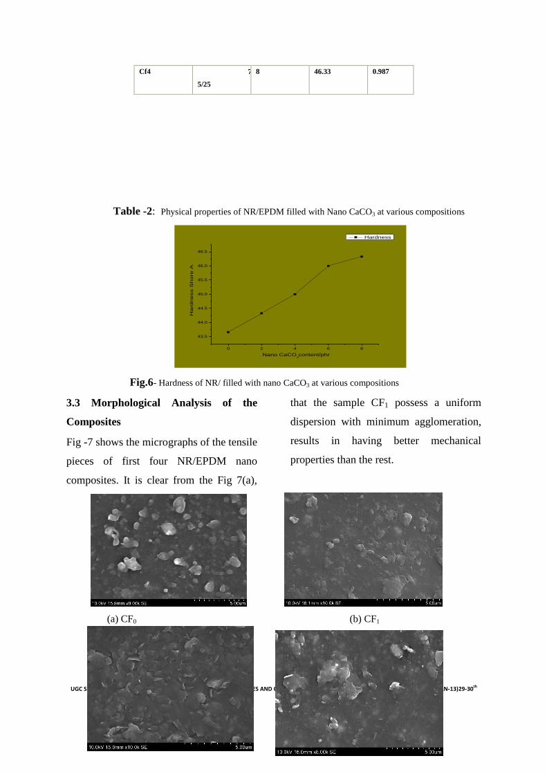
Table -2: Physical properties of NR/EPDM filled with Nano CaCO3 at various compositions
Fig.6- Hardness of NR/ filled with nano CaCO3 at various compositions
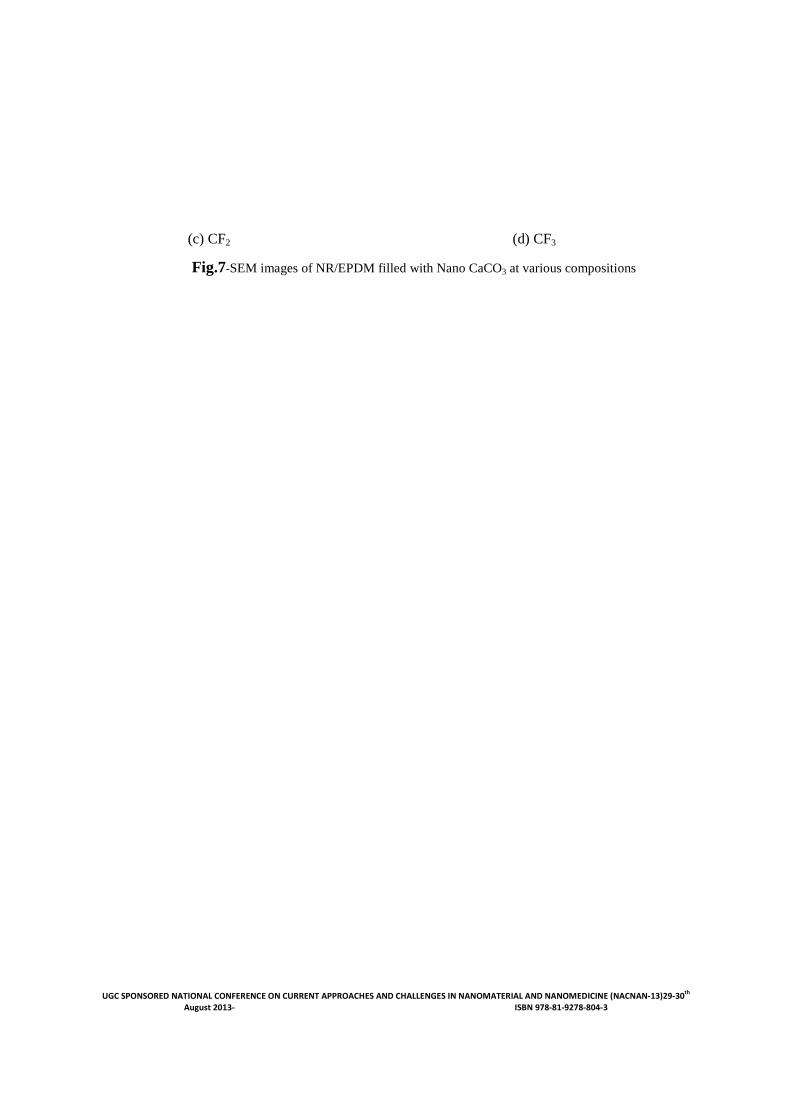
3.3 Morphological Analysis of the
Composites
Fig -7 shows the micrographs of the tensile
pieces of first four NR/EPDM nano
composites. It is clear from the Fig 7(a),
that the sample CF1 possess a uniform
dispersion with minimum agglomeration,
results in having better mechanical
properties than the rest.
(a) CF0 (b) CF1
0 2 4 6 8
43.5
44.0
44.5
45.0
45.5
46.0
46.5
Nano CaCO3content/phr
Ha
rdn
ess S
ho
re A
Hardness
Cf4 7
5/25
8 46.33 0.987
UGC SPONSORED NATIONAL CONFERENCE ON CURRENT APPROACHES AND CHALLENGES IN NANOMATERIAL AND NANOMEDICINE (NACNAN-13)29-30th August 2013- ISBN 978-81-9278-804-3
(c) CF2 (d) CF3
Fig.7-SEM images of NR/EPDM filled with Nano CaCO3 at various compositions
UGC SPONSORED NATIONAL CONFERENCE ON CURRENT APPROACHES AND CHALLENGES IN NANOMATERIAL AND NANOMEDICINE (NACNAN-13)29-30th August 2013- ISBN 978-81-9278-804-3
Conclusion.
Ultrasonication assisted polymer mediated
technique is a versatile technique for the
preparation of nanoparticles. Nanoparticles
of calcium carbonate in the range of 5-10
nm were prepared. TEM and XRD
analysis supports the size of the nano
particles. The NR/EPDM- nanoCaCO3
composites showed marked improvement
in physical and mechanical properties at
lower filler loading (2phr).Higher filler
loading results in the reduction of
composite properties due to
agglomeration.
References.
1. Sumita M, Shizuma T, Miyasaka K,
Ishikawa K,.(1983) J. Macromol.Sci. Phys.
B22, 601.
2. Saujanya C, Ashamol, Padalkar S,
Radhakrishnan S.( 2001). Control of nano
particle size of fillers by polymer blend
technique. Polymer, 42: 2255-2258.
3. Mishra S, Patil U D and Shimpi N
G.(2009). Synthesis of Mineral Nanofiller
using Solution Spray Method and its
Influence on Mechanical and Thermal
Properties of EPDM.
4. Alipour A, Naderi G, et al. (2011) Intern.
Polymer Processing XXVI (2011) 1:48-55
5. Selvin P Thomas, Sabu Thomas, and Sri
Bandyopadhyay. (2009). Polystyrene-
Calcium Phosphate Nanocomposites:
Preparation, Morphology, and Mechanical
Behavior. J. Phys.Chem, 113: 97–104.
6. Tshwafo E.Motaung, Adriaan S. Luyt,
Sabu Thomas. (2011).Morphology and
Properties of NR/EPDM Rubber Blends
Filled with Small Amounts of Titania
Nanoparticles. Polymer Composites-
2011:1289-1296.