Mechanistic peptidomics: factors that dictate specificity in the formation of endogenous peptides in...
24
Mechanistic Peptidomics: Factors that Dictate Specificity in the Formation of Endogenous Peptides in Human Milk Andres Guerrero a* , David C. Dallas b,c , Stephanie Contreras a , Sabrina Chee a , Evan A. Parker a , Xin Sun a , Lauren Dimapasoc a , Daniela Barile b, c , J. Bruce German b,c , Carlito B. Lebrilla a* . a Department of Chemistry, University of California at Davis, One Shields Avenue, Davis, CA, 95616, USA b Department of Food Science, University of California at Davis, One Shields Avenue, Davis, CA, 95616, USA. c Foods for Health Institute, University of California at Davis, One Shields Avenue, Davis, CA, 95616, USA. * To whom correspondence should be addressed to Carlito B. Lebrilla [email protected] 530-752-6364 Fax: 530-754-5609 OR Andres Guerrero [email protected] 530-752-5504 MCP Papers in Press. Published on August 29, 2014 as Manuscript M113.036194 Copyright 2014 by The American Society for Biochemistry and Molecular Biology, Inc.
-
Upload
independent -
Category
Documents
-
view
3 -
download
0
Transcript of Mechanistic peptidomics: factors that dictate specificity in the formation of endogenous peptides in...

Mechanistic Peptidomics: Factors that Dictate Specificity in the
Formation of Endogenous Peptides in Human Milk
Andres Guerreroa*
, David C. Dallasb,c
, Stephanie Contrerasa, Sabrina Chee
a, Evan A. Parker
a, Xin Sun
a, Lauren
Dimapasoca, Daniela Barile
b, c, J. Bruce German
b,c, Carlito B. Lebrilla
a*.
aDepartment of Chemistry, University of California at Davis, One Shields Avenue, Davis, CA, 95616, USA
bDepartment of Food Science, University of California at Davis, One Shields Avenue, Davis, CA, 95616, USA.
cFoods for Health Institute, University of California at Davis, One Shields Avenue, Davis, CA, 95616, USA.
*To whom correspondence should be addressed to
Carlito B. Lebrilla
530-752-6364
Fax: 530-754-5609
OR
Andres Guerrero
530-752-5504
MCP Papers in Press. Published on August 29, 2014 as Manuscript M113.036194
Copyright 2014 by The American Society for Biochemistry and Molecular Biology, Inc.

Mechanistic Peptidomics: Proteolysis Specificity on Milk
2
Abbreviations:
FA: Formic acid.
BCA: Bicinchoninic acid.
pIgR: Polymeric immunoglobulin receptor.
SC: Secretory component.

Mechanistic Peptidomics: Proteolysis Specificity on Milk
3
Summary
An extensive mass spectrometry analysis of the human milk peptidome has revealed almost 700
endogenous peptides from 30 different proteins. Two in-house computational tools were created and
used to visualize and interpret the data by both alignment of the peptide quasi-molecular ion intensities
and estimation of the differential enzyme participation. These results reveal that the endogenous
proteolytic activity in the mammary gland is remarkably specific and well-conserved. Certain proteins,
not necessarily the most abundant ones, are digested by the proteases present in milk yielding
endogenous peptides from selected regions. Our results strongly suggest that factors like the presence
of specific proteases, the position and concentration of cleavage sites and, more importantly, the
intrinsic disorder of segments of the protein drive this proteolytic specificity in the mammary gland. As a
consequence of this selective hydrolysis, proteins that typically need to be cleaved at specific positions
to exert their activity are properly digested and bioactive peptides encoded in certain protein sequences
are released. On the other hand, proteins that must remain intact to maintain their activity in the
mammary gland or in the neonatal gastrointestinal tract are unaffected by the hydrolytic environment
present in milk. These results represent insight into the intrinsic structural mechanisms that facilitate
the selectivity of the endogenous milk protease activity and may be useful to understand the
peptidomes of other biofluids.
Mechanistic Peptidomics: Proteolysis Specificity on Milk
4
Introduction
Peptidomics is defined as the systematic, comprehensive and quantitative analysis of the low
molecular weight fraction of proteins present in a biological sample at a defined time point (1). This
protein fraction includes biologically active peptide sequences, protein degradation products as well as
small proteins such as cytokines and signaling peptides (2). Endogenous peptides are produced from
their corresponding proteins by the action of proteases naturally present in the same biological system.
Consequently, the peptidome and proteome are intrinsically linked and their balance is controlled by
the presence of proteases and modulated by the levels of protease activators and inhibitors. This
relationship between proteins and their hydrolytic products has fueled the emergence of peptidomics as
a subdiscipline of proteomics. Human biofluids like blood (3), cerebrospinal fluid (4), saliva (5, 6), tears
(7) and urine (8) have been analyzed for endogenous peptides. As naturally occurring peptides reflect
both the protein content of a tissue and a specific configuration of the proteolytic machinery, they
represent a promising target for biomarker discovery (9-13). From a functional perspective, a number of
peptidomic studies have revealed different bioactivities in endogenous sequences (14-17).
Peptidomic research has revealed that the endogenous low molecular weight protein fraction is
generally composed by overlapping ladder peptide products originating from a few regions of specific
proteins. This proteolytic pattern is explained as a result of the action of endopeptidases cleaving in
specific protein regions and the subsequent partial degradation of these initial fragments by
exopeptidases (18). The presence and abundance of the resulting endogenous peptides has been
correlated with the amounts of both substrate proteins and proteolytic component (9, 19, 20), however,
the determinants of the peptidase selectivity are still a matter of scientific debate. It is accepted that
four factors determine the specificity on the proteolysis: (i) coexistence of protease and substrate
protein in the same space and time, (ii) presence of exosites that, although not involved in the

Mechanistic Peptidomics: Proteolysis Specificity on Milk
5
proteolysis itself, increase the affinity of the protease for specific substrates, (iii) presence of the correct
amino acid motif and (iv) the structural context of the excisable bond (21). The last factor is related with
the accessibility of the enzyme to the cleaving site and it is commonly accepted that proteolysis happens
in solvent-exposed, flexible substrate regions (22, 23). However, recent investigations have
demonstrated that limited proteolysis frequently happens also in helix and b-sheets secondary
structures (21, 24).
Milk provides a unique fluid for peptidomics. The proteins in milk are well characterized, as are
many of the proteases that are present. However, milk has been little studied from a peptidomic
approach. The vast majority of studies focused on the discovery of bioactive milk peptides released from
isolated milk proteins by in vitro digestion processes. In these studies, milk proteins were degraded by
bacteria cultures (25-27) or commercial proteases (28) in environments that may or may not mimic
biological conditions (e.g. stomach conditions (29, 30)), and the resulting released peptides were
analyzed for function. Using this approach, dozens of protein fragments, mostly from bovine milk but
also from human milk, have been shown to have different functions (31) including antimicrobial (32, 33),
antihypertensive (34, 35), immunomodulatory (36, 37) and opioid-like (38). However, such in vitro
digestion approaches fail to reveal the peptide content present endogenously in milk. Only a few
attempts has been reported to characterize the naturally occurring peptide content in human milk but
those have focused on the description of peptides produced from a small number of milk proteins (20,
39). More recently, our group has developed an analytical procedure to purify and analyze the
endogenous peptide content in human milk (40).
In this study, we analyzed the milk peptidome as a whole and confirmed that the endogenous
sequences produced in milk derive from specific regions of selected proteins. By combining degradation
maps, the data revealed that the proteolysis exhibited in the mammary gland is governed by the
Mechanistic Peptidomics: Proteolysis Specificity on Milk
6
protease specificities and by the degree of disorder of the digested proteins. This study is the most
extensive to date on understanding the underlying mechanisms of proteolytic control for in vivo
degradation of human milk. The conclusions regarding milk peptides are generalizable and provide
insights to the formation of endogenous peptides in other biofluids.
Experimental procedures
Chemicals and sample set
Acetonitrile (ACN), formic acid (FA) and trifluoroacetic acid (TFA) were obtained from Thermo
Fisher Scientific (Waltham, MA) and trichloroacetic acid (TCA) from EMD Millipore (Darmstadt,
Germany). Bovine serum albumin and bicinchoninic acid were obtained from Sigma-Aldrich.
Mature milk samples from fifteen mothers were collected at day 90 of lactation. Milk was
collected as part of a University of California, Davis Institutional Review Board-approved observational
study. Samples were taken from milk expressed by breast milk pumps, transferred into sterile plastic
containers and immediately stored in home freezers. Milk samples were transported on dry ice to the
laboratory and stored at –80 °C until the moment of the sample preparation. Keeping the samples
frozen was previously shown to be as effective as boiling in preventing further protein hydrolysis (40).
The peptide purification procedure has been previously described (40).
Mass Spectrometry Analysis
Samples were analyzed in positive mode on an Agilent (Santa Clara, CA) nano-LC-chip-Q-TOF
MS/MS (Chip-Q-TOF) with a chip C18 column at a flow rate of 0.3 µL/min. The gradient elution solvents
were (A) 3% ACN/0.1% FA and (B) 90% ACN/0.1% FA. The gradient employed was ramped from 1–8% B
from 0–5 min, 8–26.5% B from 5–24 min, 26.5–99% B from 24–48 min, followed by 99% B for 10 min
and 99% A for 10 min to re-equilibrate the column. The drying gas was 325 °C and flow rate was 5 L/min.

Mechanistic Peptidomics: Proteolysis Specificity on Milk
7
The required chip voltage for consistent spray varied from 1700 to 1850 V. Automated precursor
selection based on abundance was employed to select peaks for tandem fragmentation. The collision
energy was set by the formula (Slope)*(m/z)/100+Offset, with slope = 3.6 and offset = -4.8. Mass
calibration was performed during data acquisition based on an infused calibrant ion.
All ion molecules fragmented in a MS/MS experiment were incorporated into an exclusion list
for the subsequent round of mass spectrometry. The exclusion list was composed of mass-to-charge
signals, charge state and retention times. Ions on the exclusion list were thus ignored by the instrument
and were not fragmented again. Each sample was analyzed in the MS/MS mode using the iterative
exclusion lists method four times. This methodology facilitates the instrument to fragment peaks of
lesser abundance which, in turn, allowed deeper exploration of the samples. Finally, each sample was
analyzed once in the MS mode using the same parameters to obtain ion counting information.
Protein quantification
An aliquot of 5 µL from each human milk sample was used to determine the protein content
using the bicinchoninic acid method (BCA) (41). 10 µL of ethanol were added to each sample to
precipitate out the proteins. The liquid phase was carefully removed and discarded. The remaining
protein pellet was then sonicated in 5% SDS for 30 minutes and diluted 20-fold for BCA analysis. Bovine
serum albumin (BSA) was used as standard and serially diluted to build a standard curve. The samples
were incubated with the BCA reagent at 37 ºC for 30 minutes, and absorbances were measured using a
Genesys 20 spectrophotometer (Thermo Scientific). The protein content was finally determined by
interpolation of their absorbance values with the BSA standard curve.
Data analysis
Database search

Mechanistic Peptidomics: Proteolysis Specificity on Milk
8
Data files were exported as .MGF files using MassHunter Workstation Software B.05.00
(Agilent). Peptide identification was accomplished using the database searcher X!Tandem included on
GMP Manager 2.2.1 (42) against a human milk protein library composed by 975 entries constructed on
the basis of previous proteomic studies (43-45) and compiled from Uniprot. Masses were allowed 60
ppm error. No complete peptide modifications were allowed. Potential modifications allowed included
serine, threonine and tyrosine phosphorylation; methionine and tryptophan oxidation; asparagine and
glutamine deamidation; and glutamine dehydration. A non-specific cleavage ([X]|[X]) (where ‘X’ is any
amino acid) was used to search against the protein sequences. No model refinement was employed.
Peptide matches were accepted if e-values were less than or equal to 0.01 corresponding to a 99%
confidence level.
Library construction and application
The results from X!Tandem were processed computationally in a library that includes retention
times, peptide sequence, neutral mass, protein of origin as well as the number and nature of
modifications that the peptide contains. Duplicate peptide entries were removed and their
corresponding retention times averaged. The library was used to identify and quantitate, by ion
counting, the peptides present in each sample. MS experiments were used for this purpose.
Quasimolecular ion signals corresponding to different charge states of the same compound were
grouped and searched against the library using both retention time and mass. The intensity of each
signal matching an entry from the library was calculated as the area under the curve of its elution time.
Grouping and visualizing peptide signals
Peptide signal intensities were normalized to the total protein content of each sample. Peptide
sequences and their normalized intensities were aligned over their corresponding proteins of origin
using an in-house script written in Python (PepEx). Protein sequences were analyzed with Disprot VL3E

Mechanistic Peptidomics: Proteolysis Specificity on Milk
9
neural network (46) to identify intrinsically disordered regions. Predicted degree of disorder was plotted
with the proteolytic maps obtained with PepEx.
Determining differential enzymatic participation
A custom script written in Python was used to estimate the activity of selected enzymatic
systems. The program locates the position of each peptide on its corresponding protein. The termini of
each peptide were compared to a selected set of proteolytic enzyme rules. As a measure of
simplification, rules were assumed to only act on P1 and P1’. P1 is the amino acid directly before the
cleavage site on the N-terminal side and P1’ is the amino acid directly after the cleavage site on the C-
terminal side. Enzymatic cleavage rules were derived from a list published on ExPASy (47). Peptides
having termini that pass a comparison to an enzymatic rule have their mass spectral intensity (peak
volume) added to the sum of the respective enzyme. If a peptide matches multiple enzyme rules
simultaneously, the full intensity of that peptide is added to each enzymatic sum, thus the intensity
value output represents potential activity rather than uniquely specific activity. Peptides failing all
enzymatic comparisons have their intensity added to a list of remainders whose purpose is to assist in
the identification of new enzymatic systems.
Results
Nano-LC mass spectrometry analysis of endogenous peptides in milk
Peptides were identified using tandem MS. However, the duty cycle associated with tandem MS
means that the number of peptides identified in a single run will be limited. To obtain a greater number
of peptide identifications, multiple analyses were performed using an iterative-exclusion list for tandem
MS. The samples were analyzed four times—increasing the number of peptide identifications four-fold
compared to a single MS/MS analysis (Figure S1). After the results were compiled, a library composed of

Mechanistic Peptidomics: Proteolysis Specificity on Milk
10
nearly 700 peptide sequences from 30 different proteins was obtained (Table S1)1. Peptide accurate
masses and retention times in the library were matched with the results of a final MS analysis
performed on each sample. Ion abundances were used for label free quantitation, variations in
ionization efficiency and suppression effects notwithstanding. Finally, abundances were normalized to
the total protein content of each sample measured by the BCA method (Table S2).
The identified peptides originated from 30 human milk proteins; however 95% of the peptides
were proteolytic products from only four proteins, namely β-casein, αs1-casein, osteopontin and
polymeric immunoglobulin receptor (pIgR) (Figure 1). Although ion intensities are not strictly
quantitative, the results obtained were essentially similar for all milk samples. β-casein and αs1-casein
are the most abundant milk proteins and were expected to contribute significantly to the peptide
products. Osteopontin and polymeric immunoglobulin receptor (pIgR), on the other hand, are typically
found in significantly lower abundances in human milk. Additionally, other abundant proteins in milk
such as lactoferrin and α-lactalbumin were not represented in the peptide fragments in any appreciable
amounts. These results strongly suggest a large degree of selectivity during the in vivo proteolysis
leading to the production of the endogenous milk peptides from specific proteins.
Figure 1
Proteolytic mapping with PepEx
To determine the site-specificity of the proteolysis, a computational program called Peptide
Extractor (PepEx) was developed in our laboratory. PepEx uses a list of peptide entries and their
corresponding abundances as input. The program localizes the position of each peptide in their
respective proteins and plots their abundance over the sequence. The output of the software or
proteolytic map is illustrated for β-casein in Figure 2. In the horizontal axis, the sequence of the protein
is represented from the N-terminus to the C-terminus (left to right). In the vertical axis, ion intensities

Mechanistic Peptidomics: Proteolysis Specificity on Milk
11
(as a number of counts) are plotted. Each color area represents a peptide with its associated intensity
aligned over the protein sequence. For example, the β-casein peptide RETIESLSSSEESITEYKQKVEK (Figure
2, blue band) is the most abundant peptide from the C-terminus of this protein. Shorter sequences such
as RETIESLSSSEESITEYK or ETIESLSSSEESITEYK (red and green bands respectively) are also observed but
in slightly lower abundances. Mapping the peptide intensities in this way allows the visualization of the
overall proteolytic activity towards the protein. The results shown in Figure 2 indicate that β-casein is
preferentially digested at the N- and the C-termini. The middle protein region also generates
endogenous peptides but in significantly lower abundances.
Figure 2
The proteolytic characteristics of the same protein can also be compared over several samples
using PepEx. PepEx compiles the total abundances associated with each amino acid of the protein
sequence by summing the endogenous peptides that contains them. In this way, we can readily
compare proteolytic maps of different samples. The proteolytic maps of β-casein for the 15 milk samples
are shown in Figure 3. Each colored line represents the map of a different sample. A strict quantitative
comparison between milk samples, which is out of the scope of this study, would require measurements
like the use of internal standards. Nonetheless, the mere collation of the ion intensities revealed a
remarkably constant proteolytic pattern for β-casein. In all the samples, the protein termini seem to
generate the majority of the endogenous fragments, while low intensity peptides are found from
internal regions of β-casein. Furthermore, the shape of the proteolytic map is highly conserved between
samples so that the changes in the ion intensities along the protein sequence overlap. The arrows in
Figure 3 indicate specific proteolytic cleavages that can be assigned to the activity of distinct enzymes.
Figure 3

Mechanistic Peptidomics: Proteolysis Specificity on Milk
12
The selectivity of the in vivo proteolysis is not limited to β-casein. Similar results were obtained
for the other three proteins, namely osteopontin, αs1-casein and pIgR (Figure 4). Three different regions
of osteopontin generate endogenous milk peptides. Both the N- and the C- terminal regions are
digested, but the majority of the peptides derive from an internal region of the protein, mostly between
residues S169 and K203 (Figure 4a). Although pIgR contains 746 amino acid residues, all the endogenous
peptides found in human milk from this protein arise from a single region in all the samples: between
residues A598 and V650 (Figure 4c). Endogenous peptides from exactly the same region of pIgR have been
previously found in tears (7) suggesting that the specificity on the proteolysis of this protein is not
exclusive to breast milk. Similarly, only one region of αS1-casein generates endogenous peptides, namely
the N-terminal region of the protein (Figure 4b). Analyses of the peptides of other proteins (Table S1)
were also performed. Without exception, peptides were localized on specific regions of their respective
proteins.
Figure 4
Determination of Enzyme participation
To determine what proteases acted on what proteins, a computer program called Peptidomics
Enzyme Tabulator (PEnTab) was developed in our laboratory and is available online2. PEnTab uses a list
of peptide entries and their corresponding abundances as input. The program localizes the position of
each peptide in their respective proteins and determines the amino acid residues that flank it. The
termini of each peptide were compared to a selected set of proteolytic enzyme rules. Peptides having
termini that pass a comparison to an enzymatic specificity have their abundances added to the sum of
the respective enzyme. Proteases known to be present in milk include plasmin (48), trypsin (49, 50),
elastase (50), cathepsin-D (51) and thrombin (39). In addition undefined carboxy/amino peptidases have
been proposed to act in milk (20). Based on the protease specificity (cleavage rules), EnTab groups these

Mechanistic Peptidomics: Proteolysis Specificity on Milk
13
enzymes in the systems plasmin/trypsin and cathepsin-D/elastase. Peptides failing all enzymatic
comparisons were added and listed as "others". Thrombin, which is known to specifically cleave
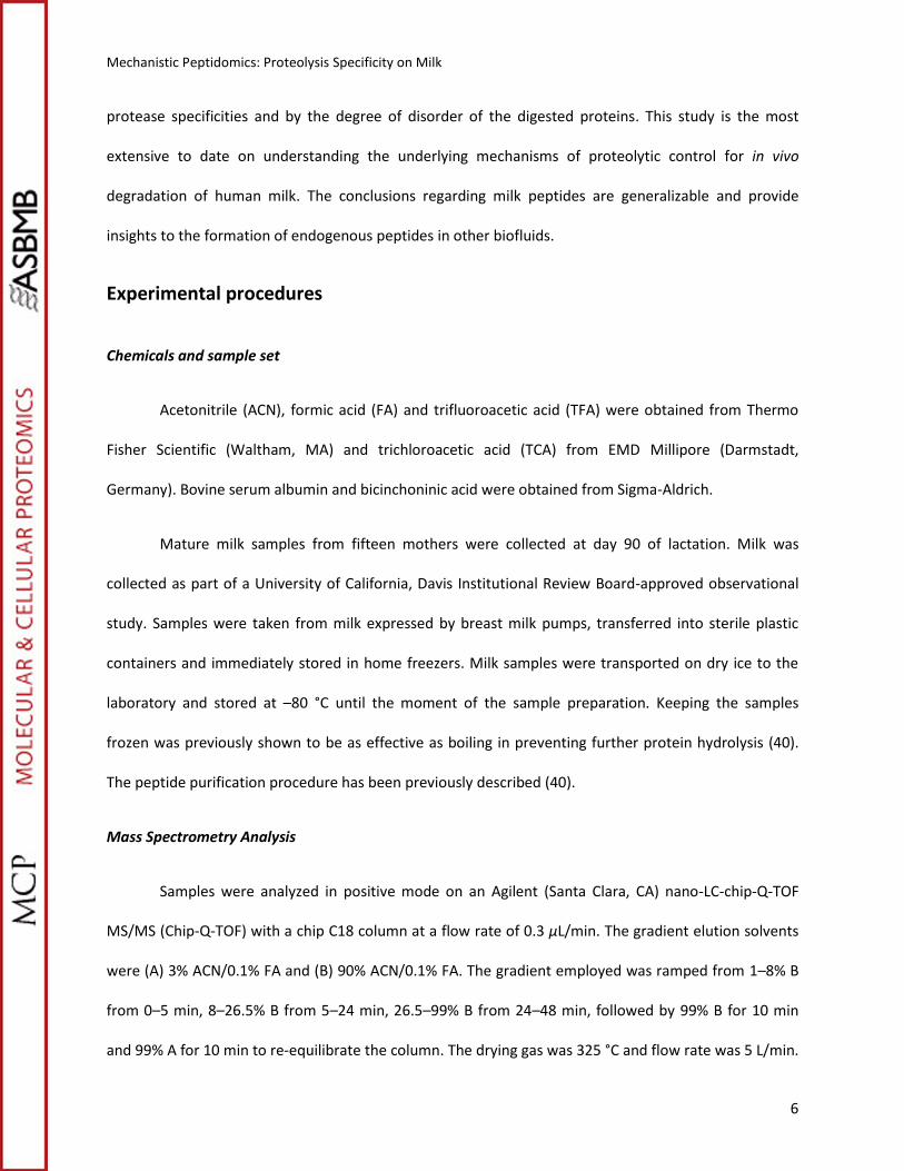
osteopontin (39), was included as separate heading in the analysis of this protein. Figure 5 shows the
results of the EnTab analysis for pIgR, β-casein, αs1-casein and osteopontin.
Figure 5
A strict peptide-based study of the enzyme participation cannot be achieved without
quantification of the peptides. Ion intensities were used for this purpose but may overestimate the
enzymatic participation of some enzymes like the system trypsin/plasmin. These proteases generate
peptides with basic amino acids at the C-terminus (R and K residues) that may show a higher ionization
response on the electrospray. Nevertheless, the results in Figure 5 show a rather low standard deviation
between samples and may be discussed from a qualitative point of view. Both, trypsin/plasmin and
elastase/cathepsin-D are acting on pIgR and β-casein. For the milk protein osteopontin, thrombin and
plasmin/trypsin are the main enzymes involved; the low participation of elastase/cathepsin-D may be
explained by the low number of potential cleavage sites for these enzymes in osteopontin. Enzyme miss
cleavages and in-source fragmentation may partially explain the high proportion of unknown cleavages,
but the participation of other enzymes may be involved too. αS1-casein is an interesting case based on
this analysis. Most of the endogenous fragments identified for αS1-casein are not associated with the
enzymes included in this analysis. These results clearly indicate the activity of one or more additional
enzymes that selectively generate the endogenous peptides from this protein.
Discussion
Intrinsic disorder and proteolysis

Mechanistic Peptidomics: Proteolysis Specificity on Milk
14
Both β-casein and osteopontin are "naturally unfolded" proteins (52) as they lack a tight and
stable tertiary structure. Because of their loose structure, more cleavage sites are exposed to proteolytic
enzymes. The same reasoning can be given for pIgR and αS1-casein. To understand the correlation
between local disorder and the formation of peptide products, we calculated the local disorder of the
pIgR and αS1-casein sequences. The local degree of disorder of these two proteins was calculated with a
computational program, the VLE3 predictor (46). The VLE3 predictor scores the likelihood of local
structural disorder based on the amino acid sequence. Regions with a score higher than 0.5 (on a 0-1
scale) are more likely to be unfolded. The result of the VLE3 predictor calculation for pIgR and αS1-casein
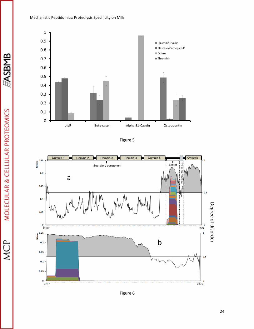
superimposed with their proteolytic map is shown in Figure 6.
Two major regions of disorder are present in pIgR, both at the C-terminus of the protein
sequence separated by a few amino acid residues (Figure 6a). pIgR is involved in the transportation of
polymeric immunoglobulins IgA and IgM across epithelial cells. At the end of the transcytosis process,
pIgR is proteolytically cleaved from its transmembrane anchor (Figure 6a) releasing the secretory
component (SC) into milk (53). The domain where the proteolytic cleavage takes place corresponds to
an unstructured sequence of the protein called the linker motif (53). The peptides from pIgR are formed
from this linker motif region. Consequently, they are the result of the proteolytic release of the SC into
the milk and represent side-products of the transcytosis of pIGR. The second disordered region, at or
near the C-terminus region, does not produce detectable peptides. pIgR is a transmembrane protein
with the C-terminal region residing in the cytosolic domain(53). Fragments formed from this region of
the protein will therefore likely remain inside the cell rather than being secreted in the milk.
Figure 6
A similar comparison between the proteolytic map and the degree of disorder was performed
for αs1-casein (Figure 6b). This protein contrasts with pIgR in that a large fraction of the protein is

Mechanistic Peptidomics: Proteolysis Specificity on Milk
15
determined to be disordered (around 70%). Again, the peptide products are localized and focused near
the N-terminus. No peptides are found from the ordered region.
All abundant peptides identified (>99% of identified signal intensity) in this study derived only
from regions of predicted disorder. These results strongly indicate that intrinsic protein disorder is a
crucial requisite for in vivo proteolysis of milk proteins within the mammary gland. A similar relationship
between protein disorder and proteolysis have been observed for the ubiquitin-independent
degradation of proteins mediated by proteasomes (54). Our results extend this structure-based
mechanism of proteolytic control to an extracellular biofluid.
Intramolecular specificity of the proteolysis
Although intrinsic disorder explains why some proteins are selectively digested in the mammary
gland, our results show that not all unstructured regions inside the protein are equally digested. For
example, the entire β-casein sequence is disordered, but regions close to the termini generate more
peptides than internal regions (Figure 3). Similarly, αs1-casein (Figure 6b) is preferentially digested at the
N-terminal, even though 70% of the protein is intrinsically unfolded and hence accessible to the
proteases. The higher abundance of sequences near the termini may be explained by the fact that only
one cleavage is necessary to generate these species, while releasing an internal fragment requires two
cleavages. Interestingly, the terminal regions of the casein proteins are known to exert different
bioactivities: the C-terminal domain of β-casein is known to generate peptides with antibacterial
properties (55), while peptides from the N-terminal region of both β-casein and αs1-casein, abundantly
phosphorylated (Table S1), are known to have a role as calcium carriers due to their abundant
phosphorylation (56).
Osteopontin is also an intrinsically disordered protein and, similarly to β-casein, yields peptides
from the terminal regions. The predominant digested region, however, is the internal region. The action

Mechanistic Peptidomics: Proteolysis Specificity on Milk
16
of thrombin, which has high proteolytic specificity, explains the particular endogenous proteolytic
pattern of osteopontin in human milk. Thrombin is known to specifically cleave between osteopontin
residues R168 and S169, exposing the integrin binding domain SVVYGLR (57) and hence, regulating the
ability of osteopontin to interact with different targets. Thrombin is known to be present in human milk
(39).We may conclude that the exposure of cleavage sites in disordered regions, the requirement of a
single cleavage for generation of peptides from the termini and the participation of specific enzymes like
thrombin explain the specificity of the endogenous proteolytic activity in the mammary gland.
Limited proteolysis in human milk
The similarity between human milk and other biofluid peptidomes is remarkable, especially
when compared with serum. Peptidomic studies of serum have shown that the most abundant peptides
from this biofluid, similarly to human milk, collapsed in a small group of clusters derived from a few
proteins, not necessarily the more abundant ones (18). The serum peptidome is mostly the consequence
of a couple of well-described proteolytic cascades: coagulation and complement activation. However,
coagulation does not occur in human milk and, although in this study we found peptides from
complement C4-A (Figure 1), their abundance is negligible. It has also been shown, when analyzing
serum peptides not proceeding form coagulation or complement activation, that proteolysis takes place
in exposed regions with special incidence at the N- and C-termini (58). Similar results have been
obtained when analyzing cell lines peptides not proceeding from the caspase cascade (59). The human
milk peptidome seems more in line with these proteolytic processes in serum masked by the
predominant proteolytic cascades. Unstructured proteins are abundant in milk and in absence of a
known tissue-specific proteolytic cascades, proteolysis happens at the most accessible sites.
Nevertheless, the products of this process are, at least partially, known bioactive products (e.g. β-casein
phosphopeptides) or side-products of activation processes (e.g. PIGR-derived peptides). The barrier

Mechanistic Peptidomics: Proteolysis Specificity on Milk
17
between what seems to be accidental good substrates for the peptidases present in milk and a limited
proteolysis targeted to the production of specific products becomes narrow. Not all the peptides found
in this study can be tracked to bioactive compounds, and further investigation of these peptides for
functional properties is warranted.
Acknowledgments
Funding provided by NIH (GM049077) is gratefully acknowledged.

Mechanistic Peptidomics: Proteolysis Specificity on Milk
18
References
1. Schulz-Knappe P, Schrader M, & Zucht H-D (2005) The Peptidomics Concept. Combinatorial Chemistry & High Throughput Screening 8(8):697-704.
2. Cunningham R, Ma D, & Li L (2012) Mass spectrometry-based proteomics and peptidomics for systems biology and biomarker discovery. Front. Biol. 7(4):313-335.
3. Shen Y, et al. (2010) Strategy for Degradomic-Peptidomic Analysis of Human Blood Plasma. Journal of Proteome Research 9(5):2339-2346.
4. Wiltfang J, et al. (2003) β-amyloid peptides in cerebrospinal fluid of patients with Creutzfeldt–Jakob disease. Annals of Neurology 54(2):263-267.
5. Huang C-M & Zhu W (2009) Profiling Human Saliva Endogenous Peptidome via a High Throughput MALDI-TOF-TOF Mass Spectrometry. Combinatorial Chemistry & High Throughput Screening 12(5):521-531.
6. Messana I, et al. (2008) Trafficking and Postsecretory Events Responsible for the Formation of Secreted Human Salivary Peptides: A Proteomics Approach. Molecular & Cellular Proteomics 7(5):911-926.
7. Hayakawa E, et al. (2013) Peptidomic analysis of human reflex tear fluid. Peptides 42(0):63-69. 8. Jurgens M, et al. (2005) Towards Characterization of the Human Urinary Peptidome.
Combinatorial Chemistry & High Throughput Screening 8(8):757-765. 9. Shen Y, et al. (2010) Blood Peptidome-Degradome Profile of Breast Cancer. PLoS ONE
5(10):e13133. 10. Ling XB, Mellins ED, Sylvester KG, & Cohen HJ (2010) Urine Peptidomics for Clinical Biomarker
Discovery. Advances in Clinical Chemistry, ed Gregory SM (Elsevier), Vol Volume 51, pp 181-213. 11. Wen Q, et al. (2013) Peptidomic Identification of Serum Peptides Diagnosing Preeclampsia. PLoS
ONE 8(6):e65571. 12. Quintana LF, et al. (2009) Application of Label-free Quantitative Peptidomics for the
Identification of Urinary Biomarkers of Kidney Chronic Allograft Dysfunction. Molecular & Cellular Proteomics 8(7):1658-1673.
13. Villanueva J, et al. (2006) Serum Peptidome Patterns That Distinguish Metastatic Thyroid Carcinoma from Cancer-free Controls Are Unbiased by Gender and Age. Molecular & Cellular Proteomics 5(10):1840-1852.
14. Sasaki K, Takahashi N, Satoh M, Yamasaki M, & Minamino N (2010) A Peptidomics Strategy for Discovering Endogenous Bioactive Peptides. Journal of Proteome Research 9(10):5047-5052.
15. Svensson M, Sköld K, Svenningsson P, & Andren PE (2003) Peptidomics-Based Discovery of Novel Neuropeptides. Journal of Proteome Research 2(2):213-219.
16. Tamvakopoulos C (2007) Mass spectrometry for the quantification of bioactive peptides in biological fluids. Mass Spectrometry Reviews 26(3):389-402.
17. Bernay B, et al. (2009) Discovering New Bioactive Neuropeptides in the Striatum Secretome Using in Vivo Microdialysis and Versatile Proteomics. Molecular & Cellular Proteomics 8(5):946-958.
18. Villanueva J, et al. (2006) Differential exoprotease activities confer tumor-specific serum peptidome patterns. The Journal of Clinical Investigation 116(1):271-284.
19. Diamandis EP (2006) Peptidomics for Cancer Diagnosis: Present and Future. Journal of Proteome Research 5(9):2079-2082.

Mechanistic Peptidomics: Proteolysis Specificity on Milk
19
20. Ferranti P, et al. (2004) Casein proteolysis in human milk: tracing the pattern of casein breakdown and the formation of potential bioactive peptides. Journal of Dairy Research 71(01):74-87
21. Timmer JC, et al. (2009) Structural and kinetic determinants of protease substrates. Nat Struct Mol Biol 16(10):1101-1108.
22. Hubbard SJ (1998) The structural aspects of limited proteolysis of native proteins. Biochimica et Biophysica Acta (BBA) - Protein Structure and Molecular Enzymology 1382(2):191-206.
23. Novotný J & Bruccoleri RE (1987) Correlation among sites of limited proteolysis, enzyme accessibility and segmental mobility. FEBS Letters 211(2):185-189.
24. Kazanov MD, et al. (2011) Structural Determinants of Limited Proteolysis. Journal of Proteome Research 10(8):3642-3651.
25. Seppo L, Jauhiainen T, Poussa T, & Korpela R (2003) A fermented milk high in bioactive peptides has a blood pressure–lowering effect in hypertensive subjects. The American Journal of Clinical Nutrition 77(2):326-330.
26. Matar C, Amiot J, Savoie L, & Goulet J (1996) The Effect of Milk Fermentation by Lactobacillus helveticus on the Release of Peptides During In Vitro Digestion. Journal of Dairy Science 79(6):971-979.
27. Smacchi E & Gobbetti M (2000) Bioactive peptides in dairy products: synthesis and interaction with proteolytic enzymes. Food Microbiology 17(2):129-141.
28. Argyri K, Miller DD, Glahn RP, Zhu L, & Kapsokefalou M (2007) Peptides Isolated from in Vitro Digests of Milk Enhance Iron Uptake by Caco-2 Cells. Journal of Agricultural and Food Chemistry 55(25):10221-10225.
29. Ye A, Cui J, & Singh H (2011) Proteolysis of milk fat globule membrane proteins during in vitro gastric digestion of milk. Journal of Dairy Science 94(6):2762-2770.
30. Picariello G, et al. (2010) Peptides surviving the simulated gastrointestinal digestion of milk proteins: Biological and toxicological implications. Journal of Chromatography B 878(3–4):295-308.
31. Clare DA & Swaisgood HE (2000) Bioactive Milk Peptides: A Prospectus. Journal of Dairy Science 83(6):1187-1195.
32. Bellamy W, Takase M, Wakabayashi H, Kawase K, & Tomita M (1992) Antibacterial spectrum of lactoferricin B, a potent bactericidal peptide derived from the N-terminal region of bovine lactoferrin. Journal of Applied Bacteriology 73(6):472-479.
33. Benkerroum N (2010) Antimicrobial peptides generated from milk proteins: a survey and prospects for application in the food industry. A review. International Journal of Dairy Technology 63(3):320-338.
34. FitzGerald RJ, Murray BA, & Walsh DJ (2004) Hypotensive Peptides from Milk Proteins. The Journal of Nutrition 134(4):980S-988S.
35. Jäkälä P & Vapaatalo H (2010) Antihypertensive Peptides from Milk Proteins. Pharmaceuticals 3(1):251-272.
36. Politis I & Chronopoulou R (2008) Milk Peptides and Immune Response in the Neonate. Bioactive Components of Milk, Advances in Experimental Medicine and Biology, ed Bösze Z (Springer New York), Vol 606, pp 253-269.
37. Huang S-M, Chen K-N, Chen Y-P, Hong W-S, & Chen M-J (2010) Immunomodulatory properties of the milk whey products obtained by enzymatic and microbial hydrolysis. International Journal of Food Science & Technology 45(5):1061-1067.
38. Meisel H & FitzGerald RJ (2000) Opioid peptides encrypted in intact milk protein sequences. British Journal of Nutrition 84(Supplement S1):27-31

Mechanistic Peptidomics: Proteolysis Specificity on Milk
20
39. Christensen B, Schack L, Kläning E, & Sørensen ES (2010) Osteopontin Is Cleaved at Multiple Sites Close to Its Integrin-binding Motifs in Milk and Is a Novel Substrate for Plasmin and Cathepsin D. Journal of Biological Chemistry 285(11):7929-7937.
40. Dallas DC, et al. (2013) Extensive in vivo Human Milk Peptidomics Reveals Specific Proteolysis Yielding Protective Antimicrobial Peptides. Journal of Proteome Research 12(5):2295-2304.
41. Smith PK, et al. (1985) Measurement of protein using bicinchoninic acid. Analytical Biochemistry 150(1):76-85.
42. Craig R & Beavis RC (2004) TANDEM: matching proteins with tandem mass spectra. Bioinformatics 20(9):1466-1467.
43. Molinari CE, et al. (2012) Proteome mapping of human skim milk proteins in term and preterm milk. J. Proteome Res.
44. Mange A, Bellet V, Tuaillon E, Van de Perre P, & Solassol J (2008) Comprehensive proteomic analysis of the human milk proteome: Contribution of protein fractionation. J. Chromatogr. B 876(2):252-256.
45. iao , lvarado R, Phinney , nnerdal (2011) Proteomic characterization of human milk fat globule membrane proteins during a 12 month lactation period. J. Proteome Res.
46. Peng K, et al. (2005) OPTIMIZING LONG INTRINSIC DISORDER PREDICTORS WITH PROTEIN EVOLUTIONARY INFORMATION. Journal of Bioinformatics and Computational Biology 03(01):35-60.
47. Gasteiger E, et al. (2005) Protein identification and analysis tools on the ExPASy server. The proteomics protocols handbook, (Springer), pp 571-607.
48. Korycha-Dahl M, Dumas BR, Chene N, & Martal J (1983) Plasmin Activity in Milk. Journal of Dairy Science 66(4):704-711.
49. Monti JC, Mermoud AF, & Jollès P (1986) Trypsin in human milk. Experientia 42(1):39-41. 50. Borulf S, Lindberg T, & MÅNsson M (1987) Immunoreactive Anionic Trypsin and Anionic Elastase
in Human Milk. Acta Pædiatrica 76(1):11-15. 51. Vetvicka V Fau - Vagner J, et al. (Human breast milk contains procathepsin D--detection by
specific antibodies. (1039-9712 (Print)). 52. Tompa P (2002) Intrinsically unstructured proteins. Trends in Biochemical Sciences 27(10):527-
533. 53. Kaetzel CS (2005) The polymeric immunoglobulin receptor: bridging innate and adaptive
immune responses at mucosal surfaces. Immunological Reviews 206(1):83-99. 54. Asher G, Reuven N, & Shaul Y (2006) 20S proteasomes and protein degradation “by default”.
BioEssays 28(8):844-849. 55. Minervini F, et al. (2003) Angiotensin I-Converting-Enzyme-Inhibitory and Antibacterial Peptides
from Lactobacillus helveticus PR4 Proteinase-Hydrolyzed Caseins of Milk from Six Species. Applied and Environmental Microbiology 69(9):5297-5305.
56. Lönnerdal B (2013) Bioactive proteins in breast milk. Journal of Paediatrics and Child Health 49:1-7.
57. Lund S, Giachelli C, & Scatena M (2009) The role of osteopontin in inflammatory processes. J. Cell Commun. Signal. 3(3-4):311-322.
58. Koomen JM, et al. (2005) Direct Tandem Mass Spectrometry Reveals Limitations in Protein Profiling Experiments for Plasma Biomarker Discovery. Journal of Proteome Research 4(3):972-981.
59. Gelman JS, Sironi J, Castro LM, Ferro ES, & Fricker LD (2011) Peptidomic Analysis of Human Cell Lines. Journal of Proteome Research 10(4):1583-1592.

Mechanistic Peptidomics: Proteolysis Specificity on Milk
21
Footnotes
1 Data uploaded at the Peptide Atlas repository
http://www.peptideatlas.org/PASS/PASS00495
2E.A. Parker, (2014) Peptidomics Enzyme Estimator, Github Repository,
https://github.com/eparker05/Peptidomics-enzyme-estimator
Figure Legends
Figure 1. Average (15 samples) normalized ion intensities of the total endogenous peptides by protein
of origin. Error bars show the standard deviation from the average.
Figure 2. Endogenous proteolytic map of β-casein in milk determined by PepEx. The height of each area
represents the ion intensity of the corresponding peptides.
Figure 3. Endogenous proteolytic map of β-casein (15 samples) determined with PepEx. Arrows indicate
the position of cleavage sites for Plasmin/Trypsin (blue) and Cathepsin-D (red).
Figure 4. Proteolytic maps of (a) osteopontin, (b) αs1-casein and (c) pIgR (15 samples) determined with
PepEx. Arrows indicate the position of cleavage sites for Plasmin/Trypsin (blue) and Cathepsin-D (red).
Figure 5: Relative participation of the proteases on the formation of endogenous peptides by protein (15
samples) estimated with EnTab. Error bars indicate standard deviation from the mean.
Figure 6. Proteolytic map of (a) pIgR and (b) αs1-Casein overlaid with the predicted degree of disorder for
each part of the sequence. Values of disorder higher than 0.5 represent naturally unfolded regions.

Mechanistic Peptidomics: Proteolysis Specificity on Milk
22
Figures
Figure 1
Figure 2

Mechanistic Peptidomics: Proteolysis Specificity on Milk
23
Figure 3
Figure 4
Mechanistic Peptidomics: Proteolysis Specificity on Milk
24
Figure 5
Figure 6