Mechanisms of neuroblastoma regression
10
704 | DECEMBER 2014 | VOLUME 11 www.nature.com/nrclinonc Division of Oncology, The Children’s Hospital of Philadelphia, 3501 Civic Center Boulevard, Philadelphia, PA 19104‑4302, USA (G.M.B., R.B.). Correspondence to: G.M.B. brodeur@ e‑mail.chop.edu Mechanisms of neuroblastoma regression Garrett M. Brodeur and Rochelle Bagatell Abstract | Recent genomic and biological studies of neuroblastoma have shed light on the dramatic heterogeneity in the clinical behaviour of this disease, which spans from spontaneous regression or differentiation in some patients, to relentless disease progression in others, despite intensive multimodality therapy. This evidence also suggests several possible mechanisms to explain the phenomena of spontaneous regression in neuroblastomas, including neurotrophin deprivation, humoral or cellular immunity, loss of telomerase activity and alterations in epigenetic regulation. A better understanding of the mechanisms of spontaneous regression might help to identify optimal therapeutic approaches for patients with these tumours. Currently, the most druggable mechanism is the delayed activation of developmentally programmed cell death regulated by the tropomyosin receptor kinase A pathway. Indeed, targeted therapy aimed at inhibiting neurotrophin receptors might be used in lieu of conventional chemotherapy or radiation in infants with biologically favourable tumours that require treatment. Alternative approaches consist of breaking immune tolerance to tumour antigens or activating neurotrophin receptor pathways to induce neuronal differentiation. These approaches are likely to be most effective against biologically favourable tumours, but they might also provide insights into treatment of biologically unfavourable tumours. We describe the different mechanisms of spontaneous neuroblastoma regression and the consequent therapeutic approaches. Brodeur, G. M. & Bagatell, R. Nat. Rev. Clin. Oncol. 11, 704–713 (2014); published online 21 October 2014; doi:10.1038/nrclinonc.2014.168 Introduction Neuroblastoma is the most common extracranial solid tumour of children; it accounts for 8–10% of childhood cancers in the USA and Europe. 1–4 Neuroblastomas in chil- dren 18 months of age or older are frequently unresectable or metastatic, require intensive multimodality therapy and are associated with a 40–50% survival rate. 1,2,5 However, neuroblastomas in children under 18 months of age behave very differently. Most infants, even with metastatic disease, can be cured with moderate-intensity chemotherapy, and some patients with a special pattern of metastasis have a high likelihood of undergoing spontaneous regres- sion without chemotherapy. 6–10 Indeed, the prevalence of spontaneous regression has been documented by mass- screening programmes undertaken in Japan, Quebec and Europe. 11–14 Furthermore, children (and adults) can present with localized, benign ganglioneuromas, which likely represent neuroblastic tumours that have become differentiated. 15–19 The exact mechanisms responsible for spontaneous regression (and differentiation) are uncer- tain, but several plausible mechanisms have been proposed to explain these phenomena. 6–10 In this Review, we explore the current understanding of the genomic, biological and immunological mechanisms that underlie spontaneous regression, and possible approaches to therapy. Genetic predisposition About 1–2% of patients with neuroblastoma have a family history of this disease. 20–23 Two genes have been identified, ALK and PHOX2B, that account for ~80% of hereditary neuroblastoma. Specifically, several groups have shown that activating mutations in ALK are responsible for ~75% cases of hereditary neuroblastoma. 20,24–26 Neuroblastomas also occur in patients with congenital central hypoventila- tion syndrome (Ondine’s curse), and inactivating muta- tions of PHOX2B are present in most of these patients, accounting for another 5% of hereditary cases. 22,27,28 Genome-wide association studies have identified several gene polymorphisms associated with a low, but significant risk of neuroblastoma, which include BARD1, LMO1, and LIN28B among others. 29–33 Genetically engineered mouse models that develop neuroblastoma are available, and include TH-MYCN, 34 MDM2+MYCN, 35 ALK+MYCN, 36 ALK, 37 and LIN28B. 38 However, no cases of hereditary neuroblastoma have been associated with germline muta- tions of genes other than ALK and PHOX2B, and none has been associated with spontaneous regression. Subtypes of neuroblastomas Several genomic alterations have been identified in neuroblastomas that generate tumours with distinct genotypes characterized by different patterns of clini- cal behaviour (Figure 1). 15 Neuroblastomas can be divided into three major subtypes based on cytogenetic profiles—subtype 1, 2A and 2B. Subtype 1 is character- ized by numerical chromosome alterations resulting in hyperdiploidy or near triploidy, but with few if any seg- mental chromosomal abnormalities (SCAs). 39,40 High expression levels of the tropomyosin receptor kinase (Trk) A is common to almost all subtype 1 tumours. Competing interests The authors declare no competing interests. REVIEWS © 2014 Macmillan Publishers Limited. All rights reserved
Transcript of Mechanisms of neuroblastoma regression

704 | DECEMBER 2014 | VOLUME 11 www.nature.com/nrclinonc
Division of Oncology, The Children’s Hospital of Philadelphia, 3501 Civic Center Boulevard, Philadelphia, PA 19104‑4302, USA (G.M.B., R.B.).
Correspondence to: G.M.B. brodeur@ e‑mail.chop.edu
Mechanisms of neuroblastoma regressionGarrett M. Brodeur and Rochelle Bagatell
Abstract | Recent genomic and biological studies of neuroblastoma have shed light on the dramatic heterogeneity in the clinical behaviour of this disease, which spans from spontaneous regression or differentiation in some patients, to relentless disease progression in others, despite intensive multimodality therapy. This evidence also suggests several possible mechanisms to explain the phenomena of spontaneous regression in neuroblastomas, including neurotrophin deprivation, humoral or cellular immunity, loss of telomerase activity and alterations in epigenetic regulation. A better understanding of the mechanisms of spontaneous regression might help to identify optimal therapeutic approaches for patients with these tumours. Currently, the most druggable mechanism is the delayed activation of developmentally programmed cell death regulated by the tropomyosin receptor kinase A pathway. Indeed, targeted therapy aimed at inhibiting neurotrophin receptors might be used in lieu of conventional chemotherapy or radiation in infants with biologically favourable tumours that require treatment. Alternative approaches consist of breaking immune tolerance to tumour antigens or activating neurotrophin receptor pathways to induce neuronal differentiation. These approaches are likely to be most effective against biologically favourable tumours, but they might also provide insights into treatment of biologically unfavourable tumours. We describe the different mechanisms of spontaneous neuroblastoma regression and the consequent therapeutic approaches.
Brodeur, G. M. & Bagatell, R. Nat. Rev. Clin. Oncol. 11, 704–713 (2014); published online 21 October 2014; doi:10.1038/nrclinonc.2014.168
Introduction Neuroblastoma is the most common extracranial solid tumour of children; it accounts for 8–10% of childhood cancers in the USA and Europe.1–4 Neuroblastomas in chil-dren 18 months of age or older are frequently unresect able or metastatic, require intensive multi modality therapy and are associated with a 40–50% survival rate.1,2,5 However, neuroblastomas in children under 18 months of age behave very differently. Most infants, even with metastatic disease, can be cured with moderate-intensity chemo therapy, and some patients with a special pattern of metasta sis have a high likelihood of undergoing spontaneous regres-sion without chemotherapy.6–10 Indeed, the preva lence of spontaneous regression has been documented by mass-screening programmes undertaken in Japan, Quebec and Europe.11–14 Furthermore, children (and adults) can present with localized, benign ganglioneuromas, which likely represent neuroblastic tumours that have become differentiated.15–19 The exact mechanisms responsible for spontaneous regression (and differentiation) are uncer-tain, but several plausible mechanisms have been proposed to explain these phenom ena.6–10 In this Review, we explore the current understanding of the genomic, biological and immunological mechanisms that underlie spontaneous regression, and possible approaches to therapy.
Genetic predisposition About 1–2% of patients with neuroblastoma have a family history of this disease.20–23 Two genes have been identified,
ALK and PHOX2B, that account for ~80% of hereditary neuroblastoma. Specifically, several groups have shown that activating mutations in ALK are responsible for ~75% cases of hereditary neuroblastoma.20,24–26 Neuroblastomas also occur in patients with congenital central hypoventila-tion syndrome (Ondine’s curse), and inactivating muta-tions of PHOX2B are present in most of these patients, accounting for another 5% of hereditary cases.22,27,28
Genome-wide association studies have identified several gene polymorphisms associated with a low, but significant risk of neuroblastoma, which include BARD1, LMO1, and LIN28B among others.29–33 Genetically engineered mouse models that develop neuroblastoma are available, and include TH-MYCN,34 MDM2+MYCN,35 ALK+MYCN,36 ALK,37 and LIN28B.38 However, no cases of hereditary neuro blastoma have been associ ated with germline muta-tions of genes other than ALK and PHOX2B, and none has been associated with s pontaneous regression.
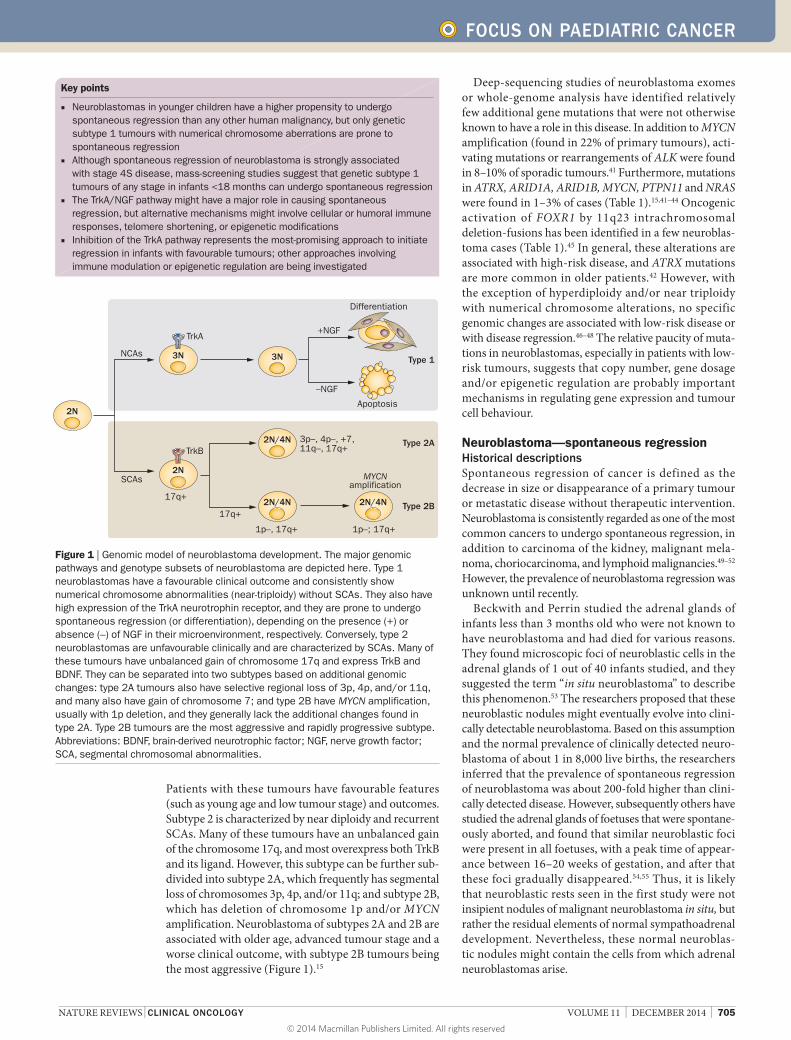
Subtypes of neuroblastomasSeveral genomic alterations have been identified in neuroblastomas that generate tumours with distinct genotypes characterized by different patterns of clini-cal behaviour (Figure 1).15 Neuroblastomas can be divided into three major subtypes based on cytogenetic p rofiles—subtype 1, 2A and 2B. Subtype 1 is character-ized by numerical chromosome alterations resulting in hyperdiploidy or near triploidy, but with few if any seg-mental chromosomal abnormalities (SCAs).39,40 High expression levels of the tropomyosin receptor kinase (Trk) A is common to almost all subtype 1 tumours.
Competing interestsThe authors declare no competing interests.
REVIEWS
© 2014 Macmillan Publishers Limited. All rights reserved
NATURE REVIEWS | CLINICAL ONCOLOGY VOLUME 11 | DECEMBER 2014 | 705
Patients with these tumours have favourable features (such as young age and low tumour stage) and outcomes. Subtype 2 is characterized by near diploidy and recurrent SCAs. Many of these tumours have an unbalanced gain of the chromosome 17q, and most overexpress both TrkB and its ligand. However, this subtype can be further sub-divided into subtype 2A, which frequently has segmental loss of chromosomes 3p, 4p, and/or 11q; and subtype 2B, which has deletion of chromosome 1p and/or MYCN amplification. Neuroblastoma of subtypes 2A and 2B are associated with older age, advanced tumour stage and a worse clinical outcome, with subtype 2B tumours being the most aggressive (Figure 1).15
Deep-sequencing studies of neuroblastoma exomes or whole-genome analysis have identified relatively few additional gene mutations that were not otherwise known to have a role in this disease. In addition to MYCN amplification (found in 22% of primary tumours), acti-vating mutations or rearrangements of ALK were found in 8–10% of sporadic tumours.41 Furthermore, mutations in ATRX, ARID1A, ARID1B, MYCN, PTPN11 and NRAS were found in 1–3% of cases (Table 1).15,41–44 Oncogenic activation of FOXR1 by 11q23 intrachromosomal deletion- fusions has been identified in a few neuroblas-toma cases (Table 1).45 In general, these alterations are associated with high-risk disease, and ATRX mutations are more common in older patients.42 However, with the exception of hyperdiploidy and/or near triploidy with numerical chromosome alterations, no specific genomic changes are associated with low-risk disease or with disease regression.46–48 The relative paucity of muta-tions in neuroblastomas, especially in patients with low-risk tumours, suggests that copy number, gene dosage and/or epigenetic regulation are probably important mechanisms in regulating gene expression and tumour cell behaviour.
Neuroblastoma—spontaneous regressionHistorical descriptionsSpontaneous regression of cancer is defined as the decrease in size or disappearance of a primary tumour or metastatic disease without therapeutic intervention. Neuroblastoma is consistently regarded as one of the most common cancers to undergo spontaneous regression, in addition to carcinoma of the kidney, malignant mela-noma, choriocarcinoma, and lymphoid malignancies.49–52 However, the prevalence of neuroblastoma regression was unknown until recently.
Beckwith and Perrin studied the adrenal glands of infants less than 3 months old who were not known to have neuroblastoma and had died for various reasons. They found microscopic foci of neuroblastic cells in the adrenal glands of 1 out of 40 infants studied, and they suggested the term “in situ neuroblastoma” to describe this phenomenon.53 The researchers proposed that these neuroblastic nodules might eventually evolve into clini-cally detectable neuro blastoma. Based on this assumption and the normal prevalence of clinically detected neuro-blastoma of about 1 in 8,000 live births, the researchers inferred that the prevalence of spontaneous regression of neuroblastoma was about 200-fold higher than clini-cally detected disease. However, subsequently others have studied the adrenal glands of foetuses that were spontane-ously aborted, and found that similar neuroblastic foci were present in all foetuses, with a peak time of appear-ance between 16–20 weeks of gestation, and after that these foci gradually disappeared.54,55 Thus, it is likely that neuroblastic rests seen in the first study were not insipient nodules of malignant neuroblastoma in situ, but rather the residual elements of normal sympatho adrenal development. Nevertheless, these normal neuroblas-tic nodules might contain the cells from which adrenal n euroblastomas arise.
Key points
■ Neuroblastomas in younger children have a higher propensity to undergo spontaneous regression than any other human malignancy, but only genetic subtype 1 tumours with numerical chromosome aberrations are prone to spontaneous regression
■ Although spontaneous regression of neuroblastoma is strongly associated with stage 4S disease, mass‑screening studies suggest that genetic subtype 1 tumours of any stage in infants <18 months can undergo spontaneous regression
■ The TrkA/NGF pathway might have a major role in causing spontaneous regression, but alternative mechanisms might involve cellular or humoral immune responses, telomere shortening, or epigenetic modifications
■ Inhibition of the TrkA pathway represents the most‑promising approach to initiate regression in infants with favourable tumours; other approaches involving immune modulation or epigenetic regulation are being investigated
17q+2N/4N2N/4N
2N
Type 1
Type 2A
2N
TrkB
3N
TrkA
NCAs
SCAs
2N/4N
3N
Apoptosis
Differentiation
Type 2B
–NGF
17q+
3p–, 4p–, +7,11q–, 17q+
+NGF
1p–, 17q+ 1p–; 17q+
MYCNamplification
Figure 1 | Genomic model of neuroblastoma development. The major genomic pathways and genotype subsets of neuroblastoma are depicted here. Type 1 neuroblastomas have a favourable clinical outcome and consistently show numerical chromosome abnormalities (near‑triploidy) without SCAs. They also have high expression of the TrkA neurotrophin receptor, and they are prone to undergo spontaneous regression (or differentiation), depending on the presence (+) or absence (–) of NGF in their microenvironment, respectively. Conversely, type 2 neuroblastomas are unfavourable clinically and are characterized by SCAs. Many of these tumours have unbalanced gain of chromosome 17q and express TrkB and BDNF. They can be separated into two subtypes based on additional genomic changes: type 2A tumours also have selective regional loss of 3p, 4p, and/or 11q, and many also have gain of chromosome 7; and type 2B have MYCN amplification, usually with 1p deletion, and they generally lack the additional changes found in type 2A. Type 2B tumours are the most aggressive and rapidly progressive subtype. Abbreviations: BDNF, brain‑derived neurotrophic factor; NGF, nerve growth factor; SCA, segmental chromosomal abnormalities.
FOCUS ON PAEDIATRIC CANCER
© 2014 Macmillan Publishers Limited. All rights reserved

706 | DECEMBER 2014 | VOLUME 11 www.nature.com/nrclinonc
Neuroblastoma stage 4S The phenomenon of spontaneous regression of neuro-blastoma was already known, but it was further high-lighted by Evans and D’Angio who identified a specific pattern of metastatic spread called stage IVS.56,57 Infants with stage IVS generally had small primary tumours with dissemination limited to the liver and skin, with minimal bone marrow involvement. These patients were noted to have a very good prognosis, and some did well even in the absence of any tumour-specific therapy.56,57 This is in contrast to patients over 12–18 months of age with a dif-ferent pattern of metastatic disease that included bone lesions and more-extensive marrow involvement. Patients with the latter pattern of metastatic disease generally had a poor prognosis.58,59 Later, the definition of stage IVS was refined as stage 4S by the International Neuroblastoma Staging System (INSS).60,61 INSS stage 4S was restricted to infants aged less than 12 months at diagnosis, with
small primary tumours (INSS stage 1 or 2), and with less than 10% marrow involvement. The most recent staging system iteration is called the International Neuroblastoma Risk Group Staging System (INRGSS), and this stage is referred to as stage MS (M for metastatic, S for special); 62 we will refer to this stage as 4S in this Review. However, it is clear that spontaneous regression is not restricted to this stage or subset of patients, as regression can be seen in infants with any stage of disease if they have b iologically favourable tumours.48,63–65
Genomics and biology of 4S tumoursA limited number of studies have been performed to identify patterns of gene expression that are character-istic of stage 4S neuroblastomas. Benard et al.46 studied 29 cases of metastatic neuroblastoma (12 of which were stage 4S) that lacked MYCN amplification. The investi-gators developed a genetic signature of 45 genes that was significantly associated with stage 4S versus stage 4 tumours, and this was validated in an independent set of 22 tumours. Lavarino et al.66 conducted a similar study on 35 infants with metastatic neuroblastoma (25 had stage 4 disease and 10 had stage 4S neuroblastoma). The tumours from patients with stage 4 disease were character ized by SCAs, whereas 90% of stage 4S tumours were near- triploid with whole chromo some gains.66 The investi-gators found a differential expression of certain genes (such as CHD5, GNB1 and RERE) in stage 4 and stage 4S neuroblastoma, with higher expression of genes mapping to the short arm of chromosome 1 in stage 4S tumours, and to chromosome 11 for stage 4 tumours. A smaller proteomic study was performed on eight tumours from infants with stage 4 and 4S, which identified another set of differentially expressed proteins across the two stages.67 However, there was essentially no overlap of genes (or proteins) differentially expressed by regressing 4S versus non-regressing infant tumours among these studies, s uggesting that further i nvestigation is needed.
Metastatic and multifocal neuroblastic tumours The unique pattern of metastasis characteristic of stage 4S disease might represent multifocal hyperplasia of neural crest precursors.68,69 Indeed, the tissues involved in the 4S dissemination pattern are reminiscent of the tissue migra-tion pattern of neural crest cells observed during develop-ment (Box 1). The concept of multifocal hyper plasia is based on the hypothesis that all cells have sustained a single initiating lesion, whereas true malignancies have sustained two or more critical mutations.68 In this regard, it is interesting that patients with constitutional activat-ing mutations of ALK and hereditary neuro blastoma do not present a 4S pattern with increased frequency.20,24–26 To date, no biopsy or genotype analysis has been per-formed on multiple discrete nodules from patients with 4S tumours to evaluate if those patients have identical or non-identical patterns of genomic changes. However, genomic analysis of primary 4S tumours reveal clonal changes, consistent with a malignant process.47,66,70,71 Genomic analyses of multiple lesions in 4S patients would likely address this issue definitively; however, considering
Table 1 | Somatically acquired mutations and rearrangements in neuroblastoma
Gene Function Alteration Frequency (%)
Reference
MYCN Transcription factor AmplificationActivating mutation
221.7
1541
ALK Tyrosine kinase receptor
Activating mutationAmplification/duplication
9.23.4
4141
PTPN11 Tyrosine phosphatase Activating mutation 2.9 41
ATRX Chromatin remodelling Inactivating mutation 2.5 42,44
ARID1A/B Chromatin remodelling Inactivating mutation 2–3 43
NRAS Signalling protein Activating mutation ~1 41
MLL-FOXR1, or PAFAH1B-FOXR1
Transcription factor Translocation 1.3 45
Box 1 | Genes involved in neural crest development
Insights into the mechanisms of neuroblastoma regression could potentially be gained by understanding the developmental biology of the neural crest, from which neuroblastomas are derived. During embryonic development, cells at the edge of the neural plate undergo epithelial‑to‑mesenchymal transition and migrate away from the neural tube.164 These cells follow different pathways, ultimately giving rise to a variety of tissues, including the enteric nervous system, melanocytes, dorsal root and sympathetic ganglia, and the chromaffin cells of the adrenal medulla. Numerous transcription factors and signalling molecules contribute to neural crest formation and to the differentiation of pluripotent neural crest cells.165 Furthermore, epigenetic control might also be important in determining cell fate.164 Much remains to be learned regarding genes normally involved in neural crest development and their role in neural crest‑derived malignancies, such as neuroblastoma. However, expression profiling of a relatively small number of foetal adrenal neuroblasts, normal foetal adrenal cortical cells, and neuroblastoma tumours has suggested that there is differential expression of genes associated with earlier (ASCL1) versus later (DLK1) time points in neural crest development in tumours classified as favourable versus unfavourable.166 Furthermore, MASH1, PHOX2B, Hand2 BMP4 and other genes are known to have critical roles in chromaffin, dorsal root and sympathetic neural development.167,168 More recently, studies in zebrafish have confirmed that PHOX2B is required for normal development of neural crest‑derived cells that comprise the sympathetic ganglia,136 and that specific PHOX2B aberrations can have varying effects on differentiation.169 Additional studies could potentially clarify if PHOX2B and other genes involved in neural crest development have a role in neuroblastoma regression as well.
REVIEWS
© 2014 Macmillan Publishers Limited. All rights reserved
NATURE REVIEWS | CLINICAL ONCOLOGY VOLUME 11 | DECEMBER 2014 | 707
the available data, we favour the hypothesis that stage 4S tumour are metastatic dissemination of genetically identi-cal cells, d eveloping as biologically favourable disease in most cases.
Lessons from neuroblastoma screeningThe exact prevalence of spontaneous regression has been difficult to define precisely, but neuroblastoma mass-screening studies undertaken in Japan, Canada and Europe have provided useful information. The outcome of infants with neuroblastoma is substantially better than that of older children with this disease. Furthermore, almost all neuroblastomas produce catecholamines, and their metabolites—homovanillic acid (HVA) and vanillyl-mandelic acid (VMA)—can be readily detected in urine.72
Therefore, mass screening of infants for neuroblastoma by measuring urine VMA and HVA was initiated in Japan,11,12 with the goal of detecting tumours earlier and hopefully improving patient outcome. Initial results were promising, so similar efforts were initiated in North America and in Europe.13,14 Analysis of the data gener-ated from these initiatives indicates that mass screening resulted in a substantial increase in the prevalence of neuroblastoma in the screened population (~1:2,000 live births, versus 1:7,000 live births in unscreened popula-tions), but overall mortal ity was unchanged. Importantly there was no signifi cant decrease in the prevalence or mortality of neuroblastoma in patients older than 1 year.73–76 Thus, the goal to reduce neuroblastoma mor-tality by mass screening in infancy was not achieved, and screening efforts have essentially stopped worldwide.
Despite the fact the main objective of mass-screening was not reached, studies of this approach have provided valuable insights into the pathogenesis and clinical behav-iour of biologically favourable tumours. The increased prevalence of neuroblastoma observed in the screened populations indicates that spontaneous regression of neuroblastoma (without clinical detection) occurs at least as frequently as neuroblastoma that is detected clinically. In addition, analyses performed on screened tumours showed that virtually all of them, regardless of their stage, were biologically favourable with respect to MYCN status and tumour cell ploidy,77–79 which is in contrast to the unfavourable biological features generally found in clinically detected tumours from older children. Importantly, these studies also indicated that biologi-cally favourable tumours rarely evolve into biologically unfavourable tumours.
Although not intended as a neuroblastoma screening tool, there have been several reports of incidental prenatal detection of neuroblastoma by maternal ultrasound.80–82 These cases are similar, both clinically and biologically, to those identified by screening, and the vast majority of infants do well with little or no therapy. Infants under 1 year of age are at risk for both anaesthetic and surgi-cal complications associated with resection of adrenal tumours,83 and so a strategy of expectant observation has been evaluated. Consistent with the findings from previ-ous studies,84–86 a recent cooperative group trial demon-strated that more than 80% of such patients who had an
adrenal mass detected in the perinatal period were spared surgical intervention, and the overall survival in this group was 100%.87 Thus, careful observation is now con-sidered standard care in young infants (up to 6 months of age) who have small tumours found incidentally.
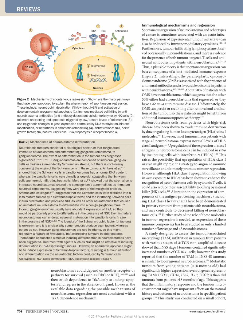
Mechanisms of spontaneous regressionNeurotrophin receptors and regressionThe Trk neurotrophin receptors—TrkA/NTRK1, TrkB/NTRK2, and TrkC/NTRK3—have critical roles in the development and maintenance of the central and peripheral nervous systems. The cognate ligands for these receptors are nerve growth factor (NGF), brain-derived neurotropic factor (BDNF) and neurotrophin-3 (NT3) growth factor, respectively. These receptors also have important roles in neuroblastoma pathogenesis.88–90 High TrkA expression levels are associated with favour-able clinical and biological features, such as younger age, lower stage, and absence of MYCN amplification, and these patients have an excellent outcome.91–94 By contrast, TrkB is coexpressed at high levels with its ligand, BDNF, in clinically and biologically unfavourable tumours, especially those with MYCN amplification.95 Activation of the TrkB–BDNF autocrine pathway can lead to inva-sion, metastasis, angiogenesis and drug resistance.95–99 The TrkA and TrkC receptors are also known as depen-dence receptors, as the absence of ligand activation will generate apoptotic signals.100,101 Coexpression of the P75/NGFR receptor can increase the sensitivity and specificity of all three Trk receptors for their cognate ligands;102,103 however, overexpression and activation of P75/NGFR in the absence of Trk expression can lead to apoptosis.101,104
Tumours from patients with low-stage and 4S disease generally express high levels of TrkA.91–94 When cells derived from these tumours were put in primary culture in the presence of NGF, they underwent neuronal dif-ferentiation, and survived for months. By contrast, the same cells died via apoptosis within a week once deprived of NGF.93,105 Thus, these in vitro culture conditions seem to recapitulate the behaviour of TrkA-expressing neuro-blastomas in patients whose tumours undergo neuronal differentiation or spontaneous regression (apoptosis), depending on the presence or absence of NGF in their microenvironment (Figure 2; Box 2).
Migrating neural crest precursors and favourable neuro blastomas that express TrkA on their cell surface survive (at least initially), despite a lack of available NGF, and the reason for this paradox is uncertain. One possible explanation comes from the identification of TrkAIII, a TrkA isoform that is expressed in normal sympatho adrenal progenitors as well as in some neuro-blastomas.106,107 This isoform results from the alternative splicing of exons 6, 7 and 9, which maintains the reading frame but abolishes the ligand-binding site, leading to constitutive activation of the kinase. Thus, the conversion from a TrkA-expressing, NGF-dependent neuroblastoma to a NGF-independent one could be the consequence of a developmentally programmed isoform switch from TrkAIII to TrkAI. Alternatively, NGF-independent
FOCUS ON PAEDIATRIC CANCER
© 2014 Macmillan Publishers Limited. All rights reserved
708 | DECEMBER 2014 | VOLUME 11 www.nature.com/nrclinonc
neuroblastomas could depend on another receptor or pathway for survival (such as TrkC or RET),108–110 and then switch dependence to TrkA, only to undergo apop-tosis and regress in the absence of ligand. However, the available data regarding the possible mechanisms of neuro blastoma regression are most consistent with a TrkA dependence mechanism.
Immunological mechanisms and regressionSpontaneous regression of neuroblastomas and other types of cancer is sometimes associated with an acute infec-tion. Regression of experimental tumour metastases can also be induced by immunomodulatory cytokines.111,112 Furthermore, tumour-infiltrating lymphocytes are obser-ved occasionally in neuroblastomas, and there is evidence for the presence of both tumour-targeted T-cells and anti-neural antibodies in patients with neuroblastoma.113–115 Thus, a plausible theory is that spontaneous regression can be a consequence of a host-mediated immune response (Figure 2). Interestingly, the paraneoplastic opsomyo-clonus syndrome (OMS) is associated with the presence of antineural antibodies and a favourable outcome in patients with neuroblastoma.113,116–118 About 50% of patients with OMS have neuroblastoma, which suggests that the other 50% either had a neuroblastoma that regressed, or they have a de novo autoimmune disease. Unfortunately, the OMS can persist or recur long after removal and eradica-tion of the tumour, so these patients might benefit from additional immunosuppressive therapy.119
Neuroblastoma cells from patients with high-risk disease have been shown to evade immune destruction by downregulating human leucocyte antigen (HLA) class I molecules.120 However, most tumours from patients with stage 4S neuroblastoma express normal levels of HLA class I antigens.121 Upregulation of the expression of class I antigens in neuroblastoma cells can be induced in vitro by incubating cells with interferon-γ (IFN-γ).120 This raises the possibility that upregulation of HLA class I in vivo might represent a strategy to augment immune surveillance and ultimately promote tumour regression. However, although HLA class I upregulation following in vitro exposure to IFN-γ has been shown to enhance the recognition of neuroblastoma cells by cytotoxic T cells, it could also reduce their susceptibility to killing by natural killer (NK) cells.120 Alteration in the expression of com-ponents of the antigen-processing machinery (includ-ing HLA class I heavy chain) have been demonstrated in primary tumours from patients with neuroblastoma, and may contribute to decreased killing of neuroblas-toma cells.120 Further study of the role of these molecules in tumour regression is needed, as expression of these immune components has been studied in only a limited number of low stage and 4S neuroblastomas.
A study designed to assess the tumour-associated macro phage (TAM) infiltration in tumours from patients with various stages of MYCN non-amplified disease showed that INSS stage 4 tumours contained significantly increased numbers of CD163+ cells; however, this study reported that the number of TAM in INSS 4S tumours is similar to locoregional neuroblastomas.126 Metastatic tumours from young patients (<18 months old) had significantly higher expression levels of genes represent-ing TAMs (CD33, CD16, IL6R, IL10, FCGR3) than did tumours from patients ≥18 months of age. This suggests that the inflammatory response and the tumour micro-environment might have important effects on the natural history and outcome of neuroblastoma in specific patient groups.126 This study was conducted on a small cohort,
Nucleus
Cytoplasm
Methylation
Telomerase
NGF
12
2
3
4
TrkA Antibodies
NK cell
TrkB
Figure 2 | Mechanisms of spontaneous regression. Shown are the major pathways that have been proposed to explain the phenomenon of spontaneous regression. These include: neurotrophin deprivation (TrkA without NGF) and activation of developmentally programmed apoptosis (1); immune‑mediated cell killing by anti‑neuroblastoma antibodies (and antibody‑dependent cellular toxicity) or by NK cells (2); telomere shortening and apoptosis triggered by low/absent levels of telomerase (3); and epigenetic changes in gene expression controlled by DNA methylation, histone modification, or alterations in chromatin remodelling (4). Abbreviations: NGF, nerve growth factor; NK, natural killer cells; TrkA, tropomyosin receptor kinase A.
Box 2 | Mechanisms of neuroblastoma differentiation
Neuroblastic tumours consist of a histological spectrum that ranges from immature neuroblastoma and differentiating ganglioneuroblastoma, to ganglioneuroma. The extent of differentiation in the tumour has prognostic significance.19,161,170,171 Ganglioneuromas are comprised of individual ganglion cells or clusters surrounded by Schwannian stroma, but there is controversy concerning the origin of the Schwann cells in these tumours. Ambros et al.172 showed that the Schwann cells in ganglioneuromas had a normal DNA content, whereas the ganglionic cells were clonally aneuploid, suggesting the Schwann cells are normal, infiltrating host cells. Mora et al.142 showed that the stromal cells in treated neuroblastomas shared the same genomic abnormalities as immature neuronal components, suggesting they were part of the malignant process. Ambros and colleagues172 suggested that the Schwann cells were recruited into a less mature tumour by a Schwann‑trophic factor, and the infiltrating Schwann cells in turn proliferated and produced NGF as well as other neurotrophins that caused an immature neuroblastoma to differentiate into a benign ganglioneuroma.173 Indeed, ganglioneuromas usually have abundant expression of TrkA, so they would be particularly prone to differentiate in the presence of NGF. Even immature neuroblastomas can undergo neuronal maturation into ganglionic cells in vitro in the presence of NGF.93,105 The identity of the Schwann‑trophic factor (or factors) is unknown, and it is unclear why some tumours produce such factors, whereas others do not. However, ganglioneuromas are rare in infants, so this might represent a feature of favourable, TrkA‑expressing tumours in older patients. Therapeutic approaches aimed at inducing differentiation in neuroblastomas have been suggested. Treatment with agents such as NGF might be effective at inducing differentiation in TrkA‑expressing tumours. However, an alternative approach might be to induce expression of Schwann‑trophic factors, resulting in tumour infiltration and differentiation via the neurotrophic factors produced by Schwann cells. Abbreviations: NGF, nerve growth factor; TrkA, tropomyosin receptor kinase A.
REVIEWS
© 2014 Macmillan Publishers Limited. All rights reserved

NATURE REVIEWS | CLINICAL ONCOLOGY VOLUME 11 | DECEMBER 2014 | 709
which likely limited the investigators’ ability to evalu-ate expression of TAM-associated genes in patients with regressing tumours, and highlights the need for further study on the role of the immune system and the tumour microenvironment in the context of disease regression.
Telomerase, telomeres and regressionTelomeres are specialized structures at the ends of chro-mosomes that are involved in the replication and stabil-ity of the chromosome itself. They have an important role in guaranteeing genomic stability and are in a state of dynamic equilibrium. The regulation of the telomere length is controlled in part by the enzyme telomerase. Of note, telomerase expression is frequently high in cancer and immortalized cells, but low in most normal and senes-cent cells.127 Hiyama and colleagues128 studied the regula-tion of telomere length and the activity of the telomerase in 100 samples of neuroblastomas. Most of the tumours that exhibited a high level of telomerase activity were associ-ated with a poor prognosis, and all tumours with MYCN amplification had high telomerase activity. Interestingly, most of the tumour samples from 4S neuroblastoma had low telomerase activity or short telomeres, a pattern that is usually associated with senescent cells (Figure 2).128 Furthermore, Samy et al.129 transfected a neuroblastoma cell line (IGR-N-91) with a dominant negative form of human telomerase, h-Tert. The h-Tert-transfected neuro-blastoma cells formed tumours that showed more apop-tosis and reduced tumorigenicity in a mouse xenograft model compared to untransfected neuroblastoma cells.
These data suggest that loss of telomerase activity is a plausible mechanism to explain spontaneous regression of neuroblastoma, and possibly of other tumours. However, low telomerase activity is associated with biologically favourable tumours, which also have hyperdiploidy, high expression of TrkA and lack high-risk genomic features, such as MYCN amplification.130 Thus, the role of telo-merase is not clear; the association between stage 4S and regression might be related to the low telomerase activity or might depend on other favourable features associated with stage 4S, such as younger age, hyperdiploidy and lack of MYCN amplification. Nevertheless, high levels of telo merase activity is generally associated with a more-aggressiv e tumour behaviour and a poor prognosis in patients with neuroblastoma.131–133
Epigenetic regulation and other mechanismsChanges in gene expression related to alterations in pro-moter methylation, histone modification or chromatin remodelling might also impact differentiation in neuro-blastoma cells. Epigenetic changes affecting expression of genes relevant to neuroblastoma development were ini-tially reported more than a decade ago,134,135 and several studies have suggested that alterations in gene methyl-ation and histone modification are related to patient outcome (Figure 2).136–139 The correlation between epi-genetic changes and neuroblastoma behaviour has been increasingly studied, particularly because next-generation sequencing analyses of neuroblastoma have reported a very limited number of previously unrecognized recurrent
somatic mutations.41–44 Furthermore, data from other childhood tumour types, particularly Wilms tumour and medulloblastoma, suggest that epigenetic changes might help to explain poorly understood aspects of disease p resentation and clinical behaviour.140–142
The development of genome-wide methylation detec-tion methods has facilitated study of epigenetics in numer-ous tumour types, and in neuroblastomas specifically.139 Preliminary data showed that there are global differ-ences in the methylomes of 22 neuroblastoma stage 4S tumours compared to the methylomes of low-risk and high-risk tumours and to the methylome of normal brain tissues.47 In the 4S samples, reduced promoter methyl-ation was observed in 97% of the genes in which differen-tial methyl ation was detected. Specifically, differentially m ethylated promoters were enriched for genes known to have binding sites for transcription factors involved in cell differentiation (such as LEF1, TCF3, ETS2, and PITX2).47 Other investigators have also reported different patterns of methylation in tumours from patients with 4S versus other disease stages,139 but additional studies are needed to confirm and extend these initial findings. Studies of agents that affect DNA methylation status, histone modification, or chromatin modifiers during differentiation and regres-sion are ongoing,143–145 but currently there are no clinical trials of epigenetic modifiers in stage 4S neuroblastomas.
Therapeutic implications of regressionTargeting the TrkA pathway One of the most promising approaches for the induction of apoptosis and tumour regression in neuroblastomas is targeting the TrkA neurotrophin receptor pathway. This is based on the observation that TrkA-expressing tumour cells placed in culture will survive and even differenti-ate in the presence of NGF, but undergo apoptosis in its absence.105 Thus, depriving cells of NGF or inhibiting TrkA signalling might be an effective approach to induce regres-sion. Lestaurti nib (CEP-701) is a small molecule that targets Trk neurotrophin receptors (TrkA, TrkB and TrkC), and it has shown preclinical activity against TrkB-expressing neuroblastoma xenografts.146–149 Furthermore, lestaurti-nib, when given at biologically effective doses, has shown signifi cant clinical activity in a phase I trial in children with recurrent and/or refractory neuroblastoma.150 Lestaurtinib is not being supported for future clinical trials, but these studies provide proof-of-principle that Trk-selective inhibi-tors could be effective in the treatment of neuroblastomas driven by Trk receptors. Indeed, several second-generation Trk inhibitors are currently in phase I clinical trials or in preclinical development.151–154 These agents are potent inhibitors of all three Trk family neurotrophin receptors, so the same agent could be used to target TrkA in f avourable 4S tumours, and TrkB in u nfavourable tumours.
In general, new agents are tested in patients with recur-rent and/or refractory disease; therefore, new Trk inhibitors will not be tested initially in infants with de novo stage 4S disease or other patients with locoregional tumours and biologically favourable features. However, there is signifi-cant mortality due to respiratory compromise among very young infants with stage 4S neuroblastoma and
FOCUS ON PAEDIATRIC CANCER
© 2014 Macmillan Publishers Limited. All rights reserved

710 | DECEMBER 2014 | VOLUME 11 www.nature.com/nrclinonc
hepato megaly.155,156 If second-generation Trk inhibitors prove to be safe and effective against TrkB-expressing, high-risk disease, it would be reasonable to consider administra-tion of these agents in patients with TrkA-expressing 4S neuroblastoma and massive liver involvement in lieu of chemotherapy or radiation therapy. Theoretically, a Trk inhibitor could initiate the process of apoptosis and regres-sion in these tumours. It would be highly desirable to have an agent, such as a Trk inhibitor, that can induce regression directly, rather than waiting for it to occur spontaneously. Trk inhibitor therapy might also be used for infants with large abdominal tumours or ‘dumbbell tumours’ with intra-spinal extension, to spare such patients the toxicity and long-term side effects of laminectomy, chemotherapy or spinal radiation.
Immunological approaches Immunotherapy using a chimeric antibody (ch14.18) directed against the disialoganglioside GD2 has been incorporated into frontline treatment of patients with high-risk neuroblastoma,157 and very preliminary studies have been performed using adoptive immunotherapy in patients with relapsed and refractory disease.158 Additional studies of the immunology of differentiation and regres-sion are expected to influence the evolution of current immunotherapeutic approaches and could potentially result in new strategies to accelerate regression in young infants with acute life-threatening, but biologically favour-able disease. Immune modulation to induce regression could potentially be advantageous for patients with 4S or locoregional disease, and a trend in the field has been to reduce conventional cytotoxic therapy and avoid aggressive surgery in patients with favourable prognoses.84,87,159 The toxicities associated with currently available immunocyto-kine therapies are considerable, and little is known regard-ing late effects of these therapies, especially in very young infants. Less toxic immune modulation therapy could be considered, including therapy designed to enhance immune surveillance by increasing HLA I expression on neuroblastoma cells. However, as noted above, strategies to enhance one component of the immune system may diminish the antitumour effects of another key component. There is an increasing need for a better understanding of the complex interactions between neuroblastoma cells and the immune system, and of the multiple i mplications of immune modulation in very young children.
Other approaches At the present time, there are no therapeutic approaches available to influence telomere length in neuroblasto-mas. However, in addition to targeted inhibition of the TrkA pathway or immunological therapy, there are other approaches that might be considered for these patients, For example, the retinoids are a class of compounds that have been shown to induce cellular differentiation and decrease proliferation of neuroblastoma cells in vitro, presumably mediated by the induction of expression of neural differ-entiation genes.160 Indeed, the retinoid isotretinoin (13-cis-retinoic acid) has been incorporated into frontline therapy for children with high-risk neuroblastoma in an effort to
induce differentiation in vivo in states of minimal residual disease following intensive, multimodality therapy.161 The precise mechanisms by which isotretinoin induces dif-ferentiation are unclear, but it seems that retinoids are a ssociated with increased expression of Trk receptors.160
Vorinostat, a histone deacetylase inhibitor, has been administered in combination with isotretinoin in children with relapsed or refractory disease in an effort to further induce neuroblastoma cell differentiation. A patient with neuroblastoma who had evaluable disease experienced a complete response to therapy on one phase I trial,162 although no objective responses were seen in a second study.163 Further studies of epigenetic changes in neuro-blastoma cells that undergo differentiation versus those that do not differentiate could lead to the development of more specific and effective agents that can alter gene expression in neuroblastomas and consequently induce tumour regression.
Conclusions Neuroblastomas show a remarkable capacity to undergo spontaneous regression. The prevalence of this phenom-enon is hard to determine precisely, but the experience from mass-screening programmes suggests that there are at least as many children who have tumours undergoing spontaneous regression without detection as there are patients with neuroblastoma detected clinically. Further exploration of this issue and a greater understanding of the normal mechanism(s) of spontaneous regression might allow the identification of tumours that have the capacity to undergo spontaneous regression and to induce regression in susceptible tumours using pharmacological, biological or immunological approaches. However, to this end, we will need to study samples from a substantial number of regressing tumours, or perhaps establish an animal model that suitably mimics the process of spontaneous regres-sion. At the present time, the most promising therapeutic approach would be aimed at inhibiting the TrkA recep-tor pathway. However, most Trk inhibitors are potent inhibitors of TrkA, TrkB and TrkC. Before these agents are used to treat infants with stage 4S disease, clinical trials of second- generation Trk inhibitors would need to demon-strate safety and efficacy against TrkB-expressing recurrent and/or refractory neuroblastomas.
Review criteria
We searched for original articles focusing on neuroblastoma regression in MEDLINE and PubMed published from 1971 to June 2014. In terms of potential mechanisms, we focused on articles that had been published in the past 15 years. The search terms used were “neuroblastoma” and one or more of the following: “regression”, “spontaneous regression”, “spontaneous differentiation”, “telomerase”, “regression and mechanisms”, “regression and telomerase”, “regression and TRK”, “epigenetics”, “immune function”, “NK cells”, “regression and immune”. We also searched “genes” and “neural crest development” and “neuroblastoma and mass screening”. All papers identified were English‑language full‑text papers. We also searched the reference lists of identified articles for further papers.
REVIEWS
© 2014 Macmillan Publishers Limited. All rights reserved

NATURE REVIEWS | CLINICAL ONCOLOGY VOLUME 11 | DECEMBER 2014 | 711
1. Brodeur, G. M. & Maris, J. M. in Principles and Practice of Pediatric Oncology (eds Pizzo, P. A. & Poplack, D. G.) 786–822 (Lippincott, Williams and Wilkins, Philadelphia, 2010).
2. Maris, J. M., Hogarty, M. D., Bagatell, R. & Cohn, S. L. Neuroblastoma. Lancet 369, 2106–2120 (2007).
3. Smith, M. A. et al. Outcomes for children and adolescents with cancer: challenges for the twenty‑first century. J. Clin. Oncol. 28, 2625–2634 (2010).
4. Gatta, G. et al. Childhood cancer survival in Europe 1999–2007: results of EUROCARE‑5 —a population‑based study. Lancet Oncol. 15, 35–47 (2014).
5. Kreissman, S. G. et al. Purged versus non‑purged peripheral blood stem‑cell transplantation for high‑risk neuroblastoma (COG A3973): a randomised phase 3 trial. Lancet Oncol. 14, 999–1008 (2013).
6. Diede, S. J. Spontaneous regression of metastatic cancer: learning from neuroblastoma. Nat. Rev. Cancer 14, 71–72 (2014).
7. Matthay, K. K. Stage 4S neuroblastoma: what makes it special? J. Clin. Oncol. 16, 2003–2006 (1998).
8. Nakagawara, A. Molecular basis of spontaneous regression of neuroblastoma: role of neurotrophic signals and genetic abnormalities. Hum. Cell 11, 115–124 (1998).
9. Nickerson, H. J. et al. Favorable biology and outcome of stage IV‑S neuroblastoma with supportive care or minimal therapy: a Children’s Cancer Group study. J. Clin. Oncol. 18, 477–486 (2000).
10. Pritchard, J. & Hickman, J. A. Why does stage 4s neuroblastoma regress spontaneously? Lancet 344, 869–870 (1994).
11. Yamamoto, K. et al. Marginal decrease in mortality and marked increase in incidence as a result of neuroblastoma screening at 6 months of age: cohort study in seven prefectures in Japan. J. Clin. Oncol. 20, 1209–1214 (2002).
12. Sawada, T. et al. Mass screening for neuroblastoma in Japan. Pediatr. Hematol. Oncol. 8, 93–109 (1991).
13. Erttmann, R. et al. 10 years’ neuroblastoma screening in Europe: preliminary results of a clinical and biological review from the Study Group for Evaluation of Neuroblastoma Screening in Europe (SENSE). Eur. J. Cancer 34, 1391–1397 (1998).
14. Woods, W. G. et al. A population‑based study of the usefulness of screening for neuroblastoma. Lancet 348, 1682–1687 (1996).
15. Brodeur, G. M. Neuroblastoma: biological insights into a clinical enigma. Nat. Rev. Cancer 3, 203–216 (2003).
16. Hoehner, J. C., Olsen, L., Sandstedt, B., Kaplan, D. R. & Pahlman, S. Association of neurotrophin receptor expression and differentiation in human neuroblastoma. Am. J. Pathol. 147, 102–113 (1995).
17. Haas, D., Ablin, A. R., Miller, C., Zoger, S. & Matthay, K. K. Complete pathologic maturation and regression of stage IVS neuroblastoma without treatment. Cancer 62, 818–825 (1988).
18. Garvin, J. H. Jr, Lack, E. E., Berenberg, W. & Frantz, C. N. Ganglioneuroma presenting with differentiated skeletal metastases. Report of a case. Cancer 54, 357–360 (1984).
19. Shimada, H. et al. Terminology and morphologic criteria of neuroblastic tumors: recommendations by the International Neuroblastoma Pathology Committee. Cancer 86, 349–363 (1999).
20. Mosse, Y. P. et al. Identification of ALK as a major familial neuroblastoma predisposition gene. Nature 455, 930–935 (2008).
21. Shojaei‑Brosseau, T. et al. Genetic epidemiology of neuroblastoma: a study of 426 cases at the Institut Gustave‑Roussy in France. Pediatr. Blood Cancer 42, 99–105 (2004).
22. Mosse, Y. P. et al. Germline PHOX2B mutation in hereditary neuroblastoma. Am. J. Hum. Genet. 75, 727–730 (2004).
23. Maris, J. M. et al. Evidence for a hereditary neuroblastoma predisposition locus at chromosome 16p12–13 Cancer Res. 62, 6651–6658 (2002).
24. Chen, Y. et al. Oncogenic mutations of ALK kinase in neuroblastoma. Nature 455, 971–974 (2008).
25. George, R. E. et al. Activating mutations in ALK provide a therapeutic target in neuroblastoma. Nature 455, 975–978 (2008).
26. Janoueix‑Lerosey, I. et al. Somatic and germline activating mutations of the ALK kinase receptor in neuroblastoma. Nature 455, 967–970 (2008).
27. Raabe, E. H. et al. Prevalence and functional consequence of PHOX2B mutations in neuroblastoma. Oncogene 27, 469–476 (2008).
28. Trochet, D. et al. Germline mutations of the paired‑like homeoBox 2B (PHOX2B) gene in neuroblastoma. Am. J. Hum. Genet. 74, 761–764 (2004).
29. Bosse, K. R. et al. Common variation at BARD1 results in the expression of an oncogenic isoform that influences neuroblastoma susceptibility and oncogenicity. Cancer Res. 72, 2068–2078 (2012).
30. Capasso, M. et al. Common variations in BARD1 influence susceptibility to high‑risk neuroblastoma. Nat. Genet. 41, 718–723 (2009).
31. Diskin, S. J. et al. Common variation at 6q16 within HACE1 and LIN28B influences susceptibility to neuroblastoma. Nat. Genet. 44, 1126–1130 (2012).
32. Nguyen le, B. et al. Phenotype restricted genome‑wide association study using a gene‑centric approach identifies three low‑risk neuroblastoma susceptibility Loci. PLoS Genet. 7, e1002026 (2011).
33. Wang, K. et al. Integrative genomics identifies LMO1 as a neuroblastoma oncogene. Nature 469, 216–220 (2011).
34. Weiss, W. A., Aldape, K., Mohapatra, G., Feuerstein, B. G. & Bishop, J. M. Targeted expression of MYCN causes neuroblastoma in transgenic mice. EMBO J. 16, 2985–2995 (1997).
35. Chen, Z. et al. Mdm2 deficiency suppresses MYCN‑driven neuroblastoma tumorigenesis in vivo. Neoplasia 11, 753–762 (2009).
36. Berry, T. et al. The ALK(F1174L) mutation potentiates the oncogenic activity of MYCN in neuroblastoma. Cancer Cell 22, 117–130 (2012).
37. Heukamp, L. C. et al. Targeted expression of mutated ALK induces neuroblastoma in transgenic mice. Sci. Transl. Med. 4, 141ra91 (2012).
38. Molenaar, J. J. et al. LIN28B induces neuroblastoma and enhances MYCN levels via let‑7 suppression. Nat. Genet. 44, 1199–1206 (2012).
39. Mosse, Y. P. et al. Neuroblastomas have distinct genomic DNA profiles that predict clinical phenotype and regional gene expression. Genes Chromosomes Cancer 46, 936–949 (2007).
40. Schleiermacher, G. et al. Segmental chromosomal alterations have prognostic
impact in neuroblastoma: a report from the INRG project. Br. J. Cancer 107, 1418–1422 (2012).
41. Pugh, T. J. et al. The genetic landscape of high‑risk neuroblastoma. Nat. Genet. 45, 279–284 (2013).
42. Cheung, N. K. et al. Association of age at diagnosis and genetic mutations in patients with neuroblastoma. JAMA 307, 1062–1071 (2012).
43. Sausen, M. et al. Integrated genomic analyses identify ARID1A and ARID1B alterations in the childhood cancer neuroblastoma. Nat. Genet. 45, 12–17 (2013).
44. Molenaar, J. J. et al. Sequencing of neuroblastoma identifies chromothripsis and defects in neuritogenesis genes. Nature 483, 589–593 (2012).
45. Santo, E. E. et al. Oncogenic activation of FOXR1 by 11q23 intrachromosomal deletion‑fusions in neuroblastoma. Oncogene 31, 1571–1581 (2012).
46. Benard, J. et al. MYCN‑non‑amplified metastatic neuroblastoma with good prognosis and spontaneous regression: a molecular portrait of stage 4S. Mol. Oncol. 2, 261–271 (2008).
47. Diskin, S. J. et al. Integrative genomic and epigenomic characterization of stage 4S neuroblastoma gene expression [abstract]. Advances in Neuroblastoma Research, POB083 (Cologne, 2014).
48. Taggart, D. R. et al. Prognostic value of the stage 4S metastatic pattern and tumor biology in patients with metastatic neuroblastoma diagnosed between birth and 18 months of age. J. Clin. Oncol. 29, 4358–4364 (2011).
49. Challis, G. B. & Stam, H. J. The spontaneous regression of cancer. A review of cases from 1900 to 1987. Acta Oncol. 29, 545–550 (1990).
50. Everson, T. C. Spontaneous regression of cancer. Ann. N.Y. Acad. Sci. 114, 721–735 (1964).
51. Everson, T. C. & Cole, W. H. Spontaneous regression of cancer (W. B. Saunders & Co., Philadelphia, 1966).
52. Papac, R. J. Spontaneous regression of cancer: possible mechanisms. In Vivo 12, 571–578 (1998).
53. Beckwith, J. B. & Perrin, E. V. In situ neuroblastomas: a contribution to the natural history of neural crest tumors. Am. J. Pathol. 43, 1089–1104 (1963).
54. Ikeda, Y., Lister, J., Bouton, J. M. & Buyukpamukcu, M. Congenital neuroblastoma, neuroblastoma in situ, and the normal fetal development of the adrenal. J. Pediatr. Surg. 16, 636–644 (1981).
55. Turkel, S. B. & Itabashi, H. H. The natural history of neuroblastic cells in the fetal adrenal gland. Am. J. Pathol. 76, 225–244 (1974).
56. D’Angio, G. J., Evans, A. E. & Koop, C. E. Special pattern of widespread neuroblastoma with a favourable prognosis. Lancet 1, 1046–1049 (1971).
57. Evans, A. E., D’Angio, G. J. & Randolph, J. A proposed staging for children with neuroblastoma. Children’s cancer study group A. Cancer 27, 374–378 (1971).
58. George, R. E. et al. High‑risk neuroblastoma treated with tandem autologous peripheral‑blood stem cell‑supported transplantation: long‑term survival update. J. Clin. Oncol. 24, 2891–2896 (2006).
59. Matthay, K. K. et al. Treatment of high‑risk neuroblastoma with intensive chemotherapy, radiotherapy, autologous bone marrow transplantation, and 13‑cis‑retinoic acid. Children’s Cancer Group. N. Engl. J. Med. 341, 1165–1173 (1999).
FOCUS ON PAEDIATRIC CANCER
© 2014 Macmillan Publishers Limited. All rights reserved

712 | DECEMBER 2014 | VOLUME 11 www.nature.com/nrclinonc
60. Brodeur, G. M. et al. Revisions of the international criteria for neuroblastoma diagnosis, staging, and response to treatment. J. Clin. Oncol. 11, 1466–1477 (1993).
61. Brodeur, G. M. et al. International criteria for diagnosis, staging, and response to treatment in patients with neuroblastoma. J. Clin. Oncol. 6, 1874–1881 (1988).
62. Monclair, T. et al. The International Neuroblastoma Risk Group (INRG) staging system: an INRG Task Force report. J. Clin. Oncol. 27, 298–303 (2009).
63. Cozzi, D. A. et al. Long‑term follow‑up of the “wait and see” approach to localized perinatal adrenal neuroblastoma. World J. Surg. 37, 459–465 (2013).
64. Fisher, J. P. & Tweddle, D. A. Neonatal neuroblastoma. Semin. Fetal Neonatal Med. 17, 207–215 (2012).
65. Kushner, B. H. et al. Survival from locally invasive or widespread neuroblastoma without cytotoxic therapy. J. Clin. Oncol. 14, 373–381 (1996).
66. Lavarino, C. et al. Specific gene expression profiles and chromosomal abnormalities are associated with infant disseminated neuroblastoma. BMC Cancer 9, 44 (2009).
67. Yu, F. et al. Proteomics‑based identification of spontaneous regression‑associated proteins in neuroblastoma. J. Pediatr. Surg. 46, 1948–1955 (2011).
68. Knudson, A. G. Jr & Meadows, A. T. Regression of neuroblastoma IV‑S: a genetic hypothesis. N. Engl. J. Med. 302, 1254–1256 (1980).
69. van Noesel, M. M. Neuroblastoma stage 4S: a multifocal stem‑cell disease of the developing neural crest. Lancet Oncol. 13, 229–230 (2012).
70. Spitz, R. et al. Favorable outcome of triploid neuroblastomas: a contribution to the special oncogenesis of neuroblastoma. Cancer Genet. Cytogenet. 167, 51–56 (2006).
71. Ambros, P. F. et al. Regression and progression in neuroblastoma. Does genetics predict tumour behaviour? Eur. J. Cancer 31A, 510–515 (1995).
72. LaBrosse, E. H., Comoy, E., Bohuon, C., Zucker, J. M. & Schweisguth, O. Catecholamine metabolism in neuroblastoma. J. Natl Cancer Inst. 57, 633–638 (1976).
73. Bessho, F. Comparison of the incidences of neuroblastoma for screened and unscreened cohorts. Acta Paediatr. 88, 404–406 (1999).
74. Schilling, F. H. et al. Neuroblastoma screening at one year of age. N. Engl. J. Med. 346, 1047–1053 (2002).
75. Woods, W. G. et al. Screening of infants and mortality due to neuroblastoma. N. Engl. J. Med. 346, 1041–1046 (2002).
76. Yamamoto, K. et al. Marginal decrease in mortality and marked increase in incidence as a result of neuroblastoma screening at 6 months of age: cohort study in seven prefectures in Japan. J. Clin. Oncol. 20, 1209–1214 (2002).
77. Brodeur, G. M., Ambros, P. F. & Favrot, M. C. Biological aspects of neuroblastoma screening. Med. Ped. Oncol. 31, 394–400 (1998).
78. Hayashi, Y., Hanada, R. & Yamamoto, K. Biology of neuroblastomas in Japan found by screening. Am. J. Pediatr. Hematol. Oncol. 14, 342–347 (1992).
79. Kaneko, Y. et al. Current urinary mass screening for catecholamine metabolites at 6 months of age may be detecting only a small portion of high‑risk neuroblastomas: A chromosome and N‑myc amplification study. J. Clin. Oncol. 8, 2005–2013 (1990).
80. Acharya, S. et al. Prenatally diagnosed neuroblastoma. Cancer 80, 304–310 (1997).
81. Ho, P. T. et al. Prenatal detection of neuroblastoma: a ten‑year experience from the
Dana‑Farber Cancer Institute and Children’s Hospital. Pediatrics 92, 358–364 (1993).
82. Saylors, R. L. 3rd, Cohn, S. L., Morgan, E. R. & Brodeur, G. M. Prenatal detection of neuroblastoma by fetal ultrasonography. Am. J. Pediatr. Hematol. Oncol. 16, 356–360 (1994).
83. Ikeda, H. et al. Surgical treatment of neuroblastomas in infants under 12 months of age. J. Pediatr. Surg. 33, 1246–1250 (1998).
84. Hero, B. et al. Localized infant neuroblastomas often show spontaneous regression: results of the prospective trials NB95‑S and NB97. J. Clin. Oncol. 26, 1504–1510 (2008).
85. Oue, T. et al. Profile of neuroblastoma detected by mass screening, resected after observation without treatment: results of the Wait and See pilot study. J. Pediatr. Surg. 40, 359–363 (2005).
86. Nishihira, H. et al. Natural course of neuroblastoma detected by mass screening: a 5‑year prospective study at a single institution. J. Clin. Oncol. 18, 3012–3017 (2000).
87. Nuchtern, J. G. et al. A prospective study of expectant observation as primary therapy for neuroblastoma in young infants: a Children’s Oncology Group study. Ann. Surg. 256, 573–580 (2012).
88. Brodeur, G. M. et al. Trk receptor expression and inhibition in neuroblastomas. Clin. Cancer Res. 15, 3244–3250 (2009).
89. Brodeur, G. M. et al. Expression of TrkA, TrkB and TrkC in human neuroblastomas. J. Neurooncol. 31, 49–55 (1997).
90. Thiele, C. J., Li, Z. & McKee, A. E. On Trk —the TrkB signal transduction pathway is an increasingly important target in cancer biology. Clin. Cancer Res. 15, 5962–5967 (2009).
91. Kogner, P. et al. Coexpression of messenger RNA for TRK protooncogene and low affinity nerve growth factor receptor in neuroblastoma with favorable prognosis. Cancer Res. 53, 2044–2050 (1993).
92. Nakagawara, A., Arima, M., Azar, C. G., Scavarda, N. J. & Brodeur, G. M. Inverse relationship between trk expression and N‑myc amplification in human neuroblastomas. Cancer Res. 52, 1364–1368 (1992).
93. Nakagawara, A. et al. Association between high levels of expression of the TRK gene and favorable outcome in human neuroblastoma. N. Engl. J. Med. 328, 847–854 (1993).
94. Stram, D. & Seeger, R. C. Lack of high‑affinity nerve growth factor receptors in aggressive neuroblastomas. J. Natl Cancer Inst. 85, 377–384 (1993).
95. Nakagawara, A., Azar, C. G., Scavarda, N. J. & Brodeur, G. M. Expression and function of TRK‑B and BDNF in human neuroblastomas. Mol. Cell Biol. 14, 759–767 (1994).
96. Acheson, A. et al. A BDNF autocrine loop in adult sensory neurons prevents cell death. Nature 374, 450–453 (1995).
97. Jaboin, J., Kim, C. J., Kaplan, D. R. & Thiele, C. J. Brain‑derived neurotrophic factor activation of TrkB protects neuroblastoma cells from chemotherapy‑induced apoptosis via phosphatidylinositol 3'‑kinase pathway. Cancer Res. 62, 6756–6763 (2002).
98. Matsumoto, K., Wada, R. K., Yamashiro, J. M., Kaplan, D. R. & Thiele, C. J. Expression of brain‑derived neurotrophic factor and p145TrkB affects survival, differentiation, and invasiveness of human neuroblastoma cells. Cancer Res. 55, 1798–1806 (1995).
99. Nakamura, K. et al. Brain‑derived neurotrophic factor activation of TrkB induces vascular endothelial growth factor expression via hypoxia‑inducible factor‑1alpha in neuroblastoma cells. Cancer Res. 66, 4249–4255 (2006).
100. Goldschneider, D. & Mehlen, P. Dependence receptors: a new paradigm in cell signaling and cancer therapy. Oncogene 29, 1865–1882 (2010).
101. Rabizadeh, S., Ye, X., Wang, J. J. & Bredesen, D. E. Neurotrophin dependence mediated by p75NTR: contrast between rescue by BDNF and NGF. Cell Death Differ. 6, 1222–1227 (1999).
102. Hantzopoulos, P. A., Suri, C., Glass, D. J., Goldfarb, M. P. & Yancopoulos, G. D. The low affinity NGF receptor, p75, can collaborate with each of the Trks to potentiate functional responses to the neurotrophins. Neuron 13, 187–201 (1994).
103. Ho, R. et al. The effect of P75 on Trk receptors in neuroblastomas. Cancer Lett. 305, 76–85 (2011).
104. Bamji, S. X. et al. The p75 neurotrophin receptor mediates neuronal apoptosis and is essential for naturally occurring sympathetic neuron death. J. Cell Biol. 140, 911–923 (1998).
105. Nakagawara, A. & Brodeur, G. M. Role of neurotrophins and their receptors in human neuroblastomas: a primary culture study. Eur. J. Cancer 33, 2050–2053 (1997).
106. Tacconelli, A. et al. TrkA alternative splicing: a regulated tumor‑promoting switch in human neuroblastoma. Cancer Cell 6, 347–360 (2004).
107. Tacconelli, A., Farina, A. R., Cappabianca, L., Gulino, A. & Mackay, A. R. Alternative TrkAIII splicing: a potential regulated tumor‑promoting switch and therapeutic target in neuroblastoma. Future Oncol. 1, 689–698 (2005).
108. Kahane, N. & Kalcheim, C. Expression of trkC receptor mRNA during development of the avian nervous system. J. Neurobiol. 25, 571–584 (1994).
109. Pachnis, V., Mankoo, B. & Costantini, F. Expression of the c‑ret proto‑oncogene during mouse embryogenesis. Development 119, 1005–1017 (1993).
110. Tsuzuki, T. et al. Spatial and temporal expression of the ret proto‑oncogene product in embryonic, infant and adult rat tissues. Oncogene 10, 191–198 (1995).
111. Salcedo, R. et al. Immunologic and therapeutic synergy of IL‑27 and IL‑2: enhancement of T cell sensitization, tumor‑specific CTL reactivity and complete regression of disseminated neuroblastoma metastases in the liver and bone marrow. J. Immunol. 182, 4328–4338 (2009).
112. Salcedo, R. et al. IL‑27 mediates complete regression of orthotopic primary and metastatic murine neuroblastoma tumors: role for CD8+ T cells. J. Immunol. 173, 7170–7182 (2004).
113. Antunes, N. L. et al. Antineuronal antibodies in patients with neuroblastoma and paraneoplastic opsoclonus‑myoclonus. J. Pediatr. Hematol. Oncol. 22, 315–320 (2000).
114. Kataoka, Y., Matsumura, T., Yamamoto, S., Sugimoto, T. & Sawada, T. Distinct cytotoxicity against neuroblastoma cells of peripheral blood and tumor‑infiltrating lymphocytes from patients with neuroblastoma. Cancer Lett. 73, 11–21 (1993).
115. Valteau, D. et al. T‑cell receptor repertoire in neuroblastoma patients. Cancer Res. 56, 362–369 (1996).
116. Cooper, R. et al. Opsoclonus‑myoclonus‑ataxia syndrome in neuroblastoma: histopathologic features‑a report from the Children’s Cancer Group. Med. Pediatr. Oncol. 36, 623–629 (2001).
117. Pranzatelli, M. R. et al. B‑ and T‑cell markers in opsoclonus‑myoclonus syndrome: immunophenotyping of CSF lymphocytes. Neurology 62, 1526–1532 (2004).
REVIEWS
© 2014 Macmillan Publishers Limited. All rights reserved

NATURE REVIEWS | CLINICAL ONCOLOGY VOLUME 11 | DECEMBER 2014 | 713
118. Rudnick, E. et al. Opsoclonus‑myoclonus‑ataxia syndrome in neuroblastoma: clinical outcome and antineuronal antibodies‑a report from the Children’s Cancer Group Study. Med. Pediatr. Oncol. 36, 612–622 (2001).
119. Russo, C., Cohn, S. L., Petruzzi, M. J. & de Alarcon, P. A. Long‑term neurologic outcome in children with opsoclonus‑myoclonus associated with neuroblastoma: a report from the Pediatric Oncology Group. Med. Pediatr. Oncol. 28, 284–288 (1997).
120. Raffaghello, L. et al. Multiple defects of the antigen‑processing machinery components in human neuroblastoma: immunotherapeutic implications. Oncogene 24, 4634–4644 (2005).
121. Squire, R., Fowler, C. L., Brooks, S. P., Rich, G. A. & Cooney, D. R. The relationship of class I MHC antigen expression to stage IV‑S disease and survival in neuroblastoma. J. Pediatr. Surg. 25, 381–386 (1990).
122. Bin, Q., Johnson, B. D., Schauer, D. W., Casper, J. T. & Orentas, R. J. Production of macrophage migration inhibitory factor by human and murine neuroblastoma. Tumour Biol. 23, 123–129 (2002).
123. Castriconi, R. et al. Natural killer cell‑mediated killing of freshly isolated neuroblastoma cells: critical role of DNAX accessory molecule‑1‑poliovirus receptor interaction. Cancer Res. 64, 9180–9184 (2004).
124. Raffaghello, L. et al. Downregulation and/or release of NKG2D ligands as immune evasion strategy of human neuroblastoma. Neoplasia 6, 558–568 (2004).
125. Ren, Y. et al. Inhibition of tumor growth and metastasis in vitro and in vivo by targeting macrophage migration inhibitory factor in human neuroblastoma. Oncogene 25, 3501–3508 (2006).
126. Asgharzadeh, S. et al. Clinical significance of tumor‑associated inflammatory cells in metastatic neuroblastoma. J. Clin. Oncol. 30, 3525–3532 (2012).
127. Kim, N. W. et al. Specific association of human telomerase activity with immortal cells and cancer. Science 266, 2011–2015 (1994).
128. Hiyama, E. et al. Correlating telomerase activity levels with human neuroblastoma outcomes. Nat. Med. 1, 249–255 (1995).
129. Samy, M. et al. Loss of the malignant phenotype of human neuroblastoma cells by a catalytically inactive dominant‑negative hTERT mutant. Mol. Cancer Ther. 11, 2384–2393 (2012).
130. Brodeur, G. M. Do the ends justify the means? Nat. Med. 1, 203–205 (1995).
131. Krams, M. et al. Full‑length telomerase reverse transcriptase messenger RNA is an independent prognostic factor in neuroblastoma. Am. J. Pathol. 162, 1019–1026 (2003).
132. Ohali, A. et al. Telomere length is a prognostic factor in neuroblastoma. Cancer 107, 1391–1399 (2006).
133. Streutker, C. J., Thorner, P., Fabricius, N., Weitzman, S. & Zielenska, M. Telomerase activity as a prognostic factor in neuroblastomas. Pediatr. Dev. Pathol. 4, 62–67 (2001).
134. Astuti, D. et al. RASSF1A promoter region CpG island hypermethylation in phaeochromocytomas and neuroblastoma tumours. Oncogene 20, 7573–7577 (2001).
135. Takita, J. et al. Absent or reduced expression of the caspase 8 gene occurs frequently in neuroblastoma, but not commonly in Ewing sarcoma or rhabdomyosarcoma. Med. Pediatr. Oncol. 35, 541–543 (2000).
136. Barbieri, E. et al. Histone chaperone CHAF1A inhibits differentiation and promotes aggressive neuroblastoma. Cancer Res. 74, 765–774 (2014).
137. Grau, E. et al. Epigenetic alterations in disseminated neuroblastoma tumour cells: influence of TMS1 gene hypermethylation in relapse risk in NB patients. J. Cancer Res. Clin. Oncol. 136, 1415–1421 (2010).
138. Yang, Q. et al. Methylation of CASP8, DCR2, and HIN‑1 in neuroblastoma is associated with poor outcome. Clin. Cancer Res. 13, 3191–3197 (2007).
139. Decock, A., Ongenaert, M., Vandesompele, J. & Speleman, F. Neuroblastoma epigenetics: from candidate gene approaches to genome‑wide screenings. Epigenetics 6, 962–970 (2011).
140. Batora, N. V. et al. Transitioning from genotypes to epigenotypes: why the time has come for medulloblastoma epigenomics. Neuroscience 264, 171–185 (2014).
141. Feinberg, A. P. The epigenetics of cancer etiology. Semin. Cancer Biol. 14, 427–432 (2004).
142. Feinberg, A. P. & Tycko, B. The history of cancer epigenetics. Nat. Rev. Cancer 4, 143–153 (2004).
143. Baylin, S. B. DNA methylation and gene silencing in cancer. Nat. Clin. Pract. Oncol. 2 (Suppl. 1), S4–S11 (2005).
144. Gros, C. et al. DNA methylation inhibitors in cancer: recent and future approaches. Biochimie 94, 2280–2296 (2012).
145. McCabe, M. T. & Creasy, C. L. EZH2 as a potential target in cancer therapy. Epigenomics 6, 341–351 (2014).
146. Evans, A. E. et al. Effect of CEP‑751 (KT‑6587) on neuroblastoma xenografts expressing TrkB. Med. Ped. Oncol. 36, 181–184 (2001).
147. Evans, A. E. et al. Antitumor activity of CEP‑751 (KT‑6587) on human neuroblastoma and medulloblastoma xenografts. Clin. Cancer Res. 5, 3594–3602 (1999).
148. Ho, R. et al. Resistance to chemotherapy mediated by TrkB in neuroblastomas. Cancer Res. 62, 6462–6466 (2002).
149. Iyer, R. et al. Lestaurtinib enhances the antitumor efficacy of chemotherapy in murine xenograft models of neuroblastoma. Clin. Cancer Res. 16, 1478–1485 (2010).
150. Minturn, J. E. et al. Phase I trial of lestaurtinib for children with refractory neuroblastoma: a new approaches to neuroblastoma therapy consortium study. Cancer Chemother. Pharmacol. 68, 1057–1065 (2011).
151. De Braud, F. G. et al. Phase 1 open label, dose escalation study of RXDX101, an oral pan‑trk, ROS1, and ALK inhibitor, in patients with advanced solid tumors with relevant molecular alterations [abstract]. J. Clin. Oncol. 32 (Suppl.), a2502 (2014).
152. US National Library of Medicine. ClinicalTrials.gov [online], http://clinicaltrials.gov/show/NCT02122913 (2014).
153. US National Library of Medicine.ClinicalTrials.gov [online], http://clinicaltrials.gov/show/NCT02048488 (2014).
154. US National Library of Medicine. ClinicalTrials.gov [online], http://www.clinicaltrials.gov/show/NCT01804530 (2014).
155. Hsu, L. L., Evans, A. E. & D’Angio, G. J. Hepatomegaly in neuroblastoma stage 4s: criteria for treatment of the vulnerable neonate. Med. Pediatr. Oncol. 27, 521–528 (1996).
156. Kushner, B. H., Kramer, K., LaQuaglia, M. P., Modak, S. & Cheung, N. K. Liver involvement in neuroblastoma: the Memorial Sloan‑Kettering Experience supports treatment reduction in young patients. Pediatr. Blood Cancer 46, 278–284 (2006).
157. Yu, A. L. et al. Anti‑GD2 antibody with GM‑CSF, interleukin‑2, and isotretinoin for neuroblastoma. N. Engl. J. Med. 363, 1324–1334 (2010).
158. Louis, C. U. et al. Antitumor activity and long‑term fate of chimeric antigen receptor‑positive T cells in patients with neuroblastoma. Blood 118, 6050–6056 (2011).
159. Baker, D. L. et al. Outcome after reduced chemotherapy for intermediate‑risk neuroblastoma. N. Engl. J. Med. 363, 1313–1323 (2010).
160. Yuza, Y., Agawa, M., Matsuzaki, M., Yamada, H. & Urashima, M. Gene and protein expression profiling during differentiation of neuroblastoma cells triggered by 13‑cis retinoic acid. J. Pediatr. Hematol. Oncol. 25, 715–720 (2003).
161. Shimada, H. et al. The International Neuroblastoma Pathology Classification (the Shimada system). Cancer 86, 364–372 (1999).
162. Fouladi, M. et al. Pediatric phase I trial and pharmacokinetic study of vorinostat: a Children’s Oncology Group phase I consortium report. J. Clin. Oncol. 28, 3623–3629 (2010).
163. Park, J. R. et al. A phase I study of vorinostat in combination with isotretinoin (RA) in patients with refractory/recurrent neuroblastoma (NB): a new approaches to neuroblastoma therapy consortium trial [abstract]. Advances in Neuroblastoma Research, OR070 (Cologne, 2014).
164. Simoes‑Costa, M. & Bronner, M. E. Insights into neural crest development and evolution from genomic analysis. Genome Res. 23, 1069–1080 (2013).
165. Betancur, P., Bronner‑Fraser, M. & Sauka‑Spengler, T. Assembling neural crest regulatory circuits into a gene regulatory network. Annu. Rev. Cell. Dev. Biol. 26, 581–603 (2010).
166. De Preter, K. et al. Human fetal neuroblast and neuroblastoma transcriptome analysis confirms neuroblast origin and highlights neuroblastoma candidate genes. Genome Biol. 7, R84 (2006).
167. Tsarovina, K. et al. Essential role of GATA transcription factors in sympathetic neuron development. Development 131, 4775–4786 (2004).
168. Unsicker, K., Huber, K., Schober, A. & Kalcheim, C. Resolved and open issues in chromaffin cell development. Mech. Dev. 130, 324–329 (2013).
169. Pei, D. et al. Distinct neuroblastoma‑associated alterations of PHOX2B impair sympathetic neuronal differentiation in zebrafish models. PLoS Genet. 9, e1003533 (2013).
170. Shimada, H. et al. Histopathologic prognostic factors in neuroblastic tumors: Definition of subtypes of ganglioneuroblastoma and an age‑linked classification of neuroblastomas. J. Natl Cancer Inst. 73, 405–413 (1984).
171. Shimada, H. et al. International neuroblastoma pathology classification for prognostic evaluation of patients with peripheral neuroblastic tumors: a report from the Children’s Cancer Group. Cancer 92, 2451–2461 (2001).
172. Ambros, I. M. et al. Role of ploidy, chromosome 1p, and Schwann cells in the maturation of neuroblastoma. N. Engl. J. Med. 334, 1505–1511 (1996).
173. Brodeur, G. M. Schwann cells as antineuroblastoma agents. N. Engl. J. Med. 334, 1537–1539 (1996).
AcknowledgementsThis work was supported in part by NIH grants R01‑CA094194 and R01‑039,771; Alex’s Lemonade Stand Foundation; and the Audrey E. Evans endowed chair (G.M.B.).
Author contributionsG.M.B. and R.B. researched data and discussed content for article, wrote, reviewed and edited the manuscript before submission.
FOCUS ON PAEDIATRIC CANCER
© 2014 Macmillan Publishers Limited. All rights reserved