From Anti-allergic to Anti-Alzheimer's: Molecular Pharmacology of Dimebon
Upload
independentCategory
view
0download
0
Accepted Manuscript
Title: Mechanisms of Anti-Cancer Action and Pharmacologyof Clofarabine
Authors: Anna Zhenchuk, Koroush Lotfi, Gunnar Juliusson,Freidoun Albertioni
PII: S0006-2952(09)00564-4DOI: doi:10.1016/j.bcp.2009.06.094Reference: BCP 10241
To appear in: BCP
Received date: 15-4-2009Revised date: 21-6-2009Accepted date: 23-6-2009
Please cite this article as: Zhenchuk A, Lotfi K, Juliusson G, Albertioni F, Mechanismsof Anti-Cancer Action and Pharmacology of Clofarabine , Biochemical Pharmacology(2008), doi:10.1016/j.bcp.2009.06.094
This is a PDF file of an unedited manuscript that has been accepted for publication.As a service to our customers we are providing this early version of the manuscript.The manuscript will undergo copyediting, typesetting, and review of the resulting proofbefore it is published in its final form. Please note that during the production processerrors may be discovered which could affect the content, and all legal disclaimers thatapply to the journal pertain.
Page 1 of 53
Accep
ted
Man
uscr
ipt
1 2 3 4 5 6 7 8 9 10 11 12 13 14 15 16 17 18 19 20 21 22 23 24 25 26 27 28 29 30 31 32 33 34 35 36 37 38 39 40 41 42 43 44 45 46 47 48 49 50 51 52 53 54 55 56 57 58 59 60 61 62 63 64 65
Review
Mechanisms of Anti-Cancer Action and Pharmacology of Clofarabine
aAnna Zhenchuk, bKoroush Lotfi, cGunnar Juliusson and aFreidoun Albertioni
aDepartment of Oncology-Pathology, Cancer Center Karolinska, Karolinska
Institute, Karolinska Hospital, SE-171 76 Stockholm; bDepartment of Medicine
and Care, Clinical Pharmacology, Faculty of Health Sciences, Linköping
University, cDepartment of Hematology, University Hospital, SE-581 85
Linköping; and cLund Strategic Center for Stem Cell Biology and Therapy,
Dept. of Hematology, Lund University Hospital, SE-221 84 Lund, Sweden.
Keywords:
ClofarabineNucleoside analogues Deoxycytidine kinaseRibonucleotide reductaseApoptosisResistance to antimetabolitesPharmacokineticsLeukemias
Abbreviations:ADA – adenosine deaminaseALL – acute lymphoblastic leukemiaAML – acute myeloid leukemiaAraC – cytarabineCAFdA – clofarabineCdA – cladribine
* Manuscript
Page 2 of 53
Accep
ted
Man
uscr
ipt
1 2 3 4 5 6 7 8 9 10 11 12 13 14 15 16 17 18 19 20 21 22 23 24 25 26 27 28 29 30 31 32 33 34 35 36 37 38 39 40 41 42 43 44 45 46 47 48 49 50 51 52 53 54 55 56 57 58 59 60 61 62 63 64 65
2
CLL – chronic lymphocytic leukemiadCK – deoxicytidine kinasedGK – deoxyguanosine kinaseDLT – dose limiting toxicityFaraA – fludarabineHSA – human serum albuminMTD – maximum tolerated doseRR – ribonucleotide reductaseVdss – volume of distribution at steady stateWBC – white blood cell
Page 3 of 53
Accep
ted
Man
uscr
ipt
1 2 3 4 5 6 7 8 9 10 11 12 13 14 15 16 17 18 19 20 21 22 23 24 25 26 27 28 29 30 31 32 33 34 35 36 37 38 39 40 41 42 43 44 45 46 47 48 49 50 51 52 53 54 55 56 57 58 59 60 61 62 63 64 65
3
ABSTRACT
Clofarabine, a next-generation deoxyadenosine analogue, was developed on the
basis of experience with cladribine and fludarabine in order to achieve higher
efficacy and avoid extramedullary toxicity. During the past decade this is the only
drug granted approval for treatment of pediatric acute leukemia. Recent clinical
studies have established the efficacy of clofarabine in treating malignancies with a
poor prognosis, such as adult, elderly, and relapsed pediatric leukemia. The
mechanisms of its anti-cancer activity involve a combination of direct inhibition of
DNA synthesis and ribonucleotide reductase and induction of apoptosis. Due to
this broad cytotoxicity, this drug is effective against various subtypes of leukemia
and is currently being tested as an oral formulation and for combination therapy of
both leukemias and solid tumors. In this review we summarize current knowledge
pertaining to the molecular mechanisms of action and pharmacological properties
of clofarabine, as well as clinical experiences with this drug with the purpose of
facilitating the evaluation of its efficacy and the development of future therapies.
Page 4 of 53
Accep
ted
Man
uscr
ipt
1 2 3 4 5 6 7 8 9 10 11 12 13 14 15 16 17 18 19 20 21 22 23 24 25 26 27 28 29 30 31 32 33 34 35 36 37 38 39 40 41 42 43 44 45 46 47 48 49 50 51 52 53 54 55 56 57 58 59 60 61 62 63 64 65
4
1. Introduction
Nucleoside analogues have had a substantial impact on the treatment of cancer,
especially hematological malignancies. The most credible explanation is the
dependency of hematopoietic cells on salvage synthesis of nucleosides [1] and
their expression of several membrane transporters that take up nucleoside
analogues [2]. Their structural similarity to physiological nucleosides allows
these analogues to be taken up by cells, metabolized, and incorporated into DNA
or RNA. Therefore, the main targets of these activated nucleoside analogues are
the enzymes involved in de novo and salvage pathways of nucleoside synthesis.
The history of nucleoside analogues dates back to the late 1950s when
cytosine arabinoside (cytarabine, AraC) was synthesized and subsequently
became the backbone in the treatment of acute myeloid leukemia (AML) and one
of the most widely used nucleoside analogues to date [3]. Single-dose treatment
with AraC was not promising, but in combination with agents that interacted with
topoisomerase, such as anthracyclines, a high rate of remission could be achieved
in AML patients [4]. The success of AraC led to the development of other
arabinosylpurine homologues (e.g. 9-β-D-arabinofuranosyladenine (araA)). Since
the clinical activity of araA was found to be limited due to its rapid deamination
Page 5 of 53
Accep
ted
Man
uscr
ipt
1 2 3 4 5 6 7 8 9 10 11 12 13 14 15 16 17 18 19 20 21 22 23 24 25 26 27 28 29 30 31 32 33 34 35 36 37 38 39 40 41 42 43 44 45 46 47 48 49 50 51 52 53 54 55 56 57 58 59 60 61 62 63 64 65
5
by adenosine deaminase (ADA) [5], ADA-resistant halogenated arabinosyl
derivatives such as 2-fluoro-9-β-arabinofuranosyladenine (fludarabine) were then
developed [6]. Thereafter, because of the insolubility of fludarabine, its 5´-
monophosphate derivative (fludarabine monophosphate, FaraA, Fludara®), was
designed for conversion into fludarabine aA by endogenous phosphatases.
However, treatment with this latter drug also results in the formation by
phosphorylase cleavage of 2-fluoroadenine, a toxic compound with only minor
antitumor activity [7]. In the early 1970s the realization that halogenated
analogues exert an anti-leukemic effect led to the synthesis of 2-
chlordeoxyadenosine (cladribine, CdA) [8], which, however, has reduced oral
bioavailability due in part to its instability in the acidic gastric environment as
well as its susceptiblity to enzymatic cleavage [9-11].
In the 1990s, in order to tackle all the above-mentioned limitations of early
nucleoside analogues, a next-generation deoxyadenosine analogue, clofarabine
(2-chloro-2'-arabino-fluoro-2'-deoxyadenosine, CAFdA, Cl-F-ara-A), a 2´-
arabino-fluoro derivative of cladribine, was synthesized [12]. The rationale
behind its design was to combine the structural features of cladribine and
fludarabine (Figure 1). Like cladribine and fludarabine, clofarabine is toxic to
both non-proliferating human lymphocytes and rapidly proliferating cells, while
Page 6 of 53
Accep
ted
Man
uscr
ipt
1 2 3 4 5 6 7 8 9 10 11 12 13 14 15 16 17 18 19 20 21 22 23 24 25 26 27 28 29 30 31 32 33 34 35 36 37 38 39 40 41 42 43 44 45 46 47 48 49 50 51 52 53 54 55 56 57 58 59 60 61 62 63 64 65
6
being resistant to phosphorylitic cleavage and deamination-stable in acidic
environments.
Preclinical studies indicated a high degree of efficacy of clofarabine and
subsequently lead to human clinical trials. In 2004 the FDA approved clofarabine
(ClolarTM, Genzyme) for the treatment of pediatric leukemic patients - a decision
followed by European Commision approval in 2006 (Evoltra®, Bioenvision).
Today the anti-cancer activity of clofarabine toward other types of tumors, as well
as in other age groups, is being actively investigated.
2. Molecular mechanisms of action
2.1. Structure and physicochemical properties
Clofarabine was designed as an adenosine analogue with improved stability both
in the plasma and the acidic gastric environment. And, indeed, the introduction of
a fluorine atom at the 2´-arabino position of cladribine increases its stability at pH
1.0 [13]. As predicted, the pKa value of clofarabine (1.75) is higher than that of
cladribine (1.28). Furthermore, it exhibits the highest lipophilicity among related
purine analogues [13] and is resistant to degradation by E. coli nucleoside
phosphorylase [14].
Page 7 of 53
Accep
ted
Man
uscr
ipt
1 2 3 4 5 6 7 8 9 10 11 12 13 14 15 16 17 18 19 20 21 22 23 24 25 26 27 28 29 30 31 32 33 34 35 36 37 38 39 40 41 42 43 44 45 46 47 48 49 50 51 52 53 54 55 56 57 58 59 60 61 62 63 64 65
7
2.2. Transport and metabolism
Clofarabine metabolites are retained by cells to a greater extent than are
metabolites of cladribine, which is believed to be one factor in the more
pronounced anti-tumoral effect and higher hematological toxicity of the former
[15]. Other factors contributing to its higher anti-cancer activity are thought to be
clofarabine’s high affinity for nucleoside transporters and deoxicytidine kinase
(dCK), as well as for key enzymes involved in DNA synthesis, e.g.
ribonucleotide reductase (RR) and DNA polymerase-α and -ε.
Most cells have two types of active nucleoside transporters: human
equilibrative nucleoside transporter (hENT) and human concentrative nucleoside
transporter (hCNT). Transport via hENTs is a sodium-independent mechanism
involving primarily bi-directional facilitative diffusion driven by a gradient in the
nucleoside concentration across the cell membrane. In contrast, the hCNTs are
sodium-dependent active transporters that depend on ATP for their ability to
transport purine nucleosides into the cell against a concentration gradient.
Clofarabine is believed to enter cells by both facilitated and active
nucleoside transport mechanisms as well as, at higher concentrations and upon
longer exposure, by passive diffusion across lipid membranes [16]. The
Page 8 of 53
Accep
ted
Man
uscr
ipt
1 2 3 4 5 6 7 8 9 10 11 12 13 14 15 16 17 18 19 20 21 22 23 24 25 26 27 28 29 30 31 32 33 34 35 36 37 38 39 40 41 42 43 44 45 46 47 48 49 50 51 52 53 54 55 56 57 58 59 60 61 62 63 64 65
8
importance of membrane nucleoside transporters in clofarabine metabolism is
indicated by the fact that the rate of clofarabine uptake is ~10-fold higher by
transport-competent than transport-deficient human leukemia cells [16]. In
particular, cells expressing hCNT3 exhibit the highest rate of clofarabine uptake
compared to the cells expressing hCNT2, hENT1, and hENT2, whereas transport-
deficient and hCNT1-expressing cells demonstrate no uptake, consistent with the
fact that hCNT1 is specific for pyrimidines. In addition, clofarabine was more
cytotoxic to cells expressing hENT1 than fludarabine or cladribine.
Upon entry into cells, clofarabine is phosphorylated to its monophosphate
derivatives, primarily by dCK, a constitutively expressed key cytosolic enzyme
involved in the salvage pathway of DNA synthesis, but also by the mitochondrial
enzyme deoxyguanosine kinase (dGK) [17]. Further intracellular phosphorylation
results in the final metabolite, clofarabine triphosphate. The cytotoxicity of
nucleoside analogues in quiescent cells (i.e. lymphocytes) depends mainly upon
selective and progressive accumulation of the triphosphate metabolites because of
the high ratio of dCK to cytosolic 5’-nucleotidase. The latter acts as a deactivator
of nucleoside analogues by dephosphorylating the triphosphate metabolites and
enabling their transport out of the cell. The importance of dCK for the efficacy of
clofarabine was confirmed by our recent finding that the cells deficient in this
activity are more resistant to the cytotoxic effect of this drug [18]. Furthermore,
Page 9 of 53
Accep
ted
Man
uscr
ipt
1 2 3 4 5 6 7 8 9 10 11 12 13 14 15 16 17 18 19 20 21 22 23 24 25 26 27 28 29 30 31 32 33 34 35 36 37 38 39 40 41 42 43 44 45 46 47 48 49 50 51 52 53 54 55 56 57 58 59 60 61 62 63 64 65
9
the concentration of dCK is typically highest in lymphoid tissues, as well as
considerably higher in tumor tissues than in normal tissues [19]. The studies with
recombinant human dCK and kinases in the crude leukemic cell extracts have
revealed that the efficiency (Vmax/Km) of clofarabine phosphorylation by these
enzymes is close to, or greater than, that of the natural substrate deoxycytidine, as
well as significantly greater than that of cladribine or fludarabine [20, 21]. The
interactions between 2-Cl and its surrounding hydrophobic residues contribute to
the high catalytic efficiency of dCK for clofarabine [22].
Although dCK activity is rate-limiting for the accumulation of fludarabine
triphosphates, this is not the case with clofarabine [15]. Accumulation of
clofarabine appears to be determined by the rate of phosphorylation of its
monophosphate to diphosphate by purine nucleotide monophosphate kinase,
similarly to cladribine [15, 23]. One explanation might be that nucleoside
analogues bearing a 2-chloroadenine nucleobase are relatively poor substrates for
the monophosphate kinase.
2.3. Mechanisms of anti-cancer activity
The anti-cancer activity of clofarabine involves three major mechanisms:
inhibition of DNA synthesis, inhibition of ribonucleotide reductase, and direct
Page 10 of 53
Accep
ted
Man
uscr
ipt
1 2 3 4 5 6 7 8 9 10 11 12 13 14 15 16 17 18 19 20 21 22 23 24 25 26 27 28 29 30 31 32 33 34 35 36 37 38 39 40 41 42 43 44 45 46 47 48 49 50 51 52 53 54 55 56 57 58 59 60 61 62 63 64 65
10
induction of apoptosis (Figure 2). This broad range of activities explains the
efficacy of the drug against both rapidly growing and quiescent tumors [14].
Clofarabine competes potently with dATP for binding to DNA polymerase-
α and –ε [24]. At a low ratio of clofarabine triphosphate to dATP, clofarabine is
incorporated primarily into internal phosphodiester linkages inhibiting DNA
repair; whereas, at higher ratios, clofarabine is detected mainly at terminal sites
inhibiting DNA elongation [15]. The most pronounced inhibition of DNA
elongation is observed at clofarabine triphosphate to dATP ratios greater than one
[25]. Incorporation of clofarabine monophosphate into DNA impairs its
elongation and repair by causing chain termination and strand breaks. However,
clofarabine triphosphate is not a potent inhibitor of DNA polymerase-β,
mitochondrial DNA polymerase-γ, or DNA primase [24].
In a recent article Chen and coworkers (2008) suggested that clofarabine
action may as well involve an RNA-directed mechanism. Clofarabine
triphosphates were shown to cause chain termination in the absence of ATP [26].
With increasing amounts of clofarabine triphosphates, there was a concentration-
dependent effect on polyadenylation due to blockage of the ability of yeast
poly(A)polymerase (yPAP) to extend the poly(A)-tail by the analogues’
incorporation into RNA [26].
Page 11 of 53
Accep
ted
Man
uscr
ipt
1 2 3 4 5 6 7 8 9 10 11 12 13 14 15 16 17 18 19 20 21 22 23 24 25 26 27 28 29 30 31 32 33 34 35 36 37 38 39 40 41 42 43 44 45 46 47 48 49 50 51 52 53 54 55 56 57 58 59 60 61 62 63 64 65
11
Besides DNA polymerase inhibition, clofarabine triphosphate potently
inhibits RR activity (50% at 65 nM) [24] by binding to the allosteric site on the
regulatory subunit, incorporating in this manner anti-cancer properties of
fludarabine. Inhibition of RR depletes the dNTP pool, with the most pronounced
reduction in the dCTP pool, followed by dATP and dGTP, and only a minor
effect on the dTTP pool. Through depletion of the dNTP pool, clofarabine self-
potentiates the incorporation of clofarabine triphosphates into DNA, thus
explaining the increased effectiveness of clofarabine in DNA synthesis inhibition
[27].
Furthermore, depletion of dNTP pools can lead to enhanced nucleoside
kinase activity and thereby increased phosphorylation of nucleoside analogues,
more pronounced accumulation and prolonged retention of the triphosphate
metabolites, and, consequently, a more potent anti-cancer effect [25]. The damage
caused by incorporation of clofarabine monophosphate into DNA initiates a chain
of events that result in activation of pro-apoptotic pathways. Clofarabine can
replace dATP and directly affects cytosolic apoptotic protease-activating factor-1
(APAF1), thus causing caspase activation [28, 29]. Clofarabine also directly
affects mitochondria by altering the transmembrane potential and, as a result,
releasing cytochrome c and apoptosis-inducing factor (AIF) [30]. Mitochondrial
damage caused by clofarabine may be explained by direct binding of the
Page 12 of 53
Accep
ted
Man
uscr
ipt
1 2 3 4 5 6 7 8 9 10 11 12 13 14 15 16 17 18 19 20 21 22 23 24 25 26 27 28 29 30 31 32 33 34 35 36 37 38 39 40 41 42 43 44 45 46 47 48 49 50 51 52 53 54 55 56 57 58 59 60 61 62 63 64 65
12
triphosphate metabolite to proteins in the mitochondrial membrane, leading to an
alteration in membrane potential as well as intra-mitochondrial accumulation of
this triphosphate [30]. The combined actions of cytochrome c, AIF, APAF-1, and
caspase-9 lead to the formation of the apoptosome complex responsible for
programmed cell death induction.
Moreover, clofarabine has been shown to induce dose- and time-dependent
down-regulation of the death suppressor proteins Bcl-X(L) and Mcl-1 and de-
phosphorylation of anti-apoptotic kinase Akt and its downstream effectors (Bad,
FKHRL1) [31]. An interesting observation was made in the T-ALL CEM cell
line when treated with clofarabine and caffeine [31]. The addition of caffeine
inhibited the down-regulation of Cdc25A, usually observed when cells are treated
with clofarabine alone, and increased the cleavage of PARP. Therefore, a
combinatorial effect on apoptosis could be expected if clofarabine is administered
together with inhibitors of chk1, a checkpoint homolog that phosphorylates
Cdc25 and prevents cells entering mitosis.
2.4. Mechanism of resistance
The effectiveness of nucleoside analogue treatment against cancer cells is
principally limited by the primary or acquired drug resistance. This commonly
Page 13 of 53
Accep
ted
Man
uscr
ipt
1 2 3 4 5 6 7 8 9 10 11 12 13 14 15 16 17 18 19 20 21 22 23 24 25 26 27 28 29 30 31 32 33 34 35 36 37 38 39 40 41 42 43 44 45 46 47 48 49 50 51 52 53 54 55 56 57 58 59 60 61 62 63 64 65
13
happens due to alteration in the activity of one or more actors in the metabolic
pathway of a drug, thus leading to its reduced effectiveness. A detailed
knowledge of the molecular mechanisms underlying such resistance would enable
the use of appropriate dosing schedules and combination therapies in the clinical
setting to overcome this limitation.
There are few studies that have also initiated the investigations into
potential mechanisms of resistance. In one such study, the resistance to
clofarabine acquired by HL60 and CCRF-CEM cell lines is directly correlated
with reduced activity of the nucleoside phosphorylating enzyme dCK, as has been
previously found to be the case with cladribine [20, 32]. We detected dCK
deficiency at the level of enzymatic activity, the protein level, and also as mRNA
expression for the cell lines resistant to clofarabine; these cells did not
consistently form detectable levels of clofarabine triphosphates. However, no
mutations in the coding region of the dCK gene were detected. The suggestion
that DNA methylation is responsible for the decreased level of dCK in resistant
cancer cells has not been confirmed [32, 33].
The problem of resistance due to reduced dCK and/or dGK activity might
be overcome by introducing the drug as a pro-nucleotide, thereby releasing the
deoxynucleoside 5´-monophosphate directly into the cell. In addition,
determination of dCK and/or dGK activities might help predict how well a patient
Page 14 of 53
Accep
ted
Man
uscr
ipt
1 2 3 4 5 6 7 8 9 10 11 12 13 14 15 16 17 18 19 20 21 22 23 24 25 26 27 28 29 30 31 32 33 34 35 36 37 38 39 40 41 42 43 44 45 46 47 48 49 50 51 52 53 54 55 56 57 58 59 60 61 62 63 64 65
14
will respond to a certain cytotoxic drug and which dose and combination of drugs
will be most effective.
Clofarabine-resistant cell lines showed cross-resistance to other
antimetabolites (i.e. fludarabine, Ara-C, difluorodeoxycytidine, dfdc)
phosphorylated by dCK [20, 32]. Thus, clofarabine-resistant cells also accumulate
lower levels of cladribine and other analogue nucleotides. On the other hand,
cells resistant to fludarabine exhibited cross-resistance to cladribine, but not
clofarabine.
No alteration in nucleoside transport or expression of the classical MDR
(multidrug-resistance) protein was detected in clofarabine-resistant cells,
suggesting that the mechanism of acquired resistance to clofarabine is a specific,
rather than a general, phenomenon [20, 32]. Clofarabine-resistant cells also
exhibit the highest number of structurally rearranged chromosomes, indicating a
high potential of clofarabine to rearrange the genome [32]. However, this was not
the cause of dCK deficiency.
Expression of a small subunit of RR (R2) was substantially reduced and the
enzyme’s activity was halved with no alteration of allosteric activity in the
clofarabine-resistant cell lines, as opposed to fludarabine-resistant cell lines,
where RR was substantially higher than in the wild type cell line [32]. Since the
resistant cell line with inhibited dCK and RR activity still produced dNTPs and
Page 15 of 53
Accep
ted
Man
uscr
ipt
1 2 3 4 5 6 7 8 9 10 11 12 13 14 15 16 17 18 19 20 21 22 23 24 25 26 27 28 29 30 31 32 33 34 35 36 37 38 39 40 41 42 43 44 45 46 47 48 49 50 51 52 53 54 55 56 57 58 59 60 61 62 63 64 65
15
kept on proliferating, it was hypothesized that p53R2, a small subunit of RR
directly induced by p53, replaces R2 to form active ribonucleotide reductase. A
truncated form of p53R2 was detected that may replace R2 in the production of
dNTPs in the resistant cell lines.
Other mechanisms of resistance to clofarabine are thought to involve
changes in Ca2+-sensitive mitochondrial phenomena and are probably not
influenced by the Fas pathway, as in the case of cladribine [34].
3. Pharmacokinetics and anti-cancer activity
3.1. In vitro investigations
As expected from its potent inhibition of DNA synthesis, clofarabine exerts
potent growth inhibition and cytotoxic activity (IC50 values = 0.028–0.29 μM) in
a wide variety of leukemia and solid tumor cell lines in vitro [35]. Thus, of 60
human tumor cell lines in the National Cancer Institute’s Developmental
Therapeutics Program panel, clofarabine potently inhibits the growth (GI50 values
= < 0.0001–0.45 μM) of 35. The cell types sensitive to clofarabine include those
of non-small-cell lung, colon, central nervous system, melanoma, ovarian, renal,
prostate, and breast tumors [36].
Page 16 of 53
Accep
ted
Man
uscr
ipt
1 2 3 4 5 6 7 8 9 10 11 12 13 14 15 16 17 18 19 20 21 22 23 24 25 26 27 28 29 30 31 32 33 34 35 36 37 38 39 40 41 42 43 44 45 46 47 48 49 50 51 52 53 54 55 56 57 58 59 60 61 62 63 64 65
16
Cariveau and coworkers (2008) also demonstrated that clofarabine exhibits
a strong radiosensitizing effect on murine tissues in vitro and in vivo, by
interfering with the repair of DNA damage [37]. Another mechanism driving the
radiosensitization activity of a combination of clofarabine with ionizing radiation
(IR) was found to be the activation of dCK by IR, which consistently enhances
clofarabine cytotoxicity [38].
In vitro exposure of mononuclear cells from chronic lymphocytic leukemia
(CLL) and AML patients to identical concentrations of clofarabine and cladribine
leads to higher intracellular accumulation of the former’s triphosphate metabolites
[20, 39]. In these cells, the major metabolite detected was clofarabine
monophosphate, with clofarabine diphosphate representing less than 10% of the
total nucleotide metabolite present, thereby confirming that dCK is not the rate-
limiting step for the formation of clofarabine triphosphate. Interestingly, no
correlation was found between the level of phosphorylated metabolites, enzymes
(dCK, dGK), and the cytotoxicity of clofarabine, suggesting that additional
factors determine its cytotoxic activity [39].
In isolated perfused rat liver, clofarabine and cladribine have a similar first-
pass metabolism of 50% [40]. The rate of elimination of both of these substances
is strongly dependent on the initial concentrations. However, the elimination rate
of clofarabine is much slower and its concentration declines without an increase
Page 17 of 53
Accep
ted
Man
uscr
ipt
1 2 3 4 5 6 7 8 9 10 11 12 13 14 15 16 17 18 19 20 21 22 23 24 25 26 27 28 29 30 31 32 33 34 35 36 37 38 39 40 41 42 43 44 45 46 47 48 49 50 51 52 53 54 55 56 57 58 59 60 61 62 63 64 65
17
in the concentration of the degradation product, 2-chloroadenine, most likely due
to the binding of clofarabine to components of hepatic tissue. The elimination
kinetics of clofarabine 5′-mono-, di-, and triphosphate exhibit tri-phasic behavior,
with a β-half-life of 8–24 hours and a very long γ-half-life (29 hours), indicating
prolonged cellular retention [15].
In a study on polarized human kidney proximal tubule cells (hRPTC11),
apical-to-basolateral fluxes of clofarabine as well as fludarabine and cladribine
across the cell layer were observed following mediation by the coupling of apical
hCNT3 to basolateral hENT2 [41]. Such directionality of transepithelial fluxes
of these three nucleoside analogues resembles that of adenosine, which is
reabsorbed in human kidney proximal tubules. These observations could explain
the delayed elimination of clofarabine. A deeper insight into the mechanisms of
hNTs transport of clofarabine could lead to strategies aimed at improving its
dosing with maximum efficacies and minimum toxicity.
The major metabolite detected in rat and dog hepatocytes after exposure to
clofarabine is P11, which accounts for 1.2% and 2.5%, respectively, of the total
radioactivity recovered [42]. P11 is suggested to be a carboxy- or methoxy-
clofarabine, but the exact location of the change remains unknown. The only
metabolite observed in the case of human hepatocytes is P14, which accounts for
Page 18 of 53
Accep
ted
Man
uscr
ipt
1 2 3 4 5 6 7 8 9 10 11 12 13 14 15 16 17 18 19 20 21 22 23 24 25 26 27 28 29 30 31 32 33 34 35 36 37 38 39 40 41 42 43 44 45 46 47 48 49 50 51 52 53 54 55 56 57 58 59 60 61 62 63 64 65
18
0.2% of the total radioactivity recovered and has been suggested to be the
sulfates’ conjugate of clofarabine at the 5' carbon.
The in vitro efficacy of clofarabine was compared to those of nelarabine
and AraC in a panel of acute lymphoblastic leukemia (ALL) cells [43]. The
concentration of clofarabine that inhibits growth by 50% is 188 times lower than
the corresponding concentration for nelarabine in all cases. Clofarabine appears to
be marginally more effective in B-lineage than in T-lineage ALL and B-lineage is
also several times more sensitive to clofarabine than AraC. There is a potential
for cross-resistance of clofarabine to many ALL therapeutics, but not
methotrexate or thiopurines. This distinct resistance profile may prove useful in
combination with other compounds.
One such combination study was undertaken with alkylating agents and
clofarabine or fludarabine in lymphocytes isolated from CLL patients [44]. DNA
damage repair mechanisms were first initiated in the CLL lymphocytes by
treatment with 4-hydroxycyclophosphamide. This DNA repair was then inhibited
by DNA chain termination in an equivalent manner by treatment with either
fludarabine or clofarabine. However, clofarabine triphosphates exhibited
increased rates of intracellular accumulation, thus leading to maximal inhibition
at 1/10 the concentration of fludarabine triphosphates.
Page 19 of 53
Accep
ted
Man
uscr
ipt
1 2 3 4 5 6 7 8 9 10 11 12 13 14 15 16 17 18 19 20 21 22 23 24 25 26 27 28 29 30 31 32 33 34 35 36 37 38 39 40 41 42 43 44 45 46 47 48 49 50 51 52 53 54 55 56 57 58 59 60 61 62 63 64 65
19
Exposure to such agents as cladribine, clofarabine, fludarabine, or AraC
leads to a 2–4-fold increase in dCK activity (most probably by direct inhibition of
ribonucleotide reductase) in the HL-60 cell line, as well as in peripheral blood
mononuclear cells [45]. This phenomenon has been exploited by treating cells
with one deoxynucleoside analogue initially in order to induce dCK activity and
thereafter administering a second analogue; and, as expected, clofarabine
pretreatment to induce dCK enhances subsequent accumulation of AraC
triphosphate in myeloid cell lines [46]. Similarly, pretreatment of HCT-116 colon,
K562 leukemia, and RL lymphoma cell lines with clofarabine enhanced the
metabolism of T-araC (1-beta-D: -[4-thio-arabinofuranosyl] cytosine), a new
cytosine analogue with superior anti-cancer activity as compared to AraC [47].
Caution must be observed, however, when combining AraC with
clofarabine since these compounds have been shown to have cross-resistance
[48]. Alternatively, valproic acid (VPA), a histone deacetylase inhibitor used as a
drug against epilepsy, exhibits a synergistic effect when combined with
clofarabine in AML cell lines and primary cells. A possible explanation could be
that VPA induces apoptosis by activating both extrinsic and intrinsic apoptotic
pathways.
A recent study demonstrated the hypomethylation property of clofarabine
in human lymphoid tumor cells as well as its ability to up-regulate the expressing
Page 20 of 53
Accep
ted
Man
uscr
ipt
1 2 3 4 5 6 7 8 9 10 11 12 13 14 15 16 17 18 19 20 21 22 23 24 25 26 27 28 29 30 31 32 33 34 35 36 37 38 39 40 41 42 43 44 45 46 47 48 49 50 51 52 53 54 55 56 57 58 59 60 61 62 63 64 65
20
of cancer-testis (CT) antigens (Sp17 and SPAN-Xb) aberrantly expressed in
tumor cells [49]. If the same property is proven to be effective in vivo, it could be
possible to administer low-dose clofarabine to patients to up-regulate the
expression of CT antigens to increase the susceptibility of the tumor cells to the
cytotoxic effect of antigen-specific cytotoxic T cells prior to administering
specific tumor vaccines targeting CT antigens. Furthermore, hypomethylating
DNA clofarabine at low doses could potentially enhance the expression of tumor
suppressor genes that are often hypermethylated, and thus inhibited, in cancer
cells, leading to activation of pro-apoptotic genes.
Silencing of 5’-nucleotidase cNT1 by siRNA interference in a panel of
leukemic cell lines has significantly augmented the cytotoxicity of clofarabine.
This investigation could prove the combination of clofarabine nucleotidases
inhibitors useful for cancer treatment [50].
3.2. In vivo investigations
In addition to numerous studies on the effects of clofarabine in cells, a
considerable amount of in vivo data on various animal models is available.
The dose and schedule-dependent cancer activity of clofarabine was
demonstrated by testing different dosing schedules in an NCI H460 lung tumor
Page 21 of 53
Accep
ted
Man
uscr
ipt
1 2 3 4 5 6 7 8 9 10 11 12 13 14 15 16 17 18 19 20 21 22 23 24 25 26 27 28 29 30 31 32 33 34 35 36 37 38 39 40 41 42 43 44 45 46 47 48 49 50 51 52 53 54 55 56 57 58 59 60 61 62 63 64 65
21
murine model with the greatest anti-tumor activity seen when the daily
clofarabine dose was subdivided into three equal doses and administered at 4-
hour intervals each day for 30 days [36, 51].
Since nucleoside transporters are expressed in many tissues, clofarabine is
expected to be widely distributed in both tumor and normal tissues. Indeed, this
has been found to be the case in mice and rats, where the tissue distribution is
rapid and widespread with the highest concentrations occurring in highly perfused
organs [42, 52].
The tissue-to-plasma ratios of clofarabine in the liver and myocardium
were considerably high (4.8 and upward) [42]. This is most probably due to the
fact that the active clofarabine phosphates must be dephosphorylated in order to
be exported from the cell as well as to the lipophilicity of the parent drug. These
observations are consistent with pharmacokinetic studies which reveal that,
following intravenous administration, clofarabine is extensively distributed
throughout the body, with a volume of distribution at steady state (Vdss) of
approximately 1.4–2.6 L per kg in mice, 3.2–3.6 L per kg in rats, and 0.9–1.2 L
per kg in dogs [36].
Intravenous administration of [14C]clofarabine 25 mg/kg/day to rats
confirmed the non-linear pharmacokinetics, showing three exponential phases of
elimination, with half-lives of 0.3, 1.3, and 12.8 hours [42]. Close to 80% of the
Page 22 of 53
Accep
ted
Man
uscr
ipt
1 2 3 4 5 6 7 8 9 10 11 12 13 14 15 16 17 18 19 20 21 22 23 24 25 26 27 28 29 30 31 32 33 34 35 36 37 38 39 40 41 42 43 44 45 46 47 48 49 50 51 52 53 54 55 56 57 58 59 60 61 62 63 64 65
22
dose administered was recovered in the urine and 10% in the feces. The major
metabolite, 6-ketoclofarabine, is believed to be formed extrahepatically via
adenosine deaminase.
The fact that clofarabine is more stable in an acidic environment and more
lipophilic than cladribine [13, 40] leads to the assumption that the former would
have more side effects, particularly neurotoxicity. However, in mice, the
accumulation of clofarabine in brain tissue was found to be lower than that of
cladribine [52], contradicting the assumption that clofarabine’s more pronounced
lipophilicity would render it a more effective drug for the treatment of
lymphoproliferative disorders in the central nervous system (CNS).
On the other hand, in a study of non-human primates, clofarabine was
found to penetrate into the cerebrospinal fluid, although to a modest extent [53].
The concentrations obtained there may nevertheless approach those known to be
cytotoxic in vitro.
A substantial anti-cancer effect has been observed against human colon
tumors transplanted into nude mice, exhibiting schedule-dependent anti-tumor
activity upon intraperitoneal, intravenous or, particularly, oral administration of
clofarabine [54]. Fifty percent of tumor growth was inhibited at 0.26 μM in a 72-
h exposure, a concentration significantly lower than that of fludarabine and
Page 23 of 53
Accep
ted
Man
uscr
ipt
1 2 3 4 5 6 7 8 9 10 11 12 13 14 15 16 17 18 19 20 21 22 23 24 25 26 27 28 29 30 31 32 33 34 35 36 37 38 39 40 41 42 43 44 45 46 47 48 49 50 51 52 53 54 55 56 57 58 59 60 61 62 63 64 65
23
cladribine. The most effective of three administration regimens tested (single,
twice-weekly, 5-day once daily) was a 5-day daily administration schedule.
Administration of oral doses in rats equivalent to intravenous doses of 10
and 25 mg/kg provides bioavailability estimates of approximately 50%, indicating
the feasibility of oral treatment [55]. Approximately 83% of clofarabine was
recovered in plasma with only 13.3% binding to plasma proteins.
Orally administered clofarabine again showed superior anti-tumor activity
with regressive or cytostatic growth curves compared to intraperitoneally
administered clofarabine in an HT29 colon tumor mouse model [56].
Additionally, in an RL lymphoma tumor mouse model, prolonged administration
(21 days) of oral clofarabine showed markedly enhanced anti-tumor activity as
compared to intravenous administration on a daily 5 days schedule, the dosing
regimen commonly used in clinical trials [57].
The hematological toxicity effects of clofarabine in Fischer 344 rats were
least severe, in terms of effects on the circulating white blood cell count (WBC),
when administered in daily oral doses of 36 and 60 mg/m2 for 21 days than in
doses of 150 and 240 mg/m2 administered orally or intravenously daily for
5 days. Intravenously dosed rats also exhibited decreased cellularity in the bone
marrow [57].
Page 24 of 53
Accep
ted
Man
uscr
ipt
1 2 3 4 5 6 7 8 9 10 11 12 13 14 15 16 17 18 19 20 21 22 23 24 25 26 27 28 29 30 31 32 33 34 35 36 37 38 39 40 41 42 43 44 45 46 47 48 49 50 51 52 53 54 55 56 57 58 59 60 61 62 63 64 65
24
In an attempt to further improve the pharmacological and toxiciological
properties of clofarabine, Heckl-Östreicher and coworkers have synthesized an
EPD-clofarabine prodrug, which is activated by membrane-associated specific
hydrolases releasing the free nucleoside into the respective cells [58]. In a
comparative pharmacokinetic study, a prodrug revealed a 36 times higher plasma
exposure and an increased terminal half-life but very low free clofarabine
concentrations. EPD-clofarabine showed high efficacy in a variety of solid tumor
xenograft models, demonstrating improved tolerability and increased and
sustained anti-tumoral growth inhibition compared to clofarabine. Furthermore,
this compound could be a potential candidate for the treatment of pancreatic
cancer as monotherapy or in combination with gemcitabine as well as in the
gemcitabine-resistant form of pancreatic cancer [59].
In summary, the in vivo studies demonstrated clofarabine to be an effective
drug with a substantial anti-cancer effect and superior oral bioavailability, as well
as a very promising candidate for combination therapies, thus successfully
highlighting its potential in human studies.
3.3. Pharmacokinetic data in humans
Page 25 of 53
Accep
ted
Man
uscr
ipt
1 2 3 4 5 6 7 8 9 10 11 12 13 14 15 16 17 18 19 20 21 22 23 24 25 26 27 28 29 30 31 32 33 34 35 36 37 38 39 40 41 42 43 44 45 46 47 48 49 50 51 52 53 54 55 56 57 58 59 60 61 62 63 64 65
25
The initial studies in humans have corroborated the potent nature of clofarabine
as an anticancer agent observed in animal studies. A consistent decline has been
observed (68–99%) in the WBC count in all acute leukemia patients treated with
40 mg/m2 of clofarabine after 5 days, indicating its high potency [60].
DNA synthesis is inhibited by 75–95% at the end of an infusion of
clofarabine at doses ranging from 22.5 to 55 mg/m2. In the lower dose (22.5 and
30 mg/m2) range a partial recovery of the inhibition of DNA synthesis was
observed after 24 hours, while at the higher doses (40 and 55 mg/m2) this
inhibition remained unchanged. This implies that there is a dose-dependent effect
on the maintenance of inhibition of DNA synthesis by clofarabine treatment [60].
Specifically, the decrease in DNA synthesis was shown to be associated with the
accumulation of clofarabine triphosphates in the blasts of adults with refractory
leukemias [60, 61].
Bonate and coworkers (2004) have analyzed pharmacokinetic data from
one Phase I [62] and two Phase II studies [63]. Weight and WBC count were the
only patient-specific factors identified as being important for the
pharmacokinetics of clofarabine [64]. However, in a study conducted by the
Genzyme Corporation in adults with advanced solid refractory tumors, neither
clofarabine clearance nor clofarabine central volume correlated with weight or
body surface area [56].
Page 26 of 53
Accep
ted
Man
uscr
ipt
1 2 3 4 5 6 7 8 9 10 11 12 13 14 15 16 17 18 19 20 21 22 23 24 25 26 27 28 29 30 31 32 33 34 35 36 37 38 39 40 41 42 43 44 45 46 47 48 49 50 51 52 53 54 55 56 57 58 59 60 61 62 63 64 65
26
Clofarabine demonstrated a β-half-life of 6.4 hours and a Vdss of 210 L
(72% between-subject variability) in a 40-kg individual with a WBC count of
104/μL, indicating extensive tissue distribution as observed in animal models. It
was concluded that the pharmacokinetic parameters for clofarabine are
proportional to the dose with no indication of a decline in intracellular triphosphate
concentration with time [64].
The total binding of clofarabine to plasma protein amounts to 47%, and only
27% is bound to human serum albumin (HSA), which could be a factor in
producing a wide tissue distribution [65].
When given to male and female patients with ALL or AML, clofarabine
exhibits balanced clearance, with a renal clearance of 10.8 L/h/m2 (28.8 L/h/m2
total systemic clearance) with 57% of the dose being excreted unchanged in the
urine [36]. The pharmacokinetics of clofarabine were further examined in 52 adults
with solid tumors involved in a dose-escalation phase I study with doses up to 129
mg/m2 administered once a week for 3 weeks every 28 days [66]. It was shown to
be time-invariant, with an average systemic clearance, Vdss, and half-life of 18.1 L
per h (9.9 L/h/m2), 72 L (39 L/m2) and 4.0 h, respectively. It was also observed
during this study that, although clofarabine is a purine analog (as is fludarabine),
its effects on lymphocyte subsets more closely resemble those of gemcitabine, a
pyrimidine analog, in having a selective detrimental effect on the B-lymphocyte
Page 27 of 53
Accep
ted
Man
uscr
ipt
1 2 3 4 5 6 7 8 9 10 11 12 13 14 15 16 17 18 19 20 21 22 23 24 25 26 27 28 29 30 31 32 33 34 35 36 37 38 39 40 41 42 43 44 45 46 47 48 49 50 51 52 53 54 55 56 57 58 59 60 61 62 63 64 65
27
subset [66]. This observation should be considered when developing appropriate
combination regimens involving clofarabine.
Under the Biopharmaceutics Classification System, clofarabine is classified
as a Class III drug. This classification I is based on the fact that the adult clearance
is ~20 L/h while oral bioavailability is ~50% with a mean absorption time of 2.0
hours (in adult patients with advanced solid tumors) [56].
4. Clinical studies
To date, numerous clinical studies on clofarabine have been conducted and some
with promising results. A wide variability in responses among the patient
populations has been observed, indicating the complexity of factors that need to
be considered before administering clofarabine [67].
In the phase I trial the maximum tolerated dose (MTD) for patients with
solid tumors was determined at 2 mg/m2 with the dose limiting toxicity (DLT)
consisting in myelosuppression, while for patients with hematological
malignancies, the MTD was determined at 40 mg/m2/day with the DLT being
hepatotoxicity [67]. This discrepancy is explained by the accepted view that drugs
for leukemia cause myelosuppression and cytopenias of limited duration and thus
are not regarded as dose-limiting conditions.
Page 28 of 53
Accep
ted
Man
uscr
ipt
1 2 3 4 5 6 7 8 9 10 11 12 13 14 15 16 17 18 19 20 21 22 23 24 25 26 27 28 29 30 31 32 33 34 35 36 37 38 39 40 41 42 43 44 45 46 47 48 49 50 51 52 53 54 55 56 57 58 59 60 61 62 63 64 65
28
A phase II study in 62 adult patients with relapsed or refractory acute
leukemia and myelodysplastic syndrome who received clofarabine at 40 mg/m2
IV once daily for 5 days every 3 to 6 weeks demonstrated a total response rate of
48% [68]. After the first clofarabine infusion, responders accumulated more
clofarabine triphosphate in blasts than non-responders. This increased only in
responders after the second clofarabine infusion, indicating that cellular
pharmacokinetics may have prognostic significance. However, another phase II
trial in adult AML patients demonstrated a minimal response, most likely due to
the fact that the patients were significantly more refractory [69].
Clofarabine showed promising results in a phase II trial in elderly
refractory AML patients at doses of 30 mg/m2/day for 5 days every 28–42 days
with almost 50% of responders (40–50% CR) [70]. In particular, this agent
appears to be effective for patients with adverse cytogenetics, with an associated
remission rate of approximately 40%. If this finding is validated in further
studies, this is an interesting and beneficial feature.
In the phase I trial involving pediatric patients with ALL and AML, the
MTD was determined to be 52 mg/m2 and the recommended phase II dose [62].
The DLT was reversible hepatotoxicity and skin rash. A total response of 30%
was observed in phase II trials in pediatric patients suffering from refractory
Page 29 of 53
Accep
ted
Man
uscr
ipt
1 2 3 4 5 6 7 8 9 10 11 12 13 14 15 16 17 18 19 20 21 22 23 24 25 26 27 28 29 30 31 32 33 34 35 36 37 38 39 40 41 42 43 44 45 46 47 48 49 50 51 52 53 54 55 56 57 58 59 60 61 62 63 64 65
29
AML and ALL treated with clofarabine at doses of 52 mg/m2/day for 5 days
every 2–6 weeks [63].
Most studies to date show the benefit of oral high-dose treatment with
clofarabine. However, it is of interest to establish a low-dose regimen of
clofarabine in tablet form when given during a longer period of treatment.
The promising in vitro data and results from animal studies led to the
development of combination treatments with clofarabine and other cytotoxic
agents. In this context, a human study involving treatment with clofarabine (30–
40 mg/m2/day) in combination with AraC (20-100 mg/m2/day) in order to
enhance dCK activity, was undertaken and was found to be more effective in
previously untreated patients with AML and MDS than single agent treatment
with comparable toxicity [71, 72].
Another phase I study on elderly AML patients has revealed that
clofarabine, in combination with idarubicine and AraC, can lead to clinical
remission. Anthracyclines are commonly used to potentiate the activity of other
nucleoside analogues, with idarubicin being the preferred anthracycline in
induction and salvage regimens of patients with AML. The response to
combinations of clofarabine with AraC, clofarabine with idarubicin, and
clofarabine plus AraC and idarubicine in AML salvage studies were
not significantly different with a total response of 39% (CR+CRp) [73].
Page 30 of 53
Accep
ted
Man
uscr
ipt
1 2 3 4 5 6 7 8 9 10 11 12 13 14 15 16 17 18 19 20 21 22 23 24 25 26 27 28 29 30 31 32 33 34 35 36 37 38 39 40 41 42 43 44 45 46 47 48 49 50 51 52 53 54 55 56 57 58 59 60 61 62 63 64 65
30
A pilot phase I study was conducted on 13 patients with AML and ALL
being treated with clofarabine in combination with cyclophosphamide and
etoposide to determine the MTD and the dose-limiting toxicities of the
combination [74]. This combination induced durable remission in children with
relapsed or refractory acute leukemia. The recommended phase II doses of
clofarabine, cyclophosphamide, and etoposide were 40 mg/m2/day, 440
mg/m2/day, and 100 mg/m2/day, respectively, each given for 5 days in induction.
The phase II portion of the study is now underway.
Treatment with 4-hydroperoxy-cyclophosphamide, an activated form of
cytoxan, induces DNA repair patches, thus increasing the incorporation of
clofarabine triphosphates into DNA and consequently enhances the cytotoxic effect
[44]. Conversely, the use of clofarabine prior to cyclophosphamide appeared to
augment both cyclophosphamide-induced DNA damage (by phosphorylated
H2AX) and apoptosis (by sub-2N DNA) [75].
In a phase I/II trial, clofarabine was given in combination with a
myeloablative dose of busulfan, an alkylating antineoplastic agent, in an attempt to
develop a pre-transplant conditioning regimen suitable for patients with refractory,
non-remissive hematologic malignancies at the time of transplant. As a result, all
20 patients engrafted rapidly, and this combination shows promising anti-tumor
activity in this group of very high-risk patients [76].
Page 31 of 53
Accep
ted
Man
uscr
ipt
1 2 3 4 5 6 7 8 9 10 11 12 13 14 15 16 17 18 19 20 21 22 23 24 25 26 27 28 29 30 31 32 33 34 35 36 37 38 39 40 41 42 43 44 45 46 47 48 49 50 51 52 53 54 55 56 57 58 59 60 61 62 63 64 65
31
In summary, the MTD in patients with solid tumors is 20 times lower than in
patients with hematological malignancies due to much lower non-hematopoietic
toxicity than the prominent myelosuppression, which is the most common adverse
affect in the case of solid tumors, whereas hepatotoxicity seems to be dose-limiting
in patients with acute leukemias. Clofarabine plasma concentrations were generally
lower in the pediatric population than in the adult population when the same doses
were administered [36]. Although it is currently approved only for pediatric
patients, clofarabine showed promising results in usually unresponsive elderly
patients. Furthermore, clinical data show that combination regimens are feasible,
but the optimal regimen for clofarabine combined with other anti-cancer agents
remains to be determined.
5. Conclusion
In a nutshell, clofarabine exhibits efficacy in hematologic malignancies such as
ALL, AML, and myelodysplastic syndrome. Like cladribine and fludarabine,
clofarabine is toxic to both non-proliferating human lymphocytes and rapidly
proliferating cells, while being resistant to phosphorylitic cleavage and
deamination and stable in acidic environments. It exhibits the highest lipophilicity
among related purine analogues [13] and is resistant to degradation by E. coli
Page 32 of 53
Accep
ted
Man
uscr
ipt
1 2 3 4 5 6 7 8 9 10 11 12 13 14 15 16 17 18 19 20 21 22 23 24 25 26 27 28 29 30 31 32 33 34 35 36 37 38 39 40 41 42 43 44 45 46 47 48 49 50 51 52 53 54 55 56 57 58 59 60 61 62 63 64 65
32
nucleoside phosphorylase [14]. Clofarabine’s high affinity for nucleoside
transporters, dCK, and key enzymes involved in DNA synthesis, as well as the
prolonged intracellular retention of its metabolites, contributes to its potent anti-
cancer activity.
In clinical trials clofarabine shows the potential in the group with the poorest
prognosis: older patients and those with adverse cytogenetics. It has also been
shown to be active both as a single agent and in combination with other cytotoxic
drugs. Although clofarabine acts as a myelosuppressive agent, its toxicity profile
makes the drug potentially useful for patients excluded from intensive
chemotherapy at diagnosis or who have relapsed after CR.
The process of approving a drug is often a very long and grueling procedure.
Taking this into consideration and the fact that clofarabine has already been
approved for the treatment of pediatric ALL with a second or higher relapse, new
promising treatments with clofarabine could be implemented at an accelerated rate.
Therefore, there is an urgent need to evaluate more thoroughly its potential in
treating cancer. More recent clinical trials indicate that this drug has a broad-
spectrum impact on a variety of leukemias, a unique property with a substantial
potential for wider application.
The toxicities connected with clofarabine administration are manageable,
thus allowing this drug to be combined effectively with other chemotherapeutic
Page 33 of 53
Accep
ted
Man
uscr
ipt
1 2 3 4 5 6 7 8 9 10 11 12 13 14 15 16 17 18 19 20 21 22 23 24 25 26 27 28 29 30 31 32 33 34 35 36 37 38 39 40 41 42 43 44 45 46 47 48 49 50 51 52 53 54 55 56 57 58 59 60 61 62 63 64 65
33
agents. However, an exact understanding of the clofarabine mechanism of action
remains to be achieved, making it difficult to explain the broad-spectrum
distribution of responses to this drug in ongoing clinical trials. Although the
clinical data suggest a higher efficacy of clofarabine in hematological
malignancies, the in vitro anti-angiogenesis activity [77] can prove the drug useful
for treatment of solid tumors.
Acknowledgments
Grant support: The Children Cancer Foundation, the Cancer and Allergy Foundation, the Cancer Society in Stockholm, the King Gustaf V Jubilee Fund, the Swedish Medical Society, the Swedish Cancer Foundation, the Swedish Research Council, the Göran Gustafsson Foundation for Research in Natural Sciences and Medicine, and the Karolinska Institute Foundations
Page 34 of 53
Accep
ted
Man
uscr
ipt
1 2 3 4 5 6 7 8 9 10 11 12 13 14 15 16 17 18 19 20 21 22 23 24 25 26 27 28 29 30 31 32 33 34 35 36 37 38 39 40 41 42 43 44 45 46 47 48 49 50 51 52 53 54 55 56 57 58 59 60 61 62 63 64 65
34
Fig. 1 Structural formulas of deoxyadenosine (dAdo) and its analogues:
cladribine, fludarabine phosphate, and clofarabine.
Fig. 2. Clofarabine’s mechanism of action. Clofarabine is transported
into the cell by either passive diffusion or facilitated/active transport by
concentrative and equilibrative nucleoside transporters (ENTs and CNTs);
in addition, there is some evidence of mitochondrial transport [16]. Upon
entering the cell, clofarabine is phosphorylated stepwise by deoxycytidine
kinase (dCK), monophosphate kinase (MPkinase), and diphosphate kinase
(DPkinase) to it triphosphate active form (clofarabineTP). ClofarabineTP
acts as an inhibitor of DNA polymerase-α (DNA pol α) and -ε (DNA pol ε) by
competing with the natural substrate dATP. When incorporated into DNA
clofarabine leads to DNA damage, which signals activation of apoptotic
pathways. It can further inhibit ribonucleotide reductase (RR), causing
dNTP pool reduction and thus reinforcing its own incorporation into DNA.
By directly affecting the mitochondrial transmembrane potential,
clofarabine releases cytochrome c and apoptosis-inducing factor (AIF) [30].
In addition, it binds cytosolic apoptotic protease-activating factor 1 (APAF-
1) and thus mediates caspase activation [30]. Consequently, caspase 9,
Page 35 of 53
Accep
ted
Man
uscr
ipt
1 2 3 4 5 6 7 8 9 10 11 12 13 14 15 16 17 18 19 20 21 22 23 24 25 26 27 28 29 30 31 32 33 34 35 36 37 38 39 40 41 42 43 44 45 46 47 48 49 50 51 52 53 54 55 56 57 58 59 60 61 62 63 64 65
35
together with other pro-apoptotic factors, forms apoptosome, leading to cell
apoptosis. Moreover, clofarabine may mediate pro-apoptotic signals by
inducing down-regulation the death suppressor proteins Bcl-XL and Mcl-1
and dephosphorylation of the anti-apoptotic kinase Akt [31].
REFERENCES
Page 36 of 53
Accep
ted
Man
uscr
ipt
1 2 3 4 5 6 7 8 9 10 11 12 13 14 15 16 17 18 19 20 21 22 23 24 25 26 27 28 29 30 31 32 33 34 35 36 37 38 39 40 41 42 43 44 45 46 47 48 49 50 51 52 53 54 55 56 57 58 59 60 61 62 63 64 65
36
[1] Fox M, Boyle JM, Kinsella AR. Nucleoside salvage and resistance to
antimetabolite anticancer agents. Br J Cancer 1991;64:428-36.
[2] Mackey JR, Baldwin SA, Young JD, Cass CE. Nucleoside transport and its
significance for anticancer drug resistance. Drug Resist Updat 1998;1:310-
24.
[3] Tallman MS. New agents for the treatment of acute myeloid leukemia. Best
Pract Res Clin Haematol 2006;19:311-20.
[4] Gale RP, Cline MJ. High remission-induction rate in acute myeloid
leukaemia. Lancet 1977;1:497-9.
[5] LePage GA, Khaliq A, Gottlieb JA. Studies of 9-beta-D-
arabinofuranosyladenine in man. Drug Metab Dispos 1973;1:756-9.
[6] Montgomery JA, Hewson K. Nucleosides of 2-fluoroadenine. J Med Chem
1969;12:498-504.
[7] Struck RF, Shortnacy AT, Kirk MC, Thorpe MC, Brockman RW, Hill DL,
et al. Identification of metabolites of 9-beta-D-arabinofuranosyl-2-
fluoroadenine, an antitumor and antiviral agent. Biochem Pharmacol
1982;31:1975-8.
[8] Christensen LF, Broom AD, Robins MJ, Bloch A. Synthesis and biological
activity of selected 2,6-disubstituted-(2-deoxy- -and- -D-erythro-
pentofuranosyl)purines. J Med Chem 1972;15:735-9.
Page 37 of 53
Accep
ted
Man
uscr
ipt
1 2 3 4 5 6 7 8 9 10 11 12 13 14 15 16 17 18 19 20 21 22 23 24 25 26 27 28 29 30 31 32 33 34 35 36 37 38 39 40 41 42 43 44 45 46 47 48 49 50 51 52 53 54 55 56 57 58 59 60 61 62 63 64 65
37
[9] Albertioni F, Juliusson G, Liliemark J. On the bioavailability of 2-chloro-2'-
deoxyadenosine (CdA). The influence of food and omeprazole. Eur J Clin
Pharmacol 1993;44:579-82.
[10] Liliemark J, Albertioni F, Hassan M, Juliusson G. On the bioavailability of
oral and subcutaneous 2-chloro-2'-deoxyadenosine in humans: alternative
routes of administration. J Clin Oncol 1992;10:1514-8.
[11] Lindemalm S, Liliemark J, Juliusson G, Larsson R, Albertioni F.
Cytotoxicity and pharmacokinetics of cladribine metabolite, 2-chloroadenine
in patients with leukemia. Cancer Lett 2004;210:171-7.
[12] Montgomery JA, Shortnacy-Fowler AT, Clayton SD, Riordan JM, Secrist
JA, 3rd. Synthesis and biologic activity of 2'-fluoro-2-halo derivatives of 9-
beta-D-arabinofuranosyladenine. J Med Chem 1992;35:397-401.
[13] Reichelova V, Liliemark J, Albertioni F. Liquid chromatographic study of
acid stability of 2-chloro-2'-arabino-fluoro-2'-deoxyadenosine, 2-chloro-2'-
deoxyadenosine and related analogues. J Pharm Biomed Anal 1995;13:711-
4.
[14] Carson DA, Wasson DB, Esparza LM, Carrera CJ, Kipps TJ, Cottam HB.
Oral antilymphocyte activity and induction of apoptosis by 2-chloro-2'-
arabino-fluoro-2'-deoxyadenosine. Proc Natl Acad Sci U S A 1992;89:2970-
4.

Page 38 of 53
Accep
ted
Man
uscr
ipt
1 2 3 4 5 6 7 8 9 10 11 12 13 14 15 16 17 18 19 20 21 22 23 24 25 26 27 28 29 30 31 32 33 34 35 36 37 38 39 40 41 42 43 44 45 46 47 48 49 50 51 52 53 54 55 56 57 58 59 60 61 62 63 64 65
38
[15] Xie C, Plunkett W. Metabolism and actions of 2-chloro-9-(2-deoxy-2-
fluoro-beta-D- arabinofuranosyl)-adenine in human lymphoblastoid cells.
Cancer Res 1995;55:2847-52.
[16] King KM, Damaraju VL, Vickers MF, Yao SY, Lang T, Tackaberry TE, et
al. A comparison of the transportability, and its role in cytotoxicity, of
clofarabine, cladribine, and fludarabine by recombinant human nucleoside
transporters produced in three model expression systems. Mol Pharmacol
2006;69:346-53.
[17] Sjoberg AH, Wang L, Eriksson S. Substrate specificity of human
recombinant mitochondrial deoxyguanosine kinase with cytostatic and
antiviral purine and pyrimidine analogs. Mol Pharmacol 1998;53:270-3.
[18] Fyrberg A, Albertioni F, Lotfi K. Cell cycle effect on the activity of
deoxynucleoside analogue metabolising enzymes. Biochem Biophys Res
Commun 2007;357:847-53.
[19] Eriksson S, Arner E, Spasokoukotskaja T, Wang L, Karlsson A, Brosjo O, et
al. Properties and levels of deoxynucleoside kinases in normal and tumor
cells; implications for chemotherapy. Adv Enzyme Regul 1994;34:13-25.
[20] Lotfi K, Mansson E, Spasokoukotskaja T, Pettersson B, Liliemark J,
Peterson C, et al. Biochemical pharmacology and resistance to 2-chloro-2'-
Page 39 of 53
Accep
ted
Man
uscr
ipt
1 2 3 4 5 6 7 8 9 10 11 12 13 14 15 16 17 18 19 20 21 22 23 24 25 26 27 28 29 30 31 32 33 34 35 36 37 38 39 40 41 42 43 44 45 46 47 48 49 50 51 52 53 54 55 56 57 58 59 60 61 62 63 64 65
39
arabino-fluoro-2'-deoxyadenosine, a novel analogue of cladribine in human
leukemic cells. Clin Cancer Res 1999;5:2438-44.
[21] Parker WB, Shaddix SC, Rose LM, Shewach DS, Hertel LW, Secrist JA,
3rd, et al. Comparison of the mechanism of cytotoxicity of 2-chloro-9-(2-
deoxy-2- fluoro-beta-D-arabinofuranosyl)adenine, 2-chloro-9-(2-deoxy-2-
fluoro- beta-D-ribofuranosyl)adenine, and 2-chloro-9-(2-deoxy-2,2-difluoro-
beta-D-ribofuranosyl)adenine in CEM cells. Mol Pharmacol 1999;55:515-
20.
[22] Zhang Y, Secrist JA, 3rd, Ealick SE. The structure of human deoxycytidine
kinase in complex with clofarabine reveals key interactions for prodrug
activation. Acta Crystallogr D Biol Crystallogr 2006;62:133-9.
[23] Albertioni F, Lindemalm S, Reichelova V, Pettersson B, Eriksson S,
Juliusson G, et al. Pharmacokinetics of cladribine in plasma and its 5'-
monophosphate and 5'-triphosphate in leukemic cells of patients with
chronic lymphocytic leukemia. Clin Cancer Res 1998;4:653-8.
[24] Parker WB, Shaddix SC, Chang CH, White EL, Rose LM, Brockman RW,
et al. Effects of 2-chloro-9-(2-deoxy-2-fluoro-beta-D-
arabinofuranosyl)adenine on K562 cellular metabolism and the inhibition of
human ribonucleotide reductase and DNA polymerases by its 5'-
triphosphate. Cancer Res 1991;51:2386-94.
Page 40 of 53
Accep
ted
Man
uscr
ipt
1 2 3 4 5 6 7 8 9 10 11 12 13 14 15 16 17 18 19 20 21 22 23 24 25 26 27 28 29 30 31 32 33 34 35 36 37 38 39 40 41 42 43 44 45 46 47 48 49 50 51 52 53 54 55 56 57 58 59 60 61 62 63 64 65
40
[25] Xie KC, Plunkett W. Deoxynucleotide pool depletion and sustained
inhibition of ribonucleotide reductase and DNA synthesis after treatment of
human lymphoblastoid cells with 2-chloro-9-(2-deoxy-2-fluoro-beta-D-
arabinofuranosyl) adenine. Cancer Res 1996;56:3030-7.
[26] Chen LS, Plunkett W, Gandhi V. Polyadenylation inhibition by the
triphosphates of deoxyadenosine analogues. Leuk Res 2008;32:1573-81.
[27] Chang CH, Cheng YC. Effects of deoxyadenosine triphosphate and 9-beta-
D-arabinofuranosyl-adenine 5'-triphosphate on human ribonucleotide
reductase from Molt-4F cells and the concept of "self-potentiation". Cancer
Res 1980;40:3555-8.
[28] Genini D, Budihardjo I, Plunkett W, Wang X, Carrera CJ, Cottam HB, et al.
Nucleotide requirements for the in vitro activation of the apoptosis protein-
activating factor-1-mediated caspase pathway. J Biol Chem 2000;275:29-34.
[29] Leoni LM, Chao Q, Cottam HB, Genini D, Rosenbach M, Carrera CJ, et al.
Induction of an apoptotic program in cell-free extracts by 2-chloro-2'-
deoxyadenosine 5'-triphosphate and cytochrome c. Proc Natl Acad Sci U S
A 1998;95:9567-71.
[30] Genini D, Adachi S, Chao Q, Rose DW, Carrera CJ, Cottam HB, et al.
Deoxyadenosine analogs induce programmed cell death in chronic

Page 41 of 53
Accep
ted
Man
uscr
ipt
1 2 3 4 5 6 7 8 9 10 11 12 13 14 15 16 17 18 19 20 21 22 23 24 25 26 27 28 29 30 31 32 33 34 35 36 37 38 39 40 41 42 43 44 45 46 47 48 49 50 51 52 53 54 55 56 57 58 59 60 61 62 63 64 65
41
lymphocytic leukemia cells by damaging the DNA and by directly affecting
the mitochondria. Blood 2000;96:3537-43.
[31] Takahashi T, Shimizu M, Akinaga S. Mechanisms of the apoptotic activity
of Cl-F-araA in a human T-ALL cell line, CCRF-CEM. Cancer Chemother
Pharmacol 2002;50:193-201.
[32] Mansson E, Flordal E, Liliemark J, Spasokoukotskaja T, Elford H,
Lagercrantz S, et al. Down-regulation of deoxycytidine kinase in human
leukemic cell lines resistant to cladribine and clofarabine and increased
ribonucleotide reductase activity contributes to fludarabine resistance.
Biochem Pharmacol 2003;65:237-47.
[33] Leegwater PA, De Abreu RA, Albertioni F. Analysis of DNA methylation of
the 5' region of the deoxycytidine kinase gene in CCRF-CEM-sensitive and
cladribine (CdA)- and 2-chloro-2'-arabino-fluoro-2'-deoxyadenosine
(CAFdA)-resistant cells. Cancer Lett 1998;130:169-73.
[34] Chandra J, Mansson E, Gogvadze V, Kaufmann SH, Albertioni F, Orrenius
S. Resistance of leukemic cells to 2-chlorodeoxyadenosine is due to a lack of
calcium-dependent cytochrome c release. Blood 2002;99:655-63.
[35] Waud WR, Schmid SM, Montgomery JA, Secrist JA, 3rd. Preclinical
antitumor activity of 2-chloro-9-(2-deoxy-2-fluoro-beta-D-
Page 42 of 53
Accep
ted
Man
uscr
ipt
1 2 3 4 5 6 7 8 9 10 11 12 13 14 15 16 17 18 19 20 21 22 23 24 25 26 27 28 29 30 31 32 33 34 35 36 37 38 39 40 41 42 43 44 45 46 47 48 49 50 51 52 53 54 55 56 57 58 59 60 61 62 63 64 65
42
arabinofuranosyl)adenine (Cl-F-ara-A). Nucleosides Nucleotides Nucleic
Acids 2000;19:447-60.
[36] Bonate PL, Arthaud L, Cantrell WR, Jr., Stephenson K, Secrist JA, 3rd,
Weitman S. Discovery and development of clofarabine: a nucleoside
analogue for treating cancer. Nat Rev Drug Discov 2006;5:855-63.
[37] Cariveau MJ, Stackhouse M, Cui XL, Tiwari K, Waud W, Secrist JA, 3rd, et
al. Clofarabine acts as radiosensitizer in vitro and in vivo by interfering with
DNA damage response. Int J Radiat Oncol Biol Phys 2008;70:213-20.
[38] Lee M, Tang X, Yang C, Cui X, Comeaux E, Allan P, et al. ATM
phosphorylation of deoxycytidine kinase is required for the synergism of
radiotherapy and nucleoside analogues. Abstract Number: 3980. Proceedings
of the 100th Annual Meeting of the AACR. Denver, CO, USA, 2009.
[39] Lindemalm S, Liliemark J, Gruber A, Eriksson S, Karlsson MO, Wang Y, et
al. Comparison of cytotoxicity of 2-chloro- 2'-arabino-fluoro-2'-
deoxyadenosine (clofarabine) with cladribine in mononuclear cells from
patients with acute myeloid and chronic lymphocytic leukemia.
Haematologica 2003;88:324-32.
[40] Albertioni F, Hassan M, Silberring J, Liliemark J. Kinetics and metabolism
of 2-chloro-2'-deoxyadenosine and 2-chloro-2'-arabino-fluoro-2'-
Page 43 of 53
Accep
ted
Man
uscr
ipt
1 2 3 4 5 6 7 8 9 10 11 12 13 14 15 16 17 18 19 20 21 22 23 24 25 26 27 28 29 30 31 32 33 34 35 36 37 38 39 40 41 42 43 44 45 46 47 48 49 50 51 52 53 54 55 56 57 58 59 60 61 62 63 64 65
43
deoxyadenosine in the isolated perfused rat liver. Eur J Drug Metab
Pharmacokinet 1995;20:225-32.
[41] Elwi AN, Damaraju VL, Kuzma ML, Mowles DA, Baldwin SA, Young JD,
et al. Transepithelial fluxes of adenosine and 2'-deoxyadenosine across
human renal proximal tubule cells: roles of nucleoside transporters hENT1,
hENT2, and hCNT3. Am J Physiol Renal Physiol 2009;296:F1439-51.
[42] Bonate PL, Arthaud L, Stuhler J, Yerino P, Press RJ, Rose JQ. The
distribution, metabolism, and elimination of clofarabine in rats. Drug Metab
Dispos 2005;33:739-48.
[43] Beesley AH, Palmer ML, Ford J, Weller RE, Cummings AJ, Freitas JR, et
al. In vitro cytotoxicity of nelarabine, clofarabine and flavopiridol in
paediatric acute lymphoblastic leukaemia. Br J Haematol 2007;137:109-16.
[44] Yamauchi T, Nowak BJ, Keating MJ, Plunkett W. DNA repair initiated in
chronic lymphocytic leukemia lymphocytes by 4-
hydroperoxycyclophosphamide is inhibited by fludarabine and clofarabine.
Clin Cancer Res 2001;7:3580-9.
[45] Spasokoukotskaja T, Sasvari-Szekely M, Hullan L, Albertioni F, Eriksson S,
Staub M. Activation of deoxycytidine kinase by various nucleoside
analogues. Adv Exp Med Biol 1998;431:641-5.
Page 44 of 53
Accep
ted
Man
uscr
ipt
1 2 3 4 5 6 7 8 9 10 11 12 13 14 15 16 17 18 19 20 21 22 23 24 25 26 27 28 29 30 31 32 33 34 35 36 37 38 39 40 41 42 43 44 45 46 47 48 49 50 51 52 53 54 55 56 57 58 59 60 61 62 63 64 65
44
[46] Cooper T, Ayres M, Nowak B, Gandhi V. Biochemical modulation of
cytarabine triphosphate by clofarabine. Cancer Chemother Pharmacol
2005;55:361-8.
[47] Parker WB, Shaddix SC, Gilbert KS, Shepherd RV, Waud WR.
Enhancement of the in vivo antitumor activity of clofarabine by 1-beta-D: -
[4-thio-arabinofuranosyl]-cytosine. Cancer Chemother Pharmacol 2008.
[48] Xie C, Edwards H, LoGrasso BS, Stout ML, Buck SA, Taub JW, et al.
Valproic acid synergistically enhances clofarabine cytotoxicity in pediatric
acute myeloid leukemia. Abstract Number: 4061. Proceedings of the 100th
Annual Meeting of the AACR. Denver, CO, USA, 2009.
[49] Zhang Y, Shahriar M, Zhang J, Ahmed SU, Lim SH. Clofarabine induces
hypomethylation of DNA and expression of Cancer-Testis antigens. Leuk
Res 2009.
[50] Albertioni F, Mirzaee S, Wrabel A, Eriksson S, Lotfi K. Gene silencing of
cytosolic 5,3-dNT (cNT1) induces apoptosis in leukemic cells in response to
purine and pyrimidine nucleoside analogues. Abstract Number: 5451. .
Proceedings of the 100th Annual Meeting of the AACR. Denver, CO, USA,
2009.
Page 45 of 53
Accep
ted
Man
uscr
ipt
1 2 3 4 5 6 7 8 9 10 11 12 13 14 15 16 17 18 19 20 21 22 23 24 25 26 27 28 29 30 31 32 33 34 35 36 37 38 39 40 41 42 43 44 45 46 47 48 49 50 51 52 53 54 55 56 57 58 59 60 61 62 63 64 65
45
[51] Stephenson K, al. e. Correlation between frequency of administration and
efficacy of clofarabine in the H460 human non-small cell lung tumor
xenograft model. Proceedings of AACR 2003;44:A814.
[52] Lindemalm S, Liliemark J, Larsson BS, Albertioni F. Distribution of 2-
chloro-2'-deoxyadenosine, 2-chloro-2'-arabino-fluoro-2'-deoxyadenosine,
fludarabine and cytarabine in mice: a whole-body autoradiography study.
Med Oncol 1999;16:239-44.
[53] Berg SL, Bonate PL, Nuchtern JG, Dauser R, McGuffey L, Bernacky B, et
al. Plasma and cerebrospinal fluid pharmacokinetics of clofarabine in
nonhuman primates. Clin Cancer Res 2005;11:5981-3.
[54] Takahashi T, Kanazawa J, Akinaga S, Tamaoki T, Okabe M. Antitumor
activity of 2-chloro-9-(2-deoxy-2-fluoro-beta-D-arabinofuranosyl) adenine,
a novel deoxyadenosine analog, against human colon tumor xenografts by
oral administration. Cancer Chemother Pharmacol 1999;43:233-40.
[55] Qian M, Wang X, Shanmuganathan K, Chu CK, Gallo JM.
Pharmacokinetics of the anticancer agent 2-chloro-9-(2-deoxy-2-fluoro-beta-
D-arabinofuranosyl)adenine in rats. Cancer Chemother Pharmacol
1994;33:484-8.
[56] Genzyme Corporation SA, TX. Clofarabine Investigator’s Brochure. 2007.
Page 46 of 53
Accep
ted
Man
uscr
ipt
1 2 3 4 5 6 7 8 9 10 11 12 13 14 15 16 17 18 19 20 21 22 23 24 25 26 27 28 29 30 31 32 33 34 35 36 37 38 39 40 41 42 43 44 45 46 47 48 49 50 51 52 53 54 55 56 57 58 59 60 61 62 63 64 65
46
[57] Mt Vernon IBE. A five-day intravenous, a five-day oral, and a 21-day oral
comparative hematological toxicity study of clofarabine in male Fischer 344
rats. 2007.
[58] Heckl-Östreicher G, Müller C, Lutz C, Kulke M, Obermeier J, Minchinton
A, et al. The use of the nucleoside-prodrug EPD-clofarabine (HDP15.0022)
in different xenograft models of solid tumors. Abstract Number: 4523.
Proceedings of the 100th Annual Meeting of the AACR. Denver, CO, USA,
2009.
[59] Obermeier J, Heckl-Östreicher G, Müller C, Kulke M, Lutz C, Wehr R, et al.
Antitumor activity of HDP15.0022, a novel orally active nucleoside
derivative, in pancreatic xenograft models. Abstract Number: 4522.
Proceedings of the 100th Annual Meeting of the AACR. Denver, CO, USA,
2009.
[60] Gandhi V, Kantarjian H, Faderl S, Bonate P, Du M, Ayres M, et al.
Pharmacokinetics and pharmacodynamics of plasma clofarabine and cellular
clofarabine triphosphate in patients with acute leukemias. Clin Cancer Res
2003;9:6335-42.
[61] Kantarjian HM, Jeha S, Gandhi V, Wess M, Faderl S. Clofarabine: past,
present, and future. Leuk Lymphoma 2007;48:1922-30.
Page 47 of 53
Accep
ted
Man
uscr
ipt
1 2 3 4 5 6 7 8 9 10 11 12 13 14 15 16 17 18 19 20 21 22 23 24 25 26 27 28 29 30 31 32 33 34 35 36 37 38 39 40 41 42 43 44 45 46 47 48 49 50 51 52 53 54 55 56 57 58 59 60 61 62 63 64 65
47
[62] Jeha S, Gandhi V, Chan KW, McDonald L, Ramirez I, Madden R, et al.
Clofarabine, a novel nucleoside analog, is active in pediatric patients with
advanced leukemia. Blood 2004;103:784-9.
[63] Jeha S, Gaynon PS, Razzouk BI, Franklin J, Kadota R, Shen V, et al. Phase
II study of clofarabine in pediatric patients with refractory or relapsed acute
lymphoblastic leukemia. J Clin Oncol 2006;24:1917-23.
[64] Bonate PL, Craig A, Gaynon P, Gandhi V, Jeha S, Kadota R, et al.
Population pharmacokinetics of clofarabine, a second-generation nucleoside
analog, in pediatric patients with acute leukemia. J Clin Pharmacol
2004;44:1309-22.
[65] Reichelova V, Juliusson G, Spasokoukotskaja T, Eriksson S, Liliemark J.
Interspecies differences in the kinetic properties of deoxycytidine kinase
elucidate the poor utility of a phase I pharmacologically directed dose-
escalation concept for 2-chloro-2'-deoxyadenosine. Cancer Chemother
Pharmacol 1995;36:524-9.
[66] Cunningham C, J. Nemunaitis, N. Senzer, D. Richards, S. Vukelja, R.
Abichandani, M. Vasconcelles. Effect of clofarabine on lymphocyte
populations in patients treated for solid tumors. . Journal of Clinical
Oncology, 2007 ASCO Annual Meeting Proceedings Part I, 2007. p. 21134.
Page 48 of 53
Accep
ted
Man
uscr
ipt
1 2 3 4 5 6 7 8 9 10 11 12 13 14 15 16 17 18 19 20 21 22 23 24 25 26 27 28 29 30 31 32 33 34 35 36 37 38 39 40 41 42 43 44 45 46 47 48 49 50 51 52 53 54 55 56 57 58 59 60 61 62 63 64 65
48
[67] Kantarjian HM, Gandhi V, Kozuch P, Faderl S, Giles F, Cortes J, et al.
Phase I clinical and pharmacology study of clofarabine in patients with solid
and hematologic cancers. J Clin Oncol 2003;21:1167-73.
[68] Kantarjian H, Gandhi V, Cortes J, Verstovsek S, Du M, Garcia-Manero G, et
al. Phase 2 clinical and pharmacologic study of clofarabine in patients with
refractory or relapsed acute leukemia. Blood 2003;102:2379-86.
[69] Foran JM FS, Wetzler M, et al. A phase II, open-label study of clofarabine
in adult patients with refractory or relapsed acute myelogenous leukemia
ASCO: ASCO Proceedings, 2003. p. 587a.
[70] Burnett AK, Mohite U. Treatment of older patients with acute myeloid
leukemia--new agents. Semin Hematol 2006;43:96-106.
[71] Faderl S, Gandhi V, Keating MJ, Jeha S, Plunkett W, Kantarjian HM. The
role of clofarabine in hematologic and solid malignancies--development of a
next-generation nucleoside analog. Cancer 2005;103:1985-95.
[72] Faderl S, Ravandi F, Huang X, Garcia-Manero G, Ferrajoli A, Estrov Z, et
al. A randomized study of clofarabine versus clofarabine plus low-dose
cytarabine as front-line therapy for patients aged 60 years and older with
acute myeloid leukemia and high-risk myelodysplastic syndrome. Blood
2008;112:1638-45.
Page 49 of 53
Accep
ted
Man
uscr
ipt
1 2 3 4 5 6 7 8 9 10 11 12 13 14 15 16 17 18 19 20 21 22 23 24 25 26 27 28 29 30 31 32 33 34 35 36 37 38 39 40 41 42 43 44 45 46 47 48 49 50 51 52 53 54 55 56 57 58 59 60 61 62 63 64 65
49
[73] Faderl S, Borthakur G, Ravandi F, Huang X, Beran M, Ferrajoli A, et al.
Results of a Randomized Phase 2 Study of Clofarabine Plus Cytarabine
(CA) Vs Clofarabine Plus Idarubicin (CI) Vs Clofarabine, Idarubicin, Plus
Cytarabine (CIA) as Salvage Therapy in Acute Myeloid Leukemia (AML)
ASH Annual Meeting Abstracts 2008;50:Poster Board I-69.
[74] Hijiya N, Gaynon PS, Fernandez M, Silverman LB, Thomson B, Chu R, et
al. Durable Remissions Observed in a Phase I/II Study of Clofarabine in
Combination with Etoposide and Cyclophosphamide in Pediatric Patients
with Refractory or Relapsed Acute Leukemia ASH Annual Meeting and
Exposition. San Francisco, CA, 2008. p. Poster Board III-7.
[75] Karp JE, Ricklis RM, Balakrishnan K, Briel J, Greer J, Gore SD, et al. A
phase 1 clinical-laboratory study of clofarabine followed by
cyclophosphamide for adults with refractory acute leukemias. Blood
2007;110:1762-9.
[76] Mineishi S, Magenau J, Pawarode A, Buck T, Jones DR, Kato K, et al.
Myeloablative Conditioning with Clofarabine and Busulfan X 4 (CloBu4) Is
Well Tolerated, Facilitates Secure Engraftment, and Exhibits Significant
Anti-Tumor Activity against Non-Remission Hematologic Malignancies
Including AML. ASH Annual Meeting Abstracts 2008;50:Poster Board II-
244.
Page 50 of 53
Accep
ted
Man
uscr
ipt
1 2 3 4 5 6 7 8 9 10 11 12 13 14 15 16 17 18 19 20 21 22 23 24 25 26 27 28 29 30 31 32 33 34 35 36 37 38 39 40 41 42 43 44 45 46 47 48 49 50 51 52 53 54 55 56 57 58 59 60 61 62 63 64 65
50
[77] Roy AM, Li R, Qu Z. Antiangiogenic activity of clofarabine. AACR
Meeting Abstracts 2006;2006:55-c-6.
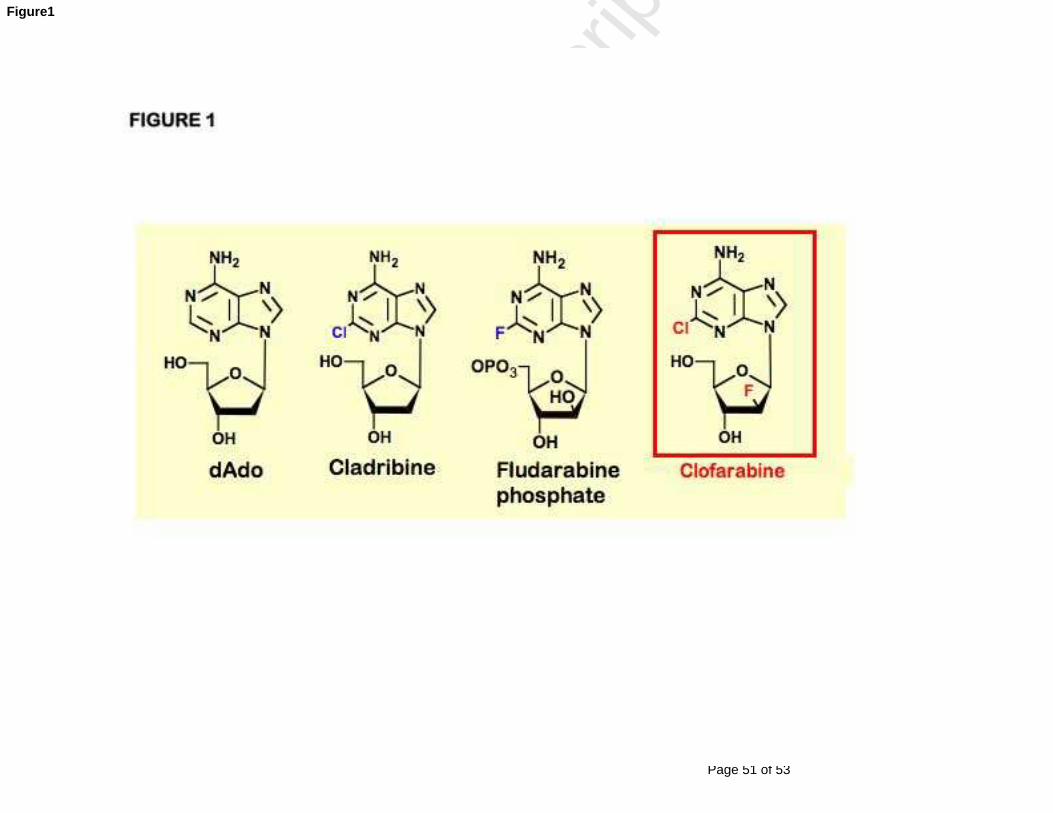
Page 51 of 53
Accep
ted
Man
uscr
ipt
Figure1
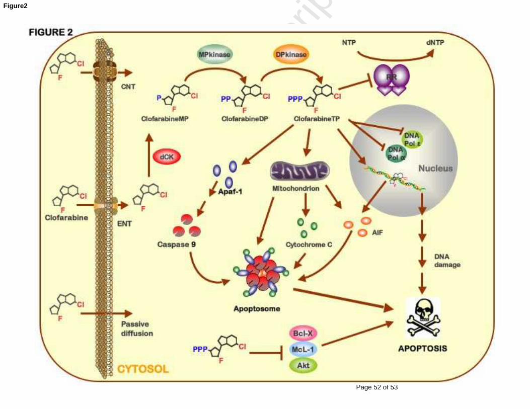
Page 52 of 53
Accep
ted
Man
uscr
ipt
Figure2

Page 53 of 53
Accep
ted
Man
uscr
ipt
Graphical Abstract
Copyright © 2022 FDOKUMEN