FAA Certification of Cold Spray Dimensional Repair. - Safe ...

TOXICOLOGICAL SCIENCES 2014doi: 10.1093/toxsci/kfu098Advance Access publication 5, 2014
Mechanism of Action of Lung Damage Caused by a Nanofilm SprayProduct
Søren T. Larsen,*,1 Constantin Dallot,*,† Susan W. Larsen,‡ Fabrice Rose,‡ Steen S. Poulsen,§ Asger W. Nørgaard,* JitkaS. Hansen,* Jorid B. Sørli,* Gunnar D. Nielsen,* and Camilla Foged†
*National Research Centre for the Working Environment, Copenhagen, Denmark; †Polytech Nice-Sophia, University of Nice, Biot, France; ‡Department ofPharmacy, Faculty of Health and Medical Sciences, University of Copenhagen, Copenhagen, Denmark; and §Department of Biomedical Research, Panum
Institute, University of Copenhagen, Copenhagen, Denmark
1To whom correspondence should be addressed. Fax: +4539165201. E-mail: [email protected].
Received January 10, 2014; accepted May 14, 2014
Inhalation of waterproofing spray products has on several oc-casions caused lung damage, which in some cases was fatal. Thepresent study aims to elucidate the mechanism of action of ananofilm spray product, which has been shown to possess un-usual toxic effects, including an extremely steep concentration-effect curve. The nanofilm product is intended for application onnon-absorbing flooring materials and contains perfluorosiloxaneas the active film-forming component. The toxicological effects andtheir underlying mechanisms of this product were studied usinga mouse inhalation model, by in vitro techniques and by identi-fication of the binding interaction. Inhalation of the aerosolizedproduct gave rise to increased airway resistance in the mice, asevident from the decreased expiratory flow rate. The toxic effectof the waterproofing spray product included interaction with thepulmonary surfactants. More specifically, the active film-formingcomponents in the spray product, perfluorinated siloxanes, inhib-ited the function of the lung surfactant due to non-covalent inter-action with surfactant protein B, a component which is crucial forthe stability and persistence of the lung surfactant film during res-piration. The active film-forming component used in the presentspray product is also found in several other products on the mar-ket. Hence, it may be expected that these products may have a toxi-city similar to the waterproofing product studied here. Elucidationof the toxicological mechanism and identification of toxicologicaltargets are important to perform rational and cost-effective toxi-cological studies. Thus, because the pulmonary surfactant systemappears to be an important toxicological target for waterproofingspray products, study of surfactant inhibition could be included intoxicological assessment of this group of consumer products.
Key Words: Waterproofing spay product; lung toxicity; inhala-tion; pulmonary surfactant.
Waterproofing spray products constitute a group of consumerproducts used to make materials, including textiles, leather, car-pets, wood, glass, concrete, masonry and stone waterproof, anddirt repellents. The products are intended both for industrial anddomestic use, wherefore both private and professionals are ex-posed. During the past decades, inhalation of these products
has caused several cases of severe intoxication (Burkhart et al.,1996; Vernez et al., 2006; Woo et al., 1983). Exposures havegiven rise to development of acute respiratory distress syndrome(Weibrecht and Rhyee, 2011), reactive airway dysfunction syn-drome (Khalid et al., 2009), and acute lung injury (Hubbs et al.,1997). Laboratory studies have shown that inhalation of wa-terproofing spray containing fluorocarbon resin can cause pul-monary collapse in mice (Yamashita and Tanaka, 1995). It hasbeen reported that only a partial dose-response relationship ap-pears in humans after exposure to waterproofing spray products,suggesting that a pronounced inter-individual difference exists(Duch et al., 2014; Vernez et al., 2006).
Recently, a new generation of waterproofing spray prod-ucts has been developed, the so-called nanofilm spray products(NFPs). A variety of NFPs for different materials are sold in,e.g., DIY stores and supermarkets, and thus both professionalsand private consumers may be exposed to the products. Whenthese nanofilm products are applied on a surface, the productforms a film in situ by chemical reaction with the surface. Thenanofilm is very durable and is only a few nanometers in thick-ness. For a description of the nanofilm chemistry, see Norgaardet al. (2010). Like the more traditional products, the NFPs areused to achieve easy-to-clean surfaces because many impuri-ties adhere poorly to hydrophobic surfaces (Quere, 2002). Somefilm products even create an ultrahydrophobic surface, i.e., thecontact angle with water exceeds 150◦, which improves thewater-repellent properties.
One NFP containing hydrolysates and condensates of a per-fluorinated silane caused lethal effects in mice upon short-terminhalation (Norgaard et al., 2010). The product has now beenwithdrawn from the market, but other products with very sim-ilar composition are still available. In mice, the dose-responserelationship of the NFP was unusually steep. Thus, no effectwas seen in the airways in mice inhaling a concentration of 16.1mg/m3, whereas lethal effects were observed at 18.4 mg/m3
of the perfluorinated silane NFP (Norgaard et al., 2010). Fur-thermore, the level of protein in the broncho-alveolar lavage
C© The Author(s) 2014. Published by Oxford University Press on behalf of Toxicological Sciences.This is an Open Access article distributed under the terms of the Creative Commons Attribution License (http://creativecommons.org/licenses/by-nc/3.0/), which permitsnon-commercial re-use, distribution, and reproduction in any medium, provided the original work is properly cited. For commercial re-use, please [email protected]
ToxSci Advance Access published May 30, 2014 at D
et Kongelige B
ibliotek on June 6, 2014http://toxsci.oxfordjournals.org/
Dow
nloaded from

2 LARSEN ET AL.
fluid, a robust marker of acute lung injury (reviewed in Matthayand Zimmerman, 2005), was elevated after exposure to NFP.Also, similar silane-based “Magic Nano sprays” gave rise topulmonary toxicity in humans (Hahn et al., 2008), and the toxi-city of the product was confirmed in rats (Pauluhn et al., 2008).Exposure to especially one of the products, glass and ceramicspray, induced lung inflammation, hemorrhages, edema, and fo-cal septal thickening in the animals.
The mechanisms behind the unusual toxicological pattern ofNFPs and other waterproofing spray products remain to be elu-cidated, but it appears that both the active film-forming sub-stances as well as the composition of the solvents play a rolein the toxicity of the product (Norgaard et al., 2014). This mayalso, at least partly, explain why outbreaks are often seen fol-lowing a change of formulation of the product, i.e., qualitativeor quantitative alterations of the solvent or the polymer (Vernezet al., 2006). Understanding the toxicological mechanisms ofaction including identification of toxicological target is impor-tant in relation to interpretation of in vivo data and a prerequisiteto develop meaningful in vitro screening assays.
Here, we show that an important toxicological target for theNFP product studied is the pulmonary surfactant, which is asurface-active mixture of lipids and proteins found in the alveoliand terminal bronchioles. The pulmonary surfactant is a prereq-uisite for a normal lung function, and extensive neutralizationof surfactant may lead to life-threatening conditions (Lopez-Rodriguez and Perez Gil, 2014). The results presented heremay be applied in the development of in vitro screening of wa-terproofing products for deterioration of pulmonary surfactantfunction and thus for acute pulmonary toxicity.
MATERIALS AND METHODS
Animals. Inbred male BALB/cA male mice aged 5–6 weeks,weight 24.4 g ± 1.9 were purchased from Taconic M&B(Ry, Denmark) and were housed in polypropylene cages(380×220×150 mm) with pinewood sawdust bedding (Ligno-cel S8, Brogaarden, Denmark). Each cage, housing up to 10mice, was furnished with bedding materials, gnaw sticks, andcardboard tubes. The photo-period was from 6 a.m. to 6 p.m.,and the temperature and mean relative humidity in the animalroom were 21◦C ± 0.2 and 55% ± 5 (mean ± SD), respec-tively. Cages were sanitized twice weekly. Food (Altromin no.1324, Altromin, Lage, Germany) and municipal tap water wereavailable ad libitum. Treatment of the animals followed proce-dures approved by The Animal Experiment Inspectorate, Den-mark (No. 2006/561-1123-C3).
Chemicals. The investigated nanofilm product (NFP)intended for coating of non-adsorbing flooring materialswas obtained from NanoCover (Aalborg, Denmark). TheNFP contains hydrolysates and condensates (siloxanes) of1H,1H,2H,2H-perfluorooctyl triisopropoxysilane dissolved in
2-propanol (Norgaard et al., 2010). The 2-propanol (99.9%),dichloromethane (99.8%), dipalmitoylphosphatidyl choline(DPPC, 99%) and methanol (99.9%) were from Sigma-Aldrich Denmark, Brøndby, Denmark. The exogenous lungsurfactant preparation, HL10, was extracted from pig lungs(LeoPharma A/S, Ballerup, Denmark) and contains phospho-lipids, SP-B, SP-C, and cholesterol as described previously(Vermehren et al., 2006). The human SP-B1–25 (Protein DataBank ID: 1DFW, amino acid sequence from N to C terminus:FPIPLPYCWLCRALIKRIQAMIPKG, Mw 2928.2, purity:75.2%), the fluorescein isothiocyanate (FITC) labeled SP-B1–25
(purity 92.5%) and the modified human non-palmityolatedSP-C peptide, SP-C33 (Almlen et al., 2010) (amino acidsequence from N to C terminus: IPSSPVHLKRLKLLLLL-LLLILLLILGALLMGL, Mw = 3615.8 Da, purity: 71.5%)were synthesized by CASLO laboratory (Lyngby, Denmark)and provided as lyophilized trifluoroacetate salts. SP-B1–25
and SP-C33 are the active parts of SP-B and SP-C, respec-tively (Almlen et al., 2010; Hawgood et al., 1998; Waltheret al., 2007). The lipophilic dye 1,1′-dioctadecyl-3,3,3′,3′-tetramethylindodicarbocyanine (DiD, purity ≥ 95% measuredat 644 nm) was from Invitrogen, Molecular Probes (Eugene,OR).
Exposure of animals and assessment of effects in the respira-tory tract. The NFP or control solvent were aerosolized by con-tinuous infusion of the solutions from a glass syringe to a Pitt no.1 jet nebulizer (Wong and Alarie, 1982) by means of an infusionpump (New England Medical Instruments Inc., Medway, MA).The exposure air-stream was subsequently led through a Vi-greaux column to ensure homogenous mixing and then directedinto a 20 l exposure chamber of stainless steel with a hemispher-ical lid and bottom made of glass. The flow rate through thechamber was ∼20 l/min resulting in an air exchange rate of ∼1min−1.
Groups of mice (n = 10) were placed in body plethysmo-graphs in the exposure chamber and exposed head-only for15 min to laboratory air in order to obtain individual baselinelevels. Then, mice were exposed for 60 min to 18.4 mg/m3
NFP or an equivalent concentration of 2-propanol (the solventcontrol group). The 18.4 mg/m3 was the Lowest-Observed-Adverse-Effect Level in mice (Norgaard et al., 2010) and is∼5 times higher than the concentration measured during a sim-ulated workplace exposure scenario (Norgaard et al., 2009).The exposure period was followed by a 30-min recovery pe-riod in which the mice were exposed to laboratory air. To assessexposure-related effects, the respiratory parameters during ex-posure and recovery were compared with pre-exposure baselinelevels, i.e., each mouse served as its own control. The NotocordHem data acquisition software (Notocord Systems SA, Croissy-sur-Seine, France) was used to collect respiratory parameters.Respiratory parameters, data acquisition, and calculations havebeen described previously (Larsen et al., 2004). Briefly, the No-tocord system calculates several respiratory parameters, includ-
at Det K
ongelige Bibliotek on June 6, 2014
http://toxsci.oxfordjournals.org/D
ownloaded from
LUNG DAMAGE CAUSED BY NFP 3
ing breathing frequency (min−1), tidal volume (ml), expiratoryflow rate (ml/s) as well as specific markers of nose and lungirritation (time of break and time of pause, respectively). A re-duction in the expiratory flow rate may be due to bronchocon-striction. Narrowing of the bronchi gives rise to an increasedresistance that reduces the expiratory flow. Also, inhibition ofthe pulmonary surfactant system will lead to increased airwayresistance, which may reduce the expiratory flow (Enhorning,2001).
Capillary surfactometry. In the healthy lung, the walls of theterminal conducting airways are covered with a thin surfactantlayer, which is important to maintain patency of the bronchioles.This is possible because the low surface tension counteracts thecapillary forces driven by the narrow bronchioles. If the func-tion of the pulmonary surfactant is inhibited, i.e., the surfacetension increases, liquid may accumulate in and block the nar-row bronchioles (Enhorning, 2001). To study this phenomenon,the capillary surfactometer (Calmia Medical Inc., Toronto, ON,Canada) was used. Like in the lung, the capillary tube is notof even width; it has a constricted part with a diameter of 0.25mm, which reflects the most narrow part of the human airways(Enhorning, 2001). In this part, a surfactant sample of 0.5 �lcan be deposited. Then, a constant airflow of 0.3 ml/min is ledthrough the capillary. This will remove the surfactant samplefrom the narrow section, leading to an open capillary with lowresistance. If the pulmonary surfactant properties are intact, thecapillary will remain open as long as the airflow continues for2 min. If, in contrast, the pulmonary surfactant is inhibited by,e.g., a chemical, the liquid surfactant will collapse and re-enterthe narrow part of the capillary repeatedly leading to an increasein resistance. In the lungs, this is apparent as a decreased expi-ratory flow. In vitro, the percent of time with an open passage(low resistance) through the capillary during the 2-min sampleperiod may be used to quantify the pulmonary surfactant func-tion.
The NFP, containing approximately 99% 2-propanol, was di-luted in 2-propanol to a total volume of 20 �l, i.e., the concen-tration of solvent was the same in all samples. The diluted NFPwas added to 50 �l of HL10 resulting in a final HL10 concen-tration of 4 mg/ml. The surfactant samples were analyzed in thecapillary surfactometer after 15 min of incubation at 37◦C.
Langmuir isotherms. The pulmonary surfactant containsphospholipids and the surfactant-associated proteins (SP) B andC as the main surface active components (Perez-Gil, 2008). SP-B and SP-C are small hydrophobic proteins associated with thephospholipids. These proteins enhance interfacial adsorption ofsurface-active molecules into the air-liquid interface in the lung.This way, they contribute to mechanical stability of the inter-facial film (Lopez-Rodriguez and Perez-Gil, 2014). Althoughphospholipids per se can reduce the surface tension of water,presence of SP-B and, to some degree, also SP-C, is necessaryin order to achieve all of the dynamic properties of the com-
plete lung surfactant, including the near-zero surface tensionvalues that are observed in the compressed film. SP-B further-more enhances the efficiency of surfactant by reducing the de-gree of film compression necessary in order to reach a very lowsurface tension (Lopez-Rodriguez and Perez-Gil, 2014). Twoother surfactant proteins, SP-A and SP-D, are not consideredto be involved in the reduction of the surface tension (Lopez-Rodriguez and Perez-Gil, 2014). To study how NFP may inter-fere with lung surfactant components, including SP-B and SP-C,a Langmuir technique (Nag et al., 1998) was applied and HL10was used as model of pulmonary surfactant. The HL10 wasdeposited on a subphase of 157 ml carbonate buffer (150mMNaCl, 2mM CaCl2, 0.2mM NaHCO3, pH = 7.0) in a KSV Mini-trough 1 (KSV Instruments Ltd, Helsinki, Finland) with a sur-face area of 243 cm2. The buffer was kept at 25.0 ± 0.1◦C. AHL10 dispersion in carbonate buffer was spread on this sub-phase with a Hamilton syringe in an amount sufficient to reacha surface pressure of 20 mN/m before compression. The com-pression was initiated 20 min after spreading to allow the sur-factant to adsorb at the interface. The barrier speed during thecompression/expansion cycles was 20 mm/min. The surfacepressure was measured by means of a Wilhelmy platinum plate(KSV Instruments Ltd). KSV software (KSV instruments Ltd)was used for data analyses. To study the effect of NFP on lungsurfactant, dry HL10 powder (15 mg) was dissolved in 189 �l ofNFP solution in 2-propanol. The components were mixed wellby vigorous vortexing, the 2-propanol was evaporated, and themixture was incubated for 1 h at 37◦C. The dry HL10-NFP mix-ture was then dispersed in 10 ml carbonate buffer and stirredovernight before spreading the dispersion on the buffer sub-phase in an amount sufficient to reach a surface pressure of20 mN/m before compression. As NFP inhibited the surfactantproperties of HL-10, it was studied whether replacement of thesurfactant proteins SP-B and SP-C could restore the surfactantproperties. To do this, SP-B or SP-C peptides were dissolved in2-propanol at 1 mg/ml and added to the HL10-NFP solution ata 2.5% weight ratio of HL10 for SP-B, and at a 7.5% weightratio of HL10 for SP-C. For the mixtures of SP-B and SP-C, a3.6% weight ratio of HL10 for SP-B and a 7.5% weight ratio ofHL10 for SP-C were applied. The 2-propanol was evaporatedcompletely, and the dry HL10-NFP-SP mixture was dispersedin carbonate buffer and stirred overnight before spreading thedispersion on the buffer subphase.
Histology. Immediately after the end of the inhalation ex-periment, lungs were fixed in situ by instillation of 4% (v/v)buffered paraformaldehyde via a polyethylene tube introducedinto the trachea. The lungs were inflated to a pressure of 25 cmH2O, and the size of the lungs was controlled through an open-ing in the pleural sack on both sides. After 5 min of fixation, thelungs were removed in toto and further fixated for at least 24 husing the same fixative.
For immunohistochemistry, the tissues were embedded inparaffin and cut into sections of ∼10 �m. The samples were
at Det K
ongelige Bibliotek on June 6, 2014
http://toxsci.oxfordjournals.org/D
ownloaded from

4 LARSEN ET AL.
blocked for 30 min in 10% (vol/vol) normal rabbit serum(code no. X0902, Dako, Denmark) and then incubated for 18h at room temperature with the primary SP-B antibody diluted1:1000 (ab40876, Abcam, Cambridge, UK). The immunoreac-tions were visualized by 1 h incubation with biotinylated swineanti-rabbit immunoglobulins (code no. E 353, Dako, Denmark)diluted 1:200 as the second layer, followed by 2 h incuba-tion with StreptABComplex/horseradish peroxidase (code no.E 353, Dako, Denmark) diluted 1:100 as the third layer, and fi-nally stained by means of 3,3-diaminobenzidine for 30 min. Thesections were counterstained with hematoxylin. The degree ofmorphological changes in the lungs and the immunostainingswere evaluated blindly.
Confocal microscopy. To study the effect of NFP on theinterfacial localization of the SP-B1–25 peptide, a water-in-chloroform emulsion was prepared using DPPC as the emul-sifier. Briefly, 67 �g (20 nmol) of SP-B1–25 peptide end-labeledwith the fluorophore FITC was dissolved in 2-propanol. It wasdeposited in a test tube by evaporation of the solvent, and 10 mg(13.62 �mol) DPPC was added and dispersed in 2.1 ml chlo-roform with ultrasound using a Sonifier cell disruptor (Bran-son, Danbury, CT). The organic phase was stained with 9.5 �gDiD/ml. A volume of 30 �l NFP was added to a final amountof 3 �l NFP/mg DPPC. In the control experiment, the NFP wassubstituted with 30 �l 2-propanol. To this volume, 350 �l ofcarbonate buffer was added, and the mixture was sonicated un-til a thick, milky-white emulsion was obtained. Fluorescencemicroscopy was performed by using a Carl Zeiss LSM510 con-focal laser scanning microscope (Carl Zeiss, Piezosystem jenaGmBH, Jena, Germany) equipped with an argon laser (458 and488 nm) and a HeNe laser (543 nm) using the LSM 510 soft-ware.
Mass spectrometry. A two-phase system was prepared in a1.5 ml glass vial by adding 0.5 ml dichloromethane to 0.5 ml ofa 25 �g/ml aqueous solution of the SP-B1–25 peptide and, sub-sequently, 40 �l NFP was added to the organic phase. The vialwas shaken vigorously for about 1 min and the emulsion wasthen allowed to separate at room temperature for about 20 min.Aliquots of both phases were diluted five times with methanoland infused directly into the electrospray ion source of a BrukermicrOTOF-Q (Bruker Daltonics, Bremen, Germany) at a flowrate of 5 �l/min. The capillary voltage was 4.5 kV in the posi-tive mode and 3.2 kV in the negative mode. The nebulizer pres-sure was 1 bar, whereas the flow rate and temperature of thedrying gas were 4 l/min and 190◦C, respectively. An ESI cal-ibration solution containing fluoroalkylphosphazines (Agilenttunemix, Agilent Technologies, Santa Clara, CA) was used forexternal mass calibration resulting in accuracies of ±5 mDa forabundant ions (≥104 counts).
Statistics. The effects of the NFP on the expiratory flow ratewere assessed by comparison with the solvent exposed controls.
Statistical significance was accepted if no overlap occurred be-tween the 95% confidence intervals of the solvent control groupand the NFP exposure group (cf. Fig. 1). Data from the capillarysurfactometer were analyzed by one-way ANOVA followed bythe Dunnett method for comparison with a control using theMinitab ver. 15 software. p values < 0.05 were considered sta-tistically significant.
RESULTS
Respiratory Effects Following Inhalation of NFP
Mice were exposed for 15 min to clean air to establish thenormal (baseline) breathing pattern of each mouse. This base-line period was followed by exposure to the aerosolized NFPat a concentration of 18.4 mg/m3. Control mice were exposedto an equivalent concentration of 2-propanol, the solvent usedin the NFP. Inhalation of the NFP aerosol reduced the expira-tory flow rate, indicating an increased airway resistance (Fig. 1).The reduction occurred gradually over the 60-min exposure pe-riod, and a statistically significantly reduced flow rate comparedwith the control group was reached after approximately 10 minof exposure. The flow rate did not normalize within the 30-minpost-exposure recovery period, where the animals were exposedto clean air.
Effects of NFP on the Pulmonary Surfactant System
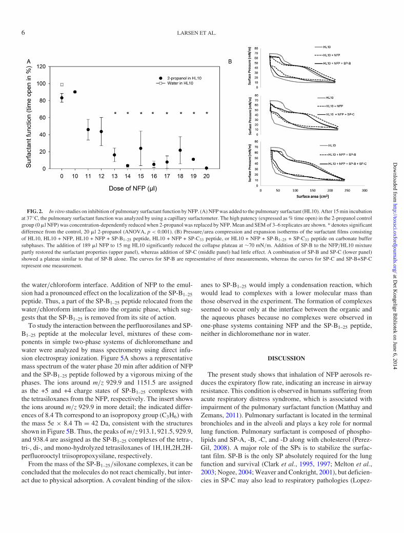
To study effects of NFP on the pulmonary surfactant sys-tem, the capillary surfactometer bioassay was applied, cf. Ma-terials and Method section. The pulmonary surfactant formula-tion HL10 (Vermehren et al., 2006) in water left the capillaryopen 98.7 ± 1.52% of the time (Fig. 2A). Addition of 20 �l 2-propanol to the HL-10 formulation reduced the % time open to83.3 ± 8.9%, which means that the solvent itself has a minor in-hibitory effect of the surfactant function. Replacing a part of the2-propanol with NFP gave rise to a concentration-dependent de-crease in % open, clearly showing an inhibitory effect of NFPon the pulmonary surfactant. The physiological consequencesof this inhibition have been shown to be liquid blocking of theterminal airways, leading to increased airway resistance (En-horning, 2001).
Mechanism of Pulmonary Surfactant Inhibition
The effect of NFP on the pulmonary surfactant was studiedusing the Langmuir technique. In this assay, the surfactant filmis repeatedly expanded and compressed, and the assay was ap-plied to investigate the properties of the pulmonary surfactantfilm under dynamic conditions. Addition of NFP to the HL10surfactant film dramatically changed the shape of the pressure-area isoterm (Fig. 2B). The condensation isotherm of the nativeHL10 was characterized by a collapse plateau at approximately70 mN/m. The addition of NFP to HL10 mainly affected theplateau part of the isotherm, resulting in the loss of the collapseplateau, which suggests a reduced mechanical strength of the
at Det K
ongelige Bibliotek on June 6, 2014
http://toxsci.oxfordjournals.org/D
ownloaded from

LUNG DAMAGE CAUSED BY NFP 5
FIG. 1. Mid-expiratory flow rate (reflecting airway resistance) following inhalation of a perfluorosiloxane-containing nanofilm product (NFP). Mice (n = 10per group) were exposed for 15 min to clean air to achieve individual baseline values. Then the animals were exposure to aerosolized 2-propanol (solvent control)or 18.4 mg/m3 NFP for 60 min, followed by a 30-min clean air recovery period. The mean values and 95% confidence limits are shown.
HL10 film. In addition, the expansion curve for HL10 was char-acterized by a fast decrease of the surface pressure to the initialsurface pressure for HL10. However, in the presence of the NFP,the drop occurred in a different manner, suggesting that the filmreorganized differently in the presence of NFP during the expan-sion phase. By adding synthetic SP-B peptide (SP-B1–25), mim-icking the natural SP-B (Hawgood et al., 1998; Walther et al.,2007), the impaired film could partly be restored (Fig. 2B). Incontrast, addition of SP-C peptide (SP-C33), a model peptide ofthe natural SP-C, did only change the initial part of the pressure-area isotherm, whereas no effect was seen at higher compressionstate compared with the HL-10 + NFP isotherm (Fig. 2B). Thissuggests that SP-C33 only to a minor degree affected the sur-factant properties of the impaired HL10 film. Furthermore, theeffect of a combination of SP-B1–25 and SP-C33 was similar toSP-B1–25 alone (Fig. 2B), indicating that SP-C33 did not poten-tiate the effect of SP-B1–25 in this set-up.
NFP Interaction with SP-B In Vivo
The interaction between SP-B and NFP observed in vitrowas confirmed in vivo. Thus, lungs from NFP-exposed micewere fixated in formalin immediately after end of the inhala-tion study, and tissue sections were stained with SP-B-specificantibodies. Whereas the presence of native SP-B was apparent
in the lungs of solvent-exposed control mice (Fig. 3A), no SP-Bcould be detected in the lungs of NFP-exposed mice (Fig. 3B),suggesting that the perfluorinated siloxanes physically or chem-ically modify one or more epitopes on SP-B essential for bind-ing to the SP-B-specific antibodies. The SP-B depletion wasmost prominent in the bronchioles.
Chemical and Physico-Chemical Investigations of NFP/SP-BInteraction
The function of SP-B in surfactant films is to interact withthe head-group of surfactant lipids including DPPC, and therebyimprove the adsorption, spreading, and surface insertion ofthese lipids (Nag et al., 1999). Thus, a prerequisite for properfunction of SP-B is its proximity to DPPC. An interaction be-tween NFP and SP-B may change the physico-chemical prop-erties of SP-B. To study whether this may be the case, the phys-ical localization of the SP-B1–25 peptide was studied by confo-cal microscopy of water-in-chloroform emulsions using DPPCas emulsifier (Fig. 4). For this purpose, the SP-B1–25 was la-beled with the fluorophore FITC, and the organic phase (red)was visualized by the addition of the lipophilic fluorophoreDiD. The addition of FITC-labeled SP-B1–25 peptide did notaffect the physical stability of the emulsion and the FITC-labeled SP-B1–25 peptide (green color) was clearly located at
at Det K
ongelige Bibliotek on June 6, 2014
http://toxsci.oxfordjournals.org/D
ownloaded from
6 LARSEN ET AL.
FIG. 2. In vitro studies on inhibition of pulmonary surfactant function by NFP. (A) NFP was added to the pulmonary surfactant (HL10). After 15 min incubationat 37◦C, the pulmonary surfactant function was analyzed by using a capillary surfactometer. The high patency (expressed as % time open) in the 2-propanol controlgroup (0 �l NFP) was concentration-dependently reduced when 2-propanol was replaced by NFP. Mean and SEM of 3–6 replicates are shown. * denotes significantdifference from the control, 20 �l 2-propanol (ANOVA, p < 0.001). (B) Pressure/area compression and expansion isotherms of the surfactant films consistingof HL10, HL10 + NFP, HL10 + NFP + SP-B1–25 peptide, HL10 + NFP + SP-C33 peptide, or HL10 + NFP + SP-B1–25 + SP-C33 peptide on carbonate buffersubphases. The addition of 189 �l NFP to 15 mg HL10 significantly reduced the collapse plateau at ∼70 mN/m. Addition of SP-B to the NFP/HL10 mixturepartly restored the surfactant properties (upper panel), whereas addition of SP-C (middle panel) had little effect. A combination of SP-B and SP-C (lower panel)showed a plateau similar to that of SP-B alone. The curves for SP-B are representative of three measurements, whereas the curves for SP-C and SP-B+SP-Crepresent one measurement.
the water/chloroform interface. Addition of NFP to the emul-sion had a pronounced effect on the localization of the SP-B1–25
peptide. Thus, a part of the SP-B1–25 peptide relocated from thewater/chloroform interface into the organic phase, which sug-gests that the SP-B1–25 is removed from its site of action.
To study the interaction between the perfluorosilanes and SP-B1–25 peptide at the molecular level, mixtures of these com-ponents in simple two-phase systems of dichloromethane andwater were analyzed by mass spectrometry using direct infu-sion electrospray ionization. Figure 5A shows a representativemass spectrum of the water phase 20 min after addition of NFPand the SP-B1–25 peptide followed by a vigorous mixing of thephases. The ions around m/z 929.9 and 1151.5 are assignedas the +5 and +4 charge states of SP-B1–25 complexes withthe tetrasiloxanes from the NFP, respectively. The insert showsthe ions around m/z 929.9 in more detail; the indicated differ-ences of 8.4 Th correspond to an isopropoxy group (C3H6) withthe mass 5e × 8.4 Th = 42 Da, consistent with the structuresshown in Figure 5B. Thus, the peaks of m/z 913.1, 921.5, 929.9,and 938.4 are assigned as the SP-B1–25 complexes of the tetra-,tri-, di-, and mono-hydrolyzed tetrasiloxanes of 1H,1H,2H,2H-perfluorooctyl triisopropoxysilane, respectively.
From the mass of the SP-B1–25/siloxane complexes, it can beconcluded that the molecules do not react chemically, but inter-act due to physical adsorption. A covalent binding of the silox-
anes to SP-B1–25 would imply a condensation reaction, whichwould lead to complexes with a lower molecular mass thanthose observed in the experiment. The formation of complexesseemed to occur only at the interface between the organic andthe aqueous phases because no complexes were observed inone-phase systems containing NFP and the SP-B1–25 peptide,neither in dichloromethane nor in water.
DISCUSSION
The present study shows that inhalation of NFP aerosols re-duces the expiratory flow rate, indicating an increase in airwayresistance. This condition is observed in humans suffering fromacute respiratory distress syndrome, which is associated withimpairment of the pulmonary surfactant function (Matthay andZemans, 2011). Pulmonary surfactant is located in the terminalbronchioles and in the alveoli and plays a key role for normallung function. Pulmonary surfactant is composed of phospho-lipids and SP-A, -B, -C, and -D along with cholesterol (Perez-Gil, 2008). A major role of the SPs is to stabilize the surfac-tant film. SP-B is the only SP absolutely required for the lungfunction and survival (Clark et al., 1995, 1997; Melton et al.,2003; Nogee, 2004; Weaver and Conkright, 2001), but deficien-cies in SP-C may also lead to respiratory pathologies (Lopez-
at Det K
ongelige Bibliotek on June 6, 2014
http://toxsci.oxfordjournals.org/D
ownloaded from

LUNG DAMAGE CAUSED BY NFP 7
FIG. 3. Interaction of NFP and SP-B investigated by histology. Lung tissuesections from solvent-exposed control mice (A) and mice exposed to NFP (B).Lungs were collected immediately after end of the 30-min recovery period. Sec-tions were stained with an SP-B-specific antibody. Arrows indicate a positive(A) and a negative (B) staining with SP-B antibodies, respectively, indicatingthat SP-B in NFP-exposed mice was altered or absent. The sections are fromone mouse per group representative of the five mice analyzed in each group.
Rodriguez and Perez-Gil, 2014). The pulmonary surfactant islocated in the alveoli and in the terminal bronchioles, ensuringa free passage of air to and from the alveoli. However, whenthe surfactant is inhibited, a reduced airway patency is seen andsurfactant will block this part of the airways leading to an in-creased airway resistance. Using capillary surfactometry (En-horning, 2001), we were able to demonstrate that NFP inhibitedthe lung surfactant, which leads to liquid blocking of the termi-nal bronchioles, explaining the reduced expiratory flow rate inthe animals. Also, our histological analyses showed that miceexposed to NFP had lower levels of native SP-B in the bronchi-oles. Exposure to higher levels (42.4 mg/m3) of the same NFPgave rise to alveolar collapse (atelectasis) and emphysema (Nor-gaard et al., 2010) showing that the alveoli also may be affectedby these products.
The primary role of SP-B is to enhance the properties ofsurface active lipids in the pulmonary surfactant during respi-
FIG. 4. Confocal microscopy images of an emulsion consisting of a car-bonate buffer in chloroform using DPPC as the emulsifier. The red fluorescentdye, DiD, was added to the organic phase. In the solvent control experiment, theFITC-labeled SP-B1–25 peptide was located at the chloroform/water interface.Replacing the 30 �l 2-propanol in the control group with an equal volume ofperfluorosiloxane-containing NFP leads to translocation of the SP-B1–25 pep-tide from the interface to the organic phase (lower right panel). Scale bar = 10�m.
ration (Lopez-Rodriguez and Perez-Gil, 2014). At the end ofexpiration, the lung area is minimized, i.e., the surfactant filmcovering the alveoli is maximally compressed, whereby a highsurface pressure ( = low surface tension) is achieved, whichcounteracts alveolar collapse. In the presence of SP-B, the de-gree of surfactant film compression necessary to give a suffi-ciently high surface pressure is reduced (Lopez-Rodriguez andPerez-Gil, 2014). In the present study, the Langmuir isotherm(Fig. 2B) clearly showed that addition of NFP to pulmonary sur-factant inhibits this function of SP-B as the reduction in area ledto a lower increase in surface pressure compared with the pul-monary surfactant itself. Once the surfactant was inhibited byNFP, its properties could partly be restored by addition of novelSP-B1–25, as apparent from the steeper pressure-area isotherm.In contrast, addition of the SP-C33 peptide had little effect underthe applied test conditions.
Chemical analyses demonstrated that the perfluorosilanesand SP-B1–25 peptide interacted non-covalently by physical ad-sorption and thus formed relatively stable complexes therebyinactivating the SP-B. It has previously been shown that per-fluorinated compounds adsorb readily to proteins such as albu-min (Bischel et al., 2010; Manus-Spencer et al., 2010), and thethyroid hormone transport protein transthyretin (Weiss et al.,2009). The affinity of perfluorinated compounds for proteinsis most likely due to the polar hydrophobic nature of perflu-orinated substances, as proposed by Biffinger and coworkers(Biffinger et al., 2004). It could be speculated that the perflu-orosilanes with their hydrophobic perfluorcarbon tail in com-
at Det K
ongelige Bibliotek on June 6, 2014
http://toxsci.oxfordjournals.org/D
ownloaded from

8 LARSEN ET AL.
FIG. 5. Non-covalent interaction between the SP-B1–25 peptide and the perfluorosiloxanes in the NFP shown by mass spectrometry. The SP-B1–25 peptideand the perfluorosiloxanes were allowed to react in a two-phase system of water and dichloromethane. (A) Electrospray ionization (+) spectrum of the waterphase diluted five times with methanol. The +5 and +4 charge states of SP-B1–25 complexes with tetrasiloxanes are observed around m/z 929.9 and m/z 1151.5,respectively. The ions around m/z 1264.2 are the +4 charge states of SP-B1–25 complexes with pentasiloxanes, whereas the +3 charge state of the SP-B1–25 peptidecan be observed around m/z 976.9. The insert shows the ions around m/z 929.9 in more detail. (B) Silane and siloxane structures: 1, non-hydrolyzed precursersilane; 2, fully hydrolyzed precurser silane; 3, tentative structure of the dihydrolyzed tetrasiloxane observed in a complex with SP-B1–25 around m/z 929.9 (cf.panel (A)).
bination with hydrophilic silanol groups (Fig. 5B) somewhatmimic the structure of the phospholipids and facilitate both hy-drophobic and hydrophilic interactions with proteins, as previ-ously shown for perfluoroalkyl acids (Bischel et al., 2010).
The physico-chemical consequences of the interaction be-tween NFP and the SP-B1–25 peptide were studied using a water-in-chloroform two-phase system. This system has an interfacethat to some degree models the water/air interface of the lung.Whereas SP-B1–25 in absence of NFP was located exclusivelyat the interface, addition of NFP led to distribution of SP-B1–25
into the organic phase, i.e., SP-B1–25 is removed from its site ofaction. Furthermore, binding or adsorption of perfluorosilane tothe SP-B1–25 peptide may hinder proper function of the peptideby the steric blocking of active sites of the molecule.
SP-B and SP-C are small hydrophobic proteins that are em-bedded in the phospholipid layer (Lopez-Rodriguez and Perez-Gil, 2014). Therefore, if a chemical should reach and potentiallyinhibit the function of SP-B, it has to be able to penetrate intothe phospholipid layer. Only solvents with a certain degree oflipophilicity can do so, and this phenomenon may well explainwhy the toxicity of a waterproofing substance increased withincreasing lipophilicity of the solvent (Norgaard et al., 2014).
New waterproofing and impregnation products are continu-ously being developed and marketed without being tested forinhalation toxicity, although exposures to these consumer prod-ucts has given rise to several cases of intoxication each year.
The toxicity of the products depends both on the active coatingsubstance and the used solvents and minor changes in either ofthese components may have significant impact on the toxicity ofthe whole product (Norgaard et al., 2010, 2014). Consequently,there is a need for robust and reliable methods for investigat-ing the acute lung toxicity of this type of consumer products. Amouse inhalation model may be useful for such screening. Asthe present study suggests that the pulmonary surfactant sys-tem is an important toxicological target for waterproofing sprayproducts, surfactant inhibition assays should be developed toallow screening of these often quite complex mixtures of chem-icals.
FUNDING
This study was funded by the Danish Working EnvironmentResearch Fund.
ACKNOWLEDGMENTS
Maria Hammer, Heidi Marie Paulsen, and Lise SchorlingStrange are acknowledged for expert technical assistance.
at Det K
ongelige Bibliotek on June 6, 2014
http://toxsci.oxfordjournals.org/D
ownloaded from

LUNG DAMAGE CAUSED BY NFP 9
REFERENCES
Almlen, A., Walther, F. J., Waring, A. J., Robertson, B., Johansson, J., andCurstedt, T. (2010). Synthetic surfactant based on analogues of SP-B andSP-C is superior to single-peptide surfactants in ventilated premature rabbits.Neonatology 98, 91–99.
Biffinger, J. C., Kim, H. W., and DiMagno, S. G. (2004). The polar hydropho-bicity of fluorinated compounds. Chembiochem. 5, 622–627.
Bischel, H. N., Manus-Spencer, L. A., and Luthy, R. G. (2010). Noncovalentinteractions of long-chain perfluoroalkyl acids with serum albumin. Environ.Sci. Technol. 44, 5263–5269.
Burkhart, K. K., Britt, A., Petrini, G., O’Donnell, S., and Donovan, J. W. (1996).Pulmonary toxicity following exposure to an aerosolized leather protector. J.Toxicol. Clin. Toxicol. 34, 21–24.
Clark, J. C., Weaver, T. E., Iwamoto, H. S., Ikegami, M., Jobe, A. H., Hull, W.M., and Whitsett, J. A. (1997). Decreased lung compliance and air trapping inheterozygous SP-B-deficient mice. Am. J. Respir. Cell Mol. Biol. 16, 46–52.
Clark, J. C., Wert, S. E., Bachurski, C. J., Stahlman, M. T., Stripp, B. R., Weaver,T. E., and Whitsett, J. A. (1995). Targeted disruption of the surfactant proteinB gene disrupts surfactant homeostasis, causing respiratory failure in new-born mice. Proc. Natl. Acad. Sci. U.S.A. 92, 7794–7798.
Duch, P., Norgaard, A. W., Hansen, J. S., Sorli, J. B., Jacobsen, P., Lyn-ggaard, F., Levin, M., Nielsen, G. D., Wolkoff, P., Ebbehoj, N., et al.(2014). Pulmonary toxicity following exposure to a tile coating product con-taining alkylsiloxanes. A clinical and toxicological evaluation. Clin. Toxi-col., doi:10.3109/15563650.2014.915412.
Enhorning, G. (2001). Pulmonary surfactant function studied with the pulsat-ing bubble surfactometer (PBS) and the capillary surfactometer (CS). Comp.Biochem. Physiol. A Mol. Integr. Physiol. 129, 221–226.
Hahn, A., Begemann, K., Meyer, H., Preussner, K., and Spielmann, H. (2008).“Nano” sealing sprays health impairments in Germany. Clin. Toxicol. 46, 370(Abstract).
Hawgood, S., Derrick, M., and Poulain, F. (1998). Structure and properties ofsurfactant protein B. Biochim. Biophys. Acta 1408, 150–160.
Hubbs, A. F., Castranova, V., Ma, J. Y., Frazer, D. G., Siegel, P. D., Ducat-man, B. S., Grote, A., Schwegler-Berry, D., Robinson, V. A., Van, D. C.,et al. (1997). Acute lung injury induced by a commercial leather conditioner.Toxicol. Appl. Pharmacol. 143, 37–46.
Khalid, I., Godfrey, A. M., and Ouellette, D. R. (2009). Chemical pneumonitisand subsequent reactive airways dysfunction syndrome after a single expo-sure to a household product: A case report. J. Med. Case. Rep. 3, 112.
Larsen, S. T., Hansen, J. S., Hammer, M., Alarie, Y., and Nielsen, G. D. (2004).Effects of mono-2-ethylhexyl phthalate on the respiratory tract in BALB/cmice. Hum. Exp. Toxicol. 23, 537–545.
Lopez-Rodriguez, E., and Perez-Gil, J. (2014). Structure-function relationshipsin pulmonary surfactant membranes: From biophysics to therapy. Biochim.Biophys. Acta. 1838, 1568–1585.
Manus-Spencer, L. A., Tse, M. L., Hebert, P. C., Bischel, H. N., and Luthy, R.G. (2010). Binding of perfluorocarboxylates to serum albumin: A comparisonof analytical methods. Anal. Chem. 82, 974–981.
Matthay, M. A., and Zemans, R. L. (2011). The acute respiratory distress syn-drome: Pathogenesis and treatment. Annu. Rev. Pathol. 6, 147–163.
Matthay, M. A., and Zimmerman, G. A. (2005). Acute lung injury and the acuterespiratory distress syndrome: Four decades of inquiry into pathogenesis andrational management. Am. J. Respir. Cell Mol. Biol. 33, 319–327.
Melton, K. R., Nesslein, L. L., Ikegami, M., Tichelaar, J. W., Clark, J. C., Whit-sett, J. A., and Weaver, T. E. (2003). SP-B deficiency causes respiratory fail-ure in adult mice. Am. J. Physiol. Lung Cell Mol. Physiol. 285, L543–L549.
Nag, K., Munro, J. G., Inchley, K., Schurch, S., Petersen, N. O., and Possmayer,F. (1999). SP-B refining of pulmonary surfactant phospholipid films. Am. J.Physiol. 277(6 Pt 1), L1179–L1189.
Nag, K., Perez-Gil, J., Ruano, M. L., Worthman, L. A., Stewart, J., Casals, C.,and Keough, K. M. (1998). Phase transitions in films of lung surfactant at theair-water interface. Biophys. J. 74, 2983–2995.
Nogee, L. M. (2004). Alterations in SP-B and SP-C expression in neonatal lungdisease. Annu. Rev. Physiol. 66, 601–623.
Norgaard, A. W., Hansen, J. S., Sorli, J. B., Levin, M., Wolkoff, P., Nielsen,G. D., and Larsen, S. T. (2014). Pulmonary toxicity of perfluorinated silane-based nanofilm spray products: Solvent dependency. Toxicol. Sci. 137, 179–188.
Norgaard, A. W., Jensen, K. A., Janfelt, C., Lauritsen, F. R., Clausen, P. A., andWolkoff, P. (2009). Release of VOCs and particles during use of nanofilmspray products. Environ. Sci. Technol. 43, 7824–7830.
Norgaard, A. W., Larsen, S. T., Hammer, M., Poulsen, S. S., Jensen, K. A.,Nielsen, G. D., and Wolkoff, P. (2010). Lung damage in mice after inhalationof nanofilm spray products: The role of perfluorination and free hydroxylgroups. Toxicol. Sci. 116, 216–224.
Pauluhn, J., Hahn, A., and Spielmann, H. (2008). Assessment of early acutelung injury in rats exposed to aerosols of consumer products: Attempt to dis-entangle the “Magic Nano” conundrum. Inhal. Toxicol. 20, 1245–1262.
Perez-Gil, J. (2008). Structure of pulmonary surfactant membranes and films:The role of proteins and lipid-protein interactions. Biochim. Biophys. Acta1778, 1676–1695.
Quere, D. (2002). Surface chemistry: Fakir droplets. Nat. Mater. 1, 14–15.
Vermehren, C., Frokjaer, S., Aurstad, T., and Hansen, J. (2006). Lung surfactantas a drug delivery system. Int. J. Pharm. 307, 89–92.
Vernez, D., Bruzzi, R., Kupferschmidt, H., De-Batz, A., Droz, P., and Lazor, R.(2006). Acute respiratory syndrome after inhalation of waterproofing sprays:A posteriori exposure-response assessment in 102 cases. J. Occup. Environ.Hyg. 3, 250–261.
Walther, F. J., Waring, A. J., Sherman, M. A., Zasadzinski, J. A., and Gordon,L. M. (2007). Hydrophobic surfactant proteins and their analogues. Neona-tology. 91, 303–310.
Weaver, T. E., and Conkright, J. J. (2001). Function of surfactant proteins B andC. Annu. Rev. Physiol. 63, 555–578.
Weibrecht, K. W., and Rhyee, S. H. (2011). Acute respiratory distress associatedwith inhaled hydrocarbon. Am. J. Ind. Med. 54, 911–914.
Weiss, J. M., Andersson, P. L., Lamoree, M. H., Leonards, P. E., van Leeuwen,S. P., and Hamers, T. (2009). Competitive binding of poly- and perfluorinatedcompounds to the thyroid hormone transport protein transthyretin. Toxicol.Sci. 109, 206–216.
Wong, K. L., and Alarie, Y. (1982). A method for repeated evaluation of pul-monary performance in unanesthetized, unrestrained guinea pigs and its ap-plication to detect effects of sulfuric acid mist inhalation. Toxicol. Appl. Phar-macol. 63, 72–90.
Woo, O. F., Healey, K. M., Sheppard, D., and Tong, T. G. (1983). Chest pain andhypoxemia from inhalation of a trichloroethane aerosol product. J. Toxicol.Clin. Toxicol. 20, 333–341.
Yamashita, M., and Tanaka, J. (1995). Pulmonary collapse and pneumonia dueto inhalation of a waterproofing aerosol in female CD-1 mice. J. Toxicol. Clin.Toxicol. 33, 631–637.
at Det K
ongelige Bibliotek on June 6, 2014
http://toxsci.oxfordjournals.org/D
ownloaded from
Copyright © 2022 FDOKUMEN