Matière Organisée Hybride Organique-Inorganique
125
HAL Id: tel-00593471 https://tel.archives-ouvertes.fr/tel-00593471 Submitted on 16 May 2011 HAL is a multi-disciplinary open access archive for the deposit and dissemination of sci- entific research documents, whether they are pub- lished or not. The documents may come from teaching and research institutions in France or abroad, or from public or private research centers. L’archive ouverte pluridisciplinaire HAL, est destinée au dépôt et à la diffusion de documents scientifiques de niveau recherche, publiés ou non, émanant des établissements d’enseignement et de recherche français ou étrangers, des laboratoires publics ou privés. Matière Organisée Hybride Organique-Inorganique Rénal Backov To cite this version: Rénal Backov. Matière Organisée Hybride Organique-Inorganique. Matériaux. Université Sciences et Technologies - Bordeaux I, 2003. tel-00593471
-
Upload
khangminh22 -
Category
Documents
-
view
3 -
download
0
Transcript of Matière Organisée Hybride Organique-Inorganique

HAL Id: tel-00593471https://tel.archives-ouvertes.fr/tel-00593471
Submitted on 16 May 2011
HAL is a multi-disciplinary open accessarchive for the deposit and dissemination of sci-entific research documents, whether they are pub-lished or not. The documents may come fromteaching and research institutions in France orabroad, or from public or private research centers.
L’archive ouverte pluridisciplinaire HAL, estdestinée au dépôt et à la diffusion de documentsscientifiques de niveau recherche, publiés ou non,émanant des établissements d’enseignement et derecherche français ou étrangers, des laboratoirespublics ou privés.
Matière Organisée Hybride Organique-InorganiqueRénal Backov
To cite this version:Rénal Backov. Matière Organisée Hybride Organique-Inorganique. Matériaux. Université Sciences etTechnologies - Bordeaux I, 2003. tel-00593471

1
N° d’ordre : 250
Habilitation à Diriger les Recherches
Présentée à
L’UNIVERSITE BORDEAUX I
Par Rénal BACKOV
Matière Organisée Hybride Organique-Inorganique
soutenue le 20 novembre 2003
Devant le Jury composé de :
Président : Philippe Barois (Directeur de recherches, CRPP UPR-CNRS 8641)
Rapporteur : Jacques Livage (Professeur, Collège de France, LCMC UMR-7574)
Rapporteur : Mir Wais Hosseini (Professeur, Université Louis Pasteur, LCCO UMR 7513)
Rapporteur : Hervé Arribart (Directeur des recherches, Compagnie de Saint-Gobain)
Examinateur : Didier Roux (Directeur de recherches, CRPP UPR-CNRS 8641)
Examinateur : Clément Sanchez (Directeur de recherches, LCMC UMR-CNRS-7574)
-2003-

2
CURRICULUM VITAE BACKOV Rénal Né le 20/05/68 Marié. e-mail : [email protected]
Parcours professionnel, études de 3éme cycle : - 06/09/01 Maître de Conférences, 31ième Section, Université Bordeaux-1, Centre de Recherches Paul Pascal (C.R.P.P.) C.N.R.S. U.P.R. 8641 Avenue Albert Schweitzer, Pessac, 33600 FRANCE. -1998/01 Chercheur Associé: University of Florida, Gainesville, FL, Etats-Unis (Equipe de M. le Professeur Daniel R. Talham) - Cristallisation d'oxalate de calcium et photoréduction de Pt II à l’interface de monocouches
de Langmuir et films de Langmuir-Blodgett. - Magnétisme associé aux solides et solides mous. - Mésophases lyotropes organiques-inorganiques. - Systèmes organométalliques (matériaux 1D, 2D, 3D et en échelles). -1997/98 A.T.E.R.: USTL Montpellier II, 32ième section, chimie minérale. (L. A. M. M. I. U.M.R. 5072 U. S. T. L. Montpellier II) -1994/97 Doctorat : Synthèse et caractérisation d’hybrides organiques-inorganiques obtenus par intercalation de molécules électroactives dans le phosphate de zirconium. Soutenu le 17 Décembre 1997, mention très honorable. allocataire M.E.S.R. / MONITEUR (32eme Section) (L. A. M. M. I. U.M.R. 5072 U. S. T. L. Montpellier II) Jury : M le Professeur J. V. Zanchetta Président (Montpellier) M le Professeur J. Rozière Directeur de thèse (Montpellier) Melle D. J. Jones (D. R.) Co-directeur de thèse (Montpellier) M le Professeur R. Clément Rapporteur (Orsay) M le Professeur F. Béguin Rapporteur (Orléans) M P. Batail (D. R.) Examinateur (Nantes)
M le Professeur J. M. Fabre Examinateur (Montpellier) -1993/94 D.E.A.: Matériaux de l'électronique et de l'ionique du solide (bourse rectorale) mention T. Bien, major de promotion.
Stage de D.E.A.: Etude des propriétés de conduction dans le système V2O5(x)B2O3(1-x). Evaluation de la symétrie locale de l'ion V4+ par Résonance Paramagnétique Electronique. (L.P.C.M.C. U.M.R. 5617 U.S.T.L. Montpellier II)

3
COMPETENCES DEVELOPPEES EN RECHERCHE
THEMATIQUES ABORDEES 1- Solides hybrides organiques-inorganiques de basse dimensionnalité. 2- Mésophases lyotropes hybrides organiques-inorganiques. 3- Technique de Langmuir-blodgett et croissance anisotrope de particules métalliques. 4- Monocouches de Langmuir et phénomènes de biominéralisation à l’interface air-liquide. 5- Conductivité de polarisation et systèmes vitreux. 6- Magnétisme associé aux hybrides organiques-inorganiques, systèmes organométalliques et films LB 7- Solides hybrides organiques-inorganiques à structures hiérarchisées : chimie des formes
CHIMIE DE LA MATIERE ORGANISEE
* Etat solide: Synthèses de matériaux hybrides de basses dimensionnalités. - synthèses hydrothermales - synthèses par chimie douce - phosphates de métaux tétravalents - phosphonates de Cuivre, Vanadyl
- hydroxyacétate et hydroxyphosphonate de cuivre, hydroxycarboxylates de cuivre, de nickel et de Cobalt
- hydroxydes doubles lamellaires - dimères organométalliques, matériaux 1D, matériaux en échelle, matériaux à structures ouvertes
- perovskites à base de Cuivre, Cadmium * Solide mou : Films de Langmuir-Blodgett, monocouches de Langmuir, mésophaes lyotropes.
- formation de nanoparticules, macroparticules métalliques - étude des interactions à l'interface entre une monocouche de Langmuir et espèces en croissance
- élaboration de films de Langmuir-Blodgett à propriétés magnétiques - chimie sol-gel, films auto-supportés, phases lyotropes, matériaux hiérarchisés.
TECHNIQUES DE CARACTERISATION EMPLOYEES - diffraction de RX sur poudre - diffraction de RX aux petits angles - XPS
- spectrophotométrie de flamme - spectroscopie de vibration (IR, Raman, diffusion inélastique incohérente de neutrons) - EXAFS/XANES
- spectroscopie électronique - spectroscopie Mössbauer (119Sn) - RMN 13C, 31P, 1H - RPE - SQUID (susceptibilité magnétique continue et alternative) - technique de Van der Paw (conductivité en courant continu) - conductivité de polarisation - mesure du coefficient de Seebeck (pouvoir thermoélectrique) - SEM, TEM-TED - microscope à angle de Brewster (BAM) - ATG-ATD
- microscopie en polarisation croisée (cristaux liquides)

4
ENCADREMENTS- RESPONSABILITES COLLECTIVES
Responsabilités :
01/présent Recherches : -Prise en charge de l'équipe du Professeur D.R. Talham pendant son séjour sabbatique en France (un an 1998/1999). Durant la première année de mon stage post-doctorale Daniel Talham, invité en séjour sabbatique en France (5 mois au CRPP-Bordeaux et 7 mois IMN-Nantes), m’a demandé de le substituer à Gainesville en veillant sur son équipe américaine, ce, sur un plan exclusivement scientifique. Les responsabilités administratives et financières étant gérées entre ce de rnier et son administration par courrier électronique. A l’époque, cette équipe était constituée de deux stagiaires post-doctoraux, de cinq étudiants de thèse et d’autres stagiaires de niveau "bachelor degree" équivalent au diplôme de Maîtrise. En plus de mes travaux de recherches (biominéralisation, synthèse de nanoparticules et microparticules en milieu confiné, mésophases lyotropes), j’étais spécialement en charge de Jonathan Woodward (thèse sur la réalisation de polymères de coordination à propriétés magnétiques de spin) tout en devant suivre les travaux de Jeff Culp (autre étudiant de thèse) qui s’intéressait alors aux matériaux lamellaires mixtes cobalt-cuivre à bases de phényle phosphonates. En fin de première année de stage post-doctorale mais également sur toute la période suivante j’étais en charge d’Eduardo Perez-Cordero, étudiant de thèse, dont la thèse était centrée sur la réalisation de mésophases lyotropes organiques-inorganiques à bases de Pérovskites lamellaires et autres hydroxydes doubles lamellaires. Cette période très enrichissante mais également assez éprouvante m’a permis d’appréhender in situ les réalités de la gestion d’une équipe de recherche où les paramètres de compétences scientifiques mais également de relations humaines sont mises à rudes épreuves, et ceci, surtout dans un pays comme les Etats-Unis où les notions de compétitivité et de production scientifique, poussées à l’extrême, amènent bien souvent des situations assez tendues entres jeunes étudiants ou étudiantes de thèse ! - Responsable de l’activité CRPP : biomimétisme, biotectonique et biomminéralisation
* Morphogenèses minérales et fluides complexes
* Morphogenèses minérales sous cisaillement
* Minéralisation et mousses
* Minéralisation et phases lamellaires
* Minéralisation et instabilités hydrodynamiques
* Systèmes poreux et réactivité
- Participation à une proposition de thématique dans le cadre du réseau d’excellence Européen (PCRDT-6) : matériaux hybrides organiques-inorganiques et magnétisme. Direction nationale : Marc Drillon (Coordinateur CRPP : R. Clérac). - Participation à une proposition de thématique dans le cadre du réseau d’excellence Européen (PCRDT-6) : matériaux hybrides organiques-inorganiques et biomatériaux. Direction nationale : Clément Sanchez (Coordinateur CRPP : R. Backov)

5
- Participation ACI "Jeunes Chercheurs 2002": Couplage entre la structure d'un fluide complexe et une instabilité hydrodynamique : du problème académique aux applications. N° d’ident: 2046 (Chef de projet: Sébastien Manneville) - Co-organisateur de l’école Galerne 2003 "matériaux hybrides organiques-inorganiques" Bordeaux, septembre 2003. Co-organisateurs : Mondain-Monval Olivier (CRPP), Zakri Cécile (CRPP), Duguet Etienne (ICMCB), Marie -Hélène Delville (ICMCB), Monat Treguet (ICMCB). - Prise en charge de l’aquipe du Professeur D.R. Talham pendant son séjour sabbatique en France 1998-1999 Enseignements : - Responsable du module 6 : "Formulation de matériaux à base de milieux dispersés." Option 3ème année Ingénieur, ENSCPB. Encadrements au C.R.P.P. depuis Septembre 2001: - Stagiaire Post-doctoral : Formation de coques de silice mésostructurées obtenues par chimie sol-gel et procédé d’émulsion, procédés d’encapsulation. Miréralisation aux interfaces SAM. (1 an 2002/03- Giulia Fornasieri) co-encadrement (à 50% avec Philippe Poulin) - Thèse "chimie douce et matière molle : mousses inorganiques". (Thèse débutée le 1er Octobre 2003 –Florent Carn) - Thèse "Matériaux hybrides organiques-inorganiques à propriétés magnétiques". Co-encadrée à 50% avec Rodolphe Clérac. (HDR détenue par Philippe Barois, responsable administratif) (Thèse débutée le 1er Octobre 2002 –Lolita Lecren) - Thèse"Matériaux structurés hybrides organiques-inorganiques". Co-encadrée à 50% avec Olivier Mondain-Monval. (Thèse débutée le 1er Septembre 2001–Alexandre Desforges) - DEA –Polymères : chimie sol-gel et mousses Co-encadrée à 50% avec Annie Colin. (6 mois d’encadrement 2003 –Florent Carn) - DEA –Polymères : Matériaux hybrides organiques-inorganiques et nanostructuration. Co-encadrée à 25% avec Serge Ravaine, Eric Cloutet et Henry Cramail. (6 mois d’encadrement 2003–Anne de Cuendias ) - DEA -ENSCPB: Conceptualisation de matériaux hybrides organiques-inorganiques par procédé d’émulsification. Co-encadrée à 50% avec Philippe Poulin. (6 mois d’encadrement 2002- Doreau Nicolas ) - DEA –PCMC : Matériaux hiérarchisés hybrides organiques-inorganiques et propiétés magnétiques. Co-encadréeà 50% avec Rodolphe Clérac. (6 mois d’encadrement 2002 -Lolita Lecren) - DESS granulats et colloïdes: Nanoparticules d’or encapsulées dans une structure sphérulite, générées par photoréduction du complexe [AuCl4]-. Co-encadrée à 50% avec Chrystel Faure (7 semaines d’encadrement 2002- Delage Céline ) - DESS granulats et colloïdes: Nanoparticules d’or encapsulées dans une structure sphérulite, générées par photoréduction du complexe [AuCl4]2-. Co-encadrée à 50% avec Chrystel Faure (7 semaines d’encadrement 2002- Tawl Karine )

6
- Maîtrise Chimie -Physiques: Des mousses solides ! Vers une synthèse de renfort pour pneumatiques. Co-encadrée à 50% avec Annie Colin. (4 mois d’encadrement 2002- Saadi Zoubida) - Maîtrise Chimie -Physiques: Nanoparticules d’or encapsulées dans une structure sphérulite, générées par photoréduction du complexe [AuCl4]-. Co-encadrée à 50% avec Chrystel Faure (4 mois d’encadrement 2003- Meyre Marie-Edith) - Licence Professionnelle de Chimie Industrielle : Mise en forme de silices mésostructurées sous flux laminaire. (4 semaines d’encadrement 2003- Gaëlle Petit) - Licence Professionnelle de chimie Industrielle : Mise en forme de silices mésostructurées sous flux laminaire.(4 semaines d’encadrement 2003- Julien Laguéri) - Licence Professionnelle de Chimie Industrielle : Formation de coques de silice mesostructurées obtenues par chimie sol-gel et procédé d’émulsion. (4 semaines d’encadrement 2002- Stéphane Jautard) - Licence Professionnelle de chimie Industrielle : Formation de coques de silice mesostructurées obtenues par chimie sol-gel et procédé d’émulsion. (4 semaines d’encadrement 2002- Julie Sarsiat) Encadrements à l’Université de Floride : 1998/2001 -“Ph.D student” (University of Florida): synthesis and characterization of new mineral liquid crystals based on perovskite structures (deux ans d’encadrement-Eduardo E. Perez-Cordero) -"Ph.D student" (University of Florida) synthesis and characterization of new ladder materials based on MxCl y(CH3CN)z/ M= Ni, Co, Mn, Fe. (trois mois d'encadrement- Chen Liu) -"Ph.D student" (University of Florida) :chemical and physical characterization of hybrid organic -inorganic low-dimensional coordination polymers. (trois ans et six mois d'encadrement- Jonathan Woodward) -“Bachelor degree student” (University of Florida): synthesis and characterization of new mineral liquid crystals based on layered double hydroxides structures. (un an d’encadrement- Sarah Lane ) -"Undergraduated student" (University of Florida): synthesis and characterization of polyaromatic electroactive molecules.(un an d'encadrement-David Zipp) Encadrements à l’Université Montpellier II : 1995/1997 - Maîtrise: simulation de courbes de conductivité obtenues pour des matériaux lamellaires hybrides organiques-inorganiques. (2 mois d'encadrement- Lionel Nicole ) - D.E.A: intercalation de composés aminés dans des hexacyanoferrates de Cuivre et de Potassium. (6 mois d'encadrement- Gaëlle Derien) - D.E.A: intercalation de pérylène dans les phosphates de Zirconium α-ZrP et δ-ZrP. (6 mois d'encadrement- Thibaud Mourgues) - Maîtrise : synthèse d'hexacyanoferrate de Cuivre et de Potassium. (2 mois d'encadrement- Lydie Tchikaya)

7
- IUT (mesures physiques): réalisation d'une cellule de conductivité de Van der Paw (méthodes des quatre points), applications à quelques matériaux hybrides organiques-inorganiques conducteurs électroniques. (6 mois d'encadrement-Thibaud Gaultier) - Licence de chimie : intercalation de dipropargylamine dans le phosphate de Titane γ-TiP. (2 mois d'encadrement)

8
ENSEIGNEMENT: MCF, ATER et Monitorat
MCF (Université Bordeaux -I)
Cours : Licence professionnelle (13 H ETD): «Les colloïdes durs » Chapitre 1 : des colloïdes durs au procédé sol-gel I-1 Introduction et quelques définitions I-2 Polymérisation-structuration I-3 Les grandeurs fractales I-4 Du monomère au gel Chapitre II : colloïdes et sols de particules " les non-silicates" II-1 Chimie des précurseurs inorganiques : les métaux de transition II-1-1 L’hydrolyse II-1-2 Condensation
a) olation b) oxolation
II-1-3 Espèces polymères et gélation II-1-4 Un exemple d’étude structurale : V2O5
II-2 Chimie des précurseurs inorganiques : les alkoxydes de métaux de transition II-2-1 Mécanismes d’hydrolyse et condensation II-2-2 Rôle du catalyseur II-2-3 Structures des produits de condensation Chapitre III : colloïdes et sols de particules " les silicates" III-1 Introduction III-2 Les silices en solutions aqueuses III-3 Mécanismes d’hydrolyse et condensation III-3-1 Comportement global III-3-2 Les précurseurs moléculaires Chapitre IV : évolution structurale d’un gel pendant le processus de solidification. Formation de films par procédés de "dip-coating" et "spin-coating" IV-1 Introduction IV-2 Structures de gels poreux : Xérogels et Aérogels IV-3 Formation de films par "dip-coating" IV-4 Formation de films par "spin-coating" Chapitre V : colloïdes durs, chimie sol-gel et applications V-1 Films et revêtements de surface V-1-1 Revêtements de surface V-1-2 Films pour l’électronique V-1-3 Films protecteurs V-1-4 Films poreux V-2 Les monolithes V-3 Poudres, grains et sphères V-4 Les composites V-5 Les membranes et gels poreux

9
Cours : DESS - granulats et colloïdes (7 H ETD): «chimie sol-gel» Cours : ENSCPB- 3éme année (7 H ETD): «matériaux obtenus par procédés sol-gel et matière molle : chimie des formes» «Biomatériaux» Chapitre I : Introduction aux biomatériaux Chapitre II : Matériaux cholestériques à base de chitine et collagène Chapitre III : Phosphate de calcium et calcification biologique Chapitre IV : Bio-silification «Matériaux poreux et mise en forme » Chapitre I : Introduction aux matériaux bio-inspirés Chapitre II : Les stratégies de synthèse Chapitre III : Les principales empreintes organiques Chapitre IV : Structures à bases de silice Chapitre V : Exemples de micro-moulages et auto-assemblages Travaux dirigés : chimie générale (DEUG TC1A) Travaux dirigés : thermodynamique, approche micrscopique (DEUG TC3A) Travaux dirigés : mécanique quantique et spectroscopie (DEUG TC3A) Travaux pratiques : chimie-physique, mécanique quantique et spectroscopie (DEUG TC3A) Travaux pratiques : thermodynamique, Licence chimie -physiques. Travaux pratiques : rhéologie, granulométrie, comportements de polymères en solutions, Licence professionnelle.
A.T.E.R. (USTL Montpellier II) 1997/1998 Cours, TP et TD réalisés au cours du monitorat. TP de Licence "Chimie Fondamentale " : synthèses et caractérisations de quelques complexes de Werner (Cobalt et Chrome), étude de ces composés par spectroscopie électronique et infrarouge. TD de DEUG 2ème Période BPC2 : nomenclature, formalisme de LEWIS, VSEPR, réactions acido-basiques.
MONITORAT (USTL Montpellier II) 1994/19997
Cours : D.E.U.G. 4éme période Introduction à la théorie des groupes: - notion de symétrie - éléments et opérations de symétrie - les groupes ponctuels - les représentations non dégénérées - rappel sur les matrices - les représentations dégénérées - les modes normaux de vibration (activité IR, Raman) - spectrométrie, transformée de Fourier, interféromètre de Michelson Travaux dirigés : D.E.U.G. 2éme période Oxydoréduction: - nombres d'oxydation - potentiels d'électrodes - équation de NENRST

10
Chimie descriptive: - formalisme de LEWIS. - géométrie moléculaire par la théorie V.S.E.P.R. Travaux pratiques: D.E.U.G. 2éme période
-Semi-micro analyse qualitative. -Aluns de chrome et de potassium. -Les complexes. -Dosage colorimétrique du cuivre II. -Préparation d'un sel double et d'un sel complexe. -Les halogènes. -Préparation et dosage d'une Schoenite

11
DIFFUSION DES TRAVAUX DE RECHERCHE
PUBLICATIONS, BREVETS, PROCEEDINGS (38)
Publications (22) : ** Palladium nanoparticles generation within microcellular polymeric foam (polyHIPES)
A. Desforges, H. Deleuze, R. Backov , O. Mondain-Monval. J. Chem. Soc; chem. Comm. (soumis)
** Inorganic monoliths hierarchically textured via concentrated direct emulsion and micellar
templates. A. Colin, M-.F. Achard, F. Carn, H. Deleuze, R. Backov. J. Chem. Soc; chem. Comm. (soumis)
** Mesoporous mineral capsules generation from reverse emulsion and sol-gel processes. G. Fornasieri, S. Badaire, R. Backov, O. Mondain-Monval, C. Zakri, P. Poulin Nature Mat., 2004 (soumis). ** Spontaneous generation of gold nanoparticles within onion mesophases. C. Faure, O. Regev, D. Roux, R. Backov Adv. Mat. (soumis) ** Structural, thermal and magnetic properties investigation of three transition metal-4,4’-bipyridine
coordination polymers : [Ni(4,4’-bipy)3(H2O)2.](ClO4)2.1.4(4,4’bipy).3H2O, [Co(4,4’- bipy)3(H2O)2](ClO4)21.4(4,4’bipy).3H2O and [Cu(4,4’-bipy)3(DMSO)2](ClO4)2.2(4,4’bipy). .W Woodward, R. Backov, H. Honuki, M.W. Meisel, D.R. Talham olyhedron, 2003, 22, 2821.
** Presence of lipids in urine, crystals and stones: Implication for the formation of kidney stones. S. R. Khan, P. A. Glenton, R. Backov and D. R. Talham. Kidney International 2002, 62, 2062. ** Lyotropic phase from hybrid organic -inorganic layered copper hydroxides. R. Backov, A. N. Morgan, S. Lane, E.E. Perez-Cordero, K. Williams, M.W. Meisel, C. Sanchez, D. R. Thalam. Mol. Cryst. Liq. Cryst. 2002, 376, 127. ** [Ni(terpy)(H2O)]-trans-[Ni-µ-(CN)2(CN)2]n, a one-dimensional linear tetracyanonicolate chain. J. D. Woodward, R. Backov, K. A. Abboud, D. R. Talham. Acta. Cryts. C. 2001, C57, 1027. ** Layered mixed-metal phenylphosphonates, CoxMn1-x(O3PC6H5).H2O: Structure and magnetic
properties. J. T. Culp, G. E. Fanucci, B. C. Watson, R. Backov, H. Ohnuki, M. W. Meisel, D. R. Talham J. Solid State Mat. 2001, 159, 362. ** The magnetic spin ladder (C5H12N2)2CuBr4: high field magnetization and scaling near quantum criticality. B. C. Watson, V. N. Koto, M. W. Meisel, D. W. Hall, G. E. Granroth, W. T. Montfrooij, S. E. Nagler, D. A. Jensen, R. Backov, M. A. Petruska, G. E. Fanucci, D. R. Talham. Phys. Rev. Letter 2001, 86, 5168.

12
** Multiple bilayer dipalmitoylphosphatidylserine (DPPS) LB films stabilized with transition metals ions. G. E. Fanucci, R. Backov, R. Fu, D. R. Talham. Langmuir 2001, 17, 1660. ** Calcium oxalate monohydrate precipiation at phosphatidylglycerol Langmuir monolayers. R. Backov, C. M. Lee, S. R. Khan, C. Mingotaud, G. E. Fanucci, D. R. Talham. Langmuir 2000, 16, 6013. ** Magnetic phase diagram of the quasi-2D mixed metal phenylphosphonates. G. E. Fanucci, J.T. Culp, B. C. Watson, R. Backov, H. Ohnuki, D. R. Talham , M. W. Meisel. Physica B 2000, 284-8, 1499. ** DC and AC conductivities of V2O5(x)B2O3(1-x) oxide glasses. H. el Mkami, B. Deroide, R. Backov, J. V. Zanchetta. J. Phys. Chem. Solids 2000, 61, 819. ** Precipitation of calcium oxalate monohydrate at phospholipid monolayers. R. Backov, S. R. Khan, K. Byer, C. Mingotaud, C. Nixon, D. R. Talham. J. Am. Soc. Nephrology 1999, 10, S359. ** Photoluminescence properties of fullerene C60 in microporous VPI5-Zeolite.
A.Lamrate, J. M. Jannot, L. C. de Ménorval, R. Backov, J. Rozière, J. L. Sauvajol, J. Allègre, B.P. Séta.
Synth. Metals 1999, 103, 2426. ** Non linear optics in zirconium phosphate layered phases. Th. Coradin, R. Backov, D. J. Jones, J. Rozière, R. Clément. Mol. Cryst. Liq. Cryst. 1998, 311, 275. ** Growth of calcium oxalate monohydrate at phospholipid Langmuir monolayers. S. Whipps, S. R. Khan, F. J. O'palko, R. Backov, D. R. Talham. J. Cryst. Growth 1998, 192, 243. ** Intercalation and post-synthesis oxidation of basic electroactive TTF-type molecules in zirconium phosphate. R. Backov, L. Binet, J. M. Fabre, D. J. Jones, J. Rozière. Mol. Cryst. Liq. Cryst. 1998, 311, 239. ** Evidence of confinment of fullerene C60 in microporous zeolite. A. Lamrate, J. M. Jannot, A. Elmidaoui, R. Backov, J. Rozière, L. C. de Ménorval, J. L. Sauvajol, J. Allègre, P. Séta. Chem. Phys. Lett. 1998, 295, 257. ** Two-dimensional organic -inorganic intercalation hybrids of tetrathiafulvalene in zirconium phosphate. R. Backov, B. Bonnet, D. J. Jones, B. Mula, J. Rozière. Mol. Cryst. Liq. Cryst. 1998, 311, 233. ** Assembly of partially oxidized tetrathiafulvalene in layered phosphates. Formation of highly conducting organic-inorganic hybrid by intercalation. R. Backov, B. Bonnet, D. J. Jones, J. Roziè re. Chem. Mater. 1997, 9, 1812.

13
** Assembly of TTF in modified layered zirconium phosphates under controlled oxidation conditions R. Backov, D. J. Jones, J. Rozière. J. Chem. Soc., Chem. Comm. 1996, 599. Brevets (5) : ** Monolithes inorganiques obtenus par chimie sol-gel en milieu confiné mousses et « macro-empreintes ». A. Colin, F. Carn, R. Backov. Brevet français, 2003, n° de dépôt FR03-09085.
** Monolithes inorganiques à structures hiérarchisées obtenus par chimie sol-gel en émulsion directe concentrée.
A. Colin, R. Backov. Brevet français, 2003, n° de dépôt FR03-03774.
** Matériaux hybrides associant une matrice organique poreuse de types poly(hipe) à des nanoparticules métalliques générées in situ. Propriétés en catalyse supportée "hydrogénation". A. Desforges, R. Backov, H. Deleuze, O. Mondain-Monval. Brevet français, 2003, n° de dépôt FR03-05427. ** Nanoparticules d’or générées dans des structures multilamellaires de type "oignons" C. Faure, R. Backov. Brevet français, 2002, n° de dépôt FR02-15153. Extension Internationale, 2003 : PCT/FR03/03464. ** Capsules minérales mésoporeuses obtenues par un procédé de chimie sol-gel en émulsion inverse (eau dans huile). S. Badaire, R. Backov, O. Mondain-Monval, C. Zakri, P. Poulin Brevet français, 2002, n°de dépôt FR02-07505. Proceedings (10)
10- Involvement of cellular membranes and their lipids in nucleation of stone forming crystals. S. R. Khan, J. M. Fasano, R. Backov, D. R. Talham. Mat. Res. Soc. Symp. Proc. 2000, 599, 269. 09- The features of self-assembling organic bilayers important to the formation of anisotropic inorganic materials in microgravity conditions. D. R. Talham, R. Backov and J. H. Adair. NASA Microgravity Materials Science Conference Proceedings, 2002, 120. 08- Magnetic phase diagram of a quasi-2D mixed metal phenylphosphonates. J. Culp, G. E. Fanucci, H. Ohnuki, R. Backov, M. Orendac, A. Ferher, B. C. Watson, J. R. Maloney, D. R. Talham and M. W. Meisel. Bull. Am. Phys. Soc. 2000, 45, 1033. 07- The magnetic spin ladder (C5H12N2)2CuBr4: high field magnetization and scaling near quantum criticality. B.C. Watson, V. N. Koto, M. W. Meisel, D. W. Hall, G. E. Granroth, W. T. Montfrooij, S. E. Nagler, D. A. Jensen, R. Backov, M. A. Petruska, G. E. Fanucci, D. R. Talham. NHMFL 2000 Annual Review, Cond-Mat, 2000, 2, 214.

14
06- Characterization of the novel low dimensional system (CH3)2NH2CuCl3. B. C. Watson, J. R. Maloney, J. M. Sock, M.W. Meisel, D. W Hall , G. E. Granroth, S. E. Nagler , D. A. Jensen, R. Backov, M. A. Petruska, D. R. Talham. NHMFL 2000 Annual Review, Cond-Mat, 2000, 2, 213. 05- Magnetic studies of two S=1/2 ladder-like compounds, BPCP and MCCL. B. C. Watson, A. N. Morgan, M.W. Meisel, D. A. Jensen, R. Backov, M. A. Petruska, D. R. Talham, D. W Hall, G. E. Granroth, S. E. Nagler. Bull. Am. Phys. Soc. 2000, 45, 586. 04-.. Lipids in urine, crystals and stones: do they have a role in the formation of kidney stones? Khan S.R., Glenton P.A., Backov R., Talham D.R. B.J.U. International 90, 2002, (Supplément 2), 257. 03- Following the polymerization and graphitization of acetylenic guest molecules using INS. .Aptel, R. Backov, D. J. Jones and J. Rozière. ISIS experimental report, Rutherford Appleton Laboratory, RB 6425, 1996. 02- Controlled oxidation and assembly of TTF in modified layered zirconium phosphate. INS of new electronic conductors. R. Backov, P. Aitchison, B. Ammundsen, D. J. Jones and J. Rozière. ISIS experimental report, Rutherford Appleton Laboratory, RB 7939, 1997. 01- Assembly of TTF in layered zirconium phosphate. INS of new organic electronic conductors. R. Backov, B. Ammundsen, J. Rozière and D. J. Jones. ISIS experimental report, Rutherford Appleton Laboratory, RB 8762, 1998. 11- Direct observation of calcium oxalate monohydrate precipitation at phospholipid monolayers with
Brewster angle microscopy. I. O. Benitez, R. Backov, S.R. Khan, D.R. Talham.
Mat. Res. Soc. Symp. Proc. 2003, 774, O.59.
COMMUNICATIONS (59)
Communications orales invitées (8) Congrés internationaux(2) 02- Soft matter and “chimie douce”: hierarchically organized materials G. fornasieri, S. Badaire, A. Desforges, A. Colin, C. Faure, P. Poulin,
O. Mondain-Monval, R. Backov. Euromat 2003, Lausanne, Suisse, Septembre 2003.
01- Nucleation of calcium oxalate monohydrate at phospholipid monolayers. R. Backov, S. R. Khan, K. Byer, C. Mingotaud, C. Nixon and D. R. Talham. International Finlayson Congres on Urology, Gainesville, U.S.A., Janvier 1999.

15
Séminaires (6) 06- Soft matter and “chimie douce”: hierarchically organized materials G. fornasieri, S. Badaire, A. Desforges, A. Colin, C. Faure, P. Poulin, O. Mondain-Monval, R. Backov 2003 Hokkaido University-Bordeaux University Bilateral Joint Symposium Supramolecular assemblies, biological molecules and materials, Mars, 2003. 05- Chimie douce et matière molle : approche biomimétique pour de nouveaux matériaux R. Backov Seminaire, Saint-Gobain Recherches, Aubervilliers, France, Juin 2003. 04- Matériaux hybrides organiques-inorganiques obtenus par chimie douce. R. Backov Seminaire, Saint-Gobain Recherches, Aubervilliers, France, Mai 2001. 03- Tunable properties of hybrid organic -inorganic solid state materials obtained by “chimie douce”. R.Backov Inorganic Division Seminar, University of Florida, Gainesville, USA Novembre 2000.
02- Matériaux hybrides organiques-inorganiques à propriétés contrôlées obtenus par chimie douce. R. Backov Séminaire, Centre de Recherches Paul Pascal, Pessac, France, Mai 2001. 01- Matériaux hybrides organiques-inorganiques à propriétés contrôlées. R. Backov Séminaire, LAMMI, Université des Sciences et Techniques du Languedoc, Montpellier, France, Mai, 2002.
Congrés Internationaux (28)
Communications orales (14) 17- Microcellular polymeric materials fron concentrated emulsions: Synthesis and application
properties. H. Deleuze, A. Desforges, R. Backov, O. Mondain-Monval. International Symposium on Polymers in Dispersed Media. Collois from preparation to applications. ENS-Lyon, Gerland, France, Avril 2004.
16- Growth of calcium oxalate monohydrate at phospholoipid monolayers.
D.R. Talham, I.O. Benitez, R. Backov, S.R. Khan. A.C.S. national meeting, Nouvelle orléans, Mars 2003.
15- Direct observation of calcium oxalate monohydrate precipitation at phospholipid monolayers with
Brewster angle microscopy. I. O. Benitez, R. Backov, S.R. Khan, D.R. Talham.
Mat. Res. Soc. Symposium, San francisco, U.S.A., Avril 2003. 14- Assembly of electroactive organic species in layered phosphates. R. Backov, R. Fourcade, D.J. Jones, B. Mula, J. M. Fabre and J. Rozière. MRS Spring Meeting-Symposium V, Interfacial Effects and Organization of Inorganic -Organic Composite Solids, San Francisco, U. S. A., Mars 1997.

16
13- Highly conducting two-dimensional organic -inorganic intercalation hybrids of tetrathiafulvalene in zirconium phosphate. R. Backov, B. Bonnet, D. J. Jones, B. Mula and J.Rozière. International Symposium on Intercalation Chemistry, Arcachon, Mai 1997. 12- Assembly of electroactive monomers and polymers in layered host substrats. R. Backov, B. Bonnet. D.J. Jones and J. Rozière. Material Chemistry 3, Royal Society of Chemistry, Exeter, U. K., Juillet 1997. 11- Assembly of electroactives molecules in inorganic ion exchangers. R. Backov, D.J. Jones and J. Rozière. International Congres Symposium ICSM'98, Montpellier, Juillet 1998. 10- Novel two-dimensional magnetic copper arrays assembled in Langmuir-Blodgett films. G.E. Fanucci, M. Petruska, R. Backov, D. R. Talham, B. C. Watson and M. W. Meisel. American Physical Society Meeting, Atlanta, U.S.A., Mars 1999. 09- Magnetic phase diagram of the quasi-2D mixed metal phenylphosphonates. G. E. Fanucci, J. T. Culp, B. C. Watson, R. Backov, D. R. Talham and M. W. Meisel. 22nd International Low Temperature Conference, Helsinky, Finlande, Août 1999. 08- Involvement of cellular membranes and their lipids in nucleation of stone forming crystals. S. R. Khan, J. M. Fasano, R. Backov, D. R. Talham, M.R.S Fall-Meeting, Boston, Symposium DD: Mineralization in Natural and Synthetic Biomaterials U.S.A. , Novembre 1999. 07- Magnetic Studies of two S=1/2 ladder-like compounds, BPCP and MCCL. B. C. Watson, A. N. Morgan, M. W. Meisel, D. A. Jensen, R. Backov, M. A. Petruska and D. R. Talham Am. Phys. Soc. meeting, Mineapolis, USA, Mars 2000. 06- Evidence of frustration induced precursor phase in a quasi-2D mixed metal phenylphosphonate. J. T. Culp, G. E. Fanucci, R. Backov, B. C. Watson,H. Ohnuki, M. W. Meisel and D. R. Talham Am. Phys. Soc. meeting, Mineapolis, USA, Mars 2000. 05- A new class of mineral liquid crystals. R. Backov, J. T. Culp and D. R. Talham. 7th International Conference on Molecular based Magnets, San Antonio, Texas USA, Septembre 2000. 04- A new class of mineral liquid crystals. D. R. Talham, R. Backov, J. T. Culp 2000 International Chemical Congres of pacific basin Societies, Honolulu, HI, USA Décembre 2000. 03- A new class of mineral liquid crystals. D.R. Talham, R. Backov, J. T. Culp 2nd International Conference on Inorganic Materials, Santa Barbara, USA, Septembre 2000.

17
02- A new class of mineral liquid crystals: nematic phases based on nanoparticles of organic-inorganic layered solids. R. Backov, E.E. Perez-Cordero and D. R. Talham. Particles 2001, Orlando, Février 2001, U. S. A, 01- Nucleation of Calcium oxalate at Langmuir monolayers.
S.R. Khan SR, R. Backov, D.R. Talham, 1st International and 11th National Chemistry Conference, April 10-14, 2001, Peshawar University, Peshawar, Pakistan.
Communications Locales (4)
Communications orales (1) 01-.. Chimie douce et matière molle : organisations aux grandes échelles. R. Backov, G. Fornasieri, S. Badaire, A. Desforges, A. Colin, C. Faure, P. Poulin, O. Mondain- Monval, S. Manneville, Z. Saadi, K. Tawl, N. Doreau, C. Delage. Journées CRPP-Curie, Pessac, Octobre 2002.
Communications par affiches (3) 03-.. Intercalation et polymerisation in situ de propargylamine et aminoacétonitrile dans le phosphate lamellaire de titane. G. Aptel, R. Backov, D.J. Jones et J. Rozière. 02-.. Intercalation de TTF dans des phosphates lamellaires de métaux IV par échange cationique ou oxydation contrôlée.. R. Backov, D.J. Jones et J. Rozière. Journée de l’école doctorale « matière condensée », Montpellier, Juin 1996. 01-.. Intercalation de TTF fonctionnalisés dans des phosphates de zirconium lamellaires. R. Backov, D.J. Jones, J. Rozière, L. Binet et J.M. Fabre Journée de l’école doctorale « matière condensée », Montpellier, Juin 1997

18
SYNTHESE D'ACTIVITE
BACKOV Rénal
"Matière organisée hybride organique-inorganique"

19
-SOMMAIRE- I- PREAMBULE………………………………………………………………………………...…..21 II- INTRODUCTION…………………………………………………………………………….…22 III- TRAVAUX POST-DOCTORAUX……………………………………………………….……24 III-1 SYSTEMES BIDIMENSIONNELS ET PROPRIETES MAGNETIQUES DE SPIN .24
III-1-a Phénylphosphonates 2D de métaux transitionnels: Systèmes polycristallins…………………………………………………………...…...24
III-1-b Phosphonates 2D de métaux transitionnels: Films de Langmuir- Blodgett……………………………………………………………………...25
III-2 CRISTAUX, INGENIERIE SUPRAMOLECULAIRE ET PROPRIETES MAGNETIQUES DE SPIN…………………………………………………….……..28
III-2-a Matériaux 3D à structures ouvertes………………………………….………29 III-2-b Matériaux en échelles……………………………………………….……….31
III-3 MONOCOUCHES DE LANGMUIR ET FILMS de LANGMUIR-BLODGETT: DEUX SYSTEMES STRUCTURANTS…………………………………….…….…..35
III-3-a Films de Langmuir-Blodgett Phospholipidiques: RMN 31P………….….…..36 III-3-b Monocouches de Langmuir et procédé de biominéralisation ………….....…39 III-3-c Films de Langmuir-Blodgett: nano- et microparticules métalliques ….…….42
III-4 SYSTEMES BIDIMENSIONNELS, MESOPHASES LYOTROPES ET MISE EN FORME AUX GRANDES ECHELLES…………………….….…………………43
IV- RECHERCHES ACTUELLES………………………………………….……………………...48
IV-1 BIOMIMETISME, BIOTECTONIQUE et BIOMINERALISATION………………..48 IV-1-a Morphogenèses minérales et fluides complexes 49
IV-1-b Morphogenèses minérales sous cisaillement…….……………………………51
IV-1-c Minéralisation et mousses……………………………………………………52
IV-1-d Minéralisation et phases lamellaires………………………………………….54
IV-1-e Systèmes poreux et réactivité………………………………………………….57
V- PERSPECTIVES GLOBALES……...…………………………….……………………………..61 VI- REMERCIEMENTS ET CONCLUSION………………………………………………………65 VII- LISTE DE PUBLICATIONS CHOISIES………………………………...……………………65 VIII- REFERENCES………………………………………………………………………………..66

20
I- PREAMBULE Les travaux de recherches décrits dans ce document s'articulent autour du domaine des matériaux hybrides organiques-inorganiques et sont en partie illustrés avec la figure 1. Dans ce cadre, ont été abordés des thèmes traitant de la chimie du solide mais également de la matière molle, ces deux approches s'entrecroisant ou bien même se complétant en fonction de la spécificité de la recherche abordée. Cette utilisation de deux domaines de compétence explique le choix du titre de ce rapport d'activité, "Matière organisée hybride organique-inorganique" qui a été préféré à celui de "Matériaux hybrides organiques-inorganiques" trop exclusif de la chimie du solide.
Figure 1. Illustrations des thèmes abordés, articulés autour de la matière organisée hybride organique-inorganique.
0.0 0.2 0.4 0.6 0.8 1.00
2
4
6
8
1 0
1 2
Tetracritical Point
MnX
Co1-X
T (
K)
x
4 8 12 16 20 240.5
1.0
1.5
2.0
2.5
3.0
3.5
H perp
H par
∆M =
Mfc
-Mzf
c(µ
emu)
T (K)
4 8 12 16 20 240.5
1.0
1.5
2.0
2.5
3.0
3.5
H perp
H par
∆M =
Mfc
-Mzf
c(µ
emu)
T (K)
Matière organisée hybride
organique-inorganique
Chimie du solide
Matière molle
Chimie de coordination(supramoléculaire)
Chimie d’intercalation
Magnétisme
Mésophases lyotropes
Magnétisme et films LB
Biominéralisation Nanoparticules métalliques
Matériaux hiérarchisés
0.0 0.2 0.4 0.6 0.8 1.00
2
4
6
8
1 0
1 2
Tetracritical Point
MnX
Co1-X
T (
K)
x
4 8 12 16 20 240.5
1.0
1.5
2.0
2.5
3.0
3.5
H perp
H par
∆M =
Mfc
-Mzf
c(µ
emu)
T (K)
4 8 12 16 20 240.5
1.0
1.5
2.0
2.5
3.0
3.5
H perp
H par
∆M =
Mfc
-Mzf
c(µ
emu)
T (K)
4 8 12 16 20 240.5
1.0
1.5
2.0
2.5
3.0
3.5
H perp
H par
∆M =
Mfc
-Mzf
c(µ
emu)
T (K)
4 8 12 16 20 240.5
1.0
1.5
2.0
2.5
3.0
3.5
4 8 12 16 20 240.5
1.0
1.5
2.0
2.5
3.0
3.5
H perp
H par
∆M =
Mfc
-Mzf
c(µ
emu)
T (K)
4 8 12 16 20 240.5
1.0
1.5
2.0
2.5
3.0
3.5
H perp
H par
∆M =
Mfc
-Mzf
c(µ
emu)
T (K)
4 8 12 16 20 240.5
1.0
1.5
2.0
2.5
3.0
3.5
4 8 12 16 20 240.5
1.0
1.5
2.0
2.5
3.0
3.5
H perp
H par
∆M =
Mfc
-Mzf
c(µ
emu)
T (K)
Matière organisée hybride
organique-inorganique
Chimie du solide
Matière molle
Matière organisée hybride
organique-inorganique
Chimie du solide
Matière molle
Matière organisée hybride
organique-inorganique
Chimie du solide
Matière molle
Chimie de coordination(supramoléculaire)
Chimie d’intercalation
Magnétisme
Mésophases lyotropes
Magnétisme et films LB
Biominéralisation Nanoparticules métalliques
Matériaux hiérarchisés

21
II- INTRODUCTION Un matériau hybride organique- inorganique est obtenu, comme son nom l'indique, par l'association de deux entités de nature différente, organique et inorganique. Le chimiste pourra obtenir un matériau spécifique où une synergie existe entre les propriétés de ces deux systèmes de base. De cette synergie pourra naître des propriétés exaltées, inhibées voire combinées dans le cas d'une polyfonctionnalité.[1] La réalisation de tels matériaux a été facilitée, avec une amélioration du contrôle des processus réactionnels, par un concept de "chimie douce"[1-3] où les synthèses sont réalisées, la plupart du temps, dans des conditions proches de la température ambiante et de la pression atmosphérique. Cette notion assez récente de chimie douce est applicable à la fois aux systèmes de basses dimensionnalités (chimie d'intercalation par exemple)[2] mais également aux systèmes plus ou moins désorganisés (chimie sol-gel par exemple).[3] La structuration d'un matériau hybride organique- inorganique doit être basée sur l'interpénétration de deux réseaux étendus organiques et inorganiques, organisés ou non. Ceci est une distinction par rapport aux matériaux composites où l'interpénétration des réseaux de bases n'est plus une condition sine qua non mais où une simple juxtaposition ou association de composés devient une condition nécessaire et suffisante. Cependant, lorsque les entités de bases atteignent des dimensions nanométriques on parle alors de nanocomposites, qui, par effet de taille, sont classés dans la familles des hybrides organiques-inorganiques.
Récemment, un nouveau concept de "chimie supramoléculaire"[4] ou de "chimie au-delà de la molécule", basé sur des processus de reconnaissances moléculaires associés à l'intervention de liaisons faibles, a permis une nouvelle avancée dans la structuration et polyfonctionnalité des matériaux synthétisés, qu'ils soient purement organiques ou hybrides organiques- inorganiques. Dans ce contexte, la structuration d'entités organiques et inorganiques s'opérant à l'échelle supramoléculaire nous pouvons, pour les échelles supramoléculaires les plus grandes, obtenir des réseaux étendus interpénétrés organiques- inorganiques. Dès lors, certains matériaux supramoléculaires peuvent être définis comme des matériaux hybrides organiques- inorganiques. Pour des structurations supramoléculaires aux échelles les plus faibles, "agrégats moléculaires", ces matériaux sont alors assez proches de ceux inhérents à la chimie de coordination.
Les notions décrites précédemment avec très certainement une trop grande simplicité laissent néanmoins entrevoir le formidable potentiel de créativité associé à la chimie et physico-chimie des matériaux hybrides organiques- inorganiques. L'état de l'art actuel, le volume conséquent de références bibliographiques et donc l'engouement des chercheurs, font de la science associée à cette classe de matériaux non pas seulement un axe de recherche particulier, mais au-delà, lui confèrent les caractéristiques d'une véritable discipline au même titre que, par exemple, la thermodynamique ou la mécanique quantique. Par ailleurs, la polyvalence des thèmes associés à cette discipline est un atout formidable pour compléter des thèmes de recherche "frontières" comme la biologie, la biochimie, etc. A titre d'exemple, une bonne compréhension des processus de chimie douce et de physico-chimie de la matière molle permet de réaliser des matériaux hybrides organiques- inorganiques spécifiques basés sur des méthodologies de biominéralisation, biotectonique et biomimétisme.[5]
Dans ce cadre une phase organique organisée sert d'entité structurante à un minéral en croissance. Des organisations à plusieurs échelles, en utilisant des systèmes thermodynamiques (émulsions, mésophases lyotropes, etc.), des stimuli mécaniques (cisaillement, transfert sur un substrat solide, etc.) ou combinaison des deux, permettent la réalisation de matériaux hiérarchiquement structurés.[5,6] Ces classes de matériaux sont étudiées depuis un certain nombre d'années par les biologistes, leurs connaissances et avancées dans ce domaine, mais également leurs limites et leurs demandes pour une réalisation rationnelle de matériaux à architectures et texturations contrôlées, ont induit la rencontre de cette communauté avec celle grandissante de la matière organisée hybride organique- inorganique. La réalité de cette communion scientifique a permis de mettre en avant une

22
nouvelle approche dans la conceptualisation de matériaux nouveaux, celle de la "chimie des formes".[7] Ces efforts communs, aux chimistes et physico-chimistes, dans le but d'obtenir un plus grand contrôle de l'organisation sur toute une gamme d'échelle, passent indubitablement par une étude minutieuse des spécificités de réactivités aux interfaces organo-minérales, qui devient de ce fait une problématique de taille où, comme nous le verrons ultérieurement, bon nombre de paramètres sont impliqués de manière partitive ou coopérative dans ces procédés de germination, nucléation et croissance minérales via un substrat organique. C'est dans le contexte décrit ci-dessus que se sont développées et se développent actuellement mes recherches, dont un panel est proposé avec la figure 1.
Ce sont mes travaux de thèse, effectués au LAMMI-UMR CNRS 5072, sous la direction de
Jacques Rozière professeur à l'Université Montpellier II, qui m'ont permis de découvrir la discipline des matériaux hybrides organiques- inorganiques. L'objectif de ce travail a été l'étude de l'intercalation de molécules électroactives de type tétrathiafulvalène dans l'espace interfoliaire de phosphates acides de métaux du groupe 4 et 14, avec comme perspectives l'assemblage et l'organisation de ces molécules pour former in situ des fils moléculaires conducteurs. Au-delà de la réalisation de matériaux hybrides organiques- inorganiques un intérêt particulier a été porté sur le contrôle des stœchiométries obtenues et donc sur les propriétés physico-chimiques qui en découlent, la conductivité électronique en l'occurrence.[8-11] Pendant ces études j'ai pu collaborer avec mon collègue Thibaud Coradin actuellement chargé de recherches au LCMC-UMR CNRS 7574 qui à cette période réalisait sa thèse au LCI-UMR CNRS 420 sous la direction de René Clément professeur à l'Université de Paris XI. Ce travail nous a permis d'obtenir, par intercalation de molécules chromophores dans l'espace interlamellaire de phosphates de zirconium de formes α et γ, des matériaux hybrides organiques- inorganiques présentant des propriétés d'optique non linéaire (ONL) de second ordre.[12] L'apparition de cette propriété d'ONL est générée par le caractère non-centrosymétrique associé à l'agencement des molécules invitées, cette configuration étant elle-même induite par l'effet de confinement offert par le système d'accueil. Pendant mes travaux de thèse j'ai également collaboré avec Patrick Séta directeur de recherches CNRS (LMPM-UMR CNRS 5635). Cette collaboration nous a permis de mettre en évidence l'effet de confinement de fullerènes C60 au sein de cavités zéolitiques d’une matrice à structure ouverte de type VPI-5.[13] Lors de cette étude nous nous sommes intéressés aux propriétés photoluminescentes de ces matériaux d'insertion hybrides oragniques-inorganiques, et avons pu spécifier le temps de vie associé à la fluorescence des molécules de C60 confinées.[14]
L'ensemble des travaux brièvement énoncés précédemment ne sera pas traité dans ce
document.

23
III- TRAVAUX POST-DOCTORAUX III-1 SYSTEMES BIDIMENSIONNELS ET PROPRIETES MAGNETIQUES DE SPIN.
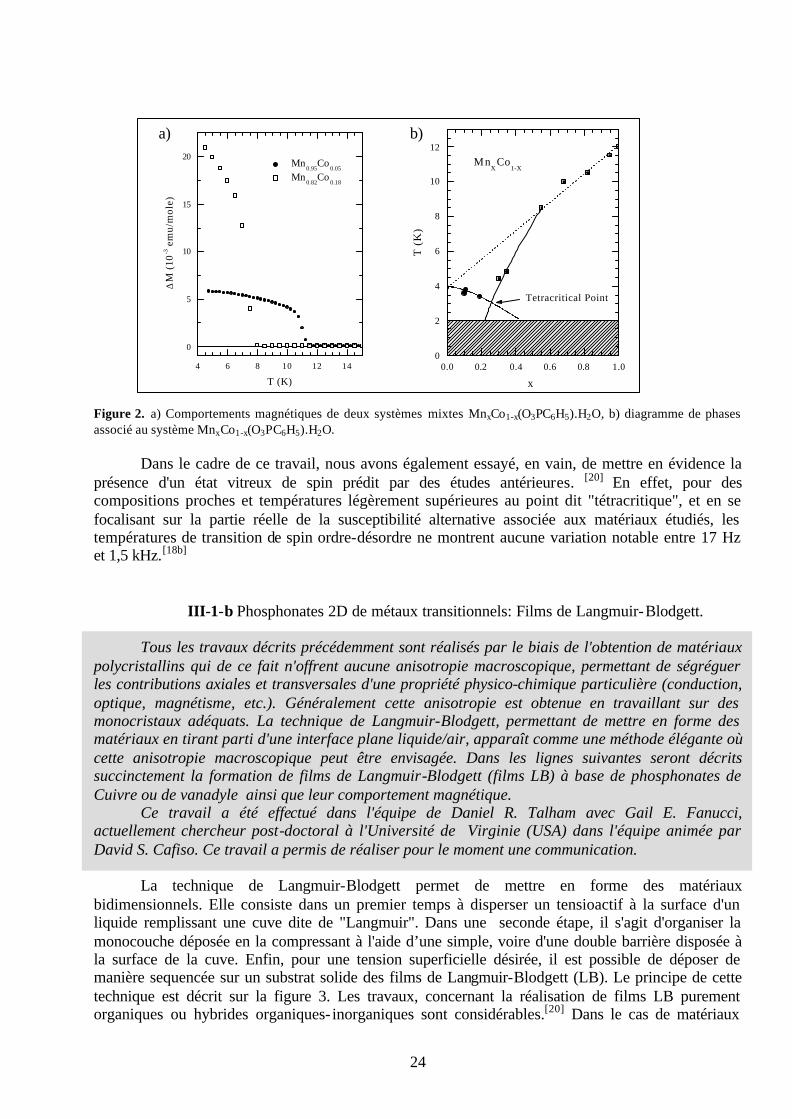
III-1-a Phénylphosphonates 2D de métaux transitionnels: Systèmes polycristallins. Toutes les propriétés des systèmes étudiés pendant mes années de thèse découlent d'entités organiques insérées ou intercalées, la partie inorganique ne jouant qu'un simple rôle de réseau hôte structurant. En fait, s'il est possible de contrôler les propriétés d'un matériau hybride organique-inorganique en se focalisant essentiellement sur la partie organique, nous pouvons également obtenir des matériaux à propriétés contrôlées en ne jouant qu'avec la partie inorganique. Cette démarche est décrite dans les lignes suivantes avec une étude portant sur des matériaux lamellaires à base de phénylphosphonates mixtes de Cobalt et de Manganèse.
Ce travail a été réalisé en collaboration avec Jeffrey T. Culp et Gail E. Fanucci, à l'Université de Floride (Gainesville-USA) dans l’équipe du Professeur Daniel R. Talham avec qui j'ai effectué mon stage post-doctoral. Les mesures magnétiques ont été effectuées par l'équipe du Professeur Marck W. Meisel, ainsi que toutes les mesures de magnétisme associées aux films de Langmuir-Blodgett et matériaux magnétiques à structures ouvertes qui seront décrits ultérieurement. Ces travaux ont permis la réalisation de deux articles et de deux communications.
Les phénylphosphonates de métaux transitionnels sont des matériaux lamellaires dont la
structure est bien référencée.[15] Des goupements phényls pointent dans l'espace interfolaire, alors que les entités phosphonates assurent une connectivité intrafeuillets via la présence de métaux de transition. Ces métaux de transition, lorsqu'ils présentent des électrons de valence non appariés, vont induire des propriétés magnétiques où les interactions de spin sont modulées par des ponts "oxo" inhérents aux groupements phosphonates.[15] L'idée de ce travail était d'associer Mn(II) et Co(II) dans la connectivité intrafeuillet, l'un et l'autre de ces éléments de transition correspondant respectivement à des modèles d'interaction de spin de types Heisenberg et Ising.[16] La répartition statistique de ces éléments transitionnels au sein de la structure permet de considérer les feuillets comme de véritables solutions solides.[17] Dans cette étude nous avons réalisé toute une série de composés répondant globalement à la stœchiométrie suivante : MnxCo1-x(O3PC6H5).H2O. Pour x étant égale à zéro nous obtenons le phénylphosphonate de Manganèse associé à une température de Néel TN(11,7K)[18a]
légèrement supérieure à T∗N (11,5K), cette dernière température caractéristique variant avec la force
du champ magnétique imposé. Ceci met en évidence un ordre faiblement ferromagnétique au-dessous de ces températures caractéristiques. Pour x étant égale à un, nous obtenons le phénylphosphonate de Cobalt caractérisé par un comportement antiferromagnétique associé à une température de Néel de 3,9K.[18b] Pour les systèmes mixtes Mn/Co, un exemple des comportements magnétiques est proposé sur la figure 2-a. Ces mesures ont été effectuées pour tous les composés réalisés et un diagramme de phase a pu être élaboré, indiqué sur la figure 2-b.[18] Ce diagramme met en évidence un écart à la théorie du champs moyen, avec un écart maximum pour la composition contenant 25% de Manganèse, ceci définissant un point "tétracritique".[17,19] Cette stœchiométrie de 25% en Manganèse apparaît en fait comme un seuil de percolation où, pour un réseau plan cubique centré, le Cobalt possède au moins un Manganèse comme premier voisin, ce dernier dominant l'environnement magnétique local.[18b]
24
Figure 2. a) Comportements magnétiques de deux systèmes mixtes MnxCo1-x(O3PC6H5).H2O, b) diagramme de phases associé au système MnxCo1-x(O3PC6H5).H2O.
Dans le cadre de ce travail, nous avons également essayé, en vain, de mettre en évidence la présence d'un état vitreux de spin prédit par des études antérieures. [20] En effet, pour des compositions proches et températures légèrement supérieures au point dit "tétracritique", et en se focalisant sur la partie réelle de la susceptibilité alternative associée aux matériaux étudiés, les températures de transition de spin ordre-désordre ne montrent aucune variation notable entre 17 Hz et 1,5 kHz.[18b]
III-1-b Phosphonates 2D de métaux transitionnels: Films de Langmuir-Blodgett. Tous les travaux décrits précédemment sont réalisés par le biais de l'obtention de matériaux polycristallins qui de ce fait n'offrent aucune anisotropie macroscopique, permettant de ségréguer les contributions axiales et transversales d'une propriété physico-chimique particulière (conduction, optique, magnétisme, etc.). Généralement cette anisotropie est obtenue en travaillant sur des monocristaux adéquats. La technique de Langmuir-Blodgett, permettant de mettre en forme des matériaux en tirant parti d'une interface plane liquide/air, apparaît comme une méthode élégante où cette anisotropie macroscopique peut être envisagée. Dans les lignes suivantes seront décrits succinctement la formation de films de Langmuir-Blodgett (films LB) à base de phosphonates de Cuivre ou de vanadyle ainsi que leur comportement magnétique. Ce travail a été effectué dans l'équipe de Daniel R. Talham avec Gail E. Fanucci, actuellement chercheur post-doctoral à l'Université de Virginie (USA) dans l'équipe animée par David S. Cafiso. Ce travail a permis de réaliser pour le moment une communication.
La technique de Langmuir-Blodgett permet de mettre en forme des matériaux bidimensionnels. Elle consiste dans un premier temps à disperser un tensioactif à la surface d'un liquide remplissant une cuve dite de "Langmuir". Dans une seconde étape, il s'agit d'organiser la monocouche déposée en la compressant à l'aide d’une simple, voire d'une double barrière disposée à la surface de la cuve. Enfin, pour une tension superficielle désirée, il est possible de déposer de manière sequencée sur un substrat solide des films de Langmuir-Blodgett (LB). Le principe de cette technique est décrit sur la figure 3. Les travaux, concernant la réalisation de films LB purement organiques ou hybrides organiques- inorganiques sont considérables.[20] Dans le cas de matériaux
4 6 8 10 12 14
0
5
10
15
20 Mn
0.95Co
0.05
Mn0.82
Co0.18
∆M
(10
-3 e
mu/
mol
e)
T (K)0.0 0.2 0.4 0.6 0.8 1.0
0
2
4
6
8
10
12
Tetracritical Point
MnX
Co1-X
T (
K)
x
a) b)

25
hybrides, il a été possible de recréer à l'interface liquide/air des structures organiques- inorganiques, récurrentes de celles existant à l'état solide, présentant des propriétés magnétiques analogues avec l'avantage d'obtenir des films et non plus des matériaux polycristallins.[21] C'est dans cette optique que s'inscrit ce travail, avec la réalisation de matériaux à base d'octadécylphospphonate de cuivre (Cu-ODPA) et de vanadyle (VO2+) (VO-ODPA).
En chimie du solide, les phosphonates de cuivre correspondent à plusieurs phases cristallographiques. Ces différences de structures sont fortement corrélées à leur état d’hydratation. Le composé Cu(R-PO3) non hydraté possède un ordre magnétique de spin à basse température.[22]
Sur la figure 4-a, nous pouvons constater des interactions antiferromagnétiques de spin associées à une aimantation spontanée et positive vers 8K (figure 4-b).
Figure 3. Schéma décrivant la technique de Langmuir-Blodgett.
Figure 4. Comportements magnétiques de spin de certains phosphonates de cuivre polycristallins. Cette aimantation spontanée est envisageable car les interactions de spin sont "antiferromagnétiques angulées" ou faiblement ferromagnétiques et induisent de ce fait un moment magnétique net positif. Sur la figure 5-b, nous pouvons constater que le composé octadécylphosphonate de cuivre (Cu-ODPA) présente également cette transition de phase magnétique vers 8K. Cette transition à basse température est dédoublée par une seconde transition à une température de l’ordre de 16K. Ces
Substrat hydrophobe
X n+
Xn+
X n+
PO3
PO32-
PO32-
PO3
PO3
PO3
O3P
2-O3P 2-O3P
O3P
O3P
X n+
XX
XX
XX
X
Région du ménisque
Monocouche de Langmuir
solution
PO32-PO3
2- PO32-PO3
2-PO 32-PO3
2-
PO32-
PO32-
X n+
X n+X n+ X n+
X n+X n+
Monocouche de Langmuirménisque
Substrat hydrophobe
X n+
Xn+
X n+
PO3
PO32-
PO32-
PO3
PO3
PO3
O3P
2-O3P 2-O3P
O3P
O3P
X n+
XX
XX
XX
X
Région du ménisque
Monocouche de Langmuir
solution
Substrat hydrophobe
X n+
Xn+
X n+
PO3
PO32-
PO32-
PO3
PO3
PO3
O3P
2-O3P 2-O3P
O3P
O3P
X n+
XX
XX
XX
X
Région du ménisque
Monocouche de Langmuir
solution
X n+
Xn+
X n+
PO3
PO32-
PO32-
PO3
PO3
PO3
O3P
2-O3P 2-O3P
O3P
O3P
X n+
XX
XX
XX
X
XX
XX
XX
X
Région du ménisque
Monocouche de Langmuir
solution
PO32-PO3
2- PO32-PO3
2-PO 32-PO3
2-
PO32-
PO32-
X n+
X n+X n+ X n+
X n+X n+
Monocouche de Langmuirménisque
PO32-PO3
2- PO32-PO3
2-PO 32-PO3
2-PO32- PO3
2-PO32-PO 3
2-PO32-
PO32-
PO32-
X n+
X n+X n+ X n+
X n+X n+
Monocouche de Langmuirménisque
4 6 8 10 12 14 160.00
0.05
0.10
0.15
T (K)
∆M =
Mfc
- M
zfc (
mem
u)
R = decyl
0 100 200 3000
4
8
12
16
χ (m
emu/
mol
)
T (K)
0 100 200 3000.0
0.2
0.4
0.6
χT (
emu
K /
mol
)
T (K)
R = ethyl R = hexadecyl
4 6 8 10 12 14 160.00
0.05
0.10
0.15
T (K)
∆M =
Mfc
- M
zfc (
mem
u)
R = decyl
0 100 200 3000
4
8
12
16
χ (m
emu/
mol
)
T (K)
0 100 200 3000.0
0.2
0.4
0.6
χT (
emu
K /
mol
)
T (K)
R = ethyl R = hexadecyla) b)

26
deux températures de transition de phases sont également présentes pour le film LB Cu-ODPA (figure 5-a). Figure 5. Comportements magnétiques de spin du composé à base d’octadécylphosphonate de cuivre, a) film LB, b) système polycristallin. Comme l’indiquent les diffractogrammes de rayons X, le système polycristallin est biphasé (figure 6). Ceci pourrait expliquer le comportement magnétique de spin observé. En effet, il a été démontré, que pour certains hydroxycarboxylates de cuivre à longues chaînes alkyles, des interactions de spin interfeuillets pourraient exister, si la longueur de cohérence des domaines magnétiques intrafeuillets est assez grandes pour induire des interactions de spin d’un feuillet à l’autre.[22d] Dans ce contexte, la distance interfeuillet associée à l’agencement des chaînes alkyles devient un paramètre pouvant générer de nouveaux comportements magnétiques. Figure 6. Diffractogrammes de rayons X des composés d’octadécylphosphonates de cuivre : film LB et composé polycristallin. Nous pouvons constater que le film LB Cu-ODPA possède une seule distance interfeuillet tout en étant cependant caractérisé par deux transitions magnétiques de spin, une première à 8K et une seconde pour une température supérieure de 21K (figure 5-a). Dans le cas du film (LB), nous avons donc un matériau biphasé au niveau de la structure intrafeuillet et non plus au niveau des distances interfeuillets. Dans une autre étude, nous avons également essayé de transposer les comportement s magnétiques de systèmes polycristallins à base d’ions vanadyles (VO2+) vers des films LB. Dans l’état solide deux matériaux présentent des interactions antiferromagnétiques. Dans le composé à base de phénylphosphonate de vanadyle les interactions de spin sont induites via les groupes phosphonates ((VO)-O-P-O-(VO)),[22a,b,c] alors que dans le cas du composé à base de méthylphosphonate de cuivre, les interactions d’échange sont obtenues via des ponts oxo inhérents aux groupes phosphonates (VO)-O-(VO).[22a,b,c] Nous avons donc élaboré un nouveau film LB à base
2 4 6 8 10 12 14 16 180
1000
2000
3000
4000
2θ
Cou
nts
Cu-ODP-LB Film
Cu-ODP-powder
2 4 6 8 10 12 14 16 180
1000
2000
3000
4000
2θ
Cou
nts
Cu-ODP-LB Film
Cu-ODP-powder
4 8 12 16 20 240.5
1.0
1.5
2.0
2.5
3.0
3.5
∆M
= M
fc -
Mzf
c (µ
emu)
T (K)
H par
H perp
0 5 10 15 20 25 300
10
20
30
∆M
= M
FC -
MZ
FC (
µem
u)
T (K)
4 8 12 16 20 240.5
1.0
1.5
2.0
2.5
3.0
3.5
∆M
= M
fc -
Mzf
c (µ
emu)
T (K)
H par
H perp
4 8 12 16 20 240.5
1.0
1.5
2.0
2.5
3.0
3.5
∆M
= M
fc -
Mzf
c (µ
emu)
T (K)
H par
H perp
0 5 10 15 20 25 300
10
20
30
∆M
= M
FC -
MZ
FC (
µem
u)
T (K)
a) b)

27
d’octadécylphosphonate de cuivre, en espérant retranscrire ces comportements magnétiques. Le film LB (VO-ODPA) ne montre pas de comportement magnétique mettant en avant des interactions d’échange. En effet, l’évolution de sa susceptibilité magnétique en fonction de la température est reproduite par une loi de Curie (figure 7-a) et sa courbe d’aimantation en fonction d’une augmentation d’un champ magnétique imposé est caractéristique d’une fonction de Brillouin (figure 7-b). Ces deux comportements, mettent en évidence l’existence de spin 1/2 sans interaction. De plus, en jouant sur l’anisotropie macroscopique offerte par la nature des films LB, l’évolution de la largeure de raie à mis hauteur du signal RPE, caractéristique des ions V4+ (sonde paramagnétique locale) en fonction de l’orientation de l’échantillon, ne montre pas la signature caractéristique d’un échange de spin.
Figure 7. Comportement magnétique d’un film LB à base d’octadécylphosphonate de vanadyle.
III-2 CRISTAUX, INGENIERIE SUPRAMOLECULAIRE ET PROPRIETES MAGNETIQUES DE SPIN.
Dans les travaux décrits précédemment, les matériaux étudiés étaient soit polycristallins, soit
mis en forme par le biais d'une interface air/liquide, ceci dans le cas particulier des films LB. Cette organisation contrôlée d’édifices à base moléculaire se retrouve également dans un thème récent à la frontière de la chimie de coordination. Cet axe de recherche repose sur une organisation prédéterminée, et de ce fait contrôlée, de l'agencement moléculaire au sein d'un motif unité caractéristique d'une maille cristallographique. S'agissant d'organisation contrôlée de briques molécules nous sommes bien en phase avec les concepts de chimie supramoléculaire, décrits en introduction. Cependant cette chimie, s'adressant dans ce cas à la réalisation de cristaux à structures prédéterminées, préfère parler de : "cristaux obtenus par ingénierie supramoléculaire".[23] Ceci débouche sur une nouvelle philosophie d'appréhender la chimie de coordination. En effet, il ne s'agira plus de rechercher une dualité structure-propriété où la structure est une finalité en soit, mais bien d'envisager en priorité une spécificité des propriétés souhaitées dont devra découler dans une seconde étape, un agencement cristallographique à caractère supramoléculaire. C'est dans ce contexte que s'inscrivent les travaux décrits dans les deux paragraphes suivants. Ces travaux ont été réalisés à l'Université de Floride, dans l'équipe de Daniel R. Talham, principalement dans le cadre de la thèse de Jonathan WoodWard (soutenue le 10 Juillet 2002).[24]
Ces travaux ont, pour le moment, permis de réaliser deux articles et une communication.
0 1 2 3 4 5
0
1
2
3
4
5
6M
(mem
u)
B (T)0 50 100 150 200 250 300
0
50
100
150
200
M (
µem
u)
T (K)
0 30 60 90 120 150 180
120
160
200
240
∆Hp-
pθ(degrees)
T = 2K
0 1 2 3 4 5
0
1
2
3
4
5
6M
(mem
u)
B (T)0 50 100 150 200 250 300
0
50
100
150
200
M (
µem
u)
T (K)
0 30 60 90 120 150 180
120
160
200
240
∆Hp-
pθ(degrees)
T = 2K
a) b) c)
0 1 2 3 4 5
0
1
2
3
4
5
6M
(mem
u)
B (T)0 50 100 150 200 250 300
0
50
100
150
200
M (
µem
u)
T (K)
0 30 60 90 120 150 180
120
160
200
240
∆Hp-
pθ(degrees)
T = 2K
0 1 2 3 4 5
0
1
2
3
4
5
6M
(mem
u)
B (T)0 50 100 150 200 250 300
0
50
100
150
200
M (
µem
u)
T (K)
0 30 60 90 120 150 180
120
160
200
240
∆Hp-
pθ(degrees)
T = 2K
a) b) c)
0 1 2 3 4 5
0
1
2
3
4
5
6M
(mem
u)
B (T)0 1 2 3 4 5
0
1
2
3
4
5
6M
(mem
u)
B (T)0 50 100 150 200 250 300
0
50
100
150
200
M (
µem
u)
T (K)
0 50 100 150 200 250 3000
50
100
150
200
M (
µem
u)
T (K)
0 30 60 90 120 150 180
120
160
200
240
∆Hp-
pθ(degrees)
T = 2K
0 1 2 3 4 5
0
1
2
3
4
5
6M
(mem
u)
B (T)0 1 2 3 4 5
0
1
2
3
4
5
6M
(mem
u)
B (T)0 50 100 150 200 250 300
0
50
100
150
200
M (
µem
u)
T (K)
0 50 100 150 200 250 3000
50
100
150
200
M (
µem
u)
T (K)
0 30 60 90 120 150 180
120
160
200
240
∆Hp-
pθ(degrees)
T = 2K
a) b) c)

28
L'acquisition des données cristallographiques a été réalisée avec l'aide précieuse de Khalil Abboud, Professeur à l'Université de Gainesville.
III-2-a Matériaux 3D à structures ouvertes.
Pour les raisons énoncées ci-dessus, la conception rationnelle de matériaux hybrides organiques- inorganiques à structures ouvertes connaît actuellement un essor considérable. Ceci étant induit par le formidable potentiel de propriétés contrôlées associées à cette catégorie de matériaux. Nous pouvons citer par exemples des propriétés d'échanges ioniques,[25] propriétés d'insertions (réseaux hôtes-molécules invitées),[26] propriétés de catalyse,[27] propriétés d'optique non- linéaire[28]
ou bien encore de magnétisme[29] ou de conductivité électronique.[30] Dans ce cadre nous avons réalisé trois nouveaux matériaux à structure ouverte correspondants aux stœchiométries suivantes: [Ni(4,4´-bipy)3(H2O)2](ClO4)2·1.4(4,4´-bipy)·3(H2O) [1], [Co(4,4 -bipy)3(H2O)2](ClO4)2·1.4(4,4´-bipy)·3(H2O) [2],et [Cu(4,4´-bipy)3(DMSO)2](ClO4)2·2(4,4´-bipy) [3].[31] Les composés 1 et 2 iso-structuraux ont été obtenus par synthèses en conditions hydrothermales. Le matériau 3 à été obtenu par cristallisation en solution de diméthylsulfoxyde. La structure globale de ces matériaux est hiérarchisée où l'unité de base est une chaîne à connectivité iono-covalente, ces chaînes sont connectées les unes aux autres via des liaisons hydrogènes et interactions π (induites par le caractère aromatique des molécules de 4,4' bipyridine) pour former des feuillets. Ces feuillets présentent des cavités hydrophobes, l'empilement des feuillets et la présence de ces cavités, génèrent la formation de canaux. La stabilité structurales de ces matériaux est associée à la présence de molécules invitées (DMSO, 4,4' bipyridine et contre- ions perchlorates). Quelques représentations de la structure du composé 1 sont proposées sur la figure 8.
Figure 8. Quelques représentations de la structure du composé : [Ni(4,4´-bipy)3(H2O)2](ClO4)2·1.4(4,4 -bipy)·3(H2O). Groupe d’espace = C2/c, a = 17.5698 Å, b = 11.4101 Å, c = 24.479 Å, β = 93.064 º, V = 4900.4 Å3, Z = 4.
Le composé 3 n’est pas iso-structural de 1 et 2, quelques représentations cristallographiques sont données sur la figure 9.
c
b
a
b
c
b
c
b
a
b

29
Figure 9. Quelques représentations de la structure du composé : [Cu(4,4´-bipy)3(DMSO)2](ClO4)2·2(4,4 -bipy). Groupe d’espace = Cc, a = 19.0931 Å, b = 11.1949 Å, c = 25.607 Å, β = 94.810 °, V = 5454.0 Å3, Z = 4.
La première différence structurale majeure existant ente le composé 3 et les deux homologues 1 et 2, est la présence de diméthylsulfoxyde dans la première sphère de coordinence du cuivre. La seconde différence de taille, est l’absence de liaisons hydrogène inter-chaînes pour le matériau 3, et enfin les cycles aromatiques des molécules de bipyridine coordinant le cuivre sont bien moins co-planaires et caractérisés par un angle de distorsion de 61,4°. Il existe également quelques différences mineures (angles de liaison et distances inter-atomiques) sur lesquelles nous ne nous étendrons pas dans ce document.[24]
Nous nous sommes intéressés aux propriétés magnétiques de ces matériaux. L’ensemble de leurs propriétés magnétiques sont similaires mettant en avant de faibles interactions d’échange entre centres métalliques pour les composés 1 et 2 , et pas d’interaction au-dessus de 2K pour le composé 3. Un exemple de comportement magnétique pour le composé 1 est proposé sur la figure 10. Figure 10. Comportement magnétique du composé [Ni(4,4 -bipy)3(H2O)2](ClO4)2·1.4(4,4´-bipy)·3(H2O).
Le comportement de la susceptibilité magnétique molaire en fonction de la température peut être simulé en utilisant un modèle de champ moléculaire d’ions isolés, corrigé par le modèle de Watanabe, introduisant un paramètre d’anisotropie.[x] Nous obtenons une constante de couplage J/kB égale à –0.42K, signifiant de faibles interactions antiferromagnétiques entre centres métalliques. La courbe d’aimantation à 2K nous permet de déterminer un paramètre axial de structure fine (D/kB) égal à 6,74 K. Le comportement magnétique du composé 1 est indiqué sur la figure 11. La valeur de la susceptibilité paramagnétique molaire prise à la température ambiante est de 1,19 10-3 emu/mol. Cette valeur expérimentale corrobore tout à fait celle attendue (1,33 10-3 emu/mol) pour des centres métalliques à spin ½ sans interaction. La dépendance en température de la susceptibilité molaire à 1000G, s’ajuste parfaitement à une loi de Curie et la dépendance en champ de l’aimantation est très
c
b
a
b
c
b
a
b
a) b)a) b)

30
bien ajustée par une fonction de Brillouin. Ces deux comportements suggèrent que les cent res métalliques sont non couplés, même à une température de 2K.
Figure 11. Comportement magnétique du composé [Cu(4,4´-bipy)3(DMSO)2](ClO4)2·2(4,4 -bipy). Dans un tout autre contexte, les cavités intrinsèques à ces matériaux leur confèrent des propriétés d’inclusion. Ces propriétés d’insertion ont été testées avec des molécules sondes, de types triphénylphosphine (TPPO), triméthylphosphine (TMPO) et caractérisées qualitativement par RMN du 31P. Ce travail ne sera pas traité dans ce document, mais constitue une partie de la thèse Jonathan Woodward (thèse soutenue en Juillet 2002) et de Bhavin Abadjaru (thèse en cours à l’Université de Floride, sous la direction de Cliford Bowers, Professeur à l’Université de Gainesville).
III-2-b Matériaux en échelles.
Une autre problématique abordée, centrée sur le comportement magnétique de composés cristallins, traite de la synthèse et caractérisation de matériaux en échelles (échelles atomiques et/ou de spin). Dans cette problématique, il faut associer une délocalisation de spin à une dimensionnalité de structure limitée et contrôlée. Il faut donc utiliser des ligands adéquats jouant un rôle de briques moléculaires, permettant d’induire ou d’inhiber l’extension du polymère de coordination. Plusieurs briques moléculaires peuvent être employées pour s’orienter vers une réalisation spécifique de matériaux en échelles. Dans le cadre de ces travaux nous nous sommes essentiellement intéressés aux ligands azidure (N3
-) et ter-pyridine. Les cristallisations ont lieu en solvant polaire (DMSO) en utilisant le sel Cu(ClO 4)2.5H2O. En fonction des conditions de synthèse et en jouant principalement avec le rapport [Cu(II)/N3
-] deux types de matériaux en échelle, ont été obtenus avec comme motifs de base un tétramère et pentamère de Cuivre nommés respectivement C4 et C5. Ces deux unités de base sont décrites dans la figure 12.
a) b)
2K
a) b)
2K

31
Figure 12. Unité de base des composés a) [Cu2(terpy)2-µ2-(N3)4][Cu2-µ2-(N3)2(N3)2], (C4) et b) [Cu2(terpy)2-µ-(N3)2(N3)2][Cu3-µ-(N3)4(N3)2], (C5). La figure 13-b met bien en évidence une structuration en échelle du composé C4, les échelles s’alignant le long de l’axe cristallographique a. La connectivité entre "jambes" et "barreaux" est assurée par des monomères [Cu(terpy)(N3)2], les barreaux des échelles sont associés aux dimères [Cu2(N3)4]. Ces échelles sont agencées dans le plan cristallographique (a,b) et forment ainsi des feuillets. La figure 13-a met en évidence une légère inter-pénétration des échelles mitoyennes par le biais d’interactions π-π des cycles aromatiques inhérents aux molécules de ter-pyridine. Dans ce composé il est important de noter l’absence d’ion perchlorate, qui de ce fait ne participent pas à l’électroneutralité du composé.
Figure 13. Quelques représentations de la structure du composé [Cu2(terpy)2-µ2-(N3)4][Cu2-µ2-(N3)2(N3)2] C4. Groupe d’espace = P21/n, a = 14.4872 Å, b = 7.1430 Å, c = 18.454 Å, β = 95.719 °, V = 1900.2 Å3, Z = 4.
Le matériau C5 est également un polymère de coordination structuré en échelle (figure 14).
Cette structuration en échelle est basée sur une superposition de pentamères [Cu5(terpy)2(N3)10]. La connectivité entre "jambes" et "barreaux" est assurée par des monomères [Cu(terpy)(N3)2]. Les unités pentamèriques se superposent suivant l’axe cristallographique a. L’agencement des échelles induit une structure en feuillets coplanaires au plan (a,c) (figure 14-a). Nous pouvons constater que dans ce cas également les échelles mitoyennes sont interpénétrées, cette caractéristique est induite par des interactions π-π propres aux cycles aromatiques des entités ter-pyridine.
a
b
b
c
a) b)
a
b
b
c
a) b)
a) b)

32
Figure 14. Quelques représentations de la structure du composé [Cu2(terpy)2-µ-(N3)2(N3)2][Cu3-µ-(N3)4(N3)2] C5. Groupe d’espace = P-1, a = 6.6035 Å, b = 12.660 Å, c = 13.110 Å, α = 88.682 º, β = 76.278 º, γ = 82.819 º, V = 1056.4 Å3, Z = 2.
Les détails cristallographiques des composés C4 et C5 ne seront pas mentionnés dans ce document (distances inter-atomiques, angles de liaisons), seuls quelques détails seront donnés pour expliciter simplement leur comportement magnétique.
Figure 15. Comportement magnétique du composé [Cu2(terpy)2-µ2-(N3)4][Cu2-µ2-(N3)2(N3)2 C4
Nous pouvons constater que la susceptibilité magnétique molaire du composé C4 augmente quand la température décroît. Cette phénoménologie est identique que ce soit pour des mesures avec, ou sans champ appliqué. Ceci met en évidence l’absence d’un ordre magnétique de spin de la température ambiante à 2K. L’inverse de la susceptibilité magnétique portée en fonction de la température met en évidence un comportement dominant de caractère antiferromagnétique (l’extrapolation des données hautes températures sur l’axe des abscisses indique une température négative égale à –46,32 K) (figure 15-b). La décroissance de ?M*T en fonction de la température (de 300 K à 50 K) (figure 15-c) confirme le caractère antiferromagnétique des interactions dominantes. Au-dessous de 5 K une seconde diminution de la valeur de ?M*T est observée (avec une pente plus importante que précédemment), mettant ainsi en évidence de faibles interactions
b
c
a
c
a) b)
b
c
a
c
a) b)
a)
c)
b)
d)
a)
c)
b)
d)

33
antiferromagnétiques au sein du matériau C4. La courbe d’aimantation à 2 K (figure 15-d) indique une augmentation de l’aimantation avec la valeur du champ appliqué, nous pouvons observer le début d’une seconde aimantation vers 40 kG. Pour le composé C4 l’interaction dominante antiferromagnétique est celle induite par les dimères [Cu2(N3)4] constitutifs des barreaux de l’échelle. Cependant les angles de liaison Cu-N-Cu égaux à 103°3 devraient induire une interaction d’échange ferromagnétique. En effet il existe un angle limite de 108°5 (angle d’orthogonalité où les contributions antiferromagnétiques et ferromagnétiques s’annulent mutuellement et au-dessous duquel les interactions sont annoncées ferromagnétiques).[32] Les raisons de cette anomalie associée aux interactions d’échanges intra-dimère [Cu2(N3)4] ne sont pas bien claires pour le moment. Cependant des calculs ab-initio reliant la structure de ce composé à son comportement magnétique sont en cours d’étude via une collaboration avec Mike Whangbo, Professeur à l’Université de Caroline du Nord. Les interactions dimères-monomères entre le dimère central [Cu2(N3)4] et les deux monomères adjacents [Cu(terpy)(N3)2] assurant la connectivité "barreaux-jambes" sont faibles mais non négligeables. Les interactions inter-tétramères, malgré de faibles distances Cu-N (jambes de l’échelle), sont nulles. Ceci s’explique par la valeur de l’angle Cu-N-Cu (107°1) très proche de la condition d’orthogonalité (108°5). De ce fait le composé C4 ne peut pas être considéré comme un matériau à échelles de spin. Le comportement magnétique du composé C5 est proposé sur la figure suivante.
Figure 16. Comportement magnétique du composé [Cu2(terpy)2-µ-(N3)2(N3)2] [Cu3-µ-(N3)4(N3)2] C5. Comme C4 le composé C5 ne présente pas d’ordre magnétique à longues distances et ceci pour une gamme de température allant de la température ambiante à 2K. L’inverse de la suceptibilité magnétique en fonction de la température intercepte (par extrapolation des valeurs à haute température) l’axe des ordonnées à une température positive de 47 K (fig.16-b). Ceci suggère que les interactions dominantes de spin sont ici de caractère ferromagnétique. Ce comportement aux hautes températures est également confirmé avec la pente positive de la courbe χM*T reportée en fonction de la température (fig.16-c). Nous observons sur cette courbe un maximun vers 6,5K (2,48 emu K mol-1). Au-dessous de cette température de 6,5K le produit χM*T décroît en fonction de la
a) b)
c) d)
a) b)
c) d)

34
température ceci traduisant la présence d’interactions antiferromagnétiques faibles. La courbe d’aimantation augmente avec la valeur du champ imposé et commence à saturer vers 30 kG en atteignant une valeur seuil de 26000 emu G mol-1. Dans ce composé trois types d’échanges ont été considérés : l’unité centrale Cu3N6 de chaque pentamère est supposée induire les interactions de super-échange dominant le comportement magnétique du matériau, le couplage entre le trimère central et les deux monomères terminaux (Cu(terpy)(N3)2 et enfin le couplage inter-pentamères. Le couplage trimère-monomères est médié via des groupements azidures en site de coordination basal et apical respectivement pour un cuivre terminal et un cuivre central, cette configuration ne permettant pas de bons couplages. Les interactions inter-pentamères sont médiées par de longues liaisons Cu-N associées à des ponts azidures asymétriques de types tête-tête et tête-bêche, cette configuration ne permet pas de bons échanges de spin.
Le composé C5 peut donc être considéré comme un empilement de trimères en interactions faibles. Aux hautes températures C5 possède un comportement paramagnétique, les spins non appariés associés à chaque pentamère ne sont pas couplés et sont distribués aléatoirement le long de l’échelle. Lorsque la température décroît, le couplage intrapentamère peut avoir lieu car l’énergie thermique devient insuffisante pour peupler les états exités. Une augmentation du produit χM*T est observée au début des interactions d’échange de spins ferromagnétiques induites par un angle Cu-N-Cu de 101°7. On remarque deux pentes bien distinctes, mettant peut être en avant deux constantes de couplages différentes. A la température de 6,5 K l’état fondamental S= 5/2 est complètement peuplé, le composé C5 consiste alors en un empilement de pentamères à interactions ferromagnétiques. A plus basses températures, la décroissance du produit χM*T annonce les interactions faibles inter-pentamères.
Ces comportements magnétiques ont été modélisés dans la thèse de Jonathan Woodward (University of Florida), ces études ne seront pas traitées dans ce document. III-3 MONOCOUCHES DE LANGMUIR ET FILMS de LANGMUIR-BLODGETT:
DEUX SYSTEMES STRUCTURANTS.
Ces travaux se focalisent principalement sur la formation d’entités inorganiques aux interfaces de substrats organiques bidimensionnels inhérents aux monocouches de Langmuir et films de Langmuir-Blodgett. Nous nous sommes intéressés à la formation de systèmes continus hybrides organiques-inorganiques à base de phospholipides et complexes de métaux transitionnels, mais également à la formation de particules métalliques et minérales. Dans ces trois cas de figures, les méthodologies employées ainsi que les techniques de caractérisations utilisées sont brièvement décrites sur la figure 17.

35
Figure 17. Schématisation des méthodologies employées et techniques de caractérisation utilisées.
III-3-a Films de Langmuir-Blodgett Phospholipidiques: RMN 31P Dans les travaux décrits précédemment, les problématiques associées à l'obtention d'une matière organisée hybride organique-inorganique n'intéressent essentiellement que les communautés propres aux chimistes et physico-chimistes. L'étude présentée dans cette partie s'adresse aux chimistes et physico-chimistes, mais de plus trouve des intérêts pratiques pour les biologistes et biochimistes. En effet, ce travail a permis d'obtenir les premiers films LB structurés à base de phosphatidylsérine (DPPS) et stabilisés par des cations de métaux transitionnels Pt(II), Cd(II), Pd(II), Au(III).[33] Dans ce contexte, la RMN devient un outil indispensable pour évaluer la structure et au-delà, toutes interactions d'une membrane phospholipidique associée à d'autres composantes propres au monde du vivant, Cholestérol, protéines etc. Pour opérer avec succès, il faut à la fois ne pas dénaturer l'aspect membranaire de l'objet étudié tout en obtenant une quantité de manière raisonnable permettant d'obtenir un signal RMN satisfaisant. Pour atteindre ce double objectif, il est fait appel à des dépôts de membranes sur des supports de mica, ces supports étant accolés les un aux autres afin d'augmenter la quantité de matière organique à analyser. Ce procédé est assez long, demande une très grande minutie et dans le meilleur des cas n'empêche pas une dégradation partielle des structures étudiées. Ce travail a été effectué dans l'équipe de Daniel R. Talham avec Gail E. Fanucci. La spectroscopie RMN a été réalisé par Riqiang Fu, Chercheurs au "National High Magnetic Field Laboratory" de Tallahassee en Floride. Cette étude a permis la réalisation d’un article.
En règle générale la déposition séquencée de bicouches phospholipidiques par utilisation de la technique LB est une tâche très difficile et seulement quelques succès ont été obtenus avec des dérivés de la molécule de phosphatidyléthanolamine.[34] Cette difficulté de transférer verticalement
- Microscope optique- MET- DET- MEB- IRTF- DRX- XPS
Etudes sur films de Langmuir-BlodgettEtudes sur monocouches de Langmuir
- Microscope optique- BAM
Ex-Situ :
- Pression de surface
Caractérisations In-Situ :
- MET- MEB- IRTF
Caractérisations Ex-Situ :
- Microscope optique- MET- DET- MEB- IRTF- DRX- XPS
Etudes sur films de Langmuir-BlodgettEtudes sur monocouches de Langmuir
- Microscope optique- BAM
Ex-Situ :
- Pression de surface
Caractérisations In-Situ :
- MET- MEB- IRTF
- Microscope optique- BAM
Ex-Situ :
- Pression de surface
Caractérisations In-Situ :
- MET- MEB- IRTF
Ex-Situ :
- Pression de surface
Caractérisations In-Situ :
- MET- MEB- IRTF
Caractérisations Ex-Situ :

36
sur supports solides ces entités phospholipidiques est attribuée à de faibles interactions de leurs parties polaires ou hydrophiles, interactions qui parfois même sont de nature répulsive.[35] Dans le but d'améliorer ces interactions et donc de permettre une structuration en bicouches, des cations calcium et uranyle ont été utilisés pour stabiliser la partie hydrophile et anionique de l'acide dipalmitolphosphatidique.[35] C'est dans ce même effort que se situe notre étude avec comme entité amphiphile la molécule de dipalmitolphosphatidysérine (DPPS).[33] Pour stabiliser les interactions intermoléculaires à l'interface eau/air différents sels métalliques ont été dissous en solution aqueuse: K2PdCl4, .K2PtCl4, K2Pt(CN)4, KAuCl4 et Cd(ClO 4)2. Par spectroscopie infrarouge à transformée de Fourrier (IRTF), nous pouvons évaluer le caractère homogène de chaque transfert en suivant les intensités relatives des modes normaux de vibration associées aux élongations symétriques et asymétriques des groupements (CH2) comme l'indique la figure 18.
Figure 18. Evolution des intensités des vibrations d’élongations symétriques et asymétriques en fonction du nombre de bicouches déposées.
Par une étude de spectroscopie infrarouge polarisée, en se focalisant sur les vibrations
d'élongations caractéristiques des groupements (CH2), l'angle des chaînes alkyles avec la perpendiculaires au substrat a été évalué à 39°. Le caractère bidimensionnel des matériaux transférés est révélé par diffraction des rayons X, avec une distribution caractéristique des harmoniques définissant une distance interfoliaire de 56Å. Les stœchiométries des matériaux déposés ont été déterminée par XPS, pour le film hybride DPPS-Pt(II), sur lequel nous avons centré notre étude, la composition suivante a été obtenue: [(DPPS-)]2Pt(II)(H20)](DPPS)4. De toutes ces analyses, résumées ci-dessus, une configuration possible de l'agencement des phospholipides au sein des films a pu être avancée (figure 19).
0 5 10 15 200,0
0,1
0,2
0,3
0,4
3000 2950 2900 2850 2800
0,00
0,15
0,30 νa(CH2) ν
s(CH
2)
Abs
orba
nce
Nombre de bicouches
νa(CH
2)
νs(CH
2)
Abs
orba
nce
Nombre d'ondes (cm-1)

37
Figure 19. Représentation schématique des bicouches phospholipidiques associées au système [(DPPS-)]2Pt(II)(H20)](DPPS)4.
Suivant cette stratégie de mise en forme, nous avons pu réaliser jusqu'à cent dépositions de
bicouches phospholipidiques organisées, ayant ainsi assez de matière organique pour obtenir un spectre RMN du 31P (figure 20).
Figure 20. Spectre RMN 31P du composé [(DPPS-)]2Pt(II)(H20)](DPPS)4.
Ce spectre RMN MAS en cross-polarisation 1H/31P a été obtenu en soixante-douze heures.
Nous pouvons déceler deux signaux associés aux résonances isotropes suivantes: δ iso=-2,9±0,2 ppm et δiso=-7,7±0,1 ppm (avec comme référence NH4H2PO4). La présence de ces deux signaux met en évidence la présence de deux types de DPPS. Le signal donnant un déplacement à -2,9 ppm est à la même position, possède la même symétrie en ayant le même nombre d'harmoniques ou "spinning side bands" que la poudre de DPPS initiale. Ceci implique qu'une grande partie des entités de DPPS se trouve sous une forme identique à celle du zwittérion phospholipidique de départ. Le signal plus
55.3 Å
6.4 Å
5 Å
16 Å
5.5 Å
39º
37º
55.3 Å
6.4 Å
5 Å
16 Å
5.5 Å
39º
37º
O
O O
O O O
O O P
-
NH 2 COOH
Pt 2+ O H 2
O H 2
O
O O
O O O
O O P
- COOH
O
O O
O O O O
O P
-
COOH NH 3 + N H 3 +
N H 3 +
O
O
O O
O O
O O P
-
COOH
O
O
O O
O O O O P -
NH 2
COOH NH 3 +
O
O
O O
O O O O
P -
C O O H
55.3 Å
6.4 Å
5 Å
16 Å
5.5 Å
39º
37º
55.3 Å
6.4 Å
5 Å
16 Å
5.5 Å
39º
37º
O
O O
O O O
O O P
-
NH 2 COOH
Pt 2+ O H 2
O H 2
O
O O
O O O
O O P
- COOH
O
O O
O O O O
O P
-
COOH NH 3 + N H 3 +
N H 3 +
O
O
O O
O O
O O P
-
COOH
O
O
O O
O O O O P -
NH 2
COOH NH 3 +
O
O
O O
O O O O
P -
C O O H

38
petit centré à -7,7 ppm est caractéristique d'une autre forme de DPPS en interaction avec le cation transitionnel Pt2+. En effet, il a été démontré que la présence de ce signal est caractéristique d'une augmentation de la structuration des phospholipides, cette organisation résultant sans doute des interactions phospholipides-complexes inorganiques.[36]
III-3-b Monocouches de Langmuir et procédé de biominéralisation.
Ce travail s’inscrit dans une thématique frontière entre la mise en forme de minéraux via une interface organique et la médecine, en particulier le domaine de la néphrologie, science s’intéressant aux problématiques liées à la formation de calculs rénaux. Ces calculs rénaux sont en fait des matériaux hybrides organiques-inorganiques où coexistent des substances minérales (Oxalate de calcium, Phosphate de calcium) associées à des entités organo-biologiques (phospholipides, lipides, protéines, cholestérol, carbohydrates, etc.). Des exemples de calculs rénaux sont proposés sur la figure 23. Dans ce contexte, certains chercheurs spécialistes de néphrologie ont émis une hypothèse où les structures organiques pouvaient servir de sites de nucléation préférentiels pour une entité minérale en croissance.[37] Cette nucléation hétérogène pouvant être exaltée ou inhibée par différents facteurs explicités dans les lignes suivantes, mais relevant tous d’une problématique de spécificité de réactivité aux interfaces organo-minérales. [37,7a]
Ces travaux ont été réalisés aux Etats-Unis, dans l’équipe de Daniel R. Talham, en collaboration avec Saeed R. Khan, Professeur à l’université de Floride (College of Medecin - Shands hospital), spécialistes des maladies rénales et ont permis la réalisation de quatre articles, un proceeding et cinq communications.
Figure 23. a) Calculs rénaux composés à 97% d’oxalate de calcium mono-hydraté et 3% de composés organique, b)calculs rénaux composés de 69% d’oxalate de calcium mono-hydraté, 28% de phosphate de calcium, c) quelques exemples de tailles critiques associées aux calculs rénaux.
Dans ce contexte, les monocouches de Langmuir apparaissent comme des systèmes simples
où les paramètres impliqués, dans cette phénoménologie de nucléation hétérogène aux interfaces organo-minérales, peuvent être ségrégués et étudiés. Dans une première étude[x] trois phospholipides différents ont été étudiés, avec comme spécificité la charge de leur tête polaire. Ces phospholipides sont les suivants : Dipalmitolphosphatidylglycérol (DPPG), dipalmitolphosphatidylcholine (DPPC) et dipalmitolphosphatidylsérine (DPPS), le caractère anionique de leur tête polaire évoluant comme tel : DPPG> DPPS=DPPG.
a) b) c)

39
Nous nous sommes en particulier intéressés à la formation d’oxalate de calcium monohydraté (COM) CaC2H2.H2O, ce minéral étant le constituant inorganique majoritaire des calculs rénaux. Dans toutes les études suivantes, trois types de morphologies sont obtenues en fonction du type de facettes nucléées et de l’état d’agrégation des cristallites attachés aux monocouches, ces différences sont indiquées sur la figure 24.
Figure 24. Différentes morphologies des cristaux de COM : a) face (10-1), b) face (010), c) agrégats.
Nous avons ensuite comptabilisé le nombre et distingué les types de cristaux attachés aux monocouches en utilisant la microscopie électronique à balayage (MEB), ceci pour des solutions sursaturés en oxalate de calcium à concentration différentes RS5 et RS10.[x] Nous avons observé une plus grande quantité de cristaux nucléés sous la monocouche de DPPG par rapport aux monocouches de DPPC et DPPS. Nous avons pu démontrer l’influence spécifiques des ions calcium sur l’agencement des monocouches phospholipides. Les ions Ca2+ jouent en fait un rôle "liant" entre la monocouche organique et le réseau inorganique en croissance.[38] Les monocouches de DPPG possédant un fort caractère anionique auront une interaction préférentielle avec les ions calcium, optimisant ainsi la formation d’oxalate de calcium monohydraté. Nous avons également remarqué une cristallisation à caractère hétérogène, où, dans tous les cas, un maximum de cristaux est observé avec la face (10-1) attachée aux monocouches. Ceci est expliqué par le fait que cette face (10-1) est riche en ions calcium.[x] Cette première série d’étude a permis de mette en évidence l’importance des densités de charge anionique (nature phospholipidique) sur les phénomènes de nucléation hétérogène aux interfaces entre cristaux de COM et monocouches phospholipidiques.
Dans une seconde étude, nous nous sommes intéressés aux effets de l’organisation d’une monocouche (structure phopsholipidique) sur les phénomènes de nucléation.[39] Pour ce faire, quatre phospholipides de même charge anionique (phosphatidylglycérol) mais avec des parties hydrophobes différentes, ont été utilisés (dipalmitolphosphatidylglycérol (DPPG), dimiristolphosphatidylglycérol (DMPG), oléole-palmitolphosphatidylglycérol (POPG), dioléolphosphatidyl-DL-glycérol (DOPG). Les comportements globaux sont représentés sur la figure 25. Trois analyses peuvent être menées de front : quatre phospholipides à une pression de surface de 20 mN/m, quatre phospholipides à une surface moyenne de 95 Å2/molécule et enfin l’étude de l’effet de ces deux conditions limites sur un même surfactant. Dans ce dernier cas, on observe une augmentation du taux de nucléation aux faibles tensions superficielles. Ceci est induit par une augmentation de la fluidité des monocouches qui peuvent alors accommoder préférentiellement leurs paramètres de maille à ceux de l’espèce minérale en croissance. Ceci correspond à une épitaxie, mais s’agissant de matière molle c’est le substrat qui s’accommode au réseau minéral en croissance et non l’inverse. Ce phénomène, d’exaltation du taux de nucléation, est accru pour le DPPG. Il faut noter que pour une surface moyenne de 95 Å2/molécule la monocouche DPPG possède une structure biphasée où s’associent une phase très désorganisée (ana logue à l’état gazeux) et une phase un peu moins désorganisée (analogue à un état liquide).
a) b) c)

40
Figure 25. Taux de nucléation associés aux différents phospholipides utilisés dans deux conditions limites (tension superficielle de 20 mN /m et Surface moyenne de 95 Å2/molécule.
Une observation de la structure de la monocouche par microscopie à angle de Brewster
(figure 26), nous a permis de mettre en évidence un autre paramètre important.
Figure 26. Image de Brewster caractéristique d’une monocouche de DPPG à une tension superficielle de 95 Å2/molécule. On constate en effet qu’une nucléation préférentielle a lieu aux interfaces entre les deux phases citées précédemment. En fait, cette interface spécifique de la monocouche de DPPG agit comme une zone de défauts qui exalte le taux de nucléation.
Lors de ces études quatre paramètres de nucléation hétérogène ont été globalement identifiés : Structure, nature, fluidité et défauts inhérents aux types de monocouches étudiés. Ces travaux mettent en évidence une action partitive de ces paramètres et non pas coopérative.[39] Dans ce contexte, des études similaires ont été menées avec adjonction de cholestérol, qui, au-deçà d’une concentration seuil, limite le taux de nucléation.[40] Ces études sont à poursuivre en y associant d’autres composés biologiques comme des polysaccharides, protéines, etc. D’autre part, dans ces études, la morphologie des cristaux ainsi observés est caractéristique de la méthode utilisée qui, par nature, est de caractère bidimensionnelle. Ceci implique une certaine différence de morphologie entre les calculs rénaux (fig. 23) et les cristaux de COM observés dans nos études (fig. 24). Dans le
20 40 60 80 100 120 140 160 180
0
10
20
30
40
50
61
15
11
0
(DPPG)
(DMPG)
(POPG)
(DOPG)
0
19
27
175
(POPG)
(DOPG)
(DMPG)
(DPPG)
Nombre de cristaux par mm2
Pres
sion
de
surf
ace
(mN
/m)
Surface moyenne (Å2/molécule)

41
a) b) c) d)
but d’améliorer la part "bio-mimétique" de nos études, des travaux sont envisagés avec des structures "oignons" où entités organique et inorganique seront cette fois-ci réellement imbriquées (partie IV-1d2) de ce document).
III-3-c Films de Langmuir-Blodgett: nano- et microparticules métalliques.
Dans cet axe de recherche nous essayons de déterminer comment l'espace interfoliaire de films de Langmuir-Blodgett peut à la fois induire la formation de particules métalliques et diriger leur croissance par effet de confinement. Dans ce cadre le système organique apparaît comme un microréacteur jouant le double rôle de structurant et de stabilisant pour une entité inorganique en croissance. Ces travaux ont été réalisés à l'Université de Floride, dans l'équipe de Daniel R. Talham en collaboration avec James Adair, professeur à l'Université dePennsylvanie et la N.A.S.A. pour des études de "mise en forme" sous conditions de microgravité. Ces travaux ont permis la réalisation d’un proceeding et cinq communications. La nano-structuration d’entités inorganiques par le biais de phases structurantes conna ît depuis une vingtaine d’années un essor considérable. Cet intérêt est induit par la miniaturisation croissante des composants électroniques mais également à un effet quantique de taille générant des comportements physico-chimiques exotiques.[41] Dans le but d’améliorer le contrôle des procédés de morphogenèse associés à la réalisation de nano-objets, plusieurs types de systèmes moléculaires organisés ont été utilisés : membranes,[42] micelles,[43] vésicules,[44] monocouches de Langmuir et films de langmuir-Blodgett.[45] Dans ce dernier exemple, nous nous sommes intéressés à la formation de nano-particules de platine en utilisant différents surfactants à fonctionnalités spécifiques : benzyldiméthylstéarylammonium (BDSA) (ammonium quaternaire), l’octadécylamine (ODA) (amine primaire), dipalmitophosphatidylsérine (DPPS) (zwittérion phospholipidique) et hexadécylaniline (HDA) (amine primaire polymérisable). L’apport de platine émane du complexe [PtCl4]2-, l’étape de réduction est catalysée par irradiation sous lampe UV (lampe au mercure de 450 W). En plaçant les échantillons à une distance de dix centimètres de la source UV, nous obtenons des nanoparticules, représentées sur la Figure 27.
Figure 27. Images MET des nanoparticules métalliques associées aux films LB à bases de : a) DPPC, b) DPPS, c) HDA et d) ODA. Nous observons que ces nano-objets (80-100 nm) ont une morphologie dans tous les cas sphérique avec une forte tendance àl’aggrégation. Nous n’avons pas d’objet anisotrope plat, que pourrait induire la bidimensionnalité des films LB. De plus, des caractérisations par XPS et diffraction de rayons-X par transmission mettent en évidence la présence de platine zéro-valent pour les systèmes Pt/ODA, Pt/HDA, Pt/DPPS, alors que le système Pt/BDSA est associé à la présence de différents oxydes de platine. Ceci s’explique par la présence d’amines primaires, pour les films LB à

42
bases de ODA, HDA et DPPS, qui se déprotonnent pendant l’étape de réduction du Pt(II) stabilisant ainsi les nanoparticules métalliques néoformées. Les molécules de BDSA possèdent des ammoniums quaternaires n’offrant pas cette spécificité. Des études quantitatives montrent que très peu de nano-objets sont synthétisés à partir de DPPS, ceci étant du à la faible interaction entre sa partie zwitterionique polaire et le complexe [PtCl4]2-.
Dans une seconde série d’expériences, nous avons rapproché les films LB à 1 centimètre la source UV, ce qui a pour conséquence d’augmenter légèrement la température associée aux réactions de réductions, qui passe de 25°C à 45°C. Les particules élaborées dans ces conditions sont présentées sur la figure 28.
Figure 28. Images MEB des particules métalliques associées aux films LB à bases de : a)BDSA, b) HDA, c) DPPSA et d) ODA. Nous pouvons donc constater que les particules sont globalement de tailles micrométrique. Dans le cas où des films LB à base de BDSA sont utilisés, nous obtenons des particules colloïdales, tout comme précédemment sous forme de différents oxydes métalliques. Dans le cas des films LB constitués à partir de DPPS, nous observons les plus petits objets qui restent de tailles nanoscopiques, la faible interaction entre sa tête polaire et le complexe de platine utilisé lors de l’étape de transfert ne permettant pas un apport de matière conséquent, inhibant de ce fait la réalisation d’objets micrométriques. Pour les films LB à base d’HDA, nous observons des particules de platine à morphologie chaotique. Des études couplées de RX et d’IRTF mettent en évidence une polymérisation de l’aniline (IRTF analogue au sel d’éméraldine) avec le traitement UV, ayant pour conséquence de dénaturer la structure 2D du film LB. Enfin, pour le système ODA/Pt, nous observons bien des objets anisotropes à morphologie en feuillets caractéristique induite par la structure bidimensionnelle inhérente aux films de Langmuir-Blodgett.
III-4 SYSTEMES BIDIMENSIONNELS, MESOPHASES LYOTROPES ET MISE EN FORME AUX GRANDES ECHELLES.
Dans les travaux cités précédemment les propriétés physico-chimiques des matériaux finaux pré-existent dans une des deux entités de départ qu’elle soit organique ou inorganique (conductivité, ONL, magnétisme de spin et nucléaire). Il est possible de réaliser des hybrides organiques-inorganiques où la propriété physico-chimique résultante émane de l’association des entités de départ sans être pré-indiquée dans aucun de ces composants. Dans ce cadre nous sommes intéressés à la formation de mésophases lyotropes en partant de systèmes polycristallins de basse dimensionnalité et en les dispersant dans un solvant approprié. Ce travail a été réalisé dans l’équipe de Daniel R. Talham, sur une collaboration engagée avec Clément Sanchez directeur de recherches CNRS (LMMC, UMR-CNRS, Université de Paris VI). Cette étude a permis la réalisation d’un article et de quatre communications. (une thèse est en cours à Gainesville sur ce sujet)
a) b) c) d)

43
Les cristaux liquides sont le plus souvent de composition purement organique. L’alignement préférentiel aux grandes échelles des unités constitutives est lié à la présence d’une anisotropie de forme. Dans la plupart des cas, s’agissant de phases organiques une entité mésogène est associée à un corps rigide à caractère poly-aromatique.[46] Leurs propriétés thermotrope et/ou lyotrope se révèlent par l’apparition de magnifique textures observées en microscopie sous analyseurs et polariseurs croisés.[47] Ces textures sont donc biréfringentes et doivent répondre à un stimula externe (champs magnétique, élongation mécanique, etc.) par un alignement des domaines biréfringents avec le champ de force appliqué. Ces comportement ont été également observés au début du XXéme siècle pour des systèmes minéraux.[48] Récemment ces études sont redevenues à l’ordre du jour avec l’observation et l’interprétation de comportements "cristaux liquides" associés aux phases V2O5,[49]
aux phases de Chevrel échangées au lithium,[50] aux structures bohémite [51] etc.[52] Ce comportement s’explicite, brièvement, par une dispersion de particules minérales anisotropes et hydrophiles dans un solvant polaire, ceci, au-delà d’une concentration seuil. Pour limiter les forces de répulsions inter-particules et augmenter la concentration seuil décrite précédemment, il est possible d’enrober des particules minérales par un polymère hydrophobe et de disperser ces composites dans un solvant non polaire comme le toluène. L’apparition des premiers systèmes hybrides organique- inorganique à comportement "cristal liquide" n’est que très récente.[53] C’est dans ce contexte que s’établit l’axe de recherche décrit dans les lignes suivantes.
Notre étude est basée tout d’abord sur la réalisation de nouveaux hydroxy-carboxylates de cuivre, cobalt et nickel polycristallins, dont les propriétés magnétiques de spin sont très bien référencées.[x] Par ailleurs, des dispersions d’hydroxy-stéarate de cuivre dans du toluène à différentes fractions massiques sont représentées sur la figure 29.
Figure 29. Dispersions d’une poudre d’hydroxystéarate de cuivre à différentes fractions massiques. a) Sans polarisateurs croisés, b) avec polariseurs croisés. Nous observons donc des systèmes biphasés (dipersion-solvant) pour les faibles pourcentages massiques, au-delà de 2% massique un gel se forme. Comme l’ind ique les clichés en polariseurs croisés, suspension et gel ont des propriétés biréfringents, sans doute caractéristique du système polycristallin de départ. Une observation au microscope optique en polariseurs croisés révèle une texture et un comportement assez intéressants, comme l’indique la figure 30.
0.5 % m
2,0 % m
a) b)
0.25 % m 1% m 2,5% m 0.5 % m

44
Figure 30. Observation d’un gel d’hydroxystéarate de cuivre/toluène (2% massique) en microscopie à polarisateurs croisés. a) système de départ (grossissement X 20), b) gouttes biréfringents observées à une température légèrement inférieure au point éclaircissant (grossissement x 40), c) systèmes après recuit (grossissement X 20), d) système de départ étiré entre lame et lamelle (grossissement X 20). La première constatation observable sur le système de départ est une association de zones biréfringentes (fig. 30a). Ces zones biréfringentes ont un comportement thermotrope et sont associées à un point de clarification à 68°C. Au-dessous de ce point éclaircissant les premiers domaines biréfringents réapparaissent sous la forme de petites gouttelettes (fig. 30b). D’autre part, un recuit induit une augmentation de taille des domaines biréfringents (fig. 30c). Finalement, ce système peut être étiré entre lame et lamelle (fig. 30d). L’ensemble de ce comportement est analogue à certains cristaux liquides de caractéristique nématique.[47] Nous avons également observé, sous loupe binoculaire entre polariseurs croisés, une augmentation de la biréfringence en compressant un gel entre deux lames de microscope à l’aide d’une spatule (figure 31).
Figure 31. Observation d’un gel d’hydroxystéarate de cuivre/toluène (2,5% massique) sous loupe binoculaire à polariseurs croisés. a) sans pression, b) avec une légère pression inférieure au point éclaircissant, c) avec une pression forte. La barre correspond à 3 mm. Cette expérience met en évidence une augmentation de la biréfringence, et donc une augmentation de l’agencement préférentiel des domaines sur une grande échelle de l’ordre du millimètre. D’autre part, après recuit et en se replaçant au dessous de la température du point éclaircissant, par trempe rapide jusqu’à température ambiante nous sommes parvenus à figer l’agencement des particules d’hydroxycarboxylate de cuivre (figure 32). Nous remarquons que les particules anisotropes sont alignées le long d’un axe préférentiel, comme le serait la répartition d’entités anisotropes dans un cristal liquide classique "nématique"[46]
a)
b)
c) d)
a) b) c)

45
Figure 32. Agencement des particules d’hydroxystéarate de cuivre observées par MEB après recuit.
La phénoménologie décrite ci-dessus n’est pas associée à une augmentation de l’espace
interlamellaire du composé d’hydroxystéarate de cuivre. De ce fait, ce comportement n’est pas dû à un gonflement des feuillets par diffusion de toluène. Un autre comportement intéressant est la formation de films translucides auto-supportés à partir de gels décrits précédemment, déposés goutte à goutte sur une feuille de Mylar et laissés sécher à l’air (Figure 33).
Figure 33. a) Particules d’hydroxystéarate de cuivre observées par MEB, b) arrête d’un film d’hydroxystéarate de cuivre observée par MEB montrant l’agencement préférentiel des particules, c) films d’hydroxystéarate de cuivre déposé sur une feuille de papier, d) films d’hydroxystéarate de nickel déposé s ur une feuille de papier. Ce comportement est récurrent pour un ensemble de composés, associés aux hydroxycarboxylate de cuivre.[53b] Nous avons également essayé d’étendre cette phénoménologie à d’autres systèmes lamellaires, comme les pérovskites et hydroxydes doubles lamellaires (fig. 34). D’autre part sur un plan plus phénoménologique, la figure 29 met en évidence des systèmes biphasés correspondant à un équilibre entre un solvant et une dispersion. Ceci est une distinction par comparaison aux systèmes minéraux étudiés à l’heure actuelle, où est observé un équilibre biphasique entre une solution isotrope et une phase biréfringente.[53a] Ceci implique l’existence d’une force attractive supplémentaire associée aux matériaux décrits dans ce document qui inhibe une transition de premier ordre "solution isotrope-phase biréfringente". Ceci pourrait être dû à une interaction hydrophobe entre les chaînes alkyles constitutives des composés. Cette hypothèse reste à vérifier. Ce travail est actuellement en cours d’étude par Eduardo E. Perez-Cordero dans le cadre de sa thèse dirigée par Daniel R. Talham. Les travaux concernant les hydroxydes doubles lamellaires ont été réalisés par Sarah Laine, dans le cadre de son "Master degree" co-encadré avec Daniel R. Talham et sont repris dans sa thèse qu’elle effectue au sein de la même équipe.
a) b) c) d)

46
Figure 34. a) Images obtenus en microscopie entre polariseurs et analyseurs croisés (grossissement X20). a) Pérovskite (C6H13NH3)2CuCl4 , b) (C6H13NH3)2CuCl4 après étirement entre lame et lamelle, c) HDL Zn0.74Cr0.26(OH)2(CH3(CH2)11SO4)0.29.0.65 H2O, d) Zn0.74Cr0.26(OH)2(CH3(CH2)11SO4)0.29.0.65 H2O après étirement entre lame et lamelle.
a) b)
c) d)

47
IV- RECHERCHES ACTUELLES.
IV-1 BIOMIMETISME, BIOTECTONIQUE et BIOMINERALISATION.
Figure 35. Illustrations des thèmes abordés au CRPP, articulés autour de la matière organisée hybride organique-inorganique.
La synthèse de matériaux inorganiques par utilisation de procédés de biominéralisation
connaît aujourd’hui un essor considérable. Cette systématique s’inspire des processus de biominéralisation propres au “monde du vivant”.[5-7] Dans ces systèmes biologiques, l’association de solutions minérales sursaturées (ou de monomères minéraux polymérisables) et de surfaces organiques à caractère membranaire permet la réalisation de systèmes organo-minéraux hiérarchiquement structurés. Ces structurations et texturations impliquent une organisation étape par étape des matériaux, allant de l’échelle atomique à celle du macroscopique ; la structure de type abalone illustre parfaitement cette organisation. Un défi offert aux scientifiques est la réalisation en laboratoire de telles architectures par mimétisme des modèles de construction moléculaire que nous inspire le "monde du vivant". A travers ces diverses approches nous essayons de contrôler les structures, textures et morphologies (aux grandes échelles) des objets organiques-inorganiques réalisés au C.R.P.P.
A cette volonté de réaliser des biomatériaux "sur mesure" nous associons une approche plus fondamentale visant à identifier et expliciter les mécanismes associés à la morphogenèse de ces architectures. Dans ce cadre, des systèmes modèles sont utilisés comme par exemple les monocouches de Langmuir et monocouches auto-assemblées sur substrats.
Ces travaux de recherches sont menés au C.R.P.P. et reposent sur l’activité de thème
Matiére organiséehybride
organique-inorganique
Mousses et minéralisation
Minéralisation et cisaillementNanoparticules métalliques
et polymères poreux
Nanoparticules métalliques etphases "ognons".
Minéralisation en émulsions
Défauts hydrodynamiques et minéralisation
Matiére organiséehybride
organique-inorganique
Mousses et minéralisation
Minéralisation et cisaillementNanoparticules métalliques
et polymères poreux
Nanoparticules métalliques etphases "ognons".
Minéralisation en émulsions
Défauts hydrodynamiques et minéralisation
Matiére organiséehybride
organique-inorganique
Matiére organiséehybride
organique-inorganique
Mousses et minéralisation
Minéralisation et cisaillementNanoparticules métalliques
et polymères poreux
Nanoparticules métalliques etphases "ognons".
Minéralisation en émulsions
Défauts hydrodynamiques et minéralisation

48
"biomimétisme, biotectonique et biominéralisation". Le Centre de Recherche Paul Pascal possède une solide expertise en réalisation et caractérisation de la matière molle (cristaux liquides, émulsions, mousses, et toutes autres structures de fluides complexes en règle générale). Répondant à une requête de son comité d’évaluation, le CRPP a créé une interface entre la matière molle et la chimie douce dans le but de développer de nouveaux thèmes de recherches où la matière molle servira d’empreinte à un composé inorganique en croissance. Ce nouveau thème de recherches, précédemment cité est effectif depuis septembre 2001, année de mon recrutement et est supporté par une jeune équipe polyvalente comprenant les personnalités suivantes : Philippe Poulin (Chargé de Recherches CNRS), Annie Colin (chargée de recherches CNRS), Sébastien Manneville (Chargé de Recherches CNRS), Chrystel Faure (Maître de conférences, Université Bordeaux I), Serge Ravaine (Maître de conférences, Université Bordeaux I), Olivier Mondain-Monval (Professeur, Université Bordeaux I), Cécile Zakri (Maître de conférences, Université Bordeaux I). Les centres d’intérêts développés en commun sont globalement représentés sur la Figure 35 et ont permis de réaliser, pour le moment, cinq brevets et 6 communications.
S’agissant de recherches actuelles certains de ces axes sont assez bien développés, d’autres le sont moyennement et certains ne le sont pas du tout, avec dans ce dernier cas, l’avantage d’y penser !
IV-1-a Morphogenèses minérales et fluides complexes
La mésostructuration de polymères inorganiques est un procédé extrêmement développé surtout depuis la découverte des MCM-41.[54] Dans ce cadre, l’utilisation de systèmes moléculaires organisés est une approche versatile et attrayante car elle débouche sur un très bon contrôle des tailles de pores et de la répartition de ces derniers au sein du squelette minérale. Les applications de tels objets à porosité contrôlée sont considérables et on peut citer à titre d’exemple: catalyse[55], extraction en phase solide et filtration[56], électronique, optique, acoustique[57-60] etc.
Figure 36. a) Photographie en microscopie optique d’une coque de silice obtenue en émulsion inverse, b) Photographie en microscopie optique entre polariseurs et analyseurs croisés de plusieurs capsules. La biréfringence des parois reflète une organisation régulière de la porosité du minéral.
Toutefois, il est nécessaire de pouvoir structurer et texturer les systèmes obtenus sur des
échelles plus grandes (de l’ordre du micron à plusieurs dizaines de microns) pour espérer ajouter une spécificité à leurs applications (relargage contrôlé de produits pharmaceutiques ou décontamination en absorbant des hydrocarbures). Dans cette optique nous utilisons une interface eau-huile par le
a) b)

49
biais d’une émulsion inverse (eau dans de l’huile). Une approche similaire a été proposée en 1996 par Schacht et al. [61] où les auteurs utilisent une émulsion directe (huile dans eau).
Figure 36a. Photographie en microscopie électronique à balayage de capsules minérales. Les échantillons sont mis sous vide pour l'observation. La mise sous vide et l’effet de charge électronique provoquent l'ouverture des coques (flèche de sur la figure b).
Figure 38. a) Diffractogramme de rayons X. La présence du pic reflète les corrélations spatiales entre les pores de la coque minérale. Nous pouvons déduire du vecteur d'onde correspondant au maximum d'intensité du pic de diffraction que la distance entre les pores est d'environ 4 nm. Aucun harmonique n’est observé, et ceci met en avant la caractère vermiculaire de la mésostructure, b) image de microscopie électronique à transmission.
Sur la figure 37 nous pouvons observer des objets sphériques et creux de diamètres compris
entre 2 et 50 µm. La biréfringence des objets observés sur la figure 36 reflète une organisation de la mésostructure associée aux coques minérales. Cette organisation est confirmée par mesures de diffraction de rayons X (fig. 38-a). La mésostructure vermiculaire a été confirmée par microscopie électronique en transmission (fig. 38-b). Des études récentes de Giulia Fornasieri (stagiaire post-doctorale au CRPP), nous ont permis de corréler diamètres et épaisseurs des coques (figure 39).
a) b)
0,10 0,15 0,20 0,25 0,30 0,35 0,40 0,45 0,50
240
260
280
300
320
340
360
380
I (a.
u.)
Q (Å-1)
a) b)

50
Figure 39. Relation entre diamètre des coques et épaisseurs des parois. La relation linéaire décrite dans la figure ci-dessus est importante. En effet, dans le cas d’utilisation d’émulsions inverses monodisperses nous connaîtrions les corrélations existant entre tailles des gouttes utilisées et épaisseurs de parois associées aux objets formés in fine. Ces travaux sont en cours d’étude.
Egalement, nous sommes parvenus à transposer cette méthodologie à la réalisation de coque de TiO2, avec l’émergence d’une phase Anatase après traitement à 600°C.
Figure 40. a) Coque de TiO2 observe en microscopie optique sous polariseurs et analyseurs croisés b) coques de TiO2 cassée observées par MEB, c) diffractogramme de RX avant et après traitement à 600°C (les raies caractéristique de la structure Anatase sont étoilées).
Des études sont en cours pour mieux contrôler la paroi des coquilles de TiO 2 en limitant l’hydrolyse du monomère utilisé par des effets chélatants. Nous nous focalisons également sur un meilleur contrôle de la mésostructure de ces coquilles de SiO 2 et TiO2. A terme, nous envisageons la réalisation de systèmes mixtes SiO 2-TiO2.
IV-1-b Morphogenèses minérales sous cisaillement.
Des procédés de morphogenèses minérales sous cisaillement sont également étudiés au laboratoire. Dans cette approche un stress mécanique sous cellule de Couette se substitue à une
0 10 20 30 40 50 60
0,2
0,4
0,6
0,8
1,0
1,2
1,4
1,6
1,8
2,0
2,2
2,4
2,6
épai
sseu
r des
par
ois
diametre des coques
0
100
200
300
400
0 5 10 15 20 25 30 35 40 45 50 55 60
2 Teta
Coun
ts
* *
*
*

51
macrostructure organique ou à un procédé d’émulsification (système thermodynamique métatstable). Dans ce contexte les matériaux hiérarchisés néoformés possèdent des morphologies anisotropes et exotiques. Cette approche mécanique de mise en forme aux grandes échelles ouvre de nouvelles voies de synthèse où la vitesse de cisaillement contrôle et dirige les processus de nucléation associés aux morphogenèses d’objets minéraux en croissance.
Figure 41. Mise en forme de silice mésoporeuse par cisaillement en cellule de Couette sous deux vitesses.
La mise en forme de matériaux minéraux en croissance peut être induite par l’effet d’empreinte d’un système moléculaire organisés (surfactants sous formes de micelles cylindriques, tubulaires, organisées en structures hexagonales, cubiques ou bien encore vermiculaires). La texturation aux grandes échelles émanant de cet effet "moulant" doit être forcément associée à un stress mécanique (induit par les structures caractéristiques du fluide réactionnel) extrinsèque au système hybride organique-inorganique en croissance. Le contrôle de la structure d’un fluide induite par l’agitation d’un barreau aimanté (fluide lamino-turbulent) est une tâche assez ardue. Les vitesses de cisaillement en cellule de Couette, peut être une manière élégante de contrôle de l’effet structurant d’un fluide sur un objet en croissance. Par exemple, sur la figure 41, nous pouvons constater que la morphologie, à grande échelle, d’un objet silicaté mésostructuré (présence de bromure de cétyltriméthylammonium) peut variée en passant d’un flux laminaire (figure 41-a) à un flux turbulent (figure 41-b). Ceci met d’ores et déjà en évidence le caractère coopératif existant entre l’effet texturant induit par la mésostructure et celui imposé par la structure du fluide réactionnel. Ces études viennent de commencer au CRPP et sont très prometteuses. A terme, différentes mésostructures (effet de chiralité, par exemple) seront associées à différentes structures de fluides (laminaires ou turbulents).
IV-1-c Minéralisation et mousses
Les mousses air-liquides sont des systèmes thermodynamiques et métastables dont la stabilité
et les comportements rhéologiques sont étudiés au laboratoire depuis plusieurs années. A mon arrivée au CRPP, avec ma collègue Annie Colin, nous avons envisagé la minéralisation contrôlée de ces systèmes. Cette approche constitue actuellement la base du travail de thèse de Florent Carn.
15 µm 15 µm
a) b)

52
Les mousses sont des systèmes thermodynamiquement métastables (coalescence d’Oswald et gravité détériorent leur texture) structurées par l’intermédiaire de bords de Plateau, nœuds et films. Ces mousses peuvent servire d’empreintes à un minérale en croissance. Des études ont commencé et ont permis d’obtenir toute une palette de morphologies en jouant simplement sur le pH des réactions sol-gel (figure 42).
Figure 42. Mousses de silice obtenues sous différents pH réactionnels.

53
Nous pouvons donc constater qu’un contrôle des procédés de chimie sol-gel permet d’aboutir à des texturations contrôlées. Nous pouvons également minéraliser complètement bords de Plateau et films (figure 43).
a)Figure 43. a) Mousse et b) mousse complètement minéralisée. Le procédé de moussage utilisé permet également de contrôler le rapport de taille entre bords de plateaux et films. Ceci est développé à l’heure actuelle. La silice est un matériau inerte pouvant être utilisé comme substituant osseux. Dans ce contexte des études de bio-compatibilité sont prévues. A terme, nous nous tournerons également vers la minéralisation de mousses où l’hydroxyapatite sera utilisé comme support inorganique. Ces synthèses seront associées à des caractérisations des micro-, méso- et macrostructure. Les propriétés de porosité et de tenues mécaniques seront également envisagées.
IV-1-d Minéralisation et phases lamellaires
IV-1-d1- Minéralisation sur monocouches organiques
Dans un contexte de Biotectonique et biomimétisme, les monocouches de Langmuir apparaissent comme des modèles simples permettant de mimer les interfaces des membranes cellulaires (bicouches phospholipidiques). Au-delà de la volonté de réalisation de nouveaux matériaux, nous nous intéresserons à la compréhension des mécanismes intimes de nucléation et de croissance aux interfaces organiques- inorganiques. Un certain nombre de paramètres ont été identifiés :
1- Densité de charge anionique ou cationique à l’interface “monocouche/espèce en croissance”. 2- Structure de la monocouche (la nucléation se résume-t-elle à une simple épitaxie?) 3- Fluidité des monocouches (la fluidité permet aux monocouches d’accommoder leurs paramètres de maille à ceux de l’espèce en croissance: commensurabilité) 4- Présence de défauts inhérents aux monocouches ou générés pendant le processus de nucléation.
A cette systématique permettant d’induire, d’appréhender et d’élucider les phénomènes de nucléation et de croissance aux interfaces, seront associées des études centrées sur des monocouches auto-assemblées sur substrat (SAMs). Dans ce cadre la fluidité est inhibée et seule la phénoménologie liée à l’épitaxie est exploitée.
a) b)

54
IV-1-d2 Minéralisation en phases oignons
Les études de rhéophysique menées au CRPP ces dernières années ont permis la mise au point d'un procédé de préparation d'une phase de vésicules multilamellaires de tensioactifs et de lipides, baptisées "sphérulites" ou ognons.[66] Dans cette optique les sphérulites peuvent jouer le rôle de microréacteurs chimiques dans lequel la réaction chimique est confinée. la possibilité de synthétiser des nanoparticules à l’intérieure de ces structures a été démontreé.[67]
Dans ce cadre il s’agira d’étendre ces réalisations de matériaux hybrides organiques- inorganiques et d’y associer une caractérisation minutieuse permettant de mieux comprendre la spécificité des réactions aux interfaces. Dans ce contexte, par le biais d’un travail commun avec Chrystel Faure, les premières réalisations de nanoparticules d’or au sein de structures oignons ont été réalisées.
Globalement trois formulations à base de dilpamitoylphosphatidylcholine(DPPC)-simulsol, (DPPC)-monooléine et génamine. Les résultats obtenus sont résumés dans les lignes suivantes. Dans les trois cas, on hydrate les tensioactifs avec une masse identique d'une solution à 10-3 M de tétrachloro-orate(III) de potassium KAuCl4, afin que le complexe [AuCl4]- soit présent dans la phase lamellaire. Dans une seconde étape, la phase lamellaire est cisaillée manuellement donnant alors des "oignons". Ce principe de formation "d’oignons" par cisaillement d’une phase lamellaire lyotrope est très bien explicité dans la littérature et sera pas explicité dans ce document.[62]
Dans le cas du système à base de phosphatidylcholine et de simulsol, l’insertion du complexe entraîne une augmentation de la distance entre bicouches lipidiques de 10Å, le pas smectique passant de 55Å à 66Å comme le montrent des expériences de diffraction de rayons X aux petits angles. Le changement de couleur intervient également lorsque l'on disperse les oignons après cisaillement de la phase lamellaire dans une solution de KAuCl4- à 10-3M (figure 44). Notons, par ailleurs, que nous avons vérifié par microscopie à contraste de phase que les oignons sont intacts dans la dispersion et que, de ce fait la structure en vésicules multilamellaires n'est pas altérée par la formation des nano-objets.
Figure 44. Cellules de quartz où les onions ont été dispersés dans une solution de K+AuCl4
- (10-3M): a) Génamine + K+AuCl4
- deux heures après dispersion b) DPPC + K+AuCl4- deux heures après dispersion, C) DPPC + K+AuCl4
- juste après dispersion.
Cette coloration rouge-rubis, observée dans les trois formulations testées, est caractéristique de nanoparticules d’or et est associée à une bande plasmon se situant à 530 nm en UV-visible.[63] De plus nous n’observons aucune absorption vers 650 nm en spectroscopie électronique (figure 45). L’absence de cette
(a) (b) (c)(a) (b) (c)

55
absorption apporte deux informations importantes : les nanoparticules confinées dans la matrice organique ne sont pas agrégées, [64,63] les nano-objets synthétisés ont une faible anisotropie de forme.[63]
Figure 45 : Evolution des spectres d’UV visible d'une dispersion d'onions (PC/AuCl4-) dans AuCl4
- (10-3M) en fonction du temps. A t=0 la dispersion est achevée.
Ces spectres d’UV-visible mettent en évidence une diminution de la bande de transfert de charge associé à la consommation du complexe AuCl4- et l’augmentation d’une bande plasmon centrée à 530 nm caractéristique de la formation de nanoparticules d’or au degré d’oxydation zéro.[63]
Le potentiel rédox du couple AuIII/ Au0 est très élevé. De ce fait, la réduction sans catalyse par rayonnement UV pourrait être induit par oxydation d’une fonction alcool en une fonction acide. [64], mais aucune vibration caractéristique d’une fonction acide n’a été détectée par spectroscopie infra-rouge. Une autre explication pourrait être une diminution du potentiel redox du couple Au+III/Au0 par coordination bidendate des ions Au(III) par les groupements hydroxyles.[65] Il a été ainsi démontré que la réaction d’oxydo-réduction pouvait être possible dans ces conditions.
Figure 46. Image du système hybride organique-inorganique obtenu par cryo-TEM.
100 200 300 400 500 600 700 800 900 1000
La bande de transfert de charge caractéristique du complexe AuCl
4
-
n'est plus présente aprés deux heures
4 heures2 heures0
La bande plasmon à 530 nm caractéristique de l'Or zéro valent nanométrique augmente et reste stable aprés deux heures de dispersion
Abs
orba
nce
longueur d'onde (nm)

56
Le caractère métallique des nano-objets synthétisés est démontré directement par XPS. La valeur des bandes d’émission X des énergies associées aux électrons 4f5/2 et 4f7/2 respectivement à 87.9 eV et 84.1 eV est caractéristique de la présence d’or métallique.[63-65] Ces pics ne sont pas dédoublés et donc il n’y a pas d’oxydes co-formés ou de complexes non consommés associés aux matériaux finals hybrides organiques- inorganiques. Par ailleurs, nous devons souligner que ces pics ont été obtenus après qu'un décapage (quelques Angström) de l'échantillon a été effectué : ceci confirme que la majorité des nanoparticules sont confinées dans la phase organique. Ce confinement présumé a été confirmé par la technique de cryo-TEM (figure 46).
Dans le but d’améliorer les propriétés de confinement et de mise en forme, les compositions seront modifiées avec l’adjonction de molécules amphiphiles à caractère amine ou thio. Egalement, la formation de nanoparticules métalliques au sein de structures oignons constituent une première étape. Nous souhaiterions, à terme nous tourner vers la réalisation in situ de nanoparticules magnétiques (ferrites, maghémites). La minéralisation de ces phases structurantes est également envisagée par utilisation de silicates.
IV-1-e Systèmes poreux et réactivité Ce thème de recherche a été amorcé en 1998 via une collaboration entre Olivier Mondain-Monval et Hervé Deleuze (LCOO-Université Bordeaux-I). Cette double compétence "matière molle (émulsions) et chimie organique" a permis la réalisation de monolithes poreux organo-fonctionnalisés.[68] Lors de mon arrivée, nous avons décidé d’utiliser de tels supports et d’y associer des procédés de nucléations hétérogènes induisant une nanofonctionnalisation inorganique ou métallique. Ce travail s’inscrit dans la thèse d’Alexandre Desforges co-encadrée par Olvier Mondain-Monval et moi-même.
Dans nos études, nous utilisons pour le moment des monomères polymérisables comme le divinylbenzène, le styrène ou bien encore toutes associations des deux. Pour améliorer la porosité de cette gamme de matériaux nous pouvons utiliser des entités porogènes, permettant d’établir une sous-porosité intra-parois (Figure 47).
Figure 47. Photo de microscopie électronique à balayage (a) macro-structure poreuse, (b) sous-structure poreuse de parois .
(a) (b)(a) (b)

57
Dans une seconde étape, nous utilisons ces matrices organiques poreuses comme substrats de nucléation hétérogène, via des synthèses in-situ, d’entités inorganiques. Dans une première étude, nous nous sommes intéressés à la formation de nanoparticules métalliques. Ces synthèses sont réalisées en utilisant des solutions (aqueuses ou non) de sels de complexes de métaux de transition (KAuCl4, K2PtCl4, K2PdCl4, etc.) à différentes concentrations. La réduction des ions transitionnels est assurée par la présence de radicaux mis en évidence par RPE (∆Hpp d’une dizaine de Gauss typiquement). Ces réductions sont également catalysées par traitement UV. La valence nulle des nano-objets synthétisés est mise en évidence par XPS et est couplée par diffraction de RX en transmission.
Figure 48. Clichés MET a) d’un polyHIPE sans porogène à un grossissement *9000, b) d’un système hybride organique-métallique (Pd) à un grossissement *40000, c) d’un système hybride organique-métallique (Pd) à un grossissement *100000 Sur la figure 48, nous pouvons constater la taille nanométrique des objets générés au sein de la matrice d’accueil organique, également nous pouvons constater que la réduction est dirigée à l’interface polymère-solution. En fonction des concentrations de sels d’ions complexes utilisés, nous sommes parvenus à contrôler la taille des nano-objets ainsi élaborés, deux histogrammes sont proposés sur la figure 49.
Figure 49. Histogrammes des tailles caractéristiques des nano-objets synthétisés en fonction des conditions de synthèse utilisés. Nous avons donc réalisé des structures hybrides permettant de réaliser des réactions de catalyse hétérogène. En prenant une réaction de catalyse type (hydrogénation d’un alcène en alcane) nous avons comparé l’activité catalytique des objets hybrides synthétisés à celle d’un système
a) b) c)a) b) c)
0 5 10 1 5 2 0 25 30 35 400
10
20
30
40
50
Solvant: eau Méthode "radical" et 0.01M K
2PdCl
4
nom
bre
taille des particules ( nm)0 2 4 6 8 1 0 12 14 16 18 2 0 2 2 2 4 26 28 30 3 2 3 4 3 6 38 40
0
10
20
30
40
50
Solvant: eau/THF 1:1 Méthode "radical" et 0.01M K
2PdCl
4
nom
bre
taille des particules (en nm)

58
extrêmement employé en catalyse supportée (Pd/charbon). Nous pouvons constater que l’activité catalytique des matériaux présentés dans ce document est supérieure au système Pd/charbon (figure 50).
Figure 50. Activité catalytique de deux systèmes utilisés et comparés au système Pd/charbon.
D’autre part le système Pd/charbon dont la texture est une poudre très fine, ne permet pas la réalisation de catalyse cyclique. En effet, la poudre de Pd/charbon est très fine et s’agrège dans l’étape de séparation entre catalyseur et produit de catalyse. Les systèmes développés en ce moment, se présentant sous forme de monolithes, offrent la possibilité d’une ségrégation aisé où la texture du catalyseur n’est pas dénaturée dans l’étape de séparation ou filtration. Des réactions de catalyses cycliques peuvent donc être envisagées, comme l’indique la figure 51.
Figure 51. Catalyse supportée cyclique pour le système Pd/PHP (monolithe)
Nous constatons sur la figure 51, que le pourcentage de conversion ne diminue pas même après quatre cycles catalytiques. Nous observons même une légère augmentation de cette activité, très certainement lié à une meilleure imprégnation du solvant utilisé (et donc de l’espèce catalysée) avec le nombre de cycles imposés. A terme, nos efforts seront centrés sur la spécificité des réactivités aux interfaces. Les surfaces des matrices organiques pourront être modifiées par des groupements, amino, thio, pour induire une stabilisation accrue des nano-objets néoformés. Egalement en fonction des temps d’irradiation UV et des concentrations de sels utilisés, nous aimerions élaborer des films métalliques
0 20 4 0 600
20
40
60
80
100
t e m p s ( e n m i n )
Pd /C (poudre) P d / P H P ( m o n o l i t h e ) Pd /PHP (poudre )
quan
tité
d'al
cool
ally
lique
rest
ant (
%)
0 1 2 3 4 565
70
75
80
85
90
95
100
105
110
% d
e co
nver
tion
apré
s 20
min
utes
de
synt
hèse
Nombre de cycles catalytiques

59
recouvrant toute la surface interne du polymère poreux. Nous envisageons l’utilisation de polymères biocompatibles en y associant des synthèses minérales de carbonate de calcium ou bien encore d’hydroxy-apatite.

60
V- PERSPECTIVES GLOBALES Dans le cadre des recherches en cours et énoncées précédemment la première des perspectives, qui paraîtra raisonnable, sera de poursuivre les travaux commencés en se focalisant sur cette chimie des formes associant chimie douce et matière molle. Un aspect nouveau sera d’associer minéralisation et instabilités hydrodynamiques. En effet, Il existe dans la nature différentes façons d’organiser la matière. Les instabilités hydrodynamiques structurent l’espace d’un fluide. Lorsque l’on soumet un fluide à un gradient de température inversé, c’est à dire lorsqu’on chauffe le bas du fluide et que l’on refroidit sa partie haute, une instabilité de convection apparaît. Des rouleaux apparaissent dans le fluide. La taille de ces rouleaux est de l’ordre de grandeur de l’épaisseur du fluide.
Figure 52. Exemple de défauts hydrodynamique de type Rayleigh-Bénard. Nous souhaitons réaliser des réactions sol-gel dans ces écoulements, de façon à induire de nouvelles structures et textures, exotiques et contrôlées. L’ensemble des études de morphogenèse associant chimie douce et matière molle mettent en avant, pour le moment, un seul type de polymère inorganique, la silice en l’occurrence. Ce choix est dicté par des processus d’hydrolyse-condensation très bien documentés et faciles d’application au laboratoire. A terme, nous nous tournerons également vers des morphosynthèses de polymères inorganiques complètement nouveaux. Dans une première approche nous essaierons de développer de nouvelles connectivités des polyèdres de base dans la familles des aluminophopshate. Les mécanismes impliqués dans la génèse des aluminophosphates sont encore mal explicités, ceci étant dû à une cinétique de condensation assez rapide qui ne permet pas de séquencer aisément les différentes étapes condensation-hydrolyse associées aux divers processus de croissance cristalline. Néanmoins, la communauté scientifique impliquée dans cet art particulier de la chimie du solide s’efforce de comprendre et de rationaliser les processus réactionnels. Cette démarche étant réalisée par le biais de synthèses en milieux quasi non-aqueux qui permettent de diminuer les cinétiques de condensation, tout en réalisant par ailleurs de nouveaux matériaux. Cette conceptualisation des mécanismes réactionnels est déduite à partir d’études minutieuses mettant en jeu des techniques de caractérisation comme la diffraction de rayons X, la RMN (CP-MAS ou non) du 31P, 27Al, 13C et toutes autres méthodes spectroscopiques (IRTF-Raman). La microstructure minérale de ces aluminophosphates est composée d’architectures anioniques (structures ouvertes). Ces structures possèdent notamment les compositions suivantes: [Al3P4O16]3-, [Al2P3O12Hx](3-x), [Al(HPO4)2(H2O)2]-, [AlP5O20H]2- et sont, dans la plupart des cas, constituées par l’association de tétraèdres PO4 et AlO4. D’autre part, la balance d’électroneutralité est

61
obtenue en utilisant des espèces cationiques: alcalino-terreux, métaux transitionnels, ions ammoniums et surfactants cationiques.. Dans cette optique, nous nous attacherons à la réalisation de nouvelles architectures minérales. Cette approche sera centrée sur une substitution méthodique de l’ion phosphate en vue de se soustraire à l’influence de la symétrie Td du groupe PO4 dans les réactions de condensation. Cette systématique est explicitée avec le schéma 1. Schéma 1. Différents écarts à la symétrie Td du groupe PO4.
Comme l’indique ce schéma, d’autres motifs phosphates associés à différents groupes
ponctuels pourraient être utilisés. Nous proposons par exemple de travailler avec l’ion phosphite (HPO3
2-, C3v), l’ion hypophosphite (H2PO2-, C2v), ou des dimères (sans pont oxo) comme l’ion
hypophosphate (P2O64-, D3d). Les différentes symétries associées à ces motifs de base devraient
permettre de synthétiser de nouveaux matériaux, aluminophosphites, aluminohypophopshites, aluminohypophosphates. Lesdits motifs de base pourront réorienter les réactions de condensation, générant ainsi de nouveaux types de connectivités. Pour optimiser les propriétés de tel ou tel matériau, ces différents motifs pourraient être associés à travers de nouvelles synthèses minérales combinées. Dans le même ordre d’idée, certaines équipes de recherche ont commencé de nouvelles synthèses en substituant le groupe phosphonate au groupe phosphate.
Ces travaux demeurent “exotiques” comparés à la masse de ceux réalisés avec les aluminophosphates conventionnels. Ces concepts de chimie minérale combinatoire pourraient être étendus à la classe des aluminosilicates (cf. schéma 2).
Aluminophosphates:
Associations de Tétraèdres AlO4 et PO4 Contrôle des structures Contrôle du rapport Al/P
ECART A LA SYMETRIE Td DU GROUPE PO4
C2v( H2PO2
-)
Alumino(phosphates-hypophosphites)
C3v( HPO3
2-)
Alumino(phosphates-phosphites)
Alumino(phosphates-hypophosphates)
D3d(P2O6
4-)
Nouvelles familles de matériaux à structures ouvertesContrôle des structures Contrôle du rapport Td/(C2v, C3v, D3d)
Chimie minérale combinée ou ingéniérie moléculaire

62
Schéma 2. Différents polyèdres de coordination.
Au-delà, il serait souhaitable de pouvoir intégrer une chimie un peu plus informée. En effet,
avec les recherches décrites dans cette synthèse d’activité, les entités organiques impliquées, au niveau soit de la mésostructure soit de la macrostructure, n’apportent qu’un effet d’empreinte et sont sacrifiées par lavages ou calcinations ultérieures. Le premier outil vers lequel nous pourrions nous diriger serait d’utiliser des assemblages moléculaires apportant à la fois une aptitude à diriger les processus de morphosynthèse d’un squelette inorganique et une spécificité de propriété comme par exemple la reconnaissance moléculaire. Dans cet objectif, on reconnaîtra l’apport de la chimie supramoléculaire. Dans cette approche, structuration et information sont apportées aux composés de manière concomitante. Dans une autre approche, le procédé de mise en forme et l’apport d’une propriété pourront être non simultanés. A titre d’exemple, nous souhaiterions modifier les surfaces internes des matériaux macroporeux (mousses et HIPE organiques ou inorganiques) avec l’objectif de les rendre biocompatibles en utilisant des procédés de nucléation hétérogène d’hydroxyapatite (ceci mené en parallèle aux études sur monocouches de Langmuir et monocouches auto-assemblées sur substrats). A terme, nous aimerions appliquer à ces matériaux des tests de résistance mécanique, de cytotoxicité et de développements cellulaires dans un premier temps en nous efforçant d’optimiser ces caractéristiques au cours d’études ultérieures.
Substition borates-silicates
DIFFERENTS POLYEDRES DE COORDINATION
Cascales et Al., Ang. Chem. Int.Ed., 1998, 37, 129.Substitution du groupe SiO4
(aluminogermanate)GeO4 et GeO6 (Td) (Oh)
Oumi Y, et al., Micropor. Mater., 1995, 4, 53
Substitution du groupe AlO4
(titanosilicate) TiO4 et TiO6
(Td) et (Oh)
SiO4 + GeO4/GeO6
alumino(silico-germanate)
AlO4 + TiO4/TiO6
(titano-alumino)silicate
CHIMIE MINERALE COMBINEE
(titano)(silico-germanate)
(titano-alumino)(germano-silicate)
Puziy, A.M., J. Rad.Nucl. Chem., 1999, 3,852
(titanogermanate)

63
Toujours dans cette volonté d’associer une spécificité de propriété à un matériau à structure hiérarchisée nous pourrons faire appel à des procédés de nano-structuration, ce qui par ailleurs, a déjà commencé à être abordé avec les structures oignons et HIPE organiques.
A une autre échelle, et sans vouloir trop spéculer, nano-structuration et chimie
supramoléculaire pourront être couplées pour essayer d’atteindre une synergie de propriétés d’où pourront émaner des processus de réponses à un stimulus externe, des procédés d’auto-réparations ou toute autre propriété collective. Dans ce cadre, les systèmes réalisés deviendront de plus en plus complexes où cette notion de complexité s’exprime comme le produit d’une multiplicité de propriétés, de procédés d’interactions et d’une capacité d’intégration. Dés lors, nous pouvons déceler l’interface grandissante et nécessaire entre physiques, chimie et biologie, interface vers laquelle nous aimerions converger.
Les activités de recherches citées dans les parties "Recherches actuelles" et "perspectives
globales" de ce document sont couplées à des démarches extra-CRPP dans le but à la fois de valider, mais également de confronter et d’optimiser nos approches de recherches sur la mise en formes de matériaux hybrides organiques- inorganiques.
- Participation à une proposition d’ACI blanche jeunes chercheurs.2003 (dossier 4001-première évaluation positive): Modèles biomimétiques pour l’élaboration de matériaux à structures hiérarchisées. (Coordinateur CRPP: R. Backov) - Participation à une proposition de thématique dans le cadre du réseau d’excellence Européen (PCRDT- 6): matériaux hybrides organiques- inorganiques et biomatériaux. Direction nationale : Clément Sanchez. (Coordinateur CRPP : R. Backov). Proposition acceptée.

64
VI- REMERCIEMENTS ET CONCLUSION
Nous avons abordé un ensemble de thématiques articulées autour de la matière organisée hybride organique- inorganique (figures 1 et 35). Chaque thème de recherche, brièvement décrits dans ce document, demande une spécificité des compétences, associée à la qualité nécessaire permettant d'envisager la phase de "diffusion scientifique". Ceci n'aurait jamais pu être réalisé sans de multiples collaborations. Je remercie donc l'ensemble des personnalités scientifiques associées à ces recherches, pour bien avoir accepté de partager leurs compétences , pour avoir consacré le temps nécessaire à la réalisation de ces travaux et au-delà je les remercie de la générosité qu'ils ont toujours mis en avant dans ces relations d'échanges et de partages des connaissances scientifiques.
A ce stade je tiens particulièrement à remercier Frédéric Nallet, professeur à l’Université Bordeaux I, pour avoir relu ce document et en avoir très certainement optimisé la qualité d’écriture.
Les thèmes de recherche associés aux hybrides organiques- inorganiques me placent dans une situation d'apprentissage permanent. De ce fait, mes connaissances actuelles, en cours de maturation, ne m'autorisent pas à tirer de grandes conclusions sur ces travaux, qui par essence ne sont qu'un début. Néanmoins, je me permettrai de terminer cet écrit sur des mots empruntés, traduisant tout de même un certain état d'esprit.
"Les hybrides permettent de créer et d'optimiser de nouveaux matériaux multifonctionnels.
Un optimiste peut se tromper, mais le plus souvent c'est lui qui a raison".
SANCHEZ Clément, Paris VI
VII- LISTE DE PUBLICATIONS CHOISIES Dans le but d’illustrer la polyvalence des thèmes abordés, sept articles sont proposés. Ces documents s’articulent autour de la chimie des matériaux hybrides organiques- inorganiques, de la matière molle et des spécificités de réactivités aux interfaces organo-minérales, de la physiques et enfin de la médecine (domaine de la néphrologie). 01- Presence of lipids in urine, crystals and stones: Implication for the formation of kidney stones. S. R. Khan, P. A. Glenton, R. Backov and D. R. Talham. Kidney International 2002, 62, 2062. 02- Layered mixed-metal phenylphosphonates, CoxMn1-x(O3PC6H5).H2O: Structure and magnetic properties. J. T. Culp, G. E. Fanucci, B. C. Watson, R. Backov, H. Ohnuki, M. W. Meisel, D. R. Talham J. Solid State Mat. 2001, 159, 362. 03- The magnetic spin ladder (C5H12N2)2CuBr4 : high field magnetization and scaling near quantum criticality.

65
B. C. Watson, V. N. Koto, M. W. Meisel, D. W. Hall, G. E. Granroth, W. T. Montfrooij, S. E. Nagler, D. A. Jensen, R. Backov, M. A. Petruska, G. E. Fanucci, D. R. Talham. Phys. Rev. Letter 2001, 86, 5168. 04- Multiple bilayer dipalmitoylphosphatidylserine (DPPS) LB films stabilized with transition metals ions. G. E. Fanucci, R. Backov, R. Fu, D. R. Talham. Langmuir 2001, 17, 1660. 05- Calcium oxalate monohydrate precipiation at phosphatidylglycerol Langmuir monolayers. R. Backov, C. M. Lee, S. R. Khan, C. Mingotaud, G. E. Fanucci, D. R. Talham. Langmuir 2000, 16, 6013. 06- Evidence of confinment of fullerene C60 in microporous zeolite.
A. Lamrate, J. M. Jannot, A. Elmidaoui, R. Backov, J. Rozière, L. C. de Ménorval, J. L. B. Sauvajol, J. Allègre, P. Séta.
Chem. Phys. Lett. 1998, 295, 257. 07- Rational design of macrocellular silica scaffolds obtained by a tunable sol-gel foaming process. F. Carn, A. Colin, M-.F Achard, H. Deleuze, Z. Saadi, R. Backov Adv. Mat. 2004, 16, 140. VIII- REFERENCES [1] (a) C. Sanchez, G.J. de A.A. Soller-Illia, F. Ribot, T. Lalot, C.R. Mayer, V. Cabuil Chem. Mater. 2001, 13, 3061. (b) D. Avnir, D. Levy, R. Reisfield J. Phys. Chem. 1984, 88, 5956. (c) R.J.P. Corriu, D. Leclercq Angew. Chem., Int. Ed. Engl. 1996, 35, 1420. (d) A. Cheetnam, C.J. Brinker, M. Mac Cartney, C. Sanchez Eds. Mater. Res. Soc. Symp. Proc. 1994, 364. (e) B. Lebeau, C. Sanchez Curr. Opin. Solid State Mater. Sci. 1999, 4, 11. (f) K. Moller, T. Bein, Chem. Mater. 1998, 10, 2950. (g) C. Rottman, G. Grader, Y de Hazan, S. Melchior, D. Avnir J. Am. Chem. Soc. 1999, 121, 8533. [2] (a) J. Rouxel Inorg. Chem. 1979, 1, 245. (b) R. Schöllhorn Chem. Mater. 1996, 8, 1747. (c) J.M. Thomas Angew. Chem., Int. Ed. Engl. 1994, 33, 913. (d) M.S. Whittingham, A.J. Jacobson Eds. Academic Press N.Y., 1982, 315. (e) D.W. O’Hare Wiley and Sons Eds. N.Y. 1992, 165. (f) E.P. Giannelis, V. Mehrota Solid State Comm. 1990, 77, 155. (g) P. Arenda, E. Ruiz-Hizky Chem. Mater 1992, 4, 1395. (h) J.E. Pillion, M.E. Thompson Chem. Mater., 1991, 94, 59. (i) M. G. Kanatzidis, C.G Wu, D.C. De Groot, J.L. Schindler, C.R. Kannewurf Chem. Mater. 1991, 3, 992. [3] (a) J. Livage, J. Lemerle Ann. Rev. Mat. Sci., 1992, 12, 103. (b) J. Livage, M. Henry, C. Sanchez, Prog. Solid State Chem . 1988, 18, 259. (c) C.J. Brinker J. Non-Crystalline Solids 1988, 100, 30. (d) R.J.P. Corriu, D. LeClercq, A. vioux, M. Pauthe, Ultrastructure Proces. Adv. Ceramics. Eds J. Mackenzy and D.R. UlUlrich (Wiley NY) 1988, 113. (e) D.C. Bradley Metal alkoxides (Academic Press, London) 1978. (f) C.J. Brinker G.W Scherrer, Sol-Gel Science(Academic Press, San diego) 1990. [4] (a) J.-M Lehn, Leçon Inaugurale, Collège de France, Paris, 1980; Le débat 1982, 18, 46; Interdisciplinarity Science Rev. 1985, 10, 72. (b) J.-M Lehn Angew. Chem. 1988, 100, 91. (c) J.-M Lehn Angew. Chem., Int. Ed. Engl. 1988, 27, 89. (d) J.-M Lehn Science 1985, 227, 849. (e) B.

66
Dietrich, J.-M. Lehn, J.P. Sauvage, J. Chem. Soc., Chem. Commun. 1973, 15. (f) S.R. Cooper, S.C. Rawle, Struct. Bonding 1990, 72, 1. (g) B. Dietrich, M.W. Hosseini, J.-M Lehn, R.B. Sessions J. Am. Chem. Soc.1981, 103, 1282. (h) A. Bencini, A, Bianchi, P. Dapporto, E. Garcia-Espanã, M. Micheloni, J.A. Ramirez, J.A. Ramirez, P. Paoletti, P.Paoli Inorg. Chem.1992, 31, 1902. (i) S. Boudon, A.DeCian, J. Fisher, M.W. Hosseini, J.-M. Lehn, G. Wipff J. Coord. Chem. 1991, 23, 113. [5] (a) S. Mann Nature 1988, 332, 119. (b) L. Addabi, S. Weiner Angew. Chem., Int. Ed. Engl. 1992, 31, 153. (c) G. Krampitz, G. Grazer Angew. Chem., Int. Ed. Engl. 1988, 27, 1145. (d) G.H. Nancollas VCH Verlagsgesellschaft, Weiheim (Eds. S. Mann, J. Webb, R.J.P. Williams) 1989, 157. (e) S. Raz, S. weiner, L. Addabi Adv. Mater 2000, 12, 38. (f) B.R. Heywood, S. Mann Adv. Mater. 1994, 6, 9. [6] (a) D. Zhao, P. Yang, B.F. Chmelka, G.D. Stuky Chem. Mater. 1999, 11, 1174. (b) D.D. Archibald, S. Mann Nature 1993, 364, 430. (c) A. Imhof, D.J. Pine Nature 1997,389, 948. (d) P. Feng, X. Bu, G.D. stucky, D.J. Pine J. Am. Chem. Soc. 2000, 5, 994. (e) G.-R. Jun, J.H. Moon, V.T. Manoharan, D.J. Pine, S.-M. yang, J. Am. Chem. Soc. 2002, 45, 13354. (f) D. Walsh, J.D. Hopwood, S. Mann Science 1994, 264, 1576. (g) S.-Y. Chang, L. Liu, S.A. Asher J. Am. Chem. Soc. 1994, 116, 6745.(h) H. Yang, A. Kuperman, N. Coombs, S. Mamiche-Afara, G.A. Ozin Nature 1996, 379, 703.(i) S. M. yang, I. Sokolov, N. Coombs, C.T. Kresge, G.A. Ozin Adv. Mater. 1999, 11, 1427. [7] (a) S. Mann Nature, 1993, 365, 499. (b) S. Mann Biomimetic materials chemistry, 1996, VCH, New York. (c) S. Mann, Angew. Chem., Int. Ed. Engl. 2000, 39, 3392. (d) S Mann, G.A. Ozin Nature, 1996, 382, 313. (e) S. Mann, J.P. Hannington, R.JP. Williams Nature, 1986, 324, 565.(f) S. Mann, S.L. Burkett, S.A. Davis, C.E. Fowler, N.H. Mendelson, S.D. Sims et al. Chem. Mater.1997, 9, 2300. (g) T. Ogasawara, W. Shenton, S.A. Davis, S. Mann Chem. Mater, 2000, 12, 2835. (h) G.A. Ozin Acc. Chem. Res. 1997, 30, 17. (i) M. Sarikaya, I.A. Aksay, AIP Press Woodbury NY, 1995. (j) P.T. Tanev, T.J. Pinnavaia Science 1996, 271, 1267. [8] R. Backov, D.J. Jones, J. Rozière J. Chem. Soc., Chem. Comm. 1996, 599. [9] R. Backov, B. Bonnet, D.J. Jones, J. Rozière Chem. Mater. 1997, 9, 1812. [10] R. Backov, B. Bonnet, D.J. Jones, J. Rozière Mol. Liq. Liq. Cryst. 1998, 311, 233. [11] R. Backov, L. Binnet, J.M. Fabre, D.J. Jones, J Rozière Mol. Liq. Liq. Cryst. 1998, 311, 239. [12] T. Corradin, R. Backov, D.J. Jones, J. Rozière, R. Clément, Mol. Liq. Liq. Cryst. 1998, 311, 275. [13] A. Lambrate, J.M. Jannot, A. Emidaoui, R. Backov, J. Rozière, L.C. de Ménorval, J.L. Sauvajol, J. Allégre, P. Séta Chem. Phys. Lett. 1998, 295, 257. [14] A. Lambrate, J.M. Jannot, L.C. de Ménorval, R. Backov, J. Rozière, J.L. Sauvajol, J. Allègre, P. Séta Synth. Metals 1991, 103, 2426. [15] (a) G. Alberti, U. Constantino, S. Alluli, N. Tomassini J. Inorg. Nucl. Chem . 1978, 40, 1113. (b), Solid State Ionics, 1988, 26, 63. (c) G. Cao, H. Lee, V.M. Lynch, T.E. Mallouk Inorg. Chem. 1988, 27, 2781. (d) D. Cunningham, P.J. Hennelly, Inorg. Chim. Acta, 1979, 37, 95.(e) Y. Ortiz-Avila, P.R. Rudolf, A. Clearfield, Inorg. Chem., 1989, 28, 2137.

67
[16] (a) G.V. Rubenaker, D.P. Raffaell, J.E. Drumheller, K. Emerson, Phys. Rev. B 1988, 37, 3563. (b) K. Zemmyo, H. Kubo, J. Phys. Soc. Jpn. 1995, 64, 1320. (c) G.C. DeFotis, G.S. Coker, J.W. Jones, C.S. Branch, H.A. King, J.S. Bergman, S. Lee, J.R. Goodey, Phys. Rev. B 1998, 58, 12178. (d) G.C DeFotis, E.M. Just, V.J. Pugh, G.A. Coffey, B.D. Hoggs, S.L. Fitzhenry, J.L. Marmorino, D.J. Krovich, R.V. Chamberlain, J. Magn. Magn. Mater. 1999, 202, 27. (e) G.C. DeFotis, D.S. Mantus, E.M. MpcGhee, K.R. Echols R.S. Weis, Phys. Rev. B 1988, 38, 11486. [17] L.J. de Jongh, in “Magnetic Properties of Layered Transitional Metal Compounds” (L.J. de Jongh Ed.), pp.1-51 Kluwer Academic, Dordrecht, 1990. [18] (a) G.E. Fanucci, J. Krystek, M.W. Meisel, L.-C. Brunel, D.R. Talham J.Am. Chem. Soc. 1998, 120, 5469. (b) J.T. Culp, G.E. Fanucci, B.C. Watson, A.N. Morgan, R. Backov, H. Ohnuhi, M.W. Meisel, D.R. talham J. Solid States Chem., 2001, 159, 362. [19] S. Fishman, A. Aharony Phys. Rev. B, 1978, 18, 3507. [20] (a) C.G. Roberts Langmuir-Blodgett Films; Plenum Press: New York, 1990. (b) M.C. Petty Langmuir-Blodgett Films: An Introduction; Cambridge University Pres: Cambridge, 1996. [21] (a) C.T. Seip, G.E. Granroth, M.W. Meisel, D.R. Talham J.Am.Chem.Soc. 1997, 119, 7084. (b) G.E. Fanucci, D.R. Talham Langmuir, 15, 3289. [22] (a) J. Le Bideau, D. Papoutsakis, J.E. Jackson, D. G. Nocera J.Am.Chem.Soc 1997, 119, 1313 . (b) D. Papoutsakis, J.E. Jackson, D. G. Nocera Inorg. Chem. 1996, 35, 800. (c) D. Beltrán-Porter, A. Beltrán-Porter, P. Amorós, R. Ibáñez, R. Matinez, A. Le Bail, G. Ferey, G. Villeneuve, J. Solid State Inorg. Chem. 1991, 28, 131.(d) V. Laget, P. Hornick, P. Rabu, M. Drillon J. Mater. Chem. 1999, 9, 169. [ 23] (a) O.M Yaghi, G. Li, H. Li Nature 1995, 378, 703. (b) M.J. Zaworotko J. Chem. Commun. 2001, 1. (c) R. Robson, B.F. Abrahams, S.R. Batten, R.W. Gable, B.F. Hoskins, J. Liu, Supramolecular Architecture; American Chemical society: Washington D.C., 1995.(d) F. Robinson, M.J. Zaworotko J. Chem. Soc., Chem. Commmun. 1995, 213. (e) O.M. Yaghi, G. Li Angew. Chem. Int. Ed. Engl. 1995, 34, 207. (f) J. Lu, G. Crisci, T. Niu, A.J. Jacobson Inorg. Chem.1997, 36, 5140. (g) T.L. Hennigar, D.C. MacQuarrie, P. Losier, R.D. Rogers, M.J. Zaworotko Angew. Chem. Int. Ed. Engl. 1997, 36, 972. (h) H. Li, M. Eddaoudi, M. O’Keefe, O.M. Yaghi Nature, 1999, 402, 276. [24] J.W. Woodward, PhD thesis dissertation: Chemical and physical characterization of hybrid organic-inorganic low-dimensional coordination polymers. University of Florida, 2002. [25] O.M. Yaghi, H. Li, C. Davis, D. Richardson, T.L. Groy Acc. Chem. Res. 1998, 32, 474. [26] M.; Fujita, Y.J. Kwon, S. Washizu, K. Ogura Inorg. Chem. 1994, 116, 1151. [27] (a) L. Carlucci, G. Ciani, D.M. Propsperio, A. Sironi J. Am. Chem. Soc. 1995, 117, 4562. (b) B.F. Hoskins, R. Robson J. Am. Chem. Soc. 1990, 112, 1546. [28] (a) O.R. Evans, R.G. Xiong, Z. Wang, G.K. Wong, W. Lin Angew. Chem. Int. Ed. Engl. 1999, 38, 536. (b) C. Janiak, T.G. Scharman, P. Albrecht, F. Marlow, R. MacDonald J. Am. Chem. Soc. 1996, 118, 6307. (c) O. Lin, O.R. Evans, R.G. Wang J. Am. Chem. Soc. 1998, 120, 13272.

68
[29] (a) M. Inoue, M. Kubo Coord. Chem. Rev 1976, 27,1. (b) S. Kawata, s. Kitagawa, M. Kondo, I. Furuchi, M. Munakata Angew. Chem. Int. Ed. Engl. 1994, 33, 1761. (c) W.R. Entley, G.S. Girolami Inorg. Chem. 1994, 33, 5165. (d) J.L. Manson, C. Campana, J.S. Miller Chem.Comm. 1998, 251. [30] (a) R. Manavalan, M.M. Patal Makromol.Sci.Chem 1983, A19, 951. (b) W. Kobel, M. Hanack Inorg. Chem. 1986, 25, 103. (c) K.Sinzger, S. Hining, M. Jopp, D. Bauer, W. Bietsch, J.U.V. Schutz, H.C. Wolf, H.C. Kremer, T. Metzenthin, R. Bau, S.I. Khan, A. Lindbaum, C.L. Langauer, E. Tillmanns J. Am. Chem. Soc 1993, 115, 7696. [31] J.D Woodward, R. backov, K.A. Abboud, H. Ohnuki, M.W. Meisel, D.R. talham polyhedron, soumis. [32] (a) O. Khan Molecular Magnetism; VCH: New york, 1993. (b) A. Bencini, D. Gatteschi Inorg. Chim. Acta. 1978, 31, 11. (c) O. Khan Inorg. Chim. Acta 1982, 62, 3. [33] G.E. Fanucci, R. Backov, R. Fu, D.R. Talham Langmuir 2001, 17, 1660. [34] (a) J.M. Soletti, M. Botreua, F. Sommer, W.L. Brunat, S. Kasas, T.M. Duc, M.R. Celio Langmuir 1996, 12, 5379. (b) J.P. Green, M.C. Phillips, G.G. Shipley Biochim. Biophys. Acta, 1993, 330, 243. (c) H. Nakahara, K. Fukada, H. Akutsu, Y. Kyogoku, J. Colloid. Interface Sci. 1978, 65, 517. [35] (a) T.D. Osborn, P. Yager, Biophys. J.1995, 68, 1364. (b) M. Losche, C. Helm, H.D. Mattes, H. Mohwald Thin Solid Films 1985, 133, 51. (c) A.P. Girard-Egrot, R.M. Morelis, P.R. Coulet Langmuir 2000, 12, 778. [36] G.E. Fanucci, C.R. Bowers, D.R. Talham J. Am. Chem. Soc. 1999, 121, 1088. [37] (a) S. Mann, D.D. Archibald, J.M. Didymus, T. Douglas, B.R. Heywood, F.C. Meldrum, N.J. reeves Science 1993, 261, 1286. (b) S.J. Cooper, R.B. Sessions, S.D. Lubetkin Langmuir 1997, 13, 7165. (c) S.J. Cooper, R.B. Sessions, S.D. Lubetkin J. Am. Chem. Soc. 1998, 120, 2090. [38] S. Whipps, S.R., F.J. O’Palko, R. Backov, D.R. Talham J. Cryst. Growth 1998, 192, 243. [39] R. Backov, C.M. Lee, S.R. Khan, C. Mingotaud, G.E. Fanucci, D.R. Talham Langmuir 2000, 16, 6013. [40] R. Backov, S.R. Khan, C. Mingotaud, K. Byer, C.M. Lee, D.R. Talham J. Am. Soc. Nephrol. 1999, 10, S359. [41] L.J. de Jongh (Ed.), Physics and chemistry of metal cluster compounds, Kluwer Publishers, Dordrescht, 1994. [42] C.A. Foss, G.L. Horniak, J.A. Stokert, C.R. Martin J. Phys. Chem. 1994, 98, 2963. [43] (a) M.P. Pileni Cryst. Res. Technol. 1998, 33, 1155. (b) J.D. Hopwood, S. Mann Chem. Mater. 1997, 9, 1819. (c) X. Zhang, K. Chan Chem. Mater., 2003, 15, 451. [44] (a) F.C. Meldrum, B.R. Heywood, S. Mann J. Colloid Interface Sci. 1993, 161, 66. (b) C. Faure, R. Backov Brevet Français 02-15153, 2002.

69
[45] (a) F.C. Meldrum, N.A. Kotov, J.H. Fendler Langmuir 1994, 10, 2035. (b) S. ravaine, G.E. Fanucci, C.T. Seip, J.H. Adair, D.R. Talham Langmuir 1998, 14, 708. (c) L. Petit, J.P. Manaud, C. Mingotaud, S. Ravaine, E. Duguet Materials Letters 2001, 51, 478. [46] G.W. Gray, P.A. Winsor Eds., Jon Wiley and Sons Inc. Liquid Crystals and Plastic Crystals, 1974. [47] D. Demus, L. Richter, VEB Deutscher Verlag Für Grundstoffindustrie, Leipzig, Second Edition, Texture of liquid crystals, 1978. [48] (a) H. Zocher Z. Anorg. Allg. Chem. 1925, 147, 91. [49] (a) P; Davidson, P. Batail, J.C.P. Gabriel, J. Livage, C. Sanchez, C. Bourgaux Prog. Polym. Sci 1997, 22, 913. (b) O. Pelletier, P. Davidson, C. Borgaux, C. Coulon, S. Regnault, J. Livage Langmuir 2000, 16, 5295. (c) B. Vigolo, C. Zakri, F. Nallet, J. Livage, C. Coulon Langmuir, 2002, 18, 9121. [50] (a) P. Davidson, J.C. Gabriel, A.M. Levelut, P. Batail Adv. Mater., 1993, 5, 665. (b) J.M. Tarascon, F.J. Disalvo, C.H. Chen, P.J. Caroll, W. Walsh, L. Rupp J. Solid State Chem., 1985, 58, 290. [51] (a) H. Zocher, C. Török Kolloid Z., 1960, 173, 1. (b) P.A. Buning, H.N.W. Lekkerkerker J. Phys. Chem, 1993, 97, 11510. [52] (a) J.C. Gabriel, C. Sanchez, P. Davidson J. Phys. Chem. 1996, 100, 11139. (b) A.B.D Brown, C. Ferrero, T. Narayanan, A.R. Rennie Eur. Phys. B, 1999, 11, 481. [53] (a) J.C.P. Gabriel, F. Camerel, B.J. Lemaire, H. Desvaux, P. Davidson, P. Batail Nature 2001, 413, 504. (b) R. backov, A.N. Morgan, S. Lane, E.E. Perez-Cordero, K. Williams, M.W. meisel, C. sanchez, D.R. talham, Mol. Cryst. Liq. Cryst. 2002, 376, 127. [54] C. T. Kresge, M. E. Leonowicz, W. J. Roth, J. C. Vartuli, J. S. Beck, Nature 1992, 359, 710. [55] M.P. Harold and al., Catalysis with inorganic membranes, MRS Bull. 1994, 19, 34. [56] R.R. Bhave, Inorganic membranes synthesis, characteristics, and applications (Van Nostrand Reinhold, New York, 1991). [57] [5] P. Feng, X. Bu, G.D. Stucky, D.J. Pine, J. Am. Chem. Soc. 2000, 122, 994. [58] M.T. Anderson, J.E. Martin, J.G. Odinek, P.P. Newcomer, J.P. Wilcoxon, Microporous Mater. 1997, 10, 13. [59] N.A. Melosh, P. Davidson, B.F. Chmelka, J. Am. Chem. Soc. 2000, 122, 823. [60] E. Litovsky, M. Shapiro, A. Shavit, J. Am. Ceram. Soc. 1996, 79, 1366. [61] S. Schacht, Q. Huo, I.G. Voigt-Martin, G.D. Stucky, F. Schüth Science, 1996, 273, 768.

70
[62] (a) O. Diat, D. Roux, D. J. Phys. II, 1993, 9. (b) O. Diat, D. Roux, F. Nallet, J. Phys. II. 1997, 3, 1427 [63] (a) K.C. Grabar, R.G. Freeman, M.B. Hommer, M. J. Natan, Anal. Chem. 1995, 67, 735. (b) C.F. Bohren, D.R. Huffman eds., Absorption and scattering of light by small particles, New York, 1983. [64] K. Esumi, T. Hosoya, K. Torigoe Langmuir 2000, 16, 2978. [65] T. Yokoyama, T. et al J. Colloid Interf. Sci. 2001, 233, 112. [66] (a) O. Diat, D. Roux, F. Nallet, J. Phys. II 1993, 3, 1427. (b) O. Diat, D. Roux, J. Phys. II, 1993, 9. (c) D. Roux, T. Gulik, J. Dedieu, C. Degert, R. Laversanne, Langmuir 1996, 12, 4668. [67] (a) F. Gauffre, D. Roux Langmuir, 1999, 15, 3738.(b) C. Faure, A. Derre, W. Neri J. Phys. Chem. B 2003, 107, 4738. [68] A. Desforges, M. Arpontet, H. Deleuze, O. Mondain-Monval, Reactive and functional polymers, 2002, 53, 183.


9 October 1998
Ž .Chemical Physics Letters 295 1998 257–265
Evidence of confinement of fullerene C in microporous60
VPI-5 zeolite
Abdelaziz Lamrabte a,b, Jean-Marc Janot a, Azzeddine Elmidaoui b, Patrick Seta a,),Louis-Charles de Menorval c, Renal Backov d, Jacques Roziere d,´ ´ `
Jean-Louis Sauvajol e, Jacques Allegre f`a Laboratoire des Materiaux et Procedes Membranaires, UMR CNRS 5635, 1919 route de Mende, F-34293 Montpellier Cedex 5, France´ ´ ´
b Laboratoire des Procedes de Separation, UniÕersite Ibn Tofail, Faculte des Sciences, Kenitra, Morocco´ ´ ´ ´ ´ ´c ´Laboratoire de Materiaux Catalytiques et Catalyse en Chimie Organique, E.N.S.C.M., UMR CNRS 5618, 8 rue de l’Ecole Normale,´
F-34296 Montpellier Cedex 5, Franced ( )Laboratoire des Agregats Moleculaires et Materiaux Inorganiques LAMMI , UniÕersite Montpellier II, place Eugene Bataillon,´ ´ ´ ´ `
F-34095 Montpellier Cedex 5, Francee Groupe de Dynamique des Phases Condensees URA-233, UniÕersite Montpellier II, place Eugene Bataillon,´ ´ `
F-34095 Montpellier Cedex 5, Francef ´ ( )Groupe d’Etude des Semiconducteurs GES , UniÕersite Montpellier II, place Eugene Bataillon, F-34095 Montpellier Cedex 5, France´ `
Received 7 June 1998; revised 27 August 1998
Abstract
Anderson’s method of incorporation of C within the cages of a VPI-5 zeolite has been used as well as a chemical vapor60
deposition procedure. 129Xe nuclear magnetic resonance has confirmed that, starting from an organic solution of fullerene,C was actually incorporated into the molecular cages of the inorganic matrix. Thermal analysis methods and X-ray60
diffraction show that the treatment does not affect the integrity of the crystalline structure of VPI-5. Photoluminescenceexperiments gave similar results to those already mentioned by other authors, indicating the probable confinement effect ofthe inorganic matrix on the C isolated molecules. q 1998 Elsevier Science B.V. All rights reserved.60
1. Introduction
Since the large-scale commercial production offullerenes, which became possible due to efficient
w xpurification techniques 1–3 , numerous attemptshave been made to incorporate them into variousmolecules or media. Organic molecules or
w xsupramolecules such as cyclodextrins 4 , or cal-
) Corresponding author.
w xixarenes 5 or more organized systems such asw xmicelles or vesicles 6 are able to encapsulate C .60
w xPolymers 7 or inorganic solids, in particular silicaw x w xgels 8 and zeolites 9 , have been considered in
order that new materials might be developed. Inter-Žesting electrical properties electric conductivity and
.photoconductivity and also optical properties havebeen brought out. The demonstration of photophysi-cal properties of systems including fullerenes haspromoted research work in areas as diverse as non-linear optics and biology. In the first area, optical
0009-2614r98r$ - see front matter q 1998 Elsevier Science B.V. All rights reserved.Ž .PII: S0009-2614 98 00963-4

( )A. Lamrabte et al.rChemical Physics Letters 295 1998 257–265258
Ž .limiting effects reverse saturable absorption havew xbeen shown 10 and, in the second, therapeutic
applications are expected relying on the cytotoxicw xproperties of C 11 which result from its excellent60
photosensitising capacities.These photophysical properties derive from a yield
of formation of the triplet state of C close to 1 and60
are extremely dependent on the environment of thew xmolecule. It has been shown 6 that when C60
Žmolecules aggregate in non-solvating media, or inmedia which are strongly anisotropic in their interac-
.tions with C , the yield of formation of the triplet60
state considerably decreases, rendering these mediauseless for the purpose of these applications. In thesame way, the lifetime of the photoexcited statesdepends on the relaxation modes and consequentlyon the nature and the organization of the surroundingmedium, as has been shown by kinetic studies on thephotoinduced electron transfer in doped polymersw x12 . These data led to the conclusion that the waysof building materials including fullerenes should, asmuch as possible, preserve the molecular state of themolecules inside the host matrix. The intramolecularcontacts between C molecules should be limited,60
and strong constraints possibly exerted in order tow xpromote confinement effects 13 . The idea of incor-
porating C in porous materials, either during the60
synthetic process or a posteriori once the microstruc-tured material is elaborated, appeared promising, asthe remarkable results of Hamilton et al. on thephotoluminescence of C incorporated in a zeolite60
w xVPI-5 14 and further results in various confinementw xmedia 15 have shown.
Although the results show appreciable modifica-tions of the photophysical properties of C , few60
conclusive elements have been brought concerningthe nature of the insertion of C in the porosity of60
the materials. In this Letter we present data concern-ing the incorporation of C in a zeolite of the VPI-560
series, which corroborate the existence of propertieslinked to confinement. We characterize the dopedmaterial using several techniques; in particular, 129Xe
Ž .nuclear magnetic resonance NMR has unambigu-ously demonstrated that C really penetrates inside60
the zeolite supercages. Nevertheless, a small fractionof C shows characteristics similar to those of60
crystalline C and might be blocked in structural60
defects.
2. Experimental
VPI-5 was the generous gift of Dr. M. Davis. Thecompound was synthesized hydrothermally accord-
w xing to his method 16 . X-ray diffraction showed thatthe material was highly crystalline. For NMR experi-
Ž .ments: 1 the zeolite sample was evacuated in aŽNMR tube equipped with a re-sealable valve suited
.for attachment to a vacuum line at room tempera-ture for 24 h. The temperature was then increased to
Ž y1 .1108C for 24 h 0.58C min and to 4508C for 24 hŽ y1 .0.58C min , then the xenon was introduced at
Ž .room temperature in the zeolite; and 2 the treatedŽ .zeolite sample 0.25 g , under nitrogen pressure, was
Ž . Žadded to a solution of C 0.025 g in benzene 3060.ml and stirred at 508C for 24 h, then put into a
hydrothermal reaction vessel, and maintained at1408C for 96 h. The product was then centrifuged offand washed thoroughly with benzene to remove anysurface species. The obtained product was trans-ferred to the NMR tube and evacuated at 2008C for10 h, then the xenon was added at room temperature.
VPI-5 and VPI-5 reacted with C were character-60Ž .ized by power X-ray diffraction XRD using a
Ž .Philips generator Cu Ka radiation and goniometer.Ž .Thermogravimetric TG and differential thermal
Ž .analysis DTA were carried out in air on a StantonRedcroft STA 781 thermobalance, with a heatingrate of 28C miny1.
129 Xe-NMR spectra were obtained at room tem-perature on a Bruker AC 250 L spectrometer operat-ing at a frequency of 69.9 MHz for 129 Xe. Typically,10000–40000 signal acquisitions were accumulatedfor each spectrum with a recycle delay of 2 s be-tween pr2 pulses. Chemical shift measurements areaccurate to within 1 ppm and are expressed relatively
w x Žto xenon gas at zero pressure 17 . Downfield high.frequency chemical shifts are considered to be posi-
tive.Static photoluminescence spectra were recorded
Ž .with identical excitation ls514.5 nm and collec-Žtion conditions spectrometer ISA Jobin-Yvon
.T64000 . Spectra were not corrected for the spectralresponse efficiency of the PM tube.
Time-resolved photoluminescence measurementswere carried out using two different techniques. Thesingle-photon counting technique has been used forexperiments at room temperature and mostly for an

( )A. Lamrabte et al.rChemical Physics Letters 295 1998 257–265 259
Žexcitation set in the visible range 514.5, 590–620.nm . Some measurements using an excitation in the
Ž .UV range 300 nm were performed on a non-incor-porated C powder sample. A Hamamatsu model60
C4334 synchroscan streak camera has been used forthe experiments performed at 8 K. For these experi-ments the excitation was set to 272 nm. Some exper-iments at room temperature were performed on thisdevice using 514.5 nm excitation wavelength. Inphoton counting experiments the 514.5 nm excitationwavelength was provided using a Spectra Physicsmodel 2030, 82 MHz mode locked argon ion laserŽ .pulses ;200 ps FWHM . The 600 nm visibleexcitation was obtained using the same argon laserpumping synchronously a cavity dumped Spectra
ŽPhysics rhodamine 6G dye laser model 375 pulses.;20 ps FWHM . The 300 nm excitation wavelength
was obtained by doubling the fundamental 600 nmdye laser beam using a Spectra Physics model 390frequency doubler. A Jobin-Yvon DH10 double
Žmonochromator focal length 100 mm, 8 nm band-.width was used without cutoff filter to collect the
fluorescence. A photomultiplier tube Hamamatsumodel H5323 cooled at y208C has been used for the
Ž .detection cathode S20, 160 ps transient time spread .The response function of the single-photon counting
Ždevice laser, PM tube, time-to-amplitude converterOrtec 457, multichannel analyzer Tracor Northern
. Ž .1750 was in the range 200 ps FWHM . The excita-tion energy was set in order to obtain a count rate ofmaximum 12 kHz. No special care for the sampleshas been used against oxygen. The emission spectrawere not corrected for the sensitivity of the photo-cathode neither from the grating response.
Decay analysis was performed using a Leven-berg–Marquardt algorithm. Lifetimes were calcu-lated by iterative adjustment after convolution of the
Ž .pump profile scattered light with a sum of expo-nentials. We assumed a Poissonnian distribution ofthe counts in the calculation of the x 2 criterion usedto estimate the quality of the adjustment. The pro-gram has been written in order to correct automati-
Ž . Ž .cally: 1 for the accumulation of background; 2 forŽ .the repetition rate of the laser; and 3 for the shift
between the pump profile and the decay. A globalanalysis of the decays was performed using the sameprogram. The spectral distribution of the lifetimeswas calculated as the product of the yield of each
component with the corresponding amplitude of theemission spectrum at the same wavelength.
ŽThe 272 nm laser UV excitation wavelength rep-.etition frequency 80 MHz, 1.6 ps FWHM was ob-
tained after admixing in a non-linear BBO crystal ofthe second and fundamental harmonic 816 nm emis-sion wavelength of a Spectra Physics TsunamiTi:sapphire laser pumped with an argon ion laserSpectra Physics model 2040E. The excitation pulsewas focused on the sample set in a helium cryostat.The emission was collected through a 0.5 m focallength spectrophotometer. The response function ofthe system has been estimated to 5 ps. The analysisof the decays was performed using the same kind ofprogram as described previously. However, as thePoissonian distribution analysis can only be appliedfor counting experiments, the x 2 criterion has notbeen corrected for the noise.
The absorption spectra were recorded on a Kon-tron Uvikon 940 absorption spectrophotometer, theC rVPI-5 sample being incorporated in a KBr pel-60
let.
3. Results and discussion
3.1. X-ray diffraction, TGA and DTA
The thermogravimetric profiles of VPI-5 andC rVPI-5 samples recorded in air presented in Fig.60
1a and b show a weight loss at 480–5808C of;16% of the dry VPI-5, which we interpreted asbreakdown and loss of C from the micropores.60
This weight loss is associated with an exothermicŽ .peak in the DTA curve see Fig. 1b due to a
combustion of C . This exothermic peak is not60
observed on the DTA of the pure VPI-5. Such aconcentration gives an approximate stoichiometry ofone fullerene C for 35 AlPO or in average one60 4
˚C molecule in the matrix every 15 A; the channels60
are about two-thirds full. Furthermore the amount ofoccluded water is reduced by 11% as compared tothe parent VPI-5. This is consistent with a reducedinternal void volume owing to the presence of C60
within the pores. However, from such data it is notpossible to say whether C is located inside the60
pores or simply adsorbed outside of the VPI-5.

( )A. Lamrabte et al.rChemical Physics Letters 295 1998 257–265260
Fig. 1. Thermogravimetric and differential thermal analysis pro-Ž . Ž .files of: a VPI-5 zeolite; and b C trapped VPI-5 sample60
prepared from a C benzene solution.60
X-ray diffraction indicates that the product wasstill highly crystalline with the same structure and nosuperlattice reflections. This material is stable in airwithout any phase transition and therefore C , be-60
cause of its size, stabilizes the VPI-5 structure.
3.2. 129Xe-NMR
Fig. 2A shows the 129 Xe-NMR spectra of xenonŽ .adsorbed on treated VPI-5 zeolite Fig. 2A, a , treated
ŽC rVPI-5 zeolite obtained by vapor deposition Fig.60.2A, b and C rVPI-5 zeolite prepared in the pres-60
Ž .ence of a C benzene solution Fig. 2A, c . The60
spectra shown in Fig. 2A, a and b are similar. Thismeans that the adsorbed xenon has the same environ-ment in these two cases. We can suggest that the C60
cannot penetrate through the porosity of the VPI-5zeolite during the C adsorption by vapor phase60
deposition. This observation is in accordance with
w xRefs. 18–20 concerning the adsorption of organicmolecules in zeolite, where the chemical shift ofadsorbed xenon increases only when the organicmolecules are adsorbed in the porosity.
Fig. 2A, c shows a decrease of the intensity of the60 ppm line, when the C was incorporated from a60
liquid phase, then the sample was desorbed undervacuum at 2008C in order to remove the benzenephase. In this case a decrease in the line intensitymeans that now C is actually incorporated in the60
porosity of VPI-5 and that the xenon can no longerpenetrate into the porosity. In these conditions ifbenzene was still adsorbed in the VPI-5 porosity, an
Ž . 129 Ž .Fig. 2. A Xe-NMR spectra of xenon adsorbed on: a VPI-5Ž .zeolite treated under vacuum at 4508C; b C rVPI-5 zeolite60
sample obtained by vapor-phase deposition and treated underŽ .vacuum at 4508C; and c C rVPI-5 zeolite sample prepared60
from a C benzene solution and treated under vacuum at 2008C.60Ž . 129B Xe-NMR chemical shift variations with equilibrium xenon
Žpressure of VPI-5 samples treated at 4508C v sVPI-5 zeolite;IsC trapped VPI-5 sample prepared by C vapor deposi-60 60
.tion .

( )A. Lamrabte et al.rChemical Physics Letters 295 1998 257–265 261
increase in the xenon chemical shift would be ob-w xserved, in accordance with Refs. 18–21 , instead of
the decrease of the intensity of the correspondingline as described in this Letter. We observed the
Žsame behavior when organic molecules HMB: hex-.amethyl-benzene were introduced into a NaY zeo-
lite, with a population of more than one molecule inthe a-cage. The spectrum of Fig. 2A, a shows twolines; the main one, in the range 50–70 ppm, corre-sponds to the xenon adsorbed in the microporosity ofVPI-5. Its chemical shift varies linearly with thexenon pressure as shown in Fig. 2B. This variationof d is similar to that described in the work ofXe
w xFraissard et al. 22 . This behavior confirms the openstate of the porosity of VPI-5 zeolite. The secondline when it exists, shifted by a few ppm, is indepen-dent of the xenon pressure. This signal correspondsprobably to the xenon adsorbed on the outer surfaceof VPI-5, which has presumably a secondary meso-porosity formed by the agglomeration of zeolitegrains.
3.3. Optical absorption measurements
Typical shapes of the absorption spectra are shownin Fig. 3. The spectra were corrected for light scatter-ing. As a result of this correction the absorption
Fig. 3. C rVPI-5 in KBr pellet absorption spectrum. The ab-60
sorption spectrum was corrected from scattered light assumingthat the absorbance of C is close to zero for wavelengths longer60
than 800 nm.
maxima were better resolved, peaking at 264 and345 nm with a shoulder around 477 nm. The absorp-tion spectrum of the C rVPI-5 sample was broader60
w xthan that of C in an organic solution 7 or even in60
solid C ; however, the main features are consistent60
with the classical absorption bands of C , the max-60
ima being red-shifted by 8 and 17 nm as comparedto hexane solution in which C is monomeric. The60
maxima are less red-shifted than those of C films60w xon KBr 23 . Absorption studies in various media
have shown a tendency for the main absorptionpeaks to be red shifted when C is located at the60
w xvicinity of polar interfaces 7 . The position of themaxima of C rVPI-5 are close to those of C60 60
Ž .incorporated in ultrathin membranes liposomes , i.e.in systems where C is close to polar aqueous60
interfaces of dielectric constant higher than hy-drophobic organic media. According to this observa-tion, the environment of C incorporated within the60
cages of the zeolite can be considered as equivalentto a medium of high polarity. This result is notsurprising, as the atoms of the matrix which form asolvent-type shell for the C molecules are mainly60
oxygen atoms. The latter may partly mix their elec-trons density with that of the fullerene, as in a chargetransfer inclusion complex. This effect could explainthe fact that the onset of the absorption spectrum ofthe C rVPI-5 is close to 900 nm. The constraints60
exerted by VPI-5 cages on C presumably lead to60w xsymmetry breaking 24 and correlatively to the oc-
currence of transitions which are forbidden in thecase of isolated C molecules.60
3.4. Static photoluminescence measurements
Photoluminescence spectra, plotted in arbitraryintensity units, of the VPI-5 and C rVPI-5 samples60
are shown in Fig. 4. The strong intensity of fluores-Žcence of our zeolite sample although lower than that
.of the C rVPI-5 sample is an important problem60
for the resolution of the photoluminescence spectrumof the C rVPI-5 material. We were not able to60
understand the origin of the luminescence of thematrix and admit that this one is probably due tosome contamination during the preparation. Never-theless, the significant changes observed between thefluorescence measured for the C rVPI-5 material60
and the pure zeolite, unambiguously support the

( )A. Lamrabte et al.rChemical Physics Letters 295 1998 257–265262
Ž . Ž . Ž .Fig. 4. Photoluminescence spectra of: a VPI-5; b C confined in VPI-5; and c pure C powder. Excitation wavelength 514.5 nm.60 60
existence of an interaction between the C and the60
zeolite. This result is an additional evidence of theincorporation of C into the zeolite. A quantitative60
estimation of the quantum yield of fluorescence ofŽ .both samples VPI-5 and C rVPI-5 has not been60
possible because of the inherent difficulty of studieson solids. Nevertheless, as fluorescence signal can-not be negative, if we calculate the difference be-tween both samples in order to have the minimum ofpositive contribution, we can have an idea of theshape of the emission spectrum of the incorporatedC . This calculated spectrum is plotted in Fig. 4.60
This difference spectrum leads to a broad structurewith mainly two bands. The less intense sharp peakin the 1.7 eV region could be attributed to theclassical main emission band of the C powder. The60
broad emission peaking in the 2.0 eV region couldbe due to the matrix confinement effect. This broad-ening of the emission of the C is in accordance60
with the results previously published by Hamilton etw xal. 14 on C trapped in VPI-5 zeolite. Similar60
observations were published for C incorporated in60w xother matrixes 25,26 . The discrepancy between the
position of the maximum of emission of the C60
trapped we observed and with the previously pub-w xlished value of Hamilton et al. 14 can probably be
Ž .attributed to: 1 the way we calculated this spec-Ž .trum; and 2 the fact that none of the original
spectra were corrected for the response of the fluo-rescence device.
3.5. Time-resolÕed photoluminescence experiments
Uncorrected emission spectra of pure VPI-5 andC rVPI-5 recorded using the photon counting de-60
vice are presented in Fig. 5. The difference spectrumcalculated as previously discussed in Section 3.4 isshown Fig. 5c. On the basis of the arguments devel-
Ž .oped previously, the spectrum Fig. 5a of theC rVPI-5 is in agreement with the previously men-60
w xtioned result 14 and with our static fluorescenceŽ .measurements Fig. 4 . This spectrum shows clearly
Ž . Ž .three peaks at 800 nm 1.55 eV , 730 nm 1.7 eVŽ .and 540 nm 2.29 eV , respectively. Comparison of
this spectrum with the one obtained under the sameŽ .conditions for C powder Fig. 5e shows clearly60
that the peaks at 800 and 730 nm proceed from thecontribution of C crystals.60
Fluorescence decays recorded for the C powder60
show clearly a non-mono-exponential behavior.ŽWhichever the excitation wavelength 300, 514.5 or
.600 nm , two lifetimes, 180 and 450 ps, were neces-

( )A. Lamrabte et al.rChemical Physics Letters 295 1998 257–265 263
Fig. 5. C rVPI-5 emission spectra recorded under 496 nm60Ž .excitation wavelength: a before washing the sample with ben-
Ž .zene and b after being abundantly rinsed with benzene;Ž . Ž . Ž . Ž .c calculated difference spectrum of spectra b and d , where d
Ž .emission spectra of pure VPI-5; e spectrum of C powder60
recorded. Spectral distribution of lifetimes issued from globalŽanalysis of C rVPI-5 fluorescence decays %s180 ps; 's60
.450 ps; v s1.2 ns; Is4.6 ns . The lines between the points arejust drawn in order to visualize the spectral distribution of thecomponents.
sary in order to obtain a good adjustment. Under thesame conditions, the value we measured for a solu-tion of C in toluene was 1.2 ns as usually observed60w x27 . The result obtained for the C powder is60
clearly different from those published by other au-thors but seems to be in accordance with the fact thatthe dynamics of deactivation of the singlet 1C state60
in the solution and in the solid are rather different. InŽ 3 .solution, the yield of triplet state formation F C 60
w xhas been established to be close to unity 28 ; incontrast in the solid state at room temperature almostno triplet state formation has been observed and thesinglet 1C state has been measured to decay in the60
w xpicosecond timescale 29 . The explanation of thebi-exponential behavior that we noticed is not clearlyunderstood. We observed that the contribution yieldof both lifetimes to the emission spectrum also de-pends on the region where we excited the sample.This result could be interpreted assuming a hetero-geneity of the sample, one emission originating from
the surface and the other from the core of the C60
crystal.As for the powder sample, the lifetime analysis of
the emission of the C rVPI-5 sample shows clearly60
a non-mono-exponential behavior of the emission. Atleast three lifetimes were necessary to achieve agood adjustment at each emission wavelength. Theglobal analysis of decays ranging from 830 to 540
Žnm gave a solution involving five lifetimes spec-.trally distributed , respectively: 10–80 ps, 180 ps,
450 ps, 1.2 ns and 4.6 ns. The contribution of eachlifetime to the emission spectrum, calculated as theproduct of each component yield with the corre-sponding emission spectrum is presented Fig. 5. Thefluorescence lifetime analysis of the pure VPI-5 sam-ple leads to the conclusion that two of these lifetimescould be attributed to the matrix: the longer 4.6 nscomponent and at least in the 500 nm region themedium 450 ns component. The 10–80 ps lifetimeŽ .-2% of the total fluorescence was also observedin the matrix. Even if the use of this short componentin the decay analysis is absolutely necessary, we
Ž .decided to ignore it since: 1 it is at the limit of ourŽ .possibility of detection; and 2 it could be an artifact
of the convolution.The contributions of the 180 and 450 ps lifetime
emissions are in agreement with both lifetime andspectral signature of the C powder. The slight60
deviation at short wavelength of the yield of the 450ps component from the spectrum of the C powder60
has been attributed as previously mentioned to theemission of the matrix. From these observations itseems to be clear that the contributions of the peaksat 800 and 730 nm originate from C aggregated60
molecules not incorporated in the pores, but mixedor adsorbed to the zeolite. In order to verify thisassignment, we performed some other experimentson new samples abundantly rinsed with benzene. Theemission spectrum of such a sample is presented inFig. 5b. The lifetime analysis of the fluorescence ofthese samples shows an important decrease of theyield of the emission of both the 180 and 450 ps
Žcomponents 12% against 46% and 17% against 29%.at 750 nm, respectively .
Finally, all these results suggest that the contribu-tion of the C incorporated in the matrix can be60
attributed mainly to the 1.2 ns component. The shapeŽ .of the calculated difference spectrum Fig. 5c is in

( )A. Lamrabte et al.rChemical Physics Letters 295 1998 257–265264
accordance with the calculated distribution of the 1.2ns component. The discrepancy of the amplitude ofthese two spectra could be due to the way wecalculate the difference between the VPI-5 andC rVPI-5 spectra but also to a heterogeneity of the60
sample. Effectively, the analysis of decays collectedwith an excitation set on a different part of thesample gave a different ratio of yield. This resultsuggests that the incorporation is not homogenous.
All these results are in agreement with thoseobtained using the streak camera device. On thissystem whichever the excitation domain and temper-
Ž .ature 272 or 514.5 nm, 8 or 300 K the emissionspectra are broad and reveal as before a continuumlike behavior with a smooth maximum around 500
Ž . Žnm 272 nm excitation, 8 K or 540 nm 514.5 nm,.300 K . Two minor peaks at 730 and 800 nm are
visible. These peaks can be associated with those ofC crystals as mentioned previously. At 8 K no fine60
structure was resolved. At this temperature the tem-poral luminescence analysis at 500 nm shows atri-exponential decay with 200 ps, 940 ps and ;35ns emission lifetimes, respectively. This result is in
Žagreement with the values we found previously 80.ps, 180 ps, 450 ps, 1.2 ns and 4.6 ns even if only
three lifetimes are necessary for the fit. The adjust-ment using more than three exponential decays ismore difficult in streak camera data analysis sincethe statistic of the noise of the signal is unknown incontrast to the photon counting technique where thePoissonian structure of the noise is used as a crite-rion. We think that the calculated value of 940 ps isin fact an average of the 450 ps and 1.2 ns compo-nents. At 8 K a decrease of the rate constant ofinternal conversion is expected and thus an increaseof the emission lifetime should be observed. This
Ž .could explain the longer lifetime 35 ns and thelower yield of the longer component compared towhat we observed in photon counting experiments atroom temperature.
At room temperature under 514.5 nm excitationthree lifetimes were estimated: 200 ps, 570 ps and3.1 ns. These results, for the same reason as dis-cussed above, are consistent with those obtained inthe photon counting experiments.
As a conclusion from the whole photolumines-cence analysis, solely the 1.2 ns lifetime, which isobserved in a region where the C in the solid state60
does not emit, can be attributed to the fluorescenceemission of the C molecules incorporated in the60
pores of the VPI-5 zeolite. The 1.2 ns lifetime issimilar to the one measured for organic solutions ofC where the C molecules are in a solvent shell60 60
and do not interact each other like in aggregates or inthe molecular fullerene crystal. In solution the photo-luminescence spectrum is broadened too as com-pared to solid C , the maximum also being blue60
w xshifted. As already discussed by Zhu et al. 15 , thesymmetry lowering effect of C insertion within the60
cages makes forbidden transition of C allowed in60
the matrix because the momentum selection rules arebroken.
This emission could be interpreted as a deactiva-tion process which converts the upper levels ofphotoexcited C in the matrix to ground states,60
these upper levels resulting from the confinement ofC in the zeolite cages.60
The quenching of the upper excited states can beachieved by photoactivated electron transfer from theembedded C molecules to the oxygen atoms of the60
inner shells of the zeolite cages. As already men-w xtioned by Wang et al. 25 the formal oxidation state
Ž y.of C interacting with oxygen C q , O in-60 60
creases the gap between the HOMO and the LUMOas compared to crystalline C .60
4. Conclusions
Our data confirm that the method of Anderson etw xal. 9 for incorporation of fullerene C within the60
open molecular cages of the zeolite VPI-5 from abenzene solution following thermal treatment of theinorganic matrix was shown to be suitable for actualinsertion of the spherical chromophore in the porousmatrix. Evidence for the incorporation of C into60
the supercages of the VPI-5 matrix was obtainedunambiguously from the strong decrease of the129 Xe-NMR signal when VPI-5 samples were treatedwith the C in a benzene solution.60
UV visible absorption and emission spectra areconsistent with interaction of the surrounding matrix.The confinement effects, which lowers the high sym-metry of the fullerene molecules might explain, thewhite continuum type photoluminescence, the broad-ening of the absorption spectrum and presumably

( )A. Lamrabte et al.rChemical Physics Letters 295 1998 257–265 265
charge transfer processes between the C and the60
oxygen atoms of the surrounding matrix. This specialpolar environment is responsible of the shift of theabsorption bands as has been shown for C small60
w xaggregates interacting with other polar interfaces 7 .
Acknowledgements
We are very indebted to Dr. M. Davis for the giftof the VPI-5 sample.
References
w x1 M. Diack, R.L. Hettich, R.N. Compton, G. Guiochon, Anal.Ž .Chem. 64 1992 2143.
w x Ž .2 A. Gugel, K.J. Mullen, Chromatography 628 1993 23.¨ ¨w x Ž .3 A. Gugel, K.J. Mullen, Chromatography 37 1993 387.¨ ¨w x4 A. Buvari-Barcza, L. Barcza, T. Braun, I. Konkoly-Thege, K.
Ž .Ludanyi, K. Vekey, Fullerene Sci. Technol. 5 1997 311.´w x5 A. Ikeda, M. Yoshimura, S. Shinkai, Tetrahedron Lett. 38
Ž .1997 2107.w x6 R.V. Bensasson, E. Bienvenue, M. Dellinger, S. Leach, P.
Ž .Seta, J. Phys. Chem. 98 1994 3492.w x7 N.S. Sariciftci, L.S. Smilowitz, A.J. Heeger, F. Wuld, Synth.
Ž .Met. 59 1993 333.w x Ž .8 L. Zhu, Li.Y. Wang, J. Shen, J. Appl. Phys. 77 1995 2801.w x9 M.W. Anderson, J. Shi, D.A. Leigh, A.E. Moody, F.A.
Wade, B. Hamilton, S.W. Carr, J. Chem. Soc., Chem. Com-Ž .mun. 1993 533.
w x10 J.E. Wray, K.C. Liu, C.H. Chen, W.R. Garrett, M.G. Payne,Ž .P. Codert, D. Templeton, Appl. Phys. Lett. 21 1994 2785.
w x11 A.W. Jensen, S. Wilson, D.I. Schuster, Bioorg. Med. Chem.
Ž .Lett. 4 1996 767.w x12 G. Sauve, N.M. Dimitrijevic, P.V. Kamat, J. Phys. Chem. 99´
Ž .1995 1199.w x13 C. Zhang, X. Xiao, G. Weikun, M.M.T. Loy, W. Dazhi, Z.
Ž .Qijin, Z. Jian, Appl. Phys. Lett. 68 1996 943.w x14 B. Hamilton, J.S. Rimmer, M. Anderson, D. Leigh, Adv.
Ž .Mater. 5 1993 583.w x15 L. Zhu, Y. Li, J. Wang, J. Shen, Chem. Phys. Lett. 239
Ž .1995 393.w x16 M.E. Davis, C. Montes, J.M. Garces, Zeolite synthesis, in:
Ž .M.L. Occelli, H.E. Robson Eds. , ACS Symp Ser., Am.Chem. Soc., Washington, DC, 1989, p.291.
w x17 A.K. Jameson, C.J. Jamesson, H.S. Gutowsky, J. Chem.Ž .Phys. 53 1970 2310.
w x18 R. Ryoo, S.B. Liu, L.C. de Menorval, K. Takegoshi, B.´Ž .Chmelka, A. Pines, J. Phys. Chem. 91 1987 6575.
w x19 L.C. de Menorval, D. Raftery, S.B. Liu, K. Takegoshi, R.Ž .Ryoo, A. Pines, J. Phys. Chem. 94 1990 27.
w x20 B.F. Chmelka, J.G. Pearson, S.B. Liu, R. Ryoo, L.C. deŽ .Menorval, A. Pines, J. Phys. Chem. 95 1991 303.
w x21 J.F. Wu, T.L. Chen, L.J. Ma, M.W. Lin, S.B. Liu, ZeolitesŽ .12 1992 86.
w x22 Q.J. Chen, J. Fraissard, H. Gauffriez, J.L. Guth, Zeolites 11Ž .1991 534.
w x23 T. Ichihashi, K. Tanigaki, T.W. Ebbesen, S. Kuroshima, S.Ž .Iijima, Chem. Phys. Lett. 190 1992 179.
w x24 S.Y. Wang, W.Z. Shen, X.C. Shen, Appl. Phys. Lett. 67Ž .1995 783.
w x25 D. Wang, J. Zuo, Q. Zhang, Y. Luo, Y. Ruau, Z. Wang, J.Ž .Appl. Phys. Lett. 81 1997 1413.
w x26 G. Gu, W. Ding, G. Cheng, S. Zhang, Y. Du, S. Yang,Ž .Chem. Phys. Lett. 270 1997 135.
w x Ž .27 L. Yang, R. Dorsinville, Opt. Commun. 124 1996 45.w x28 J.L. Anderson, Y.Z. An, Y. Rubin, C.S. Foote, J. Am. Chem.
Ž .Soc. 116 1994 9763.w x29 M. Ichida, M. Sakai, T. Tajima, A. Nakamura, Prog. Cryst.
Ž .Growth Charact. 33 1998 125.


Kidney International, Vol. 62 (2002), pp. 2062–2072
Presence of lipids in urine, crystals and stones:Implications for the formation of kidney stones
SAEED R. KHAN, PATRICIA A. GLENTON, RENAL BACKOV, and DANIEL R. TALHAM
Department of Pathology and Department of Chemistry, University of Florida, Gainesville, Florida, USA
Presence of lipids in urine, crystals and stones: Implications Lipids are integral to the organic matrices of mineral-for the formation of kidney stones. ized tissues as well as pathologic calcifications [1–3]. Even
Background. Cell membranes and their lipids play criticalthough they account for a relatively small proportion ofroles in calcification. Specific membrane phospholipids pro-the organic matrix; 7 to 14% in bone, 2 to 6% in dentin,mote the formation of calcium phosphate and become a part
of the organic matrix of growing calcification. We propose that 12 to 22% in newly mineralized enamel [2], approxi-membrane lipids also promote the formation of calcium oxalate mately 9.6% in submandibular salivary gland calculi and(CaOx) and calcium phosphate (CaP) containing kidney stones,
10.2% in supragingival calculi [4–6], lipids are proposedand become a part of their stone matrix.Methods. Human urine, crystals of CaOx and CaP produced to play a significant role in the calcification process. They
in the urine of healthy individuals, and urinary stones containing promote crystal nucleation and become incorporated instruvite, uric acid, CaOx and CaP crystals for the presence ofthe growing calcifications.membrane lipids were analyzed. Crystallization of CaOx mono-
hydrate at Langmuir monolayers of dipalmitoylphosphatidyl- Our investigations of calcium oxalate (CaOx), struviteglycerol (DPPG), dipalmitoylphosphatidylcholine (DPPC), and uric acid stones showed that all of them containdipalmitoylphosphatidylserine (DPPS), dioleoylphosphatidyl-
some lipids [7] and that lipid matrix is a good nucleatorglycerol (DOPG), palmitoyloleoylphosphatidylglycerol (POPG)of CaOx crystals from a metastable solution [8]. In addi-and dimyristoylphosphatidylglycerol (DMPG) was investi-
gated to directly demonstrate that phospholipid assemblies can tion, we have shown that membranes of renal epithelialcatalyze CaOx nucleation. cells are involved in crystallization of CaOx and CaP inResults. Urine as well as CaOx and CaP crystals made in the
vivo in kidneys of male and female rats, respectivelyurine and various types of urinary stones investigated containedsome lipids. Urine of both CaOx and uric acid stone formers [9, 10] and renal brush-border membrane vesicles iso-contained significantly more cholesterol, cholesterol ester and lated from rat kidneys can induce CaOx crystallizationtriglycerides than urine of healthy subjects. However, urine of
in vitro [11, 12]. Lipids that participate in crystallizationCaOx stone formers contained more acidic phospholipids. Theorganic matrix of calcific stones contained significantly more of calcium phosphate form complexes with calcium andacidic and complexed phospholipids than uric acid and struvite bind tightly to the crystals [13–15]. The current studystones. For each Langmuir monolayer precipitation was hetero-
was undertaken to elucidate further the role of lipids ingeneous and selective with respect to the orientation and mor-phology of the CaOx crystals. Crystals were predominantly mono- CaOx nephrolithiasis and identify membrane lipids mosthydrate, and most often grew singly with the calcium rich (10-1) likely involved in nucleation of CaOx crystals. We ana-face toward the monolayer. The number of crystals/mm2 de-
lyzed lipids of CaOx, CaP, struvite and uric acid stonescreased in the order DPPG DPPS DPPC and was in-and separated complexed and non-complexed lipids. Weversely proportional to surface pressure and mean molecular
area/molecule. also isolated and analyzed urinary lipids from uric acidConclusions. Stone forming conditions in the kidneys greatly and calcium oxalate/phosphate stone formers and inves-impact their epithelial cells producing significant differences in
tigated the lipids associated with the CaOx and CaPthe urinary lipids between healthy and stone forming individu-als. Altered membrane lipids promote face selective nucleation crystals induced in the human urine. In addition, we usedand retention of calcium oxalate crystals, and in the process monolayers at the air/water interface for in vitro studiesbecome a part of the growing crystals and stones.
to directly demonstrate that phospholipid assemblies cannucleate CaOx. These monolayers, called Langmuir mono-
Key words: nephrolithiasis, calcium oxalate, phospholipids, phosphati-layers or films, have been used extensively to investigatedylserine, cell membrane.the nucleation and growth of biominerals at organized
Received for publication February 15, 2002interfaces [16]. The monolayer studies also were usedand in revised form May 22, 2002
Accepted for publication July 22, 2002 to compare the CaOx nucleating potential of variousphospholipids. 2002 by the International Society of Nephrology
2062

Khan et al: Formation of kidney stones 2063
METHODS ones. The pooled organic phase consisting of the totallipids was then lyophilized, weighed and reconstitutedCollection of human urinewith 2:1 chloroform:methanol to a final volume of 1 mL
Twenty-four–hour urine samples were collected from with 0.1% butyl hydroxytoluene (BHT) as a preserva-male and female uric acid and calcium oxalate stone tive, blanketed with dry nitrogen and stored in the dark atformers (33 to 83 years) and healthy people with no
70C until further analysis. Lyophilized organic phaseevidence of kidney disease (31 to 54 years). Individuals provided the dry weight for total lipid.were classified as uric acid or CaOx stone formers based The organic extract was separated into various lipidon their history and composition of their stones. Individ- classes using Bio Sil A silicic acid column. The sampleuals with hyperparathyroidism and primary or enteric was applied to a 1.5 15 cm column equilibrated withhyperoxaluria were excluded. Urinary protein was deter- chloroform. Neutral lipids were eluted with 230 mL ofmined using a protein-assay kit (Bio-Rad Laboratories, chloroform, glycolipids with 900 mL of acetone and phos-Hercules, CA, USA). Absence of proteinuria, overt crys- pholipids with 230 mL of methanol. Each lipid class wastalluria and blood cells in the urine were considered concentrated with a rotary evaporator, dried under nitro-symbolic of the absence of kidney disease. In the case gen gas, lyophilized, weighed, and re-suspended in aof calcium oxalate stone formers, renal functions were known volume of chloroform:methanol (2:1) with 0.1%further evaluated by determining the rate of creatinine BHT and stored at 70C.clearance. During collection, the specimens were main- Phospholipids were further quantified using ammo-tained at room temperature, approximately 24C. Prior nium ferrothiocyanate (AMF) and Victorian Blue Rto collection, 1 mL of 20% sodium azide, an antibacterial (VBR) methods. The AMF method is suitable primarilyagent, was placed in the collection bottles. The pH and for the determination of phosphatidylcholine (PC), phos-total urinary volume were recorded. The total urinary phatidylethanolamine (PE) and sphingomylein (SM)protein was determined using dipsticks and/or a Bio-Rad while VBR method is appropriate for detecting phospha-protein assay kit. Urine was examined microscopically. tidylserine (PS), phosphatidylinositol (PI), phosphatidyl-
glycerol (PG), phosphatidic acid (PA) and cardiolipin.Induction of calcium oxalate crystals in theNeutral lipids were analyzed for total and free choles-human urineterol, cholesterol esters and triglycerides. The amount
Previously reported procedures were utilized [17]. The of glycolipids was determined by analyzing for glucose.urine samples were allowed to warm to 37C in a shaking The lipid classes were further analyzed for identificationwater bath (Fisher Scientific, Norcross, GA, USA) and of individual lipids by one-dimensional thin layer chro-divided into 250 mL aliquots. CaOx and CaP crystals matography (TLC). Neutral lipid standards were pur-were induced by the addition of 15 mL/L of 0.1 mol/L chased from Nu Chek Prep (Elysian, MN, USA); glyco-sodium oxalate or 0.1 mol/L sodium phosphate, followed lipid standards from Sigma Chemical Ltd. (St. Louis,by incubation for three hours. At the end of the incuba- MO, USA), and phospholipid standards from Avantition period, the urine specimens were centrifuged at Polar Lipids (Birmingham, AL, USA). Individual lipid10,000 g for 25 minutes, and the supernatant was as- spots were visualized by exposure to iodine vapor for 30pirated. The crystal-containing pellet was placed in a minutes and identified by comparison to known stan-micro-centrifuge tube, washed three times and then dried dards. Individual phospholipids were quantified by scrap-for 24 hours in the Flexi-Dry lyophilizer. ing the spots from the iodine-stained plates and analyzing
them for phosphate using Bartlett’s method.Isolation, identification and quantification of lipids Lipids of crystal matrix. The crystals were weighed
The methods for isolation, identification and quantifi- and then demineralized by treatment with 5 mL of 0.25cation of lipids from urine and stones are described in mol/L ethylenediaminetetraacetic acid (EDTA) at pHdetail in earlier publications [7, 18]. Here we will provide 8.0 and 4C for three days with continuous stirring. Theonly a brief account using urine as an example. extract was centrifuged at 10,000 g for five minutes.
Urinary lipids. To isolate the lipids, 400 mL of urine The supernatant was dialyzed against water for 24 hourswas mixed with 1.2 L of 2:1 chloroform:methanol. The at 4C using dialysis tubing with a 6 to 8 kD cut-offmixture was shaken and placed on an end-over-end (Spectrum Medical Industries, Gardena, CA, USA). Pro-mixer for 24 hours at 4C and then centrifuged at 7000 tein concentration in the crystal matrix was determinedrpm to achieve phase separation. The top portion was by the Lowry method using bovine serum albumin as theremoved and set aside as the aqueous layer, the middle standard. Lipids were extracted by treating the crystallayer was recovered as the interface, and the lower phase matrix with chloroform/methanol as described above.was collected as the organic layer. After evaporation to Lipids of urinary stones. The specific purpose of thisa smaller volume, the organic sample was Folch-washed study was to further investigate the differences between
stone types with respect to various lipids and determinetwice, pooling the respective phases with the previous

Khan et al: Formation of kidney stones2064
Table 1. Comparison of urinary excretion of proteins and lipidsthe amounts of complexed and non-complexed lipidsbetween healthy, calcium oxalate and uric acid stone formers
in stone matrices. Calcium lipid complexes have beenUric acid Calcium oxalatesuggested to play a critical role in the nucleation of cal-
stone formers Normal stone formerscium phosphate [13–15]. To isolate complexed and non- mg/24 hr mg/24 h mg/24 hcomplexed lipids from the same stones, we followed the (N 3) (N 8) (N 12)procedures developed by Boyan and Boskey [14]. Total proteins 71.3515.29a 28.09 2.47 42.435.18b
Glycolipids 8.571.64a 3.93 0.85 5.351.01Urinary struvite, CaOx and uric acid stones were ob-Cholesterol 3.860.34a 1.98 0.28 3.390.40btained from our departmental surgical pathology practiceCholesterol ester 2.430.35a 1.18 0.20 2.230.25b
and stored at 80C. Stone fragments were analyzed Triglycerides 34.702.22a 8.99 2.24 18.083.59b
Phospholipid (VBR) 0.4020.14 0.3510.05 0.7180.13busing x-ray diffraction and were classified according toPhospholipid (NH4) 1.600.59 1.100.17 2.080.56their composition. For example, stones with 70% or
N is the number of stone patients.more of CaOx were considered CaOx. Approximatelya P 0.05 between normal and uric acid stone formers
4 g of each stone were thoroughly washed and sonicated b P 0.05 between normal and calcium oxalate stone formersto remove blood and surface debris. After drying, stoneswere ground to a fine powder. Lowry’s method was usedto determine the protein contents of the stone matrix.
TEM or optical microscopy. The BAM sees only whatThe lipids were extracted from the stone powder withis taking place at the monolayer/water interface by mea-ice-cold chloroform:methanol:0.05 mol/L Tris-HCl, pHsuring differences in refractive indices at the surface [21].7.4 (2:1:1); 30 mL of extraction solvent to 1 g of stoneFor ex situ analyses, Langmuir monolayers were depos-using sonication at 4C. Sonication was carried out forited onto a solid support by carefully draining the trough10 minutes, after which the sample was centrifuged toto lower the monolayer onto a substrate that had beenphase separate and pellet the residue. The upper andplaced in the subphase before the monolayer was ap-lower phases were removed separately and pooled asplied. To be sure that nucleation occurred only in theaqueous and organic substances, respectively. Fresh ex-presence of the Langmuir monolayer, a control was per-traction solvent was added to the pellet and the processformed for each experiment by placing a solid supportrepeated several times pooling the respective phases.outside the barriers where there was no monolayer. ForPooled organic phases were extracted with ethanol:ethereach of the experiments described here, no COM crystals(3:1) and then centrifuged. Non-complexed lipids werewere observed on the control substrates.isolated from the supernatant. Complexed lipids were
recovered from the pellet.Statistical analysis
Langmuir monolayers Statistical analysis of the data was performed usingMicrosoft EXCEL’s version of the Student t test. ResultsDetails of the experimental procedures are given inare presented as SD.previous articles [19–21] and, therefore, only a brief de-
scription of the experimental methods is given here. Theeffects of changing head group, mean molecular area RESULTSand surface pressure were investigated. Six different phos-
Urinary lipidspholipid interfaces, with different head groups and alkylThe mean creatinine clearance rate for calcific stonetails, were investigated. Monolayers of dipalmitoylphos-
formers was 84 22.19 mg/mL/min. The urine from stonephatidylglycerol (DPPG), dipalmitoylphosphatidylcholineformers contained higher amounts of total and individual(DPPC), dipalmitoylphosphatidylserine (DPPS), dioleoyl-phospholipids than urine from healthy subjects. Quanti-phosphatidylglycerol (DOPG), palmitoyl-oleoylphospha-tative determination of phospholipids using the VBRtidylglycerol (POPG) and dimyristoylphosphatidylglyc-method showed significantly more phospholipids in theerol (DMPG) were prepared at the air/water interface bycalcium oxalate stone formers’ urine than in the urinespreading the lipid from a chloroform/methanol solutionfrom healthy individuals or uric acid stone formers (Ta-and compressing the films to the targeted surface pres-ble 1). Thin-layer chromatography detected SM, PC, PE,sure using opposing moveable barriers. The subphase wasas well as PS in all urinary samples investigated. In addi-a metastable calcium oxalate solution of relative super-tion some urine samples from both stone formers andsaturation (RS) 5. The RS values, specifically for thehealthy subjects contained PI, cardiolipin (CL), and PA.COM “whewellite” crystals, were computed using Fin-Occasionally lyso (L)-PC, -PE, and -PA were detectedlayson’s EQUIL v1.3 [22]. The Langmuir monolayersalso. More calcium oxalate stone formers’ urine containedwere held at the desired pressure at the solution/air inter-cardiolipin, and PA than urine from healthy individualsface at 25C for a specified period of time (typicallyor uric acid stone formers. Densitometric quantification4 h). Monolayers were then analyzed, either in situ, using
Brewster angle microscopy (BAM), or ex situ, using SEM, of individual phospholipids showed that urine from cal-

Khan et al: Formation of kidney stones 2065
Table 2. Average protein and lipid contents of kidney stone as and non-complexed lipids (Table 6). The amount of com-percent of the matrix
plexed lipids was highest in calcium phosphate stonesProtein Lipids and lowest in the uric acid stones. Both complexed and
Stone type % non-complexed lipids contained cholesterol, triglycer-ides, phospholipids and gangliosides.Struvite (N 5) 7433.9 263.9
Calcium oxalate (N 5) 206.3 807.5Calcium phosphate (N 3) 3311.1 675.6 Lipids of the matrix of crystals induced in healthyUric acid (N 5) 753.9 256.6 human urine
Both calcium oxalate and calcium phosphate crystalsinduced in the urine contained lipids (Table 7). Therewere no significant differences in either the nature ofcium oxalate stone formers contained higher amountslipid constituents or the lipid amounts/g of crystals be-(mg/24 h) of SM (1.57 0.76 vs. 0.98 0.6), PC (1.66 tween two types of crystals. Glucocerebrosides were the1.6 vs. 0.56 0.34), and PS (0.72 0.63 vs. 0.45 0.16)most common glycolipids, while SM was the most com-than urine from the healthy subjects.mon phospholipid. Gangliosides were the second mostThe urine of both uric acid and calcium oxalate stonecommon glycolipids and PC and PE the most commonformers contained significantly higher amounts of choles-phospholipids.terol, cholesterol ester and triglycerides than did the urine
obtained from healthy subjects. Urine also contained gly-Crystallization at Langmuir monolayerscolipids consisting of sulfatides, gangliosides, sphingo-
Observation of nucleation at phospholipid interfaces.sine, as well as glucocerebrosides. Even though urine fromCrystal growth was monitored under Langmuir mono-calcium oxalate stone formers contained more glyco-layers of each lipid held at 20 mN/m (Fig. 1A). In alllipids, differences in the total or individual glycolipidsexperiments the majority of the crystals nucleated withbetween urine from healthy subjects and calcium oxalatethe (10-1) face toward the phospholipid monolayer (Figs.stone formers were not significant.2 and 3). While the overall selectivity in crystal habit
Lipids of the matrix of urinary stones remained the same for each of the lipids, the number ofcrystals observed under the monolayers changed, de-As expected, the matrix of all stones investigated in-creasing in the order: DPPG DPPS DPPC (Fig. 1A).cluding struvite, uric acid, calcium oxalate and calciumEven under lower surface pressure (Fig. 1B), the DPPGphosphate contained both proteins and lipids (Table 2).subphase produced more crystals than either the DPPSThe protein-to-lipid ratio, however, appeared higher inor DPPC.the matrix of struvite and uric acid stones than the matrix
Effects of monolayer fluidity on nucleation. In the Lang-of calcium oxalate and calcium phosphate stones. Evenmuir monolayer experiments, changing the surface pres-though there were no significant differences in varioussure applied by the moveable barriers altered the fluiditytypes of lipids encountered in the stones (Table 3), thereand the local order within the model membranes. In-were some clear dissimilarities between struvite stonescreased pressure brought the molecules close togetherassociated with infection and other non-infectious stonesand decreased phospholipid fluidity. Lowering the sur-on the one hand and between calcium containing andface pressure increased the number of crystals/mm2uric acid stones on the other. Matrix of struvite stones(Figs. 1A and B) without having a significant effect uponcontained more cholesterol and triglycerides than others.crystal orientation. Crystals with the (10-1) face orientedCalcific stones contained more phospholipids than urictoward the monolayer still predominated except for aacid stones. One-dimensional thin layer chromatographysomewhat higher incidence of agglomerates under thewas used to separate and identify various phospholipidsDPPG monolayer. The effect of packing density of theand glycolipids. SM, PC, PE, CL and trace amounts ofheadgroup on crystal formation was probed by em-PS were detected in matrices of all stones (Table 4).ploying DPPG, DMPG, POPG and DOPG monolayers.Matrix of struvite stones also contained quantifiable PSAt surface pressure of 20 mN/m, mean molecular areaand PA. Occasionally the organic matrix of various stonesincreases in the order of DPPG DMPG POPG showed the presence of PI, lyso-PC, lyso-PA and lyso-PE.DOPG ranging from 40 to 90 A2/molecule because differ-Glycolipids identified in all stones included gangliosides,ent lipophilic tails had different sizes even though thed-sphingnosine, and glucocerebrosides (Table 5). Stru-headgroup remained the same. Clearly more crystalsvite stone matrix also contained sulfatides and digalacto-formed at lower mean molecular area (Fig. 1A). Still,diglycerides, while matrices of calcium oxalate and cal-most crystals formed with their (10-1) face toward thecium phosphate stones contained cerebrosides 1 and 2monolayer. When DPPG, DMPG, POPG and DOPGand digalacto-diglycerides.
The matrices of all stones contained both complexed monolayers were held at the same mean molecular area

Khan et al: Formation of kidney stones2066
Table 3. Lipid constituents of the organic matrix
Total Cholesterol Phospholipids Phospholipidscholesterol ester Triglycerides Glycolipids (AMF) (VBR)
Stone type mg/g stone
Struvite 1.530.72 0.210.25 10.719.17 0.130.05 0.570.5 0.060.04Calcium oxalate 0.640.27 0.370.10 1.640.6 0.160.06 0.180.1 0.050.03Calcium phosphate 0.760.5 0.370.26 1.450.13 0.170.1 0.310.23 0.050.02Uric acid 0.20.07 0.110.04 1.60.34 0.090.03 0.080.03 0.030.01
Number of stones is listed in Table 2.
Table 4. Major phosphilipids of stone matrix quantified by the Bartlett method
SM PC PE CL PS PA
Stone type % of total phospholipids
Struvite 30.412.9 11.410.3 8.76.8 19.616.6 7.12.4 21.929.0Calcium oxalate 32.66.6 16.84.9 34.77.6 11.23.0Calcium phosphate 32.218.0 15.012.4 22.48.6 12.75.7Uric acid 20.511.2 28.35.5 29.81.7 19.611.6
Abbreviations are: SM, sphingomyelin; PC, phosphatidylcholine: PE, phosphatidylethanolamine; CL, cardiolipin; PS, phosphatidylserine; PA, phosphatidic acid.
Table 5. Major glycolipids of stone matrix quantified by densitometric analysis of the spots after chromatography
Gangliosides d-sphingonosine Sulfatides Digalactodiglycerides Cerebrosides Glucocerebrosides
Stone type % of total glycolipids
Struvite 14.73.9 3.00.6 9.64.4 13.81.6 58.733.7Calcium oxalate 187.6 1.90.6 2.62.3 36.711.7 48.2 23.1Calcium phosphate 3.02.3 40.86.2 50.0 0.5Uric acid 52.033.4 47.0 36.2
Table 6. Complexed and non-complexed lipids of the stone matrix nal epithelial cells [24]. Increased lipiduria is caused alsoby intake of a variety of drugs and some common chemi-Non-complexed lipids Complexed lipidscals. Aminoglycoside antibiotics like gentamicin injure theStone type mg/g stoneproximal tubular epithelial cells inducing myeloid bodies
Struvite 3.52.53 0.70.50in their lysosomes [25]. These bodies are later extrudedCalcium oxalate 9.933 2.340.82
Calcium phosphate 5.061.2 2.762.8 out of the cells and cause an increase in urinary levels ofUric acid 4.191.45 0.760.18 phospholipids such as PE, PC, PS and PI. Nephrotoxins
N is the same as in Table 2. such as mercuric chloride [25] or ethylene glycol [9, 10]induce shedding of the microvillous brush border of therenal epithelium reflected in increased urinary excretionof phospholipids such as sphingomyelin. Thus, phospho-of 95 A2/molecule (Fig. 1B), again most crystals formedlipiduria is often an indication of membranuria.with their 10-1 face toward the monolayer.
Animal model and tissue culture studies have shownthat an exposure to high levels of oxalate and/or CaOx
DISCUSSION crystals challenges the renal epithelial cells. Productionof proteins such as osteopontin [26, 27], bikunin [28, 29]Human urine usually contains only very small amountsand Tamm-Horsfall protein [30] is increased. Cholesterolof lipids. However, under certain nephrotic syndromescontents of proximal tubular cells are known to increasethe urinary excretion of cholesterol, cholesterol esters,also when they are injured [31]. Prolonged exposure totriglycerides, free fatty acids and phospholipids is consid-oxalate and CaOx crystals results in cellular damage,erably increased [23]. Many of these lipids originate fromresulting in shedding of microvillous brush border andthe plasma. In some diseases urinary excretion of specificultimately in sloughing of the cells into the urine [9, 10].lipids is increased. Patients with mitochondrial encepha-These changes in membrane lipids must manifest as in-lomyopathy, for example, excrete PE, PS and cardiolipin
derived from mitochondria and sulfatides specific to re- creases in their urinary contents. Thus, our results show-

Khan et al: Formation of kidney stones 2067
Table 7. Lipids present in the matrix of calcium oxalate and calcium phosphate crystals produced in normal human urine
Total Cholesterol Phospholipids Phospholipidscholesterol ester Triglycerides Glycolipids (AMF) (VBR)
Crystal type mg/total crystal/24 h
Calcium oxalate 0.370.09 0.200.04 3.112.9 1.151.2 0.060.04 0.090.04Calcium phosphate 0.270.1 0.210.03 2.512.4 1.351.04 0.070.03 0.020.01
Fig. 1. Histograms showing the distribution of the observed COM crystal orientations under Langmuir monolayers of six different phospholipidinterfaces from a calcium oxalate subphase of relative supersaturation of 5. Data are shown for high (A) and low (B) surface pressure. The numberof densities reported, are on average over at least 40 mm2. Note that DPPC, DPPS, DPPG, have different polar headgroups, while the seriesDPPG, DMPG, POPG, and DOPG has the same headgroup but different lipophilic tails. Symbols are: () total crystals: ( ) faces 10-1; ( ) faces010; () agglomerates.
ing significantly increased urinary excretion of proteins cellular membranes that are present at the calcificationsite either as a limiting membrane of the so-called matrixand lipids by stone formers may be indicative of a renal
response to prolonged exposure to oxalate and/or depo- vesicles or as cellular degradation products [2, 32]. Evenbiomaterial associated calcification, such as that of bio-sition of CaOx/CaP/uric acid crystals in the tubules. Lipid-
uria alone may be a non-specific reaction of the kidneys prosthetic heart valves fabricated from porcine aorticvalves or bovine pericardium, is associated with cellularto various challenges, but in the case of stone formers,
it is perhaps a surrogate marker for crystallization in the membrane fragments derived from the pig cusp cells inthe case of porcine valves and from connective tissue cellskidneys.
According to the current concepts on calcification, in the case of bovine pericardium [33]. Calcification ofintrauterine devices also appears to be initiated by cellu-initial deposition of calcium phosphate (CaP) occurs on

Khan et al: Formation of kidney stones2068
Fig. 2. Views of the calcium oxalate monohydrate (COM) crystal structure perpendicular (A and C) and parallel (B and D) to the (10-1) face.Views C and D are space-filling representations, showing the different sizes of the ions. Calcium ions and oxalate ions are indicated. The structureconsists of sheets parallel to the (10-1) face that alternate between calcium-rich and oxalate-rich. Anionic additives are expected to stabilize thecalcium-rich layers, giving the surface shown in C. The large ionic radius of Ca2 causes it to project well beyond the oxalate ion (view D) anddominate the surface. Key to atom coloring: calcium is green; oxygen, red; carbon, black; hydrogen, blue.
lar membranous material that is deposited on the devices struvite and uric acid stones (Table 6). Formation of acomplex between calcium and acidic phospholipids isduring their exposure to the uterine fluid [34]. Dental
plaque and calculus formation is yet another example of considered the initial step in calcification [13–15]. Phos-phatidylserine is one of the key membrane lipids in-calcification initiated by cellular membranes [4–6]. Mem-
branes of microorganisms present in the dental plaque volved in the formation of such complexes and, as re-ported here and earlier [7], was detected in matrices ofnucleate calcium phosphate and thus initiate calculus
formation. One of the main reasons for cellular mem- all stones examined. However, PS amounts were so smallthat we were unable to quantify it in all samples andbranes to act as specific nucleators of calcium phosphate
is proposed to be the presence of lipids and particularly thus it was not included in Table 4. Lipid matrix of stru-vite stones contained comparatively higher amounts ofthe acidic phospholipids therein. We have proposed that
membranes and their phospholipids are similarly in- triglycerides, cholesterol (Table 3) and sulfatides (Table5), which may be a result of bacterial infection associatedvolved in nucleation of calcium oxalate crystals and for-
mation of kidney stones [11, 12, 35]. with struvite stone formation. In addition, cardiolipinconstituted a higher percentage of phospholipids in theOur results show that lipids are present in stone matri-
ces of all stones irrespective of the inorganic nature of matrix of struvite stones. Cardiolipin is a significant partof bacterial membranes. The presence of cardiolipin intheir major crystalline components, be they calcium oxa-
late, calcium phosphate, struvite or uric acid. However, other stones is most likely a result of mitochondrial inclu-sion in the matrix. Mitochondrial membranes also con-calcium oxalate and calcium phosphate stones contained
2 to 4 times more lipids than proteins (Table 2), while tain significant amounts of cardiolipin. Ultrastructuralexamination of calcium oxalate stones has shown theiruric acid and struvite stones contained fewer lipids than
proteins. Furthermore, calcium oxalate and calcium phos- matrix to contain cellular degradation products includingmitochondria [35].phate stones contained more complexed lipids than

Khan et al: Formation of kidney stones 2069
that both CaP and CaOx crystals induced in vitro in healthyhuman urine contained neutral lipids, phospholipids aswell as glycolipids. Moreover, cholesterol, cholesterol es-ters, triglycerides, glucocerebrosides, gangliosides, sphingo-myelin, phosphatidylcholine and phosphatidylethanola-mine were identified, lipids that are the major constituentsof renal cell membranes [36].
Langmuir monolayers of various membrane phospho-lipids were used to investigate their potential involve-ment in the crystallization of CaOx [19–21]. While themonolayer experiments are not physiological, they pro-vide direct physical evidence that CaOx can be nucleatedat phospholipid interfaces. Previous studies have shownthat nucleation at a monolayer from a low supersatura-tion calcium oxalate solution is heterogeneous and selec-tive with respect to the nature, orientation and morphol-ogy of the precipitated crystal [19–21, 37]. The rate ofcrystallization is higher at monolayers of acidic phospho-lipids. Crystals are mostly CaOx monohydrate and themajority nucleate as single cuboidal structures with their(10-1) face oriented toward the monolayer (Figs. 2 and3). As seen in Figure 2, layers of the cations alternate withlayers of anions parallel to the (10-1) direction so thatthis crystal surface either can be rich in calcium ions orrich in oxalate ions. In the presence of anionic phospho-lipids, it is reasonable to envision the layer of calciumions at the interface as the point of interaction and crystalnucleation. Once the crystal forms, the anionic phospho-lipid will remain adsorbed to the surface, stabilizing thecalcium layer and making the (10-1) face prominent inthe observed crystal habit.
Monolayer studies can be extended to explore furtherthe variables that can influence nucleation. For example,the effect of the fluidity of the monolayer was studiedby maintaining the same head group with different alkyl
Fig. 3. Scanning electron micrograph of the CaOx crystals growing on tails and under different applied surface pressures. Whena Langmuir monolayer. Bar 1 m. (A) A single CaOx monohydrate comparing the same headgroups under different condi-growing with 10-1 face toward the monolayer. (B) An aggregate of
tions, CaOx monohydrate crystals were observed to formCaOx monohydrate crystals growing on the monolayer.at both high and low pressures (Fig. 1). Despite the sig-nificantly different mean molecular area/headgroup rep-resented in the experiments, the selectivity for the (10-1)
We propose that membrane lipids present in the urine face relative to the other faces remained nearly constant.promote crystallization of CaP and CaOx and then be- The monolayers held at 20 milliNewton/m cannot allcome a part of growing stones. It is possible, however, present the same template because the lipophitic tailsthat cellular membranes and lipids found in stone matrix have different sizes, yet they show similar selectivityare accidentally incorporated by binding to surfaces of (Figs. 1A). Even though more crystals were observedgrowing and aggregating crystals, since crystals and stones under DPPG, the selectivity for the (10-1) and (010) COMform in urine rich with these entities. Moreover, crystals faces was similar for DMPG and POPG, which cannotand growing stones can physically damage the renal cells achieve the same packing geometry as DPPG. It appearscausing increased sloughing of cells into the urine. Simi- that the highest density of crystals resulted under thelar arguments have been made about the accidental in- phospholipids that have the potential to achieve the small-clusion of urinary proteins. To deal with this issue, it is est mma in association with the higher anionic density.now customary to generate crystals in the healthy urine The monolayer does not have to be held in a com-and study the macromolecules, which become associated pressed state to nucleate CaOx, but if it is capable of
organizing in a smaller area, the crystal precipitation in-with the induced crystals [17]. Our results demonstrate

Khan et al: Formation of kidney stones2070
Fig. 4. Schematic presentation of changes incell membrane phospholipids and nucleationof calcium oxalate crystals based on Wiessneret al [49] and Khan [50]. Two-dimensional(A–D) and three-dimensional (a–d) views.(A, a) Normal membrane with only the neu-tral phospholipids (yellow circles) on the outersurface. (B, b) Movement of acidic phospho-lipids (red circles) from inside to the outside.(C, c) Lateral movement of acidic phospholip-ids into specific domains and concentrationof calcium ions. (D, d) Interaction betweencalcium and oxalate ions and nucleation ofCaOx crystal.
creases. It is clear from Figure 1 that crystallization was may lead to exceptionally fluid sites that can catalyzecrystal nucleation and adhesion.enhanced at lower applied surface pressures. For each of
the monolayers, crystal numbers increased as the mono- Templating by organic interfaces is often invoked inbiomineralization. However, there does not appear tolayer expanded. Also, when the DPPG, DMPG, DOPG
and POPG monolayers were compared at the same area be a direct lattice match between closely packed phos-pholipid monolayers and CaOx monohydrate [19–21].(Fig. 1B), those at the lower corresponding surface pres-
sure generated more crystals. These observations are More likely, there is a more complex process involvingconcentration of Ca2 ions at the phospholipid interfaceconsistent with the concept that crystal formation is en-
hanced if the interface is more fluid, or has the ability followed by reorganization of the lipids to stabilize thenascent crystal face. The (10-1) face of CaOx monohy-to rearrange to accommodate the nucleating or growing
crystal. drate can be calcium-rich (Fig. 2), and the tendency ofthe phospholipids to bind Ca2 may provide a mechanismCrystal attachment to the inner medullary collecting
duct cells has previously been correlated with membrane to sufficiently concentrate calcium ions, mimicking thecalcium-rich face, to lead to specific nucleation. Sincefluidity [38]. Results of other tissue culture studies also
indicate that dynamic regions at the lipid interface lead the structure is layered in the (10-1) direction, the phos-pholipid anions do not need to be displaced to bindto increased chances of crystal nucleation and or crystal
attachment [39–41]. Membrane damage, which is so oxalate beneath the calcium layer (Fig. 2B). The phos-pholipids stabilize this face, making it the predominantprevalent after exposure to oxalate and CaOx crystals,

Khan et al: Formation of kidney stones 2071
manuscript and generating Figure 4, and Mr. Jonathan D. Woodwardfacet in the observed crystals. The calcium rich (10-1)for producing Figure 2.
crystal face has been implicated also in adhesion of CaOxmonohydrate crystals to membranes in lipid enriched Reprint requests to Saeed R. Khan, Ph.D., Department of Pathology,
Box 100275, College of Medicine, University of Florida, Gainesville,cell culture studies [38]. The calcium-rich face of CaOxFlorida 32610-0275, USA.dihydrate crystals has similarly been implicated in their E-mail: [email protected]
attachment to renal epithelial cells in culture [39].The influence of the lipid headgroups on precipitation
APPENDIXof CaOx monohydrate correlates with the anionic char-acter of the head group (glycerol serine choline). Abbreviations used in this article are: AMF, ammonium ferrothiocy-
anate; BAM, Brewster angle microscopy; CaOx, calcium oxalate; CaP,Most crystals were observed at the DPPG interface,calcium phosphate; CL, cardiolipin; COM, calcium oxalate monohy-which has an anionic headgroup, while fewer crystals weredrate; DMPG, dimyristoylphosphatidylglycerol; DOPG, dileophospha-
observed at the negatively charged zwitterionic DPPS tidylglyerol; DPPC, dipalmitoylphosphatidycholine; DPPG, dipalmi-toylphosphatidylglycerol; DPPS, dipalmitoylphosphatidylserine; PA,interface and the neutral zwitterionic DPPC interface.phosphatidic acid; PC, phosphatidylcholine; PE, phosphatidylethano-The observations further suggest that calcium bindinglamine; PG, phosphatidylglycerol; PI, phosphatidylinositol; POPG, pal-
at the interface, either electrostatic or specific, plays a mitoyloleoylphosphatidylglycerol; PS, phosphatidylserine; RS, relativesupersaturation; SEM, scanning electron microscopy; SM, sphingomy-role in CaOx monohydrate nucleation. All three lipidslein; TEM, transmission electron microscopy; TLC, thin layer chroma-are known to bind Ca2 from aqueous solution and con-tography; VBR, Victorian Blue R.
centrate ions at lipid interfaces. However, calcium bind-ing is greater at the negatively charged lipids.
REFERENCESIncreased urinary excretion of oxalate and calcium are1. Wuthier RE: Lipids of mineralizing epiphyseal tissues in the bo-the major risk factors for stone formation. Exposure to
vine fetus. J Lipid Res 9:68–79, 1968both high calcium and oxalate induces several changes 2. Boskey AL: Current concepts of physiology and biochemistry ofin the renal epithelial cells [42–49]. Catastrophic changes calcification. Clin Orthop 157:225–257, 1981
3. Anderson HC: Calcific diseases. Arch Pathol Lab Med 107:341–can induce lateral and transmembrane migration of phos-348, 1983pholipids, sequestering them in specific domains (Fig. 4). 4. Slomiany BL, Murty VLN, Aono M, et al: Lipid composition of
Oxalate exposure induces a redistribution of phospha- the matrix of human submandibular salivary gland stones. ArchOral Biol 27:673–677, 1982tidylserine from the inner leaflet of the plasma mem-
5. Boskey AL, Burstein LS, Mandel ID: Phospholipids associ-brane to the outside [43, 45, 47] and promotes adhesionated with human parotid gland sialoliths. Arch Oral Biol 28:655–
of CaOx crystals to the epithelium. Membranous vesicles 657, 19836. Boskey AL, Boyan-Salyers BD, Burnstein LS, Mandel ID: Lip-derived from such cells with phosphatidylserine on the
ids associated with mineralization of human submandibular glandsurface can promote crystallization of calcific crystals.sialoliths. Arch Oral Biol 26:779–785, 1981
Partitioning of acidic lipids in specific domains available 7. Khan SR, Shevock PN, Hackett RL: Presence of lipids in uri-nary stones: Results of preliminary studies. Calcif Tissue Int 42:91–to calcium and oxalate ions will attract them and nucleate96, 1988CaOx crystals.
8. Khan SR, Shevock PN, Hackett RL: In vitro precipitation ofThis study demonstrates the presence of lipids in hu- calcium oxalate in the presence of whole matrix or lipid compo-
nents of the urinary stones. J Urol 139:418–422, 1988man urine, CaOx crystals generated therein, and in both,9. Khan SR, Hackett RL: Calcium oxalate urolithiasis in the rat: Isthe calcium as well as and non-calcium containing uri-
it a model for human stone disease? A review of recent literature.nary stones. Since millions of tubular epithelial cells are Scanning Microsc 2:759–774, 1985discarded daily into the urine, it is highly likely that 10. Khan SR: Animal models of kidney stone formation: An analysis.
World J Urol 15:236–243, 1997membranes of some of these cells accidentally become11. Khan SR, Whalen PO, Glenton PA: Heterogeneous nucleationincorporated into the growing stones. We also provide of calcium oxalate crystals in the presence of membrane vesicles.
evidence that membrane phospholipids promote hetero- J Crystal Growth 134:211–218, 199312. Fasano JM, Khan SR: Intratubular crystallization of calcium oxa-geneous nucleation of CaOx crystals, and suggest the
late in the presence of membrane vesicles: An in vitro study. Kidneypossibility that their aggregation may be promoted also Int 59:169–178, 2001by an interaction with the cell membranes. Negatively 13. Boskey AL: Role of calcium-phospholipid-phosphate complexes
in tissue mineralization. Met Bone Dis Relat Res 1:137–142, 1978charged phospholipids may be involved also in crystal14. Boyan BD, Boskey AL: Co-isolation of proteolipids and cal-attachment to the cell membranes. Thus, cell membranes cium-phospholipid-phosphate complxes. Calcif Tissue Res 36:214–
and their lipids, by virtue of their involvement in crystal 218, 198415. Boskey AL, Ullrich W, Spevak L, Gilder H: Persistence of com-nucleation, aggregation and retention, and accidental in-
plexed acidic phospholipids in rapidly mineralizing tissues is duecorporation become a part of the organic matrix of grow-to affinity for mineral and resistance to hydrolytic attack: In vitro
ing stones. data. Calcif Tissue Int 58:45–51, 199616. Mann S, Heywood BR, Rajam S, Birchall JD: Controlled crystal-
lization of calcium carbonate under stearic acid monolayers. NatureACKNOWLEDGMENTS334:692–694, 1988
17. Atmani F, Glenton PA, Khan SR: Identification of proteins iso-This study was partially supported by NIH grants RO1 DK41434,DK53962, DK59765. We thank Mrs. Patricia Khan for reviewing the lated from calcium oxalate and calcium phosphate crystals induced

Khan et al: Formation of kidney stones2072
in the urine of healthy and stone forming subjects. Urol Res 26:201– cification: Pathology, mechanisms, and strategies for prevention.J Biomed Mater Res 22:11–36, 1988207, 1998
34. Khan SR: Wilkinson EJ: Scanning electron microscopy, x-ray dif-18. Khan SR, Glenton PA: Increased urinary excretion of lipids byfraction, and electron microsprobe analysis of calcific deposits onpatients with kidney stones. Br J Urol 77:506–511, 1996intrauterine contraceptive devices. Hum Pathol 16:732–738, 198519. Whipps S, Khan SR, Opalko FJ, et al: Growth of calcium oxalate
35. Khan SR: Heterogeneous nucleation of calcium oxalate crystalsmonohydrate at phospholipid Langmuir monolayers. J Crystallogrin mammalian urine. Scanning Microsc 9:597–616, 1995Growth 192:243–249, 1998
36. Molitoris BA, Simon FR: Renal cortical brush-border and basolat-20. Backov R, Khan SR, Mingotaud C, et al: Precipitation of cal-eral membranes: Cholesterol and phospholipid composition andcium oxalate monohydrate at phospholipid monolayers. J Am Socrelative turnover. J Membr Biol 83:207–215, 1985Nephrol 10(Suppl):S359–S363, 1999
37. Letellier SR, Lochhead MJ, Cambell AA, Vogel V: Oriented21. Backov R, Lee CM, Khan SR, et al: Calcium oxalate monohydrategrowth of calcium oxalate monohydrate crystals beneath phospho-precipitation at phosphatidylglycerol langmuir monolayers. Lang-lipid monolayers. Biochim Biophys Acta 1380:31–45, 1998muir 16:6013–6019, 2000
38. Mandel NS: Crystal-membrane interaction in kidney stone dis-22. Werness P, Brown C, Smith L, Finlayson B: EQUIL 2: Basicease. J Am Soc Nephrol 5(Suppl):S37–S45, 1994computer program for the calculation of urinary saturation. J Urol
39. Lieskae JC, Toback FG, Deganello S: Face-selective adhesion134:1242–1244, 1985of calcium oxalate dihydrate crystals to renal epithelial cells. Calcif23. Martin RS, Small DM: Physicochemical characterization of theTissue Intl 58:195–200, 1996urinary lipid from humans with nephrotic syndrome. J Lab Clin
40. Lieske JC, Toback FG, Deganello S: Direct nucleation of calciumMed 103:798–810, 1984oxalate dihydrate crystals onto the surface of living epithelial cells24. Uyama E, Kutsukake Y, Hara A, et al: Abnormal excretion ofin culture. Kidney Intl 54:796–803, 1998urinary phospholipids and sulfatides in patients with mitochon-
41. Lieske JC, Huang E, Toback FG: Regulation of renal epithelialdrial encephalomyopathies. Biochem Biophys Res Comm 194:266–cell affinity for calcium oxalate monohydrate crystals. Am J Physiol273, 1993278:F130–F137, 200025. Josepovitz C, Levine R, Lane B, Kaloyanides J: Contrast- 42. Trump BF, Berezeski IK: Calcium-mediated cell injury and celling effects of gentamicin and mercuric chloride on urinary excre- death. FASEB J 9:219–228, 1995tion of enzymes and phospholipids in the rat. Lab Invest 52:375– 43. Khan SR, Byer KJ, Thamilselvan S, et al: Crystal-cell interaction
386, 1985 and apoptosis in oxalate-associated injury of renal epithelial cells.26. Lieske JC, Hammes MS, Hoyer JR, Toback FG: Renal cell osteo- J Am Soc Nephrol 10(Suppl):S457–S463, 1999
pontin production is stimulated by CaOx monohydrate crystals. 44. Koul H, Kenington L, Nair G, et al: Ox-induced initiation ofKidney Int 51:679–686, 1997 DNA synthesis in LLC-PK1 cells, a line of renal epithelial cells.
27. Gokhale JA, Glenton PA, Khan SR: Immunocytochemical Biochem Biophys Res Comm 205:1632–1637, 1994Tamm-Horsfall. Nephron 73:456–461, 1996 45. Wiessner JH, Hasegawa AT, Hung LY, Mandel NS: Oxalate-
28. Iida S, Peck AB, Johnson-Tardieu J, et al: Temporal changes in induced exposure of PS on surface of renal epithelial cells in cul-mRNA expression for bikunin in the kidneys of rats during CaOx ture. J Am Soc Nephrol 10(Suppl):S441–S445, 1999nephrolithiasis. J Am Soc Nephrol 10:986–996, 1999 46. Jonassen JA, Cooney R, Kennington L, et al: Ox-induced changes
29. Iida S, Peck AB, Byer KJ, Khan SR: Expression of bikunin mRNA in the viability and growth of human renal epithelial cells. J Amin renal epithelial cells after oxalate exposure. J Urol 162:1480– Soc Nephrol 10(Suppl):S446–S451, 19991486, 1999 47. Koul HK, Koul S, Fu S, et al: Oxalate: From crystal formation
30. Gokhale JA, Glenton PA, Khan SR: Chracterization of Tamm- to crystal retention. J Am Soc Nephrol 10:S417–421, 1999Horsfall protein in a rat nephrolithiasis model. J Urol 166:1492– 48. Cao L-C, Jonassen J, Honeyman TW, Scheid C: Oxalate-induced1497, 2001 redistribution of phosphatidylserine in renal epithelial cells, impli-
31. Zager RA, Burkhart KM, Johnson ACM, Sacks BM: Increased cation for kidney stone disease. Am J Nephrol 21:69–77, 2001proximal tubular cholesterol content: Implications for cell injury 49. Wiessner JH, Hasegawa AT, Hung LY, et al: Mechanisms ofand acquired cytoresistance. Kidney Int 56:1788–1797, 1999 calcium oxalate crystal attachment to injured renal collecting duct
32. Boskey AL: Phospholipids and calcification, in Calcified Tissue, cells. Kidney Int 59:637–644, 2001edited by Hukins DWL, Boca Raton, CRC Press, 1989, pp 215–243 50. Khan SR: Interactions between stone forming calcific crystals and
macromolecules. Urol Int 59:59–71, 199733. Schoen FJ, Harasaki H, Kim KM, et al: Biomaterial-assisted cal-

Calcium Oxalate Monohydrate Precipitation atPhosphatidylglycerol Langmuir Monolayers
Renal Backov,† Christine M. Lee,† Saeed R. Khan,‡ Christophe Mingotaud ,§Gail E. Fanucci,† and Daniel R. Talham*,†
Department of Chemistry, University of Florida, Gainesville, Florida 32611-7200,CNRS, Centre de Recherches Paul Pascal, F-33600 Pessac, France, and Department of
Pathology, College of Medicine, University of Florida, Gainesville, Florida 32610
Received December 31, 1999. In Final Form: April 6, 2000
Calcium oxalate monohydrate (COM) has been crystallized beneath Langmuir monolayers of four differentphosphatidylglycerol lipids, allowing investigation of the influence of the density of headgroup packingand monolayer compressibility on heterogeneous precipitation. The COM crystals were nucleated frommetastable solutions at Langmuir monolayers of the phospholipids dipalmitoylphosphatidylglycerol (DPPG),dioleoyphosphatidylglycerol (DOPG), palmitoyloleoylphosphatidylglycerol (POPG), and dimyristoylphos-phatidylglycerol (DMPG). Crystal formation was monitored at the air/water interface using Brewsterangle microscopy, and in transferred films with scanning electron microscopy. For each Langmuir mono-layer it was observed that precipitation is heterogeneous and is selective with respect to the orientationand morphology of the precipitated crystals, with a majority of crystals having the (101h) face orientedtoward the monolayer interface. When the phospholipid monolayers are held at the same pressure, morecrystals are observed under DPPG, which is the film with the smallest area per molecule. However, inapparent contrast, nucleation rates increase when the films are expanded. Also, when the four monolayersare held at the same area, nucleation occurs preferentially beneath films at lower surface pressures. Theresults suggest that the compressibility of the monolayer and the potential of the monolayer to achievea small area per molecule are more important characteristics that lead to heterogeneous crystallizationof COM than is providing a prearranged template. We also observe some evidence that domain boundariesbetween different monolayer phases can act as nucleation sites.
IntroductionCalcium oxalate and calcium phosphate are the prin-
cipal crystalline materials found in human urinary stones.In these calcific deposits, inorganic crystals are mixedwith an organic matrix of urinary macromolecules thatincludes proteins, lipids, and carbohydrates.1,2 In thekidney, oxalate and phosphate are concentrated, resultingin supersaturation with respect to their calcium salts.Heterogeneous precipitation is normal, and in nonstone-formers the effects are minimized by the production ofprotein and small molecule inhibitors. In stoneformers,it is often found that the production of inhibitors isunusually small, or that there are high levels of nucleationpromoters.3 Amongthepromotersareacidicphospholipids,originating as cellular degradation products, that havebeen shown to catalyze calcium oxalate and calciumphosphate precipitation.4-6 It has been hypothesized thattransmembrane movement can result in an unusually highconcentration, or partitioning, of acidic phospholipids atouter membrane surfaces, leading to nucleation or adhe-sion sites for the inorganic crystals.7,8
An important part of the study of urinary stoneformation is to develop an understanding of the interac-tions between the stone crystals and the components ofthe organic matrix. We recently began a series of studieslooking at calcium oxalate precipitation at phospholipidinterfaces prepared as Langmuir monolayers, where theorganic film serves as a model for domains of like lipidsfound at the outer surface of a membrane bilayer.9,10 Initialstudies by our group9,10 and by Letellier et al.11 have shownthat phospholipid monolayers can indeed catalyze het-erogeneous precipitation of calcium oxalate from meta-stable solutions. There is a strong selectivity for thecalcium oxalate monohydrate (COM) over the othercommon calcium oxalate phase, the dihydrate, whichparallels observations on urinary stones.7,12 A comparisonof different phospholipids showed an increased rate ofCOM crystal formation under negatively charged phos-pholipids, implicating a mechanism whereby COM nucle-ation is promoted by the concentration of calcium ions atthe interface.9-11 Consistent with this mechanism is theobservation that the majority of crystals orient with thecalcium-rich (101h) face parallel to the phospholipid mono-layer.7,13* To whom correspondence should be addressed.
† Department of Chemistry, University of Florida.‡ College of Medicine, University of Florida.§ CNRS.(1) Khan, S. R.; Hackett, R. L. J. Urol. 1993, 150, 239-245.(2) Khan, S. R.; Atmani, F.; Glenton, P.; Hou, Z.-C.; Talham, D. R.;
Khurshid, M. Calcif. Tissue Int. 1996, 59, 357-365.(3) Khan, S. R. Urol. Int. 1997, 59, 59-71.(4) Khan, S. R.; Whalen, P. O.; Glenton, P. A. J. Cryst. Growth 1993,
134, 211-218.(5) Boskey, A. L.; Posner, A. S. Calcif. Tissue Res. 1976, 22S, 197-
201.(6) Boyce, W. H.; Garvey, F. K. J. Urol. 1956, 76, 213-217.(7) Mandel, N. J. Am. Soc. Nephrol. 1994, 5, S37-S45.
(8) Bigelow, M. W.; Wiessner, J. H.; Kleinman, J. G.; Mandel, N. S.Calcif. Tissue Int. 1997, 60, 375-379.
(9) Whipps, S.; Khan, S. R.; O’Palko, F. J.; Backov, R.; Talham, D.R. J. Cryst. Growth 1998, 192, 243-249.
(10) Backov, R.; Khan, S. R.; Mingotaud, C.; Byer, K.; Lee, C. M.;Talham, D. R. J. Am. Soc. Nephrol. 1999, 10, S359-S363.
(11) Letellier, S. R.; Lochhead, M. J.; Cambell, A. A.; Vogel,V. Biochim.Biophys. ActasGen. Subjects 1998, 1380, 31-45.
(12) Khan, S. R.; Cockrell, C. A.; Finlayson, B.; Hackett, R. L. J.Urol. 1984, 132, 153-157.
(13) Deganello, S.; Piro, O. E. N. Jb. Miner. Mh. 1981, 2, 81-88.
6013Langmuir 2000, 16, 6013-6019
10.1021/la991684v CCC: $19.00 © 2000 American Chemical SocietyPublished on Web 06/17/2000

In the present study, we further explore the featuresof phospholipid monolayers that contribute to heteroge-neous precipitation of calcium oxalate. While the chemicalidentity of the phospholipid headgroups has already beenshown to influence precipitation, other variables have beenproposed to regulate inorganic nucleation at organicmonolayers, including the packing density and packinggeometry of the amphiphiles,14,15 and the monolayercompressibility.16,17 To explore these variables, we havechosen to investigate calcium oxalate growth at mono-layers of four phosphatidylglycerols possessing differentlipophilic tails. The four amphiphiles, shown in Chart 1,are dipalmitoyl-L-R-phosphatidyl-DL-glycerol (DPPG),dimyristoyl-L-R-phosphatidyl-DL-glycerol (DMPG), â-ole-oyl-γ-palmitoyl-L-R-phosphatidyl-DL-glycerol (POPG), anddioleoyl-L-R-phosphatidyl-DL-glycerol (DOPG). By study-ing this series, the effect of the packing density of theheadgroup on crystal formation can be probed whilemaintaining the monolayers at the same applied pressure.Similarly, crystal growth under monolayers with the samemean molecular area, but at different surface pressures,allows comparison of the influence of monolayer com-pressibility. In the course of these experiments we also
observed evidence that phase boundaries in the Langmuirmonolayer can promote formation of calcium oxalatecrystals.
Experimental SectionMaterials. DPPG (98%), DOPG (98%), POPG (98%), and
DMPG (98%) were purchased from Sigma (St. Louis, MO) andused as received without further purification. Calcium chloride(97%), sodium oxalate (99.99+%), and tris(hydroxymethyl)-aminoethane hydroxychloride (Tris‚HCl) (99+%) were used asreceived from Aldrich Chemical Co. (Milwaukee, WI). The waterused in LB experiments was purified via a Sybron/BarnsteadNanopure system (Boston, MA) and had a resistivity of 18 MΩcm. Glass microscope coverslips, used as substrates for SEManalyses, were cleaned using a piranha etch (1 h immersion infreshly prepared 3:1 H2SO4/H2O (Caution: piranha solution canreact violently)), then sonicated, and rinsed carefully with H2O.
Instrumentation. Langmuir-Blodgett experiments werecarried out on a custom-made Teflon double barrier troughmeasuring 12.8 cm × 75 cm and controlled with a KSVInstruments (Stratford, CT) model 3000 LB system. Surfacepressure was measured with a platinum Wihelmy plate sus-pended from a KSV microbalance. Scanning electron microscopy(SEM) was performed using a JEOL (Peabody, MA) 35Cmicroscope at an accelerating voltage of 15 kV. Brewster anglemicroscopy (BAM) was performed using a laboratory-madeinstrument.18
Methods. Film balance parameters include a linear compres-sion rate of 5 (mN/m)/min and a barrier speed of 1.6 (Å2/mol)/minto achieve target pressures or target mean molecular areas. Alllipids were dissolved in a 5:1 chloroform/methanol spreadingsolvent at concentrations near 1.0 mg/mL. Typically a volume of100 µL of the corresponding phospholipid solutions was spreadonto the subphase. The transfer of Langmuir monolayers to solidsupports for analysis by SEM was accomplished by carefullydraining the subphase from the trough to lower the monolayeronto a substrate that had been placed in the subphase before themonolayer was applied. Substrates were positioned at an angleof approximately 35° with respect to the bottom of the trough toallow for better drainage.
Supersaturated calcium oxalate monohydrate subphases wereprepared as follows. An aqueous solution that was 150 mM NaCland 5 mM Tris‚HCl buffer was ajusted to pH 7.0 by adding theappropriate volume of 0.05 M KOH(aq). This solution was splitinto two equal volumes before adding the appropriate amountsof CaCl2 to one and Na2C2O4 to the other (0.35 and 0.5 mM ofeach component for relative supersaturation (RS) 5 and 10,respectively). The RS values were calculated using Finlayson’scomputer program EQUIL v.1.3.19 The completely dissolvedsolutions were mixed together just prior to their use to generatethe subphase solutions.
For Brewster angle microscopy experiments, DPPG was spreadon either pure water or RS5 or RS10 calcium oxalate subphases.The monolayers were compressed to a target area or targetpressure and allowed to stand for periods of 4-12 h and imagescollected. For the calcium oxalate subphases, bright birefringentspotswereobservedafter several hours that correspondtocalciumoxalate crystals. These features were not observed on the purewater subphases. The resolution of the BAM is not sufficient tofocus an image of the crystals, so the bright spots that originatefrom the crystals appear much larger than their actual size.
Crystal Counting Procedures. For each crystal growthexperiment, 1.2 mm2 on each of eight glass coverlips was carefullyexamined using SEM. Each experiment was reproduced fourtimes; thus, the reported crystal surface densities for eachexperimental condition are averaged over nearly 40 mm2.
Results
Monolayer Experiments. Langmuir monolayers ofthe phospholipids DPPG, DMPG, POPG, and DOPG were(14) Mann, S. Nature 1993, 365, 499-505.
(15) Mann, S.; Archibald, D. D.; Didymus, J. M.; Douglas, T.; Heywood,B. R.; Meldrum, F. C.; Reeves, N. J. Science 1993, 261, 1286-1292.
(16) Cooper, S. J.; Sessions, R. B.; Lubetkin, S. D. Langmuir 1997,13, 7165-7172.
(17) Cooper, S. J.; Sessions, R. B.; Lubetkin, S. D. J. Am. Chem. Soc.1998, 120, 2090-2098.
(18) Cuvillier, N.; Bernon, R.; Doux, J.-C.; Merzeau, P.; Mingotaud,C.; Delhaes, P. Langmuir 1998, 14, 5573-5580.
(19) Werness, P.; Brown, C.; Smith, L.; Finlayson, B. J. Urol. 1985,134, 1242-1244.
Chart 1
6014 Langmuir, Vol. 16, No. 14, 2000 Backov et al.

each prepared at the air/water interface by spreading thelipid fromachloroform/methanol solutionandcompressingthe films using opposing moveable barriers. Surfacepressure vs area isotherms were measured on 0.35 and0.5 mM Ca2+ subphases, which correspond to the Ca2+ ionconcentration in the RS 5 and RS 10 calcium oxalatesolutions, respectively. The isotherms are similar to thosepreviously published.20-24 The inclusion of oxalate ion toform the RS 5 and RS 10 calcium oxalate subphases doesnot change the isotherms relative to those on pure Ca2+
subphases, and there are no significant differences in theisotherms recorded at the two subphase concentrations.The isotherms of DPPG, DMPG, POPG, and DOPG on RS5 subphases are shown in Figure 1. The lipids DMPG,POPG, and DOPG each form liquid expanded (LE) phasesas they are compressed, with no transition to a liquidcondensed (LC) phase. For a given surface pressure, thearea per molecule in the LE phase varies in the orderDMPG < POPG < DOPG, reflecting the presence of zero,one, and two olefinic groups, respectively, in the lipid tails.The DPPG isotherm is much richer than the others, as atroom temperature on Ca2+-containing subphases both LEand LC phases are formed. As the monolayer is com-pressed, the pure LE phase onsets near 100 Å2/molecule,and between approximately 45 and 80 Å2/molecule, theLE and LC phases coexist in equilibrium. Below 45 Å2/molecule, the film is in the pure LC phase.22
Crystal Nucleation Experiments. Calcium oxalateprecipitation under each monolayer was studied usingprocedures described previously.9,10 Monolayers were heldat either constant pressure or constant area over thesupersaturated subphases for 4 h, at which point thesubphases were slowly drained to lower the monolayersonto prepositioned glass supports for SEM analysis.Earlier studies,9-11 including observations by Brewsterangle microscopy9 and optical microscopy,11 showed thatCOM crystallization occurs at the phospholipid interfaces,and that this deposition procedure allows observation ofcrystals formed at the Langmuir monolayer interface. Inaddition, for each experiment described in the presentstudy, control glass supports were placed outside thebarriers, where there was no monolayer, to be sure thatneither homogeneous precipitation nor water evaporationduring the deposition procedure was a significant sourceof COM formation. No COM crystals were observed onthe control slides by SEM.
Crystal formation at the four lipid monolayers wascompared in two series of experiments. First, the mono-layers were studied at a common surface pressure of 20mN/m (Figure 2). Film “creep” at 20mN/m was less than2%/h during the course of the experiments. At this surfacepressure, the mean molecular area (mma) increases inthe order DPPG < DMPG < POPG < DOPG, rangingfrom 40 Å2/molecule for DPPG to 90 Å2/molecule for DOPG.In this same-pressure series of experiments, the area permolecule of the glycerol headgroup can be changed whilekeeping the mobility of the amphiphiles within themonolayers approximately the same. In the second seriesof experiments, the monolayers were held at a commonmma of 95 Å2/molecule. At this area, the applied surface pressure increases in the order DPPG < DMPG <
POPG < DOPG, and ranges from 1.3 to 15 mN/m. Thesame-area-per-molecule experiment allows comparison ofcrystal growth under monolayers with the same averageheadgroup spacing, but with different molecular mobilityand film compressibility. The two series of experimentsalso allow each monolayer to be compared at two differentsurface pressures (and corresponding mma). For the threephospholipids DMPG, POPG, and DOPG, all of the
(20) Mingotaud, A.-F.; Mingotaud, C.; Patterson, L. K. Handbook ofMonolayers, 1st ed.; Academic Press: London, 1993; Vol. 1.
(21) Mombers, C.; Degier, J.; Demel, R. A.; Deenen, L. L. V. Biochim.Biophys. Acta 1980, 52-62.
(22) El Mashak, E. M.; Tocanne, J.-F. Biochim. Biophys. Acta 1980,596, 165-179.
(23) Tamm, L. K. Biochemistry 1986, 23, 7470-7476.(24) Mingotaud, C.; Chauvet, J. P.; Patterson, L. K. J. Phys. Chem.
1996, 100, 18554-18567.
Figure 1. Pressure vs area isotherms of the phospholipidsDPPG, DMPG, POPG, and DOPG at 25 ( 0.5 °C on RS 5 calciumoxalate aqueous solutions at pH 7. The isotherms were recordedunder linear compression, with maximum forward and back-ward barrier speeds of 1.6 (Å2/mol)/min. DPPG exhibits bothLE and LC phases, while DMPG, POPG, and DOPG form onlyLE phases at increased surface pressure.
COM Precipitation at PG Langmuir Monolayers Langmuir, Vol. 16, No. 14, 2000 6015

comparisons are with the monolayer in the same LE phase.Phospholipid DPPG is more complicated because at highpressure the monolayer is in the LC phase while at 95Å2/molecule, the monolayer is in the LE phase.
The total number of crystals (all formations) observedunder each monolayer in the same-pressure and same-area comparisons is indicated by the histograms in Figure2. The three COM crystal formations that are commonlyobserved during those studies are shown in Figure 3. ThesefacetsalsogrowwhenCOMisprecipitatedhomogeneously,but the crystal shapes differ when COM is formed frombulked solutions due to extensive twinning. Earlierelectron diffraction studies9 have confirmed that each ofthese crystal formations is COM, and they are easilyidentified from SEM images by visual inspection of thecrystal habit. As Figure 2 indicates, no crystals wereobserved under the DOPG monolayer; however, COM wasobserved under the other three phospholipid layers. Thebreakdown of the incidence of each type of crystalformation within each experiment is shown in Figures4-6 for the DPPG, DMPG, and POPG monolayers,respectively, where the data are compared for eachphospholipid at the two different points on their isotherms.In the experiments where COM was precipitated, between60% and 90% of the observed crystals formed with the(101h) face toward the interface. The other habit of thesingle crystal that was observed is oriented with the (010)face toward the surface. Agglomerates of crystals werealso observed and usually accounted for about 10% of thecrystal formations. The one exception was under the DPPGmonolayer at low surface pressure where over 20% of thecrystals were agglomerates. In earlier studies,9,11 elec-trostatic arguments were used to account for the highlyspecific crystal orientation that is observed. The (101h) faceof COM is calcium rich7,13 and has a slight positive chargesurplus. The atomic arrangements in the (101h) and(010)11,13 faces of COM are shown in Chart 2. The calculatedcalcium densities are 0.04 and 0.03 ions/Å2, respectively.Each of the lipid assemblies is known to bind Ca2+, therebyproviding a mechanism to sufficiently concentrate calciumions, mimicking the calcium-rich face, and lead to specificnucleation. Such a mechanism was proposed previouslyby Heywood et al.25,26 for the crystallization of calciumcarbonate at fatty acid monolayers. The calcium-rich (101h)
crystal face has also been implicated in studies of adhesionof COM to membranes in lipid-enriched cell culturestudies.7
Same Pressure/Different Area. Figure 2 indicatesthe total number of crystals observed under each mono-layer while held at 20 mN/m. The general trend is thatmore crystals are generated at the monolayers withsmaller corresponding mma. It should be rememberedthat the DPPG monolayer is in the LC phase at thispressure, while the other three lipids are in the LE phase,although the trend still applies to the three LE monolayersalone. The distributions of the types of crystal formationsare described in Figures 4-6, and comparison of the 20mN/m data indicates that the breakdown is similar ineach case, with a slightly higher incidence of (010)orientation under the DPPG monolayer. In this compari-son, we expect the mobility of the phospholipids to besimilar within each monolayer, while the spatial distribu-
(25) Heywood, B. R.; Mann, S. Langmuir 1992, 8, 1492-1498.(26) Heywood, B. R.; Mann, S. J. Am. Chem. Soc. 1992, 114, 4681-
4686.
Figure 2. Histogram showing the average number of COMcrystals (all formations) observed under each monolayer, heldat the conditions indicated. The subphase is RS 5 calcium oxalateat 25 °C and pH 7. Samples were removed from the LB troughafter 4 h, and the reported surface densities are averages overnearly 40 mm2, as described in the Experimental Section.
Figure 3. SEM images of commonly observed crystal forma-tions of COM grown at a DPPG interface from a calcium oxalatesubphase of RS 5: (a) the crystal is oriented with the (101h) facetoward the monolayer, (b) the (010) face is toward the monolayer,(c) an agglomeration of crystals. The scale bar corresponds to1 µm.
6016 Langmuir, Vol. 16, No. 14, 2000 Backov et al.

tion of the headgroups and charge density at the interfaceare variable. This experiment suggests that when themonolayers are held at the same pressure, a higher chargedensity at the monolayer leads to an increase in crystalformation.
Same Area/Different Pressure. In this series ofexperiments, each monolayer was held at 95 Å2/molecule,where the resulting surface pressures range from a highof near 15 mN/m for the DOPG monolayer to 1.3 mN/mfor DPPG. From Figure 2, the general trend is that morecrystals are formed at lower surface pressure. Thedistribution in the type of observed crystal formationsis again similar (Figures 5-7), except for a somewhathigher incidence of agglomerates under the DPPG mono-layer. At this area per molecule, all monolayers are in theLE phase. In this same-area/different-pressure compari-son, we expect the average charge density at the mono-layers to be similar, while the mobility of the phospholipidmolecules within the monolayers changes as a result ofthe different applied surface pressure. The result from
this experiment suggest that when the monolayers arecompared at a common mean molecular area, a lowerapplied surface pressure leads to an increased incidenceof crystal formation.
Same Phospholipid/Different Pressure and Area.After completing the same-pressure and same-area studiesdescribed above, we can now compare crystal growth undereach monolayer held at two different sets of conditions.As seen in Figures 2 and 5-7, with the exception of theDOPG film where no crystals are observed, there is anincrease in crystal formation as the monolayers areexpanded. Expansion leads to a decrease in the averagecharge density at the monolayer, and an increase in the
Figure 4. Histogram showing the distribution of COM crystalformations grown under DPPG from an RS 5 calcium oxalatesubphase after 4 h. The filled bars correspond to the experimentwhere the monolayer was held at 95 Å2/molecule and the dashedrectangles correspond to the monolayer held at 20 mN/m.
Figure 5. Histogram showing the distribution of COM crystalformations grown under DMPG from an RS 5 calcium oxalatesubphase after 4 h. The filled bars correspond to the experimentwhere the monolayer was held at 95 Å2/molecule and the dashedrectangles correspond to the monolayer held at 20 mN/m.
Figure 6. Histogram showing the distribution of COM crystalformations grown under POPG from an RS 5 calcium oxalatesubphase after 4 h. The filled bars correspond to the experimentwhere the monolayer was held at 95 Å2/molecule and the dashedrectangles correspond to the monolayer held at 20 mN/m.
Chart 2
COM Precipitation at PG Langmuir Monolayers Langmuir, Vol. 16, No. 14, 2000 6017

mobility of the phospholipid amphiphiles. For the DMPGand POPG monolayers, there is no phase change as thesefilms are expanded from 20 mN/m. For the DPPGmonolayer, however, expansion results in a phase changefrom the LC phase at 20 mN/m to the LE phase at 95Å2/molecule and 1.3 mN/m, and the increase in COMcrystallization is significantly larger for the DPPG mono-layer than for the others.
Discussion
Studies on crystal growth at organized monolayers haveelucidated a number of factors that govern the monolayer’srole in controlling nucleation and growth.27-29 A widelycited mechanism is direct templating of the crystal by theorganized monolayer.11,15,27,28 Here, the packing of mol-ecules in a close-packed monolayer mimics a face of thenucleating crystal and catalyzes nucleation relative tobulk precipitation. Templating often leads to high selec-tivity for a specific crystal orientation, or even a specificcrystal polymorph,14,27,30 as nucleation takes place whena crystal face “recognizes” the organic interface. Themechanism has parallels in the epitaxial growth ofinorganic solids at inorganic surfaces. Related to this areexamples where the stereochemistry of the monolayer istransferred to a growing crystal.31 Direct templating isachievedthroughspecific interactionbetweenthe templateand the growing crystal face that occurs when themonolayer packing has the right geometry and theindividual molecules within the monolayer have the rightorientation27 to interact with the inorganic crystal face.
Even when direct templating is not possible, organicinterfaces can catalyze nucleation by concentrating in-terfacial charge. This mechanism is commonly invoked inprotein-induced biomineralization, where the long-rangetranslational symmetry necessary for direct templatingis not possible, although the protein can sufficientlyconcentrate specific ions so that nucleation is induced.15
This mechanism has also been observed in studies ofnucleation at organic monolayers. In fact, it was previouslysuggested that the concentration of calcium ions by thephospholipid monolayers is responsible for COM precipi-tation at the Langmuir monolayers.9,11
Studies demonstrating the direct templating and in-terfacial charge mechanisms of particle nucleation atorganized monolayers have generally been performed athigh surface pressure where the organic interface is close-packed and rigid.11,27,32,33 Recent studies by Cooper et al.16,17
and by Ahn et al.33 have indicated that a highly organizedmonolayer is not necessary to promote nucleation, andthat monolayers at low or medium pressure can lead tohigher nucleation rates. The greater dynamical freedomand compressibility at low or medium pressure allows themonolayer to reorganize itself to accommodate (optimizethe geometrical and stereochemical fit) the nucleating orgrowing crystal. Cooper16,17 has pointed out a parallel withconventional epitaxial growth theory where a compressibleinorganic substrate can more easily accommodate anoverlayerbecause it canadjust to small latticemismatches.When applied to organic interfaces, the surface can stilltemplate nucleation, but the process is cooperative andthe template does not need to be preformed and rigid.
We can now review our experimental observations onthe formation of COM crystals in the context of thesemechanisms. It is clear from Figure 2 that crystallizationis enhanced at lower applied surface pressures. For eachof the monolayers where crystals are observed, thenumbers increase as the monolayer is expanded. Also,when the different monolayers are compared at the samearea, those at the lower corresponding surface pressuregenerate more crystals. These observations are consistentwith ideas that crystal formation is enhanced if theinterface has the ability to rearrange to accommodate thenucleating or growing crystal.
The next question is whether the monolayer rearrangesto “template” the crystal growth, or does it inducecrystallization by concentrating ions at the interface.Previous work by Letellier et al.11 and by our own group9,10
looking at COM nucleation under different phospholipidheadgroups failed to find a direct lattice match betweenclose-packed phospholipid monolayers and the adsorbedfaces of COM. In each of those studies, it was suggestedthat crystal formation was promoted by concentration ofCa2+ ions at the phospholipid interfaces. In the presentstudy, COM is observed to form under three of thephosphatidylglycerols at both high and low pressures(Figure 2). Despite the significantly different mma perheadgroup represented in those experiments, the selec-tivity for the (101h) face relative to the (010) (or any otherface) remains nearly constant (Figures 4-6). This obser-vation is especially evident for the three monolayers heldat 20 mN/m that cannot all present the same template,yet show similar selectivities (Figures 4-6). Althoughthere are more crystals observed under DPPG, theselectivity for the (101h) and (010) COM faces are similarfor the other two monolayers that cannot achieve the samepacking geometry as DPPG.
Considering all of the experimental results together,we observe that the highest density of crystals resultsunder the phospholipids that have the potential to achievethe smallest mma (at the temperature studied). Themonolayer does not need to be held in a compressed state,
(27) Mann, S.; Heywood, B. R.; Rajam, S.; Walker, J. B. A. J. Phys.D Appl. Phys. 1991, 24, 154-164.
(28) Mann, S.; Heywood, B. R.; Rajam, S.; Birchall, J. D. Proc. R. Soc.London, A 1989, A 423, 457-471.
(29) Mann, S.; Heywood, B. R. Chem. Br. 1989, 98-101.(30) Mann, S.; Heywood, B. R.; Rajam, S.; Birchall, J. D. Nature
1988, 334, 692-695.(31) Landau, E. M.; Levanon, M.; Leiserowitz, L.; Lahav, M.; Sagiv,
J. Nature 1985, 318, 353-356.
(32) Zhao, X. K.; Yang, J.; McCormick, L. D.; Fendler, J. H. J. Phys.Chem. 1992, 96, 9933-9939.
(33) Ahn, D. J.; Berman, A.; Charych, D. J. Phys. Chem. 1996, 100,12455-12461.
Figure 7. BAM image of a DPPG monolayer on a calciumoxalate subphase of RS 5 held near 100 Å2/molecule, just beforethe surface pressure begins to increase. The gray areas are theLE phase that is in equilibrium with the gas analogous phase,which appears as the darkest regions. Calcium oxalate crystalformations are observed as the bright white features. The BAMcannot focus on the crystals, so they can appear larger thanthey are. It is interesting to note that the crystals commonlyappear at the interface between the two phases.
6018 Langmuir, Vol. 16, No. 14, 2000 Backov et al.

but if it is capable of organizing in a smaller area, thecrystal precipitation is increased. For any set of conditions,more crystals form under the DPPG monolayer than underany of the others. DPPG can form an LC phase at roomtemperature, while the other phospholipids do not.Although DMPG also has two saturated alkyl tails, theshorter chains do not condense at room temperature,prohibiting this monolayer from achieving the same closepacking as DPPG. The smaller mma results in a higherdensity of Ca2+ ions at the interface, which might accountfor the increased incidence of COM crystallization. Inparallel, the stability gained through adsorption of theorganic film to the growing crystal face will be greater asthe mma decreases because it can achieve a higher surfacecoverage of the phosphatidylglycerol headgroup. Whilethe observation holds true with or without including DPPGin the comparison, the highest crystal densities wereobserved under this monolayer, which can form a LC phaseat room temperature, and is therefore capable of organiz-ing at a much lower mma.
At this point in time, we cannot determine whether theinfluence of the monolayer on crystal formation is moreimportant at the nucleation event, or during crystalgrowth. The SEM and BAM techniques employed detectmature crystals on the order of 1-10 µm in size. To observethe crystals, they must grow, and nucleated crystals thatdo not mature will not be observed. While it is likely thatsimilar factors affect both events, it is also possible thatthe factors we have identified influence COM crystalgrowth alone.
Finally, we have also observed COM crystallization athigher areas where the LE and gas analogous phases ofDPPG are in equilibrium, suggesting an additionalmechanism for heterogeneous crystallization, not observedunder the single-phase monolayers. Figure 7 shows a BAMimage of the DPPG monolayer just before the onset ofpressure (> 100 Å2/molecule). Crystals are observed asvery bright spots at the interface between the gaseousregions (very dark) and the LE regions (slighly lighter).The BAM experiment does not have sufficient resolutionto focus on the crystals, and they appear larger than theyactually are because of their high refractive index. Thecrystals in Figure 7 are seen in regions where there is aboundary between LE and gaseous regions of the films.It is possible that nucleation of a crystal will cause a defectin the film, leading to a phase boundary. However, thefeatures in Figure 7 are of too large a scale to have beencaused by the crystals, and they are typical of the filmdynamics during a compression cycle. It is more likelythat the crystals form at these phase boundaries, andthat high-energy sites at the interface may catalyzecrystallization. Several factors might be responsible. Theline tension between the LE and gaseous phases is large,and potentially reduced by crystal formation. A largegradient in the dipole electric field and increased fluidityat the phase boundary may also be important factors.
Similar interface effects are more difficult to observe inthe LE/LC coexistence region of DPPG. Crystals areobserved by BAM, and they are most often associated withLC regions of the film. However, we cannot differentiatethe order of events. It is possible that the LC domain isformed first and COM nucleates at the LC domain or atthe edge of the domain. Alternatively, it is COM nucleationthat condenses phospholipids at the crystal surface,catalyzing the formation of adjacent LC domains.
Although we cannot yet quantify the nucleation mech-anism, the observation of enhanced crystal formation atphase boundaries may have some relevance to phospho-lipid domains in bilayer membranes. Animal studies havedemonstrated increased calcium oxalate crystallizationin the kidneys following injuries to cells lining the renaltubules.34 The crystals appeared to form in associationwith membranes of the vesicles produced during cellulardegradation. Lipids in bilayer membranes are mobile, andlipid domains are dynamic. The results of the monolayerstudies suggest that dynamic regions at the organicinterface may lead to an increased chance of particlenucleation. Similarly, defects, such as those caused bymembrane rupture, may lead to high-energy sites thatcan catalyze nucleation.
Conclusions
This study used phospholipid Langmuir monolayers tofurther explore the features of organic interfaces that leadto heterogeneous precipitation of the biomineral calciumoxalate monohydrate. A series of four phosphatidylglycerollipids were chosen to investigate how variables includingthe density of headgroup packing, monolayer compress-ibility, and monolayer phase influence COM precipitation.The results suggest that there are a number of factorsthat contribute to formation of COM at the organicmonolayer interface.
First, the monolayer must have the potential to form anarrangement that favors the incipient crystal face. Themonolayer does not need to be preorganized, but theamphiphiles must be able to achieve a packing necessaryto sufficiently concentrate charge and contribute to surfacestabilization through adsorption.
Second, increased molecular mobility and film com-pressibility lead to enhanced crystal precipitation. Thissuggests that the interaction between the organic layerand the inorganic crystal face is cooperative. A dynamicmonolayer can achieve an organization that is favorablefor nucleation. A rigid monolayer might be able to templatea crystal face, but if the lattice match is not perfect, it mayactually suppress nucleation and growth. And finally,phase boundaries can be favorable sites for crystallization.The phospholipid molecules at boundary sites are the mostlikely to rearrange to accommodate an incipient crystal.
Langmuir monolayer studies continue to prove usefulfor elucidating the mechanisms of heterogeneous nucle-ation of inorganic particles at organic interfaces. Theprincipal benefit is the ability to control specific variablesthat influence crystal formation. Future studies on COMprecipitation will attempt to quantify the influence ofmonolayer phase boundaries, and we will also explore theeffects that additives to the Langmuir monolayer have oncrystal nucleation.
Acknowledgment. We thank the National Aeronau-ticsandSpaceAdministration,GrantNAG8-1244 (D.R.T.),and the National Institutes of Health, Grant RO1-DK41434 (S.R.K.), each for partial support of this work.D.R.T. thanks the CNRS for supporting his stay as aVisiting Scientist at the Centre de Recherches Paul Pascal,during which time part of this work was performed.
LA991684V
(34) Khan, S. R. Scan. Microsc. 1995, 9, 89-101.
COM Precipitation at PG Langmuir Monolayers Langmuir, Vol. 16, No. 14, 2000 6019

Assembly of Partially Oxidized Tetrathiafulvalene inLayered Phosphates. Formation of ConductingOrganic-Inorganic Hybrids by Intercalation†
Renal Backov, Bernard Bonnet, Deborah J. Jones, and Jacques Roziere*
Laboratoire des Agregats Moleculaires et Materiaux Inorganiques, ESA CNRS 5072,Universite Montpellier II, Place E. Bataillon, 34095 Montpellier Cedex 5, France
Received February 10, 1997. Revised Manuscript Received June 4, 1997X
Intercalation and ion-exchange reactions have been used for the synthesis of novelinorganic-organic hybrid materials comprised of tetrathiafulvalene (TTF) conducting chainsinterleaved with insulating zirconium phosphate layers. Two different synthetic strategiesare described. In the first approach, we have attempted the oxidative insertion of neutralTTF into a Zr(PO4)(HPO4)2‚2H2O host preexchanged with Cu2+. In the second method,surfactant or long-chain alcohols have been used to expand the layers of the host structure,allowing the intercalation of partially charged TTFmolecules. This latter approach producedmaterials with metal-like conductivity that exceeds by a factor of 107 that of the precursors.These compounds have been characterized by vibrational, UV-vis, and EPR spectroscopies,and transport properties (conductivity and thermoelectrical power) have been measured asa function of the temperature from -150 to 50 °C. The properties observed show markedsimilarity with those displayed by mixed valence TTF halides and pseudohalides. Thereactions occur topotactically, and the increase in interlayer distance is compatible with anorientation of TTF perpendicular, or nearly, to the host layers.
1. Introduction
Compounds having a two-dimensional structure pro-vide a specific environment that can be used for thespatial organisation or confinement of organic mol-ecules.1 The nature of the guest, its concentration, thecharge density, and electron-transfer properties of thehost layers are factors decisive in the conception of newmaterials. Polymer layered host nanocomposites2 arejust one example of the growing family of hybrid organicinorganic materials derived from layered solids.3
It was first recognized some 15 years ago that layeredsolids could be used to provide the means of separatingstacks of organic π-donor molecules,4 emulating theorganization often observed in organic metals preparedin a one-step reaction.5 The best examples to date are
provided by bis(ethylenedithio)tetrathiafulvalene-FeOCl6and by TTF-FePS37 systems, which have room-tem-perature conductivities of 0.26 and 3 S cm-1, respec-tively. In general, the use of a redox-active host matrixcan be considered to restrict the range of materials thatcan be prepared insofar as the reaction between astructural ion and TTF is “all or nothing”, since thereis no control over the ultimate partial oxidation stateof TTF. The aims of the study presented here were tomake use of techniques of intercalation chemistrysionexchange, preexpansion, exfoliation and flocculationaround a guest ion, etc.sso as to both assemble andspatially organize partially oxidized TTFδ+ within theinterlayer gallery of an appropriate inorganic host while,at the same time, exercising a degree of control over theextent of electron transfer.Zirconium phosphate (ZrP) is a well-known layered
solid which crystallizes in two forms known as R and γ.These differ in the structural arrangement in the layers,the surfaces carrying O3POH groups in the former8a,band O2P(OH)2 groups in the latter.8c,d R-structuredmetal(IV) phosphates have, in particular, been exten-
† Dedicated to Professor Dr. Walter Siebert on the occasion of his60th birthday.
X Abstract published in Advance ACS Abstracts, July 15, 1997.(1) (a) Intercalation Chemistry; Whittingham, M. S., Jacobson, A.
J., Eds.; Academic Press: New York, 1982. (b) Schollhorn, R. Chem.Mater. 1996, 8, 1747. (c) Costantino, U. Inorganic Ion ExchangeMaterials; Clearfield, A., Ed.; CRC Press: Boca Raton, FL, 1982; pp111-132. (d) Comprehensive Supramolecular Chemistry; Alberti, G.,Bein, T., Eds.; Pergamon: New York, 1996; Vol. 7.
(2) (a) Ding, Y.; Jones, D. J.; Maireles-Torres, P.; Roziere, J. Chem.Mater. 1995, 7, 562. (b) Costantino U.; Marmottini, F. Mater. Chem.Phys. 1993, 35, 193. (c) Wang, Z.; Lan, T.; Pinnavaia, T. J. Chem.Mater. 1996, 8, 2200. (c) Hutchinson, J. C.; Bissessur, R.; Shriver, D.F. Chem. Mater. 1996, 8, 1597. (d) Wu, C.-G.; DeGroot, D. C.; Marcy,H. O.; Schindler, J. L.; Kannewurf, C. R.; Liu, Y.-J.; W. Hirpo,Kanatzidis, M. G. Chem. Mater. 1996, 8, 1992. (e) Messersmith, P. B.;Giannelis, E. P. Chem. Mater. 1993, 5, 1064. (f) Aranda, P.; Ruiz-Hitzky, E. Chem. Mater. 1992, 4, 1395. (g) Yanagisawa, T.; Yokoyama,C.; Kuroda, K.; Kato, C. Bull. Chem. Soc. Jpn. 1990, 63, 47. (h)Mehotra, V.; Giannelis, E. P. Solid State Ionics 1992, 51, 115. (i) Ruiz-Hitzky, E. Adv. Mater. 1993, 5, 334.
(3) O’Hare, D. New J. Chem. 1994, 18, 989.(4) (a) Antonio, M. R.; Averill, B. A. J. Chem. Soc., Chem. Commun.
1981, 382. (b) Bringley, J. F.; Averill, B. A.; Fabre, J.-M. Mol. Cryst.Liq. Cryst. 1988, 170, 215. (c) Van Damme, H.; Obrecht, F.; Letellier,M. Nouv. J. Chim. 1984, 8, 681. (d) Averill B. A.; Kauzlarich, S. M.Mol. Cryst. Liq. Cryst. 1984, 107, 55.
(5) (a) Munakata, M.; Kuroda-Sowa, T.; Maekawa, M.; Hirota, A.;Kitagawa, S. Inorg. Chem. 1995, 34, 2705. (b) Inoue, M. B.; Inoue, M.;Bruck, M. A.; Fernando, Q. J. Chem. Soc., Chem. Commun. 1992, 515.(c) Penicaud, A.; Boubekeur, K.; Batail, P.; Canadell, E.; Auban-Senzio,P.; Jerome, D. J. Am. Chem. Soc. 1993, 115, 4101. (d) Batail, P.;Boubekeur, K.; Fourmigue, M.; Dolbecq, A.; Gabriel, J.-C.; Guirauden,A.; Livage, C.; Uriel, S. New. J. Chem. 1994, 18, 999. (e) Geiser, U.;Wang, H. H.; Donega, K. M.; Anderson, B. A.; Williams, J. M.; Kwak,J. F. Inorg. Chem. 1986, 25, 401. (f) Urayama, H.; Yamochi, H.; Saito,G.; Nozawa, K.; Sugano, T.; Kinoshita, M.; Sato, S.; Oshima, K.;Kawamoto, A.; Tanaka, J. Chem. Lett. 1988, 55.
(6) (a) Bringley, J. F.; Fabre, J.-M.; Averill, B. A. J. Am. Chem. Soc.1990, 112, 4577. (b) Bringley, J. F.; Fabre, J.-M.; Averill, B. A. Chem.Mater. 1992, 4, 522.
(7) Leaustic, A.; Audiere, J. P.; Lacroix, P. G.; Clement, R.; Lomas,L.; Michalowicz, A.; Dunham, W. R.; Francis, A. H. Chem. Mater. 1995,7, 1103.
1812 Chem. Mater. 1997, 9, 1812-1818
S0897-4756(97)00083-5 CCC: $14.00 © 1997 American Chemical Society

sively used in the recent past for the assembly ofinorganic9 and organic1c,10 species. Both are known asion-exchangers and materials appropriate for the inser-tion of basic molecules and polymers and as media forinterlayer reactivity, properties that are based onproton-transfer characteristics of the hydrogen phos-phate. Although the acid protons of zirconium phos-phate have been shown to oxidize neutral moleculessuch as cobaltocene,11 electron-transfer properties canbe more readily induced by making use of the above ion-exchange characteristics to preinsert a redox center intothe interlayer region, in an approach first developed forthe intercalative redox polymerization of aniline.12
We describe below two original methods for theinsertion/formation of partially charged TTF in layeredzirconium phosphates. The first of these is based onthe use of varying amounts of the ion-exchange capacityof γ-ZrP for Cu(II) to create an oxidizing precursormatrix then used in reaction with neutral TTF. Itallows the degree of charge transfer and amount ofinserted TTF to be directly related to the associatedelectrical properties, and preliminary results have beenbriefly reported.13 In the second method, long-chainalcohols and surfactants have been used to expand theinterlayer distance and increase the hydrophobicity ofthe interlayer region, so facilitating insertion of partiallycharged TTF. The choice of a specific expanding agentenables modulation of the proportion of protons trans-ferred or exchanged, so defining an effective ion-exchange capacity for the modified zirconium phosphateprecursor. This approach leads to materials that haveby far the highest conductivity of any phase based onzirconium phosphate.
2. Experimental Section
2.1. Synthesis. γ-ZrPO4‚H2PO4‚2H2O (γ-ZrP) and R-Zr-(HPO4)2‚H2O (R-ZrP) were synthesized according to the meth-ods described by Alberti.14,15
Precursor Phases. Cu(II) exchanged forms were preparedby suspending γ-ZrP (0.2 g) in an aqueous solution of Cu(OAc)2(100 mL, 0.005-0.0125 mol dm-3) to give γ-ZrPO4‚(H2-2xPO4)-Cux‚nH2O with x ) 0.035, 0.11, 0.21, 0.33, and 0.50 and n )2-4 (Cu determined by atomic absorption spectroscopy andwater content by thermogravimetric analysis).
γ-ZrPO4‚[HO(CH2)7CH3)]0.87(H2PO4)‚1.22H2O was preparedby contacting the solid (1 g) with octanol (100 mL) in a closedvessel for 24 h with stirring. Powder X-ray diffraction (XRD)
indicated expansion to 29.0 Å and TGA analysis effectedrapidly after centrifugation confirmed the above composition.
R-Zr(H1-x/2PO4)2[(H3C)3N(CH2)11CH3]x‚1.3H2O, x ) 0.65-0.8.Expansion of R-ZrP with trimethyldodecylammonium (TM-DDA) ion cannot be achieved by direct ion exchange. R-ZrP(1 g) was preexpanded by contact overnight with propylaminevapor (50 mL, closed vessel), and the propylammonium ionexchanged in a second stage for TMDDA (dispersion of 1 g of[Zr(PO4)2(CH3CH2CH2NH3)2] in a solution of 4 g of TMDDABrin 200 mL of ethanol/water 1/1 v/v). Over several syntheses,single-phase materials of interlayer spacing 20-26 Å wereobtained containing 0.6-0.8 TMDDA/Zr.Synthesis of (TTF)3(BF4)2.16 TTF (0.875 g) was dissolved in
acetonitrile (30 mL) and a solution of 48% aqueous fluoroboricacid (0.542 g) and 30% hydrogen peroxide (0.162 g; additionof hydrogen peroxide to ice-cold fluoroboric acid) was added.On cooling over an ice-bath, the solution afforded blackcrystals. (TTF)3(BF4)2 (784) calcd: C 27.6, H 1.53, S 49.0%.Found: C 28.1, H 1.64, S 49.7%. (TTF)3(BF4)2 contains bothfully ionized and neutral TTF in its structure.17Synthesis of TTFBr0.76.18 A stoichiometric amount of Br2 in
CH3CN (1.22 mol dm-3) was added dropwise to a solution ofTTF (0.5 g) in CH3CN (100 mL). The violet-black precipitatewas recovered on a glass frit and washed with acetonitrile.TTFBr0.76 was separated from any other bromide salts byrecrystallization in warm ethanol. The violet crystals whichprecipitated from the red-violet ethanol solution gave a singleRaman line at 1444 cm-1, corresponding to the presence ofTTF0.76+ (see below).Insertion of TTF+. γ-ZrP (0.2 g) or R-ZrP-TMDDA (0.2 g)
was dispersed in solution of (TTF)3(BF4)2 in acetonitrile (0.3mol dm-3, 50 mL) and stirred at 40 °C for 6 days. The solidwas recovered by centrifugation, washed, and dried. γ-ZrPO4-(H1.2PO4)(TTF)0.80‚2H2O (480) calcd: C 11.9, H 1.38, S 21.2%.Found: C 11.9, H 1.73, S 21.3%. R-Zr(H0.65PO4)2‚(TTF)0.7‚1.1H2O(446) calcd: C 11.1, H 1.43, S 20.1%. Found: C 12.5, H 1.63,S 21.4%.Insertion of TTF. All manipulation was performed under
flowing nitrogen.Insertion in Cu(II)-Exchanged Phases. A TTF solution
(acetonitrile, 0.03 mol dm-3, 50 mL) was added to samples ofγ-ZrPO4‚(H2-2xPO4)Cux‚nH2O (0.1 g) dispersed in acetonitrile(30 mL). After stirring at 40 °C for 6 days, the solid phasewas recovered by centrifugation, washed, and dried.Insertion in γ-ZrP-Octanol and R-ZrP-TMDDA. The
precursor phase of ZrP (R or γ, 0.3 g) was dispersed in asolution of TTF (1 g, 0.004 89 mol) in ethanol (200 mL) withstirring. A stoichiometric amount of bromine in acetonitrile(0.002 44 mol of Br2 mL-1) was added drop by drop over aperiod of 6 h, with gentle heating to 40 °C. The coffee-coloredsolid phase was then recovered by centrifugation and washedthree times with 50 mL volumes of ethanol/acetonitrile.2.2. Characterization. Host R- and γ-ZrP and all inter-
calation compounds derived from them were characterized bypowder X-ray diffraction (XRD) using a Philips generator (CuKR radiation) and goniometer. Elemental (CHS) analysis wasperformed using an Erba Science 1108 instrument, andanalysis for Cu (both before and after the insertion reactionwith TTF) used atomic absorption spectrometry (AAS). Ther-mogravimetric (TGA) and differential thermal analyses (DTA)were carried out in air on a Stanton Redcroft STA 781thermobalance. The heating rate was 2 °C min-1. Infraredspectra were recorded in transmittance mode at room tem-perature on KBr disks using a Bomem DA8 FTIR spectrom-eter. Raman spectra were obtained either on pressed disksusing grazing incidence or on powders contained within a thin-walled capillary tube and spun in a rotating cell, with aSpectraphysics Ar laser (λ ) 514.5 nm) and Dilor spectrometer.Use of higher energy lines (488.0 nm) leads to rapid degrada-tion of all samples by progressive oxidation of TTFδ+ as seen
(8) (a) Clearfield, A.; Smith, G. D. Inorg. Chem. 1969, 8, 431. (b)Albertsson, J.; Oskarsson, A.; Tellgren, A.; Thomas, J. O. J. Phys.Chem. 1977, 81, 574. (c) Christiansen, A. N.; Krogh Andersen, A.;Krogh Andersen, I. G.; Alberti, G.; Nielsen, M.; Lehmann, M. S. ActaChem. Scand. 1990, 44, 865. (d) Poojary, D. M.; Shpeizer, B.; Clearfield,A. J. Chem. Soc., Dalton Trans. 1995, 111.
(9) (a) Olivera-Pastor, P.; Maireles-Torres, P.; Rodrıguez-Castellon,E.; Jimenez-Lopez, A.; Cassagneau, T.; Jones, D. J.; Roziere, J. Chem.Mater. 1996, 8, 1759. (b) Clearfield, A. InMultifunctional MesoporousInorganic Solids; Sequeira, C. A. C., Hudson, M. J., Eds.; NATO ASI;Kluwer Academic: Dordrecht, 1993; Vol. 400, pp 159-178.
(10) (a) Alberti, G.; Casciola, M.; Costantino, U.; Vivani, R. Adv.Mater. 1996, 8, 291. (b) Clearfield, A. Curr. Opin. Solid State Mater.Sci. 1996, 1, 268. (c) Garcia, M. E.; Naffin, J. L.; Deng, N.; Mallouk, T.E. Chem. Mater. 1995, 7, 1968.
(11) Johnson, J. W. J. Chem. Soc., Chem. Commun. 1980, 263.(12) Jones, D. J.; El Mejjad, R.; Roziere, J. Supramolecular Archi-
tecture: Synthetic Control in Thin Films and Solids; Bein, T., Ed.; ACSSymp. Ser. 1992, 499, 220-230.
(13) Backov, R.; Jones, D. J.; Roziere, J. J. Chem. Soc., Chem.Commun. 1996, 599.
(14) Alberti, G.; Bernasconi, M. G.; Casciola, M. Reactive Polym.1989, 11, 245.
(15) Alberti, G.; Torracca, E. J. Inorg. Nucl. Chem. 1968, 30, 317.
(16) Wudl, F. J. Am. Chem. Soc. 1975, 97, 1962.(17) Legros, J.-P.; Bousseau, M.; Valade, L.; Cassoux, P.Mol. Cryst.
Liq. Cryst. 1983, 100, 181.(18) La Placa, S. J.; Corfield, P. W. R.; Thomas, R.; Scott, B. A. Solid
State Commun. 1975, 17, 635.
Highly Conducting Organic-Inorganic Hybrids Chem. Mater., Vol. 9, No. 8, 1997 1813

by the displacement to lower wavenumbers of the position ofthe line corresponding to the ν(CdC) mode of TTF, which issensitive to the degree of charge transfer on the ion. Trans-mission UV-vis-NIR spectra were recorded on KBr disksbetween 310 and 2500 nm on a Varian spectrometer. Directcurrent electrical conductivity and thermopower measure-ments were performed on pressed disks (thickness 0.5 mm).Conductivity measurements were made between -180 and 80°C using the “four-point method” of Van der Pauw and fine(50 µm) gold needles for the current and voltage electrodes.Thermoelectric power was measured from -140 to 25 °C usinga slow ac technique and copper electrodes. Electron spinresonance (4-298 K) spectra were recorded using a BrukerER 200D spectrometer operating at 9 GHz (X-band).
3. Results and Discussion
3.1. Assembly of TTF under Controlled Oxida-tion Conditions. Direct ion exchange of TTF+ from asolution of (TTF)3(BF4)2 occurs with protons of the(HO)2PO2 groups of γ-ZrP, giving a new compound ofinterlayer distance 19.7 Å. TGA and elemental analysisconcur to provide a formula of ZrPO4(H1.2PO4)(TTF)0.80‚2H2O. Facile exchange also takes place with R-ZrP-TMDDA to give R-Zr(H0.65PO4)2‚(TTF)0.7‚1.1H2O of basalspacing 15 Å.When R- or γ-ZrP is contacted with a solution con-
taining neutral TTF, however, neither ion exchange noracid-base mechanisms can operate, and no reactionoccurs. Nevertheless, oxidative intercalation becomespossible with neutral TTF if the γ-ZrP matrix is preex-changed with Cu(II). The exchange of Cu(II) into γ-ZrPhas been studied by Clearfield.19 He showed that thesystem is mono- or biphasic, depending on the degreeof copper loading, although the interlayer distance isinvariant, 12.4 Å. In this work, γ-ZrPO4‚(H2-2xPO4)-Cux‚nH2O with x ) 0.035, 0.11, 0.21, 0.33, and 0.50 andn ) 2-4 was used, the second and third of these sampleslying in the biphasic region. Figure 1 shows the XRDpatterns of γ-ZrP, γ-ZrPO4‚(H2-2xPO4)Cux‚nH2O and thematerial recovered after redox ion exchange with TTF.The interlayer distance observed, independent of thecopper content of the precursor, is 19.7 Å, identical withthat given by materials prepared by direct ion exchangewith TTF+. The expansion of 10-11 Å corresponds tothe long dimension of the TTF molecule and so mayindicate an orientation with the long axis perpendicularto the plane of the ZrP sheets; this point is addressedin greater detail in section 3.3.The results of chemical analyses of the products of
the redox ion-exchange reaction are given in Table 1. Itis noted that no copper could be detected in any of theproducts after reaction with TTF. Copper is thereforereduced and eliminated, presumably as colloidal Cu0.
The stoichiometries of Table 1 derived from analyticaldata show that the amount of TTF inserted in copper-exchanged γ-ZrP closely depends on the degree of copperloading of the precursor phase, with precursors of lowcopper loading leading to phases containing little TTF,and vice versa. Moreover, the copper content of theprecursor influences also the degree of oxidation ofinserted TTF as determined by Raman spectroscopy anddescribed below. The equation for the reaction maythen be written
and the number of meq of Cu(II) in precursor Cu(II)-exchanged γ-ZrP is equal to the product of the partialcharge, n+, on the TTF taken up, z.The positions of bands and lines in the vibrational
spectra of TTF are sensitive to its degree of partialcharge. The position of the stretching vibration ν(CdC)in Raman spectroscopy is particularly useful, since ithas been shown to vary linearly between 1520 cm-1 forthe neutral molecule and 1415 cm-1 for the fully ionizedspecies.20 For the intercalated zirconium phosphates,only the compound prepared by direct ion exchange withTTF+ gave a signal corresponding to the presence of a
(19) Clearfield, A.; Kalnins, J. M. J. Inorg. Nucl. Chem. 1978, 40,1933.
(20) Matsuzaki, S.; Moriyama, T.; Toyoda, K. Solid State Commun.1980, 34, 857.
Table 1. Oxidative Insertion of TTF in Cu2+-Exchanged γ-ZrP: Effect of the Stoichiometry of the Precursor Phase onthe Uptake of TTF and Its Charge
mol of preexchangedCuII ZrH2-2xCux(PO4)2‚yH2Oa
chargeon TTFb empirical formulac
relationship CuII exchangedTTF insertion and charged
conductivity at25°C/S cm-1
0.035 0.78 Zr(PO4)2(TTF)0.1(H)1.92‚0.9H2O 1.11 10-3.5
0.11 0.90 Zr(PO4)2(TTF)0.24(H)1.78‚0.9H2O 0.98 10-3.6
0.21 0.94 Zr(PO4)2(TTF)0.46(H)1.57‚1.1H2O 1.03 10-3.9
0.33 0.96 Zr(PO4)2(TTF)0.75(H)1.28‚0.9H2O 1.09 10-3.9
0.5 0.98 Zr(PO4)2(TTF)1.05(H)1.0‚1.9H2O 1.03 10-4.5
a From atomic absorption spectrometry. b From Raman spectroscopy. c From elemental analysis C, H, S. d TTF content × partial charge/meq Cu(II) in precursor ) bc/2a. The expected value of d is 1.
Figure 1. XRD patterns of (a) γ-ZrP, (b) γ-ZrPO4‚(H2-2xPO4)-Cux‚nH2O, (c) γ-Zr(PO4)2H2-nz(TTFn+)z‚H2O.
γ-ZrPO4‚(H2-2xPO4)Cux‚nH2O + zTTF (soln) f
γ-Zr(PO4)2H2-nz(TTFn+)z‚H2O + xCu0
1814 Chem. Mater., Vol. 9, No. 8, 1997 Backov et al.

fully ionized species, all those prepared via Cu(II)-exchanged precursors are characterized by the presenceof partially oxidized TTF (Figure 2). For example, forγ-ZrPO4‚(H2-2xPO4)Cux‚nH2O with x ) 0.035, the lineat 1440 cm-1 indicates the presence of TTF with partialoxidation state 0.78, and for that with x ) 0.21 (biphasicprecursor), the Raman line is asymmetric, with amaximum at 1422 cm-1 and a shoulder at 1438 cm-1,positions which correspond to charges of 0.94+ (domi-nant) and 0.78+ (minor; Table 1). At loadings higherthan ca. 0.4 Cu/Zr, almost complete oxidation of TTFoccurs.The IR spectra (Figure 3) are superpositions of bands
arising from the inorganic matrix and from intercalatedTTF. Two factors are worthy of particular comment.First, that the spectral baseline is significantly affectedby increasing absorption toward high frequencies, dueto a band of maximum at ca. 4900 cm-1. Materials forwhich the lowest degree of charge transfer to TTF hasoccurred give IR spectra having greatest increasingbackground absorption, evidence for an increasing pres-ence of charge-transfer stacks (see below). Second, theintensity of the IR absorption at 1340 cm-1 increasesinversely with the partial charge on TTF (and henceinversely with the TTF content of the intercalate). This
vibration corresponds to the IR component of the ν(CdC)mode and its enhanced intensity, which has also beenreported for the MPS3/TTF system,21 originates in avibronic interaction with the mixed-valence charge-transfer transition seen in NIR.22
The electrical resistivity was measured using the four-point technique. The use of gold electrodes ensured thatany transport properties observed result from electronicrather than ionic (protonic) charge carriers. The polar-ization effect was monitored by applying the dc currentover an extended period of time (no change observedover 30 min). The use of disks of compacted powderprovides a value for the electronic conductivity whichis averaged over all crystallographic directions, althoughanisotropy of electrical properties is to be expected. Theresistivities observed are clearly a function of the degreeof charge transfer and of TTF content of the intercalates.Thus the compound prepared by direct ion exchangewith TTF+ has conductivity of 10-5.5 S cm-1, whereasmaterials prepared by redox ion exchange are increas-ingly less resistive as the initial Cu(II) content of theprecursor phase decreases. The most conductive phasehas a conductivity of 10-3.5 S cm-1, a 100-fold increase.There is therefore a direct relationship between thedegree of copper loading on γ-ZrP, the subsequentuptake of TTF and its partial charge, and the electronicconductivity. The lowest partial charge on TTF givesthe most highly conducting phase, which suggests, giventhe limited uptake under these conditions (0.1 TTF/Zr),that TTF0.78+ must be stacked in islets sufficiently closeto allow orbital overlap. The conductivity of this mate-rial still lies, however, in the semiconductor range. Anyfurther increase requires the low partial charge on TTFto be maintained, while increasing the TTF uptake.Such a result seems difficult to achieve using the redoxintercalation method, in view of the observed inter-change between the Cu(II) content, TTF uptake, andpartial charge. This led us to develop the approachdescribed below designed to separate the electrontransfer and insertion reactions into consecutive steps.3.2. Assembly of TTF0.72+ in r-ZrP and γ-ZrP.
3.2.1. X-ray Diffraction, Chemical, and Thermal Analy-sis. In the precursor phases the interlayer distance isconsistent with a double layer of octanol molecules inγ-ZrP (d002 ) 29.0 Å) and a single layer of TMDDA ionsin R-ZrP (d002 ) 22.6 Å). For the latter, the intercalationreaction could be achieved only by ion exchange withpropylammonium, while for the former the driving forcefor the reaction is probably hydrogen-bond formationwith the protons on the internal layers. The stoichiom-etry of these precursor phases is limited by the coveringeffect to R-Zr(H1.30PO4)2[(H3C)3N(CH2)11CH3]0.70‚1.3H2O(R-ZrP-TMDDA) and, for intercalation of octanol, bythe ion-exchange capacity of γ-ZrP to γ-ZrPO4‚[HO(CH2)7-CH3)]0.87(H2PO4)‚1.2H2O (γ-ZrP-octanol). Reaction ofTTF oxidized in situ by bromine with preexpandedphases of R- and γ-ZrP leads to materials in which theinterlayer distance, given by the d002 diffraction line, islowered in each case compared with that of its immedi-ate precursor. Thus γ-ZrP-octanol gives a TTF-inserted phase of interlayer distance 19.4 Å, and R-ZrP-
(21) Miyazaki, T.; Matsuzaki, S.; Ichimura, K.; Sano, M. Solid StateCommun. 1993, 85, 949.
(22) Bozio, R.; Zanon, I.; Girlando, A.; Pecile, C. J. Chem. Phys.1979, 71, 2282.
Figure 2. Raman spectra of TTF+ (a) and of redox intercalatesobtained using Cu(II)-exchanged precursors with x ) 0.21 (b)and 0.035 (c). Partial charges are 1+, 0.94+, and 0.78+,respectively.
Figure 3. IR spectra of TTF+ (a) and of redox intercalatesobtained using Cu(II)-exchanged precursors with x ) 0.21 (b)and 0.035 (c). Partial charges are 1+, 0.94+, and 0.78+,respectively.
Highly Conducting Organic-Inorganic Hybrids Chem. Mater., Vol. 9, No. 8, 1997 1815

TMDDA to a compound of interlayer spacing 14.3 Å(Figure 4). The results of chemical analyses and TGAare in good agreement and indicate compositions ofR-Zr(PO4)2H1.33TTF0.72+)0.93‚1.2H2O [R-ZrP-(TTF0.72+)0.93]and γ-ZrPO4‚(H1.1PO4)(TTF0.72+)1.22‚1.2H2O [γ-ZrP-(TTF0.72+)1.22], where the partial charge on TTF wasdetermined from the position of the ν(CdC) line inRaman spectroscopy (see section 3.2.2). TGA showswater to be lost up to 150 °C, while TTF is theneliminated in two distinct stages below 550 °C, with achange in rate of loss of TTF at ca. 375 °C.3.2.2. Vibrational and UV-Vis-NIR Spectroscopy.
The Raman spectra of TTF-intercalated phases pre-pared by insertion from Br2-oxidized TTF solutions showa single line in the region 1400-1550 cm-1 with amaximum at 1447 cm-1. This position corresponds toa partial oxidation state of 0.72+, in the range reportedfor salts prepared by bromine oxidation of TTF inethanol, TTFBr0.71-0.76.20 Compared with the spectraof Figure 2, where the compounds were prepared byredox intercalation with a Cu(II)-containing matrix, theRaman line is less broad and more symmetrical, infer-ring that the TTF species inserted has a rather limitedrange of, or indeed a unique, partial charge.As for γ-ZrP-TTFδ+ prepared via Cu(II) exchanged
phases, IR spectra of R-ZrP-(TTF0.72+)0.93 and γ-ZrP-(TTF0.72+)1.22 show the formally IR-inactive ν(CdC)stretch at 1360 cm-1, and a strikingly broad absorptionwhich increases in intensity from 1600 to 5000 cm-1.This feature is notably absent from either of theprecursors γ-ZrP-octanol or R-ZrP-TMDDA and is adirect result of the presence of intercalated TTF0.72+.Figure 5 shows the UV-vis-NIR spectra over the
range 4000-32000 cm-1 (2500-310 nm; 0.5-4.0 eV)given by R-ZrP-(TTF0.72+)0.93 and γ-ZrP-(TTF0.72+)1.22.Four absorption peaks are observed, with maxima near4600, 12400, 18400, and 25100 cm-1 (0.57, 1.54, 2.28,and 3.11 eV) for γ-ZrP-(TTF0.72+)1.22. A slight shift ofthe broad band in the NIR region to higher energies (∆ν) 200 cm-1) is observed for R-ZrP-(TTF0.72+)0.93. Usingthe notation of Torrance23 developed for mixed-valenceTTF halides, these features will be referred to as bandsA-D, respectively. Bands C and D are intramolecular
excitations of charged TTF which are generally observedfor both monovalence and mixed valence salts of TTF.Their maxima are shifted to higher frequency when TTFis not structurally isolated, as for (TTF+)2 or in TTF-Br0.79. Bands C and D are observed at positions closeto those reported for this latter compound. At lowerenergy, B and A are charge-transfer bands, and Adescribed as a mixed-valence charge-transfer band, dueto the presence of mixed valence stacks. The presenceof this peak, which is the origin of the continuum seenin IR, has been related to the high dc conductivity oforganic metals.23
3.2.3. Transport Properties. Four probe electricalconductivity measurements were made on pressed pel-lets of powdered samples over the temperature range-150 to 30 °C. The room-temperature conductivityrecorded for various preparations of R-ZrP-(TTF0.72+)0.93and γ-ZrP-(TTF0.72+)1.22 lies between 3 and 5 S cm-1,significantly above that of any other intercalationcompound derived from ZrP.24 Most interestingly, thetemperature dependence of the conductivity is differentfor the two compounds. Near 25 °C, γ-ZrP-(TTF0.72+)1.22behaves as a metallic conductor, its conductivity in-creasing slowly as the temperature is lowered. Over thesame range, the conductivity of R-ZrP-(TTF0.72+)0.93 isstill weakly thermally activated, suggesting that thisphase has metal-like semiconductor behavior. Further-more, a plot of the logarithm of the conductivity ofγ-ZrP-(TTF0.72+)1.22 as a function of temperature showsa distinct hump, indicative of a phase transition, at 235( 10 K (Figure 6). Below 230 K, the conductivity ofboth TTF0.72+ intercalates drops rapidly, with a thermal
(23) Torrance, J. B.; Scott, B. A.; Welber, B.; Kaufman, F. B.; Seiden,P. E. Phys. Rev. B 1979, 19, 730. (24) Pillion, J. E.; Thompson, M. E. Chem. Mater. 1991, 3, 779.
Figure 4. XRD patterns of γ-ZrP-(TTF0.72+)1.22 (a) andR-ZrP-(TTF0.72+)0.93 (b).
Figure 5. UV-vis-NIR spectra of γ-ZrP-(TTF0.72+)1.22 (a) andR-ZrP-(TTF0.72+)0.93 (b).
1816 Chem. Mater., Vol. 9, No. 8, 1997 Backov et al.

activation similar to that observed for mixed-valenceTTF halides.25,26 Attempts were made to fit the con-ductivity data to hopping models based on 2- or 3-di-mensional hopping, but the results were inconclusivedue to the limited temperature range of the data.The electrical behavior has been further characterized
by thermoelectric power measurements (Figure 7). Thesmall negative value of the Seebeck coefficient of γ-ZrP-(TTF0.72+)1.22, -13 µV K-1 at 25 °C, and its slightlynegative temperature dependence near room tempera-ture, suggest an n-type metal-like behavior. At lowertemperature, the thermopower becomes strongly nega-tive, characteristic of a semiconducting gap. Differentbehavior is observed for the TTF intercalate derivedfrom R-ZrP, where the Seebeck coefficient is slightlypositive at room temperature, 0.061 µV K-1, and shows
a marked upturn at 275 K and then almost linear de-pendence on temperature down to 150 K. Opposing signand temperature dependence has also been observed forother, apparently closely related, systems, notably themixed-valence TTF halides, where TTFBr0.7626 showsthe characteristics of an n-type semiconductor, whileTTFI0.7125 has p-type and metallic behavior.3.2.4. Electron Paramagnetic Resonance. Splitting
of the EPR signal into two distinct absorption lines isobserved for both compounds, even at room tempera-ture, and persists down to the lowest temperaturemeasured (∼4 K). Since spectra were recorded onnonaligned samples, the splitting might arise simplyfrom anisotropic g values. At 300 K, g| and g⊥ are2.0040(5) and 2.0088(5) for γ-ZrP-(TTF0.72+)1.22 [2.0039-(5) and 2.0089(5) at 87 K] and 2.0030(3) and 2.0079(3)for R-ZrP-(TTF0.72+)0.93, in good agreement with valuesgiven by other mixed valence salts of TTF.27Despite the similarity between the room-temperature
line widths of both compounds (22-23 G), their tem-perature dependence is quite different (Figure 8).Whereas the line width of R-ZrP-(TTF0.72+)0.93 showspractically no variation in the range 4-300 K, that ofγ-ZrP-(TTF0.72+)1.22 decreases steeply below 250 K, atemperature close to the transition temperature ob-served in the log σ vs T plot of Figure 6. The line widththen gradually broadens again below 150 K to regainits room-temperature value of 22 G at 4 K.3.3. Discussion. The synthesis of electronically
conducting ZrP phases by redox insertion depends onthe ability of the Cu(II)-exchanged matrix to assemblepartially charged TTF species. The results describedin section 2.1 and summarized in Table 1 demonstratethat this objective is attained only by the use of pre-cursor phases of low Cu(II) loading and within themonophasic region defined by Clearfield.19 In the rangex ) 0.08-0.3 in γ-ZrPO4‚(H2-2xPO4)Cux‚nH2O, the cop-per-exchanged phases are biphasic, and above thisregion, TTF is almost completely oxidized to the 1+state. Indirectly, information is obtained on the natureof the biphasic character in the intermediate range byreaction of γ-ZrPO4(H1.58PO4)Cu0.21‚nH2O with TTF. TheRaman spectrum of this product (Figure 2b) in the rangeof the ν(CdC) vibration has two components corre-
(25) (a) Somoano, R. B.; Gupta, A.; Hadek, V.; Datta, T.; Jones, M.;Deck, R.; Hermann, A. M. J. Chem. Phys. 1975, 63, 4970. (b) Wudl,F.; Schaefer, D. E.; Walsh, W. M., Jr.; Rupp, L. W.; DiSalvo, F. J.;Waszczak, J. V.; Kaplan, M. L.; Thomas, G. A. J. Chem. Phys. 1977,66, 377.
(26) Chaikin, P. M.; Craven, R. A.; Etemad, S.; La Placa, S. J.; Scott,B. A.; Tomkiewicz, Y.; Torrance, J. B.; Welber, B. Phys. Rev. B 1980,22, 5599. (27) Tomkiewicz, Y.; Taranko, A. R. Phys. Rev. B 1978, 18, 733.
Figure 6. Electrical conductivity of TTF0.72+ intercalates. logσ vs T for γ-ZrP-(TTF0.72+)1.22 (a) and R-ZrP-(TTF0.72+)0.93 (b).
Figure 7. Temperature dependence of Seebeck coefficient ofγ-ZrP-(TTF0.72+)1.22 (a) and R-ZrP-(TTF0.72+)0.93 (b).
Figure 8. Temperature dependence of EPR line width ofγ-ZrP-(TTF0.72+)1.22 (a) and R-ZrP-(TTF0.72+)0.93 (b).
Highly Conducting Organic-Inorganic Hybrids Chem. Mater., Vol. 9, No. 8, 1997 1817

sponding to the presence of TTF in partial oxidationstates 0.78+ and 0.94+ and probably reflecting theexistence of phases in the precursor respectively poorerand richer in copper(II). The monophasic region at lowcopper loading is limited to x < 0.08, under which con-ditions the amount of TTF oxidized and taken up is low.In a modification to the synthesis, therefore, we
developed a different approach, in which partial oxida-tion states of TTF known to be stabilized in the bulkstate as, e.g., mixed-valence halides TTFX0.69 - 0.78 arepreferentially formed and inserted into a preconditionedZrP matrix. Appropriate precursor phases have anincreased interlayer distance and hydrophobicity, andan effective ion-exchange capacity which results fromthe activation of a proportion of the sites on the ZrPsurface (through proton transfer to the guest or hydro-gen bonding interactions). The effective ion-exchangecapacity of a given precursor should match the overallcharge transfer to the TTF units; indeed, the productof stoichiometry and partial charge is 0.67+ in R-ZrP-(TTF0.72+)0.93, close to that of its immediate precursorR-ZrP-(TMDDA+)0.70, and the charge × stoichiometryproduct is 0.87+ in γ-ZrP(TTF0.72+)1.22, identical withthe number of activated sites in γ-ZrP(octanol)0.87.For intercalation reactions occurring topotactically
and in the absence of exploitable powder X-ray diffrac-tion patterns, structural interpretation is necessarilyspeculative. However, the mixed-valence nature of theTTF stacks implies that the organic and inorganic layersare not in register, i.e., maximum occupation of theavailable surface area is independent of the chargedistribution on the layers. The surface area availableto inserted TTF then defines the stoichiometry of thephases formed. Considering that the structural featuresof a layer of TTF stacks in mixed-valence halides18,28can be transposed into the interlayer region of zirconiumphosphates with an average surface area per TTF0.72+
of 27.9 Å2 (mixed valence bromide18,28), the ratio of theoccupied surface area/available surface area in γ-ZrPis then 1.25 (27.9/35), rationalizing the experimentallyobserved stoichiometry. This is an indication that thearrangement of TTF0.72+ between the layers of γ-ZrPmight indeed be similar to that in TTFBr0.76 or TTFI0.71,where the long axis of TTF is arranged alternativelyperpendicular and parallel (“edge-on”) to layers of halideions, allowing the stacks to develop in the interlayerspace. The implication then is that the interlayerdistance of γ-ZrP-TTF0.72+ should be approximatelyequal to the sum of the thickness of the layers (9 Å)and the long dimension of TTF, 10.7 Å; in agreement,the observed interlayer distance of γ-ZrP-(TTF0.72+)1.22is 19.4 Å. A schematic representation of the possiblespatial arrangement in such a hybrid organic-inorganicsystem is given in Figure 9. At this stage, any orienta-tion of the TTF layers parallel to those of the hostmatrix can be envisaged. Additional information couldbe obtained from profile refinement29 of the diffractionpatterns, as previously reported for TTF-FeOCl.Insertion of TTF into other layered matrixes is
accompanied by lesser expansion. Neutron profilerefinement of FeOCl-TTF29 shows TTF to be oriented
edge-on, compatible with the 5.7 Å gallery height. Here,as with FePS3,7 where an identical expansion of 5.7 Åis observed, TTF is inserted via a redox process withmetal ions in the layers. Possibly the specific assemblyof TTF0.72+ in the present case allows the layeredstructure adopted in TTF0.71 - 0.78 mixed halides to beretained in layered γ-zirconium phosphate. For R-ZrP,the ratio between the surface occupied by TTF and theavailable surface area (24 Å2) is 0.86, lower than theobserved stoichiometry, 0.93. In addition, the interlayerspacing of 14.3 Å (layer thickness in R-ZrP ca. 6.3 Å)suggests that the TTF stacks make an angle of ca. 50°with the plane formed by zirconium phosphate, in anarrangement that effectively increases the occupationof the ZrP surface per TTF. These factors lead us toconsider that the packing of TTF in R-ZrP may involvetilted, end-on TTF ions only.The similarity between mixed-valence TTF halides
and zirconium phosphates goes beyond structural fac-tors. The conductivity of both γ-ZrP-(TTF0.72+)1.22 andTTFBr0.76 show a similar dependence on temperatureand evidence for a phase transition, although the room-temperature conductivity of ZrP-(TTF0.72+) is lower bya factor 3-4 that of compacted powders of TTFBr0.76measured under identical conditions (100 times lowerthan that of single crystals). In addition, similar valuesof the Seebeck coefficient are measured for R-ZrP-(TTF0.72+)0.93 as for TTFI0.71,26 small and positive, andfor γ-ZrP-(TTF0.72+)1.22 as for TTFBr0.76,25a small andnegative.The synthetic method has the particular advantage
of versatility and can probably be generalized to theinsertion of partially oxidized TTF in other layeredhosts. The use of appropriate precursors allows maxi-mization of the packing density in the interlayer regionand control of the oxidation state of TTF. The ZrP-TTF0.72+ phases reported here display properties notobserved previously in any of the hundreds of ZrPintercalates known. The drawback, of course, is thatthe preparation of single crystals is precluded, soexcluding the evaluation of the expected marked ani-sotropy of electrical and spectroscopic properties. As astep in this direction, we are currently making use ofnew methods of exfoliation of ZrP and reprecipitatingin the presence of TTF0.72+. Self-assembly in this wayleads to oriented, and self-supporting films, the fullcharacterization of which will be reported subsequently.
Acknowledgment. We thank Dr. Gerard Brun forhis assistance with thermoelectric power measurements.We have enjoyed discussions with Professor Jean-MarcFabre and Dr. Bernard Mula throughout the course ofthis work.CM970083R
(28) Scott, B. A.; La Placa, S. J.; Torrance, J. B.; Silverman, B. D.;Welber, B. J. Am. Chem. Soc. 1977, 99, 6631.
(29) Kauzlarich, S. M.; Stanton, J. L.; Faber, J., Jr.; Averill, B. A.J. Am. Chem. Soc. 1986, 108, 7946.
Figure 9. Schematic representation of the proposed arrange-ment of TTF0.72+ in γ-ZrP.
1818 Chem. Mater., Vol. 9, No. 8, 1997 Backov et al.

Multiple Bilayer Dipalmitoylphosphatidylserine LB FilmsStabilized with Transition Metal Ions
Gail E. Fanucci,†,# Renal Backov,† Riqiang Fu,‡ and Daniel R. Talham*,†
Department of Chemistry, University of Florida, Gainesville, Florida 32611-7200, andNational High Magnetic Field Laboratory, Tallahassee, Florida 32310
Received July 10, 2000
Langmuir-Blodgett films of dipalmitoyl-DL-R-phosphatidyl-L-serine (DPPS) have been transferred fromaqueous subphases containing the 0.1 mM transition metal compounds K2PdCl4, K2PtCl4, K2Pt(CN)4,KAuCl4, and Cd[ClO4]2. This report provides a detailed description of the formation and structuralcharacterization of the multiple bilayer films of DPPS that are formed. The films were characterizedthrough a combination of attenuated total reflectance Fourier transform infrared spectroscopy, X-raydiffraction, X-ray photoelectron spectroscopy, and solid-state 31P NMR. The results show that the filmsare layered, there are two types of DPPS molecule in the films, and the metals are incorporated as positiveions that bridge phospholipids by complexing amines of the serine headgroups. Water molecules likelyoccupy the remaining coordination sites of the metal complexes, leading to a proposed stoichiometry of[Pt(DPPS-)2(OH2)2](DPPS)4. The metal ions stabilize film formation by screening repulsive interactionsbetween the serine headgroups through the formation of coordinate covalent bonds.
IntroductionThe Langmuir-Blodgett (LB) technique is a useful way
of forming sequential layers of ultrathin organic films.1-4
Organized assemblies of functionalized molecules havebeen prepared using this method for applications such asenergy transfer in controlled geometries, molecular elec-tronics, nonlinear optics, coatings, and sensing.4,5 How-ever, one of the most obvious features of the structure ofLB films is their similarity to biological membranes.4,6 Inthis respect, many have studied Langmuir monolayers oflipids or lipid-sterol mixtures in attempts to understandthe role these components play in the structure andfunction of biological membranes.4,6,7 Others have usedfloating monolayers of lipids to study membrane mimeticprocesses, such as templated particle growth.8,9 Althoughmost studies of this nature have been confined to theexamination of monolayers on the water surface, thereare reasons to study the structure of transferred lipidbilayers. These studies typically include the transfer of amonolayer of a pure or mixed lipid system for investigationby FTIR, atomic force microscopy (AFM), or scanning
electron microscopy (SEM).10-12 In a few cases, successfultransfer of single bilayers of phospholipids onto solidsubstrates has been reported;12-14 however, for a numberof spectroscopic techniques, one or two bilayers may notprovide enough material to obtain adequate signals, thusmaking multiple-bilayer samples necessary.
In general, the deposition of phospholipid bilayers byLB techniques is a difficult task. Apart from a few reportsof success with phosphatidylethanolamine derivatives,12,15,16
usually no more than three layers can be deposited. Theuse of “horizontal dipping” has resulted in very usefulstudies of lipid monolayers and bilayers,4,6,11 but again, itis difficult to transfer well-organized multiple bilayerphospholipid samples with this technique. The poortransfer of lipid bilayers with traditional vertical deposi-tion techniques has been attributed to the weakly at-tractive and sometimes repulsive interactions betweenthe lipidheadgroups.14,17,18 Somesuccesshasbeenachievedtransferring dipalmitoylphosphatidic acid (DPPA) ormixed phospholipid films that include DPPA with calciumor uranyl ions present in the subphase.19-21 This approachincorporates ionic interactions between the negativelycharged DPPA molecules and the positively chargedcalcium/uranyl ions to help stabilize film formation.
† University of Florida.# Current address: Department of Chemistry, University of
Pennsylvania, Philadelphia, PA 19104.‡ National High Magnetic Field Laboratory.* To whom correspondence should be addressed.(1) Blodgett, K. B. J. Am. Chem. Soc. 1935, 57, 1007-1022.(2) Blodgett, K. B.; Langmuir, I. Phys. Rev. 1937, 51, 964-982.(3) Gaines, G. J. Insoluble Monolayers at Liquid-Gas Interfaces;
Wiley-Interscience: New York, 1966.(4) Roberts, G. G. Langmuir-Blodgett Films; Plenum Press: New
York, 1990.(5) Ulman, A. An Introduction to Ultrathin Organic Films: From
Langmuir-Blodgett to Self-Assembly; Academic Press: Boston, 1991.(6) Petty, M. C. Langmuir-Blodgett Films. An Introduction; Cam-
bridge University Press: Cambridge, 1996.(7) Lafont, S.; Rapaport, H.; Somjen, G. J.; Renault, A.; Howes, P.
B.; Kjaer, K.; Als-Nielsen, J.; Leiserowitz, L.; Lahav, M. J. Phys. Chem.B 1998, 102, 761-765.
(8) (a) Mann, S. Nature 1993, 365, 499-505. (b) Mann, S.; Archibald,D. D.; Didymus, J. M.; Douglas, T.; Heywood, B. R.; Meldrum, F. C.;Reeves, N. J. Science 1993, 261, 1286-1292.
(9) (a) Whipps, S.; Khan, S. R.; O Palko, F. J.; Backov, R.; Talham,D. R. J. Cryst. Growth 1998, 192, 243-249. (b) Backov, R.; Lee, C. M.;Khan, S. R.; Mingotaud, C.; Fanucci, G. E.; Talham, D. R. Langmuir2000, 16, 6013-6019.
(10) Tang, Z.; Jing, W.; Wang, E. Langmuir 2000, 16, 1696-1702.(11) Parikh, A. N.; Beers, J. D.; Shreve, A. P.; Swanson, B. I. Langmuir
1999, 15, 5369-5381.(12) Solletti, J. M.; Botreua, M.; Sommer, F.; Brunat, W. L.; Kasas,
S.; Duc, T. M.; Celio, M. R. Langmuir 1996, 12, 5379-5386.(13) Bahng, M. K.; Cho, N. J.; Park, J. S.; Kim, K. Langmuir 1998,
14, 463-470.(14) Osborn, T. D.; Yager, P. Biophys. J. 1995, 68, 1364-1373.(15) Green, J. P.; Phillips, M. C.; Shipley, G. G. Biochim. Biophys.
Acta 1973, 330, 243-353.(16) Nakahara, H.; Fukada, K.; Akutsu, H.; Kyogoku, Y. J. Colloid
Interface. Sci. 1978, 65, 517-526.(17) Losche, M.; Helm, C.; Mattes, H. D.; Mohwald, H. Thin Solid
Films 1985, 133, 51-64.(18) Girard-Egrot, A. P.; Morelis, R. M.; Coulet, P. R. Langmuir 2000,
12, 778-783.(19) Cui, D. F.; Howarth, V. A.; Petty, M. C.; Ancelin, H.; Yarwood,
J. Thin Solid Films 1990, 192, 391-396.(20) Hasmonay, H.; Caillaud, M.; Dupeyrat, M. Biochim. Biophys.
Res. Commun. 1979, 89, 338-344.(21) Peng, B. J.; Prakash, M.; Macdonald, R.; Dutta, P.; Ketterson,
J. B. Lanmguir 1986, 3, 1096-1097.
1660 Langmuir 2001, 17, 1660-1665
10.1021/la000971s CCC: $20.00 © 2001 American Chemical SocietyPublished on Web 02/02/2001

However, there has been less success in transferringsingle-component LB bilayers of zwitterionic phospho-lipids such as phosphatidylcholine, and almost no mentionof phosphatidylserine LB bilayers can be found in theliterature.
In this article, we describe the successful transfer ofbilayers of dipalmitoylphosphatidylserine (DPPS) fromsubphases containing late transition metal ions, origi-nating from the salts K2PdCl4, K2PtCl4, K2Pt(CN)4,KAuCl4, and Cd[ClO4]2. Films with of up to 100 bilayershave been formed. Infrared spectroscopy, X-ray diffraction(XRD), X-ray photoelectron spectroscopy (XPS), and solid-state 31P NMR spectroscopy combine to show that thestarting metal complexes are decomposed, and the metalions are incorporated into the film by complexing the aminegroups of two proximal serine phospholipids (Scheme 1).Not all of the DPPS molecules coordinate metal ions, andthe films contain a mixture of anionic and neutral DPPS.Water molecules likely occupy the remaining coordinationsites of the metal complexes, leading to a proposedstoichiometry of [Pt(DPPS-)2(OH2)2](DPPS)4. The metalions stabilize film formation by screening repulsiveinteractions between the serine headgroups through theformation of coordinate covalent bonds. The depositionprocess is facile, and the observation could lead to thestudy of other examples of phospholipid LB multilayers.
Experimental SectionMaterials. Dipalmitoy-DL-R-phosphatidyl-L-serine (DPPS,
98%) was purchased in powdered form from Sigma (St. Louis,MO) and used as received without further purification. Potassiumtetrachloroplatinate(II) (99.99%), potassium tetracyanoplatinate-(II) trihydrate (98%), potassium tetrachloroaurate(III) (99.999%),potassium tetrachloropalladate(II) (99.99%), cadmium perchlo-rate hydrate, calcium chloride, and octadecyltrichlorosilane (OTS,C18H37SiCl3, 95%) were purchased from Aldrich (Milwalkee, WI).The water used in all LB experiments was purified via a Sybron/Barnstead Nanopure system (Boston, MA) and had a resistivityof 18 MΩ‚cm. HPLC grade chloroform, stabilized with amylene,was used as received from Acros (Pittsburgh, PA). In allexperiments the subphase pH was adjusted by adding theappropriate amount of a 0.1 M HCl solution.
Substrate Preparation. Single crystal (100) silicon wafers,purchased from Semiconductor Processing Co. (Boston, MA), wereused as deposition substrates for XPS. XRD samples wereprepared on petrograph slides that were purchased from BuehlerLtd. (Lake Bluff, IL). Germanium single crystals, 50 mm × 10mm×3 mm with 45° faces, purchased from Wilmad Glass (Buena,NJ), were used as substrates for attenuated total reflectance(ATR) FTIR experiments. Mylar film, purchased from DuPont(Wilmington, DE), was used as a substrate for the NMR sample.The Mylar was cleaned by sonication in ethanol and water. Thesilicon and glass substrates were cleaned with the RCA process22
and dried under nitrogen. Substrate surfaces were madehydrophobic by the deposition of a layer of OTS.22,23 GermaniumATR crystals were cleaned by oxygen plasma etch and washedwith chloroform in a Soxhlet extractor before coating with OTS.
Instrumentation. Langmuir-Blodgett films were preparedon a KSV 3000 (Statford, CT) trough modified to operate withdouble barriers having surface area of 960 cm2 (12.8 cm × 75cm). Surface pressure was measured with a platinum Wilhelmyplate suspended from a KSV microbalance. Infrared spectra wererecorded on a Mattson Instruments (Madison, WI) ResearchSeries I FTIR spectrometer with a deuterated triglycine sulfate(DTGS) detector. LB films were deposited onto OTS-coated GeATR parallelograms, and a Harrick (Ossining, NY) TMP stagewas used to hold the substrate during ATR experiments. AllATR-FTIR spectra consist of 500 scans at 4 cm-1 resolution andwere referenced to the appropriate background. X-ray diffractionwas performed with a Phillips APD 3720 X-ray powder diffrac-tometer with the Cu KR line, λ ) 1.54 Å, as the source. XPS wasperformed on a Perkin-Elmer (Eden Prairie, MN) PHI 5000 seriesspectrometer. The Mg KR line at 1253.6 eV was used as theanode source with voltage and power settings of 15 kV and 300W, respectively. All XPS spectra were recorded with a 45° takeoffangle. XPS survey scans consisted of five scans performed with0.5 eV/step, 20 ms/step, and 89.45 eV pass energy, and multiplexscans consisted of 80 scans per region taken in 10 sweeps/cycleoperating at 0.1 eV/step, 50 ms/step, and 37.75 eV pass energy.To calculate elemental ratios, sensitivity factors provided by themanufacturer in the operating software were used. All 31P NMRspectra were recorded at room temperature on a Bruker DMX-600 NMR spectrometer (B0 ) 14 T) with Larmor frequencies of243 MHz for 31P and 600 MHz for 1H, equipped with a Brukerdouble-resonance magic angle spinning (MAS) probe with 4 mmrotor. In the experiments, the 31P magnetization was generatedby conventional cross-polarization (CP)24 (7 µs 1H 90° pulse lengthand 600 µs contact time) and then detected under high-powerproton decoupling with TPPM.25 A recycle delay of 5 s was usedfor signal accumulation. The number of scans varied for eachsample. For instance, 60 000 scans were used for the DPPS film.This sample consisted of 100 bilayers of DPPS, transferred froma 0.1 M K2PtCl4 subphase, onto a 35 mm × 60 mm piece of Mylar.The sample was cut into irregularly shaped pieces and packedwithin the rotor with no preferential orientation. An 8 kHzspinning rate was used throughout the experiments. The spinningrate was controlled within (3 Hz by a Bruker pneumatic MASunit. Phosphorus chemical shifts are referenced with respect toexternal NH4H2PO4.
Results and Discussion
Formation of DPPS Films. Langmuir Monolayers.The pressure vs area isotherm of DPPS on a 0.5 mM PtCl4
2-
subphase at pH 3.7 is shown in Figure 1. DPPS on themetal complex subphase forms a liquid condensed phaseat the air-water interface with a mean molecular area(MMA) at film collapse of 44 ( 0.5 Å2, which is consistentwith the presence of two alkyl chains in the hydrophobicpart of the molecule.26 The isotherm is similar to that of
(22) Kern, W. J. Electrochem. Soc. 1990, 137, 1887-1892.(23) Maoz, R.; Sagiv, J. J. Colloid Interface Sci. 1984, 100, 465-496.(24) Pines, A.; Gibby, M. B.; Waugh, J. S. J. Chem. Phys. 1973, 59,
569-590.(25) Bennett, A. E.; Rienstra, C. M.; Auger, M.; Lakshimi, K. V.;
Griffin, R. G. J. Chem. Phys. 1995, 103, 6951-6958.(26) Mingotaud, A.-F.; Mingotaud, C.; Patterson, L. K. Handbook of
Monolayers, 1st ed.; Academic Press: London, 1993; Vol. 1.
Scheme 1. Idealized Representation of the Structurethe DPPS-Pt Bilayers
DPPS LB Films Langmuir, Vol. 17, No. 5, 2001 1661

DPPS on a pure water subphase at the same pH (Figure1), indicating that it is the alkyl tails that regulatemolecular packing. Similar isotherms were obtained forDPPS on subphases containing the other metal com-plexes.27
Transfer onto Solid Supports. It is well-known that itis difficult to form multiple bilayer films of phospholipidsusing LB deposition methods, and only a few successfulreports of films formed in this way can be found in theliterature. However, multilayer films comprised of Y-typebilayers (head-to-head, tail-to-tail) of DPPS can readilybe deposited from aqueous PtCl4
2- subphases. Filmscontaining up to 100 bilayers have been formed. Thisphenomenon is not specific to PtCl4
2-, as transfers withother complexes in the subphase such as Pt(CN)4
2-,PdCl4
2-, AuCl4-, and Cd2+ were also successful. In contrast,
DPPS could not be transferred with the hard ion Ca2+.The conditions for depositing each of the films are
summarized in Table 1. In general, a single Y-type DPPSbilayer film is transferred onto a hydrophobic substrateby compressing the Langmuir monolayer to the desiredsurface pressure (Table 1) and letting the film stabilizeon the water surface for 5-10 min before deposition begins.Deposition onto a hydrophobic surface begins on thedownstroke, and a bilayer is completed on the upstroke.
It is important to note that poor transfer ratios (∼0.5-0.7) normally result for the first two bilayers of the DPPS-Pt films. To facilitate deposition, the first two bilayers
were annealed at 45 °C for 10-12 h. Bilayers that weredeposited after this treatment had transfer ratios of 0.95( 0.05 in all cases. Additionally, it is found that continuousdeposition procedures do not produce very good bilayers,as the film becomes rigid on the water surface over time,suggesting that the DPPS molecules are gradually cross-linked by the metal complexes. Therefore, a depositionmethod was adopted whereby after two bilayers aredeposited, the monolayer on the water surface is removedand a new monolayer compressed for the next twodeposition cycles. During this cleaning and reapplicationperiod of about 20 min, the sample is placed in an ovenat 45 °C. The deposition procedure is similar to methodsthat have been successfully used to prepare metal phos-phonate LB films.28,29
CharacterizationofDPPS-PtBilayers.The layerednature of the deposited films was established by XRD,and diffractograms from 20-bilayer DPPS samples de-posited from PtCl4
2-, Pt(CN)42-, Cd2+, AuCl4
-, and PdCl42-
subphases are shown in Figure 2. An interlayer distanceof 55 ( 0.2 Å is determined from the Bragg reflections,and this value is consistent with a bilayer structure. Theinterlayer distances for DPPS films deposited in thepresence of the other metal complexes are similar, andthese values are summarized in Table 1. For each film,FTIR confirmed that DPPS was indeed deposited, andXPS survey scans confirmed the presence of the respectivemetal ion in the transferred films. More detailed structuralcharacterization was performed on the DPPS-Pt films,deposited from both K2PtCl4 and K2Pt(CN)4, and theseresults are presented next.
X-ray Photoelectron Spectroscopy. The elemental com-positions of the DPPS-metal complex films were deter-mined by XPS. For the DPPS-Pt film formed with eitherthe chloroplatinate or cyanoplatinate ions in the subphase,XPS shows that C, O, P, N, and Pt are the only elementspresent in the film. Multiple XPS samples have been
(27) Ravaine, S.; Fanucci, G. E.; Seip, C. T.; Adair, J. A.; Talham, D.R. Langmuir 1998, 14, 708-713.
(28) Seip, C. T.; Granroth, G. E.; Meisel, M. W.; Talham, D. R. J. Am.Chem. Soc. 1997, 119, 7084-7094.
(29) Fanucci, G. E.; Talham, D. R. Langmuir 1999, 15, 3289-3295.
Figure 1. Pressure vs area isotherms for DPPS on a 0.1 MK2PtCl4 subphase, pH 3.7 (open circles), and DPPS on a purewater subphase, pH 3.7 (solid circles).
Table 1. Deposition Conditions for DPPS LB Films fromSubphases Containing 0.1 mM Metal Ionsa
subphase complextarget press.
(mN/m) pH d001 ((0.1 Å)
K2PtCl4 37 3.7 55.5K2Pt(CN)4 35 3.0 55.8K2PdCl4 30 2.3 55.3KAuCl4 33 2.7 55.3Cd[ClO4]2‚xH2O 20 3.0 55.5
a For all depositions, films were compressed with a barrier speedof 10 mm/min. Hydrophobic substrates were dipped down and upwith speeds of 8 and 6 mm/min, respectively.
Figure 2. XRD patterns from 20-bilayer DPPS films depositedfrom subphases containing (a) K2Pt(CN)4, (b) K2PdCl4, (c)KAuCl4, (d) Cd(ClO4)2, and (e) K2PtCl4.
1662 Langmuir, Vol. 17, No. 5, 2001 Fanucci et al.

studied, each consisting of six bilayers, and similar resultshave been obtained in each case. There is no chlorideobserved in films deposited from the chloroplatinatesubphase, indicating that the metal ions are incorporatedinto the film as positive ions and not as negatively chargedcomplexes. (Similarly, IR results discussed below indicatethat there is no cyanide present in films deposited in thepresence of the cyanoplatinate.)
Although most commonly used to obtain qualitative dataabout the composition of LB and self-assembled monolayerthin films,12,30,31 XPS has also been used to obtainquantitative information about the relative percentagesof elements within the films.32-35 In particular, we havebeen successful using XPS to measure the relativepercentages of metal ions and phosphorus atoms in metal
phosphonate LB films.28,29,34 Applied here to the DPPS-Pt films, the experimentally observed relative percentagesof Pt (using the 4f 77 and 74 eV lines) and P (2p 134.5 eV)are 14 ( 3% and 86 ( 3%, respectively, independent of thesource of Pt2+ in the subphase. The results correspondwell to a Pt:P ratio of 1:6, which implies the following filmcomposition: [(DPPS-)2Pt](DPPS)4. Furthermore, accord-ing to XPS, the ratio of phosphorus to nitrogen is equalto 0.9, close to the expected value of 1.0, indicating thatthe serine headgroups are intact and that no hydrolysisof the lipid has occurred during the deposition process.36
Infrared Analysis. Figure 3 shows the C-H stretchingbands for DPPS-Pt films with increasing numbers ofbilayers, deposited from a PtCl4
2- subphase. The frequencyand full width at half-maximum of both the νs(CH2)andνa(CH2) bands indicate that the alkyl chains are well-organized and close-packed.5 As additional layers aredeposited, the position and line shape of each band remainthe same, and the intensity increases linearly with numberof layers. This linear dependence in Figure 3 demonstratesthat the same amount of film is transferred during eachdeposition cycle and that the organization and structureof the alkyl chains do not change during the repeatedcycles.37
Perhaps the most significant feature in the IR spectrumof the DPPS-Pt film deposited from [Pt(CN)4]2- is theabsence of a cyanide stretch, further suggesting thatplatinum in the film is not electrostatically bound in theform of a negatively charged complex, but rather coor-dinated by the phospholipid. The most likely coordinationsite is the amine of the serine headgroup.38,39 In Figure4, a strong carbonyl stretch at 1750 cm-1 and the absence
(30) Akhter, S.; Lee, H.; Hong, H.-G.; Mallouk, T. E.; White, J. M.J. Vac. Sci. Technol. 1989, 7, 1608-1613.
(31) Connor, J. A. XPS Studies of Inorganic and OrganometallicCompounds; Heyden & Son Ltd.: London, 1977.
(32) Seah, M. P.; Dench, W. A. Surf. Interface Anal. 1979, 1, 1-11.(33) Wagner, C. D.; Davis, L. E.; Zeller, M. V.; Taylor, J. A.; Raymond,
R. M.; Gale, L. H. Surf. Interface Anal. 1981, 3, 211-225.(34) Pike, J. K.; Byrd, H.; Morrone, A. A.; Talham, D. R. Thin Solid
Films 1994, 243, 510-514.(35) Pike, J. K.; Byrd, H.; Morrone, A. A.; Talham, D. R. Chem. Mater.
1994, 6, 1757-1765.
(36) The P/N ratio of 1 helps confirm that the XPS analysis reasonablyaccounts for photoelectron attenuation through the lipophilic region ofthe film.
(37) Tilt angle analyses of the alkyl chains, determined from polarizedinfrared spectroscopy, are provided as Supporting Information.
(38) Khokhar, A. R.; Lumetta, G. J. J. Coord. Chem. 1992, 26, 251-257.
Figure 3. (top) ATR-FTIR spectra of the methylene stretchingvibrations as a function of the number of deposited bilayers.(bottom) Plot of the absorbance intensity of the asymmetricand symmetric methylene vibrations for the DPPS-Pt film asa function of the number of transferred bilayers. Data are shownfor the DPPS-Pt film that was transferred from the chloro-platinate subphase. Similar spectra and results were obtainedfor the DPPS-Pt film transferred from the cyanoplatinatesubphase.
Figure4. Comparison of (a)ATR-FTIRspectrumof a20-bilayerDPPS-Pt film that was transferred from the cyanoplatinatesubphase and (b) transmission FTIR spectrum of powderedDPPS ground into KBr. The absorbance scale applies to the LBfilm.
DPPS LB Films Langmuir, Vol. 17, No. 5, 2001 1663

of any carboxylate ion stretches signifies that all of thecarboxylic acid groups in the films are fully protonatedand that these groups are not interacting with theincorporated Pt2+ ions.38-41 Furthermore, little differenceis discernible in the energy of phosphate stretching modes(1020-1200 cm-1) between the films and a solid sampleof DPPS (Figure 4), indicating that the phosphate groupsare also not interacting with the Pt2+ ions.42,43 Differencesin intensity could be attributed to the orientational effectpresent in the film and absent in the solid. Finally, subtlechanges in the N-H stretch modes are consistent withmetal binding by a portion of the amine groups.38-40 TheN-H stretch modes are superimposed over a broadadsorption centered around 3500 cm-1 that likely resultsfrom water molecules hydrogen bonding in the hydrophilicnetwork. Poorly resolved bands occurring near 3220 and3530 cm-1 in the DPPS starting material are also presentin the DPPS-Pt films. In addition, a new band appearsnear 3400 cm-1, indicating a change in some of the aminegroups, consistent with coordination to the metal ion.
31P NMR Analysis. It has recently been demonstratedthat 31P MAS NMR in combination with IR results can beused to study the structure and organization of phospho-rus-containing LB films.44 Here, application of thistechnique is extended to the DPPS phospholipid films.Figure 5 shows the CP MAS NMR spectrum of the DPPS-Pt LB film, deposited from K2PtCl4, acquired at a spinningrate of 8.2 kHz. In this spectrum, two signals can bediscerned with isotropic peaks at δiso ) -2.9 ( 0.2 and δiso) -7.7 ( 0.1 ppm (referenced to NH4H2PO4). Although31P is a nucleus of almost 100% natural abundance, CPwas used to enhance the sensitivity because of the smallsample size. Data were collected over 72 h with a recycledelay that was 6 times shorter than that needed withdirect detection methods.
The presence of two 31P NMR signals indicates that thetransferred film is composed of two types of DPPS
molecules. The signal with δiso ) -2.9 ppm has similarposition, shape, and spinning sideband pattern as that ofa powder sample of DPPS, indicating that most of theDPPS molecules in the film have the serine headgroupsin a state that is similar to the zwitterionic powderedlipid. However, the breadth of the signal is almost twicethat of the solid, suggesting some structural disorder inthe film. The narrower peak at δiso ) -7.7 ppm correspondsto the second type of DPPS molecules. This narrow signalhas a spinning sideband pattern that is similar to that ofthe broader signal. At first thought, it may be possiblethat the second type of phospholipid originates fromhydrolysis of the serine headgroup. However, there is noevidence for the hydrolyzed phospholipid in the FTIRspectrum (Figure 4). In addition, the spinning sidebandpattern that is observed differs significantly from that ofa hydrolyzed lipid, such as dipalmitoylphosphatidic acid,DPPA. For DPPA, a phospholipid with a PO3H- head-group, no spinning sideband pattern is detected at similarand slower spinning rates, and its isotropic chemical shiftoccurs at -1.7 ppm. Hence, this second type of DPPS inthe film does not correspond to hydrolyzed lipid, butinstead the NMR data are consistent with a portion of theDPPS molecules interacting with the incorporated Pt2+
ions. Furthermore, the narrow signal suggests enhancedstructural organization of the phospholipids that mightoccur as a result of linking the amphiphiles through aninorganic complex.44
Conclusions
The observations presented here indicate that thetransfer of DPPS LB films is facilitated by ionic/covalentinteractions between the serine headgroup and the latetransition metal ions. These interactions are evident atthe air/water interface through changes in the pressurevs area isotherm and are also detected in the transferredfilm. It has often been observed that metal ions facilitatethe transfer of Langmuir monolayers by screening elec-trostatic repulsions between anionic headgroups andadding ionic lattice energy to the LB film structure.Traditionally, hard ions are used and the interactions arepredominantly electrostatic. However, LB transfer ofDPPS with the hard ion Ca2+ has not been successful. Inthe case of DPPS with late transition metals, the interac-tion appears to be coordinate covalent binding of theheadgroup to the metal ion.
The metal ion appears to bridge serine groups, althoughnot all DPPS molecules in the transferred films arecoordinating. For the Pt2+-containing films, 31P NMR andFTIR data suggest two types of DPPS molecules, consistentwith XPS results that predict a 6:1 DPPS:Pt ratio. If eachPt2+ ion is coordinated by the amines of two DPPS-
headgroups, then charge neutrality leaves four uncoor-dinated DPPS molecules. It is likely the H2O moleculesfinish out the Pt2+ coordination sphere, giving a proposedformula of [Pt(DPPS-)2(OH2)2](DPPS)4, as depicted inScheme 1. The coordinated water molecules may help forma hydrogen-bonding network that would enhance thecohesion within the hydrophilic layer. It is interestingthat despite the large excess of metal complex availablein the subphase, in each case, the metal ions areincorporated into the film in substoichiometric ratios.
Finally, DPPS multilayers are easily deposited usingthe conditions reported here. The process should readilybe adaptable to other phospholipid monolayers, includingthose with functionalized lipophilic tails. Studies ontransferred films should help complement the numerousstudies of phospholipid Langmuir monolayers.
(39) Condrate, R. A.; Nakamoto, K. J. Chem. Phys. 1965, 42, 2590-2598.
(40) Aletras, V.; Hadjiliadis, N.; Lymberopoulou-Karaliota, A.; Rom-beck, I.; Lippert, B. Inorg. Chim. Acta 1994, 227, 17-23.
(41) Caubet, A.; Moreno, V.; Molins, E.; Miravitlles, C. J. Inorg.Biochem. 1992, 48.
(42) Hartwig, J. F.; Lippard, S. J. J. Am. Chem. Soc. 1992, 114, 5646-5654.
(43) Navarro, R.; Garcia, J.; Urriolabeitia, E. P.; Cativiela, C.; Diaz-de-Villegas, M. D. J. Organomet. Chem. 1995, 490, 35-43.
(44) Fanucci, G. E.; Bowers, C. R.; Talham, D. R. J. Am. Chem. Soc.1999, 121, 1088-1089.
Figure 5. 1H/31P cross-polarized MAS NMR spectrum of a 100-bilayer DPPS-Pt film. This film was deposited from thechloroplatinate subphase. The two isotropic resonances aredesignated with asterisks.
1664 Langmuir, Vol. 17, No. 5, 2001 Fanucci et al.

Acknowledgment. This work was supported in partby NASA (NAG8-1675) and the National Science Foun-dation.
Supporting Information Available: Polarized infra-red data that include dichroic ratios and alkyl chain tilt anglesfor the DPPS-Pt film are provided; additionally, a cartoon
representation of the film structure determined from IR, XPS,XRD, and NMR including molecular distances and orientationsalong with calculated distances. This material is available freeof charge via the Internet at http://pubs.acs.org.
LA000971S
DPPS LB Films Langmuir, Vol. 17, No. 5, 2001 1665

VOLUME 86, NUMBER 22 P H Y S I C A L R E V I E W L E T T E R S 28 MAY 2001
5168
Magnetic Spin Ladder C5H12N2CuBr4: High-Field Magnetizationand Scaling near Quantum Criticality
B. C. Watson, V. N. Kotov, and M. W. MeiselDepartment of Physics and The Center for Condensed Matter Sciences, University of Florida, P.O. Box 118440,
Gainesville, Florida 32611-8440
D. W. HallNational High Magnetic Field Laboratory, Florida State University, Tallahassee, Florida 32310
G. E. Granroth, W. T. Montfrooij, and S. E. NaglerOak Ridge National Laboratory, Building 7692, MS 6393, P.O. Box 2008, Oak Ridge, Tennessee 37831
D. A Jensen, R. Backov, M. A. Petruska, G. E. Fanucci, and D. R. TalhamDepartment of Chemistry, University of Florida, P.O. Box 117200, Gainesville, Florida 32611-7200
(Received 9 August 2000)
The magnetization, MH # 30 T, 0.7 # T # 300 K, of C5H12N2CuBr4 has been used to identifythis system as an S 12 Heisenberg two-leg ladder in the strong-coupling limit, J 13.3 K andJk 3.8 K, with Hc1 6.6 T and Hc2 14.6 T. An inflection point in MH, T 0.7 K at halfsaturation, Ms2, is described by an effective XXZ chain. The data exhibit universal scaling behavior inthe vicinity of Hc1 and Hc2, indicating that the system is near a quantum critical point.
DOI: 10.1103/PhysRevLett.86.5168 PACS numbers: 75.10.Jm, 75.40.Cx, 75.50.Ee, 75.50.Xx
Magnetic spin ladders are a class of low dimensionalmaterials with structural and physical properties betweenthose of 1D chains and 2D planes. In a spin ladder, thevertices possess unpaired spins that interact along the legsvia Jk and along the rungs via J, but are isolated fromequivalent sites on adjacent ladders, i.e., interladder J 0 øJk, J. Recently, a considerable amount of attention hasbeen given to the theoretical and experimental investigationof spin ladder systems as a result of the observation that themicroscopic mechanisms in these systems may be relatedto the ones governing high temperature superconductivity[1,2]. The phase diagram of the antiferromagnetic spinladder in the presence of a magnetic field is particularlyinteresting. At T 0 with no external applied field, theground state is a gapped, disordered quantum spin liquid.At a field Hc1, there is a transition to a gapless Luttingerliquid phase, with a further transition at Hc2 to a fullypolarized state. Both Hc1 and Hc2 are quantum criticalpoints [2]. Near these points, the magnetization has beenpredicted to obey a universal scaling function [3], but untilnow, this behavior has not been observed experimentally.
A number of solid-state materials have been proposedas examples of spin ladder systems, and an extensive set ofexperiments have been performed on Cu2C5H12N22Cl4,referred to as Cu(Hp)Cl [4]. The initial work identifiedthis material as a two-leg S 12 spin ladder [4–12].Although quantum critical behavior has been preliminar-ily identified in this system near Hc1, this assertion isbased on the use of scaling parameters identified fromthe experimental data rather than the ones predictedtheoretically [11,12]. Furthermore, more recent work
0031-90070186(22)5168(4)$15.00
has debated the appropriate classification of the lowtemperature properties [13–19]. Clearly, additionalphysical systems are necessary to experimentally test thepredictions of the various theoretical treatments of two-legS 12 spin ladders.
Herein, we report evidence that identifies bis(piperi-dinium)tetrabromocuprate(II), C5H12N2CuBr4 [20],hereafter referred to as BPCB, as a two-leg S 12ladder that exists in the strong-coupling limit, JJk . 1.High-field, low-temperature magnetization, MH # 30 T,T $ 0.7 K, data of single crystals and powder sampleshave been fit to obtain J 13.3 K, Jk 3.8 K, andD 9.5 K, i.e., at the lowest temperatures finite magneti-zation appears at Hc1 6.6 T and saturation is achievedat Hc2 14.6 T. An unambiguous inflection point inthe magnetization, MH, T 0.7 K, and its derivative,dMdH, is observed at half the saturation magnetization,Ms2. This feature is symmetric about Ms2, consistentwith expectations for a simple spin ladder. Any presenceof asymmetry, as was observed in Cu(Hp)Cl [5–8], mostlikely arises from other factors. Our Ms2 feature cannotbe explained by the presence of additional exchangeinteractions, e.g., diagonal frustration JF , but is welldescribed by an effective XXZ chain, onto which theoriginal spin ladder model (for strong coupling) can bemapped in the gapless regime Hc1 , H , Hc2 [21].After determining Hc1 and with no additional adjustableparameters, the magnetization data are observed to obeya universal scaling function [3]. This observation sup-ports our identification of BPCB as a two-leg S 12Heisenberg spin ladder with J 0 ø Jk.
© 2001 The American Physical Society

VOLUME 86, NUMBER 22 P H Y S I C A L R E V I E W L E T T E R S 28 MAY 2001
The crystal structure of BPCB has been determined to bemonoclinic with stacked pairs of S 12 Cu21ions form-ing magnetic dimer units [20]. The CuBr22
4 tetrahedra arecocrystallized along with the organic piperidinium cationsso that the crystal structure resembles a two-leg ladder,Fig. 1. The rungs of the ladder are formed along the c
axis (19.8± above the c axis and 123.4± away from the a-cplane [20]) by adjacent flattened CuBr22
4 tetrahedra relatedby a center of inversion. The ladder extends along the aaxis with 6.934 Å between Cu21 spins on the same rungand 8.597 Å between rungs. The magnetic exchange, J,between the Cu21 spins on the same rung is mediated bythe orbital overlap of Br ions on adjacent Cu sites. Theexchange along the legs of the ladder, Jk, is also medi-ated by somewhat longer nonbonding (Br · · · Br) contactsand possibly augmented by hydrogen bonds to the organiccations. A frustrating diagonal exchange, JF , is possible,although it should be weak JF ø Jk, and so the potentialof a finite JF on the short diagonal was considered in ouranalysis.
Shiny, black crystals of BPCB were prepared by slowevaporation of solvent from a methanol solution of[(pipdH)Br] and CuBr2, and milling of the smallestcrystals was used to produce the powder samples. Thestochiometry was verified using CHN analysis, and 9 GHzESR measurements were completely consistent with thepreviously reported data, i.e., gpowder 2.13 [20]. Inaddition, deuterated specimens were produced and used inneutron scattering studies performed at the HFIR at OakRidge National Laboratory. No evidence for long-rangemagnetic order or structural transitions was observeddown to 11 K by powder diffraction and 1.5 K for singlecrystal diffraction in the h 0 1 scattering plane. The low-field magnetic measurements were performed using aSQUID magnetometer. The high-field work was conducted
FIG. 1. Schematic of the crystal structure of BPCB. The legs(rungs) are along the a axis (c axis); see text.
at the NHMFL using a 30 T, 33 mm bore resistive magnetand a vibrating (82 Hz) sample magnetometer equippedwith a Cernox thermometer [22].
The low-field, 0.1 T, magnetic susceptibility, x , of apowder sample, 166.7 mg, is shown as a function of tem-perature in Fig. 2. The data from single crystals, with themagnetic field oriented along the a, b, and c axes in sepa-rate measurements, are indistinguishable from the resultsobtained with the powder specimen. The general shape ofthe curve is typical of low-dimensional magnetic systems,and more specifically, it possesses a rounded peak at8 K and an exponential temperature dependence belowthe peak. Consistent with the neutron scattering results,no evidence of long-range ordering was observed down to2 K. A small extrinsic Curie-like impurity contribution(1.5% of the total number of Cu spins) and atemperature-independent diamagnetic term (xdia 22.84 3 1024 emumol, which is the sum of the corediamagnetism, estimated from Pascal’s constants to be22.64 3 1024 emumol, and the background contribu-tion of the sample holder) were subtracted from the datain Fig. 2. The Curie-Weiss temperature u, and the Curieconstant C, can be extracted from a fit xT xdia 1
CT 1 u, 50 K , T , 300 K, and we find C 0.433 6 0.002 emu Kmol and u 5.3 6 0.1 K [23].These values are close to C 0.425 emu Kmol S 12, g 2.13 and u J 1 2Jk4 5.2 K [24].
Initially, using exact diagonalization methods with12 spins, the xT data were fit to obtain the valuesJ 13.3 K, Jk 3.8 K for a ladder Hamiltonian and
0 20 40 60 80 1000
5
10
15
20
25
χ (1
0-3 e
mu
/ mol
)
T (K)
0 5 10 15 200.0
0.5
1.0
T = 1 K Alt. Chain
Ladder
M/M
S
H (T)
FIG. 2. The xT of a powder sample (166.7 mg) in 0.1 T.The line is the result of an exact diagonalization of a ladderHamiltonian with 12 spins when J 13.3 K and Jk 3.8 K;see text. The inset shows the MH, T 1 K expectations of anexact diagonalization of the alternating chain and ladder Hamil-tonians with the exchange values given in the text.
5169

VOLUME 86, NUMBER 22 P H Y S I C A L R E V I E W L E T T E R S 28 MAY 2001
J1 13.7 K, J2 5.3 K for an alternating chain Hamil-tonian. Both fits are indistinguishable from the solid lineshown in Fig. 2. Therefore, using only the low-field xT data, we were unable to distinguish between the ladderand alternating chain models, and this situation was notimproved by fitting the MH # 5 T, T 2 K data.However, in extensions up to the saturation magnetization,Ms, the alternating chain model generated MH, T , Jkcurves that were asymmetric about Ms2, as reportedfor Cu(Hp)Cl [8,23], and the spin ladder descriptionpredicted symmetric behavior; see Fig. 2 inset. Since ourexperimental resolution was estimated to be sufficient toallow us to differentiate between the two models, the highmagnetic field studies were initiated.
The high-field, H # 30 T, magnetization of a powdersample, 208.2 mg, is shown in Fig. 3. Since Ms wasreached in our studies, we were able to directly mea-sure and subtract a small, temperature-independent con-tribution xdia 22.84 3 1024 emumole, which is thesame value obtained in our low-field work. Measure-ments were also made on a single crystal, 18.9 mg, withH k a-axis and for T $ 1.6 K. Within the resolution, thedata are the same for the powder and single crystal samples.Furthermore, the data were acquired while ramping thefield in both directions, and no hysteresis was observed.
The low energy states of the spin ladder Hamiltonian canbe mapped, in the strong-coupling limit, onto the S 12XXZ chain [21], allowing MH, T to be modeled. The
0 5 10 15 20 25 30
0.0
0.2
0.4
0.6
0.8
1.00.7 K
1.75 K
3.31 K
M /
MS
H (T)
0 10 200.0
0.1
H (T)
T = 0.7 K
d(M
/ M
S )
/ dH
(T
-1 )
FIG. 3. The normalized magnetization, MMs, of a powdersample (208.2 mg). The data traces are limited to 150 (of3000) points for clarity. The lines are spin ladder predictionsof an effective XXZ chain when J 13.3 K and Jk 3.8 K.At T 0.7 K, the inflection point at Ms2 is clearly visible,and the inset shows the derivative of this data. The dashed lineis the alternating chain model prediction for 0.7 K when J1, J2are taken to be the values obtained from fitting the data in Fig. 2;see text.
5170
solid lines in Fig. 3 were obtained by numerical integra-tion of the Bethe ansatz equations for the effective XXZchain [25], using the parameters describing the spin lad-der fit for xT . All of the data, Figs. 2 and 3, are repro-duced by one set of exchange values when using the laddermodel. On the other hand, the alternating chain model failsto fit all of the data with a single set of parameters. For ex-ample, the dashed line in Fig. 3 is MMsH, T 0.7 Kcalculated from the alternating chain mapping onto theXXZ chain when J1 13.7 K and J2 5.3 K, i.e., thevalues obtain from fitting xT , Fig. 2, by an alternatingchain model. In addition, our data were analyzed witha ladder model that also incorporated a frustrating inter-action, JF [26], and we can estimate an upper bound ofJF , 0.5 K. Consequently, all of the data are consis-tent with a strongly coupled ladder description for BPCB,where J 13.3 6 0.2 K, and Jk 3.8 6 0.1 K.
To leading order, gmBHc1 J 2 Jk, and gmBHc2 J 1 2Jk [26,27]. Using the previously mentioned pa-rameters, we obtain Hc1 6.6 T and Hc2 14.6 T, iden-tical with the experimental results. The inset in Fig. 3shows the derivative curve, dMMsdH, of our dataat the lowest temperature. The symmetric double bumpstructure and its evolution with temperature has been stud-ied theoretically [17] but has not been observed previouslyin S 12 two-leg ladder materials. Even though ourtheoretical curve somewhat overestimates the sharpness ofdMMsdH, the overall agreement between theory andexperiment, including the evolution of MH, T , Fig. 3, isexcellent, and involves no adjustable parameters once Hc1is defined. Furthermore, the fact that we see only one fea-ture at Ms2 between Hc1 and Hc2 is evidence that ourstrongly interacting dimers are not coupling to form 2D[28] or 3D [29,30] networks.
At Hc1, BPCB undergoes a transition from gapped dimerpairs to a gapless Luttinger liquid phase with fermionicexcitations, where the magnetization is proportional to thefermion density [3,31,32]. This transition can be describedas a condensation of a dilute gas of bosons (dimers), andquasiparticle interactions are irrelevant at the transitionpoint. At Hc2, an analogous situation exists where the tran-sition is between the Luttinger liquid and spin polarizedphases. When T , gmBjH 2 Hc1j, and gmBjHc2 2 Hjare &Jk, the 1D magnetization is predicted to obey theuniversal scaling law (assuming JJk ¿ 1) that may bewritten as
MH, T Ms
q
2kBTJkM gmBH 2 Hc1kBT ,
1 2MH, T
Ms
q2kBTJkM gmBHc2 2 HkBT ,
where the universal function M is the fermion density [3].This theoretically predicted scaling behavior is comparedto the data in Fig. 4, where the agreement is impressive.It is important to stress that the scaling shown in Fig. 4

VOLUME 86, NUMBER 22 P H Y S I C A L R E V I E W L E T T E R S 28 MAY 2001
-4 -2 0 2 4
0.0
0.5
1.0
[ M / M
S ] / [ ( 2T / J
|| ) 1/2 ]
HC1
= 6.6 T0.7 K1.8 K2.0 K3.3 K4.5 K Theory
gµB(H - H
C1) / k
BT
0.0
0.5
1.0
gµB(H - H
C2) / k
BT
[ M /
MS -
1] /
[ (
2T /
J || )1/
2 ] +
1
0 2 40.0
0.1
0.2
0.3
Data TheoryM
( H
= H
C1
) / M
S
T (K)
FIG. 4. The scaled data in the vicinity of Hc1 and Hc2. Thesolid lines are the predictions of the theory when Hc1 is fixed.The inset shows the T12 scaling behavior at Hc1.
has been theoretically predicted [3] and is not a resultof extracting scaling variables on the basis of the data[12]. In an isolated spin ladder, scaling is expected at thelowest temperatures, T & Jk. A deviation from scaling isobserved for T 0.7 K, which suggests that other weakinteractions, such as JF or J 0, may begin to have a subtleinfluence, while the data up to 4.47 K appear to obey thescaling theory. The T12 scaling of the magnetization atthe critical point H Hc1, Fig. 4, is further evidence thatBPCB is a two-leg spin ladder with J 0 ø Jk [33].
In summary, analysis of MH # 30 T, T $ 0.7 K hasallowed us to identify BPCB as a two-leg S 12 spinladder in the strong-coupling limit, JJk 3.5. A singleset of exchange constants, J 13.3 K and Jk 3.8 K,are able to accurately describe all of the data. The MH Hc1 or Hc2, 1 K , T , 4.5 K data exhibit scaling be-havior in the universality class of the 1D dilute Bose gastransition [2,3]. Although we have considered the poten-tial existence of additional exchange interactions JF andJ 0, effects arising from these parameters are not prominentin the present data. However, since subtle differences arisebetween the theoretical predictions and the data at the low-est temperature, additional perturbing interactions may bepresent.
We have enjoyed input from many colleagues, includ-ing J. H. Barry, A. Feher, M. Orendác, A. Orendácova,
F. Mila, and A. Yashenkin. We thank G. Chaboussantfor sending Ref. [8]. This work was supported, inpart, by the NSF through DMR-9704225 (B. C. W. andM. W. M.), DMR-9357474 (V. N. K.), the NHMFL viaDMR-9527035, DMR-9900855 (D. R. T. and co-workers),and by the State of Florida. Oak Ridge National Labora-tory is managed for the DOE by UT-Battelle, LLC, underContract No. DE-AC05-00OR22725.
[1] E. Dagotto, Rep. Prog. Phys. 62, 1525 (1999).[2] S. Sachdev, Science 288, 475 (2000); Quantum Phase
Transitions (Cambridge University Press, Cambridge,1999).
[3] S. Sachdev, T. Senthil, and R. Shankar, Phys. Rev. B 50,258 (1994).
[4] B. Chiari et al., Inorg. Chem. 29, 1172 (1990).[5] P. R. Hammar and D. H. Reich, J. Appl. Phys. 79, 5392
(1996).[6] C. A. Hayward, D. Poilblanc, and L. P. Lévy, Phys. Rev. B
54, R12 649 (1996).[7] G. Chaboussant et al., Phys. Rev. B 55, 3046 (1997).[8] G. Chaboussant, Ph.D. thesis, Université Joseph Fourier,
Grenoble, 1997 (unpublished).[9] Zheng Weihong, R. R. P. Singh, and J. Oitmaa, Phys. Rev.
B 55, 8052 (1997).[10] G. Chaboussant et al., Phys. Rev. Lett. 79, 925 (1997).[11] G. Chaboussant et al., Phys. Rev. Lett. 80, 2713 (1998).[12] G. Chaboussant et al., Eur. Phys. J. B 6, 167 (1998).[13] P. R. Hammar et al., Phys. Rev. B 57, 7846 (1998).[14] N. Elstner and R. R. P. Singh, Phys. Rev. B 58, 11 484
(1998).[15] R. Calemczuk et al., Eur. Phys. J. B 7, 171 (1999).[16] H. Ohta et al., J. Phys. Soc. Jpn. 68, 732 (1999).[17] X. Wang and L. Yu, Phys. Rev. Lett. 84, 5399 (2000).[18] H. Mayaffre et al., Phys. Rev. Lett. 85, 4795 (2000).[19] M. B. Stone et al., cond-mat/0103023 (unpublished).[20] B. R. Patyal, B. L. Scott, and R. D. Willett, Phys. Rev. B
41, 1657 (1990).[21] K. Totsuka, Phys. Rev. B 57, 3454 (1998).[22] B. L. Brandt, D. W. Liu, and L. G. Rubin, Rev. Sci. Instrum.
70, 104 (1999).[23] A complete analysis is given by B. C. Watson, Ph.D. thesis,
University of Florida, 2000 (unpublished).[24] D. C. Johnston et al., cond-mat/0001147 (unpublished).[25] M. Takahashi and M. Suzuki, Prog. Theor. Phys. 48, 2187
(1972).[26] F. Mila, Eur. Phys. J. B 6, 201 (1998).[27] M. Reigrotzki, H. Tsunetsugu, and T. M. Rice, J. Phys.
Condens. Matter 6, 9235 (1994).[28] H. Kageyama et al., Phys. Rev. Lett. 82, 3168 (1999).[29] W. Shiramura et al., J. Phys. Soc. Jpn. 67, 1548 (1998).[30] B. Kurniawan et al., Phys. Rev. Lett. 82, 1281 (1999).[31] A. M. Tsvelik, Phys. Rev. B 42, 10 499 (1990).[32] I. Affleck, Phys. Rev. B 43, 3215 (1991).[33] T. Giamarchi and A. M. Tsvelik, Phys. Rev. B 59, 11 398
(1999).
5171