
Materials Chemistry B - RSC Publishing
12
This journal is © The Royal Society of Chemistry 2020 J. Mater. Chem. B, 2020, 8, 8207--8218 | 8207 Cite this: J. Mater. Chem. B, 2020, 8, 8207 Advancements in the development on new liquid embolic agents for use in therapeutic embolisation Jasmine Lord, a Hugh Britton, b Sebastian G. Spain * a and Andrew L. Lewis b Liquid formulations have a well-established role in therapeutic embolisation of blood vessels with the widespread use of cyanoacrylate glues, precipitating polymer suspensions, sclerosing agents and viscous emulsions of oil and chemotherapeutic agents. There is currently an emerging market for next generation liquid embolics which aim to address some of the short-comings of the currently used products. These next generation systems use varying chemistries in their approach to formulate new systems including polymerising, precipitating and phase-transitioning mechanisms to form solidified masses in situ within the vasculature. Some of these emerging technologies have been developed to possess improved imaging properties such as inherent radiopacity, rather than relying on having to mixing with radiopaque materials such as tantalum powder and reduction of X-ray imaging artefacts (streaking). Others offer solvent-free formulations which gel on contact with blood thereby allowing precise control over gel formation during the embolisation process without the use of potentially toxic solvents. In this review, we discuss the role of liquid agents in therapeutic embolisation and the potential of emerging technologies under development for use in the next generation of embolics. 1. Introduction Therapeutic embolisation is a well-established treatment with a wide variety of embolic devices available on the market including coils, balloons and particles. The range of commercially avail- able embolic agents represents the breadth of embolisation treatments that are performed and highlights the need to tailor each procedure on a case-by-case basis, dependent upon the size and extent of vasculature to be blocked and the need to block more proximal or distal to the position of the catheter through which they are delivered. There may, however, be complications with these techniques, some dependent upon the skill and experience of the physician and others that are influenced by the properties of the embolic agents. For example, microcatheter blocking 1 and off-target embolisations 2 are the primary issues faced and can be a result of both of human error and poor handling characteristics of the embolic agent. Liquid embolic materials are no different in this respect but the ability to solidify in situ offers alternative characteristics to the more commonly used particulate systems that, although delivered in a liquid carrier, are preformed particles that are not colloidally stable, and provides additional tools and capabilities to the treating physician for therapeutic embolisation applications. Those liquid embolic agents that transition in phase func- tion by solidification in situ when injected into the vasculature, changing from liquid to solid form by processes such as precipitation or polymerisation. The in situ formation of a gel enables the liquid formulation to be easily delivered using a microcatheter provided that the liquid materials are of an appropriate viscosity when in their solution state and circum- vents the problem of microcatheter blockage due to aggregation of particles or microspheres. Once the liquid undergoes a sol–gel transition, it provides an added advantage over preformed materials, such as coils, in that the resulting gelling mass can mould itself to fill the geometry of the vessel into which it is injected along with any irregularities. Hence, the final gel form is unique and conforms ideally to the contours of the vessel being treated. It acts to fully occlude the vessel and does not rely on thrombus formation to complete the occlusion. There- fore, liquid embolic materials are advantageous in patients with coagulopathies or patients receiving anti-coagulation therapy in which their ability to form blood clots is impaired. 3,4 Furthermore, the formation of a single solid mass of embolic material at the treatment site also provides the advantage of a reduced risk of fragment migration once implanted. This is often a concern with embolic materials, such as particulates, which pose the risk of dislodging once implanted leading to off-target embolisation. a Department of Chemistry, University of Sheffield, Dainton Building, Brook Hill, Sheffield, S3 7HF, UK. E-mail: s.g.spain@sheffield.ac.uk b Biocompatibles UK Ltd (a BTG International group company), Lakeview, Riverside Way, Watchmoor Park, Camberley, GU15 3YL, UK Received 25th June 2020, Accepted 7th August 2020 DOI: 10.1039/d0tb01576h rsc.li/materials-b Journal of Materials Chemistry B REVIEW Open Access Article. Published on 07 August 2020. Downloaded on 3/9/2022 8:54:25 PM. This article is licensed under a Creative Commons Attribution-NonCommercial 3.0 Unported Licence. View Article Online View Journal | View Issue
-
Upload
khangminh22 -
Category
Documents
-
view
1 -
download
0
Transcript of Materials Chemistry B - RSC Publishing
This journal is©The Royal Society of Chemistry 2020 J. Mater. Chem. B, 2020, 8, 8207--8218 | 8207
Cite this: J.Mater. Chem. B, 2020,
8, 8207
Advancements in the development on new liquidembolic agents for use in therapeuticembolisation
Jasmine Lord,a Hugh Britton,b Sebastian G. Spain *a and Andrew L. Lewisb
Liquid formulations have a well-established role in therapeutic embolisation of blood vessels with the
widespread use of cyanoacrylate glues, precipitating polymer suspensions, sclerosing agents and viscous
emulsions of oil and chemotherapeutic agents. There is currently an emerging market for next generation
liquid embolics which aim to address some of the short-comings of the currently used products. These
next generation systems use varying chemistries in their approach to formulate new systems including
polymerising, precipitating and phase-transitioning mechanisms to form solidified masses in situ within the
vasculature. Some of these emerging technologies have been developed to possess improved imaging
properties such as inherent radiopacity, rather than relying on having to mixing with radiopaque materials
such as tantalum powder and reduction of X-ray imaging artefacts (streaking). Others offer solvent-free
formulations which gel on contact with blood thereby allowing precise control over gel formation
during the embolisation process without the use of potentially toxic solvents. In this review, we discuss
the role of liquid agents in therapeutic embolisation and the potential of emerging technologies under
development for use in the next generation of embolics.
1. Introduction
Therapeutic embolisation is a well-established treatment with awide variety of embolic devices available on the market includingcoils, balloons and particles. The range of commercially avail-able embolic agents represents the breadth of embolisationtreatments that are performed and highlights the need to tailoreach procedure on a case-by-case basis, dependent upon the sizeand extent of vasculature to be blocked and the need to blockmore proximal or distal to the position of the catheter throughwhich they are delivered. There may, however, be complicationswith these techniques, some dependent upon the skill andexperience of the physician and others that are influenced bythe properties of the embolic agents. For example, microcatheterblocking1 and off-target embolisations2 are the primary issuesfaced and can be a result of both of human error and poorhandling characteristics of the embolic agent. Liquid embolicmaterials are no different in this respect but the ability to solidifyin situ offers alternative characteristics to the more commonlyused particulate systems that, although delivered in a liquidcarrier, are preformed particles that are not colloidally stable,
and provides additional tools and capabilities to the treatingphysician for therapeutic embolisation applications.
Those liquid embolic agents that transition in phase func-tion by solidification in situ when injected into the vasculature,changing from liquid to solid form by processes such asprecipitation or polymerisation. The in situ formation of a gelenables the liquid formulation to be easily delivered using amicrocatheter provided that the liquid materials are of anappropriate viscosity when in their solution state and circum-vents the problem of microcatheter blockage due to aggregationof particles or microspheres.
Once the liquid undergoes a sol–gel transition, it provides anadded advantage over preformed materials, such as coils, in that theresulting gelling mass can mould itself to fill the geometry of thevessel into which it is injected along with any irregularities. Hence,the final gel form is unique and conforms ideally to the contours ofthe vessel being treated. It acts to fully occlude the vessel and doesnot rely on thrombus formation to complete the occlusion. There-fore, liquid embolic materials are advantageous in patients withcoagulopathies or patients receiving anti-coagulation therapy inwhich their ability to form blood clots is impaired.3,4 Furthermore,the formation of a single solid mass of embolic material at thetreatment site also provides the advantage of a reduced risk offragment migration once implanted. This is often a concern withembolic materials, such as particulates, which pose the risk ofdislodging once implanted leading to off-target embolisation.
a Department of Chemistry, University of Sheffield, Dainton Building, Brook Hill,
Sheffield, S3 7HF, UK. E-mail: [email protected] Biocompatibles UK Ltd (a BTG International group company), Lakeview, Riverside
Way, Watchmoor Park, Camberley, GU15 3YL, UK
Received 25th June 2020,Accepted 7th August 2020
DOI: 10.1039/d0tb01576h
rsc.li/materials-b
Journal ofMaterials Chemistry B
REVIEW
Ope
n A
cces
s A
rtic
le. P
ublis
hed
on 0
7 A
ugus
t 202
0. D
ownl
oade
d on
3/9
/202
2 8:
54:2
5 PM
. T
his
artic
le is
lice
nsed
und
er a
Cre
ativ
e C
omm
ons
Attr
ibut
ion-
Non
Com
mer
cial
3.0
Unp
orte
d L
icen
ce.
View Article OnlineView Journal | View Issue

8208 | J. Mater. Chem. B, 2020, 8, 8207--8218 This journal is©The Royal Society of Chemistry 2020
2. Established liquid embolics
Excluding sclerosants that chemically damage the vessel wall inorder to induce its collapse, there are currently three mainliquid agents used for embolisation procedures in the clinic;cyanoacrylate glues (such as Trufills, Codman), ethylene-vinylalcohol (EVA) copolymer solution (such as Onyxs, Medtronic)and iodinated poppyseed oil (Lipiodols, Guerbet). Lipiodols
has been used for many years as an imaging agent and is usedoff-label in the treatment of heptatocellular carcinoma (HCC),where is it mixed with aqueous chemotherapy solutions tocreate a viscous emulsion that aids in temporarily reducingblood flow.5 It does not however, cause a permanent stasis offlow in the vessel. The cyanoacrylate glues and EVA solutionsare used in a much wider range of vascular conditions butagain, often in an off-label setting, including the treatmentof arteriovenous malformations (AVMs),6 gastrointestinalbleeding7 and aneurysms.8 These liquid embolics are primarilydelivered using narrow lumen microcatheters with hydrophiliccoatings that provide lubricity and reduce interactions with theinner lumen to aid the delivery of the liquids as they passthrough the microcatheter during injection. In the case of EVAsolutions that are formulated in the organic solvent dimethylsulfoxide (DMSO), the microcatheter must be made of chemi-cally inert materials to avoid being softened or dissolved by thispowerful solvent. Balloon microcatheters are frequently utilisedin the delivery of liquid embolics due to their ability to reduceblood flow during injection of the solidifying liquid therebyproviding more control over the delivery process. The balloonis inflated behind the distal tip of the microcatheter in orderto prevent blood flow past the tip, allowing the precipitation
process to occur at the tip of the catheter as the solution mixeswith the blood, reducing the risk of material fragmentationand stringing.
2.1. Cyanoacrylate glues
There are a number of glues commercially available includingTruFills (Codman), Histoacryls (B. Braun) and Glubrans 2(GEM) amongst many others. A recent entry to the market hasbeen Sapheon VenaSealt (Medtronic Inc.) specifically indi-cated for the closure of the Greater Saphenous Vein (GSV) totreat varicose veins. The glue formulations are predominantlybased on the monomer N-butyl-2-cyanoacrylate (nBCA), a deri-vative of Supergluet. Other cyanoacrylate derivatives have beendeveloped and tested in which the aliphatic side chain has beenvaried which affects rate of polymerisation and physical proper-ties of the resulting polymer mass. There were some reportsthat the N-butyl version caused sarcomas and hence the iso-butyl derivative has become favoured and most commonlyused.9,10 On entering blood vessels, the nBCA monomer rapidlypolymerises under the ionic conditions found in blood to forman embolus within the vasculature (Fig. 1). Care must be takenduring the preparation of the glue to prevent premature poly-merisation of the monomer on contact with ionic solutions.Prior to the injection of glue, the microcatheter must be flushedwith a non-ionic 5% dextrose solution in order to preventpolymerisation within the microcatheter which could result inmicrocatheter blockage. Delivery of glue through pre-flushedmicrocatheters ensures that the glue rapidly polymerises oncontact with blood upon exiting the tip of the microcatheter atthe delivery site. The fast rate of polymerisation is particularly
Fig. 1 In situ polymerisation of N-butyl-2-cyanoacrylate (nBCA). (Nu� refers to a nucleophilic species, which would most commonly be water in thecase of delivery into the blood stream).
Review Journal of Materials Chemistry B
Ope
n A
cces
s A
rtic
le. P
ublis
hed
on 0
7 A
ugus
t 202
0. D
ownl
oade
d on
3/9
/202
2 8:
54:2
5 PM
. T
his
artic
le is
lice
nsed
und
er a
Cre
ativ
e C
omm
ons
Attr
ibut
ion-
Non
Com
mer
cial
3.0
Unp
orte
d L
icen
ce.
View Article Online

This journal is©The Royal Society of Chemistry 2020 J. Mater. Chem. B, 2020, 8, 8207--8218 | 8209
beneficial in cases where rapid embolisation is required suchas trauma or gastrointestinal bleeding.
The low viscosity nature of the nBCA glue results in theformation of a solid cast within the blood vessels with occlu-sion focused distally.11 Embolisation with this system providespermanent occlusion of the targeted vessels due to the initia-tion of an acute inflammatory response in the blood vesselwalls and surrounding tissue. This is thought to be a conse-quence of the polymerising species reacting with proteins andcellular structures in the vessel wall, the local generation ofheat and potential to release toxic by-products such as formal-dehyde during the polymerisation.10,12,13 This leads to fibrosisof the surrounding area resulting in permanent occlusion ofthe treated blood vessels.14
The rate of polymerisation can be predictably controlled bythe addition of glacial acetic acid and Lipiodols (Guerbet Llc.)which act as polymerisation retardants.15 The addition of thesepolymerisation retardants to create dilutions in the range25–67% is recommended as a method to slow the rate ofpolymerisation due to the reduced ionic contact of the glue withblood; 67% nBCA will polymerised in around a second whereas25% nBCA takes around 6 seconds.11 This provides greaterflexibility in the working times of the glue.16 The use of Lipiodols
in combination with glue increases the overall viscosity of theliquid to improve control of the injection and reduce distalembolisation. Additionally, the Lipiodols contrast agent has theadvantage of imparting radiopacity to the embolic solutionenabling the injection to be monitored by real-time fluoroscopy,although Trufills glue kits contain both ethiodised oil andtantalum powder if additional radiopacity is desired.
Following the delivery of glue, it is necessary to immediatelywithdraw the microcatheter. This is to prevent the entrapmentof the microcatheter at the site of administration due to thehighly adhesive nature of the material.17 Gluing of the micro-catheter in place may also occur due to excessive degrees ofreflux of the material which is dependent on the volume injectedor early polymerisation. Hence, careful attention must be madeto the preparation of the liquid embolic prior to procedure, thedelivery technique and the volume of liquid used. Intermittentinjection of the polymerising liquid embolic can result inmicrocatheter blockage which requires the removal and place-ment of a new microcatheter when this occurs.18 The occurrenceof microcatheter entrapment requires the microcatheter to bebroken to leave the distal end in place within the vasculature.19 Itis generally attempted to keep the remaining microcatheterlength as short as possible to avoid the proximal end fromblocking or rupturing nearby blood vessels. However, if thiscannot be achieved then the microcatheter is cut at the pointwhere it enters the artery and is then secured at the site of entrylocated at either the groin if femoral access was used or the wristif radial access was used.20
Microcatheters with a hydrophilic coating have been shownto demonstrate reduced adhesion towards cyanoacrylate glue.21
Additionally, microcatheters are available with detachable tips,such as Apollot marketed by Medtronic Inc., which allow ashort section of the microcatheter tip to be safely detached and
left in place if adhesion to the embolic material were tooccur.22,23 However, the use of these microcatheters in theclinic must be carefully considered due to the resulting highercost. The risk of microcatheter entrapment is largely dependenton the operator’s proficiency and embolic material formulationhence specially designed microcatheters offer limited benefits.Despite the risks of microcatheter blocking and entrapment,nBCA glue is still in widespread clinical use, particularly due toits low cost, with interventional radiologists that are experi-enced and proficient in its delivery.
2.2. Ethylene vinyl alcohol copolymer, Onyxss
Onyxs (Medtronic Inc.) is a liquid embolic system based on apoly(ethylene-co-vinyl alcohol) (EVA) copolymer (Fig. 2) dis-solved in DMSO with micronised tantalum powder added insuspension. Prior to delivery, Onyxs must be agitated for aminimum of 20 minutes to achieve a uniform suspension of theDMSO polymer solution and tantalum radiopacifying agent asthe metal particles sediment quickly over time due to theirdensity.24 On delivery into the vasculature, the polymerprecipitates out of solution as it is diluted by the aqueousconditions of blood which act as a non-solvent for the polymer.The precipitation of the polymer results in the entrapment ofthe suspended tantalum powder and the gradient diffusion ofDMSO from the precipitated plug. Once precipitated, Onyxs
forms a gel with a sponge-like consistency making it ideal foruse in the treatment of AVMs as the occluded vessels are softand easily removed by surgical methods.25,26 The tantalumcontained within the precipitated gel imparts long-term radio-pacity to the material meaning the embolised vessels can beeasily located by X-ray imaging. The suspended tantalumimparts a black colour to the suspension hence care must betaken when treating sites near to the skin surface as this canshow through the skin layers and cause a tattoo-like stainingwhich can also be observed internally by endoscopy (Fig. 3 and 4).In addition, it is important to be cognisant of the location of apreviously treated site when considering follow-on therapies, asapplication of treatments such as radiofrequency ablation in thevicinity of the occluded mass can induce sparking between themetal particles.
Onyxs is marketed with a range of different viscositiesincluding Onyxs 18 which is 18 mPa s, Onyxs 34 which is34 mPa s and Onyxs 500 which is 500 mPa s. The differentOnyxs formulations available allow the selection of appropriateviscosities depending on the size and blood flow rate in thevasculature targeted. For example, low viscosity Onyxs 18 issuited for treatments requiring high degrees of distal penetra-tion, such as AVMs, as the low viscosity solution can penetratedeep into the nidus of the growth.29 Whereas, high viscosityOnyxs 500 is better suited for embolisation of aneurysms,
Fig. 2 Chemical structure of poly(ethylene-co-vinyl alcohol).
Journal of Materials Chemistry B Review
Ope
n A
cces
s A
rtic
le. P
ublis
hed
on 0
7 A
ugus
t 202
0. D
ownl
oade
d on
3/9
/202
2 8:
54:2
5 PM
. T
his
artic
le is
lice
nsed
und
er a
Cre
ativ
e C
omm
ons
Attr
ibut
ion-
Non
Com
mer
cial
3.0
Unp
orte
d L
icen
ce.
View Article Online

8210 | J. Mater. Chem. B, 2020, 8, 8207--8218 This journal is©The Royal Society of Chemistry 2020
particularly of wide necks which are not suitable for surgicalclipping, as the high viscosity can resist potential wash-out ofthe material due to the flow of blood past the aneurysm neck.30
In contrast to nBCA glue, Onyxs does not initiate anysignificant inflammatory response. However, injection of theorganic solvent DMSO necessitates further patient sedation toreduce discomfort. The solvent also penetrates through theskin and results in an unpleasant garlic-like odour.31 Injectionof DMSO can also result in the occurrence of vasospasm, aphenomenon in which the blood vessels suddenly constrictcausing reduced diameters and blood flow, due to the cytotoxi-city of the solvent.32 If this occurs then the injection must behalted until the vasospasm stops which increases the proce-dural time and patient risks. Vasospasms can be preventedusing slow injection times33 and hence the recommendedinjection rate of Onyxs is 0.16 mL min�1.24 The administrationof Onyxs is requires use of specialist syringes and micro-catheters due to degradation of the more commonly employedversions by the DMSO solvent.32 Suitable microcatheters for
injection of the DMSO based liquid embolic include Rebart(Medtronic Inc.), Progreats (Terumo) and Apollot (Medtronic Inc.).
This precipitating system resolves some of the issues experi-enced with nBCA glue as Onyxs is non-adhesive in natureand so has a reduced risk of microcatheter blockage andentrapment.34,35 The entrapment is further reduced with theuse of the manufacturers (Medtronic Inc.) specialist DMSO-compatible microcatheter, Apollot, with a detachable tipwhich can be left in place inside the patient if the microcathetertip becomes entrapped in the solidified embolic plug duringthe procedure. This provides a safe and effective method ofdealing with microcatheter entrapment if it occurs to reducepatient risk of complication associated with leaving a largelength of microcatheter in place. The delivery process canalso be carried out using intermittent injections allowing thedelivery to be paused to evaluate the progress of the embolusand then continued with further penetration of the embolusinto the vasculature. As the solution is injected through thecatheter, a small mass of polymer gel forms and adheres to thetip, swelling as further solution is administered. A skin formsat the solution-blood interface as the material precipitates,forming a solution filled sac which is further expanded by thegrowing mass of fresh solution exiting the catheter tip. Thiscauses continuous rupture of the forming skin as the emboluscontinues to expand with subsequent reskinning as the freshlyexposed solution precipitates. This process has often beendescribed as ‘lava-like’ in nature and allows for level of controlover the procedure not possible with glue-based embolics.36
This ‘pushability’ of the material in paused increments means asingle microcatheter can be used for multiple injections duringeach procedure without the need to place new microcathetersafter each injection. Once sufficient material has been delivered,the end-point of the embolisation is determined when thedelivered liquid embolic no longer flows within the vessel dueto its solidification and is easily visualised under real-time fluoro-scopy. Following implantation of Onyxs, the solidified embolicmaterial can be readily located by X-ray imaging techniques.
Fig. 3 Endoscopy after embolisation of the right gastric artery branches with Onyxs demonstrating black staining/discoloration of the mucosa in theprepyloric region with relatively normal surrounding gastric mucosa (yellow) next to hemoclips (blue arrows). Reproduced with permission from J. Clin.Imag. Sci. 2018, 8, 46.27
Fig. 4 Showing the tattooing of effect Onyxs 18 on an upper eyelidpulsating mass pre-(A) and post-(B) treatment. Reproduced fromJ. NeuroInt. Surg. 2018, 10, 240–244.28
Review Journal of Materials Chemistry B
Ope
n A
cces
s A
rtic
le. P
ublis
hed
on 0
7 A
ugus
t 202
0. D
ownl
oade
d on
3/9
/202
2 8:
54:2
5 PM
. T
his
artic
le is
lice
nsed
und
er a
Cre
ativ
e C
omm
ons
Attr
ibut
ion-
Non
Com
mer
cial
3.0
Unp
orte
d L
icen
ce.
View Article Online

This journal is©The Royal Society of Chemistry 2020 J. Mater. Chem. B, 2020, 8, 8207--8218 | 8211
Using computed tomography (CT) imaging, which is convention-ally used for patient follow-up, the embolic is visualised as a densebright white region due to the high radiopacity imparted by thetantalum radiopacifying agent. However, due to the metallicnature and high radiodensity of tantalum, Onyxs often exhibitsstreak artefacts under X-ray imaging as a result of beam hard-ening and scattering of the X-ray energy used during scanning(Fig. 5).37 This imaging artefact can obscure the surroundinganatomy of the treated vascular abnormalities and hinder furtherdiagnosis or monitoring of disease progression. This can alsopose difficulties in assessment of optimal embolisation or require-ment of further treatments. For this reason, Onyx manufacturersEv3 recently launched a new ‘‘L’’ range of Onyx which containsless tantalum to reduce the incidence of imaging artefacts.
2.3. Lipiodolss
Lipiodol Ultrafluides (Guerbet Llc.) is a contrast agent basedon a mixture of ethyl esters of iodinated poppy seed oil that wasinvented in 1901 and is indicated for use in lympangiography.It has been used off label for many years to create viscousemulsions of chemotherapy for treatment of hepatocellularcarcinoma, whereby the high viscosity causes reduction ofblood flow in the tumour and minimises drug wash-out andhence could be classed as a transient liquid embolic agent.The ethiodised oil is often mixed with the anticancer agentdoxorubicin or combinations of multiple chemotherapeuticagents,38 followed by a subsequent embolisation of the feedingtumour vessels performed using particles or microspheres inorder to permanently block the blood flow; this procedure istermed conventional Trans-Arterial Chemo-Embolisation (cTACE).Embolisation by combination of Lipiodols with other embolicdevices has been demonstrated to improve tumour response ratesto treatment versus just Lipiodol emulsion alone.39
The contrast agent provides a temporary embolic effect inlarger arteries40 but ultimately accumulates in hypervascularised
tumours as there is no mechanism for its removal due to the lackof Kuppfer cells in tumour tissue.41 The retention of Lipiodols
within tumours is readily imaged under X-ray based methodsand is beneficial to the physician as it provides an indicationthat the tumour has been successfully targeted.5 The treatmentof HCC by cTACE is performed in a set of at least two sequentialcTACE procedures with significant response often observed astumour necrosis by the second treatment.42
3. Emerging liquid embolics
Potential materials for use as liquid embolics can be spilt into3 main types; polymerising, precipitating and phase transitioning.Polymerising systems contain monomers or macro-monomers ina carrier solution which polymerise upon contact with a suitableinitiator. The initiator can be mixed prior to the delivery of thepolymerising embolic allowing these materials to gel over apredictable and predetermined time. Therefore, when usingpolymerising systems premixed with an initiator, delivery mustbe carried out within a limited time-frame in order to provideeffective embolisation and avoid microcatheter blocking andentrapment. An alternative to this is to deliver polymerisingsystem via a dual-lumen microcatheter to ensure the reactingcomponents are kept separate during delivery until mixing atthe tip-section of the microcatheter. This delivery method relieson a rapid rate of reaction to prevent wash-out of the embolicmaterial by the blood flow before gelation has occurred andspecialist catheter construction which can be expensive and mayhinder the trackability of the catheter, limiting its ability tonavigate close to the target site.
Precipitating systems are administered as a polymer dis-solved in a low toxicity and biocompatible carrier solvent. Oncontact with a precipitation stimulant, such as non-solvent ofaqueous conditions or salt present within the blood, precipita-tion of the previously dissolved polymer chains occurs to form agel in situ. Following gelation, the carrier solvent diffuses awayand generally aqueous-based media from the blood replaces itwithin the polymer matrix. Precipitating polymers offer theadvantage that they form gels only when in contact withprecipitating stimulus present within physiological fluids andtherefore avoid the problem of gelation within the micro-catheter which poses the risk of microcatheter blockage. Thisallows delivery to be paused during the embolisation procedurein order to prevent escape of material into non-target vesselsand allow more control. There are some disadvantages asso-ciated with precipitating liquid embolics in that if the onset ofprecipitation is not fast enough, the blood flow through thevessel can result in wash-out leading to local or systemic toxicityas a result of the free polymer. Also, a suitable carrier solventmust be selected in order to avoid any toxic effects on injectioninto the vasculature.
In addition to polymerising and precipitating systems, poly-mers which undergo a sol–gel phase transition in response toexternal stimuli have also been demonstrated to be useful inthe area of therapeutic embolisation. These materials respond
Fig. 5 Beam hardening and streak artefact (indicated by white arrow)observed with Onyxs in the right hepatic artery by contrast enhanced axialCT. Reproduced from Clin. Radiol., 2015, 70, 326–32.37
Journal of Materials Chemistry B Review
Ope
n A
cces
s A
rtic
le. P
ublis
hed
on 0
7 A
ugus
t 202
0. D
ownl
oade
d on
3/9
/202
2 8:
54:2
5 PM
. T
his
artic
le is
lice
nsed
und
er a
Cre
ativ
e C
omm
ons
Attr
ibut
ion-
Non
Com
mer
cial
3.0
Unp
orte
d L
icen
ce.
View Article Online

8212 | J. Mater. Chem. B, 2020, 8, 8207--8218 This journal is©The Royal Society of Chemistry 2020
to their surrounding environment due to changes in para-meters such as temperature, pH, ion concentrations or electricfields. Hence, phase transitioning polymers can be selectedwhich respond to physiological parameters such as body tem-perature, blood pH or the salt concentrations in blood. Phasetransitioning systems can be designed to gel under conditionssimilar to those experienced in an embolisation procedure suchas the change in temperature of the injected liquid from roomtemperature to body temperature as its injected through themicrocatheter or the contact with blood and the change inpH and ionic strength as the liquid exits the microcatheter atthe delivery site. There is a limitation for phase transitioningmaterials that are solely thermo-responsive and gel at tempera-tures around that of body temperature. This is due to the longlengths of microcatheters used (typically 100–150 cm in length)which can culminate in the injected liquid solution reachingbody temperature as it travels along the length of the micro-catheter. This will likely result in microcatheter blockage if thephase transition occurs before exiting the microcatheter.
3.1. Polymerising formulations
3.1.1 Instylla hydrogel embolic system. Instylla’s HydrogelEmbolic System (Instylla – Incept Inc.) is a polyethylene glycol-based system which polymerises in situ on mixing with aninitiator. The two components are injected from separate syringesand flow down a dual lumen arrangement in the microcatheter,formed by inserting a narrow inner tube within the microcatherlumen that finishes short of the microcatheter tip to allow a shortmixing zone. The low viscosity liquid undergoes rapid gelation toform an embolic gel within the vasculature. The polymericmaterial is not radiopaque, hence the material must be mixedwith contrast agent prior to delivery to monitor the injection.
The product has been tested in animal models in which highvascular penetration was observed likely due to the low viscosityof the liquid embolic. The liquid embolic system has beentested using a range of microcatheters, including balloon
microcatheters, and alongside coil embolisation treatments.Successful polymerisation of the gel was also observed in highflow rate tests of 150 mL min�1. Due to the successes demon-strated in early testing of the liquid embolic system, furtherin vitro and in vivo testing of Instylla’s Hydrogel Embolic Systemis currently being performed.43
3.1.2 PPODA-QT. Vernon et al reported on a polymerisingsystem used in the filling of aneurysms in a swine model whichis composed of poly(propylene glycol)diacrylate (PPODA) thatis mixed with pentaerythritol tetrakis(3-mercaptopropionate)(QT).44 The gelling solution is formulated with a catalyticamount of hydroxide (HO�) that deprotonates the QT thiolgroup which then undertakes nucleophilic attack of the PPODAacrylate group in a Michael-type addition reaction (Fig. 6). Therate of gelation can be controlled by changing the pH butinjection over a 1–2 minute timeframe was reported to takearound 10 minutes.
3.2. Precipitating formulations
3.2.1 Squidt/Squid Perit. Squidt and Squid Perit (Emboflu,Switzerland) are similar to Onyxs in that they are precipitatingliquid embolics composed of an EVA copolymer in a DMSOsuspension with micronised tantalum powder. Squidt is indicatedfor neuro applications and Squid Perit for peripheral use. In bothSquidt formulations a smaller grain size of tantalum powder isused which has a slower rate of sedimentation in comparisonto Onyxs (Fig. 7).45 This provides longer working times of thematerial due to its ability to retain homogeneous suspension overa longer time frame which is particularly useful when usingprolonged injection times. Squidt is marketed in a range offormulations including Squidt 12 and Squidt 18 which haveviscosities of 12 mPa s and 18 mPa s, respectively. This allows theappropriate viscosity to be selected depending on the desiredclinical outcome, for example the lower viscosity Squidt 12 isideal for achieving deeper penetration into the vasculature oftenrequired with the treatment of AVMs. Whereas, Squidt 18 is better
Fig. 6 Components & reaction scheme for gelation of PPODA-QT.
Review Journal of Materials Chemistry B
Ope
n A
cces
s A
rtic
le. P
ublis
hed
on 0
7 A
ugus
t 202
0. D
ownl
oade
d on
3/9
/202
2 8:
54:2
5 PM
. T
his
artic
le is
lice
nsed
und
er a
Cre
ativ
e C
omm
ons
Attr
ibut
ion-
Non
Com
mer
cial
3.0
Unp
orte
d L
icen
ce.
View Article Online

This journal is©The Royal Society of Chemistry 2020 J. Mater. Chem. B, 2020, 8, 8207--8218 | 8213
suited to the treatment of endoleaks where the higher viscosityof Squidt 18 is better able to resist wash out by blood flow.Squidt formulations are also available with 30% less tantalumin the suspension, marketed as Squidt 12LD and Squidt18LD.46 The lower amount of tantalum in the liquid embolicformulation reduces the imaging artefact of streaking which isoften observed with Onyxs. Despite the improved propertiesover the well-established Onyxs treatment, Squidt has notfound widespread use in clinic likely due to its high cost andrequirement of DMSO compatible microcatheters.
3.2.2 Precipitating hydrophobic injectable liquid (PHILss).The precipitating hydrophobic injectable liquid PHILs (Micro-Vention (Terumo Corp)) is indicated for the embolisation of theperipheral and neurovasculature, including AVMs and hyper-vascular tumours.47–49 The liquid embolic is delivered usingDMSO as the carrier solvent which on mixing with blooddiffuses away as the polymeric material precipitates out of
solution on contact with the aqueous environment withinthe vasculature to form a solid embolus. The polymer is basedon a 2,4,6-triidophenol-lactide-lactide-co-glycolic acrylate andhydroxyethyl-methacrylate (HEMA) (2 : 1 monomer ratio) copo-lymer which is non-adhesive in nature so avoids adhesion tothe microcatheter during delivery (Fig. 8). Again, due to theDMSO carrier solvent used in the formulation, PHILs must beused with specialist DMSO compatible connectors and micro-catheters, similar to embolisation using Onyxs or Squidt. Theradiopacifying agent iodine is covalently bound to the polymer,hence no pre-mixing of materials is required to achieve ahomogeneous suspension before delivery as is the case fortantalum based radiopacifying agents and again, less streakartefacts in imaging has been noted.50 Therefore, PHILs issupplied in pre-loaded syringes which reduces the overallprocedural time. It is also marketed in a range of viscosities(25, 30 and 35 wt% concentrations giving 16, 36 and 72 mPa ssolution viscosities) allowing the appropriate viscosity to beselected for individual treatments depending on the desiredlevel of vascular penetration and flow rate scenarios.
It has been demonstrated that PHILs precipitates with agreater volume of precipitate formed per mL of liquid embolicinjection than Onyxs,52 thereby requiring a reduced volume ofliquid embolic in the embolisation process potentially reducingmaterials cost. In a study of AVM treatment followed byresection, no major microcatheter blockages were reportedduring use and histological analysis of the PHIL material inthe tissues showed it to be quite different to Onyx, beingmore brittle and less pliable in nature and eliciting a greaterinflammatory reaction in the surrounding tissue.50
3.2.3 Iodinated polyvinyl alcohol (I-PVAL). A precipitatingliquid embolic based upon iodinated polyvinyl alcohol (I-PVAL)dissolved in N-methyl pyrrolidone (NMP) was reported in theliterature some years ago and evaluated in a porcine model ofwide-necked aneurysms.53 The material proved safe and effec-tive with good visibility but only more recently has it beencommercialised by Antia Therapeutics AG. This precipitatingliquid embolic known as Easyxt, is still composed of PVAL(also known as PVA or PVOH) with iodine containing moietiescovalently bound to the polymer backbone through ether
Fig. 7 X-ray images showing the longer tantalum suspension time (asindicated by the white arrow) of Squidt 18 (A) in comparison to Onyxs 18(B) 15 minutes after agitation (image from Emboflu promotional brochurefor Squidt).
Fig. 8 Chemical structure of the PHIL copolymer – The arrangement of glycolide/lactide/lactide units in the iodinated monomer is likely randomised onthe PHIL product. Ratio Monomer : HEMA is estimated to be 2 : 1.51
Journal of Materials Chemistry B Review
Ope
n A
cces
s A
rtic
le. P
ublis
hed
on 0
7 A
ugus
t 202
0. D
ownl
oade
d on
3/9
/202
2 8:
54:2
5 PM
. T
his
artic
le is
lice
nsed
und
er a
Cre
ativ
e C
omm
ons
Attr
ibut
ion-
Non
Com
mer
cial
3.0
Unp
orte
d L
icen
ce.
View Article Online

8214 | J. Mater. Chem. B, 2020, 8, 8207--8218 This journal is©The Royal Society of Chemistry 2020
linkages (Fig. 9).54 However now, as with Onyxs, Squidt andPHILs, the product is formulated in DMSO as the carriersolvent which diffuses away from the embolus as the polymerprecipitates out of solution on contact with the aqueous con-ditions of blood. The exact reason for the developers switchingcarrier solvent is unknown but may be as a result of a simplerregulatory pathway to product approval. As with the otherproducts, this requires the use of DMSO compatible micro-catheters to prevent the deterioration of the microcathetersby the solvent. The inherent radiopacity imparted by thecovalently bound iodine allows the liquid embolic to be clearlyvisualised by computed tomography (CT) imaging withoutthe shading and beam-hardening artefacts observed with thetantalum radiopacifying agent used with Onyxs.55 Also, thecovalent binding of the radiopaque group alleviates the need topre-mix the liquid embolic with a radiopacifying agent prior todelivery as the radiopaque materials are supplied in a readyto use solution in DMSO. Easyxt has been developed for theembolisation of hypervascular lesions including tumours andAVMs. The liquid embolic is currently undergoing clinical trialsbut early data has demonstrated effective embolisation withhomogeneous visibility under CT imaging.55–57
3.2.4 Eudragit-E (poly(MM-co-BM-co-DMAEMA)). Tamuraet al. reported on the endovascular embolisation of brain AVMsusing a copolymer of methyl methacrylate, butyl methacrylateand dimethylaminoethyl methacrylate (poly(MM-co-BM-co-DMAEMA), Fig. 10) known as Eudragit-E and dissolved in acarrier mixture of 50 : 50 ethanol and iopamidol contrast agent.58
The polymer itself has a history of medical use as a tablet coatingfor taste masking and is one of a family of amorphous non-biodegradable, non-absorbable, non-toxic materials marketedunder the generic Eudragit name (Evonik Industries, Germany).Prior to delivery, the microcatheter needs to be first flushed withethanol to prevent premature precipitation and occlusion of thelumen. Injection into the swine Rete mirabilia (a commonmodel for AVM evaluation) and subsequent histopathology hasrevealed a tissue reaction similar to nBCA with inflammationand thrombosis accompanied by extensive endothelial damage.59
This is likely to be largely due to the toxicity of the ethanol carrier,which is itself a sclerosant. There was not however, any evidenceof haemorrhage or extravasation of the material. Radiopacity isonly transient, provided by the iopamidol contrast agent which
eventually diffuses out of the precipitated polymer mass. Despitethese promising findings, there is little in the literature sincethese reports to suggest the company is actively developing this asa liquid embolic commercial offering.
3.3. Phase transitioning
3.3.1 GPX. GPX (Fluidx Medical Technology) is a bio-mimetic aqueous based liquid embolic system. The liquidembolic is based on the liquid glue secreted by sandcastleworms to create tubular structures using grains of sand.60,61
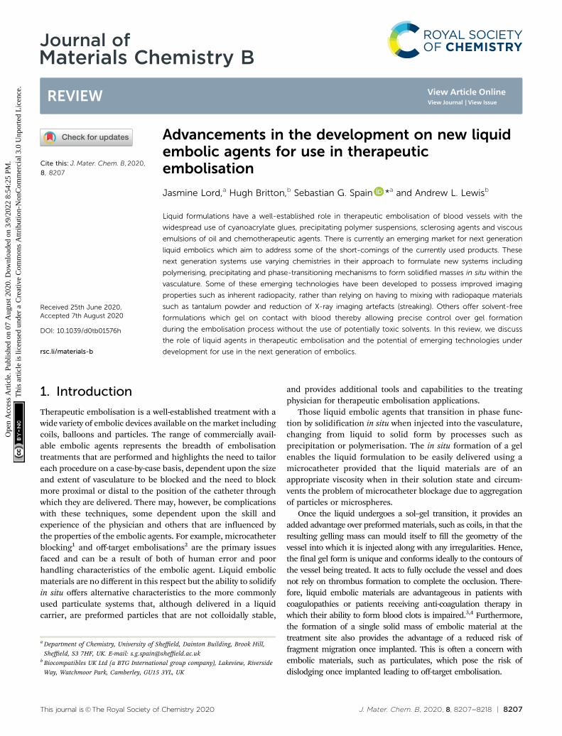
Using the same principal as the sandcastle worms, GPXconsists of polyelectrolyte complexes which are stabilised inaqueous solutions of high ionic strength (Fig. 11). The formula-tion consists of protamine sulfate (from salmon sperm) andsodium inositol hexaphosphate (phytic acid), is rendered radio-paque by addition of tantalum powder particles (1.5 mm in size)and maintained in an un-coacervated state by the additionof 1.2 M sodium chloride. The stabilised solution can bedelivered through the narrow lumens of microcatheters usedin embolisation procedures. On exiting the microcatheter at theinjection site, the polyelectrolyte complexes become destabi-lised due to the reduction of the ionic strength to physiologicalstrength through blood dilution. This causes the polyelectrolytecomplexes to aggregate to form a gelled embolus within the
Fig. 9 Putative structure of Easyxt copolymer – The product is likely be either a mixture of poly(vinyl-4-mono-iodo benzyl ether) MIB-PVAL (A) &poly(vinyl-2,3,5-triiodo-benzyl ether) TIB-PVAL (B), or copolymer C.
Fig. 10 Chemical structure of Eudragit E.
Review Journal of Materials Chemistry B
Ope
n A
cces
s A
rtic
le. P
ublis
hed
on 0
7 A
ugus
t 202
0. D
ownl
oade
d on
3/9
/202
2 8:
54:2
5 PM
. T
his
artic
le is
lice
nsed
und
er a
Cre
ativ
e C
omm
ons
Attr
ibut
ion-
Non
Com
mer
cial
3.0
Unp
orte
d L
icen
ce.
View Article Online
This journal is©The Royal Society of Chemistry 2020 J. Mater. Chem. B, 2020, 8, 8207--8218 | 8215
vasculature with rapid gelation exhibited within seconds. Twodifferent viscosity formulation are being proposed which have atoothpaste-like delivery and control. The resulting embolicmass is non-cytotoxic, non-haemolytic, and non-inflammatory.Despite the fact the material is based upon a glue-like substance,the embolus formed was also found to be non-adhesive in natureto the microcatheter used to deliver the embolic material.Embolisation using GPX has been performed in rabbit renalarteries demonstrating a high level of vascular penetration andcomplete occlusion of the kidney.62
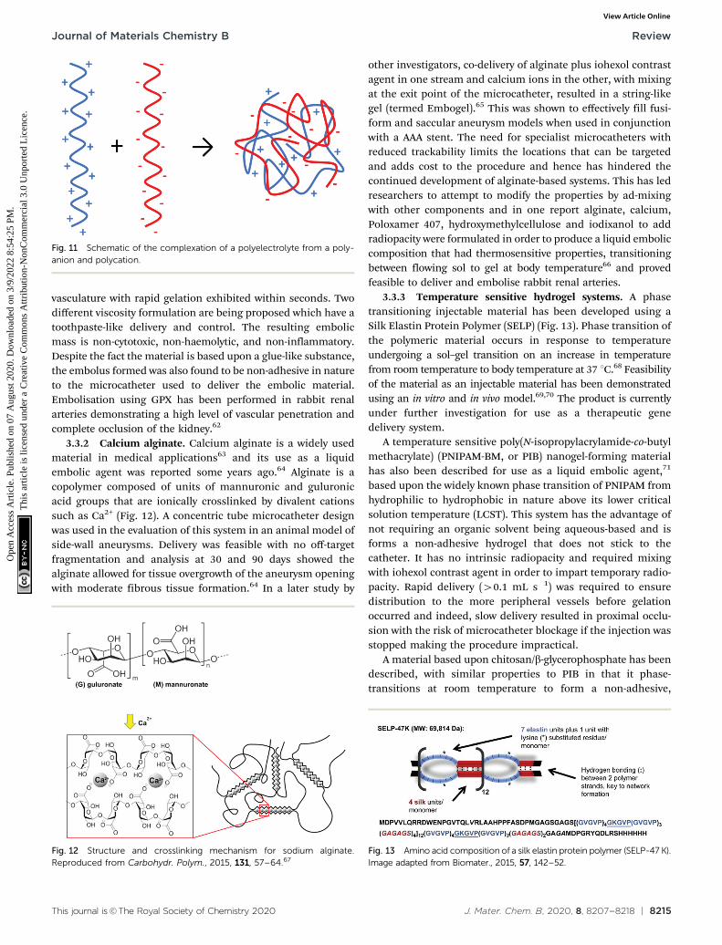
3.3.2 Calcium alginate. Calcium alginate is a widely usedmaterial in medical applications63 and its use as a liquidembolic agent was reported some years ago.64 Alginate is acopolymer composed of units of mannuronic and guluronicacid groups that are ionically crosslinked by divalent cationssuch as Ca2+ (Fig. 12). A concentric tube microcatheter designwas used in the evaluation of this system in an animal model ofside-wall aneurysms. Delivery was feasible with no off-targetfragmentation and analysis at 30 and 90 days showed thealginate allowed for tissue overgrowth of the aneurysm openingwith moderate fibrous tissue formation.64 In a later study by
other investigators, co-delivery of alginate plus iohexol contrastagent in one stream and calcium ions in the other, with mixingat the exit point of the microcatheter, resulted in a string-likegel (termed Embogel).65 This was shown to effectively fill fusi-form and saccular aneurysm models when used in conjunctionwith a AAA stent. The need for specialist microcatheters withreduced trackability limits the locations that can be targetedand adds cost to the procedure and hence has hindered thecontinued development of alginate-based systems. This has ledresearchers to attempt to modify the properties by ad-mixingwith other components and in one report alginate, calcium,Poloxamer 407, hydroxymethylcellulose and iodixanol to addradiopacity were formulated in order to produce a liquid emboliccomposition that had thermosensitive properties, transitioningbetween flowing sol to gel at body temperature66 and provedfeasible to deliver and embolise rabbit renal arteries.
3.3.3 Temperature sensitive hydrogel systems. A phasetransitioning injectable material has been developed using aSilk Elastin Protein Polymer (SELP) (Fig. 13). Phase transition ofthe polymeric material occurs in response to temperatureundergoing a sol–gel transition on an increase in temperaturefrom room temperature to body temperature at 37 1C.68 Feasibilityof the material as an injectable material has been demonstratedusing an in vitro and in vivo model.69,70 The product is currentlyunder further investigation for use as a therapeutic genedelivery system.
A temperature sensitive poly(N-isopropylacrylamide-co-butylmethacrylate) (PNIPAM-BM, or PIB) nanogel-forming materialhas also been described for use as a liquid embolic agent,71
based upon the widely known phase transition of PNIPAM fromhydrophilic to hydrophobic in nature above its lower criticalsolution temperature (LCST). This system has the advantage ofnot requiring an organic solvent being aqueous-based and isforms a non-adhesive hydrogel that does not stick to thecatheter. It has no intrinsic radiopacity and required mixingwith iohexol contrast agent in order to impart temporary radio-pacity. Rapid delivery (40.1 mL s�1) was required to ensuredistribution to the more peripheral vessels before gelationoccurred and indeed, slow delivery resulted in proximal occlu-sion with the risk of microcatheter blockage if the injection wasstopped making the procedure impractical.
A material based upon chitosan/b-glycerophosphate has beendescribed, with similar properties to PIB in that it phase-transitions at room temperature to form a non-adhesive,
Fig. 11 Schematic of the complexation of a polyelectrolyte from a poly-anion and polycation.
Fig. 12 Structure and crosslinking mechanism for sodium alginate.Reproduced from Carbohydr. Polym., 2015, 131, 57–64.67
Fig. 13 Amino acid composition of a silk elastin protein polymer (SELP-47 K).Image adapted from Biomater., 2015, 57, 142–52.
Journal of Materials Chemistry B Review
Ope
n A
cces
s A
rtic
le. P
ublis
hed
on 0
7 A
ugus
t 202
0. D
ownl
oade
d on
3/9
/202
2 8:
54:2
5 PM
. T
his
artic
le is
lice
nsed
und
er a
Cre
ativ
e C
omm
ons
Attr
ibut
ion-
Non
Com
mer
cial
3.0
Unp
orte
d L
icen
ce.
View Article Online

8216 | J. Mater. Chem. B, 2020, 8, 8207--8218 This journal is©The Royal Society of Chemistry 2020
non-toxic hydrogel at 37 1C. This material was evaluated in arabbit renal artery model72 and in the rete mirabile in swine.73
Interestingly, these materials have been formulated withindocyanine green and evaluated for potential intraoperativefluorescent imaging and local therapy of hepatocellularcarcinoma.74 Whilst the dye did not affect the gelation rate,the gel transition temperature was above 4 1C making thehandling and avoidance of premature gelling of this materialvery difficult.
3.3.4 PuraMatrixt. PuraMatrixt (3D Matrix Co., Ltd,Tokyon, Japan) is based upon a peptide with 16 amino acidscomprised of arginine, aspartate and alanine repeatingunits (RADARADARADARADA, RADA16-I). Although originallydeveloped as a cell scaffold material, it has been formulated at2.5 wt% in aqueous media mixed with iodinated contrast agentto impart radiopacity as a proposed liquid embolic agent.75
This peptide sequence has an alternating hydrophobic/chargedmotif that promotes b-strand formation and self-assembly intoa nanofiber-based hydrogel material.76 Evaluation in an embo-lisation setting confirmed the ability of the material to gel andinduce cessation of blood flow in target vessels. Pathologicalanalysis revealed an ability to reach distal locations due to itsnon-adhesive nature and little inflammation in the vessel andsurrounding tissues.
3.3.5 Shear-thinning biomaterials. Shear-thinning bio-materials (STB) are an attractive approach to liquid embolicsas their rheological properties are such that their viscositydrops as it is sheared by injection through the narrow lumenof a microcatheter. Once the shear stress is removed as it exitsthe catheter and enters the less confined environment of theblood vessel, the material returns to its gel state and form anembolus in situ. Currently under investigation as a liquidembolic alternative is a STB based on a nanocomposite hydro-gel composed of gelatin and silicate nanoplatelets (Fig. 14).70,77
This STB demonstrated injectability through a range of micro-catheters and needles and formed a sufficient embolus thatcould withstand physiological pressures without displacementin the vasculature. The application of shear disrupts chargeinteractions between the gelatin and silicate particles, droppingthe viscosity as the apparent molecular weight decreases, whichare restored again to reinforce the gel once the shear is removed.
The aqueous nature of the system alleviates the need to useDMSO compatible microcatheters as required by a numberof liquid embolics. However, the material used in the GPXformulation is not inherently radiopaque so would need to be
delivered alongside a contrast agent to enable visualisation ofthe embolisation procedure. Liquid contrast agent would likelyalter the rate of gelation of the liquid embolic system andparticulate tantalum may interfere with the gelatin-silicateinteractions, hence further testing of the STB is still required.
4. Conclusions and futureperspectives
There are numerous liquid embolics currently under develop-ment aiming to alleviate some of the current issues faced withcommercially available liquid embolics and provide additionaltools for use in therapeutic embolisation. There are however, anumber of prerequisites which must be fulfilled in order todemonstrate improved properties and clinical benefit over thecurrently established agents already available on the market.These properties include reduced streak artefact on imaging,appropriate gelation triggers to avoid catheter blockage andsuitable viscosities to occlude at the desired location underdifferent flow regimes within the vasculature and control overthe delivery without risk of fragmentation and stringing to off-target sites. Some desirable attributes for the next generation ofliquid embolics would be the possession of inherent radio-pacity to avoid the need to pre-mix materials prior to injectionand systems which are free of solvent to reduce the risk of toxicside effects often associated with the use of solvent and theneed to use solvent-compatible microcatheters and deliveryaccessories. These properties are currently unmet needs andthe next generation of liquid embolics systems reviewed hereinare likely to address these to a greater or lesser extent.
Conflicts of interest
ALL and HB are employees of Biocompatibles UK Ltd, a devel-oper of embolisation products for use in minimally invasiveprocedures. JL and SGS have received funding from Biocompa-tibles UK Ltd.
Acknowledgements
J. L. would like to thank Biocompatibles UK Ltd and the UKEPSRC Centre for Doctoral Training in Polymers & Soft Matter(EP/L016281/1) for funding a CASE PhD studentship.
References
1 J. D. Barr, T. J. Lemley and C. N. Petrochko, J. Vasc. Interv.Radiol., 1998, 9, 113–118.
2 R. Duran, K. Sharma, M. R. Dreher, K. Ashrafi, S. Mirpour,M. Lin, R. E. Schernthaner, T. R. Schlachter, V. Tacher,A. L. Lewis, S. Willis, M. den Hartog, A. Radaelli, A. H.Negussie, B. J. Wood and J.-F. H. Geschwind, Theranostics,2016, 6, 28–39.
3 T. Yonemitsu, N. Kawai, M. Sato, T. Sonomura, I. Takasaka,M. Nakai, H. Minamiguchi, S. Sahara, Y. Iwasaki, T. Naka
Fig. 14 Diagram of shear thinning behaviour of a nanocomposite hydro-gel containing gelatin and silicate nanoplatelets. Image adapted from Sci.Transl. Med., 2016, 8, 365–77.
Review Journal of Materials Chemistry B
Ope
n A
cces
s A
rtic
le. P
ublis
hed
on 0
7 A
ugus
t 202
0. D
ownl
oade
d on
3/9
/202
2 8:
54:2
5 PM
. T
his
artic
le is
lice
nsed
und
er a
Cre
ativ
e C
omm
ons
Attr
ibut
ion-
Non
Com
mer
cial
3.0
Unp
orte
d L
icen
ce.
View Article Online

This journal is©The Royal Society of Chemistry 2020 J. Mater. Chem. B, 2020, 8, 8207--8218 | 8217
and M. Shinozaki, Cardiovasc. Intervent. Radiol., 2010, 33,1192–1197.
4 T. Yonemitsu, N. Kawai, M. Sato, H. Tanihata, I. Takasaka,M. Nakai, H. Minamiguchi, S. Sahara, Y. Iwasaki, Y. Shima,M. Shinozaki, T. Naka and M. Shinozaki, J. Vasc. Interv.Radiol., 2009, 20, 1176–1187.
5 R. Lencioni, Hepatology, 2010, 52, 762–773.6 L. Pierot, C. Cognard, D. Herbreteau, H. Fransen, W. J. van
Rooij, E. Boccardi, A. Beltramello, N. Sourour, K. Kupcs,A. Biondi, A. Bonafe, W. Reith and A. Casasco, Eur. Radiol.,2013, 23, 2838–2845.
7 E. J. Speir, R. M. Ermentrout and J. G. Martin, Tech. Vasc.Interv. Radiol., 2017, 20, 258–262.
8 A. J. Ringer and R. Rahme, in Cerebrovascular and Endo-vascular Neurosurgery, Springer International Publishing,Cham, 2018, pp. 321–333.
9 F. Leonard, R. K. Kulkarni, G. Brandes, J. Nelson andJ. J. Cameron, J. Appl. Polym. Sci., 1966, 10, 259–272.
10 H. V. Vinters, K. A. Galil, M. J. Lundie and J. C. E. Kaufmann,Neuroradiology, 1985, 27, 279–291.
11 R. J. Rosen and S. Contractor, Semin. Intervent. Radiol., 2004,21, 59–66.
12 M. F. Brothers, J. C. Kaufmann, A. J. Fox and J. P. Deveikis,Am. J. Neuroradiol., 1989, 10, 777–786.
13 R. I. White, J. V. Strandberg, G. S. Gross, K. H. Barth,T. F. Groves and F. Starr, Radiology, 1977, 125, 677–687.
14 G. Wikholm, Am. J. Neuroradiol., 1995, 16, 479–482.15 S. Vaidya, K. R. Tozer and J. Chen, Semin. Intervent. Radiol.,
2008, 25, 204–215.16 M. J. Gounis, B. B. Lieber, A. K. Wakhloo, R. Siekmann and
L. N. Hopkins, Am. J. Neuroradiol., 2002, 23, 938–944.17 H. Hill, J. F. B. Chick, A. Hage and R. N. Srinivasa, Diagn.
Interv. Radiol., 2018, 24, 98–103.18 Y. Takeuchi, H. Morishita, Y. Sato, S. Hamaguchi, N. Sakamoto,
H. Tokue, T. Yonemitsu, K. Murakami, H. Fujiwara, K. Sofue,T. Abe, H. Higashihara, Y. Nakajima and M. Sato, Jpn. J. Radiol.,2014, 32, 500–517.
19 J. S. Pollak and R. I. White, J. Vasc. Interv. Radiol., 2001, 12,907–913.
20 The n-BCA Trial Investigators, Am. J. Neuroradiol, 2002, 23,748–755.
21 J. Mathis, A. Evans, A. DeNardo, K. Kennett, J. Crandall,M. Jensen and J. Dion, Am. J. Neuroradiol, 1997, 18,1087–1091.
22 B. C. Flores, A. P. See, G. M. Weiner, B. T. Jankowitz,A. F. Ducruet and F. C. Albuquerque, J. Neurosurg., 2019,130, 963–971.
23 S. Paramasivam, D. Altschul, S. Ortega-Gutiarrez, J. Fifi andA. Berenstein, J. Neurointerv. Surg., 2015, 7, 485–461.
24 Micro Therapeutics, Onyx Liquid Embolic System – Instructionsfor Use, 2003.
25 S. Luzzi, M. Del Maestro, D. Bongetta, C. Zoia, A. V.Giordano, D. Trovarelli, S. Raysi Dehcordi and R. J. Galzio,World Neurosurg., 2018, 116, 340–353.
26 W. J. van Rooij, M. Sluzewski and G. N. Beute, Am.J. Neuroradiol., 2007, 28, 172–177.
27 D. Raissi, Q. Yu and S. H. Mardini, J. Clin. Imaging Sci., 2018,8, 46.
28 N. P. Munro, S. Woodhams, J. D. Nawrocki, M. S. Fletcherand P. J. Thomas, BJU Int., 2003, 92, 240–244.
29 R. Siekmann, Interv. Neuroradiol., 2005, 11, 131–140.30 R. Ashour and M. Ali Aziz-Sultan, Neurol. Res., 2014, 36,
363–367.31 R. Loffroy, S. Favelier, P.-Y. Genson and B. Guiu, Cardiovasc.
Intervent. Radiol., 2012, 35, 221.32 J. C. Chaloupka, F. Vinuela, H. V. Vinters and J. Robert, Am.
J. Neuroradiol, 1994, 15, 1107–1115.33 J. C. Chaloupka, D. C. Huddle, J. Alderman, S. Fink,
R. Hammond and H. V. Vinters, Am. J. Neuroradiol, 1999,20, 401–410.
34 W. Taki, Y. Yonekawa, H. Iwata, A. Uno, K. Yamashita andH. Amemiya, Am. J. Neuroradiol, 1990, 11, 163–168.
35 T. Terada, Y. Nakamura, K. Nakai, M. Tsuura, T. Nishiguchi,S. Hayashi, T. Kido, W. Taki, H. Iwata and N. Komai,J. Neurosurg., 1991, 75, 655–660.
36 R. Regine, F. Palmieri, M. De Siero, A. Rescigno, V. Sica,R. Cantarela and V. Villari, Interv. Med. Appl. Sci., 2015, 7,22–29.
37 J. B. Jia, C. S. Green, A. J. Cohen and M. Helmy, Clin. Radiol.,2015, 70, 326–332.
38 J. M. Llovet, M. I. Real, X. Montana, R. Planas, S. Coll,J. Aponte, C. Ayuso, M. Sala, J. Muchart, R. Sola, J. Rodesand J. Bruix, Lancet, 2002, 359, 1734–1739.
39 K. Takayasu, Y. Shima, Y. Muramatsu, N. Moriyama,T. Yamada, M. Makuuchi, H. Hasegawa and S. Hirohashi,Radiology, 1987, 163, 345–351.
40 T. de Baere, A. Denys, R. Briquet, P. Chevallier, J. Dufauxand A. Roche, J. Vasc. Interv. Radiol., 1998, 9, 305–310.
41 T. de Baere, X. Zhang, B. Aubert, G. Harry, C. Lagrange,J. Ropers, J. Dufaux, J. Lumbroso, P. Rougier, M. Ducreuxand A. Roche, Radiology, 1996, 201, 731–735.
42 C. Georgiades, J. F. Geschwind, N. Harrison, A. Hines-Peralta, E. Liapi, K. Hong, Z. Wu, I. Kamel andC. Frangakis, Radiology, 2012, 265, 115–123.
43 S. Ganguli, J. Weintraub, T. DiBartholomeo, R. Lareau,H. Claesson and R. Bean, J. Vasc. Interv. Radiol., 2019, 30, S170.
44 C. R. Brennecka, M. C. Preul, W. D. Bichard andB. L. Vernon, World Neurosurg., 2012, 78, 469–480.
45 J. Mason, C. Dodge and G. Benndorf, Interv. Neuroradiol.,2018, 24, 574–579.
46 R. Pop, L. Mertz, A. Ilyes, D. Mihoc, J. S. Richter,M. Manisor, S. Kremer and R. Beaujeux, J. Neurointerv. Surg.,2019, 11, 706–709.
47 S. Lamin, H. S. Chew, S. Chavda, A. Thomas, M. Piano,L. Quilici, G. Pero, M. Holtmannspolter, M. E. Cronqvist,A. Casasco, L. Guimaraens, L. Paul, A. Gil Garcia, A. Aleuand R. Chapot, Am. J. Neuroradiol., 2017, 38, 127–131.
48 A. Helmy and N. Shaida, Cardiovasc. Intervent. Radiol., 2017,40, 1094–1098.
49 S. S. Sirakov, A. Sirakov, K. Minkin, H. Hristov, K. Ninov,M. Penkov, V. Karakostov, K. Orlov, A. Gorbatykh, D. Kislitsinand R. Raychev, Interv. Neuroradiol., 2019, 25, 58–65.
Journal of Materials Chemistry B Review
Ope
n A
cces
s A
rtic
le. P
ublis
hed
on 0
7 A
ugus
t 202
0. D
ownl
oade
d on
3/9
/202
2 8:
54:2
5 PM
. T
his
artic
le is
lice
nsed
und
er a
Cre
ativ
e C
omm
ons
Attr
ibut
ion-
Non
Com
mer
cial
3.0
Unp
orte
d L
icen
ce.
View Article Online

8218 | J. Mater. Chem. B, 2020, 8, 8207--8218 This journal is©The Royal Society of Chemistry 2020
50 N. Kocer, H. Hanimoglu, S. Batur, S. G. Kandemirli,O. Kizilkilic, Z. Sanus, B. Oz, C. Islak and M. Y. Kaynar,Diagnostic Interv. Radiol., 2016, 22, 184–189.
51 D. F. Vollherbst, C. M. Sommer, C. Ulfert, J. Pfaff,M. Bendszus and M. A. Mohlenbruch, Am. J. Neuroradiol.,2017, 38, 1377–1382.
52 D. F. Vollherbst, C. M. Sommer, C. Ulfert, J. Pfaff, M. Bendszusand M. A. Mohlenbruch, Am. J. Neuroradiol., 2017, 38,1377–1382.
53 O. Dudeck, O. Jordan, K. T. Hoffmann, A. F. Okuducu,I. Husmann, T. Kreuzer-Nagy, K. Tesmer, P. Podrabsky,H. Bruhn, J. Hilborn, D. A. Rufenacht, E. Doelker andR. Felix, Am. J. Neuroradiol., 2006, 27, 1849–1855.
54 Antia Therapeutics S.A., EP2545085 (A1), 2011.55 Z. Kulcsar, A. Karol, P. W. Kronen, P. Svende, K. Klein,
O. Jordan and I. Wanke, Eur. Radiol., 2017, 27, 1248–1256.56 G. Agusti, O. Jordan, G. Andersen, E. Doelker and Y. Chevalier,
J. Appl. Polym. Sci., 2015, 132, 41791.57 Antia Therapeutics S.A., US9434800B2, 2015.58 G. Tamura, N. Kato, T. Yamazaki, Y. Akutsu, H. Hosoo,
H. Kasuya and M. Sonobe, Neurol. Med. Chir., 2015, 55, 253–260.59 H. Arakawa, Y. Murayama, C. R. Davis, D. L. Howard,
W. L. Baumgardner, M. P. Marks and H. M. Do, Am.J. Neuroradiol., 2007, 28, 1191–1196.
60 R. J. Stewart, C. S. Wang, I. T. Song and J. P. Jones, Adv.Colloid Interface Sci., 2017, 239, 88–96.
61 J. P. Jones, M. Sima, R. G. O’Hara and R. J. Stewart, Adv.Healthcare Mater., 2016, 5, 795–801.
62 M. Johnson, Global Embolization Symposium and Technologies,2018.
63 K. Y. Lee and D. J. Mooney, Prog. Polym. Sci., 2012, 37, 106–126.64 T. A. Becker, M. C. Preul, W. D. Bichard, D. R. Kipke and
C. G. Mcdougall, Neurosurgery, 2007, 60, 1119–1128.
65 B. P. Barnett, A. H. Hughes, S. Lin, A. Arepally andP. H. Gailloud, J. Vasc. Interv. Radiol., 2009, 20, 507–512.
66 L. Huang, M. Shen, R. Li, X. Zhang, Y. Sun, P. Gao, H. Fu,H. Liu, Y. He, Y. Du, J. Cao and Y. Duan, Oncotarget, 2016, 7,73280–73291.
67 M. Bruchet and A. Melman, Carbohydr. Polym., 2015, 131,57–64.
68 J. Cappello, J. Crissman, M. Crissman, F. Ferrari, G. Textor,O. Wallis, J. Whitledge, X. Zhou, D. Burman, L. Aukermanand E. Stedronsky, J. Controlled Release, 1998, 53, 105–117.
69 Z. Megeed, M. Haider, D. Li, B. W. O’Malley, J. Cappello andH. Ghandehari, J. Controlled Release, 2004, 94, 433–445.
70 A. Poursaid, R. Price, A. Tiede, E. Olson, E. Huo, L. McGill,H. Ghandehari and J. Cappello, Biomaterials, 2015, 57,142–152.
71 H. Zhao, C. Zheng, G. Feng, Y. Zhao, H. Liang, H. Wu,G. Zhou, B. Liang, Y. Wang and X. Xia, AJNR. Am.J. Neuroradiol., 2013, 34, 169–176.
72 Y. Wang, N. Xu, Q. Luo, Y. Li, L. Sun, H. Wang, K. Xu,B. Wang and Y. Zhen, Interv. Neuroradiol., 2011, 17, 87–92.
73 X. Ning, C. Zhao, J. Pang, Z. Ding, Y. Wang, K. Xu, H. Chen,B. Li and Q. Luo, Exp. Ther. Med., 2015, 10, 316–322.
74 A. Salis, G. Rassu, M. Budai-Szucs, I. Benzoni, E. Csanyi,S. Berko, M. Maestri, P. Dionigi, E. P. Porcu, E. Gavini andP. Giunchedi, Expert Opin. Drug Delivery, 2015, 12,1583–1596.
75 Y. Baba, M. Higashi and K. Awai, Minim. Invasive Ther. AlliedTechnol., 2018, 27, 17–21.
76 A. R. Cormier, X. Pang, M. I. Zimmerman, H.-X. Zhou andA. K. Paravastu, ACS Nano, 2013, 7, 7562–7572.
77 R. K. Avery, H. Albadawi, M. Akbari, Y. S. Zhang, M. J.Duggan, D. V. Sahani, B. D. Olsen, A. Khademhosseini andR. Oklu, Sci. Transl. Med., 2016, 8, 156–168.
Review Journal of Materials Chemistry B
Ope
n A
cces
s A
rtic
le. P
ublis
hed
on 0
7 A
ugus
t 202
0. D
ownl
oade
d on
3/9
/202
2 8:
54:2
5 PM
. T
his
artic
le is
lice
nsed
und
er a
Cre
ativ
e C
omm
ons
Attr
ibut
ion-
Non
Com
mer
cial
3.0
Unp
orte
d L
icen
ce.
View Article Online