Biomaterials Science - RSC Publishing
22
Biomaterials Science REVIEW Cite this: DOI: 10.1039/d1bm01540k Received 30th September 2021, Accepted 17th March 2022 DOI: 10.1039/d1bm01540k rsc.li/biomaterials-science Articulation inspired by nature: a review of biomimetic and biologically active 3D printed scaffolds for cartilage tissue engineering Donagh G. O’Shea, a,c Caroline M. Curtin a,b,c and Fergal J. O’Brien * a,b,c In the human body, articular cartilage facilitates the frictionless movement of synovial joints. However, due to its avascular and aneural nature, it has a limited ability to self-repair when damaged due to injury or wear and tear over time. Current surgical treatment options for cartilage defects often lead to the formation of fibrous, non-durable tissue and thus a new solution is required. Nature is the best innovator and so recent advances in the field of tissue engineering have aimed to recreate the microenvironment of native articular cartilage using biomaterial scaffolds. However, the inability to mirror the complexity of native tissue has hin- dered the clinical translation of many products thus far. Fortunately, the advent of 3D printing has provided a potential solution. 3D printed scaffolds, fabricated using biomimetic biomaterials, can be designed to mimic the complex zonal architecture and composition of articular cartilage. The bioinks used to fabricate these scaffolds can also be further functionalised with cells and/or bioactive factors or gene therapeutics to mirror the cellular composition of the native tissue. Thus, this review investigates how the architecture and compo- sition of native articular cartilage is inspiring the design of biomimetic bioinks for 3D printing of scaffolds for cartilage repair. Subsequently, we discuss how these 3D printed scaffolds can be further functionalised with cells and bioactive factors, as well as looking at future prospects in this field. 1. Introduction “Innovation inspired by nature”– this is how biomimicry is described by author and scientist Janine M. Benyus in her 1997 book of the same title and although a relatively new term, the concept of biomimicry is evident throughout the history of human innovation. 1,2 From the study of birds to inspire the design of aeroplanes to the study of human anatomy to inspire next generation medical devices, mimick- ing nature has allowed us to create innovative new techno- logies and improve our own quality of life. But how does biomimicry apply to tissue engineering (TE)? TE, also known as regenerative medicine, is an emerging multi- disciplinary field of modern medicine which uses a combination of biomaterials, cells and bioactive factors or gene therapeutics, sometimes referred to as the ‘TE triad’, to bioengineer living tissues for a range of applications. 3 These diverse applications range from disease modelling 4 and organ-on-a-chip develop- ment, 5 to the regeneration of a variety of tissue types including cardiac tissue, 6 musculoskeletal tissue 7 and skin, 8 amongst others. In essence, the field of TE relies on the fabrication of bio- material implants called ‘scaffolds’, which can support cell growth and whose microenvironment, architecture and function- ality mimic that of native human tissue. Therefore, it could be argued that biomimicry forms the foundations of the field of TE. In recent years, TE scaffolds have grown in popularity as a potential treatment option for a range of conditions. In the field of orthopaedic medicine, for example, these scaffolds have emerged as a promising treatment option for chondral defects (CDs), which are localised areas of damage to the articular carti- lage of a synovial joint. In severe cases, the defect can penetrate into the underlying subchondral bone leading to the formation of an osteochondral defect. 9 Once the friction-reducing articular cartilage tissue has worn away, the underlying bone surfaces can rub against one another causing significant stiffness and pain, thus hindering joint mobility. 10 CDs can be caused by traumatic injury or wear and tear over time and can lead to the development of osteoarthritis, a degenerative joint disease which affects 9.6% of men and 18% of women over the age of 60 years worldwide. 11 Unfortunately, due to the avascular and aneural nature of articu- lar cartilage, these defects will not heal on their own. Current treatment options for CDs include surgical pro- cedures such as microfracture, autograft or allograft pro- a Tissue Engineering Research Group, Department of Anatomy and Regenerative Medicine, RCSI University of Medicine and Health Sciences, Dublin, Ireland. E-mail: [email protected] b Trinity Centre for Biomedical Engineering, Trinity College Dublin, Ireland c Advanced Materials and Bioengineering Research Centre (AMBER), RCSI and TCD, Dublin, Ireland This journal is © The Royal Society of Chemistry 2022 Biomater. Sci. Open Access Article. Published on 22 March 2022. Downloaded on 4/19/2022 5:46:16 AM. This article is licensed under a Creative Commons Attribution-NonCommercial 3.0 Unported Licence. View Article Online View Journal
-
Upload
khangminh22 -
Category
Documents
-
view
0 -
download
0
Transcript of Biomaterials Science - RSC Publishing
BiomaterialsScience
REVIEW
Cite this: DOI: 10.1039/d1bm01540k
Received 30th September 2021,Accepted 17th March 2022
DOI: 10.1039/d1bm01540k
rsc.li/biomaterials-science
Articulation inspired by nature: a review ofbiomimetic and biologically active 3D printedscaffolds for cartilage tissue engineering
Donagh G. O’Shea, a,c Caroline M. Curtin a,b,c and Fergal J. O’Brien *a,b,c
In the human body, articular cartilage facilitates the frictionless movement of synovial joints. However, due
to its avascular and aneural nature, it has a limited ability to self-repair when damaged due to injury or wear
and tear over time. Current surgical treatment options for cartilage defects often lead to the formation of
fibrous, non-durable tissue and thus a new solution is required. Nature is the best innovator and so recent
advances in the field of tissue engineering have aimed to recreate the microenvironment of native articular
cartilage using biomaterial scaffolds. However, the inability to mirror the complexity of native tissue has hin-
dered the clinical translation of many products thus far. Fortunately, the advent of 3D printing has provided a
potential solution. 3D printed scaffolds, fabricated using biomimetic biomaterials, can be designed to mimic
the complex zonal architecture and composition of articular cartilage. The bioinks used to fabricate these
scaffolds can also be further functionalised with cells and/or bioactive factors or gene therapeutics to mirror
the cellular composition of the native tissue. Thus, this review investigates how the architecture and compo-
sition of native articular cartilage is inspiring the design of biomimetic bioinks for 3D printing of scaffolds for
cartilage repair. Subsequently, we discuss how these 3D printed scaffolds can be further functionalised with
cells and bioactive factors, as well as looking at future prospects in this field.
1. Introduction
“Innovation inspired by nature” – this is how biomimicry isdescribed by author and scientist Janine M. Benyus in her1997 book of the same title and although a relatively newterm, the concept of biomimicry is evident throughout thehistory of human innovation.1,2 From the study of birds toinspire the design of aeroplanes to the study of humananatomy to inspire next generation medical devices, mimick-ing nature has allowed us to create innovative new techno-logies and improve our own quality of life.
But how does biomimicry apply to tissue engineering (TE)?TE, also known as regenerative medicine, is an emerging multi-disciplinary field of modern medicine which uses a combinationof biomaterials, cells and bioactive factors or gene therapeutics,sometimes referred to as the ‘TE triad’, to bioengineer livingtissues for a range of applications.3 These diverse applicationsrange from disease modelling4 and organ-on-a-chip develop-
ment,5 to the regeneration of a variety of tissue types includingcardiac tissue,6 musculoskeletal tissue7 and skin,8 amongstothers. In essence, the field of TE relies on the fabrication of bio-material implants called ‘scaffolds’, which can support cellgrowth and whose microenvironment, architecture and function-ality mimic that of native human tissue. Therefore, it could beargued that biomimicry forms the foundations of the field of TE.
In recent years, TE scaffolds have grown in popularity as apotential treatment option for a range of conditions. In the fieldof orthopaedic medicine, for example, these scaffolds haveemerged as a promising treatment option for chondral defects(CDs), which are localised areas of damage to the articular carti-lage of a synovial joint. In severe cases, the defect can penetrateinto the underlying subchondral bone leading to the formationof an osteochondral defect.9 Once the friction-reducing articularcartilage tissue has worn away, the underlying bone surfaces canrub against one another causing significant stiffness and pain,thus hindering joint mobility.10 CDs can be caused by traumaticinjury or wear and tear over time and can lead to the developmentof osteoarthritis, a degenerative joint disease which affects 9.6%of men and 18% of women over the age of 60 years worldwide.11
Unfortunately, due to the avascular and aneural nature of articu-lar cartilage, these defects will not heal on their own.
Current treatment options for CDs include surgical pro-cedures such as microfracture, autograft or allograft pro-
aTissue Engineering Research Group, Department of Anatomy and Regenerative
Medicine, RCSI University of Medicine and Health Sciences, Dublin, Ireland.
E-mail: [email protected] Centre for Biomedical Engineering, Trinity College Dublin, IrelandcAdvanced Materials and Bioengineering Research Centre (AMBER), RCSI and TCD,
Dublin, Ireland
This journal is © The Royal Society of Chemistry 2022 Biomater. Sci.
Ope
n A
cces
s A
rtic
le. P
ublis
hed
on 2
2 M
arch
202
2. D
ownl
oade
d on
4/1
9/20
22 5
:46:
16 A
M.
Thi
s ar
ticle
is li
cens
ed u
nder
a C
reat
ive
Com
mon
s A
ttrib
utio
n-N
onC
omm
erci
al 3
.0 U
npor
ted
Lic
ence
.
View Article OnlineView Journal

cedures, or cell-based techniques.12 Microfracture is a pro-cedure whereby tiny fractures are made in the subchondralbone allowing the release of bone marrow stem cells into thedefect which ultimately develop into fibrocartilage.13 Howeverunlike articular cartilage, fibrocartilage is rich in collagen typeI and thus possesses inferior mechanical properties. Autograftprocedures, such as osteochondral autograft transfer systems(OATS) and mosaicplasty, involve transplanting osteochondraltissue from low load bearing regions of the knee into defectslocated in high load bearing regions.14,15 However the pro-cedure is only suitable for smaller defects due to limited tissueavailability at the donor site and can also be associated withdonor site morbidity due to infection. Allografts are articularcartilage transplants taken from another donor and thus arenot constrained by tissue availability at the donor site.However the availability of donors and the risk of the patient’simmune system rejecting the graft limits the use of thisprocedure.16,17 Cell-based procedures such as autologouschondrocyte implantation (ACI) have shown promise andinvolve removing cells from healthy articular cartilage, expand-ing them in culture and then implanting the expanded chon-drocytes into the chondral defect under a collagen membrane.However dedifferentiation of the chondrocytes during in vitrocell expansion can occur leading to a reduced capacity of thecells to lay down new ECM when implanted back into thedefect.18 Therefore, despite providing much needed sympto-matic relief to patients, current surgical procedures are notwithout limitations, often resulting in a variable healingresponse and the formation of non-durable tissue.16,19 Thisissue becomes even more prominent following surgical treat-ment of larger defects.20
Without successful intervention, the osteoarthritic joint candeteriorate to a point where total joint replacement is the onlyremaining option to relieve pain and discomfort. Total joint re-placement involves complete removal of the arthritic joint andinsertion of a prosthesis in its place. These prostheses are typi-cally made from a metal such as a titanium alloy, polymers,ceramics or a composite.21 Although total joint replacementcan result in dramatic improvements in patient quality of lifefollowing initial post-operative rehabilitation, revision surgerycan be required over time due to implant failure. While thelifetime risk of requiring revision surgery is relatively low inpatients aged over 70 years (1–6%), this risk is significantlyhigher in younger patients with 1 in 3 patients aged 50 to 55years likely to require revision surgery. More than half of theserevision surgeries are needed within 6 years of the initial jointreplacement surgery.22 Therefore the benefits of this procedureneed to be weighed against the potential risk of future sur-geries and poor health outcomes, particularly for youngerpatients.
Several fabrication techniques have been investigated toengineer scaffolds which could be surgically implanted intothese defects in order to support regrowth of cartilage and/orbone tissue. Examples of these techniques includingelectrospinning,23–29 solvent casting/melt moulding and par-ticulate leaching,30–33 freeze drying34–36 and gas foaming
techniques,37–42 amongst others. Scaffolds for bone and carti-lage repair fabricated using the freeze drying method havebeen the subject of extensive research here in the RCSI TissueEngineering Research Group (TERG). Using a controlled freezedrying cycle, our lab fabricates highly porous scaffolds fromslurries of biomaterials native to the human body such as col-lagen type I, hyaluronic acid (HyA) and chondroitin sulfate(CS).43–47 The composition and stiffness of these scaffolds canbe tailored to promote cartilage or bone regeneration asrequired.44
While significant progress in the field of TE for cartilagerepair has been made using scaffolds fabricated via the tech-niques outlined above, the inability to mimic the complexity ofnative tissue has hindered the clinical translation of many pro-ducts thus far.48,49 Fortunately, the development of 3D printedscaffolds has provided a potential solution. These scaffoldscan be designed to mirror the complex zonal architecture ofarticular cartilage and can be reinforced with polymers toimprove their mechanical strength. The biomaterial inks usedto print these scaffolds can also be functionalised with cellssuch as mesenchymal stem cells (MSCs) or mature chondro-cytes, bioactive factors such as growth factors, or gene thera-peutics such as plasmid DNA (pDNA) or microRNA (miR) topromote cartilage growth.50–53 These functionalised biomater-ial inks are also called ‘bioinks’.54 There are a number of 3Dprinting techniques used in this field including droplet-based,54–56 laser-based57,58 and extrusion-based methods.5
However, due to its versatility and compatibility with cells anda wide range of biomaterials, extrusion-based 3D printing isone of the most popular 3D printing methods in the field ofTE for cartilage repair.59,60 Thus, this review will focus on thedevelopment of bioinks for extrusion-based 3D printing only.
In the field of 3D printing for cartilage repair, there is anever growing emphasis placed on the importance of designingadvanced biomimetic bioinks whose matrix compositionreflects that of native articular cartilage. This biomimeticapproach helps in the regeneration of functional tissue andminimises adverse reactions when the 3D printed scaffold isimplanted in vivo. Therefore, the scope of this review will focuson how the architecture and composition of native articularcartilage is inspiring the design of biomimetic bioinks forextrusion-based 3D printing of scaffolds for cartilage repair.
2. Architecture and composition ofarticular cartilage
Articular cartilage performs two crucial functions in humansynovial joints – the first is to facilitate frictionless movementof the joint, and the second is to withstand repeated compres-sive loading. Both the architecture and the composition of thetissue play a significant role in facilitating these functions.Therefore, a thorough understanding of the structure and boththe biomaterial and cellular composition of native articularcartilage is crucial when designing biomimetic bioinks for car-tilage repair.
Review Biomaterials Science
Biomater. Sci. This journal is © The Royal Society of Chemistry 2022
Ope
n A
cces
s A
rtic
le. P
ublis
hed
on 2
2 M
arch
202
2. D
ownl
oade
d on
4/1
9/20
22 5
:46:
16 A
M.
Thi
s ar
ticle
is li
cens
ed u
nder
a C
reat
ive
Com
mon
s A
ttrib
utio
n-N
onC
omm
erci
al 3
.0 U
npor
ted
Lic
ence
.View Article Online

2.1. Architecture of articular cartilage
The extracellular matrix (ECM) of articular cartilage is largelycomposed of collagen (primarily type II), proteoglycans andwater.61 This ECM is synthesised and maintained by highlyspecialised cells known as chondrocytes whose morphology,density and organisation varies with cartilage depth leading tothe formation of three distinct zones within the articular carti-lage.62 The uppermost superficial zone (10–20% articular carti-lage thickness) is characterised by the presence of high densityflattened chondrocytes embedded in an ECM with collagenfibres aligned parallel to the joint surface and the presence ofthe lubricant proteoglycan 4 (PRG4 or lubricin). The middlezone (40–60% articular cartilage thickness) consists ofrounded chondrocytes within a more disorganised collagenmatrix, while the deep zone (30–40% articular cartilage thick-ness) consists of large chondrocytes surrounded by a pericellu-lar matrix consisting of collagen type VI, and thicker collagenfibres organised perpendicular to the joint surface.63,64 Thedeep zone is then underpinned by calcified cartilage and thesubchondral bone. The structure of articular cartilage is out-lined in Fig. 1 below.
2.2. Composition of articular cartilage
Collagen is a protein consisting of a triple helix of three poly-peptide chains called α-chains, and is the primary macro-molecule component found in the ECM of articular cartilage.65
There are many different types of collagen present in thehuman body however fibril-forming collagens, such as col-lagen types I and II, are by far the most abundant.66 Collagentype I, which is the main collagen type found in fibrocartilage,
consists of two α1(I) chains and one α2(I) chain. In contrast,collagen type II, which is the primary collagen type found inarticular cartilage, consists of three identical α1(II) chainswhich contain a higher proportion of hydroxylysine, glucosyland galactosyl residues. These residues mediate interactionswith surrounding proteoglycans in the ECM of articular carti-lage.65 The tightly packed collagen type II and IX fibres foundaligned parallel to the articulating surface in the superficialzone of articular cartilage are also largely responsible for itstensile properties.64
Proteoglycans are the second largest macromolecule com-ponent of articular cartilage and consist of a protein core withglycosaminoglycan (GAG) side chains. GAGs are negativelycharged polysaccharides and can be non-sulfated, such asHyA, or sulfated, such as CS or keratin sulfate. Aggrecan is themost abundant proteoglycan found in articular cartilage andinteracts with HyA to form large negatively charged aggregateswithin the matrix of collagen type II fibrils.67 Water comprises60–80% of the wet weight of cartilage and in this aqueousenvironment, aggrecan molecules become hydrated and swell.This swelling is resisted by the surrounding collagen fibrils,leading to the formation of an equilibrium between the swell-ing forces of aggrecan and the tensile forces of the collagenmatrix.68,69 This is the mechanism by which articular cartilagewithstands repeated compressive loading so effectively.
2.3. Chondrocytes – the resident cells of articular cartilage
As mentioned above, chondrocytes are the primary cell typefound in articular cartilage and play an integral role in the syn-thesis and maintenance of articular cartilage ECM. In the
Fig. 1 The structure of articular cartilage – the diagram on the left shows a synovial joint with a cross-sectional schematic depicting healthy articu-lar cartilage: A – morphology and organisation of chondrocytes in the superficial, middle and deep zones respectively; B – orientation of collagenfibres in the superficial, middle and deep zones respectively. The histological image (haemotoxylin and eosin (H&E) staining) on the right is takenfrom the femoral condyle of a rabbit knee joint and demonstrates the zonal distribution of chondrocytes within articular cartilage (scale bar =100 µm). Histological image reproduced and adapted from Matsiko et al.62 with permission from MDPI (Copyright © 2013, MDPI). Figure createdwith Biorender.com.
Biomaterials Science Review
This journal is © The Royal Society of Chemistry 2022 Biomater. Sci.
Ope
n A
cces
s A
rtic
le. P
ublis
hed
on 2
2 M
arch
202
2. D
ownl
oade
d on
4/1
9/20
22 5
:46:
16 A
M.
Thi
s ar
ticle
is li
cens
ed u
nder
a C
reat
ive
Com
mon
s A
ttrib
utio
n-N
onC
omm
erci
al 3
.0 U
npor
ted
Lic
ence
.View Article Online

superficial and middle zones, chondrocytes primarily syn-thesise collagen types II, IX and XI and aggrecan, all of whichare critical to the compressive and tensile properties of thetissue. However, in the deep zone, terminally differentiatedchondrocytes mainly produce collagen type X and aggrecan.70
However, despite the importance of these cells, chondrocytesmake up just 2% of the total cartilage tissue volume in ahealthy adult and so are present in quite a small quantity.71
This sparse distribution means that very little cell–cell contactoccurs within the tissue, therefore chondrocyte activity islargely dictated by interactions with the surrounding ECM.72
During development, chondrocytes are formed followingexposure of mesenchymal stem cells (MSCs) to certain biologi-cal cues in a process known as chondrogenesis. MSCs are atype of stem cell which are capable of differentiating intoseveral connective tissue cell types including those from anadipogenic, osteogenic and chondrogenic lineage, dependingon exposure to defined conditions.73,74 Chondrogenesis beginswith proliferation and condensation of MSCs, leading to theinitiation of cell–ECM and cell–cell interactions via gap junc-tions, and ultimately the formation of chondroprogenitorcells.75 Following this, chondroprogenitor cells differentiateinto chondrocytes and this stage is characterised by the depo-sition of the cartilage ECM components collagen types II, IXand XI, and aggrecan.76 Production of these ECM componentsis a direct result of the expression of the genes Col2a1, Col9A1,Col11a1 and ACAN, and thus these genes are often consideredto be markers of chondrogenesis in the field of TE for cartilage
repair. This process is driven by a number of growth factorsincluding bone morphogenetic proteins (BMPs), transforminggrowth factor-β1 and 3 (TGF-β1 and 3) and insulin-like growthfactor-1 (IGF-1).77 The transcription factors, SOX-5, 6 and 9(known as the SOX-trio), also play a key role in promotingchondrogenesis with SOX-9, in particular, directly influencingup-regulation of pro-chondrogenic genes such as Col2a1 andACAN.78
In the formation of articular cartilage, the chondrogenicpathway stops here. However, chondrocytes can undergo term-inal differentiation to form hypertrophic chondrocytes, whicheventually leads to ossification of the tissue.79 This process isknown as endochondral ossification and is the way in whichlong bones are formed during foetal development. Thesehypertrophic chondrocytes synthesise collagen type X (regu-lated by the gene Col10a1), and also express the enzymesmatrix metalloproteinase-13 (MMP-13) and alkaline phospha-tase (ALP), which are involved in the degradation of cartilageECM and regulation of bone mineralisation respectively.76,80,81
Thus, the expression of Col10a1, MMP-13 and ALP are oftenconsidered markers of hypertrophy in cartilage TE.82 The tran-scription factor Runx2 is also known to promote chondrocytehypertrophy, and the growth factor, vascular endothelialgrowth factor (VEGF), plays a key role in vascularisation of thetissue during endochondral ossification.83,84 In healthy articu-lar cartilage, hypertrophic chondrocytes are found in the deepzone, near the border with the subchondral bone.64 The chon-drogenic pathway is outlined in Fig. 2 below.
Fig. 2 A schematic of the process of chondrogenesis, starting with MSC proliferation and condensation and ending with endochondral ossification.The factors that promote transition from one stage of the chondrogenic pathway to the next are highlighted with a green indicator. The character-istic ECM proteins of each stage are highlighted below. Figure created with Biorender.com.
Review Biomaterials Science
Biomater. Sci. This journal is © The Royal Society of Chemistry 2022
Ope
n A
cces
s A
rtic
le. P
ublis
hed
on 2
2 M
arch
202
2. D
ownl
oade
d on
4/1
9/20
22 5
:46:
16 A
M.
Thi
s ar
ticle
is li
cens
ed u
nder
a C
reat
ive
Com
mon
s A
ttrib
utio
n-N
onC
omm
erci
al 3
.0 U
npor
ted
Lic
ence
.View Article Online

3. Biomimetic 3D printed scaffoldsfor cartilage repair
When designing a biomimetic 3D printed scaffold for cartilagerepair, there are several key considerations which should betaken into account based on the biomaterial and cellular com-position of native articular cartilage. Firstly, a highly hydratedenvironment is crucial not only to cell survival, but also to thebiofunctionality of the tissue. From a 3D printing perspective,hydrogels are well placed to provide such an environment.Hydrogels are hydrophilic polymer networks which are exten-sively water swollen and are already widely employed as bioinkcomponents throughout the field of TE.85 Secondly, several bio-materials such as collagen, HyA and CS play a synergistic role inthe unique mechanical and biological functions of articular car-tilage and thus are desirable as bioink components. Thirdly,chondrocytes are required for tissue regeneration and thus thebioink could be functionalised with MSCs, chondroprogenitorcells or mature chondrocytes to accelerate healing. However,these cells must also be provided with appropriate biologicalcues in order for them to differentiate into, or maintain, a chon-drogenic lineage and prevent hypertrophy.
The physiochemical and rheological properties of thebioink used to develop the scaffold are also a key consider-
ation. An ideal bioink should exhibit a decrease in viscosityduring the printing process to allow extrusion (i.e. possessshear thinning properties), but also be capable of crosslinkingimmediately post-printing to retain its shape and ensuremechanical stability of the 3D printed construct.86,87 Inaddition to this, the 3D printed scaffold must also besufficiently porous to facilitate the attachment, proliferationand differentiation of cells, as well as allowing diffusion ofsolutes and nutrients and deposition of ECM.54,88 This is ofteninfluenced by the polymer concentration or the crosslinkdensity of the bioink. The bioink, and thus the 3D printedscaffold, must also be biodegradable at a rate appropriate tothat of the regenerating tissue to allow for gradual replacementof the scaffold with deposited ECM from native cells.89 It isalso critical that both the bioink and its degradation productsare non-toxic and non-immunogenic.3 The ideal properties ofa bioink are summarised in Fig. 3 below.
3.1. Mimicking the composition of articular cartilage whendeveloping 3D printed scaffolds
Strategies for fabricating biomimetic 3D printed scaffolds forcartilage repair strive to maintain the synergistic biofunction-ality of native biomaterials by using biomimetic bioink formu-lations with desirable physiochemical and rheological pro-
Fig. 3 A summary of the ideal bioink properties to facilitate 3D printing of biomaterial scaffolds with high shape fidelity and to promote regener-ation of cartilage tissue when the scaffold is implanted in vivo. Figure created with Biorender.com.
Biomaterials Science Review
This journal is © The Royal Society of Chemistry 2022 Biomater. Sci.
Ope
n A
cces
s A
rtic
le. P
ublis
hed
on 2
2 M
arch
202
2. D
ownl
oade
d on
4/1
9/20
22 5
:46:
16 A
M.
Thi
s ar
ticle
is li
cens
ed u
nder
a C
reat
ive
Com
mon
s A
ttrib
utio
n-N
onC
omm
erci
al 3
.0 U
npor
ted
Lic
ence
.View Article Online
perties. This can be challenging as many biomaterials nativeto human articular cartilage do not innately possess good 3Dprinting properties. The following sections will look at hownative biomaterials are being adapted to formulate biomimeticbioinks for cartilage repair with favourable 3D printingproperties.
3.1.1. Collagen bioinks for cartilage repair. The incorpor-ation of collagen into bioinks for cartilage repair is an obvioustarget due to its innate biocompatibility and the role it playswithin the ECM of native articular cartilage. Collagen also pos-sesses naturally occurring cell binding sites in the form of theamino acid motif Arg-Gly-Asp (RGD), and so can easily facili-tate cell adhesion and growth.90 The vast majority of collagencontaining bioinks for cartilage repair reported in the litera-ture are formulated using the fibril-forming collagen type I(COL-I). However, despite the advantages of incorporating col-lagen into bioinks for cartilage repair, it is a notoriously chal-lenging biomaterial to work with. COL-I exists as a liquid atlower temperatures and low pH but as the temperature and pHtend towards physiological conditions (37 °C and pH 7.4),COL-I molecules start to self-assemble into fibrils leading tohydrogel formation.91 These collagen hydrogels, however, tendto be mechanically weak and the fibrillation process is difficultto control and can take up to 30 minutes at 37 °C which is pro-blematic for shape retention and mechanical stability post-printing.92 Several strategies have been employed to overcomethese challenges whilst still maintaining the natural biofunc-tionality of COL-I.
One such approach involves controlling the physical cross-linking of COL-I hydrogels via the manipulation of hydrogelconcentration, pH and temperature. Traditionally, COL-Ihydrogels were formulated at lower concentrations of <10 mgmL−1 in acetic acid; however, these hydrogels were not cellfriendly and they possessed very poor mechanical properties.93
However, more recent studies have been successful in formu-lating high concentration (up to 20 mg mL−1), neutralisedCOL-I hydrogels by carefully adjusting and buffering the pH ofthe formulation to physiological pH and salt concentration.94
Very high concentration COL-I hydrogels are also now availableas commercial bioink formulations such as Lifeink® 200(Advanced BioMatrix, CA, USA) which contains 35 mg mL−1
COL-I, or Viscoll (Imtek Ltd, Russia) which contains up to80 mg mL−1 COL-I.95,96 These high concentration COL-Ibioinks possess better 3D printing properties and mechanicalproperties than lower concentration COL-I formulations,resulting in greater shape fidelity of the printed construct andthey can also support 3D bioprinting of cells.97,98 However,temperature control when 3D printing with COL-I bioinks iscritical – the print head should be maintained at 4–10 °C tofacilitate printing of the bioink, but the print bed should bemaintained at 37 °C to facilitate thermal crosslinking of thedeposited bioink filaments.94,99 COL-I prints can also befurther crosslinked post-printing using cell friendly chemicalcrosslinkers such as genipin.100,101
Another approach to improving the mechanical propertiesof COL-I bioinks involves chemically modifying the COL-I
hydrogel with crosslinkable chemical groups. The COL-I mole-cule may be chemically modified at the primary amine site onthe lysine residue with photocrosslinkable methacrylategroups to form methacrylated collagen (ColMA).102–105 Thisallows the COL-I bioink to be crosslinked in the presence of aphotoinitiator, such as Irgacure 2959 (I2959) or lithiumphenyl-2,4,6-trimethylbenzoylphosphinate (LAP), and UV lightduring or immediately following the printing process. Theaddition of methacrylate groups has also been shown to conferthermoreversible gelation properties to the hydrogel withoutaffecting the fibrillation process, biodegradability or bioactivityof COL-I.103,106 Similarly, COL-I has also been functionalisedwith norbornene groups to form a thiol–ene photocrosslink-able hydrogel which was shown to have improved 3D printingproperties without a loss of bioactivity.107
Sacrificial support baths, as employed in FreeformReversible Embedding of Suspended Hydrogels (FRESH), canalso be used to facilitate 3D printing of COL-I bioinks.108 TheFRESH method of 3D printing involves 3D printing a bioinkinto a thermoreversible gelatin slurry support bath which exhi-bits yield-stress rheological behaviour, similar to that exhibitedby Bingham plastic fluids. This rheological phenomenonfacilitates seamless movement of the print needle through thesupport bath while extruding the bioink, while also embed-ding the printed filament into the support bath to maintainthe shape fidelity of the 3D printed construct109 (Fig. 4). Thesupport bath can also be supplemented with specific cross-linking agents to crosslink the construct as it prints and oncethe print is completed, the support bath can be melted away at37 °C. When 3D printing with a COL-I based bioink, theFRESH support bath is supplemented with a neutral pH buffersuch as phosphate buffered saline (PBS) or 4-(2-hydroxyethyl)piperazine-1-ethanesulfonic acid (HEPES) buffer to allow fibril-lation to occur. The fibrillation process is then completedupon incubation of the prints at 37 °C for at least 30 minutesand the melted gelatin slurry can then be discarded. Thismethod has been used to 3D print complex biological struc-tures using both low concentration,108 and highconcentration110,111 COL-I bioink formulations.
Collagen can also be partially hydrolysed to form gelatin,which is often used to formulate bioinks for cartilage repair.Gelatin has superior solubility and rheological propertieswhen compared to native collagen and it also retains the RGDcell binding motif which is present in native collagen.112 Thus,it is a popular biomimetic collagen substitute for 3D printingpurposes. Gelatin is also biocompatible and biodegradable,and already has approval from the United States Food andDrug Administration (US FDA) for use in the food and bio-medical industry, thus it does not present the same regulatoryhurdles as other biomaterials.113 However, gelatin in its unmo-dified state is not suitable for TE applications due to its lowviscosity at body temperature.114 Therefore, gelatin is oftenincorporated into composite bioink formulations with otherbiomaterials such as silk fibroin115–117 or alginate,118 or elsechemically modified to confer crosslinking functionality to themolecule. The most common example of this is the chemical
Review Biomaterials Science
Biomater. Sci. This journal is © The Royal Society of Chemistry 2022
Ope
n A
cces
s A
rtic
le. P
ublis
hed
on 2
2 M
arch
202
2. D
ownl
oade
d on
4/1
9/20
22 5
:46:
16 A
M.
Thi
s ar
ticle
is li
cens
ed u
nder
a C
reat
ive
Com
mon
s A
ttrib
utio
n-N
onC
omm
erci
al 3
.0 U
npor
ted
Lic
ence
.View Article Online

modification of gelatin with methacrylate groups to formgelatin methacrylate (GelMA).119,120 Similar to ColMA, GelMAis photocrosslinkable in the presence of a photoiniator, suchas I2959 or LAP, and UV light, and both GelMA-only bioinksand GelMA-containing composite bioinks have been shown tobe biocompatible and promote regeneration of cartilagetissue.121–126 Lim et al. also further built on the functionalityof GelMA by chemically modifying the molecule with tyrosineresidues which aid integration of the 3D printed scaffold intothe chondral defect.127
3.1.2. Hyaluronic acid bioinks for cartilage repair.Hyaluronic acid (HyA) is a non-sulfated GAG consisting ofrepeating disaccharide units of D-glucuronic acid and N-acetyl-D-glucosamine which is ubiquitous within the human body.128
In articular cartilage, HyA plays a crucial role in the load-bearing and friction-reducing properties of the tissue, and soit is a desirable biomaterial in the field of TE for cartilagerepair.128,129 HyA also interacts with MSCs and chondrocytesvia CD44 cell surface receptors, which serve to anchor cells toproteoglycan aggregates in articular cartilage in vivo, and alsocontribute to the synthesis and maintenance of cartilageECM.130 The critical role that HyA plays in promoting cartilageregeneration has been repeatedly highlighted in the literature,where HyA and its derivatives have been shown to possessinnate chondrogenic properties.131–140
Due to their innate biocompatibility, chondrogenic poten-tial and shear thinning properties, HyA hydrogels are popularas bioink formulations for cartilage repair.141 However, themain drawback with these hydrogels is that they are notreadily crosslinkable for 3D printing applications and so havepoor shape retention post-printing.142 Therefore, when used toformulate bioinks, HyA hydrogels are often blended with otherbiomaterials or chemically modified to confer crosslinkingfunctionality to the bioink. Biomaterials commonly blendedwith HyA to formulate bioinks for cartilage repair include
hydrogels such as alginate, which crosslinks in the presence ofdivalent cations,143–146 or GelMA,147 which is photocrosslink-able, amongst others. As alginate is also a hydrophilic linearpolysaccharide, it is sometimes employed as a crosslinkableGAG mimetic, and may even be sulfated to further mimic natu-rally occurring sulfated GAGs such as CS.148 However, as algi-nate hydrogels do not possess any naturally occurring cell-binding moieties,149 blending with HyA can facilitate improvedcell adhesion to the bioink.
HyA is also quite a chemically versatile molecule and canbe modified at the carboxyl, hydroxyl and amide groupsrespectively to confer a wide range of crosslinking functional-ity.150 Similar to ColMA and GelMA which are discussed above,HyA can also be chemically modified with photocrosslinkablemethacrylate groups to form methacrylated HyA or HyA meth-acrylate (often abbreviated to ‘MeHA’ or ‘HAMA’) which cross-links in the presence of UV light and a photoiniator.123,151–156
Modification of HyA with other chemical groups such as nor-bornene groups,157 thiols,158,159 and amino acidderivatives,160,161 amongst others, also allow for the formationof crosslinkable hydrogels. Furthermore, the HyA moleculecan be modified to allow for protein–ligand binding (e.g.biotin–avidin binding system) or enzymatic crosslinking func-tionality which has been shown to improve the printability ofthe hydrogels, and promote subsequent cell adhesion andproliferation.162,163 Some studies also report blending modi-fied HyA and other biomaterials, such as alginate, to furtherimprove the 3D printing properties and chondrogenic poten-tial of the bioink,162 or co-printing with thermoplastics suchas PLA or PCL to improve the mechanical properties of the 3Dprinted scaffold.143,158,159
However, when chemically modifying HyA hydrogels, it isimportant to consider the effects of the modification on thephysiochemical properties and overall biofunctionality of theHyA molecule. For example, the carboxylic acid group on the
Fig. 4 FRESH 3D printing is conducted by extruding a bioink into a thermoreversible gelatin slurry support bath which can subsequently be meltedand removed by heating to 37 °C (A and B). Heating to 37 °C also allows thermal fibrillation of collagen-based bioinks to occur. The FRESH gelatinslurry provides mechanical support to the 3D printed bioink filaments and facilitates 3D printing of complex anatomical structures using collagen-based bioinks (C). A, B and C are re-printed and adapted with from Hinton et al.108 with permission from the American Association for theAdvancement of Science (AAAS) (Copyright © 2015, The Authors).
Biomaterials Science Review
This journal is © The Royal Society of Chemistry 2022 Biomater. Sci.
Ope
n A
cces
s A
rtic
le. P
ublis
hed
on 2
2 M
arch
202
2. D
ownl
oade
d on
4/1
9/20
22 5
:46:
16 A
M.
Thi
s ar
ticle
is li
cens
ed u
nder
a C
reat
ive
Com
mon
s A
ttrib
utio
n-N
onC
omm
erci
al 3
.0 U
npor
ted
Lic
ence
.View Article Online
glucuronic acid subunit is deprotonated at physiological pHand thus confers a negative charge on the molecule in vivo.164
This negative charge largely contributes to the swelling pro-perties of HyA as the HyA hydrogel becomes hydrated andswells in response to electrostatic repulsion between adjacentdeprotonated carboxylic acid groups.165 Therefore, chemicalmodification of the HyA molecule at the carboxylic acid groupcan affect the overall charge of the molecule and thus theswelling properties of the hydrogel. Similarly, chemical modifi-cation of HyA can also affect cell interactions with the hydrogelvia CD44 binding. Kwon et al. demonstrated that high degreesof HyA chemical modification (approximately 40%) can nega-tively affect CD44-hydrogel interactions and subsequently havenegative effects on MSC chondrogenesis in cell-laden hydro-gels. However, HyA hydrogels with a lower degree of modifi-cation (approximately 10–20%) still exhibit improved CD44binding and chondrogenic potential when compared to inerthydrogels.154 Therefore, the degree of HyA modification is animportant consideration when formulating biomimeticbioinks for cartilage repair using modified HyA hydrogels.
3.1.3. Chondroitin sulfate bioinks for cartilage repair.Chondroitin sulfate (CS) is a sulfated GAG and, as previouslymentioned, is mostly present in articular cartilage as part ofthe larger aggrecan molecule. The large number of negativelycharged CS chains on aggrecan draw water into the molecule,via a similar mechanism to HyA, which largely contributes toits swelling properties.166 It is these unique swelling propertiesthat allow articular cartilage to withstand repeated compressiveloading so effectively.167 The sulfate group on CS gives themolecule its negative charge and has also been shown tomediate interactions between CS and positively chargedgrowth factors, such as TGF-β.168,169 Thus, CS contributes toboth the mechanical and biological properties of articular car-tilage. In the literature, CS has been reported to promote chon-drogenesis by facilitating pre-cartilage condensation of MSCs,up-regulation of pro-chondrogenic genes and subsequentdeposition of ECM components.170,171 In addition to this, CShas also been shown to inhibit chondrocyte hypertrophyunder dynamic loading conditions.172 Therefore, it is not sur-prising that CS hydrogels have garnered attention in the fieldof TE for cartilage repair.
Although CS hydrogels are more popular in the formulationof injectable, gradient and photocrosslinkable hydrogels forcartilage repair,173–180 they have also been used to formulate anumber of biomimetic bioinks. When used in the formulationof bioinks, CS is typically included as part of a compositebioink and is often chemically modified with methacrylate(often referred to as CSMA)181,182 or catechol183,184 groups toimprove bioink printability or adhesion to the cartilage defectin vivo. For example, Costantini et al. developed a cartilageECM mimetic, crosslinkable bioink by blending alginate,GelMA and CS aminoethyl methacrylate which promoted chon-drogenic differentiation of bone marrow-derived MSCs.181
Abbadessa et al. also developed a thermo-sensitive and photo-crosslinkable CSMA and poly(N-(2-hydroxypropyl) methacryla-mide-mono/dilactate)-polyethylene glycol triblock copolymer
bioink with suitable 3D printing properties, and which alsofacilitated the fabrication of scaffolds with tailorable porositythat promoted chondrogenic cell proliferation.185
3.1.4. Decellularised extracellular matrix bioinks for carti-lage repair. As discussed above, the mechanical and biologicalfunctions of native articular cartilage are highly dependent onthe composition and complex biomaterial interactions withinthe tissue. In order to better mimic this intricate microenvi-ronment, some researchers have opted to develop novelbioinks formulated from decellularised cartilage ECM (dECM).The ultimate goal when decellularising cartilage tissue is toremove all cellular material which may trigger adverse reac-tions in vivo, whilst simultaneously maintaining the ultrastruc-ture and bioactivity of the tissue. There are a number of waysin which this may be achieved including the use of chemicalagents, solvents, biologic agents or via physical means such asfreeze–thaw, with each method presenting its own pros andcons.186,187 As described by Crapo et al., dECM should contain<50 ng double stranded DNA per mg ECM dry weight, <200 bpDNA fragment length and lack visible nuclear material whenstained with 4′,6-diamidino-2-phenylindole (DAPI) or haema-toxylin and eosin (H&E) before being used for biomedicalapplications in order to minimise adverse reactions in thehost.188
Cartilage tissue for formulating dECM-based bioinks is typi-cally derived from porcine,189–192 caprine193 or bovine194
sources. Following the removal of cellular material and sterili-sation of the dECM to remove any pathogenic compounds, thedECM must then be further processed into a hydrogel formu-lation before being used as a bioink. This is usually achievedby freeze-drying the dECM, then blending or grinding theresulting lyophilate into small particles which can then besolubilised under acidic conditions using the enzymepepsin.195 Adjustment of the pH and salt concentration tophysiological conditions then neutralises the pepsin andallows the formation of a dECM hydrogel which, similar toCOL-I hydrogels, undergoes thermal crosslinking at37 °C.189,195,196 Therefore, similar to COL-I hydrogels, com-plete gelation can take up to 30 mins which poses problemsfor the mechanical stability of the 3D printed construct post-printing.
To overcome this issue, dECM hydrogels are often blendedwith other biomaterials when formulating bioinks for cartilagerepair, which also has the added benefit of adding additionalfunctionality to the bioink. Blending dECM with synthetic bio-materials such as polyvinyl alcohol (PVA) or PEG allows for theformulation of biomimetic bioinks with the bioactivity ofnative cartilage ECM, but with the enhanced printability andcrosslinking control of a synthetically made bioink.193,197
dECM has also been blended with silk fibroin to greatlyimprove its printability and fabricate structures which mimicthe native architecture of cartilage tissue, and which can sub-sequently be crosslinked using 1-ethyl-3-(-3-dimethyl-aminopropyl) carbodiimide (EDC)/N-hydroxysuccinimide(NHS).191 Blending dECM hydrogels with a versatile biomater-ial, such as alginate, allows formulation of bioinks with tune-
Review Biomaterials Science
Biomater. Sci. This journal is © The Royal Society of Chemistry 2022
Ope
n A
cces
s A
rtic
le. P
ublis
hed
on 2
2 M
arch
202
2. D
ownl
oade
d on
4/1
9/20
22 5
:46:
16 A
M.
Thi
s ar
ticle
is li
cens
ed u
nder
a C
reat
ive
Com
mon
s A
ttrib
utio
n-N
onC
omm
erci
al 3
.0 U
npor
ted
Lic
ence
.View Article Online

able stiffness and good printability.192 dECM only or dECMcomposite bioinks can also be co-printed with polymers suchas PCL to bring the mechanical properties of the 3D printedscaffold in line with those of native articular cartilage.191,192 Inthe case of dECM only bioinks, co-printing with a polymer alsoprovides mechanical scaffolding for the bioink during the 3Dprinting and crosslinking processes.191
Similar to other biomimetic biomaterials, dECM can alsobe chemically modified with methacrylate groups to confercrosslinking functionality and thus improve mechanical stabi-lity of the printed construct. For example, Visscher et al. for-mulated a methacrylated dECM hydrogel for auricular cartilageregeneration, which was blended with gelatin, HyA and gly-cerol to improve bioink printability and initial mechanicalstability of the 3D printed construct. This bioink was shown tobe more stiff and maintain a higher level of chondrocyte viabi-lity and proliferation than GelMA controls.190 Overall, theinclusion of dECM in bioinks for cartilage repair has beenshown to enhance the biocompatibility of the bioink andchondrogenesis of MSCs.189,191,197 These bioinks can also befurther funtionalised to facilitate controlled release of bio-active factors, such as TGF-β, to further increase expression ofpro-chondrogenic genes and subsequent deposition of carti-lage ECM components.192,193
3.1.5. Interpenetrating network bioinks for cartilagerepair. As previously mentioned, the individual matrix com-ponents of articular cartilage play a synergistic role at a mole-cular level in the load-bearing and friction-reducing propertiesof the tissue. Therefore, when trying to mirror the propertiesof the native tissue and the microenvironment of the ECM,composite bioinks consisting of a network of two or more poly-mers can be more advantageous than bioinks consisting of asingle polymer network. If a bioink consists of two individualcrosslinked polymer networks that are physically entangled ata molecular level but still mutually independent from eachother, it is referred to as an interpenetrating network (IPN).198
IPNs consisting of two interpenetrating components with verydifferent mechanical properties (e.g. one brittle and rigid, andone soft and ductile) are called double networks, and are ofparticular interest in this field due to the fact that native carti-lage ECM is innately composed of double networks.199 IPNbioinks differ from conventional composite bioink formu-lations in that the individual polymers that make up thebioink matrix cannot be separated from each other withoutbreaking crosslinks.200 An example of such a bioink would bea GelMA and alginate bioink formulation, similar to that for-mulated by Wang et al.,148 which can be photocrosslinked andthen subsequently ionically crosslinked. This facilitates thefabrication of a scaffold whereby the two composite polymersare interlocked at a molecular level, but not actually chemicallybonded to each other.
IPN bioinks offer several advantages over single polymernetwork bioinks or non-IPN composite bioinks in the field ofcartilage TE. One such advantage is that IPN bioinks haveenhanced compressive stiffness and toughness when com-pared to the individual biomaterial components that make up
the bioink.201 For example, an alginate and polyacrylamideIPN hydrogel formulated by Liao et al. was found to have acompressive modulus approximately four times greater thanits individual biomaterial counterparts.202 Similarly, Li et al.found that the equilibrium modulus of a gellan gum and PEGdiacrylate (PEGDA) double network IPN was approximately 10times higher than that of the respective constituent hydro-gels.203 Thus, IPN bioinks can be tailored to mimic themechanical stiffness of articular cartilage ECM, without sig-nificantly increasing the polymer concentration or crosslinkdensity. This is beneficial for TE applications as high polymerconcentrations or crosslink densities can hinder cell migrationand attachment, as well as diffusion of nutrients and wasteproducts.
Another advantage of IPN bioinks is the ability to tunematerial properties, such as viscoelasticity or stiffness, in anindependent manner in order to influence cell behaviour.200
This fine control of the bioink microenvironment is desirableas it has been shown that the mechanical and rheological pro-perties of a cell’s environment have a significant influence oncell spreading, proliferation and differentiation.204–207 Forexample, Lee et al. demonstrated that chondrocytes embeddedin fast relaxing viscoelastic alginate hydrogels undergo higherlevels of proliferation and produce a more extensive and inter-connected ECM than those embedded in slow relaxing hydro-gels.208 Park et al. also demonstrated that MSCs cultured onstiffer substrates were more likely to differentiate into smoothmuscle cells, while MSCs cultured on softer substrates tendedtowards a chondrogenic or adipogenic lineage (dependent onthe presence or absence of TGF-β).205 This alludes to the criti-cal role that bioink properties play in regulating chondrocytebehaviour and thus the ability to control these parameters ishighly desirable in the formulation of bioinks for cartilagerepair.
A number of research groups have employed IPNs ofnatural and synthetic biomaterials to formulate bioinks forcartilage repair with enhanced mechanical properties, biocom-patibility and chondrogenic potential. Wang et al. andSchipani et al. both developed alginate and GelMA-based IPNbioinks, which were shown to possess superior mechanicalproperties to their constituent hydrogel components and pro-moted chondrogensis of MSCs and subsequent deposition ofarticular cartilage ECM components.124,148 Wu et al. designeda double network IPN bioink composed of gellan gum andPEGDA which had desirable 3D printing properties and couldundergo non-covalent and covalent crosslinking post-printingrespectively to fabricate scaffolds with versatile mechanicalproperties.209 Ni et al. optimised a double network bioink con-sisting of silk fibroin and hydroxyl propyl methyl cellulosemethacrylate (HPMC-MA), whereby silk fibroin β-sheetsformed the more brittle primary network and photocros-slinked HPMC-MA formed the ductile secondary network.Formation of the double network improved the mechanicalproperties of the bioink and the bioink was also shown tofacilitate proliferation and chondrogenesis of MSCs.210 Thus,this demonstrates that a wide range of biomaterial properties
Biomaterials Science Review
This journal is © The Royal Society of Chemistry 2022 Biomater. Sci.
Ope
n A
cces
s A
rtic
le. P
ublis
hed
on 2
2 M
arch
202
2. D
ownl
oade
d on
4/1
9/20
22 5
:46:
16 A
M.
Thi
s ar
ticle
is li
cens
ed u
nder
a C
reat
ive
Com
mon
s A
ttrib
utio
n-N
onC
omm
erci
al 3
.0 U
npor
ted
Lic
ence
.View Article Online

can be synergistically combined through the fabrication ofthese biomimetic networks.
The main advantages and disadvantages of each bioinktype outlined above are summarised in Table 1 below.
3.2. Functionalising 3D printed scaffolds for cartilage repairwith cells
Complete regeneration of functional cartilage tissue requiresmature chondrocytes which are capable of producing a col-lagen type II and GAG-rich ECM to gradually replace theimplanted TE scaffold. In the field of TE for cartilage repair,there are two ways in which cells may be incorporated into thescaffold. The first method involves implanting a cell-freescaffold with sufficient porosity into the cartilage defect andallowing the patient’s own cells to infiltrate the scaffold andlay down their own ECM.43 The second method involves func-tionalising a bioink with cells which have been harvestedfrom the patient and expanded in vitro, and subsequently 3Dprinting a scaffold embedded with the patient’s expandedcells in a process known as ‘3D bioprinting’.5 However, 3D bio-printing scaffolds for cartilage repair is not without its chal-lenges. Thus, the following sections will discuss the consider-ations that should be taken into account when formulatingcell-laden bioinks for 3D bioprinting scaffolds for cartilagerepair.
3.2.1. Selecting a cell type for cartilage bioprinting. As pre-viously discussed, MSCs have the ability to differentiate intochondroprogenitor cells and chondrocytes when provided withthe appropriate biological and physical cues, and thus any ofthese cell types can be used for cartilage TE. However, regard-less of the cell type selected, two main challenges remainwhen incorporating cells into bioinks for cartilage repair. Thefirst is maintaining the viability of cells following exposure tosignificant shear stress during the 3D printing process, andthe second challenge is ensuring that the cells differentiateinto, and/or maintain, an articular chondrocyte phenotype.With regards maintaining cell viability, the bioink can provideprotection to the encapsulated cells against shear stress if it isnot too viscous itself,5 and thus most studies report post-print-ing cell viability levels in excess of 80%, regardless of celltype.99,104,143,214 Maintaining an articular chondrocyte pheno-type is essential to prevent the formation of mechanicallyinferior fibrocartilage and to ensure that the cells do notbecome hypertrophic, and this can be controlled throughcareful selection of biomaterials and providing the cells withthe correct biological cues.
MSCs are one of the most popular cell types used in carti-lage TE due to their proliferative and differentiation abilities,and are usually obtained from bone marrow or adiposetissue.218,219 In fact, MSCs are currently employed clinically topromote cartilage regeneration as part of the microfracturesurgical procedure, during which tiny fractures are made inthe subchondral bone allowing the release of bone marrowMSCs into the defect which ultimately develop into fibrocarti-lage.13 There are extensive reports in the literature that showthat MSCs from human,104,123,192,215,220 rat,221,222 bovine,157
rabbit223,224 and porcine124,192 sources can be successfullydirected towards a chondrogenic lineage when encapsulatedwithin a suitably formulated biomimetic bioink. For example,Rathan et al. reported that MSCs encapsulated in a dECM-functionalised alginate bioink expressed higher levels of thepro-chondrogenic genes Col2a1 and ACAN than those encapsu-lated in the non-functionalised alginate control. However,gene expression of Col1a1 and RUNX2 was also elevated,suggesting that a proportion of MSCs had also followed anendochondral or osteogenic pathway.192 This highlights theimportance of bioink design when attempting to direct thedifferentiation of MSCs.
Articular chondroprogenitor cells (ACPCs) can also be usedto functionalise bioinks for cartilage repair, and similar toMSCs, have been shown to produce a collagen type II and GAGrich matrix when encapsulated within biomimeticbioinks.214,225,226 ACPCs can be more advantageous than MSCsin the field of TE for cartilage repair as they have alreadystarted on a chondrogenic pathway and express high levels ofSOX-9, also known as the master regulator ofchondrogenesis.227,228 Similar to MSCs, ACPCs also have agood proliferative capacity, are capable of migration andexpress similar cell surface markers.229
Mature chondrocytes from human, porcine, rabbit, bovineand equine sources have also been successfully shown toproduce articular cartilage-like ECM components in bio-mimetic 3D bioprinted scaffolds for cartilagerepair.99,124,143,212,230–232 However, the main drawback withusing mature articular chondrocytes is that availability oftissue from which to isolate these cells is limited and thetissue that can be harvested contains quite a low density ofchondrocytes.233 Mature chondrocytes also have a tendency toundergo dedifferentiation when cultured in vitro, as is oftenseen following the autologous chondrocyte implantation pro-cedure (ACI).234 However, similar to MSCs, this risk can bereduced by providing the cells with the correct biological andphysical cues through appropriate selection of biomaterialsand/or inclusion of bioactive factors, in order to maintain anarticular cartilage phenotype.
3.2.2. Selecting a cell density for cartilage bioprinting.Having selected a cell type to incorporate into the bioink, thenext step is to decide on a suitable cell density for the bioink,and this is usually expressed as the number of cells in millions(M) per millilitre (mL) of bioink. In healthy articular cartilageof the human femoral condyle, the density of chondrocyteswithin the tissue varies with distance from the articularsurface, ranging from approximately 24 000 cells per mm3 inthe superficial zone, to 10 000 cells per mm3 in the middlezone, to 7000 cells per mm3 in the deep zone.235 In bioprintingterms, this is equivalent to approximately 24M cells per mL inthe superficial zone, 10M cells per mL in the middle zone and7M cells per mL in the deep zone. Therefore, the majority ofbioinks for cartilage repair are functionalised withMSCs,123,157,210,221–224 ACPCs,225,226 mature articularchondrocytes,212,230,236 or co-cultures of MSCs andchondrocytes124,232 at cell densities of 5M to 20M cells per mL
Review Biomaterials Science
Biomater. Sci. This journal is © The Royal Society of Chemistry 2022
Ope
n A
cces
s A
rtic
le. P
ublis
hed
on 2
2 M
arch
202
2. D
ownl
oade
d on
4/1
9/20
22 5
:46:
16 A
M.
Thi
s ar
ticle
is li
cens
ed u
nder
a C
reat
ive
Com
mon
s A
ttrib
utio
n-N
onC
omm
erci
al 3
.0 U
npor
ted
Lic
ence
.View Article Online

Tab
le1
Asu
mmaryoftheke
yad
vantagesan
ddisad
vantagesofpopularbiomim
eticbiomaterialsusedto
form
ulate
bioinks
forca
rtila
gerepair
Category
Biomaterial
Con
centrations
used
Crosslinking
method
Key
Adv
antages
Key
Disad
vantages
Ref.
Collagen
Collagentype
I0.5–5%
Thermal
crosslinking
Goo
dcellviab
ility(>80
%)p
ost-printing
andcontainsRGDcell-bindingmotif
Hyd
rogelscanbe
mechan
icallyweakan
dthermal
crosslinkingprocessis
difficu
ltto
control.M
ustbe
printedat
low
tempe
ratures(2–8
°C)
99,1
08an
d21
1–21
3
Methacrylated
colla
gen
0.3%
Thermal
and
photo-
crosslinking
Goo
dcellviab
ility(>80
%)p
ost-printing
andcontainsRGDcell-bindingmotif.
Improved
mechan
ical
prop
erties
versus
unmod
ifiedcolla
gentype
Ibioinks
Mustbe
printedat
lowtempe
ratures
(2–8
°C).Use
ofUVligh
tdu
ringph
oto-
crosslinkingmay
havenegativeeff
ects
oncellviab
ility
104
Gelatin
4.5–10
%(aspa
rtof
acompo
site
bioink)
Not
read
ily
crosslinka
ble
Goo
dcellviab
ility(>90
%)p
ost-printing
andcontainsRGDcell-bindingmotif.
Already
has
USFD
Aap
proval
forus
ein
thebiom
edical
indu
stry
Not
read
ilycrosslinka
blefor3D
printing
purposes
initsun
mod
ifiedstate.
Low
viscosityat
body
tempe
rature
115–11
7,21
4an
d21
5
Methacrylated
gelatin
5–30
%Ph
oto-
crosslinking
Goo
dcellviab
ility(>80
%)p
ost-printing
andcontainsRGDcell-bindingmotif.
Improved
mechan
ical
prop
erties
versus
unmod
ifiedgelatinbioinks
Use
ofUVligh
tdu
ringph
oto-crosslinking
may
havenegativeeff
ects
oncellviab
ility
119–12
6an
d21
6
Glycosaminog
lycan
Hyaluronic
acid
1–30
%(aspa
rtof
acompo
site
bioink)
Not
read
ily
crosslinka
ble
Goo
dcellviab
ility(>80
%)p
ost-printing
andcontainsCD44
cell-bindingdo
main.
Playsim
portan
trole
inthebiolog
ical
and
mechan
ical
prop
erties
ofarticu
lar
cartilag
e
Not
read
ilycrosslinka
blefor3D
printing
purposes
initsun
mod
ifiedstate.
Poor
resolution
unless
blen
dedwithother
biom
aterialsor
chem
icallymod
ified
143–14
7
Methacrylated
hyaluronic
acid
0.5–4%
Photo-
crosslinking
Goo
dcellviab
ility(>80
%)p
ost-printing
andcontainsCD44
cell-bindingdo
main.
Improved
mechan
ical
prop
erties
versus
unmod
ifiedhyaluronic
acid
hyd
rogels
Higher
degreesof
functionalisationcan
negativelyaff
ectCD44
cell-bindingan
dch
ondrog
enesis
123,
142an
d15
1–15
6
Chon
droitin
sulfate
1–10
%(aspa
rtof
acompo
site
bioink)
Not
read
ily
crosslinka
ble
Goo
dcellviab
ility(>90
%)p
ost-printing.
Playsim
portan
trole
inthebiolog
ical
and
mechan
ical
prop
erties
ofarticu
lar
cartilag
e
Not
read
ilycrosslinka
blefor3D
printing
purposes
initsun
mod
ifiedstate.
Poor
resolution
unless
blen
dedwithother
biom
aterialsor
chem
icallymod
ified
217
Chon
droitin
sulfate
methacrylate
2–4%
(aspa
rtof
acompo
site
bioink)
Photo-
crosslinking
Goo
dcellviab
ility(>80
%)p
ost-printing.
Improved
mechan
ical
prop
erties
versus
unmod
ifiedch
ondroitinsu
lfatehyd
rogels
Often
requ
ired
tobe
form
ulated
aspa
rtof
acompo
site
bioinkto
improveprintability
181,
182an
d18
5
Com
posite
Decellularised
ECM
0.2–20
%Thermal
crosslinking
Goo
dcellviab
ility(>70
%)p
ost-printing.
Possessesbioa
ctivityof
several
biom
aterialsnativeto
articu
larcartilag
e.Can
bech
emicallymod
ifiedto
confer
crosslinkingfunctionalityan
dim
prove
printability
Thermal
crosslinkingof
hyd
rogelscantake
upto
1hat
37°C
andis
adifficu
ltprocess
tocontrol.P
oorprintresolution
unless
blen
dedwithother
biom
aterials,
chem
icallymod
ifiedor
printedus
inga
supp
ortba
th
190–19
3an
d19
7
Interpen
etrating
networks
——
Haveen
han
cedcompressive
stiffnessan
dtoug
hnesswhen
compa
redto
the
individu
albiom
aterialc
ompo
nen
tsthat
mak
eup
thebioink.
Ability
totune
materialp
rope
rties,su
chas
viscoe
lasticity
orstiffness,in
aninde
pende
ntman
ner
inorde
rto
influe
nce
cellbe
haviour.C
anpo
ssessbioa
ctivityof
severalb
iomaterials
nativeto
articu
larcartilag
e
Disad
vantagesde
pende
nton
biom
aterial
selection
124,
148,
202,
203,
209an
d21
0
Biomaterials Science Review
This journal is © The Royal Society of Chemistry 2022 Biomater. Sci.
Ope
n A
cces
s A
rtic
le. P
ublis
hed
on 2
2 M
arch
202
2. D
ownl
oade
d on
4/1
9/20
22 5
:46:
16 A
M.
Thi
s ar
ticle
is li
cens
ed u
nder
a C
reat
ive
Com
mon
s A
ttrib
utio
n-N
onC
omm
erci
al 3
.0 U
npor
ted
Lic
ence
.View Article Online

to mimic the native cellular composition of human articularcartilage.
However, studies have also been conducted to investigatethe effects of low cell densities, in the range of 1 to 2M cellsper mL, on chondrogenesis in 3D bioprinted scaffolds.Henrionnet et al. fabricated scaffolds using an alginate, gelatinand fibrinogen bioink, functionalised with MSCs at tworespective cell densities – 1M cells per mL and 2M cells permL. Interestingly, following 28 days culture in TGF-β1 sup-plemented media, scaffolds containing the lower cell densityshowed more enhanced expression of the chondrogenicmarkers Col2a1, ACAN and SOX-9.215 Similarly, Koo et al. 3Dbioprinted a collagen-based scaffold containing rabbit articu-lar chondrocytes at a density of 1M cells per mL which success-fully facilitated neo-cartilage formation in osteochondraldefects of the rabbit knee.99 Thus, when provided with a suit-able micro-environment, low cell densities can also be used topromote cartilage tissue regeneration.
3.2.3. Incorporating bioactive factors into 3D printedscaffolds for cartilage repair. The term ‘bioactive factors’ typi-cally refers to mineral ions, growth factors (GFs) or intracellu-lar signalling molecules and recent research in the field of TEhas recognised the significant role played by these factors inthe regeneration of cartilage and bone tissue.237 It has beenwidely reported that pro-osteogenic factors such as bone mor-phogenetic protein 2 (BMP-2)50,238–241 and transforminggrowth factor β (TGF-β),242 as well as pro-angiogenic factorssuch as vascular endothelial growth factor (VEGF)239,241,243
and platelet-derived growth factor (PDGF)244 have been used topromote bone regeneration. Likewise, the critical role playedby the TGF-β superfamily and the SOX family of transcriptionfactors (SOX-5, SOX-6 and SOX-9 – known as the SOX-Trio),amongst others, in promoting chondrogenesis and suppres-sing hypertrophy has also been recognised.245,246 As these bio-active factors are key in the formation of articular cartilage, itmakes logical sense to exploit these factors to mimic naturallyoccurring pro-chondrogenic cues in 3D printed scaffolds forcartilage repair.
Due to the pivotal role that GFs play in promoting prolifer-ation and chondrogenic differentiation of MSCs during longbone formation, they are the most common bioactive factorused to functionalise 3D printed scaffolds for cartilage repair.A number of growth factor families have attracted interest inthe field of TE for cartilage repair. The TGF-β family are knownpromoters of chondrogenesis, however TGF-β1 and 3 in par-ticular has been shown to elicit a strong chondrogenicresponse during in vitro and in vivo studies.247–250 Thus, theseTGF-β isoforms are particularly popular in the field of 3Dprinting for cartilage repair, both as a supplement for chon-drogenic cell media and as a bioink component to enhancechondrogenesis of MSCs,148,192,193,223,251–253 The BMP familyare a subgroup of the TGF-β superfamily that have been shownto regulate almost every step of the chondrogenic pathway. Ofparticular relevance to cartilage repair, BMP-2, 4 and 7promote MSC condensation at the beginning of the chondro-genic pathway, as well as promoting chondrogenic differen-
tiation of chondroprogenitor cells by maintain the expressionof the pro-chondrogenic SOX transcription factors.76,254
Similar to TGF-β, BMPs can also be added to bioinks for osteo-chondral repair.255
Platelet-rich plasma (PRP) is also used as a source ofendogenous GFs for cartilage TE. PRP is obtained by centrifu-ging a sample of peripheral blood to create a concentratedpellet of platelets, which release proteins and GFs followingactivation.256 GFs obtained from platelets include IGF-1,PDGF, TGF-β1 and basic fibroblast growth factor (bFGF),amongst others, and intra-articular injections of PRP havebeen shown to promote the repair of smaller cartilage defectsin clinical studies.257–259 Thus, a number of recent studieshave attempted to harness the chondrogenic potential of PRPby incorporating it into 3D printed scaffolds for cartilagerepair.221,260–262 For example, Irmak et al. developed a patient-specific photo-activated PRP and GelMA bioink which facili-tated the controlled release of the GFs PDGF, TGF-β1 andbFGF, and enhanced the expression of pro-chondrogenic genesand deposition of articular cartilage matrix components.260
The platelets adhered to the GelMA-based bioink via integrinreceptors and were activated upon exposure to near-infraredlight, allowing for the controlled release of GFs. Similarly, Luoet al. incorporated freshly activated PRP into a 3D bioprintedMSC-laden GelMA scaffold and implanted it intramuscularlyinto a mouse. They found that addition of the PRP promotedchondrogenic differentiation of the embedded MSCs anddeposition of articular cartilage-specific ECM components.221
3.2.4. Cell aggregates, micro-tissues and organoids asbioink alternatives. Although not within the scope of thisreview, it is worth noting that a number of ‘scaffold-free’approaches to regenerating cartilage tissue have recently cometo the fore in this field. These approaches typically involve gen-erating precursor tissues in vitro which are already committedto a chondrogenic lineage, and then implanting these tissuesin vivo to promote healing of articular cartilage defects.263
These cellular aggregates, micro-tissues and organoids canalso be 3D bioprinted into more complex structures using avariety of techniques, and thus a biomaterial scaffold may notbe required to support the growth of the new tissue.264 Thesecellular building blocks can also be used in tandem with moretraditional biomaterial-based TE approaches in a processknown as modular assembly.265 An extensive review of thistopic is outlined by Burdis et al.263
3.3. Gradient 3D printed scaffolds for cartilage repair
In recent years, there has been a growing acceptance withinthe field of TE for cartilage repair that homogenous biomater-ial scaffolds with a uniform architecture and composition arenot well positioned to mirror the complexity and functionalityof the native tissue. This is where 3D printing has really comeinto its own, allowing researchers to fabricate gradientscaffolds whose structure and composition mimic the zonalarchitecture of articular cartilage. Gradients within 3D printedscaffolds can be achieved by creating zonal differences in para-meters such as biomaterial composition, pore size, cell density
Review Biomaterials Science
Biomater. Sci. This journal is © The Royal Society of Chemistry 2022
Ope
n A
cces
s A
rtic
le. P
ublis
hed
on 2
2 M
arch
202
2. D
ownl
oade
d on
4/1
9/20
22 5
:46:
16 A
M.
Thi
s ar
ticle
is li
cens
ed u
nder
a C
reat
ive
Com
mon
s A
ttrib
utio
n-N
onC
omm
erci
al 3
.0 U
npor
ted
Lic
ence
.View Article Online

or presence of bioactive factors to promote deposition of zone-specific matrix components. Some examples of 3D printed gra-dient scaffolds for cartilage repair are outlined below.
3.3.1. Biomaterial gradients. Biomaterial gradients withina 3D printed scaffold for cartilage repair can be used to inducezonal production of matrix components by embedded cells.For example, Zhang et al. 3D printed a gradient ostechondralscaffold consisting of a hydrogel cartilage layer, a 60%/40%hydrogel and nanohydroxyapatite calcified cartilage layer and a30%/70% subchondral bone layer. Following 12 weeks implan-tation in osteochondral defects of the rat knee, the gradientscaffolds demonstrated superior healing to the non-gradientcontrol scaffolds and a zonal organisation of cells and carti-lage and bone matrix components was evident throughout thedefect site.266
3.3.2. Pore size and architecture gradients. The pore archi-tecture and specifically pore size of a TE scaffold has longbeen known to influence cell attachment and differentiation,43
and some recent studies have exploited this to promote zonaldifferentiation of cells within 3D printed gradient scaffolds.For example, Sun et al. 3D printed a composite scaffold usingPCL and a MSC-laden hydrogel which had a pore size gradientranging from 150 µm in the superficial layer to 750 µm in thedeep layer. They reported that cells cultured in the superficiallayer displayed a more articular chondrocyte phenotype, asdetermined by the expression of Col2a1 and deposition ofGAGs, while cells in the deep layer displayed a more hyper-trophic phenotype, as determined by the expression ofCol10a1.267 Similarly, Cao et al. used PCL and to 3D print ascaffold framework with a gradient architecture and pore sizeranging from 300 µm in the superficial zone to 700 µm in thedeep zone. When a MSC-laden alginate-based hydrogel wascast into the PCL framework, cells in the gradient scaffold pro-duced higher levels of articular cartilage matrix componentsthan those in non-gradient scaffold controls following threeweeks in culture.268
3.3.3. Cell density gradients. Several research groups havedesigned multi-layered scaffolds for cartilage repair with celldensity gradients that match the zonal gradient of the nativetissue, as outlined in section 3.2. For example, Ren et al. 3Dbioprinted a collagen type II chondrocyte-laden scaffold witha cell density gradient ranging from 21M cells per mL in thesuperficial zone, to 14M cells per mL in the middle zoneand 7M cells per mL in the deep zone (a 3 : 2 : 1 ratio). Theyfound that ECM production was positively correlated withcell density and that there was zonal differences in ECM pro-perties based on cell density, similar to those seen in thenative tissue.236 Similarly, Dimaraki et al. 3D bioprinted PCL-reinforced alginate-based zonal scaffolds containing humanchondrocytes at a density of 20M cells per mL in the super-ficial zone, 10M cells per mL in the middle zone and 5Mcells per mL in the deep zone. They found that distinctzonal cell densities could be partially maintained over a 25day culture period and that, similar to Ren et al., creating acell density gradient resulted in gradient deposition of carti-lage ECM components.269
3.3.4. Cell type gradients. When 3D bioprinting cell-ladenscaffolds for cartilage repair, there is evidence to suggest thatusing more than one cell type can help create a zonal gradientand promote articular cartilage matrix production. Forexample, Levato et al. found that MSCs synthesised the highestlevels of cartilage matrix, followed by ACPCs, followed bymature chondrocytes, when encapsulated within a GelMAhydrogel. However, despite outperforming ACPCs in matrixproduction, MSCs expressed much more of the hypertrophicmarker Col10a1 than ACPCs, while ACPCs produced thehighest levels of the lubricin gene, PRG4. Due to this differ-ence in gene expression, the authors decided that when 3Dbioprinting a bi-layered scaffold for cartilage repair, MSCswere more suitable in the middle/deep zone layer, while ACPCswere more suitable in the superficial layer.225 This allowed forthe fabrication of an articular cartilage mimetic scaffold withdistinct cellular and ECM distribution. There is also evidenceto suggest that co-cultures of MSCs and mature chondrocyteshelp to support more robust chondrogenesis and preventexpression of hypertrophic markers within biomimetic 3D bio-printed scaffolds.124,232
3.3.5. Bioactive factor gradients. Bioactive factor gradientscan also be incorporated into 3D printed scaffolds for cartilagerepair to promote zonal differentiation of embedded cells. Sunet al. employed this approach to 3D print a gradient scaffoldwhich contained TGF-β3-laden PLGA microspheres in thesuperficial and middle zones of the scaffold, and BMP-4-ladenPLGA microspheres in the deep zone. Following 12 weeksimplantation in cartilage defects of the rabbit knee, the con-trolled release of TGFβ-3 and BMP-4 induced zonal expressionof PRG4, aggrecan, and collagen types II and X, resemblingnative articular cartilage.223
4. Bioink formulation – what worksbest?
Thus far in this review we have discussed how a variety ofdifferent biomimetic biomaterials, cell types and densities,and bioactive factors can be used to formulate bioinks for car-tilage repair. In doing so, we have highlighted a number of bio-mimetic bioink formulations, each of which possesses desir-able 3D printing properties, biocompatibility and chondro-genic potential. However, clear evidence is yet to accrue onwhich bioink formulation works best. Although it is a relativelysimple and logical question to pose, differences in experi-mental procedures make it difficult to draw direct compari-sons between studies.
Despite this fact, several formulation trends can still beobserved across studies. With regards biomaterial compositionof bioinks, the majority of studies use composite bioinks con-sisting of two or more biomaterial components. In thesestudies, one of these components is often a GAG-derived (e.g. aHyA-derived biomaterial) or a GAG-mimetic (e.g. alginate) bio-material, while the other is a collagen-derived (e.g. gelatin orGelMA) biomaterial. At least one of these biomaterial com-
Biomaterials Science Review
This journal is © The Royal Society of Chemistry 2022 Biomater. Sci.
Ope
n A
cces
s A
rtic
le. P
ublis
hed
on 2
2 M
arch
202
2. D
ownl
oade
d on
4/1
9/20
22 5
:46:
16 A
M.
Thi
s ar
ticle
is li
cens
ed u
nder
a C
reat
ive
Com
mon
s A
ttrib
utio
n-N
onC
omm
erci
al 3
.0 U
npor
ted
Lic
ence
.View Article Online

ponents also possesses crosslinking functionality. Withregards the cellular composition of these bioinks, the use ofMSC and/or chondrocyte-laden bioinks is more popular thanACPC-laden bioinks. However, regardless of the cell type used,higher cell densities (in the range of 10 to 20M cells per mL)are more commonly employed when a single cell density ispresent throughout the entire 3D bioprinted scaffold. Where acell density gradient is employed in a 3D bioprinted scaffold,it generally mirrors that found in native articular cartilage, i.e.approximately 20M mL−1 in the superficial zone, 10M mL−1 inthe middle zone and 5M mL−1 in the deep zone.
Investigation of the composition of 3D printed scaffoldsused in successful in vivo studies also indicates which bioinkformulations achieve functional regeneration of articular carti-lage in a living, moving organism. These scaffolds are usually3D printed ex vivo and surgically implanted into the cartilagedefect, however in some instances the scaffold may be 3Dprinted in situ directly into the defect (Fig. 5C).270,271 Similarto in vitro studies, it is difficult to draw direct comparisonsbetween the results of in vivo studies due to differences inanimal models used and experimental design.272 However,similar to above, there are formulation trends present acrossstudies. Firstly, in a large number of successful in vivo studies,
the bioink (either composite or single component) is co-printed with a polymer to improve the mechanical propertiesof the 3D printed scaffold99,125,223,267,270,273,274 (Fig. 5B).These polymers are usually thermoplastics with relativelylow melting points, such as PCL. Secondly, gradient 3Dprinted scaffolds are frequently used to promote chondral orosteochondral zonal differentiation of cells and deposition ofECM components (e.g. pore size gradient, as shown inFig. 5A).267
5. Future perspectives
From its inception with the development of the first TE bioma-terial-based scaffolds for skin repair in the 1970s and 1980s,TE has positioned itself as having immense potential in thefield of modern medicine.275 The advent of 3D printedscaffolds has further advanced this field, allowing for the fabri-cation of complex biomimetic biomaterial structures withdefined architecture, which can be functionalised with cellsand bioactive factors in a manner that mimics the nativetissue. However, the question still remains – how can the gapbetween bench and bedside, which currently hinders the clini-
Fig. 5 3D bioprinted biomimetic scaffolds for cartilage repair have been shown to facilitate regeneration of articular cartilage in vivo. (A) Surgicalimplantation of a PCL-reinforced gelatin, fibrinogen, HA and glycerol with a pore size gradient (ranging from 150 µm in superficial zone to 750 µmin the deep zone). Following 6 months implantation, the gradient scaffolds displayed better tissue repair than the non-gradient scaffolds in chondraldefects of the rabbit knee. (B) Repair of articular cartilage 3 months and 6 months post-implantation of a 3D bioprinted PCL-reinforced aptamer-functionalised GelMA and dECM scaffold. (C) In situ 3D bioprinting of a scaffold for cartilage repair using a GelMA and MeHA MSC-laden bioink in achondral defect of the sheep knee, and subsequent repair of the defect following implantation for 2 months. A is re-printed and adapted from Sunet al.267 with permission from Elsevier (Copyright © 2021, Elsevier B.V.), B is re-used and adapted with permission from Yang et al.270 (Copyright ©2021, American Chemical Society), and C is re-used and adapted with permission from Di Bella et al.271 (Copyright © 2017, John Wiley & Sons, Ltd).
Review Biomaterials Science
Biomater. Sci. This journal is © The Royal Society of Chemistry 2022
Ope
n A
cces
s A
rtic
le. P
ublis
hed
on 2
2 M
arch
202
2. D
ownl
oade
d on
4/1
9/20
22 5
:46:
16 A
M.
Thi
s ar
ticle
is li
cens
ed u
nder
a C
reat
ive
Com
mon
s A
ttrib
utio
n-N
onC
omm
erci
al 3
.0 U
npor
ted
Lic
ence
.View Article Online

cal use of 3D printed scaffolds for the treatment of cartilagedefects, finally be bridged? The answer to this question maylie in the field of gene therapy.
Gene therapy involves the introduction of a specific geneticsequence into a cell via complexation with a targeted deliveryvector in order to introduce a new gene, or promote or silenceexpression of a pre-existing gene. This, in turn, leads to anincrease or decrease in the expression of a particular proteinof interest. At the beginning the century, the field of genetherapy suffered a severe setback following reports of severeside-effects and deaths in clinical trials for gene therapeutictreatments of X-linked severe combined immunodeficiency(SCID) and ornithine transcarbamylase deficiency respect-ively.276 However, in recent years a range of gene therapeuticshave been granted marketing authorisations worldwide forthe treatment of a number of orphan diseases and cancers,amongst other conditions.277 In the last year, in particular,the field of gene therapy has been catapulted into the lime-light following the successful development and regulatoryapproval of two messenger RNA (mRNA)-based vaccinesagainst SARS-CoV-2 (also known as Covid-19); themRNA-1273 SARS-CoV-2 vaccine,278 and the BNT162b2 mRNACovid-19 vaccine279 (colloquially known as the ‘Moderna’ and‘Pfizer/BioNTech’ vaccines respectively). Both of these vac-cines contain mRNA encoding for the SARS-CoV-2 spikeprotein which is encapsulated in lipid nanoparticles toenable uptake by cells, subsequently leading to expression ofthe spike protein, followed by the desired immuneresponse.280
Aside from mRNA, other examples of genetic sequenceswhich can be introduced into cells include pDNA, miRNA andsilencing RNA (siRNA), amongst others.281 Delivery vectorsused to transport these genetic sequences into the cell can beviral or non-viral in nature. Lentiviral and adeno-associatedviral vectors are the most commonly used viral vectors in thefield of gene therapy and although they exhibit high transfec-tion efficiency, safety concerns such as insertional mutagen-esis have traditionally hindered their clinical use.276,282,283
Therefore, non-viral delivery vectors including layered doublehydroxides (LDHs),284 lipid-based vectors such asLipofectamine,285 polymers such as polyethylenimine (PEI),286
inorganic nanoparticles such as nanohydroxyapatite (nHA)240
and cell-penetrating peptides (CPPs) such as the RALA amphi-pathic peptide287 and glycosaminoglycan binding enhancedtransduction (GET) peptides288 are becoming increasinglypopular in this field. Our own research group has been pio-neering the concept of gene-activated TE scaffolds for a myriadof applications including cartilage repair,245 bonerepair,241,286,289 skin repair290–292 and peripheral nerverepair.293
Incorporating these gene therapeutic nanoparticles intobioinks for cartilage repair would allow for the fabrication ofmulti-layered scaffolds with distinct zonal biological cues.While this approach has been taken with the use of GFs, theirrelease from 3D printed scaffolds can be difficult to controland can result in off-target side-effects.276 Thus, gene therapy
can be used to promote the expression of pro-chondrogenicfactors in vivo without the risk of adverse effects. The conceptof gene-activation of bioinks was pioneered by members of ourgroup, Gonzalez-Fernandez et al., who developed a MSC-laden,pore-forming alginate-based bioink which facilitated the con-trolled release of RALA and pDNA gene therapeutic nano-particles. This bioink was used to 3D print a PCL-reinforcedgradient scaffold for osteochondral repair which containednanoparticles consisting of RALA and pDNA for TGF-β3,BMP-2 and SOX-9 respectively in the cartilage layer, and nano-particles consisting of nHA and pDNA for BMP-2 in the bonelayer. In an in vivo mouse model, these bi-layered gene-acti-vated scaffolds facilitated the production of a bone layer, over-laid with a collagen type II and GAG-rich articular cartilagelayer.50
Therefore, the development of these novel bioinks is build-ing on recent successes in the fields of TE and gene therapy todeliver next generation 3D printed gene-activated scaffolds.These gene-activated scaffolds could potentially bridge theaforementioned gap between the bench and bedside, leadingto a novel clinical treatment for a myriad of conditions includ-ing chondral and osteochondral defects, skin wounds andspinal cord injury, to name a few. Thus, this field has thepotential to greatly improve quality of life for patientsworldwide.
6. Conclusion
To conclude, the development of TE scaffolds, which are 3Dprinted using biomimetic bioinks functionalised with cellsand bioactive factors or gene therapeutics, may finally offer theopportunity to develop scaffolds which fully recapitulate thecomplex zonal architecture of native articular cartilage andthus increase the likelihood of an effective clinical treatmentsfor cartilage repair. The ability to restore functionality to thearticular joints of patients would have a profound effect on thequality of life of millions of patients worldwide.
Author contributions
DOS, CC and FOB devised the structure of the manuscript,and DOS drafted and wrote the manuscript. DOS createdfigures used unless otherwise stated. DOS, CC, and FOBrevised the manuscript critically and suggested references. CCand FOB supervised the project and FOB provided funding forthe project. All authors have read and approved the final sub-mitted manuscript.
Conflicts of interest
The authors have no conflicts of interest to declare.
Biomaterials Science Review
This journal is © The Royal Society of Chemistry 2022 Biomater. Sci.
Ope
n A
cces
s A
rtic
le. P
ublis
hed
on 2
2 M
arch
202
2. D
ownl
oade
d on
4/1
9/20
22 5
:46:
16 A
M.
Thi
s ar
ticle
is li
cens
ed u
nder
a C
reat
ive
Com
mon
s A
ttrib
utio
n-N
onC
omm
erci
al 3
.0 U
npor
ted
Lic
ence
.View Article Online
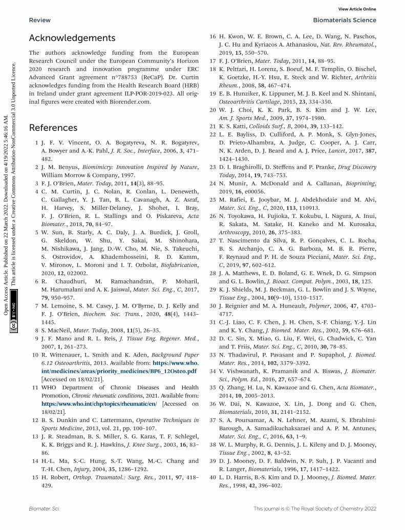
Acknowledgements
The authors acknowledge funding from the EuropeanResearch Council under the European Community’s Horizon2020 research and innovation programme under ERCAdvanced Grant agreement n°788753 (ReCaP). Dr. Curtinacknowledges funding from the Health Research Board (HRB)in Ireland under grant agreement ILP-POR-2019-023. All orig-inal figures were created with Biorender.com.
References
1 J. F. V. Vincent, O. A. Bogatyreva, N. R. Bogatyrev,A. Bowyer and A.-K. Pahl, J. R. Soc., Interface, 2006, 3, 471–482.
2 J. M. Benyus, Biomimicry: Innovation Inspired by Nature,William Morrow & Company, 1997.
3 F. J. O’Brien, Mater. Today, 2011, 14(3), 88–95.4 C. M. Curtin, J. C. Nolan, R. Conlan, L. Deneweth,
C. Gallagher, Y. J. Tan, B. L. Cavanagh, A. Z. Asraf,H. Harvey, S. Miller-Delaney, J. Shohet, I. Bray,F. J. O’Brien, R. L. Stallings and O. Piskareva, ActaBiomater., 2018, 70, 84–97.
5 W. Sun, B. Starly, A. C. Daly, J. A. Burdick, J. Groll,G. Skeldon, W. Shu, Y. Sakai, M. Shinohara,M. Nishikawa, J. Jang, D.-W. Cho, M. Nie, S. Takeuchi,S. Ostrovidov, A. Khademhosseini, R. D. Kamm,V. Mironov, L. Moroni and I. T. Ozbolat, Biofabrication,2020, 12, 022002.
6 R. Chaudhuri, M. Ramachandran, P. Moharil,M. Harumalani and A. K. Jaiswal, Mater. Sci. Eng., C, 2017,79, 950–957.
7 M. Lemoine, S. M. Casey, J. M. O’Byrne, D. J. Kelly andF. J. O’Brien, Biochem. Soc. Trans., 2020, 48(4), 1443–1445.
8 S. MacNeil, Mater. Today, 2008, 11(5), 26–35.9 J. F. Mano and R. L. Reis, J. Tissue Eng. Regener. Med.,
2007, 1, 261–273.10 R. Wittenauer, L. Smith and K. Aden, Background Paper
6.12 Osteoarthritis, 2013. Available from: https://www.who.int/medicines/areas/priority_medicines/BP6_12Osteo.pdf[Accessed on 18/02/21].
11 WHO Department of Chronic Diseases and HealthPromotion, Chronic rheumatic conditions, 2021. Available from:https://www.who.int/chp/topics/rheumatic/en/ [Accessed on18/02/21].
12 B. S. Dunkin and C. Lattermann, Operative Techniques inSports Medicine, 2013, vol. 21, pp. 100–107.
13 J. R. Steadman, B. S. Miller, S. G. Karas, T. F. Schlegel,K. K. Briggs and R. J. Hawkins, J. Knee Surg., 2003, 16, 83–86.
14 H.-L. Ma, S.-C. Hung, S.-T. Wang, M.-C. Chang andT.-H. Chen, Injury, 2004, 35, 1286–1292.
15 H. Robert, Orthop. Traumatol.: Surg. Res., 2011, 97, 418–429.
16 H. Kwon, W. E. Brown, C. A. Lee, D. Wang, N. Paschos,J. C. Hu and Kyriacos A. Athanasiou, Nat. Rev. Rheumatol.,2019, 15, 550–570.
17 F. J. O’Brien, Mater. Today, 2011, 14, 88–95.18 K. Pelttari, H. Lorenz, S. Boeuf, M. F. Templin, O. Bischel,
K. Goetzke, H.-Y. Hsu, E. Steck and W. Richter, ArthritisRheum., 2008, 58, 467–474.
19 E. B. Hunziker, K. Lippuner, M. J. B. Keel and N. Shintani,Osteoarthritis Cartilage, 2015, 23, 334–350.
20 W. J. Choi, K. K. Park, B. S. Kim and J. W. Lee,Am. J. Sports Med., 2009, 37, 1974–1980.
21 K. S. Katti, Colloids Surf., B, 2004, 39, 133–142.22 L. E. Bayliss, D. Culliford, A. P. Monk, S. Glyn-Jones,
D. Prieto-Alhambra, A. Judge, C. Cooper, A. J. Carr,N. K. Arden, D. J. Beard and A. J. Price, Lancet, 2017, 387,1424–1430.
23 D. I. Braghirolli, D. Steffens and P. Pranke, Drug DiscoveryToday, 2014, 19, 743–753.
24 N. Munir, A. McDonald and A. Callanan, Bioprinting,2019, 16, e00056.
25 M. Rafiei, E. Jooybar, M. J. Abdekhodaie and M. Alvi,Mater. Sci. Eng., C, 2020, 113, 110913.
26 N. Toyokawa, H. Fujioka, T. Kokubu, I. Nagura, A. Inui,R. Sakata, M. Satake, H. Kaneko and M. Kurosaka,Arthroscopy, 2010, 26, 375–383.
27 T. Nascimento da Silva, R. P. Gonçalves, C. L. Rocha,B. S. Archanjo, C. A. G. Barboza, M. B. R. Pierre,F. Reynaud and P. H. de Souza Picciani, Mater. Sci. Eng.,C, 2019, 97, 602–612.
28 J. A. Matthews, E. D. Boland, G. E. Wnek, D. G. Simpsonand G. L. Bowlin, J. Bioact. Compat. Polym., 2003, 18, 125.
29 K. J. Shields, M. J. Beckman, G. L. Bowlin and J. S. Wayne,Tissue Eng., 2004, 10(9–10), 1510–1517.
30 J. Reignier and M. A. Huneault, Polymer, 2006, 47, 4703–4717.
31 C.-J. Liao, C. F. Chen, J. H. Chen, S.-F. Chiang, Y.-J. Linand K. Y. Chang, J. Biomed. Mater. Res., 2002, 59, 676–681.
32 D. C. Sin, X. Miao, G. Liu, F. Wei, G. Chadwick, C. Yanand T. Friis, Mater. Sci. Eng., C, 2010, 30, 78–85.
33 N. Thadavirul, P. Pavasant and P. Supaphol, J. Biomed.Mater. Res., 2014, 102, 3379–3392.
34 V. Vishwanath, K. Pramanik and A. Biswas, J. Biomater.Sci., Polym. Ed., 2016, 27, 657–674.
35 Q. Zhang, H. Lu, N. Kawazoe and G. Chen, Acta Biomater.,2014, 10, 2005–2013.
36 W. Dai, N. Kawazoe, X. Lin, J. Dong and G. Chen,Biomaterials, 2010, 31, 2141–2152.
37 S. A. Poursamar, A. N. Lehner, M. Azami, S. Ebrahimi-Barough, A. Samadikuchaksaraei and A. P. M. Antunes,Mater. Sci. Eng., C, 2016, 63, 1–9.
38 W. L. Murphy, R. G. Dennis, J. L. Kileny and D. J. Mooney,Tissue Eng., 2002, 8, 43–52.
39 D. J. Mooney, D. F. Baldwin, N. P. Suh, J. P. Vacanti andR. Langer, Biomaterials, 1996, 17, 1417–1422.
40 L. D. Harris, B.-S. Kim and D. J. Mooney, J. Biomed. Mater.Res., 1998, 42, 396–402.
Review Biomaterials Science
Biomater. Sci. This journal is © The Royal Society of Chemistry 2022
Ope
n A
cces
s A
rtic
le. P
ublis
hed
on 2
2 M
arch
202
2. D
ownl
oade
d on
4/1
9/20
22 5
:46:
16 A
M.
Thi
s ar
ticle
is li
cens
ed u
nder
a C
reat
ive
Com
mon
s A
ttrib
utio
n-N
onC
omm
erci
al 3
.0 U
npor
ted
Lic
ence
.View Article Online

41 A. Salerno, E. Di Maio, S. Iannace and P. A. Netti, J. PorousMater., 2012, 19, 181–188.
42 I. Manavitehrani, T. Y. L. Le, S. Daly, Y. Wang, P. K. Maitz,A. Schindeler and F. Dehghani, Mater. Sci. Eng., C, 2019,96, 824–830.
43 C. M. Murphy, M. G. Haugh and F. J. O’Brien,Biomaterials, 2010, 31, 461–466.
44 C. M. Murphy, A. Matsiko, M. G. Haugh, J. P. Gleeson andF. J. O’Brien, J. Mech. Behav. Biomed. Mater., 2012, 11, 53–62.
45 F. J. O’Brien, B. A. Harley, I. V. Yannas and L. J. Gibson,Biomaterials, 2005, 26, 433–441.
46 T. J. Levingstone, A. Matsiko, G. R. Dickson, F. J. O’Brienand J. P. Gleeson, Acta Biomater., 2014, 10, 1996–2004.
47 C. M. Tierney, M. G. Haugh, J. Liedl, F. Mulcahy, B. Hayesand F. J. O’Brien, J. Mech. Behav. Biomed. Mater., 2009, 2,202–209.
48 L. Moroni, J. A. Burdick, C. B. Highley, S. J. Lee,Y. Morimoto and S. Takeuchi, Nat. Rev. Mater., 2018, 3,21–37.
49 R. M. Raftery, D. P. Walsh, L. B. Ferreras, I. M. Castaño,G. Chen, M. Lemoine, G. Osman, K. M. Shakesheff,J. E. Dixon and F. J. O’Brien, Biomaterials, 2019, 216,119277.
50 T. Gonzalez-Fernandez, S. Rathan, C. Hobbs, P. Pitacco,F. E. Freeman, G. M. Cunniffe, N. J. Dunne,H. O. McCarthy, V. Nicolosi, F. J. O’Brien and D. J. Kelly,J. Controlled Release, 2019, 301, 13–27.
51 X. Hu, Y. Man, W. Li, L. Li, J. Xu, R. Parungao, Y. Wang,S. Zheng, Y. Nie, T. Liu and K. Song, Polymers, 2019, 11,10.
52 A. C. Daly, F. E. Freeman, T. Gonzalez-Fernandez,S. E. Critchley, J. Nulty and D. J. Kelly, Adv. HealthcareMater., 2017, 6, 22.
53 M. E. Prendergast and J. A. Burdick, Adv. Mater., 2020, 32,13.
54 J. Malda, J. Visser, F. P. Melchels, T. Jüngst,W. E. Hennink, W. J. A. Dhert, J. Groll andD. W. Hutmacher, Adv. Mater., 2013, 25, 5011–5028.
55 M. Singh, H. M. Haverinen, P. Dhagat and G. E. Jabbour,Adv. Mater., 2010, 22, 673–685.
56 A. Negro, T. Cherbuin and M. P. Lutolf, Sci. Rep., 2018, 8,17099.
57 Dassault Systèmes, 3D Printing – Additive, (accessed 01/04/2020).
58 L. Koch, M. Gruene, C. Unger and B. Chichkov, Curr.Pharm. Biotechnol., 2013, 14, 91–97.
59 N. Paxton, W. Smolan, T. Böck, F. P. Melchels, J. Groll andT. Jüngst, Biofabrication, 2017, 9, 4.
60 J. Groll, J. A. Burdick, D.-W. Cho, B. Derby, M. Gelinksy,S. C. Heilshorn, T. Jüngst, J. Malda, V. A. Mironov,K. Nakayama, A. Ovsianikov, W. Sun, S. Takeuchi, J. J. Yooand T. B. F. Woodfield, Biofabrication, 2019, 11, 1.
61 A. M. Bhosale and J. B. Richardson, Br. Med. Bull., 2008,87, 77–95.
62 A. Matsiko, T. J. Levingstone and F. J. O’Brien, Materials,2013, 6, 637–668.
63 A. R. Armiento, M. J. Stoddart, M. Alini and D. Eglin, ActaBiomater., 2018, 65, 1–20.
64 A. J. S. Fox, A. Bedi and S. A. Rodeo, Sports Health, 2009, 1,461–468.
65 K. Gelse, E. Poschl and T. Aigner, Adv. Drug Delivery Rev.,2003, 55, 1531–1546.
66 K. E. Kadler, A. Hill and E. G. Canty-Laird, Curr. Opin. CellBiol., 2008, 20, 495–501.
67 J. Casale and J. S. Crane, Biochemistry, Glycosaminoglycans,StatPearls Publishing LLC, 2021, https://www.ncbi.nlm.nih.gov/books/NBK544295/ [Accessed on 18/05/21].
68 P. J. Roughley and J. S. Mort, J. Exp. Orthop., 2014, 1, 8.69 A. R. Poole, T. Kojima, T. Yasuda, F. Mwale, M. Kobayashi
and S. Laverty, Clin. Orthop. Relat. Res., 2001, 391S, S26–S33.
70 H. Akkiraju and A. Nohe, J. Dev. Biol., 2015, 3, 177–192.71 J. W. Alford and Brian J. Cole, Am. J. Sports Med., 2005,
33(2), 295–306.72 J. A. Buckwalter and H. J. Mankin, Instr. Course Lect.,
1998, 47, 477–486.73 D. Baksh, L. Song and R. S. Tuan, J. Cell. Mol. Med., 2004,
8, 301–316.74 M. Dominici, K. Le Blanc, I. Mueller, I. Slaper-
Cortenbach, F. C. Marini, D. S. Krause, R. J. Deans,A. Keating, D. J. Prockop and E. M. Horwitz, Cryotherapy,2006, 8, 315–317.
75 C. N. D. Coelho and R. A. Kosher, Dev. Biol., 1991, 144,47–53.
76 M. B. Goldring, K. Tsuchimochi and K. Ijiri, J. Cell.Biochem., 2006, 97, 33–44.
77 L. Shum and G. Nuckolls, Arthritis Res., 2002, 4, 94–106.
78 J. D. Green, V. Tollemar, M. Dougherty, Z. Yan, L. Yin,J. Ye, Z. Collier, M. K. Mohammed, R. C. Haydon,H. H. Luu, R. Kang, M. J. Lee, S. H. Ho, T.-C. He, L. L. Shiand A. Athiviraham, Genes Dis., 2015, 2, 307–327.
79 L. Yang, K. Y. Tsang, H. C. Tang, D. Chan andK. S. E. Cheah, Proc. Natl. Acad. Sci. U. S. A., 2014, 111,12097–12102.
80 M. Wang, E. R. Sampson, H. Jin, J. Li, Q. H. Ke, H.-J. Imand D. Chen, Arthritis Res. Ther., 2013, 15, 1.
81 U. Sharma, D. Pal and R. Prasad, Indian J. Clin. Biochem.,2014, 29, 269–278.
82 R. Dreier, Arthritis Res. Ther., 2010, 12, 216.83 M. Ding, Y. Lu, S. Abbassi, F. Li, X. Li, Y. Song,
V. Geoffroy, H.-J. Im and Q. Zheng, J. Cell. Physiol., 2012,227, 3446–3456.
84 Y.-Q. Yang, Y.-Y. Tan, R. Wong, A. Wenden, L.-K. Zhangand A. B. M. Rabie, Int. J. Oral Sci., 2012, 4, 64–68.
85 E. M. Ahmed, J. Adv. Res., 2015, 6, 105–121.86 S. Uman, A. P. Dhand and J. A. Burdick, J. Appl. Polym.
Sci., 2019, 137, 25.87 M. Guvendiren, H. D. Lu and J. A. Burdick, Soft Matter,
2012, 2, 260–272.
Biomaterials Science Review
This journal is © The Royal Society of Chemistry 2022 Biomater. Sci.
Ope
n A
cces
s A
rtic
le. P
ublis
hed
on 2
2 M
arch
202
2. D
ownl
oade
d on
4/1
9/20
22 5
:46:
16 A
M.
Thi
s ar
ticle
is li
cens
ed u
nder
a C
reat
ive
Com
mon
s A
ttrib
utio
n-N
onC
omm
erci
al 3
.0 U
npor
ted
Lic
ence
.View Article Online

88 F. L. C. Morgan, L. Moroni and M. B. Baker, Adv.Healthcare Mater., 2020, 9, 15.
89 S. Ji and M. Guvendiren, Front. Bioeng. Biotechnol., 2017,5, 23.
90 A. Panwar and L. P. Tan, Molecules, 2016, 21, 685.91 E. O. Osidak, V. I. Kozhukhov, M. S. Osidak and
S. P. Domogatsky, Int. J. Bioprint., 2020, 6, 270.92 M. Hospodiuk, M. Dey, D. Sosnoski and I. T. Ozbolat,
Biotechnol. Adv., 2017, 35, 217–239.93 V. L. Cross, Y. Zheng, N. W. Choi, S. S. Verbridge,
B. A. Sutermaster, L. J. Bonassar, C. Fischback andA. D. Stroock, Biomaterials, 2010, 31, 8596–8607.
94 S. Rhee, J. L. Puetzer, B. N. Mason, C. A. Reinhart-Kingand L. J. Bonassar, ACS Biomater. Sci. Eng., 2016, 2(10),1800–1805.
95 Advanced BioMatrix, Lifeink 200 – Neutralized Type ICollagen Bioink, 3 5 mg mL−1 (accessed 27/05/21).
96 Imtek Ltd, Viscoll Collagen Solution – Pig collagen type I,sterile solution (accessed 27/05/21).
97 E. O. Osidak, P. A. Karalkin, M. S. Osidak, V. A. Parfenov,D. E. Sivogrivov, F. D. A. S. Pereira, A. A. Gryadunova,E. V. Koudan, Y. D. Khesuani, V. A. Kasyanov,S. I. Belousov, S. V. Krasheninnikov, T. E. Grigoriev,S. N. Chvalun, E. A. Bulanova, V. A. Mironov andS. P. Domogatsky, J. Mater. Sci.: Mater. Med., 2019, 30(31).
98 J. M. Lee, S. K. Q. Suen, W. L. Ng, W. C. Ma andW. Y. Yeong, Macromol. Biosci., 2021, 21, 1.
99 Y. W. Koo, E.-J. Choi, J. Y. Lee, H.-J. Kim, G. H. Kim andS. H. Do, J. Ind. Eng. Chem., 2018, 66, 343–355.
100 G. Montalbano, G. Borciani, G. Cerqueni, C. Licini,F. Banche-Niclot, D. Janner, S. Sola, S. Fiorilli,M. Mattioli-Belmonte, G. Ciapetti and C. Vitale-Brovarone,Nanomaterials, 2020, 10, 1681.
101 Y. B. Kim, H. Lee and G. H. Kim, ACS Appl. Mater.Interfaces, 2016, 8, 32230–32240.
102 T.-U. Nguyen, K. E. Watkins and V. Kishore, J. Biomed.Mater. Res., Part A, 2019, 107, 1541–1550.
103 K. E. Drzewiecki, A. S. Parmar, I. D. Gaudet, J. R. Branch,D. H. Pike, V. Nanda and D. I. Shreiber, Langmuir, 2014,30, 11204–11211.
104 N. S. Kajave, T. Schmitt, T.-U. Nguyen and V. Kishore,Mater. Sci. Eng., C, 2020, 107, 110290.
105 N. S. Kajave, T. Schmitt, T.-U. Nguyen, A. K. Gaharwar andV. Kishore, Biomed. Mater., 2021, 16, 3.
106 I. D. Gaudet and D. I. Shreiber, Biointerphases, 2012, 7, 25.107 K. Guo, H. Wang, S. Li, H. Zhang, S. Li, H. Zhu, Z. Yang,
L. Zhang, P. Chang and X. Zheng, ACS Appl. Mater.Interfaces, 2021, 13, 7037–7050.
108 T. J. Hinton, Q. Jallerat, R. N. Palchesko, J. H. Park,M. S. Grodzicki, H.-J. Shue, M. H. Ramadan, A. R. Hudsonand A. W. Feinberg, Sci. Adv., 2015, 1, e1500758.
109 D. J. Shiwarski, A. R. Hudson, J. W. Tashman andA. W. Feinberg, APL Bioeng., 2021, 5, 010904.
110 A. Lee, A. R. Hudson, D. J. Shirwarski, J. W. Tashman,T. J. Hinton, S. Yerneni, J. M. Bliley, P. G. Campbell andA. W. Feinberg, Science, 2019, 365, 482–487.
111 T. Sousa, N. S. Kajave, P. Dong, L. Gu, S. Florczyk andV. Kishore, 3D Printing and Additive Manufacturing, 2021,Ahead of print.
112 M. Santoro, A. M. Tatara and A. G. Mikos, J. ControlledRelease, 2014, 190, 210–218.
113 M. B. Łabowska, K. Cierluk, A. M. Jankowska, J. Kulbacka,J. Detyna and I. Michalak, Materials, 2021, 14, 858.
114 W. Schuurman, P. A. Levett, M. W. Pot, P. R. van Weeren,W. J. A. Dhert, D. W. Hutmacher, F. P. W. Melchels,T. J. Klein and J. Malda, Macromol. Biosci., 2013, 13, 551–561.
115 Y. P. Singh, A. Bandyopadhyay and B. B. Mandal, ACSAppl. Mater. Interfaces, 2019, 11, 33684–33696.
116 S. Chawla, A. Kumar, P. Admane, A. Bandyopadhyay andS. Ghosha, Bioprinting, 2017, 7, 1–13.
117 S. Chawla, G. Desando, E. Gabusi, A. Sharma, D. Trucco,J. Chakraborty, C. Manferdini, M. Petretta, G. Lisignoliand S. Ghosh, J. Mater. Res., 2021, 36, 4051–4067.
118 S. Schwarza, S. Kuthab, T. Distler, C. Gögele, K. Stölzel,R. Detsch, A. R. Boccaccini and G. Schulze-Tanzil, Mater.Sci. Eng., C, 2020, 116, 111189.
119 J. Visser, B. Peters, T. J. Burger, J. Boomstra,W. J. A. Dhert, F. P. W. Melchels and J. Malda,Biofabrication, 2013, 5, 3.
120 Q. Gao, X. Niu, L. Shao, L. Zhou, Z. Lin, A. Sun, J. Fu,Z. Chen, J. Hu and Y. Liu, Biofabrication, 2019, 11, 3.
121 J. Liu, L. Li, H. Suo, M. Yan, J. Yin and J. Fu, Mater. Des.,2019, 171, 107708.
122 V. H. M. Mouser, R. Levato, A. Mensinga, W. J. A. Dhert,D. Gawlitta and J. Malda, Connect. Tissue Res., 2018, 61,137–151.
123 J. S. Lee, H. S. Park, H. Jung, H. Lee, H. Hong, Y. J. Lee,Y. J. Suh, O. J. Lee, S. H. Kim and C. H. Park, Addit.Manuf., 2020, 33, 101136.
124 R. Schipani, S. Scheurer, R. Florentin, S. E. Critchley andD. J. Kelly, Biofabrication, 2020, 12, 3.
125 F. Gao, Z. Xu, Q. Liang, H. Li, L. Peng, M. Wu, X. Zhao,X. Cui, C. Ruan and W. Liu, Adv. Sci., 2019, 6, 15.
126 S. Duchi, C. Onofrillo, C. D. O’Connell, R. Blanchard,C. Augustine, A. F. Quigley, R. M. I. Kapsa, P. Pivonka,G. G. Wallace, C. Di Bella and P. F. M. Choong, Sci. Rep.,2017, 7, 5387.
127 K. S. Lim, F. Abinzano, P. N. Bernal, A. A. Sanchez,P. Atienza-Roca, I. A. Otto, Q. C. Peiffer, M. Matsusaki,T. B. F. Woodfield, J. Malda and R. Levato, Adv. HealthcareMater., 2020, 9, 1901792.
128 J. A. Burdick and G. D. Prestwich, Adv. Mater., 2011, 23,H41–H56.
129 E. D. Bonnevie, D. Galesso, C. Secchieri, I. Cohen andL. J. Bonassar, PLoS One, 2015, 10, e0143415.
130 C. B. Knudson, Birth Defects Res., Part C, 2003, 69, 174–196.
131 C. Chung and J. A. Burdick, Tissue Eng., Part A, 2008, 15,2.
132 W. S. Toh, T. C. Lim, M. Kurisawa and M. Spector,Biomaterials, 2012, 33, 3835–3845.
Review Biomaterials Science
Biomater. Sci. This journal is © The Royal Society of Chemistry 2022
Ope
n A
cces
s A
rtic
le. P
ublis
hed
on 2
2 M
arch
202
2. D
ownl
oade
d on
4/1
9/20
22 5
:46:
16 A
M.
Thi
s ar
ticle
is li
cens
ed u
nder
a C
reat
ive
Com
mon
s A
ttrib
utio
n-N
onC
omm
erci
al 3
.0 U
npor
ted
Lic
ence
.View Article Online
133 I. L. Kim, S. Khetan, B. M. Baker, C. S. Chen andJ. A. Burdick, Biomaterials, 2013, 34, 5571–5580.
134 I. E. Erickson, A. H. Huang, S. Sengupta, S. Kestle,J. A. Burdick and R. L. Mauck, Osteoarthritis Cartilage,2009, 17, 1639–1648.
135 A. A. Hegewald, J. Ringe, J. Bartel, I. Krüger, M. Notter,D. Barnewitz, C. Kaps and M. Sittinger, Tissue Cell, 2004,36, 431–438.
136 R. B. Jakobsen, A. Shahdadfar, F. P. Reinholt andJ. E. Brinchmann, Knee Surg. Sports Traumatol. Arthrosc.,2010, 18, 1407–1416.
137 A. Matsiko, T. J. Levingstone, F. J. O’Brien andJ. P. Gleeson, J. Mech. Behav. Biomed. Mater., 2012, 11, 41–52.
138 I. L. Kim, R. L. Mauck and J. A. Burdick, Biomaterials,2011, 32, 8771–8782.
139 L. A. Solchaga, J. E. Dennis, V. M. Goldberg andA. I. Caplan, J. Orthop. Res., 1999, 17, 205–213.
140 Y. Jin, R. H. Koh, S.-H. Kim, K. M. Kim, G. K. Park andN. S. Hwang, Mater. Sci. Eng., C, 2020, 115,111096.
141 L. Valot, J. Martinez, A. Mehdi and G. Subra, Chem. Soc.Rev., 2019, 48, 3999–4340.
142 D. Petta, U. D’Amora, L. Ambrosio, D. W. Grijpma,D. Eglin and M. D’Este, Biofabrication, 2020, 12, 3.
143 C. Antich, J. de Vicente, G. Jiménez, C. Chocarro,E. Carrillo, E. Montañez, P. Gálvez-Martín andJ. A. Marchal, Acta Biomater., 2020, 106, 114–123.
144 M. Lafuente-Merchan, S. Ruiz-Alonso, A. Espona-Noguera,P. Galvez-Martin, E. López-Ruiz, J. A. Marchal,M. L. López-Donaire, A. Zabala, J. Ciriza, L. Saenz-del-Burgo and J. L. Pedraz, Mater. Sci. Eng., C, 2021, 126,112160.
145 S. J. Lee, J. M. Seok, J. H. Lee, J. Lee, W. D. Kim andS. A. Park, Polymers, 2021, 13, 794.
146 C. C. Piras and D. K. Smith, J. Mater. Chem. B, 2020, 8,8171–8188.
147 I. Noh, N. Kim, H. N. Tran, J. Lee and C. Lee, Biomater.Res., 2019, 23, 3.
148 B. Wang, P. J. Díaz-Payno, D. C. Browe, F. E. Freeman,J. Nulty, R. Burdis and D. J. Kelly, Acta Biomater., 2021,128, 130–142.
149 K. Y. Lee and D. J. Mooney, Prog. Polym. Sci., 2012, 37,106–126.
150 C. B. Highley, G. D. Prestwich and J. A. Burdick, Curr.Opin. Biotechnol., 2016, 40, 35–40.
151 B. S. Spearman, N. K. Agrawal, A. Rubiano,C. S. Simmons, S. Mobini and C. E. Schmidt, J. Biomed.Mater. Res., 2020, 108A, 279–291.
152 M. T. Poldervaart, B. Goversen, M. de Ruijter,A. Abbadessa, F. P. W. Melchels, F. C. Öner, W. J. A. Dhert,T. Vermonden and J. Alblas, PLoS One, 2017, 12, 6.
153 J. A. Burdick, C. Chung, X. Jia, M. A. Randolph andR. Langer, Biomacromolecules, 2005, 6, 386–391.
154 M. Y. Kwon, C. Wang, J. H. Galarraga, E. Puré, L. Han andJ. A. Burdick, Biomaterials, 2019, 222, 119451.
155 Q. Mei, J. Rao, H. P. Bei, Y. Liu and X. Zhao,Int. J. Bioprint., 2021, 7, 367.
156 M. Kesti, M. Müller, J. Becher, M. Schnabelrauch,M. D’Este, D. Eglin and M. Zenobi-Wong, Acta Biomater.,2015, 11, 162–172.
157 J. H. Galarraga, M. Y. Kwon and J. A. Burdick, Sci. Rep.,2019, 9, 19987.
158 S. Stichler, T. Böck, N. Paxton, S. Bertlein, R. Levato,V. Schill, W. Smolan, J. Malda, J. Teßmar, T. Blunk andJ. Groll, Biofabrication, 2017, 9, 4.
159 J. Hauptstein, T. Böck, M. Bartolf-Kopp, L. Forster,P. Stahlhut, A. Nadernezhad, G. Blahetek, A. Zernecke-Madsen, R. Detsch, T. Jüngst, J. Groll, J. Teßmar andT. Blunk, Adv. Healthcare Mater., 2020, 9(15).
160 D. Nguyen, D. A. Hägg, A. Forsman, J. Ekholm,P. Nimkingratana, C. Brantsing, T. Kalogeropoulos,S. Zaunz, S. Concaro, M. Brittberg, A. Lindahl,P. Gatenholm, A. Enejder and S. Simonsson, Sci. Rep.,2017, 7, 658.
161 A. K.-X. Lee, Y.-H. Lin, C.-H. Tsai, W.-T. Chang, T.-L. Linand M.-Y. Shie, Biomedicines, 2021, 9(7), 714.
162 S. Nedunchezian, P. Banerjee, C.-Y. Lee, S.-S. Lee,C.-W. Lin, C.-W. Wu, S.-C. Wu, J.-K. Chang andC.-K. Wang, Mater. Sci. Eng., C, 2021, 124, 112072.
163 P. L. Thi, J. Y. Son, Y. Lee, S. B. Ryu, K. M. Park andK. D. Park, Macromol. Res., 2020, 28, 400–406.
164 K. J. Wolf and S. Kumar, ACS Biomater. Sci. Eng., 2019, 5,3753–3765.
165 J.-T. Kim, D. Y. Lee, Y.-H. Kim, I.-K. Lee and Y.-S. Song,J. Sens. Sci. Technol., 2012, 21, 256–262.
166 C. Kiani, L. Chen, Y. J. Wu, A. J. Yee and B. B. Yang, CellRes., 2002, 12, 19–32.
167 H. Watanabe, Y. Yamada and K. Kimata, J. Biochem., 1998,124, 687–693.
168 J. J. Lim and J. S. Temenoff, Biomaterials, 2013, 34, 5007–5018.
169 J. Radhakrishnan, A. Subramanian, U. M. Krishnan andS. Sethuraman, Biomacromolecules, 2017, 18, 1–26.
170 S. Varghese, N. S. Hwang, A. C. Canver,P. Theprungsirikul, D. W. Lin and J. Elisseeff, Matrix Biol.,2008, 27, 12–21.
171 C. S. Moura, J. C. Silva, S. Faria, P. R. Fernandes, C. L. daSilva, J. M. S. Cabral, R. Linhardt, P. J. Bártolo andF. C. Ferreira, J. Biosci. Bioeng., 2020, 129, 756–764.
172 E. A. Aisenbrey and S. J. Bryant, Biomaterials, 2019,190–191, 51–62.
173 T. Li, X. Song, C. Weng, X. Wang, L. Sun, X. Gong, L. Yangand C. Chen, Appl. Mater. Today, 2018, 10, 173–183.
174 C. Tang, B. D. Holt, Z. M. Wright, A. M. Arnold, A. C. Moyand S. A. Sydlik, J. Mater. Chem. B, 2019, 7, 2442–2453.
175 X. Li, Q. Xu, M. Johnson, X. Wang, J. Lyu, Y. Li,S. McMahon, U. Greiser, S. A and W. Wang, Biomater. Sci.,2021, 9, 4139–4148.
176 S. Lee, J. Choi, J. Youn, Y. Lee, W. Kim, S. Choe, J. Song,R. L. Reis and G. Khang, Biomolecules, 2021, 11, 8.
Biomaterials Science Review
This journal is © The Royal Society of Chemistry 2022 Biomater. Sci.
Ope
n A
cces
s A
rtic
le. P
ublis
hed
on 2
2 M
arch
202
2. D
ownl
oade
d on
4/1
9/20
22 5
:46:
16 A
M.
Thi
s ar
ticle
is li
cens
ed u
nder
a C
reat
ive
Com
mon
s A
ttrib
utio
n-N
onC
omm
erci
al 3
.0 U
npor
ted
Lic
ence
.View Article Online

177 M. Liu, X. Zeng, C. Ma, H. Yi, Z. Ali, X. Mou, S. Li, Y. Dengand N. He, Bone Res., 2017, 5, 17014.
178 P. A. Levett, F. P. W. Melchels, K. Schrobback,D. W. Hutmacher, J. Malda and T. J. Klein, Acta Biomater.,2014, 10, 214–223.
179 A. Khanlari, M. S. Detamore and S. H. Gehrke,Macromolecules, 2013, 46, 9609–9617.
180 X. Liu, S. Liu, R. Yang, P. Wang, W. Zhang, X. Tan, Y. Renand B. Chi, Carbohydr. Polym., 2021, 270, 118330.
181 M. Costantini, J. Idaszek, K. Szöke, J. Jaroszewicz,M. Dentini, A. Barbetta, J. E. Brinchmann andW. Święszkowski, Biofabrication, 2016, 8(3).
182 A. Abbadessa, V. H. M. Mouser, M. M. Blokzijl,D. Gawlitta, W. J. A. Dhert, W. E. Hennink, J. Malda andT. Vermonden, Biomacromolecules, 2016, 17, 2137–2147.
183 A. Scalzone, M. A. Bonifacio, S. Cometa, F. Cucinotta,E. De Giglio, A. M. Ferreira and P. Gentile, Front. Bioeng.Biotechnol., 2020, 8, 712.
184 J. Shin, E. H. Kang, S. Choi, E. J. Jeon, J. H. Cho, D. Kang,H. Lee, I. S. Yun and S.-W. Cho, ACS Biomater. Sci. Eng.,2021, 7, 4230–4243.
185 A. Abbadessa, M. M. Blokzijl, V. H. M. Mouser, P. Marica,J. Malda, W. E. Hennink and T. Vermonden, Carbohydr.Polym., 2016, 149, 163–174.
186 A. J. Vernengo, S. Grad, D. Eglin, M. Alini and Z. Li, Adv.Funct. Mater., 2020, 30, 1909044.
187 T. W. Gilbert, T. L. Sellaro and S. F. Badylak, Biomaterials,2006, 27, 3675–3683.
188 P. M. Crapo, T. W. Gilbert and S. F. Badylak, Biomaterials,2011, 32, 3233–3243.
189 F. Pati, J. Jang, D.-H. Ha, S. W. Kim, J.-W. Rhie, J.-H. Shim,D.-H. Kim and D.-W. Cho, Nat. Commun., 2014, 5, 3935.
190 D. O. Visscher, H. Lee, P. P. M. van Zuijlen, M. N. Helder,A. Atala, J. J. Yoo and S. J. Lee, Acta Biomater., 2021, 121,193–203.
191 C. S. Jung, B. K. Kim, J. Lee, B.-H. Min and S.-H. Park,Tissue Eng. Regener. Med., 2018, 15, 155–162.
192 S. Rathan, L. Dejob, R. Schipani, B. Haffner, M. E. Möbiusand D. J. Kelly, Adv. Healthcare Mater., 2019, 8, 7.
193 X. Zhang, Y. Liu, C. Luo, C. Zhai, Z. Li, Y. Zhang, T. Yuan,S. Dong, J. Zhang and W. Fan, Mater. Sci. Eng., C, 2021,118, 111388.
194 W. Chen, Y. Xu, Y. Li, L. Jia, X. Mo, G. Jiang and G. Zhou,Chem. Eng. J., 2020, 382, 122986.
195 A. Abaci and M. Guvendiren, Adv. Healthcare Mater., 2020,9, 2000734.
196 D. O. Freytes, J. Martin, S. S. Velankar, A. S. Lee andS. F. Badylak, Biomaterials, 2008, 29, 1630–1637.
197 M. Setayeshmehr, S. Hafeez, C. van Blitterswijk,L. Moroni, C. Mota and M. B. Baker, Int. J. Mol. Sci., 2021,22, 8.
198 M. S. Silverstein, Polymer, 2020, 207, 122929.199 S. L. Vega, M. Y. Kwon and J. A. Burdick, Eur. Cells Mater.,
2017, 30, 59–75.200 A. P. Dhand, J. H. Galarraga and J. A. Burdick, Trends
Biotechnol., 2021, 39, 519–538.
201 D. Chimene, R. Kaunas and A. K. Gaharwar, Adv. Mater.,2020, 32, 1.
202 I.-C. Liao, F. T. Moutos, B. T. Estes, X. Zhao and F. Guilak,Adv. Funct. Mater., 2013, 23, 5833–5839.
203 W. Li, D. Wu, D. Hu, S. Zhu, C. Pan, Y. Jiao, L. Li, B. Luo,C. Zhou and L. Lu, Mater. Sci. Eng., C, 2020, 107, 110333.
204 O. Chaudhuri, J. Cooper-White, P. A. Janmey,D. J. Mooney and V. B. Shenoy, Nature, 2020, 584, 535–546.
205 J. S. Park, J. S. Chu, A. D. Tsou, R. Diop, Z. Tang, A. Wangand S. Li, Biomaterials, 2011, 32, 3921–3930.
206 A. S. Mao, J.-W. Shin and D. J. Mooney, Biomaterials, 2016,98, 184–191.
207 A. J. Steward and D. J. Kelly, J. Anat., 2015, 227, 717–731.208 H.-p. Lee, L. Gu, D. J. Mooney, M. E. Levenston and
O. Chaudhuri, Nat. Mater., 2017, 16, 1243–1251.209 D. Wu, Y. Yu, J. Tan, L. Huang, B. Luo, L. Lu and C. Zhou,
Mater. Des., 2018, 160, 486–495.210 T. Ni, M. Liu, Y. Zhang, Y. Cao and R. Pei, Bioconjugate
Chem., 2020, 31, 1938–1947.211 X. Yang, Z. Lu, H. Wu, W. Li, Li Zheng and J. Zhao, Mater.
Sci. Eng., C, 2018, 83, 195–201.212 N. Diamantides, C. Dugopolski, E. Blahut, S. Kennedy and
L. J. Bonassar, Biofabrication, 2019, 11, 045016.213 A. Schwab, C. Hélary, R. G. Richards, M. Alini, D. Eglin
and M. D’Este, Mater. Today Bio, 2020, 7, 100058.214 Y. Zhou, R. Qin, T. Chen, K. Zhang and J. Gui, Mater. Des.,
2021, 203, 109621.215 C. Henrionnet, L. Pourchet, P. Neybecker, O. Messaoudi,
P. Gillet, D. Loeuille, D. Mainard, C. Marquette andA. Pinzano, Stem Cells Int., 2020, 2020, 2487072.
216 I. Pepelanova, K. Kruppa, T. Scheper and A. Lavrentieva,Bioengineering, 2018, 5, 55.
217 M. Lafuente-Merchan, S. Ruiz-Alonso, A. Zabala, P. Gálvez-Martín, J. A. Marchal, B. Vázquez-Lasa, I. Gallego,L. Saenz-del-Burgo and J. L. Pedraz, Macromol. Biosci.,2022, 22(3), 2100435.
218 R. Contentin, M. Demoor, M. Concari, M. Desancé,F. Audigié, T. Branly and P. Galéra, Stem Cell Rev. Rep.,2020, 16(1), 126–143.
219 J. A. Semba, A. A. Mieloch and J. D. Rybk, Bioprinting,2020, 18, e00070.
220 O. Jeon, Y. B. Lee, T. J. Hinton, A. W. Feinberg andE. Alsberg, Mater. Today Chem., 2019, 12, 61–70.
221 C. Luo, R. Xie, J. Zhang, Y. Liu, Z. Li, Y. Zhang, X. Zhang,T. Yuan, Y. Chen and W. Fan, Tissue Eng., Part C, 2020, 26,306–316.
222 J. Yin, M. Yan, Y. Wang, J. Fu and H. Suo, ACS Appl. Mater.Interfaces, 2018, 10, 6849–6857.
223 Y. Sun, Y. You, W. Jiang, B. Wang, Q. Wu and K. Dai, Sci.Adv., 2020, 6(37).
224 Y. Sun, Y. You, W. Jiang, Z. Zhai and K. Dai, Theranostics,2019, 9(23), 6949–6961.
225 R. Levato, W. R. Webb, I. A. Otto, A. Mensinga, Y. Zhang,M. van Rijen, R. van Weeren, I. M. Khan and J. Malda,Acta Biomater., 2017, 1(61), 41–53.
Review Biomaterials Science
Biomater. Sci. This journal is © The Royal Society of Chemistry 2022
Ope
n A
cces
s A
rtic
le. P
ublis
hed
on 2
2 M
arch
202
2. D
ownl
oade
d on
4/1
9/20
22 5
:46:
16 A
M.
Thi
s ar
ticle
is li
cens
ed u
nder
a C
reat
ive
Com
mon
s A
ttrib
utio
n-N
onC
omm
erci
al 3
.0 U
npor
ted
Lic
ence
.View Article Online

226 P. Diloksumpan, M. de Ruijter, M. Castilho, U. Gbureck,T. Vermonden, P. R. van Weeren, J. Malda and R. Levato,Biofabrication, 2020, 12(2).
227 E. Vinod, R. Parameswaran, B. Ramasamy andU. Kachroo, Cartilage, 2021, 12(2_suppl), 34S–52S.
228 I. M. Khan, J. C. Bishop, S. Gilbert and C. W. Archer,Osteoarthritis Cartilage, 2009, 17, 518–528.
229 C. T. Jayasuriya and Q. Chen, Connect. Tissue Res., 2015,56(4), 265–271.
230 H. Chen, F. Fei, X. Li, Z. Nie, D. Zhou, L. Liu, J. Zhang,H. Zhang, Z. Fei and T. Xu, Bioact. Mater., 2021, 6(10),3580–3595.
231 J. M. Baena, G. Jiménez, E. López-Ruiz, C. Antich,C. Griñán-Lisón, M. Perán, P. Gálvez-Martín andJ. A. Marchal, Exp. Biol. Med., 2019, 244, 1.
232 J. Idaszek, M. Costantini, T. A. Karlsen, J. Jaroszewicz,C. Colosi, S. Testa, E. Fornetti, S. Bernardini, M. Seta,K. Kasarełło, R. Wrzesień, S. Cannata, A. Barbetta,C. Gargioli, J. E. Brinchman and W. Święszkowski,Biofabrication, 2019, 11, 4.
233 K. M. Hubka, R. L. Dahlin, V. V. Meretoja, F. K. Kasperand A. G. Mikos, Tissue Eng., Part B, 2014, 20(6), 641–654.
234 K. Pelttari, H. Lorenz, S. Boeuf, M. F. Templin, O. Bischel,K. Goetzke, H.-Y. Hsu, E. Steck and W. Richter, ArthritisRheum., 2008, 58(2), 467–474.
235 E. B. Hunziker, T. M. Quinn and H.-J. Häuselmann,Osteoarthritis Cartilage, 2002, 10, 564–572.
236 X. Ren, F. Wang, C. Chen, X. Gong, L. Yin and L. Yang,BMC Musculoskeletal Disord., 2016, 17, 301.
237 L. Zhou, G. J. V. M. van Osch, J. Malda, M. J. Stoddart,Y. Lai, R. G. Richards, K. K.-w. Ho and L. Qin, Adv.Healthcare Mater., 2020, 9(23).
238 R. M. Raftery, I. M. Castaño, S. Sperger, G. Chen,B. Cavanagh, G. A. Feichtinger, H. Redl, A. Hacobian andF. J. O’Brien, J. Controlled Release, 2018, 283, 20–31.
239 C. M. Curtin, E. G. Tierney, K. McSorley, S.-A. Cryan,G. P. Duffy and F. J. O’Brien, Adv. Healthcare Mater., 2015,4, 223–227.
240 C. M. Curtin, G. M. Cunniffe, F. G. Lyons, K. Bessho,G. R. Dickson, G. P. Duffy and F. J. O’Brien, Adv. Mater.,2012, 24, 749–754.
241 R. M. Raftery, I. M. Castaño, G. Chen, B. Cavanagh,B. Quinn, C. M. Curtin, S.-A. Cryan and F. J. O’Brien,Biomaterials, 2017, 149, 116–127.
242 Z. Yu, Y. Li, Y. Wang, Y. Chen, M. Wu, Z. Wang, M. Song,F. Lu, X. Lu and Z. Dong, Biosci. Rep., 2019, 39.
243 J. R. García, A. Y. Clark and A. J. García, J. Biomed. Mater.Res., Part A, 2016, 104, 889–900.
244 A. I. Caplan and D. Correa, J. Orthop. Res., 2011, 29, 1795–1803.
245 R. M. Raftery, A. G. Gonzalez Vazquez, G. Chen andF. J. O’Brien, Adv. Healthcare Mater., 2020, 9, 1901827.
246 N. van Gastel, S. Stegen, G. Eelen, S. Schoors, A. Carlier,V. W. Daniëls, N. Baryawno, D. Przybylski, M. Depypere,P.-J. Stiers, D. Lambrechts, R. Van Looveren, S. Torrekens,A. Sharda, P. Agostinis, D. Lambrechts, F. Maes,
J. V. Swinnen, L. Geris, H. Van Oosterwyck, B. Thienpont,P. Carmeliet, D. T. Scadden and G. Carmeliet, Nature,2020, 579, 111–117.
247 I. Sekiya, J. T. Vuoristo, B. L. Larson and D. J. Prockop,Proc. Natl. Acad. Sci. U. S. A., 2002, 99, 4397–4402.
248 B. Johnstone, T. M. Hering, A. I. Caplan, V. M. Goldbergand J. U. Yoo, Exp. Cell Res., 1998, 238, 265–272.
249 F. Barry, R. E. Boynton, B. Liu and J. M. Murphy, Exp. CellRes., 2001, 268, 189–200.
250 M. E. Joyce, A. B. Roberts, M. B. Sporn andM. E. Bolander, J. Cell Biol., 1990, 110, 2195–2207.
251 L. Bian, D. Y. Zhai, E. Tous, R. Rai, R. L. Mauck andJ. A. Burdick, Biomaterials, 2011, 32, 6425–6434.
252 X. Cui, K. Breitenkamp, M. G. Finn, M. Lotz andD. D. D’Lima, Tissue Eng., Part A, 2012, 18, 1304–1312.
253 J. Kundu, J.-H. Shim, J. Jang, S.-W. Kim and D.-W. Cho,J. Tissue Eng. Regener. Med., 2015, 9(11), 1286–1297.
254 B. S. Yoon and K. M. Lyons, J. Cell. Biochem., 2004, 93(1),93–103.
255 F. E. Freeman, P. Pitacco, L. H. A. Van Dommelen,J. Nulty, D. C. Browe, J.-Y. Shin, E. Alsberg and D. J. Kelly,Sci. Adv., 2020, 6(33).
256 Y. Zhu, M. Yuan, H. Y. Meng, A. Y. Wang, Q. Y. Guo,Y. Wang and J. Peng, Osteoarthritis Cartilage, 2013, 21(11),1627–1637.
257 E. Kon, R. Buda, G. Filardo, A. Di Martino, A. Timoncini,A. Cenacchi, P. M. Fornasari, S. Giannini andM. Marcacci, Knee Surg. Sports Traumatol. Arthrosc., 2010,18, 472–479.
258 G. Filardo, E. Kon, R. Buda, A. Timoncini, A. Di Martino,A. Cenacchi, P. M. Fornasari, S. Giannini andM. Marcacci, Knee Surg. Sports Traumatol. Arthrosc., 2011,19, 528–535.
259 E. Kon, B. Mandelbaum, R. Buda, G. Filardo,M. Delcogliano, A. Timoncini, P. M. Fornasari,S. Giannini and M. Marcacci, Arthroscopy, 2011, 27, 1490–1501.
260 G. Irmak and M. Gümüşderelioğlu, Biomed. Mater., 2020,15, 6.
261 Z. Li, X. Zhang, T. Yuan, Y. Zhang, C. Luo, J. Zhang, Y. Liuand W. Fan, Tissue Eng., Part A, 2020, 26, 15–16.
262 G. Irmak and M. Gümüşderelioğlu, Mater. Sci. Eng., C,2021, 125, 112092.
263 R. Burdis and D. J. Kelly, Acta Biomater., 2021, 126, 1–14.264 I. Martin, J. Malda and N. C. Rivron, Curr. Opin. Organ
Transplant., 2019, 24, 562–567.265 B. S. Schon, G. J. Hooper and T. B. F. Woodfield, Ann.
Biomed. Eng., 2017, 45, 100–114.266 H. Zhang, H. Huang, G. Hao, Y. Zhang, H. Ding, Z. Fan
and L. Sun, Adv. Funct. Mater., 2021, 31(1), 2006697.267 Y. Sun, Q. Wu, Y. Zhang, K. Dai and Y. Wei, Nanomedicine,
2021, 37, 102426.268 Y. Cao, P. Cheng, S. Sang, C. Xiang, Y. An, X. Wei, Z. Shen,
Y. Zhang and P. Li, Regener. Biomater., 2021, 8, 3.269 A. Dimaraki, P. J. Díaz-Payno, M. Minneboo, M. Nouri-
Goushki, M. Hosseini, N. Kops, R. Narcisi, M. J. Mirzaali,
Biomaterials Science Review
This journal is © The Royal Society of Chemistry 2022 Biomater. Sci.
Ope
n A
cces
s A
rtic
le. P
ublis
hed
on 2
2 M
arch
202
2. D
ownl
oade
d on
4/1
9/20
22 5
:46:
16 A
M.
Thi
s ar
ticle
is li
cens
ed u
nder
a C
reat
ive
Com
mon
s A
ttrib
utio
n-N
onC
omm
erci
al 3
.0 U
npor
ted
Lic
ence
.View Article Online
G. J. V. M. van Osch, L. E. Fratila-Apachitei andA. A. Zadpoor, Appl. Sci., 2021, 11, 7821.
270 Z. Yang, T. Zhao, C. Gao, F. Cao, H. Li, Z. Liao, L. Fu, P. Li,W. Chen, Z. Sun, S. Jiang, Z. Tian, G. Tian, K. Zha, T. Pan,X. Li, X. Sui, Z. Yuan, S. Liu and Q. Guo, ACS Appl. Mater.Interfaces, 2021, 13, 23369–23383.
271 C. Di Bella, S. Duchi, C. D. O’Connell, R. Blanchard,C. Augustine, Z. Yue, F. Thompson, C. Richards, S. Beirne,C. Onofrillo, S. H. Bauquier, S. D. Ryan, P. Pivonka,G. G. Wallace and P. F. Choong, Tissue Eng. Regener. Med.,2018, 12, 611–621.
272 A. G. González Vázquez, L. A. Blokpoel Ferreras,K. E. Bennett, S. M. Casey, P. A. J. Brama and F. J. O’Brien,Adv. Healthcare Mater., 2021, 10(20), e2100878.
273 M. Chen, Y. Li, S. Liu, Z. Feng, H. Wang, D. Yang, W. Guo,Z. Yuan, S. Gao, Y. Zhang, K. Zha, B. Huang, F. Wei,X. Sang, Q. Tian, X. Yang, X. Sui, Y. Zhou, Y. Zheng andQ. Guo, Bioact. Mater., 2021, 6, 1932–1944.
274 S. E. Critchley, E. J. Sheehy, G. M. Cunniffe, P. Diaz-Payno,S. F. Carroll, O. Jeon, E. Alsberg, P. A. J. Brama andD. J. Kelly, Acta Biomater., 2020, 113, 130–143.
275 F. Berthiaume, T. J. Maguire and M. L. Yarmush, Annu.Rev. Chem. Biomol. Eng., 2011, 2, 403–430.
276 D. C. Kelly, R. M. Raftery, C. M. Curtin, C. M. O’Driscolland F. J. O’Brien, J. Orthop. Res., 2019, 37, 1671–1680.
277 C.-C. Ma, Z.-L. Wang, T. Xu, Z.-Y. He and Y.-Q. Wei,Biotechnol. Adv., 2020, 40, 107502.
278 L. R. Baden, H. M. El Sahly, B. Essink, K. Kotloff, S. Frey,R. Novak, D. Diemert, S. A. Spector, N. Rouphael,C. B. Creech, J. McGettigan, S. Khetan, N. Segall, J. Solis,A. Brosz, C. Fierro, H. Schwartz, K. Neuzil, L. Corey,P. Gilbert, H. Janes, D. Follmann, M. Marovich,J. Mascola, L. Polakowski, J. Ledgerwood, B. S. Graham,H. Bennett, R. Pajon, C. Knightly, B. Leav, W. Deng,H. Zhou, S. Han, M. Ivarsson, J. Miller and T. Zaks, N.Engl. J. Med., 2021, 384, 403–416.
279 F. P. Polack, S. J. Thomas, N. Kitchin, J. Absalon,A. Gurtman, S. Lockhart, J. L. Perez, G. Pérez Marc,E. D. Moreira, C. Zerbini, R. Bailey, K. A. Swanson,S. Roychoudhury, K. Koury, P. Li, W. V. Kalina, D. Cooper,R. W. Frenck Jr., L. L. Hammitt, Ö. Türeci, H. Nell,
A. Schaefer, S. Ünal, D. B. Tresnan, S. Mather,P. R. Dormitzer, U. Şahin, K. U. Jansen and W. C. Gruber,N. Engl. J. Med., 2020, 383, 2603–2615.
280 L. Schoenmaker, D. Witzigmann, J. A. Kulkarni,R. Verbeke, G. Kersten, W. Jiskoot and D. J. A. Crommelin,Int. J. Pharm., 2021, 601, 120586.
281 T. Gonzalez-Fernandez, D. J. Kelly and F. J. O’Brien, Adv.Ther., 2018, 1(7), 1800038.
282 C. E. Dunbar, K. A. High, J. K. Joung, D. B. Kohn,K. Ozawa and M. Sadelain, Science, 2018, 359(6372),eaan4672.
283 X. Guo and L. Huang, Acc. Chem. Res., 2012, 45, 971–979.284 L. S. Costard, D. C. Kelly, R. N. Power, C. Hobbs,
S. Jaskaniec, V. Nicolosi, B. L. Cavanagh, C. M. Curtin andF. J. O’Brien, Pharmaceutics, 2020, 12, 1219.
285 F. Cardarelli, L. Digiacomo, C. Marchini, A. Amici,F. Salomone, G. Fiume, A. Rossetta, E. Gratton, D. Pozziand G. Caracciolo, Sci. Rep., 2016, 6, 25879.
286 E. G. Tierney, G. P. Duffy, A. J. Hibbitts, S.-A. Cryan andF. J. O’Brien, J. Controlled Release, 2012, 158, 304–311.
287 H. O. McCarthy, J. McCaffrey, C. M. McCrudden,A. Zholobenko, A. A. Ali, J. W. McBride, A. S. Massey,S. Pentlavalli, K.-H. Chen, G. Cole, S. P. Loughran,N. J. Dunne, R. F. Donnelly, V. L. Kett and T. Robson,J. Controlled Release, 2014, 189, 141–149.
288 J. E. Dixon, G. Osman, G. E. Morris, H. Markides,M. Rotherham, Z. Bayoussef, A. J. El Haj, C. Denning andK. M. Shakesheff, Proc. Natl. Acad. Sci. U. S. A., 2016, 113,E291–E299.
289 D. P. Walsh, R. M. Raftery, R. Murphy, G. Chen, A. Heise,F. J. O’Brien and S.-A. Cryan, Biomater. Sci., 2021, 9, 4984–4999.
290 A. L. Laiva, R. M. Raftery, M. B. Keogh and F. J. O’Brien,Int. J. Pharm., 2018, 544(2), 372–379.
291 A. L. Laiva, F. J. O’Brien and M. B. Keogh, Biotechnol.Bioeng., 2021, 118(2), 725–736.
292 M. Suku, A. L. Laiva, F. J. O’Brien and M. B. Keogh, J. Pers.Med., 2021, 11, 4.
293 W. A. Lackington, R. M. Raftery and F. J. O’Brien, ActaBiomater., 2018, 75, 115–128.
Review Biomaterials Science
Biomater. Sci. This journal is © The Royal Society of Chemistry 2022
Ope
n A
cces
s A
rtic
le. P
ublis
hed
on 2
2 M
arch
202
2. D
ownl
oade
d on
4/1
9/20
22 5
:46:
16 A
M.
Thi
s ar
ticle
is li
cens
ed u
nder
a C
reat
ive
Com
mon
s A
ttrib
utio
n-N
onC
omm
erci
al 3
.0 U
npor
ted
Lic
ence
.View Article Online