Journal of - Materials Chemistry B - RSC Publishing
17
Materials for biology and medicine Journal of Materials Chemistry B rsc.li/materials-b REVIEW ARTICLE Daniela A. Wilson et al. Enzyme catalysis powered micro/nanomotors for biomedical applications ISSN 2050-750X Indexed in Medline! Volume 8 Number 33 7 September 2020 Pages 7293–7560
-
Upload
khangminh22 -
Category
Documents
-
view
2 -
download
0
Transcript of Journal of - Materials Chemistry B - RSC Publishing
Materials for biology and medicine
Journal of Materials Chemistry Brsc.li/materials-b
REVIEW ARTICLE Daniela A. Wilson et al . Enzyme catalysis powered micro/nanomotors for biomedical applications
ISSN 2050-750X
Index
ed in
Medlin
e!
Volume 8Number 337 September 2020Pages 7293–7560

This journal is©The Royal Society of Chemistry 2020 J. Mater. Chem. B, 2020, 8, 7319--7334 | 7319
Cite this: J.Mater. Chem. B, 2020,
8, 7319
Enzyme catalysis powered micro/nanomotors forbiomedical applications
Motilal Mathesh,† Jiawei Sun† and Daniela A. Wilson *
With recent developments in the field of autonomous motion for artificial systems, many researchers are
focusing on their biomedical application for active and targeted delivery. In this context, enzyme
powered motors are at the forefront since they can utilize physiologically relevant fuels as their
substrate and carry out catalytic reactions to power motion under in vivo conditions. This review
focuses on the design and fabrication of enzyme powered motors together with their propulsion
mechanism by using fuels present in biological environments. In addition, the recent advances in the
field of enzyme powered motors for biomedical applications have been discussed together with the
parameters that need to be considered for designing such systems. We believe that this review will
provide insights and better understanding for the development of next generation biomedical
technologies based on enzyme powered motors.
1. Introduction
Nanotechnology has played a crucial role over the past fewdecades in the development of nanomedicine, from targeteddrug delivery, therapeutics, and diagnostic agents to the fabri-cation of biosensors and medical devices for maximizing theirpositive responses while minimizing side effects.1 In the pastdecade, micro/nanomotors (MNMs) have been proved to be a
potential tool for addressing complicated in vivo problems, suchas drug delivery, cell sensing, imaging, wound healing and tissueuptake. Compared to passive diffusion, active motion is powerfuland shows faster cargo delivery and overcomes drawbackssuch as systemic toxicity and reliance on passive diffusion fortransport.2 The idea of fabricating artificial motors is inspiredfrom biological motors in living systems that are ubiquitous andcapable of performing precise tasks like intracellular transport,cell division, and cell locomotion by converting chemical energyinto mechanical force.3 One of the typical examples is kinesin,a motor protein, that is able to walk along microtubules byunbinding and rebinding to the filaments through adenosine
Institute of Molecules and Materials, Radboud University, Heyendaalseweg 135,
6525 AJ, Nijmegen, The Netherlands. E-mail: [email protected]
Motilal Mathesh
Motilal Mathesh received hisPhD degree (2016) from DeakinUniversity (Australia), in the fieldof bio-nanotechnology. During hisPhD studies, he worked on enzymearchitectonics on graphene oxides.He joined Prof. Wilson’s group in2017 as a postdoctoral fellow andwas awarded the Marie CurieIndividual fellowship in 2018.Recently, he was awarded theAlfred Deakin Postdoctoral Fellow-ship and joined as a ResearchFellow at Deakin University,
Australia. His current research interest focuses on the design andfabrication of supramolecular nanomotors for biomedicalapplications and light driven systems.
Jiawei Sun
Jiawei Sun graduated from SichuanUniversity, Chengdu (China) in2017 with her master’s degree in(MSc) Pharmaceutical Science.Currently, she is a PhD candidatein the Department of SystemsChemistry at Radboud UniversityNijmegen, in the Netherlands. Herresearch focuses on developing self-propelled assembled nano-motorswith the enzyme system as a fuel,and observing the potential forbiomedical application, such asdrug delivery and biosensing.
† These authors contributed equally to this work.
Received 14th May 2020,Accepted 14th July 2020
DOI: 10.1039/d0tb01245a
rsc.li/materials-b
Journal ofMaterials Chemistry B
REVIEW
Ope
n A
cces
s A
rtic
le. P
ublis
hed
on 2
2 Ju
ly 2
020.
Dow
nloa
ded
on 7
/8/2
022
9:55
:14
AM
. T
his
artic
le is
lice
nsed
und
er a
Cre
ativ
e C
omm
ons
Attr
ibut
ion
3.0
Unp
orte
d L
icen
ce.
View Article OnlineView Journal | View Issue

7320 | J. Mater. Chem. B, 2020, 8, 7319--7334 This journal is©The Royal Society of Chemistry 2020
triphosphate (ATP) hydrolysis.4,5 In past decades, tremendousprogress has been made in developing hybrid (biological andnon-biological) devices with ATP-dependent motor proteins suchas kinesin, myosin, and dynein. Back in 2001, Vogel et al.developed a molecular shuttle with kinesin protein motors,capable of moving cargo along an engineered pathway usingATP as a fuel.6 Whitesides et al. designed a synthetic motorpowered by hydrogen peroxide (H2O2) in which platinum (Pt)was introduced on the surface of the motor as a catalyst todecompose H2O2, resulting in an impulse of oxygen bubbles forautonomous motion.7 Since then mimicking the behavior of bio-motors and bio-organisms by synthetic motors with catalysts hasattracted considerable attention, together with intensive studiestowards the realization of fuel dependent self-propulsion ofsynthetic MNMs.8 H2O2 has been widely used as a fuel becauseof the production of oxygen, while Pt has also become a popularcatalyst. Besides, H2O2, hydrazine,9 water,10 acid,11 base,12 andbromine (iodine)13 have also been used as fuels for propellingthe motors, with iridium (Ir), aluminum (Al), zinc (Zn),Al/palladium (Pd) and Pt being used as catalysts.
In spite of great progress in this field, we are still far fromusing motors for in vivo tasks due to the bottlenecks present.The main limitations are their ability to move in biologicalfluids, the size and the poor biocompatibility of both materialsused for fabrication and the fuels used for active motion.14
For biomedical applications, enzymes are ideal candidatesas catalysts that can power the motors because of their bio-compatibility, high turnover numbers and great selectivityunder physiological conditions.15,16 Back in 2005, Heller et al.reported the propulsion of carbon microfibers at an air–glucoseinterface by the ion flow produced by a catalytic reaction, usingglucose oxidase (GOx) and bilirubin oxidase (BOD) as catalysts.17
From then on, more and more enzymes have been introduced inthe intelligent systems as biocatalysts for developing enzyme-powered motors (EPMs), such as catalase,18,19 urease,20–22 atpase,23
lipase24 etc. Besides biocatalysts, the use of biocompatible
substrates is also essential, in particular, the ones present inliving systems are considered as good candidates. Motors withproof of concept studies relying on toxic fuels limit their use forbiomedical application, since it is not possible to use themin in vivo scenarios.25 Moreover, the external addition ofsubstrates to the biological system seems unrealistic, with theirsafety issues that need to be evaluated. To have a better view ofthis field, several reviews have been published recently. Themimicking of natural motility behaviour using synthetic softmaterials has been discussed by Samuel Sanchez et al.26 Thebiocompatibility of propulsion techniques has been discussedby Halder and Sun.27 Several reviews towards biomedicalapplications have also been published. In 2018, Guan et al.discussed the use of micro/nanorobots for active drugdelivery.28 Besides, Soto et al. discussed the use of micro/nanomotors for medical applications and emphasized the currentand foreseeable perspectives of their commercialization.29 More-over, the importance of biocompatibility of motors for biomedicalapplications has also been discussed by Peng et al.30 As apromising catalyst candidate, using enzyme to power micro- andnanoswimmers has been discussed by Sanchez et al.31 and Senet al.15 An overview of most recent advances of micro/nanomotorshas been discussed by Stadler et al.32 However, of all the reviews,using enzyme motors for biomedical applications has not beendiscussed. The importance of biofuels and the accessibility ofbiofuels for in vivo powering motors are also essential for thefuture perspectives of enzyme powered motors.
Our discussion in this review will first focus on the biologi-cal fuels available in biological systems and will highlightseveral possibilities of using them for motion, as they are notadequately reported in the literature. Besides, several popularbiocatalysts that can carry out catalytic reactions using thesebiological fuels as substrates are provided. As already describedbefore, MNMs equipped with inorganic catalysts will not bediscussed here as it is not the scope of this review.33,34 Thechemotaxis behavior of EPMs is also discussed, as they areessential for further biomedical applications. Finally, the recentprogress on EPMs as well as their biomedical applicationsbased on the material used for fabrication has been addressed.At the end, an outlook of current challenges and future pro-spects to enable them for in vivo applications is discussed. Webelieve that this review will lay a platform for the design andfabrication of autonomous, intelligent and stimuli responsivesystems for next generation in vivo focused MNMs.
2. Fuels in biological environments
Using enzyme-powered micro/nanomotors for biomedical appli-cations holds great promise for many medical challenges, suchas targeted drug delivery, bio-sensing and imaging. Designingsuch a system has quite some restrictions, with primarychallenges such as the fuels that can be used, whether thefuels are available in a biological system and whether thefuel concentration (conc.) can support the motion of motors,therefore warranting the urgent search for a ‘‘green fuel’’
Daniela A. Wilson
Daniela A. Wilson received herPhD degree (2007) ‘‘summa cumlaude’’ from ‘‘Gh. Asachi’’Technical University of Iasi,Romania. She is currently a fullprofessor and chair of the systemsChemistry Department at theInstitute for Molecules andMaterials, Radboud University,Nijmegen, in the Netherlands.She is also a theme leader fornanomedicine in the RadboudInstitute for Molecular Lifesciences. Her research interests
focus on the design of intelligent, self-propelled, and self-guidedsupramolecular assemblies and their communication andinteraction as next generation nanoengineered delivery systems.
Review Journal of Materials Chemistry B
Ope
n A
cces
s A
rtic
le. P
ublis
hed
on 2
2 Ju
ly 2
020.
Dow
nloa
ded
on 7
/8/2
022
9:55
:14
AM
. T
his
artic
le is
lice
nsed
und
er a
Cre
ativ
e C
omm
ons
Attr
ibut
ion
3.0
Unp
orte
d L
icen
ce.
View Article Online
This journal is©The Royal Society of Chemistry 2020 J. Mater. Chem. B, 2020, 8, 7319--7334 | 7321
present in the biological system. Furthermore, the biologicalmicroenvironment with pH and temperature changes also needsto be considered, and also whether the addition of fuels to thesystem could change the microenvironment or if the product fromthe catalytic reactions could induce a toxic response in our bodyshould be studied.33,35 In the next section, we are going to look atthe possible fuels for MNMs that can be used for biomedicalapplications and discuss their possible associated risks.
2.1 Hydrogen peroxide (H2O2)
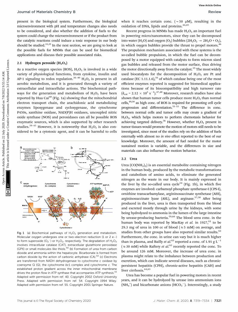
As a reactive oxygen species (ROS), H2O2 is involved in a widevariety of physiological functions, from cytokine, insulin andAP-1 signaling to redox regulation.36–40 H2O2 is present in allaerobic metabolism, and it is generated through a variety ofextracellular and intracellular actions. The biochemical path-ways for the generation and metabolism of H2O2 have beenreported by Hua Cai40 (Fig. 1a) showing that the mitochondrialelectron transport chain, the arachidonic acid metabolizingenzymes lipoxygenase and cycloxygenase, the cytochromeP450s, xanthine oxidase, NAD(P)H oxidases, uncoupled nitricoxide synthase (NOS) and peroxidases can all be possible ROSenzymatic sources, which is also supported by other researchstudies.39–43 However, it is noteworthy that H2O2 is also con-sidered to be a cytotoxic agent, and it can be harmful to cells
when it reaches certain conc. (B50 mM), resulting in theoxidation of DNA, lipids and proteins.44,45
Recent progress in MNMs has made H2O2 an important fuelin powering micro/nanomotors, since they can be decomposedinto water (H2O) and oxygen (O2) bubbles (2H2O2 - 2H2O + O2),in which oxygen bubbles provide the thrust to propel motors.46
The propulsion mechanism associated with these systems is theso-called bubble propulsion, in which the fuel can be decom-posed by a motor equipped with catalysts to form micron sizedgas bubbles and released from the motor surface, thus drivingthe motor directionally away from the catalyst.35 The most widelyused biocatalysts for the decomposition of H2O2 are Pt andcatalase (EC 1.11.1.6),16 of which catalase being one of the mostefficient enzymes reported is suggested for biomedical applica-tions because of its biocompatibility and high turnover rate(kcat = 2.12 � 105 s�1).47,48 Moreover, research studies have alsoshown that human tumor cells produce more H2O2 than normalcells,49,50 as high conc. of ROS is required for promoting cell cycleprogression and differentiation.51–53 The difference in conc.between normal cells and tumor cells may create a gradient ofH2O2, which helps motors to perform chemotaxis behavior forachieving targeted delivery.56 However, whether H2O2 present intumor tissues would promote the motion of motors still needs to beinvestigated, since most of the studies rely on the addition of fuelsexternally with almost no in vivo effect reported to the best of ourknowledge. Moreover, the amount of fuel needed for the motorsustained motion is variable, and the differences in size andmaterials can also influence the motion behavior.
2.2 Urea
Urea (CO(NH2)2) is an essential metabolite containing nitrogenin the human body, produced by the metabolic transformationsand catabolism of amino acids, to eliminate the generatednitrogen as the waste in our body. It is mainly expressed inthe liver by the so-called urea cycle54 (Fig. 1b), in which fiveenzymes are involved: carbamoyl phosphate synthetase-I (CPS-I),ornithine transcarbamylase, argininosuccinate synthetase (ASS),argininosuccinate lyase (ASL), and arginase.57,58 After beingproduced in the liver, urea is then transported from the bloodand excreted mostly through urine by the kidneys, with somebeing hydrolyzed to ammonia in the lumen of the large intestineby urease-producing bacteria.59,60 The blood urea conc. in thehuman body was reported by MacKay et al. in 1927,61 to be29.3 mg of urea in 100 cc of blood (E5 mM) on average, andstudies from other groups have also reported similar results.62
Furthermore, the conc. in urine can vary but it is much higherthan in plasma, and Bailly et al.63 reported a conc. of 1.91 g L�1
(E30 mM) while Raftery et al.62 recently reported the conc. Tobe around 126 mM. Moreover, the increase of urea conc. inplasma might relate to the imbalance between production andexcretion, which can indicate several diseases, such as chronic-persistent hepatitis (CPH), chronic-active hepatitis (CAH) andliver cirrhosis.64,65
Urea has become a popular fuel in powering motors in recentyears, and it can be hydrolyzed by urease into ammonium ions(NH4
+) and bicarbonate anions (HCO3�). Interestingly, a study
Fig. 1 (a) Biochemical pathways of H2O2 generation and metabolism.Molecular oxygen undergoes one or two-electron reduction (1 or 2 e�)to form superoxide (O2
�) or H2O2, respectively. The degradation of H2O2
involves intracellular catalase (CAT), extracellular glutathione peroxidase(GPX) or small molecules like thiols.40 (b) Formation of urea from carbondioxide and ammonia within the hepatocyte. Bicarbonate is formed fromcarbon dioxide by the action of carbonic anhydrase (CA).54 (c) Electronsare transferred from NADH dehydrogenase to cytochrome c oxidase bycoenzyme Q (Q), the cytochrome bc1 complex and cytochrome c. Theestablished proton gradient across the inner mitochondrial membranedrives the proton flow in ATP synthase that accompanies ATP synthesis.55
Adapted with permission from ref. 40. Copyright 2002 Oxford UniversityPress. Adapted with permission from ref. 54. Copyright 1994 Wiley.Adapted with permission from ref. 55. Copyright 2001 Springer Nature.
Journal of Materials Chemistry B Review
Ope
n A
cces
s A
rtic
le. P
ublis
hed
on 2
2 Ju
ly 2
020.
Dow
nloa
ded
on 7
/8/2
022
9:55
:14
AM
. T
his
artic
le is
lice
nsed
und
er a
Cre
ativ
e C
omm
ons
Attr
ibut
ion
3.0
Unp
orte
d L
icen
ce.
View Article Online

7322 | J. Mater. Chem. B, 2020, 8, 7319--7334 This journal is©The Royal Society of Chemistry 2020
by Butler et al. has shown that during hydrolysis, the diffusioncoefficient of urease is increased, and the change is consideredto be a result of the formation of the local electric fieldgenerated by the faster diffusion of NH4
+ ions.66 Furthermore,when urease is introduced on the surface of a particle, thebiocatalytic conversion of urea can result in a diffusive motionof the particles.67 However, to power urease motors, the conc.required for the fuel is quite high, even though urease is relativelyrobust and has a high turnover rate (kurease = 2.34 � 104 s�1)compared to other enzymes.68,69 As reported by researchers,50 mM conc. is necessary for the particles to move and showan increased diffusion coefficient,15,31,70 which is hard to beachieved in normal biological fluids, except urine. However,if the motors can be used in organs where there is higher ureaconc., they would be advantageous for biomedical application,since particles will not show motion before reaching thetargeted area. For example, in the treatment of bladder canceror in intravascular drug/gene delivery, passing through thebladder permeability barrier is considered to be tougher thanpassing through the blood–brain barrier (BBB),71,72 and in sucha scenario, active particles might be helpful for the deliveryof drugs benefitting from the enhanced penetration efficiencyand a recent study by Sanchez et al.67 has already proved thepossibility of this strategy. However, the risk of introducingurease into our body also needs to be considered.73
2.3 Glucose
Glucose is an essential nutrient for the human body and amajor energy source for cells delivered by the bloodstream. Thecirculation of glucose in the bloodstream is mainly due toexogenous nutrients (food) and endogenous glucose produc-tion. The liver plays a major role in controlling the conc. ofblood glucose by balancing the uptake and storage and therelease of glucose via several metabolic pathways like glycogen-esis, glycogenolysis and gluconeogenesis. In these procedures,b-cells act as sensors and secret insulin for the regulation ofglucose.74 The normal conc. of glucose in the blood stream isbetween 3.9 and 7.1 mM with the level varying throughout theday.75 It is worth noting that the liver glucose conc. can beinfluenced by the plasma glucose conc. in an opposite way,i.e. when the plasma conc. increases, the liver shows a lowerconc. and vice versa.76 Moreover, in many solid tumor tissues,the glucose level is lower than normal tissues (less than 1 mM),because of the disorganized vasculature system and theinefficient capillary bed.77–79 Glucose 1-oxidase (EC 1.1.3.4) isa FAD-dependent enzyme consisting of two identical 80 kDasubunits, widely used to catalyze the oxidation of b-D-glucose(C6H12O6) to D-gluconolactone (C6H10O6) and H2O2 in thepresence of molecular O2.80 Besides glucose oxidase (GOX),three other types of enzyme can also oxidize glucose: glucosedehydrogenases, quinoprotein glucose dehydrogenases andglucose 2-oxidases. Although glucose dehydrogenases requirea soluble cofactor, quinoprotein glucose dehydrogenase isnot so stable and glucose 2-oxidases lack specificity; thesedrawbacks of other enzymes make GOX a better choice forapplications.81 GOX was first discovered from Aspergillus niger,
and it can also be produced naturally in some fungi and insects.As it is considered to be safe, GOX has been used as an additivein food processing for decades, and also for the fabrication ofglucose biosensors.82 It has been coupled together with catalaseor other inorganic catalysts for cascade catalytic reactions, topower MNMs. Recently, Zhang et al. reported a motor system fortumor therapy, in which glucose oxidase (GOD) and manganesedioxide (MnO2) were used as pair enzymes to consume glucosein solid tumor, resulting in glucose depletion and significanttumor suppression.83 Further research is required for biomedicalapplications using glucose powered motors.
2.4 Adenosine 50-triphosphate (ATP)
It is well known that ATP is an essential cellular energy. It playsan important role in many biological processes such as musclecontraction, synthesis and degradation of biological moleculesand intracellular or extracellular signaling.84 ATP is synthesizedfrom adenosine diphosphate (ADP) and inorganic phosphate(Pi) by F1F0–ATP synthase in an energy-requiring reaction. InF1F0–ATP synthase, the F0 portion of the ATP synthase synthe-sises ATP in biological systems and the F1 portion of ATP isembedded in the membrane and catalyzes ion translocation,while an electrochemical proton (or Na+) gradient providesenergy for ATP synthesis.55,85 The human plasma ATP conc.has been measured, around 1 mmol L�1,86 while the intracellularconc. is maintained between 1 and 10 mmol L�1.87,88 However, itis noteworthy that the addition of extracellular adenosine (Ade)is proved to be toxic to cells.89
ATP has been used as a fuel for many motor proteins foundin eukaryotic cells, for example ATP transports kinesins alongthe microtubule filaments based on ATP binding, hydrolysisand ADP release.90 ATP synthase itself has been known to be aubiquitous biological nanomotor, because of the rotationmotion during ATP synthesis.23,91 This biological nanomotorhas also been used in developing devices with multiple func-tions in terms of monitoring, diagnosing and curing diseases.Kinesin, as one of the ATP motor proteins, has been usedfor making molecular shuttles for constructing nanoscaleassembly lines,6 and transporting and stretching DNA mole-cules across a surface.92 However, the use of ATP might causethe accumulation of ADP which inhibits the activity of thesebiomotors,93 and hence the balance between enzymes andsubstrates still needs to be addressed.
2.5 Other fuels
In the quest for physiologically relevant fuels, other chemicalspresent in the biological system were also studied by researchersas fuels for powering motors; for instance, a major extracellularmatrix (ECM) constituent, collagen, has been used as a fuelfor collagenase powered superparamagnetic nanoparticles, anddemonstrated enhanced tissue penetration.94 Triglyceride, amajor constituent of body fat in humans, which is also presentin blood for the bidirectional transference of adipose fat andblood glucose from the liver,95 was recently used as a fuel forpowering motors based on lipase, and the study showsthat lipase motors can destroy triglyceride droplets while
Review Journal of Materials Chemistry B
Ope
n A
cces
s A
rtic
le. P
ublis
hed
on 2
2 Ju
ly 2
020.
Dow
nloa
ded
on 7
/8/2
022
9:55
:14
AM
. T
his
artic
le is
lice
nsed
und
er a
Cre
ativ
e C
omm
ons
Attr
ibut
ion
3.0
Unp
orte
d L
icen
ce.
View Article Online
This journal is©The Royal Society of Chemistry 2020 J. Mater. Chem. B, 2020, 8, 7319--7334 | 7323
consuming triglyceride.96 Apart from them, a plethora of fuelsstill needs to be investigated for powering MNMs.
3. Enzyme chemotaxis behavior
In nature, microorganisms control the directionality of their motionby sensing the environment. One of the important behaviouralresponses is called chemotaxis, meaning that microorganismscan sense and move along the gradient of chemical species, tofind and locate a region with better living conditions.97,98
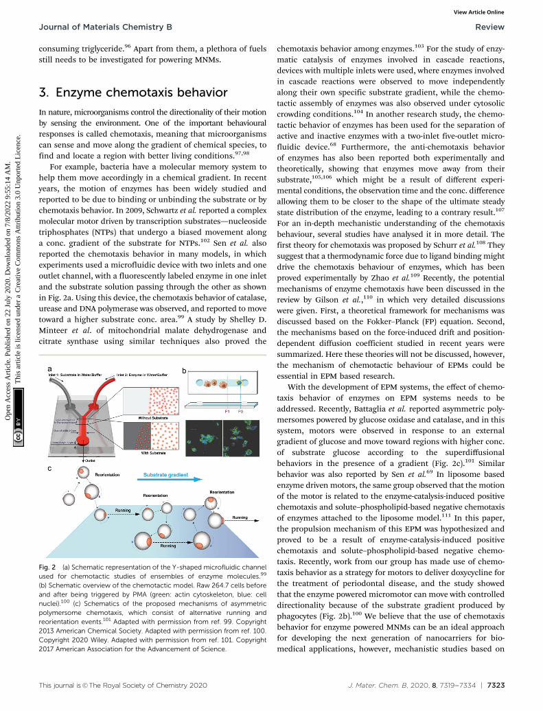
For example, bacteria have a molecular memory system tohelp them move accordingly in a chemical gradient. In recentyears, the motion of enzymes has been widely studied andreported to be due to binding or unbinding the substrate or bychemotaxis behavior. In 2009, Schwartz et al. reported a complexmolecular motor driven by transcription substrates—nucleosidetriphosphates (NTPs) that undergo a biased movement alonga conc. gradient of the substrate for NTPs.102 Sen et al. alsoreported the chemotaxis behavior in many models, in whichexperiments used a microfluidic device with two inlets and oneoutlet channel, with a fluorescently labeled enzyme in one inletand the substrate solution passing through the other as shownin Fig. 2a. Using this device, the chemotaxis behavior of catalase,urease and DNA polymerase was observed, and reported to movetoward a higher substrate conc. area.99 A study by Shelley D.Minteer et al. of mitochondrial malate dehydrogenase andcitrate synthase using similar techniques also proved the
chemotaxis behavior among enzymes.103 For the study of enzy-matic catalysis of enzymes involved in cascade reactions,devices with multiple inlets were used, where enzymes involvedin cascade reactions were observed to move independentlyalong their own specific substrate gradient, while the chemo-tactic assembly of enzymes was also observed under cytosoliccrowding conditions.104 In another research study, the chemo-tactic behavior of enzymes has been used for the separation ofactive and inactive enzymes with a two-inlet five-outlet micro-fluidic device.68 Furthermore, the anti-chemotaxis behaviorof enzymes has also been reported both experimentally andtheoretically, showing that enzymes move away from theirsubstrate,105,106 which might be a result of different experi-mental conditions, the observation time and the conc. differenceallowing them to be closer to the shape of the ultimate steadystate distribution of the enzyme, leading to a contrary result.107
For an in-depth mechanistic understanding of the chemotaxisbehaviour, several studies have analysed it in more detail. Thefirst theory for chemotaxis was proposed by Schurr et al.108 Theysuggest that a thermodynamic force due to ligand binding mightdrive the chemotaxis behaviour of enzymes, which has beenproved experimentally by Zhao et al.109 Recently, the potentialmechanisms of enzyme chemotaxis have been discussed in thereview by Gilson et al.,110 in which very detailed discussionswere given. First, a theoretical framework for mechanisms wasdiscussed based on the Fokker–Planck (FP) equation. Second,the mechanisms based on the force-induced drift and position-dependent diffusion coefficient studied in recent years weresummarized. Here these theories will not be discussed, however,the mechanism of chemotactic behaviour of EPMs could beessential in EPM based research.
With the development of EPM systems, the effect of chemo-taxis behavior of enzymes on EPM systems needs to beaddressed. Recently, Battaglia et al. reported asymmetric poly-mersomes powered by glucose oxidase and catalase, and in thissystem, motors were observed in response to an externalgradient of glucose and move toward regions with higher conc.of substrate glucose according to the superdiffusionalbehaviors in the presence of a gradient (Fig. 2c).101 Similarbehavior was also reported by Sen et al.69 In liposome basedenzyme driven motors, the same group observed that the motionof the motor is related to the enzyme-catalysis-induced positivechemotaxis and solute–phospholipid-based negative chemotaxisof enzymes attached to the liposome model.111 In this paper,the propulsion mechanism of this EPM was hypothesized andproved to be a result of enzyme-catalysis-induced positivechemotaxis and solute–phospholipid-based negative chemo-taxis. Recently, work from our group has made use of chemo-taxis behavior as a strategy for motors to deliver doxycycline forthe treatment of periodontal disease, and the study showedthat the enzyme powered micromotor can move with controlleddirectionality because of the substrate gradient produced byphagocytes (Fig. 2b).100 We believe that the use of chemotaxisbehavior for enzyme powered MNMs can be an ideal approachfor developing the next generation of nanocarriers for bio-medical applications, however, mechanistic studies based on
Fig. 2 (a) Schematic representation of the Y-shaped microfluidic channelused for chemotactic studies of ensembles of enzyme molecules.99
(b) Schematic overview of the chemotactic model. Raw 264.7 cells beforeand after being triggered by PMA (green: actin cytoskeleton, blue: cellnuclei).100 (c) Schematics of the proposed mechanisms of asymmetricpolymersome chemotaxis, which consist of alternative running andreorientation events.101 Adapted with permission from ref. 99. Copyright2013 American Chemical Society. Adapted with permission from ref. 100.Copyright 2020 Wiley. Adapted with permission from ref. 101. Copyright2017 American Association for the Advancement of Science.
Journal of Materials Chemistry B Review
Ope
n A
cces
s A
rtic
le. P
ublis
hed
on 2
2 Ju
ly 2
020.
Dow
nloa
ded
on 7
/8/2
022
9:55
:14
AM
. T
his
artic
le is
lice
nsed
und
er a
Cre
ativ
e C
omm
ons
Attr
ibut
ion
3.0
Unp
orte
d L
icen
ce.
View Article Online
7324 | J. Mater. Chem. B, 2020, 8, 7319--7334 This journal is©The Royal Society of Chemistry 2020
EPMs are necessary for further understanding of these motorsin particular when placed in biological environments.
4. Enzyme powered motors (EPMs)
The first example of enzyme powered artificial systems wasdemonstrated by Mano and Heller by binding glucose oxidaseand bilirubin oxidase on either side of a carbon fiber at thewater–O2 interface.17 Later in 2007, Feringa et al. fabricatedcarbon nanotubes covalently functionalized with glucoseoxidase and catalase enzymes, reaching speeds up to 0.8 cm s�1
in the presence of glucose and oxygen.112 These demonstrationslaid the platform for the fabrication of micro/nanomotors (MNMs)powered by enzymes by various research groups (Fig. 3). Asdiscussed in the previous section, there is a plethora of fuelsavailable in the biological environment that can be exploited asfuels to power micro/nanomotors (MNMs) with the help ofenzymes, using them as substrates for carrying out catalyticreactions and thus providing the propulsion force. This sectionwill showcase the enzymes utilized to power MNMs to datetogether with the main focus on using biologically relevant fuelstogether with their fabrication and propulsion mechanism.
4.1 Catalase based EPMs
Catalase decomposes H2O2 to produce oxygen that can powerMNMs based on the bubble propulsion mechanism. The firstreport of using catalase as a sole enzyme to drive micromotorswas demonstrated by Sanchez et al., with the help of a roll uptechnique. The fabrication was carried out using thin Au/Tifilms rolled up with a covalently bound catalase enzyme thatcould reach speeds up to 8 body-lengths per s, much higherthan the corresponding Pt-based micromotors.120 Later, thesame group used mesoporous silica clusters (MSC) to fabricate
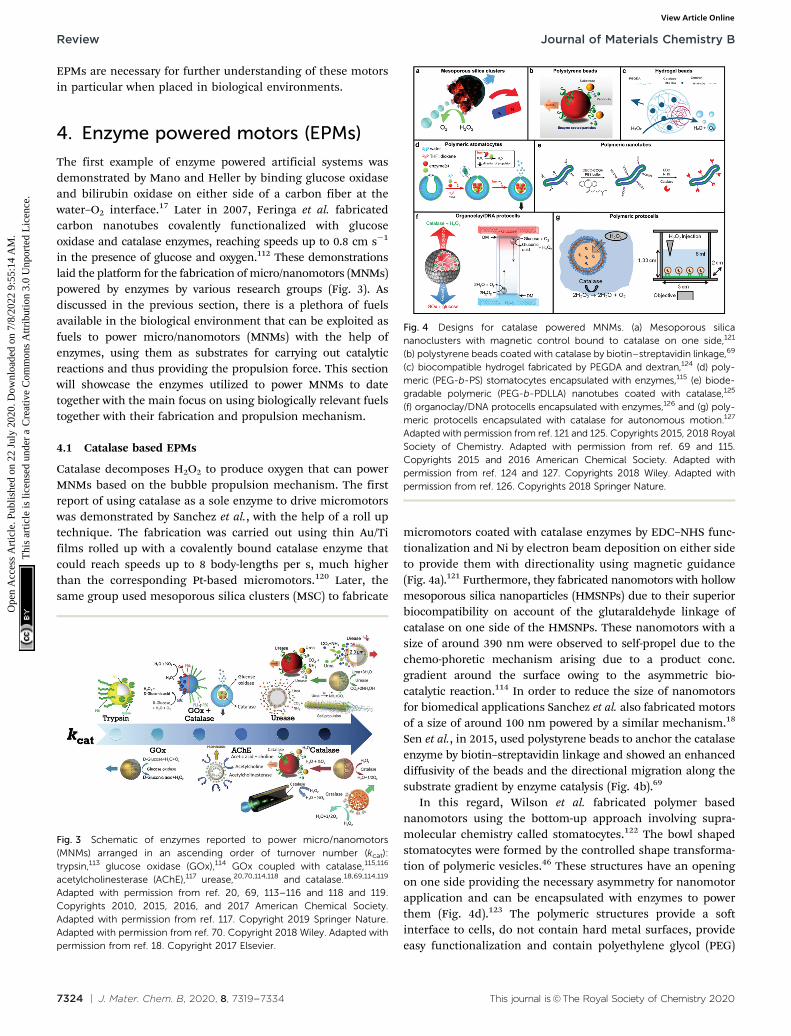
micromotors coated with catalase enzymes by EDC–NHS func-tionalization and Ni by electron beam deposition on either sideto provide them with directionality using magnetic guidance(Fig. 4a).121 Furthermore, they fabricated nanomotors with hollowmesoporous silica nanoparticles (HMSNPs) due to their superiorbiocompatibility on account of the glutaraldehyde linkage ofcatalase on one side of the HMSNPs. These nanomotors with asize of around 390 nm were observed to self-propel due to thechemo-phoretic mechanism arising due to a product conc.gradient around the surface owing to the asymmetric bio-catalytic reaction.114 In order to reduce the size of nanomotorsfor biomedical applications Sanchez et al. also fabricated motorsof a size of around 100 nm powered by a similar mechanism.18
Sen et al., in 2015, used polystyrene beads to anchor the catalaseenzyme by biotin–streptavidin linkage and showed an enhanceddiffusivity of the beads and the directional migration along thesubstrate gradient by enzyme catalysis (Fig. 4b).69
In this regard, Wilson et al. fabricated polymer basednanomotors using the bottom-up approach involving supra-molecular chemistry called stomatocytes.122 The bowl shapedstomatocytes were formed by the controlled shape transforma-tion of polymeric vesicles.46 These structures have an openingon one side providing the necessary asymmetry for nanomotorapplication and can be encapsulated with enzymes to powerthem (Fig. 4d).123 The polymeric structures provide a softinterface to cells, do not contain hard metal surfaces, provideeasy functionalization and contain polyethylene glycol (PEG)
Fig. 3 Schematic of enzymes reported to power micro/nanomotors(MNMs) arranged in an ascending order of turnover number (kcat):trypsin,113 glucose oxidase (GOx),114 GOx coupled with catalase,115,116
acetylcholinesterase (AChE),117 urease,20,70,114,118 and catalase.18,69,114,119
Adapted with permission from ref. 20, 69, 113–116 and 118 and 119.Copyrights 2010, 2015, 2016, and 2017 American Chemical Society.Adapted with permission from ref. 117. Copyright 2019 Springer Nature.Adapted with permission from ref. 70. Copyright 2018 Wiley. Adapted withpermission from ref. 18. Copyright 2017 Elsevier.
Fig. 4 Designs for catalase powered MNMs. (a) Mesoporous silicananoclusters with magnetic control bound to catalase on one side,121
(b) polystyrene beads coated with catalase by biotin–streptavidin linkage,69
(c) biocompatible hydrogel fabricated by PEGDA and dextran,124 (d) poly-meric (PEG-b-PS) stomatocytes encapsulated with enzymes,115 (e) biode-gradable polymeric (PEG-b-PDLLA) nanotubes coated with catalase,125
(f) organoclay/DNA protocells encapsulated with enzymes,126 and (g) poly-meric protocells encapsulated with catalase for autonomous motion.127
Adapted with permission from ref. 121 and 125. Copyrights 2015, 2018 RoyalSociety of Chemistry. Adapted with permission from ref. 69 and 115.Copyrights 2015 and 2016 American Chemical Society. Adapted withpermission from ref. 124 and 127. Copyrights 2018 Wiley. Adapted withpermission from ref. 126. Copyrights 2018 Springer Nature.
Review Journal of Materials Chemistry B
Ope
n A
cces
s A
rtic
le. P
ublis
hed
on 2
2 Ju
ly 2
020.
Dow
nloa
ded
on 7
/8/2
022
9:55
:14
AM
. T
his
artic
le is
lice
nsed
und
er a
Cre
ativ
e C
omm
ons
Attr
ibut
ion
3.0
Unp
orte
d L
icen
ce.
View Article Online
This journal is©The Royal Society of Chemistry 2020 J. Mater. Chem. B, 2020, 8, 7319--7334 | 7325
that is highly biocompatible.128 The stomatocytes were encapsu-lated with catalase in the stomach that could propel in thepresence of H2O2 with speeds up to 60 mm s�1 due to theexpulsion of oxygen bubbles produced through the opening,providing forward thrust. In the same study, stomatocytes wereencapsulated with a two enzyme system comprising GOx andcatalase that participates in a cascade reaction in the presence ofglucose as a substrate and propels due to self-diffusiophoresis.115
In order to have a completely biodegradable polymer, apoly(ethylene glycol)-block-poly(D,L-lactide) (PEG-b-PDLLA) diblockcopolymer was used to fabricate tubular structures functionalizedwith catalase, showing enhanced diffusion (Fig. 4e).125 PEG–PDLLAwas also used to fabricate stomatocytes with spatial control forfunctionalization by providing two different functional groupson the inside and outside of stomatocytes. The stomach of thestomatocytes was functionalized with GOx and catalase, showingpropelled motion with speeds reaching up to 15.8 mm s�1 in thepresence of glucose.129 The same group also fabricated asym-metric hydrogel based micromotors composed of dextran andpoly(ethylene glycol) diacrylate (PEGDA) with entrapped catalase(Fig. 4c). The low molecular weight dextran can diffuse intohydrogels upon UV polymerization forming an opening forbubble propulsion, when H2O2 is decomposed by catalase inthe PEGDA phase reaching speeds up to 100 mm s�1.124 Further-more, control over the speed of these micromotors was achievedby controlling the surface of the opening.130
Protocells have also been fabricated by encapsulatingcatalase in the lumen of a biotinylated polymersome. In thepresence of H2O2 the force generated by the enzymatic reactionon the surface of protocells was enough to break adhesivecontacts between the polymersome and the surface, to drive theirautonomous motion, mimicking the formation and breakage ofadhesive contacts as showcased by mammalian cells (Fig. 4g).127
In another study, catalase containing organoclay/DNA semi-permeable microcapsules were fabricated that displayed oxygengas bubble dependent buoyancy (Fig. 4f).126 When the samesystem was co-encapsulated with GOx and catalase a sustainedvertical oscillatory movement was established due to antagonisticbubble generation and depletion. Such systems could be used forthe flotation of macroscopic objects, self-sorting of mixed proto-cell communities and delivery of a biocatalyst from an inert tochemically active environment.126
4.2 Urease based EPMs
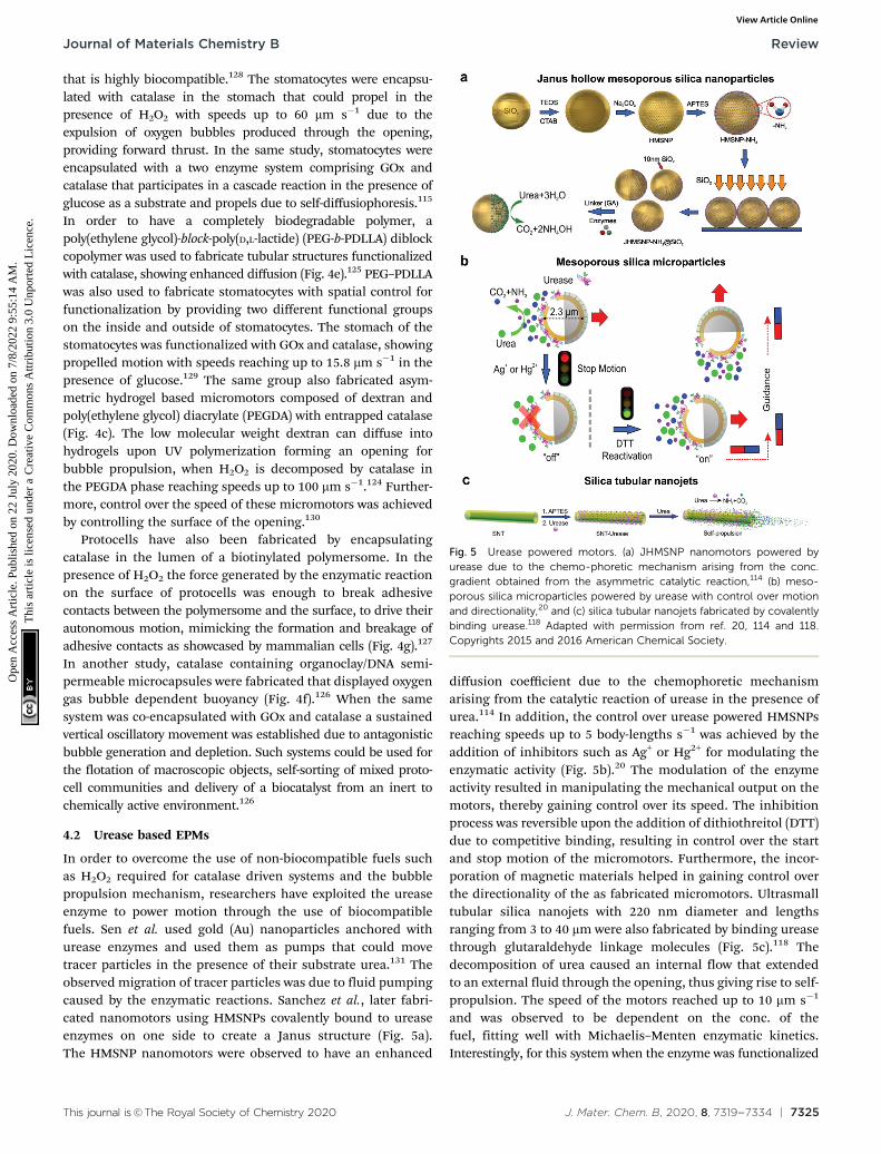
In order to overcome the use of non-biocompatible fuels suchas H2O2 required for catalase driven systems and the bubblepropulsion mechanism, researchers have exploited the ureaseenzyme to power motion through the use of biocompatiblefuels. Sen et al. used gold (Au) nanoparticles anchored withurease enzymes and used them as pumps that could movetracer particles in the presence of their substrate urea.131 Theobserved migration of tracer particles was due to fluid pumpingcaused by the enzymatic reactions. Sanchez et al., later fabri-cated nanomotors using HMSNPs covalently bound to ureaseenzymes on one side to create a Janus structure (Fig. 5a).The HMSNP nanomotors were observed to have an enhanced
diffusion coefficient due to the chemophoretic mechanismarising from the catalytic reaction of urease in the presence ofurea.114 In addition, the control over urease powered HMSNPsreaching speeds up to 5 body-lengths s�1 was achieved by theaddition of inhibitors such as Ag+ or Hg2+ for modulating theenzymatic activity (Fig. 5b).20 The modulation of the enzymeactivity resulted in manipulating the mechanical output on themotors, thereby gaining control over its speed. The inhibitionprocess was reversible upon the addition of dithiothreitol (DTT)due to competitive binding, resulting in control over the startand stop motion of the micromotors. Furthermore, the incor-poration of magnetic materials helped in gaining control overthe directionality of the as fabricated micromotors. Ultrasmalltubular silica nanojets with 220 nm diameter and lengthsranging from 3 to 40 mm were also fabricated by binding ureasethrough glutaraldehyde linkage molecules (Fig. 5c).118 Thedecomposition of urea caused an internal flow that extendedto an external fluid through the opening, thus giving rise to self-propulsion. The speed of the motors reached up to 10 mm s�1
and was observed to be dependent on the conc. of thefuel, fitting well with Michaelis–Menten enzymatic kinetics.Interestingly, for this system when the enzyme was functionalized
Fig. 5 Urease powered motors. (a) JHMSNP nanomotors powered byurease due to the chemo-phoretic mechanism arising from the conc.gradient obtained from the asymmetric catalytic reaction,114 (b) meso-porous silica microparticles powered by urease with control over motionand directionality,20 and (c) silica tubular nanojets fabricated by covalentlybinding urease.118 Adapted with permission from ref. 20, 114 and 118.Copyrights 2015 and 2016 American Chemical Society.
Journal of Materials Chemistry B Review
Ope
n A
cces
s A
rtic
le. P
ublis
hed
on 2
2 Ju
ly 2
020.
Dow
nloa
ded
on 7
/8/2
022
9:55
:14
AM
. T
his
artic
le is
lice
nsed
und
er a
Cre
ativ
e C
omm
ons
Attr
ibut
ion
3.0
Unp
orte
d L
icen
ce.
View Article Online

7326 | J. Mater. Chem. B, 2020, 8, 7319--7334 This journal is©The Royal Society of Chemistry 2020
on the outside of the nanotubes, only Brownian motion wasobserved showcasing the need for internal flows for propulsion.
4.3 Glucose oxidase (GOx) based EPMs
Glucose oxidase catalyzes the oxidation of glucose to D-glucono-1,5-lactone and H2O2 that can be used in tandem with enzymessuch as catalase for powering nanomotors. The first example ofusing GOx in tandem with bilirubin oxidase (BOD) to powerartificial systems was shown by Mano and Heller in 2005.17 Inthis system, one side of the fibre was decorated with GOxcarrying out the oxidation of glucose and another side byBOD carrying out the reduction of oxygen (Fig. 6a). Thisreaction results in the travelling of generated protons in theelectrical double layer of the fibre, dragging water moleculeswith them and thus propelling the nanomotor in the oppositedirection. Since then, there have been many MNMs fabricatedby using GOx and catalase to power motion as discussed incatalase powered systems. Here, we will discuss a few moreexamples of MNMs that are driven by the self-diffusiophoresismechanism. Stadler et al. fabricated micromotors featuring aJanus architecture with a poly(dopamine) (PDA)-coated silica(SiO2/PDA) particle as the core, followed by PEGylation withpoly(L-lysine)-grafted-poly-(ethylene glycol) (PLL-g-PEG) on oneside and immobilizing GOx and catalase on the other side.The micromotors were observed to have enhanced diffusiondependent on glucose conc. The locomotion was based on self-diffusiophoresis due to the generation of a conc. gradient of
molecules accomplished by the conversion of one moleculeinto two.116 Later, a similar system was modified with Ptnanoparticles instead of catalase enzymes that showcased a100% increase in diffusion properties due to a higher density ofGOx immobilization and no net loss in enzyme activity formetallic nanoparticles. The system was also co-immobilizedwith trypsin that cleaves peptide bonds at the carboxylic acidsites of amino acids coated with superparamagnetic manga-nese ferrite nanoparticles (MF-NPs) (Fig. 6b). The microswim-mer, could thus be propelled by two fuel sources and could beguided remotely with the help of a magnet, hence, giving rise todirectionality.113 He et al. fabricated carbonaceous nanoflasks(CNF) that could self-propel in the presence of glucose and thedirectionality could be premeditated depending on the surfacewettability of the nanoflasks (Fig. 6c). The hydrophilic CNFexhibited backward movement dominated by a local glucosegradient and hydrophobic CNF exhibited forward movement bythe generated local glucose acid gradient.132 Simulation resultsrevealed that the hydrophilic nanoflask motor generates apuller-like flow field, whereas the hydrophobic motor createsa pusher-like flow field resulting in backward and forwardmovement, respectively. In the same year, his group alsofabricated nanomotors by grafting polymer brushes on oneside and GOx on the other side of Au nanoparticles (Fig. 6d).These particles were observed to have positive chemotaxistowards the glucose gradient with speeds reaching up to 120body-lengths s�1. The grafted polymer brush provided themotor with improved translational diffusion resulting in bac-teria with swarming behavior.133
4.4 Other EPMs
Although the majority of research is focused on the above-mentioned enzymatic systems, researchers have used otherenzymes for powering motion. One such example are lipases,an industrially important enzyme that hydrolyze carboxylic esterbonds in hydrophobic compounds such as triglycerides.134
Recently, lipase was immobilized covalently on MSNPs and wereobserved to have enhanced Brownian motion in the presence ofdissolvable triglyceride solution (Fig. 7a). The lipase enzyme hadtwo functions; (a) powering the motor and (b) to degradetriglyceride (tributyrin).24 At the same time, another group alsoreported four different types of micromotor fabricated withpoly(glycidylmethacrylate)/polystyrene (PGMA/PS) particles andusing hydrophobic interactions to bind lipase (Fig. 7b).135
The micromotors revealed substrate conc. dependent enhanceddiffusion and facilitated enzymatic reactions. The propulsionmechanism of both the above-mentioned motors was basedon a product gradient due to the degradation of triglyceridemolecules.
Apart from lipase, adenosine 50-triphosphatase has also beenstudied by Sen et al. to fabricate active phospholipid vesiclesusing the hydration assembly of L-a-phosphatidylcholine (EPC)in the presence of ATPase (Fig. 7c).136 These biocompatiblephospholipid vesicles exhibited enhanced diffusion in thepresence of their substrate ATP by hydrolyzing the phosphatebonds. In the same study, similar results were also obtained for
Fig. 6 Glucose oxidase powered motors. (a) Carbon fibre decorated withGOx on one side and BOD on other side resulting in bioelectrochemicalpropulsion,16 (b) double fueled Janus swimmers decorated with GOx,trypsin, Pt and Mn–Fe nanoparticles with magnetic guidance,113 (c) surfacewettability directed carbonaceous nanoflasks with GOx and catalase,132
and (d) polymer brush functionalized Au nanoswimmers with swarmingchemotactic behavior.133 Adapted with permission from ref. 16. Copyrights2014 Royal Society of Chemistry. Adapted with permission from ref. 113and 132. Copyrights 2017 and 2019 American Chemical Society. Adaptedwith permission from ref. 133. Copyrights 2019 Wiley.
Review Journal of Materials Chemistry B
Ope
n A
cces
s A
rtic
le. P
ublis
hed
on 2
2 Ju
ly 2
020.
Dow
nloa
ded
on 7
/8/2
022
9:55
:14
AM
. T
his
artic
le is
lice
nsed
und
er a
Cre
ativ
e C
omm
ons
Attr
ibut
ion
3.0
Unp
orte
d L
icen
ce.
View Article Online

This journal is©The Royal Society of Chemistry 2020 J. Mater. Chem. B, 2020, 8, 7319--7334 | 7327
acid phosphatase (AP) enzymes that were bound to vesiclesusing a biotin–streptavidin linkage.136
4.5 Biologically relevant fluid driven motors
Until now we have discussed EPMs using biocompatible fuels suchas glucose, physiologically relevant urea and H2O2; herein MNMspowered by another important biologically relevant fuel i.e. acidicconditions prevalent in stomach and tumor cells together withwater will be discussed. The first acidic environment based ontubular micromotors was fabricated using polyaniline with the zinc(Zn) coating on the outer surface and inner surface of the tube,respectively (Fig. 8a). A spontaneous redox reaction was carriedout on the Zn surface, thereby generating hydrogen (H2), and theas-produced H2 bubbles provided forward thrust to the tubeand hence propelling them at a speed of 100 body-lengths s�1.11
Later the system was modified into double conical micromotorsfabricated only with Zn for higher capacity, combinatorial deliveryof cargoes, autonomous release of encapsulated payloads, and self-destruction.137 Simultaneously, magnesium (Mg) based micromo-tors were also fabricated that could propel in the presence of H2Oand acid. Wang et al. fabricated Mg based Janus micromotors thatself-propelled in seawater without an external fuel due to theoxidation of the Mg surface which reduces water to H2 bubbles(Fig. 8c). The micromotor was coated with Ni for magneticguidance and Au for increasing the propulsion efficiency associatedwith the microgalvanic corrosion mechanism.138 The system wasfurther modified to fabricate tubular micromotors that couldpropel in the gastrointestinal tract at a speed of 60 mm s�1 dueto H2 bubbles produced from the water-based reaction (Fig. 8b).Furthermore, Mg based micromotors were also demonstrated topropel under acidic conditions by a spontaneous reaction betweenthe Mg microsphere and the surrounding protons generating
H2 bubbles providing efficient micromotor thrust (Fig. 8d).139 Withrespect to acidic conditions, calcium carbonate (CaCO3) Janusmicromotors were fabricated to achieve motion under slightlyacidic conditions found in the tumor microenvironment withvelocities of 1.8 mm s�1 (Fig. 8e).140 The propulsion mechanismwas based on self-diffusiophoresis due to the controlled decom-position of CaCO3 into Ca2+, HCO3
�, OH� and H+ ions, therebyinducing diffusioosmotic flows generated by the difference indiffusion coefficients of the ions produced.141
5. Biomedical applications of EPMs
Enzymatically powered motors can propel themselves throughan aqueous solution by self-generated gradients and gasbubbles. With recent advancements, MNMs have been observedto have active motion using fuels present in bodily fluids orphysiologically relevant molecules that could be used byenzymes as substrates. In this section, we will discuss examplesfor EPMs based on materials used for fabrication and discusstheir relevant biomedical applications.
5.1 Mesoporous silica based EPMs
Mesoporous silica offers various advantages in terms of easysynthesis, high loading capacity, large surface area and goodbiocompatibility.142 They are easy to functionalize with the
Fig. 7 Other enzyme powered motors. (a) Mesoporous silica nanoparticlescovalently immobilized with lipase for nanomotor application,24 (b) enzymeimmobilized particles showcasing different micromotor configurationsimmobilized with lipase,135 and (c) ATPase and AP as membrane boundenzymes for enzyme powered vesicles.136 Adapted with permission fromref. 24. Copyrights 2019 Wiley. Adapted with permission from ref. 135.Copyrights 2019 Elsevier. Adapted with permission from ref. 136. Copyrights2019 American Chemical Society.
Fig. 8 Biologically relevant fluid driven systems. Acid driven micromotorsystems based on (a) Zn11 and (b) Mg;139 water driven systems (c) JanusMg,138 (d) tubular Mg base micromotors147 and (e) calcium carbonatebased Janus micromotors driven by acidic conditions in the tumourmicroenvironment.140 Adapted with permission from ref. 11 and 147.Copyrights 2012 and 2016 American Chemical Society. Adapted withpermission from ref. 138. Copyrights 2013 Royal Society of Chemistry.Adapted with permission from ref. 139. Copyrights 2017 Wiley. Adaptedwith permission from ref. 140. Copyrights 2016 Springer Nature.
Journal of Materials Chemistry B Review
Ope
n A
cces
s A
rtic
le. P
ublis
hed
on 2
2 Ju
ly 2
020.
Dow
nloa
ded
on 7
/8/2
022
9:55
:14
AM
. T
his
artic
le is
lice
nsed
und
er a
Cre
ativ
e C
omm
ons
Attr
ibut
ion
3.0
Unp
orte
d L
icen
ce.
View Article Online

7328 | J. Mater. Chem. B, 2020, 8, 7319--7334 This journal is©The Royal Society of Chemistry 2020
possibility of various surface chemistry modifications, highpotential for the physical entrapment of particles and havebeen clinically approved by FDA for biomedical applications.143
In 2012, MSNs were used for the fabrication of nanomotors forcapture and cargo transport that was powered by the catalaseenzyme. The asymmetry to the nanomotor was achieved bycoating with Au on one side that was used for functionalizingsingle-stranded DNA and the other side was functionalized withcatalase that provided the driving force in the presence of a lowconc. of H2O2.144 The system was able to capture cargo (DNAfunctionalized particle) with the help of analyte DNA strandswhose ends are complimentary to the DNA strands on nano-motors and cargoes, thereby acting as a bridge due to the highselectivity of the DNA hybridization process.144 The system wasenvisioned to be integrated in lab-on-chip devices for biome-dical diagnostic applications. Wang et al. used MSNs to fabri-cate acoustically propelled nanomotors that could releaseinsulin in the presence of glucose.145 Even though the systemwas not powered by enzymes, they were used to fabricateglucose responsive nanovalves based on phenylboronic acid(PBA)–GOx supramolecular nanostructures. The MSN particleswere loaded with insulin gated with (PBA)–GOx nanovalves toprevent the unloading of insulin. GOx present in the nanovalvesproduces H2O2 in the presence of glucose, thereby decreasingthe pH and cleaving the C–B bond and thus releasing theloaded insulin. The use of active propelled motion by ultra-sound helped in increasing the insulin release efficiently.145
Sanchez et al. coated MSNs with urease enzymes for poweringthe DOX loaded nanobots in the presence of urea (Fig. 9a).The nanobots could self-propel in ionic media and showedimproved effect on HeLa cells in the presence of urea whencompared to its passive counterparts, due to a synergistic effectbetween improved drug release kinetics and ammonia produc-tion by the catalytic decomposition of urea.70 In another study,the above system was modified to the target by specific inter-action and to reduce the proliferation of 3D bladder cancerspheroids found in the urinary bladder (Fig. 9b).146 For this, theMSN was modified with urease for powering the system andbound with an anti-FGFR3 antibody. The enhanced diffusion ofthe nanomotors at biologically relevant conc. of urea allowedthem to explore a greater area in comparison to passive diffu-sion, thus, improving the chances of interaction between theantigen and the antibody and decreasing the proliferation of thespheroids.146 Furthermore, the urease powered MSN was furtherused for the delivery of DOX to HeLa cells using a pH gatedsystem (Fig. 9c). The nanomotors were loaded with DOX graftedwith benzimidazole groups on the outer surface, and capped bythe formation of inclusion complexes between benzimidazoleand cyclodextrin-modified urease (CD-U).148 BenzimidazoleCD-U nanovalves prevented the release of DOX at physiologicalpH, but in the presence of acidic pH prevalent in the tumormicroenvironment the protonation of benzimidazole causesthe subsequent dethreading of inclusion complexes therebyreleasing the cargo. Cellular studies revealed that the presenceof active nanomotors enhanced both tumor internalization andintracellular cargo delivery.148 Ramon Martınez-Manez et al.
modified the above system by powering the motors with thecatalase enzyme and glutathione (GSH) responsive valves forcargo delivery (Fig. 9d). Glutathione is a tripeptide that carriesout redox reactions and is present in high conc. in intracellularcompartments.150 In brief, MSN particles were attached withAu nanoparticles on one side that were further covalentlyfunctionalized with catalase. Later, the MSN scaffold was loadedwith DOX and capped with disulfide linked chains.149 In thepresence of H2O2 the MSN nanomotors showed enhanced diffusionresulting in higher internalization in HeLa cells and due to thepresence of GSH, the disulfide bonds are broken thereby releasingDOX molecules. The latter two systems could sense the environmentand release the cargo molecules in the presence of a stimulus thusdisplaying features essential for biomedical applications.
5.2 Polymer based EPMs
Recently, polymers have attracted attention for the fabricationof MNMs due to easy functionalization, ease of scaling up,and appropriate physicochemical properties for biomedicalapplications with good biocompatibility.151 In addition, theycan be self-assembled into supramolecular structures that canprovide a soft interface to cells in comparison to hard metalsurface based MNMs together with good biodegradability. Heet al. fabricated Janus capsules loaded with DOX by template-assisted polyelectrolyte layer-by-layer (LBL) deposition usingpoly(styrenesulfonate) sodium salt (PSS) and poly(allylaminehydrochloride) (PAH).152 This was followed by the deposition ofAu, Cr and Ni, with Au coated with catalase enzymes thatpowered the motors by the biocatalytic decomposition of H2O2.The Janus capsule was observed to swim at 25 mm s�1 and wasdirected towards HeLa cells using an external magnetic field andupon application of near-infra red (NIR) light, it induced theshell breakage of Au particles thereby releasing DOX molecules.Such systems provide self-driving with navigation capabilitiesand perform drug loading and targeted transportation togetherwith remotely controlled release in the vicinity of cells.152 Using asimilar technique, biodegradable poly-L-lysine hydrochloride/bovineserum albumin multilayered microtubes were fabricated with theintegration of a thermal-sensitive gelatin hydrogel containing Aunanoparticles, DOX and catalase enzymes.153 The catalase reactionpropelled the motors, with Au increasing the local temperature uponillumination with NIR light, thereby releasing DOX molecules.In addition to this, the protein based structure provided highbiodegradability in the presence of a-chymotrypsin and hencesatisfying all the requirements for biomedical applications.153
Battaglia et al. fabricated chemotactic vesicles based onpolymersomes encapsulated with GOx alone or in combinationwith catalase exhibiting self-propulsion in response to a glucosegradient. The polymersomes upon further functionalizationwith low-density lipoprotein receptor-related protein 1 (LRP-1)showed a four-fold increase in blood brain barrier crossing(Fig. 10a).101 The asymmetric polymerosome propulsion velo-city was directly proportional to the glucose gradient and largerparticles were observed to have better binding affinity to thevessels due to minimized rotational diffusion.101 In 2019,Stadler et al. fabricated biocompatible self-propelled swimmers
Review Journal of Materials Chemistry B
Ope
n A
cces
s A
rtic
le. P
ublis
hed
on 2
2 Ju
ly 2
020.
Dow
nloa
ded
on 7
/8/2
022
9:55
:14
AM
. T
his
artic
le is
lice
nsed
und
er a
Cre
ativ
e C
omm
ons
Attr
ibut
ion
3.0
Unp
orte
d L
icen
ce.
View Article Online

This journal is©The Royal Society of Chemistry 2020 J. Mater. Chem. B, 2020, 8, 7319--7334 | 7329
functionalized with collagenase enzymes for using collagen as afuel and supermagnetic nanoparticles that can impart heatgeneration and used calcium for triggering the motion
(Fig. 10b).154 Collagenase enzymatically breaks collagen in theextracellular matrix and the asymmetry required for self-diffusiophoretic motion is provided by the calcium gradient
Fig. 9 Mesoporous silica based EPMs. (a) Urease powered motors for DOX delivery showing a decrease in cell viability with increasing urea conc.70
(b) Urease powered motors with antibodies against 3D bladder cancer spheroids showing decreased cell viability corresponding to urea conc.146
(c) Schematic representation of benzimidazole and cyclodextrin-modified urease powered nanomotors (i), showing internalization into HeLa cells viaTEM (ii) and high resolution confocal images with different signals corresponding to the WGA membrane marker (green), DNA-marker Hoechst 33342(blue), and DOX (red).148 (d) Schematic representation of the performance of nanobots with self-propulsion and glutathione-responsive cargo deliverycapabilities inside a cell.149 Adapted with permission from ref. 70. Copyrights 2017 Wiley. Adapted with permission from ref. 146 and 148. Copyrights 2018and 2019 American Chemical Society. Adapted with permission from ref. 149. Copyrights 2019 Royal Society of Chemistry.
Journal of Materials Chemistry B Review
Ope
n A
cces
s A
rtic
le. P
ublis
hed
on 2
2 Ju
ly 2
020.
Dow
nloa
ded
on 7
/8/2
022
9:55
:14
AM
. T
his
artic
le is
lice
nsed
und
er a
Cre
ativ
e C
omm
ons
Attr
ibut
ion
3.0
Unp
orte
d L
icen
ce.
View Article Online

7330 | J. Mater. Chem. B, 2020, 8, 7319--7334 This journal is©The Royal Society of Chemistry 2020
that also enhanced the enzymatic activity. The microswimmersenhanced penetration into cancer cell spheroids and furtherresulted in a decreased fraction of live cells when exposed to analternate magnetic field due to heat generation.154 With respect totumor cell penetration, Wilson et al. fabricated both small andultra-small stomatocytes encapsulated with the catalase enzyme inthe size ranging around 150 nm (Fig. 10c).155 The smaller size incomparison to normal stomatocytes resulted in increased cellularuptake in the presence of H2O2 as a fuel due to the enhancedpermeation and retention (EPR) effect. These systems can befurther extrapolated as cargo delivery vehicles or for targeteddelivery of drugs. In this context, poly(lactic-co-glycolic acid)microspheres were fabricated for the delivery of the doxycyclinedrug, used for the treatment of periodontal disease to prevent
bacterial infection. The micromotors were powered with cata-lase enzymes that could induce motion and provide directionalmotion in the presence of a H2O2 gradient produced bymacrophage cells incubated with phorbol-12-myristate-13-acetate.100 All the above examples showcase the advantages ofpolymer based MNMs powered by enzymes and warrant furtherinvestigation for their biomedical application.
5.3 Inorganic oxides and MOF based EPMs
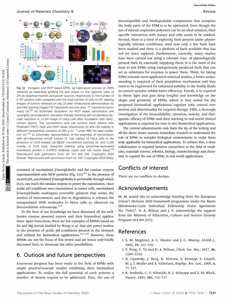
Silica materials are well studied and are reported to havenegligible cytotoxicity towards cell proliferation with goodbiocompatibility at adequate doses.156 Fischer et al. fabricatedmicropropellers using SiO2 particles coated with nickel andfunctionalized with the urease enzyme. The micropropellerswere able to penetrate the mucin gels by increasing the pHlocally due to the biocatalytic reaction of urease enzymes whichliquefies the mucus and also provides the driving force.160 Themicropropellers were directed by an external magnetic field andcould possibly be used for drug delivery applications by over-coming the limitations of acidic conditions present in thestomach.160 CaCO3 is another example for inorganic oxide thatis highly biocompatible, has low costs, is highly reproducibleand can be easily loaded with drug molecules that couldbe released under acidic pH conditions in the tumor micro-environment. With regards to micromotors, CaCO3 has beenstudied to power microspheres loaded with enzyme creatinephosphate kinase (CPK) that produce ATP. The microspheresonce bound to microtubules were able to glide with an averagevelocity of 92 � 7 nm s�1 on a surface coated with kinesin andused for cargo delivery in the presence of ADP and CP.161 Inanother example, CaCO3 cores modified with the urease enzymeon one side for powering motors, hyaluronic acid on anotherside for targeting and loaded with camptothecin for the anti-tumor effect (Fig. 11a).157 The nanomotors showed enhancedpenetration and cellular uptake into tumor cells. Upon reachingthe tumor cells the disintegration of the core due to the presenceof acidic pH conditions aids in localized delivery to CPT mole-cules thereby resulting in increased necrosis. Using such nano-motors is a feasible strategy to boost the antitumor efficacy bypromoting the local accumulation, deep tumor penetration,tumor cell capture and the intracellular release of chemotherapeuticdrugs.157 Recently, metal organic frameworks (MOF) have gainedinterest due to their porous structure and disintegration based onpH stimuli.151 Ma et al. fabricated MOF based nanomotors byencapsulating them with photosensitizers to generate cytotoxic1O2 for efficient photodynamic therapy (PDT), powered by GOxand catalase that caused starvation of cells due to the utilizationof glucose (Fig. 11b).158 The catalase enzyme also decomposedH2O2 present in the cells to produce 3O2 molecules that help inthe generation of 1O2 molecules. Overall, the active systemincreased the cellular uptake by increasing the diffusivity by27% and the synergistic effects of PDT and starvation therapyresulted in decreased cell viability of HeLa cells accountingto 73%, far better than individual therapies used in the study.158 Inanother study, the pH regulated delivery of DOX molecules to HeLacells was carried out using a MOF based system. The micromotors
Fig. 10 Polymer based EPMs. (a) Schematic representation of a chemo-tactic polymersome using a combination of copolymers. The polymer-somes encapsulate glucose oxidase and/or catalase enzymes (i), percentageof the injected dose found in the rat brain parenchyma and the capillaryfraction 10 min after the carotid artery in situ perfusion of polymersomesloaded with Gox + Cat and empty, as well as pristine asymmetric polymer-somes loaded with Gox and Cat (ii) and (iii) immunofluorescence histologiesof rat hippocampus sections of animals treated with asymmetric polymer-somes loaded with Gox + Cat.101 (b) Schematic representation of collage-nase loaded polymeric swimmers (i) and their penetration into spheroids (ii),CLSM images of the spheroids aged over 24 h and 48 h, upon addition ofpolymeric nanomotors for 24 h. The blue channel represents the stainedspheroids, and the green channel represents the swimmers (iii) and swim-mers located inside the spheroid (represented as % of internalization),determined as the percentage of green pixels in the spheroid area of images(iv).154 (c) Schematic representation of enhanced cellular uptake of ultra-small stomatocytes due to the EPR effect.155 Reproduced with permissionfrom ref. 101. Copyrights 2017 American Association for the Advancementof Science. Reproduced with permission from ref. 154 and 155. Copyrights2019 American Chemical Society.
Review Journal of Materials Chemistry B
Ope
n A
cces
s A
rtic
le. P
ublis
hed
on 2
2 Ju
ly 2
020.
Dow
nloa
ded
on 7
/8/2
022
9:55
:14
AM
. T
his
artic
le is
lice
nsed
und
er a
Cre
ativ
e C
omm
ons
Attr
ibut
ion
3.0
Unp
orte
d L
icen
ce.
View Article Online
This journal is©The Royal Society of Chemistry 2020 J. Mater. Chem. B, 2020, 8, 7319--7334 | 7331
consisted of succinylated b-lactoglobulin and the catalase enzymesuperassembled onto MOF particles (Fig. 11c).159 In the presence ofneutral pH, succinylated b-lactoglobulin is permeable through whichH2O2 can reach the catalase enzyme to power the nanomotors. Onceacidic pH conditions were encountered in tumor cells, succinylatedb-lactoglobulin undergoes reversible gelation that seizes themotion of micromotors and due to degradation it releases theencapsulated DOX molecules to HeLa cells as observed viadeconvolution microscopy.159
To the best of our knowledge we have discussed all the well-known enzyme powered motors and their biomedical applica-tions. Apart from these, there are few examples of MNMs based onZn and Mg metals studied by Wang et al. that can power motionin the presence of acidic pH conditions present in the stomachand utilized for biomedical applications.162–165 However, theseMNMs are not the focus of this review and are hence only brieflydiscussed here, to showcase the other possibilities.
6. Outlook and future perspectives
Enormous progress has been made in the field of EPMs withample proof-of-concept studies exhibiting their biomedicalapplications. To realize the full potential of such systems anumber of factors require to be addressed. First, the use of
biocompatible and biodegradable components that comprisethe body parts of the EPM is to be optimized. Even though theuse of stimuli responsive polymers can be an ideal solution, theirspecific interaction with tissues and cells needs to be studied.Second, there is a need of exploring fuels present under physio-logically relevant conditions; until now only a few fuels havebeen studied and there is a plethora of fuels available that hasnot yet been explored. Furthermore, currently, many studieshave been carried out using a relevant conc. of physiologicallypresent fuels by externally supplying them. It is the need of thehour to test EPMs using endogenously produced fuels that canact as substrates for enzymes to power them. Third, for takingEPMs towards more application-oriented studies, a better under-standing is required of their propulsion mechanism and theyneed to be engineered for enhanced stability in the bodily fluidsas current systems exhibit lower efficiency. Fourth, it is requiredto throw light and gain deep insights and knowledge on theshape and geometry of EPMs, which is best suited for theproposed biomedical applications together with control overmotion and directionality for targeted therapy. Fifth, a thoroughinvestigation of the bioavailability, retention, toxicity, and ther-apeutic efficacy of EPMs and their tracking in real world clinicalapplications is required for true in vivo biomedical applications.
The current advancements only form the tip of the iceberg andall the above issues warrant immediate research to understand therole of EPMs in complex biological environments to make themtruly applicable for biomedical applications. To achieve this, a closecollaboration is required between researchers in the field of medi-cine, materials science, robotics, biology, nanotechnology and chem-istry to expand the use of EPMs in real world applications.
Conflicts of interest
There are no conflicts to declare.
Acknowledgements
M. M. would like to acknowledge funding from the EuropeanUnion’s Horizon 2020 framework programme under the MarieSkłodowska-Curie Individual Fellowship Grant AgreementNo. 794657. D. A. Wilson and J. S. acknowledge the supportfrom the Ministry of Education, Culture and Science (GravityProgram 024.001.035).
References
1 S. M. Moghimi, A. C. Hunter and J. C. Murray, FASEB J.,2005, 19, 311–330.
2 F. Peng, Y. Tu and D. A. Wilson, Chem. Soc. Rev., 2017, 46,5289–5310.
3 R. Lipowsky, J. Beeg, R. Dimova, S. Klumpp, S. Liepelt,M. J. I. Muller and A. Valleriani, Biophys. Rev. Lett., 2009, 4,77–137.
4 K. Svoboda, C. F. Schmidt, B. J. Schnapp and S. M. Block,Nature, 1993, 365, 721–727.
Fig. 11 Inorganic and MOF based EPMs. (a) Fabrication process of JNMsobtained by separately grafting HA and urease on the opposite sides ofJPs as targeting moieties and power sources, respectively (i), the number ofki-67-positive cells compared with the total number of cells in IHC stainingimages of tumors retrieved on day 21 after intratumoral administration (ii),and H&E staining images (‘‘N’’ represents necrotic area, ‘‘T’’ represents tumormass) (iii).157 (b) Schematic illustration for MOF based nanomotors andsynergetic photodynamic-starvation therapy involving self-accelerated cas-cade reactions (i), CLSM image of HeLa cells after incubation with nano-motors (green). The cytoskeleton and cell nucleus were stained withPhalloidin-TRITC (red) and DAPI (blue), respectively (ii) and cell viability indifferent nanoparticle solutions of 100 g mL�1 under 980 nm laser irradia-tion (iii).158 (c) Schematic representation of the assembly of micromotorswith pH-responsive on/off motion (i), Cell viability of HeLa cells in thepresence of DOX-loaded cat-b@ZIF micromotor particles (ii), and CLSMoverlay of DOX (red), lysosome staining using lysosomal-associatedmembrane protein 1 (LAMP1) antibody (cyan) and cell nuclei (blue).159
Reproduced with permission from ref. 157 and 158. Copyrights 2019Elsevier. Reproduced with permission from ref. 159. Copyrights 2019 Wiley.
Journal of Materials Chemistry B Review
Ope
n A
cces
s A
rtic
le. P
ublis
hed
on 2
2 Ju
ly 2
020.
Dow
nloa
ded
on 7
/8/2
022
9:55
:14
AM
. T
his
artic
le is
lice
nsed
und
er a
Cre
ativ
e C
omm
ons
Attr
ibut
ion
3.0
Unp
orte
d L
icen
ce.
View Article Online

7332 | J. Mater. Chem. B, 2020, 8, 7319--7334 This journal is©The Royal Society of Chemistry 2020
5 A. Yildiz, M. Tomishige, R. D. Vale and P. R. Selvin, Science,2004, 303, 676.
6 H. Hess, J. Clemmens, D. Qin, J. Howard and V. Vogel,Nano Lett., 2001, 1, 235–239.
7 R. F. Ismagilov, A. Schwartz, N. Bowden and G. M. Whitesides,Angew. Chem., Int. Ed., 2002, 41, 652–654.
8 Z. Wu, X. Lin, T. Si and Q. He, Small, 2016, 12, 3080–3093.9 W. Gao, A. Pei, R. Dong and J. Wang, J. Am. Chem. Soc.,
2014, 136, 2276–2279.10 W. Gao, A. Pei and J. Wang, ACS Nano, 2012, 6, 8432–8438.11 W. Gao, A. Uygun and J. Wang, J. Am. Chem. Soc., 2012, 134,
897–900.12 W. Gao, M. D’Agostino, V. Garcia-Gradilla, J. Orozco and
J. Wang, Small, 2013, 9, 467–471.13 R. Liu and A. Sen, J. Am. Chem. Soc., 2011, 133, 20064–20067.14 D. Patra, S. Sengupta, W. Duan, H. Zhang, R. Pavlick and
A. Sen, Nanoscale, 2013, 5, 1273–1283.15 X. Zhao, K. Gentile, F. Mohajerani and A. Sen, Acc. Chem.
Res., 2018, 51, 2373–2381.16 S. Gaspar, Nanoscale, 2014, 6, 7757–7763.17 N. Mano and A. Heller, J. Am. Chem. Soc., 2005, 127,
11574–11575.18 X. Ma and S. Sanchez, Tetrahedron, 2017, 73, 4883–4886.19 J. Simmchen, A. Baeza, D. Ruiz-Molina and M. Vallet-Regı,
Nanoscale, 2014, 6, 8907–8913.20 X. Ma, X. Wang, K. Hahn and S. Sanchez, ACS Nano, 2016,
10, 3597–3605.21 T. Patino, N. Feiner-Gracia, X. Arque, A. Miguel-Lopez,
A. Jannasch, T. Stumpp, E. Schaffer, L. Albertazzi andS. Sanchez, J. Am. Chem. Soc., 2018, 140, 7896–7903.
22 M. Luo, S. Li, J. Wan, C. Yang, B. Chen and J. Guan,Langmuir, 2020, 36, 7005–7013.
23 T. Hornung, J. Martin, D. Spetzler, R. Ishmukhametov andW. D. Frasch, in Single Molecule Enzymology, Springer,2011, pp. 273–289.
24 L. Wang, A. C. Hortelao, X. Huang and S. Sanchez, Angew.Chem. Int. Ed., 2019, 58, 7992–7996.
25 X. Ma, A. C. Hortelao, T. Patino and S. Sanchez, ACS Nano,2016, 10, 9111–9122.
26 L. Wang, S. Song, J. van Hest, L. K. E. A. Abdelmohsen,X. Huang and S. Sanchez, Small, 2020, 1907680.
27 A. Halder and Y. Sun, Biosensors Bioelectron., 2019, 139, 111334.28 M. Luo, Y. Feng, T. Wang and J. Guan, Adv. Funct. Mater.,
2018, 28, 1706100.29 F. Soto and R. Chrostowski, Front. Bioeng. Biotechnol., 2018,
6, 170.30 J. Ou, K. Liu, J. Jiang, D. A. Wilson, L. Liu, F. Wang,
S. Wang, Y. Tu and F. Peng, Small, 2020, 1906184.31 T. Patino, X. Arque, R. Mestre, L. Palacios and S. Sanchez,
Acc. Chem. Res., 2018, 51, 2662–2671.32 M. Fernandez-Medina, M. A. Ramos-Docampo, O. Hovorka,
V. Salgueirino and B. Stadler, Adv. Funct. Mater., 2020,30, 1908283.
33 J. Wang and W. Gao, ACS Nano, 2012, 6, 5745–5751.34 K. K. Dey, F. Wong, A. Altemose and A. Sen, Curr. Opin.
Colloid Interface Sci., 2016, 21, 4–13.
35 L. K. Abdelmohsen, F. Peng, Y. Tu and D. A. Wilson,J. Mater. Chem. B, 2014, 2, 2395–2408.
36 H. Zhang, A. M. Gomez, X. Wang, Y. Yan, M. Zheng andH. Cheng, Cardiovasc. Res., 2013, 98, 248–258.
37 T. Finkel, Curr. Opin. Cell Biol., 1998, 10, 248–253.38 D. B. Zorov, M. Juhaszova and S. J. Sollott, Physiol. Rev.,
2014, 94, 909–950.39 E. Veal and A. Day, Antioxid. Redox Signaling, 2011, 15, 147–151.40 H. Cai, Cardiovasc. Res., 2005, 68, 26–36.41 B. Commoner, J. Townsend and G. E. Pake, Nature, 1954,
174, 689–691.42 R. K. Root and J. A. Metcalf, J. Clin. Invest., 1977, 60,
1266–1279.43 S. Miwa, J. St-Pierre, L. Partridge and M. D. Brand, Free
Radical Biol. Med., 2003, 35, 938–948.44 B. Halliwell, M. V. Clement, J. Ramalingam and L. H. Long,
IUBMB Life, 2000, 50, 251–257.45 F. Lacy, T. Kailasam Mala, T. O’Connor Daniel, W. Schmid-
Schonbein Geert and J. Parmer Robert, Hypertension, 2000,36, 878–884.
46 D. A. Wilson, R. J. M. Nolte and J. C. M. van Hest, Nat.Chem., 2012, 4, 268–274.
47 B. K. Vainshtein, W. R. Melik-Adamyan, V. V. Barynin, A. A.Vagin and A. I. Grebenko, Nature, 1981, 293, 411–412.
48 A. Deisseroth and A. L. Dounce, Physiol. Rev., 1970, 50,319–375.
49 T. P. Szatrowski and C. F. Nathan, Cancer Res., 1991, 51, 794.50 S. Toyokuni, K. Okamoto, J. Yodoi and H. Hiai, FEBS Lett.,
1995, 358, 1–3.51 J. Boonstra and J. A. Post, Gene, 2004, 337, 1–13.52 D. Trachootham, J. Alexandre and P. Huang, Nat. Rev. Drug
Discovery, 2009, 8, 579–591.53 F. Q. Schafer and G. R. Buettner, Free Radical Biol. Med.,
2001, 30, 1191–1212.54 D. S. Dimski, J. Vet. Intern. Med., 1994, 8, 73–78.55 M. Yoshida, E. Muneyuki and T. Hisabori, Nat. Rev. Mol.
Cell Biol., 2001, 2, 669–677.56 F. Peng, Y. Tu, J. C. M. van Hest and D. A. Wilson, Angew.
Chem. Int. Ed., 2015, 54, 11662–11665.57 H. Kamin and P. Handler, Annu. Rev. Biochem., 1957, 26,
419–490.58 S. M. Morris Jr, Annu. Rev. Nutr., 2002, 22, 87–105.59 A. J. Cooper, Cirrhosis, Hyperammonemia, and Hepatic
Encephalopathy, Springer, 1993, pp. 21–37.60 J. Huizenga, C. Gips and A. Tangerman, Ann. Clin. Biochem.,
1996, 33, 23–30.61 E. M. MacKay and L. L. MacKay, J. Clin. Invest., 1927, 4, 295–306.62 A. Taylor and P. Vadgama, Ann. Clin. Biochem., 1992, 29,
245–264.63 J. P. Bretaudiere, H. T. Phung and M. Bailly, Clin. Chem.,
1976, 22, 1614–1617.64 K. P. Maier, H. Talke and W. Gerok, Klin. Wochenschr.,
1979, 57, 661–665.65 B. K. Burton, Clin. Liver Dis., 2000, 4, 815–830.66 H. S. Muddana, S. Sengupta, T. E. Mallouk, A. Sen and
P. J. Butler, J. Am. Chem. Soc., 2010, 132, 2110–2111.
Review Journal of Materials Chemistry B
Ope
n A
cces
s A
rtic
le. P
ublis
hed
on 2
2 Ju
ly 2
020.
Dow
nloa
ded
on 7
/8/2
022
9:55
:14
AM
. T
his
artic
le is
lice
nsed
und
er a
Cre
ativ
e C
omm
ons
Attr
ibut
ion
3.0
Unp
orte
d L
icen
ce.
View Article Online

This journal is©The Royal Society of Chemistry 2020 J. Mater. Chem. B, 2020, 8, 7319--7334 | 7333
67 A. C. Hortelao, R. Carrascosa, N. Murillo-Cremaes, T. Patinoand S. Sanchez, ACS Nano, 2019, 13, 429–439.
68 K. K. Dey, S. Das, M. F. Poyton, S. Sengupta, P. J. Butler,P. S. Cremer and A. Sen, ACS Nano, 2014, 8, 11941–11949.
69 K. K. Dey, X. Zhao, B. M. Tansi, W. J. Mendez-Ortiz,U. M. Cordova-Figueroa, R. Golestanian and A. Sen, NanoLett., 2015, 15, 8311–8315.
70 A. C. Hortelao, T. Patino, A. Perez-Jimenez, A. Blanco andS. Sanchez, Adv. Funct. Mater., 2018, 28, 1705086.
71 M. D. Shelley, M. D. Mason and H. Kynaston, Cancer Treat.Rev., 2010, 36, 195–205.
72 P. Tyagi, P.-C. Wu, M. Chancellor, N. Yoshimura andL. Huang, Mol. Pharmaceutics, 2006, 3, 369–379.
73 D. P. Griffith, D. M. Musher and C. Itin, Invest. Urol., 1976,13, 346–350.
74 B. R. Landau, J. R. Leonards and F. M. Barry, Am. J. Physiol.,1961, 201, 41–46.
75 J. W. Appelboom, W. A. Brodsky and W. S. Rehm, J. Gen.Physiol., 1959, 43, 467–479.
76 G. F. Cahill, J. Ashmore, A. S. Earle and S. Zottu, Am.J. Physiol., 1958, 192, 491–496.
77 G. Bergers and L. E. Benjamin, Nat. Rev. Cancer, 2003, 3,401–410.
78 A. Hirayama, K. Kami, M. Sugimoto, M. Sugawara, N. Toki,H. Onozuka, T. Kinoshita, N. Saito, A. Ochiai andM. Tomita, Cancer Res., 2009, 69, 4918–4925.
79 S. A. Skinner, P. J. Tutton and P. E. O’Brien, Cancer Res.,1990, 50, 2411–2417.
80 H. J. Hecht, H. M. Kalisz, J. Hendle, R. D. Schmid andD. Schomburg, J. Mol. Biol., 1993, 229, 153–172.
81 R. Wilson and A. P. F. Turner, Biosens. Bioelectron., 1992, 7,165–185.
82 C. M. Wong, K. H. Wong and X. D. Chen, Appl. Microbiol.Biotechnol., 2008, 78, 927–938.
83 Y.-H. Zhang, W.-X. Qiu, M. Zhang, L. Zhang andX.-Z. Zhang, ACS Appl. Mater. Interfaces, 2018, 10,15030–15039.
84 H. Imamura, K. P. Huynh Nhat, H. Togawa, K. Saito,R. Iino, Y. Kato-Yamada, T. Nagai and H. Noji, Proc. Natl.Acad. Sci. U. S. A., 2009, 106, 15651.
85 C. von Ballmoos, A. Wiedenmann and P. Dimroth, Annu.Rev. Biochem., 2009, 78, 649–672.
86 M. W. Gorman, E. O. Feigl and C. W. Buffington, Clin.Chem., 2007, 53, 318–325.
87 M. L. Casem, in Case Studies in Cell Biology, ed. M. L. Casem,Academic Press, Boston, 2016, pp. 263–281.
88 F. M. Gribble, G. Loussouarn, S. J. Tucker, C. Zhao, C. G.Nichols and F. M. Ashcroft, J. Biol. Chem., 2000, 275,30046–30049.
89 M. Leist, B. Single, A. F. Castoldi, S. Kuhnle andP. Nicotera, J. Exp. Med., 1997, 185, 1481–1486.
90 M. J. Schnitzer and S. M. Block, Nature, 1997, 388, 386–390.91 H. Noji, R. Yasuda, M. Yoshida and K. Kinosita, Nature,
1997, 386, 299–302.92 S. Diez, C. Reuther, C. Dinu, R. Seidel, M. Mertig,
W. Pompe and J. Howard, Nano Lett., 2003, 3, 1251–1254.
93 W. R. Schief, R. H. Clark, A. H. Crevenna and J. Howard,Proc. Natl. Acad. Sci. U. S. A., 2004, 101, 1183.
94 M. A. Ramos-Docampo, M. Fernandez-Medina, E. Taipaleenmaki,O. Hovorka, V. N. Salgueirino and B. Stadler, ACS Nano,2019, 13, 12192–12205.
95 V. Truong, S. Huang, J. Dennis, M. Lemire, N. Zwingerman,D. Aıssi, I. Kassam, C. Perret, P. Wells, P.-E. Morange,M. Wilson, D.-A. Tregouet and F. Gagnon, Sci. Rep., 2017,7, 11207.
96 L. Wang, A. C. Hortelao, X. Huang and S. Sanchez, Angew.Chem., 2019, 131, 8076–8080.
97 H. C. Berg and D. A. Brown, Nature, 1972, 239, 500–504.98 A. C. Grimm and C. S. Harwood, Appl. Environ. Microbiol.,
1997, 63, 4111–4115.99 S. Sengupta, K. K. Dey, H. S. Muddana, T. Tabouillot,
M. E. Ibele, P. J. Butler and A. Sen, J. Am. Chem. Soc.,2013, 135, 1406–1414.
100 J. Wang, B. J. Toebes, A. S. Plachokova, Q. Liu, D. Deng,J. A. Jansen, F. Yang and D. A. Wilson, Adv. HealtcareMater., 2020, 9, 1901710.
101 A. Joseph, C. Contini, D. Cecchin, S. Nyberg, L. Ruiz-Perez,J. Gaitzsch, G. Fullstone, X. Tian, J. Azizi, J. Preston,G. Volpe and G. Battaglia, Sci. Adv., 2017, 3, e1700362.
102 H. Yu, K. Jo, K. L. Kounovsky, J. J. D. Pablo andD. C. Schwartz, J. Am. Chem. Soc., 2009, 131, 5722–5723.
103 F. Wu, L. N. Pelster and S. D. Minteer, Chem. Commun.,2015, 51, 1244–1247.
104 X. Zhao, H. Palacci, V. Yadav, M. M. Spiering, M. K. Gilson,P. J. Butler, H. Hess, S. J. Benkovic and A. Sen, Nat. Chem.,2018, 10, 311–317.
105 J. Agudo-Canalejo, P. Illien and R. Golestanian, Nano Lett.,2018, 18, 2711–2717.
106 A.-Y. Jee, S. Dutta, Y.-K. Cho, T. Tlusty and S. Granick, Proc.Natl. Acad. Sci. U. S. A., 2018, 115, 14–18.
107 M. Feng and M. K. Gilson, Annu. Rev. Biophys., 2020, 49, 87–105.108 J. M. Schurr, B. S. Fujimoto, L. Huynh and D. T. Chiu,
J. Phys. Chem. B, 2013, 117, 7626–7652.109 X. Zhao, H. Palacci, V. Yadav, M. M. Spiering, M. K. Gilson,
P. J. Butler, H. Hess, S. J. Benkovic and A. Sen, Nat. Chem.,2018, 10, 311.
110 M. Feng and M. K. Gilson, Annu. Rev. Biophys., 2020, 49,87–105.
111 A. Somasundar, S. Ghosh, F. Mohajerani, L. N. Massenburg,T. Yang, P. S. Cremer, D. Velegol and A. Sen, Nat. Nano-technol., 2019, 14, 1129–1134.
112 D. Pantarotto, W. R. Browne and B. L. Feringa, Chem.Commun., 2008, 1533–1535.
113 P. S. Schattling, M. A. Ramos-Docampo, V. Salgueirino andB. Stadler, ACS Nano, 2017, 11, 3973–3983.
114 X. Ma, A. Jannasch, U.-R. Albrecht, K. Hahn, A. Miguel-Lopez,E. Schaffer and S. Sanchez, Nano Lett., 2015, 15, 7043–7050.
115 L. K. E. A. Abdelmohsen, M. Nijemeisland, G. M. Pawar,G.-J. A. Janssen, R. J. M. Nolte, J. C. M. van Hest andD. A. Wilson, ACS Nano, 2016, 10, 2652–2660.
116 P. Schattling, B. Thingholm and B. Stadler, Chem. Mater.,2015, 27, 7412–7418.
Journal of Materials Chemistry B Review
Ope
n A
cces
s A
rtic
le. P
ublis
hed
on 2
2 Ju
ly 2
020.
Dow
nloa
ded
on 7
/8/2
022
9:55
:14
AM
. T
his
artic
le is
lice
nsed
und
er a
Cre
ativ
e C
omm
ons
Attr
ibut
ion
3.0
Unp
orte
d L
icen
ce.
View Article Online
7334 | J. Mater. Chem. B, 2020, 8, 7319--7334 This journal is©The Royal Society of Chemistry 2020
117 X. Arque, A. Romero-Rivera, F. Feixas, T. Patino, S. Osunaand S. Sanchez, Nat. Commun., 2019, 10, 2826.
118 X. Ma, A. C. Hortelao, A. Miguel-Lopez and S. Sanchez,J. Am. Chem. Soc., 2016, 138, 13782–13785.
119 S. Sanchez, A. A. Solovev, Y. Mei and O. G. Schmidt, J. Am.Chem. Soc., 2010, 132, 13144–13145.
120 A. A. Solovev, S. Sanchez, M. Pumera, Y. F. Mei andO. G. Schmidt, Adv. Funct. Mater., 2010, 20, 2430–2435.
121 X. Ma and S. Sanchez, Chem. Commun., 2015, 51, 5467–5470.122 I. Ortiz-Rivera, M. Mathesh and D. A. Wilson, Acc. Chem.
Res., 2018, 51, 1891–1900.123 M. Nijemeisland, L. K. E. A. Abdelmohsen, W. T. S. Huck,
D. A. Wilson and J. C. M. van Hest, ACS Cent. Sci., 2016, 2,843–849.
124 S. Keller, S. P. Teora, G. X. Hu, M. Nijemeisland andD. A. Wilson, Angew. Chem. Int. Ed., 2018, 57, 9814–9817.
125 B. J. Toebes, L. K. E. A. Abdelmohsen and D. A. Wilson,Polym. Chem., 2018, 9, 3190–3194.
126 B. V. V. S. P. Kumar, A. J. Patil and S. Mann, Nat. Chem.,2018, 10, 1154–1163.
127 W.-S. Jang, H. J. Kim, C. Gao, D. Lee and D. A. Hammer,Small, 2018, 14, 1801715.
128 M. Mathesh and D. A. Wilson, Adv. Intell. Sys., 2020,2, 2000028.
129 B. J. Toebes, F. Cao and D. A. Wilson, Nat. Commun., 2019,10, 5308.
130 S. Keller, G. X. Hu, M. I. Gherghina-Tudor, S. P. Teora andD. A. Wilson, Adv. Funct. Mater., 2019, 29, 1904889.
131 S. Sengupta, D. Patra, I. Ortiz-Rivera, A. Agrawal, S. Shklyaev,K. K. Dey, U. Cordova-Figueroa, T. E. Mallouk and A. Sen,Nat. Chem., 2014, 6, 415–422.
132 C. Gao, C. Zhou, Z. Lin, M. Yang and Q. He, ACS Nano,2019, 13, 12758–12766.
133 Y. Ji, X. Lin, Z. Wu, Y. Wu, W. Gao and Q. He, Angew. Chem.Int. Ed., 2019, 58, 12200–12205.
134 M. Mathesh, B. Luan, T. O. Akanbi, J. K. Weber, J. Liu,C. J. Barrow, R. Zhou and W. Yang, ACS Catal., 2016, 6,4760–4768.
135 Y. Hu and Y. Sun, Biochem. Eng. J., 2019, 149, 107242.136 S. Ghosh, F. Mohajerani, S. Son, D. Velegol, P. J. Butler and
A. Sen, Nano Lett., 2019, 19, 6019–6026.137 S. Sattayasamitsathit, H. Kou, W. Gao, W. Thavarajah,
K. Kaufmann, L. Zhang and J. Wang, Small, 2014, 10,2830–2833.
138 W. Gao, X. Feng, A. Pei, Y. Gu, J. Li and J. Wang, Nanoscale,2013, 5, 4696–4700.
139 J. Li, P. Angsantikul, W. Liu, B. Esteban-Fernandez de Avila,S. Thamphiwatana, M. Xu, E. Sandraz, X. Wang, J. Delezuk,W. Gao, L. Zhang and J. Wang, Angew. Chem. Int. Ed., 2017,56, 2156–2161.
140 M. Guix, A. K. Meyer, B. Koch and O. G. Schmidt, Sci. Rep.,2016, 6, 1–7.
141 J. J. McDermott, A. Kar, M. Daher, S. Klara, G. Wang, A. Senand D. Velegol, Langmuir, 2012, 28, 15491–15497.
142 J. G. Croissant, Y. Fatieiev, A. Almalik and N. M. Khashab,Adv. Healtcare Mater., 2018, 7, 1700831.
143 F. H. Ma Xing, L. Chunyan, L. Xiaojia, Z. Fanyu andW. Yong, J. Mater. Sci. Technol., 2017, 33, 1067–1074.
144 J. Simmchen, A. Baeza, D. Ruiz, M. J. Esplandiu andM. Vallet-Regı, Small, 2012, 8, 2053–2059.
145 P. Dıez, B. Esteban-Fernandez de Avila, D. E. Ramırez-Herrera,R. Villalonga and J. Wang, Nanoscale, 2017, 9, 14307–14311.
146 A. C. Hortelao, R. Carrascosa, N. Murillo-Cremaes, T. Patinoand S. Sanchez, ACS Nano, 2018, 13, 429–439.
147 J. Li, S. Thamphiwatana, W. Liu, B. Esteban-Fernandez deAvila, P. Angsantikul, E. Sandraz, J. Wang, T. Xu, F. Soto,V. Ramez, X. Wang, W. Gao, L. Zhang and J. Wang, ACSNano, 2016, 10, 9536–9542.
148 A. Llopis-Lorente, A. Garcıa-Fernandez, N. Murillo-Cremaes,A. C. Hortelao, T. Patino, R. Villalonga, F. Sancenon,R. Martinez-Manez and S. Sanchez, ACS Nano, 2019, 13,12171–12183.
149 A. Llopis-Lorente, A. Garcıa-Fernandez, E. Lucena-Sanchez,P. Dıez, F. Sancenon, R. Villalonga, D. A. Wilson andR. Martınez-Manez, Chem. Commun., 2019, 55, 13164–13167.
150 D. Montero, C. Tachibana, J. Rahr Winther and C. Appenzeller-Herzog, Redox Biol., 2013, 1, 508–513.
151 S. Wang, X. Liu, Y. Wang, D. Xu, C. Liang, J. Guo andX. Ma, Nanoscale, 2019, 11, 14099–14112.
152 Y. Wu, X. Lin, Z. Wu, H. Mohwald and Q. He, ACS Appl.Mater. Interfaces, 2014, 6, 10476–10481.
153 Z. Wu, X. Lin, X. Zou, J. Sun and Q. He, ACS Appl. Mater.Interfaces, 2015, 7, 250–255.
154 M. A. Ramos-Docampo, M. Fernandez-Medina, E. Taipaleenmaki,O. Hovorka, V. Salgueirino and B. Stadler, ACS Nano, 2019,13, 12192–12205.
155 J. Sun, M. Mathesh, W. Li and D. A. Wilson, ACS Nano,2019, 13, 10191–10200.
156 Q. He and J. Shi, J. Mater. Chem., 2011, 21, 5845–5855.157 Z. Chen, T. Xia, Z. Zhang, S. Xie, T. Wang and X. Li, Chem.
Eng. J., 2019, 375, 122109.158 Y. You, D. Xu, X. Pan and X. Ma, Appl. Mater. Today, 2019,
16, 508–517.159 S. Gao, J. Hou, J. Zeng, J. J. Richardson, Z. Gu, X. Gao, D. Li,
M. Gao, D.-W. Wang, P. Chen, V. Chen, K. Liang, D. Zhaoand B. Kong, Adv. Funct. Mater., 2019, 29, 1808900.
160 D. Walker, B. T. Kasdorf, H.-H. Jeong, O. Lieleg andP. Fischer, Sci. Adv., 2015, 1, e1500501.
161 Y. Jia, W. Dong, X. Feng, J. Li and J. Li, Nanoscale, 2015, 7,82–85.
162 W. Gao, R. Dong, S. Thamphiwatana, J. Li, W. Gao,L. Zhang and J. Wang, ACS Nano, 2015, 9, 117–123.
163 X. Wei, M. Beltran-Gastelum, E. Karshalev, B. Esteban-Fernandez de Avila, J. Zhou, D. Ran, P. Angsantikul,R. H. Fang, J. Wang and L. Zhang, Nano Lett., 2019, 19,1914–1921.
164 M. Zhou, T. Hou, J. Li, S. Yu, Z. Xu, M. Yin, J. Wang andX. Wang, ACS Nano, 2019, 13, 1324–1332.
165 E. Karshalev, Y. Zhang, B. Esteban-Fernandez de Avila,M. Beltran-Gastelum, Y. Chen, R. Mundaca-Uribe,F. Zhang, B. Nguyen, Y. Tong, R. H. Fang, L. Zhang andJ. Wang, Nano Lett., 2019, 19, 7816–7826.
Review Journal of Materials Chemistry B
Ope
n A
cces
s A
rtic
le. P
ublis
hed
on 2
2 Ju
ly 2
020.
Dow
nloa
ded
on 7
/8/2
022
9:55
:14
AM
. T
his
artic
le is
lice
nsed
und
er a
Cre
ativ
e C
omm
ons
Attr
ibut
ion
3.0
Unp
orte
d L
icen
ce.
View Article Online