A Sophisticated Location-based LPG Gas Leakage Detection ...

ORIGINAL ARTICLE EXPERIMENTAL ALLERGY AND IMMUNOLOGY
Mast cells are critically involved in serum-mediated
vascular leakage in chronic urticaria beyond high-affinity
IgE receptor stimulation1; 21; 2
F. Bossi1,2, B. Frossi3, O. Radillo4, M. Cugno5, A. Tedeschi6, P. Riboldi7, R. Asero8, F. Tedesco1 &C. Pucillo3
1Department of Life Sciences, Surgical and Health Sciences, University of Trieste; 2Department of Medical, Surgical and Health Sciences,
University of Trieste, Trieste; 3Department of Biomedical Science and Technology, University of Udine, Udine; 4Institute for Maternal and
Child Health, IRCCS ‘‘Burlo Garofolo’’, Trieste; 5Department of Internal Medicine, University of Milan, Milan; 6Allergy and Clinical Immunol-
ogy Unit, IRCCS Foundation, Ospedale Maggiore Policlinico, Milan; 7Department of Internal Medicine, IRCCS Istituto Auxologico, University
of Milan, Milan; 8Ambulatorio di Allergologia, Clinica San Carlo, Paderno Dugnano, Milan, Italy3
To cite this article: Bossi F, Frossi B, Radillo O, Cugno M, Tedeschi A, Riboldi P, Asero R, Tedesco F, Pucillo C. Mast cells are critically involved in serum-medi-
ated vascular leakage in chronic urticaria beyond high-affinity IgE receptor stimulation. Allergy 2011; DOI: 10.1111/j.1398-9995.2011.02704.x.
Chronic urticaria (CU) is characterized by spontaneous
recurrent bouts of wheals and pruritus with or without
accompanying angio-oedema, which persist for at least
6 weeks and is associated with severely impaired quality of
life. Chronic urticaria affects at least 0.1% individuals in the
general population, and women are more involved than men.
No correlation has been found between exogenous allergen
and the onset of CU, and the pathogenic mechanisms respon-
sible for the development of CU are still not fully elucidated.
The observation that CU is often associated with thyroid dis-
eases (1), most frequently Hashimoto’s thyroiditis (2), led to
hypothesize an autoimmune aetiology for CU, although there
is no evidence that the two clinical conditions are pathogeni-
cally related. This suspect was further corroborated by the
detection of antibodies to the alpha chain of Fc�RI and to
IgE in 35–40% and 5–10% of patients, respectively (3). These
Keywords
chronic urticaria; endothelial cells; high-
affinity IgE receptor; mast cells; vascular
leakage.
Correspondence
Fleur Bossi, via Valerio 28,
34127 Trieste4 , Italy.
Tel.: +39 040 5584035
Fax: +39 040 5584023
E-mail: [email protected]
FB and BF equally contributed to this work.
Accepted for publication 5 August 2011
DOI:10.1111/j.1398-9995.2011.02704.x
Edited by: Sarbjit Saini
Abstract
Background: Chronic urticaria (CU) is one of the most common skin disorders
whose pathogenic mechanisms are not fully clarified. Autoimmune aetiology can
be ascribed to 45% of patients with CU, and basophil histamine release is posi-
tive in 40% of cases. Our aim was to use a novel approach to evaluate the serum
permeabilizing effect to identify the mediators of endothelial cell (EC) leakage
and to define the role of mast cells (MCs) in the process.
Methods: Permeabilizing activity of sera from 19 patients with CU and 11 healthy
blood donors was evaluated by measuring serum-induced degranulation of two
MC lines, expressing (LAD2) or lacking (HMC-1) the IgE receptor. Mast cell
supernatant (SN) was then incubated with an EC monolayer, and endothelial per-
meability was evaluated by Fluorescein isothiocyanate–bovine serum albumin
leakage in a transwell system.
Results: All 19 patient sera failed to induce direct EC leakage, but 15/19 and 17/
19 promoted degranulation of HMC-1 and LAD2, respectively. Interestingly,
85% of autologous serum skin test-negative sera were able to cause MC degranu-
lation. Also, 17/19 SNs from HMC-1 and all SNs from LAD2 incubated with
CU sera increased endothelial permeability. Endothelial cell leakage remained
unchanged after Ig depletion and was prevented by antihistamine, platelet-activa-
ting factor or leukotriene antagonist.
Conclusions: Our study shows that CU sera are able to degranulate MCs through
an IgE- and IgG-independent mechanism. The nature of histamine-releasing fac-
tors involved is still unclear, but our finding opens new ways to the understanding
of the pathogenesis of CU, particularly in patients not showing circulating auto-
antibodies to Fc�RI or IgE.
A L L 2 7 0 4 B Dispatch: 22.8.11 Journal: ALL CE: Chandran K.
Journal Name Manuscript No. Author Received: No. of pages: 8 PE: Jeyanthi
Allergy
ª 2011 John Wiley & Sons A/S 1
1
2
3
4
5
6
7
8
9
10
11
12
13
14
15
16
17
18
19
20
21
22
23
24
25
26
27
28
29
30
31
32
33
34
35
36
37
38
39
40
41
42
43
44
45
46
47
48
49
50
51
52
53
54
55
56
57
58
antibodies have been implicated in the induction of histamine
release by basophils and mast cells (MCs) (4–6), although not
all antibodies are functionally active, suggesting that other as
yet unidentified factors are responsible for the increase in the
vascular permeability leading to the development of wheals
and often angio-oedema (6).
Mast cells represent an important component of the cell
infiltrate in the skin lesions of CU and favour the formation of
wheals with the release of vasoactive substances, in particular
histamine, detected at increased levels in the lesional skin (7).
Besides histamine, serotonin, proteases, proteoglycans, tumour
necrosis factor (TNF)-a and leukotrienes (LTs) are also
released by MCs, and they may all be directly or indirectly
involved in the development of wheals (8). Moreover, MC-
derived mediators such as interleukin-6 (IL-6), vascular endo-
thelial growth factor (VEGF) (9) or platelet-activating factor
(PAF) (10) could be involved in inducing and maintaining CU
symptoms. While autoantibodies to Fc�RI and IgE have been
shown to induce histamine release from MCs (5), the mecha-
nism of cell stimulation by the sera from patients lacking these
antibodies is still undefined, although some of the activating
factors may derive from the cells infiltrating and surrounding
the dermal postcapillary venules in the wheal (11, 12).
In this study, we have evaluated the presence of permeabiliz-
ing factors in the sera of patients with CU by a different
approach using an in vitro assay of vascular permeability.
Endothelial cells (ECs) have received very little attention in
studies addressing the mechanisms of wheal formation in
patients with CU despite the fact that they are the target of the
vasoactive molecules released by MCs and possibly by other
cells present in the infiltrate. The presence of adhesion mole-
cules on the surface of these cells in biopsy specimens of
patients with CU and the increase in adhesion molecule expres-
sion by MC SN stimulated with serum from patients with CU
(13) suggest that ECs are activated in the lesional skin.
Our aim was to investigate the permeabilizing effect of the
sera obtained from patients during the active phase of the
disease and the contribution of MCs. Moreover, we sought
to identify the factors responsible for the increase in vascular
leakage using specific antagonists of the main vasoactive mol-
ecules released by MCs. We felt that this approach may rep-
resent a valuable tool to investigate the pathogenesis of CU
in both autologous serum skin test (ASST)-negative and the
ASST-positive patients with CU.
Methods
Patients
Serum and EDTA-plasma samples were collected from 19
patients with CU and from 11 sex- and age-matched healthy
donors. Chronic urticaria was diagnosed based on the recur-
rence of spontaneous wheals with or without angio-oedema
for more than 6 weeks. Patients with physical urticaria were
excluded. At the time of blood sampling, disease activity was
estimated according to the number of wheals and pruritus
present, following the urticaria activity score (UAS) recom-
mended by the recent EAACI guidelines as reported in
Table 1 (14). No patient was taking, nor had been taking,
LT receptor antagonists, or PAF inhibitors. At the time of
their visit, to perform the ASST, patients were off antihista-
mines for at least 5 days, and their clinical features are shown
in Table 2. The study was performed following the ethical
guidelines of the Declaration of Helsinki, and all subjects
gave an informed consent.
Autologous serum skin test
Autologous serum skin test was performed following the
method of Sabroe et al. (15), by intradermal injection of
0.05 ml of fresh autologous serum and reading the wheal and
flare reaction at 30 min. Intradermal injection of saline solu-
tion (0.9% NaCl) was performed as negative control and skin
prick test with 10 mg/ml histamine as positive control. Patients
Table 1 Urticaria activity score
Score Wheals Pruritus
0 None None
1 (mild) <20 wheals/24 h Present but not annoying
or troublesome
2 (moderate) 20–50 wheals/24 h Troublesome but does
not interfere with normal
daily activity or sleep
3 (intense) >50 wheals/24 h
or large confluent
areas of wheals
Severe pruritus, interfering
with normal daily activity
or sleep
Table 2 Clinical characterization of patients
Patient
no. Sex
Age
(years)
Clinical
manifestation ASST UAS
1 F 38 U +++ 2
2 F 36 U ++++ 1
3 F 38 U + 1
4 M 40 U ) 1
5 F 43 U + AE + 3
6 M 74 U + AE ) 1
7 M 76 U ) 1
8 F 20 U ++ 2
9 F 76 U + AE + A ++++ 6
10 F 62 U + AE +++ 5
11 F 35 U + AE ++ 2
12 F 36 U ++ 2
13 F 29 U ) 2
14 M 48 U + AE +++ 3
15 F 47 U + AE ) 2
16 F 48 U ) 0
17 M 64 U ) 1/2
18 F 54 U + AE ++++ 6
19 F 43 U + AE +++ 6
Median
(range)
48 (20–76) 2.5 (0–6)
F, female; M, male; U, urticaria; AE, angiooedema; A, asthma;
ASST, autologous serum skin test; UAS, urticaria activity score.
Mast cells and vascular leakage in chronic urticaria Bossi et al.
2 ª 2011 John Wiley & Sons A/S
1
2
3
4
5
6
7
8
9
10
11
12
13
14
15
16
17
18
19
20
21
22
23
24
25
26
27
28
29
30
31
32
33
34
35
36
37
38
39
40
41
42
43
44
45
46
47
48
49
50
51
52
53
54
55
56
57
58

showing a red wheal with a diameter of at least 1.5 mm greater
than the control saline solution were considered positive.
Reagents and antibodies
Fluorescein isothiocyanate (FITC)–conjugated bovine serum
albumin (BSA), histamine, loratadine (LOR) and LT D4
were purchased from Sigma-Aldrich (Milan, Italy). Platelet-
activating factor receptor antagonist CV-3988 (CV) was from
Biomol (Plymouth Meeting, PA, USA) and Montelukast
(ML; Lukasm) from Sigma-Tau (Rome, Italy).
Primary cells and cell line
Endothelial cells were isolated from human umbilical vein
(HUVEC) by collagenase digestion (16) and from human
adult dermal microvasculature (ADMEC) of normal skin
biopsy obtained after written consent from patients undergo-
ing reductive plastic surgery as previously described (17).
The human MC line HMC-1, established from a patient
with MC leukaemia, was kindly provided by L. Macchia
(University of Bari, Italy). Cells were cultured in Iscove’s
medium supplemented with 10% FCS5 , 1.2 mM monothio-
glycerol, 100 U/ml penicillin, 100 lg/ml streptomycin and
2 mM glutamine.
The human MC line LAD2 was kindly provided by A. Kir-
shenbaum (NIH, Bethesda, MD, USA). The cell line was
established from bone marrow aspirates of patient with MC
sarcoma leukaemia and is closely related to hMCs (18).
LAD2 cells were grown in serum-free medium StemPro-34
(Invitrogene, Carlsbad, CA, USA) containing 2 mM gluta-
mine and 100 ng/ml human stem cell factor (Peprotech6 ) and
were periodically tested for c-Kit and Fc�RI expression on the
cell surface by flow cytometry (FACScan; Becton Dickinson7 ).
Degranulation assay
HMC-1 and LAD2 cells were suspended at 2 · 106/ml in
Tyrodes/BSA 0.05% and incubated with patient or healthy
donor sera (1 : 100) for 30 min. The enzymatic activity of the
b-hexosaminidase released was evaluated as previously
described (19). Positive control was obtained by cell stimula-
tion with 1 lM ionomycin for 30 min.
Assessment of vascular permeability
The in vitro assay was previously published (16). Briefly,
2 · 104 ECs were seeded onto polycarbonate inserts of a 24-
well Transwell system (Costar8 ) and used 5 days after plating.
The stimuli were added to the upper chamber together with
FITC–BSA, and the fluorescence was evaluated in the lower
chamber at various time intervals by an Infinite200 (TECAN
Italia S.r.l., Milan, Italy).
Basophil histamine release (BHR) assay
Leucocyte suspension containing 7 · 104 basophils was pre-
pared by dextran sedimentation of 0.01 M EDTA-treated
human blood, and net histamine release was calculated as
previously reported (20).
IgG depletion
Briefly, sera were diluted in binding buffer (20 mM sodium
phosphate buffer, 0.15 M NaCl, pH 7) and incubated with c-
Bind plus Sepharose (GE Healthcare 9) for 1 h at room tem-
perature. After centrifugation, the SN was used to induce
MC degranulation, and the results were compared with those
of the same sera prior to Ig depletion.
Statistical analysis
The results were expressed as means ± SDs of triplicate
determinations of three different experiments and analysed
using the SAS Institute’s StatView 5.0 statistical package
(Cary, NC, USA). The data were compared by ANOVA test
using the post hoc analysis for paired multiple comparisons
with the Fisher protected least significant difference test (21).
P values of <0.05 were considered statistically significant.
Results
Evaluation of the permeabilizing effect of CU sera
In the initial experiments, we tested the permeabilizing effect
of sera from both ASST-positive and ASST-negative patients
with CU. The results were compared with the data obtained
with histamine used as a positive control. The effect of serum
was evaluated at different time points from 15 to 60 min
using transwell inserts coated with HUVEC monolayers. As
expected, histamine caused prompt leakage of FITC–BSA
within 15 min from the start of the experiment, whereas the
sera from patients were totally ineffective at all time points
examined giving results comparable with the data of sera
from healthy blood donors. Figure 1A shows the results
obtained by incubating the sera with HUVEC for 30 min.
Similar results were obtained using plasma samples from the
same patients (data not shown). Based on the clinical obser-
vation that skin vessels are involved in the development of
urticaria, we next analysed the effect of sera from patients
and controls on ADMEC. As shown in Fig. 1B, the overall
response of ADMEC was higher than that of HUVEC, but
again, the leakage induced by the sera was negligible after
30 min of incubation and remained unchanged after 1 h (data
not shown), suggesting failure of the sera from patients with
CU to exert a direct permeabilizing effect on ECs.
Stimulatory effect of CU sera on MCs
Because sera from patients with CU did not induce any leak-
age of ECs, we analysed the stimulatory effect of both
patient and control sera on human MCs. To this end, we
used two human MC lines: (i) the cell line HMC-1, which
exhibits a phenotype similar to that of human MCs (22), but
lacks surface expression of the Fc�RI because of the absence
of the alpha- and beta-chains of the IgE–receptor complex
Bossi et al. Mast cells and vascular leakage in chronic urticaria
ª 2011 John Wiley & Sons A/S 3
1
2
3
4
5
6
7
8
9
10
11
12
13
14
15
16
17
18
19
20
21
22
23
24
25
26
27
28
29
30
31
32
33
34
35
36
37
38
39
40
41
42
43
44
45
46
47
48
49
50
51
52
53
54
55
56
57
58
and does not degranulate in response to IgE-dependent sig-
nals (23); and (ii) the human LAD2 cells, which closely
resemble CD34+-derived human MCs, have functional Fc�RI
and FccRI receptors (18) and release b-hexosaminidase fol-
lowing both Fc�RI-dependent and Fc�RI-independent activa-
tion (24). As shown in Fig. 2, both cell lines responded to
ionomycin stimulation with the release of 50 ± 5% of their
b-hexosaminidase content, whereas the values obtained with
the control sera did not exceed 18 ± 3% for the HMC-1 and
10 ± 2% for the LAD2 cells.
Chronic urticaria sera elicited a stimulating effect on both
HMC-1 (32 ± 6%) and LAD2 cells (25 ± 5%) significantly
higher (P < 0.005) than that of the control sera. Chronic
urticaria sera failed to induce degranulation of rat MCs in
vitro (data not shown), suggesting that the stimulating factors
in these sera are species specific. This hampered the analysis
of these sera using a previously published in vivo model of
vascular permeability (16, 17).
MC-dependent EC leakage
To determine whether the serum-induced stimulation of MCs
resulted in the release of vasoactive molecules, we next inves-
tigated the ability of the SNs of serum-treated MCs to cause
vascular permeability using a Transwell model system. Super-
natants from both MC lines were tested to assess the contri-
bution of Fc�RI that is thought to play a pivotal role in the
autoimmune CU (3). As shown in Fig. 3A,B, all SNs of
LAD2 and 16 of 19 SNs of HMC-1 (except numbers 4, 7,
12) induced BSA leakage in an in vitro assay established in
our laboratory (16, 17). For comparison, 11 SNs (A–M) col-
lected from MTs stimulated with control sera were used, and
they resulted unable to induce any EC leakage reaching the
value of the medium alone. The increase in EC permeability
varied with different samples, but overall, ECs responded
more effectively to LAD2 SN. To find out the contribution
of autoantibodies specific for IgE or Fc�RI, the sera of three
patients (numbers 9, 10 and 14) selected based on their good
response in the BHR assay were IgG depleted and used to
stimulate the degranulation of MC and to induce EC leak-
age. The complete removal of IgG was checked by western
blot (data not shown). Sera from patient number 9 and 10
presumably contained anti-IgE autoantibodies because the
histamine-releasing activity was reduced by lactic acid strip-
ping of membrane-bound IgE (from 10% to 4% and from
22% to 6%, respectively); conversely, serum from patient
number 14 presumably contained anti-Fc�RI autoantibodies
because its histamine-releasing activity was enhanced follow-
ing basophil treatment with lactic acid (from 5.4% to
16.6%). A similar procedure for IgG depletion was adopted
with the serum from one patient (number 12), which was
found to be negative in the BHR assay and was used as con-
trol. As shown in Fig. 3C, serum-induced EC permeability
following IgG depletion was markedly decreased in one
A B
Figure 1 Serum-induced Fluorescein isothiocyanate–bovine serum
albumin leakage. Permeability of human umbilical vein endothelial
cell (HUVEC13 ) (A) or adult dermal microvasculature (B) induced by
serum obtained from 19 patients with chronic urticaria or by serum
from 11 representative blood donors as controls or histamine
(10)6 M) or culture medium as negative control. Horizontal bars
indicate geometric means ±SD 14.
A B
Figure 2 Serum-induced mast cell degranulation. HMC-1 (A) and
LAD2 (B) cells were incubated with the patient with chronic urti-
caria15 and healthy donor sera, and the percentage of degranulation
response was evaluated. Ionomycin was used as positive control
and medium as negative control. Horizontal bars indicate geometric
means ± SD.
POOR
QUALITY
POOR
QUALITY
Mast cells and vascular leakage in chronic urticaria Bossi et al.
4 ª 2011 John Wiley & Sons A/S
1
2
3
4
5
6
7
8
9
10
11
12
13
14
15
16
17
18
19
20
21
22
23
24
25
26
27
28
29
30
31
32
33
34
35
36
37
38
39
40
41
42
43
44
45
46
47
48
49
50
51
52
53
54
55
56
57
58
serum (number 10), although not to the levels of the control
sera, but remained unchanged in the positive sera (numbers 9
and 14) and in the negative control (number 12).
Notably, 7/19 ASST-negative sera (numbers 4, 6, 7, 13,
15 and 17) from patients with CU were positive in the EC
leakage assay using the SN of LAD2 cells, and five of these
sera (numbers 6, 13, 15, 16 and 17) were positive in a simi-
lar test performed with the SN of HMC-1 cells (Table 3).
No correlation was observed between the BSA–FITC leak-
age and the degranulation response of both HMC-1 and
LAD2 cells (Fig. 4). Basophil histamine release was per-
formed on the sera from 12 patients (numbers 4, 9–16 and
18–19) and found to be positive in four cases (numbers 9,
10, 11 and 14). No correlation was found between BHR
and degranulation of HMC-1 or LAD2 cells (data not
shown).
A B C
Figure 316 Endothelial leakage is mediated by chronic urticaria (CU)
sera stimulation of mast cells. Supernatant (SN) of HMC-1 (A) and
LAD2 (B) MC line stimulated with the 19 patients with CU and 11
healthy donor sera (A–M) induced adult dermal microvasculature
monolayer leakage. Histamine (10)6 M) was used as positive con-
trol and medium as negative control. (C) Endothelial leakage evalua-
tion using SN of LAD2 cells stimulated with sera before and after
IgG depletion. #P < 0.001 vs medium, *P < 0.005 vs controls (A–
M), **P < 0.001 vs controls (A–M), ***P < 0.0001 vs controls (A–
M), §P < 0.001 vs before depletion.
POOR
QUALITY
Table 3 Correlation among ASST, degranulation and permeability assays
Patient no. ASST Degranulation HMC-1 Permeability HMC-1 Degranulation LAD2 Permeability LAD2
1 +++ + + + +
2 ++++ + + + +
3 + + + + +
4 ) ) ) + +
5 + ) + + +
6 ) + + + +
7 ) ) ) + +
8 ++ + + + +
9 ++++ + + + +
10 +++ + + + +
11 ++ + + + +
12 ++ + ) + +
13 ) + + + +
14 +++ + + + +
15 ) + + ) +
16 ) + + + +
17 ) + + + +
18 ++++ + + + +
19 +++ ) + ) +
ASST, autologous serum skin test.
Bossi et al. Mast cells and vascular leakage in chronic urticaria
ª 2011 John Wiley & Sons A/S 5
1
2
3
4
5
6
7
8
9
10
11
12
13
14
15
16
17
18
19
20
21
22
23
24
25
26
27
28
29
30
31
32
33
34
35
36
37
38
39
40
41
42
43
44
45
46
47
48
49
50
51
52
53
54
55
56
57
58
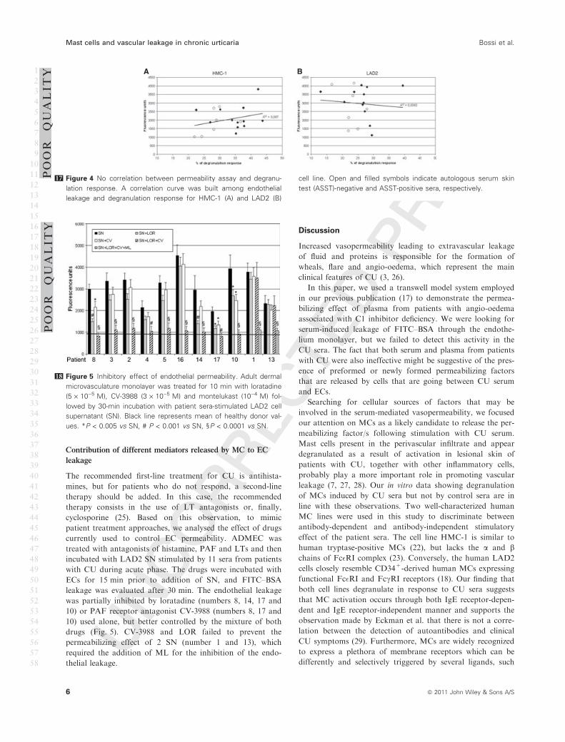
Contribution of different mediators released by MC to EC
leakage
The recommended first-line treatment for CU is antihista-
mines, but for patients who do not respond, a second-line
therapy should be added. In this case, the recommended
therapy consists in the use of LT antagonists or, finally,
cyclosporine (25). Based on this observation, to mimic
patient treatment approaches, we analysed the effect of drugs
currently used to control EC permeability. ADMEC was
treated with antagonists of histamine, PAF and LTs and then
incubated with LAD2 SN stimulated by 11 sera from patients
with CU during acute phase. The drugs were incubated with
ECs for 15 min prior to addition of SN, and FITC–BSA
leakage was evaluated after 30 min. The endothelial leakage
was partially inhibited by loratadine (numbers 8, 14, 17 and
10) or PAF receptor antagonist CV-3988 (numbers 8, 17 and
10) used alone, but better controlled by the mixture of both
drugs (Fig. 5). CV-3988 and LOR failed to prevent the
permeabilizing effect of 2 SN (number 1 and 13), which
required the addition of ML for the inhibition of the endo-
thelial leakage.
Discussion
Increased vasopermeability leading to extravascular leakage
of fluid and proteins is responsible for the formation of
wheals, flare and angio-oedema, which represent the main
clinical features of CU (3, 26).
In this paper, we used a transwell model system employed
in our previous publication (17) to demonstrate the permea-
bilizing effect of plasma from patients with angio-oedema
associated with C1 inhibitor deficiency. We were looking for
serum-induced leakage of FITC–BSA through the endothe-
lium monolayer, but we failed to detect this activity in the
CU sera. The fact that both serum and plasma from patients
with CU were also ineffective might be suggestive of the pres-
ence of preformed or newly formed permeabilizing factors
that are released by cells that are going between CU serum
and ECs.
Searching for cellular sources of factors that may be
involved in the serum-mediated vasopermeability, we focused
our attention on MCs as a likely candidate to release the per-
meabilizing factor/s following stimulation with CU serum.
Mast cells present in the perivascular infiltrate and appear
degranulated as a result of activation in lesional skin of
patients with CU, together with other inflammatory cells,
probably play a more important role in promoting vascular
leakage (7, 27, 28). Our in vitro data showing degranulation
of MCs induced by CU sera but not by control sera are in
line with these observations. Two well-characterized human
MC lines were used in this study to discriminate between
antibody-dependent and antibody-independent stimulatory
effect of the patient sera. The cell line HMC-1 is similar to
human tryptase-positive MCs (22), but lacks the a and b
chains of Fc�RI complex (23). Conversely, the human LAD2
cells closely resemble CD34+-derived human MCs expressing
functional Fc�RI and FccRI receptors (18). Our finding that
both cell lines degranulate in response to CU sera suggests
that MC activation occurs through both IgE receptor-depen-
dent and IgE receptor-independent manner and supports the
observation made by Eckman et al. that there is not a corre-
lation between the detection of autoantibodies and clinical
CU symptoms (29). Furthermore, MCs are widely recognized
to express a plethora of membrane receptors which can be
differently and selectively triggered by several ligands, such
A B
Figure 417 No correlation between permeability assay and degranu-
lation response. A correlation curve was built among endothelial
leakage and degranulation response for HMC-1 (A) and LAD2 (B)
cell line. Open and filled symbols indicate autologous serum skin
test (ASST)-negative and ASST-positive sera, respectively.
Figure 518 Inhibitory effect of endothelial permeability. Adult dermal
microvasculature monolayer was treated for 10 min with loratadine
(5 · 10)5 M), CV-3988 (3 · 10)5 M) and montelukast (10)4 M) fol-
lowed by 30-min incubation with patient sera-stimulated LAD2 cell
supernatant (SN). Black line represents mean of healthy donor val-
ues. *P < 0.005 vs SN, # P < 0.001 vs SN, §P < 0.0001 vs SN.
POOR
QUALITY
POOR
QUALITY
Mast cells and vascular leakage in chronic urticaria Bossi et al.
6 ª 2011 John Wiley & Sons A/S
1
2
3
4
5
6
7
8
9
10
11
12
13
14
15
16
17
18
19
20
21
22
23
24
25
26
27
28
29
30
31
32
33
34
35
36
37
38
39
40
41
42
43
44
45
46
47
48
49
50
51
52
53
54
55
56
57
58

as IgG, peptides, microbial derivates, fragments of activated
complement acting as pro-inflammatory and/or regulatory
stimulus for these cells depending on the different physiologi-
cal context (30). Depending on the nature of the stimulus,
MC activation can lead to selective response in term of qual-
ity and quantity of released mediators. Therefore, it is possi-
ble to induce cytokine production without degranulation and
vice versa. This could explain the lack of correlation between
the increased vasopermeability and the degree of degranula-
tion induced by the same sera, suggesting the contribution of
other mediators to serum-mediated vascular leakage besides
those released with the granules. The lack of permeability
effect of SNs from MC stimulated by control sera (Fig. 3)
supports the specificity of the assay for patients with CU.
The fact that the permeabilizing activity of two of three sera
was unchanged following Ig depletion and was reduced but
not completely abrogated in the third sample analysed sug-
gests that serum factors other than autoantibodies to IgE or
Fc�RI play a critical role in the onset of CU. The presence of
non-IgG histamine-releasing factor/s acting on dermal MTs
has been suggested by Sabroe and Greaves in 5–10% of
patients with CU, but their nature remains to be elucidated
(31). In this context, our data, showing a lack of correlation
between degranulation of HMC-1 or LAD2 and BHR, are in
line with the presence of different activating factors in sera
from patients with CU. In the light of these considerations,
although serum of patient number 19 did not induce hista-
mine release from MC lines, we cannot exclude that any per-
meabilizing factors other than histamine are secreted. Several
therapeutic evidences support this hypothesis. Urticaria epi-
sodes associated with the release of high quantities of IL-6
are resistant to anti-H1 treatment (32). Furthermore,
although the majority of urticarias are sensitive to anti-H1,
symptom amelioration is achieved only with steroids or with
a combination of antileukotrienes and anti-H1 drugs (33).
The patients who are resistant to antihistamines may be trea-
ted with immunosuppressive drugs, such as cyclosporine, that
prevent cytokine synthesis. This has important therapeutic
implications because there is a consensus on the use of sec-
ond-generation antihistamines as first-line symptomatic drug
to treat patients with CU. The rationale for using these drugs
is supported by our finding that antihistamine can substan-
tially reduce serum-induced EC permeability in the 4/11
patients analysed in the inhibition experiment. However, it is
important to emphasize that the combination of antihista-
mines and PAF antagonists was more effective than antihis-
tamines alone and resulted in total abrogation of BSA
leakage, suggesting that a combination therapy can be more
appropriate than the use of a single drug. Of interest is the
observation that the serum of two patients exhibited a per-
meabilizing activity neutralized exclusively by an antileukotri-
ene drug, which has been used in patients with CU resistant
to antihistamine treatment (25).
In conclusion, we have provided evidence for a novel and
direct role of MCs in CU. We stress that the CU is a multi-
faceted disease with different still unknown factor(s) leading
to MC activation and EC permeability, and we underline the
need of an in vitro test, herein described, that allows to per-
sonalize the therapy. The combined degranulation and leak-
age assays proved to be more sensitive than ASST being
positive, though to a different degree, in all patients with CU
examined as opposed to 40–60% of ASST (34). Nevertheless,
our data open new insights into the role of MCs in the onset
and amplification of immune response in CU disorder.
Author’s contributions
FB performed all the experiments on ECs. BF performed all
the experiments on MT lines. FB, BF and OR performed the
analysis of the results. MC, AT, PR and RA followed the
patients and collected the serum samples. FB, BF, FT and
CP contributed to the design of the research project and the
writing of the manuscript. OR, MC, AT, PR and RA con-
tributed to write and revise the manuscript.
Conflict of interest
FB, BF,OR, MC, AT, PR, RA, FT and CP declare no com-
peting financial interest.
Funding
This work was supported by Ministero dell’Istruzione, Uni-
versita e Ricerca (PRIN 2007), Regione Friuli Venezia Giu-
lia, AIRC (Associazione Italiana Ricerca sul Cancro) to FT
and CP, Agenzia Spaziale Italiana (Progetto OSMA) to CP.
References
1. Kikuchi Y, Fann T, Kaplan AP. Antithy-
roid antibodies in chronic urticaria and
angioedema. J Allergy Clin Immunol
2003;112:218.
2. Leznoff A, Josse RG, Denburg J, Dolovich
J. Association of chronic urticaria and
angioedema with thyroid autoimmunity.
Arch Dermatol 1983;119:636–640.
3. Kaplan AP, Greaves M. Pathogenesis of
chronic urticaria. Clin Exp Allergy
2009;39:777–787.
4. Hide M, Francis DM, Grattan CE, Hakimi
J, Kochan JP, Greaves MW. Autoantibod-
ies against the high-affinity IgE receptor as
a cause of histamine release in chronic
urticaria. N Engl J Med 1993;328:1599–
1604.
5. Niimi N, Francis DM, Kermani F, O’Don-
nell BF, Hide M, Kobza-Black A et al. Der-
mal mast cell activation by autoantibodies
against the high affinity IgE receptor in
chronic urticaria. J Invest Dermatol
1996;106:1001–1006.
6. Fiebiger E, Maurer D, Holub H, Reininger
B, Hartmann G, Woisetschlager M et al.
Serum IgG autoantibodies directed against
the alpha chain of Fc epsilon RI: a selective
marker and pathogenetic factor for a distinct
subset of chronic urticaria patients? J Clin
Invest 1995;96:2606–2612.
7. Elias J, Boss E, Kaplan AP. Studies of the
cellular infiltrate of chronic idiopathic urti-
caria: prominence of T-lymphocytes, mono-
cytes, and mast cells. J Allergy Clin Immunol
1986;78(5 Pt 1):914–918.
8. Hennino A, Berard F, Guillot I, Saad N,
Rozieres A, Nicolas JF. Pathophysiology of
urticaria. Clin Rev Allergy Immunol
2006;30:3–11.
Bossi et al. Mast cells and vascular leakage in chronic urticaria
ª 2011 John Wiley & Sons A/S 7
1
2
3
4
5
6
7
8
9
10
11
12
13
14
15
16
17
18
19
20
21
22
23
24
25
26
27
28
29
30
31
32
33
34
35
36
37
38
39
40
41
42
43
44
45
46
47
48
49
50
51
52
53
54
55
56
57
58
9. Levi-Schaffer F, Pe’er J. Mast cells and
angiogenesis. Clin Exp Allergy 2001;31:521–
524.
10. Kuna P, Kaplan AP. Relationship of hista-
mine-releasing factors and histamine-releas-
ing inhibitory factors to chemokine group of
cytokine. Allergy Asthma Proc 1996;17:5–11.
11. Bhattacharyya SP, Drucker I, Reshef T, Kir-
shenbaum AS, Metcalfe DD, Mekori YA.
Activated T lymphocytes induce degranula-
tion and cytokine production by human
mast cells following cell-to-cell contact.
J Leukoc Biol 1998;63:337–341.
12. Tedeschi A, Asero R, Marzano AV, Lorini
M, Fanoni D, Berti E et al. Plasma levels
and skin-eosinophil-expression of vascular
endothelial growth factor in patients with
chronic urticaria. Allergy 2009;64:1616–1622.
13. Lee KH, Kim JY, Kang DS, Choi YJ, Lee
WJ, Ro JY. Increased expression of endothe-
lial cell adhesion molecules due to mediator
release from human foreskin mast cells stim-
ulated by autoantibodies in chronic urticaria
sera. J Invest Dermatol 2002;118:658–663.
14. Zuberbier T, Asero R, Bindslev-Jensen C,
Walter Canonica G, Church MK, Gimenez-
Arnau A et al. EAACI/GA(2)LEN/EDF/
WAO guideline: definition, classification and
diagnosis of urticaria. Allergy 2009;64:1417–
1426.
15. Sabroe RA, Grattan CE, Francis DM, Barr
RM, Kobza Black A, Greaves MW. The
autologous serum skin test: a screening test
for autoantibodies in chronic idiopathic urti-
caria. Br J Dermatol 1999;140:446–452.
16. Bossi F, Fischetti F, Pellis V, Bulla R, Fer-
rero E, Mollnes TE et al. Platelet-activating
factor and kinin-dependent vascular leakage
as a novel functional activity of the soluble
terminal complement complex. J Immunol
2004;173:6921–6927.
17. Bossi F, Fischetti F, Regoli D, Durigutto P,
Frossi B, Gobeil F Jr et al. Novel patho-
genic mechanism and therapeutic approaches
to angioedema associated with C1 inhibitor
deficiency. J Allergy Clin Immunol
2009;124:1303–1310.10
18. Kirshenbaum AS, Akin C, Wu Y, Rottem
M, Goff JP, Beaven MA et al. Characteriza-
tion of novel stem cell factor responsive
human mast cell lines LAD 1 and 2 estab-
lished from a patient with mast cell sar-
coma/leukemia; activation following
aggregation of FcepsilonRI or FcgammaRI.
Leuk Res 2003;27:677–682.
19. Gri G, Piconese S, Frossi B, Manfroi V,
Merluzzi S, Tripodo C et al. CD4+CD25+
regulatory T cells suppress mast cell degran-
ulation and allergic responses through
OX40-OX40L interaction. Immunity
2008;29:771–781.
20. Asero R, Tedeschi A, Lorini M, Salimbeni
R, Zanoletti T, Miadonna A. Chronic urti-
caria: novel clinical and serological aspects.
Clin Exp Allergy 2001;31:1105–1110.
21. Fisher R. The design of experiments, 1st
edn. Xxxxxx: Olver and Boyd, 1935.11
22. Butterfield JH, Weiler D, Dewald G, Gleich
GJ. Establishment of an immature mast cell
line from a patient with mast cell leukemia.
Leuk Res 1988;12:345–355.
23. Nilsson G, Blom T, Kusche-Gullberg M,
Kjellen L, Butterfield JH, Sundstrom C
et al. Phenotypic characterization of the
human mast-cell line HMC-1. Scand J
Immunol 1994;39:489–498.
24. Kulka M, Sheen CH, Tancowny BP, Gram-
mer LC, Schleimer RP. Neuropeptides acti-
vate human mast cell degranulation and
chemokine production. Immunology
2008;123:398–410.
25. Zuberbier T, Asero R, Bindslev-Jensen C,
Walter Canonica G, Church MK, Gimenez-
Arnau AM et al. EAACI/GA(2)LEN/EDF/
WAO guideline: management of urticaria.
Allergy 2009;64:1427–1443.
26. Grattan CE, Sabroe RA, Greaves MW.
Chronic urticaria. J Am Acad Dermatol
2002;46:645–657; quiz 657–60.
27. Haas N, Toppe E, Henz BM. Microscopic
morphology of different types of urticaria.
Arch Dermatol 1998;134:41–46.
28. Grattan CE, Boon AP, Eady RA, Winkel-
mann RK. The pathology of the autologous
serum skin test response in chronic urticaria
resembles IgE-mediated late-phase reactions.
Int Arch Allergy Appl Immunol 1990;3:198–
204.
29. Eckman JA, Hamilton RG, Saini SS. Inde-
pendent evaluation of a commercial test for
‘‘autoimmune’’ urticaria in normal and
chronic urticaria subjects. J Invest Dermatol
2009;129:1584–1586.
30. Frossi B, Gri G, Tripodo C, Pucillo C.
Exploring a regulatory role for mast cells:
‘MCregs’? Trends Immunol ?????;31:97–102.12
31. Sabroe RA, Greaves MW. Chronic idio-
pathic urticaria with functional autoantibod-
ies: 12 years on. Br J Dermatol
2006;154:813–819.
32. Fujii K, Konishi K, Kanno Y, Ohgou N.
Acute urticaria with elevated circulating
interleukin-6 is resistant to anti-histamine
treatment. J Dermatol 2001;28:248–250.
33. Eymard B, Cousin F, Nicolas JF. Prevention
of aspirin-induced angioedema with an asso-
ciation of leukotriene inhibitors and antihis-
tamines. Ann Dermatol Venereol 2003;130(8–
9 Pt 1):787–788.
34. Greaves M. Chronic urticaria. J Allergy Clin
Immunol 2000;105:664–672.
Mast cells and vascular leakage in chronic urticaria Bossi et al.
8 ª 2011 John Wiley & Sons A/S
1
2
3
4
5
6
7
8
9
10
11
12
13
14
15
16
17
18
19
20
21
22
23
24
25
26
27
28
29
30
31
32
33
34
35
36
37
38
39
40
41
42
43
44
45
46
47
48
49
50
51
52
53
54
55
56
57
58

Author Query Form
Journal: ALL
Article: 2704
Dear Author,
During the copy-editing of your paper, the following queries arose. Please respond to these by marking up
your proofs with the necessary changes/additions. Please write your answers on the query sheet if there is
insufficient space on the page proofs. Please write clearly and follow the conventions shown on the attached
corrections sheet. If returning the proof by fax do not write too close to the paper’s edge. Please remember
that illegible mark-ups may delay publication.
Many thanks for your assistance.
Query
reference
Query Remarks
1 AUTHOR: Please check and approve the edit made in the article title.
2 AUTHOR: An Copyright Transfer Agreement has not yet been received for
this paper. Please go to: www.wiley.com/go/ctaaglobal and download a form
and return it to Fax No +44 131 226 3803 with your corrections to the
proofs. We cannot publish your paper until the Copyright Transfer Agreement
is received
3 AUTHOR: Please check all affiliation addresses.
4 AUTHOR: Please check and provide full postal address details for correspon-
dence.
5 AUTHOR: Please expand FCS.
6 AUTHOR: Please give address information for Peprotech: town, state (if
applicable), and country.
7 AUTHOR: Please give address information for Becton Dickinson: town, state
(if applicable), and country.
8 AUTHOR: Please give address information for Costar: town, state (if applica-
ble), and country.
9 AUTHOR: Please give address information for GE Healthcare: town, state (if
applicable), and country.
10 AUTHOR: If there are fewer than 7 authors for all et al. References, please
supply all of their names. If there are 7 or more authors, please supply the first
6 author names then et al.
11 AUTHOR: Please provide location of publisher in ref. 22.
12 AUTHOR: Please provide year of publication for ref. 30.
13 AUTHOR: Please check the expansion of HUVEC.

14 AUTHOR: Figure 1 is of poor quality. Please check required artwork specifi-
cations at http://authorservices.wiley.com/submit_illust.asp?site=1
15 AUTHOR: Figure 2 is of poor quality. Please check required artwork specifi-
cations at http://authorservices.wiley.com/submit_illust.asp?site=1
16 AUTHOR: Figure 3 is of poor quality. Please check required artwork specifi-
cations at http://authorservices.wiley.com/submit_illust.asp?site=1
17 AUTHOR: Figure 4 is of poor quality. Please check required artwork specifi-
cations at http://authorservices.wiley.com/submit_illust.asp?site=1
18 AUTHOR: Figure 5 is of poor quality. Please check required artwork specifi-
cations at http://authorservices.wiley.com/submit_illust.asp?site=1

USING e-ANNOTATION TOOLS FOR ELECTRONIC PROOF CORRECTION
Required software to e-Annotate PDFs: Adobe Acrobat Professional or Adobe Reader (version 8.0 or
above). (Note that this document uses screenshots from Adobe Reader X)
The latest version of Acrobat Reader can be downloaded for free at: http://get.adobe.com/reader/
Once you have Acrobat Reader open on your computer, click on the Comment tab at the right of the toolbar:
1. Replace (Ins) Tool Î for replacing text.
Strikes a line through text and opens up a text
box where replacement text can be entered.
How to use it
‚ Highlight a word or sentence.
‚ Click on the Replace (Ins) icon in the Annotations
section.
‚ Type the replacement text into the blue box that
appears.
This will open up a panel down the right side of the document. The majority of
tools you will use for annotating your proof will be in the Annotations section,
rkevwtgf"qrrqukvg0"YgÓxg"rkemgf"qwv"uqog"qh"vjgug"vqqnu"dgnqy<
2. Strikethrough (Del) Tool Î for deleting text.
Strikes a red line through text that is to be
deleted.
How to use it
‚ Highlight a word or sentence.
‚ Click on the Strikethrough (Del) icon in the
Annotations section.
3. Add note to text Tool Î for highlighting a section
to be changed to bold or italic.
Highlights text in yellow and opens up a text
box where comments can be entered.
How to use it
‚ Highlight the relevant section of text.
‚ Click on the Add note to text icon in the
Annotations section.
‚ Type instruction on what should be changed
regarding the text into the yellow box that
appears.
4. Add sticky note Tool Î for making notes at
specific points in the text.
Marks a point in the proof where a comment
needs to be highlighted.
How to use it
‚ Click on the Add sticky note icon in the
Annotations section.
‚ Click at the point in the proof where the comment
should be inserted.
‚ Type the comment into the yellow box that
appears.
USING e-ANNOTATION TOOLS FOR ELECTRONIC PROOF CORRECTION
For further information on how to annotate proofs, click on the Help menu to reveal a list of further options:
5. Attach File Tool Î for inserting large amounts of
text or replacement figures.
Inserts an icon linking to the attached file in the
appropriate pace in the text.
How to use it
‚ Click on the Attach File icon in the Annotations
section.
‚ Enkem"qp"vjg"rtqqh"vq"yjgtg"{qwÓf"nkmg"vjg"cvvcejgf"file to be linked.
‚ Select the file to be attached from your computer
or network.
‚ Select the colour and type of icon that will appear
in the proof. Click OK.
6. Add stamp Tool Î for approving a proof if no
corrections are required.
Inserts a selected stamp onto an appropriate
place in the proof.
How to use it
‚ Click on the Add stamp icon in the Annotations
section.
‚ Select the stamp you want to use. (The Approved
stamp is usually available directly in the menu that
appears).
‚ Enkem"qp"vjg"rtqqh"yjgtg"{qwÓf"nkmg"vjg"uvcor"vq"appear. (Where a proof is to be approved as it is,
this would normally be on the first page).
7. Drawing Markups Tools Î for drawing shapes, lines and freeform
annotations on proofs and commenting on these marks.
Allows shapes, lines and freeform annotations to be drawn on proofs and for
comment to be made on these marks..
How to use it
‚ Click on one of the shapes in the Drawing
Markups section.
‚ Click on the proof at the relevant point and
draw the selected shape with the cursor.
‚ To add a comment to the drawn shape,
move the cursor over the shape until an
arrowhead appears.
‚ Double click on the shape and type any
text in the red box that appears.
Copyright © 2022 FDOKUMEN