Atmospheric Pressure MALDI Fourier Transform Mass Spectrometry of Labile Oligosaccharides
Upload
khangminh22Category
view
3download
0
Mass Spectrometry Imaging as a NewMethod: To Reveal the Pathogenesisand the Mechanism of TraditionalMedicine in Cerebral IschemiaYan Liang1,2, Qiaoqiao Feng1,2 and Zhang Wang2,3*
1College of Pharmacy, Chengdu University of Traditional Chinese Medicine, Chengdu, China, 2State Key Laboratory ofSouthwestern Chinese Medicine Resources, Chengdu University of Traditional Chinese Medicine, Chengdu, China, 3College ofEthnomedicine, Chengdu University of Traditional Chinese Medicine, Chengdu, China
Mass spectrometry imaging (MSI) can describe the spatial distribution of molecules invarious complex biological samples, such as metabolites, lipids, peptides and proteinsin a comprehensive way, and can provide highly relevant supplementary informationwhen combined with other molecular imaging techniques and chromatographytechniques, so it has been used more and more widely in biomedical research. Theapplication of mass spectrometry imaging in neuroscience is developing. It is veryadvantageous and necessary to use MSI to study various pathophysiologicalprocesses involved in brain injury and functional recovery during cerebral ischemia.Therefore, this paper introduces the techniques of mass spectrometry, including theprinciple of mass spectrometry, the acquisition and preparation of imaging samples,the commonly used ionization techniques, and the optimization of the current appliedmethodology. Furthermore, the research on the mechanism of cerebral ischemia bymass spectrometry was reviewed, such as phosphatidylcholine involved, dopamine,spatial distribution and level changes of physiological substances such as ATP in theKrebs cycle; The characteristics of mass spectrometry imaging as one of the methodsof metabolomics in screening biomarkers related to cerebral ischemia were analyzedthe advantages of MSI in revealing drug distribution and the mechanism of traditionaldrugs were summarized, and the existing problems of MSI were also analyzed andrelevant suggestions were put forward.
Keywords: mass spectrometry imaging, cerebral ischemia, metabolomics, spatial distribution of drugs, traditionalmedicines, mechanism
INTRODUCTION
Mass Spectrometry Imaging (MSI) is an imaging method based on mass spectrometry, which usesmass spectrometry to scan biological samples directly, not only can analyze the spatial distributioncharacteristics of hundreds of molecules simultaneously on the same tissue section, but also canobtain the overall distribution information of drugs and metabolites at the animal level by high-resolution mass spectrometry (Luo et al., 2017). Mass spectrometry imaging can be divided into:Matrix-Assisted Laser Desorption/Ionization Mass Spectrum Image (MALDI-MSI), DesorptionElectrospray Ionization Mass Spectrum Image (DESI-MSI) and so on. Among them, MALDI
Edited by:Paul Chazot,
Durham University, United Kingdom
Reviewed by:Zhi Yong Du,
Capital Medical University, ChinaHemi Luan,
Southern University of Science andTechnology, China
*Correspondence:Zhang Wang
Specialty section:This article was submitted to
Ethnopharmacology,a section of the journal
Frontiers in Pharmacology
Received: 01 March 2022Accepted: 13 May 2022Published: 03 June 2022
Citation:Liang Y, Feng Q and Wang Z (2022)Mass Spectrometry Imaging as a NewMethod: To Reveal the Pathogenesis
and the Mechanism of TraditionalMedicine in Cerebral Ischemia.Front. Pharmacol. 13:887050.
doi: 10.3389/fphar.2022.887050
Frontiers in Pharmacology | www.frontiersin.org June 2022 | Volume 13 | Article 8870501
REVIEWpublished: 03 June 2022
doi: 10.3389/fphar.2022.887050

imaging is the most important, but DESI imaging has moreadvantages in real-time clinicopathological diagnosis becauseof its convenient workflow (Lanekoff, Stevens et al., 2014).
As one of the young mass spectrometry techniques, MSI has agreat value in many fields such as medical research, biologicalresearch, drug research and so on. It has become a hot spot inmass spectrometry research. In order to obtain high-quality massspectrometry imaging results, from the ion source to the massanalyzer, and then to data acquisition and processing, every stepneeds to be done to the extreme to achieve the overallperformance optimization, so as to obtain the mostsatisfactory results. The key issues to be paid attention to MSIare imaging rate, reproducibility and wide spectrum capability.Compared with the main clinical optical imaging such asfluorescence immune-labeling imaging, mass spectrometryimaging has many advantages, such as multiple molecularinformation in tissue sections, no staining and labeling, andlow imaging cost. Mass spectrometry is a mass spectrometerwith the highest requirements for vacuum. Therefore, the currentimprovement in imaging rates makes the image more accurate ofbiological tissues. Prior applications of mass spectrometryimaging focused on qualitative testing; such as microbialidentification, nucleic acid typing, in practical application, onlyqualitative analysis is far from enough, the mainstream needs stillto solve quantitative problems. To complete quantitativelyanalysis requires imaging equipment have a good repeatability,isotope labeling method can be used to assist in the realization ofmolecular quantitative problems. With the continuousimprovement of new instrument technology, it can achievehigh sensitivity in a wide mass number range, so as to ensurethat the test can be completed in a wide mass number range andthe information of slices can be better expressed. Both small andlarge biological molecules can be accurately imaged to achievereal imaging freedom.
Mass spectrometry imaging develops rapidly and can beapplied to food safety, geography, environmental science, andit has great potential especially in life science and medicine.Existing imaging technologies include optical imaging,chemical imaging and so on. These techniques can onlymonitor the expression of one molecule and require amolecular probe. Molecules with similar structures are proneto false positive results. Molecular mass spectrometry imaging ofbiological tissue is helpful to solve these problems. Through thecombination of mass spectrometry ion scanning technology andprofessional image processing software, the biological tissuesection is directly analyzed, relative abundance and spatialdistribution of compounds in the tissue are analyzed andstudied. Mass spectrometry has been widely used for theanalysis of various biomolecules in tissue sections, such asproteins, peptides, lipids and drug metabolites. In this way, thespatial distribution of potential biomarkers can be obtained, andcombined with other molecular imaging techniques, it can beused for histopathological characteristics, disease diagnosis andbiomarker discovery. MSI technology has found biomarkers inbreast cancer, thyroid cancer, colon cancer and other cancers,becoming a powerful tool for tumors (Yun et al., 2020), normalcells and tumor cells have different cell phenotype, and different
tumor cells have different membrane lipid phenotype, and themicro-environment also affects the membrane lipid phenotype oftumor cells. Studies on in situ detection of tumor cell membranelipids showed that monounsaturated fatty acids, three glycerylphosphatide and nucleotide were upregulated, inosine and aphosphatidylethanolamine were downregulated in tumorregions of gastric cancer tumor tissues (Dan et al., 2018).Neurotransmitters such as dopamine, 5-hydroxytryptamine, 3-methoxytyramine and tyrosine were efficiently detected by massspectrometry in a derivative form (Xi et al., 2019) Massspectrometry (MS), based on a new method of imaging drugmetabolomics, provides a novel and intuitive means for theanalysis of the mechanism of action or toxicologicalmechanism of drugs or new drugs, even new drug candidateswith multiple targets or unclear targets. Using MALDI-MSI, itwas found that (s)-Oxiracetam could cause changes of 15metabolic small molecules related to glucose aerobic oxidationand other pathways (Wan, Huihui et al., 2017). In conclusion,mass spectrometry imaging has shown its advantages in tumordiagnosis, brain neuroscience, drug development, and biomarkerscreening and identification.
Ischemic stroke accounts for 69.6%–70.8% of cerebral strokein China, and is the first leading cause of death and disability inChina (Wu, Wu et al., 2019). Cerebral ischemia reperfusioninjury refers to the phenomenon that cerebral cell injurycaused by cerebral ischemia is further aggravated after bloodreperfusion is restored, and the obvious changes are neuronalinjury and cerebral edema (Guang, 2018). The pathogenesis ofcerebral ischemia injury includes oxygen free radical damage,calcium ion overload, mitochondrial damage, neurotoxic effects,inflammatory damage and blood-brain barrier damage, theycause and effect each other and interact into a network,eventually causing nerve cell edema, injury, necrosis andapoptosis. Although intravenous thrombolysis, antiplateletaggregation and anticoagulation therapy (Stoll and Nieswandt2019) can be used in the treatment of stroke, reperfusion afterthrombolysis can cause complications such as edema of bleedingtransformed brain cells and exacerbation of brain celldysfunction. How to alleviate cerebral ischemia reperfusioninjury is one of the keys to improve the efficacy of ischemicstroke. Blood-brain barrier (BBB) plays an important role as thestructural basis of cerebral ischemia. In addition, mitochondriaare an important medium of cell signal and energy homeostasis,so mitochondrial dysfunction is one of the importantpathogeneses of cerebral ischemia reperfusion injury (Carinci,Vezzani et al., 2021), specifically manifested as mitochondrialmembrane structure was damaged and mitochondrial DNA wasdamaged, mitochondrial permeability transition pore (MPTP)opening up and so on, and then participate in the process ofneuronal apoptosis (Wei et al., 2019). Mass spectrometry imaginghas great potential in studying the spatial distribution ofbiomolecules in complete tissue samples.
At present, MSI has been widely used in protein recognition,biomarker discovery, medical diagnosis and other research(Chughtai and Heeren 2010). MALDI and DESI have broadapplication prospects in brain neuroscience research andclinical diagnosis (Xi et al., 2019). It can be used in
Frontiers in Pharmacology | www.frontiersin.org June 2022 | Volume 13 | Article 8870502
Liang et al. Reveal Cerebral Ischemia by MSI

neuroscience for the imaging of neurotransmitters and lowmolecular weight metabolites, lipids and peptides. The furtherinsights into the underlying mechanisms that are going on in thenervous system, may be provided by using IMS to study thespatio-temporal dynamic distribution patterns of molecularspecies that coordinate neuronal function, includingdevelopment, learning and memory, and cognition andbehavior (Hanrieder, Phan et al., 2013). By analyzing therelated physiological substances (such as lipids, dopamine andATP in the tricarxylic acid cycle) involved in the pathogenesis ofcerebral ischemia, metabolomics, distribution and mechanism ofdrugs in cerebral ischemia, the spatial distribution and levelchanges of drugs were clarified. It will play an important rolein the discovery, clinical diagnosis, treatment and prognosis ofcerebral ischemia, and make mass spectrometry imaging moremature in the detection of the mechanism of cerebral ischemia.
MASS SPECTROMETRY IMAGING
Morphological abnormalities in tissue samples observed withconventional microscopy do not provide researchers withmuch biochemical information, and conventional biochemicaltechniques often lose information about tissue location. Bysuccessfully combining these two aspects of information todisplay objects and simultaneously determine the biochemistryand location of unknown molecules, mass spectrometry imagingcan be useful in elucidating pathogenesis or identifyingtherapeutic targets.
The Principle of Mass SpectrometryImagingFirstly, the samples tested are obtained and prepared in anappropriate way. According to the preset acquisition program,the mass spectrometer uses laser or high-energy ion beam to scanthe samples, so that the molecules or ions on the surface aredesorbed and ionized. Then the mass charge ratio and ionstrength of each pixel ion on the sample surface are obtainedby mass analyzer. The mass spectrum peak of any ion withspecified mass charge ratio was searched in the mass spectrumdata of each pixel with the help of mass spectral imaging software,and the two-dimensional distribution map of the correspondingmolecule or ion on the sample surface was drawn by combiningthe signal strength of the corresponding ion and its position onthe sample surface. Then, the above software is used for furtherdata processing of the two-dimensional distribution map ofcontinuous slices of samples to obtain the three-dimensionalspatial distribution of the objects to be measured in the samples.
The key performance indexes of mass spectrometry aresensitivity and imaging rate. The key steps are tissuesectioning and matrix spraying. In order to improve theintegrity of mass spectrometry imaging results, it is necessaryto exert the maximum value in each of these processes. Sensitivitymeans the minimum number of ions the mass spectrometer canfind. Factors affecting sensitivity include ionization efficiency, iontransport efficiency, and substrate (impurity) interference.
Ionization efficiency refers to the degree to which a sample isionized. Sample molecules can be easily detected by an iondetector when they are ionized enough. But different ionsources target different ranges. For example, MALDI canachieve very high ionization efficiency for both large and smallmolecules when the matrix is suitable, which is the reason whyMALDI has high sensitivity. In contrast, DESI and SIMS plasmasources have obvious advantages in small molecule ionization,while in large molecule, the ionization ability is worse thanMALDI. As the internal source (in vacuum), the ionsproduced by MALDI are generated in the vacuumenvironment of the mass spectrometer, so the ion transferefficiency is higher. While the ions generated by external ionsources, such as AP-MALDI and DESI, in the atmosphere need tobe transferred into the vacuum environment, and only less than1% of the ions are usually transferred to the mass analyzer. It hasgreat influence on the sensitivity of mass spectrometry, which canonly be compensated by the ability of the mass analyzer. In massspectrometric imaging, matrix interference also disturbs the massspectrometer itself. Therefore, we can use common techniquessuch as chromatography-mass spectrometry, which is to removethe matrix in the sample first, and then separate the relativelypure target. It can effectively eliminate the defects of mass spectralimaging itself.
As for the scanning rate of mass spectral imaging, it generallydepends on three factors: laser frequency, two-dimensionalmobile platform and data acquisition. The preparation oftissue sections is mainly related to the authenticity andaccuracy of mass spectrometry imaging results. Samplepreprocessing is the basis of mass spectrometry. In order toreduce the time of sample preprocessing and improve theresolution and reproducibility of MALDI mass spectrometry,many researchers focus on the study of matrix free massspectrometry. A matrix free MALDI-MS detection method wasestablished by selective enrichment of low abundance biologicalsmall molecules and proteins through nanotechnology. A newtype of ionization-assisted substrate for porous nano materialshas been developed. The ionization-assisted substrate cansignificantly reduce the sample preprocessing time for massspectrometry analysis, and the operation is simple.
Acquisition and Preparation of MassSpectrometry Image SamplesThe process of sample preparation is a key link affecting theauthenticity and accuracy of mass spectrometry imaging results,and its processing methods and techniques are closely related tothe properties of the object to be measured and the type and stateof the sample. Generally, MSI used in pharmaceutical research ismostly used to analyze animals, tissues, cells and solidpreparations. Proper and rapid sample collection and fixationare the guarantee to maintain the true spatial distribution andabundance of molecules or ions in the sample. To avoiddisplacement and degradation of the tissue, the sample shouldbe fixed quickly collection. The most common fixation methodfor tissue samples such as organs and whole animal samples israpid freezing.
Frontiers in Pharmacology | www.frontiersin.org June 2022 | Volume 13 | Article 8870503
Liang et al. Reveal Cerebral Ischemia by MSI

Tissue, whole animals, and solid preparations are usuallysectioned. Embedding is often required for fragile tissues such aseye tissue, large samples such as whole animals and solidpreparations that are not easily sectioned. The samples weretransferred to the mass spectrum target by melting mountingmethod and adhesive tape method, respectively, by directlysticking the frozen tissue sections to the target at roomtemperature, or by fixing the samples on the mass spectrumtarget with conductive double-sided adhesive tape. Immediatelyafter the sample is transferred to the mass target, the mass targetcontaining the sample should be dried to keep the sample stable. Thecommonly used drying methods are freeze drying, vacuum drying,solvent dehydration drying and nitrogen drying. Generally, the driedsamples can be directly analyzed by mass spectrometry. In thedetection of specific substances to be measured in complexsamples, solvent cleaning, surface enzymatic hydrolysis andchemical derivatization are often used to properly treat theanalytical surface. Cleaning the mass spectrometric target withappropriate solvent can not only dehydrate and fix, but alsoimprove the results of mass spectrometric determination.
Mass Spectrometry Imaging IonizationTechniqueThe ionization methods commonly used in MSI analysis are:matrix-assisted laser desorption ionization (MALDI), desorptionelectrospray ionization (DESI) and secondary ion massspectrometry (SIMS). Some ionization techniques such asatmospheric pressure infrared (APIR) mass spectrometry,nanostructure-initiator mass spectrometry (NIMS), attributesthat fit MSI analysis objects are also used in MSI analysis.
MALDI-MSI first needs to uniformly deposit the organic smallmolecule matrix absorbing ultraviolet light on the tissue sections tobe analyzed. The commonly used methods include sublimationelectrospray, fluid atomization, ultrasonic spray, etc. Theadvantages of MALDI imaging are as follows: 1) The imagingspatial resolution is high, reaching the cell level (less than 5 μm);2) It is suitable for the analysis of different kinds of molecules(including metabolites, lipids, proteins, nucleic acids and drugs).MALDI imaging provides mapping of biomolecules in a large massrange from intact lipids to proteins. Most are applied to brain tissueanalysis. The resulting images feature hundreds of uniquelypositioned compound peaks that give an indication of functionand may represent neurologically important peptides and proteins.
DESI mass spectrometry imaging uses the high-speed chargedfog generated by electrospray to bombard the tissue sections in aninclined direction. The charged fog drops will extract and ionizethe molecules in the biological sections and enter the iontransport tube of the mass spectrometry together with them.The main advantages of DESI mass spectrometry imaging are: Noneed for external matrix assisted ionization, the analysis process isfaster; No laser, simple ion source structure, low maintenancecost; The requirement for mass spectrometers is low, a simple iontrap mass spectrometer can support DESI imaging. Comparedwith MALDI imaging, DESI imaging has more advantages inreal-time clinicopathological diagnosis because of its convenientworkflow. However, the spatial resolution accuracy of DESI
imaging is low, and it is difficult to analyze the compoundswith large molecular weight (such as polypeptides, proteins andnucleic acids) (Xi et al., 2019).
Some chemical components that carry large molecularfragments cannot be imprinted by conventional massspectrometry; Professor Nicholas Winograd of PennsylvaniaState University has developed a method called SIMS thatallows a complete scan of a sample in three dimensions. Onthe other hand, SIMS can only image small molecules. ProfessorWinograd has refined this approach by using a new type of SIMSbeam (carbon-60 magnetic sphere) that is less chemicallydamaging to objects than traditional SIMS beam. The energyof C60 is comparable to other ion beams, but does not reachbelow the surface of the sample. So, the sample is continuouslystripped layer by layer, resulting in a longitudinal plane pattern,ultimately resulting a three-dimensional molecular image. Thismethod has good spatial resolution and can obtain the cellularcharacteristics and analyze the distribution of macrophages andstar cells. SIMS probes can reach depths of up to 100 nm,providing nanoscale resolution.
Understanding the inner components of cells is the key tounderstanding that healthy cells differ from diseased cells. AkosVertes and others have come up with a combination of twotechniques to analyze living cell analysis, based on the principlethat biological samples can also absorb energy directly. First, theyused an atmospheric pressure infrared (APIR) MALDI laser todirectly activate water in the tissue, vaporizing the sample andobtaining ionized particles that were analyzed by massspectrometry. But not all vaporized particles are charged. Mostare actually uncharged and will be missed by APIR MALDI. Tocapture these neutral particles, Vertes et al. used a second method,LAESI (Laser Ablation Electrospray), which captures a large numberof charged droplets and then re-ionizes them. By treating the entiresample and combining the two methods, more molecules can becovered and the analysis quality is higher. Unlike the usual massspectrometry process, Verte’s method also adds height to the imageto enable 3D metabolite imaging, allowing researchers to obtain anatural configuration.
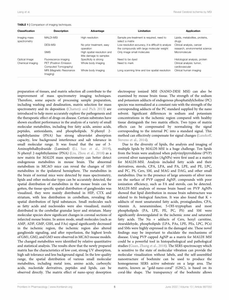
Mass spectrometry has great potential in the detection ofmolecules, Dr. Gary Siuzdak has developed a new techniquecalled NIMS, which analyses very small regions with greatsensitivity, allowing the analysis of peptide arrays, blood, urineand individual cells, as well as tissue imaging. NIMS uses lasers orion beams to vaporize materials from nanoscale vesicles,overcoming the deficiencies of conventional mass spectrometrymethods that lack the required sensitivity and require matrixmolecules to ionize the analyzed objects. Many types of smallmolecules, such as lipids, saccharides, and steroids, can beanalyzed in this way. The comparison of the characteristics ofdifferent imaging technologies is shown in Table 1.
Optimization of Mass SpectrometryImaging Methodology Related to CerebralIschemiaRecent advances in mass spectrometry emphasis themethodology. Freezing or formalin fixation, sample
Frontiers in Pharmacology | www.frontiersin.org June 2022 | Volume 13 | Article 8870504
Liang et al. Reveal Cerebral Ischemia by MSI
preparation of tissues, and matrix selection all contribute to theimprovement of mass spectrometry imaging techniques.Therefore, some aspects of processing sample preparation,including washing and desalination, matrix selection for massspectrometry and its deposition (Chatterji and Pich 2013) areintroduced to help more accurately explore the pathogenesis andthe therapeutic effect of drugs on disease. Certain substrates haveshown excellent performance in the analysis of a variety of smallmolecular metabolites, including free fatty acids, amino acids,peptides, antioxidants, and phospholipids. N-phenyl 2-naphthylamine (PNA) has strong ultraviolet absorptioncapacity, low background interference and salt tolerance insmall molecular range. It was found that the use of 3-Aminophthalhydrazide (Luminol) (Li, Sun et al., 2019),N-phenyl 2-naphthylamine (PNA) (Liu, Zhou et al., 2018) as anew matrix for MALDI mass spectrometry can better detectendogenous metabolites in mouse brain. The abnormalmetabolism in MCAO mice can reveal the changes of 105metabolites in the ipsilateral hemisphere. The metabolites inthe brain of normal mice were detected by mass spectrometry,lipids and other molecular types can be accurately detected, thespatial distribution of metabolites in the mouse brain can begotten, the tissue-specific spatial distribution of gangliosides wasvisualized, they were mainly distributed in HIP, CTX andstriatum, with less distribution in cerebellum, revealing thespatial distribution of lipid substances. Small molecules suchas fatty acids and nucleosides were also visualized, mainlydistributed in the cerebellar granular layer and striatum. Manymolecular species show significant changes in coronal sections ofinfarcted mouse brains. In anion mode, small molecules (such asAMP, ADP, GMP, GSH, and FAs) signal significantly decreasedin the ischemic region, the ischemic region also alteredganglioside signaling, and after reperfusion, the highest levelsof GM1, GM2, and GM3 were observed inMCAOmouse models.The changed metabolites were identified by relative quantitativeand statistical analysis. The results show that the newly preparedmatrix has the characteristics of low cost, strong UV absorption,high salt tolerance and less background signal. In the low-qualityrange, the spatial distribution of various small molecularmetabolites, including metal ions, amino acids, carboxylicacids, nucleotide derivatives, peptides and lipids, can beobserved directly. The matrix effect of nano-spray desorption
electrospray ionized MSI (NANO-DESI MSI) can also beexamined by mouse brain tissue. The strength of the sodiumand potassium adducts of endogenous phosphatidylcholine (PC)species was normalized at a constant rate with the strength of thecorresponding adducts of the PC standard supplied by the nanosolvent. Significant differences in sodium and potassiumconcentrations in the ischemic region compared with healthytissue distinguish the two matrix effects. Two types of matrixeffects can be compensated by normalizing the signalcorresponding to the internal PC into a standard signal. Thismethod can effectively compensate for signal changes (Lanekoff,Stevens et al., 2014).
Due to the diversity of lipids, the analysis and imaging ofmultiple lipids by MALDI-MSI is a huge challenge. Ten lipidsfrom the brain were analyzed when polyvinylpyrrolidone (PVP)covered silver nanoparticles (AgNPs) were first used as a matrixfor MALDI-MSI. Analysis included fatty acids and theirderivatives, sterols, CPA, LPA, and PA, LPE, and PE, LPC,and PC, PS, Cers, SM, and MAG and DAG, and other smallmetabolites. Due to the presence of large amounts of silver ionson the surface of PVP capped AgNP, compounds with poorionization efficiency, such as FA and sterols, can be detected.MALDI-MSI analysis of mouse brain based on PVP AgNPsshowed that lipid distribution in mouse brain substructure wasrelated to its biological function. It was also found that K +adducts of most unsaturated fatty acids, prostaglandins, CPA,vitamin A, neuraminidase, 5-OH-tryptophan and mostphospholipids (PA, LPE, PE, PC, PS) and SM weresignificantly downregulated in the ischemic zone and saturatedfatty acids. The Na + adducts of Cers, hexyl carnitine,stearaldehyde, phospholipids (LPA, PAs, LPE, PEs, LPC, PCs)and SMs were highly expressed in the damaged site. These novelfindings may be important to elucidate the mechanisms ofdisease. Using PVP capped AgNP as a matrix for MALDI MSIcould be a powerful tool in histopathological and pathologicalstudies (Guan, Zhang et al., 2018). The SERS spectroscopy whichis sensitive to the state of molecular vibration can provide themolecular visualization without labels, and the self-assemblednanostructure of boehmite can be used to produce thehomogeneous SERS active substrate on a large area. Thematrix, known as “gold-nano-coral” (GNC), is based on itscoral-like shape. The transparency of the boehmite allows
TABLE 1 | Comparison of imaging techniques.
Classification Description Advantage Limitation Application
Imaging massspectrometry
MALDI-MSI High resolution Sample pre-treatment is required, need toselect a matrix
Lipids, metabolites, proteins,drugs
DESI-MSI No prior treatment, easyoperation
Low resolution accuracy, it is difficult to analyzethe compounds with large molecular weight
Clinical analysis, cancerresearch, environmental science
SIMS High spatial resolution andlittle damage to samples
Only image small molecules Micromolecule
Optical Image Fluorescence imaging Specificity is strong Need to be dyed Histological analysis, proteinChemical imaging PET (Positron Emission
Computed Tomography)Whole body imaging Need to mark Clinical analysis: tumor,
cardiovascularMRI (Magnetic ResonanceImaging)
Whole body imaging Long scanning time and low spatial resolution Clinical human imaging
Frontiers in Pharmacology | www.frontiersin.org June 2022 | Volume 13 | Article 8870505
Liang et al. Reveal Cerebral Ischemia by MSI

measurements to be made from the back of the substrate aseffectively as from the front. Tissue imaging was performed usingischemic mouse brains glued to the GNC substrate. Byconstructing a intensity map using differential bands from twometabolically distinct regions, namely the ischemic core and thecontralateral control region, the findings were as determined byimaging mass spectrometry: the adenine ring vibratory bandclearly delineates ischemia, with degradation of high energyadenine phosphate nucleotides in the core. This detectionfunction makes SERS technology particularly promising forrevealing acute energy disorders in tissues (Yamazoe, Nayaet al., 2014). New substrates have great potential applicationsin biomedical research.
Tissue preparation is the key to successful MALDI-MSIexperiments. Rapid tissue change in animal models presentssignificant challenges for the analysis of peptides andmetabolites in MSI. The effect of the thawing duration oftissue sections on the stability of metabolites was determinedby a modified method of tissue fixation with thermal stabilityin vitro and freezing in situ in a mouse model of middle cerebralartery occlusion (MCAO) after stroke. Higher stability andbiomolecular visualization was demonstrated when usingfrozen mice compared to thermal stabilization. The resultswere further improved when funnel freezing was combinedwith rapid thawing of brain slices (Mulder, Esteve et al., 2016).The thin layers near the rat brain were imaged by Fouriertransform infrared (FT-IR) spectroscopy and laser ablationinductively coupled plasma mass spectrometry (LA-ICP-MS).Each hyperspectral data set was fused into a multi-sensorhyperspectral data cube and multivariate analysis wasperformed. Using models based on PLS-DA or RDFalgorithms to identify and classify target regions (IZ, PIZ,WM, and GM), multi-sensor hyperspectral imaging candeepen the understanding of ischemic brain biochemicalprocesses and automatically identify different types ofbiochemical process organizations (Balbekova, Lohninger et al.,2018).
THE PHYSIOLOGICAL SUBSTANCESRELATED TO THE PATHOGENESIS OFCEREBRAL ISCHEMIA WERE DETECTEDBY MASS SPECTROMETRY
The pathophysiological processes of cerebral ischemia arecomplex and multimodal, and understanding the underlyingpathologic processes can help guide future research andimprove the prognosis of some patients. Compared with othertissues, the brain is affected by many factors, such as strictdependence on energy substrates such as glucose and oxygen,automatic regulation of blood flow, blood-brain barrier, etc.making cerebral ischemia a very heterogeneous andmultifactorial process. Lipids, especially phospholipids, arefundamental to the structure and function of the centralnervous system. Monoamine neurotransmitters are released byspecialized neurons that regulate behavior, movement, and
cognitive function. The distribution and dynamics ofmonoamine may be related to the mechanism of cerebralischemia, and energy-related metabolites have been recognizedas useful markers of energy crisis during focal ischemia.
LipidsMass spectrometry imaging can be used to detect the cerebrallipid dynamics of cerebral ischemia-reperfusion models,suggesting potential biomarkers and pharmacological targets.Phospholipids are highly susceptible to free radical attack andoxidative modification and play an important role in brainfunction and neurodegenerative diseases. Phospholipids are themain components of biofilms, which are divided intoglycerophospholipids and sphingomyelins, which arecomposed of glycerol and sphingomyelins respectively. Thedynamic changes of phospholipid levels after cerebral ischemiacan guide the discovery of new biomarkers and therapeutictargets to reduce the degree of infarction in the brain afterischemic stroke. The application of phospholipid imaging hasbeen demonstrated based on the changes in lipid distribution andlevels in the early stage of cerebral infarction, which can help toincrease the understanding of the degree of infarction.
MALDI-IMS analysis and imaging studied ischemic mediatedlipid changes in rats, which greatly simplified the spectralrepresentation of tissue lipids. There have been some imagingstudies on the spatial distribution and level changes ofphospholipids in ischemic brain using MALDI-IMS. MALDI-TOF-MS/MS was used to characterize the different types ofphospholipids in the brain of rats in the stroke model, andthese phospholipids showed a unique distribution between thenormal and damaged areas of the brain. Research showsischemia/reperfusion injury in rats changed especially the CA1domain in hippocampus of phospholipids appearance, confirmthe hemolysis phosphatidyl choline, phosphatidyl choline,phosphatidyl ethanolamine and sphingomyelin changed.Compared with normal brain regions, PC was decreased infocal ischemic regions. In addition to a sustained decrease inphosphatidylcholine (PC) levels, PC images of all saturated, co-saturated, and monounsaturated fatty acids (MUFA) residuesshowed parenchymal increases after ischemia. Ischemia reducedthe abundance of PC 16:0/20:4 and PC 16:0/22:6 and disruptedthe stratification of the former. However, images of phospholipidin the brain parenchyma showed a continuous decrease insphinomyelin levels after ischemia, parallel to the increase inceramide levels (Wang, Wu et al., 2012).Lysophosphatidylcholine (LPC) was significantly increased infocal cerebral ischemia. In IMS analysis, PC and LPC changescan be seen outside the damage area. PCs is hydrolyzed byphospholipase A2 (PLA2) to produce LPCs. LPC is producedduring injury after cerebral ischemia and activated by PLA2,while PC is one of its precursor molecules (Koizumi, Yamamotoet al., 2010). The ion abundance of ceramides, a lipid associatedwith apoptosis, was greatly increased in brain regions. MALDI-IMS detected a significant increase in ceramides in the damagedarea, a significant loss of potassium adduct signal in the mostabundant phosphocholine molecular species (PC), and acorresponding increase in sodium adduct ions. This change in
Frontiers in Pharmacology | www.frontiersin.org June 2022 | Volume 13 | Article 8870506
Liang et al. Reveal Cerebral Ischemia by MSI
PC alkali attachment ions is thought to be the result of edema andextracellular fluid inflow due to loss of Na/K ATPase caused byinjury (Hankin, Farias et al., 2011). IMS detected significantmolecular changes in the same hippocampal CA1 domainafter cerebral ischemia: Hyperacute PC increased, subacute tochronic PC increased. Combined histopathology showedneuronal death associated with CA1 gliosis, inflammation, andthe accumulation of activated microglia. It is shown that IMS canprovide comprehensive and supplementary information on themechanism of hippocampal CA1 cell death after global cerebralischemia (Miyawaki, Imai et al., 2016). Other studies have shownthat low ATP production and uncontrolled ion flow across themembrane trigger the production of different types ofphospholipases, which in turn produce excess LPC, DAG, andother second messengers during ischemia. The changes in thecomposition of total phospholipids caused by brain ischemia canbe analyzed by IMS data model to understand the directly relateddamaged tissues. The results showed that the identifiedphospholipids could be used as a phospholipid biomarker forischemic injury (Shanta, Choi et al., 2012). These studies revealthe value of MALDI IMS in detecting biochemical changes intissue lipids and will provide the data needed to ultimatelyunderstand the biochemical mechanisms associated with tissuedamage.
MALDI-IMS can also detect gangliosides which aremembranous lipids enriched in the central nervous system. Afull understanding of how gangliosides regulated in the centralnervous system is critical because of the importance of the agingbrain and several neurodegenerative injuries and diseases, whichrefer to a chronic progressive disease resulting from thedegeneration and loss of neurons in the brain and spinal cordsuch as stroke, Alzheimer’s disease, Parkinson’s disease,Huntington’s disease, and several lysosomal retentiondisorders, including Tay In Sach’s disease (Wang andWhitehead 2020), gangliosides play an important role. Massspectrometry can distinguish gangliosides not only by theiroligosaccharide composition, but also by their carbon lengthwithin the sphingosine base. After MCAO reperfusion injury,the expression ratio of various gangliosides detected by MALDI-MSI was significantly different between the ipsilateral andcontralateral cortex. MCAO resulted in transient induction ofGM2 and GM3 signals in the ipsilateral hemisphere at theboundary of infarcted tissue (Whitehead, Chan et al., 2011). Inaddition, the expression profiles of series A gangliosides GD1a,GM1, GM2, and GM3 have been found to change in the presenceof ischemic injury. A significant increase in GM2 was observed inischemic regions of stroke rats (Caughlin, Hepburn et al., 2015).In conclusion, changes in ganglioside levels reveal GM2 and GM3as physiological markers involved in the pathogenesis of stroke.
Using the SIMS-MSI technique to identify lipids at 4, 8, and24 h after ischemic stroke in mice with middle cerebral arteryocclusion, the associated lipid content varied between differentbrain regions. According to the study, the infarct core, penumbra(tissue that has not yet converted to core) and surroundinghealthy tissue vary in lipid content 24 h after tMCAO.Phosphatidylinositol 4-phosphate is seen in the penumbra. 2Drenderings of multiple phosphatidylcholine (PC) and Lyso-PC
isotypes show infarct evolution. High-resolution secondary ionmass spectrometry, used to assess the sodium/potassium ratio,showed a significant increase in sodium content and a significantdecrease in potassium species in the ischemic region (core andpenumbra) at 24 h after tMCAO, compared with healthy tissue.In transgenic mouse models with increased susceptibility toischemic stroke, more significant differences in sodium/potassium ratios were found between penumbra and healthyregions (Mulder, Ogrinc Potočnik et al., 2019).
DESI-MSI can not only analyze the temporal and spatialchanges of liposomes in the brain tissues of chronic cerebralhypopfusion rats with bilateral common carotid artery occlusion(BCCAO) during the progression and regression of focal cerebralinflammation, but also detect the differences between ischemicand non-ischemic brain lipids, and also detect the involvementmechanism of ion receptors. Studies have shown that the levels ofarachidonic acid (ARA), docosahexaenoic acid (DHA), dihomo-C-linolenic acid, hydroxyl eicosenotrienoic acid (HETE) -ALAand glyceryl phosphatide ethanolamine in the hippocampus andcortex of model animals were significantly reduced comparedwith control animals. Capric acid increased after 30 days in themodel group. C-linolenic acid ion and stearic acid have thehighest recognition potential. These findings suggest that lipiddynamics are altered in BCCAO-induced chronic ischemia in ratsand indicate potential biomarkers and pharmacological targets(Severiano, Oliveira-Lima et al., 2020). N-methyl-D-aspartic acid(NMDA) receptors are also involved in processes associated withneurodegenerative diseases and brain trauma, as key mediators ofpathologic glutamate-driven excitatory toxicity and subsequentneuronal death in acute ischemic stroke (Dingledine, Borges et al.,1999). These receptors were analyzed in mouse brain slices viaDESI-IMS. Studies on the spatial synthesis ofN-acylphosphatidylethanolamine (NAPE) andN-acylphosphatidylethanolamine (NAE) during ischemia-reperfusion and their spatial degradation compared with otherphospholipids showed that the content of many NAPE speciesincreased significantly throughout the infarct area, and thecontent of NAE increased slightly. In the ischemic region,sodium adducts for phosphatidylcholine andlysophosphatidylcholine accumulated, while potassium adductsfor phosphatidylcholine disappeared, indicating Na/K pumpdamage. Free fatty acids, such as arachidonic acid anddocosahexaenoic acid, are more abundant in the peripherythan in the center of the ischemic region and exhibit adifferent spatial distribution. NAPEs are synthesizedthroughout the ischemic region, and free fatty acids can befound in the periphery of the ischemic region (Janfelt, Wellneret al., 2012). In rats with focal cerebral ischemia, n-acyl-phosphatidylethanolamine, lysophosphatidylcholine andceramide accumulated within 24 h, while sphingomyelindisappeared. At the later stage of decomposition, there arebis(monoacylglycero)phosphate (BMP), 2-aracidacylglycerol,ceramide phosphate, sphingosine-1-phosphate,lysophosphatidylserine and cholesterol esters. At day five toseven, dihydroxyl derivatives of docosahexene anddocosaentaenoic acid, some of which may be decomposingagents, such as hemolysin, and BMP co-located with
Frontiers in Pharmacology | www.frontiersin.org June 2022 | Volume 13 | Article 8870507
Liang et al. Reveal Cerebral Ischemia by MSI

macrophage biomarker CD11b and cholesterol esters were foundin the damaged area. These results suggest that BMP andN-acylphosphatidylethanolamine may serve as biomarkers forphagocytosis of macrophages/microglia and dead neurons,respectively (Nielsen, Lambertsen et al., 2016).
Mass spectrometry can also be used in combination with othertechniques to increase the reliability of test lipids, such asmagnetic resonance imaging (MRI) and positron emissiontomography (PET). MRI was used to image the rat brain afterischemic stroke to locate the infarct area, and PET was used toimage neuroinflammation and neurodegeneration. Immediatelyafter the PET scan, the rat brain was frozen and sliced to imagewith MALDI-MSI. Three months after stroke, PET imagingshows little detection of neurodegeneration and nerveinflammation, indicating that the brain has stabilized. MALDI-MSI revealed significant differences in lipid distribution (such asphosphatidylcholine and sphinomyelin between scar and healthybrain), indicating that the recovery process is still ongoing. Thedata demonstrate the ability to combine MALD-MSI with in vivoPET to image different aspects of stroke recovery (Henderson,Hart et al., 2018), contributing to the reliability of the results. MSIcan also be used to identify biomarkers of fatty acid metabolism inclinical testing. A differential model was established betweenischemic stroke patients and healthy subjects by UHPLC massspectrometry to identify biomarkers of fatty acid metabolites inischemic stroke. This study may help to understand the role offatty acid metabolites in stroke occurrence (Zhang, Ma et al.,2020). GluN2D−/− mice were studied in ischemic stroke modelusing MALDI FT-ICR mass spectrometry, and the role ofGluN2D subunit of NMDA receptor in cerebral ischemia wasidentified. Analysis of tissue sections by MALDI FT-ICR massspectrometry revealed an increase in several calcia-related species(Vitamin D metabolites, LysoPC and several PS species) in wild-type mouse brain tissue. In addition, GluN2D−/− mice alsoshowed elevated PC and decreased DG, indicating decreasedfree fatty acids released by cerebral ischemia. These trendssuggest that GluN2D−/− mice show a higher rate ofneurological recovery and neuroprotection against ischemicstroke compared to wild-type mice. The neuroprotective causemay be the result of increased PGP in knockout mice, whichcontributes to increased cardiolin synthesis and reducedsensitivity to apoptotic signals (Andrews, Donahue et al.,2020). A combination of analytical methods, including massspectrometry, provides detailed information about the contentand distribution of lipids and their oxidation products, and thusconstitutes an emerging field of oxidized lipidomics. Lipid-specific oxidative modifications are critical for many cellularfunctions, disease states, and responses to oxidative stress. Inparticular, the selective oxidation of mitochondria-specificphospholipids, cardiolipins, is associated with the occurrenceand development of apoptosis in injured neurons, suggesting anew target for drug discovery (Sparvero, Amoscato et al., 2010).
NeurotransmittersAs a neurotransmitter, dopamine regulates many physiologicalfunctions of the central nervous system. The use of DESI-MSI incoronal and sagittal sections of rat brain allows direct spatial
monitoring of neurotransmitters and choline withoutderivatization agents or deuterium agents. Amino acids, γ-aminobutyric acid (GABA), glutamate, aspartic acid, serine,and acetylcholine, dopamine, and choline have beensuccessfully imaged by DESI in combination with a mixedquadrupole mass spectrometer, which can determine thespatial distribution of the analyzed compounds in differentbrain regions. Previously, aspartic acid and serine, which wererarely detected by mass spectrometry, can also be imaged with thecurrent mass spectrometry technology to clarify their spatialdistribution. DESI-MSI can directly reveal the spatialdistribution of neurotransmitters in rat brain slices undersimple environment, and can be used for spatial screening ofneurotransmitters. DESI-MSI applied to neurotransmitterimaging (Fernandes, Vendramini et al., 2016) has theadvantages of low environmental requirements and no needfor stroma, and can generate serotonin (5-HT), dopamine(DA) and norepinephrine (NE) levels in mouse brain atlas.The resulting atlas revealed several nuclei rich in 5-HT andcatecholamine (DA or NE), and identified 5-HT and NE inthe paraventricular nucleus (PVT) of the thalamus. In vivoanalysis of 5-HT fluctuations in response to acute tryptophandepletion and the injection of isotopically labeled tryptophanrevealed a strong dynamic association between the rapt nucleus,PVT, and amygdala, but not other nuclei. The findings suggestthe presence of a highly dynamic 5-HT-mediated PVT gap, whichmay play a role in the brain monoamine system. Direct mappingof brain monoamines is extremely challenging because it requiresquantitative detection and spatial resolution. Mass spectroscopicimaging of processed brain monoamines in combination withother methods such as histochemical fluorescence techniques canmake the analysis much easier and faster (Sugiyama, Guerriniet al., 2019). The matrix-assisted laser desorption/ionization massspectrometry imaging in combination with multivariate analysiscould detect age-related increases in 3,4-dihydroxyphenylacetaldehyde and histamine, indicatingoxidative stress and aging deficits in astrocytes, it may helpunderstand the central effects of neuroactive substances (Elvaet al., 2022).
Tricarboxylic Acid Cycle ATP-RelatedPhysiological SubstancesEnergy metabolism is closely related to the pathogenesis ofcerebral ischemia. MALDI-MSI can be used to detect changesin ATP metabolism during oxidative phosphorylation of cellmitochondria. During cerebral ischemia, restricted transport ofoxygen and glucose affects ATP synthesis and metabolism,glycolysis, TCA cycle and related metabolic pathways.Interference with the inflow and outflow of metal ions andchanges in antioxidant substances (Yousuf, Atif et al., 2011)also occur, accompanied by acidosis (Siesjö 1988),inflammation (Jin, Yang et al., 2010), excess production ofreactive oxygen species (Schmidley 1990) and eventuallyneuron death (Unal-Cevik, Kilinç et al., 2004). Previousstudies have reported changes in ATP, adenosine,hypoxanthine, inosine and PCr levels after MCAO. According
Frontiers in Pharmacology | www.frontiersin.org June 2022 | Volume 13 | Article 8870508
Liang et al. Reveal Cerebral Ischemia by MSI

to the study of Huihui Liu et al., 1, 5-naphthalenediamine (1, 5-Dan) hydrochloride was prepared and used for MALDI-MSI ofsmall molecules in mouse liver, brain and kidney. It was foundthat 19 endogenous metabolites involved in the metabolicnetwork, such as ATP metabolism, the tricarboxylic acid(TCA) cycle, glutamate-glutamine cycle and malate-asparticacid shuttle, as well as metal ions and phospholipids, andantioxidants, changed relatively significantly 24 h after MCAO.The results were in good agreement with those obtained byMRM-MS analysis (Liu, Chen et al., 2014). The local responseto energy metabolism during cerebral ischemia is too unevento decipher the redox distribution between the hypoxic coreand adjacent salvable regions such as penumbra. The temporaland spatial variations of adenosine and NADH in a mousemiddle cerebral artery occlusion model can be revealed by MSIcombined with quantitative metabolomics. Unlike the ATP-reduced core, the penumbra shows abnormally elevated ATP,despite limited blood supply. Notably, the elevated NADH inthe ischemic region was clearly defined by the ATP-consumingnucleus. The results show that metabolism in ischemicpenumbra does not respond passively to complexcirculation, but actively compensates for energy expenditure(Hattori, Kajimura et al., 2010).
Neonatal hypoxic ischemic encephalopathy is a kind ofcerebral ischemia. Brain imaging and blood tests wereperformed in neonates with hypoxic ischemic encephalopathyusing nuclear magnetic resonance spectroscopy andspectroscopic analysis to assess acyl carnitine levels as amarker of neonatal cerebral ischemia prognosis. Reducedlevels of long-chain acylcarnitine were found in neonatalpatients with hypoxic ischemic encephalopathy (López-Suárez,Concheiro-Guisán et al., 2019). Mass spectrum imaging can alsobe used to study the immediate (0 h), subacute (36 h), and late(144 h) responses of neonatal rat brain to experimental HI(hypoxic-ischemic encephalopathy) injury, showing abnormalplasma membrane distribution of Na +/K + ions in areasaffected by edema at the striatal level. In the subacute phase,the initial accumulation of a series ofN-acylphosphatidylethanolamine (NAPE) molecules wasdetected in the HI brain, and types of cytotoxicity andangiogenic edema were detected. Abnormal distribution of themonostatic acid gangliosides GM2 and GM3 was observed intwo-thirds of the heavily infected brain regions. These specificmolecular changes may be further used in neonatology practice topropose and test new therapeutic strategies for neonatal HIencephalopathy (Luptakova, Baciak et al., 2018). In this study,hypothermia is known to improve the neurological function afterperinatal cerebral hypoxia and ischemia, and the spatial changesof target metabolites in the brain are revealed by targetingmetabolomics in combination with quantitative imaging massspectrometry. Analysis of 107 metabolites showed that lowtemperatures reduced the carbon biomass associated withacetyl groups, such as pyruvate and acetyl COA; Instead, itincreases deacetyl metabolites such as carnitine and choline.Quantitative MSI showed that hypothermia decreasedacetylcholine levels in the hippocampus and amygdala. Thisreduction is associated with an inverse increase in carnitine in
the same anatomical area. These findings suggest thathypothermia achieves its neuroprotective effect bysynergistically inhibiting the acetyl-coA-mediated cellularacetylation state, which resides at the junction of glycolysis,amino acid catabolism and ketone catabolism (Takenouchi,Sugiura et al., 2015). The treatment of neonatal hypoxicischemic encephalopathy with topiramate by MSI was studied.Of the one hundred and ten children, fifty-seven were giventopiramate and fifty-three were given placebo. Pharmacokineticsof topiramate, energy-related Krebs cycle intermediates, and lipidperoxide-recognition biomarkers were evaluated using massspectrometry and MRI to assess brain injury. Topiramatereached the therapeutic range of 37.5% and 75.5% of patientsat 24 h and 48 h, respectively. Demonstrated that topiramatereduced seizures in patients who reached treatment levels inthe first few hours after the initiation of treatment (Nuñez-Ramiro, Benavente-Fernández et al., 2019). Mass spectrumimaging provides a new basis for the process of drugtreatment of diseases.
The series of pathological changes of hypoxia induced bycerebral ischemia has been the focus of research. Massspectrometry imaging can be used to verify the pathogenesisand the effectiveness of treatment. Hypoxic preconditioning(HPC) has a protective effect on hypoxic injury. Matrix-assisted laser desorption ionization time-of-flight massspectrometry (MALDI-TOF/TOF-MS) was used to identifythe differential expression of key proteins in the mouse brainduring HPC by Western blot. Consistent results were observedfor pro-protein, ATP synthase subunit, malate dehydrogenase,guanine nucleotide binding protein subunit β-1 andproteasome subunit α2 types in western blotting. Proteinsassociated with ATP synthesis and the limonate cycle weredownregulated, while proteins associated with glycolysis andoxygen binding were upregulated. Proteomic analysis of themouse brain following HPC provides further insights into themolecular pathways involved in HPC’s protective effects, andthese findings provide new insights into the mechanismsunderlying hypoxia and HPC (Cui, Zhou et al., 2015). Toinvestigate whether heme oxygenase two plays a protective roleagainst stroke in mice, the mass of the ipsilateral andcontralateral hemispheres of the MCAO models wasanalyzed by quantitative imaging mass spectrometer tooptimize the distribution of local metabolites. Underdeoxygenation, the blood flow velocity of precapillaryarterioles in mice destroyed by heme oxygenase two wassignificantly increased, and the metabolic intermediates ofcentral carbon metabolism and glutamate synthesis in thebrain were increased. This suggests a higher metabolicrequirement for inducing hypoxemia in these mice. Inresponse to local ischemia, the heme oxygenase-2 damagedmice showed a larger ischemic core region, consistent with asignificant reduction in contralateral hemisphere energymetabolism in the penumbra. Ultimately, these findingssuggest that heme oxygenase two is not only involved in themechanism that protects impaired ipsilateral hemisphereenergy metabolism in ischemic contralateral hemispherehyaline cleavage rate (Goto, Morikawa et al., 2018).
Frontiers in Pharmacology | www.frontiersin.org June 2022 | Volume 13 | Article 8870509
Liang et al. Reveal Cerebral Ischemia by MSI
MASS SPECTROMETRY IMAGING ANDMETABOLOMICS
Metabolomics is a global systematic biology method. Byobserving the changes of metabolites or the changes ofmetabolites over time after stimulating or disturbingorganisms, Metabolomics can identify small molecularsubstances produced by cell tissues and microorganisms inhigh throughput (Nicholson, Connelly et al., 2002; Graham,Rey et al., 2018), which is a technique for studying metabolicpathways. MSI is helpful to study the metabolic pathwaysoccurring in drug treatment of diseases, and plays animportant role in the detection of metabolites produced byvarious metabolic pathways and biomarkers.
MALDI-MSI revealed the metabolite changes in transientmiddle cerebral artery occlusion (tMCAO) mouse. Threemetabolites creatine (Cr), phosphocreatine (P-CR) andceramide (Cer) are associated with cell life and death. MSIrevealed the presence of several ionic substances and revealedspatial variations of some metabolites identified as precisesubstances, including Cr, P-Cr, Cer, phosphatidylcholine,L-glutamine and L-histidine. After tMCAO, P-Cr and Ceraccumulated in the ischemic core and surrounding regionsover time after the ischemic attack. Upregulation of P-Cr andCer was detected 1 h after tMCAO. P-cr and Cer can be used asneuroprotective therapies as biomarkers for cerebral ischemia(Abe, Niizuma et al., 2018). The results of the cerebral infarct size,cerebral edema, and behavioral tests showed that the braindamage was severe on the first day of I/R, and brain functionsignificantly recovered from day 7, while the brain damageintensified from day 7 to day 28. In addition, MALDI-MSIanalysis showed that ATP, glucose and citric acid expressionwere highest at day 7 during I/R, suggesting that energymetabolism and oxidative phosphorylation play an importantrole. In addition, the targeted metabolomics, according to theresults of isocitrate levels associated with cerebral ischemiareperfusion, oxygen glutaric acid/proportion and glutamatepyruvate levels are highest at the 7th day of TCA cycle, 7 daysafter the prompt ischemia-reperfusion instantaneousspontaneous recovery may with the period of ischemiccerebral oxidative phosphorylation and energy metabolism inthe brain. In conclusion, the results suggest that some smallmolecule metabolites are involved in brain injury and functionalrecovery during brain I/R, which has important implications forthe development of therapeutic drugs and diagnostic markers(Cheng, Yang et al., 2020). Advanced targeted MS images canhelp carry out more accurate metabolite analysis. The MS imagescan reveal differences in abundances of selected target metabolitesbetween cancerous and noncancerous regions of the kidney tissueby infrared (IR) laser ablation-remote-electrospray ionization(LARESI) platform. Combined with a tandem massspectrometer (MS/MS) operated in selected reactionmonitoring (SRM) or multiple reaction monitoring (MRM)modes can be developed and employed for imaging of targetmetabolites in human kidney cancer tissue (Joanna et al., 2020),and laser desorption/ionizationMS imaging combined with silvernanoparticle-enhanced target enabled rapid visualization of the
differences between the clear cell renal cell carcinoma and thehealthy part of the kidney tissue (Adrian et al., 2018). Multiplemetabolic alterations induced by normal aging in specific regionsof mouse brains can be visualized by integrating Fourier-transform ion cyclotron resonance mass spectrometry imagingand combined supervised and unsupervised machine learningmodels. The imaging approach helped visualize heterogeneousage-induced metabolic perturbations in mitochondrial function,neurotransmission, and lipid signaling (Theodosia et al., 2021).
Using proteomics to identify changes in proteins in MCAOmodel, identification of proteins involved in cerebral ischemiamay allow the discovery of putative biomarkers or therapeutictargets for ischemic stroke. MALDI-MS was used to study thedistribution of proteins in mouse brain after ischemic injury andto identify proteins involved in brain injury. Brain slices wereanalyzed by MALDI-TOF and the infarct (IC) and contralateral(CL) regions were compared. The ion profiles of the related M/Zvalues were obtained. The protein was identified and theabundance of IC and CL was different. Thirteen of them werefound to have increased infarct area. Identification and analysisconfirmed that the expression of ATP5i, COX6C and UMP-CMPkinases in IC was altered compared with CL (Llombart, Trejoet al., 2017). The MALDI-MS is capable of spatially resolving theunique distribution of multiple metabolites, includingnucleotides, cofactors, phosphorylated sugars, amino acids,lipids, and carboxylic acids, in normal mouse brain tissue,demonstrating its suitability for obtaining chemically diversemetabolite spectra in a single mammalian cell. Differentiallyexpressed 33 protein spots in plasma samples after focalcerebral ischemia/reperfusion were found to belong to eightproteins. A group of acute phase proteins (A2 macroglobulin,complement C3, inter-α-trypsin inhibitor heavy chain H3, serumalbumin, haptoglobin, and thyroxine transporter proteins)showed significant changes (Chen, Vendrell et al., 2011). Inaddition, MS imaging in a rat model of transient cerebralartery occlusion combined with metabolic pathway analysisvisualized the temporal and spatial behavior of metabolites inthe central metabolic pathway regulated by ischemia-reperfusion.These findings highlight the potential application of in situmetabolomics imaging in visualizing the temporal and spatialdynamics of the tissue metabolome, which will facilitatebiological discoveries in preclinical and clinical settings(Miura, Fujimura et al., 2010).
MASS SPECTROMETRY IMAGING WASUSED TO STUDY DRUG DISTRIBUTION
The integrated analysis of mass spectroscopy and high sensitivityomics makes it more convenient to study the spatial imaging ofmolecular tissue distribution. Cerebral ischemia affects the bloodsupply of the brain and causes abnormal metabolism of braintissue, thus causing neurological function to be damaged andabnormal. So far, MSI of neurotransmitters has been performedmainly by MALDI. In this study, Moises Freitas-Andrade usedMALDI-MSI to investigate the astrocyte conjunctions 43 (Cx43)gap conjunctions (GJ) associated with neuron survival. Cx43 may
Frontiers in Pharmacology | www.frontiersin.org June 2022 | Volume 13 | Article 88705010
Liang et al. Reveal Cerebral Ischemia by MSI

be a potential target for stroke therapy. They investigated the roleof Danegapide (ZP1609), an antiarrhythmic dipeptide thatspecifically enhances GJ conductance, in two different rodentmodels of stroke. In the study, Danegapide increased Cx43conjugation in astrocytes. Pretreated with MALDI-IMS, theNCE of danegapide in brain tissue sections was detected 1 hafter reperfusion, indicating the successful transport of thedipeptide on the blood-brain barrier. In addition, the use ofdanegapide in a new mouse model of cerebral ischemia/reperfusion showed a significant reduction in infarct volume.This provides evidence for the efficacy of danegapide in thetreatment of ischemic/reperfusion stroke. The mass spectrumimaging was performed on the coronary brain sections, and thespatial distribution of danegapide could be observed by cleaningthe obtained ion images. The effect of danegapide on neuronalfunction could be clearly understood by comparing the controlgroup with the stroke group (Freitas-Andrade, Bechberger et al.,2020). Mass spectrometry imaging plays an important role inassessing the protective mechanisms of drugs. Mirko Muzziexplored the bioenergetics of D-pyrrole in stroke, using massspectrometry to evaluate the effects of D-pyrrole on bioenergetics,Ca2+ flux, electrophysiological function, and death in primarynerve culture and exposure to oxygen-glucose in hippocampalsections. The distribution of D-pyrrole in ischemic brainconfirmed that D-pyrrole can bind F1FoATP synthase andincrease mitochondrial ATP production, which may play acertain role in experimental ischemic brain injury.Dexamethasone promotes mitochondrial ATP production incultured neurons or glial cells, reduces energy depletion,prevents intracellular Ca2+ overload, and provides cellprotection to OGD during culture exposure. The drug can alsofight ATP depletion, mitochondrial swelling, hypoxiadepolarization, loss of synaptic activity and neuron death inhippocampus brain slices caused by OGD. Ost-ischemiatherapy with D-pyrrole reduced the size of cerebral infarcts,and the concentration of D-pyrrole achieved in the ischemicpenumbra was equivalent to the neuroprotective concentrationfound in vitro (Muzzi, Gerace et al., 2018). Cilnidipine has beenreported to show antihypertensive and a neuroprotective effect ina rat model of cerebral ischemia, but has little distribution in thenormal brain, suggesting that it is inhibited by normal brainabsorption by efflux transporters such as P-glycoprotein (P-gp).Compared with control LLC PK1 cells, the intracellularaccumulation of Cilnidipine was reduced in P-gpoverexpressing pig kidney epithelial cells (LLC-GA5-COL150cells), and this decrease was significantly inhibited by the P-gpinhibitor Verapamil. In addition, p-Gp knockout mice showed asignificantly increased concentration of Cilnidipine in the brainafter administration of Cilnidipine compared with wild-typemice. The concentration of Cilnidipine in the ischemichemisphere was 1.6 times higher than that in the lateralhemisphere when it was administered to male spontaneouslyhypertensive rats (SHR) and obstructed the distal central andipsilateral common carotid arteries. This result is supported byvisualizing the Cilnidipine distribution using MALDI-Tof/MSimaging. The results showed that Cilnidipine is normallyexcluded from the brain by P-gp-mediated efflux transport,
but P-gp function is impaired in the ischemic brain, andtherefore Cilnidipinee is distributed in the ischemic region(Yano, Takimoto et al., 2014).
MASS SPECTROMETRY IMAGING IS USEDTO REVEAL THE MECHANISMS OFTRADITIONAL MEDICINES IN CEREBRALISCHEMIA
AsMALDI-MSI has the characteristics of high throughput in-situimaging, its application in the research of central nervous systemdiseases and related drug therapy is helpful to explore themechanism of disease and drug. For clinical treatment, it isurgent to use drugs for chemical intervention in cerebralischemia-reperfusion injury to promote the recovery ofneurological function. Notoginsenoside R1, Dl-3-n-butylphthalide (NBP) and thymoquinone have been found tohave therapeutic effects on cerebral ischemia. Ting Zhu’s studyexplored the potential mechanism of notoginsenoside R1 (NG-R1) on the regulation of small molecule metabolism afterischemic stroke. It was found that NG-R1 reduced infarct sizeby improving neuronal injury and inhibiting glial activation inMCAO/R rats. Frozen sections of brain tissue were imaged usingMALDI-MSI and detailed structural confirmation of theidentified metabolites was performed simultaneously.According to the results of mass spectrum imaging, NG-R1can reduce the abnormal accumulation of glucose and citricacid for 7 days after MCAO/R surgery, and NG-R1 canincrease the contents of glutamate and malate aspartic acid,ATP metabolism, antioxidant content and phospholipidcontent after MCAO/R surgery. NG-R1 regulates atP-metabolism tricarboxylic acid (TCA) cycle malate-aspartatetransport antioxidant activity and iron and phospholipidhomeostasis in the striatum and hippocampus of MCAO/Rrats, and protects the brain from ischemia/reperfusion injuryby regulating brain small-molecule metabolism (Zhu,Wang et al.,2020).
Dl-3-n-butylphthalide (NBP) is a drug used in the treatmentof ischemic stroke. Run-Zhe Liu systematically studied the effectsof Dl-3-n-butylphthalide on small molecule brain metabolism.The spatial distribution of small molecules in the brain of dl-3-N-Butylphthalide treated rats was detected by matrix-assisted laserdesorption ionization time of flight mass spectrometry (MALDI-TOF-MS). MALDI-TOF-MS was used to observe the content anddistribution of small molecules related to the metabolism of focalcerebral ischemia. These small molecules were found to beinvolved in glucose metabolism, ATP metabolism, glutamate-glutamine cycle, malate aspartate shuttle, oxidative stress, andinorganic ion homeostasis. Among the thirteen metabolitesidentified by MALDI-TOF-MSI, seven were further verified asATP, ADP, AMP, GMP, N-acetyl aspartic acid, ascorbic acid andglutathione. These results showed that Dl-3-n-butylphthalidesignificantly improved ATP metabolism, antioxidant levels andsodium potassium balance in MCAO rats. Imaging showed thatMCAO caused severe damage to the cerebral cortex and the right
Frontiers in Pharmacology | www.frontiersin.org June 2022 | Volume 13 | Article 88705011
Liang et al. Reveal Cerebral Ischemia by MSI
striatum. The level changes of many small molecules involved inmetabolism indicated metabolic abnormalities in severe injuredareas. Dl-3-n-butylphthalide mitigated the abnormal changes ofthese small molecules in the ischemic brain region, therebyimproved the metabolism in ischemic brain region (Liu, Fanet al., 2017).
Thymoquinone is one of the main components present inNigella glandulifera Freyn et Sint., known to have multiplebiological functions in inflammation, oxidative stress, cancer,aging and lowering blood sugar levels. The nerve protectiveeffect and regulation of small molecule metabolites in cerebralischemia-reperfusion injury are also found to play a role. The roleof Thymoquinone was investigated by exploring the changes ofsmall molecular metabolites in the brain by MALDI-MSI.Thymoquinone was found to significantly improveneurobehavioral scores, reduce infarct size, reduce cerebraledema, and increase the number of normal neurons afterinjury. MALDI-MSI showed that Thymoquinone reducedabnormal accumulation of glucose, citric acid, succinic acid,and potassium ions. Thymoquinone also increases the numberof energy-related molecules (ADP, AMP, GMP, and creatine),antioxidants (glutathione, ascorbic acid, and taurine), and othermetabolically related molecules (glutamate, glutamine, asparticacid, and n-acetyl groups). In conclusion, based on theneuroprotective effect of Thymoquinone on cerebral ischemia-reperfusion injury, the regulation of thymoquinone on energymetabolism and small molecule metabolism was revealed (Tian,Liu et al., 2020).
At present, there are few studies on the application of MSI inthe treatment of cerebral ischemia with traditional medicines.The combination of traditional medicines and new technologiesis crucial to elucidate the mechanism, which is helpful for us togenerate new understanding and discovery on the treatment ofcerebral ischemia with traditional medicines. Firstly, current MSIcan be divided into molecular imaging of drugs in whole animalsand individual tissue imaging. Compared with tissue imaging, theMSI of molecular for drug analysis in vivo of whole animals basedon Air flow-assisted Ionization (AFAI) is easy to use and doesn’tneed to be operated in a high vacuum system. In particular, fordrug imaging analysis of whole animals such as rats, theinformation obtained is more comprehensive and richer (Luo,He et al., 2013). Therefore, we can directly observe the targeteddistribution of small molecules by using mass spectrometry, andthen accurately analyze the target tissues. It has a guiding functionfor the next research. Secondly, it is a commonly used method tostudy the mechanism of drug through cerebral ischemia-reperfusion injury rat model. Conventional pharmacologicalmethods such as TTC staining, HE staining and Nissl-bodiesobservation were used for detection. The pathologicalmechanisms involved in cerebral ischemia include oxygen freeradical damage and mitochondrial damage, which ultimately leadto neuronal apoptosis. As for the tricarboxylic acid cycle and ATPenergy metabolism involved in the mechanism of mitochondrialinjury caused by cerebral ischemia, dynamic energy changes ofATP/ADP in ischemic brain can be obtained by massspectrometry imaging, and neuronal apoptosis can be detectedby TUNEL staining and flow cytometry. Ultrastructural changes
of brain tissues were detected by transmission electronmicroscopy, and metabolomics analysis of drug moleculareffects was performed by LC-MS. Finally, related proteins andgenes were detected by RT-PCR, WB and immunofluorescence.Mitochondrial damage leads to excessive production of reactiveoxygen species and active nitrogen, so the related factors mainlyinclude SOD, iNOS, etc. The Bcl-2 protein family is mainlyinvolved in the mitochondria mediated apoptosis pathway. Innormal cells, the Bcl-2 protein family and Bax protein family arein equilibrium and do not undergo apoptosis. Apoptosis inducedby cerebral ischemia leads to changes in the ratio of Bcl-2 to Bax,so Bax and Bcl-2 are common genes associated with themechanism of apoptosis. In conclusion, mass spectrometryimaging technology and traditional methods are helpful tofurther reveal the mechanism of traditional medicine in thetreatment of cerebral ischemia.
CONCLUSION
In summary, we conclude the different types and principles ofmass spectrometry imaging. The application of massspectrometry to physiological substances such as lipids,neurotransmitters, and the tricarboxylic acid cycle relatedsubstances are described. We described the application of massspectrometry to metabolomics, drug distribution, and thetreatment of cerebral ischemia with traditional drugs. MSI hasshown great advantages in the study of neurodegenerativediseases, especially cerebral ischemia, and has become apowerful tool in the study of the pathogenesis and drugtherapy of cerebral ischemia.
DISCUSSION
In this paper, we mainly study the application of massspectrometry imaging to physiological substances, metabolites,drug distribution and traditional drug distribution in vivo. In thephysiological section, we mainly describe the application of MSIto lipids, neurotransmitters and products related to the energymetabolism of the tricarboxylic acid cycle, current researchfocuses on the spatial distribution and level dynamics ofdifferent types of lipids, neurotransmitters and energymetabolites in brain regions. The research of metabonomicsmainly uses mass spectrometry imaging to find metabolicbiomarkers, combines metabonomics and proteomicstechniques to analyze different metabolic pathways, andfurther analyzes the unique metabolic pathways. Currentstudies in the drug distribution section include the use of massspectrometry imaging to determine the brain distribution ofDanegapid, D-Pyrrole and Cilnidipinee. The traditionalmedicine section describes the involvement of MSI inelucidating the mechanisms of Notoginsenoside R1, DL-3-N-Butylphthalide (NBP) and Thymoquinone in the treatment ofcerebral ischemia, The physiological processes involved mainlyinclude energy metabolism and peroxide-related ATP,AMP,GMP, glutathione, etc.
Frontiers in Pharmacology | www.frontiersin.org June 2022 | Volume 13 | Article 88705012
Liang et al. Reveal Cerebral Ischemia by MSI
Although mass spectrometry imaging can help consolidate theadvantages of omics techniques in the development ofbiomarkers and accelerate the transition from routine analysisto clinical practice, however, there are still some challenges inusing mass spectrometry to detect and popularize markers: 1)Samples generally require complex pre-treatment processes suchas enzymatic digestion and isotope labeling, which are time-consuming and require strict standard operating procedures; 2)Avariety of instrument platforms and ionization technologiescoexist, each with its own advantages and limitations, and it isdifficult to form a relatively consistent method such as genomeresearch; 3)Due to the current technical bottleneck, thecharacteristic peptide analysis cannot fully represent theprotein level, and a single mass spectrometry analysis can onlyanalyze one specimen, which limits the clinical detection flux; 4)The large database generated by mass spectrometry requires amore complete data analysis strategy, and the comparability andrepeatability of results between different research groups arepoor, requiring a large number of samples to verify. Whilemass spectrometry still faces challenges, we expect these issuesto be addressed as the technology matures.
The current status of mass spectrometry imaging detection ofphysiological substances and metabolites is mainly that themolecular coverage provided by a single MSI platform resultin a limited ability to detect and image several classes of moleculesin complex samples, such as neutral lipids. Multiple reactionmonitoring (MRM) uses unique precursor ion-product iontransitions to add specificity which leads to higher selectivity.And the combination of mass spectrometry and other techniquescan improve the accuracy of tissue imaging results. Laserdesorption ionization multimodal imaging methods for siliconnanopost arrays (NAPA-LDI or NAPA) already exist, in whichsingle tissue sections are continuously imaging under low andhigh laser effects, producing the eigenspectra of MALDI andNAPA ionization, respectively, providing supplementarycoverage of MALDI by improving the detection of neutrallipids and some small metabolites (Fincher, Korte et al., 2020).MSI combined with a novel analytical platform for liquidchromatography-mass spectrometry (LC-MS), can elucidate amore comprehensive metabolic behavior with temporal andspatial information in tissues. The coronal sections of mousebrain can be imaged by mass spectrometry to compare thespatially metabolites extracted from three different brainregions, including the whole cortex (CTX), hippocampus (HI)and striatum (CPu), were measured by liquid chromatography-mass spectrometry. There is a relatively high correlation betweenmass spectroscopy and LC-MS data in the whole cortex andstriatum. Demonstrating that the combination of the two MSplatforms visualizes different temporal and spatial metabolitesduring the pathological process will help to understand thecomplex pathogenesis of ischemia-reperfusion (Irie, Fujimuraet al., 2014). In IMS studies, emphasis is placed on the use ofcomplementary methods to calibrate metabolite abundance andcompare IMS results with biochemical data obtained throughdifferent methods to help identify potential artifacts (Dienel2020). Combined with classical histology, MSI allows drugs,
metabolites, lipids, peptides, or proteins to be associated withhistopathological features or tissue substructures. IMS has greatpotential in detecting molecular regulation compared withconventional histochemical, for example, tissues and cellsderived from the central nervous system, for understandingthe mechanisms of neurodegenerative diseases (Michno,Wehrli et al., 2019). There are many examples of combiningMSI with other imaging modalities such as fluorescence, confocalRaman spectroscopy and MRI. For example, brainpathophysiology has been studied using MRI and MSI toestablish a correlation between in vivo and in vitro molecularimaging techniques. Endogenous metabolites and small peptideregulation were assessed according to disease status. MSI iscombined with mature molecular imaging techniques toadvance the understanding of biological andpathophysiological processes (Porta Siegel, Hamm et al., 2018).MALDI-MSI is increasingly being used to studyneurodegenerative and psychiatric diseases at the molecularlevel, including Alzheimer’s disease (AD), Parkinson’s Disease(PD), and schizophrenia (SCZ) (Schubert, Weiland et al., 2016).The role of proteins in neurodegenerative diseases such ascerebral ischemia could be elucidate by mass spectrometry andpeptide spectra. Different proteomic workflows for sampleprocessing used to isolate/enrich specific cell populations orbrain regions. (Scifo, Calza et al., 2017). Mass spectrometryimaging has repeatedly been shown to reveal general spatialinformation about the distribution of proteins and peptides inthe pathological brain. MALDI-MS provides insight into theaccumulation patterns of different Aβ peptide truncations inplaques in different brain regions and is increasingly suitablefor clinical research in the biomedical field (Michno, Wehrli et al.,2019).
The current status of mass spectrometry imaging detectionof traditional medicines is mainly that MSI has become moreand more widely used in quality control, mechanism andactive components of traditional medicines. Molecular imagingof drugs in vivo is the key to study the target of drug action.Imaging analysis of multiple types’ compounds can visuallyand relatively quantitatively reflect the characteristicdistribution of molecules in animal tissues by Air Flow-Assisted Ionization IMS Method (AFAI) (Luo, He et al., 2013),which is a new mass spectrometry molecular imaging method.It is more suitable for high quality and high sensitivity molecularimaging analysis of small molecule compounds such asdrugs because of on matrix. And it also strengthens thecorrelation between drug and biomarker distribution in vivoand in situ information. Comprehensive molecular profileinformation with spatial distribution characteristics and spatio-temporal dynamic changes can be obtained, and functionalmetabolites related to the mechanism of drug action can befound. It provides a novel and intuitive analytical methodfor the study of the mechanism of action or toxicologicalmechanism of drugs or new drug candidates, and even for thestudy of new drug candidates with multiple targets or uncleartargets (He, Luo et al., 2015). Imaging of tissue characteristicmolecules is helpful to clarify their active components and
Frontiers in Pharmacology | www.frontiersin.org June 2022 | Volume 13 | Article 88705013
Liang et al. Reveal Cerebral Ischemia by MSI

mechanisms of action. For traditional drugs, the activecomponents can be extracted and modified to develop newdrugs and promote new and convenient clinical treatment ofdrugs. Simple mass spectrometry imaging technologyhas some limitations, so it is expected to provide a new wayof thinking when combined with other software andtraditional pathological biopsy technology. For example, DESI-MSI is time-consuming, which can be combined withmolecular pathological analysis techniques such as HEstaining, immunohistochemistry, immunofluorescence andother molecular pathological analysis techniques, successivelyanalysis by mass spectrometry location in the same section.Through compared analysis, molecular typing and molecularstaging can be carried out more accurately, which is expectedto become a new diagnostic tool of molecular pathology. Itpromotes the development of precision medicine andfacilitates the process of personalized clinical diagnosis andtreatment. It is not there yet, the technology is not ready, butthe idea opens the door to a whole new world.
AUTHOR CONTRIBUTIONS
YL conceived the study and wrote the manuscript. QF hastranslated and inserted references to this article. ZWsupervised the study and gave final approval of the version tobe published. The final version of the manuscript was read andapproved by all authors.
FUNDING
This work was supported by the national natural science foundationof China (NO. 81102895), the special scientific research fund fordoctoral programs of higher education in China’s ministry ofeducation (NO. 20115132120007), China Postdoctoral ScienceFoundation (2012M511916), the key project of Sichuan provinceapplied and basic research program (NO. 2016JY0017), the researchprograms of Sichuan provincial Chinese medicine administration(NO. 2016Q051, 2012-E-040).
REFERENCES
Abe, T., Niizuma, K., Kanoke, A., Saigusa, D., Saito, R., Uruno, A., et al. (2018).Metabolomic Analysis of Mouse Brain after a Transient Middle Cerebral ArteryOcclusion by Mass Spectrometry Imaging. Neurol. Med. Chir. (Tokyo) 58,384–392. doi:10.2176/nmc.oa.2018-0054
Andrews, W. T., Donahue, D., Holmes, A., Balsara, R., Castellino, F. J., andHummon, A. B. (2020). In Situmetabolite and Lipid Analysis of GluN2D-/- andWild-type Mice after Ischemic Stroke UsingMALDIMSI. Anal. Bioanal. Chem.412, 6275–6285. doi:10.1007/s00216-020-02477-z
Arendowski, A., Nizioł, J., Ossoliński, K., Ossolińska, A., Ossoliński, T.,Dobrowolski, Z., et al. (2018). Laser Desorption/ionization MS Imaging ofCancer Kidney Tissue on Silver Nanoparticle-Enhanced Target. Bioanalysis 10(2), 83–94. doi:10.4155/bio-2017-0195
Balbekova, A., Lohninger, H., van Tilborg, G. A. F., Dijkhuizen, R. M., Bonta, M.,Limbeck, A., et al. (2018). Fourier Transform Infrared (FT-IR) and LaserAblation Inductively Coupled Plasma-Mass Spectrometry (LA-ICP-MS)Imaging of Cerebral Ischemia: Combined Analysis of Rat Brain Thin Cutstoward Improved Tissue Classification. Appl. Spectrosc. 72, 241–250. doi:10.1177/0003702817734618
Carinci, M., Vezzani, B., Patergnani, S., Ludewig, P., Lessmann, K., Magnus, T.,et al. (2021). Different Roles of Mitochondria in Cell Death and Inflammation:Focusing on Mitochondrial Quality Control in Ischemic Stroke andReperfusion. Biomedicines 9, 169. doi:10.3390/biomedicines9020169
Caughlin, S., Hepburn, J. D., Park, D. H., Jurcic, K., Yeung, K. K.-C., Cechetto, D. F.,et al. (2015). Increased Expression of Simple Ganglioside Species GM2 andGM3 Detected by MALDI Imaging Mass Spectrometry in a Combined RatModel of Aβ Toxicity and Stroke. PLoS One 10, e0130364. doi:10.1371/journal.pone.0130364
Chatterji, B., and Pich, A. (2013). MALDI Imaging Mass Spectrometry andAnalysis of Endogenous Peptides. Expert Rev. Proteomics 10, 381–388.doi:10.1586/14789450.2013.814939
Chen, R., Vendrell, I., Chen, C. P., Cash, D., O’Toole, K. G., Williams, S. A., et al.(2011). Proteomic Analysis of Rat Plasma Following Transient Focal CerebralIschemia. Biomark. Med. 5, 837–846. doi:10.2217/bmm.11.89
Cheng, X., Yang, Y. L., Li, W. H., Liu, M., Zhang, S. S., Wang, Y. H., et al. (2020).Dynamic Alterations of Brain Injury, Functional Recovery, and MetabolitesProfile after Cerebral Ischemia/Reperfusion in Rats Contributes to PotentialBiomarkers. J. Mol. Neurosci. 70, 667–676. doi:10.1007/s12031-019-01474-x
Chughtai, K., and Heeren, R. M. (2010). Mass Spectrometric Imaging for BiomedicalTissue Analysis. Chem. Rev. 110, 3237–3277. doi:10.1021/cr100012c
Cui, C., Zhou, T., Li, J., Wang, H., Li, X., Xiong, J., et al. (2015). Proteomic Analysisof the Mouse Brain after Repetitive Exposure to Hypoxia. Chem. Biol. Interact.236, 57–66. doi:10.1016/j.cbi.2015.04.010
Dan, Z., Guo, L., and Li, Z. (2018). “Research on Direct Microextraction MassSpectrometry Imaging of Metabolites from Gastric Cancer,” in Proceedings ofthe 2018 Chinese Conference on Mass Spectrometry, F, 2018 [C] (Guangzhou:CMSC 2018).
Dienel, G. A. (2020). Metabolomic and Imaging Mass Spectrometric Assays ofLabile Brain Metabolites: Critical Importance of Brain Harvest Procedures.Neurochem. Res. 45, 2586–2606. doi:10.1007/s11064-020-03124-w
Dingledine, R., Borges, K., Bowie, D., and Traynelis, S. F. (1999). The GlutamateReceptor Ion Channels. Pharmacol. Rev. 51, 7–61.
Fernandes, A. M. A. P., Vendramini, P. H., Galaverna, R., Schwab, N. V., Alberici,L. C., Augusti, R., et al. (2016). Direct Visualization of Neurotransmitters in RatBrain Slices by Desorption Electrospray Ionization Mass Spectrometry Imaging(DESI - MS). J. Am. Soc. Mass Spectrom. 27, 1944–1951. doi:10.1007/s13361-016-1475-0
Fincher, J. A., Korte, A. R., Yadavilli, S., Morris, N. J., and Vertes, A. (2020).Multimodal Imaging of Biological Tissues Using Combined MALDI andNAPA-LDI Mass Spectrometry for Enhanced Molecular Coverage. Analyst145, 6910–6918. doi:10.1039/d0an00836b
Freitas-Andrade, M., Bechberger, J., Wang, J., Yeung, K., Whitehead, S., ShultzHansen, R., et al. (2020). Danegaptide Enhances Astrocyte Gap JunctionalCoupling and Reduces Ischemic Reperfusion Brain Injury inMice. Biomolecules10, 353. doi:10.3390/biom10030353
Fridjonsdottir, E., Vallianatou, T., Mantas, I., Shariatgorji, R., Nilsson, A.,Schembri, L. S., et al. (2022). Region-Specific and Age-dependentMultitarget Effects of Acetylcholinesterase Inhibitor Tacrine onComprehensive Neurotransmitter Systems. ACS Chem. Biol. 17 (1),147–158. doi:10.1021/acschembio.1c00803
Goto, S., Morikawa, T., Kubo, A., Takubo, K., Fukuda, K., Kajimura, M., et al.(2018). Quantitative Imaging Mass Spectroscopy Reveals Roles of HemeOxygenase-2 for Protecting against Transhemispheric Diaschisis in theBrain Ischemia. J. Clin. Biochem. Nutr. 63, 70–79. doi:10.3164/jcbn.17-136
Graham, S. F., Rey, N. L., Yilmaz, A., Kumar, P., Madaj, Z., Maddens, M., et al.(2018). Biochemical Profiling of the Brain and Blood Metabolome in a MouseModel of Prodromal Parkinson’s Disease Reveals Distinct Metabolic Profiles.J. Proteome Res. 17, 2460–2469. doi:10.1021/acs.jproteome.8b00224
Guan, M., Zhang, Z., Li, S., Liu, J., Liu, L., Yang, H., et al. (2018). SilverNanoparticles as Matrix for MALDI FTICR MS Profiling and Imaging ofDiverse Lipids in Brain. Talanta 179, 624–631. doi:10.1016/j.talanta.2017.11.067
Frontiers in Pharmacology | www.frontiersin.org June 2022 | Volume 13 | Article 88705014
Liang et al. Reveal Cerebral Ischemia by MSI

Guang, L. (2018). Research on Mechanism of Erigeron Breviscapus Injection inRegulating ROS/RNS-MMPs-TJs Signaling Pathway to Protect BBB InjuryInduced by Cerebral Ischemia in Rats [D]. Chengdu: Chengdu Universityof TCM.
Hankin, J. A., Farias, S. E., Barkley, R. M., Heidenreich, K., Frey, L. C., Hamazaki,K., et al. (2011). MALDI Mass Spectrometric Imaging of Lipids in Rat BrainInjury Models. J. Am. Soc. Mass Spectrom. 22, 1014–1021. doi:10.1007/s13361-011-0122-z
Hanrieder, J., Phan, N. T., Kurczy, M. E., and Ewing, A. G. (2013). Imaging MassSpectrometry in Neuroscience. ACS Chem. Neurosci. 4, 666–679. doi:10.1021/cn400053c
Hattori, K., Kajimura, M., Hishiki, T., Nakanishi, T., Kubo, A., Nagahata, Y., et al.(2010). Paradoxical ATP Elevation in Ischemic Penumbra Revealed byQuantitative Imaging Mass Spectrometry. Antioxid. Redox Signal 13,1157–1167. doi:10.1089/ars.2010.3290
He, J., Luo, Z., Huang, L., He, J., Chen, Y., Rong, X., et al. (2015). Ambient MassSpectrometry Imaging Metabolomics Method Provides Novel Insights into theAction Mechanism of Drug Candidates. Anal. Chem. 87, 5372–5379. doi:10.1021/acs.analchem.5b00680
Henderson, F., Hart, P. J., Pradillo, J. M., Kassiou, M., Christie, L., Williams, K. J.,et al. (2018). Multi-modal Imaging of Long-Term Recovery Post-stroke byPositron Emission Tomography and Matrix-Assisted Laser Desorption/ionisation Mass Spectrometry. Rapid Commun. Mass Spectrom. 32, 721–729.doi:10.1002/rcm.8090
Irie, M., Fujimura, Y., Yamato, M., Miura, D., and Wariishi, H. (2014). IntegratedMALDI-MS Imaging and LC-MS Techniques for Visualizing SpatiotemporalMetabolomic Dynamics in a Rat Stroke Model. Metabolomics 10, 473–483.doi:10.1007/s11306-013-0588-8
Janfelt, C., Wellner, N., Leger, P. L., Kokesch-Himmelreich, J., Hansen, S. H.,Charriaut-Marlangue, C., et al. (2012). Visualization by Mass Spectrometry of2-dimensional Changes in Rat Brain Lipids, IncludingN-Acylphosphatidylethanolamines, during Neonatal Brain Ischemia. Faseb J.26, 2667–2673. doi:10.1096/fj.11-201152
Jin, R., Yang, G., and Li, G. (2010). Inflammatory Mechanisms in Ischemic Stroke:Role of Inflammatory Cells. J. Leukoc. Biol. 87, 779–789. doi:10.1189/jlb.1109766
Koizumi, S., Yamamoto, S., Hayasaka, T., Konishi, Y., Yamaguchi-Okada, M.,Goto-Inoue, N., et al. (2010). Imaging Mass Spectrometry Revealed theProduction of Lyso-Phosphatidylcholine in the Injured Ischemic Rat Brain.Neuroscience 168, 219–225. doi:10.1016/j.neuroscience.2010.03.056
Lanekoff, I., Stevens, S. L., Stenzel-Poore, M. P., and Laskin, J. (2014). MatrixEffects in Biological Mass Spectrometry Imaging: Identification andCompensation. Analyst 139, 3528–3532. doi:10.1039/c4an0050410.1039/c4an00504j
Li, B., Sun, R., Gordon, A., Ge, J., Zhang, Y., Li, P., et al. (2019). 3-Aminophthalhydrazide (Luminol) as a Matrix for Dual-Polarity MALDI MSImaging. Anal. Chem. 91, 8221–8228. doi:10.1021/acs.analchem.9b00803
Liu, H., Chen, R., Wang, J., Chen, S., Xiong, C., Wang, J., et al. (2014). 1,5-Diaminonaphthalene Hydrochloride Assisted Laser Desorption/ionizationMass Spectrometry Imaging of Small Molecules in Tissues Following FocalCerebral Ischemia. Anal. Chem. 86, 10114–10121. doi:10.1021/ac5034566
Liu, H., Zhou, Y., Wang, J., Xiong, C., Xue, J., Zhan, L., et al. (2018). N-Phenyl-2-naphthylamine as a Novel MALDI Matrix for Analysis and In Situ Imaging ofSmall Molecules. Anal. Chem. 90, 729–736. doi:10.1021/acs.analchem.7b02710
Liu, R.-Z., Fan, C.-X., Zhang, Z.-L., Zhao, X., Sun, Y., Liu, H.-H., et al. (2017).Effects of Dl-3-N-Butylphthalide on Cerebral Ischemia Infarction in Rat Modelby Mass Spectrometry Imaging. Ijms 18, 2451. doi:10.3390/ijms18112451
Llombart, V., Trejo, S. A., Bronsoms, S., Morancho, A., Feifei, M., Faura, J., et al.(2017). Profiling and Identification of New Proteins Involved in Brain IschemiaUsing MALDI-Imaging-Mass-Spectrometry. J. Proteomics 152, 243–253.doi:10.1016/j.jprot.2016.11.014
López-Suárez, O., Concheiro-Guisán, A., Sánchez-Pintos, P., Cocho, J. A.,Fernández Lorenzo, J. R., and Couce, M. L. (2019). Acylcarnitine Profile inNeonatal Hypoxic-Ischemic Encephalopathy. Med. Baltim. 98, e15221. doi:10.1097/md.0000000000015221
Luo, Z., He, J., Chen, Y., He, J., Gong, T., Tang, F., et al. (2013). Air Flow-AssistedIonization Imaging Mass Spectrometry Method for Easy Whole-Body
Molecular Imaging under Ambient Conditions. Anal. Chem. 85, 2977–2982.doi:10.1021/ac400009s
Luo, Z.-G., He, J.-M., Li, T.-G., Zhou, Z., Huang, L.-J., and Zhang, R.-P. (2017).Development of a Novel Imaging Method for Whole-Body AnimalBiopharmaceutical Analysis Using High Resolution Mass SpectrometryTechnique. J. Chin. Mass Spectrom. Soc. 1, 417–424. doi:10.7538/zpxb.2017.0036
Luptakova, D., Baciak, L., Pluhacek, T., Skriba, A., Sediva, B., Havlicek, V., et al.(2018). Membrane Depolarization and Aberrant Lipid Distributions in theNeonatal Rat Brain Following Hypoxic-Ischaemic Insult. Sci. Rep. 8, 6952.doi:10.1038/s41598-018-25088-2
Michno, W., Wehrli, P. M., Blennow, K., Zetterberg, H., and Hanrieder, J. (2019).Molecular Imaging Mass Spectrometry for Probing Protein Dynamics inNeurodegenerative Disease Pathology. J. Neurochem. 151, 488–506. doi:10.1111/jnc.14559
Miura, D., Fujimura, Y., Yamato, M., Hyodo, F., Utsumi, H., Tachibana, H., et al.(2010). Ultrahighly Sensitive In Situ Metabolomic Imaging for VisualizingSpatiotemporal Metabolic Behaviors. Anal. Chem. 82, 9789–9796. doi:10.1021/ac101998z
Miyawaki, S., Imai, H., Hayasaka, T., Masaki, N., Ono, H., Ochi, T., et al. (2016).Imaging Mass Spectrometry Detects Dynamic Changes of Phosphatidylcholinein Rat Hippocampal CA1 after Transient Global Ischemia. Neuroscience 322,66–77. doi:10.1016/j.neuroscience.2016.02.013
Mulder, I. A., Esteve, C., Wermer, M. J., Hoehn, M., Tolner, E. A., van denMaagdenberg, A. M., et al. (2016). Funnel-freezing versus Heat-Stabilization forthe Visualization of Metabolites by Mass Spectrometry Imaging in a MouseStroke Model. Proteomics 16, 1652–1659. doi:10.1002/pmic.201500402
Mulder, I. A., Ogrinc Potočnik, N., Broos, L. A. M., Prop, A., Wermer, M. J. H.,Heeren, R. M. A., et al. (2019). Distinguishing Core from Penumbra by LipidProfiles Using Mass Spectrometry Imaging in a Transgenic Mouse Model ofIschemic Stroke. Sci. Rep. 9, 1090. doi:10.1038/s41598-018-37612-5
Muzzi, M., Gerace, E., Buonvicino, D., Coppi, E., Resta, F., Formentini, L., et al.(2018). Dexpramipexole Improves Bioenergetics and Outcome in ExperimentalStroke. Br. J. Pharmacol. 175, 272–283. doi:10.1111/bph.13790
Nicholson, J. K., Connelly, J., Lindon, J. C., and Holmes, E. (2002). Metabonomics:a Platform for Studying Drug Toxicity and Gene Function. Nat. Rev. DrugDiscov. 1, 153–161. doi:10.1038/nrd728
Nielsen, M. M., Lambertsen, K. L., Clausen, B. H., Meyer, M., Bhandari, D. R.,Larsen, S. T., et al. (2016). Mass Spectrometry Imaging of Biomarker Lipids forPhagocytosis and Signalling during Focal Cerebral Ischaemia. Sci. Rep. 6, 39571.doi:10.1038/srep39571
Nizioł, J., Sunner, J., Beech, I., Ossoliński, K., Ossolińska, A., Ossoliński, T., et al.(2020). Localization of Metabolites of Human Kidney Tissue with InfraredLaser-Based Selected Reaction Monitoring Mass Spectrometry Imaging andSilver-109 Nanoparticle-Based Surface Assisted Laser Desorption/IonizationMass Spectrometry Imaging. Anal. Chem. 92, 4251–4258. doi:10.1021/acs.analchem.9b04580
Nuñez-Ramiro, A., Benavente-Fernández, I., Valverde, E., Cordeiro, M., Blanco,D., Boix, H., et al. (2019). Topiramate Plus Cooling for Hypoxic-IschemicEncephalopathy: A Randomized, Controlled, Multicenter, Double-BlindedTrial. Neonatology 116, 76–84. doi:10.1159/000499084
Porta Siegel, T., Hamm, G., Bunch, J., Cappell, J., Fletcher, J. S., and Schwamborn,K. (2018). Mass Spectrometry Imaging and Integration with Other ImagingModalities for Greater Molecular Understanding of Biological Tissues. Mol.Imaging Biol. 20, 888–901. doi:10.1007/s11307-018-1267-y
Schmidley, J. W. (1990). Free Radicals in Central Nervous System Ischemia. Stroke21, 1086–1090. doi:10.1161/01.str.21.7.1086
Schubert, K. O., Weiland, F., Baune, B. T., and Hoffmann, P. (2016). The Use ofMALDI-MSI in the Investigation of Psychiatric and NeurodegenerativeDisorders: A Review. Proteomics 16, 1747–1758. doi:10.1002/pmic.201500460
Scifo, E., Calza, G., Fuhrmann, M., Soliymani, R., Baumann, M., and Lalowski, M.(2017). Recent Advances in Applying Mass Spectrometry and Systems Biologyto Determine Brain Dynamics. Expert Rev. Proteomics 14, 545–559. doi:10.1080/14789450.2017.1335200
Severiano, D. L. R., Oliveira-Lima, O. C., Vasconcelos, G. A., Lemes Marques, B.,Almeida de Carvalho, G., Freitas, E. M. M., et al. (2020). Cerebral Lipid
Frontiers in Pharmacology | www.frontiersin.org June 2022 | Volume 13 | Article 88705015
Liang et al. Reveal Cerebral Ischemia by MSI

Dynamics in Chronic Cerebral Hypoperfusion Model by DESI-MS Imaging.Neuroscience 426, 1–12. doi:10.1016/j.neuroscience.2019.11.014
Shanta, S. R., Choi, C. S., Lee, J. H., Shin, C. Y., Kim, Y. J., Kim, K. H., et al. (2012).Global Changes in Phospholipids Identified by MALDI MS in Rats with FocalCerebral Ischemia. J. Lipid Res. 53, 1823–1831. doi:10.1194/jlr.M022558
Siesjö, B. K. (1988). Acidosis and Ischemic Brain Damage. Neurochem. Pathol. 9,31–88. doi:10.1007/bf03160355
Sparvero, L. J., Amoscato, A. A., Kochanek, P. M., Pitt, B. R., Kagan, V. E., andBayir, H. (2010). Mass-spectrometry Based Oxidative Lipidomics and LipidImaging: Applications in Traumatic Brain Injury. J. Neurochem. 115,1322–1336. doi:10.1111/j.1471-4159.2010.07055.x
Stoll, G., and Nieswandt, B. (2019). Thrombo-inflammation in Acute IschaemicStroke - Implications for Treatment. Nat. Rev. Neurol. 15, 473–481. doi:10.1038/s41582-019-0221-1
Sugiyama, E., Guerrini,M.M., Honda, K., Hattori, Y., Abe,M., Källback, P., et al. (2019).Detection of a High-Turnover Serotonin Circuit in the Mouse Brain Using MassSpectrometry Imaging. iScience 20, 359–372. doi:10.1016/j.isci.2019.09.036
Takenouchi, T., Sugiura, Y., Morikawa, T., Nakanishi, T., Nagahata, Y., Sugioka, T.,et al. (2015). Therapeutic Hypothermia Achieves Neuroprotection via aDecrease in Acetylcholine with a Concurrent Increase in Carnitine in theNeonatal Hypoxia-Ischemia. J. Cereb. Blood Flow. Metab. 35, 794–805. doi:10.1038/jcbfm.2014.253
Tian, F., Liu, R., Fan, C., Sun, Y., Huang, X., Nie, Z., et al. (2020). Effects ofThymoquinone on Small-Molecule Metabolites in a Rat Model of CerebralIschemia Reperfusion Injury Assessed Using MALDI-MSI. Metabolites 10, 27.doi:10.3390/metabo10010027
Unal-Cevik, I., Kilinç, M., Can, A., Gürsoy-Ozdemir, Y., and Dalkara, T. (2004).Apoptotic and Necrotic Death Mechanisms Are Concomitantly Activated inthe Same Cell after Cerebral Ischemia. Stroke 35, 2189–2194. doi:10.1161/01.STR.0000136149.81831.c5
Vallianatou, T., Shariatgorji, R., Nilsson, A., Karlgren, M., Hulme, H.,Fridjonsdottir, E., et al. (2021). Integration of Mass Spectrometry Imagingand Machine Learning Visualizes Region-specific Age-Induced and Drug-Target Metabolic Perturbations in the Brain. ACS Chem. Neurosci. 12 (10),1811–1823. doi:10.1021/acschemneuro.1c00103
Wan, L., Huihui, L., Hanjie, J., Chen, W., Yongfei, G., Yi, S., et al. (2017). (S)-Oxiracetam Is the Active Ingredient in Oxiracetam that Alleviates the CognitiveImpairment Induced by Chronic Cerebral Hypoperfusion in Rats. Sci. Rep. 7(1), 10052. doi:10.1038/s41598-017-10283-4
Wang, H. Y., Wu, H. W., Tsai, P. J., and Liu, C. B. (2012). MALDI-massSpectrometry Imaging of Desalted Rat Brain Sections Reveals Ischemia-Mediated Changes of Lipids. Anal. Bioanal. Chem. 404, 113–124. doi:10.1007/s00216-012-6077-5
Wang, W. X., andWhitehead, S. N. (2020). Imaging Mass Spectrometry Allows forNeuroanatomic-specific Detection of Gangliosides in the Healthy and DiseasedBrain. Analyst 145, 2473–2481. doi:10.1039/c9an02270h
Wei, L., Yu, H., and Wang, L. (2019). Research Progress on MitochondrialDysfunction and Protective Effect of Traditional Chinese Medicine afterCerebral Ischemia-Reperfusion Injury. Nanjing: Journal of SoutheastUniversity. (Medical Science Edition), 928–933.
Whitehead, S. N., Chan, K. H. N., Gangaraju, S., Slinn, J., Li, J., and Hou, S. T.(2011). Imaging Mass Spectrometry Detection of Gangliosides Species
in the Mouse Brain Following Transient Focal Cerebral Ischemia andLong-Term Recovery. PLoS One 6, e20808. doi:10.1371/journal.pone.0020808
Wu, S., Wu, B., Liu, M., Chen, Z., Wang,W., Anderson, C. S., et al. (2019). Stroke inChina: Advances and Challenges in Epidemiology, Prevention, andManagement. Lancet Neurol. 18, 394–405. doi:10.1016/s1474-4422(18)30500-3
Xi, H., Lui, H., and Mao, L. (2019). Application Progress of Mass SpectrometryImaging in Neuroscience. Chin. J. Anal. Chem. 1, 1592–1600. doi:10.19756/j.issn.0253-3820.191432
Yamazoe, S., Naya, M., Shiota, M., Morikawa, T., Kubo, A., Tani, T., et al. (2014).Large-area Surface-Enhanced Raman Spectroscopy Imaging of Brain Ischemiaby Gold Nanoparticles Grown on Random Nanoarrays of TransparentBoehmite. ACS Nano 8, 5622–5632. doi:10.1021/nn4065692
Yano, K., Takimoto, S., Motegi, T., Tomono, T., Hagiwara, M., Idota, Y., et al.(2014). Role of P-Glycoprotein in Regulating Cilnidipine Distribution to Intactand Ischemic Brain. Drug Metab. Pharmacokinet. 29, 254–258. doi:10.2133/dmpk.dmpk-13-rg-072
Yousuf, S., Atif, F., Ahmad, M., Ishrat, T., Khan, B., and Islam, F. (2011).Neuroprotection Offered by Majun Khadar, a Traditional Unani Medicine,during Cerebral Ischemic Damage in Rats. Evid. Based Complement. Altern.Med. 2011, 754025. doi:10.1093/ecam/nep224
Yun, J., Zhang, H., and Yu, K. (2020). Application Progress of MassSpectrometry Imaing Technology in Tumor Research. J. Instrum.Analysis 1, 28–34.
Zhang, L., Ma, F., Qi, A., Liu, L., Zhang, J., Xu, S., et al. (2020). Integration of Ultra-high-pressure Liquid Chromatography-Tandem Mass Spectrometry withMachine Learning for Identifying Fatty Acid Metabolite Biomarkers ofIschemic Stroke. Chem. Commun. (Camb) 56, 6656–6659. doi:10.1039/d0cc02329a
Zhu, T., Wang, L., Tian, F., Zhao, X., Pu, X. P., Sun, G. B., et al. (2020). Anti-ischemia/reperfusion Injury Effects of Notoginsenoside R1 on Small MoleculeMetabolism in Rat Brain after Ischemic Stroke as Visualized by MALDI-MSImaging. Biomed. Pharmacother. 129, 110470. doi:10.1016/j.biopha.2020.110470
Conflict of Interest: The authors declare that the research was conducted in theabsence of any commercial or financial relationships that could be construed as apotential conflict of interest.
Publisher’s Note: All claims expressed in this article are solely those of the authorsand do not necessarily represent those of their affiliated organizations, or those ofthe publisher, the editors and the reviewers. Any product that may be evaluated inthis article, or claim that may be made by its manufacturer, is not guaranteed orendorsed by the publisher.
Copyright © 2022 Liang, Feng and Wang. This is an open-access article distributedunder the terms of the Creative Commons Attribution License (CC BY). The use,distribution or reproduction in other forums is permitted, provided the originalauthor(s) and the copyright owner(s) are credited and that the original publicationin this journal is cited, in accordance with accepted academic practice. No use,distribution or reproduction is permitted which does not comply with these terms.
Frontiers in Pharmacology | www.frontiersin.org June 2022 | Volume 13 | Article 88705016
Liang et al. Reveal Cerebral Ischemia by MSI
GLOSSARY
ADP adenosine diphosphate
AgNPs silver nanoparticles
AMP adenosine monophosphate
APIR atmospheric pressure infrared
AP-MALDI atmospheric pressure matrix assisted laser desorption/ionization
ARA arachidonic acid
BCCAO bilateral common carotid artery occlusion
BMP bis(monoacylglycero)phosphate
Cer ceramide
CL contralateral
CNS central nervous system
Cr creatine
CTX cortex
Cx43 conjunctions 43
DA dopamine
DESI-MSI desorption Electrospray Ionization Mass Spectrum Image
DHA docosahexaenoic acid
FT-IR Fourier transform infrared
GABA γ -aminobutyric acid
GJ gap conjunctions
GMP guanylic acid
GNC gold-nano-coral
GSH glutathione, r-glutamyl cysteingl + glycine
HE hematoxylin-eosin
HIP hippocampus
HPC hypoxic preconditioning
5-HT serotonin
IC infarct
LA-ICP-MS laser ablation inductively coupled plasma mass spectrometry
LPC lysophosphatidylcholine
MALDI-MSI matrix-Assisted Laser Desorption/Ionization MassSpectrum Image
MCAO middle cerebral artery occlusion
MPTP mitochondrial permeability transition pore
MRI magnetic resonance imaging
MSI mass spectrometry imaging
MUFA monounsaturated fatty acids
NAPE N-acylphosphatidylethanolamine
NAE N-acylphosphatidylethanolamine
NBP Dl-3-n-butylphthalide
NE norepinephrine
NG-R1 notoginsenoside R1
NIMS nanostructure-initiator mass spectrometry
NMDA N-methyl-D-aspartic acid
PC phosphatidylcholine
P- CR phosphocreatine
PET positron emission tomography
P-gp P-glycoprotein
PLA2 phospholipase A2
PNA N-phenyl 2-naphthylamine
PVP polyvinylpyrrolidone
PVT paraventricular nucleus
SDAPCIL surface desorption atmospheric pressure chemical ionization
SHR spontaneously hypertensive rats
SIMS secondary ion mass spectrometry
SOD superoxide dismutase
TCA tricarboxylic acid
TTC 2, 3, 5-triphenyltetrazolium chloride
Frontiers in Pharmacology | www.frontiersin.org June 2022 | Volume 13 | Article 88705017
Liang et al. Reveal Cerebral Ischemia by MSI
Copyright © 2022 FDOKUMEN