Markers of increased cardiovascular risk in postmenopausal women: focus on oxidized-LDL and HDL...
12
Hindawi Publishing Corporation Disease Markers Volume 35 (2013), Issue 1, Pages 1–12 http://dx.doi.org/10.1155/2013/724706 Research Article Markers of Increased Cardiovascular Risk in Postmenopausal Women: Focus on Oxidized-LDL and HDL Subpopulations Filipa Mascarenhas-Melo, 1 José Sereno, 1 Edite Teixeira-Lemos, 1,2 Sandra Ribeiro, 3,4 Petronila Rocha-Pereira, 4,5 Ethan Cotterill, 6 Frederico Teixeira, 1 and Flávio Reis 1 1 Laboratory of Pharmacology & Experimental erapeutics, IBILI, Faculty of Medicine, University of Coimbra, 3000-548 Coimbra, Portugal 2 ESAV and Educational Technologies and Health Study Centre, Polytechnic Institute of Viseu, 3504-510 Viseu, Viseu, Portugal 3 Service of Biochemistry, Faculty of Pharmacy, University of Porto, 4050-313 Porto, Portugal 4 Institute of Molecular and Cellular Biology, University of Porto, 4150-180 Porto, Portugal 5 Research Centre for Health Sciences, Beira Interior University, 6201-001 Covilh˜ a, Portugal 6 Centre for Mathematics, University of Coimbra, 3001-454 Coimbra, Coimbra, Portugal Correspondence should be addressed to Fl´ avio Reis; [email protected] Received 14 February 2013; Accepted 8 May 2013 Academic Editor: Sudhir Srivastava Copyright © 2013 Filipa Mascarenhas-Melo et al. is is an open access article distributed under the Creative Commons Attribution License, which permits unrestricted use, distribution, and reproduction in any medium, provided the original work is properly cited. Objective. To evaluate the effect of gender and menopause in cardiovascular risk (CVR) in a healthy population based on both classical and nontraditional markers. Methods. 56 men and 68 women (48 pre- and 20 postmenopause) were enrolled in the study. e following markers were analyzed: blood pressure (BP), body mass index (BMI), waist circumference (WC), glucose, total cholesterol (total-c), triglycerides (TGs), low-density lipoprotein cholesterol (LDL-c), oxidized-LDL (Ox-LDL), HDL-c and subpopulations, paraoxonase-1 activity, hsCRP, uric acid, tumor necrosis factor alpha (TNF-), adiponectin, vascular endothelial growth factor (VEGF), and intercellular adhesion molecular 1 (ICAM1). Results. Relative to the women, men present significantly increased BMI, WC, BP, glucose, total-c, TGs, LDL-c, Ox-LDL, uric acid, and TNF- and reduced adiponectin and total and large HDL-c. e protective profile of women is lost aſter menopause with a significantly increased BMI, WC, BP, glucose, LDL-c, Ox- LDL, hsCRP, and VEGF and decreased total and large HDL-c. Significant correlations were found in women population and in postmenopausal women between Ox-LDL and total, large, and small HDL-c and between TNF- and total, large, and small HDL- c, LDL-c, and Ox-LDL. Conclusions. Men present higher CVR than women who lost protection aſter menopause, evidenced by nontraditional markers, including Ox-LDL and HDL subpopulations. 1. Introduction Cardiovascular disease (CVD) is the leading cause of mor- bidity and mortality in men and women worldwide [1]. e apparent cardioprotective effects of endogenous estrogens seem to prevent CVD in premenopausal women, when compared with age-matched men; following menopause and the consequent loss of hormonal effects, gender-based differences in CVD are reduced [1], but the precise causes remain to be fully elucidated. Although previous studies have spotlighted the effects of estrogens, no conclusive evidence has proven their role in reducing the incidence of CVD [2], and some studies indicate that the cardiovascular effects usually attributed to menopause are merely a consequence of the older age of menopausal women [3, 4]. So it seems clear that more studies are needed to understand the precise influence of gender and menopause in cardiovascular risk (CVR). Moreover, it seems imperative to develop a more flexible technique, using not only the classical markers of CVD but also new “nontraditional” ones that have been increasingly associated with disease and pathology. Chronic inflammation is currently viewed as a key factor in the development of atherosclerosis, contributing to raise the overall CVR in CVD populations. An inflammatory
-
Upload
independent -
Category
Documents
-
view
0 -
download
0
Transcript of Markers of increased cardiovascular risk in postmenopausal women: focus on oxidized-LDL and HDL...

Hindawi Publishing CorporationDisease MarkersVolume 35 (2013), Issue 1, Pages 1–12http://dx.doi.org/10.1155/2013/724706
Research ArticleMarkers of Increased Cardiovascular Risk in PostmenopausalWomen: Focus on Oxidized-LDL and HDL Subpopulations
Filipa Mascarenhas-Melo,1 José Sereno,1 Edite Teixeira-Lemos,1,2 Sandra Ribeiro,3,4
Petronila Rocha-Pereira,4,5 Ethan Cotterill,6 Frederico Teixeira,1 and Flávio Reis1
1 Laboratory of Pharmacology & Experimental Therapeutics, IBILI, Faculty of Medicine, University of Coimbra, 3000-548 Coimbra,Portugal
2 ESAV and Educational Technologies and Health Study Centre, Polytechnic Institute of Viseu, 3504-510 Viseu, Viseu, Portugal3 Service of Biochemistry, Faculty of Pharmacy, University of Porto, 4050-313 Porto, Portugal4 Institute of Molecular and Cellular Biology, University of Porto, 4150-180 Porto, Portugal5 Research Centre for Health Sciences, Beira Interior University, 6201-001 Covilha, Portugal6Centre for Mathematics, University of Coimbra, 3001-454 Coimbra, Coimbra, Portugal
Correspondence should be addressed to Flavio Reis; [email protected]
Received 14 February 2013; Accepted 8 May 2013
Academic Editor: Sudhir Srivastava
Copyright © 2013 Filipa Mascarenhas-Melo et al. This is an open access article distributed under the Creative CommonsAttribution License, which permits unrestricted use, distribution, and reproduction in any medium, provided the original work isproperly cited.
Objective. To evaluate the effect of gender and menopause in cardiovascular risk (CVR) in a healthy population based on bothclassical and nontraditional markers. Methods. 56 men and 68 women (48 pre- and 20 postmenopause) were enrolled in thestudy. The following markers were analyzed: blood pressure (BP), body mass index (BMI), waist circumference (WC), glucose,total cholesterol (total-c), triglycerides (TGs), low-density lipoprotein cholesterol (LDL-c), oxidized-LDL (Ox-LDL), HDL-c andsubpopulations, paraoxonase-1 activity, hsCRP, uric acid, tumor necrosis factor alpha (TNF-𝛼), adiponectin, vascular endothelialgrowth factor (VEGF), and intercellular adhesion molecular 1 (ICAM1). Results. Relative to the women, men present significantlyincreased BMI, WC, BP, glucose, total-c, TGs, LDL-c, Ox-LDL, uric acid, and TNF-𝛼 and reduced adiponectin and total and largeHDL-c. The protective profile of women is lost after menopause with a significantly increased BMI, WC, BP, glucose, LDL-c, Ox-LDL, hsCRP, and VEGF and decreased total and large HDL-c. Significant correlations were found in women population and inpostmenopausal women between Ox-LDL and total, large, and small HDL-c and between TNF-𝛼 and total, large, and small HDL-c, LDL-c, and Ox-LDL. Conclusions. Men present higher CVR than women who lost protection after menopause, evidenced bynontraditional markers, including Ox-LDL and HDL subpopulations.
1. Introduction
Cardiovascular disease (CVD) is the leading cause of mor-bidity and mortality in men and women worldwide [1]. Theapparent cardioprotective effects of endogenous estrogensseem to prevent CVD in premenopausal women, whencompared with age-matched men; following menopauseand the consequent loss of hormonal effects, gender-baseddifferences in CVD are reduced [1], but the precise causesremain to be fully elucidated. Although previous studies havespotlighted the effects of estrogens, no conclusive evidencehas proven their role in reducing the incidence of CVD
[2], and some studies indicate that the cardiovascular effectsusually attributed to menopause are merely a consequenceof the older age of menopausal women [3, 4]. So it seemsclear that more studies are needed to understand the preciseinfluence of gender and menopause in cardiovascular risk(CVR). Moreover, it seems imperative to develop a moreflexible technique, using not only the classical markers ofCVD but also new “nontraditional” ones that have beenincreasingly associated with disease and pathology.
Chronic inflammation is currently viewed as a key factorin the development of atherosclerosis, contributing to raisethe overall CVR in CVD populations. An inflammatory

2 Disease Markers
imbalance, as manifested by increased proinflammatorycytokines, such as the tumor necrosis factor alpha (TNF-𝛼)and/or reduced levels of anti-inflammatory and antiathero-genic mediators such as adiponectin, has been considered akey factor for the increased CVR in some pathology [5, 6]and thus deserves more attention in the menopausal stage.Similar importance is now attributed to the phenomenon ofangiogenesis, which has the vascular endothelial growth fac-tor (VEGF) as a (new and increasingly important) biomarker[7].
Low-density lipoprotein (LDL) oxidation is associatedwith coronary artery disease (CAD), since it is a promoterof key steps in the onset and evolution of atherosclerosis,including stimulation of monocyte infiltration and smoothmuscle cell migration and proliferation; conversely, high lev-els of high-density lipoprotein cholesterol (HDL-c) preventsthe development of atherosclerosis and CAD, in particulardue to the transport of reserve cholesterol and the inhibitionof oxidized LDL- (Ox-LDL-) induced monocyte infiltration[8]. The functionality, in addition to the concentration, oflipoproteins seems to play a role in the development andprogression of atherosclerosis. Paraoxonase 1 (PON1) is anHDL-containing enzyme with antiatherogenic and antioxi-dant properties, including protection against LDL oxidation[9]. In fact, Ox-LDL and HDL are indeed antagonists in thedevelopment of CVD [7]. Several studies have shown a strongnegative correlation between their levels and the developmentof atherosclerosis, but their role in the determination ofCVR in gender and menopause remains to be clarified[10, 11]. Recent findings suggest that monitoring the typeof HDL particles (carry distinct and specific proteins orlipids and differentiated by their density and size—large,intermediate, and small), rather than their total quantity, is amore reasonable way of determining the CV risk, suggestingthat different subpopulations may have a different role inreverse cholesterol transport and CVD risk protection [12].In fact, some recent studies have been reporting that largeHDL levels are reduced in patients with CAD compared tohealthy subjects and inversely related to both disease severityand progression of coronary lesions [13].
This study aimed to evaluate the influence of gender andmenopause on CVR in a population of healthy volunteers,using both traditional and new nontraditional markers andfocusing onOx-LDL and onHDL subpopulations given theirrecognized association with CVD.
2. Materials and Methods
2.1. Subjects and Ethical Consideration. One hundred andtwenty-four volunteers, including 56 men and 68 women(48 pre- and 20 postmenopausal), aged from 16 to 75,were randomly recruited during the performance of routinelaboratory analysis in a clinical laboratory. The volunteerswere selected after not expressing any diagnosis or takingmedication for CVD, no family history of cardiovasculardisease, and with serum levels of traditional routine lipidmeasures and glycemia within the normal range. Writteninformed consent was obtained from volunteers, each of
whom completed the same questionnaire. Participants werenot using exogenous steroids, not taking any medication,and did not declare any disease. Menopausal status (pre- andpostmenopausal women) was defined by the questionnaire,andmenstrual status was self-reported during the interviews.The definition of the World Health Organization [14] wasused, which considers postmenopausal status as absence ofmenstruation for at least 12 months. The mean durationof menopause in our sample was 11.9 ± 1.6 years. Nosurgical-evoked menopause was included. Pregnant womenwere excluded from this study. The smoking habits of thepopulations were self-reported as (a) men: 47 nonsmokersand 9 smokers; (b) women: 56 nonsmokers and 12 smokers;(c) premenopausal women: 37 nonsmokers and 11 smokers;(d) postmenopausal women: 19 non-smokers and 1 smoker.The studywas performed in agreementwith the code of ethicsof the World Medical Association (Declaration of Helsinki)and received authorization from the local ethics committee.
2.2. Data and Blood Collection. The following data wasobtained from each subject by trained personnel: weight andheight (without shoes and wearing light outdoor clothing)were measured in order to calculate body mass index (BMI),waist circumference (WC), and systolic and diastolic bloodpressure (SBP and DBP), the latter of which were assessedin the sitting position after a 5min rest. Blood sampleswere collected by venopuncture from the subjects after anovernight fasting period, via both EDTA-containing tubesand tubes without anticoagulant, in order to obtain plasma,buffy-coat, and serum, and processed within 2 hours ofcollection. Aliquots were immediately stored at −80∘C untilassayed.
2.3. Laboratory Procedures
2.3.1. Lipid Profile. Serum total cholesterol (total-c), HDLcholesterol (HDL-c), LDL cholesterol (LDL-c), and triglyc-erides (TGs) were analysed on a Hitachi 717 analyser (RocheDiagnostics) using standard laboratorial methods. Total-c reagents and TGs kit were obtained from bioMerieuxSA (Lyon, France). HDL-c plus and LDL-c plus tests wereobtained from F. Hoffmann-La Roche Ltd. (Roche Diag-nostics Div., Basel, Switzerland). Serum glucose levels weremeasured using a glucose oxidase commercial kit (Sigma,St. Louis, Mo, USA). Plasma concentration of Ox-LDL wasevaluated by using a standard commercial enzyme-linkedimmunoassay (Oxidized LDL ELISA, Mercodia, Uppsala,Sweden) (intra- and interassay precision of <7.3% and <6.2%,resp.).
2.3.2. HDL Subpopulations Assay. Subpopulationswere sepa-rated and quantified using a Lipoprint kit fromQuantimetrixCorp. (Redondo Beach, CA, USA). The assay involves apolyacrylamide gel electrophoresis assay and a completeLipoprint System for data acquisition and quantification oflarge, intermediate, and small subpopulations of HDL.

Disease Markers 3
2.3.3. PON1 Paraoxonase Activity. It was assessedspectrophotometrically and expressed in nmol of p-nitrophenol/mL/min. In brief, paraoxonase activitywas measured by adding serum to 1mL Tris/HClbuffer (100mmol/L, pH 8.0) containing 2mmol/LCaCl2
and 5.5mmol/L paraoxon (O,O-diethyl-O-p-nitrophenylphosphate; Sigma Chemical Co.). The rate ofgeneration of p-nitrophenol was determined at 412 nm, 37∘C,via the use of a continuously recording spectrophotometer(Beckman DU-68).
2.3.4. Serum Inflammatory, Angiogenic, and EndothelialMarkers. Serum adiponectin, TNF-𝛼, and VEGF contentswere assessed using Quantikine enzyme-linked immunoas-says kits from R&D Systems (Minneapolis, USA) (intra-assay precision: <4.7%, <8.5%, and <6.7%; interassay pre-cision: <10.6%, <6.9%, and <8.8%, resp.); serum intercellu-lar adhesion molecule 1 (iCAM1) levels were evaluated byusing an ElISA kit from Abcam (Cambridge, MA, USA)(intra- and interassay precision of <10% and <12%, resp.);high-sensitivity C-reactive protein (hsCRP) was evaluatedby immunoturbidimetry, using commercially available kits(CRP (latex) high sensitivity, Roche Diagnostics); uric acidwas analyzed on a Hitachi 717 analyser (Roche Diagnostics)using standard laboratory methods.
2.4. Statistical Analysis. Statistical analysis was performedby using the IBM statistical package for social sciences(SPSS) for Windows, version 20.0, (SPSS, Inc., Chicago, IL,USA). The distribution of continuous variables was analyzedusing the Kolmogorov-Smirnov tests, to assess significantdepartures from normality. Comparisons between groupswere performed using the independent samples t-test andthe Mann-Whitney test. Adjustment of statistical differencesfor confounding factors (age and BMI) was performed usinganalysis of covariance (ANCOVA). The association betweencategorical variables was analyzed using Pearson’s test. Statis-tical significance was accepted at P less than 0.05.
3. Results
3.1. Anthropometric Data. The demographic and anthro-pometric data of volunteer subjects (men and women -pre- and postmenopause) are summarized in Table 1. Onehundred and twenty-four volunteers were recruited: 56 menand 68 women (48 in the premenopausal and 20 in thepostmenopausal stage). The men population presented sig-nificantly higher age and BMI than the women one (Table 1);all data was analyzed after age and BMI adjustment. Menpresented significantly higher values ofWC, fasting glycemia,and systolic and diastolic BP, relative to the women subjects(𝑃 = 0.000 for all).
Postmenopausal women presented significantly higherage and BMI than premenopausal women (Table 1), anddata was analyzed after adjusting for both parameters. Post-menopausal women showed significantly higher values ofWC, fasting glycemia, and systolic and diastolic BP (𝑃 =0.000 for all).
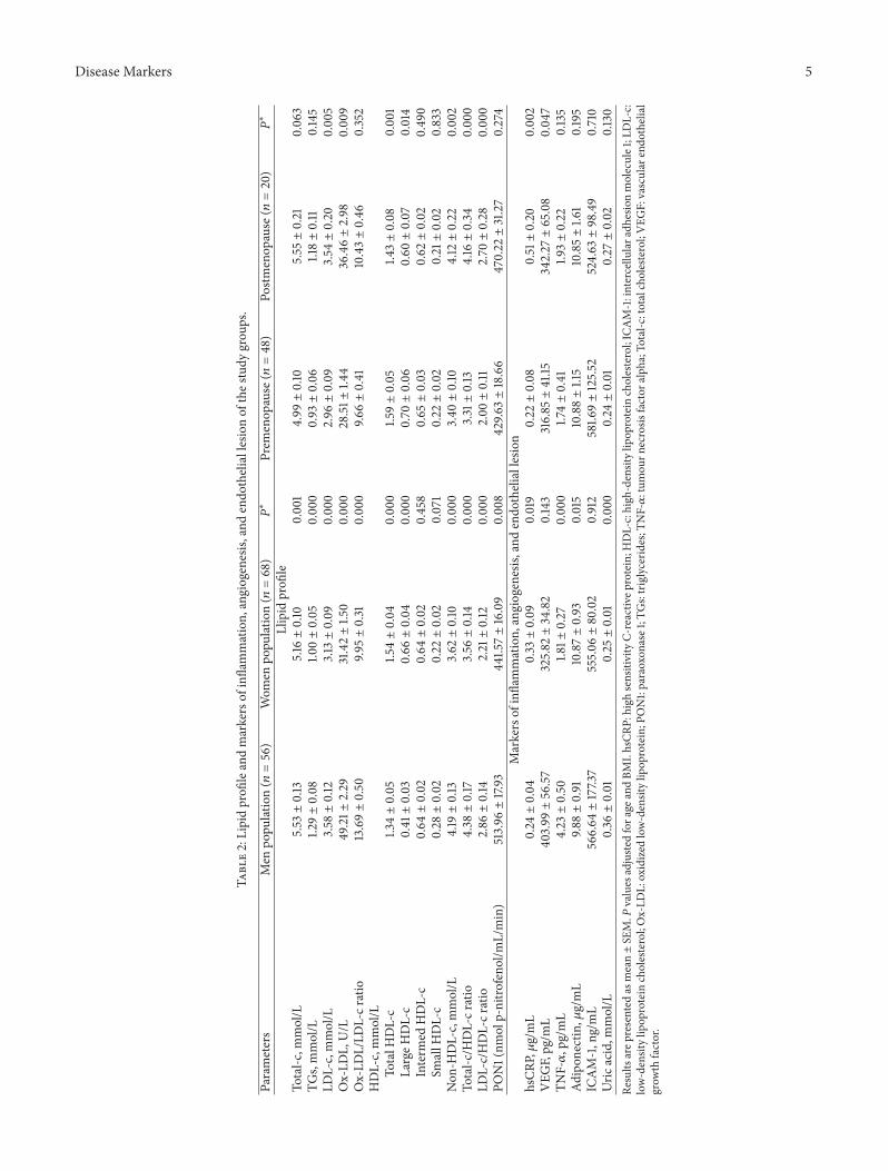
3.2. Classical Lipid Profile. Table 2 summarizes the resultsobtained for all markers of lipid, inflammatory, and angio-genic profile tested in the study.With respect to classical lipidprofile, several significant differences were found betweenmen and women volunteers: (1) total-c (𝑃 = 0.001), TGs, andLDL-c (𝑃 = 0.000, for both) serum content were significantlyhigher in men, and (2) serum HDL-c concentrations weresignificantly (𝑃 = 0.000) lower (Figure 1). Despite thesedifferences, it must be emphasized that, with the exceptionof total-c (slightly above acceptable limits), all of these valueswere within the normal range.
Concerning the effects of menopause, the following sig-nificant differences were observed: (1) serum LDL-c contentswere significantly (𝑃 = 0.005) higher in postmenopausalwomen, and (2) HDL-c levels were significantly (𝑃 = 0.001)lower (Figures 1(a) and 1(c), resp.). No statistically significantdifferences were found between both groups with respect tototal-c and TGs levels (Table 2).
3.3. Ox-LDL, HDL Subpopulations, and Paraoxonase Activity.Nonclassical markers of lipid profile confirm the notoriouslyprotective status of women (and specifically, premenopausalwomen): Ox-LDL content is significantly higher in menand postmenopausal groups (𝑃 = 0.000 and 𝑃 = 0.009,resp.) (Figure 1(b)).The concentration of the more protectiveHDL subpopulation (large HDL) was significantly higherin women (as opposed to men) and premenopausal (asopposed to postmenopausal) women (𝑃 = 0.000 and 𝑃 =0.014, resp.) (Figure 1(d)).Nodifferenceswere foundbetweengroups with regard to the content of the “less protective”HDL subpopulation (small HDL) or of intermediate HDL(Figure 1(d)). The increased CVR in men is also manifestedby the higher Ox-LDL/LDL-c ratio (𝑃 = 0.000), while nostatistical significant differences were found between the pre-and postmenopausal groups (Table 2). PON1 activity waslower in women (𝑃 = 0.008) and unchanged between pre-and postmenopausal subgroups.
3.4. Markers of Inflammation, Angiogenesis, and EndothelialLesion. TNF-𝛼 levels were higher (𝑃 = 0.000) in men thanin women, a tendency that was coupled with decreased (𝑃 =0.015) adiponectin contents (Table 2), with values unchangedfor pre- and postmenopausal subjects. hsCRP contents werehigher in women (𝑃 = 0.019) relative to men and inpostmenopausal (𝑃 = 0.002) relative to premenopausalsubjects. Uric acid concentration was higher in men (𝑃 =0.000), but no differences were found between the pre- andpostmenopausal subjects. Values for the marker of endothe-lial lesion (iCAM-1) were found to be unchanged acrossgroups. VEGF serum levels were significantly increased inpostmenopausal women (𝑃 = 0.047), with unchanged levelsacross gender (Table 2).
3.5. Analysis of Correlations between Markers of CVR
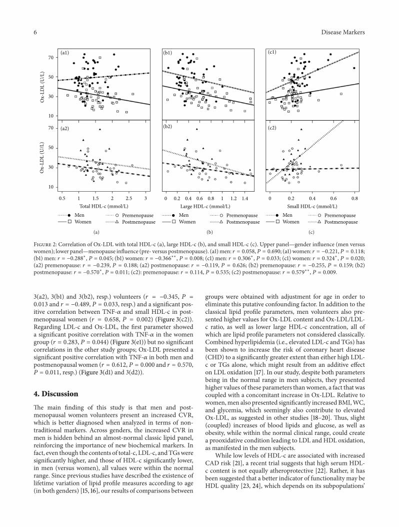
3.5.1. Ox-LDL Correlations with HDL Subpopulations. Asignificant negative correlation was found between largeHDL-c and Ox-LDL for men and women (Figure 2(b1)), and

4 Disease Markers
Table 1: Demographic and anthropometric data of the study groups.
ParametersMen
population(𝑛 = 56)
Womenpopulation(𝑛 = 68)
P Premenopause(𝑛 = 48)
Postmenopause(𝑛 = 20) P
Age, years (range) 53.07 ± 1.90(22–75)
43.16 ± 1.81(21–69) 0.000 35.40 ± 1.39
(21–54)61.80 ± 1.40(49–69) 0.000
BMI, Kg/m2 27.55 ± 0.61 25.28 ± 0.50 0.003 24.51 ± 0.59 27.14 ± 0.81 0.016Waist circumference, cm 99.50 ± 1.53 89.16 ± 1.32 0.000 87.57 ± 1.48 93.08 ± 2.60 0.000SBP, mmHg 141.77 ± 2.57 131.95 ± 2.82 0.000 123.04 ± 2.59 154.00 ± 4.38 0.000DBP, mmHg 85.36 ± 1.40 82.85 ± 1.51 0.000 79.74 ± 1.70 90.53 ± 2.41 0.000Glycemia, mmol/L 5.44 ± 0.08 4.94 ± 0.06 0.000 4.78 ± 0.06 5.33 ± 0.14 0.000Results are presented as mean ± SEM. P values adjusted for age and BMI. BMI: body mass index; SBP: systolic blood pressure; DBP: diastolic blood pressure.
(mm
ol/L
)
0
1
2
3
4
aaa
bb
LDL-c
(a)
Oxidized-LDL
(U/L
)
0
10
20
30
40
50
60
aaa
bb
(b)
Total HDL-c (m
mol
/L)
0
0.6
1.2
1.8aaa
bbb
(c)
MenWomen
PremenopausePostmenopause
(mm
ol/L
)
aaa
0
0.2
0.4
0.6
0.8b
Large HDL-c Intermediate HDL-c Small HDL-c
(d)
Figure 1: Serum LDL-c (a), Ox-LDL (b), total HDL-c (c), and large, intermediate, and small HDL subpopulations (d), in the study groups.Results are presented as mean ± SEM. P values adjusted for age and BMI. a = 𝑃 < 0.05 and aaa = 𝑃 < 0.001 versus men; b = 𝑃 < 0.05, bb =𝑃 < 0.01 and bbb = 𝑃 < 0.001 versus premenopause.
postmenopausal (Figure 2(b2)) volunteers (𝑟 = −0.288, 𝑃 =0.045; 𝑟 = −0.366, 𝑃 = 0.008; 𝑟 = −0.570, 𝑃 = 0.011, resp.); asignificant positive correlation was also established betweensmall HDL-c and Ox-LDL for the same groups (𝑟 = 0.306,𝑃 = 0.033; 𝑟 = 0.324, 𝑃 = 0.020; 𝑟 = 0.579, 𝑃 = 0.009, resp.)(Figure 2(c1) and 2(c2), resp.). No significant correlationswere found between these parameters for the premenopausal
group (Figure 2(b2) and 2(c2), resp.). Moreover, total HDL-c showed no correlation with Ox-LDL in any study group(Figure 2(a1) and 2(a2)).
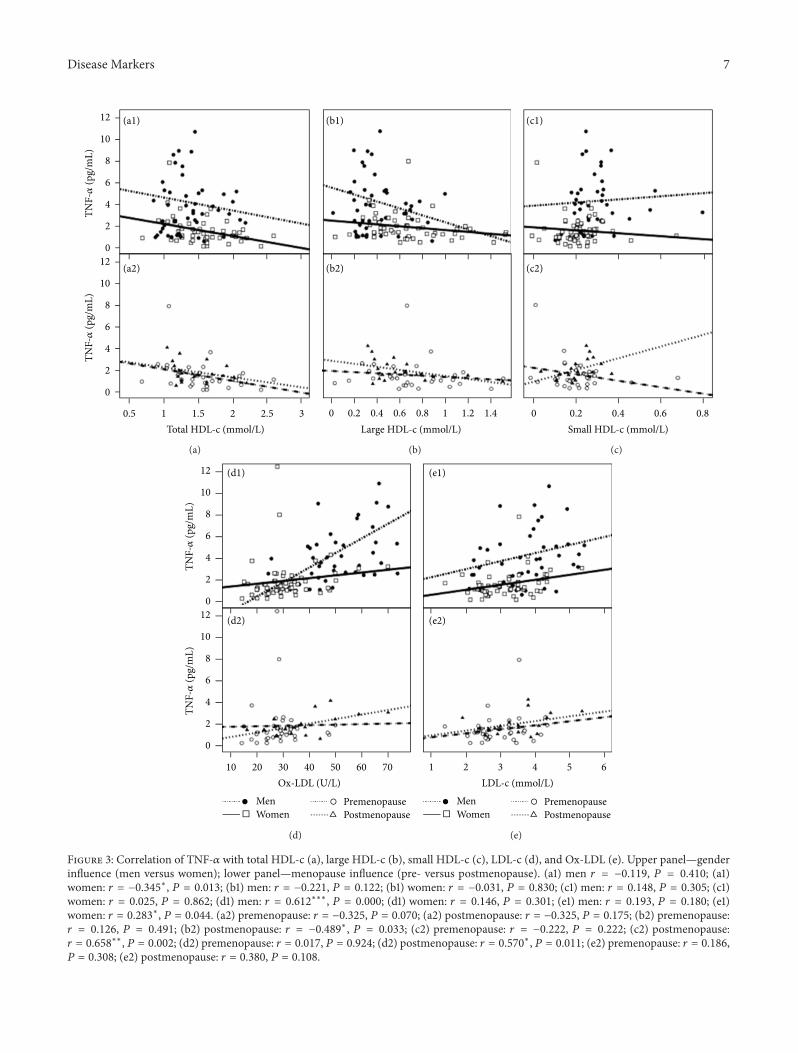
3.5.2. TNF-𝛼 Correlations with Lipid Parameters. Analysingthe correlation between TNF-𝛼 and (resp.) total HDL-c, largeHDL-c, and small HDL-c, a significant negative correlationwas found between TNF-𝛼 and both total HDL-c and largeHDL-c in women and (resp.) postmenopausal (Figure 3(a1),
Disease Markers 5
Table2:Lipidprofi
leandmarkersof
inflammation,
angiogenesis,
andendo
theliallesio
nof
thes
tudy
grou
ps.
Parameters
Men
popu
latio
n(𝑛=56)
Wom
enpo
pulatio
n(𝑛=68)
P∗Prem
enop
ause
(𝑛=48)
Postm
enop
ause
(𝑛=20)
P∗Llipid
profi
leTo
tal-c
,mmol/L
5.53±0.13
5.16±0.10
0.001
4.99±0.10
5.55±0.21
0.063
TGs,mmol/L
1.29±0.08
1.00±0.05
0.00
00.93±0.06
1.18±0.11
0.145
LDL-c,mmol/L
3.58±0.12
3.13±0.09
0.00
02.96±0.09
3.54±0.20
0.005
Ox-LD
L,U/L
49.21±
2.29
31.42±1.5
00.00
028.51±
1.44
36.46±2.98
0.00
9Ox-LD
L/LD
L-cr
atio
13.69±0.50
9.95±0.31
0.00
09.6
6±0.41
10.43±0.46
0.352
HDL-c,mmol/L
TotalH
DL-c
1.34±0.05
1.54±0.04
0.00
01.5
9±0.05
1.43±0.08
0.001
LargeH
DL-c
0.41±0.03
0.66±0.04
0.00
00.70±0.06
0.60±0.07
0.014
Interm
edHDL-c
0.64±0.02
0.64±0.02
0.458
0.65±0.03
0.62±0.02
0.490
SmallH
DL-c
0.28±0.02
0.22±0.02
0.071
0.22±0.02
0.21±0.02
0.833
Non
-HDL-c,mmol/L
4.19±0.13
3.62±0.10
0.00
03.40±0.10
4.12±0.22
0.002
Total-c
/HDL-cr
atio
4.38±0.17
3.56±0.14
0.00
03.31±0.13
4.16±0.34
0.00
0LD
L-c/HDL-cr
atio
2.86±0.14
2.21±0.12
0.00
02.00±0.11
2.70±0.28
0.00
0PO
N1(nm
olp-nitro
feno
l/mL/min)
513.96±17.93
441.5
7±16.09
0.008
429.6
3±18.66
470.22±31.27
0.274
Markersof
inflammation,
angiogenesis,
andendo
thelialles
ion
hsCR
P,𝜇g/mL
0.24±0.04
0.33±0.09
0.019
0.22±0.08
0.51±0.20
0.002
VEG
F,pg/m
L403.99±56.57
325.82±34.82
0.143
316.85±41.15
342.27±65.08
0.047
TNF-𝛼,pg/mL
4.23±0.50
1.81±
0.27
0.00
01.74±0.41
1.93±0.22
0.135
Adipon
ectin
,𝜇g/mL
9.88±0.91
10.87±0.93
0.015
10.88±1.15
10.85±1.6
10.195
ICAM-1,ng/mL
566.64±177.37
555.06±80.02
0.912
581.6
9±125.52
524.63±98.49
0.710
Uric
acid,m
mol/L
0.36±0.01
0.25±0.01
0.00
00.24±0.01
0.27±0.02
0.130
Results
arep
resented
asmean±SE
M.P
values
adjuste
dfora
geandBM
I.hsCR
P:high
sensitivityC-
reactiv
eprotein;H
DL-c:high
-densitylipop
rotein
cholesterol;ICAM-1:intercellu
lara
dhesionmolecule1;LDL-c:
low-densitylipop
rotein
cholesterol;Ox-LD
L:oxidized
low-densitylipop
rotein;P
ON1:paraoxon
ase1;T
Gs:triglycerid
es;T
NF-𝛼:tum
ourn
ecrosis
factor
alph
a;To
tal-c
:totalcholesterol;VEG
F:vascular
endo
thelial
grow
thfactor.
6 Disease Markers
MenWomen Postmenopause
Premenopause
0.5 1 1.5 2 32.5Total HDL-c (mmol/L)
Ox-
LDL
(U/L
)O
x-LD
L (U
/L)
10
30
50
70
10
30
50
70
(a1)
(a2)
(a)
0 0.2 0.4 0.6 0.8 1 1.2 1.4Large HDL-c (mmol/L)
(b1)
(b2)
MenWomen Postmenopause
Premenopause
(b)
0 0.2 0.4 0.6 0.8Small HDL-c (mmol/L)
(c1)
(c2)
MenWomen Postmenopause
Premenopause
(c)
Figure 2: Correlation of Ox-LDL with total HDL-c (a), large HDL-c (b), and small HDL-c (c). Upper panel—gender influence (men versuswomen); lower panel—menopause influence (pre- versus postmenopause). (a1)men: 𝑟 = 0.058,𝑃 = 0.690; (a1) women: 𝑟 = −0.221,𝑃 = 0.118;(b1) men: 𝑟 = −0.288∗, 𝑃 = 0.045; (b1) women: 𝑟 = −0.366∗∗, 𝑃 = 0.008; (c1) men: 𝑟 = 0.306∗, 𝑃 = 0.033; (c1) women: 𝑟 = 0.324∗, 𝑃 = 0.020;(a2) premenopause: 𝑟 = −0.239, 𝑃 = 0.188; (a2) postmenopause: 𝑟 = −0.119, 𝑃 = 0.626; (b2) premenopause: 𝑟 = −0.255, 𝑃 = 0.159; (b2)postmenopause: 𝑟 = −0.570∗, 𝑃 = 0.011; (c2): premenopause: 𝑟 = 0.114, 𝑃 = 0.535; (c2) postmenopause: 𝑟 = 0.579∗∗, 𝑃 = 0.009.
3(a2), 3(b1) and 3(b2), resp.) volunteers (𝑟 = −0.345, 𝑃 =0.013 and 𝑟 = −0.489, 𝑃 = 0.033, resp.) and a significant pos-itive correlation between TNF-𝛼 and small HDL-c in post-menopausal women (𝑟 = 0.658, 𝑃 = 0.002) (Figure 3(c2)).Regarding LDL-c and Ox-LDL, the first parameter showeda significant positive correlation with TNF-𝛼 in the womengroup (𝑟 = 0.283, 𝑃 = 0.044) (Figure 3(e1)) but no significantcorrelations in the other study groups; Ox-LDL presented asignificant positive correlation with TNF-𝛼 in both men andpostmenopausal women (𝑟 = 0.612, 𝑃 = 0.000 and 𝑟 = 0.570,𝑃 = 0.011, resp.) (Figure 3(d1) and 3(d2)).
4. Discussion
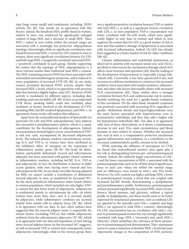
The main finding of this study is that men and post-menopausal women volunteers present an increased CVR,which is better diagnosed when analyzed in terms of non-traditional markers. Across genders, the increased CVR inmen is hidden behind an almost-normal classic lipid panel,reinforcing the importance of new biochemical markers. Infact, even though the contents of total-c, LDL-c, andTGsweresignificantly higher, and those of HDL-c significantly lower,in men (versus women), all values were within the normalrange. Since previous studies have described the existence oflifetime variation of lipid profile measures according to age(in both genders) [15, 16], our results of comparisons between
groups were obtained with adjustment for age in order toeliminate this putative confounding factor. In addition to theclassical lipid profile parameters, men volunteers also pre-sented higher values for Ox-LDL content and Ox-LDL/LDL-c ratio, as well as lower large HDL-c concentration, all ofwhich are lipid profile parameters not considered classically.Combined hyperlipidemia (i.e., elevated LDL-c and TGs) hasbeen shown to increase the risk of coronary heart disease(CHD) to a significantly greater extent than either high LDL-c or TGs alone, which might result from an additive effecton LDL oxidation [17]. In our study, despite both parametersbeing in the normal range in men subjects, they presentedhigher values of these parameters thanwomen, a fact that wascoupled with a concomitant increase in Ox-LDL. Relative towomen,men also presented significantly increased BMI,WC,and glycemia, which seemingly also contribute to elevatedOx-LDL, as suggested in other studies [18–20]. Thus, slight(coupled) increases of blood lipids and glucose, as well asobesity, while within the normal clinical range, could createa prooxidative condition leading to LDL and HDL oxidation,as manifested in the men subjects.
While low levels of HDL-c are associated with increasedCAD risk [21], a recent trial suggests that high serum HDL-c content is not equally atheroprotective [22]. Rather, it hasbeen suggested that a better indicator of functionality may beHDL quality [23, 24], which depends on its subpopulations’
Disease Markers 7
0.5 1 1.5 2 32.5Total HDL-c (mmol/L)
0
2
4
6
8
10
12
0
2
4
6
8
10
12
TNF-𝛼
(pg/
mL)
TNF-𝛼
(pg/
mL)
(a1)
(a2)
(a)
0 0.2 0.4 0.6 0.8 1 1.2 1.4Large HDL-c (mmol/L)
(b1)
(b2)
(b)
0 0.2 0.4 0.6 0.8Small HDL-c (mmol/L)
(c1)
(c2)
(c)
10 20 30 40 50 60 70Ox-LDL (U/L)
MenWomen Postmenopause
Premenopause
0
2
4
6
8
10
12
0
2
4
6
8
10
12
TNF-𝛼
(pg/
mL)
TNF-𝛼
(pg/
mL)
(d1)
(d2)
(d)
1 2 3 4 5 6LDL-c (mmol/L)
MenWomen Postmenopause
Premenopause
(e1)
(e2)
(e)
Figure 3: Correlation of TNF-𝛼 with total HDL-c (a), large HDL-c (b), small HDL-c (c), LDL-c (d), and Ox-LDL (e). Upper panel—genderinfluence (men versus women); lower panel—menopause influence (pre- versus postmenopause). (a1) men 𝑟 = −0.119, 𝑃 = 0.410; (a1)women: 𝑟 = −0.345∗, 𝑃 = 0.013; (b1) men: 𝑟 = −0.221, 𝑃 = 0.122; (b1) women: 𝑟 = −0.031, 𝑃 = 0.830; (c1) men: 𝑟 = 0.148, 𝑃 = 0.305; (c1)women: 𝑟 = 0.025, 𝑃 = 0.862; (d1) men: 𝑟 = 0.612∗∗∗, 𝑃 = 0.000; (d1) women: 𝑟 = 0.146, 𝑃 = 0.301; (e1) men: 𝑟 = 0.193, 𝑃 = 0.180; (e1)women: 𝑟 = 0.283∗, 𝑃 = 0.044. (a2) premenopause: 𝑟 = −0.325, 𝑃 = 0.070; (a2) postmenopause: 𝑟 = −0.325, 𝑃 = 0.175; (b2) premenopause:𝑟 = 0.126, 𝑃 = 0.491; (b2) postmenopause: 𝑟 = −0.489∗, 𝑃 = 0.033; (c2) premenopause: 𝑟 = −0.222, 𝑃 = 0.222; (c2) postmenopause:𝑟 = 0.658
∗∗, 𝑃 = 0.002; (d2) premenopause: 𝑟 = 0.017, 𝑃 = 0.924; (d2) postmenopause: 𝑟 = 0.570∗, 𝑃 = 0.011; (e2) premenopause: 𝑟 = 0.186,𝑃 = 0.308; (e2) postmenopause: 𝑟 = 0.380, 𝑃 = 0.108.
8 Disease Markers
type (large versus small) and constituents, including PON1activity [25, 26]. Our results are in agreement with thistheory: indeed, the beneficial HDL profile found in women,relative to men, was reinforced by significantly enlargedcontent of large HDL and a decrease in small HDL. Thus,HDL-c values in men (while within the normal range) areassociated with a seemingly less protective subpopulationtypology. Interestingly, while no significant correlations werefound between totalHDL-c versusOx-LDL content inmen orwomen, there were significant correlations between Ox-LDLand both largeHDL-c (negatively correlated) and smallHDL-c (positively correlated) in each group, thereby supportingthe notion that the typology of HDL subpopulations is amore relevant marker than traditional total HDL-c content.TheHDL-containing enzyme PON1 has been associated withantioxidant and antiatherogenic properties, and is reduced insome populations of increased CVR [25, 26]. In our study,women presented decreased PON1 activity, despite theirincreased HDL-c levels, which is in opposition with previousdata that showed a slightly higher value [27]. However, PON1activity is modulated by different aspects, including diet,smoking, and exercise habits [28–30]. In fact, as an importantCVR factor, smoking habits could also modulate othermediators or factors involved in the development of CVD,including BMI, hsCRP, oxidized lipoproteins, and HDL-c. Inagreement, future works will analyse this influence.
Apart from the nontraditional markers of lipid profile (inparticular Ox-LDL and HDL subpopulations), men subjectsalso presented a proinflammatory pattern, which is a majorcontributor for the development of atherogenesis. Indeed,men population showed higher serum concentration of TNF-𝛼 and uric acid, accompanied by decreased adiponectinlevels. The reduced plasma cytokine concentrations, includ-ing IL-6 and TNF-𝛼, in women have been attributed tothe inhibitory effect of estrogens on the expression ofinflammatory marker genes [31–33]. The body fat distri-bution, particularly abdominal visceral and subcutaneousadiposity, has been associated with gender-related variationin inflammatory markers, including hsCRP, IL-6, TNF-𝛼,and adiponectin. In fact, fat distribution is known to differacross genders, with men having more visceral and lesssubcutaneous fat [34]. In our study, even after having adjustedfor BMI, we cannot exclude a contribution of abdominalvisceral adiposity in men, as manifested by increased WC,for the more prejudicial inflammatory status found relativeto women population, which included not only higher TNF-𝛼 content but also lower levels of adiponectin. Adiponectinis synthesized mainly in subcutaneous fat cells—of whichwomen have more than men—and it is secreted mostlyby adipocytes, while inflammatory cytokines are secretedmainly from nonfat cells in adipose tissue [35, 36], whichis in agreement with our data. In fact, other authors havesuggested that the visceral adipose tissue is responsible forrelease factors (including TNF-𝛼) that inhibit adiponectinsynthesis from the subcutaneous adipocytes [37–39], whichis in agreement with our data and hypothesis of men havingmore visceral adipose tissue (as the increased WC suggests),as well as increased TNF-𝛼 content and, consequently, loweradiponectin. Interestingly, while in the women group there
was a significant positive correlation between TNF-𝛼 contentand total HDL-c, as well as a significant inverse correlationwith LDL-c, in men population TNF-𝛼 concentration wasbetter correlated with Ox-LDL levels, which were signifi-cantly higher in men than in women and which in turnsupports the notion that Ox-LDL is a good marker of risk inmen and that oxidative damage of lipoproteins is associatedwith increased inflammation. Indeed, Ox-LDL has alreadybeen suggested as a better marker of risk for other conditions[40–42].
Chronic inflammation and endothelial dysfunction, asobserved in patients with increased serum uric acid (SUA),are likely to intervene in themechanisms throughwhich SUAaffects renal structure and operation [43], thereby leading tothe development of hypertension in (especially young) indi-viduals [44]. Conversely, it has been questioned if uric acidincreases as a defense mechanism to counteract the increasedoxidative stress associated with insulin resistance, inflamma-tion, and other risk factors that usually cluster with increasedSUA concentrations [45]. Many studies show a strongercorrelation between SUA and cardiovascular risk in women,thereby justifying the need for a gender-based evaluationof this correlation. On the other hand, metabolic syndromewas positively associated with increasing SUA, regardless ofgender. Abdominal obesity and hypertriglyceridemia werethe main factors associated with hyperuricemia even innormotensive individuals, and they may add a higher riskfor hypertensive individuals [46]. Our data is in agreementwith each of these observations, as it shows a simultaneousincrease in uric acid, TGs, waist circumference and bloodpressure in men relative to women. Whether the increaseduric acid in men is a compensatory protective mechanismagainst inflammation and oxidation, or a contributor to theincreased cardiometabolic profile, remains to be clarified.
While assessing the influence of menopause on CVR,we found that nontraditional markers once again play akey role in distinguishing between pre- and postmenopausalwomen. Indeed, the relatively larger concentration of LDL-c and the lower concentration of HDL-c associated with thepostmenopausal state seem to be of little clinical significance,since in each case values are within the normal range,and no differences were found in total-c and TGs levels.However, Ox-LDL content was higher and largeHDL-c lowerin postmenopausal women, a trend that was coupled withincreased hsCRP, thereby demonstrating a dyslipidaemicand proinflammatory profile. Furthermore, postmenopausalwomen presented significantly increased BMI, waist circum-ference, blood pressure, and glycemia, without a typicalprofile of dyslipidaemia, and these trends seem to be betterexpressed by nonclassical parameters, such as oxidized LDL(as opposed to the typically used LDL-c content) and largeHDL-c (as opposed to total HDL-c content). Interestingly,Ox-LDL concentration has no correlation with total-c con-tent in postmenopausal women but was strongly significantlycorrelated with large HDL-c (inversely) and small HDL-c(directly), which underlines the relevance of those parame-ters in postmenopausal risk determination.Thus, menopauseseems to cause a reduction of absolute HDL-c levels but moreimportantly changes in the composition of HDL particles.
Disease Markers 9
Such a conclusion is in agreement with Eapen et al. [47] whoreported an alteration in the distribution of HDL subspeciesin menopause, with a reduction seen in the proportion oflarge, buoyant HDL2 particles, which are believed to bemore active in reverse cholesterol transport [48]; in contrast,the number of smaller, more dense HDL3 subfragmentsincreases [49, 50]. In addition, ex vivo biochemical analysis oflipoproteins from pre- and postmenopausal women suggeststhat the postmenopausal HDL particle exhibits impairedability to limit LDL oxidation [51]. Despite the impairedcomposition of HDL particles in postmenopausal women, wedid not find differences in PON1 activity. It has been pointedout that the latter HDL-associated enzyme has protectiveeffects against inflammation in the arterial wall and that it iscapable of destroying the biologically active lipids in mildlyoxidized LDL; several studies have previously reported thatCHD is associated with low PON activity [52, 53]. In our data,however, PON1 activity is unchanged in postmenopausalwomen, which is in agreement with previous data from Zagoet al. [? ], who also found no differences in pre- versuspostmenopausal women, as well as with the study of Horteret al. [54], who failed to find any significant associationbetween CHD and paraoxonase or arylesterase activity inpostmenopausal women.
Adipocytokine levels have previously been associatedwith estrogens, suggesting that menopause impacts oninflammatory mediators [55]. In our study, although theTNF-𝛼 increase in postmenopausal women (relative to pre-menopausal women) did not achieve statistical difference,the values presented strongly significant correlations withthe previous markers: namely, a positive correlation withlarge HDL-c and Ox-LDL and an inverse correlation withsmall HDL-c. Surprisingly, no correlation was found betweenTNF-𝛼 levels and total HDL-c and LDL-c concentrations,which are the clinically checked parameters. Moreover, nocorrelation was found between TNF-𝛼 and either HDL-c contents (total or subpopulations) or LDL-c levels (oxi-dized or nonoxidized measures) in premenopausal women,thereby demonstrating that the nontraditional parametersgain particular relevance after menopause and are deservingof closer attention in postmenopausal women. With regardto other mediators/markers of the inflammatory process,we found a significant increased concentration of hsCRP inthe postmenopausal women. Previous studies on menopauseand inflammation have showed that obesity is associatedwith an inflammatory state that is often characterized byelevated plasma hsCRP levels [56]. In our study, hsCRPwas significantly increased in postmenopausal women, whoalso presented excess weight/obesity. hsCRP is an importantmarker for this condition, because after menopause the con-sequent decrease in estrogen levels contributes to widespreadweight gain in women; furthermore, increased contents ofhsCRP, a marker of systemic inflammation, are associatedwith increased incidence of CVD, independently of otherconventional risk factors [57]. By contrast, adiponectin levelsof the pre- and postmenopausal groups were the same, andadiponectin was uncorrelated with age, in agreement witha study of Goodarzi et al. [58], but in disagreement withLoucif et al. [59]. So it seems that menopause impacts less
on adiponectin as a mediator, suggesting that women havehigher adiponectin contents than men, irrespective of theirmenopausal state. A similar association was found for TNF-𝛼, with women having significantly lower concentrations,whether before or after menopause.
VEGF serum levels are lower in premenopausal thanpostmenopausal women, which is in disagreement with thedata from another study [59]. It should be noted that noneof the volunteers entering in our study were under hormonereplacement therapy (HRT), which could influence the levelsofVEGF, althoughpublished studies are contradictory on thiseffect [60, 61].
The data obtained suggest that men subjects present anincrease CV risk when compared to women which is mainlyviewed by nontraditional markers and might result fromslight, but concomitant, variations of several factors (despitevalues within the normal clinical range), including incre-ment of TC, LDL-c, TG, and glycemia and reduced HDL-c, together with increased BMI. The accumulation of factorsseems to promote an oxidative and inflammatory profile,with increased contents of Ox-LDL and reduced of large-HDL-c, as well as of the proinflammatory cytokine TNF-𝛼, and reduced of anti-inflammatory adiponectin. This clus-ter of procardiometabolic alterations (even of slight inten-sity), together with increased uric acid and blood pressure,might represent an enhanced CV risk when compared withwomen subjects, which seem to be protected, namely, beforemenopause. Similarly, after menopause, women present anincreased BMI, waist circumference, and glycemia, whichmight underlie the modifications of cardiometabolic profile.Indeed, postmenopausal women, despite only slightly highervalues of LDL-c and lower of HDL-c (an unchanged of TCand TGs), presented an oxidative, angiogenic, and proin-flammatory profile, with increased contents of Ox-LDL andVEGF and reduced of large HDL-c, together with increasedhsCRP, suggesting once again that a cluster of slight variationsof traditional markers (despite in the normal clinical range)promotes a poor cardiometabolic profile.
To conclude, in a population without previous diagnosisand medication for CVD, as well as without familiar historyof CVD, and with serum levels of traditional routine lipidmeasures and glycemia within the normal range, there arenontraditional measures related with lipid, inflammatory,and angiogenic profiles that cannot be minimized and woulddeserve attention, as they show an increased CVR in menand in postmenopausal women. Although the issue is notconsensual when refers to factors that might affect the CVRin a healthy population, this data suggests more attentionto nonclassical parameters as putative biomarkers of the CVstatus in a population without any noticeable classical signal,marker, symptom, or previous history appointment of CVD.However, the study has some limitations that deserve furtherresearch in future work: (a) the possibility of bias related withthe study design and criteria for inclusion of subjects; (b)the enlargement of sample size will strength the results; (c) abetter age and BMI matching of populations would improvethe data; (d) cardiovascular outcomes (e.g., events) will allowbetter association between markers and CVR.
10 Disease Markers
5. Conclusions
Men and postmenopausal women present a lipid profileindicative of an increased CVR, relative to women and pre-menopausal women, respectively. This increased risk may bequantified by nontraditional lipid profile markers, including(the relative content of)Ox-LDL andHDL subpopulations, aswell as markers of inflammation and angiogenesis (includingTNF-𝛼 and adiponectin for comparisons across genders andVEGF for comparisons between pre- and postmenopausalwomen). In addition, postmenopausal women manifest aproatherogenic risk that should receive special attention andmight warrant early pharmacotherapeutic intervention toprevent premature development of CVD. Finally, this studysuggest that some of the classical markers of cardiovascu-lar (namely, lipidic) profile, often insensitive to risk, maypresent normal or only slightly elevated values and stillwithin the normal clinical range, recommending that routinebiochemical data would improve with the inclusion of otherreliable and specific markers with additional information ofcardiometabolic risk.
Conflict of Interests
The authors report no conflict of interests.
Acknowledgments
This study was supported by the Portuguese Foundationfor Science and Technology, through a Ph.D. Grant(SFRH/BD/65483/2009) and Strategic Project (PEst-C/SAU/UI3282/2011), as well as by the COMPETE.
References
[1] C. Leuzzi, R.Marzullo, andM.G.Modena, “Ismenopause a riskfactor for ischemic heart disease in women?” Giornale Italianodi Cardiologia, vol. 13, no. 6, pp. 401–406, 2012.
[2] M. Kaushik, S. P. Sontineni, and C. Hunter, “Cardiovasculardisease and androgens: a review,” International Journal ofCardiology, vol. 142, no. 1, pp. 8–14, 2010.
[3] E. Casiglia, V. Tikhonoff, S. Caffi et al., “Menopause does notaffect blood pressure and risk profile, and menopausal womendo not become similar to men,” Journal of Hypertension, vol. 26,no. 10, pp. 1983–1992, 2008.
[4] H. Smulyan, R. G. Asmar, A. Rudnicki, G.M. London, andM. E.Safar, “Comparative effects of aging in men and women on theproperties of the arterial tree,” Journal of the American Collegeof Cardiology, vol. 37, pp. 1374–1380, 2001.
[5] H. Mangge, G. Almer, M. Truschnig-Wilders, A. Schmidt, R.Gasser, and D. Fuchs, “Inflammation, adiponectin, obesity andcardiovascular risk,” Current Medicinal Chemistry, vol. 17, no.36, pp. 4511–4520, 2010.
[6] S.-S. Huang, P.-H. Huang, Y.-H. Chen, K.-H. Chiang, J.-W.Chen, and S.-J. Lin, “Association of adiponectin with future car-diovascular events in patients after acutemyocardial infarction,”Journal of Atherosclerosis andThrombosis, vol. 17, no. 3, pp. 295–303, 2010.
[7] F. Girardi, E. Franceschi, and A. A. Brandes, “Cardiovascularsafety of VEGF-targeting therapies: current evidence and han-dling strategies,” Oncologist, vol. 15, no. 7, pp. 683–694, 2010.
[8] A. N. N. Mertens and P. Holvoet, “Oxidized LDL and HDL:antagonists in atherothrombosis,” FASEB Journal, vol. 15, no. 12,pp. 2073–2084, 2001.
[9] M. I. Mackness, P. N. Durrington, and B. Mackness, “The roleof paraoxonase 1 activity in cardiovascular disease: potential fortherapeutic intervention,” American Journal of CardiovascularDrugs, vol. 4, no. 4, pp. 211–217, 2004.
[10] K. Mahdy Ali, A. Wonnerth, K. Huber, and J. Wojta, “Cardio-vascular disease risk reduction by raising HDL cholesterol—current therapies and future opportunities,” British Journal ofPharmacology, vol. 167, pp. 1177–1194, 2012.
[11] S. Redondo, J. Martınez-Gonzalez, C. Urraca, and T. Tejerina,“Emerging therapeutic strategies to enhance HDL function,”Lipids in Health and Disease, vol. 10, article 175, 2011.
[12] B. F. Asztalos, P. S. Roheim, R. L. Milani et al., “Distribution ofapoA-I-containing HDL subpopulations in patients with coro-nary heart disease,” Arteriosclerosis, Thrombosis, and VascularBiology, vol. 20, no. 12, pp. 2670–2676, 2000.
[13] A. Pirillo, G. D. Norata, and A. L. Catapano, “High-densitylipoprotein subfractions—what the clinicians need to know,”Cardiology, vol. 124, pp. 116–125, 2013.
[14] World Health Organization Scientific Group, “Research on themenopause in the 1990s,”WHOTechnical Services DepartmentSeries 866, WHO, Geneva, Switzerland.
[15] G. M. Singh, G. Danaei, P. M. Pelizzari et al., “The ageassociations of blood pressure, cholesterol, and glucose: analysisof health examination surveys from international populations,”Circulation, vol. 125, pp. 2204–2211, 2012.
[16] F. B. Dalpino, L. Menna-Barreto, and E. C. de Faria, “Influencesof sex and age on biological rhythms of serum lipids andlipoproteins,” Clinica Chimica Acta, vol. 406, no. 1-2, pp. 57–61,2009.
[17] V. Manninen, L. Tenkanen, P. Koskinen et al., “Joint effects ofserum triglyceride and LDL cholesterol and HDL cholesterolconcentrations on coronary heart disease risk in the HelsinkiHeart Study. Implications for treatment,”Circulation, vol. 85, no.1, pp. 37–45, 1992.
[18] S. Zelzer, N. Fuchs, G. Almer et al., “High density lipoproteincholesterol level is a robust predictor of lipid peroxidation irre-spective of gender, age, obesity, and inflammatory or metabolicbiomarkers,” Clinica Chimica Acta, vol. 412, no. 15-16, pp. 1345–1349, 2011.
[19] K. Kotani, N. Sakane, M. Ueda et al., “Oxidized high-densitylipoprotein is associated with increased plasma glucose in non-diabetic dyslipidemic subjects,” Clinica Chimica Acta, vol. 414,pp. 125–129, 2012.
[20] M. Maytin, J. Leopold, and J. Loscalzo, “Oxidant stress in thevasculature,” Current Atherosclerosis Reports, vol. 1, no. 2, pp.156–164, 1999.
[21] R. Movva and D. J. Rader, “Laboratory assessment of HDLheterogeneity and function,” Clinical Chemistry, vol. 54, no. 5,pp. 788–800, 2008.
[22] D. J. Rader, “Illuminating HDL—is it still a viable therapeutictarget?” The New England Journal of Medicine, vol. 357, no. 21,pp. 2180–2183, 2007.
[23] E. Eren, N. Yilmaz, andO. Aydin, “High density lipoprotein andit’s dysfunction,”The Open Biochemistry Journal, vol. 6, pp. 78–93, 2012.

Disease Markers 11
[24] H. Soran, S. Hama, R. Yadav et al., “HDL functionality,” CurrentOpinion in Lipidology, vol. 23, pp. 353–366, 2012.
[25] M. Mackness, P. Durrington, and B. Mackness, “Paraoxonase 1activity, concentration and genotype in cardiovascular disease,”Current Opinion in Lipidology, vol. 15, no. 4, pp. 399–404, 2004.
[26] M. I. Mackness, B. Mackness, and P. N. Durrington, “Paraox-onase and coronary heart disease,” Atherosclerosis Supplements,vol. 3, no. 4, pp. 49–55, 2002.
[27] P. Kleemola, R. Freese, M. Jauhiainen, R. Pahlman, G. Alfthan,and M. Mutanen, “Dietary determinants of serum paraoxonaseactivity in healthy humans,” Atherosclerosis, vol. 160, no. 2, pp.425–432, 2002.
[28] E. Thomas-Moya, M. Gianotti, A. M. Proenza, and I. Llado,“Paraoxonase 1 response to a high-fat diet: gender differencesin the factors involved,”Molecular Medicine, vol. 13, no. 3-4, pp.203–209, 2007.
[29] E. Thomas-Moya, M. Gianotti, I. Llado, and A. M. Proenza,“Effects of caloric restriction and gender on rat serum paraox-onase 1 activity,” Journal of Nutritional Biochemistry, vol. 17, no.3, pp. 197–203, 2006.
[30] E. Thomas-Moya, Y. Gomez-Perez, M. Fiol, M. Gianotti, I.Llado, and A. M. Proenza, “Gender related differences inparaoxonase 1 response to high-fat diet-induced oxidativestress,” Obesity, vol. 16, no. 10, pp. 2232–2238, 2008.
[31] W. B. Ershler and E. T. Keller, “Age-associated increasedinterleukin-6 gene expression, late-life diseases, and frailty,”Annual Review of Medicine, vol. 51, pp. 245–270, 2000.
[32] J. An, R. C. J. Ribeiro, P. Webb et al., “Estradiol repressionof tumor necrosis factor-𝛼 transcription requires estrogenreceptor activation function-2 and is enhanced by coactivators,”Proceedings of the National Academy of Sciences of the UnitedStates of America, vol. 96, no. 26, pp. 15161–15166, 1999.
[33] S. D. Imahara, S. Jelacic, C. E. Junker, and G. E. O’Keefe, “Theinfluence of gender on human innate immunity,” Surgery, vol.138, no. 2, pp. 275–282, 2005.
[34] B. Ludescher, A. Najib, S. Baar et al., “Gender specific correla-tions of adrenal gland size and body fat distribution: a wholebody MRI study,” Hormone and Metabolic Research, vol. 39, no.7, pp. 515–518, 2007.
[35] J. N. Fain, “Release of interleukins and other inflammatorycytokines by human adipose tissue is enhanced in obesity andprimarily due to the nonfat cells,” Vitamins and Hormones, vol.74, pp. 443–477, 2006.
[36] T. Ahonen, M. Vanhala, H. Kautiainen, E. Kumpusalo, and J.Saltevo, “Sex differences in the association of adiponectin andlow-grade inflammation with changes in the body mass indexfrom youth to middle age,” Gender Medicine, vol. 9, no. 1, pp.1–8, 2011.
[37] C.M. Halleux, M. Takahashi, M. L. Delporte et al., “Secretion ofadiponectin and regulation of apM1 gene expression in humanvisceral adipose tissue,” Biochemical and Biophysical ResearchCommunications, vol. 288, no. 5, pp. 1102–1107, 2001.
[38] N. Maeda, M. Takahashi, T. Funahashi et al., “PPAR𝛾 ligandsincrease expression and plasma concentrations of adiponectin,an adipose-derived protein,” Diabetes, vol. 50, no. 9, pp. 2094–2099, 2001.
[39] Y. Matsuzawa, “Establishment of a concept of visceral fatsyndrome and discovery of adiponectin,” Proceedings of theJapan Academy. Series B, vol. 86, pp. 131–141, 2010.
[40] H. Huang, W. Mai, D. Liu, Y. Hao, J. Tao, and Y. Dong, “Theoxidation ratio of LDL: a predictor for coronary artery disease,”Disease Markers, vol. 24, no. 6, pp. 341–349, 2008.
[41] Y. Huang, Y. Hu, W. Mai et al., “Plasma oxidized low-densitylipoprotein is an independent risk factor in young patients withcoronary artery disease,”DiseaseMarkers, vol. 31, no. 5, pp. 295–301, 2011.
[42] H. Huang, R. Ma, D. Liu et al., “Oxidized low-density lipopro-tein cholesterol and the ratio in the diagnosis and evaluationof therapeutic effect in patients with coronary artery disease,”Disease Markers, vol. 33, pp. 295–302, 2012.
[43] C. Zoccali, R. Maio, F. Mallamaci, G. Sesti, and F. Perticone,“Uric acid and endothelial dysfunction in essential hyperten-sion,” Journal of the American Society of Nephrology, vol. 17, no.5, pp. 1466–1471, 2006.
[44] D. I. Feig and R. J. Johnson, “Hyperuricemia in childhoodprimary hypertension,”Hypertension, vol. 42, no. 3, pp. 247–252,2003.
[45] F. J. Nieto, C. Iribarren, M. D. Gross, G. W. Comstock, and R.G. Cutler, “Uric acid and serum antioxidant capacity: a reactionto atherosclerosis?” Atherosclerosis, vol. 148, no. 1, pp. 131–139,2000.
[46] S. L. Rodrigues, M. P. Baldo, D. P. Capingana et al., “Genderdistribution of serum uric acid and cardiovascular risk factors:population based study,”Arquivos Brasileiros deCardiologia, vol.98, no. 1, pp. 13–21, 2012.
[47] D. J. Eapen, G. L. Kalra, L. Rifai, C. A. Eapen, N. Merchant, andB. V. Khan, “Raising HDL cholesterol in women,” InternationalJournal of Women’s Health, vol. 1, no. 1, pp. 181–191, 2009.
[48] Y. Huang, A. Von Eckardstein, S. Wu, and G. Assmann,“Cholesterol efflux, cholesterol esterification, and cholesterylester transfer by LpA-I and LpA-I/A-II in native plasma,”Arteriosclerosis, Thrombosis, and Vascular Biology, vol. 15, no. 9,pp. 1412–1418, 1995.
[49] J. C. Stevenson, D. Crook, and I. F. Godsland, “Influence ofage and menopause on serum lipids and lipoproteins in healthywomen,” Atherosclerosis, vol. 98, no. 1, pp. 83–90, 1993.
[50] Z. Li, J. R. McNamara, J.-C. Fruchart et al., “Effects of genderand menopausal status on plasma lipoprotein subspecies andparticle sizes,” Journal of Lipid Research, vol. 37, no. 9, pp. 1886–1896, 1996.
[51] V. Zago, S. Sanguinetti, F. Brites et al., “Impaired high densitylipoprotein antioxidant activity in healthy postmenopausalwomen,” Atherosclerosis, vol. 177, no. 1, pp. 203–210, 2004.
[52] P.M. Laplaud, T.Dantoine, andM. J. Chapman, “Paraoxonase asa risk marker for cardiovascular disease: facts and hypotheses,”Clinical Chemistry and Laboratory Medicine, vol. 36, no. 7, pp.431–441, 1998.
[53] P. N. Durrington, B. Mackness, and M. I. Mackness, “Paraox-onase and atherosclerosis,” Arteriosclerosis, Thrombosis, andVascular Biology, vol. 21, no. 4, pp. 473–480, 2001.
[54] M. J. Horter, S. Sondermann, H. Reinecke et al., “Associationsof HDL phospholipids and paraoxonase activity with coronaryheart disease in postmenopausal women,” Acta PhysiologicaScandinavica, vol. 176, no. 2, pp. 123–130, 2002.
[55] S. C. Hong, S. W. Yoo, G. J. Cho et al., “Correlation betweenestrogens and serum adipocytokines in premenopausal andpostmenopausal women,” Menopause, vol. 14, no. 5, pp. 835–840, 2007.
[56] B. J. Arsenault, C. P. Earnest, J.-P. Despres, S. N. Blair, andT. S. Church, “Obesity, coffee consumption and CRP levelsin postmenopausal overweight/obese women: importance ofhormone replacement therapy use,”European Journal of ClinicalNutrition, vol. 63, no. 12, pp. 1419–1424, 2009.

12 Disease Markers
[57] J. Danesh, J. G. Wheeler, G. M. Hirschfield et al., “C-reactiveprotein and other circulating markers of inflammation in theprediction of coronary heart disease,”The New England Journalof Medicine, vol. 350, no. 14, pp. 1387–1397, 2004.
[58] M. T. Goodarzi, H. Babaahmadi-Rezaei, M. Kadkhodaei-Eliaderani, and S. Haddadinezhad, “Relationship of serumadiponectin with blood lipids, HbA1c, and hs-CRP in type IIdiabetic postmenopausal women,” Journal of Clinical Labora-tory Analysis, vol. 21, no. 3, pp. 197–200, 2007.
[59] Y. Loucif, J. Methot, K. Tremblay, D. Brisson, and D. Gaudet,“Contribution of adiponectin to the cardiometabolic riskof postmenopausal women with loss-of-function lipoproteinlipase gene mutations,” Menopause, vol. 18, no. 5, pp. 558–562,2011.
[60] R. Agrawal, G. Prelevic, G. S. Conway, N. N. Payne, J. Ginsburg,and H. S. Jacobs, “Serum vascular endothelial growth factorconcentrations in postmenopausal women: the effect of hor-mone replacement therapy,” Fertility and Sterility, vol. 73, no.1, pp. 56–60, 2000.
[61] H. Sumino, T. Nakamura, S. Ichikawa et al., “Serum level ofvascular endothelial growth factor is decreased by hormonereplacement therapy in postmenopausal womenwithout hyper-cholesterolemia,” Atherosclerosis, vol. 148, no. 1, pp. 189–195,2000.