Quantifying the High-Velocity, Low-Amplitude Spinal Manipulative Thrust: A Systematic Review
Upload
independentCategory
view
3download
0
Manipulativeassessmentandtreatmentoftheshouldercomplex:casereports
ThomasDonahue,DCa,ThomasBergmann,DCb,SaraDonahue,DCc, andMichaelDody,DCd
aPrivate Practice of Chiropractic, Montana. bProfessor, Northwestern HealthSciences University, Bloomington, MN. cPrivate Practice of Chiropractic,Minnesota. dPrivate Practice of Chiropractic, Colorado.Submit requests for reprints to: Thomas Donahue, DC, 2435 Dahlia Lane,Billings, Montana 59102.Paper submitted June 5, 2003; in revised form June 20, 2003.
ABSTRACTObjective: To describe a unique method of evaluation and aconservative management plan for patients with shoulder dys-function of mechanical origin. Possible causes for this clinicalpresentation and a brief review of the literature are offered.
Clinical Features: In the 3 cases described here, symptomsincluded acute shoulder pain, limitation of movement, positiveorthopedic tests, palpable tenderness, muscle spasm and muscleweakness. The 3 cases all resulted in differing diagnoses.
Intervention and Outcome: Following the use of a proposedjoint dysfunction isolation test, thrusting forms of manualadjusting procedures, electrical modalities and soft tissuetherapy were applied. Three cases representing different com-mon shoulder problems (an acute episode of a chronic prob-lem, a progressive problem resulting in capsulitis, and a occultproblem associated with a motor vehicle accident) respondedfavorably to treatment.
Conclusions: There is the need for a non-surgical, conservativeapproach to treatment of shoulder problems before consideringthe more aggressive treatment approaches that carry greateriatrogenic risks. The patients’ signs and symptoms respondedto a unique method of evaluation and manipulative therapywhen other approaches had failed. The risk/benefit ratio sug-gests that conservative care be considered a potential option forsimilar conditions. (J Chiropr Med 2003;2:145–152)
KEY WORDS: Shoulder; Chiropractic Manipulation;Muscle Testing
INTRODUCTION
Approximately 4,000,000 people in the US seek medicalcare each year for shoulder problems, resulting in
nearly 1.5 million visits to orthopedic surgeons. (1)Shoulder pain and dysfunction is also a common com-plaint prompting patients to seek chiropractic care.Shoulder pain has also been reported to be the secondleading cause occupational injury claims. (2)
The shoulder is a complex of 4 joints that allow it to bethe most movable joint in the body. It must withstandthe worst of our everyday work tasks as well as ourrecreation activities. Mobility has been developed at theexpense of stability in this complex structure. Therefore,it must depend on muscle, tendons and fascia ratherthan bony support. (3,4) Given the complexity of theshoulder, it is not surprising that it is 1 of the mostcommon joints presenting with dysfunctional pathol-ogy. (4) Most shoulder problems arise from directtrauma, overuse, or underuse, commonly resulting inimpingements syndromes. Studies have shown thatthese conditions can be managed with conservative ap-proaches, including chiropractic care. (5–8) Knowledgeand understanding of the anatomy and the intricaterelationships of each of the joint systems is essential forsuccessful assessment and treatment of shoulder prob-lems. This paper focuses on a unique form of evaluationleading to manipulative intervention in the case ofshoulder dysfunction.
Functional Evaluation of the Shoulder Complex
The evaluation of the shoulder begins with a detailedhistory and thorough physical assessment. It is essentialto determine whether the clinical picture is truly attrib-utable to the shoulder complex itself or represents apathologic process elsewhere, such as a cervical pathol-ogy, nerve entrapment, neurologic deficit, or a problemanywhere along the kinetic chain. (9) The history willdirect how extensive the examination must be. Theshoulder can be the site of pain from a variety of sources(Table 1) that must be distinguished. The examination isimportant to develop the diagnostic impression and toprovide insight as to the biomechanical status of thepatient. Provocative tests and motor, sensory and reflextesting for cervical spine and shoulder must be per-formed. (10,11) Active, passive and resisted shoulderROM should be performed. These tests along with painratings using a visual analog scale and functional status
FALL2003 • Number4 • Volume2
1450899-3467/03/1002-049$3.00/0JOURNAL OF CHIROPRACTIC MEDICINECopyright © 2003 by National University of Health Sciences
questionnaires (eg, Shoulder Function AssessmentScale, (12) Shoulder Pain and Disability Index, (13)Shoulder Rating Questionnaire (14)) serve as outcomemeasures to monitor the effects of care.
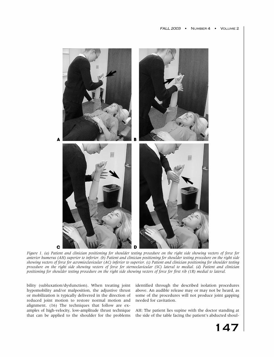
To begin the specific shoulder assessment, the patientlies supine with the head elevated and the shouldergirdle flat on the table. Test the non-affected shoulderfirst to determine normal, realizing that there may beoccult problems in the non-affected shoulder. The pa-tient makes a fist, extending the elbow and raising thearm in forward flexion so it is directly above the gleno-humeral joint (GH) and perpendicular to the floor (Fig-ure 1). The radial and ulnar areas of the fist face thehead direction and the foot direction respectively. Thedoctor stands facing the patient at shoulder level in alunge position. The doctor’s shoulders are in the sameposition as the patient’s shoulders. The procedure isdescribed to the patient, stating that the arm will begently and evenly pushed in 4 different directions andthat the patient will not be overpowered. The 4 direc-tions are then gently demonstrated on the extendedarm. Once the patient understands, start the examina-tion process for each of the 4 joint complexes by saying,“I want you to resist me, but not fight me, and I wantyou to continue to hold your arm in the position I put itin.” “Are your ready?” “Hold, resist.” It is recommendedthat each test be repeated 3 times unless a frank weak-ness is encountered immediately. Watch for a “cog-wheel” affect where the shoulder gets weak then strongthen weak again. The more the shoulder is tested theclearer the weakness becomes. A normal functioningshoulder will not become weak nor have a cogwheelaffect. It must be emphasized that this procedure is donevery gently requiring no more than 50% of the patient’smaximum voluntary contraction (MVC) ability. Fur-thermore, weakness can occur with and without pain.
Glenohumeral Joint (GH): Using the patient’s rightshoulder for an example, the doctor’s right hand graspsthe patient’s radial side wrist while the doctor’s left
hand holds the patient’s right elbow to maintain exten-sion. Test the GH slowly and gently by applying a steadycaudal pressure looking for weakness or inability tohold with cephalic resistance (Figure 1a). If there isweakness or slippage inferiorly, a GH joint dysfunctionis identified and noted in the record as AH = + indicat-ing the likelihood of an anterior humerus (AH) mis-alignment.
Acromioclavicular Joint (AC): Using the patient’s rightshoulder as an example, the doctor’s right hand graspsthe ulnar side of the patient’s wrist while the doctor’sleft hand holds the patient’s right elbow to maintainextension. Test the AC slowly and gently by applying asteady pressure in a cephalic direction looking for weak-ness or inability to hold against resistance (Figure 1b). Ifthere is weakness or slippage superiorly, an AC jointdysfunction is identified and noted in the record as AC = +.
Sternoclavicular Joint (SC): Using the patient’s rightshoulder as an example, the doctor’s left hand graspsthe patient’s dorsum and ulnar wrist area while thedoctor’s right hand holds the patient’s left ribcage areajust inferior to the left shoulder joint, to stabilize thepatient’s body during the test. Test the SC slowly andgently with a steady pressure in a medial directionlooking for weakness or inability to hold against resis-tance (Figure 1c). If there is weakness or slippage medi-ally, a SC joint dysfunction is identified and noted in therecord as SC = +.
1st Rib (1R): Using the patient’s right shoulder as anexample, the doctor’s left hand grasps the patient’s pal-mar and ulnar wrist area while the doctor’s right handholds the patient’s left ribcage area just inferior to theleft shoulder joint, to stabilize the patient’s body duringthe test. Test the 1R slowly and gently with a steadypressure in a lateral direction looking for weakness orinability to hold against resistance (Figure 1d). If there isweakness or slippage laterally, a 1R joint dysfunction isidentified and noted in the record as 1R = +.
Manipulative Treatment of theShoulder Complex
Many forms of manual therapy exist. (15) Manualtherapies, whose primary effect is on joint structures,are physical maneuvers designed to induce joint motionthrough non-thrust techniques (mobilization) or thrusttechniques (adjustment or thrust manipulation). Theyare intended to treat disorders of the neuromusculoskel-etal (NMS) system by decreasing pain and improvingjoint range and quality of motion. This leads to theircommon application in the treatment of NMS disordersthat are associated with joint pain and/or joint hypomo-
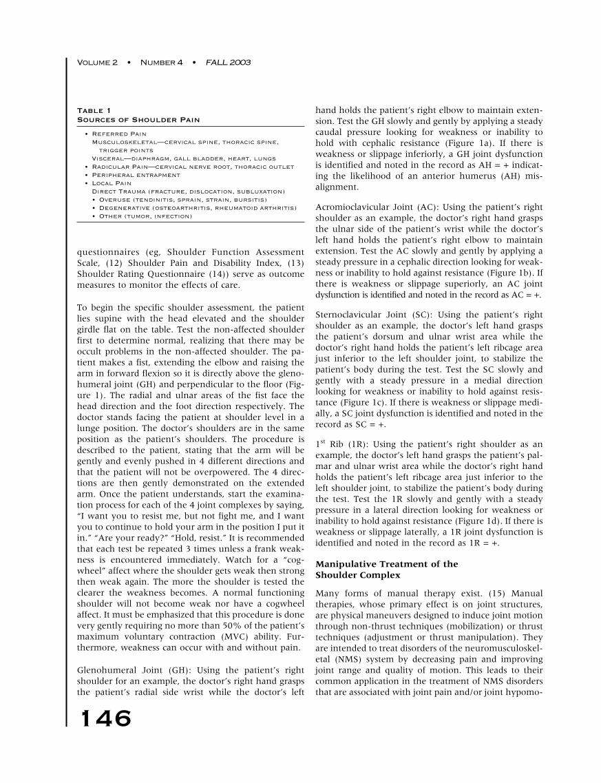
Table 1Sources of Shoulder Pain
• Referred PainMusculoskeletal—cervical spine, thoracic spine,
trigger pointsVisceral—diaphragm, gall bladder, heart, lungs
• Radicular Pain—cervical nerve root, thoracic outlet• Peripheral entrapment• Local Pain
Direct Trauma (fracture, dislocation, subluxation)• Overuse (tendinitis, sprain, strain, bursitis)• Degenerative (osteoarthritis, rheumatoid arthritis)• Other (tumor, infection)
Volume2 • Number4 • FALL2003
146
bility (subluxation/dysfunction). When treating jointhypomobility and/or malposition, the adjustive thrustor mobilization is typically delivered in the direction ofreduced joint motion to restore normal motion andalignment. (16) The techniques that follow are ex-amples of high-velocity, low-amplitude thrust techniquethat can be applied to the shoulder for the problems
identified through the described isolation proceduresabove. An audible release may or may not be heard, assome of the procedures will not produce joint gappingneeded for cavitation.
AH: The patient lies supine with the doctor standing atthe side of the table facing the patient’s abducted shoul-
Figure 1. (a) Patient and clinician positioning for shoulder testing procedure on the right side showing vectors of force foranterior humerus (AH) superior to inferior. (b) Patient and clinician positioning for shoulder testing procedure on the right sideshowing vectors of force for acromioclavicular (AC) inferior to superior. (c) Patient and clinician positioning for shoulder testingprocedure on the right side showing vectors of force for sternoclavicular (SC) lateral to medial. (d) Patient and clinicianpositioning for shoulder testing procedure on the right side showing vectors of force for first rib (1R) medial to lateral.
FALL2003 • Number4 • Volume2
147
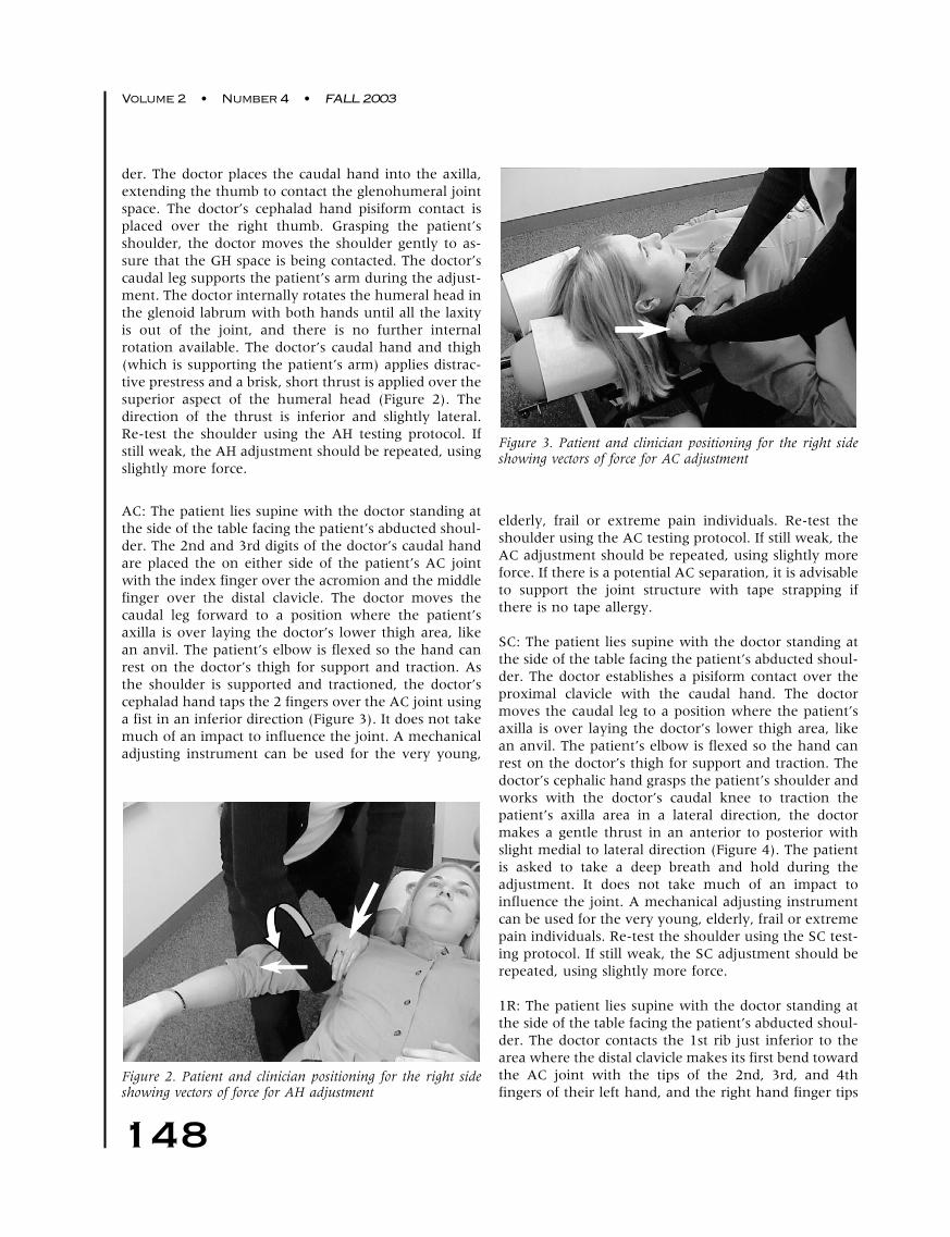
der. The doctor places the caudal hand into the axilla,extending the thumb to contact the glenohumeral jointspace. The doctor’s cephalad hand pisiform contact isplaced over the right thumb. Grasping the patient’sshoulder, the doctor moves the shoulder gently to as-sure that the GH space is being contacted. The doctor’scaudal leg supports the patient’s arm during the adjust-ment. The doctor internally rotates the humeral head inthe glenoid labrum with both hands until all the laxityis out of the joint, and there is no further internalrotation available. The doctor’s caudal hand and thigh(which is supporting the patient’s arm) applies distrac-tive prestress and a brisk, short thrust is applied over thesuperior aspect of the humeral head (Figure 2). Thedirection of the thrust is inferior and slightly lateral.Re-test the shoulder using the AH testing protocol. Ifstill weak, the AH adjustment should be repeated, usingslightly more force.
AC: The patient lies supine with the doctor standing atthe side of the table facing the patient’s abducted shoul-der. The 2nd and 3rd digits of the doctor’s caudal handare placed the on either side of the patient’s AC jointwith the index finger over the acromion and the middlefinger over the distal clavicle. The doctor moves thecaudal leg forward to a position where the patient’saxilla is over laying the doctor’s lower thigh area, likean anvil. The patient’s elbow is flexed so the hand canrest on the doctor’s thigh for support and traction. Asthe shoulder is supported and tractioned, the doctor’scephalad hand taps the 2 fingers over the AC joint usinga fist in an inferior direction (Figure 3). It does not takemuch of an impact to influence the joint. A mechanicaladjusting instrument can be used for the very young,
elderly, frail or extreme pain individuals. Re-test theshoulder using the AC testing protocol. If still weak, theAC adjustment should be repeated, using slightly moreforce. If there is a potential AC separation, it is advisableto support the joint structure with tape strapping ifthere is no tape allergy.
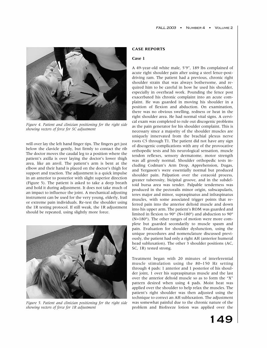
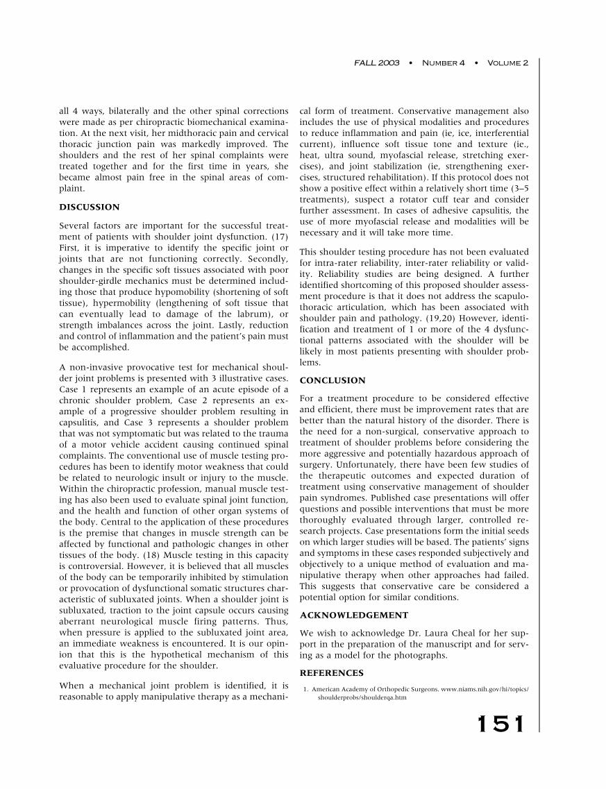
SC: The patient lies supine with the doctor standing atthe side of the table facing the patient’s abducted shoul-der. The doctor establishes a pisiform contact over theproximal clavicle with the caudal hand. The doctormoves the caudal leg to a position where the patient’saxilla is over laying the doctor’s lower thigh area, likean anvil. The patient’s elbow is flexed so the hand canrest on the doctor’s thigh for support and traction. Thedoctor’s cephalic hand grasps the patient’s shoulder andworks with the doctor’s caudal knee to traction thepatient’s axilla area in a lateral direction, the doctormakes a gentle thrust in an anterior to posterior withslight medial to lateral direction (Figure 4). The patientis asked to take a deep breath and hold during theadjustment. It does not take much of an impact toinfluence the joint. A mechanical adjusting instrumentcan be used for the very young, elderly, frail or extremepain individuals. Re-test the shoulder using the SC test-ing protocol. If still weak, the SC adjustment should berepeated, using slightly more force.
1R: The patient lies supine with the doctor standing atthe side of the table facing the patient’s abducted shoul-der. The doctor contacts the 1st rib just inferior to thearea where the distal clavicle makes its first bend towardthe AC joint with the tips of the 2nd, 3rd, and 4thfingers of their left hand, and the right hand finger tips
Figure 2. Patient and clinician positioning for the right sideshowing vectors of force for AH adjustment
Figure 3. Patient and clinician positioning for the right sideshowing vectors of force for AC adjustment
Volume2 • Number4 • FALL2003
148
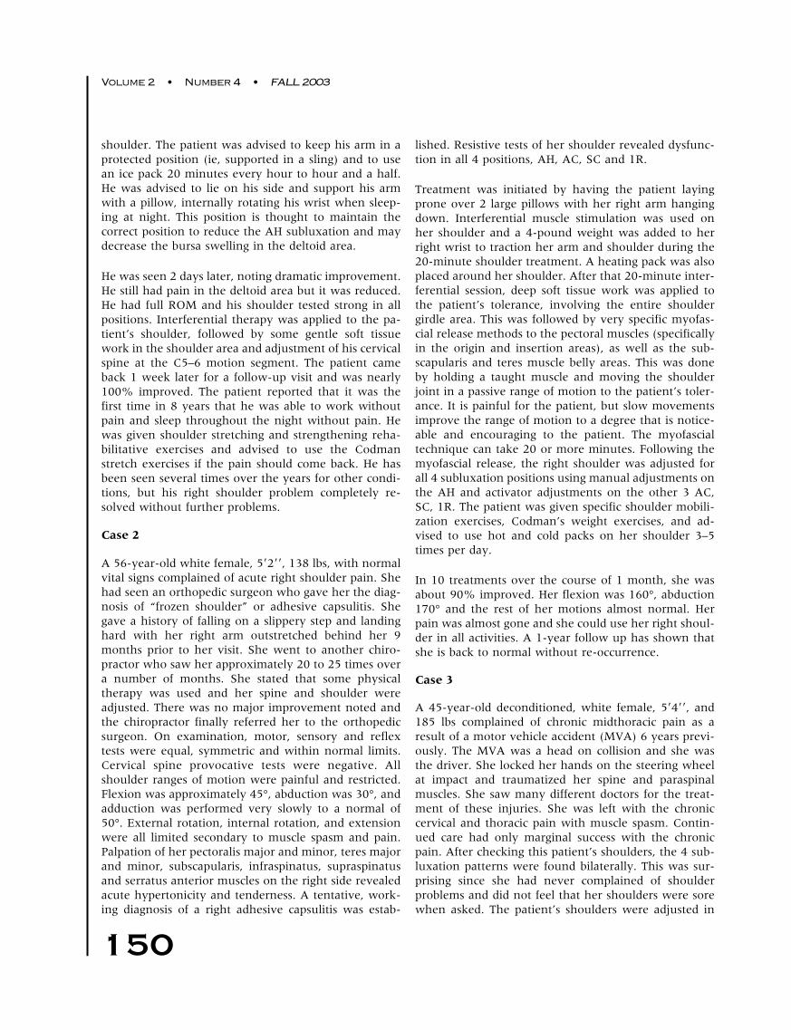
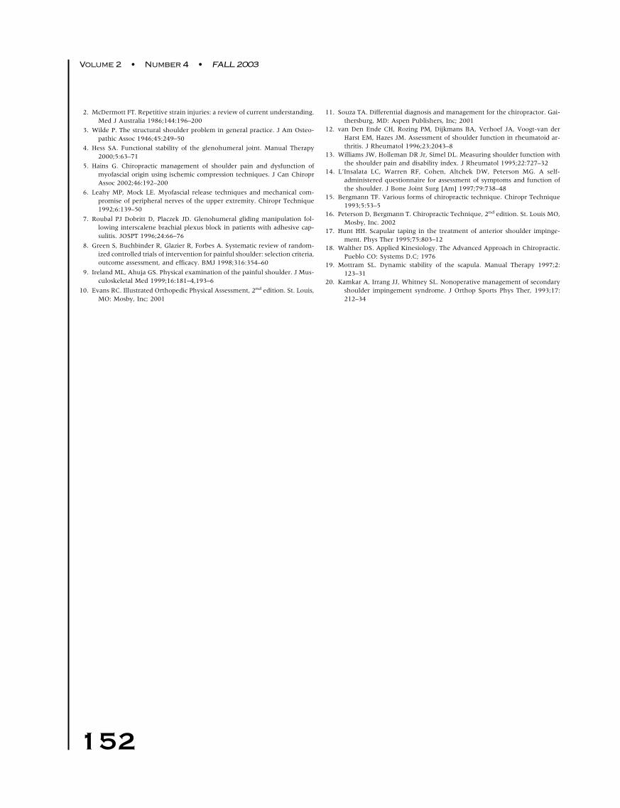
will over lay the left hand finger tips. The fingers get justbelow the clavicle gently, but firmly to contact the ribThe doctor moves the caudal leg to a position where thepatient’s axilla is over laying the doctor’s lower thigharea, like an anvil. The patient’s arm is bent at theelbow and their hand is placed on the doctor’s thigh forsupport and traction. The adjustment is a quick impulsein an anterior to posterior with slight superior direction(Figure 5). The patient is asked to take a deep breathand hold it during adjustment. It does not take much ofan impact to influence the joint. A mechanical adjustinginstrument can be used for the very young, elderly, frailor extreme pain individuals. Re-test the shoulder usingthe 1R testing protocol. If still weak, the 1R adjustmentshould be repeated, using slightly more force.
CASE REPORTS
Case 1
A 49-year-old white male, 5’9”, 189 lbs complained ofacute right shoulder pain after using a steel fence-post-driving ram. The patient had a previous, chronic rightshoulder strain that was always bothersome, and re-quired him to be careful in how he used his shoulder,especially in overhead work. Pounding the fence postexacerbated his chronic complaint into an acute com-plaint. He was guarded in moving his shoulder in aposition of flexion and abduction. On examination,there was no obvious swelling, redness or heat in theright shoulder area. He had normal vital signs. A cervi-cal exam was completed to rule out discogenic problemsas the pain generator for his shoulder complaint. This isnecessary since a majority of the shoulder muscles areuniquely innervated from the brachial plexus nerveroots C5 through T1. The patient did not have any signof discogenic complications with any of the provocativeorthopedic tests and his neurological sensation, muscletendon reflexes, sensory dermatome, motor strengthwas all grossly normal. Shoulder orthopedic tests in-cluding Codman’s Arm Drop, Apprehension, Dugas,and Yergason’s were essentially normal but producedshoulder pain. Palpation over the coracoid process,greater tuberosity, bicipital groove, and in the subdel-toid bursa area was tender. Palpable tenderness wasproduced in the pectoralis minor origin, subscapularis,teres major and minor, supraspinatus and infraspinatusmuscles, with some associated trigger points that re-ferred pain into the anterior deltoid muscle and downinto his upper arm. The patient’s ROM was guarded andlimited in flexion to 90° (N=180°) and abduction to 90°(N=180°). The other ranges of motion were more com-plete but guarded secondarily to muscle spasm andpain. Evaluation for shoulder dysfunction, using theunique procedures and nomenclature discussed previ-ously, the patient had only a right AH (anterior humeralhead subluxation). The other 3 shoulder positions (AC,SC, 1R) tested strong.
Treatment began with 20 minutes of interferentialmuscle stimulation using the 80–150 Hz settingthrough 4 pads: 1 anterior and 1 posterior of his shoul-der joint, 1 over his supraspinatus muscle and the lastover the anterior deltoid muscle so as to form the “X”pattern desired when using 4 pads. Moist heat wasapplied over the shoulder to help relax the muscles. Thepatient’s right shoulder was then adjusted using thetechnique to correct an AH subluxation. The adjustmentwas somewhat painful due to the chronic nature of theproblem and Biofreeze lotion was applied over the
Figure 4. Patient and clinician positioning for the right sideshowing vectors of force for SC adjustment
Figure 5. Patient and clinician positioning for the right sideshowing vectors of force for 1R adjustment
FALL2003 • Number4 • Volume2
149
shoulder. The patient was advised to keep his arm in aprotected position (ie, supported in a sling) and to usean ice pack 20 minutes every hour to hour and a half.He was advised to lie on his side and support his armwith a pillow, internally rotating his wrist when sleep-ing at night. This position is thought to maintain thecorrect position to reduce the AH subluxation and maydecrease the bursa swelling in the deltoid area.
He was seen 2 days later, noting dramatic improvement.He still had pain in the deltoid area but it was reduced.He had full ROM and his shoulder tested strong in allpositions. Interferential therapy was applied to the pa-tient’s shoulder, followed by some gentle soft tissuework in the shoulder area and adjustment of his cervicalspine at the C5–6 motion segment. The patient cameback 1 week later for a follow-up visit and was nearly100% improved. The patient reported that it was thefirst time in 8 years that he was able to work withoutpain and sleep throughout the night without pain. Hewas given shoulder stretching and strengthening reha-bilitative exercises and advised to use the Codmanstretch exercises if the pain should come back. He hasbeen seen several times over the years for other condi-tions, but his right shoulder problem completely re-solved without further problems.
Case 2
A 56-year-old white female, 5�2��, 138 lbs, with normalvital signs complained of acute right shoulder pain. Shehad seen an orthopedic surgeon who gave her the diag-nosis of “frozen shoulder” or adhesive capsulitis. Shegave a history of falling on a slippery step and landinghard with her right arm outstretched behind her 9months prior to her visit. She went to another chiro-practor who saw her approximately 20 to 25 times overa number of months. She stated that some physicaltherapy was used and her spine and shoulder wereadjusted. There was no major improvement noted andthe chiropractor finally referred her to the orthopedicsurgeon. On examination, motor, sensory and reflextests were equal, symmetric and within normal limits.Cervical spine provocative tests were negative. Allshoulder ranges of motion were painful and restricted.Flexion was approximately 45°, abduction was 30°, andadduction was performed very slowly to a normal of50°. External rotation, internal rotation, and extensionwere all limited secondary to muscle spasm and pain.Palpation of her pectoralis major and minor, teres majorand minor, subscapularis, infraspinatus, supraspinatusand serratus anterior muscles on the right side revealedacute hypertonicity and tenderness. A tentative, work-ing diagnosis of a right adhesive capsulitis was estab-
lished. Resistive tests of her shoulder revealed dysfunc-tion in all 4 positions, AH, AC, SC and 1R.
Treatment was initiated by having the patient layingprone over 2 large pillows with her right arm hangingdown. Interferential muscle stimulation was used onher shoulder and a 4-pound weight was added to herright wrist to traction her arm and shoulder during the20-minute shoulder treatment. A heating pack was alsoplaced around her shoulder. After that 20-minute inter-ferential session, deep soft tissue work was applied tothe patient’s tolerance, involving the entire shouldergirdle area. This was followed by very specific myofas-cial release methods to the pectoral muscles (specificallyin the origin and insertion areas), as well as the sub-scapularis and teres muscle belly areas. This was doneby holding a taught muscle and moving the shoulderjoint in a passive range of motion to the patient’s toler-ance. It is painful for the patient, but slow movementsimprove the range of motion to a degree that is notice-able and encouraging to the patient. The myofascialtechnique can take 20 or more minutes. Following themyofascial release, the right shoulder was adjusted forall 4 subluxation positions using manual adjustments onthe AH and activator adjustments on the other 3 AC,SC, 1R. The patient was given specific shoulder mobili-zation exercises, Codman’s weight exercises, and ad-vised to use hot and cold packs on her shoulder 3–5times per day.
In 10 treatments over the course of 1 month, she wasabout 90% improved. Her flexion was 160°, abduction170° and the rest of her motions almost normal. Herpain was almost gone and she could use her right shoul-der in all activities. A 1-year follow up has shown thatshe is back to normal without re-occurrence.
Case 3
A 45-year-old deconditioned, white female, 5�4��, and185 lbs complained of chronic midthoracic pain as aresult of a motor vehicle accident (MVA) 6 years previ-ously. The MVA was a head on collision and she wasthe driver. She locked her hands on the steering wheelat impact and traumatized her spine and paraspinalmuscles. She saw many different doctors for the treat-ment of these injuries. She was left with the chroniccervical and thoracic pain with muscle spasm. Contin-ued care had only marginal success with the chronicpain. After checking this patient’s shoulders, the 4 sub-luxation patterns were found bilaterally. This was sur-prising since she had never complained of shoulderproblems and did not feel that her shoulders were sorewhen asked. The patient’s shoulders were adjusted in
Volume2 • Number4 • FALL2003
150
all 4 ways, bilaterally and the other spinal correctionswere made as per chiropractic biomechanical examina-tion. At the next visit, her midthoracic pain and cervicalthoracic junction pain was markedly improved. Theshoulders and the rest of her spinal complaints weretreated together and for the first time in years, shebecame almost pain free in the spinal areas of com-plaint.
DISCUSSION
Several factors are important for the successful treat-ment of patients with shoulder joint dysfunction. (17)First, it is imperative to identify the specific joint orjoints that are not functioning correctly. Secondly,changes in the specific soft tissues associated with poorshoulder-girdle mechanics must be determined includ-ing those that produce hypomobility (shortening of softtissue), hypermobility (lengthening of soft tissue thatcan eventually lead to damage of the labrum), orstrength imbalances across the joint. Lastly, reductionand control of inflammation and the patient’s pain mustbe accomplished.
A non-invasive provocative test for mechanical shoul-der joint problems is presented with 3 illustrative cases.Case 1 represents an example of an acute episode of achronic shoulder problem, Case 2 represents an ex-ample of a progressive shoulder problem resulting incapsulitis, and Case 3 represents a shoulder problemthat was not symptomatic but was related to the traumaof a motor vehicle accident causing continued spinalcomplaints. The conventional use of muscle testing pro-cedures has been to identify motor weakness that couldbe related to neurologic insult or injury to the muscle.Within the chiropractic profession, manual muscle test-ing has also been used to evaluate spinal joint function,and the health and function of other organ systems ofthe body. Central to the application of these proceduresis the premise that changes in muscle strength can beaffected by functional and pathologic changes in othertissues of the body. (18) Muscle testing in this capacityis controversial. However, it is believed that all musclesof the body can be temporarily inhibited by stimulationor provocation of dysfunctional somatic structures char-acteristic of subluxated joints. When a shoulder joint issubluxated, traction to the joint capsule occurs causingaberrant neurological muscle firing patterns. Thus,when pressure is applied to the subluxated joint area,an immediate weakness is encountered. It is our opin-ion that this is the hypothetical mechanism of thisevaluative procedure for the shoulder.
When a mechanical joint problem is identified, it isreasonable to apply manipulative therapy as a mechani-
cal form of treatment. Conservative management alsoincludes the use of physical modalities and proceduresto reduce inflammation and pain (ie, ice, interferentialcurrent), influence soft tissue tone and texture (ie.,heat, ultra sound, myofascial release, stretching exer-cises), and joint stabilization (ie, strengthening exer-cises, structured rehabilitation). If this protocol does notshow a positive effect within a relatively short time (3–5treatments), suspect a rotator cuff tear and considerfurther assessment. In cases of adhesive capsulitis, theuse of more myofascial release and modalities will benecessary and it will take more time.
This shoulder testing procedure has not been evaluatedfor intra-rater reliability, inter-rater reliability or valid-ity. Reliability studies are being designed. A furtheridentified shortcoming of this proposed shoulder assess-ment procedure is that it does not address the scapulo-thoracic articulation, which has been associated withshoulder pain and pathology. (19,20) However, identi-fication and treatment of 1 or more of the 4 dysfunc-tional patterns associated with the shoulder will belikely in most patients presenting with shoulder prob-lems.
CONCLUSION
For a treatment procedure to be considered effectiveand efficient, there must be improvement rates that arebetter than the natural history of the disorder. There isthe need for a non-surgical, conservative approach totreatment of shoulder problems before considering themore aggressive and potentially hazardous approach ofsurgery. Unfortunately, there have been few studies ofthe therapeutic outcomes and expected duration oftreatment using conservative management of shoulderpain syndromes. Published case presentations will offerquestions and possible interventions that must be morethoroughly evaluated through larger, controlled re-search projects. Case presentations form the initial seedson which larger studies will be based. The patients’ signsand symptoms in these cases responded subjectively andobjectively to a unique method of evaluation and ma-nipulative therapy when other approaches had failed.This suggests that conservative care be considered apotential option for similar conditions.
ACKNOWLEDGEMENT
We wish to acknowledge Dr. Laura Cheal for her sup-port in the preparation of the manuscript and for serv-ing as a model for the photographs.
REFERENCES
1. American Academy of Orthopedic Surgeons. www.niams.nih.gov/hi/topics/shoulderprobs/shoulderqa.htm
FALL2003 • Number4 • Volume2
151
2. McDermott FT. Repetitive strain injuries: a review of current understanding.Med J Australia 1986;144:196–200
3. Wilde P. The structural shoulder problem in general practice. J Am Osteo-pathic Assoc 1946;45:249–50
4. Hess SA. Functional stability of the glenohumeral joint. Manual Therapy2000;5:63–71
5. Hains G. Chiropractic management of shoulder pain and dysfunction ofmyofascial origin using ischemic compression techniques. J Can ChiroprAssoc 2002;46:192–200
6. Leahy MP, Mock LE. Myofascial release techniques and mechanical com-promise of peripheral nerves of the upper extremity. Chiropr Technique1992;6:139–50
7. Roubal PJ Dobritt D, Placzek JD. Glenohumeral gliding manipulation fol-lowing interscalene brachial plexus block in patients with adhesive cap-sulitis. JOSPT 1996;24:66–76
8. Green S, Buchbinder R, Glazier R, Forbes A. Systematic review of random-ized controlled trials of intervention for painful shoulder: selection criteria,outcome assessment, and efficacy. BMJ 1998;316:354–60
9. Ireland ML, Ahuja GS. Physical examination of the painful shoulder. J Mus-culoskeletal Med 1999;16:181–4,193–6
10. Evans RC. Illustrated Orthopedic Physical Assessment, 2nd edition. St. Louis,MO: Mosby, Inc; 2001
11. Souza TA. Differential diagnosis and management for the chiropractor. Gai-thersburg, MD: Aspen Publishers, Inc; 2001
12. van Den Ende CH, Rozing PM, Dijkmans BA, Verhoef JA, Voogt-van derHarst EM, Hazes JM. Assessment of shoulder function in rheumatoid ar-thritis. J Rheumatol 1996;23:2043–8
13. Williams JW, Holleman DR Jr, Simel DL. Measuring shoulder function withthe shoulder pain and disability index. J Rheumatol 1995;22:727–32
14. L’Insalata LC, Warren RF, Cohen, Altchek DW, Peterson MG. A self-administered questionnaire for assessment of symptoms and function ofthe shoulder. J Bone Joint Surg [Am] 1997;79:738–48
15. Bergmann TF. Various forms of chiropractic technique. Chiropr Technique1993;5:53–5
16. Peterson D, Bergmann T. Chiropractic Technique, 2nd edition. St. Louis MO,Mosby, Inc. 2002
17. Hunt HH. Scapular taping in the treatment of anterior shoulder impinge-ment. Phys Ther 1995;75:803–12
18. Walther DS. Applied Kinesiology. The Advanced Approach in Chiropractic.Pueblo CO: Systems D.C; 1976
19. Mottram SL. Dynamic stability of the scapula. Manual Therapy 1997;2:123–31
20. Kamkar A, Irrang JJ, Whitney SL. Nonoperative management of secondaryshoulder impingement syndrome. J Orthop Sports Phys Ther, 1993;17:212–34
Volume2 • Number4 • FALL2003
152
Copyright © 2022 FDOKUMEN