Evaluation of fracture resistance of ceramics: Edge fracture tests
Upload
theaileyschoolCategory
view
4download
0
journal of orthopaedic & sports physical therapy | volume 37 | number 9 | september 2007 | 529
insert into the medial and lateral sesa-moids, respectively. The intermetatarsalligament also anchors the lateral sesa-moid to the second MTP joint plantarplate.5 A fat-pad cushion and bursa sep-arates the plantar dermis and the sesa-moids.57 In general, the medial sesamoidis larger and longer and the lateral sesa-moid is smaller, rounder, and protectedby the soft tissue of the first intermeta-tarsal space.5,43
The sesamoids protect the first meta-tarsal head by absorbing shock, increas-ing the FHB and FHL moment arms, andprotecting and guiding the FHL passingbetween them.3-5,53 The estimated resul-tant force acting across the first metatar-
Dancers comprise a unique athletic population. They executerepetitive foot and ankle motions (alternating between extremeplantar flexion and dorsiflexion) with minimal to no supportivefootwear, which predisposes them to overuse injuries of the
foot and ankle.10,11,39 Dancers apply a large amount of stress to thehallucal sesamoids when moving onto their toes to grand plié anddemi-relevé (ankle plantar flexion and metatarsophalangeal [MTP]joint dorsiflexion), full relevé (ankle and MTP joint plantar flexion),and during eccentric deceleration occurring with jumps and hops.14,53,58
STUDY DESIGN: Case report.
BACKGROUND: Misdiagnosed or undertreatedsesamoid bone pathology in dancers may resultin prolonged pain, disability, and career limitation.A thorough understanding of sesamoid disordersand appropriate treatment facilitates timelyrecovery. The potential loss of hallux plantar flexionstrength consequent to sesamoidectomy is amajor consideration for dancers.
CASE DESCRIPTION: An 18-year-old dancestudent sustained a delayed-union fracture of herlateral (fibular) sesamoid. Treatment includedan inductive coupling external bone stimulatorwith pulsed electromagnetic field, activity, andweight-bearing restrictions, protective padding,strengthening, functional retraining, and progres-sive return to dance.
OUTCOME: Following use of an external bone
stimulator for 12 months, the dancer successfullyreturned to her previous level of dancing. RepeatedSF-36 and Dance Functional Outcome Systemscores confirmed this improvement.
DISCUSSION: Loss of hallux plantar flexionstrength with sesamoid resection can be devastat-ing to a dancer who requires push-off strength formultiple turns and jumps. Treatment with bonestimulation was therefore selected over moreinvasive measures. The dancer was compliantwith systematic functional progression. Improve-ment, as seen on radiographs and outcomescores, accompanied her full functional recovery.J Orthop Sports Phys Ther 2007;37(9):529–540.doi:10.2519/jospt.2007.2472
KEY WORDS: bone stimulator, dance injury,foot, hallux
1Director, ADAM Center, Long Island University, Brooklyn, NY; Director, Physical Therapy Services at Alvin Ailey, New York, NY. 2Podiatrist, New York College of Podiatric Medicine,New York, NY. 3Physical therapy doctoral candidate, Physical Therapy Program, Ithaca College, Ithaca, NY. Informed consent was obtained from the patient prior to examinationand treatment, as well as prior to submission of this manuscript for publication. Address correspondence to Shaw Bronner, ADAM Center, Long Island University, One UniversityPlaza, Ste M411, Brooklyn, NY 11201. E-mail: [email protected].
Sesamoid pathology occurring in dancers,if misdiagnosed or undertreated, may re-sult in prolonged pain, disability, and ca-reer limitation. We present a case reportof a delayed-union sesamoid fracture in adancer because treatment considerationsmay differ for this population.
AnatomyThe medial (tibial) and lateral (fibular)sesamoids are located on the plantarsurface of the foot, inferior to the firstmetatarsal head, and are containedwithin tendons of the flexor hallucis bre-vis (FHB).5,30,43,53 They connect distally tothe base of the proximal phalanx by theplantar plate, an extension of the FHB.The superior surfaces are smooth, rela-tively flat, and covered by hyaline car-tilage for articulation with the sulci ofthe first metatarsal head. The sesamoidsare separated by a crista or ridge on thefirst metatarsal head and are connectedto each other by the intersesamoidalligament. There is no direct connection
Management of a Delayed-UnionSesamoid Fracture in a Dancer
SHAW BRONNER, PT, PhD, OCS1 • THOMAS NOVELLA, DPM2 • LAURA BECICA3
between the sesamoids and the flexorhallucis longus (FHL) tendon that runsbetween them, although a portion of thefibrous tendon sheath attaches to bothsesamoids. In addition, the abductorhallucis and adductor hallucis tendons
[ CASE REPORT ]

530 | september 2007 | volume 37 | number 9 | journal of orthopaedic & sports physical therapy
[ CASE REPORT ]sal head and sesamoids is approximately93% to 119% of body weight during thepush-off phase of the gait cycle.31,33,42
Incidence and EtiologyThe annual incidence of musculoskel-etal injury in dancers ranges from 67%to 95%.10,22,50,63 Foot and ankle injuriesin professional dancers comprise 34%to 54% of all injuries. Specific reportson sesamoid injury incidence in dancerswere not found. The incidence of foot andankle injuries in runners is 30% to 45%,44
comparable to the incidence reported indancers. Among runners, sesamoid inju-ries comprise 12% of great toe, 4% of footand ankle, and 1.2% of all injuries.35,43 Itis reported that medial sesamoids are in-jured more frequently than lateral sesa-moids,60 although no specific incidencerates have been reported. In our clinicalexperience, sesamoid injury rates couldbe even higher in dancers due to the in-creased demand on the forefoot.
Overuse injuries are the most com-mon type of injuries in dancers, with inci-dence reported to be from 60% to 76% inprofessional dancers and as high as 93%in preprofessional dancers.10,48,50,63 Theonset of sesamoid symptoms is associ-ated with repetitive stress and recent in-crease in weight-bearing activity.5 In ourexperience, when dance students enter apreprofessional training program, theyexperience a sudden increase in hours ofdancing, which makes them particularlyvulnerable to injury. Acute trauma mostoften involves hyperextension (dorsiflex-ion), with or without abduction of thehallux, at the MTP joint.57 For both re-petitive and traumatic injuries, activitiesthat increase weight-bearing loads tendto increase pain.
Sesamoid injury risk factors for danc-ers include a cavus foot, plantar-flexedfirst ray, frequent and forced turnout,nonprotective footwear, dance style, hardfloors, and gender (female).5,11,20,28,40,64 Acavus foot is relatively rigid, likely re-sulting in limited shock attenuationwhen landing from jumps. A plantar-flexed first ray increases weight-bearing
pressures medially over the first MTPjoint. Due to constant turnout (externalrotation of the hips, knees, and ankles)when dancing, dancers may walk withsignificant “out-toeing,” with increased
weight-bearing pressures medially overthe first MTP joint.39 With unrealisticexpectation of perfect turnout of 180°,dancers with insufficient hip externalrotation may force their turnout, result-
FIGURE 1. Technique analysis. (A) Dancer in first positionwith forced turnout and foot pronation. The lack offemoral external rotation in relation to her foot placementis visible, particularly on the left limb. Because she hasforced her knees into hyperextension, her femurs are inexaggerated internal rotation and she is gripping withher feet. (B) When this dancer achieves first positionby turning out from her hips and continues to engageher turnout muscles, there is less discrepancy betweenfemoral internal and tibial external rotation and pronation
is minimized. (C) Dancer in parallel position demonstrating bilateral femoral internal rotation relative to her tibia,and a thrust into the posterolateral corner of her knees with hyperextension. (D) Dancer in first position demi-pliéwith turnout of her lower extremities well controlled.
A B
C D

journal of orthopaedic & sports physical therapy | volume 37 | number 9 | september 2007 | 531
ing in excessive foot pronation (knownas “rolling in”), which increases loadingof the medial foot structures, includingthe sesamoids (FIGURE 1A and 1B).28,39,48
Studies have demonstrated a positivecorrelation between self-reported lowerextremity injury (defined as “of a suf-ficient severity to affect dance trainingor performance”) and “functional” turn-out, which exceeds available physiologicrange of motion (ROM).17,48 In studentstraining at a preprofessional ballet pro-gram, 5.9% of their injuries occurred atthe medial forefoot.48
Dance footwear ranges from danc-ing barefoot, in thin leather-soled bal-let slippers lacking shock absorption,to high-heeled character shoes thatprolong and increase loading on the fe-male forefoot.23,42 Hard floor surfacesincrease shock attenuation demands onthe feet,21,61 while raked (inclined) stagesare reported to be associated with 28% to37% of all injuries.18 Finally, females areat greater risk for stress fractures,11 andthe vascularity of their sesamoids is lessextensive than that of males.54
Evaluation and ManagementThe onset of sesamoid symptoms is usu-ally insidious and poorly localized.5 Swell-ing and redness may be present in chroniccases. Pain is elicited with passive dorsi-flexion of the first MTP joint and directpalpation of the injured sesamoid.5,8,20,30
Dorsiflexion may also be limited at thefirst MTP joint.39
Differential diagnosis of sesamoidpathology includes inflammation (sesa-moiditis) and fractures of the sesamoid,as well as injury to other forefoot struc-tures, such as turf-toe, hallux valgus andrigidus, and FHL or FHB tendonitis(TABLE 1).30 The diagnosis of sesamoidfracture can be difficult, as radiographicchanges may lag the onset of symptomsby 6 months.5,20 If activity and weight-bearing modifications (padding, footwear, assistive devices) are unsuccessful,further testing may be necessary. Serialplain radiographs, bone scans, magneticresonance imaging, or computerized to-
mography scans may be indicated to di-agnose and differentiate sesamoid stressfractures, acute and delayed-union frac-tures, or avascular necrosis.5,8,30
Sesamoid fractures must also be dif-ferentiated from sesamoid partitions.The sesamoids contain multiple ossifica-tion centers that usually fuse by 8 to 12years of age.5,57 Partitions due to fusionfailure may result in bipartite, tripartite,or multipartite sesamoids, with resultingvariations in size and shape. The estimat-ed incidence of partite medial sesamoidsranges from 7% to 11% of the population,occurring more frequently in femalesthan males.5,30,43,57 Partitions are relative-ly rare in the lateral sesamoid (less than1%)5 and are distinguished from fracturesby smooth margins.
Sesamoiditis due to overuse (themost frequently seen) is typically man-aged with activity and weight-bearingmodifications for several weeks.5,25,30,43
The most common management ofsesamoid fractures consists of immo-bilization with a short-leg walking castor rigid shoe for 4 to 65 or even 6 to 8
weeks,8 followed by padding. Crutchesor a cane may be necessary to assuresufficient reduction of weight bearing.Sesamoid fractures may injure the ten-uous sesamoid arterial supply, resultingin delayed union, nonunion, or avascularnecrosis.15,56 In one series of 369 stressfractures in athletes, the most commonarea of delayed union and nonunionwas the sesamoids.52 Nonunion occurswith a large fracture gap, inadequateimmobilization, a malaligned fracture,infection, or inadequate blood supply.26
When symptoms persist for a minimumof 6 months and there is radiographicevidence of delayed union or avascularnecrosis, alternative interventions in-clude complete or partial resection,5,8,57
bone grafting,1,5,43 open reduction inter-nal fixation,53 or bone stimulators.2
Early reports recommended resectionof both the injured and noninjured sesa-moids to maintain balance and preventincreased load and pain in the remainingsesamoid.20 Others perform completeexcision of the injured sesamoid, but donot advocate removal of both.15,37,57,59 The
TABLE 1 Diagnosis of Sesamoid Pathology
Sesamoid pathology
• Sesamoid dislocation with plantar capsular rupture
• Sesamoid osteoarthritis
• Sesamoid stress fracture, acute fracture, delayed union, nonunion, osteochondritis, avascular necrosis
• Sesamoiditis
• Symptomatic bipartite sesamoids
Differential diagnosis
• Capsular tear (first MTP joint)
• Flexor hallucis longus distal stenosing tenosynovitis
• Flexor hallucis brevis tendinitis
• Gout
• Ganglion
• Hallux valgus
• Hallux rigidus
• Metatarsal and phalangeal stress reaction and fractures
• Metatarsalgia
• Plantar interdigital (Morton’s) neuroma
• Submetatarsal bursitis
• Turf toe (metatarsal plantar plate disruption)
Abbreviation: MTP, metatarsophalangeal.

532 | september 2007 | volume 37 | number 9 | journal of orthopaedic & sports physical therapy
[ CASE REPORT ]literature emphasizes maintaining theintegrity of FHB tendon insertions intothe base of the proximal phalanx withcareful reconstruction following sesa-moid excision.5,8,20 Postoperative man-agement of sesamoidectomy ranges from1 to 3 weeks of non–weight-bearing, fol-lowed by ambulation in a rigid shoe forup to 4 weeks, and resumption of ac-tivities as tolerated at 6 weeks.5,20,37,57,60
Complications of medial sesamoidec-tomy include continued pain, cock-upand claw-toe deformities, hallux valgusand rigidus, weakness of the great toe inas many as 50% of patients, and the in-ability to stand on tiptoe in up to 30%of patients.4,5,37,53,60 Case series of lateralsesamoidectomy were not found.
Aper et al3,4 studied the mechanical ef-fects of complete sesamoid resection andsesamoid hemi-resection on the momentarms of the FHB and FHL tendons in ca-davers. A significant FHB moment armloss was seen with complete resection ofboth sesamoids.3 Total medial or lateralsesamoid resection (or both) caused asignificant reduction in the FHL tendonmoment arm. These reductions in mo-ment arms explain the reports of weak-ness of the great toe, and inability tostand on tiptoe following complete resec-tion. However, isolated hemi-resection ofeither the medial or the lateral sesamoiddid not cause significant change in theFHB or FHL moment arms.3,4
Partial sesamoidectomy is an alter-native treatment for fractures with sig-nificant fragment separation or diastasisof bipartite sesamoids.5,8 Postoperativecare includes 1 week non-weight bear-ing, 6 weeks in a short-leg walking cast,and return to sport-specific training at8 weeks.
Alternative nonsurgical methods fortreating delayed and nonunion fracturesinclude electrical stimulation and low-intensity pulsed ultrasound, with healingrates similar to surgical procedures.24,26,34
However, these inventions have been fo-cused on fresh fractures, arthrodesis, andnonunion fractures of long bones such asthe tibia.2,34 No reports were found us-
ing bone stimulation for the treatment ofsesamoid fractures.
Electrical stimulation options for frac-ture healing and delayed and nonunionsinclude invasive, semi-invasive, and non-invasive stimulators.2 The most commonlyused noninvasive stimulators utilize in-ductive coupling and capacitive couplingstimulators. Studies using inductive cou-pling stimulators achieved 77% to 81%successful healing, over treatment timesvarying widely from 2 to 12 months.2,26
Studies using capacitive coupling stimu-lators on nonunion fractures resulted in68% to 77% successful union in 21 to 26weeks of treatment.2 Studies using low-intensity pulsed ultrasound on nonunionsreport an 85% success rate with an aver-age treatment time of 6 months.2,24
A thorough understanding of sesa-moid disorders and appropriate treat-ment is required to achieve timelyrecovery. Therefore, the purpose of thiscase report was to describe the manage-ment of a dancer with a delayed diagnosisand subsequent delayed-union fracture ofthe lateral sesamoid.
CASE DESCRIPTION
In August 2002, an 18-year-old
dance student developed occasionalpain under her right great toe when
dancing. An orthopaedist in her homecountry told her that her toe was in-flamed and advised rest. No radiographswere obtained. She came to New YorkCity to pursue her dance studies and, withincreased dancing load, she experiencedsimilar pain more frequently. During con-sultation with an orthopaedist for anoth-er injury, toe inflammation was discussedagain, but no radiographs were taken norwas she advised to limit her activities.By January 2003, jumping and walkingwere limited by pain and her first MTPjoint area was noticeably swollen. Uponreturn to the orthopaedist, radiographsrevealed a right lateral (fibular) sesa-moid fracture. She was not immobilizednor given weight-bearing restrictions atthat time. She was advised not to dance
for 8 weeks and, although pain decreased,3 follow-up radiographs taken every 2months revealed no improvement.
In September 2003, she consulted adance medicine podiatrist (T.N.) whoobtained new radiographs showing aright nonunion lateral sesamoid fracture(FIGURE 2A). The assessment of nonunionwas based on radiographs demonstrat-ing a complete fracture with a 3-mmgap between bone fragments presentfor 13 months. (A 6- to 12-month spanis generally accepted as definitive of non-union.) Her numeric pain rating scale(NPRS) was only 1/10 at that time, be-cause she was not dancing. Patients areasked to report the highest pain experi-
FIGURE 2. Right foot anterior-posterior viewradiographs. (A) Radiograph demonstrating a lateralsesamoid nonunion fracture in September 2003.(B) Final radiograph demonstrating fracture healingin January 2007.
journal of orthopaedic & sports physical therapy | volume 37 | number 9 | september 2007 | 533
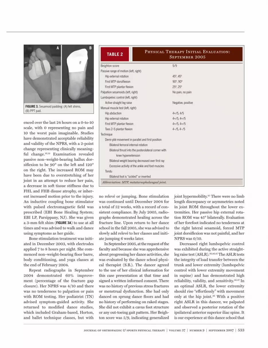
enced over the last 24 hours on a 0-to-10scale, with 0 representing no pain and10 the worst pain imaginable. Studieshave demonstrated acceptable reliabilityand validity of the NPRS, with a 2-pointchange representing clinically meaning-ful change.16,19 Examination revealedpassive non–weight-bearing hallux dor-siflexion to be 90° on the left and 120°on the right. The increased ROM mayhave been due to overstretching of herjoint in an attempt to reduce her pain,a decrease in soft tissue stiffness due toFHL and FHB disuse atrophy, or inher-ent increased motion prior to the injury.An inductive coupling bone stimulatorwith pulsed electromagnetic field wasprescribed (EBI Bone Healing System;EBI LP, Parsippany, NJ). She was givena 3-mm felt shim (FIGURE 3A) to use at alltimes and was advised to walk and danceusing symptoms as her guide.
Bone stimulation treatment was initi-ated in December 2003, with electrodesapplied 7 to 8 hours per night. She com-menced non–weight-bearing floor barre,body conditioning, and yoga classes atthe end of February 2004.
Repeat radiographs in September2004 demonstrated 60% improve-ment (percentage of the fracture gapclosure). Her NPRS was 4/10 and therewas no tenderness to palpation or painwith ROM testing. Her podiatrist (TN)advised symptom-guided activity. Shereturned to modified dance studies,which included Graham-based, Horton,and ballet technique classes, but with
no relevé or jumping. Bone stimulationwas continued until December 2004 fora total of 52 weeks, with a record of con-sistent compliance. By July 2005, radio-graphs demonstrated healing across thefracture line. Upon return to her danceschool in the fall 2005, she was advised toslowly add relevé to her classes and initi-ate jumping 6 weeks later.
In September 2005, at the request of thefaculty and because she was apprehensiveabout progressing her dance activities, shewas evaluated by the dance school physi-cal therapist (S.B.). The dancer agreedto the use of her clinical information forthis case presentation at that time andsigned a written informed consent. Therewas no history of previous stress fracturesor menstrual dysfunction. She had onlydanced on sprung dance floors and hadno history of performing on raked stages.She did not exhibit a cavus foot structureor any out-toeing gait pattern. Her Beigh-ton score was 5/9, indicating generalized
joint hypermobility.45 There were no limblength discrepancy or asymmetries notedin joint ROM throughout the lower ex-tremities. Her passive hip external rota-tion ROM was 45° bilaterally. Evaluationof her forefoot indicated no tenderness atthe right lateral sesamoid, forced MTPjoint dorsiflexion was not painful, and herNPRS was 0/10.
Decreased right lumbopelvic controlwas exhibited during the active straight-leg raise test (ASLR).36,46,47 The ASLR teststhe integrity of load transfer between thetrunk and lower extremity (lumbopelviccontrol with lower extremity movementin supine) and has demonstrated highreliability, validity, and sensitivity.46,47 Inan optimal ASLR, the lower extremityshould rise “effortlessly” with movementonly at the hip joint.36 With a positiveright ASLR in this dancer, we palpatedand observed a posterior rotation of theipsilateral anterior superior iliac spine. Itis our experience at this dance school that
TABLE 2Physical Therapy Initial Evaluation:
September 2005
Beighton score 5/9
Passive range of motion (left, right)
Hip external rotation 45º, 45º
First MTP dorsiflexion 90º, 90º
First MTP plantar flexion 25º, 25º
Palpation sesamoids (left, right) No pain, no pain
Lumbopelvic control (left, right)
Active straight leg raise Negative, positive
Manual muscle test (left, right)
Hip abduction 4+/5, 4/5
Hip external rotation 4+/5, 4+/5
First MTP plantar flexion 4+/5, 4+/5
Toes 2–5 plantar flexion 4–/5, 4–/5
Technique
Demi-plié movement in parallel and first position
Bilateral femoral internal rotation
Bilateral thrust into the posterolateral corner with
knee hyperextension
Bilateral weight bearing decreased over first ray
Excessive activity of the ankle and foot muscles
Tondu
Bilateral foot is “sickled” or inverted
Abbreviation: MTP, metatarsophalangeal joint.
FIGURE 3. Sesamoid padding: (A) felt shims,(B) PPT pad.

534 | september 2007 | volume 37 | number 9 | journal of orthopaedic & sports physical therapy
[ CASE REPORT ]dancers frequently have ipsilateral andconcurrent anterior hip pain, iliopsoasoveruse, and posterior pelvic pain whentesting positive on the ASLR. Even whenthis pain is resolved, the motor controlmay continue to be altered. Therefore,we regularly use this test to assess thelumbopelvic region.
The dancer demonstrated bilateralstrength reduction in hip abduction andhip external rotation, and first MTP jointand toes 2 through 5, plantar flexion (TA-
BLE 2). Ankle plantar flexion strength wasnot tested with unilateral weight bearingat this time, as we were concerned aboutprotecting her sesamoid. During tech-nique evaluation of the parallel demi-pliémovement, it was noted that in movingfrom parallel stance to plié to return shedisplayed bilateral femoral internal rota-tion relative to her tibia, a thrust into theposterolateral corner of her knee withhyperextension, reduced weight bearingover the first ray, and excessive activity or“gripping” of her ankle and foot muscles(FIGURE 1C). In first position (lower ex-tremities are externally rotated with theheels together), she was observed to ex-ternally rotate her femurs as she reachedthe depth of the demi-plié (FIGURE 1D); butshe displayed excessive femoral internalrotation at the initiation and completionof the movement when her knees straight-ened (FIGURE 1A). Her knee thrust, anklegripping, and weight-bearing pattern wassimilar to that observed in parallel. Whilethe compensations noted in first positionmight have been due to a discrepancybetween her available hip passive ROMand the amount of external rotation de-manded in a “functional turnout” posi-tion, this pattern was also observed in theparallel demi-plié movement. This pat-tern, in both parallel and first position,suggested problem with lumbopelvic-hipdynamic control, as well as poor or com-pensated control of her lower extremityalignment. We frequently observe dancestudents placing their feet in a functionalfirst position and then straightening theirknees, which causes relative femoral in-ternal rotation,49 without ever engaging
TABLE 3Management for Forefoot
Injuries in Dancers
Abbreviations: FHB, flexor hallucis brevis; FHL, flexor hallucis longus; NSAIDS, nonsteroidal anti-inflammatory drugs.
Initial Phase
• Appropriate protective padding and footwear or casting and assistive devices
• Activity modification: no dancing
• NSAIDS
• Ice
• Technique analysis
• Cross-training for cardiovascular endurance and lumbopelvic strength
Phase 2 (September 2005)
• Technique class: modify turnout, improve supportive proximal muscle control
(lumbopelvic-hip muscles)
• Protective padding with acceptable footwear for class
• Work in first or second position; no third, fourth, or fifth positions
• Flatfoot only
- No pointe shoes
- No demi-relevé
- No grand plié except second
- No jumping
• Cross-training for cardiovascular endurance and lumbopelvic strength
• Reduce muscular weakness and imbalances
• Do not perform isolated FHL/FHB strengthening until bone healing is demonstrated (increases tensile load on
sesamoid fracture)
• Improve postural alignment for dance-specific function
• Balance and proprioception bilateral and unilateral (flatfoot) training
• Continue icing after activity
Phase 3 (November 2005)
• Continue protective padding with acceptable footwear for class
• Begin work in third position
• Begin grand plié (first and third positions) in modified range
• Begin bilateral relevé: partial range progression from ¼ to ½ to ¾ to full height
• Begin plyometric training on reformer
- 1 spring to 2 springs
- Parallel to turned out
- Jumps to hops
- Toe push-offs
• Continue icing after activity
Phase 4 (February 2006)
• Continue protective padding with acceptable footwear for class as necessary
• Begin work in fifth position in all activities
• Begin unilateral relevé: balances and turns
• Begin simple 2-legged jumps at barre progress to center floor and simple single-limb hops
• Continue plyometric progression
• Continue icing after activity as necessary
Phase 5 (March 2006)
• Return to all traveling and jumping dance activities
• Cross-training for lower extremity power and eccentric control
• Wean from pads and icing as tolerated

journal of orthopaedic & sports physical therapy | volume 37 | number 9 | september 2007 | 535
their hip turnout (deep external rotators)muscles or other key hip stabilizers suchas the adductors. We teach the studentsto engage their deep external rotator andadductor muscles in parallel, as well as infirst position throughout the demi-pliémovement to provide lower extremityalignment control. The excessive activityor “gripping” of her ankle and foot mus-cles may have been a compensatory effortto reduce the weight bearing over her firstray or pronation. Her tondu (a brush ofthe foot, keeping contact with the floor,while plantar flexing the ankle and toes)in parallel and turned out revealed an in-verted or “sickled” foot. Evaluation of theankle-foot complex in isolation indicatedpoor coordination of toe plantar flexionwith ankle plantar flexion. Our interven-tion goals were to address impairments,correct technique neuromuscular con-trol problems, and carefully progress thisdancer to full functional dance activities.
TABLE 3 outlines a general sequence ofmanagement of forefoot injuries in danc-ers developed by the first author (S.B.).9 Aphase 2 rehabilitation program (TABLE 3)was initiated with emphasis on cross-train-ing to build up her strength and enduranceand technique reorganization. Cross-train-ing activities included non–weight-bearingand weight-bearing single-joint and mul-tijoint strengthening of the weak muscles,and elliptical walker and stationary bikefor aerobic conditioning. Rotation discs,with and without resistance, were used topromote activation of her turnout and ad-ductor muscles, with focus on concurrentlumbopelvic-hip stabilization to improveproximal control of her lower extrem-ity alignment. Rotation discs eliminatethe friction of the floor and minimize theability of the dancer to force her turnout.When working with the rotation discsunilaterally, as in a ballet barre, problemsin organizing the lumbopelvic-hip regionof the stance limb are initially magnified.This dancer was unable to maintain herlumbopelvic-hip alignment on her stanceside as she performed a dégagé (smalllower extremity movement with the footdisengaging from floor contact), with the
“gesture” or moving limb. Instead, shedemonstrated contralateral sidebendingof her pelvis and concurrent loss of herturnout on the stance side. With practicein unilateral stance on the disc, the dancerlearned to engage her stance limb abductorand adductor muscles to coordinate fron-tal plane pelvic and hip control and deepexternal rotators to maintain her turnoutin the transverse plane.
Tactile cues and light elastic bandswere used to help provide feedback incoordinating her toes, foot, and ankle inpointing and flexing. Multijoint demi-pliéand relevé were initially performed on thePilates reformer to reduce weight-bearingforces and then progressed to full weightbearing as she progressed to phase 3. Be-cause she was observed to push into ankleinversion when in relevé (with decreasedweight bearing over her first MTP joint),particular attention was paid to controlof neutral ankle alignment in verticaltransitions from stance to demi-plié andrelevé. This was accomplished by work-ing in parallel initially with a small ballbetween her ankles to insure distributedweight bearing throughout the forefootand then in first position.
She was given a forefoot pad madeof 3-mm shock-absorbing PPT (LangerLab, Deer Park, NY), with a lateralsesamoid cutout to insert into her bal-
let slipper (FIGURE 3B). This material isa nice adjunct to a felt shim as it is pli-able without degrading and enables thedancer to “feel the floor.” She receivedpermission to wear her ballet shoes in allclasses to accommodate use of this pad.She also continued to wear the felt shimin her street shoes. Class progression wascarefully followed to ensure progressivecontrolled loading. She agreed to curtailany activities that caused her NPRS to begreater than 3/10. African and tap tech-nique classes were not permitted, due tothe increased vertical forces sustained inthese techniques.
When consistent control of her lumbo-pelvic-hip and lower extremity alignmentduring vertical level changes (verticaltransitions from stance to demi-plié andrelevé) in parallel and turned-out posi-tions was attained, she integrated bilat-eral relevé into her technique classes,progressing from one-fourth to one-halfto three-fourths to full-height relevé(phase 3). When unilateral relevé andpush-off activities were well organizedand tolerated in therapy, she added this,along with turns, to her classes in Febru-ary 2006 (phase 4). This careful progres-sion entailed a total of 6 physical therapyvisits over a course of 6 months, with thestudent working on her program regular-ly in the physical therapy room. By March
TABLE 4 Outcomes Measures
Abbreviations: ADL, activities of daily living subscale of DFOS; Dance, dance subscale of DFOS;DFOS, Dance Functional Outcome System; NPRS, numeric pain rating scale; PT, physical therapy.
December September September September May2002 2003 2004 2005 2006
Podiatrist Podiatrist PT PTEvaluation Evaluation Evaluation Discharge
NPRS 10/10 1/10 4/10 0/10 0/10
DFOS total 6/90 (7%) 44/90 (49%) 85/90 (94%)
ADL 6/40 25/40 38/40
Dance 0/50 19/50 47/50
Relevé balance 0/5 1/5 4/5
Jumping 0/10 0/10 8/10
Grand allegro 0/10 0/10 10/10
SF-36 physical component 30 40 54
SF-36 mental component 40 44 44

536 | september 2007 | volume 37 | number 9 | journal of orthopaedic & sports physical therapy
[ CASE REPORT ]2006, the dancer progressed to phase5, which was reflected in her outcomesmeasures at the end of the semester.
OUTCOMES
The dancer completed her prepro-fessional dance studies in May 2006.She was participating in a full com-
plement of 15 dance classes per week thatincluded ballet, Graham-based modern,Horton, and tap. She continued to wearthe PPT shim in her ballet slipper and tapshoe, but no longer wore a ballet slipper orshim in her modern (Graham-based andHorton) dance classes. SF-36 and DanceFunctional Outcome System (DFOS) ques-tionnaires (APPENDIX) were administered toher at intake and when she completed hertraining at the dance school. She also ret-rospectively answered the questionnairesand NPRS for December 2002, when shewas initially diagnosed with the sesamoidfracture (TABLE 4). The SF-36 is a generic,patient-based health assessment tool andhas demonstrated reliability, validity, andresponsiveness across an extensive rangeof medical conditions and age ranges.6
The DFOS is a dance-specific lower ex-tremity questionnaire.12 This instrumentwas developed upon finding ceiling effectswhen using region-specific measures andproblems with validity when using sport-specific measures. A preliminary studyindicates high reliability, validity, andresponsiveness of the DFOS when com-pared to the SF-36, but further study isnecessary. Although the administration ofthe questionnaires retrospectively with re-spect to her initial injury is a limitation ofthis report (the authors were not involvedin her initial care), use of a retrospectivequestionnaire has shown validity with re-spect to knee symptoms.27 The scores areincluded to reflect how compromised thedancer felt her function was. In addition,the NPRS was collected by both her podia-trist (T.N.) and physical therapist (S.B.).
Although she was taking parts of herGraham-based, Horton, and ballet tech-nique classes by Spring 2005, it is notewor-thy how low her DFOS (total and dance
component) and SF-36 physical compo-nent scores were when initially seen bythe physical therapist in September 2005.However, with the combination of reduc-tion in activity, padding, and bone stimula-tion, her NPRS was 0/10 at that time.
By discharge in May 2006, her unilat-eral weight-bearing ankle plantar flexionstrength was within normal limits andequal bilaterally (16/16 repetitions). Herother strength deficits were improved to5/5 and her ASLR test was negative bilat-erally. Her technique evaluation reflectedgood control of her lower extremity align-ment in both parallel and turned-outpositions, as well as during transitionalvertical movements (stance to demi-pliéand relevé). Her final DFOS total was94%, with her dance component at 47/50,and SF-36 physical component score was54. She remained cautious about her bal-ancing and jumping, noted in those spe-cific scores, but reported no pain. At herlast follow-up visit in January 2007, ra-diographic evaluation demonstrated com-plete trabecular bridging of the fractureline (FIGURE 1B) and she reported contin-ued full and pain-free functional activi-ties, including all types of dance.
DISCUSSION
Sesamoid fractures are com-
monly treated with rest from high-impact activity and protection in
a rigid shoe or short walking cast for 6to 8 weeks. The shoe or cast eliminatesthe increased plantar flexion pressuresat the sesamoids during toe-off in gait.Some physicians feel that elimination ofthe high-impact overuse activity is suf-ficient management. This dancer wasnot treated with a rigid shoe or assistivedevice at the time of her initial diagnosisof sesamoid fracture. In hindsight, theprolonged pain and difficulty she expe-rienced with activities of daily living in-dicated that further modification of herweight-bearing stresses should have beenconsidered.
The diagnosis of a nonunion fracturereflected lack of healing over a 13-month
period. With failure of conservative treat-ment, management options includedsesamoidectomy or bone stimulation.The potential loss of hallux plantar flex-ion strength could disable a dancer whomust achieve a full unilateral demi-relevéheight and use that push-off strength formultiple turns as well as jumps. There-fore, noninvasive treatment with bonestimulation was preferred. Although nocases were found specific to sesamoidnonunion treatment, both electric andultrasound stimulation of nonunionfractures appear to be acceptable alterna-tives to surgical intervention if the boneyalignment is acceptable and gap is not toolarge.2,24,26,29,34,38,51,62
Because low-intensity pulsed ultra-sound was not readily available and isexpensive, an external inductive cou-pling bone stimulator was selected. Animportant consideration in selection ofbone stimulation is the need for optimalcompliance by the patient and under-standing that this treatment involves alonger period. The dancer was extremelycompliant in wearing the stimulatornightly. In addition, her activity restric-tions and functional progressions werecarefully adhered to. Compliance withfootwear and padding and the supportby her dance faculty in these needs alsofacilitated her progress.
Weakness proximal to the symptom-atic area and altered lower extremitykinematics have been demonstrated insubjects with lower extremity pathologiessuch as ankle sprains and patellofemoralpain syndrome.7,13,32,41 In a previous report,a program that emphasized correction ofthe strength impairments with concurrentproximal lumbopelvic-hip control dem-onstrated effectiveness in the reduction ofpatellofemoral pain and improved func-tional kinematics.41 We observed proximalweakness and alteration in lumbopelvic-hip control in this dancer. Our interven-tion goals included improving proximalmotor control and distal weight bearingto improve technique kinematics.
While addressing specific impair-ments, physical therapy intervention at

journal of orthopaedic & sports physical therapy | volume 37 | number 9 | september 2007 | 537
this dance school stresses technique re-education, maximizing optimal placementcontrol to minimize abnormal stresses toany one region. As our students may bedancing 30 or more hours per week, wehave found that correction of faulty tech-nique kinematics and problems in neu-romuscular control is probably our mosteffective area of intervention and appearsto prevent future problems as well. Thealtered kinematics noted in parallel andfirst position demi-plié movement oninitial evaluation was likely a combina-tion of long-term habits combined withcompensation to avoid weight bearing onthe painful MTP joint area. In our experi-ence, the decrease in proximal lumbopel-vic-hip control with forced turnout is acommon problem observed in students.Students may place themselves in turned-out positions yet fail to engage muscularcontrol with change in vertical levelsor positions. Shifting her weight to herlateral forefoot and excessive ankle andfoot muscle activity may have been anattempt to minimize her pain or an at-tempt to distally control foot pronation.By improving her proximal control, thedancer gained alternative ways to controlher knee and foot alignment, while stillminimizing foot pronation and excessiveweight bearing on the first metatarsalregion.
This dancer complied with careful,systematic functional progression. Theimprovement in her radiographs and out-comes scores correlated with her abilityto relevé, turn, and jump without symp-toms. A limitation of this case report isour inability to determine the extent towhich bone healing was due to the bonestimulator or to the reduction in activitystressors that contributed to her non-union. The US Food and Drug Adminis-tration requires a minimum of 9 monthsfrom initial injury for nonunion studiesof bone stimulators, with cases serving astheir own control with prior failed treat-ment.65 This case fulfills that criterion, butthe lack of immobilization and reductionof stressors following the initial diagnosisof fracture was beyond our control. The
bone stimulator was used longer than thestandard time frame of 6 months; how-ever, there were no contraindications foruse for 12 months.
The combination of the bone stimula-tor and methodical control and progres-sion of stresses to the sesamoid helped tocreate an optimal healing environmentfor this delayed-union fracture. It is ourhope that in the future, clinicians willconsider judicious use of immobilizationand weight-bearing precautions to opti-mize sesamoid fracture healing and selectsurgery for delayed or nonunion fractureswith caution.
In summary, dancers spend a great dealof their time in relevé and the potentialloss of plantar flexion strength secondaryto sesamoidectomy would be devastating.Treatment with bone stimulation was,therefore, selected over more invasivemeasures. This case report of a delayed-union sesamoid fracture demonstratesthe success of noninvasive intervention.The dancer was compliant with system-atic functional progression. Improvementin her radiographs and outcome scores re-flect her full functional recovery.
ACKNOWLEDGEMENTS
We gratefully thank Lester
Mayers, MD for his editorialcomments and the anonymous
reviewers for their constructive advice.
REFERENCES
1. Anderson RB, McBryde AM, Jr. Autogenous bonegrafting of hallux sesamoid nonunions. FootAnkle Int. 1997;18:293-296.
2. Anglen J. The clinical use of bone stimulators. JSouth Orthop Assoc. 2003;12:46-54.
3. Aper RL, Saltzman CL, Brown TD. The effect ofhallux sesamoid excision on the flexor hallucislongus moment arm. Clin Orthop Relat Res.1996:209-217.
4. Aper RL, Saltzman CL, Brown TD. The effect ofhallux sesamoid resection on the effective mo-ment of the flexor hallucis brevis. Foot Ankle Int.1994;15:462-470.
5. Beaman DN, Nigo LJ. Hallucal sesamoid injury.Oper Tech Sports Med. 1999;7:7-13.
6. Beaton DE, Hogg-Johnson S, Bombardier C.
Evaluating changes in health status: reliabilityand responsiveness of five generic health statusmeasures in workers with musculoskeletal dis-orders. J Clin Epidemiol. 1997;50:79-93.
7. Beckman SM, Buchanan TS. Ankle inversioninjury and hypermobility: effect on hip and anklemuscle electromyography onset latency. ArchPhys Med Rehabil. 1995;76:1138-1143.
8. Biedert R, Hintermann B. Stress fractures of themedial great toe sesamoids in athletes. FootAnkle Int. 2003;24:137-141.
9. Bronner S. Functional rehabilitation of thespine: the lumbopelvis as a key point of control.In: Brownstein B, Bronner S, eds. FunctionalMovement in Orthopaedic and Sports PhysicalTherapy. New York, NY: Churchill Livingstone,Inc; 1997.
10. Bronner S, Ojofeitimi S, Rose D. Injuries in amodern dance company: effect of comprehen-sive management on injury incidence and timeloss. Am J Sports Med. 2003;31:365-373.
11. Bronner S, Ojofeitimi S, Spriggs J. Occupationalmusculoskeletal disorders in dancers. Phys TherRev. 2003;8:57-68.
12. Bronner S, Spriggs J, Ojofeitimi S, Brownstein B.Outcomes measures in healthy and injured elitedancers: DFOS and SF-36. J Orthop Sports PhysTher. 2003;33.
13. Bullock-Saxton JE. Local sensation changes andaltered hip muscle function following severeankle sprain. Phys Ther. 1994;74:17-28; discus-sion 28-31.
14. Burton EM, Amaker BH. Stress fracture of thegreat toe sesamoid in a ballerina: MRI appear-ance. Pediatr Radiol. 1994;24:37-38.
15. Chamberland PD, Smith JW, Fleming LL. Theblood supply to the great toe sesamoids. FootAnkle. 1993;14:435-442.
16. Childs JD, Piva SR, Fritz JM. Responsiveness ofthe numeric pain rating scale in patients withlow back pain. Spine. 2005;30:1331-1334.
17. Coplan JA. Ballet dancer’s turnout and its rela-tionship to self-reported injury. J Orthop SportsPhys Ther. 2002;32:579-584.
18. Evans RW, Evans RI, Carvajal S, Perry S. A sur-vey of injuries among Broadway performers. AmJ Public Health. 1996;86:77-80.
19. Farrar JT, Young JP, Jr., LaMoreaux L, WerthJL, Poole RM. Clinical importance of changesin chronic pain intensity measured on an11-point numerical pain rating scale. Pain.2001;94:149-158.
20. Fleischli J, Cheleuitte E. Avascular necrosis ofthe hallucial sesamoids. J Foot Ankle Surg.1995;34:358-365.
21. Fritz M, Peikenkamp K. Simulation of the influ-ence of sports surfaces on vertical groundreaction forces during landing. Med Biol EngComput. 2003;41:11-17.
22. Garrick JG, Requa RK. Ballet injuries. An analy-sis of epidemiology and financial outcome. AmJ Sports Med. 1993;21:586-590.
23. Gastwirth BW, O’Brien TD, Nelson RM, MangerDC, Kindig SA. An electrodynographic study offoot function in shoes of varying heel heights. J

538 | september 2007 | volume 37 | number 9 | journal of orthopaedic & sports physical therapy
[ CASE REPORT ]Am Podiatr Med Assoc. 1991;81:463-472.
24. Gebauer D, Mayr E, Orthner E, Ryaby JP. Low-in-tensity pulsed ultrasound: effects on nonunions.Ultrasound Med Biol. 2005;31:1391-1402.
25. Gerber M, Roberto PD. Sesamoids. In: Hether-ington VJ, ed. Hallux Valgus and Forefoot Sur-gery. New York, NY: Churchill Livingstone; 1994.
26. Gossling HR, Bernstein RA, Abbott J. Treatmentof ununited tibial fractures: a comparison of sur-gery and pulsed electromagnetic fields (PEMF).Orthopedics. 1992;15:711-719.
27. Hahn T. Criterion related validity of self-reportedknee symptoms among athletes. Scand J MedSci Sports. 2002;12:282-287.
28. Hamilton WG. Foot and ankle injuries in danc-ers. Clin Sports Med. 1988;7:143-173.
29. Heckman JD, Ryaby JP, McCabe J, Frey JJ,Kilcoyne RF. Acceleration of tibial fracture-heal-ing by non-invasive, low-intensity pulsed ultra-sound. J Bone Joint Surg Am. 1994;76:26-34.
30. Hockenbury RT. Forefoot problems in athletes.Med Sci Sports Exerc. 1999;31:S448-458.
31. Hutton WC, Dhanendran M. The mechanics ofnormal and hallux valgus feet—a quantitativestudy. Clin Orthop Relat Res. 1981:7-13.
32. Ireland ML, Willson JD, Ballantyne BT, DavisIM. Hip strength in females with and withoutpatellofemoral pain. J Orthop Sports Phys Ther.2003;33:671-676.
33. Jacob HA. Forces acting in the forefoot duringnormal gait--an estimate. Clin Biomech (Bristol,Avon). 2001;16:783-792.
34. Kesani AK, Gandhi A, Lin SS. Electrical bonestimulation devices in foot and ankle sur-gery: types of devices, scientific basis, andclinical indications for their use. Foot Ankle Int.2006;27:148-156.
35. Knuttzen K, Hart L. Running. In: Caine DJ, CaineCG, Lindner KJ, eds. Epidemiology of SportsInjuries. Champaign, IL: Human Kinetics; 1996.
36. Lee D. The Pelvic Girdle. Edinburgh, UK:Churchill Livingstone; 2004.
37. Lee S, James WC, Cohen BE, Davis WH, An-derson RB. Evaluation of hallux alignment andfunctional outcome after isolated tibial sesa-moidectomy. Foot Ankle Int. 2005;26:803-809.
38. Lerner A, Stein H, Soudry M. Compound high-energy limb fractures with delayed union: ourexperience with adjuvant ultrasound stimulation(exogen). Ultrasonics. 2004;42:915-917.
39. Macintyre J, Joy E. Foot and ankle injuries indance. Clin Sports Med. 2000;19:351-368.
40. Malone TR, Hardaker WT. Rehabilitation of
the foot and ankle in ballet dancers. J OrthopSports Phys Ther. 1990;11:355-361.
41. Mascal CL, Landel R, Powers C. Managementof patellofemoral pain targeting hip, pelvis, andtrunk muscle function: 2 case reports. J OrthopSports Phys Ther. 2003;33:647-660.
42. McBride ID, Wyss UP, Cooke TD, Murphy L, Phil-lips J, Olney SJ. First metatarsophalangeal jointreaction forces during high-heel gait. Foot Ankle.1991;11:282-288.
43. McBryde AM, Jr., Anderson RB. Sesamoidfoot problems in the athlete. Clin Sports Med.1988;7:51-60.
44. McBryde AMJ, Jackson DW, James CM. Injuriesin runners and joggers. In: Schneider RC, Ken-nedy JC, Plant ML, eds. Sports Injuries: Mecha-nisms, Prevention, and Treatment. Baltimore,MD: Williams & Wilkins; 1985.
45. McCormack M, Briggs J, Hakim A, Grahame R.Joint laxity and the benign joint hypermobilitysyndrome in student and professional balletdancers. J Rheumatol. 2004;31:173-178.
46. Mens JM, Vleeming A, Snijders CJ, Koes BW,Stam HJ. Reliability and validity of the activestraight leg raise test in posterior pelvic painsince pregnancy. Spine. 2001;26:1167-1171.
47. Mens JM, Vleeming A, Snijders CJ, Koes BW,Stam HJ. Validity of the active straight leg raisetest for measuring disease severity in patientswith posterior pelvic pain after pregnancy.Spine. 2002;27:196-200.
48. Negus V, Hopper D, Briffa NK. Associationsbetween turnout and lower extremity injuries inclassical ballet dancers. J Orthop Sports PhysTher. 2005;35:307-318.
49. Neumann DA. Kinesiology of the Musculoskel-etal System: Foundations for Physical Rehabili-tation. St Louis, MO: Mosby; 2002.
50. Nilsson C, Leanderson J, Wykman A, StrenderLE. The injury panorama in a Swedish profes-sional ballet company. Knee Surg Sports Trau-matol Arthrosc. 2001;9:242-246.
51. Nolte PA, van der Krans A, Patka P, Janssen IM,Ryaby JP, Albers GH. Low-intensity pulsed ultra-sound in the treatment of nonunions. J Trauma.2001;51:693-702; discussion 702-693.
52. Orava S, Hulkko A. Delayed unions and non-unions of stress fractures in athletes. Am JSports Med. 1988;16:378-382.
53. Pagenstert GI, Valderrabano V, HintermannB. Medial sesamoid nonunion combined withhallux valgus in athletes: a report of two cases.Foot Ankle Int. 2006;27:135-140.
54. Perez Carro L, Echevarria Llata JI, MartinezAgueros JA. Arthroscopic medial bipartitesesamoidectomy of the great toe. Arthroscopy.1999;15:321-323.
55. Pretterklieber ML. Dimensions and arterial vas-cular supply of the sesamoid bones of the hu-man hallux. Acta Anat (Basel). 1990;139:86-90.
56. Pretterklieber ML, Wanivenhaus A. The arterialsupply of the sesamoid bones of the hallux: thecourse and source of the nutrient arteries as ananatomical basis for surgical approaches to thegreat toe. Foot Ankle. 1992;13:27-31.
57. Richardson EG. Injuries to the hallucal sesa-moids in the athlete. Foot Ankle. 1987;7:229-244.
58. Sammarco GJ. The Foot and Ankle in Dance.Volume 3. Philadelphia, PA: W.B. Saunders Com-pany; 1991.
59. Saxena A. Return to athletic activity afterfoot and ankle surgery: a preliminary reporton select procedures. J Foot Ankle Surg.2000;39:114-119.
60. Saxena A, Krisdakumtorn T. Return to activityafter sesamoidectomy in athletically active indi-viduals. Foot Ankle Int. 2003;24:415-419.
61. Seals J. Dance surfaces. In: Ryan AJ, StephensRE, eds. Dance Medicine: A ComprehensiveGuide. Chicago, IL: Pluribus Press, Inc; 1987.
62. Sharrard WJ. A double-blind trial of pulsedelectromagnetic fields for delayed unionof tibial fractures. J Bone Joint Surg Br.1990;72:347-355.
63. Solomon R, Solomon J, Micheli LJ, McGray E.The cost of injuries in a professional ballet com-pany: a five-year study. Med Probl Perform Art.1999;14:164-169.
64. Toussirot E, Jeunet L, Michel F, Kantelip B,Wendling D. Avascular necrosis of the hallucalsesamoids update with reference to two case-reports. Joint Bone Spine. 2003;70:307-309.
65. US Department of Health and Human Services,Food and Drug Administration. Guidance Docu-ment for Industry and CDRH Staff for the Prepa-ration of Investigational Device Exemptionsand Premarket Approval Applications for BoneGrowth Stimulator Devices. Available at: http://www.fda.gov/ohrms/dockets/98fr/98D0238.pdf.Accessed August 21, 2006.
@ MORE INFORMATIONWWW.JOSPT.ORG

journal of orthopaedic & sports physical therapy | volume 37 | number 9 | september 2007 | 539
APPENDIX
Please answer every section, and mark in each section the one statement which most
applies to you. We realize that two statements in any one section may relate to you, but
just mark the one, which most closely describes your level now. These questions are
based only on what you can do at this time. Do not compare yourself to other dancers. If a
section is not applicable, please skip it.
GENERAL ACTIVITY
1. Overall Activity Level
___ I have no limitations. I am able to do everything, including strenuous dancing
and exercise.
___ I can dance, but at a lower level. I must guard myself and limit the amount
of heavy dancing.
___ Light dancing is possible with occasional problems. I must avoid certain
movements.
___ No dancing is possible. Daily activities are possible with occasional problems.
___ Daily activities cause moderate problems.
___ Daily activities cause severe problems.
2. Movement Quality
___ I feel confident that I can perform at the same level and quality as prior to my
injury. I am able to articulate my limbs with 100% certainty or clarity.
___ I feel confident that I am almost at the same level and quality of performance as
prior to my injury. I am able to articulate my limbs with 80% certainty or clarity.
___ I am improving but have a ways to go before I am back to the level and quality
I was prior to my injury. I am able to articulate my limbs with 60% certainty
or clarity.
___ I am improving but can only control my movement quality some of the time.
I am able to articulate my limbs with 40% certainty or clarity.
___ I am improving but only beginning to focus on movement quality. I am able to
articulate my limbs with 20% certainty or clarity.
___ I am improving but am working on basics and not able to focus on quality at
this time.
3. Walking
___ Normal and unlimited, including hills.
___ Slight problems, relatively unlimited distances.
___ Mild problems, most surfaces, up to half a mile or 10 blocks.
___ Moderate problems, flat surfaces, no more than 1/4 mile or 5 blocks.
___ Severe problems, only 1/8 mile or 2-3 blocks.
___ Severe problems, need cane or crutches.
4. Stairs
___ Normal, unlimited up and down stairs.
___ Slight problems, need to be careful, particularly (circle one) up/down stairs.
___ Mild problems, have to go slowly, particularly (circle one) up/down stairs.
___ Moderate problems, only 10-15 steps possible, particularly (circle one)
up/down stairs.
___ Severe problems, require a banister for support, particularly (circle one)
up/down stairs.
___ Severe problems, only 0-5 steps with support, especially (circle one)
up/down stairs.
5. Stability and Symptoms
___ I can do everything without symptoms of: giving out, locking, catching, grinding,
or feeling weak.
___ I only have symptoms (of giving out, locking, catching, grinding, or feeling weak)
with strenuous dancing or exercise.
___ I only have symptoms (of giving out, locking, catching, grinding, or feeling weak)
with moderate dancing; it limits my vigorous activities.
___ Because I have symptoms (of giving out, locking, catching, grinding, or feeling
weak) with light dancing, it limits almost all of my dancing. I occasionally have
symptoms with walking or light household work.
___ I have symptoms frequently with simple activities such as walking. I must guard
my injury at all times.
___ I have severe problems with symptoms (of giving out, locking, catching, grinding,
or feeling weak). I can’t do much of anything without having symptoms.
6. Pain
___ I have no pain.
___ I have occasional pain with strenuous dance or exercise. I don’t think that things
are entirely back to normal. Limitations are mild and tolerable, if I am careful.
___ There is occasional pain with moderate dancing or light exercise.
___ I have pain with any dancing, exercise, or light recreational activities. Occasional
pain is brought on by daily activities.
___ Pain is a significant problem with activities as simple as walking. The pain is
relieved by rest. I can’t participate in dancing or exercise.
___ I have pain at all times, even during walking, standing, or light household work.
DANCE TECHNIQUE SPECIFIC
7. Plié
___ Able to fully perform grand plié in all positions, including fourth and fifth.
___ Able to perform grand plié in first and second only.
___ Able to perform grand plié in second position only.
___ Cannot grand plié, but can demi-plié in all positions.
___ Have some difficulty with demi-plié.
___ Cannot demi-plié.
8. Développé
___ I am able to fully perform all parts of développé to the front or side without
a problem.
___ I have slight problems performing développé to the front or side.
___ I have mild problems fully extending my leg in développé to the front or side, and
must développé at a lower height.
___ I have moderate problems fully extending my leg in développé to the front or
side and must mark it, but I can fully passé.
DANCE FUNCTIONAL OUTCOME SYSTEM

540 | september 2007 | volume 37 | number 9 | journal of orthopaedic & sports physical therapy
[ CASE REPORT ]
___ I do not développé to the front or side at all, but can do a full passé.
___ I cannot perform a full passé.
9. Relevé Balance (If you do pointe work, indicate whether you can perform the
indicated level on pointe.)
___ Able to attain and maintain my balance in relevé/pointe on the involved side
without a problem.
___ Able to attain and maintain my balance in relevé/pointe on the involved side with
only slight problems.
___ Able to attain and maintain my balance in relevé/pointe on the involved side with
moderate difficulty.
___ Able to relevé but can’t maintain the balance on the involved side without barre
assistance.
___ Able to maintain my balance on flat foot, but cannot balance in relevé.
___ Cannot relevé or maintain my balance on the involved side on flat foot.
10. Rond de jambe
___ Able to fully perform as much and as often as required, at 90º: grand rond de
jambe en l’aire a la seconde (rotational movements of the leg in the air).
___ Able to perform at reduced speed: rond de jambe en l’aire a la seconde
(rotational movements of the leg in the air).
___ Able to perform with mild problems such as reduced number and speed: rond
de jambe en l’aire a la seconde (rotational movements of the leg in the air).
___ Able to perform with moderate problems such as reduced number, speed, and
height (at 45°): rond de jambe en l’aire a la seconde (rotational movements of
the leg in the air).
___ I mark or avoid all rond de jambe en l’aire type movements (rotational
movements of the leg in the air).
___ I am unable to perform rond de jambe en l’aire a la seconde (rotational
movements of the leg in the air) at all.
11. Kneeling/Floorwork
___ Able to fully perform floorwork or kneeling activities without limitations.
___ Able to perform floorwork or kneeling activities with mild limitations.
___ Able to perform floorwork or kneeling activities with moderate limitations.
___ Able to perform floorwork or kneeling activities, with more moderate limitations:
may require less repetitions or slight modification
___ Severe problems, require support or modification.
___ Severe problems, unable to do.
12. Turning
___ Able to fully perform unlimited multiple turns of all kinds, on either leg (to the
extent you were able prior to your injury).
___ Able to perform, but not quite fully, turns of all kinds, on either leg (to the extent
you were able prior to your injury).
___ Able to perform with slight problems, turns of most kinds, on either leg. I have to
be careful about placement.
___ I have moderate problems with turning. I am able to do single inside and outside
turns on the involved side.
___ Severe problems, no turning. I only do turn preparation and balance in relevé on
the involved side.
___ Severe problems, unable to balance on the involved side.
13. Jumping
___ Able to fully perform everything: all grand and petit allegro (big and small
jumping) combinations, including beats (to the extent you were able prior to
your injury). Take off power is normal and unlimited. Able to maintain my
balance when landing from a jump or hop.
___ Able to perform, but not quite fully, grand and petit allegro (big and small
jumping) combinations (to the extent you were able prior to your injury). Take off
power and ability to maintain my balance when landing is pretty good.
___ Able to perform with slight problems and some guarding: grand and petit
allegro, and balance when landing from jumps or hops. I avoid most difficult
jumps. Unable to do repeated jumps.
___ I have moderate problems with jumping. I am only doing simple jumps
in the center.
___ Severe problems, affects all jumping in center floor. Can do simple jumps
at the barre.
___ Severe problems, no jumping activity possible.
14. Grand Allegro/Across the Floor/Traveling/Running
___ Able to fully perform all traveling combinations (change of direction, pivots,
quick stops and starts, or run) at full speed.
___ Able to perform, but not quite fully, all traveling combinations (change of
direction, pivots, quick stops and starts, or run).
___ Able to perform, with slight problems, traveling combinations (change of
direction, pivots, quick stops and starts, or run) at reduced speed.
___ I have moderate problems, and must move slowly and carefully in traveling
combinations (change of direction, pivots, quick stops and starts, or run).
___ I have severe problems, and must avoid most traveling combinations. I stick to
barre and adagio (or center floor).
___ I avoid all traveling combinations.
Compared to before my injury, if I had to give my dancing performance a grade from 0 to
100, with 0 being the worst and 100 being the best, I would give myself a __________.
*Part A is considered general activities of daily living and Part B reflects dance-specific
function. The 0-to-5 and 0-to-10 scoring assigns the highest number to the top answer,
which reflects the most optimal function, and 0 to the last answer. Under General Activity,
highest possible scores are as follows: overall activity, 10; movement quality, 10; walking, 5;
stairs, 5; stability and symptoms, 5; pain, 5; subtotal, 40. Under Dance Technique Specific,
highest possible point scores are as follows: plié, 5; développé, 5; relevé balance, 5; rond
de jambe, 5; kneeling/floorwork, 5; turning, 5; jumping, 10; across the floor/traveling/
running, 10; subtotal, 50. Overall highest possible total score is 90. Overall score is
calculated as a percentage of 90 possible points. If a question is unanswered, a ratio of the
answered questions is calculated.
Copyright © 2022 FDOKUMEN