Major Outbreak of Dermatophyte Infections Leading Into ...
10
© Copyright 2021 by the Society of Academy of Cosmetology and Dermatology / Journal of the Turkish Academy of Dermatology published by Galenos Publishing House. 91 Khalifa E. Sharquie 1 , Raed I. Jabbar 2 Major Outbreak of Dermatophyte Infections Leading Into Imitation of Different Skin Diseases: Trichophyton Mentagrophytes is the Main Criminal Fungus DOI: 10.4274/jtad.galenos.2021.83007 ORIGINAL ARTICLE ORIGINAL ARTICLE 1 University of Baghdad College of Medicine, Medical City Teaching Hospital, Department of Dermatology, Baghdad, Iraq 2 Fallujah Teaching Hospital, Al-Anbar Health Directorate, Department of Dermatology, Anbar, Iraq Address for Correspondence: Khalifa E. Sharquie MD, University of Baghdad College of Medicine, Medical City Teaching Hospital, Department of Dermatology, Baghdad, Iraq Phone: +009647901468515 E-mail: [email protected] ORCID ID: orcid.org/0000-0002-0265-2040 Received: 26.09.2021 Accepted: 01.12.2021 Introduction Dermatophytes are the most common agents of superficial fungal infections all over the world and widespread in the developing countries [1]. These fungi possess affinity for keratinized tissue, and can cause superficial infections of the skin, nails and hair. Trichophyton (T), epidermophyton (E), and microsporum (M) are the three main genera of dermatophytes that cause dermatophytosis [2]. J Turk Acad Dermatol 2021;15(4):91-100 Sharquie and Jabbar. Outbreak of Dermatophyte Infections in Iraq ABSTRACT Background: It is well known that fungal skin infections lead into different clinical presentations but when some countries face epidemic or outbreak of infection, this will lead into more confusions in clinical presentation, so more new odd clinical faces of the disease. Materials and Methods: This is a case series study where 500 cases with misdiagnosis or suspicion of different skin diseases but with suspicion of fungal skin infections that presenting with unusual clinical pictures were collected during the period from 2016-2021 years. Skin samples from lesions of the cases including scales and hair were collected and examined by direct microscopic examination. In addition, skin scraping and hair were taken from 92 patients (as a sample for culture and/or polymerase chain reaction) with suspicion and misdiagnosis of dermatophyte infections during the period from April to December 2019, for mycological diagnostics and sequencing. Results: This study included 500 patients, 298 (59.6%) male and 202 (40.4%) females with varying types of dermatophyte infections. All patients had atypical clinical presentations with prior misdiagnosis of different non-fungal skin diseases. Dermatophyte infections were mimicking the following diseases: Psoriasis-like lesion (50.2%) was the most common clinical presentation while dermatitis-like seen in 107 (21.4%) patients, photosensitivity-like in 24 (4.8%) cases,keratoderma in 17 (3.4%), seborrheic dermatitis-like in 14 (2.8%), napkin dermatitis- like in 12 (2.4%), folliculitis-like in 12 (2.4%), rosacea-like in 12 (2.4%), moth eaten alopecia in 11 (2.2%), lupus erythematosus-like in 10 (2%), discoid lupus erythematosus-like in 8 (1.6%), granuloma annularae-like in 4 (0.8%), pityriasis alba-like in 3 (0.6%) and others. Conclusion: Iraq is running major outbreak of dermatophyte infections that have bizzare clinical pictures and imitating many skin diseases commonly psoriasis, dermatitis, photosensivity and keratoderma. Trichophyton mentagrophytes represented the most frequent dermatophyte that has been isolated. Keywords: Atypical presentation, Dermatophytosis, Zoophilic, Anthropophilic
-
Upload
khangminh22 -
Category
Documents
-
view
0 -
download
0
Transcript of Major Outbreak of Dermatophyte Infections Leading Into ...

©Copyright 2021 by the Society of Academy of Cosmetology and Dermatology / Journal of the Turkish Academy of Dermatology published by Galenos Publishing House.
91
Khalifa E. Sharquie1, Raed I. Jabbar2
Major Outbreak of Dermatophyte Infections Leading Into Imitation of Different Skin Diseases: Trichophyton Mentagrophytes is the Main Criminal Fungus
DOI: 10.4274/jtad.galenos.2021.83007
ORIGINAL ARTICLEORIGINAL ARTICLE
1University of Baghdad College of Medicine, Medical City Teaching Hospital, Department of Dermatology, Baghdad, Iraq2Fallujah Teaching Hospital, Al-Anbar Health Directorate, Department of Dermatology, Anbar, Iraq
Ad dress for Cor res pon den ce: Khalifa E. Sharquie MD, University of Baghdad College of Medicine, Medical City Teaching Hospital, Department of Dermatology, Baghdad, IraqPhone: +009647901468515 E-mail: [email protected] ORCID ID: orcid.org/0000-0002-0265-2040Received: 26.09.2021 Ac cep ted: 01.12.2021
IntroductionDermatophytes are the most common agents of superficial fungal
infections all over the world and widespread in the developing
countries [1].
These fungi possess affinity for keratinized tissue, and can cause
superficial infections of the skin, nails and hair. Trichophyton (T),
epidermophyton (E), and microsporum (M) are the three main
genera of dermatophytes that cause dermatophytosis [2].
J Turk Acad Dermatol 2021;15(4):91-100
Sharquie and Jabbar. Outbreak of Dermatophyte Infections in Iraq
ABSTRACT
Background: It is well known that fungal skin infections lead into different clinical presentations but when some countries face epidemic or outbreak of infection, this will lead into more confusions in clinical presentation, so more new odd clinical faces of the disease.
Materials and Methods: This is a case series study where 500 cases with misdiagnosis or suspicion of different skin diseases but with suspicion of fungal skin infections that presenting with unusual clinical pictures were collected during the period from 2016-2021 years. Skin samples from lesions of the cases including scales and hair were collected and examined by direct microscopic examination. In addition, skin scraping and hair were taken from 92 patients (as a sample for culture and/or polymerase chain reaction) with suspicion and misdiagnosis of dermatophyte infections during the period from April to December 2019, for mycological diagnostics and sequencing.
Results: This study included 500 patients, 298 (59.6%) male and 202 (40.4%) females with varying types of dermatophyte infections. All patients had atypical clinical presentations with prior misdiagnosis of different non-fungal skin diseases. Dermatophyte infections were mimicking the following diseases: Psoriasis-like lesion (50.2%) was the most common clinical presentation while dermatitis-like seen in 107 (21.4%) patients, photosensitivity-like in 24 (4.8%) cases,keratoderma in 17 (3.4%), seborrheic dermatitis-like in 14 (2.8%), napkin dermatitis-like in 12 (2.4%), folliculitis-like in 12 (2.4%), rosacea-like in 12 (2.4%), moth eaten alopecia in 11 (2.2%), lupus erythematosus-like in 10 (2%), discoid lupus erythematosus-like in 8 (1.6%), granuloma annularae-like in 4 (0.8%), pityriasis alba-like in 3 (0.6%) and others.
Conclusion: Iraq is running major outbreak of dermatophyte infections that have bizzare clinical pictures and imitating many skin diseases commonly psoriasis, dermatitis, photosensivity and keratoderma. Trichophyton mentagrophytes represented the most frequent dermatophyte that has been isolated.
Keywords: Atypical presentation, Dermatophytosis, Zoophilic, Anthropophilic
J Turk Acad Dermatol 2021;15(4):91-100Sharquie and Jabbar. Outbreak of Dermatophyte Infections in Iraq
92
Currently, fungal infections of the skin, hairs and nails have
become a common ecumenical dilemma. Havlickova et al. [3], 2008
reported that 20-25% of the world’s population has skin mycoses
and appeared to be a frequent form of infection.
There is no more distinguishing between zoophilic and
anthropophilic fungi regarding infection to the human being. So
both types can infect humans and when zoophilic infect human
then human can infect another human [2].
In the last year’s zoophilic fungi replacing anthropophilic fungi
as the etiological pattern of dermatophytosis [4,5,6,7,8,9,10]. The
incidence and distribution of this infection have been studied in
different geographical areas of the world including Iraq [11,12],
Kuwait [13], Jordan [14], Saudi Arabia [15], Greece [16] and Europe
[17].
Dogra and Narang [18] in 2017 reported cases with unusual
presentation of dermatophytosis in Indian peoples which are
termed as atypical tinea. Atypical tinea can be seen even in
immunocompetent patients who are not using any topical and
systemic medications such as corticosteroids.
Superficial fungal infection of the skin may sometimes imitate
other dermatoses and multiple atypical clinical varieties have been
recorded which include eczematous dermatitis-like, psoriasis-like,
impetigo-like, seborrheic dermatitis-like, erythema multiforme-
like, keratoderma-like, pyoderma gangrenosum-like lupus
erythematosus-like, lichen planus like, dermatitis herpetiformis-
like, rosacea-like, herpes-like, and polymorphous light eruption-like
[18,19,20].
There is an authentic epidemic of varieties of chronic recalcitrant
cutaneous fungal infection due to T. mentagrophytes (TM) type
VIII in India [21] and a wide variation in clinical pictures is seen.
Tinea corporis, tinea faciei and tinea cruris are the most common
presentations. Lesions usually show mild to severe degree of
inflammation, and huge lesions with a tendency to coalesce and
spread are common [22].
Numerous reports and literature recorded that TM type VIII is
becoming increasingly prevalent in many countries. TM type VIII
has been isolated from skin scrapings of patients in Iraq, Germany,
Switzerland, Finland, Cambodia, Estonia and Iran [23,24,25,26,27].
As Iraq is running an outbreak of dermatophyte infections leading
into epidemic state, new strange clinical presentations are expected.
So the aim of present work is to record all abnormal and odd cases
of fungal skin infections that had imitated other skin diseases.
Materials and Methods This is a case series study carried out during the period from
2016-2021. The present study included any patients with positive
potassium hydroxide (KOH) examination of the suspected
cutaneous fungal infection, those with suspected fungal infection
but with negative KOH examination were excluded from the
study. Patients from northern to southern Iraqi governorate were
involved in the present study but the majority of the patients
were from the capital Baghdad. Oral consent was obtained from
each patient after explanation of the nature of the study. Close-up
photographs were taken at the same place with constant distance
and illumination.
Demographic data and detailed disease history were gathered
from all patients. Visual inspection with appropriate light source
and proper dermatological examination including woods light
examination were carried out yielding different clinical types of
superficial fungal infection. Family members living in the same
house were also examined for any suspected skin lesions.
All cases presented with unusual clinical pictures that could be
easily missed with other skin diseases. The following diagnostic
parameters were applied to have higher index of suspicion in favor
of fungal infections: unusual sites, scaly lesions with hair loss, bizarre
forms with arches and angulations, geographical in shapes, fire in
field pictures, target wavy lesions, asymmetry of lesions, psoriasis of
scalps with patchy alopecia, unusual ages. Also increasing number
of cases in the same family or close relatives, higher number among
the same society.
The diagnosis was based on clinical features confirmed with
direct microscopic examination (10-20% KOH). Skin scrapings from
the edge of the lesion, infected hair, nail clippings and debris
from under the edges of the infected nails were gathered. Direct
microscopic examination using KOH of the skin scrapings, hair and/
or nail clippings was done in all cases after immersion in 10% KOH
for skin scales and hair examination and 20% KOH for nail specimens
examination.
In addition, skin scraping and hair were taken from 92 patients,
[as a sample for culture and or polymerase chain reaction (PCR)],
with suspicion and misdiagnosis of dermatophyte infections
during the period from April to December 2019. Skin scraping
and hair were collected in the Center of Dermatology, Medical City
Teaching Hospital, Baghdad, Iraq and sent to Laboratory of Medical
Microbiology, Mölbis, Germany for mycological diagnostics and
sequencing [24].
The patients were divided into several groups according to the
clinical presentation: psoriasis-like, dermatitis-like, rosacea-like,
photosensitivity-like, seborrhiec dermatitis-like, keratoderma of
palms and/ or soles and others.
Statistical Analysis
Statistical Package for Social Sciences version 23 was used for data
input and analysis. Data were statistically described in terms of
J Turk Acad Dermatol 2021;15(4):91-100 Sharquie and Jabbar. Outbreak of Dermatophyte Infections in Iraq
93
mean, frequencies (number of cases), standard deviation, male to
female ratio and percentage (%).
ResultsThis study included 500 patients with dermatophyte infections,
298 (59.6%) male and 202 (40.4%) females with varying types of
dermatophyte infections, their ages ranged from 4 months-70 years
with median of 30 years. The duration of the disease ranged from
2-36 months with a mean of 4 months. The number of familial
house contacts were ranged from 1-30 with a median of 5 cases.
All patients had atypical clinical presentation (Figures 1-8) with
prior misdiagnosis or suspicion of different non-fungal skin
diseases receiving various topical and systemic therapies while KOH
examination was positive for all included patients. Both by PCR
and/or culture for the 92 patients, dermatophyte was detected in 63
(68%) out of 92 samples. PCR positive were 57 (90%) of 63 samples,
culture positive was 43 (81%). The dermatophyte species isolated
were belonged to the 3 genera T, E and M. Eight dermatophytes
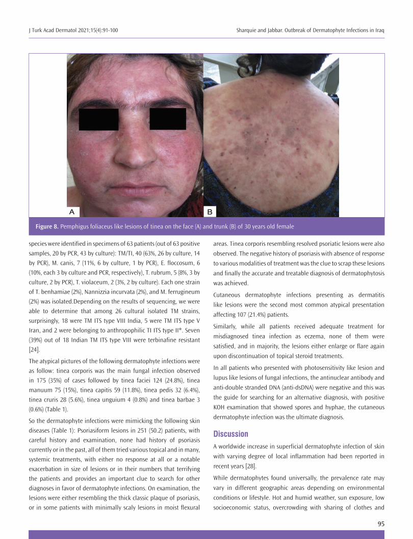
Figure 1. Psoriasis like lesion of tinea on the leg of 23 years old male
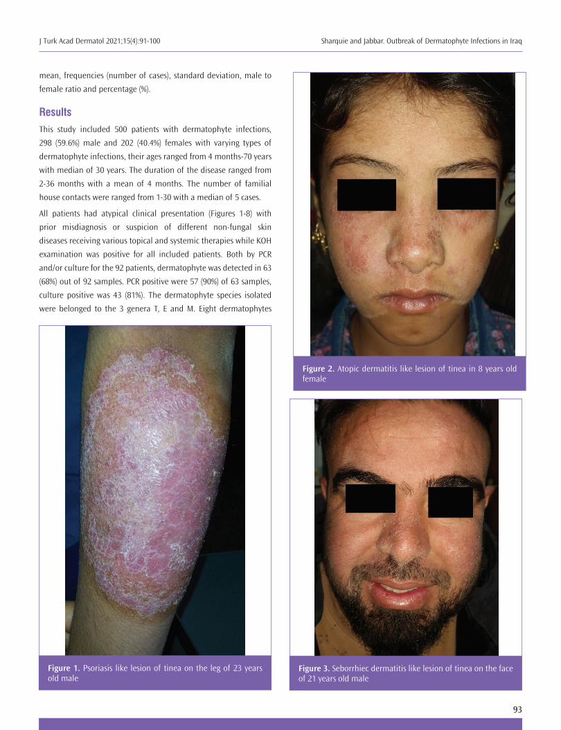
Figure 2. Atopic dermatitis like lesion of tinea in 8 years old female
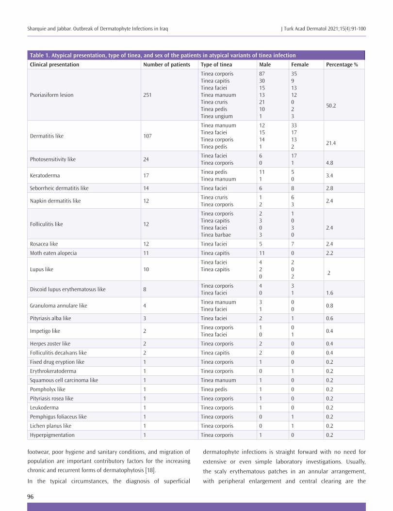
Figure 3. Seborrhiec dermatitis like lesion of tinea on the face of 21 years old male

J Turk Acad Dermatol 2021;15(4):91-100Sharquie and Jabbar. Outbreak of Dermatophyte Infections in Iraq
94
Figure 4. Left foot keratoderma lesion of tinea in 40 years old female
Figure 5. Moth eaten alopecia of tinea capitis in 11 years old male
Figure 6. Rosacea like lesion of tinea in 52 years old female
Figure 7. Tinea lesions on the malar area resembling lupus erythematosus in 29 years old male
J Turk Acad Dermatol 2021;15(4):91-100 Sharquie and Jabbar. Outbreak of Dermatophyte Infections in Iraq
95
species were identified in specimens of 63 patients (out of 63 positive
samples, 20 by PCR, 43 by culture): TM/TI, 40 (63%, 26 by culture, 14
by PCR), M. canis, 7 (11%, 6 by culture, 1 by PCR), E. floccosum, 6
(10%, each 3 by culture and PCR, respectively), T. rubrum, 5 (8%, 3 by
culture, 2 by PCR), T. violaceum, 2 (3%, 2 by culture). Each one strain
of T. benhamiae (2%), Nannizzia incurvata (2%), and M. ferrugineum
(2%) was isolated.Depending on the results of sequencing, we were
able to determine that among 26 cultural isolated TM strains,
surprisingly, 18 were TM ITS type VIII India, 5 were TM ITS type V
Iran, and 2 were belonging to anthropophilic TI ITS type II*. Seven
(39%) out of 18 Indian TM ITS type VIII were terbinafine resistant
[24].
The atypical pictures of the following dermatophyte infections were
as follow: tinea corporis was the main fungal infection observed
in 175 (35%) of cases followed by tinea faciei 124 (24.8%), tinea
manuum 75 (15%), tinea capitis 59 (11.8%), tinea pedis 32 (6.4%),
tinea cruris 28 (5.6%), tinea unguium 4 (0.8%) and tinea barbae 3
(0.6%) (Table 1).
So the dermatophyte infections were mimicking the following skin
diseases (Table 1): Psoriasiform lesions in 251 (50.2) patients, with
careful history and examination, none had history of psoriasis
currently or in the past, all of them tried various topical and in many,
systemic treatments, with either no response at all or a notable
exacerbation in size of lesions or in their numbers that terrifying
the patients and provides an important clue to search for other
diagnoses in favor of dermatophyte infections. On examination, the
lesions were either resembling the thick classic plaque of psoriasis,
or in some patients with minimally scaly lesions in moist flexural
areas. Tinea corporis resembling resolved psoriatic lesions were also
observed. The negative history of psoriasis with absence of response
to various modalities of treatment was the clue to scrap these lesions
and finally the accurate and treatable diagnosis of dermatophytosis
was achieved.
Cutaneous dermatophyte infections presenting as dermatitis
like lesions were the second most common atypical presentation
affecting 107 (21.4%) patients.
Similarly, while all patients received adequate treatment for
misdiagnosed tinea infection as eczema, none of them were
satisfied, and in majority, the lesions either enlarge or flare again
upon discontinuation of topical steroid treatments.
In all patients who presented with photosensitivity like lesion and
lupus like lesions of fungal infections, the antinuclear antibody and
anti-double stranded DNA (anti-dsDNA) were negative and this was
the guide for searching for an alternative diagnosis, with positive
KOH examination that showed spores and hyphae, the cutaneous
dermatophyte infection was the ultimate diagnosis.
DiscussionA worldwide increase in superficial dermatophyte infection of skin
with varying degree of local inflammation had been reported in
recent years [28].
While dermatophytes found universally, the prevalence rate may
vary in different geographic areas depending on environmental
conditions or lifestyle. Hot and humid weather, sun exposure, low
socioeconomic status, overcrowding with sharing of clothes and
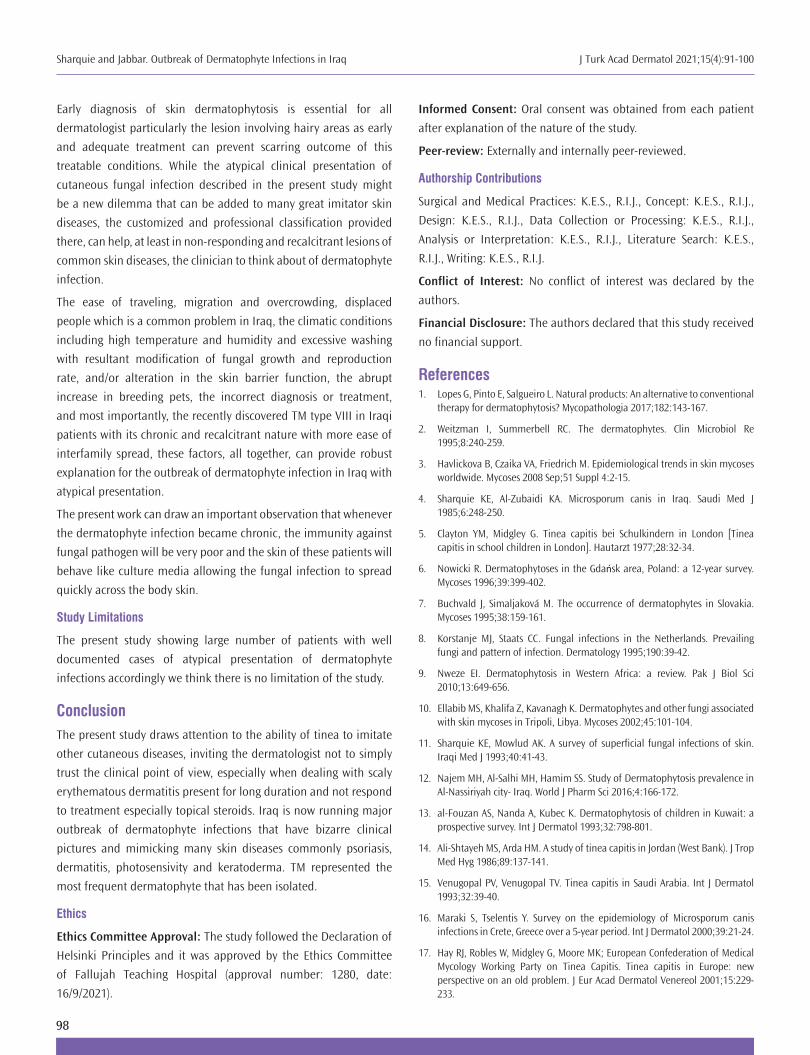
Figure 8. Pemphigus foliaceus like lesions of tinea on the face (A) and trunk (B) of 30 years old female
J Turk Acad Dermatol 2021;15(4):91-100Sharquie and Jabbar. Outbreak of Dermatophyte Infections in Iraq
96
footwear, poor hygiene and sanitary conditions, and migration of
population are important contributory factors for the increasing
chronic and recurrent forms of dermatophytosis [18].
In the typical circumstances, the diagnosis of superficial
dermatophyte infections is straight forward with no need for
extensive or even simple laboratory investigations. Usually,
the scaly erythematous patches in an annular arrangement,
with peripheral enlargement and central clearing are the
Table 1. Atypical presentation, type of tinea, and sex of the patients in atypical variants of tinea infection
Clinical presentation Number of patients Type of tinea Male Female Percentage %
Psoriasiform lesion 251
Tinea corporisTinea capitisTinea facieiTinea manuumTinea crurisTinea pedisTinea ungium
8730151321101
3591312023
50.2
Dermatitis like 107
Tinea manuumTinea facieiTinea corporisTinea pedis
1215141
3317132
21.4
Photosensitivity like 24Tinea facieiTinea corporis
60
171 4.8
Keratoderma 17Tinea pedisTinea manuum
111
50
3.4
Seborrheic dermatitis like 14 Tinea faciei 6 8 2.8
Napkin dermatitis like 12Tinea crurisTinea corporis
12
63
2.4
Folliculitis like 12
Tinea corporisTinea capitisTinea facieiTinea barbae
2303
1030
2.4
Rosacea like 12 Tinea faciei 5 7 2.4
Moth eaten alopecia 11 Tinea capitis 11 0 2.2
Lupus like 10Tinea facieiTinea capitis
420
202
2
Discoid lupus erythematosus like 8Tinea corporisTinea faciei
40
31 1.6
Granuloma annulare like 4Tinea manuum Tinea faciei
31
00
0.8
Pityriasis alba like 3 Tinea faciei 2 1 0.6
Impetigo like 2Tinea corporisTinea faciei
10
01
0.4
Herpes zoster like 2 Tinea corporis 2 0 0.4
Folliculitis decalvans like 2 Tinea capitis 2 0 0.4
Fixed drug eryption like 1 Tinea corporis 1 0 0.2
Erythrokeratoderma 1 Tinea corporis 0 1 0.2
Squamous cell carcinoma like 1 Tinea manuum 1 0 0.2
Pompholyx like 1 Tinea pedis 1 0 0.2
Pityriasis rosea like 1 Tinea corporis 1 0 0.2
Leukoderma 1 Tinea corporis 1 0 0.2
Pemphigus foliaceus like 1 Tinea corporis 0 1 0.2
Lichen planus like 1 Tinea corporis 0 1 0.2
Hyperpigmentation 1 Tinea corporis 1 0 0.2

J Turk Acad Dermatol 2021;15(4):91-100 Sharquie and Jabbar. Outbreak of Dermatophyte Infections in Iraq
97
typical clinical presentation. However, when superficial
cutaneous dermatophyte infections presented in a challenging
clinical manifestations that is, totally or at least partially, non-
distinguishable from other skin pathologies, it will result in
delayed diagnosis as physicians do not include dermatophyte
infections in their first diagnostic options. Many published
literatures highlighted on atypical presentation of these
infections [18,19,29,30,31,32,33,34,35].
Although the source of the infection is mainly animals, multiple
infections between family members as a result of interpersonal
contact within the family were observed in the present work.
This observation could confirm that zoophilic species can be
transmitted from one person to another causing dermatophytes
infection.
In the present work, we introduced a large number of atypical
superficial dermatophyte infections that were easily missed and
wrongly diagnosed with other skin diseases and were considered
as imitating skin diseases. These atypical mimicking presentations
were also reported by other studies [18,20,29,30,31,32,33,34,35] but
the present study was the most extensive evaluation.
Though rarely reported in Iraqi literature, the atypical clinical
pictures of superficial fungal infection are now being routinely seen
in different Iraqi cities. Tinea has now qualified for the group of great
imitator similar to syphilis, cutaneous tuberculosis, sarcoidosis, and
cutaneous leishmaniasis.
Psoriasis-like lesion in its classic plaque, flexural or early resolving
lesions, was the most common clinical presentation (50.2%) in our
study which is inconsistent with another study in which eczema like
lesion was the main clinical manifestation [20]. This observation
may be due to variability in the clinical presentation of cutaneous
fungal infection across the countries and even in the same country
which can be attributed to host factors, climatic factors and
dermatophyte species.
The present study recorded that TM/T. interdigitale (TI), (63%) was the
most common isolated causative species. While these results agreed
with many earlier studies where TM was the most common isolate
from the specimens [36,37], it disagreed with many previous reports
in which M.canis was the main causative strain of dermatophytes
infection [20,38,39,40] while other studies [24,41,42] showed that T.
rubrum was the main causative agents.
In this study, tinea corporis was the main fungal infection that
observed in 35% of cases followed by tinea faciei 24.8%, tinea
manuum 15%, tinea capitis 11.8%, tinea pedis 6.4%, tinea cruris
5.6%, tinea unguium 0.8% and tinea barbae 0.6%. These results
were comparable to other Iraqi and international studies where
tinea corporis and tinea capitis were the most prevalent clinical
types [10,12,43,44]. The increased prevalence of tinea corporis and
tinea capitis can be attributed to overcrowding, sharing of personal
hygiene equipments including towels, combs and shavings tools
[45] and rural drainage in many studied patients. In addition,
some social behaviors in the Iraqi community, handshake can play
a role in spreading infection as contact is an important portal for
transmission of infection.
A statistically significant relation between dermatophytes
infection and gender of the patients had been observed in the
present study where the dermatophytes infection was higher in
males with 59.6% cases, in comparison to females with 40.4%
cases. Tinea corporis, tinea capities, tinea pedis, tinea cruris
and tinea barbae had been recorded with higher frequency in
males than females. While, tinea faciei, tinea manuum and tinea
unguium displayed higher infections in females than males. The
incidence of dermatophytosis in males was more than females
in many earlier studies a finding in line with the present study
[12,37,46,47,48]. On the other hand, the results inconsistent
with other studies where the incidence of dermatophytosis was
higher in females than males [49,50,51]. This difference between
dermatophytes infection and gender of the patients may be due
to physiological differences between male and female, life style
and the differences in the social behavior [52].
Tinea capitis was the main fungal infection of preadolescent
children. This is consistent with previous studies in Iraq [40,53] and
other regions of the world [10,54,55]. Boys were affected more than
girls this could be due to their outdoor playing and sport activities
[53,54,56,57]. But tinea capitis was not uncommonly seen among
adult patients as well documented by the present work.
Tinea cruris was more abundant in males (78.57%) due to the
presence of scrotum which provides warm and moist environment
suitable for dermatophyte growth [58].
Tinea barbeae was only the disease of males (100%) as it requires
the hairy beard area of the patients.
On contrary, as the house duties are the responsibility of most
women in our society, so it’s not surprising to find higher frequency
of tinea ungium in females (75%) than males (25%). Also, cosmetic
manicuring practices undertaken by women can be considered as
another explanation for this findings [59].
Surprisingly, in this study, TM constituted the most frequently
recorded dermatophyte species in Iraq replacing T. rubrum, this
observation is on line with the new emerged and mostly terbinafine
resistant genotype VIII India. This can be explained by spread of TM
type VIII-from India to neighboring Arab and Persian countries, e.g.
Bahrain, and Iran, which facilitate their transmission to Iraq. The
percentage of terbinafine resistance is high in the strains isolated
in Baghdad, Iraq, so treatment failure of terbinafine are to be
expected [24].
J Turk Acad Dermatol 2021;15(4):91-100Sharquie and Jabbar. Outbreak of Dermatophyte Infections in Iraq
98
Early diagnosis of skin dermatophytosis is essential for all
dermatologist particularly the lesion involving hairy areas as early
and adequate treatment can prevent scarring outcome of this
treatable conditions. While the atypical clinical presentation of
cutaneous fungal infection described in the present study might
be a new dilemma that can be added to many great imitator skin
diseases, the customized and professional classification provided
there, can help, at least in non-responding and recalcitrant lesions of
common skin diseases, the clinician to think about of dermatophyte
infection.
The ease of traveling, migration and overcrowding, displaced
people which is a common problem in Iraq, the climatic conditions
including high temperature and humidity and excessive washing
with resultant modification of fungal growth and reproduction
rate, and/or alteration in the skin barrier function, the abrupt
increase in breeding pets, the incorrect diagnosis or treatment,
and most importantly, the recently discovered TM type VIII in Iraqi
patients with its chronic and recalcitrant nature with more ease of
interfamily spread, these factors, all together, can provide robust
explanation for the outbreak of dermatophyte infection in Iraq with
atypical presentation.
The present work can draw an important observation that whenever
the dermatophyte infection became chronic, the immunity against
fungal pathogen will be very poor and the skin of these patients will
behave like culture media allowing the fungal infection to spread
quickly across the body skin.
Study Limitations
The present study showing large number of patients with well
documented cases of atypical presentation of dermatophyte
infections accordingly we think there is no limitation of the study.
ConclusionThe present study draws attention to the ability of tinea to imitate
other cutaneous diseases, inviting the dermatologist not to simply
trust the clinical point of view, especially when dealing with scaly
erythematous dermatitis present for long duration and not respond
to treatment especially topical steroids. Iraq is now running major
outbreak of dermatophyte infections that have bizarre clinical
pictures and mimicking many skin diseases commonly psoriasis,
dermatitis, photosensivity and keratoderma. TM represented the
most frequent dermatophyte that has been isolated.
Ethics
Ethics Committee Approval: The study followed the Declaration of
Helsinki Principles and it was approved by the Ethics Committee
of Fallujah Teaching Hospital (approval number: 1280, date:
16/9/2021).
Informed Consent: Oral consent was obtained from each patient
after explanation of the nature of the study.
Peer-review: Externally and internally peer-reviewed.
Authorship Contributions
Surgical and Medical Practices: K.E.S., R.I.J., Concept: K.E.S., R.I.J.,
Design: K.E.S., R.I.J., Data Collection or Processing: K.E.S., R.I.J.,
Analysis or Interpretation: K.E.S., R.I.J., Literature Search: K.E.S.,
R.I.J., Writing: K.E.S., R.I.J.
Conflict of Interest: No conflict of interest was declared by the
authors.
Financial Disclosure: The authors declared that this study received
no financial support.
References1. Lopes G, Pinto E, Salgueiro L. Natural products: An alternative to conventional
therapy for dermatophytosis? Mycopathologia 2017;182:143-167.
2. Weitzman I, Summerbell RC. The dermatophytes. Clin Microbiol Re 1995;8:240-259.
3. Havlickova B, Czaika VA, Friedrich M. Epidemiological trends in skin mycoses worldwide. Mycoses 2008 Sep;51 Suppl 4:2-15.
4. Sharquie KE, Al-Zubaidi KA. Microsporum canis in Iraq. Saudi Med J 1985;6:248-250.
5. Clayton YM, Midgley G. Tinea capitis bei Schulkindern in London [Tineacapitis in school children in London]. Hautarzt 1977;28:32-34.
6. Nowicki R. Dermatophytoses in the Gdańsk area, Poland: a 12-year survey. Mycoses 1996;39:399-402.
7. Buchvald J, Simaljaková M. The occurrence of dermatophytes in Slovakia. Mycoses 1995;38:159-161.
8. Korstanje MJ, Staats CC. Fungal infections in the Netherlands. Prevailing fungi and pattern of infection. Dermatology 1995;190:39-42.
9. Nweze EI. Dermatophytosis in Western Africa: a review. Pak J Biol Sci 2010;13:649-656.
10. Ellabib MS, Khalifa Z, Kavanagh K. Dermatophytes and other fungi associated with skin mycoses in Tripoli, Libya. Mycoses 2002;45:101-104.
11. Sharquie KE, Mowlud AK. A survey of superficial fungal infections of skin. Iraqi Med J 1993;40:41-43.
12. Najem MH, Al-Salhi MH, Hamim SS. Study of Dermatophytosis prevalence in Al-Nassiriyah city- Iraq. World J Pharm Sci 2016;4:166-172.
13. al-Fouzan AS, Nanda A, Kubec K. Dermatophytosis of children in Kuwait: a prospective survey. Int J Dermatol 1993;32:798-801.
14. Ali-Shtayeh MS, Arda HM. A study of tinea capitis in Jordan (West Bank). J Trop Med Hyg 1986;89:137-141.
15. Venugopal PV, Venugopal TV. Tinea capitis in Saudi Arabia. Int J Dermatol 1993;32:39-40.
16. Maraki S, Tselentis Y. Survey on the epidemiology of Microsporum canisinfections in Crete, Greece over a 5-year period. Int J Dermatol 2000;39:21-24.
17. Hay RJ, Robles W, Midgley G, Moore MK; European Confederation of Medical Mycology Working Party on Tinea Capitis. Tinea capitis in Europe: new perspective on an old problem. J Eur Acad Dermatol Venereol 2001;15:229-233.

J Turk Acad Dermatol 2021;15(4):91-100 Sharquie and Jabbar. Outbreak of Dermatophyte Infections in Iraq
99
18. Dogra S, Narang T. Emerging atypical and unusual presentations of dermatophytosis in India. Clin Dermatol Rev 2017;1:12-18.
19. Sahoo AK, Mahajan R. Management of tinea corporis, tinea cruris, and tinea pedis: A comprehensive review. Indian Dermatol Online J 2016;7:77-86.
20. Atzori L, Pau M, Aste N, Aste N. Dermatophyte infections mimicking other skin diseases: a 154-person case survey of tinea atypica in the district of Cagliari (Italy). Int J Dermatol 2012;51:410-415.
21. Verma S, Madhu R. The Great Indian Epidemic of Superficial Dermatophytosis: An Appraisal. Indian J Dermatol 2017;62:227-236.
22. VermaS,VasaniR,ReszkeR,MatusiakŁ, Szepietowski JC.Prevalenceandclinical characteristics of itch in epidemic-like scenario of dermatophytoses in India: a cross-sectional study. J Eur Acad Dermatol Venereol 2020;34:180-183.
23. Nenoff P, Verma SB, Ebert A, Süß A, Fischer E, Auerswald E, Dessoi S, Hofmann W, Schmidt S, Neubert K, Renner R, Sohl S, Hradetzky U, Krusche U, Wenzel HC, Staginnus A, Schaller J, Müller V, Tauer C, Gebhardt M, Schubert K, Almustafa Z, Stadler R, Fuchs A, Sitaru C, Retzlaff C, Overbeck C, Neumann T, Kerschnitzki A, Krause S, Schaller M, Walker B, Walther T, Köhler L, Albrecht M, Willing U, Monod M, Salamin K, Burmester A, Koch D, Krüger C, Uhrlaß S. Spread of Terbinafine-Resistant Trichophyton mentagrophytes Type VIII (India) in Germany-”The Tip of the Iceberg?”. J Fungi (Basel) 2020;6:207.
24. Uhrlass S, Alhilaifawi AFH, Sharquie KE, Alhamami HR, Al-Qahar GHA, Tabra MKH, Koch D, Muetze H, Kruege C, Burmester A, Wiegand C, Hipler UC. Dermatophyte survey from Baghdad, Iraq – emergence of terbinafine resistant Trichophyton mentagrophytes Type VIII India. Submitted (MAY-2020) to the EMBL/GenBank/DDBJ databases.
25. Järv H, Uhrlaß S, Simkin T, Nenoff P, Alvarado RE; Chryssanthou E, Monod M. Terbinafine resistant Trichophyton mentagrophytes genotype VIII, Indian type, isolated in Finland. J Fungi 2019;5:117.
26. Uhrlass S, Sithach M, Koch D, Wittig F, Muetze H, Krueger C, Nenoff P. Trichophyton mentagrophytes—A new genotype in Cambodia. J Fungi 2019;5:460.
27. Taghipour S, Shamsizadeh F, Pchelin IM, Rezaei-Matehhkolaei A, Zarei Mahmoudabadi A, Valadan R, Ansari S, Katiraee F, Pakshir K, Zomorodian K, Abastabar M. Emergence of Terbinafine Resistant Trichophyton mentagrophytes in Iran, Harboring Mutations in the Squalene Epoxidase (SQLE) Gene. Infect Drug Resist 2020;13:845-850.
28. Yadav A, Urhekar AD, Mane V, Danu MS, Goel N, Ajit KG. Optimizationandisolation of dermatophytes from clinical samples and in vitro antifungal susceptibility testing by disc diffusion method. Research & Reviews: Journal of Microbiology and Biotechnology 2013;2:19-34.
29. Serarslan G. Pustular psoriasis-like tinea incognito due to Trichophyton rubrum. Mycoses 2007;50:523-524.
30. Romano C, Maritati E, Gianni C. Tinea incognito in Italy: a 15-year survey. Mycoses 2006;49:383-387.
31. Meymandi S, Wiseman MC, Crawford RI. Tinea faciei mimicking cutaneous lupus erythematosus: a histopathologic case report. J Am Acad Dermatol 2003;48:S7-8.
32. Ghislanzoni M. Tinea incognito due to Trichophyton rubrum responsive to topical therapy with isoconazole plus corticosteroid cream. Mycoses 2008;51 Suppl 4:39-41.
33. Nenoff P, Mügge C, Herrmann J, Keller U. Tinea faciei incognito due to Trichophyton rubrum as a result of autoinoculation from onychomycosis. Mycoses 2007;50 Suppl 2:20-25.
34. Atzori L, Pau M, Aste M. Erythema multiforme ID reaction in atypical dermatophytosis: a case report. J Eur Acad Dermatol Venereol 2003;17:699-701.
35. Aste N, Pau M, Aste N, Atzori L. Tinea corporis mimicking herpes zoster. Mycoses 2011;54:463-465.
36. Sowmya N, Appalaraju B, Surendran P, Srinivas CR. Isolation, Identification and comparatative analysis of SDA and DTM for dermatophytes from clinical samples in a tertiary care hospital. IOSR Journal of Dental and Medical Sciences 2014;13:68-73.
37. Rashidian S, Falahati M, Kordbacheh P, Mahmoudi M, Safara M, Sadeghi Tafti H, Mahmoudi S, Zaini F. A study on etiologic agents and clinical manifestationsofdermatophytosisinYazd,Iran.CurrMedMycol2015;1:20-25.
38. Ndako JA,OsemwegieOO,SpencerTHI,OlopadeBK,YunusaGA,Banda J.Prevalence of Dermatophytes and other associated fungi among school children. Global Advanced Research Journal of Medicine and Medical Sciences 2012;1:49-56.
39. Veasey JV, Miguel BAF, Mayor SAS, Zaitz C, Muramatu LH, Serrano JA. Epidemiological profile of tinea capitis in São Paulo City. An Bras Dermatol 2017;92:283-284.
40. Sharquie KE, Al Ani SM, Ali TM. Microsporum canis epidemic in Iraq children.Journal of the Faculty of Medicine-Baghdad 1992;34:95-99.
41. Mohammed SJ, Noaimi AA, Sharquie KE. A Survey Of Dermatophytes Isolated From Iraqi Patients In Baghdad City. AL-Qadisiya Medical Journal 2015;11:10-15.
42. Toukabri N, Dhieb C, El Euch D, Rouissi M, Mokni M, Sadfi-Zouaoui N. Prevalence, Etiology, and Risk Factors of Tinea Pedis and Tinea Unguium in Tunisia. Can J Infect Dis Med Microbiol 2017;2017:6835725.
43. Mahmoud AL. A study of dermatophytoses in Sana’a, Yemen Republic.Mycoses 2002;45:105-108.
44. LangeM,Nowicki R, Barańska-RybakW,BykowskaB.Dermatophytosis inchildren and adolescents in Gdansk, Poland. Mycoses 2004;47:326-329.
45. Brook GF, Butel JS, Morse SA, Jawetz and Adelbergs medical microbiology. 2nd. Middle East. Beirut; Lebanon: 2001.
46. Morar N, Dlova NC, Gupta AK, Aboobaker J. Tinea capitis in Kwa-Zulu Natal, South Africa. Pediatr Dermatol 2004;21:444-447.
47. Rahman MH. Hadiuzzaman MD, Bhuiyan MKJ, Islam N, Ansari NP, Mumu SA, Chowdhury IJ. Prevalence of superficial fungal infections in the rural areas of Bangladesh. Iran J Dermatol 2011;14:86-91.
48. Sahin I, Oksuz S, Kaya D, Sencan I, Cetinkaya R. Dermatophytes in the rural area of Duzce, Turkey. Mycoses 2004;47:470-474.
49. BrajacI,Stojniń-SosaL,PrpińL,LoncarekK,GruberF.TheepidemiologyofMicrosporum canis infections in Rijeka area, Croatia. Mycoses 2004;47:222-226.
50. Brilhante RS, Cordeiro RA, Rocha MF, Monteiro AJ, Meireles TE, Sidrim JJ. Tinea capitis in a dermatology center in the city of Fortaleza, Brazil: the role of Trichophyton tonsurans. Int J Dermatol 2004;43:575-579.
51. Vella Zahra L, Gatt P, Boffa MJ, Borg E, Mifsud E, Scerri L, Vella Briffa D, Pace JL. Characteristics of superficial mycoses in Malta. Int J Dermatol 2003;42:265-271.
52. Hay RJ, Adrians BM. Bacterial infection. In: Champion RH, Burton JL, Burns DA, Breatandem JM, editors. Textbook of Dermatology, 6th ed, London: Blackwell Sciences; 1998. p. 1113-1116.
53. Abed Ali FAH, Al-Janabi JKA, Mohammed K, Alhattab MK. Prevalence of dermatophyte fungal infection in Hillah, Iraq. International Journal of Chem Tech Research 2017;10:827-837.
54. Naseri A, Fata A, Najafzadeh MJ, Shokri H. Surveillance of dermatophytosis in northeast of Iran (Mashhad) and review of published studies. Mycopathologia 2013;176:247-253.

J Turk Acad Dermatol 2021;15(4):91-100Sharquie and Jabbar. Outbreak of Dermatophyte Infections in Iraq
100
55. Maulingkar SV, Pinto MJ, Rodrigues S. A clinico-mycological study of dermatophytoses in Goa, India. Mycopathologia 2014;178:297-301.
56. Moneib HA, El-Shiemy SMH, Saudi WM, El-Fangary MM, Nabil T, Mohy SM. Hair loss among a group of Egyptian children: a clinical and dermoscopic study. J Egypt Women Dermatol Soc 2017;14:9-24.
57. Nnoruka EN, Obiagboso I, Maduechesi C. Hair loss in children in South-East Nigeria: common and uncommon cases. Int J Dermatol 2007;46(Suppl 1):18-22.
58. Abdel-Rahman SM, Farrand N, Schuenemann E, Stering TK, Preuett B, Magie R, Campbell A. The prevalence of infections with Trichophyton tonsurans in schoolchildren: the CAPITIS study. Pediatrics 2010;125:966-973.
59. Olde Hartman TC, van Rijswijk E. Fungal nail infection. BMJ 2008;337:a429.