Outbreak, Surveillance - OSIR Journal
30
Outbreak, Surveillance & Investigation Reports Volume 10, Issue 2 June 2017 Journal of ONLINE ACCESS www.osirjournal.net QR CODE
-
Upload
khangminh22 -
Category
Documents
-
view
0 -
download
0
Transcript of Outbreak, Surveillance - OSIR Journal

Outbreak, Surveillance& Investigation Reports
Volume 10, Issue 2
June 2017Journal of
ONLINE
ACCESS
www.osirjournal.net
QR CODE

eISSN: 2286-8933
Executive Board Tanarak Plipat, Thailand
Chief Editors Alden Henderson, USA
Angela Song-En Huang, Taiwan
Chuleeporn Jiraphongsa, Thailand
Nitaya Chanruang Mahabhol, Thailand
Pawin Padungtod, Vietnam
Wiwat Rojanapitthayakorn, Thailand
OSIR Editors David M. Castellan, Canada
Do Thi Hong Hien, Vietnam
Dorothy Southern, Myanmar
Fadzilah Binti Kamaludin, Malaysia
Henry C. Baggett, USA
Hishamuddin Badaruddin, Singapore
Huai Yang, China
Jiang Li, China
Justin Denny, USA
Associate Editor Yin Myo Aye, Thailand
Chief of Administration Vanlaya Srethapranai, Thailand
IT Narakorn Sae-lew, Thailand
Disclaimer: OSIR is not responsible for any inaccurate or libelous information in these publications or the use of
information contained or linked in articles published in the journal.
Outbreak, Surveillance and Investigation Reports (OSIR) Journal
Field Epidemiology Training Program, Bureau of Epidemiology, Department of Disease Control,
Ministry of Public Health, Tiwanond Road, Muang District, Nonthaburi 11000, Thailand
Tel: +662-5901734, Fax: +662-5918581, Email: [email protected]
Website: <http://www.osirjournal.net>
Kachen Wongsathapornchai, Thailand
Marcel Curlin, USA
Maria Concepcion Roces, Philippines
Michael Martin, Thailand
Monaya Ekgatat, Thailand
Richard Brown, Thailand
Rodger Detels, USA
Wan Mansor Bin Hamzah, Malaysia
Ying Lu, Thailand
Volume 10, Issue 2, June 2017
Contents
Original Articles:
1. A Large Common Source Outbreak of Salmonella typhimurium
Linked to Kuala Terengganu Night Markets, Malaysia, 2014 …………………………. 1-7
2. Clinical Profile and Circulating Dengue Virus Serotype among
Adults Admitted to Yangon General Hospital during the 2015
Dengue Outbreak …………..……………………………………………………………………………. 8-13
3. Identification of a Tuberculosis Cluster through Epidemiological
and Geographical Tracing of a Patient with Multidrug-resistant
Tuberculosis in Lopburi Province, Thailand, 2014 …………............................................ 14-22
Invited Perspective Article:
4. The Grammar of Science: Let’s ‘Log’ (Part 1) …………....................................................... 23-27
OSIR, June 2017, Volume 10, Issue 2, p.1-7
1
A Large Common Source Outbreak of Salmonella typhimurium Linked to
Kuala Terengganu Night Markets, Malaysia, 2014
Balkis Ab Karim1,*, A Liza Latip1, Anita Surani Abd Shukor2, Norafidah A Rashid2, Wan Madihah
Wan Mohd2, Fadzilah Kamaludin3
1 Terengganu State Health Department, Malaysia Kuala Terengganu District Health Office,
Terengganu, Malaysia
2 Kuala Terengganu District Health Office, Terengganu, Malaysia
3 Office of Deputy General of Health (Public Health), Ministry of Health Malaysia
*Corresponding author, email address: [email protected]
Abstract
On 1 Mar 2014, the Terengganu District Health Office was notified of ten patients presented with acute gastroenteritis at
Sultanah Nur Zahirah Hospital. Their illness was linked to consumption of foods from two night markets. An outbreak
investigation was initiated to determine the source of the outbreak. Case finding was conducted in the hospital, and
community. Patients were interviewed about demographics, symptoms and food consumption history. Stool samples from
patients and food handlers as well as food and environmental samples were collected for laboratory analysis. Suspected
food premises were inspected. A case-control study was conducted. Of 169 cases, 68.6% and 32.5% ate food from night
markets A and B respectively while 1.2% ate food from both night markets. Major symptom was diarrhea (98.2%). There
was one death from hypovolemic shock. Salmonella typhimurium was isolated from 13 patients and one food handler. All
isolates showed genetic similarity by pulsed-field gel electrophoresis. The food handler tested to have the infection served
the white fried rice sold in both night markets. Cases were 14 (95% CI = 4.05-46.61) and nine (95% CI = 3.36-24.3) times
more likely to have consumed white fried rice from night markets A & B respectively. The source of infection was likely to
be white fried rice that was prepared at the same place, contaminated by an infected food handler and sold at both night
markets.
Keywords: Food poisoning, Salmonella typhimurium, night market, food handler
Introduction
Night markets have been around for decades in
Malaysia. They are called ‘Pasar Malam’ and are
popular places for social gatherings. Makeshift stalls
selling local products and foods are the main
attractions at these sites.
The Local Authority in Kuala Terengganu District,
Terengganu State, designates and licenses the
locations of night markets. There are 22 designated
locations and three to five locations are opened from 6
to 10 pm every day. Each location consists of 100 to
200 food stalls. The designated stall is usually an
empty space of about nine square meters without
basic infrastructure and amenities such as water
supply, electricity, washing, and drainage facilities.
Food stall operators move daily to different locations
following a weekly schedule. In the makeshift stalls,
the operators sell cooked or partially cooked foods
prepared from home or at the shops. Most vendors
prepare food items at home in the morning and bring
it to the night market in the evening.
Major foodborne outbreaks were observed to be
associated with markets.1 Hazards related to markets
are common due to biological cross-contamination,
polluted water, inadequate preservation and storage,
and poor environmental sanitation.2
At 19:00 on 1 Mar 2014, the Emergency Department
in Sultanah Nur Zahirah (SNZ) Hospital notified the
Kuala Terengganu District Health Office of 10 cases
of suspected food poisoning. A rapid assessment team
was assembled to verify and assess the extent of the
OSIR, June 2017, Volume 10, Issue 2, p.1-7
2
outbreak, identify the causative agent and source,
make relevant recommendations, and institute
appropriate control measures.
Methods
We interviewed the hospitalized and out-patient cases,
and reviewed their medical records in SNZ Hospital.
We searched for additional cases among out-patients
attendance in health clinics and SNZ hospital, family
members and friends of the acute gastroenteritis
(AGE) cases who had history of consumed foods from
Night Markets A (NMA) and B (NMB). Information
was obtained on demographic details, date and time
of onset of symptoms, and foods consumed, including
the source of foods.
Case-control Study
We conducted a case-control study to identify the
potential vehicle of the outbreak. We defined a case
as a person who developed one or more of the
following signs or symptoms: diarrhea, abdominal
pain, vomiting, fever, nausea, or dizziness after
consuming foods from NMA or NMB on 28 Feb to 1
Mar 2014. Controls were family members and friends
of cases and other vendors who consumed foods
bought from NMA or NMB, yet did not develop
symptoms of AGE. Logistic regression was used to
calculate crude odds ratios.
Stool samples were sent for enteropathogenic
bacterial culture at the laboratory in SNZ Hospital,
and serotyping and pulsed-field gel electrophoresis
(PFGE) at Institute of Medical Research in Kuala
Lumpur. No clinical samples were collected from
controls.
Environmental Investigations
Food premises in NMA and NMB were inspected
using a standard format for restaurants and food
stalls issued by the Ministry of Health which covers
food processing and storage, personal hygiene of food
handlers, quality of cooking utensils, water supply
and drainage system, solid waste disposal, and
kitchen infrastructure. Food handlers were
interviewed regarding food preparation and cooking
methods of the suspected foods. Left-over raw food
materials and ingredients from the kitchen and,
environmental swabs from working surface and
utensils were also taken.
We traced back the supply chain for chicken, eggs and
vegetables. Food and environmental samples were
taken from these premises and sent to the
Terengganu State Health Department Food
Laboratory for bacteriological testing.
Results
Descriptive Findings
Total 169 cases fulfilled the clinical case definition,
including 116 (68.6%) had history of consuming food
from NMA and 55 (32.5%) from NMB, with two (1.2%)
cases consuming food from both night markets. No
significant difference was observed in the
demographic profiles of those who consumed foods at
NMA and NMB (Table 1).
Table 1. Characteristics of acute gastroenteritis cases from
two night markets in Kuala Terengganu District,
Terengganu State, Malaysia, 28 Feb to 1 Mar 2014
Night Market A
(%) (n = 116) Night Market B
(%) (n = 55)
Mean age SD 22.80 12.13 19.7012.13
Age group
Adult 92 (79.3) 39 (70.9)
Child 24 (20.7) 16 (29.1)
Gender
Male 55 (47.4) 26 (47.3)
Female 61 (52.6) 29 (52.7)
District
Kuala Terengganu 97 (83.6) 49 (89.1)
Marang 13 (11.2) 4 (7.3)
Hulu Terengganu 4 (3.4) 0
Setiu 2 (1.7) 1 (1.8)
Treatment Status (n=53)
In-patient 65 (56.0) 43 (81.1)
Out-patient 34 (29.3) 7 (13.2)
No treatment 17 (14.7) 3 (5.7)
Outcome
Alive 116 (100.0) 54 (98.2)
Died 0 1 (1.8)
Major presenting symptoms were diarrhea (98.2%),
followed by abdominal pain (91.8%), vomiting (78.4%),
fever (63.7%), dizziness (49.7%), and nausea (46.2%).
The epidemic curve contained two peaks: the first
peak consisted of cases with a history of eating food
from NMA and the second peak with cases in NMB.
Median incubation period of cases consumed food
from NMA and NMB was 9.5 hours and 11 hours
respectively (Figure 1).
About 63% of 169 cases were hospitalized (60% from
NMA, 40% from NMB). One death was reported, the
cause of death was hypovolemic shock secondary to
severe dehydration (0.6% case fatality rate).
Laboratory Results
Total 118 stool samples collected were from 107 cases

OSIR, June 2017, Volume 10, Issue 2, p.1-7
3
Figure 1. Date and time of onset of acute gastroenteritis cases from two night markets in Kuala Terengganu District,
Terengganu State, Malaysia, 28 Feb to 1 Mar 2014
and 11 asymptomatic food handlers, and 100% were
cultured. Among them, 53 (49.5%) out of 107 cases
and two (18%) out of 11 food handlers were cultured
positive for Salmonella. Of 55 isolates tested positive,
24 were sent for subtyping and PFGE, and S.
typhimurium was detected in 15 (63%), including 13
cases and two food handlers. Fourteen isolates
showed fingerprint similarity: 13 from cases
(including the fatal case) and a food handler who
prepared white fried rice at both night markets
(Figure 2).
Case-control Study
The data were stratified by night markets where the
case bought the food items. Total 168 subjects (116
cases and 52 controls) consumed food from NMA,
*No stool sample was taken from non-case. NMA = Night market A, NMB = Night market B
0
2
4
6
8
10
12
14
16
18
16
:00
18
:00
20
:00
22
:00
12
:00
02
:00
04
:00
06
:00
08
:00
10
:00
12
:00
14
:00
16
:00
18
:00
20
:00
22
:00
24
:00
02
:00
04
:00
06
:00
08
:00
10
:00
12
:00
14
:00
16
:00
18
:00
20
:00
22
:00
24
:00
02
:00
28 Feb 1 Mar 2 Mar 3 Mar
Nu
mb
er o
f ca
se
Date & time
Night market B
Night market A
118 stool samples*
collected and cultured
107 cases
11 food handlers
2 isolated Salmonella spp.
53 isolated Salmonella spp.
(33 NMA, 20 NMB)
22 isolates sent for Salmonella
subtyping & PFGE
2 isolates sent for Salmonella subtyping
& pulsed-field gel electrophoresis (PFGE)
1 S. typhimurium
positive
13 S. typhimurium positive
(11 NMA, 2 NMB)
1 PFGE showed similar
fingerprint with 13 cases
13 PFGE showed similar fingerprint
(11 NMA, 2 NMB)
Figure 2. Laboratory investigation and results of acute gastroenteritis cases from two night markets
in Kuala Terengganu District, Terengganu State, Malaysia, 28 Feb to 1 Mar 2014

OSIR, June 2017, Volume 10, Issue 2, p.1-7
4
while 131 subjects (55 cases, 76 controls) consumed
food from NMB. As there were two cases consumed
food from both night markets, the number of cases
were accounted based on each night market.
Cases were 14 (95% CI = 4.05-46.61) and nine (95%
CI = 3.36-24.3) times more likely to have consumed
white fried rice from NMA and NMB respectively
(Tables 2 and 3).
Environmental Investigation
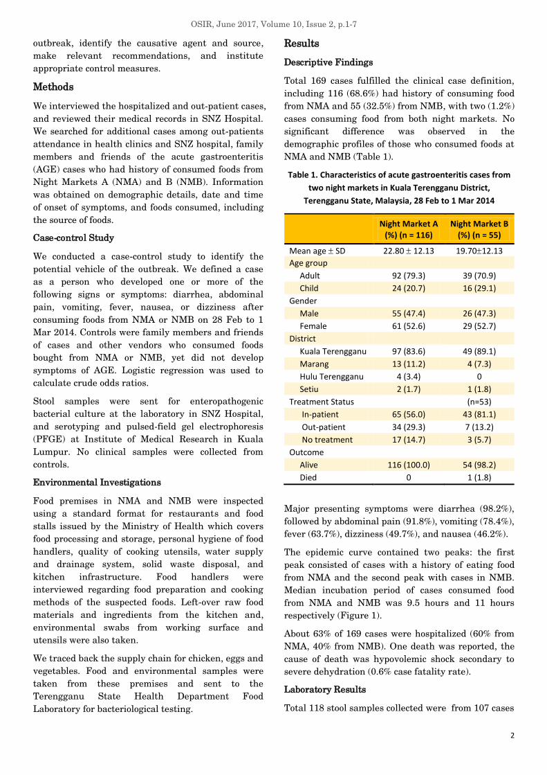
The foods were prepared at a shop, and cooked and
readily sold at a food stall named FS1 Kitchen in both
night markets. Food premise inspection at the
preparation shop scored 44.8% which was lower than
acceptable score of more than 70%. Violation of food
hygiene standard was identified at processing of raw
materials, and storage of mixed raw materials and
cooked food items (Figure 3). Temperature of the
chiller was 16˚C while the standard should be less
than 8˚C. No enteropathogenic bacterial isolated from
26 food and environmental samples taken from the
kitchen. However, high count of coliform was detected.
The main ingredients of white fried rice sold at the
FS1 kitchen were rice and chicken. Hazard analysis
and critical control point (HACCP)3 showed two main
violation points. Chicken was prepared
unhygienically. The half-cooked chicken were cut into
small pieces, kept in a plastic container, mixed with
gravy consisting of sugar, monosodium glutamate and
salt, and left in the ambient temperature for two
hours. The chicken was fried with rice at the night
markets. The ready-to-eat white fried rice was
displayed in an opened big casserole for at least 4-8
hours at room temperature (Figure 4).
Out of 24 food samples taken from chicken, eggs and
mixed vegetables, Salmonella corvalli were detected
in fresh and semi-frozen chickens from the cool box.
Discussion
The epidemic curve showed the outbreak occurred
first at NMA and subsequently followed by another
outbreak at NMB. Both showed similar point source
pattern, with an interval of 1-day lag suggesting a
common exposure. Those who consumed white fried
rice sold at the FS1 kitchen in both NMA and NMB
were found to have higher risk of getting ill.
The infecting agent was S. typhimurium and the
cases had symptoms compatible to infection by S.
typhimurium4. The source of the outbreak was the
food handler from the FS1 kitchen who could have
contaminated the white fried rice during food
preparation. Isolation of S. typhimurium and the
similar fingerprint pattern from cases and food
handlers showed an epidemiological link between the
cases and the food handler.
S. typhimurium is reported as one of the most
common serotypes infecting humans.5 Our findings
were consistent with a large outbreak reported in
Sydney in year 2011 which involved 154 cases
positive for S. typhimurium, and was linked to
consuming chicken salad roll at a restaurant.6
Table 2. Results of a case-control study from people who consumed food at Night Market A
in Kuala Terengganu District, Terengganu State, Malaysia, 28 Feb to 1 Mar 2014 (n=116)
Ate Did not ate Odds Ratio 95% CI
Case Control Case Control
White Fried Rice 53 3 63 49 13.74 4.05-46.61
Red Fried Rice 30 8 86 44 1.92 0.81-4.54
Fried Mee 15 24 101 28 0.17 0.08-0.37
Fried Keow Teow 23 15 93 37 0.61 0.29-1.30
Nagelkerke R2 = 0.405, Hosmer and Lemeshow Test = 0.967
Table 3. Results of a case-control study from people who consumed food at Night Market B
in Kuala Terengganu District, Terengganu State, Malaysia, 28 Feb to 1 Mar 2014 (n=55)
Ate Did not ate Odds Ratio 95% CI
Case Control Case Control
White Fried Rice 24 6 31 70 9.03 3.36-24.3
Red Fried Rice 22 8 33 68 5.67 2.28-14.07
Fried Mee 9 30 46 46 0.30 0.13-0.70
Fried Keow Teow 4 17 51 59 0.27 0.09-0.86
Nagelkerke R2 = 0.471, Hosmer and Lemeshow Test = 0.994
OSIR, June 2017, Volume 10, Issue 2, p.1-7
5
(a) Improper storage of the utensils (b, c) Improper storage of raw foods in the refrigerator and temperature tested to be
higher than the acceptable standard
(d) Chili paste container not sealed (e) Dirty kitchen floor (f) Water in the buckets for washing of raw
materials and utensils and kept at room temperature
Figure 3. Photos of food preparation shop for selling food at two night markets in Kuala Terengganu District,
Terengganu State, Malaysia, March 2014
Figure 4. Ready-to-eat fried noodle and rice (left) from the FS1 Kitchen at two night markets (middle and right)
in Kuala Terengganu District, Terengganu State, Malaysia, March 2014
Salmonellosis outcomes differ substantially by
serotypes. A study on invasive disease, S.
typhimurium was significantly less invasive,
compared to S. enteritidis, Heidelberg, Choleraesuis,
and Dublin.7 Case fatality rate in this outbreak was
0.6% which was consistent for non-typhoidal
Salmonella infection reported elsewhere as less than
1%.7 Although we detected S. corvalli from
environmental samples, the serotype had been less
frequently reported in humans, compared to the
environment. In addition, this serotype was not
discovered from human cases in this outbreak.
Ready-to-eat foods are commonly sold in the food
markets of the developing countries as it is accessible
and affordable for people in the community as well as
tourists discovering the local food culture.2 Raw
vegetables and ready-to-eat foods pose higher risk for
bacterial contamination such as Staphylococcus8,
Salmonella8,9, Campylobacter9, norovirus, and
hepatitis A and E10. Hence, according to the World
Health Organization, multidisciplinary approach in
microbiology, food science, health promotion and
sanitation management are essential to provide safe
and nutritious foods in the markets.2
Multi-drug resistant S. typhimurium (phage type
DT104) isolates had been reported from several
countries.11 However, in this outbreak, information on
antibiotic treatment was not available, and phage
typing and antibiotic sensitivity test specifically for
these strains were not performed.
Although the interviews were conducted within one
week of event, the information obtained from
OSIR, June 2017, Volume 10, Issue 2, p.1-7
6
personal interviews could not be verified and the food
handlers’ food handling practices were not observed.
However, the environmental investigation supported
the epidemiological findings. There was poor food
handling practices in the kitchen as well as in the
night market that might have allowed cross
contamination. The stool samples from asymptomatic
food handlers on duty during the event were positive
for S. typhimurium, suggesting that contamination
could have happened during the preparation of white
fried rice.
Another limitation was no samples taken from
controls. If there were asymptomatic cases among
controls, the odds ratio would be underestimated.
Action Taken and Recommendations
The FS1 kitchen was immediately closed for two
weeks under the Ministry of Health Communicable
Disease Act of 198812. The two food handlers with
Salmonella were barred from handling the foods until
all three consecutive stool cultures were tested
negative for Salmonella spp. The food premise
operator was recommended to improve the cooking
facilities, including fixing tile flooring for easy
maintenance and storage of dried raw materials and
utensils. Top loading refrigerator for raw materials
and chiller for cooked foods were suggested. Food
handlers were advised and educated in the hygienic
preparation and serving of foods.
Reassessment of the kitchen was done after two
weeks, and showed an improvement and its score
increase to 83% (Figure 5). In total 18 night markets
visited, 776 food premises were inspected of which
477 (61.5%) were scored grade A and 77 (19.9%) grade
B. Grade A was given to food premise with the scoring
of 80% and more, and grade B was 60 to 79% score.
There was no proper documents on health
examination and immunization card in 222 food
premises (28.2%), and were compounded by the Local
Authority.
Health education pamphlets on food safety,
prevention of poisoning and how to choose safer food
from night market were distributed to food handlers
and public at night markets. A radio talk session was
given via local radio station aimed to increase
consumers’ awareness on the food safety issues
particularly ready to eat foods from night markets.
Health clinics in neighboring districts: Marang, Setiu
and Hulu Terengganu were alerted on the second day
of the outbreak. They were required to notify any
acute gastroenteritis case related to consumption of
foods from NMA and NMB.
Figure 5. Improved situation after 2 weeks of closure of the
food preparation shop for selling food at two night markets
in Kuala Terengganu District, Terengganu State, Malaysia,
March 2014
The findings of this outbreak were presented to the
Food Safety and Quality Unit Terengganu State
Health Department and the following
recommendations were suggested: strengthen the
monitoring of night market food premises including
home kitchens where the foods are prepared, institute
effective health promotion and education strategies
for night market food handlers and consumers,
strengthen the enforcement of food safety law related
to night markets, advocate local authority to enforce
food premise licensing, and improve night markets
infrastructures such as provision of safe water supply,
and standard mobile stalls.
Conclusions
This is a common source outbreak caused by S.
typhimurium with case fatality rate of 0.6%. The
most probable source of infection was the
asymptomatic food handler who may have
contaminated the white fried rice during food
preparation. Possible contributing factors were
unhygienic food handlers and food handling practices,

OSIR, June 2017, Volume 10, Issue 2, p.1-7
7
and poor sanitation and substandard kitchen
infrastructure. The outbreak was controlled first by
removing the source (infected food handler),
educating to food vendors and consumers, and prompt
outbreak management with multi-departments’
involvement.
Suggested Citation
Ab Karim B, Latip AL, Abd Shukor AS, A
Rashid N, Wan Mohd WM, Kamaludin F. A
large common source outbreak of Salmonella
typhimurium linked to Kuala Terengganu night
markets, Malaysia, 2014. OSIR. 2017
Jun;10(2):1-7.
References
1. Luquero FJ, Banga CN, Remartínez D, Palma
PP, Baron E, Grais RF. Cholera epidemic in
Guinea-Bissau (2008): the importance of
"place". PLoS One. 2011 May 4;6(5):e19005.
2. World Health Organization. A guide to health
food markets. Geneva: World Health
Organization; 2006 [cited 2017 Jan 6].
<http://www.who.int/foodsafety/publications/c
apacity/healthymarket_guide.pdf>.
3. United States Food and Drug Administration.
Hazard analysis critical control point
(HACCP). 2017 Mar 4 [cited 2017 May 16].
<https://www.fda.gov/food/guidanceregulation
/haccp/>.
4. Heymann LD. Control of communicable
diseases manual. 18th ed. Washington DC:
American Public Health Association, 2004.
5. Majowicz SE, Musto J, Scallan E, Angulo FJ,
Kirk M, O'Brien SJ, et al. The global burden
of nontyphoidal Salmonella gastroenteritis.
Clin Infect Dis. 2010 Mar 15;50(6):882-9.
6. Norton S, Huhtinen E, Conaty S, Hope K,
Campbell B, Tegel M, et al. A large point-
source outbreak of Salmonella typhimurium
linked to chicken, pork and salad rolls from a
Vietnamese bakery in Sydney. Western Pac
Surveill Response J. 2012 Jun 21;3(2):16-23.
7. Jones TF, Ingram LA, Cieslak PR, Vugia DJ,
Tobin-D'Angelo M, Hurd S, et al.
Salmonellosis outcomes differ substantially by
serotype. J Infect Dis. 2008 Jul 1;198(1):109-
14.
8. Ananchaipattana C, Bari ML, Inatsu Y.
Bacterial contamination into ready-to-eat
foods sold in Middle Thailand. Biocontrol Sci.
2016;21(4):225-230.
9. Jørgensen F, Sadler-Reeves L, Shore J, Aird
H, Elviss N, Fox A, et al. An assessment of
the microbiological quality of lightly cooked
food (including sous-vide) at the point of
consumption in England. Epidemiol Infect.
2017 May;145(7):1500-1509. Epub 2017 Feb
27.
10. Terio V, Bottaro M, Pavoni E, Losio MN,
Serraino A, Giacometti F, et al. Occurrence of
hepatitis A and E and norovirus GI and GII in
ready-to-eat vegetables in Italy. Int J Food
Microbiol. 2017 May 16;249:61-65. Epub 2017
Mar 14.
11. Threlfall EJ. Epidemic Salmonella
typhimurium DT 104- a truly international
multiresistant clone. Journal of antimicrobial
Chemotherapy, 2000;46:7-10.
12. Laws of Malaysia. Act 342: prevention and
control of infectious diseases Act 1988. 1988
Sep 8 [cited 2017 Jan 4].
<www.moh.gov.my/index.php/database_stores
/attach_download/317/19>.
OSIR, June 2017, Volume 10, Issue 2, p.8-13
8
Clinical Profile and Circulating Dengue Virus Serotype among Adults Admitted
to Yangon General Hospital during the 2015 Dengue Outbreak
Theingi Win Myat1,*, Hlaing Myat Thu1, Hlaing Mya Win2, Khin Saw Than2, Zaw Than Tun2, Khin
Mar Aye1, Nila Zaw1, Khin Sandar Aye1, Kyaw Zin Thant1
1 Department of Medical Research, Ministry of Health and Sports, Myanmar
2 Yangon General Hospital, Yangon Region, Myanmar
*Corresponding author, email address: [email protected]
Abstract
During the 2015 dengue season in Myanmar, there was an unusual increase in occurrence of adult dengue cases. To
identify circulating serotypes and clinical profiles of adult dengue during the outbreak, blood samples were collected from
clinically suspected dengue patients admitted to Yangon General Hospital during July to September 2015. Among 75
samples tested for NS1Ag and immunoglobulins IgG/IgM, 33 (44.0%) were serologically confirmed, including 11 (33.3%)
primary and 22 (66.7%) secondary infection. The mean age was 20.8 years (range 13-49 years). There were 77.3% (17/22) of
secondary infection and 45.5% (5/11) of primary infection developed into severe types of dengue infection. Bleeding
manifestations occurred in 13 (39.4%) patients, with gastrointestinal bleeding as the most common form. Out of the 33
samples serologically confirmed, dengue virus was detected in six (18.2%) and all were serotype 1 which has been the
predominant serotype in Myanmar since 2009. These findings contributed information on the recent adult dengue
outbreak and aided to bridge the knowledge gap concerning adult dengue in Myanmar. Further molecular research should
be conducted on serotype negative samples.
Keywords: dengue outbreak, adult dengue, dengue virus serotypes, Myanmar
Introduction
Dengue is the most important arthropod-borne viral
disease of public health significance.1 In 2012,
approximately 390 million dengue infections occurred
annually worldwide and about four billion people
which was 55% of the world's population were living
in 128 dengue endemic countries.2 Among dengue
infections around the world, nearly two million cases
developed into severe dengue hemorrhagic fever
(DHF), resulting in 21,000 deaths.3
Although dengue is typically acknowledged to be a
childhood disease, there is evidence of a changing
epidemiology of the disease among older age groups.4
An increasing occurrence of adult dengue infections
have been reported from Latin America since the
early 1980s and also from Asian countries such as
Singapore, Indonesia, Bangladesh and Sri Lanka.5-8
Myanmar is a dengue endemic country as well and
epidemic peaks occur every 2-3 years. During 2009-
2015, the number of reported dengue cases increased
from 24,285 to 42,913, an increase of 77% over six
years. The most common affected age group was 5-9
years (50-60%), and serotypes 2 and 3 were found to
be more associated with severe dengue.8-9 As most
studies on dengue in Myanmar have focused on
children, epidemiological and serotype data
concerning adult dengue is relatively rare.10 A few
studies reported an increase in the number of adult
dengue infections in 1994, 2007 and 2009, most
presenting with bleeding manifestations.11-12
In July 2015, the admission number of adult dengue
cases in Yangon General Hospital was increased
when compared to the data from previous months.
Identification of the dengue serotypes that caused
this outbreak is important and would help to fill the
knowledge gap on adult dengue epidemiology. This
study was, therefore, conducted, aiming to describe
the clinical profile of adults diagnosed with dengue
during the 2015 outbreak and identify the circulating
dengue serotypes.

OSIR, June 2017, Volume 10, Issue 2, p.8-13
9
Methods
Study Setting
A cross-sectional descriptive study was carried out at
Yangon General Hospital.
Selection Criteria
We recruited all patients aged more than 12 years
and admitted with any of clinical diagnoses of dengue,
namely dengue fever (DF), DHF and dengue shock
syndrome (DSS). Patients who were admitted to
intensive care unit with severe shock or had fever
lasting for more than five days were excluded.
Case Definition
In hospital settings, the dengue case definition of
World Health Organization in 1997 was applied for
clinical diagnosis and for grading the severity of
dengue13.
Probable dengue was defined as an acute febrile
illness with two or more of the following
manifestations: headache, retro-orbital pain, myalgia,
arthralgia, rash, hemorrhagic manifestations and
leucopenia. A DF case was a probable case confirmed
with supportive serology. A DHF case was defined if
the following signs or symptoms were clinically
observed: high fever of acute onset, and hemorrhagic
manifestations with at least positive tourniquet test
and hepatomegaly; plus one of the following
laboratory findings: thrombocytopenia (≤100,000 cells
per mm3) or hemoconcentration (hematocrit >45%).
In addition, DHF was classified into four grades,
depending on clinical presentation and severity. DHF
Grade I included those with fever accompanied by
non-specific constitutional symptoms, positive
tourniquet test and/or easy bruising. In Grade II,
there was spontaneous bleeding usually in the forms
of skin or other hemorrhages. Grade III patients were
those exhibited circulatory failure (rapid and weak
pulse, and narrowing of pulse pressure <20mmHg), or
hypotension (presence of cold and clammy skin, and
restlessness). Grade IV was deemed when profound
shock occurred with undetectable blood pressure or
pulse. Grade III and IV were considered to be DSS.13
Sample Collection and Serological Confirmation
Approximately 3 ml of blood was collected from each
patient under aseptic conditions on the first day of
admission after proper history taking and clinical
examination. Blood collection was carried out
between Monday and Friday. The blood samples were
labeled and transported to the Virology Research
Division, Department of Medical Research, Ministry
of Health and Sports, Myanmar. The sera were tested
by standard diagnostics BIOLINE Dengue Duo NS1
Ag and IgG/IgM test kit (SD, Korea) on the same day
of blood collection. The presence of immunoglobulin M
(IgM) line was regarded as primary dengue infection
while observation of either immunoglobulin G (IgG)
line alone or both IgM and IgG lines were regarded as
secondary infection. The positive samples were stored
at -20C until serotype identification was done.
Serotyping
RNA was extracted from the seropositive samples
using QIAamp viral RNA extraction columns (Qiagen)
as per manufacturer’s instruction. The extracted RNA
was transcribed to cDNA which was used for reverse
transcription polymerase chain reaction (RT-PCR)
with sense primer Seah DV1 and anti-sense primer
(DSP1, DSP2, DSP3 and DSP4). PCR products were
then checked for specific target by gel
electrophoresis.14
Ethical Issue
Written informed consent was obtained from each
study participant. The study was approved by the
Ethics Review Committee, Department of Medical
Research.
Data Analysis
For descriptive analyses, frequencies and percentages
were used for categorical variables, and means and
standard deviations for continuous variables. Chi-
square tests were used to determine statistically
significant differences between groups and p-value of
less than 0.05 was considered as significant.
Results
The clinically suspected dengue cases admitted to
Yangon General Hospital mostly in July and August
2015 (Figure 1). A total of 75 suspected dengue
patients (30 in July, 30 in August and 15 in
September) were enrolled in the study. Among the
participants, 33 (44.0%) were serologically confirmed
to have dengue infection, including one (3.0%) DF
case and 32 (97.0%) DHF cases.
Of 33 confirmed cases, there were 11 (33.3%) primary
and 22 (66.7%) secondary dengue infections. The
mean age was 20.8 years (standard deviation 7.6
years, range 13-49 years). Majority (85.0%) of the
cases are 15-24 years old (Figure 2). There were 23
males and 10 females, with a male to female ratio of
2.3:1.
All cases presented with fever; 11 (33%) and 22 (67%)
cases were admitted on onset days 1-3 and days 4-5 of
fever respectively. Most common presentations were
tourniquet test positive (84.8%) and headache (60.6%).

OSIR, June 2017, Volume 10, Issue 2, p.8-13
10
Figure 1. Clinically diagnosed dengue cases admitted to Yangon General Hospital, Myanmar, June to November 2015
Figure 2. Age and gender distribution of adult dengue cases admitted to Yangon General Hospital,
Myanmar, July to September 2015 (n=33)
Bleeding manifestations in one or more forms
occurred in 13 (39.4%) patients. Thrombocytopenia
(platelet count <100,000/mm3) was found in 24
(72.7%) and mean platelet count was 61x109/l.
Regarding to platelet counts, 13 (54%) were 50-
100x109/l, 10 (42%) were 20-50x109/l, and one (4%)
was less than 20x109/l. Hemoconcentration was
present in 15 (45.5%) patients. Among 13 DHF cases
that presented with bleeding manifestations, there
were thrombocytopenia in nine (69.2%) and
hemoconcentration in seven (53.8%) patients.
Out of 11 primary dengue infections, 1 (9.1%)
developed into DF 5 (45.5%) while five (45.5%) each
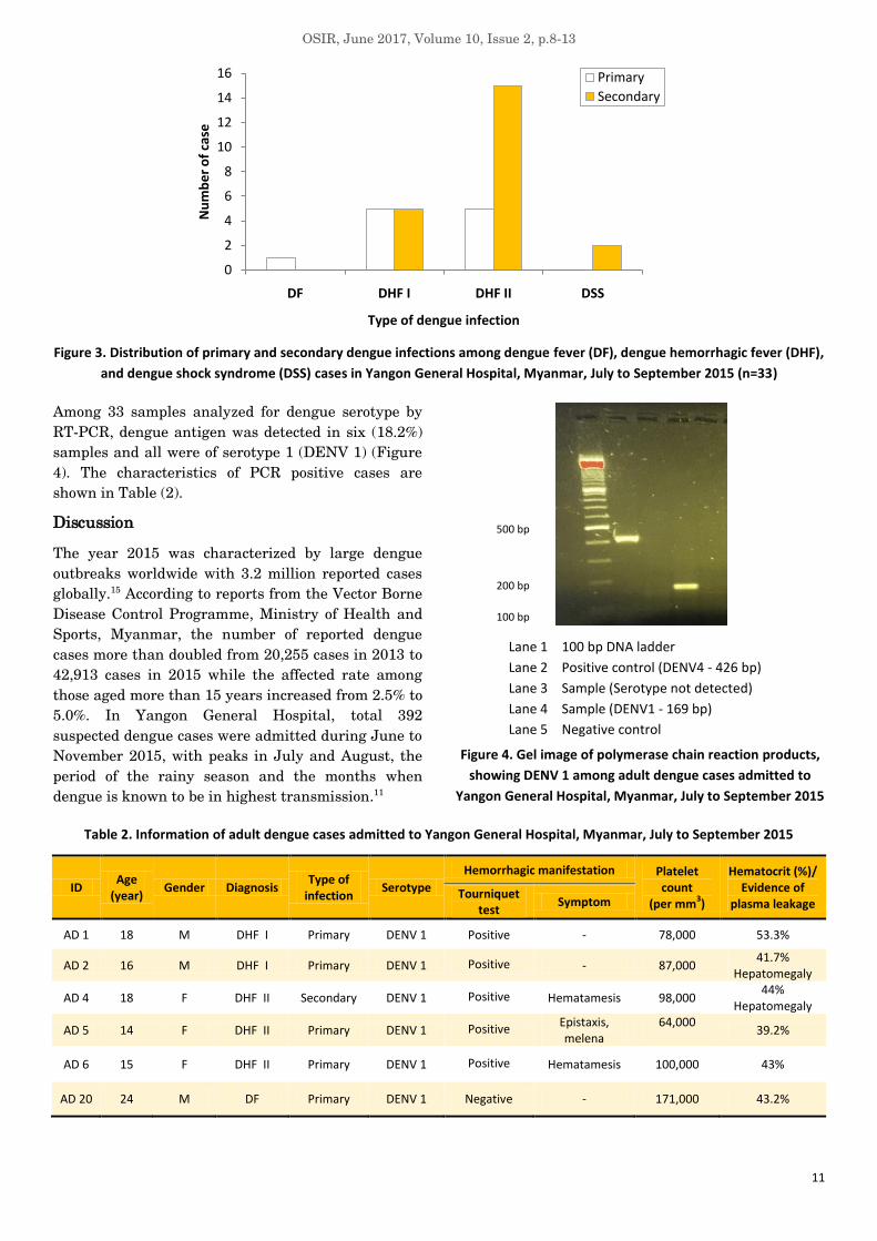
were progressed to DHF I and DHF II (Figure 3). About 77.3% (17/22) of secondary infection and 45.5%
(5/11) of primary infection developed into severe
dengue. There was no significant association between
severity of dengue infection and type of infection
(primary or secondary).
Table 1. Clinical manifestation and laboratory results of
serologically confirmed adult dengue cases in Yangon
General Hospital, Myanmar, July to September 2015 (n=33)
Clinical manifestation Number (%)
Fever 33 (100) Tourniquet test positive 28 (84.8) Headache 20 (60.6) Vomiting 12 (36.4) Skin rash 11 (33.3) Drowsiness 10 (30.3) Muscle and joint pain 9 (27.3) Abdominal pain 8 (24.2) Hematamesis 8 (24.2) Melena 6 (18.2) Epistaxis 4 (12.1) Bleeding gum 2 (6.1) Hemoptysis 1 (3.0) Hepatomegaly 12 (36.4) Platelet count <100,000/mm
3
24 (72.7)
Hematocrit >45% 15 (45.5)
0
20
40
60
80
100
120
140
160
180
Jun Jul Aug Sep Oct Nov
Nu
mb
er
of
case
Month
0
5
10
15
20
25
30
35
40
13-14 15-19 20-24 >24
Male
Female
Pe
rce
nt
Age group (year)
OSIR, June 2017, Volume 10, Issue 2, p.8-13
11
Figure 3. Distribution of primary and secondary dengue infections among dengue fever (DF), dengue hemorrhagic fever (DHF),
and dengue shock syndrome (DSS) cases in Yangon General Hospital, Myanmar, July to September 2015 (n=33)
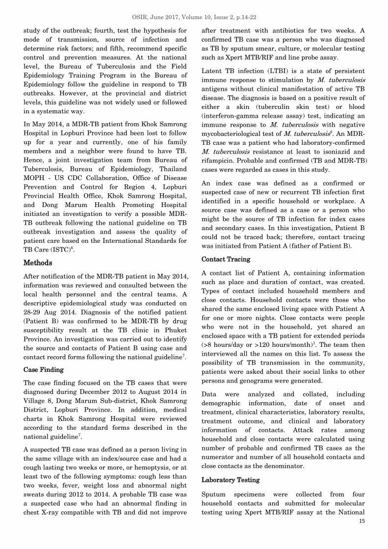
Among 33 samples analyzed for dengue serotype by
RT-PCR, dengue antigen was detected in six (18.2%)
samples and all were of serotype 1 (DENV 1) (Figure
4). The characteristics of PCR positive cases are
shown in Table (2).
Discussion
The year 2015 was characterized by large dengue
outbreaks worldwide with 3.2 million reported cases
globally.15 According to reports from the Vector Borne
Disease Control Programme, Ministry of Health and
Sports, Myanmar, the number of reported dengue
cases more than doubled from 20,255 cases in 2013 to
42,913 cases in 2015 while the affected rate among
those aged more than 15 years increased from 2.5% to
5.0%. In Yangon General Hospital, total 392
suspected dengue cases were admitted during June to
November 2015, with peaks in July and August, the
period of the rainy season and the months when
dengue is known to be in highest transmission.11
Lane 1 100 bp DNA ladder
Lane 2 Positive control (DENV4 - 426 bp)
Lane 3 Sample (Serotype not detected)
Lane 4 Sample (DENV1 - 169 bp)
Lane 5 Negative control
Figure 4. Gel image of polymerase chain reaction products,
showing DENV 1 among adult dengue cases admitted to
Yangon General Hospital, Myanmar, July to September 2015
Table 2. Information of adult dengue cases admitted to Yangon General Hospital, Myanmar, July to September 2015
ID Age
(year) Gender Diagnosis
Type of infection
Serotype
Hemorrhagic manifestation Platelet count
(per mm3)
Hematocrit (%)/ Evidence of
plasma leakage Tourniquet
test Symptom
AD 1 18 M DHF I Primary DENV 1 Positive - 78,000 53.3%
AD 2 16 M DHF I Primary DENV 1 Positive - 87,000 41.7%
Hepatomegaly
AD 4 18 F DHF II Secondary DENV 1 Positive Hematamesis 98,000 44%
Hepatomegaly
AD 5 14 F DHF II Primary DENV 1 Positive Epistaxis, melena
64,000
39.2%
AD 6 15 F DHF II Primary DENV 1 Positive Hematamesis 100,000 43%
AD 20 24 M DF Primary DENV 1 Negative - 171,000 43.2%
0
2
4
6
8
10
12
14
16
DF DHF I DHF II DSS
Nu
mb
er
of
case
Type of dengue infection
Primary
Secondary
500 bp
200 bp
100 bp

OSIR, June 2017, Volume 10, Issue 2, p.8-13
12
Based on serology, secondary infections accounted for
nearly two-thirds of all cases. In dengue endemic
countries like Myanmar where all four dengue
serotypes are co-circulating and the vector Aedes
aegypti are abundant year round, most of the
population might have been infected at least once by
a dengue virus in their childhood. A higher proportion
of secondary dengue infection in adults had also been
reported from other dengue endemic countries such
as Thailand and Sri Lanka.8,16
In this study, the age of confirmed dengue cases
ranged from 13-49 years and the majority (85%) was
in 15-24 year age group. However, another study from
Yangon General Hospital between 2000 and 200812
and one from Pyin Oo Lwin in 200917 revealed lower
attack rates in 15-24 year age group. Therefore, this
study indicated that an increasing number of dengue
infection occurred in economically productive young
adults, a fact that might have significant adverse
financial effects on the community. A previous study
from 12 countries in Southeast Asia, using available
data from 2001-2010, showed an aggregate annual
economic burden of dengue reaching USD 950 million
among the studied nations, with approximately 52%
of these costs coming from productivity loss.18
Fever, headache, vomiting and skin rash were the
most common presentations found in this study with
hematamesis (24.2%) and melena (18.2%). Similarly,
the study from Pyin Oo Lwin revealed that fever and
vomiting were the most common clinical
presentations in adults while hematamesis and
melena was found in 33% and 41% of patients
respectively.12 Therefore, physicians should be aware
that gastrointestinal bleeding may be a common
hemorrhagic manifestation in adults diagnosed with
dengue. Thrombocytopenia is not an uncommon
presentation among adults hospitalized with dengue
infection. Our thrombocytopenia of 72.7% was
comparable to a study in Sri Lanka which reported a
rate of 79%.8
Evidence of an association between sequential dengue
infection and increased risk of more serious disease
has long been reported.19 In this study, 77.3% of
secondary infection developed severe dengue types of
infection, DHF II and DSS, while only 45.5% of severe
dengue was observed among primary infection.
Moreover, 97.0% of cases in this study were DHF and
all DSS cases were also secondary infection.
DENV-1 has been the predominant serotype in
Myanmar since 2009 although the other serotypes
have been identified,20 which was also predominant
during recent years in other countries such as
Thailand, Nepal and Singapore.21-23 Intensive
virological surveillance should be continued to detect
changes of serotypes.
Limitations
The findings of this study might not be fully
representative of the current adult dengue outbreak
due to small number of cases enrolled. Analysis of the
association between severity and serotype was not
possible as only six samples were identified as being
DENV-1. Additional molecular analysis of PCR
negative samples was recommended to ensure that
these were truly negative as different primer
sequences and thermal cycling conditions or further
nucleotide sequencing methods could detect similar
nucleotide sequence of the target virus.
Acknowledgements
We would like to thank Director General and Board of
Directors from the Department of Medical Research
for their permission to conduct this study. We also
appreciate the Medical Superintendent and staff of
Yangon General Hospital for their kind permission to
review the data and recruit the patients, and Dr.
John Aaskov from the Queensland University of
Technology for providing dengue PCR primers.
Suggested Citation
Myat TW, Thu HM, Win HM, Than KS, Tun ZT,
Aye KM, et al. Clinical profile and circulating
dengue virus serotype among adults admitted to
Yangon General Hospital during the 2015
dengue outbreak. OSIR. 2017 Jun;10(2):8-13.
References
1. World Health Organization and the Special
Programme for Research and Training in
Tropical Diseases. Dengue: guidelines for
diagnosis, treatment, prevention and control.
New ed. Geneva: World Health Organization;
2009. p. 137-44.
2. Bhatt S, Gething PW, Brady OJ, Messina JP,
Farlow AW, Moyes CL, et al. The global
distribution and burden of dengue. Nature.
2013 Apr 25;496(7446):504-7. Epub 2013 Apr
7.
3. Beatty M. Global burden of dengue. 2015
[cited 2015 Jan 15].
<http://www.denguewatch.org>.
4. Guha-Sapir D, Schimmer B. Dengue fever:
new paradigms for a changing epidemiology.
Emerg Themes Epidemiol. 2005 Mar 2;2(1):1.
5. Bhatia R, Dash AP and Sunyoto T. Changing
epidemiology of dengue in South-East Asia.

OSIR, June 2017, Volume 10, Issue 2, p.8-13
13
WHO South-East Asia Journal of Public
Health. 2013; 2(1): 23-27.
6. Goh KT. Dengue-a re-emerging infectious
disease in Singapore. Ann Acad Med
Singapore. 1997 Sep;26(5):664-70.
7. Rahman M, Rahman K, Siddque AK, Shoma
S, Kamal AHM, Ali KS, et al. First outbreak
of dengue hemorrhagic fever, Bangladesh.
Emerg Infect Dis. 2002;8(7):738-40.
8. Malavige GN, Velathanthiri VG,
Wijewickrama ES, Fernando S, Jayaratne SD,
Aaskov J, et al. Patterns of disease among
adults hospitalized with dengue infections.
QJM. 2006 May;99(5):299-305. Epub 2006
Apr 7.
9. Thein S, Aung MM, Shwe TN, Aye M, Zaw A,
Aye K, et al. Risk factors in dengue shock
syndrome. Am J Trop Med Hyg. 1997
May;56(5):566-72.
10. Myanmar. Department of Medical Research
(Lower Myanmar). Ministry of Health.
Annual report 2014. Yangon: Department of
Medical Research (Lower Myanmar); 2014.
11. Thein S, Nwe MT, Min H, Sein AK, Tint K,
Nyein K, et al. An outbreak of fever with
haemorrhagic manifestations in children and
young adults in Lashio Township, 1994. Myan
J Cur Med Pract. 1998;2:203-6.
12. Oo AS, Myint MK, Win AS, Khaing MTT,
Maw AM, Aung Y, et al. Clinical profile of
dengue hemorrhagic fever in adults in Pyin
Oo Lwin Hospital in 2009. The Myanmar
Health Sciences Research Journal. 2013;24
(1):1-6.
13. World Health Organization. Dengue
hemorrhagic fever: diagnosis, treatment and
control. 1997 [cited 2016 Jan 23].
<http://apps.who.int/iris/bitstream/10665/419
88/1/9241545003_eng.pdf>.
14. Seah CLK, Chow VTK, Tan HC, Chan YC.
Rapid, single-step RT-PCR typing of dengue
viruses using five NS3 gene primers. J Virol
Methods. 1995;51(2-3):193-200.
15. World Health Organization. Dengue and
severe dengue. 2016 [cited 2016 Jan 23].
<http://www.who.int/mediacentre/factsheets/f
s117/en/>.
16. Wichmann O, Hongsiriwon S,
Bowonwatanuwong C, Chotivanich K,
Sukthana Y, Pukrittayakamee S. Risk factors
and clinical features associated with severe
dengue infections in adults and children
during the 2001 epidemic in Chonburi,
Thailand. Trop Med Int Health. 2004
Sep;9(9):1022-9.
17. Thein S, Mauk KKA. Dengue hemorrhagic
fever in Adults. Myanmar Journal of Current
Medical Practice. 2010;14:7-8.
18. Shepard DS, Undurraga EA, Halasa YA.
Economic and disease burden of dengue in
Southeast Asia. PLoS Negl Trop Dis. 2013;7
(2):e2055. Epub 2013 Feb 21.
19. Halstead SB, Nimmannitya S, Yamarat C,
Russel PK. Hemorhagic fever in Thailand;
recent knowledge regarding etiology. Jpn J
Med Sci Biol. 1967 Dec;20 Suppl:96-103.
20. Myat TW. Inter-host and intra-host genetic
diversity of dengue virus strains in children
with dengue infection [thesis]. Yangon:
University of Medicine 2, Yangon. 2013.
21. Fried JR, Gibbons RV, Kalayanarooj S,
Thomas SJ, Srikiatkhachorn A, Yoon IK, et
al. Serotype-specific differences in the risk of
dengue hemorrhagic fever: an analysis of data
collected in Bangkok, Thailand from 1994 to
2006. PLoS Negl Trop Dis. 2010 Mar
2;4(3):e617.
22. Pandey BD, Nabeshima T, Pandey K,
Rajendra SP, Shah Y, Adhikari BR, et al.
First isolation of dengue virus from the 2010
epidemic in Nepal. Trop Med Health. 2013
Sep;41(3):103-11.
23. Herriman R. Asia dengue fever update:
Philippines, Malaysia, Thailand, Vietnam and
Singapore. 2015 Jul 3 [cited 2016 June 20].
<http://outbreaknewstoday.com/asia-dengue-
fever-update-philippines-malaysia-thailand-
vietnam-and-singapore-16816/>.

OSIR, June 2017, Volume 10, Issue 2, p.14-22
14
Identification of a Tuberculosis Cluster through Epidemiological and
Geographical Tracing of a Patient with Multidrug-resistant Tuberculosis in
Lopburi Province, Thailand, 2014
Kaewalee Soontornmon1,*, Yin Myo Aye2, Namhwan Phankhor3, Supaporn Watanatorn4, Wilailuck
Modmoltin5, Chuleeporn Jiraphongsa2
1 Bureau of Tuberculosis, Department of Disease Control, Ministry of Public Health, Thailand
2 Field Epidemiological Training Program, Bureau of Epidemiology, Department of Disease Control,
Ministry of Public Health, Thailand
3 Khok Samrong Hospital, Lopburi Province, Thailand
4 Office of Disease Prevention and Control Region 4, Saraburi Province, Thailand
5 Provincial Health Office, Lopburi Province, Thailand
*Corresponding author, email address: [email protected]
Abstract
In May 2014, a suspected multidrug-resistant tuberculosis (MDR-TB) outbreak in Lopburi Province was investigated
following the national guidelines for tuberculosis (TB) outbreak investigation and assessed the quality of patient care based
on the International Standards for TB Care. The case finding focused on TB cases diagnosed during December 2012 to
August 2014. Medical charts were reviewed at Khok Samrong Hospital and contacts of a MDR-TB case who was lost to
follow up were traced back. Study findings found an epidemiologically linked cluster of TB cases with five geographically
related cases and four cases were from the same family. Factors that might have contributed to this TB outbreak were
identified as well, including delay in diagnosis and sub-standard care, low socioeconomic status, delay in conducting contact
tracing, and an ineffective TB database system. Diagnosis, treatment and prevention activities should be improved to
prevent further TB outbreaks in the communities.
Keywords: tuberculosis, multidrug-resistant, contact tracing, quality of care, Thailand
Introduction
Tuberculosis (TB) is an airborne infectious disease
that can be transmitted by the bacterium
Mycobacterium tuberculosis.1 In 2015, Thailand was
ranked in the top 22 high TB burden countries.2
Multidrug-resistant TB (MDR-TB) is caused by a TB
bacterium that is resistant to at least isoniazid and
rifampicin, the two most potent first-line drugs for TB
infection.3 According to the information from
Supranational Reference Laboratory in Thailand,
MDR-TB was found among 2.0% of new cases and
18.8% of previously treated cases in 2012.4
During 2005, among immigrants in the United States,
four MDR-TB cases who were Hmong refugees
migrated from a refugee camp in Lopburi Province of
Thailand were identified. Tracing back and screening
of 15,455 refugees in the camp resulted in 272 TB
cases; of which, 24 (42.1%) out of 57 samples were
MDR-TB.5 Following another MDR-TB outbreak in
2010 which affected 15 cases in a community from the
western part of Thailand6, the first national guideline
for investigation of TB outbreaks was developed. The
guideline recommends performing an investigation
when there are at least two TB patients who share
the same place or activity during a 3-month period; or
at least one new or relapse MDR-TB case; or at least
one extensively drug-resistant TB (XDR-TB) case in
the community.7 The guideline also suggests five
steps for completing an investigation of a TB
outbreak: first, perform a case review for diagnosis
and outbreak verification; second, identify source
and/or contact cases, and collect laboratory and
environmental samples; third, conduct a descriptive
OSIR, June 2017, Volume 10, Issue 2, p.14-22
15
study of the outbreak; fourth, test the hypothesis for
mode of transmission, source of infection and
determine risk factors; and fifth, recommend specific
control and prevention measures. At the national
level, the Bureau of Tuberculosis and the Field
Epidemiology Training Program in the Bureau of
Epidemiology follow the guideline in respond to TB
outbreaks. However, at the provincial and district
levels, this guideline was not widely used or followed
in a systematic way.
In May 2014, a MDR-TB patient from Khok Samrong
Hospital in Lopburi Province had been lost to follow
up for a year and currently, one of his family
members and a neighbor were found to have TB.
Hence, a joint investigation team from Bureau of
Tuberculosis, Bureau of Epidemiology, Thailand
MOPH - US CDC Collaboration, Office of Disease
Prevention and Control for Region 4, Lopburi
Provincial Health Office, Khok Samrong Hospital,
and Dong Marum Health Promoting Hospital
initiated an investigation to verify a possible MDR-
TB outbreak following the national guideline on TB
outbreak investigation and assess the quality of
patient care based on the International Standards for
TB Care (ISTC)8.
Methods
After notification of the MDR-TB patient in May 2014,
information was reviewed and consulted between the
local health personnel and the central teams. A
descriptive epidemiological study was conducted on
28-29 Aug 2014. Diagnosis of the notified patient
(Patient B) was confirmed to be MDR-TB by drug
susceptibility result at the TB clinic in Phuket
Province. An investigation was carried out to identify
the source and contacts of Patient B using case and
contact record forms following the national guideline7.
Case Finding
The case finding focused on the TB cases that were
diagnosed during December 2012 to August 2014 in
Village 8, Dong Marum Sub-district, Khok Samrong
District, Lopburi Province. In addition, medical
charts in Khok Samrong Hospital were reviewed
according to the standard forms described in the
national guideline7.
A suspected TB case was defined as a person living in
the same village with an index/source case and had a
cough lasting two weeks or more, or hemoptysis, or at
least two of the following symptoms: cough less than
two weeks, fever, weight loss and abnormal night
sweats during 2012 to 2014. A probable TB case was
a suspected case who had an abnormal finding in
chest X-ray compatible with TB and did not improve
after treatment with antibiotics for two weeks. A
confirmed TB case was a person who was diagnosed
as TB by sputum smear, culture, or molecular testing
such as Xpert MTB/RIF and line probe assay.
Latent TB infection (LTBI) is a state of persistent
immune response to stimulation by M. tuberculosis
antigens without clinical manifestation of active TB
disease. The diagnosis is based on a positive result of
either a skin (tuberculin skin test) or blood
(interferon-gamma release assay) test, indicating an
immune response to M. tuberculosis with negative
mycobacteriological test of M. tuberculosis8. An MDR-
TB case was a patient who had laboratory-confirmed
M. tuberculosis resistance at least to isoniazid and
rifampicin. Probable and confirmed (TB and MDR-TB)
cases were regarded as cases in this study.
An index case was defined as a confirmed or
suspected case of new or recurrent TB infection first
identified in a specific household or workplace. A
source case was defined as a case or a person who
might be the source of TB infection for index cases
and secondary cases. In this investigation, Patient B
could not be traced back; therefore, contact tracing
was initiated from Patient A (father of Patient B).
Contact Tracing
A contact list of Patient A, containing information
such as place and duration of contact, was created.
Types of contact included household members and
close contacts. Household contacts were those who
shared the same enclosed living space with Patient A
for one or more nights. Close contacts were people
who were not in the household, yet shared an
enclosed space with a TB patient for extended periods
(>8 hours/day or >120 hours/month)1. The team then
interviewed all the names on this list. To assess the
possibility of TB transmission in the community,
patients were asked about their social links to other
persons and genograms were generated.
Data were analyzed and collated, including
demographic information, date of onset and
treatment, clinical characteristics, laboratory results,
treatment outcome, and clinical and laboratory
information of contacts. Attack rates among
household and close contacts were calculated using
number of probable and confirmed TB cases as the
numerator and number of all household contacts and
close contacts as the denominator.
Laboratory Testing
Sputum specimens were collected from four
household contacts and submitted for molecular
testing using Xpert MTB/RIF assay at the National

OSIR, June 2017, Volume 10, Issue 2, p.14-22
16
TB reference laboratory in Bangkok. The Xpert
MTB/RIF assay was approved by the World Health
Organization in 2013 to diagnose pulmonary and
extra pulmonary specimens in adults and children9.
This assay is a rapid and polymerase chain reaction
(PCR)-based TB diagnostic test that was being
incorporated into the recommendations for diagnosing
MDR-TB in programmatic settings of Thailand.
The Xpert MTB/RIF can be used to identify both the
presence of M. tuberculosis and rifampicin resistance
in less than two hours. It has high sensitivity and
specificity for both smear-positive and smear-negative
diseases.10,11 Any specimen diagnosed as rifampicin-
resistant TB by Xpert MTB/RIF would additionally be
confirmed as MDR-TB by line probe assay at the
national TB reference laboratory.
Line probe assay is a deoxyribonucleic acid strip
assay that uses PCR and hybridization to detect
genetic mutations in specific genes that confer
rifampicin-resistance and isoniazid-resistance from M.
tuberculosis cultured isolates and acid-fast bacilli
(AFB) smear positive specimens. A published case
series of TB patients in Thailand confirmed that
mutations in the rpoB gene were specific to
rifampicin-resistance.12 Resistance to isoniazid has
been demonstrated to be significantly associated with
mutations in the katG, inhA and ahpC genes.13 It has
been proven to diagnose MDR-TB within 24 hours in
several controlled laboratory studies.
Assessing Quality of Care
Informal interviews were carried out with Patient B’s
family members and neighbors about their treatment
experiences, how they took medication and cared for
themselves, and basic knowledge of TB. Information
obtained from interview and records review were
used to assess the quality of care by four dimensions
(diagnosis, treatment, TB co-infection with human
immunodeficiency virus (HIV) and other co-morbid
conditions, and public health and prevention) as
stated in the ISTC guidelines14 as well as explore the
impact of TB on socioeconomic status.
At the end of the investigation, preliminary results
were disseminated and practical recommendations
were provided to the local health care staff and all
stakeholders. A report was also submitted to the
Provincial Health Office and the Department of
Disease Control, Ministry of Public Health.
Results
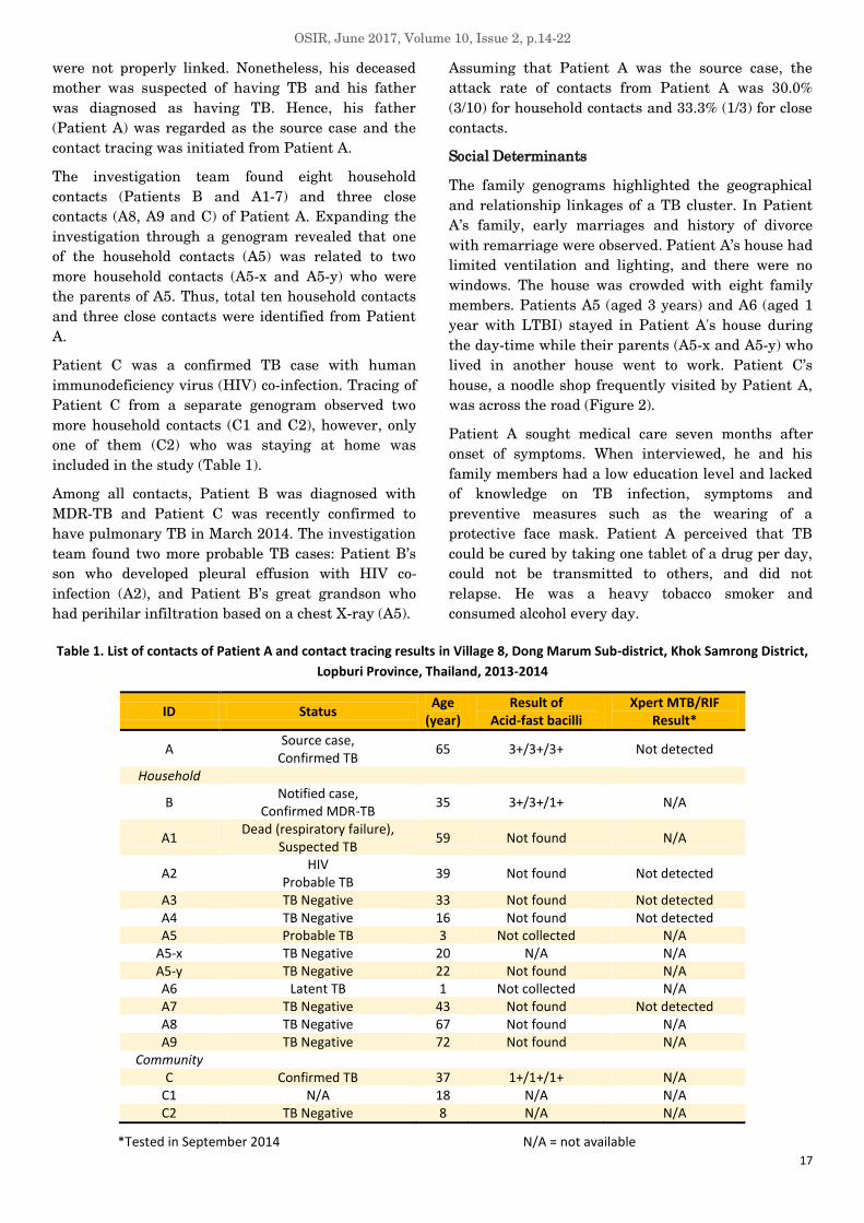
The case investigation revealed five TB cases from a
cluster with epidemiological and geographical linkage,
including three confirmed cases (Patients A, B and C)
identified by reviewing medical records in the TB
clinic of Khok Samrong Hospital during February
2013 and June 2014, and two more probable cases
discovered in Patient A’s family through contact
tracing. Evidence from the investigation implied that
Patient A was likely to be the source and spread the
disease to the other four cases as he was the first one
who developed the symptoms one year earlier than
others (Figure 1).
Case Description
Patient A was the first TB case in this outbreak. He
started to have TB symptoms while staying with his
son (Patient B) in Phuket during August 2012.
Patient A returned to Lopburi in February 2013, and
was diagnosed with TB and received treatment there.
In January 2014, Patient B was diagnosed with TB
and started treatment in Phuket. After receiving
treatment for two months, results of his sputum AFB
still revealed 3+. Patient B went to Lopburi during
the second month of TB treatment to visit his mother
who developed acute respiratory failure with
suspected TB and eventually died. After his mother’s
funeral, Patient B returned to Phuket, yet did not
visit the hospital again for TB treatment.
Contact Tracing
As Patient B’s sputum was tested to have M.
tuberculosis resistant to isoniazid and rifampicin, TB
clinic staff in Phuket tried to contact him. However,
he could not be traced back for further investigation
since the TB database systems across the country
Figure 1. Timeline of confirmed (Patients A, B and C) and probable (Patients A2 and A5) TB cases in Village 8,
Dong Marum Sub-district, Khok Samrong District, Lopburi Province, Thailand, 2012-2014
OSIR, June 2017, Volume 10, Issue 2, p.14-22
17
were not properly linked. Nonetheless, his deceased
mother was suspected of having TB and his father
was diagnosed as having TB. Hence, his father
(Patient A) was regarded as the source case and the
contact tracing was initiated from Patient A.
The investigation team found eight household
contacts (Patients B and A1-7) and three close
contacts (A8, A9 and C) of Patient A. Expanding the
investigation through a genogram revealed that one
of the household contacts (A5) was related to two
more household contacts (A5-x and A5-y) who were
the parents of A5. Thus, total ten household contacts
and three close contacts were identified from Patient
A.
Patient C was a confirmed TB case with human
immunodeficiency virus (HIV) co-infection. Tracing of
Patient C from a separate genogram observed two
more household contacts (C1 and C2), however, only
one of them (C2) who was staying at home was
included in the study (Table 1).
Among all contacts, Patient B was diagnosed with
MDR-TB and Patient C was recently confirmed to
have pulmonary TB in March 2014. The investigation
team found two more probable TB cases: Patient B’s
son who developed pleural effusion with HIV co-
infection (A2), and Patient B’s great grandson who
had perihilar infiltration based on a chest X-ray (A5).
Assuming that Patient A was the source case, the
attack rate of contacts from Patient A was 30.0%
(3/10) for household contacts and 33.3% (1/3) for close
contacts.
Social Determinants
The family genograms highlighted the geographical
and relationship linkages of a TB cluster. In Patient
A’s family, early marriages and history of divorce
with remarriage were observed. Patient A’s house had
limited ventilation and lighting, and there were no
windows. The house was crowded with eight family
members. Patients A5 (aged 3 years) and A6 (aged 1
year with LTBI) stayed in Patient A's house during
the day-time while their parents (A5-x and A5-y) who
lived in another house went to work. Patient C’s
house, a noodle shop frequently visited by Patient A,
was across the road (Figure 2).
Patient A sought medical care seven months after
onset of symptoms. When interviewed, he and his
family members had a low education level and lacked
of knowledge on TB infection, symptoms and
preventive measures such as the wearing of a
protective face mask. Patient A perceived that TB
could be cured by taking one tablet of a drug per day,
could not be transmitted to others, and did not
relapse. He was a heavy tobacco smoker and
consumed alcohol every day.
Table 1. List of contacts of Patient A and contact tracing results in Village 8, Dong Marum Sub-district, Khok Samrong District,
Lopburi Province, Thailand, 2013-2014
ID Status Age
(year) Result of
Acid-fast bacilli Xpert MTB/RIF
Result*
A Source case,
Confirmed TB 65 3+/3+/3+ Not detected
Household
B Notified case,
Confirmed MDR-TB 35 3+/3+/1+ N/A
A1 Dead (respiratory failure),
Suspected TB 59 Not found N/A
A2 HIV
Probable TB 39 Not found Not detected
A3 TB Negative 33 Not found Not detected A4 TB Negative 16 Not found Not detected A5 Probable TB 3 Not collected N/A
A5-x TB Negative 20 N/A N/A A5-y TB Negative 22 Not found N/A A6 Latent TB 1 Not collected N/A A7 TB Negative 43 Not found Not detected A8 TB Negative 67 Not found N/A A9 TB Negative 72 Not found N/A
Community C Confirmed TB 37 1+/1+/1+ N/A
C1 N/A 18 N/A N/A C2 TB Negative 8 N/A N/A
*Tested in September 2014 N/A = not available

OSIR, June 2017, Volume 10, Issue 2, p.14-22
18
Figure 2. Genograms of tuberculosis cases and their
contacts in Village 8, Dong Marum Sub-district,
Khok Samrong District, Lopburi Province, Thailand, 2014
Quality of Care
Quality of patient care was assessed for five TB cases
(A, B, C, A2 and A5) in this outbreak. Strengths and
weaknesses in the quality of care linked to the ISTC
guidelines were observed and compared (Table 2). A
delay in diagnosis of TB was found for Patient A and
diagnostic testing such as gastric aspirate was not
performed to confirm TB in children under five (A5).
Taking a sub-optimal dosage of anti-TB drugs by
Patient A and misconceptions about TB care were
identified as well.
At the end of second and third months of treatment,
Patient B had a positive result for M. tuberculosis by
culture, and resistance to isoniazid and rifampicin by
drug susceptibility testing while Patient A had no
growth in culture. Patient A was treated for eight
months before being cured; however, Patient B
traveled to Phuket and Chumphon Provinces, and
refused to receive any treatment. In February 2015,
Patient B was admitted to Chumphon Hospital with
pneumonia and started treatment as an MDR-TB
patient in March 2015.
From public health and prevention perspectives,
although the staff in the TB clinic conducted the
second step of the investigation by interviewing
Patient A about household contacts in his family, they
did not complete the clinical evaluation process at the
very beginning. However, they performed all steps of
contact investigation by June 2014. For TB infection
control in Khok Samrong Hospital, the TB clinic was
separated from the general out-patient department.
There was also a ‘fast track’ infection control system
in the TB clinic for suspected patients. Moreover, all
TB cases in this hospital were recorded in an
electronic database system and reported in a timely
manner.
Discussion
This outbreak investigation revealed an
epidemiologically linked cluster of TB cases. Out of
five geographically related cases identified, four cases
were from the same family. We found the practices
that did not follow the ISTC guidelines which
negatively impacted on the quality of patient care.
Potential factors contributing to this community TB
outbreak were delay in diagnosis of the source case,
sub-standard care, low socioeconomic status, delay in
conducting contact tracing, and an ineffective
national TB database system. Elimination of these
contributing factors could help TB-related health
personnel achieving their ultimate goal to eliminate
TB in the community.
The source case spent six months in a private clinic
before being diagnosed with TB. This delay in
diagnosis and sub-standard care led to a prolonged
period of possible TB transmission. Furthermore, no
specimens were collected from the source's two
grandchildren, who were diagnosed with probable TB
and latent TB infection, and no pediatric anti-TB
drug was given to them. Sputum specimens of the
source's contacts were sent to Bangkok for Xpert
MTB/RIF testing. As Thailand is a middle income
country, case finding and diagnosis remain major
challenges to TB control. In 2013, the Global Fund
and National Health Security Office supported the
use of Xpert MTB/RIF and line probe assay for early
detection of M. tuberculosis and diagnosis of drug
resistance in high risk MDR-TB groups, people living
with HIV/AIDS and MDR-TB suspected cases1. The
Xpert MTB/RIF is a rapid test and should be installed
at or near the point of care.
However, many challenges existed in district
hospitals with limited staff and infrastructure. We
recommended the Ministry of Public Health to ensure
a good logistics for accurate diagnoses and proper
management of TB in district hospitals by allocating
up-to-date equipment in TB laboratories.
Therapeutically, there is no evidence to support the
efficacy of taking rifampicin for three times a week as
recommended by WHO. In addition, a sub-optimal
TB case
Deceased
Divorced
One household each
OSIR, June 2017, Volume 10, Issue 2, p.14-22
19
Table 2. Comparison between the guideline and actual practices during a tuberculosis outbreak in Village 8,
Dong Marum Sub-district, Khok Samrong District, Lopburi Province, Thailand, 2013-2014
Guideline from the International Standards for Tuberculosis Care
14
Actual practice
in a tuberculosis outbreak
Diagnosis (standard 1-3 and 6)
To ensure early diagnosis, all clinically suspect patients, including children, should be evaluated for tuberculosis (TB).
Patient A did not seek appropriate health care and the provider at a private clinic did not suspect the disease for six months, leading to ongoing transmission.
All patients should have at least two specimens for smear microscopy or one specimen for Xpert MTB/RIF, and all children should have bacteriological confirmation.
All adult patients had at least two specimens. However, no specimen was tested for a pediatric case (Patient A5) since health staff were not familiar with collecting specimens from children.
Treatment (standard 7-11 and 13)
Prescribe an appropriate regimen. The dose and regimen of anti-TB drugs should conform to the recommendation from World Health Organization.
All adult patients received an appropriate regimen from health care staff (fixed does combination of HRZE three tablets/day). Patient A5 took rifampicin 25 mg/kg/day three times weekly.
A patient-centered approach to treatment should be developed in order to promote adherence, improve quality of life and relief suffering.
Patients lived with poverty and lack of basic knowledge on TB disease. Though Patient A took medicine every day, he took only one tablet which was considered as sub-optimal.
Response to treatment should be monitored by sputum smear microscopy at the time of completion of the initial phase. Smear-positive cases should be assessed for drug resistance.
Regular follow up with smear microscopy was performed. At the end of third month, if acid-fast bacilli (AFB) was still positive, sputum was sent for culture.
Records of all medication given, bacteriological response, outcomes and adverse reactions should be accessible and systematically maintained.
There were individual folders for each patient and each contained complete information.
Addressing human immunodeficiency virus (HIV) infection and co-morbid conditions (standard 14-15 and 17)
All providers should conduct a thorough assessment for co-morbid conditions and other factors that could affect TB treatment response or outcome.
Patient A had alcohol dependence and was a heavy tobacco smoker.
HIV testing and counseling should be conducted for all patients. For all patients with HIV and TB, antiretroviral therapy should be initiated within two months.
Patient C received anti-retroviral therapy within two weeks after confirmation for HIV infection by laboratory result.
Public health and prevention (standard 18-21)
Persons in close contact with patients who have infectious TB should be evaluated and managed in line with the recommendation.
There was no evaluation for contact cases after Patient A was diagnosed with TB.
Children less than five years of age who are close contacts of a person with infectious TB, and who do not have active TB should be treated as presumed latent TB infection.
Children under five were evaluated and treated for TB and latent TB infection, but no pediatric formula was provided.
Each health care facility should develop and implement an appropriate TB infection control plan to minimize possible transmission.
The TB clinic was isolated from the general out-patient department for minimizing possible transmission.
Providers must report about new and relapse cases, and their treatment outcomes to local public health authorities.
Neat and systematic recording and reporting was observed.
HRZE = Isoniazid, rifampicin, pyrazinamide and ethambutol
OSIR, June 2017, Volume 10, Issue 2, p.14-22
20
dosage of anti-TB drugs can lead to drug-resistant
strains of M. tuberculosis. These problems suggest
that both public and private health care systems need
more supervision in TB diagnosis, especially in
obtaining gastric aspirate in children who have close
contact with MDR-TB cases for identifying their drug
resistant status. Members of a family with a low
socioeconomic status, living in congested and crowded
conditions with poor ventilation, and having personal
behaviors of smoking may increase susceptibility to
TB as well as more negative treatment outcomes15.
In 2012, WHO stated that a person with TB could
infect up to 10-15 people through close contact over
the course of a year.16 For MDR-TB transmission, a
study on household contacts suggested that
circulating MDR-TB strains in Peru were less likely
to result in the disease among household contacts
compared to drug-sensitive strains.17 A mathematical
model demonstrated that even when the most fit
MDR-TB strain was assumed to be less fit than the
drug-sensitive strain. However, the MDR strain
would eventually outcompete the drug-susceptible
strain.18 In our study, the delay in conducting contact
tracing increased the likelihood of disease
transmission from Patient A. He had only been asked
about the number of members in his family and none
had been contacted for a clinical evaluation until his
son (Patient B) was diagnosed with MDR-TB merely
16 months later. Due to this time lapse, the
investigation team could not find additional TB cases
and this might have potentially promoted further
transmission. Treatment of contacts with latent TB
infection is recommended for individuals at increased
risk of developing TB such as those with HIV.19
Therefore, actions should be taken to limit the
transmission of not only drug-sensitive strains, but
also drug-resistant strains by conducting contact
tracing as early as possible and giving treatment to
TB cases as a prevention measures.
Lastly, from public health and prevention
perspectives, ineffectiveness in the national TB
database system allowed Patient B to avoid receiving
treatment. At the time of his initial diagnosis, he was
not required by law to receive treatment. The stand-
alone database system, which was not linked with
other surveillance databases, was a major reason
behind the fact that other hospitals were not
informed about MDR-TB. Implementation of a
national-linked database system would enable health
care providers to aware of the patient’s status and
trace the history of all suspected TB cases. Policies
and laws should be advocated for regulation of
persons with serious communicable diseases such as
MDR-TB.
Limitations
Two TB cases (Patients B and A2) could not be traced
or interviewed. Thus, identification of their contacts
was not possible. This probably resulted in under-
reporting of actual TB cases in this outbreak. In
addition, we used clinical criteria to diagnose TB in
two patients (A2 and A5) since the sputum result was
cultured negative for one and could not be collected
from the other. However, based on the guidelines of
Thai National TB Control Programme in 20131,
pulmonary TB could be diagnosed with a negative or
unknown smear result. In the process of contact
tracing, sputum samples from four contacts were sent
for molecular testing using Xpert MTB/RIF in order
to increase the sensitivity of TB diagnosis and early
detection of resistant to rifampicin if TB developed.
Public Health Recommendations
Our study suggested that there was an opportunity to
prevent future TB outbreaks by addressing the short
and long-term public health responses to TB. This
included improving access to care, upgrading TB
laboratory equipment in clinics and hospitals,
improving adherence to TB follow up, and advocating
an inclusive and shared national database system for
tracing TB patients in Thailand. To reduce the
transmission of M. tuberculosis, the following specific
interventions should be implemented. First,
community awareness could be raised by promoting
social campaigns on important occasions such as the
World TB Day. Moreover, regular trainings for all
health personnel from both public and private sectors
should be provided to improve the quality of care.
Conducting a contact investigation for every TB case,
especially for MDR-TB, should be the first priority for
TB clinics in order to receive accreditation. Lastly, a
national database for TB, based on the current
system for the National AIDS Programming which
facilitates health personnel to track patients for early
treatment and follow-up, should be established.
Acknowledgements
The authors would like to thank the staff from Field
Epidemiology Training Program, Thailand. We also
appreciate Patama Monkongdee, Michael Martin and
TB program staff from the Thailand MOPH – US
CDC Collaboration (TUC) for editing the laboratory
information. Additionally, we acknowledge the
support of the staff from Office of Disease Prevention
and Control for Region 4, Lopburi Province, Khok

OSIR, June 2017, Volume 10, Issue 2, p.14-22
21
Samrong Hospital and Dong Marum Health
Promoting Hospital. We are also grateful to Dorothy
L. Southern (US CDC) for reviewing and editing this
manuscript.
Suggested Citation
Soontornmon K, Aye YM, Phankhor N,
Watanatorn S, Modmoltin W, Jiraphongsa C.
Identification of a tuberculosis cluster through
epidemiological and geographical tracing of a
patient with multidrug resistant tuberculosis in
Lopburi Province, Thailand, 2014. OSIR. 2017
Jun;10(2):14-22.
References
1. Thailand. Bureau of Tuberculosis,
Department of Disease Control. Ministry of
Public Health. National tuberculosis control
programme guidelines, 2013. 2nd ed.
Bangkok: The War Veterans Organization of
Thailand under Royal Patronage of His
Majesty the King; 2013. Thai.
2. World Health Organization. Global
tuberculosis report 2015. Geneva: World
Health organization; 2015 [cited 2016 Sep 1].
<http://www.who.int/tb/publications/global_re
port/gtbr15_main_text.pdf>.
3. Caminero JA, editor. Guidelines for clinical
and operational management of drug-
resistant tuberculosis. Paris: International
Union against Tuberculosis and Lung Disease;
2013 [cited 2016 Sep 1].
<http://www.theunion.org/what-we-
do/publications/technical/english/mdr-
tbguide_6-19-13_web.pdf>.
4. Reechaipichitkul W, editor. Guideline for
programmatic management of drug-resistant
tuberculosis. Bangkok: The Agricultural
Cooperative Federation of Thailand Ltd;
2013.p.1-6. Thai [cited 2016 Sep 1].
<http://www.tbnfmthailand.org/Download/DR-
TB%20Proof%2023%20FINAL.pdf>.
5. Oeltmann JE, Varma JK, Ortega L, Liu Y,
O’Rourke T, Cano M, et al. Multidrug-
resistant tuberculosis outbreak among US-
bound Hmong Refugees, Thailand, 2005.
Emerg Infect Dis. 2008;14(11):1715-21.
6. Jiraphonsa C, Wangteeraprasert T,
Henpaserttae N, Sanguanwongse N, Panya L,
Sukkasitvanichkul J, et al. Community
outbreak of multidrug resistance tuberculosis,
Kanchanaburi Province, Thailand on 2002-
June 2010. Journal of Preventive Medicine
Association of Thailand. 2011;1(3):261-71.
Thai [cited 2016 Sep 1].
<http://thailand.digitaljournals.org/index.php/
JOMAT/article/view/12756>.
7. Thailand. Bureau of Tuberculosis.
Department of Disease Control. Ministry of
Public Health. Guideline of TB outbreak
investigation. 2nd ed. Bangkok: The War
Veterans Organization of Thailand under
Royal Patronage of His Majesty the King;
2013. Thai [cited 2016 Sep 1].
<http://www.tbthailand.org/document.php?id=
67>.
8. World Health Organization. Guidelines on the
management of latent tuberculosis infection.
Spain: WHO Press; 2015 [cited 2016 Sep 1].
<http://www.who.int/tb/publications/latent-
tuberculosis-infection/en/>.
9. World Health Organization. Automated real-
time nucleic acid and amplification technology
for rapid and simultaneous detection of
tuberculosis and rifampicin resistance: Xpert
MTB/RIF assay for diagnosis of pulmonary
and extra-pulmonary TB in adults and
children. Geneva: World Health Organization;
2013 [cited 2016 Sep 1].
<http://apps.who.int/iris/bitstream/10665/112
472/1/9789241506335_eng.pdf>.
10. Boehme CC, Nabeta P, Hillemann D, Nicol
MP, Shenai S, Krapp F, et al. Rapid
molecular detection of tuberculosis and
rifampin resistance. N Engl J
Med.2010;363:1005-15.
11. Boehme CC, Nicol MP, Nabeta P, Michael JS,
Gotuzzo E, Tahirli R, et al. Feasibility,
diagnostic accuracy, and effectiveness of
decentralised use of the Xpert MTB/RIF test
for diagnosis of tuberculosis and multidrug
resistance: a multicentre implementation
study. Lancet. 2011 Apr 30;377(9776):1495-
505. Epub 2011 Apr 18.
12. Vattanaviboon P, Sukchawalit R,
Jearanaikoon P, Chuchottaworn C,
Ponglikitmongkol M. Analysis of RNA
polymerase gene mutation in three isolates of
rifampicin resistant Mycobacterium
tuberculosis. Southeast Asian J Trop Med
Public Health. 1995;26 Suppl 1:333-6.
13. Hillemann D, Rusch-Gerdes S, Richter E.
Evaluation of the GenoType MTBDRplus
OSIR, June 2017, Volume 10, Issue 2, p.14-22
22
Assay for rifampin and isoniazid
susceptibility testing of Mycobacterium
tuberculosis strains and clinical specimens. J
Clin Microbiol. 2007;45:2635-4015.
14. TB CARE I. International Standards for
Tuberculosis Care. 3rd ed. The Hague: TB
CARE I; 2014 [cited 2016 Sep 1].
<http://www.who.int/tb/publications/ISTC_3rd
Ed.pdf>.
15. Lönnroth K, Williams BG, Stadlin S,
Jaramillo E, Dye C. Alcohol use as a risk
factor for tuberculosis–a systematic review.
BMC Public Health. 2008 Aug 14;8:289.
16. World Health Organization. TB fact sheets
2016 [cited 2016 Dec 30].
<http://www.who.int/mediacentre/factsheets/f
s104/en/>.
17. Grandjean L, Gilman RH, Martin L, Soto E,
Castro B, Lopez S, et al. Transmission of
multidrug-resistant and drug-susceptible
tuberculosis within households: a prospective
cohort study. PLOS Medicine. 2015;12(6):
e1001843.
18. Cohen T, Murray M. Modeling epidemics of
multidrug-resistant M. tuberculosis of
heterogeneous fitness. Nature Medicine.
2004;10(10):1117-21.
19. Centers for Disease Control and Prevention.
Guidelines for the investigation of contacts of
persons with infectious tuberculosis;
recommendations from the National
Tuberculosis Controllers Association and
CDC. MMWR. 2005;54(RR15):1-37.

OSIR, June 2017, Volume 10, Issue 2, p.23-27
23
The Grammar of Science: Let’s ‘Log’ (Part 1)
Jaranit Kaewkungwal*
Mahidol University, Thailand
* Corresponding author, email address: [email protected]
No. I am not asking you to go “Rock’n Roll’ with me at
the “Let’s Rock: The Retro Festival” in UK. And I am
not asking you to go cut down trees in the forest. But
I would like you to reflect back to the “logarithm”
function in mathematics and statistics that you
learned in your high school days. Many people get
blocked out and start to not like statistics or
mathematics when they see numbers get converted
into “logarithm”. Why do we have to do it in “log-
scale”? Why not just use or analyze data in simple
numbering scale? There are reasons behind this. And
it is as fun as going rock and roll at the concert!
Back to basic - “Logarithm” is rockin!
You may think mathematics is fixed and full of
unchanging rules and truths. But it may not be so. As
an example of the “logarithm” you will be surprised
that modern mathematicians will give very different
definitions from the mathematicians several
centuries ago1. If we go back through the history of
“logarithm”, we will see that the concept and methods
had been evolved over time. Historically, the concept
of logarithm was independently invented in the 17th
century by at least two mathematicians, the Scottish
John Napier (1550-1617) and the Swiss Joost Bürgi
(1552-1632). In summary, logarithm was developed to
speed up calculations, mainly to reduce the time
required for multiplying numbers with many digits2.
Napier was the first mathematician who named the
term ”logarithm” from the Greek roots - “logos”
meaning proportion + “arithmus” meaning number;
this is because he used it to relate numbers to
another value when he wanted to calculate complex
formula for multiplication of very large numbers in
astronomy. Almost at the same time period, Burgi
came up with the concept of logarithm similar to
Napier when he tried to make mathematical
operations simpler by combining multiplication,
division, square roots, and cube roots together in one
table. Burgi did not get much credit as a founder of
the logarithm because he did not share his work at
that time while Napier published his findings3.
By mathematical definition, “logarithm” is the
exponent or power to which a base must be raised to
yield a given number2. That is, x is the logarithm of n
to the base b if bx = n; in which we can write: x = logbn.
To make it clear, look at these two examples:
23 = 8, so we can say that 3 is the logarithm of 8 to
base 2; or 3 = log28
102 = 100, so we can say that 2 is the logarithm of
100 to base 10; or 2 = log10100
Perhaps many of us are more familiar with the
second example, “logarithm base 10” which is called
“common” or “Briggsian” logarithm. Historically,
Napier’s ideas were taken up and revised by the
English mathematician Henry Briggs (1561-1630)
who had invented the common logarithm table and
made it accepted throughout Europe. Again, his
innovation was used to solve the burden of
mathematicians, astronomers, and other scientists in
performing the long and tedious calculations2. The
common table of logarithm base 10 concept is shown
in figure 1.
Figure 1. Common Logarithm, Log base 10
The basic idea behind “logarithm” is that “addition
and subtraction” are easier to perform than
“multiplication and division” which Napier had said
that the latter operation require a “tedious
expenditure of time” and are subject to “slippery
errors”2. As an example of the law of exponent, the

OSIR, June 2017, Volume 10, Issue 2, p.23-27
24
multiplication of numbers could be presented as the
exponents additively: i.e., bxby = bx+y. Thus by
correlating the geometric sequence of numbers b, b2,
b3,… (b = base) and the arithmetic sequence 1, 2, 3,…,
we don’t have to do series of “multiplication and
division” but simply do “addition and subtraction”.
For the sake of simple example, we will express in
terms using common logarithm base 10 (we can do
similar operations with logarithms of other bases);
the calculation of long and tedious numbers like
100000 x 1000000000 could be done by:
Exponent or power base 10: bxby = bx+y
100000 x 1000000000 = 105 x 109 = 105+9 = 1014 =
100000000000000
• Logarithm base 10: logbmn = logbm + logbn
log10(100000 x 1000000000) = log10(100000) +
log10(1000000000) = 5+9 = 14,
then convert back (so-called antilogarithm) = 1014
= 100000000000000
What is so natural about the “natural log”?
Besides “common” logarithm or “log base 10”, when
you look at different statistical procedures, you will
see a lot of “natural” logarithm or “log base e”. Let’s
go back a bit in time. Natural log was developed by
Leonhard Euler (1707-1783) (pronounced "oiler") who
was a mathematician of 18th century and is
considered one of the greatest mathematicians of all
time2. In fact, Euler studied with Johann Bernoulli
(“Bernoulli” is another statistical term that you see a
lot in statistics textbook – we may have another
article on this later). Despite his blindness later in
life, Euler had written nearly 900 books or produced
on average one mathematical paper every week,
covering almost all aspects of mathematics, from
geometry to calculus to trigonometry to algebra to
number theory, as well as optics, astronomy,
cartography, mechanics, weights and measures and
even the theory of music4. A lot of mathematical
notation created or popularized by Euler included, for
examples, e the base of the natural logarithm, f(x) the
function f as applied to the variable or argument x, ∑
sigma, the sum of total of a set of numbers, etc.
So what is “natural logarithm”?
The “natural log” is usually written as logex or ln(x).
Why do they use “ln” not “nl” for “natural log”? One of
the explanations is given that it comes from the Latin
name is “logarithmusnaturali”. And the natural log is
the inverse of “e”. The “e” is sometimes called “Euler’s
number” which Euler said that “e” is not his name but
rather means “exponential”. As one of the easy way to
define “natural log”, we can say that the natural log
gives you the time needed to reach a certain level of
growth5.
ln(x) = amount of time needed to reach a certain
level of continuous growth
= time needed to grow to x (with 100%
continuous compounding)
ex = amount of continuous growth after a certain
amount of time
= amount of growth after time x (with 100%
continuous compounding)
Again what is “e”? (“ln” is the inverse of “e”)
One way to explain the “e” or the amount of growth is
to think about the scenario of calculating interest
growth of your deposited money (Figure 2). Suppose
you open a bank account with $1 deposited and the
interest rate of 100% per year growing continuously.
Figure 2. Growth and time of interest
As shown in figure 2(a), at the end of year 1, you will
have $2; at year 2, you will have $4; at year 3, the
money will increase to $8, and so on. The formula for
increase could be written as [1+100%]x where x = 1
time (1 year) to reach 100%.
But if the interest rate of 100% is given at two times-
period over the year (thus 50% increase every half
year), the money that you will get at the end of each
year will be different. As shown in figure 2(b), you
started at $1, but at the end of year 1, you will have
$2.25, not $2. The formula for this increase is
[1+100%/x]x where x = 2 times (in 1 year) to reach
100%.
Again, if the interest rate of 100% is given at four
times-period over the year (thus 25% increase
quarterly), you money that you will get at the end of
each year will also be different. As shown in figure
2(c), you started at $1, but at the end of year 1, you
will have $2.441, not $2 or $2.25. The formula for this
increase is [1+100%/x]x where x = 4 times (in 1 year)
to reach 100%.

OSIR, June 2017, Volume 10, Issue 2, p.23-27
25
If this is the case - how much growth will you get
after many x units of time per year to reach 100%
continuous growth? It seems that – “the larger
number of x, the larger number of gain.” (When x=1, 2,
4, …, from initial $1 with 100% growth per year, you
get $2, $2.25, $2.441.. at the end of the year,
respectively!). If so, you should ask for x = 100 times
per year to reach 100% growth rate so that at the end
of the year you will get much higher than $2. The
bank will not accept that for sure! But wait – the
bank has no such worry. It has been proved that
when x is getting higher to a certain limit, the gain
will be somewhat stable as shown in figure 3.
Figure 3. Natural logarithm, Log base e
As you can see, when x keeps getting larger and
larger, the growth is slowing down. Or
mathematically said - as the number of compounding
increases, the computed value appears to be
approaching some fixed value -
"2.718281828459045235360287……”. This number
we're approaching is called "e". So “e” is defined as
“the maximum continuous compounding of 100%
growth at one time period”6. It is also called magic
number, Euler’s number, or irrational constant.
Irrational number means its value can't be given
precisely in decimal notation. Like in geometry, the
ratio of the circumference of a circle to its diameter,
its value is 22/7 or "3.141592653589……" which is
another irrational number. So in any mathematical
formula, we usually write this number as a letter-
name (e or ) because that was easier2.
The number "e" is the "natural" exponential, because
it arises naturally in math and physical sciences. We
may think the numbering system of base 10
(0..10..20..) that we are familiar is “natural” to us
because almost all of us have 10 fingers. But we
should say that the numbering base 10 is “common”
to us; that is why logarithm base 10 is sometimes
called “common log” as mentioned before. But in
mathematical and scientific sense, there are several
other bases that we also actually use but hardly
realize it, for examples, base-60 in hours-minutes-
seconds, base-12 in feet-inches, or based-2 (0-1 or on-
off) or base-16 in computer science. Base 10 is good
for counting in simple way, but it becomes more
complicate when we monitor continuous growth like
calculating interest rate in the banking example. The
exponential functions are thus useful for modeling
many systems that occur in our “natural” world6,7. It
represents continuous growth in "real life" situations6.
We will see that “e” is the “natural” base rate of
growth of any systems or processes that grow
continually and exponentially; for example,
population, radioactive decay, interest, bacteria, and
more. Even jagged systems that don’t grow smoothly
can be approximated by “e”8.
In brief, we can simply say that “e” and “ln” can tell
us the relationship of growth and time5 such that:
ex where x is “time”, we will get growth at that time
ln(x) where x is “growth”, we will get time it would
take to get that growth
For example:
e4 = 54.59815
= After 4 units of time with 100% growth rate, we
get the amount of growth increasing to 54.59815
times of the original amount that we start with.
(i.e., if we start with 1, it will increase to
54.59815 at the end of 4 units of time)
ln(54.59815) or le(54.59815) = 4
= If we want growth of 54.59615 times from what
we started with, at the growth rate of 100%, we
have to wait for 4 units of time.
However, the growth rate does not have to always be
continually at 100%. It could be at any rate. The
generic formula for exponential growth isert; as an
example of growth rate at 150% and timing is 4 units
of time, we will get:
How “taking log” helps solve complex calculation
Based on its development, the logarithm has become
a magic tool for mathematicians, physicists, and
engineers used for simplifying complex calculations
as it would make the multiplication and division of
large numbers into an easier form of looking up
values in a table and then adding them for addition
and subtracting them for division3. This notation can
generally apply to different kinds of log – “common
log”, “natural log” or log of other bases. The following
explanation will use “natural log” or “ln” as examples.

OSIR, June 2017, Volume 10, Issue 2, p.23-27
26
Let’s start with basic properties of “log”.
• ln(x) where x is “growth”, we will get time it
would take to get that growth5.
Figure 4. Basic properties of logarithm
What is ln(1)?
• ln(1) = 0
• As we want to have growth of 1 but 1 is our
starting point (we put in $1 the bank and wait to
get $1 in the bank!) – we don’t have to wait, so
time = 0.
What is ln(3)?
• ln(3) = 1.0986
• As we want to have growth of 3 when 1 is our
starting point (we put in $1 the bank and wait to
get $3 in the bank) – we have to wait 1.0986 units
of time.
What is ln(1/3) or ln(0.33)?
• ln(0.33) = -1.0986
• Now we want to look at a fraction growth of 1/3
when 1 is our current point or reference and our
continuously growth is still 100%. As ln(3) means
we will get the amount three times from the
current amount. So ln(1/3) means we have to
inverse it; and it is equal to -1.0986. That means -
if we have time machine going backwards to the
past1.0986 units of time we would have 1/3 ($0.33)
of our current amount of 1 ($1) today.
As shown in figure 4, the value we get from ln(1/3)
is equivalent to –ln(3).Thus, ln(1/x) = – ln(x)
What is ln(-x)?
• ln(-3) = impossible!
• It is impossible that money or others (say,
bacteria) will grow from 1 to -3 or any other
negative amount. (Note that in real life we may
have negative money printed in red in bank
account because we overspent from what we have
and the bank allows us to do so before claiming
that we are bankrupt! But the truth is we cannot
have “negative” amount of money! Thus,
ln(negative number) = undefined. “Undefined”
means that there is no amount of time we can
wait to get a negative amount.
• Using the same logic, the real logarithmic
function ln(x) is defined only for x>0. We can't
find a number x that would get ex = 0. As that x
does not exist, then ln(0) is also undefined.
Now, how logarithm turns “multiplication into
addition” and “division into subtraction” (Figure 5).
• ln(x) – where x is “growth”, we will get time it
would take to get that growth5
Figure 5. Operations of logarithms
How long does it take to grow money from the current
amount of $1 to $4?
• ln(4) = 1.3862; so we have to wait 1.3862 unit of
time.
• But the growth from $1 to $4 can happen in a
complex situation such that the growth was
doubling the amount at 2 time points, from $1
double to $2 and then from $2 double to $4. Thus,
ln(4) = Time to double and double again;
ln(4) = ln(2x2) = [ln(2)+ln(2)] = [0.6931+0.6931] =
1.3862
Same answer as simple ln(4)!
How long does it take to grow money from the current
amount of $1 to $6?
• ln(6) = 1.7917; so we have to wait 1.7917 unit of
time.
• But, the growth from $1 to $6 can happen in a
complex situation such that the growth was triple
first and then double, from $1 triple to $3 and
then from $3 double to $6. Thus, ln(6) = Time to
triple and then double;
ln(6)= ln(3 x 2) = [ln(3) + ln(2)] = [1.0986 + 0.6931]
= 1.7917.
How long does it take to grow money from the current
amount of $1 to $2?
• ln(2) = 0.6931
• But the growth from $1 to $2 can happen in a
situation that the growth increase four times first

OSIR, June 2017, Volume 10, Issue 2, p.23-27
27
and then get decrease 2 times downwards, from
$1 triple to $4 and from $4 doubling downwards
to $2. Thus, ln(2) = Time to 4-times increase and
then 2-times decrease;
ln(2) = ln (4/2) = [ln(4)-ln(2)] = [1.3862-0.6931] =
0.6931
• Or, the growth from $1 to $2 may occur in
another different situation that the growth
increase six times first and then get decrease 3
times downwards, from $1 increase 6-times to $6
and from $6 reduced down 3-times to $2. Thus,
ln(2) = Time to 6-times increase and then 3-times
decrease;
ln(2) = ln(6/3) = [ln(6)-ln(3)] = [1.7917-1.0986] =
0.6931
The basic rules of logarithm turning “multiplication
into addition” and “division into subtraction” are as
follow:
ln(a x b) = ln(a)+ln(b)
ln (a/b) = ln(a)-ln(b)
In the next issue, we will discuss how statisticians
use ‘Log’ in managing and analyzing data. (To be
continued)
Suggested Citation
Kaewkungwal J. The grammar of science: let’s
‘log’ (Part 1). OSIR. 2017 Jun;10(4):23-27.
References
2. Murray FJ. Logarithm, mathematics.
encyclopedia Britannica. 2017 Mar 21 [cited
2017 Apr 4].
<https://www.britannica.com/topic/logarithm>.
3. Connell C. History of logarithms. 2015 Apr 29
[cited 2017 Apr 4].
<http://people.math.umass.edu/~tevelev/391A
_2015/oconnell.pdf>.
4. The Story of Mathematics. 18th century
mathematics – Euler [cited 2017 Apr 4].
<http://www.storyofmathematics.com/18th_eu
ler.html>.
5. Azad K. Demystifying the natural logarithm
(ln). Calculus, better explained [cited 2017
Apr 4].
<https://betterexplained.com/articles/demystif
ying-the-natural-logarithm-ln/>.
6. Stapel E. "The 'natural' exponential 'e'."
Purplemath [cited 2017 Apr 4].
<http://www.purplemath.com/modules/expofc
ns5.htm>.
7. Bogomolny A. What is so natural about
'natural logarithms'? Interactive mathematics
miscellany and puzzles [cited 2017 Apr 4].
<http://www.cut-
theknot.org/arithmetic/algebra/NaturalLogs.s
html>.
1. Clark KM, Montelle C. Logarithms: the Early
history of a familiar function – introduction.
Convergence. January 2011.