Pneumonic Plague Outbreak, Northern Madagascar, 2011
9
HAL Id: hal-01300528 https://hal.archives-ouvertes.fr/hal-01300528 Submitted on 11 Apr 2016 HAL is a multi-disciplinary open access archive for the deposit and dissemination of sci- entific research documents, whether they are pub- lished or not. The documents may come from teaching and research institutions in France or abroad, or from public or private research centers. L’archive ouverte pluridisciplinaire HAL, est destinée au dépôt et à la diffusion de documents scientifiques de niveau recherche, publiés ou non, émanant des établissements d’enseignement et de recherche français ou étrangers, des laboratoires publics ou privés. Pneumonic Plague Outbreak, Northern Madagascar, 2011 Vincent Richard, Julia M. Riehm, Perlinot Herindrainy, Rahelinirina Soanandrasana, Maherisoa Ratsitoharina, Fanjasoa Rakotomanana, Samuel Andrianalimanana, Holger C. Scholz, Minoarisoa Rajerison To cite this version: Vincent Richard, Julia M. Riehm, Perlinot Herindrainy, Rahelinirina Soanandrasana, Maherisoa Rat- sitoharina, et al.. Pneumonic Plague Outbreak, Northern Madagascar, 2011. Emerging Infectious Diseases, Centers for Disease Control and Prevention, 2015, 21 (1), pp.8-15. 10.3201/eid2101.131828. hal-01300528
-
Upload
khangminh22 -
Category
Documents
-
view
0 -
download
0
Transcript of Pneumonic Plague Outbreak, Northern Madagascar, 2011
HAL Id: hal-01300528https://hal.archives-ouvertes.fr/hal-01300528
Submitted on 11 Apr 2016
HAL is a multi-disciplinary open accessarchive for the deposit and dissemination of sci-entific research documents, whether they are pub-lished or not. The documents may come fromteaching and research institutions in France orabroad, or from public or private research centers.
L’archive ouverte pluridisciplinaire HAL, estdestinée au dépôt et à la diffusion de documentsscientifiques de niveau recherche, publiés ou non,émanant des établissements d’enseignement et derecherche français ou étrangers, des laboratoirespublics ou privés.
Pneumonic Plague Outbreak, Northern Madagascar,2011
Vincent Richard, Julia M. Riehm, Perlinot Herindrainy, RahelinirinaSoanandrasana, Maherisoa Ratsitoharina, Fanjasoa Rakotomanana, Samuel
Andrianalimanana, Holger C. Scholz, Minoarisoa Rajerison
To cite this version:Vincent Richard, Julia M. Riehm, Perlinot Herindrainy, Rahelinirina Soanandrasana, Maherisoa Rat-sitoharina, et al.. Pneumonic Plague Outbreak, Northern Madagascar, 2011. Emerging InfectiousDiseases, Centers for Disease Control and Prevention, 2015, 21 (1), pp.8-15. �10.3201/eid2101.131828�.�hal-01300528�
Yersinia pestis, the causative agent of plague, is endemic to Madagascar, particularly to the central high-lands. Although plague has not been previously reported in northern Madagascar, an outbreak of pneumonic plague occurred in this remote area in 2011. Over a 27-day pe-riod, 17 suspected, 2 presumptive, and 3 confirmed human cases were identified,and all 15 of untreated patients died. Molecular typing of Y. pestis isolated from 2 survivors and 5 Rattus rattus rat samples identified the Madagascar-specif-ic 1.ORI3-k single-nucleotide polymorphism genotype and 4 clustered regularly interspaced short palindromic repeat patterns. This outbreak had a case-fatality rate of 100% for nontreated patients. The Y. pestis 1.ORI3-k single-nucleo-tide polymorphism genotype might cause larger epidemics. Multidrug-resistant strains and persistence of the pathogen in natural foci near human settlements pose severe risks to populations in plague-endemic regions and require out-break response strategies.
Yersinia pestis is the causative agent of plague, a severe and life-threatening zoonotic disease. During 3 pandem-
ics, different genotypes of this bacterium spread to various countries and caused millions of deaths (1–6). Y. pestis geno-type 1.ORI3 of the biovar Orientalis was introduced to Mad-agascar in 1898 during the third pandemic and has persisted there endemically ever since (6,7). Worldwide, an average of 4,000 human plague cases is reported each year. Madagascar is one of the most active plague foci and has an annual aver-age of 1,500 confirmed cases (8,9). The pathogen emerges in multiannual cycles; a peak of pathogen prevalence was observed in Madagascar in 1997 and caused ≈3000 cases, 20 times more than in 1994 (126 cases) (9).
Bubonic plague, the most common form of plague, results from the bite of an infected flea. The infection may spread hematogenously and cause secondary pneu-monic plague. If the pathogen is transmitted as an aerosol by droplets or by contaminated dust, primary pneumonic plague may result. After a latency period of 1–5 days, pneumonic plague progresses to the stage of hemoptysis. At this lethal stage of the disease, which lasts ≤3 days, pa-tients are highly infectious (10,11). Plague can be treated with antimicrobial drugs if diagnosed early and if caused by a drug-sensitive strain (9).
Worldwide, only a few pneumonic plague outbreaks have been reported (12–15). However, to understand the epidemic potential of Y. pestis, extensive outbreak analy-ses are essential. In 1997, a pneumonic plague outbreak occurred in Madagascar near the capital of Antananari-vo (13). Health authorities responded immediately, and strain cultivation was successful. Three other pneumonic plague outbreaks have been reported, 1 in Uganda (2004) and 2 in the Democratic Republic of Congo with 87 cases (2005) and 117 cases (2006), respectively (14,15). Dur-ing the 1920s–1930s, valuable descriptions of 2 plague epidemics in Manchuria, China (≈10,000 cases) were re-ported (10,12).
In this report, we describe an outbreak and highly pro-gressive spread of pneumonic plague in northern Madagas-car, a remote region that was supposedly free of Y. pestis, in 2011. We investigated whether Y. pestis might cause larger outbreaks or epidemics with high case-fatality rate within a short period.
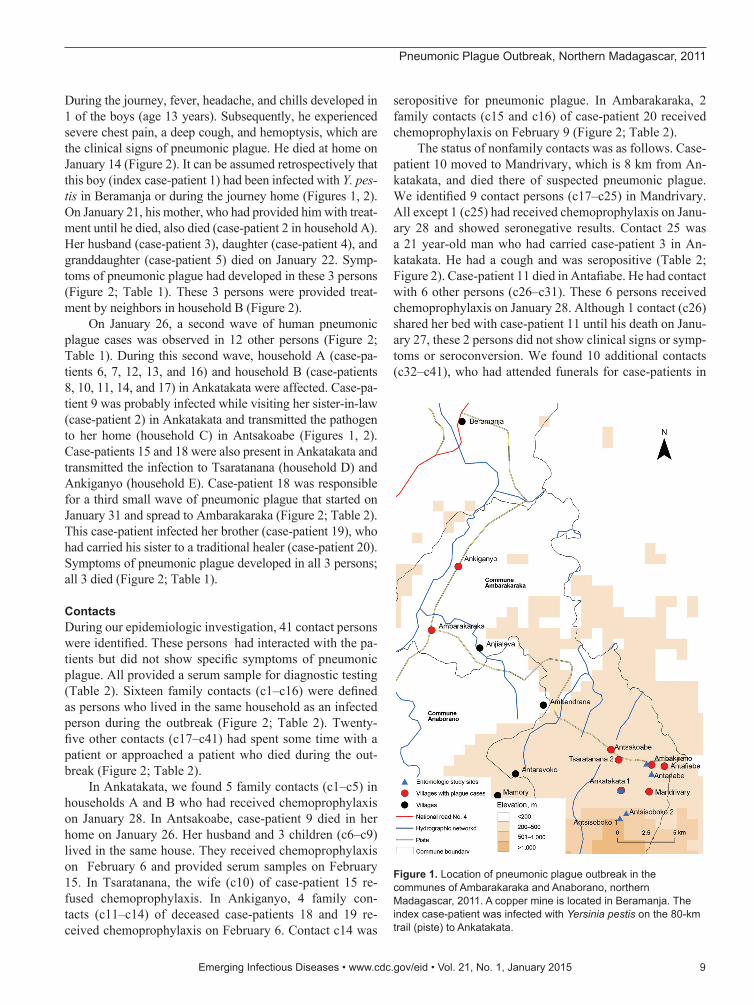
Outbreak Progression and InvestigationThe outbreak investigation protocol was approved by the Ethical Committee, Ministry of Health of Madagascar. The outbreak area contained 7 villages along a field path in the communes of Ambarakaraka and Anaborano, Ambilobe District, in northern Madagascar at an altitude <500 m. In January 2011, two brothers (boys) were working in a cop-per mine in Beramanja, Madagascar. On January 6, the boys returned home to Ankatakata (distance 50 km) (Figure 1).
Pneumonic Plague Outbreak, Northern Madagascar, 2011
Vincent Richard, Julia M. Riehm, Perlinot Herindrainy, Rahelinirina Soanandrasana, Maherisoa Ratsitoharina, Fanjasoa Rakotomanana, Samuel Andrianalimanana,
Holger C. Scholz, and Minoarisoa Rajerison
8 Emerging Infectious Diseases • www.cdc.gov/eid • Vol. 21, No. 1, January 2015
SYNOPSIS
Author affiliations: Institut Pasteur de Dakar, Dakar, Senegal (V. Richard); Bundeswehr Institute of Microbiology, Munich, Germany (J.M. Riehm, H.C. Scholz); German Center for Infection Research, Munich (J.M. Riehm, H.C. Scholz); Institut Pasteur de Madagascar, Anatananarivo, Madagascar (P. Herindrainy, R. Soanandrasana, M. Ratsitoharina, F. Rakotomanana, M. Rajerison); and Ministry of Public Health, Anatananarivo (S. Andrianalimanana)
DOI: http://dx.doi.org/10.3201/eid2101.131828
Pneumonic Plague Outbreak, Northern Madagascar, 2011
During the journey, fever, headache, and chills developed in 1 of the boys (age 13 years). Subsequently, he experienced severe chest pain, a deep cough, and hemoptysis, which are the clinical signs of pneumonic plague. He died at home on January 14 (Figure 2). It can be assumed retrospectively that this boy (index case-patient 1) had been infected with Y. pes-tis in Beramanja or during the journey home (Figures 1, 2). On January 21, his mother, who had provided him with treat-ment until he died, also died (case-patient 2 in household A). Her husband (case-patient 3), daughter (case-patient 4), and granddaughter (case-patient 5) died on January 22. Symp-toms of pneumonic plague had developed in these 3 persons (Figure 2; Table 1). These 3 persons were provided treat-ment by neighbors in household B (Figure 2).
On January 26, a second wave of human pneumonic plague cases was observed in 12 other persons (Figure 2; Table 1). During this second wave, household A (case-pa-tients 6, 7, 12, 13, and 16) and household B (case-patients 8, 10, 11, 14, and 17) in Ankatakata were affected. Case-pa-tient 9 was probably infected while visiting her sister-in-law (case-patient 2) in Ankatakata and transmitted the pathogen to her home (household C) in Antsakoabe (Figures 1, 2). Case-patients 15 and 18 were also present in Ankatakata and transmitted the infection to Tsaratanana (household D) and Ankiganyo (household E). Case-patient 18 was responsible for a third small wave of pneumonic plague that started on January 31 and spread to Ambarakaraka (Figure 2; Table 2). This case-patient infected her brother (case-patient 19), who had carried his sister to a traditional healer (case-patient 20). Symptoms of pneumonic plague developed in all 3 persons; all 3 died (Figure 2; Table 1).
ContactsDuring our epidemiologic investigation, 41 contact persons were identified. These persons had interacted with the pa-tients but did not show specific symptoms of pneumonic plague. All provided a serum sample for diagnostic testing (Table 2). Sixteen family contacts (c1–c16) were defined as persons who lived in the same household as an infected person during the outbreak (Figure 2; Table 2). Twenty-five other contacts (c17–c41) had spent some time with a patient or approached a patient who died during the out-break (Figure 2; Table 2).
In Ankatakata, we found 5 family contacts (c1–c5) in households A and B who had received chemoprophylaxis on January 28. In Antsakoabe, case-patient 9 died in her home on January 26. Her husband and 3 children (c6–c9) lived in the same house. They received chemoprophylaxis on February 6 and provided serum samples on February 15. In Tsaratanana, the wife (c10) of case-patient 15 re-fused chemoprophylaxis. In Ankiganyo, 4 family con-tacts (c11–c14) of deceased case-patients 18 and 19 re-ceived chemoprophylaxis on February 6. Contact c14 was
seropositive for pneumonic plague. In Ambarakaraka, 2 family contacts (c15 and c16) of case-patient 20 received chemoprophylaxis on February 9 (Figure 2; Table 2).
The status of nonfamily contacts was as follows. Case-patient 10 moved to Mandrivary, which is 8 km from An-katakata, and died there of suspected pneumonic plague. We identified 9 contact persons (c17–c25) in Mandrivary. All except 1 (c25) had received chemoprophylaxis on Janu-ary 28 and showed seronegative results. Contact 25 was a 21 year-old man who had carried case-patient 3 in An-katakata. He had a cough and was seropositive (Table 2; Figure 2). Case-patient 11 died in Antafiabe. He had contact with 6 other persons (c26–c31). These 6 persons received chemoprophylaxis on January 28. Although 1 contact (c26) shared her bed with case-patient 11 until his death on Janu-ary 27, these 2 persons did not show clinical signs or symp-toms or seroconversion. We found 10 additional contacts (c32–c41), who had attended funerals for case-patients in
Emerging Infectious Diseases • www.cdc.gov/eid • Vol. 21, No. 1, January 2015 9
Figure 1. Location of pneumonic plague outbreak in the communes of Ambarakaraka and Anaborano, northern Madagascar, 2011. A copper mine is located in Beramanja. The index case-patient was infected with Yersinia pestis on the 80-km trail (piste) to Ankatakata.
SYNOPSIS
different villages. Three of these contacts had mild pulmo-nary infection, but had not consulted a physician and were seronegative (Figure 2; Table 2).
Diagnosis and Case AssessmentTwo weeks after the death of the index case-patient on January 28, health personnel arrived in Ankatakata. Treat-ment with antimicrobial drugs was provided to 5 patients (case-patients 12, 13, 15, 16, and 17), and chemoprophy-laxis was provided to contact persons and health person-nel according to recommendations of the World Health Organization (WHO) (9) (Figure 2; Tables 1, 2). Because we did not know that the outbreak was ongoing in Ankig-anyo (30 km from Ankatakata), treatment was not provided to case-patients 18, 19, and 20 (Figures 1, 2). It was only af-ter these 3 case-patients died that contacts of these patients received chemoprophylaxis (Figure 2; Table 2).
Because persons who died were not sampled post-mortem, only a limited number of clinical samples were
collected (Table 1). Of collected sputum samples, 2 (from case-patients 12 and 13) were positive for Y. pestis–specific F1 antigen by immunochromatographic rapid dipstick test (Table 1) (16). All samples were subsequently transported (>900 km) to the WHO Collaborating Center for Plague at the Institut Pasteur de Madagascar in Antananarivo. Spu-tum samples were incubated in bacterial culture media and inoculated into laboratory mice. However, isolation of Y. pestis was not successful (Tables 1, 3). Collected serum samples were analyzed by using a Y. pestis–specific F1 an-tigen IgG ELISA as described (17). Positive samples were quantified by using serial dilutions. Three of 5 paired serum samples (from case-patients 12, 13, and 17) had 4-fold in-creases in titer in the second serum (Table 1). Of samples from contact persons, 2 of 41 single serum samples showed a positive result (Table 2).
Subsequent case assignment was conducted according to WHO recommendations for plague-endemic countries (18). Three types of cases were identified: suspected cases (specific
10 Emerging Infectious Diseases • www.cdc.gov/eid • Vol. 21, No. 1, January 2015
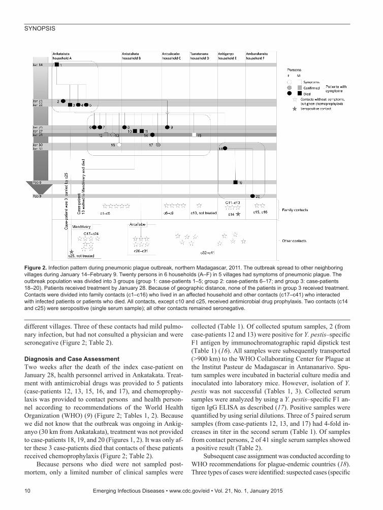
Figure 2. Infection pattern during pneumonic plague outbreak, northern Madagascar, 2011. The outbreak spread to other neighboring villages during January 14–February 9. Twenty persons in 6 households (A–F) in 5 villages had symptoms of pneumonic plague. The outbreak population was divided into 3 groups (group 1: case-patients 1–5; group 2: case-patients 6–17; and group 3: case-patients 18–20). Patients received treatment by January 28. Because of geographic distance, none of the patients in group 3 received treatment. Contacts were divided into family contacts (c1–c16) who lived in an affected household and other contacts (c17–c41) who interacted with infected patients or patients who died. All contacts, except c10 and c25, received antimicrobial drug prophylaxis. Two contacts (c14 and c25) were seropositive (single serum sample); all other contacts remained seronegative.
Pneumonic Plague Outbreak, Northern Madagascar, 2011
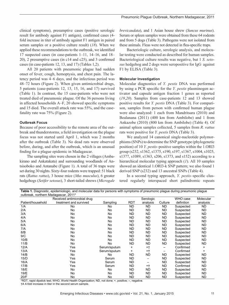
clinical symptoms), presumptive cases (positive serologic result for antibody against F1 antigen), confirmed cases (4-fold increase in titer of antibody against F1 antigen in paired serum samples or a positive culture result) (18). When we applied these recommendations to the outbreak, we identified 17 suspected cases (in case-patients 1–11, 14–16, and 18–20), 2 presumptive cases (in c14 and c25), and 3 confirmed cases (in case-patients 12, 13, and 17) (Tables 1,2).
All 20 patients with pneumonic plague had sudden onset of fever, cough, hemoptysis, and chest pain. The la-tency period was 4–6 days, and the infectious period was 48–72 hours (Figure 2). When given antimicrobial drugs, 5 patients (case-patients 12, 13, 15, 16, and 17) survived (Table 1). In contrast, the 15 case-patients who were not treated died of pneumonic plague. Of the 36 persons living in affected households A–F, 20 showed specific symptoms and 15 died. The overall attack rate was 55%, and the case-fatality rate was 75% (Figure 2).
Outbreak FocusBecause of poor accessibility to the remote area of the out-break and thunderstorms, a field investigation on the plague focus was not started until April 1, which was 2 months after the outbreak (Table 3). No dead rats were observed before, during, and after the outbreak, which is an unusual finding for a plague epidemic in Madagascar.
The sampling sites were chosen in the 2 villages (Amba-kirano and Ankatakata) and surrounding woodlands of An-tsisoboko and Antanabe (Figure 1). A total of 36 traps were set during 30 nights. Sixty-four rodents were trapped: 51 black rats (Rattus rattus), 3 house mice (Mus musculus), 6 greater hedgehogs (Setifer setosus), 3 short-tailed shrews (Microgale
brevicaudata), and 1 Asian house shrew (Suncus murinus). Serum or spleen samples were obtained from these 64 rodents and from 5 dogs (Table 3). Pathogens were not isolated from these animals. Fleas were not detected in flea-specific traps.
Bacteriologic culture, serologic analysis, and molecu-lar testing were conducted as described for human samples. Bacteriological culture results was negative, but 1 S. seto-sus hedgehog and 2 dogs were seropositive for IgG against F1 by ELISA (Table 3).
Molecular InvestigationMolecular diagnostics of Y. pestis DNA was performed by using a PCR specific for the Y. pestis plasminogen ac-tivator and capsule antigen fraction 1 genes as reported (19,20). Samples from case-patients 12 and 13 showed positive results for Y. pestis DNA (Table 3). For compari-son, samples from person with confirmed human plague were also analyzed: 1 each from Mandritsara (2010) and Bealanana (2011) (400 km from Ambilobe) and 1 from Ankazobe (2010) (800 km from Ambilobe) (Table 4). Of animal spleen samples collected, 5 samples from R. rattus rats were positive for Y. pestis DNA (Table 3).
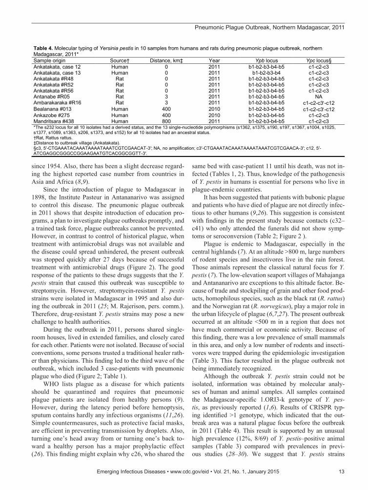
We analyzed 14 canonical single-nucleotide polymor-phisms (SNPs) to determine the SNP genotype (phylogenetic position) of 10 Y. pestis–positive samples within the 1.ORI3 group (s232, s1362, s1375, s190, s197, s1367, s1004, s1025, s1377, s1089, s1363, s206, s1373, and s152) according to a hierarchical molecular typing approach (1). All 10 samples showed an identical 1.ORI3-k SNP pattern; we also found 1 derived SNP (s232) and 13 ancestral SNPs (Table 4).
In a second typing approach, Y. pestis–specific clus-tered regularly interspaced short palindromic repeats
Emerging Infectious Diseases • www.cdc.gov/eid • Vol. 21, No. 1, January 2015 11
Table 1. Diagnostic, epidemiologic, and molecular data for persons with symptoms of pneumonic plague during pneumonic plague outbreak, northern Madagascar, 2011*
Patient/household Received antimicrobial drug
treatment and survived Sampling RDT Serologic analysis Culture
WHO case definition
Molecular analysis
1/A No No ND ND ND Suspected ND 2/A No No ND ND ND Suspected ND 3/A No No ND ND ND Suspected ND 4/A No No ND ND ND Suspected ND 5/A No No ND ND ND Suspected ND 6/A No No ND ND ND Suspected ND 7/A No No ND ND ND Suspected ND 8/B No No ND ND ND Suspected ND 9/C No No ND ND ND Suspected ND 10/B No No ND ND ND Suspected ND 11/B No No ND ND ND Suspected ND 12/A Yes Serum/sputum + +† – Confirmed + 13/A Yes Serum/sputum + +† – Confirmed + 14/B No No ND ND ND Suspected ND 15/D Yes Serum ND – ND Suspected ND 16/A Yes Serum ND – ND Suspected ND 17/B Yes Serum ND +† ND Confirmed ND 18/E No No ND ND ND Suspected ND 19/E No No ND ND ND Suspected ND 20/F No No ND ND ND Suspected ND *RDT, rapid dipstick test; WHO, World Health Organization; ND, not done; +, positive; –, negative. †A 4-fold increase in titer in the second serum sample.
SYNOPSIS
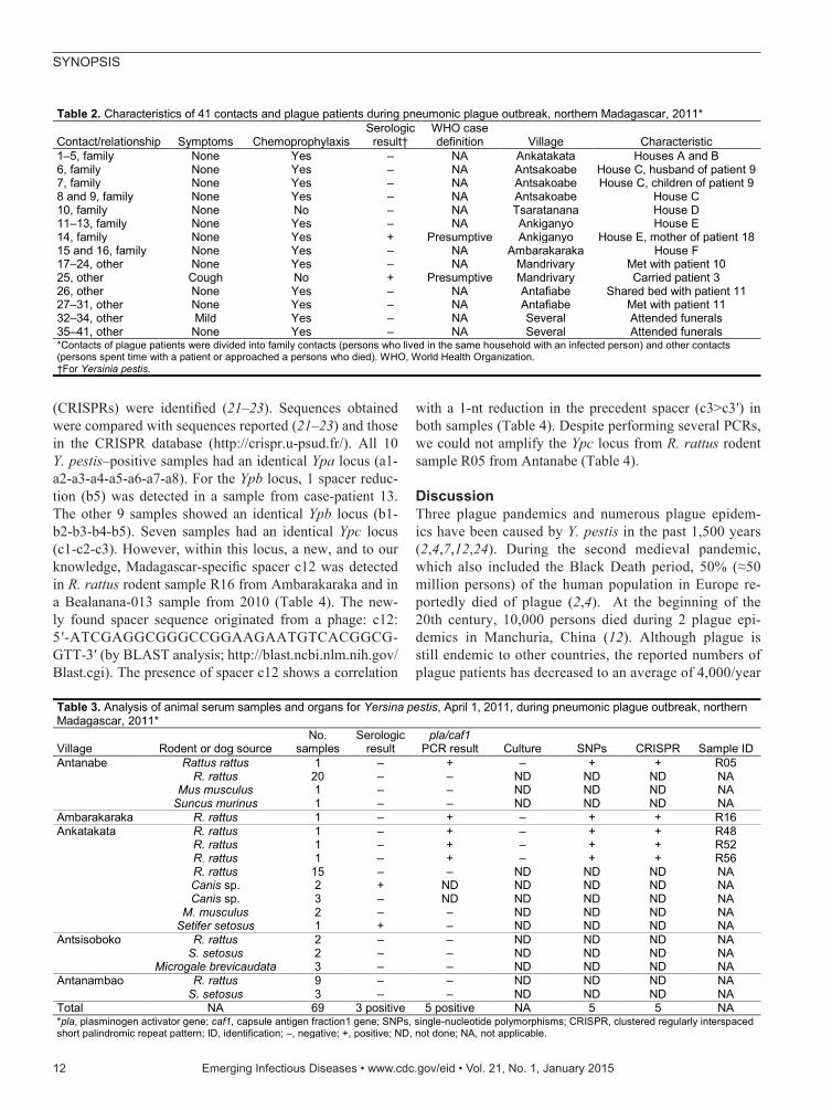
(CRISPRs) were identified (21–23). Sequences obtained were compared with sequences reported (21–23) and those in the CRISPR database (http://crispr.u-psud.fr/). All 10 Y. pestis–positive samples had an identical Ypa locus (a1-a2-a3-a4-a5-a6-a7-a8). For the Ypb locus, 1 spacer reduc-tion (b5) was detected in a sample from case-patient 13. The other 9 samples showed an identical Ypb locus (b1-b2-b3-b4-b5). Seven samples had an identical Ypc locus (c1-c2-c3). However, within this locus, a new, and to our knowledge, Madagascar-specific spacer c12 was detected in R. rattus rodent sample R16 from Ambarakaraka and in a Bealanana-013 sample from 2010 (Table 4). The new-ly found spacer sequence originated from a phage: c12: 5′-ATCGAGGCGGGCCGGAAGAATGTCACGGCG-GTT-3′ (by BLAST analysis; http://blast.ncbi.nlm.nih.gov/Blast.cgi). The presence of spacer c12 shows a correlation
with a 1-nt reduction in the precedent spacer (c3>c3′) in both samples (Table 4). Despite performing several PCRs, we could not amplify the Ypc locus from R. rattus rodent sample R05 from Antanabe (Table 4).
DiscussionThree plague pandemics and numerous plague epidem-ics have been caused by Y. pestis in the past 1,500 years (2,4,7,12,24). During the second medieval pandemic, which also included the Black Death period, 50% (≈50 million persons) of the human population in Europe re-portedly died of plague (2,4). At the beginning of the 20th century, 10,000 persons died during 2 plague epi-demics in Manchuria, China (12). Although plague is still endemic to other countries, the reported numbers of plague patients has decreased to an average of 4,000/year
12 Emerging Infectious Diseases • www.cdc.gov/eid • Vol. 21, No. 1, January 2015
Table 3. Analysis of animal serum samples and organs for Yersina pestis, April 1, 2011, during pneumonic plague outbreak, northern Madagascar, 2011*
Village Rodent or dog source No.
samples Serologic
result pla/caf1
PCR result Culture SNPs CRISPR Sample ID Antanabe Rattus rattus 1 – + – + + R05 R. rattus 20 – – ND ND ND NA Mus musculus 1 – – ND ND ND NA Suncus murinus 1 – – ND ND ND NA Ambarakaraka R. rattus 1 – + – + + R16 Ankatakata R. rattus 1 – + – + + R48 R. rattus 1 – + – + + R52 R. rattus 1 – + – + + R56 R. rattus 15 – – ND ND ND NA Canis sp. 2 + ND ND ND ND NA Canis sp. 3 – ND ND ND ND NA M. musculus 2 – – ND ND ND NA Setifer setosus 1 + – ND ND ND NA Antsisoboko R. rattus 2 – – ND ND ND NA S. setosus 2 – – ND ND ND NA Microgale brevicaudata 3 – – ND ND ND NA Antanambao R. rattus 9 – – ND ND ND NA S. setosus 3 – – ND ND ND NA Total NA 69 3 positive 5 positive NA 5 5 NA *pla, plasminogen activator gene; caf1, capsule antigen fraction1 gene; SNPs, single-nucleotide polymorphisms; CRISPR, clustered regularly interspaced short palindromic repeat pattern; ID, identification; –, negative; +, positive; ND, not done; NA, not applicable.
Table 2. Characteristics of 41 contacts and plague patients during pneumonic plague outbreak, northern Madagascar, 2011*
Contact/relationship Symptoms Chemoprophylaxis Serologic
result† WHO case definition Village Characteristic
1–5, family None Yes – NA Ankatakata Houses A and B 6, family None Yes – NA Antsakoabe House C, husband of patient 9 7, family None Yes – NA Antsakoabe House C, children of patient 9 8 and 9, family None Yes – NA Antsakoabe House C 10, family None No – NA Tsaratanana House D 11–13, family None Yes – NA Ankiganyo House E 14, family None Yes + Presumptive Ankiganyo House E, mother of patient 18 15 and 16, family None Yes – NA Ambarakaraka House F 17–24, other None Yes – NA Mandrivary Met with patient 10 25, other Cough No + Presumptive Mandrivary Carried patient 3 26, other None Yes – NA Antafiabe Shared bed with patient 11 27–31, other None Yes – NA Antafiabe Met with patient 11 32–34, other Mild Yes – NA Several Attended funerals 35–41, other None Yes – NA Several Attended funerals *Contacts of plague patients were divided into family contacts (persons who lived in the same household with an infected person) and other contacts (persons spent time with a patient or approached a persons who died). WHO, World Health Organization. †For Yersinia pestis.
Pneumonic Plague Outbreak, Northern Madagascar, 2011
since 1954. Also, there has been a slight decrease regard-ing the highest reported case number from countries in Asia and Africa (8,9).
Since the introduction of plague to Madagascar in 1898, the Institute Pasteur in Antananarivo was assigned to control this disease. The pneumonic plague outbreak in 2011 shows that despite introduction of education pro-grams, a plan to investigate plague outbreaks promptly, and a trained task force, plague outbreaks cannot be prevented. However, in contrast to control of historical plague, when treatment with antimicrobial drugs was not available and the disease could spread unhindered, the present outbreak was stopped quickly after 27 days because of successful treatment with antimicrobial drugs (Figure 2). The good response of the patients to these drugs suggests that the Y. pestis strain that caused this outbreak was susceptible to streptomycin. However, streptomycin-resistant Y. pestis strains were isolated in Madagascar in 1995 and also dur-ing the outbreak in 2011 (25; M. Rajerison, pers. comm.). Therefore, drug-resistant Y. pestis strains may pose a new challenge to health authorities.
During the outbreak in 2011, persons shared single-room houses, lived in extended families, and closely cared for each other. Patients were not isolated. Because of social conventions, some persons trusted a traditional healer rath-er than physicians. This finding led to the third wave of the outbreak, which included 3 case-patients with pneumonic plague who died (Figure 2; Table 1).
WHO lists plague as a disease for which patients should be quarantined and requires that pneumonic plague patients are isolated from healthy persons (9). However, during the latency period before hemoptysis, sputum contains hardly any infectious organisms (11,26). Simple countermeasures, such as protective facial masks, are efficient in preventing transmission by droplets. Also, turning one’s head away from or turning one’s back to-ward a healthy person has a major prophylactic effect (26). This finding might explain why c26, who shared the
same bed with case-patient 11 until his death, was not in-fected (Tables 1, 2). Thus, knowledge of the pathogenesis of Y. pestis in humans is essential for persons who live in plague-endemic countries.
It has been suggested that patients with bubonic plague and patients who have died of plague are not directly infec-tious to other humans (9,26). This suggestion is consistent with findings in the present study because contacts (c32–c41) who only attended the funerals did not show symp-toms or seroconversion (Table 2; Figure 2 ).
Plague is endemic to Madagascar, especially in the central highlands (7). At an altitude >800 m, large numbers of rodent species and insectivores live in the rain forest. Those animals represent the classical natural focus for Y. pestis (7). The low-elevation seaport villages of Mahajanga and Antananarivo are exceptions to this altitude factor. Be-cause of trade and stockpiling of grain and other food prod-ucts, homophilous species, such as the black rat (R. rattus) and the Norwegian rat (R. norvegicus), play a major role in the urban lifecycle of plague (6,7,27). The present outbreak occurred at an altitude <500 m in a region that does not have much commercial or economic activity. Because of this finding, there was a low prevalence of small mammals in this area, and only a low number of rodents and insecti-vores were trapped during the epidemiologic investigation (Table 3). This factor resulted in the plague outbreak not being immediately recognized.
Although the outbreak Y. pestis strain could not be isolated, information was obtained by molecular analy-ses of human and animal samples. All samples contained the Madagascar-specific 1.ORI3-k genotype of Y. pes-tis, as previously reported (1,6). Results of CRISPR typ-ing identified >1 genotype, which indicated that the out-break area was a natural plague focus before the outbreak in 2011 (Table 4). This result is supported by an unusual high prevalence (12%, 8/69) of Y. pestis–positive animal samples (Table 3) compared with prevalences in previ-ous studies (28–30). We suggest that Y. pestis strains
Emerging Infectious Diseases • www.cdc.gov/eid • Vol. 21, No. 1, January 2015 13
Table 4. Molecular typing of Yersinia pestis in 10 samples from humans and rats during pneumonic plague outbreak, northern Madagascar, 2011* Sample origin Source† Distance, km‡ Year Ypb locus Ypc locus§ Ankatakata, case 12 Human 0 2011 b1-b2-b3-b4-b5 c1-c2-c3 Ankatakata, case 13 Human 0 2011 b1-b2-b3-b4 c1-c2-c3 Ankatakata #R48 Rat 0 2011 b1-b2-b3-b4-b5 c1-c2-c3 Ankatakata #R52 Rat 0 2011 b1-b2-b3-b4-b5 c1-c2-c3 Ankatakata #R56 Rat 0 2011 b1-b2-b3-b4-b5 c1-c2-c3 Antanabe #R05 Rat 3 2011 b1-b2-b3-b4-b5 NA Ambarakaraka #R16 Rat 3 2011 b1-b2-b3-b4-b5 c1-c2-c3-c12 Bealanana #013 Human 400 2010 b1-b2-b3-b4-b5 c1-c2-c3-c12 Ankazobe #275 Human 400 2010 b1-b2-b3-b4-b5 c1-c2-c3 Mandritsara #438 Human 800 2011 b1-b2-b3-b4-b5 c1-c2-c3 *The s232 locus for all 10 isolates had a derived status, and the 13 single-nucleotide polymorphisms (s1362, s1375, s190, s197, s1367, s1004, s1025, s1377, s1089, s1363, s206, s1373, and s152) for all 10 isolates had an ancestral status. †Rat, Rattus rattus. ‡Distance to outbreak village (Ankatakata). §c3, 5′-CTGAAATACAAATAAAATAAATCGTCGAACAT-3′; NA, no amplification; c3′-CTGAAATACAAATAAAATAAATCGTCGAACA-3′; c12, 5′-ATCGAGGCGGGCCGGAAGAATGTCACGGCGGTT-3′.
SYNOPSIS
containing the major CRISPR profile, which was found in 6 of 10 samples, was responsible for the present outbreak (Table 4). This CRISPR genotype has also been found in samples from the central highlands. The isolate from case-patient 13 lost the b5 spacer (Table 4). This phenomenon has been reported for other CRISPR profiles, and a different genotype has been assessed (21–23). A third Y. pestis CRIS-PR genotype was found in the R. rattus rodent R16 sample and in the human Bealanana 013 sample from 2010; this genotype includes the new element c3′–c12 of phage origin (Table 4) (21). The loss of 1 nt at the end of the spacer, as observed in in c3>c3′, has been previously reported (22).
Despite performing several PCRs, we could not ampli-fy the complete Ypc locus in the R. rattus rodent R05 sample from Antanabe (Table 4), a finding that has been previously reported (21). We suggest that 4 CRISPR genotypes of Y. pestis were present in the outbreak area, which indicates that Ambilobe was a natural plague focus even before the outbreak. Unnoticed presence of pathogens near human populations requires higher surveillance activity, as recently reported (7). In contrast, it has been reported that hereditary resistance against Y. pestis might develop in rats (7,31).
One question that also needs to be addressed is why plague caused millions of deaths during devastating pandemics in the past while today plague is restricted to some geographic locations. One possible explanation would be the presence of additional virulence factors in historical plague strains, which have been lost from cur-rent Y. pestis strains.
To answer this question, 2 ancient Y. pestis genomes were sequenced and compared with sequences of current Y. pestis isolates. The ancient genomes were from a pan-demic European Y. pestis pestis biovar Antiqua isolate (genotype 1.ANT) that originated during the Black Death period (3), and from a pandemic Y. pestis pestis biovar An-tiqua isolate (genotype 0.ANT), which was isolated from a patient who died during the plague of Justinian in Germany (5). Sequences of current Y. pestis genomes used for com-parison were from the nonhuman pathogenic Chinese Y. pestis microtus strain (91001: 0.PE4, biovar Xilingolensis) and from the Y. pestis pestis strain (CO92: 1.ORI1, biovar Orientalis) (3,5).
Analyses showed that genomes of ancient Y. pestis strains did not contain additional virulence genes that might explain higher virulence. (3,5). Furthermore, because plague epidemics were caused by different biovars or genotypes in Asia, Europe, and Africa (3,5,6,10,12–15), we assume that various Y. pestis subtypes are similar in virulence. Thus, fac-tors other than differences in virulence might better explain the decrease in the reported plague cases. It is more likely that the general perception and understanding of infectious agents; improved hygiene; trade management; knowl-edge of plague pathogenesis in humans; vector control;
specific outbreak management, including selective isola-tion of infectious patients; and chemoprophylaxis and treat-ment with antimicrobial drugs, have accounted for the de-crease in plague cases (9,11,15,26).
In conclusion, the 1.ORI3-k genotype of Y. pestis iden-tified in the present study has virulence comparable with that of ancestral genotypes that caused other epidemics. The course and outcome of a human plague epidemic depend on this virulence, as well as on education, public awareness, life style, infrastructure, isolation of patients, and medical care. However, there is an unpredictable pathogenic poten-tial in drug-resistant strains that has not been estimated and needs to be studied.
AcknowledgmentsWe thank Abdou Salam Nasser, Ravoniarisoa Marie Georgette, Noromihaja Randriananja, and Joely Razafilalaintsoa for assis-tance during the epidemiologic study, and Michaela Projahn and Gabriele Echle for assistance during pathogen typing and labora-tory analysis.
This study was supported by the Institut Pasteur de Madagascar, the President’s Malaria Initiative/US Agency for International Development, and the US Department of Homeland Security (project no. DHS-09-ST-108-001/MGN3EL7-01).
Dr. Richard is a scientist in the Department of Epidemiology at the Institute Pasteur, Dakar, Senegal. His research interests are tropical diseases, public health priorities, analysis of surveillance data from sentinel network and cohort studies, and evaluation of emergent risks and antibiotic resistance testing.
References
1. Morelli G, Song Y, Mazzoni CJ, Eppinger M, Roumagnac P, Wagner DM, et al. Yersinia pestis genome sequencing identi-fies patterns of global phylogenetic diversity. Nat Genet. 2010;42:1140–3. http://dx.doi.org/10.1038/ng.705
2. Benedictow OJ. The black death 1346–1353: the complete history. Martelsham (UK): Boydell Press; 2004.
3. Bos KI, Schuenemann VJ, Golding GB, Burbano HA, Waglechner N, Coombes BK, et al. A draft genome of Yersinia pestis from victims of the Black Death. Nature. 2011;478:506–10. http://dx.doi.org/10.1038/nature10549
4. Little LK. Life and after life of the first plague pandemic. In: Little LK, editor. Plague and the end of antiquity: the pandemic of 541–750. Cambridge (UK): Cambridge University Press; 2007. p. 3–32.
5. Wagner DM, Klunk J, Harbeck M, Devault A, Waglechner N, Sahl JW, et al. Yersinia pestis and the Plague of Justinian 541–543 AD: a genomic analysis. Lancet Infect Dis. 2014;14:319–26. http://dx.doi.org/10.1016/S1473-3099(13)70323-2
6. Vogler AJ, Chan F, Nottingham R, Andersen G, Drees K, Beckstrom-Sternberg SM, et al. A decade of plague in Mahajanga, Madagascar: insights into the global maritime spread of pandemic plague. MBio. 2013;4:e00623–12. http://dx.doi.org/10.1128/mBio.00623-12
14 Emerging Infectious Diseases • www.cdc.gov/eid • Vol. 21, No. 1, January 2015
Pneumonic Plague Outbreak, Northern Madagascar, 2011
7. Andrianaivoarimanana V, Kreppel K, Elissa N, Duplantier JM, Carniel E, Rajerison M, et al. Understanding the persistence of plague foci in Madagascar. PLoS Negl Trop Dis. 2013;7:e2382. http://dx.doi.org/10.1371/journal.pntd.0002382
8. Human plague: review of regional morbidity and mortality, 2004–2009. Wkly Epidemiol Rec. 2009;85:40–5.
9. World Health Organization. Plague manual: epidemiology, distribution, surveillance and control. WHO/CDS/CSR/EDC/99.2. Geneva: The Organization; 1999.
10. Wu L-T. A treatise on pneumonic plague. Geneva: League of Nations Health Organization; 1926.
11. Gani R, Leach S. Epidemiologic determinants for modeling pneumonic plague outbreaks. Emerg Infect Dis. 2004;10:608–14. http://dx.doi.org/10.3201/eid1004.030509
12. Wu L-T, Chun JWH, Pollitzer R, Wu CY. Plague: a manual for medical and public health workers. Shanghai: Weishengshu National Quarantine Service; 1936.
13. Ratsitorahina M, Chanteau S, Rahalison L, Ratsifasoamanana L, Boisier P. Epidemiological and diagnostic aspects of the outbreak of pneumonic plague in Madagascar. Lancet. 2000;355:111–3. http://dx.doi.org/10.1016/S0140-6736(99)05163-6
14. Begier EM, Asiki G, Anywaine Z, Yockey B, Schriefer ME, Aleti P, et al. Pneumonic plague cluster, Uganda, 2004. Emerg Infect Dis. 2006;12:460–7. http://dx.doi.org/10.3201/eid1203.051051
15. Bertherat E, Thullier P, Shako JC, England K, Koné M-L, Arntzen L, et al. Lessons learned about pneumonic plague diagnosis from 2 outbreaks, Democratic Republic of the Congo. Emerg Infect Dis. 2011;17:778–84. http://dx.doi.org/10.3201/eid1705.100029
16. Chanteau S, Rahalison L, Ralafiarisoa L, Foulon J, Ratsitorahina M, Ratsifasoamanana L, et al. Development and testing of a rapid diagnostic test for bubonic and pneumonic plague. Lancet. 2003;361:211–6. http://dx.doi.org/10.1016/S0140-6736 (03)12270-2
17. Rasoamanana B, Leroy F, Boisier P, Rasolomaharo M, Buchy P, Carniel E, et al. Field evaluation of an immunoglobulin G anti-F1 enzyme-linked immunosorbent assay for serodiagnosis of human plague in Madagascar. Clin Diagn Lab Immunol. 1997;4:587–91.
18. World Health Organization. International meeting on preventing and controlling plague: the old calamity still has a future. Wkly Epidemiol Rec. 2006;81:278–84.
19. Rahalison L, Vololonirina E, Ratsitorahina M, Chanteau S. Diagnosis of bubonic plague by PCR in Madagascar under field conditions. J Clin Microbiol. 2000;38:260–3.
20. Riehm JM, Rahalison L, Scholz HC, Thoma B, Pfeffer M, Razanakoto LM, et al. Detection of Yersinia pestis using real-time PCR in patients with suspected bubonic plague. Mol Cell Probes. 2011;25:8–12. http://dx.doi.org/10.1016/j.mcp.2010.09.002
21. Pourcel C, Salvignol G, Vergnaud G. CRISPR elements in Yersinia pestis acquire new repeats by preferential uptake of bacteriophage DNA, and provide additional tools for evolutionary studies. Microbiology. 2005;151:653–63. http://dx.doi.org/10.1099/mic.0.27437-0
22. Cui Y, Li Y, Gorgé O, Platonov ME, Yan Y, Guo Z, et al. Insight into microevolution of Yersinia pestis by clustered regularly inter-spaced short palindromic repeats. PLoS ONE. 2008;3:e2652. http://dx.doi.org/10.1371/journal.pone.0002652
23. Riehm JM, Vergnaud G, Kiefer D, Damdindorj T, Dashdavaa O, Khurelsukh T, et al. Yersinia pestis lineages in Mongolia. PLoS ONE. 2012;7:e30624. http://dx.doi.org/10.1371/journal.pone.0030624
24. Pryor EG. The great plague of Hong Kong. J Hong Kong Branch R Asiat Soc. 1975;15:61–70.
25. Galimand M, Guiyoule A, Gerbaud G, Rasoamanana B, Chanteau S, Carniel E, et al. Multidrug resistance in Yersinia pestis mediated by a transferable plasmid. N Engl J Med. 1997;337:677–80. http://dx.doi.org/10.1056/NEJM199709043371004
26. Kool JL. Risk of person-to-person transmission of pneumonic plague. Clin Infect Dis. 2005;40:1166–72. http://dx.doi.org/ 10.1086/428617
27. Gage KL, Kosoy MY. Natural history of plague: perspec-tives from more than a century of research. Annu Rev En-tomol. 2005;50:505–28.http://dx.doi.org/10.1146/annurev.ento.50.071803.130337
28. Thiagarajan B, Bai Y, Gage KL, Cully JF Jr. Prevalence of Yersinia pestis in rodents and fleas associated with black-tailed prairie dogs (Cynomys ludovicianus) at Thunder Basin National Grassland, Wyoming. J Wildl Dis. 2008;44:731–6. http://dx.doi.org/10.7589/0090-3558-44.3.731
29. Riehm JM, Tserennorov D, Kiefer D, Stuermer IW, Tomaso H, Zoller L, et al. Yersinia pestis in small rodents, Mongolia. Emerg Infect Dis. 2011;17:1320–2. http://dx.doi.org/10.3201/eid1707.100740
30. Martin-Alonso A, Soto M, Foronda P, Aguilar E, Bonnet G, Pacheco R, et al. Bartonella spp. and Yersinia pestis reservoirs, Cusco, Peru. Emerg Infect Dis. 2014;20:1069–70. http://dx.doi.org/10.3201/eid2006.131194
31. Alonso J-M, Vilmer E, Mazigh D, Mollaret HH. Mechanisms of acquired resistance to plague in mice by Yersinia enterocolitica O3. Curr Microbiol. 1980;4:117–22. http://dx.doi.org/10.1007/BF02602904
Address for correspondence: Julia M. Riehm, Department of Bacteriology and Toxicology, Bundeswehr Institute of Microbiology, Neuherbergstrasse 11, D-80937 Munich, Germany; email: [email protected]
Emerging Infectious Diseases • www.cdc.gov/eid • Vol. 21, No. 1, January 2015 15