Magnetism and stability of the Co:TiO 2(1 0 0) interface probed by X-ray photoemission and ex situ...
6
Magnetism and stability of the Co:TiO 2 (1 0 0) interface probed by X-ray photoemission and ex situ magnetometry L. Sangaletti a, * , F. Federici Canova a , A. Sepe a , S. Pagliara a , M.C. Mozzati b , P. Galinetto b , C.B. Azzoni b , A. Speghini c , M. Bettinelli c a Universita ´ Cattolica, Dipartimento di Matematica e Fisica, via dei Musei 41, 25121 Brescia, Italy b CNISM and Dipartimento di Fisica ‘‘A. Volta’’, Universita ` di Pavia, via Bassi 6, 27100 Pavia, Italy c Dipartimento Scientifico e Tecnologico, Universita ` di Verona and INSTM, UdR Verona, Strada Le Grazie 15, 37134 Verona, Italy Available online 22 June 2007 Abstract Ferromagnetic Co:TiO 2 thin films have been grown by Co evaporation on a sputtered TiO 2 (1 0 0) surface. Annealing in ultra-high vacuum induced a recovery of the oxygen stoichiometry and consequently the oxidation of titanium to Ti 4+ at the surface, while oxida- tion of the Co layer has not been observed. Room temperature ferromagnetism is observed, along with a paramagnetic contribution. Differences of the hysteresis cycle with respect to that usually observed for bulk cobalt are ascribed both to the reduced film thickness and to the disorder induced by the sputtering treatment. Ó 2007 Elsevier B.V. All rights reserved. Keywords: Rutile; Ferromagnetism; Photoemission; Cobalt; Raman spectroscopy 1. Introduction Advances in the search of ferromagnetic materials have prompted a large number of studies on the TM:TiO 2 trans- parent ferromagnets (TM = Cr, Mn, Fe, Co, Ni, Cu) [1]. These systems, usually regarded as diluted magnetic oxides (DMOs), display room temperature ferromagnetism and transparency in the visible wavelength range which make them appealing for possible applications in the field of spintronics. Recently, a few studies have been also devoted to the TM:TiO 2 rutile interface prepared by evaporation of TM atoms and annealing of the unreacted interface [2,3]. In particular, several investigations have been devoted to the Co:TiO 2 interface. Indeed, the Co x Ti 1x O 2 DMO is one of the most studied magnetic oxides in the field of spin- tronics materials research. The first TiO 2 based ferromag- netic oxide was obtained by laser molecular-beam epitaxy growth of a thin TiO 2 layer in the anatase phase [4]. Soon after this report, other epitaxial layers have been prepared also in the TiO 2 rutile structure, with several claims of intrinsic ferromagnetism [5–7]. Recently, a photoemission study has been reported on the growth of Co on TiO 2 (1 0 0) rutile crystal, that displayed different magnetic behavior with annealing temperature and, for the highest annealing temperature reported (i.e. 700 °C), ferromagne- tism ascribed to a mixed Co–Ti–O oxide [2]. In the present study we analyze the stability of the Co:- TiO 2 interface upon annealing in ultra-high vacuum up to 700 °C. The interface is grown on the (1 0 0) surface of a ru- tile TiO 2 single crystal which has been heavily sputtered be- fore the evaporation of the thin Co layer. Heavy sputtering was carried out to produce a rough surface and promote the diffusion of cobalt into the TiO 2 host matrix. In spite of the annealing at high temperatures cobalt always dis- plays a metallic behavior and no trace of oxidation. More- over, both Ti and O recover their stoichiometry after annealing at 700 °C and the lineshape of Ti 2p and O 1s core lines closely resembles that of a stoichiometric TiO 2 surface. The results of ex situ magnetic measurements can be interpreted as a superposition of paramagnetic 0039-6028/$ - see front matter Ó 2007 Elsevier B.V. All rights reserved. doi:10.1016/j.susc.2007.06.030 * Corresponding author. Tel.: +39 030 2406744; fax: +39 030 2406742. E-mail address: [email protected] (L. Sangaletti). www.elsevier.com/locate/susc Surface Science 601 (2007) 4375–4380
-
Upload
independent -
Category
Documents
-
view
1 -
download
0
Transcript of Magnetism and stability of the Co:TiO 2(1 0 0) interface probed by X-ray photoemission and ex situ...
www.elsevier.com/locate/susc
Surface Science 601 (2007) 4375–4380
Magnetism and stability of the Co:TiO2(100) interface probedby X-ray photoemission and ex situ magnetometry
L. Sangaletti a,*, F. Federici Canova a, A. Sepe a, S. Pagliara a, M.C. Mozzati b,P. Galinetto b, C.B. Azzoni b, A. Speghini c, M. Bettinelli c
a Universita Cattolica, Dipartimento di Matematica e Fisica, via dei Musei 41, 25121 Brescia, Italyb CNISM and Dipartimento di Fisica ‘‘A. Volta’’, Universita di Pavia, via Bassi 6, 27100 Pavia, Italy
c Dipartimento Scientifico e Tecnologico, Universita di Verona and INSTM, UdR Verona, Strada Le Grazie 15, 37134 Verona, Italy
Available online 22 June 2007
Abstract
Ferromagnetic Co:TiO2 thin films have been grown by Co evaporation on a sputtered TiO2(100) surface. Annealing in ultra-highvacuum induced a recovery of the oxygen stoichiometry and consequently the oxidation of titanium to Ti4+ at the surface, while oxida-tion of the Co layer has not been observed. Room temperature ferromagnetism is observed, along with a paramagnetic contribution.Differences of the hysteresis cycle with respect to that usually observed for bulk cobalt are ascribed both to the reduced film thicknessand to the disorder induced by the sputtering treatment.� 2007 Elsevier B.V. All rights reserved.
Keywords: Rutile; Ferromagnetism; Photoemission; Cobalt; Raman spectroscopy
1. Introduction
Advances in the search of ferromagnetic materials haveprompted a large number of studies on the TM:TiO2 trans-parent ferromagnets (TM = Cr, Mn, Fe, Co, Ni, Cu) [1].These systems, usually regarded as diluted magnetic oxides(DMOs), display room temperature ferromagnetism andtransparency in the visible wavelength range which makethem appealing for possible applications in the field ofspintronics. Recently, a few studies have been also devotedto the TM:TiO2 rutile interface prepared by evaporation ofTM atoms and annealing of the unreacted interface [2,3].In particular, several investigations have been devoted tothe Co:TiO2 interface. Indeed, the CoxTi1�xO2 DMO isone of the most studied magnetic oxides in the field of spin-tronics materials research. The first TiO2 based ferromag-netic oxide was obtained by laser molecular-beam epitaxygrowth of a thin TiO2 layer in the anatase phase [4]. Soon
0039-6028/$ - see front matter � 2007 Elsevier B.V. All rights reserved.
doi:10.1016/j.susc.2007.06.030
* Corresponding author. Tel.: +39 030 2406744; fax: +39 030 2406742.E-mail address: [email protected] (L. Sangaletti).
after this report, other epitaxial layers have been preparedalso in the TiO2 rutile structure, with several claims ofintrinsic ferromagnetism [5–7]. Recently, a photoemissionstudy has been reported on the growth of Co onTiO2(100) rutile crystal, that displayed different magneticbehavior with annealing temperature and, for the highestannealing temperature reported (i.e. 700 �C), ferromagne-tism ascribed to a mixed Co–Ti–O oxide [2].
In the present study we analyze the stability of the Co:-TiO2 interface upon annealing in ultra-high vacuum up to700 �C. The interface is grown on the (10 0) surface of a ru-tile TiO2 single crystal which has been heavily sputtered be-fore the evaporation of the thin Co layer. Heavy sputteringwas carried out to produce a rough surface and promotethe diffusion of cobalt into the TiO2 host matrix. In spiteof the annealing at high temperatures cobalt always dis-plays a metallic behavior and no trace of oxidation. More-over, both Ti and O recover their stoichiometry afterannealing at 700 �C and the lineshape of Ti 2p and O 1score lines closely resembles that of a stoichiometric TiO2
surface. The results of ex situ magnetic measurementscan be interpreted as a superposition of paramagnetic
Inte
nsity
(ar
b. u
nit)
540 538 536 534 532 530 528 526 524
Binding Energy (eV)
(b) O1s XPS
Sputtered TiO2(100)Co:TiO2(100) unreactedCo:TiO2(100) annealed at 700°C
Inte
nsity
(ar
b. u
nits
)
480 476 472 468 464 460 456 452Binding Energy (eV)
(a) Ti 2p XPS
Sputtered TiO2(100)Co:TiO2(100) unreactedCo:TiO2(100) annealed at 700°C
T i 2p3/2
T i 2p1/2
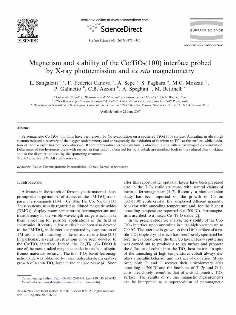
Fig. 1. (a) Ti 2p XPS spectra of the sputtered TiO2(100) surface (points),of the unreacted Co:TiO2 layer (thick line) and of the annealed (700 �C)Co:TiO2 interface (thin line). To allow comparison between the lineshapes,the sputtered and unreacted spectra have been normalized to the sameintensity. (b) O 1s spectra of the sputtered TiO2(100) surface (points), ofthe unreacted Co:TiO2 layer (thick line) and of the annealed (700 �C)Co:TiO2 interface (thin line).
4376 L. Sangaletti et al. / Surface Science 601 (2007) 4375–4380
behavior, that we ascribe to the effect of residual oxygenvacancies created by the sputtering treatment, and ferro-magnetic behavior that we ascribe to unreacted cobalt atthe interface. Unlike a recent study based on stoichiometricTiO2(10 0) surfaces [2], we do not find any evidence ofintermediate Co–Ti–O phases.
2. Experimental
X-ray photoemission spectra have been collected byusing a monochromatized Al Ka X-ray source and a hemi-spherical analyser equipped with a 32-channel detector.The overall resolution was set at 0.6 eV, as measured tothe Ag 3d5/2 core line of a polycrystalline Ag thin film.The spot size on the sample was smaller than 1 · 1 mm2.
Sputtering was achieved by using an Ar+ ion gun, withenergies up to 3.0 KeV and a sputtering time of 10 min.Annealing was performed in situ by resistive heating ofthe sample holder. Annealing treatments were carried outat 300, 500, 600, and 700 �C for 5 min each time, by keep-ing the base pressure in the chamber below 1 · 10�9 mbar.
Cobalt was evaporated by electron-beam bombardmentfrom a Co rod mounted on an Omicron evaporator. Theevaporation rate was calibrated by measuring the attenua-tion of Ti 2p signal after the deposition of several Co lay-ers. The present film resulted to be about 0.5 nm thickbefore the annealing treatments.
Micro-Raman spectra were collected at room tempera-ture by a Dilor Labram spectrograph with a He–Ne laser(632.8 nm) as exciting source and a light power less than10 mW at the sample. The microscope, confocally coupledto the spectrograph with a 100· objective of numericalaperture 0.9 and a confocal hole opened at 200 lm, yieldsa depth of focus of about 2.5 lm.
Static magnetic moments were measured at 500 and2000 Oe from 352 K to 2 K with a SQUID Quantum De-sign Magnetometer. Magnetization loops were collectedat RT for magnetic fields (H) ranging between 0 and±4000 Oe.
3. Results and discussion
In Fig. 1 the XPS core level spectra of Ti 2p and O 1s areshown for the clean, sputtered, substrate (points), for theCo:TiO2 unreacted interface (thick line), and for the an-nealed Co:TiO2 interface (thin line). As can be observed,the sputtered thin film shows broad Ti 2p3/2 and Ti 2p1/2
spin orbit split peaks, detected at about 458 eV and464 eV, respectively (Fig. 1a). These broad features are as-cribed to the effect of sputtering that both creates disorderon the sample surface and makes it oxygen poor, yieldingTi-rich regions. The deposition of the unreacted Co thinlayer (thick line) does not change remarkably the Ti 2pspectral weight, which basically shows a slight reductionof the intensity. This can be assumed as an indication thatCo does not react strongly with the disordered TiO2(10 0)surface, because for the deposition of reactive elements
such as Hf and Cr, a reduction of Ti was reported for,e.g., the TiO2(110) surface [9].
Remarkable changes occur upon annealing at the high-est temperature (700 �C, thin line). The Ti 2p spin orbitsplit peaks are narrower with a FWHM comparable to thatobserved on reconstructed TiO2 single crystal surfaces (see,e.g., Ref. [8] and references therein). This evidence is as-sumed as an indication that the oxygen surface contenthas increased and that the original stoichiometry hasrecovered.
A similar trend is observed for the O 1s core level peak(Fig. 1b). Broad peaks are detected after the sputteringtreatment on the clean surface, as well as after Co deposi-tion, whereas annealing at 700 �C yields a narrow and sym-metric line, centered at 531.5 eV.
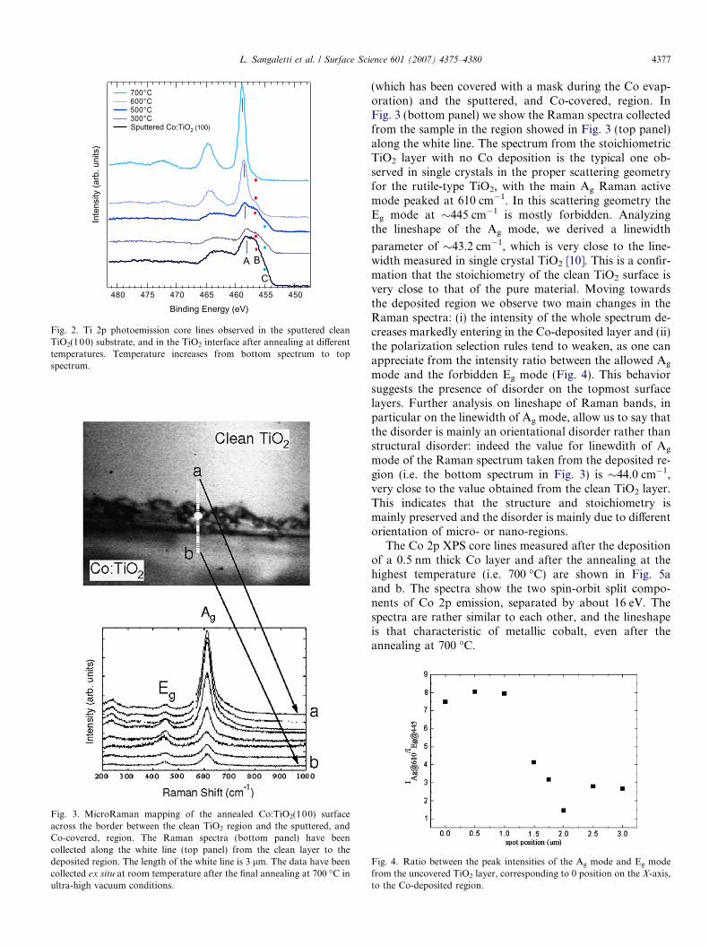
Rather interesting is the evolution of the Ti 2p emissionwith increasing annealing temperature (Fig. 2). In this case,the Ti 2p3/2 core line shows three features (A, B, and C).The intensity of B and C features progressively quenchesas the annealing temperature is increased, leaving the nar-row line observed for the sample treated at 700 �C (Fig. 2,topmost spectrum). The three features can be ascribed toTi4+, Ti3+ and Ti2+ species, respectively.
The effects of sputtering and Co evaporation have alsobeen detected by micro-Raman spectroscopy measure-ments across the border between the clean TiO2 layer
Inte
nsity
(arb
. uni
ts)
480 475 470 465 460 455 450
Binding Energy (eV)
700°C 600°C 500°C 300°C Sputtered Co:TiO2 (100)
A B
C
Fig. 2. Ti 2p photoemission core lines observed in the sputtered cleanTiO2(100) substrate, and in the TiO2 interface after annealing at differenttemperatures. Temperature increases from bottom spectrum to topspectrum.
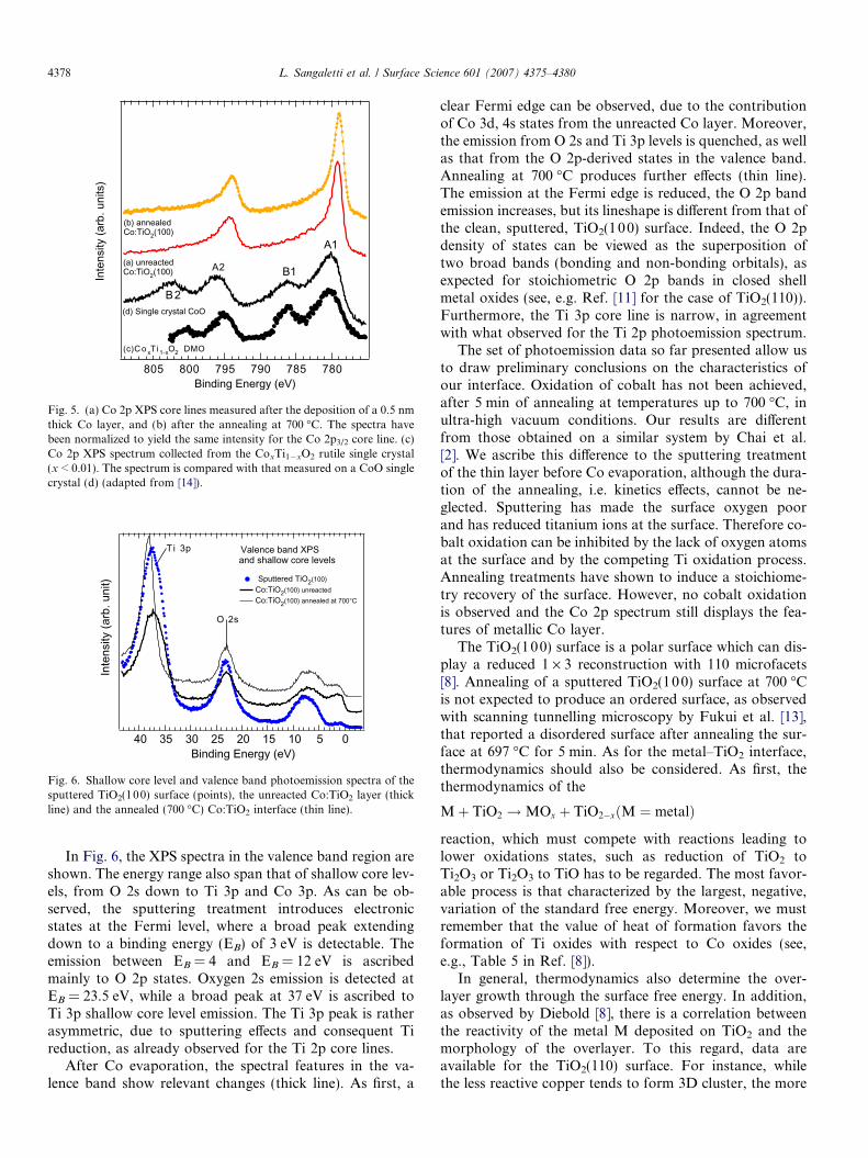
Fig. 3. MicroRaman mapping of the annealed Co:TiO2(100) surfaceacross the border between the clean TiO2 region and the sputtered, andCo-covered, region. The Raman spectra (bottom panel) have beencollected along the white line (top panel) from the clean layer to thedeposited region. The length of the white line is 3 lm. The data have beencollected ex situ at room temperature after the final annealing at 700 �C inultra-high vacuum conditions.
L. Sangaletti et al. / Surface Science 601 (2007) 4375–4380 4377
(which has been covered with a mask during the Co evap-oration) and the sputtered, and Co-covered, region. InFig. 3 (bottom panel) we show the Raman spectra collectedfrom the sample in the region showed in Fig. 3 (top panel)along the white line. The spectrum from the stoichiometricTiO2 layer with no Co deposition is the typical one ob-served in single crystals in the proper scattering geometryfor the rutile-type TiO2, with the main Ag Raman activemode peaked at 610 cm�1. In this scattering geometry theEg mode at �445 cm�1 is mostly forbidden. Analyzingthe lineshape of the Ag mode, we derived a linewidth
parameter of �43.2 cm�1, which is very close to the line-width measured in single crystal TiO2 [10]. This is a confir-mation that the stoichiometry of the clean TiO2 surface isvery close to that of the pure material. Moving towardsthe deposited region we observe two main changes in theRaman spectra: (i) the intensity of the whole spectrum de-creases markedly entering in the Co-deposited layer and (ii)the polarization selection rules tend to weaken, as one canappreciate from the intensity ratio between the allowed Ag
mode and the forbidden Eg mode (Fig. 4). This behaviorsuggests the presence of disorder on the topmost surfacelayers. Further analysis on lineshape of Raman bands, inparticular on the linewidth of Ag mode, allow us to say thatthe disorder is mainly an orientational disorder rather thanstructural disorder: indeed the value for linewdith of Ag
mode of the Raman spectrum taken from the deposited re-gion (i.e. the bottom spectrum in Fig. 3) is �44.0 cm�1,very close to the value obtained from the clean TiO2 layer.This indicates that the structure and stoichiometry ismainly preserved and the disorder is mainly due to differentorientation of micro- or nano-regions.
The Co 2p XPS core lines measured after the depositionof a 0.5 nm thick Co layer and after the annealing at thehighest temperature (i.e. 700 �C) are shown in Fig. 5aand b. The spectra show the two spin-orbit split compo-nents of Co 2p emission, separated by about 16 eV. Thespectra are rather similar to each other, and the lineshapeis that characteristic of metallic cobalt, even after theannealing at 700 �C.
Fig. 4. Ratio between the peak intensities of the Ag mode and Eg modefrom the uncovered TiO2 layer, corresponding to 0 position on the X-axis,to the Co-deposited region.
Inte
nsity
(arb
. uni
ts)
805 800 795 790 785 780Binding Energy (eV)
A1
B1A2
B2
(a) unreactedCo:TiO2(100)
(b) annealedCo:TiO2(100)
(d) Single crystal CoO
(c)C o xT i1-xO2 DMO
Fig. 5. (a) Co 2p XPS core lines measured after the deposition of a 0.5 nmthick Co layer, and (b) after the annealing at 700 �C. The spectra havebeen normalized to yield the same intensity for the Co 2p3/2 core line. (c)Co 2p XPS spectrum collected from the CoxTi1�xO2 rutile single crystal(x < 0.01). The spectrum is compared with that measured on a CoO singlecrystal (d) (adapted from [14]).
Inte
nsity
(arb
. uni
t)
40 35 30 25 20 15 10 5 0Binding Energy (eV)
Valence band XPSand shallow core levels
Sputtered TiO2(100)Co:TiO2(100) unreactedCo:TiO2(100) annealed at 700°C
T i 3p
O 2s
Fig. 6. Shallow core level and valence band photoemission spectra of thesputtered TiO2(100) surface (points), the unreacted Co:TiO2 layer (thickline) and the annealed (700 �C) Co:TiO2 interface (thin line).
4378 L. Sangaletti et al. / Surface Science 601 (2007) 4375–4380
In Fig. 6, the XPS spectra in the valence band region areshown. The energy range also span that of shallow core lev-els, from O 2s down to Ti 3p and Co 3p. As can be ob-served, the sputtering treatment introduces electronicstates at the Fermi level, where a broad peak extendingdown to a binding energy (EB) of 3 eV is detectable. Theemission between EB = 4 and EB = 12 eV is ascribedmainly to O 2p states. Oxygen 2s emission is detected atEB = 23.5 eV, while a broad peak at 37 eV is ascribed toTi 3p shallow core level emission. The Ti 3p peak is ratherasymmetric, due to sputtering effects and consequent Tireduction, as already observed for the Ti 2p core lines.
After Co evaporation, the spectral features in the va-lence band show relevant changes (thick line). As first, a
clear Fermi edge can be observed, due to the contributionof Co 3d, 4s states from the unreacted Co layer. Moreover,the emission from O 2s and Ti 3p levels is quenched, as wellas that from the O 2p-derived states in the valence band.Annealing at 700 �C produces further effects (thin line).The emission at the Fermi edge is reduced, the O 2p bandemission increases, but its lineshape is different from that ofthe clean, sputtered, TiO2(100) surface. Indeed, the O 2pdensity of states can be viewed as the superposition oftwo broad bands (bonding and non-bonding orbitals), asexpected for stoichiometric O 2p bands in closed shellmetal oxides (see, e.g. Ref. [11] for the case of TiO2(110)).Furthermore, the Ti 3p core line is narrow, in agreementwith what observed for the Ti 2p photoemission spectrum.
The set of photoemission data so far presented allow usto draw preliminary conclusions on the characteristics ofour interface. Oxidation of cobalt has not been achieved,after 5 min of annealing at temperatures up to 700 �C, inultra-high vacuum conditions. Our results are differentfrom those obtained on a similar system by Chai et al.[2]. We ascribe this difference to the sputtering treatmentof the thin layer before Co evaporation, although the dura-tion of the annealing, i.e. kinetics effects, cannot be ne-glected. Sputtering has made the surface oxygen poorand has reduced titanium ions at the surface. Therefore co-balt oxidation can be inhibited by the lack of oxygen atomsat the surface and by the competing Ti oxidation process.Annealing treatments have shown to induce a stoichiome-try recovery of the surface. However, no cobalt oxidationis observed and the Co 2p spectrum still displays the fea-tures of metallic Co layer.
The TiO2(100) surface is a polar surface which can dis-play a reduced 1 · 3 reconstruction with 110 microfacets[8]. Annealing of a sputtered TiO2(10 0) surface at 700 �Cis not expected to produce an ordered surface, as observedwith scanning tunnelling microscopy by Fukui et al. [13],that reported a disordered surface after annealing the sur-face at 697 �C for 5 min. As for the metal–TiO2 interface,thermodynamics should also be considered. As first, thethermodynamics of the
Mþ TiO2 !MOx þ TiO2�xðM ¼ metalÞ
reaction, which must compete with reactions leading tolower oxidations states, such as reduction of TiO2 toTi2O3 or Ti2O3 to TiO has to be regarded. The most favor-able process is that characterized by the largest, negative,variation of the standard free energy. Moreover, we mustremember that the value of heat of formation favors theformation of Ti oxides with respect to Co oxides (see,e.g., Table 5 in Ref. [8]).
In general, thermodynamics also determine the over-layer growth through the surface free energy. In addition,as observed by Diebold [8], there is a correlation betweenthe reactivity of the metal M deposited on TiO2 and themorphology of the overlayer. To this regard, data areavailable for the TiO2(110) surface. For instance, whilethe less reactive copper tends to form 3D cluster, the more
L. Sangaletti et al. / Surface Science 601 (2007) 4375–4380 4379
reactive hafnium forms a continuous overlayer [9]. Accord-ing to the values for the heat of formation of the most sta-ble oxide of the metals, the oxidation of cobalt is slightlymore favorable than that of copper, but far from the haf-nium case. Therefore, for Co a growth close to 3D clusteris expected. These thermodynamics remarks would explainthe two, seemingly independent, processes observed uponannealing: titanium oxide recovers its stoichiometry, whilecobalt, that is expected to grow as separated 3D clustersalso because of the low coverage, remains metallic, at leastfor the 5 min annealing treatments up to 700 �C.
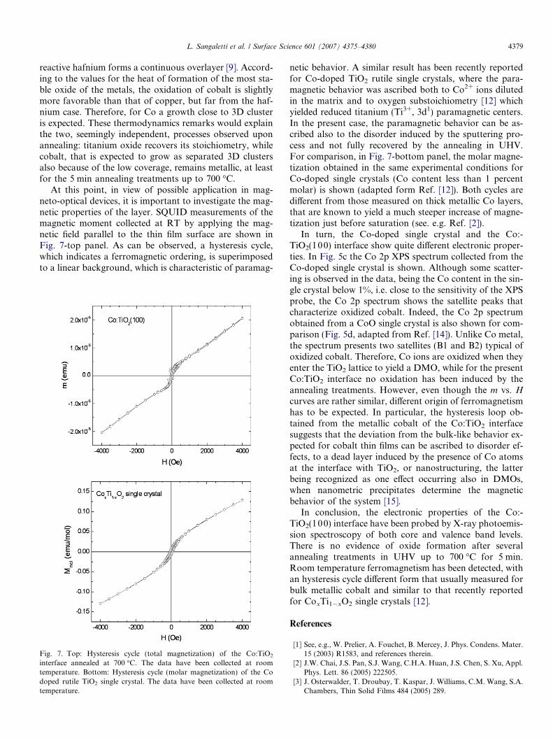
At this point, in view of possible application in mag-neto-optical devices, it is important to investigate the mag-netic properties of the layer. SQUID measurements of themagnetic moment collected at RT by applying the mag-netic field parallel to the thin film surface are shown inFig. 7-top panel. As can be observed, a hysteresis cycle,which indicates a ferromagnetic ordering, is superimposedto a linear background, which is characteristic of paramag-
Fig. 7. Top: Hysteresis cycle (total magnetization) of the Co:TiO2
interface annealed at 700 �C. The data have been collected at roomtemperature. Bottom: Hysteresis cycle (molar magnetization) of the Codoped rutile TiO2 single crystal. The data have been collected at roomtemperature.
netic behavior. A similar result has been recently reportedfor Co-doped TiO2 rutile single crystals, where the para-magnetic behavior was ascribed both to Co2+ ions dilutedin the matrix and to oxygen substoichiometry [12] whichyielded reduced titanium (Ti3+, 3d1) paramagnetic centers.In the present case, the paramagnetic behavior can be as-cribed also to the disorder induced by the sputtering pro-cess and not fully recovered by the annealing in UHV.For comparison, in Fig. 7-bottom panel, the molar magne-tization obtained in the same experimental conditions forCo-doped single crystals (Co content less than 1 percentmolar) is shown (adapted form Ref. [12]). Both cycles aredifferent from those measured on thick metallic Co layers,that are known to yield a much steeper increase of magne-tization just before saturation (see. e.g. Ref. [2]).
In turn, the Co-doped single crystal and the Co:-TiO2(100) interface show quite different electronic proper-ties. In Fig. 5c the Co 2p XPS spectrum collected from theCo-doped single crystal is shown. Although some scatter-ing is observed in the data, being the Co content in the sin-gle crystal below 1%, i.e. close to the sensitivity of the XPSprobe, the Co 2p spectrum shows the satellite peaks thatcharacterize oxidized cobalt. Indeed, the Co 2p spectrumobtained from a CoO single crystal is also shown for com-parison (Fig. 5d, adapted from Ref. [14]). Unlike Co metal,the spectrum presents two satellites (B1 and B2) typical ofoxidized cobalt. Therefore, Co ions are oxidized when theyenter the TiO2 lattice to yield a DMO, while for the presentCo:TiO2 interface no oxidation has been induced by theannealing treatments. However, even though the m vs. Hcurves are rather similar, different origin of ferromagnetismhas to be expected. In particular, the hysteresis loop ob-tained from the metallic cobalt of the Co:TiO2 interfacesuggests that the deviation from the bulk-like behavior ex-pected for cobalt thin films can be ascribed to disorder ef-fects, to a dead layer induced by the presence of Co atomsat the interface with TiO2, or nanostructuring, the latterbeing recognized as one effect occurring also in DMOs,when nanometric precipitates determine the magneticbehavior of the system [15].
In conclusion, the electronic properties of the Co:-TiO2(100) interface have been probed by X-ray photoemis-sion spectroscopy of both core and valence band levels.There is no evidence of oxide formation after severalannealing treatments in UHV up to 700 �C for 5 min.Room temperature ferromagnetism has been detected, withan hysteresis cycle different form that usually measured forbulk metallic cobalt and similar to that recently reportedfor CoxTi1�xO2 single crystals [12].
References
[1] See, e.g., W. Prelier, A. Fouchet, B. Mercey, J. Phys. Condens. Mater.15 (2003) R1583, and references therein.
[2] J.W. Chai, J.S. Pan, S.J. Wang, C.H.A. Huan, J.S. Chen, S. Xu, Appl.Phys. Lett. 86 (2005) 222505.
[3] J. Osterwalder, T. Droubay, T. Kaspar, J. Williams, C.M. Wang, S.A.Chambers, Thin Solid Films 484 (2005) 289.
4380 L. Sangaletti et al. / Surface Science 601 (2007) 4375–4380
[4] Y. Matsumoto, M. Murakami, T. Shono, T. Hasegawa, T. Fukum-ura, M. Kawasaki, P. Ahmet, T. Chikyow, S.-Y. Koshihara, H.Koinuma, Science 291 (2001) 854.
[5] S.A. Chambers, S. Thevuthasan, R.F.C. Farrow, R.F. Marks, J.U.Thiele, L. Folks, M.G. Samant, A.J. Kellock, N. Ruzycki, D.L.Ederer, U. Diebold, Appl. Phys. Lett. 79 (2001) 3467.
[6] W.K. Park, R.J.O. Hertogs, J.S. Moodera, A. Punnoose, M.S.Seehra, J. Appl. Phys. 91 (2002) 8093.
[7] N.-J. Seong, S.-G. Yoon, C.-R. Cho, Appl. Phys. Lett. 81 (2002)4209.
[8] U. Diebold, Surf. Sci. Rep. 48 (2003) 53.[9] U. Diebold, J.M. Pan, T.E. Madey, Surf. Sci. 333 (1995) 845.
[10] P.S. Porto, P.A. Fleury, T.C. Damen, Phys. Rev. 154 (1967) 522.
[11] U. Diebold, H.S. Tao, N.D. Shinn, T.E. Madey, Phys. Rev. B 50(1994) 14474.
[12] L. Sangaletti, M.C. Mozzati, C.B. Azzoni, P. Galinetto, A. Speghini,M. Bettinelli, C. Calestani, J. Phys. Condens. Mater. 18 (2006)7643.
[13] K.-i. Fukui, R. Tero, Y. Iwasawa, Jpn. J. Appl. Phys. 40 (2001) 4331,Part 1.
[14] F. Parmigiani, L. Sangaletti, J. Elect. Spectrosc. Rel. Phenom. 98–99(1999) 287.
[15] S.R. Shinde, S.B. Ogale, J.S. Higgins, H. Zheng, A.J. Millis, V.N.Kulkarni, R. Ramesh, R.L. Greene, T. Venkatesan, Phys. Rev. Lett.92 (2004) 166601.


![(2Z,N 0 0 0 E)-N 0 0 0 -[(2-Hydroxy-1-naphthyl)- methylidene]furan-2-carbohydrazonic acid](https://static.fdokumen.com/doc/165x107/631360d4b033aaa8b2100e91/2zn-0-0-0-e-n-0-0-0-2-hydroxy-1-naphthyl-methylidenefuran-2-carbohydrazonic.jpg)