Low-level plasma HIVs in patients on prolonged suppressive highly active antiretroviral therapy are...
14
Low-level Plasma HIVs in Patients on Prolonged Suppressive Highly Active Antiretroviral Therapy Are Produced Mostly by Cells Other Than CD4 T-cells Gautam K. Sahu 1,* , David Paar 2 , Simon D. W. Frost 3 , Melissa M. Smith 1 , Scott Weaver 1,4 , and Miles W. Cloyd 1 1 Department of Microbiology and Immunology, University of Texas Medical Branch, 301 University Blvd, BSB #3.132, Galveston, TX 77555, USA 2 UTMB-Correctional Managed Care, University of Texas Medical Branch, 301 University Blvd, BSB #3.132, Galveston, TX 77555, USA 3 Department of Pathology, University of California San Diego, La Jolla, California, 92093 4 Department of Pathogy, University of Texas Medical Branch, 301 University Blvd, BSB #3.132, Galveston, TX 77555, USA Abstract The cellular source(s) and the clinical significance of persistent low-level viremia, below 50 HIV- RNA copies per ml of plasma, achieved in many patients with high adherence to highly active antiretroviral therapy (HAART) remain unclear. Also, it is not clear if residual plasma HIVs during HAART can become predominant populations in the rebounding plasma viral loads after therapy interruption. Since, different HIV quasispecies tend to compartmentalize in various cell types and tissue locations in patients during chronic infection, the phylogenetic relationships between HIV sequences amplified from residual plasma viruses and CD4 T cells of five patients on long-term suppressive therapy were examined. Three of these patients stopped therapy voluntarily for three weeks, but only one of them demonstrated viral load rebound in plasma. In phylogenetic analyses, the residual plasma viruses were found to be distinct genetically from the majority of CD4 T cell-associated virus populations in 4 of 5 patients. The compartmental analyses revealed that in all patients, plasma- and CD4 T cell-derived viral sequences were compartmentalized separately. Interestingly, the plasma sequences obtained before and after HAART-off in two patients were produced apparently from the same compartment, which was different from the circulating CD4 T cell-compartment. These results suggest the possibility that residual plasma viruses in patients on long-term suppressive HAART may be produced persistently from a cellular source yet to be identified, and are capable of spreading quickly in vivo, accounting for the rapid rebound of viral loads in plasma after therapy interruption. Keywords HIV+ patients; suppressive therapy; residual plasma viral loads; HAART-interruption; virus compartmentalization * Corresponding author: Phone: (409) 772-6829, Fax: (409) 747-6869, [email protected]. NIH Public Access Author Manuscript J Med Virol. Author manuscript; available in PMC 2010 July 29. Published in final edited form as: J Med Virol. 2009 January ; 81(1): 9–15. doi:10.1002/jmv.21366. NIH-PA Author Manuscript NIH-PA Author Manuscript NIH-PA Author Manuscript
Transcript of Low-level plasma HIVs in patients on prolonged suppressive highly active antiretroviral therapy are...

Low-level Plasma HIVs in Patients on Prolonged SuppressiveHighly Active Antiretroviral Therapy Are Produced Mostly byCells Other Than CD4 T-cells
Gautam K. Sahu1,*, David Paar2, Simon D. W. Frost3, Melissa M. Smith1, Scott Weaver1,4,and Miles W. Cloyd1
1 Department of Microbiology and Immunology, University of Texas Medical Branch, 301University Blvd, BSB #3.132, Galveston, TX 77555, USA2 UTMB-Correctional Managed Care, University of Texas Medical Branch, 301 University Blvd,BSB #3.132, Galveston, TX 77555, USA3 Department of Pathology, University of California San Diego, La Jolla, California, 920934 Department of Pathogy, University of Texas Medical Branch, 301 University Blvd, BSB #3.132,Galveston, TX 77555, USA
AbstractThe cellular source(s) and the clinical significance of persistent low-level viremia, below 50 HIV-RNA copies per ml of plasma, achieved in many patients with high adherence to highly activeantiretroviral therapy (HAART) remain unclear. Also, it is not clear if residual plasma HIVsduring HAART can become predominant populations in the rebounding plasma viral loads aftertherapy interruption. Since, different HIV quasispecies tend to compartmentalize in various celltypes and tissue locations in patients during chronic infection, the phylogenetic relationshipsbetween HIV sequences amplified from residual plasma viruses and CD4 T cells of five patientson long-term suppressive therapy were examined. Three of these patients stopped therapyvoluntarily for three weeks, but only one of them demonstrated viral load rebound in plasma. Inphylogenetic analyses, the residual plasma viruses were found to be distinct genetically from themajority of CD4 T cell-associated virus populations in 4 of 5 patients. The compartmentalanalyses revealed that in all patients, plasma- and CD4 T cell-derived viral sequences werecompartmentalized separately. Interestingly, the plasma sequences obtained before and afterHAART-off in two patients were produced apparently from the same compartment, which wasdifferent from the circulating CD4 T cell-compartment. These results suggest the possibility thatresidual plasma viruses in patients on long-term suppressive HAART may be producedpersistently from a cellular source yet to be identified, and are capable of spreading quickly invivo, accounting for the rapid rebound of viral loads in plasma after therapy interruption.
KeywordsHIV+ patients; suppressive therapy; residual plasma viral loads; HAART-interruption; viruscompartmentalization
*Corresponding author: Phone: (409) 772-6829, Fax: (409) 747-6869, [email protected].
NIH Public AccessAuthor ManuscriptJ Med Virol. Author manuscript; available in PMC 2010 July 29.
Published in final edited form as:J Med Virol. 2009 January ; 81(1): 9–15. doi:10.1002/jmv.21366.
NIH
-PA Author Manuscript
NIH
-PA Author Manuscript
NIH
-PA Author Manuscript
INTRODUCTIONHIV-1 is member of the Lentivirus genus of the family Retroviridae and gradually depletesCD4 T cells in infected humans, leading to immunodeficiency and ultimately death.HAART consisting of viral protease and reverse-transcriptase inhibitors reduces the viralloads in plasma in many patients to below the detection limit (~50 copies of HIV-RNA perml) of currently available HIV RNA load assays [Gulick et al., 1997; Hammer et al., 1997;Luzuriaga et al., 1997]; however, it cannot completely eliminate HIV from patients[Blankson et al., 2002]. It is clear that virus production continues at levels below 50 copies/ml of plasma during suppressive HAART [Dornadula et al., 1999; Gunthard et al., 1999;Martinez-Picado et al., 2000; Hermankova et al., 2001; Havlir et al., 2003; Palmer et al.,2003; Kieffer et al., 2004; Nettles et al., 2004; Nettles et al., 2005; Tobin et al., 2005] andplasma viral loads return to detectable levels in most cases when therapy is stopped [Daveyet al., 1999; Garcia et al., 1999; Mata et al., 2005]. Although a very small percentage ofhighly stable, resting memory CD4+ T lymphocytes (~1–10 cells per 106 CD4 cells) areknown to carry latent, but replication-competent proviral DNA, even after long-term drugtreatment [Chun et al., 1997; Wong et al., 1997; Hermankova et al., 2003], the cellularsource(s) of persistent low-level viremia below 50 copies/ml of plasma during suppressivetherapy remain unclear.
In the presence of protease and reverse transcriptase inhibitors used in HAART, the residualviruses circulating in blood likely remain immature and therefore, non-infectious, but aftertherapy withdrawal, they should be produced as mature and infectious particles. However, itis unclear if these residual plasma viruses can spread rapidly in the body after HAART-withdrawal, resulting in the quick return of detectable plasma viral loads in patients.Because of error-prone HIV replication, a high-degree of genetic heterogeneity in viralpopulations is observed, even within an infected individual [Saag et al., 1988; Shankarappaet al., 1999]. The viral quasispecies also tend to compartmentalize in the various tissues andcell-types in patients during chronic infections [refs in Zarate et al., 2007]. It is not clear ifthe viruses present in plasma during suppressive therapy are compartmentalized in theircellular source(s). This study attempted to investigate the phylogenetic relationshipsbetween HIVs present in CD4 T cells versus residual plasma HIVs in five patients onsuppressive HAART, as well as plasma HIVs isolated 3 weeks after HAART-withdrawalfrom three of them.
METHODSStudy subjects
Five HIV-positive individuals (designated as P1, P2, P3, P4 and P5), who were treated withHAART for a long time in the Infectious disease clinic at UTMB and had viral loads of lessthan or around 50 copies per ml in plasma for the last two years or more (except anyoccasional blips in plasma viral loads), were studied. The study subjects signed the consentforms approved by the Internal Review Board (IRB), UTMB. About 50 ml of blood wascollected from each by venipuncture at the enrollment. HAART was withdrawn from threeof these patients (i.e., P3, P4 and P5) for a period of three weeks only and every week, bloodsamples were collected and CD4 counts and viral loads in plasma were monitored. Theblood samples were collected in 2002 and later analyzed.
Blood processing and isolation of viral nucleic acids from plasma and CD4 T cellsPeripheral blood mononuclear cells (PBMCs) and plasma were separated using the ficoll-hypaque method. Then CD4 T-cells were isolated from PBMCs by a negative selectionmethod (StemCell Technologies Inc, Canada) described elsewhere [Sahu et al., 2006]. Total
Sahu et al. Page 2
J Med Virol. Author manuscript; available in PMC 2010 July 29.
NIH
-PA Author Manuscript
NIH
-PA Author Manuscript
NIH
-PA Author Manuscript

DNA was isolated from CD4+ T cells by using the Wizard genomic DNA purification kit(Promega, USA) and stored at −20°C. Plasma was centrifuged at 850 × g for 20 min at 4°Cin order to remove any contaminating blood cells or cell-debris. About 20 ml of plasma wasthen ultracentrifuged at 120,000 × g for 2h at 4°C to pellet the virus particles. Viral RNAswere isolated using a commercial kit (High Pure Viral RNA kit, Roche, USA).
Amplification, cloning and sequencing of HIV-tat regionsHIV-tat regions were amplified by nested reverse transcription (RT)-polymerase chainreaction (PCR) and nested PCR methods from about one-third volume of isolated plasmaviral RNA (in 50 μl) and ~3 μg of CD4 T cells’ DNA, respectively. The superscript-III one-step RT-PCR system with platinum Taq DNA polymerase (Invitrogen) and the Expand HighFidelity PCR system (Roche) were used to amplify tat from plasma RNA and CD4 T-cell’sDNA, respectively. Five microliters of PCR product was taken for second round PCR in a50μl volume using the Expand High Fidelity PCR system (Roche). The appropriate primersand programs were used. PCR-products were cloned and sequenced.
Phylogenetic and compartmental analysesThe nucleotide sequences were aligned using CLUSTAL-W algorithms and phylogenetictrees were constructed by using neighbor joining, maximum parsimony and maximumlikelihood methods implemented in the PAUP 4.0 software package. The HKY85 formulawas used for distance calculations [Swofford, 1998].
Compartmental analysis utilized the Slatkin-Maddison (SM) test implemented in the HyPhypackage developed by Kosakovsky Pond, Frost and Muse [Pond et al., 2005]. The SM test[Slatkin and Maddison, 1989] determines the minimum number of migrations (M) betweenthe separated sequence groups (compartments) in the constructed phylogenetic tree and thep-values are obtained using number of migration events (M) that can be expected in arandomly structured populations, derived by permuting sequences between groups. The p-values of <0.01 were considered as the evidence of compartmentalization.
RESULTSBlood samples were collected from each patient at enrollment and viral loads weremeasured. All patients’ plasma viral loads remained below 50 copies per ml of plasma,except in one patient, P3, who had viral load of 66 copies/ml (Table I). The mean durationof undetectable viral loads prior to enrollment was ~2.5 yrs, except that each of patients P1and P4 encountered only one blip in plasma viral loads of 476 and 793 copies/ml,respectively (Table I).
Although plasma viral loads remain undetectable by clinical tests in these patients duringtherapy, HIV-tat regions from purified plasma HIV RNAs in 4 of 5 patients still could beamplified (Table I) by following ultracentrifugation/RT-PCR-methods described by others[Kieffer et al., 2005]. Three weeks of HAART interruption in three patients (i.e., P3, P4 andP5) was done to examine the genetic relationship between rebounding virus populations inplasma versus CD4-associated viruses. Only in one patient (P3), viral loads in plasmaincreased marginally (up to 372 copies/ml), whereas the other two patients (P4 and P5) didnot demonstrate any rises (i.e., above 50 copies/ml) in plasma viral loads during these threeweeks of HAART-interruption (Fig. 1). However, viral sequences still could be amplifiedfrom plasma of patient P4, but not patient P5, after 3 weeks of HAART-off usingultracentrifugation/RT-PCR methods.
Various phylogenetic methods (such as neighbor joining, maximum parsimony andlikelihood methods) showed that patient-specific tat sequences derived from plasma
Sahu et al. Page 3
J Med Virol. Author manuscript; available in PMC 2010 July 29.
NIH
-PA Author Manuscript
NIH
-PA Author Manuscript
NIH
-PA Author Manuscript
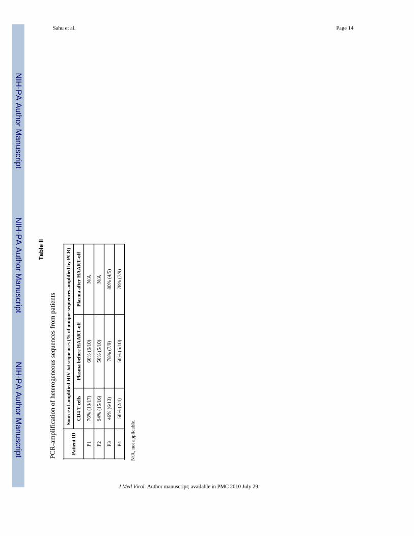
clustered separately, as expected, in all phylogenetic trees that were constructed (notshown). Using these methods, CD4 T cell-derived tat sequences were also found to clusterwith plasma viral sequences in the patient-specific manner in the same clades of allphylogenetic trees, ruling out cross-contamination among samples or from laboratory strains(not shown). The tat region was chosen for PCR-amplification because, in addition tophylogenetic analyses, the functionality of isolated tat sequences in HIV LTR-transactivation assays could also be tested. Using routine nested PCR methods with high fidelityenzymes, heterogeneous viral populations from patients’ samples were amplified. Forexample, about 46–94% of amplified sequences were found to be unique (Table II). Thissuggests that intensive template-resampling bias in PCRs was not occurring as it ought tooccur when the target copy number is low [Liu et al., 1996]. Although tat sequences isolatedfrom each patient formed distinct groups in all phylogenetic trees expectedly (as mentionedabove), compartment-specific (such as CD4 T cell- and plasma-derived) sequences in eachpatient appeared to cluster separately (Fig. 2). For example, in both patients P1 and P2,plasma viral populations formed a distinct cluster from the majority of the CD4 T cell-associated viruses in phylogenetic trees (Fig. 2). The plasma tat sequences derived frompatients P3 and P4 also clustered relatively distantly from their CD4 T cell-associated tatsequences (either obtained from direct amplification in total CD4 T cells’ DNA or fromreplicating virus in culture)(Fig. 2). Interestingly, viral tat sequences isolated from patientP3’s CD4 T cell-depleted PBMCs grouped with the plasma-derived sequences obtainedbefore and after HAART-withdrawal (Fig. 2).
To check if plasma and CD4 T cell-associated sequences are actually compartmentalized,compartmental analysis was done using the Slatkin-Maddison (SM) test (see materials andmethods). Essentially, the p-values and mean number of migration events (M) between twoseparated sequence-groups were calculated (Fig. 3). The p-values of <0.01 were consideredevidence of compartmentalization. The lower M values indicate less traffic between twoseparated groups, providing greater evidence for close phylogenetic relatedness betweensequences within each group. Patient P1 and P2: Using CD4 T cell- and plasma-derivedsequences, SM test yielded p- and M-values of <0.001 and 1, respectively, in both thesepatients (Fig. 3, panel P1 and P2), suggesting that these sequences are compartmentalized intheir corresponding sources. Patient P3: This patient showed moderate rebound in plasmaviral loads after 3 weeks of HAART-interruption. When the CD4 sequence group (shown asa box) was compared with any other sequence-groups (see Fig. 3, panel P3), p values alwaysremained less than 0.001 and M values remained at 1, suggesting that CD4 sequences form adistinct compartment. When two different plasma sequence groups (lower two boxes in Fig3, panel P3) and PBMC-minus CD4 T cell-derived sequence-group (top right box) werecompared in various combinations (broken arrows), p-values always remained higher than0.01, demonstrating that these three sequence-groups actually represent a singlecompartment. These data suggest that the viruses circulating in plasma at low-levels duringHAART were capable of spreading infection in the body after HAART-interruption andemerged as the predominant population in rebounding viral loads. Furthermore, it is alsosuggestive that non-CD4 T cells could be the cellular source for residual plasma virusesduring suppressive therapy. Patient P4: Although this patient did not show rebound inplasma viral loads (i.e. >50 copies/ml) after 3 weeks of HAART-off, the viral sequences inplasma were amplified and compared them with the sequences obtained from plasma priorto therapy-interruption. The data obtained from the SM test (Fig. 3, panel P4) suggest that,similar to patient P3, this patient also had plasma and CD4-derived sequencescompartmentalized. The residual plasma viral sequences amplified before or after HAART-off were genetically similar, suggesting that they were chronically produced from the samecompartment (p = 0.038, M= 4).
Sahu et al. Page 4
J Med Virol. Author manuscript; available in PMC 2010 July 29.
NIH
-PA Author Manuscript
NIH
-PA Author Manuscript
NIH
-PA Author Manuscript

DISCUSSIONThe successful amplifications of HIV sequences from plasma having viral loads of <50copies/ml showed that viral production still continues at very low-levels in patientsreceiving long-term effective HAART, which is in agreement with previous reports byothers [Dornadula et al., 1999; Gunthard et al., 1999; Martinez-Picado et al., 2000;Hermankova et al., 2001; Havlir et al., 2003; Palmer et al., 2003; Kieffer et al., 2004; Nettleset al., 2004; Nettles et al., 2005; Tobin et al., 2005]. This study has shown that low-levelviruses in the plasma of 4 of 5 patients receiving long-term suppressive HAART werephylogenetically distinct from the majority of viral populations present in CD4 T cells, amajor cellular reservoir for HIV (Fig 2 and 3). Using the SM test for sequencecompartmentalization, it was found that sequences derived from CD4 T cells or plasma ofthese patients were, in fact, compartmentalized in their sources. Many studies have observedthis compartmentalization phenomenon in various tissue locations, providing evidence forcell type-specific viral adaptation during chronic HIV infections [refs in Zarate et al., 2007].Moreover, because of suppressive therapy, the virus spreading between various tissue sitesin the body are impeded severely, but viral replication still can be active locally due to sub-optimal concentrations of drugs, resulting in micro-evolution and therefore, increase ingenetic distances between viral populations residing in such sites [Si-Mohamed et al., 2000;Smit et al., 2004; Delobel et al., 2005]. The low-level plasma viruses in these patients onsuppressive HAART could be produced from such a cell type in which viral populationsmight have evolved independently, but have been restricted to this reservoir in the presenceof HAART. An alternative explanation is that the amplified plasma viral sequences may notbe representative of all the viral populations residing in CD4 T cells and therefore, theobservation can be misinterpreted as sequence compartmentalization. In all 4 patients,heterogeneous viral sequences from both CD4 T cells and plasma were amplified by PCR(Table II), and none of these patients showed any exception to sequencecompartmentalization. Furthermore, sequences amplified from plasma that were collected attwo different time-points in patient P3 and P4 showed a high degree of genetic similarityamong each other in phylogenetic and SM tests, relative to the sequences amplified fromCD4 T cells. This demonstrated that the observed compartmentalization of HIV sequenceswas not an artifact. These findings suggested that the production of low-level plasma virusesduring effective HAART was not because of time-to-time reactivation of CD4 T-cell-associated latent viral quasispecies. If this would occur, then the plasma and CD4 T cell-derived viruses would not be compartmentalized separately.
In patient P3 who encountered viral load rebound after therapy withdrawal (Fig. 1), the datashowed greater evidence of close phylogenetic relatedness between plasma viral sequencesobtained before and after therapy withdrawal, than between plasma sequences from any timepoints versus CD4-derived sequences (Fig 2 and 3). These data demonstrate, for the firsttime, that the residual viruses are capable of rekindling infection and spreading in the bodywhen drug-mediated inhibition is lifted. This rekindling causes quick rebound of viral loadsin plasma and much of those viruses do not arise from CD4 T cell-associated viralpopulations, as similarly suggested by others [Chun et al., 2000;Ramratnam et al., 2000].However, the resumption of baseline viral loads after HAART-off likely requires virusproduction from CD4 T-cells [Riva et al., 2003;Tierney et al., 2003]. Although anotherpatient P4 did not show any rises in plasma viral loads (i.e., >50 copies/ml) after therapy-off,the closer phylogenetic relatedness between plasma sequences isolated before and aftertherapy cessation, relative to CD4 T cell-associated replicating viruses, led us to speculatethat a stable non-CD4 cellular site might be making HIVs persistently at low-levels, evenduring therapy.
Sahu et al. Page 5
J Med Virol. Author manuscript; available in PMC 2010 July 29.
NIH
-PA Author Manuscript
NIH
-PA Author Manuscript
NIH
-PA Author Manuscript
Although ~11% of tat clones isolated from patient P3’s plasma were defective intranscriptional trans-activation function (not shown), the corresponding viruses were beingproduced still from its’ cellular reservoir, which was either activated persistently, or did notrequire active Tat for low-level HIV-production. Moreover, protease region analysesrevealed that none of the clones isolated from plasma possessed mutations known to conferresistance to nelfinavir, a protease inhibitor, which was used in the HAART regimen to treatthis patient (not shown). These further indicate that a stable cellular source producing drug-sensitive HIVs persistently may be responsible for low-level viruses in plasma duringtherapy.
It is not clear which cell-type(s) could be the source for low-level plasma viruses in patientson suppressive HAART. However, intriguingly, in patient P3 the residual viruses werefound to be very close in terms of phylogenetic relatedness to the sequences obtained fromCD4 T cell-depleted PBMCs, which mainly consist of monocytes and NK cells as additionalcellular reservoirs for HIV. Since these cell types are known to produce HIV RNA inpatients during HAART [Sonza et al., 2001; Zhu et al., 2002], it can be speculated that theymight be candidates for low-level viruses in plasma during therapy. Additionally, HIV-infected CD4 T cells residing in the gut may also be such a source [Poles et al., 2006].Although the sample size (n=5) in this study is small, the provocative data obtained in thestudy may inspire studies on a larger number of patients to identify any specific cellularsource(s) responsible for residual plasma viruses during therapy. If successful, itsidentification would help in developing strategies for complete suppression of residualviremia in patients during pronged effective HAART, which could delay the emergence ofdrug-resistant mutants during low-adherence to HAART and also prevent rapid spreading ofinfection during HAART interruptions.
AcknowledgmentsThis work was partially funded by General Clinical Research Center, UTMB, and partially by National Institutes ofHealth (grant: AI 062453). SDWF was supported by the National Institutes of Health (grants AI47745, AI57167).
ReferencesBlankson JN, Persaud D, Siliciano RF. The challenge of viral reservoirs in HIV-1 infection. Annu Rev
Med. 2002; 53:557–93. [PubMed: 11818490]Chun TW, Carruth L, Finzi D, Shen X, DiGiuseppe JA, Taylor H, Hermankova M, Chadwick K,
Margolick J, Quinn TC, Kuo YH, Brookmeyer R, Zeiger MA, Barditch-Crovo P, Siliciano RF.Quantification of latent tissue reservoirs and total body viral load in HIV-1 infection. Nature. 1997;387:183–8. [PubMed: 9144289]
Chun TW, Davey RT Jr, Ostrowski M, Shawn Justement J, Engel D, Mullins JI, Fauci AS.Relationship between pre-existing viral reservoirs and the re-emergence of plasma viremia afterdiscontinuation of highly active anti-retroviral therapy. Nat Med. 2000; 6:757–61. [PubMed:10888923]
Davey RT Jr, Bhat N, Yoder C, Chun TW, Metcalf JA, Dewar R, Natarajan V, Lempicki RA,Adelsberger JW, Miller KD, Kovacs JA, Polis MA, Walker RE, Falloon J, Masur H, Gee D, BaselerM, Dimitrov DS, Fauci AS, Lane HC. HIV-1 and T cell dynamics after interruption of highly activeantiretroviral therapy (HAART) in patients with a history of sustained viral suppression.Proceedings of the National Academy of Sciences of the United States of America. 1999;96:15109–14. [PubMed: 10611346]
Delobel P, Sandres-Saune K, Cazabat M, L’Faqihi FE, Aquilina C, Obadia M, Pasquier C, Marchou B,Massip P, Izopet J. Persistence of distinct HIV-1 populations in blood monocytes and naive andmemory CD4 T cells during prolonged suppressive HAART. Aids. 2005; 19:1739–50. [PubMed:16227781]
Sahu et al. Page 6
J Med Virol. Author manuscript; available in PMC 2010 July 29.
NIH
-PA Author Manuscript
NIH
-PA Author Manuscript
NIH
-PA Author Manuscript
Dornadula G, Zhang H, VanUitert B, Stern J, Livornese L Jr, Ingerman MJ, Witek J, Kedanis RJ,Natkin J, DeSimone J, Pomerantz RJ. Residual HIV-1 RNA in blood plasma of patients takingsuppressive highly active antiretroviral therapy. JAMA. 1999; 282:1627–32. [see comment].[PubMed: 10553788]
Garcia F, Plana M, Vidal C, Cruceta A, O’Brien WA, Pantaleo G, Pumarola T, Gallart T, Miro JM,Gatell JM. Dynamics of viral load rebound and immunological changes after stopping effectiveantiretroviral therapy. AIDS. 1999; 13:F79–86. [PubMed: 10449278]
Gulick RM, Mellors JW, Havlir D, Eron JJ, Gonzalez C, McMahon D, Richman DD, Valentine FT,Jonas L, Meibohm A, Emini EA, Chodakewitz JA. Treatment with indinavir, zidovudine, andlamivudine in adults with human immunodeficiency virus infection and prior antiretroviral therapy.New England Journal of Medicine. 1997; 337:734–9. [see comment]. [PubMed: 9287228]
Gunthard HF, Frost SD, Leigh-Brown AJ, Ignacio CC, Kee K, Perelson AS, Spina CA, Havlir DV,Hezareh M, Looney DJ, Richman DD, Wong JK. Evolution of envelope sequences of humanimmunodeficiency virus type 1 in cellular reservoirs in the setting of potent antiviral therapy.Journal of Virology. 1999; 73:9404–12. [PubMed: 10516049]
Hammer SM, Squires KE, Hughes MD, Grimes JM, Demeter LM, Currier JS, Eron JJ Jr, Feinberg JE,Balfour HH Jr, Deyton LR, Chodakewitz JA, Fischl MA. A controlled trial of two nucleosideanalogues plus indinavir in persons with human immunodeficiency virus infection and CD4 cellcounts of 200 per cubic millimeter or less. AIDS Clinical Trials Group 320 Study Team. NewEngland Journal of Medicine. 1997; 337:725–33. [see comment]. [PubMed: 9287227]
Havlir DV, Strain MC, Clerici M, Ignacio C, Trabattoni D, Ferrante P, Wong JK. Productive infectionmaintains a dynamic steady state of residual viremia in human immunodeficiency virus type 1-infected persons treated with suppressive antiretroviral therapy for five years. J Virol. 2003;77:11212–9. [PubMed: 14512569]
Hermankova M, Ray SC, Ruff C, Powell-Davis M, Ingersoll R, D’Aquila RT, Quinn TC, Siliciano JD,Siliciano RF, Persaud D. HIV-1 drug resistance profiles in children and adults with viral load of<50 copies/ml receiving combination therapy. Jama. 2001; 286:196–207. [PubMed: 11448283]
Hermankova M, Siliciano JD, Zhou Y, Monie D, Chadwick K, Margolick JB, Quinn TC, Siliciano RF.Analysis of human immunodeficiency virus type 1 gene expression in latently infected restingCD4+ T lymphocytes in vivo. Journal of Virology. 2003; 77:7383–92. [PubMed: 12805437]
Kieffer TL, Finucane MM, Nettles RE, Quinn TC, Broman KW, Ray SC, Persaud D, Siliciano RF.Genotypic analysis of HIV-1 drug resistance at the limit of detection: virus production withoutevolution in treated adults with undetectable HIV loads. J Infect Dis. 2004; 189:1452–65.[PubMed: 15073683]
Kieffer TL, Kwon P, Nettles RE, Han Y, Ray SC, Siliciano RF. G-->A hypermutation in protease andreverse transcriptase regions of human immunodeficiency virus type 1 residing in resting CD4+ Tcells in vivo. J Virol. 2005; 79:1975–80. [PubMed: 15650227]
Liu SL, Rodrigo AG, Shankarappa R, Learn GH, Hsu L, Davidov O, Zhao LP, Mullins JI. HIVquasispecies and resampling. Science. 1996; 273:415–6. [PubMed: 8677432]
Luzuriaga K, Bryson Y, Krogstad P, Robinson J, Stechenberg B, Lamson M, Cort S, Sullivan JL.Combination treatment with zidovudine, didanosine, and nevirapine in infants with humanimmunodeficiency virus type 1 infection. New England Journal of Medicine. 1997; 336:1343–9.[PubMed: 9134874]
Martinez-Picado J, DePasquale MP, Kartsonis N, Hanna GJ, Wong J, Finzi D, Rosenberg E, GunthardHF, Sutton L, Savara A, Petropoulos CJ, Hellmann N, Walker BD, Richman DD, Siliciano R,D’Aquila RT. Antiretroviral resistance during successful therapy of HIV type 1 infection.Proceedings of the National Academy of Sciences of the United States of America. 2000;97:10948–53. [PubMed: 11005867]
Mata RC, Viciana P, de Alarcon A, Lopez-Cortes LF, Gomez-Vera J, Trastoy M, Cisneros JM.Discontinuation of antiretroviral therapy in patients with chronic HIV infection: clinical, virologic,and immunologic consequences. AIDS Patient Care & Stds. 2005; 19:550–62. [PubMed:16164382]
Nettles RE, Kieffer TL, Kwon P, Monie D, Han Y, Parsons T, Cofrancesco J Jr, Gallant JE, Quinn TC,Jackson B, Flexner C, Carson K, Ray S, Persaud D, Siliciano RF. Intermittent HIV-1 viremia
Sahu et al. Page 7
J Med Virol. Author manuscript; available in PMC 2010 July 29.
NIH
-PA Author Manuscript
NIH
-PA Author Manuscript
NIH
-PA Author Manuscript
(Blips) and drug resistance in patients receiving HAART. JAMA. 2005; 293:817–29. [PubMed:15713771]
Nettles RE, Kieffer TL, Simmons RP, Cofrancesco J Jr, Moore RD, Gallant JE, Persaud D, SilicianoRF. Genotypic resistance in HIV-1-infected patients with persistently detectable low-level viremiawhile receiving highly active antiretroviral therapy. Clin Infect Dis. 2004; 39:1030–7. [PubMed:15472857]
Palmer S, Wiegand AP, Maldarelli F, Bazmi H, Mican JM, Polis M, Dewar RL, Planta A, Liu S,Metcalf JA, Mellors JW, Coffin JM. New real-time reverse transcriptase-initiated PCR assay withsingle-copy sensitivity for human immunodeficiency virus type 1 RNA in plasma. J ClinMicrobiol. 2003; 41:4531–6. [PubMed: 14532178]
Poles MA, Boscardin WJ, Elliott J, Taing P, Fuerst MM, McGowan I, Brown S, Anton PA. Lack ofdecay of HIV-1 in gut-associated lymphoid tissue reservoirs in maximally suppressed individuals.J Acquir Immune Defic Syndr. 2006; 43:65–8. [PubMed: 16936559]
Pond SL, Frost SD, Muse SV. HyPhy: hypothesis testing using phylogenies. Bioinformatics. 2005;21:676–9. [PubMed: 15509596]
Ramratnam B, Mittler JE, Zhang L, Boden D, Hurley A, Fang F, Macken CA, Perelson AS,Markowitz M, Ho DD. The decay of the latent reservoir of replication-competent HIV-1 isinversely correlated with the extent of residual viral replication during prolonged anti-retroviraltherapy. Nat Med. 2000; 6:82–5. [PubMed: 10613829]
Riva E, Antonelli G, Scagnolari C, Pistello M, Capobianchi MR, Monforte A, Pezzotti P, Dianzani F.Human immunodeficiency virus (HIV) DNA load and level of immunosuppression in treatment-naive HIV-1-infected patients. J Infect Dis. 2003; 187:1826–8. [PubMed: 12751043]
Saag MS, Hahn BH, Gibbons J, Li Y, Parks ES, Parks WP, Shaw GM. Extensive variation of humanimmunodeficiency virus type-1 in vivo. Nature. 1988; 334:440–4. [PubMed: 3405290]
Sahu GK, Lee K, Ji J, Braciale V, Baron S, Cloyd MW. A novel in vitro system to generate and studylatently HIV-infected long-lived normal CD4+ T-lymphocytes. Virology. 2006; 355:127–37.[PubMed: 16919704]
Shankarappa R, Margolick JB, Gange SJ, Rodrigo AG, Upchurch D, Farzadegan H, Gupta P, RinaldoCR, Learn GH, He X, Huang XL, Mullins JI. Consistent viral evolutionary changes associatedwith the progression of human immunodeficiency virus type 1 infection. J Virol. 1999; 73:10489–502. [PubMed: 10559367]
Si-Mohamed A, Kazatchkine MD, Heard I, Goujon C, Prazuck T, Aymard G, Cessot G, Kuo YH,Bernard MC, Diquet B, Malkin JE, Gutmann L, Belec L. Selection of drug-resistant variants in thefemale genital tract of human immunodeficiency virus type 1-infected women receivingantiretroviral therapy. J Infect Dis. 2000; 182:112–22. [PubMed: 10882588]
Slatkin M, Maddison WP. A cladistic measure of gene flow inferred from the phylogenies of alleles.Genetics. 1989; 123:603–13. [PubMed: 2599370]
Smit TK, Brew BJ, Tourtellotte W, Morgello S, Gelman BB, Saksena NK. Independent evolution ofhuman immunodeficiency virus (HIV) drug resistance mutations in diverse areas of the brain inHIV-infected patients, with and without dementia, on antiretroviral treatment. J Virol. 2004;78:10133–48. [PubMed: 15331746]
Sonza S, Mutimer HP, Oelrichs R, Jardine D, Harvey K, Dunne A, Purcell DF, Birch C, Crowe SM.Monocytes harbour replication-competent, non-latent HIV-1 in patients on highly activeantiretroviral therapy. Aids. 2001; 15:17–22. [PubMed: 11192864]
Swofford, DL. PAUP*. Phylogenetic Analysis Using Parsimony (*and Other Methods). Version 4.Sinauer Associates; Sunderland, Massachusetts: 1998.
Tierney C, Lathey JL, Christopherson C, Bettendorf DM, D’Aquila RT, Hammer SM, KatzensteinDA. Prognostic value of baseline human immunodeficiency virus type 1 DNA measurement fordisease progression in patients receiving nucleoside therapy. J Infect Dis. 2003; 187:144–8.[PubMed: 12508159]
Tobin NH, Learn GH, Holte SE, Wang Y, Melvin AJ, McKernan JL, Pawluk DM, Mohan KM, LewisPF, Mullins JI, Frenkel LM. Evidence that low-level viremias during effective highly activeantiretroviral therapy result from two processes: expression of archival virus and replication ofvirus. Journal of Virology. 2005; 79:9625–34. [PubMed: 16014925]
Sahu et al. Page 8
J Med Virol. Author manuscript; available in PMC 2010 July 29.
NIH
-PA Author Manuscript
NIH
-PA Author Manuscript
NIH
-PA Author Manuscript
Wong JK, Hezareh M, Gunthard HF, Havlir DV, Ignacio CC, Spina CA, Richman DD. Recovery ofreplication-competent HIV despite prolonged suppression of plasma viremia. Science. 1997;278:1291–5. [see comment]. [PubMed: 9360926]
Zarate S, Pond SL, Shapshak P, Frost SD. Comparative study of methods for detecting sequencecompartmentalization in human immunodeficiency virus type 1. J Virol. 2007; 81:6643–51.[PubMed: 17428864]
Zhu T, Muthui D, Holte S, Nickle D, Feng F, Brodie S, Hwangbo Y, Mullins JI, Corey L. Evidence forhuman immunodeficiency virus type 1 replication in vivo in CD14(+) monocytes and its potentialrole as a source of virus in patients on highly active antiretroviral therapy. Journal of Virology.2002; 76:707–16. [PubMed: 11752161]
Sahu et al. Page 9
J Med Virol. Author manuscript; available in PMC 2010 July 29.
NIH
-PA Author Manuscript
NIH
-PA Author Manuscript
NIH
-PA Author Manuscript
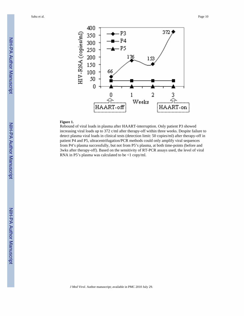
Figure 1.Rebound of viral loads in plasma after HAART-interruption. Only patient P3 showedincreasing viral loads up to 372 c/ml after therapy-off within three weeks. Despite failure todetect plasma viral loads in clinical tests (detection limit: 50 copies/ml) after therapy-off inpatient P4 and P5, ultracentrifugation/PCR methods could only amplify viral sequencesfrom P4’s plasma successfully, but not from P5’s plasma, at both time-points (before and3wks after therapy-off). Based on the sensitivity of RT-PCR assays used, the level of viralRNA in P5’s plasma was calculated to be <1 copy/ml.
Sahu et al. Page 10
J Med Virol. Author manuscript; available in PMC 2010 July 29.
NIH
-PA Author Manuscript
NIH
-PA Author Manuscript
NIH
-PA Author Manuscript
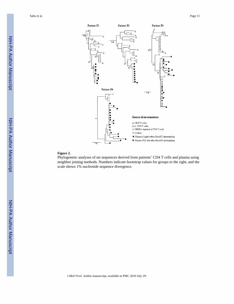
Figure 2.Phylogenetic analyses of tat sequences derived from patients’ CD4 T cells and plasma usingneighbor joining methods. Numbers indicate bootstrap values for groups to the right, and thescale shows 1% nucleotide sequence divergence.
Sahu et al. Page 11
J Med Virol. Author manuscript; available in PMC 2010 July 29.
NIH
-PA Author Manuscript
NIH
-PA Author Manuscript
NIH
-PA Author Manuscript
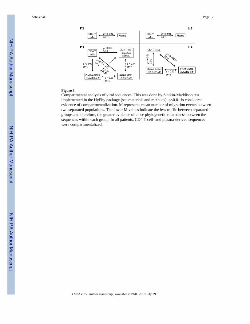
Figure 3.Compartmental analysis of viral sequences. This was done by Slatkin-Maddison testimplemented in the HyPhy package (see materials and methods). p<0.01 is consideredevidence of compartmentalization. M represents mean number of migration events betweentwo separated populations. The lower M values indicate the less traffic between separatedgroups and therefore, the greater evidence of close phylogenetic relatedness between thesequences within each group. In all patients, CD4 T cell- and plasma-derived sequenceswere compartmentalized.
Sahu et al. Page 12
J Med Virol. Author manuscript; available in PMC 2010 July 29.
NIH
-PA Author Manuscript
NIH
-PA Author Manuscript
NIH
-PA Author Manuscript

NIH
-PA Author Manuscript
NIH
-PA Author Manuscript
NIH
-PA Author Manuscript
Sahu et al. Page 13
Tabl
e I
HIV
pat
ient
s, an
tiret
rovi
ral r
egim
ens,
dura
tion
of tr
eatm
ent,
CD
4 co
unts
and
pla
sma
vira
l loa
ds a
t enr
ollm
ent i
n th
e st
udy
Patie
nt ID
Ant
iret
rovi
ral d
rugs
at
enro
llmen
t
Dur
atio
n of
AR
Tbe
fore
enr
ollm
ent
(yea
rs)
Blo
od C
D4
coun
ts a
ten
rollm
ent (
cells
/μl)
PVL
at e
nrol
lmen
t(c
opie
s/m
l)
Dur
atio
n of
unde
tect
able
PV
Lbe
fore
sam
plin
g(y
ears
)
No.
of v
iral
blip
s in
last
3 ye
ars b
efor
een
rollm
ent
Det
ectio
n of
HIV
-RN
Ain
pla
sma
by R
T-P
CR
at
enro
llmen
t
P1A
ZT, I
DV
, 3TC
798
6<5
02.
51
(476
c/m
l)ye
s
P2A
ZT, I
DV
, 3TC
6.9
923
<50
30
yes
P3A
ZT, 3
TC, N
FV5.
834
266
20
yes
P4A
BC
, AZT
, 3TC
3.8
602
<50
2.2
1 (7
93 c
/ml)
yes
P5dd
I, d4
T, N
FV6.
752
8<5
02.
70
no
Abb
revi
atio
ns: A
RT,
ant
iretro
vira
l the
rapy
; PV
L, p
lasm
a vi
ral l
oads
; AB
C, a
baca
vir;
AZT
, zid
ovud
ine;
NFV
, nel
finav
ir; d
dI, d
idan
osin
e; d
4T, s
tavu
dine
; ID
V, i
ndin
avir;
3TC
, lam
ivud
ine.
PV
L w
ere
mea
sure
d du
ring
HA
AR
T on
eve
ry v
isit
occu
rrin
g at
3–4
mon
th-in
terv
als.
How
ever
, the
se w
ere
mea
sure
d ev
ery
wee
k af
ter H
AA
RT-
inte
rrup
tion.
Vira
l loa
ds o
f >40
0 co
pies
/ml i
n pl
asm
a du
ring
HA
AR
Tw
as in
clud
ed a
s vira
l blip
s
J Med Virol. Author manuscript; available in PMC 2010 July 29.
NIH
-PA Author Manuscript
NIH
-PA Author Manuscript
NIH
-PA Author Manuscript
Sahu et al. Page 14
Tabl
e II
PCR
-am
plifi
catio
n of
het
erog
eneo
us se
quen
ces f
rom
pat
ient
s
Patie
nt ID
Sour
ce o
f am
plifi
ed H
IV-ta
t seq
uenc
es (%
of u
niqu
e se
quen
ces a
mpl
ified
by
PCR
)
CD
4 T
cel
lsPl
asm
a be
fore
HA
AR
T-o
ffPl
asm
a af
ter
HA
AR
T-o
ff
P176
% (1
3/17
)60
% (6
/10)
N/A
P294
% (1
5/16
)50
% (5
/10)
N/A
P346
% (6
/13)
78%
(7/9
)80
% (4
/5)
P450
% (2
/4)
50%
(5/1
0)78
% (7
/9)
N/A
, not
app
licab
le.
J Med Virol. Author manuscript; available in PMC 2010 July 29.