Low Cost and Ecofriendly Phytosynthesis of Silver Nanoparticles Using Cassia roxburghii Stem Extract...
19
American Journal of Advanced Drug Delivery www.ajadd.co.uk American Journal of Advanced Drug Delivery www.ajadd.co.uk Original Article Low Cost and Ecofriendly Phytosynthesis of Silver Nanoparticles Using Cassia roxburghii Stem Extract and its Antimicrobial and Antioxidant Efficacy PoojaMoteriya and SumitraChanda* Phytochemical, Pharmacological and Microbiological Laboratory, Department of Biosciences, Saurashtra University - Rajkot, 360 005, Gujarat, India ABSTRACT A new and novel approach for the green synthesis of silver nanoparticles is the need of the hour. Use of plant parts for the synthesis of silver nanoparticles is ecofriendly, economic and cost effective. In the present paper, silver nanoparticles were synthesized using aqueous stem extract of Cassia roxburghii DC as a reducing agent. The biosynthesized AgNPs were characterized by various spectral analysis like UV–Vis, FTIR, XRD, TEM and Zeta potential measurement. UV–vis spectra showed maxima absorption peak at 432 nm. XRD and TEM analysis revealed AgNPs to be face- centered, cubic structures spherical in shape with an average particle size of 32-35 nm. The synergistic antibacterial activity was evaluated against two Gram positive, two Gram negative bacteria and four fungi with fifteen commercial antibiotics alone and antibiotics plus synthesized AgNPs. Antioxidant activity of AgNPs was evaluated by ABTS and FRAP assay. The AgNPs showed synergistic antibacterial activity even better than some antibiotics and also good antioxidant activity. The results suggest that Cassia roxburghii stem could be exploited for the fabrication of AgNPs with potential therapeutic application in nanomedicine especially against multi drug resistant microorganisms which are cost effective and ecofriendly and simple. They can be definitely used in cosmetics, medical and pharmaceutical applications Keywords: Cassia roxburghii, Green synthesis, Silver nanoparticles, Spectral analysis, Antimicrobial. Date of Receipt- 09/08/2014 Date of Revision- 22/08/2014 Date of Acceptance- 27/08/2014 Address for Correspondence Phytochemical, Pharmacological and Microbiological Laboratory, Department of Biosciences, Saurashtra University - Rajkot, 360 005, Gujarat, India E-mail: [email protected]
-
Upload
independent -
Category
Documents
-
view
3 -
download
0
Transcript of Low Cost and Ecofriendly Phytosynthesis of Silver Nanoparticles Using Cassia roxburghii Stem Extract...

American Journal of Advanced Drug Delivery
www.ajadd.co.uk
American Journal of Advanced Drug Delivery www.ajadd.co.uk
Original Article
Low Cost and Ecofriendly Phytosynthesis of Silver Nanoparticles Using Cassia roxburghii Stem Extract and its Antimicrobial and Antioxidant Efficacy
PoojaMoteriya and SumitraChanda*
Phytochemical, Pharmacological and Microbiological Laboratory, Department of Biosciences, Saurashtra University - Rajkot, 360 005, Gujarat, India
ABSTRACT
A new and novel approach for the green synthesis of silver nanoparticles is the need of the hour. Use of plant parts for the synthesis of silver nanoparticles is ecofriendly, economic and cost effective. In the present paper, silver nanoparticles were synthesized using aqueous stem extract of Cassia roxburghii DC as a reducing agent. The biosynthesized AgNPs were characterized by various spectral analysis like UV–Vis, FTIR, XRD, TEM and Zeta potential measurement. UV–vis spectra showed maxima absorption peak at 432 nm. XRD and TEM analysis revealed AgNPs to be face-centered, cubic structures spherical in shape with an average particle size of 32-35 nm. The synergistic antibacterial activity was evaluated against two Gram positive, two Gram negative bacteria and four fungi with fifteen commercial antibiotics alone and antibiotics plus synthesized AgNPs. Antioxidant activity of AgNPs was evaluated by ABTS and FRAP assay. The AgNPs showed synergistic antibacterial activity even better than some antibiotics and also good antioxidant activity. The results suggest that Cassia roxburghii stem could be exploited for the fabrication of AgNPs with potential therapeutic application in nanomedicine especially against multi drug resistant microorganisms which are cost effective and ecofriendly and simple. They can be definitely used in cosmetics, medical and pharmaceutical applications
Keywords: Cassia roxburghii, Green synthesis, Silver nanoparticles, Spectral analysis, Antimicrobial.
Date of Receipt- 09/08/2014 Date of Revision- 22/08/2014 Date of Acceptance- 27/08/2014
Address for Correspondence Phytochemical, Pharmacological and Microbiological Laboratory, Department of Biosciences, Saurashtra University - Rajkot, 360 005, Gujarat, India
E-mail: [email protected]

Chanda et al____________________________________________________ ISSN 2321-547X
AJADD[2][4][2014]557-575
INTRODUCTION
There are no qualms that antimicrobial agents have safeguarded the humanity from torment of infectious diseases. Mankind has faced the dangerous attacks of microorganisms and tried to endure it with the use of antibiotics or antimicrobial agents. However, multidrug-resistant bacteria are becoming more common and they are frequently resistant to almost all the current antibiotics. Many biochemical and physiological mechanisms may be responsible for resistance1.Drug-resistant bacterial infections because considerable patient mortality and morbidity and rising antibiotic resistance is seriously threatening the vast medical advancements made possible by antibiotics over the past years2.The need of the hour suggests that new approaches are required to combat emerging infections and the global spread of drug-resistant bacterial pathogens3,4.Free radicals are extremely reactive species that cause oxidative damage to various biomolecules like lipids, proteins, DNA in human body and are responsible for a number of pathologies like cardiovascular diseases, diabetes, cancer, Parkinson’s disease, Alzheimer’s disease, acquired immunodeficiency syndrome, Huntington’s disease and many other chronic diseases5,6. Antioxidants are compounds that react with free radicals and prevent undesirable oxidation processes. Unlike synthetic drugs, antimicrobials and antioxidants of natural origin from plants are not associated with any side effects and that is why they are becoming more and more popular. The plant extracts provide ample opportunities for new drug discoveries because of the enormous diversity present in them7. All plants have same or different secondary metabolites which are responsible for its biological activity8,9.
The Cassia genus belonging to the family Fabaceae represents one of the large
and most diverse group of flowering plants including herbs and trees. They are widely distributed in most tropical and subtropical countries. Cassia species have biological and pharmacological activities and have many medicinal uses in traditional system of medicine. They are reported for antimicrobial and antioxidant activity10, nephroprotective activity11, antidiabetic activity12, hepatoprotective activity13, immunomodulatory activity14 etc. Cassia roxburghii is one of the medicinal plants used in ethnomedicine for the treatment of various liver ailments15.The therapeutic applicability of silver and medicinal plants in treating bacterial infections is well known16, 17. Silver nanoparticles have diverse application like molecular diagnostics, catalysis, drug delivery 18,19
including antimicrobial and antioxidantproperties20,21 .
Silver nanoparticles (NPs) can be synthesized by various chemical and biological methods. Biological method include use of enzyme, microorganism or plant extracts but the later one is preferred because plant extracts act as reducing and stabilizing agent and they also influence the shape and size of the synthesized NPs22.This is because different extracts contain different concentrations and combinations of phytoconstituents23,17; different parts of the same plant (stem, flower, leaf) may influence differently the shape and size of NPs synthesized. Silver NPs can be prepared from any part of the plant like plant extract Cleistanthuscollinus24, fruit Piper longum25, rind Bruceajavanica26, leaf Tribulu-sterrestris27, stem Shoreatumbuggaia28, etc.
Amongst different parts of the plant used for the synthesis of NPs leaf is used very frequently while stem and flowers are less attempted. Hence in the present work, stem of C. roxburghii is used for the synthesis of silver NPs. The

Chanda et al____________________________________________________ ISSN 2321-547X
AJADD[2][4][2014]557-575
characterization was done by various spectral analyses like UV-Vis, FTIR, XRD, TEM and Zeta potential measurement. The synthesized silver NPs were evaluated for their synergistic antimicrobial activity with fifteen commercial antibiotics against four pathogenic bacteria and four fungi which included one clinical isolate. The antioxidant capacities of the synthesized silver NPs were also checked by using ABTS and FRAP antioxidant assays.
MATERIALS AND METHODS
Chemicals Fresh young stem of Cassia
roxburghii DC was collected from Saurashtra University campus, Rajkot Gujarat, India. All the chemicals were obtained from Hi Media Laboratories and Sisco research Laboratories Pvt. Limited, Mumbai, India. Ultra purified water was used for experiment. Preparation of the extract
Fresh stem was thoroughly washed with tap water, followed by double distilled water and cut into small pieces. 5 g of cut stem pieces were boiled for 5 min in 100 ml ultra-pure water and filtered through Whatmann No. 1 filter paper. The filtered C. roxburghii stem extract was used for the synthesis of silver NPs. Synthesis of silver NPs
Aqueous solution (1mM) of silver nitrate (AgNO3) was prepared and used for the synthesis of silver NPs. 6ml of extract was added to 40 ml of 1 mM AgNO3 solution for the reduction of Ag+ ions. The synthesis of silver NPs was carried out at room temperature (25°C + 2°C) for 24 h in dark. Effect of boiling time
In order to standardized the effect of boiling time for the preparation of aqueous extract of C. roxburghii, 5g of cut stem pieces were taken in 100 ml ultra-pure water
and boiled it for 5 min, 10 min and 15 min. and then filtered through Whatmann No. 1 filter paper. The filtered C. roxburghii stem extract was used for the synthesis of silver NPs. Effect of extract amount
In order to standardized the effect of extract amount to be added to 1 mM AgNO3 solution, different amount of extract (1.5 ml, 3.0 ml, 4.5 ml and 6.0 ml) was added to AgNO3 solution. The formation of AgNPs was monitored as a function of time of reaction on a spectrophotometer by taking O.D. at 440 nm at an interval of 15 min. Characterization of the synthesized silver NPs
Synthesis of silver NPs solution with stem extract may be easily observed by ultraviolet – visible (UV-Vis) spectroscopy. The reduction of the Ag+ ions in solution was monitored by periodic sampling of aqueous component and measuring the UV-Vis spectra of the solution. UV-Vis spectra of these aliquots were monitored as a function of time of reaction on a spectrophotometer (Shimadzu UV-1601) in 400-700 nm range operated at a resolution of 10 nm. FTIR analysis of silver NPs
Possible functional groups involved in the synthesis and stabilization of silver NPs was studied by FTIR spectroscopy. The FTIR was recorded in the range of 400-4000 cm-1 NicoletIS10 (Thermo Scientific, USA) The various modes of vibrations were identified and assigned to determine the different functional groups present in the Cassia stem extract. Zeta Potential Measurement
Zeta potential is an essential parameter for the characterization of stability in aqueous nano suspensions. The zeta

Chanda et al____________________________________________________ ISSN 2321-547X
AJADD[2][4][2014]557-575
potential measurement was performed using a Microtra (Zetatra Instruments).
XRD measurement
The silver NPs solution thus obtained was purified by repeated centrifugation at 10000 rpm for 10 min followed by redispersion of the pellet of silver NPs into Acetone. After air drying of the purified silver particles, the structure and composition were analyzed by XRD. The dried mixture of silver NPs was collected for the determination of the formation of Ag NPs by an X’Pert Pro x-ray diffractometer (PAN analytical BV) operated at a voltage of 40 kV and a current of 30mA with Cu Kα radiation in θ- 2 θ configurations. The crystallite domain size was calculated from the width of the XRD peaks, assuming that they are free from non-uniform strains, using the Scherrer formula. D= 0.94 λ / β Cos θ where D is the average crystallite domain size perpendicular to the reflecting planes, λ is the X-ray wavelength, β is the full width at half maximum (FWHM), and θ is the diffraction angle.
TEM analysis
TEM analysis was done to visualize the shape as well as measure the diameter of the biologically synthesized silver NPs. The sample was dispersed in double distilled water. A drop ofthin dispersion was placed on a “staining mat”. Carbon coated copper grid was inserted into the drop with the coated side upwards. After about ten minutes, the grid was removed and air dried. Then screened in JEOL JEM 2100 Transmission Electron Microscope. Antimicrobial activity
The antimicrobial activity of AgNPs with 15 commercial antibiotics and antibiotics alone was determined against 2 Gram positive bacteria (Staphylococcus aureus ATCC NO 29737andBacillus cereus ATCC NO 11778) and 2 Gram negative bacteria (Escherichia
coli NCIM NO 2931and Pseudomonas aeruginosa ATCC NO.27853) and 4 fungal (Candidaalbicans NCIM NO 3102, Candidaglabrata NCIM NO 3448, Cryptococcaeneoformans NCIM NO 3542 and No.44, a clinical isolate candida, obtained from Spandan diagnostic center, Rajkot, Gujarat, India) strains, by using agar disc diffusion method29. All the microorganisms were obtained from NCL, Pune, India. Determination of antioxidant activities ABTS assay
The ABTS cation radical scavenging activity of silver NPs was measured by the method as described by30. ABTS radical cations are produced by reacting ABTS (7 mM) and potassium persulfate (2.45 mM) and incubating the mixture at room temperature in dark for 16 h. The ABTS working solution obtained was furthure diluted with methanol to give an absorbance of 0.85±0.20 at 734 nm. 1.0 ml of different concentration (1to 1000 µg ml-1) of silver NPs and fractions diluted by methanol was added 3.0 ml of ABTS working solution. The reaction mixture was incubated at room temperature for 5 min, and then the absorbance was measured at 734 nm using a UV-Vis (Systronics Spectrophotometer) against a blank sample. Ascorbic acid (1 to 10 µg ml-1) was used as a positive control. Percentage of inhibition was calculated using the formula (C-T/C *100). FRAP assay
The reducing ability of AgNPs was determined by FRAP assay31. FRAP assay is based on the ability of antioxidants to reduce Fe+3 to Fe+2 in the presence of TPTZ, forming an intense blue Fe+2 – TPTZ complex with an absorption maximum at 593 nm. This reaction is pH-dependent (optimum pH 3.6). 0.1 ml extract is added to 3.0 ml FRAP reagent [10 parts 300 mM sodium acetate buffer at pH 3.6, 1 part 10 mM TPTZ (2, 4, 6- tripyridyl-s-

Chanda et al____________________________________________________ ISSN 2321-547X
AJADD[2][4][2014]557-575
triazine) in 40 mM HCL and 1 part 20 mM Fecl3] and the reaction mixture is incubated at 37 °C for 10 min and then the absorbance was measured at 593 nm. FeSO4 (100 to 1000 µM ml-1) was used as positive control. The antioxidant capacity based on the ability to reduce ferric ions of sample was calculated from the linear calibration curve and expressed at M FeSO4 equivalents per gram of extracted compound. RESULTS
Standardization Effect of boiling time
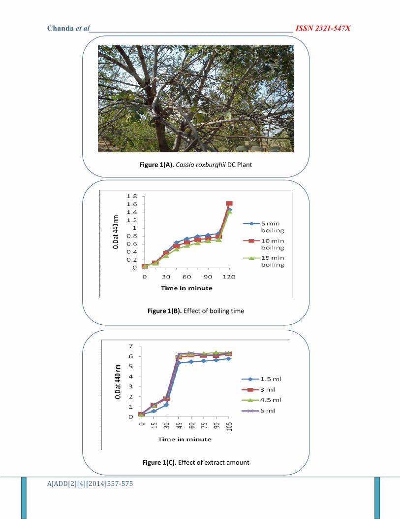
In order to evaluate the effect of boiling time for the C. roxburghii stem extract preparation, the stem was boiled for 5, 10 and 15 min. The UV-Vis spectrum of silver NPs was recorded at different time interval of reaction medium. There was slight difference in the formation of AgNPs by 5, 10 or 15 min boiling time but best was 5 min boiling time (Fig. 1B). Hence 5 min boiling time was fixed for the preparation of aqueous extract of stem of C. roxburghii. Effect of extract amount
There was a clear increase in AgNPs formation with increase in amount of extract added (Fig. 1C). Addition of 6.0 ml of extract showed substantial increase in the formation of AgNPs, so it was taken for the synthesis of NPs. Characterization
As soon as stem extract (a very light brownish solution) was added to colour less silver nitrate solution, the light brownish solution turned to brown colour and the intensity of the colour increased with time and finally turned to dark brown (Fig. 2A). UV-visspectral analysis
The UV-visible spectra of silver NPs was recorded at different time interval of the
reaction medium (0, 30, 40, 60, 120 min) using C. roxburghii stem extract with 1 mM AgNO3 (Fig. 2B). Maximum absorption peak was at 432 nm (Fig. 2B).The peak intensity increased with time indicating increase in the concentration of synthesized NPs. FTIR spectral analysis
FTIR analysis was done to identify the possible bio-reducing biomolecules in the stem extract. The spectra of AgNPs revealed strong bands at 3527.92, 2339.73, 1264.38, 966.37, 830.38, 754.19 and 667.39 respectively (Fig. 3A). The intense bands at 3527.92 cm−1 are characteristic group of primary O-H stretching of alcohols, phenols. The peak at 1264.38 cm−1 corresponds to C-N, C-O stretch of aromatic amines. The peak of 966.37 cm−1 corresponds to C-H wag (-CH2X) alkylhalides compound. The peaks of 830.38, 754.19, 667.39 are assigned to the stretching of C-Cl, C -Br respectively of alkyl halides group. Zeta potential
Zeta potential is an essential parameter for the characterization of stability in aqueous nano suspensions. The zeta potential of C. roxburghii stem AgNPs was -5.98 mv (Fig. 3B) suggesting that the surface of the NPs was negatively charged that dispersed in the medium. X-ray diffraction (XRD)
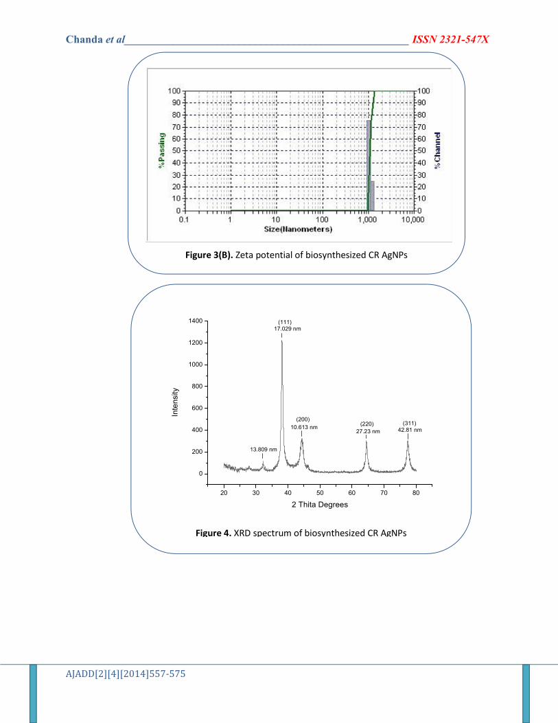
XRD analysis showed four distinct diffraction peaks at 2 theta values of 17.029, 10.613, 27.23 and 12.81 indexed to the (1 1 1), (2 0 0), (2 2 0) and (311) crystalline planes of the fcc structure of metallic silver (lattice Constant a = 4.086 A◦, was matched well with Joint Committee on Powder Diffraction Standards (JCPDS) values) (Fig.4).

Chanda et al____________________________________________________ ISSN 2321-547X
AJADD[2][4][2014]557-575
TEM analysis The optical signature of AgNPs was
elucidated in terms of the distribution of sizes and shapes observed by transmission electron microscopy (TEM) images. A drop of silver NP solution was placed on to a carbon coated Cu grid and the sample was allowed to dry. The TEM images were recorded at different magnification to find the individual particles. TEM of AgNPs synthesized using C. roxburghii stem clearly showed that the silver NPs were spherical in shape. The average size of the AgNPs was in the range of 32-35 nm (Fig. 5A, B, C). The size and selected area electron diffraction (SAED) pattern of the AgNPs synthesized using C. roxburghii stem extract was recorded confirming the crystalline nature of AgNPs (Fig. 5D).The inset in each pattern showed the respective selected region. The presence of bright circular rings in the SAED patterns confirmed the crystalline nature of the silver NPs. The spots corresponding to various orientations appearing inside the concentric rings also showed that the obtained silver NPs had a good crystallinity. Energy-dispersive (EDX) spectroscopy analysis was performed to see the presence of elemental silver (Fig. 5E). The strong signal in the silver region confirmed the formation of AgNPs. The peaks for copper and carbon were also found but these were originated from the carbon-coated copper grid used for TEM sample preparation and EDX analysis (Fig. 5E). Antimicrobial activity
In the present work, 15 commercial antibiotics were tested alone and with AGNPs against 2 Gram positive, 2 Gram negative and 4 fungi which included 1 clinical isolate. The diameter of inhibition zone and increase in fold area for the entire test organisms was measured. The antibacterial activity of AgNPs with antibiotics was better than antibiotics alone against almost all the tested bacterial strains (Tables 2).Out of 11 antibiotics tested
AgNPs showed more activity than 8-9 antibiotics against both Gram negative bacteria E. coli and P. aeruginosa; maximum increase was against E. coli (1.25).It was observed that CFP30 and TE30had highest increase in the fold area against E. coli and P. aeruginosa respectively (Table 2a). B. cereus was more susceptible than S. aureus among the two Gram positive bacteria; maximum increase was that of CC10 followed by CEP30 against B.cereus. Antifungal activity was moderate though highest increase in fold area was 1.08 by FLC10 against C. neoformans followed by KT30 against C. albicans. Clinical isolate 44 showed poor antifungal activity (Table 2b). Antioxidant activity
ABTS scavenging activity showed increase in free radical scavenging activity which increased with the increase in the concentration of the NPs (Fig. 6). The IC50 values of cassia stem AgNPs was 90 µg/ml. FRAP assay is based on the ability of antioxidants to reduce Fe3+ to Fe2+ in the presence of TPTZ, forming an intense blue Fe2+-TPTZ complex with an absorption maximum at 593 nm. This reaction is pH-dependent (optimum pH 3.6). Ferric reducing antioxidant power (FRAP) of AgNPs was 2.20 (Mg-1).
DISCUSSION
Synthesizing silver NPs by the use of plant extracts is ecofriendly, economic and non-hazaradous and this is green approach. In the present study, an attempt was made to synthesize silver NPs from C. roxburghii stem extract. For synthesis of silver NPs optimization or standardization of boiling time for aqueous extract preparation of stem of C. roxburghii and amount of extract to be added to silver nitrate solution was an important step for the biosynthesis of AgNPs. Zayed et al 32 also reported that amount of

Chanda et al____________________________________________________ ISSN 2321-547X
AJADD[2][4][2014]557-575
plant extract played a critical role in AgNPs formation.
The first indication of silver NPs formation from plant extract is visual i.e. the appearance of a yellow is brown color due to the excitation of surface plasmon vibrations33. The formation of silver NPs varies from plant to plant as evidenced from the colour change of the aqueous solution reported by Moteriya et al 34. It takes from few minutes to hours as reported by others Lukman et al. 2011, Shameli et al 35 and Chanda36. In the present work, the colour change from slight brownish colourto brown was within 15 min indicating the formation of NPs; the intensity steadily increased up to 2 h and it turned dark brown within 24 h. Similar results were reported by Christensen et al 37.
UV–Vis spectroscopy is a valuable tool to observe the formation of NPs in aqueous solution. UV-spectra revealed maximum absorption peak at 432. The peak intensity increased with time indicating increase in the concentration of synthesized NPs. Philip38reported similar results for AgNPs synthesized from Mangifereindica leaf extract. Mallikarjuna et al.39 and Sathish kumar et al. 40 reported maximum absorption peak of sliver NPs at 436 and 435 from Ocimum sanctum and Cinnamon zeylanicum extracts respectively.
In order to identify the possible biomolecules present in C. roxburghii stem which are responsible for the reduction of silver and its stabilization, FTIR measurement were carried out.
There were many functional groups present and these functional groups represent phytoconstituents like flavonoids, triterpenes and alkaloids of the stem which probably had an effective role in the green synthesis of silver NPs and also may be responsible for capping and stabilization of the synthesized NPs (Table 1). The flavonoids present in the stem extract are powerful reducing agents which may be suggestive for the formation of
AgNPs by reduction of silver nitrate. The carboxylate group presentin proteins can acts as surfactant to attach on the surface of NPs and results in AgNPs stabilization. Thus C. roxburghii stem extract may play a dual role as stabilizing and reducing agents of Ag NPs. Similar results were reported in T. purpurea leaf 41.But, the exact mechanism is unclear and needs further investigation.
The zeta potential of C. roxburghii stem AGNPs was -5.98 mv. The negative charge on the surface of the synthesized AgNPs can cause strong repulsive force among particles which may prevent from aggregation. Hence, it can be concluded that the synthesized NPs are fairly stable. The phytoconstituents present in the stem extract may be responsible for stabilizing the synthesized NPs.
The sharp peak in XRD analysis clearly indicated the crystalline nature of the synthesized AgNPs. TEM analysis revealed that C. roxburghii stem AgNPs were spherical in shape. The average size of the AgNPs was in the range of 32-35 nm. The spherical shape of NPs was reported in Piperpedicellatum extract42 and Hibiscus cannabinus extract43. The shape and size of NPs formed varies from plant to plant and part used and also the phytoconstituents present in them at the time of synthesis. Similar results were reported by Gengan et al44 Kumar et al 45.The sharp signal peak of silver strongly indicated the reduction of sliver ion by C. roxburghii stem in to elemental sliver. TEM images showed that the surfaces of the AgNPs were surrounded by a black thin layer of some material which might be due to the capping organic constituents of stem extract. Khan et al. 46 and Satish kumar et al.47 also reported that synthesized AgNPs were surrounded by a capping material present in the plant extract.
In the present study, the antimicrobial activity of synthesized AgNPs and 15 commercial antibiotics was tested against bacteria and fungi individually and in

Chanda et al____________________________________________________ ISSN 2321-547X
AJADD[2][4][2014]557-575
combination i.e. Antibiotics alone and AgNPs plus antibiotics. The diameter of zone of inhibition and increase in fold area for all the bacterial and fungal strains was measured. Increase in fold area can give an idea about the synergistic activity of the compounds tested against the bacterial or fungal strains. Increase in fold area was more against Gram negative bacteria than Gram positive bacteria. Similar results were reported by Antony et al48. Thakur et al.49 reported antibacterial activity against P. aeruginosa by Acacia Arabica gum AgNPs. Niraimathiet al.50reported antibacterial activity of AgNPs against S. aureus and E. coli. Antifungal activity was moderate though highest increase in fold area was 1.08 by FLC10 against C. neoformans followed by KT30 against C. albicans. Antifungal activity of AgNPs with commercial antibiotics is reported by Lee et al.51 and Kotakadi et al.52. We are perhaps for the first time reporting synergistic effect of 15 antibiotics with AgNPs against pathogenic microorganisms and it is a new finding.
Antioxidant property was evaluated using ABTS radical cation scavenging assay and FRAP assay. ABTS•+ is a blue chromophore generated from the oxidation of ABTS by potassium persulfate, in the presence of the plant extract, preformed cation radical gets reduced and employs a specific absorbance at 734 nm, a wavelength remote from the visible region, and it requires a short reaction time.Ferric reducing antioxidant power (FRAP) of AgNPs was 2.20 (M g-1). Abdel-Aziz et al.53 also reported antibacterial and antioxidant activity of silver NPs synthesized from Chenopodiummurale leaf extract.
In the present work, silver NPs were successfully synthesized using Cassia rohburghii stem extract. The method is simple, ecofriendly and efficient. The average size of the NPs was in the range of 32-35 nm and they were spherical in shape. The synthesized AgNPs showed very good
synergistic antibacterial activity i.e. antibiotics plus AgNPs showed more inhibitory activity than antibiotics alone and also good antioxidant activity. Thus, these ecofriendly silver NPs can be used as an excellent antimicrobial agent against multi drug resistant pathogenic microorganisms and also can be a good antioxidant agent to tackle oxidative stress related disorders. However, more work especially in vivo studies are required and studies in this direction are in progress.
ACKNOWLEDGEMENTS
The authors thanks Prof. S.P. Singh, Head, Department of Biosciences, Saurashtra University, Rajkot, Gujarat, India for providing excellent research facilities. We acknowledge the support extended by Prof. Shipra Baluja, Department of Chemistry and Prof. D. G. Kuberkar, Department of Physics, Saurashtra University for FTIR and XRD analysis of the samples. REFERENCES
1. Rad JS, Alfatemi SH, Rad MS, Iriti M. In-vitro antioxidant and antibacterial activities of Xanthium strumarium L. extracts on methicillin-susceptible and methicillin-resistant Staphylococcus aureus. Ancient Sci Life. 2013;33:109-13.
2. Hoseini Alfatemi SM, Sharifi Rad J, Sharifi Rad M, Mohsenzadeh S, JAT da Silva. Chemical composition, antioxidant activity and In vitro antibacterial activity of Achillea wilhelmsii C. Koch essential oil on methicillin-susceptible and methicillin resistant Staphylococcus aureus spp. 3 Biotech. 2014; 1-6.
3. Tillotson GS, Theriault N. New and alternative approaches to tackling antibiotic resistance. 2013; F1000 Prime Reports 1-9

Chanda et al____________________________________________________ ISSN 2321-547X
AJADD[2][4][2014]557-575
4. Sharifi Rad J, Hoseini Alfatemi SM, Sharifi Rad M. In vitro assessment of antibacterial activity of Salicorniaherbacea L. seed extracts against multidrug resistant gram positive and gram-negative bacteria. Int J Biosci. 2014;4(6): 217-222.
5. Lee SE, Hwang HJ, Ha JS, Jeong HS, Kim JH Screening of medicinal plant extracts for antioxidative activity. Life Sci. 2003; 73:167–179.
6. Sharifi Rad J, Hoseini Alfatemi SM, Sharifi Rad M, Iriti M. Free Radical Scavenging and Antioxidant Activities of Different Parts of Nitraria schoberi L. TBAP. 2014;4 (1): 44-51.
7. Sharifi Rad J, Hoseini Alfatemi SM, Sharifi Rad M, Sen DJ. Phytochemical and Antimicrobial Evaluation of the Essential Oils and Antioxidant Activity of Aqueous Extracts from Flower and Stem of Sinapisarvensis L. Am J Advan Drug Deliv. 2013; 1 (1), 001-010.
8. Moteriya P, Satasiya R, Chanda S. Evaluation of antioxidant potential and phenol and flavonoid content of some flower extracts of Saurashtra region. World J of Pharmaceut Sci. 2014; 2(7): 622-634.
9. Moteriya P, Ram J, Rathod T, Chanda S. In vitro antioxidant and antibacterial potential of leaf and stem of Gloriosasuperba L. Am J of Phytomed and ClinTherapeut. 2014; 2(6):703-787.
10. Chanda S, Kaneria M, Baravalia Y. Antioxidant and antimicrobial properties of various polar solvent extracts of stem and leaves of four Cassia species. Afr J Biotech.2012;11:2490-2503.
11. Annie S, Rajagopal PL, Malini S. Effect of Cassia auriculata Linn. root extract on cisplatin and gentamicin-induced renal injury. Phytomedicine.2005; 12:555-560.
12. SabuMC, Subburaju T. Effect of Cassia auriculata Linn. On serum glucose level,
glucose utilization by isolated rat hemidiaphragm. J Ethnopharmacol.2002; 80:203-206
13. Das S, Sarma G, Barman S. Hepatoprotective activity of aqueous extract of fruit pulp of Cassia fistula (AFCF) against carbon tetrachloride (CCl4) induced liver damage in albino rats. J Clin Diagnos Res.2008; 2:1133-1138.
14. Ali NH, Kazmi SU, Faizi S. Modulation of humoral immunity by Cassia fistula and Amoxy-Cassia. Pak J Pharm Sci.2008; 21:21-23.
15. Ntandou GFN, Banzouzi JT, Mbatchi B, Elion-Itou RDG, Etou-Ossibi AW, Ramos S, Benoit-Vical F, Abena AA, Quamba JM. Analgesic and antiinflammatory effects of Cassia siameae Lam. stem bark extracts. J Ethnopharmacol.2010; 127:108-111.
16. El-Toumy SA, El Souda SS, Mohamed TK, Brouard I, Bermejo J.Anthraquinone glycosides from Cassia roxburghii and evaluation of its free radical scavenging activity. Carbohydrate Res.2012; 360:47–51.
17. Chanda S, Rakholiya K, Dholakia K, Baravalia Y Antimicrobial, antioxidant and synergistic property of two nutraceutical plants: Terminaliacatappa L. and Colocasiaesculentum L. Turk J Biol.2013; 37:81-91.
18. Bansal V, Bharde A, Ramanathan R, Bhargava SK. Inorganic materials using 'unusual' microorganisms. Adv Colloid Interface Sci. 2012; 150: 179-182.
19. Mittal AK, Chisti Y, Banerjee UC. Synthesis of metallic nanoparticles using plant extracts. Biotechnol Adv.2013;31: 346–356.
20. Abdel-Aziz MS, Shaheen MS, El-Nekeety AA,Wahha MAA. Antioxidant and antibacterial activity of silver nanoparticles biosynthesized using

Chanda et al____________________________________________________ ISSN 2321-547X
AJADD[2][4][2014]557-575
Chenopodiummurale leaf extract. J Saudi Chem Soc. 2013;In Press.
21. Jeeva K, Thiyagarajan M, Elangovan V, Geetha N, Venkatachalam P. Caesalpiniacoriaria leaf extracts mediated biosynthesis of metallic silver nanoparticles and their antibacterial activity against clinically isolated pathogens. Indus Crops Prod.2014; 52:714– 720.
22. Rad MS, Rad JS, Heshmati GA, Miri A, Sen DJ. Biological Synthesis of Gold and Silver Nanoparticles by Nitraria schoberi Fruits. Am JAdvan Drug Deliv. 2013; 1(2): 174-179.
23. Mukunthan K, Balaji S. Cashew apple juice (Anacardiumoccidentale L.) speeds up the synthesis of silver nanoparticles. Int J Green Nanotechnol. 2012; 4:71–79.
24. Kanipandian N, Kannan S, Ramesh R, Subramanian P, Thirumurugan R. Characterization, antioxidant and cytotoxicity evaluation of green synthesized silver nanoparticles using Cleistanthuscollinus extract as surface modifier. Mat Res Bull.2014; 49:494-502.
25. Reddy NJ, Vali DN, Rani M, Rani SS. Evaluation of antioxidant, antibacterial and cytotoxic effects of green synthesized silver nanoparticles by Piper longum fruit. Mat Sci Engg C. 2014; 34:115-122.
26. Yudha SS, Notriawan D, Angasa E, Suharto TE, Hendri J, Nishinna Y. Green synthesis of silver nanoparticles using aqueous rinds extract of Bruceajavanica (L.) Merr at ambient temperature. Mat Lett. 2013; 97:181-183.
27. Ashokkumar S, Ravi S, Kathiravan V, Velmurugan S. Synthesis, characterization and catalytic activity of silver nanoparticles using Tribulusterrestris leaf extract.
Spectrochim Acta Part A: MolBiomol Spectro. 2014; 121:88-93.
28. Ankanna S, Savitramma N. Biological synthesis of silver nanoparticles by using stem of Shoreatumbuggaia Roxb. and its antimicrobial efficacy. Asian J Pharm Clinical Res.2011; 4:137-141.
29. Rakholiya K,Chanda S. In vitro interaction of certain antimicrobial agents in combination with plant extracts against some pathogenic bacterial strains. Asian Pac J Trop Biomed. 2012; S876-S880.
30. Re R, Pellergrini N, Proteggentle A, Pannala A, Yang M, Rice-Evans C. Antioxidant activity applying an improved ABTS radical cationdecolourisation assay. Free Rad Biol Med.1999; 26:1231-1237.
31. Benzie IF, Strain JJ. The ferric reducing ability of plasma (FRAP) as a measure of “antioxidant power”: the FRAP assay. Anal Biochem. 1996; 239: 70-76.
32. Zayed MF, Eisa WH, Shabaka AA. Malvaparviflora extract assisted green synthesis of silver nanoparticles. SpectrochimActa Part A: MolBiomol Spectrosco. 2012; 98:423–428.
33. Krishnaraj C, Jagan EG, Rajasekar S, Selvakumar P, Kalaichelvan PT, Mohan N. Synthesis of silver nanoparticles using Acalyphaindica leaf extracts and its antibacterial activity against water borne pathogens. Colloids Surfaces B: Biointer. 2010; 76: 50–56
34. Moteriya P, Padalia H, Jadeja R, Chanda S (2014). Screening of silver nanoparticle synthetic efficacy of some medicinal plants of Saurashtra region – a review. Natural products: Research reviews, Ed. Gupta VK, Indian Institute of Integrative Medicine, Jammu –Tavi, India Volume 3 (In press).
35. Shameli K, Ahmad MB, Al-Mulla EAJ, Ibrahim NA, Shabanzadeh P, Rustaiyan A, Abdollahi Y, Bagheri S, Mohammadi

Chanda et al____________________________________________________ ISSN 2321-547X
AJADD[2][4][2014]557-575
SA,Usman MS, Zidan M. Green biosynthesis of silver nanoparticles using Callicarpamaingayi stem bark extraction. Molecules. 2012; 17: 8506-8517.
36. ChandaS. Silver nanoparticles (medicinal plants mediated): A new generation of antimicrobials to combat microbial pathogens- a review. In : Microbial pathogens and strategies for combating them: Science, Technology and Education, Ed. Mendez-Vilas A, FORMATEX Research Center, Badajoz, Spain, pp. 2014; 1314-1323.
37. Christensen L, Vivekanandhan S, Misra1 M, Mohanty AK. Biosynthesis of silver nanoparticles using Murrayakoenigii (curry leaf): An investigation on the effect of broth concentration in reduction mechanism and particle size. Adv Matt Lett. 2011; 2:429-434.
38. Philip D.Mangiferaindica leaf-assisted biosynthesis of well-dispersed silver nanoparticles. Spectrochimica Acta Part A. 2011; 78:327–331.
39. Mallikarjuna K, Narasimha G, Dillip G R, Praveen B, Shreedhar B, Sree Lakshmi C, Reddy B V S, Deva Prasad RB. Green synthesis of silver nanoparticles using Ocimum leaf extract and their characterization. Digest J Nanomat Biostr. 2011;6(1):181-186.
40. Sathishkumar M. Sneha K. Won WS. Cho C-W. Kim S. Yun Y-S. Cynamonzeylanicum bark extract and powder mediated green synthesis of nanocrystalline silver particles and its bactericidal activity. Colloids Surf B: Biointer. 2009;73, 332- 338.
41. Ajitha B, Reddy YAK, Reddy PS. Biogenic nano-scale silver particles by Tephrosiapurpurea leaf extract and their inborn antimicrobial activity. Spectro Acta Part A. Mol Biomol Spectro. 2014; 121:164-172.
42. Tamulya C, Hazarikaa M, Borah SC, Das MR, Boruah MP. In situ biosynthesis of Ag, Au and bimetallic nanoparticles using Piper pedicellatum C.DC: Green chemistry approach. Colloid Surf B: Biointer. 2013; 102:627– 634
43. Bindhu MR, Uma devi M. Synthesis of monodispersed silver nanoparticles using Hibiscus cannabinus leaf extract and its antimicrobial activity. Spectrochim Acta Part A: MolBiomol Spectro. 2013; 101:184–190.
44. Gengan RM, Anand K, Phulukdaree A, Chuturgoon A. A549 lung cell line activity of biosynthesized silver nanoparticles using Albiziaadianthifolia leaf. Colloids Surf B: Biointer. 2013; 105: 87– 91.
45. Kumar R, Roopan SM, Prabhakarn A, Khanna VG, Chakroborty S (2012) Agricultural waste Annonasquamosa peel extract: Biosynthesis of silver nanoparticles. Spectrochim Acta Part A. 2012; 90:173– 176.
46. Khan Z, Bashir O, Hussain JI, Kumar S, Ahmad R. Effects of ionic surfactants on the morphology of silver nanoparticles using Paan (Piper betel) leaf petiole extract. Colloid Surf B: Biointer.2012; 98:85-90.
47. Sathishkumar G, Gobinath C, Karpagam K, Hemamalini V, Premkumar K,Sivaramakrishnan S. Phytosynthesis of silver nanoscale particles using Morindacitrifolia L. and its inhibitory activity against human pathogens. Colloid Surf B: Biointer.2012; 95:235-240.
48. Antony JJ, Sivalingam P, Siva D, Kamalakkannan S, Anbarasu K, Sukirtha R, Krishnan M, Achiraman S. Comparative evaluation of antibacterial activity of silver nanoparticles synthesized using Rhizophoraapiculata

Chanda et al____________________________________________________ ISSN 2321-547X
AJADD[2][4][2014]557-575
and glucose. Colloid Surf B: Biointer. 2011; 88:134– 140.
49. Thakur M, Pandey S, Mewada A, Shah R, Oza G, Sharon M. Understanding the stability of silver nanoparticles bio-fabricated using Acacia arabica (Babool gum) and its hostile effect on microorganisms. SpectroActa Part A. MolBiomolSpectro.2013; 109:344-347.
50. Niraimathi KL, Sudha V, Lavanya R, Brindha P. Biosynthesis of silver nanoparticles using Alternantherasessilis (Linn.) extract and their antimicrobial, antioxidant activities. Colloids Surf B: Biointer. 2013; 102:288– 291.
51. Lee KJ, Park SH, Govarthanan M, Hwang PH, Seo YS, Min WHC, Lee SK,
Kannan BT Oh. Synthesis of silver nanoparticles using cow milk and their antifungal activity against phytopathogens. Mat Lett. 2013; 105:128–131.
52. Kotakadi VS, Gaddam SA, Rao Y, Prasad TNVKV, Reddy AV, SaiGopal DVR. Biofabrication of silver nanoparticles using Andrographispaniculata. Eur J Med Chem. 2014; 73: 135-140.
53. Abdel-Aziz MS, Shaheen MS, El-Nekeety AA, Wahha MAA. Antioxidant and antibacterial activity of silver nanoparticles biosynthesized using Chenopodiummurale leaf extract. J Saudi Chem Soc. 2013 (In Press).
Table 1. Phytochemicals test of C. roxburghii stem powder
Test Result
Flavanoids +++
Tannins -
Phlobatanins -
Triterpenes +++
Steroids -
Saponins -
Cardiac glycoside -
Meyer’s ++
Dragondroff +++
Wagners ++
Legal’s -

Chanda et al____________________________________________________ ISSN 2321-547X
AJADD[2][4][2014]557-575
Table 2a. Synergistic activity of AgNPs of C. roxburghii stem with different standard antibiotics against Gram negative and Gram positive bacteria
Escherichia coli (NCIM NO 2931) Pseudomonas
aeruginosa (ATCC NO 27853)
Bacillus cereus (ATCC NO 11778)
Staphylococcus aureus (ATCC NO 29737)
Anti biotic
Anti biotic
(A)
Anti biotic
+ AgNPs(B)
Increase In
fold area
Anti biotic
(A)
Anti biotic
+ AgNPs(B)
Increase In
Fold area
Anti biotic
(A)
Anti biotic
+ AgNPs (B)
Increase In
fold area
Anti biotic
(A)
Anti biotic
+ AgNPs (B)
Increase In
fold area
AMP 9 10 0.23 23 26.5 0.32 - - - 33 35 0.12
PB100 9 10 0.23 13.5 16.5 0.49 11 12.2 0.23 9 10.5 0.36
Gen10 13.5 17 0.58 22 25 0.29 19.5 22.2 0.29 16 17.5 0.19
C30 25.5 26.5 0.07 14 16 0.30 33 33 0 24 27 0.26
P10 - - - 14 16 0.30 - - - 33 33 0
Ak10 17.5 21 0.44 24.5 29 0.40 22 22 0 17 18.5 0.18
TE30 22 26 0.28 26.5 35 0.74 27.5 30 0.19 28 31 0.22
CEP30 10 10 0 11 11 0 13 18 0.91 37 37 0
AMC10 9 9 0 20.5 25.5 0.54 - 9 31.5 35 0.23
CFP30 9 13.5 1.25 34 37.5 0.21 13.5 16 0.40 24 25.5 0.12
CC10 12 16 0.78 - - - 14 19.5 0.94 11.5 13.5 0.37
AMP – Ampicillin, PB100- Polymyxin, Gen10 – Gentamicin, C30 – Chloramphenicol, P10 – Penicilli-G, AK10 – Amikacin, TE30 – Tetracycline, CEP30 – Cephalothin, AMC10 – Amoxyclav, CFP30 – Cefpirome, CC10 – Clotrimazole Mean surface area of the inhibition zone was calculated for each from the mean diameter. Increase in fold area was calculated as (B2 – A2) ⁄A2, where A and B are the inhibition zones for Ag-NPs and antibiotics + Ag-NPs, respectively.
Table 2b. Synergistic activity of AgNPs of C. stem with different standard antibiotics against fungi
Candida glabrata (NCIM NO 3448)
44 Candida albicans (NCIM NO 3102)
Cryptococcae neoformans (NCIM NO 3542)
Anti fungal
Anti fungal
(A)
Anti fungal
+ AgNPs
(B)
Increase in
fold area
Anti fungal
(A)
Anti fungal
+ AgNPs(B)
Increase in
fold area
Anti fungal
(A)
Anti fungal
+ AgNPs(B)
Increase in
fold area
Anti fungal
(A)
Anti fungal
+ AgNPs(B)
Increase in
fold area
NS100 29 31 0.14 21 21.5 0.04 17 19 0.24 23 23 0
KT30 24.5 30 0.49 - - - 15 19.5 0.69 15.5 19.5 0.58
FLC10 21.5 23 0.14 - - - 20.5 23.5 0.31 13.5 19.5 1.08
AP100 15 15 0 12 12.5 0.08 10.5 11 0.09 12 12 0
NS100 – Nystatin, KT30- Ketoconazole, , FLC10 – Fluconazole, AP10 – Ampotericin ; Mean surface area of the inhibition zone was calculated for each from the mean diameter; Increase in fold area was calculated as (B2 – A2) ⁄A2, where A and B are the inhibition zones for Ag-NPs and antibiotics + Ag-NPs, respectively.

Chanda et al____________________________________________________ ISSN 2321-547X
AJADD[2][4][2014]557-575
20 30 40 50 60 70 80
0
200
400
600
800
1000
1200
1400
(311)(220)(200)
(111)
42.81 nm27.23 nm10.613 nm
17.029 nm
13.809 nm
Inte
nsity
2 Thita Degrees
Green synthesis of silver nanoparticles using C. roxburghii stem extract and AgNO3. Characterization by
spectral analysis like UV-Vis, TEM, FTIR and XRD. Evaluation of antimicrobial and antioxidant potential
Chanda et al____________________________________________________ ISSN 2321-547X
AJADD[2][4][2014]557-575
Figure 1(A). Cassia roxburghii DC Plant
Figure 1(B). Effect of boiling time
Figure 1(C). Effect of extract amount

Chanda et al____________________________________________________ ISSN 2321-547X
AJADD[2][4][2014]557-575
Figure 2(A). Color change in the reaction mixture within 2 h
Figure 2(B). UV-visible spectrum of biosynthesized CR-AgNPs at different time interval, showed peak at 432 nm
45075010501350165019502400300036001/cm
40
45
50
55
60
65
70
75
80
85
90
95
100%T
3931
.06
3527
.92
2339
.73
1264
.38
966.
37
830.
38
754.
19
667.
39
A
Figure 3(A). FTIR spectrum of biosynthesized CR AgNPs
Chanda et al____________________________________________________ ISSN 2321-547X
AJADD[2][4][2014]557-575
Figure 3(B). Zeta potential of biosynthesized CR AgNPs
20 30 40 50 60 70 80
0
200
400
600
800
1000
1200
1400
(311)(220)(200)
(111)
42.81 nm27.23 nm10.613 nm
17.029 nm
13.809 nm
Inte
nsi
ty
2 Thita Degrees
Figure 4. XRD spectrum of biosynthesized CR AgNPs

Chanda et al____________________________________________________ ISSN 2321-547X
AJADD[2][4][2014]557-575
Figure 5. TEM images (A, B, C) of Ag nanoparticles in low and high magnification, (D) SAED patterns of the silver nanoparticles, (E) EDX spectrum showed higher percentage of silver
signal

Chanda et al____________________________________________________ ISSN 2321-547X
AJADD[2][4][2014]557-575
Figure 6. ABTS radical scavenging activity of silver nanoparticles