14-3-3z Cooperates with ErbB2 to Promote Ductal Carcinoma ...
Upload
independentCategory
view
3download
0
doi:10.1182/blood-2007-07-103291Prepublished online February 7, 2008;
D. Bernstein, Randall T. Peterson and David A SweetserFarshid Dayyani, Jianfeng Wang, Jing-Ruey J Yeh, Eun-Young Ahn, Erica Tobey, Dong-Er Zhang, Irwin cooperate with AML1-ETO to affect myeloid cell proliferation and survivalLoss of TLE1 and TLE4 from the del(9q) commonly deleted region in AML
(4217 articles)Neoplasia �Articles on similar topics can be found in the following Blood collections
http://bloodjournal.hematologylibrary.org/site/misc/rights.xhtml#repub_requestsInformation about reproducing this article in parts or in its entirety may be found online at:
http://bloodjournal.hematologylibrary.org/site/misc/rights.xhtml#reprintsInformation about ordering reprints may be found online at:
http://bloodjournal.hematologylibrary.org/site/subscriptions/index.xhtmlInformation about subscriptions and ASH membership may be found online at:
digital object identifier (DOIs) and date of initial publication. theindexed by PubMed from initial publication. Citations to Advance online articles must include
final publication). Advance online articles are citable and establish publication priority; they areappeared in the paper journal (edited, typeset versions may be posted when available prior to Advance online articles have been peer reviewed and accepted for publication but have not yet
Copyright 2011 by The American Society of Hematology; all rights reserved.20036.the American Society of Hematology, 2021 L St, NW, Suite 900, Washington DC Blood (print ISSN 0006-4971, online ISSN 1528-0020), is published weekly by
For personal use only. by guest on June 5, 2013. bloodjournal.hematologylibrary.orgFrom
1
Loss of TLE1 and TLE4 from the del(9q) commonly deleted region in AML cooperate with AML1-ETO to affect myeloid cell proliferation and survival Farshid Dayyani1*, Jianfeng Wang1*, Jing-Ruey J. Yeh2, Eun-Young Ahn3, Erica Tobey1, Dong-Er Zhang3, Irwin D. Bernstein4,5, Randall T. Peterson2, and David A. Sweetser1,4 1Department of Pediatrics, Division of Pediatric Hematology/Oncology and 2Cardiovascular Research Center and Cardiology Division, Massachusetts General Hospital, and Harvard Medical School, Boston, MA; 3Department of Molecular and Experimental Medicine, The Scripps Research Institute, La Jolla, CA 92037, 4Children’s Oncology Group, Arcadia, CA; 5Department of Pediatric Oncology, Fred Hutchinson Cancer Research Center, Seattle, WA *These authors contributed equally. Correspondence: D.A. Sweetser, Massachusetts General Hospital, 55 Fruit Street – Jackson 904, Boston, MA 02114, E-mail [email protected], phone 617-724-5311, FAX 617-725-8623;
Blood First Edition Paper, prepublished online February 7, 2008; DOI 10.1182/blood-2007-07-103291
Copyright © 2008 American Society of Hematology
For personal use only. by guest on June 5, 2013. bloodjournal.hematologylibrary.orgFrom
2
Abstract Deletions on chromosome 9q are seen in a subset of acute myeloid leukemia (AML) cases and are specifically associated with t(8;21) AML. We previously defined the commonly deleted region in del(9q) AML and characterized the genes in this interval. To determine the critical lost gene(s) that might cooperate with the AML1-ETO fusion gene produced by t(8;21) we developed a set of shRNAs directed against each gene in this region. Within this library, shRNAs to TLE1 and TLE4 were the only shRNAs capable rescuing AML1-ETO expressing U937T-A/E cells from AML1-ETO induced cell cycle arrest and apoptosis. Knockdown of TLE1 or TLE4 levels increased the rate of cell division of the AML1-ETO expressing Kasumi-1 cell line, while forced expression of either TLE1 or TLE4 caused apoptosis and cell death. Knockdown of Gro3, a TLE homologue in zebrafish, cooperated with AML1-ETO to cause an accumulation of non-circulating hematopoietic blast cells. Our data is consistent with a model in which haploinsufficiency of these TLEs overcomes the negative survival and anti-proliferative effects of AML1-ETO on myeloid progenitors allowing preleukemic stem cells to expand into acute myeloid leukemia. This study is the first to implicate the TLEs as potential tumor suppressor genes in myeloid leukemia.
For personal use only. by guest on June 5, 2013. bloodjournal.hematologylibrary.orgFrom
3
Introduction One of the most common genetic aberrations in AML is the balanced chromosomal translocation t(8;21). This translocation, seen in 8-13% of de novo AML cases1-3, creates the RUNX1-MTG8/AML1-ETO fusion gene. AML1-ETO is insufficient for leukemogenesis as evidenced by mouse models4-7, the detection of fusion gene transcripts in AML patients in long term remission8,9, as well as the finding of transcripts in newborns who did not develop t(8;21) AML for over 10 years10. Although AML1-ETO expression promotes the maintenance of early hematopoietic precursors 11,12, it markedly inhibits short-term expansion of primary human bone marrow cells and the proliferation of committed progenitors and CD34+ cells13. This suggests a model in which secondary mutations are required to further transform preleukemic stem cells and allow their progeny to expand.
Deletion of a portion of the long arm of chromosome 9, del(9q), is a recurring abnormality in malignant myeloid diseases reported in about 2% of AML cases and is non-randomly associated with t(8;21). Approximately 36-50% of samples with del(9q) have t(8;21) and, conversely, 7-14% of pediatric AML samples with t(8;21) have del(9q)1,14-16. After numerical abnormalities, del(9q) is the single most common associated structural chromosomal abnormality seen with t(8;21) AML, indicating loss of function of a gene or genes on chromosome 9q may be one of the most important cooperating genes in t(8;21) AML.
In a search for this cooperating gene(s), we recently narrowed the commonly deleted region (CDR) in 43 del(9q) AML samples to less than 2.4 Mb at 9q21.32-9q21.33. There are ten known genes within, or immediately adjacent to, this region - TLE (transducin like enhancer of split) –1, TLE-4, FRMD3, UBQLN1, GKAP42, KIF27, HNRPK, SLC28A3, RMI1 (Q9H9A7) and NTRK2, and 3 novel or potential genes, RASEF, C9orf103 (ENSG00000148057), and C9orf64 (Q8N2B1) 17. Sequence analysis of the coding regions of these genes failed to identify clearly inactivating mutations in the remaining allele in del(9q) AML samples. However, the expression of several of these genes (TLE1, C9orf103, UBQLN1, KIF27, c9orf64, RMI1, and NTRK2) appeared specifically low in del(9q) AML samples and we hypothesized that haploinsufficiency, or reduced expression of a critical gene(s) in this region due to promoter mutations or epigenetic changes, cooperated with AML1-ETO or other genetic events to cause leukemia17. To identify this critical gene, we created shRNAs to knockdown expression of genes in the del(9q) CDR and sought to determine whether any of these could rescue the cell death and apoptosis caused by inducible AML1-ETO expression in the U937T-A/E cell line.
Materials and Methods Cell culture The U937T-A/E cell line expresses AML1-ETO under a control of a “tetracycline-off” repressor. Withdrawal of tetracycline induces AML1-ETO expression, which causes a block in cell cycle progression and apoptosis18 (Figure S1). The U937T-A/E cell line was cultured in RPMI with 10% fetal calf serum (FCS) (Cambrex, East Rutherford, New Jersey), 100U/ml penicillin and 100µg/ml streptomycin with 1mg/ml G418, 0.5ug/ml puromycin and 1µg/ml tetracycline. To induce AML1-ETO expression, cells were washed three times and then cultured without tetracycline (using Tet System Approved Fetal Bovine Serum, Clontech, Mountain View, CA). The t(8;21) positive leukemia cell line Kasumi-1 was cultured in RPMI 1640 medium with 15% FCS. The Phoenix Ampho packaging cell line, the human embryonic kidney cell line 293T, and U2OS cells were cultured in DMEM supplemented with 10% FCS, 100U/ml penicillin and
For personal use only. by guest on June 5, 2013. bloodjournal.hematologylibrary.orgFrom
4
100µg/ml streptomycin. Kasumi-1, Phoenix Ampho, U2OS, and 293T cell lines were obtained from ATCC (Rockville, MD, USA). Evaluation of gene expression Total RNA extracted with Trizol® Reagent (Invitrogen) was reverse transcribed into cDNA using the RETROscriptTM kit (Ambion, Austin, TX). To evaluate AML1-ETO expression in U937T-A/E cells we used the primers: 821-A1/821-E1 5’-AGCTTCACTCTGACCATCAC-3’/5’-TCAGCCTAGATTGCGTCTTC-3’, with reaction conditions as previously described8. For beta-2-microglobulin we used 95oC 5 min; 35 cycles of 95oC 30 sec, 60oC 30 sec, 72oC 30 sec; followed by 72oC 7 min. Expression of del(9q) CDR genes was evaluated by quantitative RT-PCR (MyiQ, BioRad, Hercules, CA) using the primers in Table S1 (iQ SYBR Green Supermix, BioRad) and Assays on Demand, Applied Biosystems Incorporated (ABI, Foster City, CA) TLE1 – Hs00270768_m1 for TLE1 (iQ Supermix, BioRad) with normalization to β-2-microglobulin. HNRPK expression was evaluated by Western blot using a custom HNRPK polyclonal antibody from Invitrogen (gift of Emmett Schmidt). The specificities of the TLE primers were verified using cDNA containing plasmids for TLE1, TLE2, TLE3 (gifts of Stefano Stifani19), and TLE4 (KIAA1261 gift of Takahiro Nagase20). Representative products for each transcript were sequenced to verify specificity. Del(9q) shRNA construction We were able to demonstrate expression of all genes except UBQLN1, NTRK2, and RASEF in U937T-A/E cells, despite the strong expression of these latter three genes in U2OS cells (results not shown). ShRNA target sequences for the expressed genes TLE1, TLE4, FRMD3, GKAP1, SLC28A3, KIF27, C9orf103, C9orf64, RMI1 and HNRPK, as well as target sequences in common to both TLE1 and TLE4, were selected using the Whitehead siRNA selection program (http://jura.wi.mit.edu/bioc/siRNAext/home.php)21 and Ambion web based resources (http://www.ambion.com/techlib/misc/siRNA_finder.html). Three to four shRNAs for each gene were individually cloned into the pSUPER_retro_GFP_neo retroviral vector (Oligoengine, Seattle, WA) as recommended by the manufacturer and transfected into 293T or U2OS cells. Those most effective in knocking down endogenous message levels (results not shown) were used for subsequent analyses. For the TLEs, an additional less effective shRNA was also used. The shRNAs and target sequences for these genes were TLE1si1 (GTTCACTATCCCGGAGTCC), TLE1si3 (AAGATAACCTCCTCAATGC), TLE4si2 (AGTGATGACAACTTGGTGG), TLE1/4si2 (GGTCTGCTTCTCATGCTGC), TLE1/4si3 (TGATGGCACCAAGCTCTGG), FRMDsi4 (ATGGCCAGATGTCTGCAAA), GKAPsi2 (TACTGGAAAGTCTCAAACT), SLC28A3si3 (TGGCAGGACAGCTTTATGG), KIF27si3 (GGACCACAGCATGTTACAG), C9orf103si3 (AGTCAAGCTTAAATTGAAG), c9orf64si1 (TGCACCTGGTGGTTGAAAG), RMIsi3 (GGAATTGCAACCATTGACT), HNRPKsi8 (GATTTGGCTGGATCTATTA) along with a control scrambled shRNA T4si3SCR (CAGTCGCCATTAGTTCCAC). Retro- and lentivirus production and cell infection ShRNAs with the H1 promoter were transferred from pSUPER_retro_GFP_neo into the lentiviral vector FUGW (gift of Carlos Lois22). Full-length TLE1 and TLE4 cDNAs (gifts of Stefano Stifani19 and Takahiro Nagase20) were cloned into the MSCV-IRES-GFP retroviral vector. Retroviral and lentiviral supernatants were prepared as previously described
For personal use only. by guest on June 5, 2013. bloodjournal.hematologylibrary.orgFrom
5
(http://www.stanford.edu/group/nolan/protocols/pro_helper_dep.html)22. Retroviral supernatants were concentrated using the method described by Kanbe E. et al23. Two rounds of lentivirus or retrovirus infections 24 hrs apart were performed with either U937T-A/E or Kasumi-1 cell lines. In each case, greater than 95% of cells were infected as judged by GFP expression (not shown). The efficacy of shRNA-mediated knockdown of the endogenous transcripts was assessed by RT-PCR, or Western blot for HNRPK, 4 days after infection. Cell cycle analysis and annexin V staining. For cell cycle analysis, 1 x 10⁶ cells were fixed in 80% ethanol for at least 1 hour at 4°C. Cells were then spun down for 5 min at 300xg and incubated in 1ml PI/RNase staining buffer (BD Pharmingen) for 15 min at RT. Cell cycle distribution was analyzed using Modfit VS.2 software (Verity. Software, Topsham, ME). For cell division assays, 5 x 10⁵ cells were resuspended in 1ml RPMI1640 and 5 µl of a 1 mM DiI stock solution (Molecular Probes Inc., Eugene, OR) was added. After incubation for 20 min at 37°C cells were washed three times with RPMI 1640 and resuspended in RPMI 1640, 10%FCS. Cells were then incubated at 37°C, 5% CO2 until analysis. Intracellular staining with anti-Cyclin D1 (Cell Signaling) and anti-Ki67-PE (BD Pharmingen) was performed using Cytofix® and Cytoperm® (BD Pharmingen) following the manufacturer’s protocol. In each assay, a gate was first set on GFP positive cells and these cells were further analyzed. To calculate the median fluorescence intensity, median fluorescence channel of isotype was subtracted from the specific antibody. Annexin-V Cy5-conjugated antibody and 7-AAD staining (BD PharMingen) were used to determine cell death and apoptosis. All samples were analyzed on a FACS Calibur flow cytometer (BD Pharmingen) using the Cell Quest Pro™ software. Patient samples Cryopreserved diagnostic bone marrow (BM) specimens from de novo AML patients with t(8;21), including a subset with del (9q) were obtained from the Children's Oncology Group AML reference laboratory. The karyotypes were confirmed by central review. Twenty or more cells were analyzed to identify clonal abnormalities that were defined in accordance with International System for Human Cytogenetic Nomenclature (ISCN) guidelines24. CD34+ cells from normal donors were also obtained. Informed consent and assent was obtained from parents and patients as appropriate using IRB approved protocols. The Massachusetts General Hospital IRB and the Children’s Oncology Group Myeloid Disease Committee approved this study. Immunoprecipitation For immunoprecipitation, 293T cells were transfected with pFLAG-CMV2 (Sigma) or pFLAG-AML-ETO, along with HA-tagged TLE1 or TLE4 in a pcDNA3-zeo vector as indicated in the figure legends using Polyfect (Qiagen). After 48 hours, cells were washed once with PBS and lysed in RIPA buffer (25 mM Tris, pH 7.4, 150 mM KCl, 5 mM EDTA, 1% Nonidet P-40, 0.5% sodium deoxycholate, and 0.1% SDS). Protein lysates were diluted in PBS and immunoprecipitated with anti-FLAG antibody (M2, Sigma). Immunocomplexes were recovered with Protein G-Sepharose (Amersham Biosciences) and washed four times with PBS containing 0.1% Nonidet P-40.
For Western blot analysis, total cell extracts or immunocomplexes recovered as described above were separated on gradient (4%-20%) polyacrylamide-SDS gels (BioRad) and transferred to Immobilon-P (Millipore, Corp.) or nitrocellulose membranes (Amersham Biosciences).
For personal use only. by guest on June 5, 2013. bloodjournal.hematologylibrary.orgFrom
6
Membranes were probed with one of the following antibodies as indicated in the figure legends; anti-TLE1, anti-TLE4, anti-tubulin (all from Santa Cruz), anti-HA (Covance Research Products, Denver, PA) or anti-FLAG (M2, Sigma), followed by appropriate HRP-conjugated secondary antibodies. The protein bands were detected by chemiluminescence. Zebrafish All zebrafish experiments were approved by the Massachusetts General Hospital Subcommittee on Research Animal Care and conform to national and institutional guidelines. The Tg(hsp:AML1-ETO) zebrafish line was generated using the pHSP/AML1-ETO construct containing zebrafish hsp70-4 promoter and human AML1-ETO cDNA25. Morpholino oligonucleotides and microinjection Examination of the annotated June 2004 zebrafish (Danio rerio) Zv4 sequence assembly (http://genome.ucsc.edu/) indicated the presence of two members of the Gro/TLE family, gro2 and gro3. We designed antisense morpholino oligonucleotides to each of these genes targeting the splice donor sites in the 5’ region of the gene. For microinjection, 300 µM of the morpholino oligonucleotides (obtained from Gene-Tools, LLC, Philomath, OR) in 0.3X Danieau’s buffer (17 mM NaCl2, 2 mM KCl, 0.12 mM MgSO4, 1.8 mM Ca(NO3)2 and 1.5 mM HEPES, pH 7.6) were prepared and injected at the one to four cell stage as described26. We subsequently determined only the gro3 morpholino oligonucleotides (sequences 5’-AGCGAGCAGAGATATTTACCTGTGG and 5’-ATAATTTATGATACTCACCGGATGC) were effective in knocking down the appropriate transcript and used these for our experiments. Heat treatment and phenotyping of zebrafish embryos Petri dishes containing zebrafish embryos were transferred from the growth temperature of 23-28.5 °C to a 37-40 °C incubator at 14-18 hours post fertilization (hpf) and incubated for 1 hour before returning them back to the growth temperature. The heat treatment was repeated once at six hours after the first heat treatment to maintain the induction. We have previously described a characteristic phenotype of AML1-ETO induction in zebrafish that included an expansion of primitive myeloblasts in the intermediate cell mass with associated lack of circulating cells25. The percentages of embryos without circulation were scored by visual inspection between 30-40 hpf. The stages described in this report are based on the developmental stages of normal zebrafish embryos at 28.5 °C 27. Fluorescence microangiography was done as described 28. In situ hybridization of zebrafish embryos Wild-type and AML1-ETO transgenic fish embryos were injected with 300 uM Gro3 morpholino (MO). Non-injected and injected embryos were heat-shocked at 37 °C for 1 hour at 18 hpf. The embryos were fixed at 32 hpf and were subjected to in situ hybridization of L-plastin (a macrophage marker) or mpo (a granulocyte marker). Digoxogenin-labeled antisense riboprobes for mpo and L-plastin were made as previously described29. Whole-mount in situ hybridization was performed as described30. Cytology Blood cells collected from the zebrafish embryos were transferred onto glass slides by cytospin and stained with Protocol® Wright-Giemsa stain (Fisher Diagnostics) following manufacturer’s instruction.
For personal use only. by guest on June 5, 2013. bloodjournal.hematologylibrary.orgFrom
7
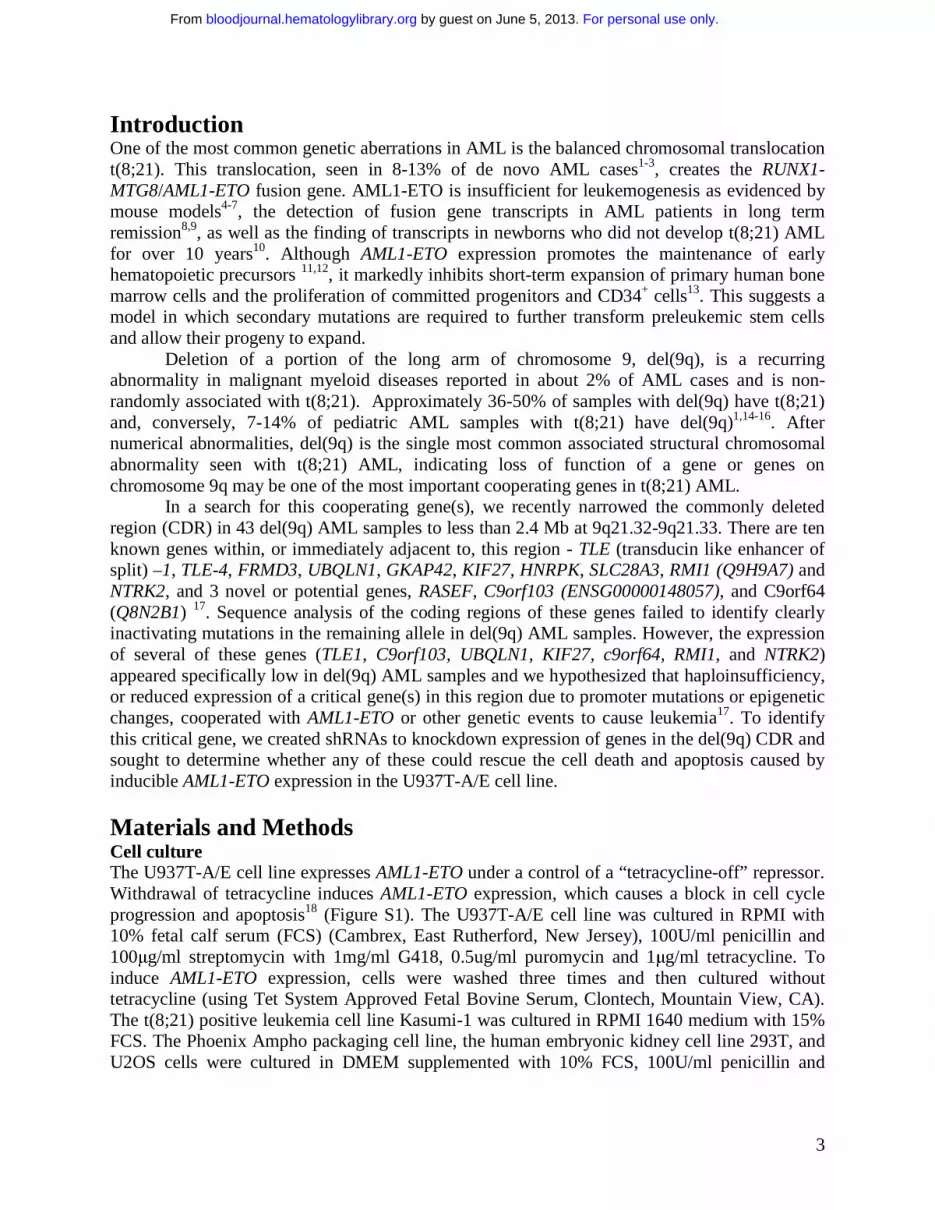
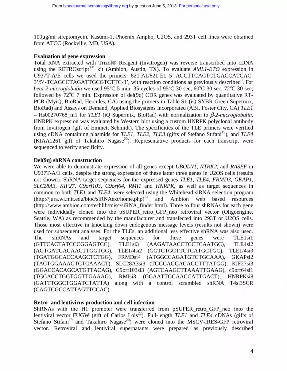
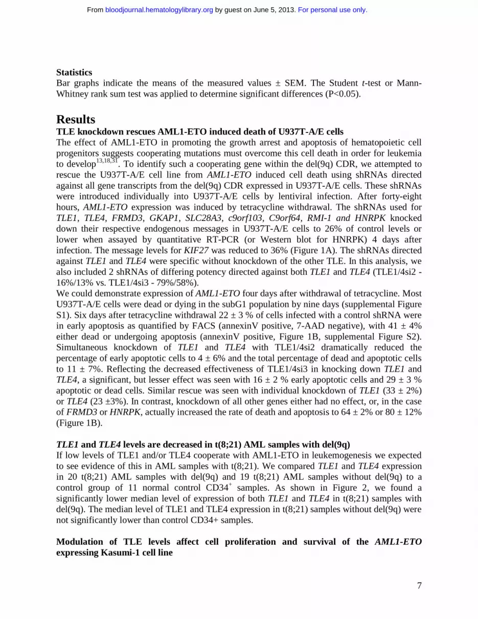
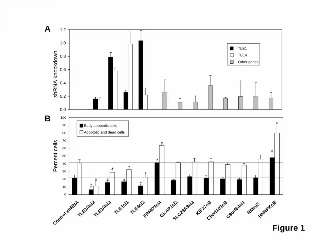
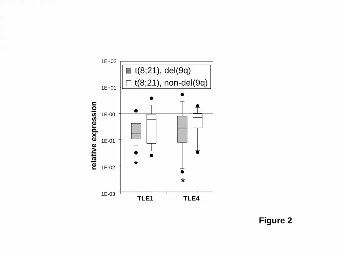
Statistics Bar graphs indicate the means of the measured values ± SEM. The Student t-test or Mann-Whitney rank sum test was applied to determine significant differences (P<0.05). Results TLE knockdown rescues AML1-ETO induced death of U937T-A/E cells The effect of AML1-ETO in promoting the growth arrest and apoptosis of hematopoietic cell progenitors suggests cooperating mutations must overcome this cell death in order for leukemia to develop13,18,31. To identify such a cooperating gene within the del(9q) CDR, we attempted to rescue the U937T-A/E cell line from AML1-ETO induced cell death using shRNAs directed against all gene transcripts from the del(9q) CDR expressed in U937T-A/E cells. These shRNAs were introduced individually into U937T-A/E cells by lentiviral infection. After forty-eight hours, AML1-ETO expression was induced by tetracycline withdrawal. The shRNAs used for TLE1, TLE4, FRMD3, GKAP1, SLC28A3, c9orf103, C9orf64, RMI-1 and HNRPK knocked down their respective endogenous messages in U937T-A/E cells to 26% of control levels or lower when assayed by quantitative RT-PCR (or Western blot for HNRPK) 4 days after infection. The message levels for KIF27 was reduced to 36% (Figure 1A). The shRNAs directed against TLE1 and TLE4 were specific without knockdown of the other TLE. In this analysis, we also included 2 shRNAs of differing potency directed against both TLE1 and TLE4 (TLE1/4si2 - 16%/13% vs. TLE1/4si3 - 79%/58%). We could demonstrate expression of AML1-ETO four days after withdrawal of tetracycline. Most U937T-A/E cells were dead or dying in the subG1 population by nine days (supplemental Figure S1). Six days after tetracycline withdrawal 22 ± 3 % of cells infected with a control shRNA were in early apoptosis as quantified by FACS (annexinV positive, 7-AAD negative), with 41 ± 4% either dead or undergoing apoptosis (annexinV positive, Figure 1B, supplemental Figure S2). Simultaneous knockdown of TLE1 and TLE4 with TLE1/4si2 dramatically reduced the percentage of early apoptotic cells to 4 ± 6% and the total percentage of dead and apoptotic cells to 11 ± 7%. Reflecting the decreased effectiveness of TLE1/4si3 in knocking down TLE1 and TLE4, a significant, but lesser effect was seen with 16 ± 2 % early apoptotic cells and 29 ± 3 % apoptotic or dead cells. Similar rescue was seen with individual knockdown of TLE1 (33 ± 2%) or TLE4 (23 ±3%). In contrast, knockdown of all other genes either had no effect, or, in the case of FRMD3 or HNRPK, actually increased the rate of death and apoptosis to 64 ± 2% or 80 ± 12% (Figure 1B). TLE1 and TLE4 levels are decreased in t(8;21) AML samples with del(9q) If low levels of TLE1 and/or TLE4 cooperate with AML1-ETO in leukemogenesis we expected to see evidence of this in AML samples with t(8;21). We compared TLE1 and TLE4 expression in 20 t(8;21) AML samples with del(9q) and 19 t(8;21) AML samples without del(9q) to a control group of 11 normal control CD34+ samples. As shown in Figure 2, we found a significantly lower median level of expression of both TLE1 and TLE4 in t(8;21) samples with del(9q). The median level of TLE1 and TLE4 expression in t(8;21) samples without del(9q) were not significantly lower than control CD34+ samples. Modulation of TLE levels affect cell proliferation and survival of the AML1-ETO expressing Kasumi-1 cell line
For personal use only. by guest on June 5, 2013. bloodjournal.hematologylibrary.orgFrom
8
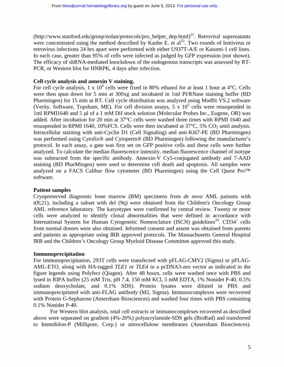
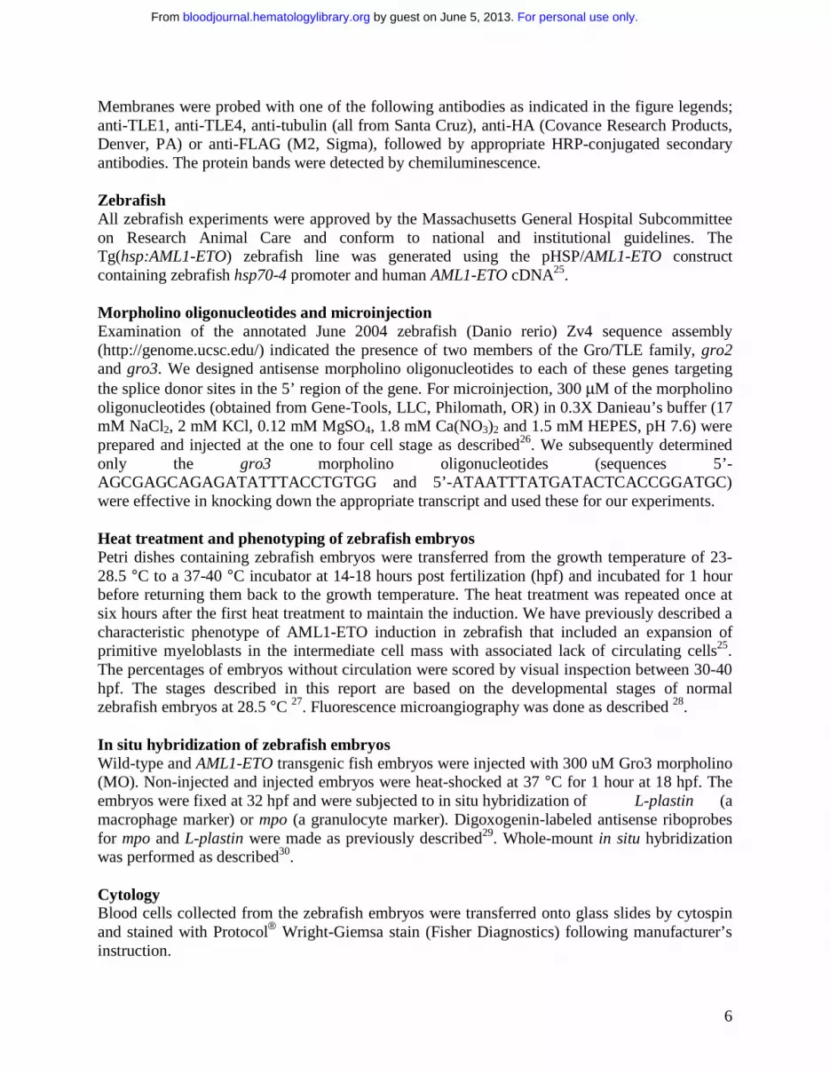
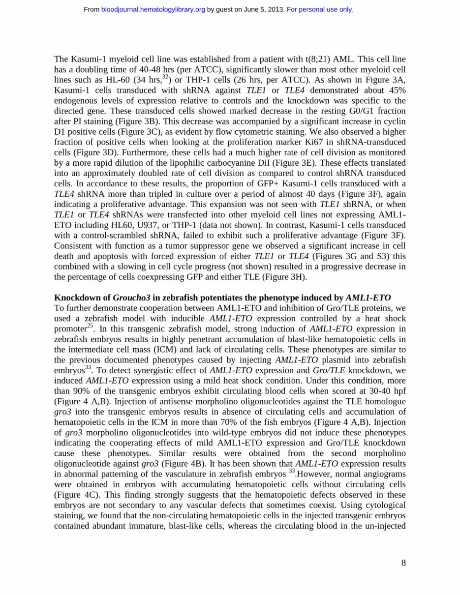
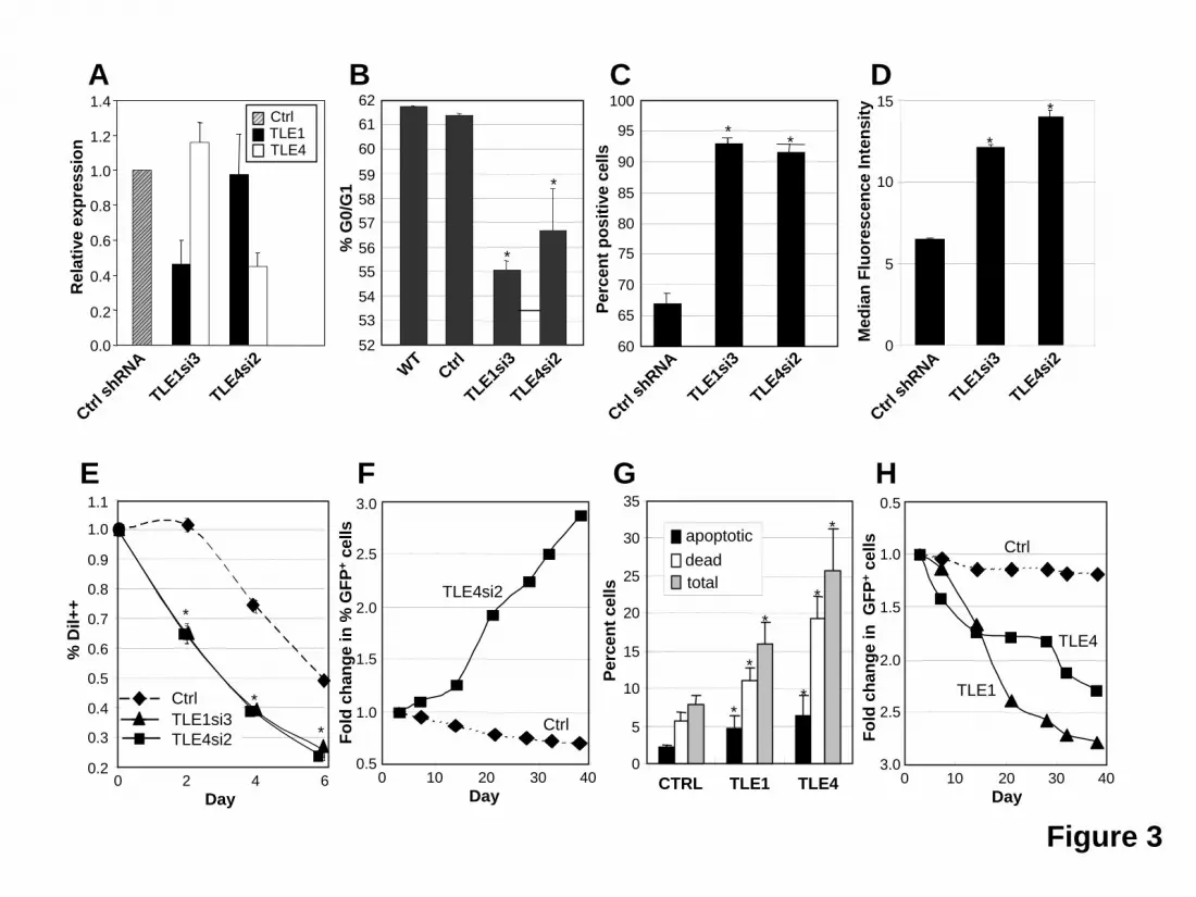
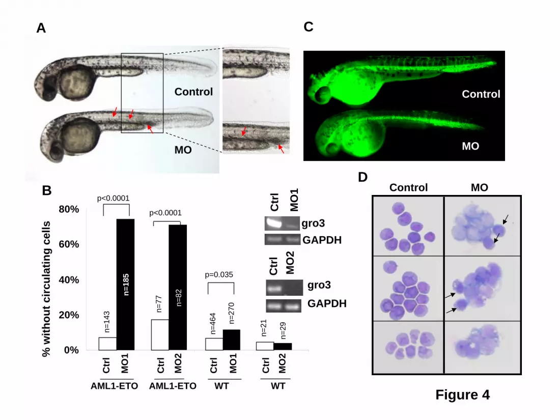
The Kasumi-1 myeloid cell line was established from a patient with t(8;21) AML. This cell line has a doubling time of 40-48 hrs (per ATCC), significantly slower than most other myeloid cell lines such as HL-60 (34 hrs,32) or THP-1 cells (26 hrs, per ATCC). As shown in Figure 3A, Kasumi-1 cells transduced with shRNA against TLE1 or TLE4 demonstrated about 45% endogenous levels of expression relative to controls and the knockdown was specific to the directed gene. These transduced cells showed marked decrease in the resting G0/G1 fraction after PI staining (Figure 3B). This decrease was accompanied by a significant increase in cyclin D1 positive cells (Figure 3C), as evident by flow cytometric staining. We also observed a higher fraction of positive cells when looking at the proliferation marker Ki67 in shRNA-transduced cells (Figure 3D). Furthermore, these cells had a much higher rate of cell division as monitored by a more rapid dilution of the lipophilic carbocyanine DiI (Figure 3E). These effects translated into an approximately doubled rate of cell division as compared to control shRNA transduced cells. In accordance to these results, the proportion of GFP+ Kasumi-1 cells transduced with a TLE4 shRNA more than tripled in culture over a period of almost 40 days (Figure 3F), again indicating a proliferative advantage. This expansion was not seen with TLE1 shRNA, or when TLE1 or TLE4 shRNAs were transfected into other myeloid cell lines not expressing AML1-ETO including HL60, U937, or THP-1 (data not shown). In contrast, Kasumi-1 cells transduced with a control-scrambled shRNA, failed to exhibit such a proliferative advantage (Figure 3F). Consistent with function as a tumor suppressor gene we observed a significant increase in cell death and apoptosis with forced expression of either TLE1 or TLE4 (Figures 3G and S3) this combined with a slowing in cell cycle progress (not shown) resulted in a progressive decrease in the percentage of cells coexpressing GFP and either TLE (Figure 3H). Knockdown of Groucho3 in zebrafish potentiates the phenotype induced by AML1-ETO To further demonstrate cooperation between AML1-ETO and inhibition of Gro/TLE proteins, we used a zebrafish model with inducible AML1-ETO expression controlled by a heat shock promoter25. In this transgenic zebrafish model, strong induction of AML1-ETO expression in zebrafish embryos results in highly penetrant accumulation of blast-like hematopoietic cells in the intermediate cell mass (ICM) and lack of circulating cells. These phenotypes are similar to the previous documented phenotypes caused by injecting AML1-ETO plasmid into zebrafish embryos33. To detect synergistic effect of AML1-ETO expression and Gro/TLE knockdown, we induced AML1-ETO expression using a mild heat shock condition. Under this condition, more than 90% of the transgenic embryos exhibit circulating blood cells when scored at 30-40 hpf (Figure 4 A,B). Injection of antisense morpholino oligonucleotides against the TLE homologue gro3 into the transgenic embryos results in absence of circulating cells and accumulation of hematopoietic cells in the ICM in more than 70% of the fish embryos (Figure 4 A,B). Injection of gro3 morpholino oligonucleotides into wild-type embryos did not induce these phenotypes indicating the cooperating effects of mild AML1-ETO expression and Gro/TLE knockdown cause these phenotypes. Similar results were obtained from the second morpholino oligonucleotide against gro3 (Figure 4B). It has been shown that AML1-ETO expression results in abnormal patterning of the vasculature in zebrafish embryos 33.However, normal angiograms were obtained in embryos with accumulating hematopoietic cells without circulating cells (Figure 4C). This finding strongly suggests that the hematopoietic defects observed in these embryos are not secondary to any vascular defects that sometimes coexist. Using cytological staining, we found that the non-circulating hematopoietic cells in the injected transgenic embryos contained abundant immature, blast-like cells, whereas the circulating blood in the un-injected
For personal use only. by guest on June 5, 2013. bloodjournal.hematologylibrary.orgFrom
9
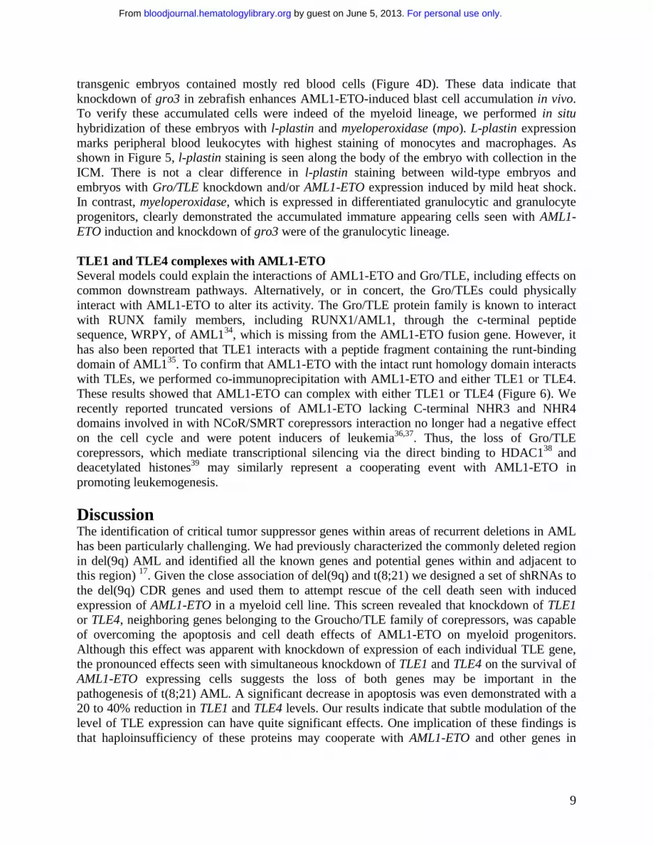
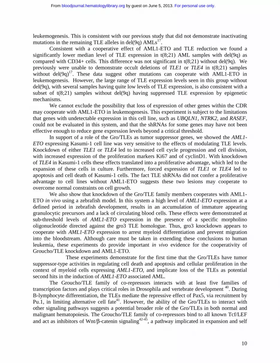
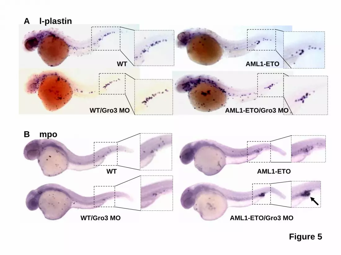
transgenic embryos contained mostly red blood cells (Figure 4D). These data indicate that knockdown of gro3 in zebrafish enhances AML1-ETO-induced blast cell accumulation in vivo. To verify these accumulated cells were indeed of the myeloid lineage, we performed in situ hybridization of these embryos with l-plastin and myeloperoxidase (mpo). L-plastin expression marks peripheral blood leukocytes with highest staining of monocytes and macrophages. As shown in Figure 5, l-plastin staining is seen along the body of the embryo with collection in the ICM. There is not a clear difference in l-plastin staining between wild-type embryos and embryos with Gro/TLE knockdown and/or AML1-ETO expression induced by mild heat shock. In contrast, myeloperoxidase, which is expressed in differentiated granulocytic and granulocyte progenitors, clearly demonstrated the accumulated immature appearing cells seen with AML1-ETO induction and knockdown of gro3 were of the granulocytic lineage. TLE1 and TLE4 complexes with AML1-ETO Several models could explain the interactions of AML1-ETO and Gro/TLE, including effects on common downstream pathways. Alternatively, or in concert, the Gro/TLEs could physically interact with AML1-ETO to alter its activity. The Gro/TLE protein family is known to interact with RUNX family members, including RUNX1/AML1, through the c-terminal peptide sequence, WRPY, of AML134, which is missing from the AML1-ETO fusion gene. However, it has also been reported that TLE1 interacts with a peptide fragment containing the runt-binding domain of AML135. To confirm that AML1-ETO with the intact runt homology domain interacts with TLEs, we performed co-immunoprecipitation with AML1-ETO and either TLE1 or TLE4. These results showed that AML1-ETO can complex with either TLE1 or TLE4 (Figure 6). We recently reported truncated versions of AML1-ETO lacking C-terminal NHR3 and NHR4 domains involved in with NCoR/SMRT corepressors interaction no longer had a negative effect on the cell cycle and were potent inducers of leukemia36,37. Thus, the loss of Gro/TLE corepressors, which mediate transcriptional silencing via the direct binding to HDAC138 and deacetylated histones39 may similarly represent a cooperating event with AML1-ETO in promoting leukemogenesis.
Discussion The identification of critical tumor suppressor genes within areas of recurrent deletions in AML has been particularly challenging. We had previously characterized the commonly deleted region in del(9q) AML and identified all the known genes and potential genes within and adjacent to this region) 17. Given the close association of del(9q) and t(8;21) we designed a set of shRNAs to the del(9q) CDR genes and used them to attempt rescue of the cell death seen with induced expression of AML1-ETO in a myeloid cell line. This screen revealed that knockdown of TLE1 or TLE4, neighboring genes belonging to the Groucho/TLE family of corepressors, was capable of overcoming the apoptosis and cell death effects of AML1-ETO on myeloid progenitors. Although this effect was apparent with knockdown of expression of each individual TLE gene, the pronounced effects seen with simultaneous knockdown of TLE1 and TLE4 on the survival of AML1-ETO expressing cells suggests the loss of both genes may be important in the pathogenesis of t(8;21) AML. A significant decrease in apoptosis was even demonstrated with a 20 to 40% reduction in TLE1 and TLE4 levels. Our results indicate that subtle modulation of the level of TLE expression can have quite significant effects. One implication of these findings is that haploinsufficiency of these proteins may cooperate with AML1-ETO and other genes in
For personal use only. by guest on June 5, 2013. bloodjournal.hematologylibrary.orgFrom
10
leukemogenesis. This is consistent with our previous study that did not demonstrate inactivating mutations in the remaining TLE alleles in del(9q) AMLs17.
Consistent with a cooperative effect of AML1-ETO and TLE reduction we found a significantly lower median level of TLE expression in t(8;21) AML samples with del(9q) as compared with CD34+ cells. This difference was not significant in t(8;21) without del(9q). We previously were unable to demonstrate occult deletions of TLE1 or TLE4 in t(8;21) samples without del(9q)17. These data suggest other mutations can cooperate with AML1-ETO in leukemogenesis. However, the large range of TLE expression levels seen in this group without del(9q), with several samples having quite low levels of TLE expression, is also consistent with a subset of t(8;21) samples without del(9q) having suppressed TLE expression by epigenetic mechanisms.
We cannot exclude the possibility that loss of expression of other genes within the CDR may cooperate with AML1-ETO in leukemogenesis. This experiment is subject to the limitations that genes with undetectable expression in this cell line, such as UBQLN1, NTRK2, and RASEF, could not be evaluated in this system, and that the shRNAs for some genes may have not been effective enough to reduce gene expression levels beyond a critical threshold.
In support of a role of the Gro/TLEs as tumor suppressor genes, we showed the AML1-ETO expressing Kasumi-1 cell line was very sensitive to the effects of modulating TLE levels. Knockdown of either TLE1 or TLE4 led to increased cell cycle progression and cell division, with increased expression of the proliferation markers Ki67 and of cyclinD1. With knockdown of TLE4 in Kasumi-1 cells these effects translated into a proliferative advantage, which led to the expansion of these cells in culture. Furthermore, forced expression of TLE1 or TLE4 led to apoptosis and cell death of Kasumi-1 cells. The fact TLE shRNAs did not confer a proliferative advantage to cell lines without AML1-ETO suggests these two lesions may cooperate to overcome normal constraints on cell growth.
We also show that knockdown of the Gro/TLE family members cooperates with AML1-ETO in vivo using a zebrafish model. In this system a high level of AML1-ETO expression at a defined period in zebrafish development, results in an accumulation of immature appearing granulocytic precursors and a lack of circulating blood cells. These effects were demonstrated at sub-threshold levels of AML1-ETO expression in the presence of a specific morpholino oligonucleotide directed against the gro3 TLE homologue. Thus, gro3 knockdown appears to cooperate with AML1-ETO expression to arrest myeloid differentiation and prevent migration into the bloodstream. Although care must be taken in extending these conclusions to human leukemia, these experiments do provide important in vivo evidence for the cooperativity of Groucho/TLE knockdown and AML1-ETO.
These experiments demonstrate for the first time that the Gro/TLEs have tumor suppressor-type activities in regulating cell death and apoptosis and cellular proliferation in the context of myeloid cells expressing AML1-ETO, and implicate loss of the TLEs as potential second hits in the induction of AML1-ETO associated AML.
The Groucho/TLE family of co-repressors interacts with at least five families of transcription factors and plays critical roles in Drosophila and vertebrate development 40. During B-lymphocyte differentiation, the TLEs mediate the repressive effect of Pax5, via recruitment by Pu.1, in limiting alternative cell fate41. However, the ability of the Gro/TLEs to interact with other signaling pathways suggests a potential broader role of the Gro/TLEs in both normal and malignant hematopoiesis. The Groucho/TLE family of co-repressors bind to all known Tcf/LEF and act as inhibitors of Wnt/β-catenin signaling42-45, a pathway implicated in expansion and self
For personal use only. by guest on June 5, 2013. bloodjournal.hematologylibrary.orgFrom
11
renewal of the hematopoietic stem cell compartment46,47. Similarly, the Gro/TLEs inhibit NF-kB signaling48, a pathway constitutively activated in AML and believed to play an important role in hematopoietic cell proliferation and survival and in chemoresistance49-51. The Gro/TLE gene family is also a key effector of Notch signaling, a pathway implicated in HSC fate determination and self-renewal52-54. In Drosophila, Groucho is one of the primary effectors maintaining silencing of downstream target genes in the absence of Notch signaling55. Furthermore, the TLEs bind to and regulate RUNX1/AML1 and Pu.1, key myeloid transcription factors34,56,57. It remains to be determined whether dysregulated pathways downstream of TLEs and AML1-ETO cooperate to promote leukemia or whether the direct interaction of TLEs modulates AML1-ETO activity. Although AML1-ETO was initially thought to behave primarily as a repressive transcription factor, it is now known to activate numerous genes potentially important in transformation. These include the macrophage colony stimulating factor receptor58, the Notch ligand Jagged159, and the Wnt pathway mediator plakoglobin60. Loss of TLE activity may be needed for AML1-ETO to exert its full effect in activating expression of such genes. One model for the interaction between TLE loss and AML1-ETO is suggested by our recent finding of AML1-ETO variants arising from alternative splicing or mutation. These variants delete C-terminal domains involved in the binding of NCoR/SMRT corepressors that recruit histone deacetylases and no longer had a negative effect on the cell cycle but were potent inducers of leukemia36,37. The loss of Gro/TLE corepressors, which mediate transcriptional silencing via the direct binding to HDAC138 and deacetylated histones39, may similarly represent cooperating events with AML1-ETO in promoting leukemogenesis.
The ability to interact with many different transcription factors suggests the function of the Gro/TLEs will likely vary depending on the cell type and the timing of expression during development. Our studies demonstrating low levels of TLE1 and TLE4 expression in myeloid cell lines and subsets of AML samples, and potential tumor suppressor-like activities of the Gro/TLEs, stands in contrast to recent reports that over-expression of various Gro/TLE family members is seen in Grade I astrocytomas61, higher grade meningiomas62, pituitary adenomas63,64, and synovial sarcomas65,66. In addition, over-expression of murine Tle1/Grg1 in a transgenic model predisposed to the development of lung adenocarcinoma67. Similar dual functions have been described for several other proteins, including WT168, E2F169 and K-RAS70. This apparent dual Gro/TLE function mirrors the observation that RUNX1/AML1, which is inhibited by Gro/TLE binding, can act alternatively as an oncogene or a tumor suppressor gene71-74, a finding that might reflect differential effects on stem cells and progenitors75.
In summary, we demonstrate modulation of activity of two members of the Gro/TLE co-repressor family, TLE1 and TLE4, cooperates with AML1-ETO to affect myeloid cell survival, apoptosis and proliferation. These results provide a mechanistic rationale for the strong association of del(9q) and t(8;21) and implicate loss of TLE activity in the pathogenesis of AML1-ETO associated AML. Further understanding of the mechanism of these interactions and of the downstream pathways affected could have therapeutic implications.
For personal use only. by guest on June 5, 2013. bloodjournal.hematologylibrary.orgFrom
12
Acknowledgements We wish to thank David Scadden, Daniel Tenen and Howard Weinstein for helpful discussions and support during the course of this work. This work was supported by the Doris Duke Charitable Foundation Clinical Scientist Development Award (D.A.S.), American Society of Hematology Junior Faculty Scholar Award (D.A.S.), MassGeneral Hospital Marathon Fund (D.A.S.), Mattina Proctor Fund (D.A.S.), Lauri Strauss Leukemia Foundation (F.D.), Tosteson Fellowship from the Massachusetts Biomedical Research Corporation (J.R.J.Y.), Lady Tata Memorial Trust (E.Y.A.), American Cancer Society Clinical Research Professorship Award (I.D.B), American Cancer Society PRD-95-124-12 (I.D.B.), Leukemia & Lymphoma Society SCOR 7-40-03 (I.D.B.), and NIH grants 1R01CA115772 (D.A.S), 5T32CA71345-09 (F.D.), CA118498 (R.T.P.), HL079267 (R.T.P.), CA96735 (D.E.Z.), CA98543 (I.D.B.), U10 CA98543 (I.D.B.), and U24 CA114766 (I.D.B.).
Authorship F.D. and J.W. contributed equally to this study. F.D., J.W., J-R.J.Y, and E-Y.A. designed and performed research, analyzed data and contributed to writing the paper. E.T. performed research; D-E.Z., I.D.B. and R.T.P designed research and contributed to writing the paper; D.A.S. designed research, supervised the study and wrote the paper. The authors report no financial conflict of interest that influences the results or interpretation of this manuscript.
For personal use only. by guest on June 5, 2013. bloodjournal.hematologylibrary.orgFrom

13
References 1. Grimwade D, Walker H, Oliver F, et al. The importance of diagnostic cytogenetics on outcome in AML: analysis of 1,612 patients entered into the MRC AML 10 trial. The Medical Research Council Adult and Children's Leukaemia Working Parties. Blood. 1998;92:2322-2333.
2. Mauritzson N, Albin M, Rylander L, et al. Pooled analysis of clinical and cytogenetic features in treatment-related and de novo adult acute myeloid leukemia and myelodysplastic syndromes based on a consecutive series of 761 patients analyzed 1976-1993 and on 5098 unselected cases reported in the literature 1974-2001. Leukemia. 2002;16:2366-2378.
3. Ravindranath Y, Chang M, Steuber CP, et al. Pediatric Oncology Group (POG) studies of acute myeloid leukemia (AML): a review of four consecutive childhood AML trials conducted between 1981 and 2000. Leukemia. 2005;19:2101-2116.
4. Fenske TS, Pengue G, Mathews V, et al. Stem cell expression of the AML1/ETO fusion protein induces a myeloproliferative disorder in mice. Proc Natl Acad Sci U S A. 2004;101:15184-15189.
5. Higuchi M, O'Brien D, Kumaravelu P, Lenny N, Yeoh E-J, Downing JR. Expression of a conditional AML1-ETO oncogene bypasses embryonic lethality and establishes a murine model of human t(8;21) acute myeloid leukemia. Cancer Cell. 2002;1:63-74.
6. Rhoades KL, Hetherington CJ, Harakawa N, et al. Analysis of the role of AML1-ETO in leukemogenesis, using an inducible transgenic mouse model. Blood. 2000;96:2108-2115.
7. Yuan Y, Zhou L, Miyamoto T, et al. AML1-ETO expression is directly involved in the development of acute myeloid leukemia in the presence of additional mutations. Proc Natl Acad Sci USA. 2001;98:10398-10403.
8. Kusec R, Laczika K, Knobl P, et al. AML1/ETO fusion mRNA can be detected in remission blood samples of all patients with t(8;21) acute myeloid leukemia after chemotherapy or autologous bone marrow transplantation. Leukemia. 1994;8:735-739.
9. Jurlander J, Caligiuri MA, Ruutu T, et al. Persistence of the AML1/ETO fusion transcript in patients treated with allogeneic bone marrow transplantation for t(8;21) leukemia. Blood. 1996;88:2183-2191.
10. Wiemels JL, Xiao Z, Buffler PA, et al. In utero origin of t(8;21) AML1-ETO translocations in childhood acute myeloid leukemia. 2002;99:3801-3805.
11. Basecke J, Schwieger M, Griesinger F, et al. AML1/ETO promotes the maintenance of early hematopoietic progenitors in NOD/SCID mice but does not abrogate their lineage specific differentiation. Leuk Lymphoma. 2005;46:265-272.
12. Mulloy JC, Cammenga J, Berguido FJ, et al. Maintaining the self-renewal and differentiation potential of human CD34+ hematopoietic cells using a single genetic element. Blood. 2003;102:4369-4376.
For personal use only. by guest on June 5, 2013. bloodjournal.hematologylibrary.orgFrom
14
13. Mulloy JC, Cammenga J, MacKenzie KL, Berguido FJ, Moore MA, Nimer SD. The AML1-ETO fusion protein promotes the expansion of human hematopoietic stem cells. Blood. 2002;99:15-23.
14. Schoch C, Haase D, Haferlach T, et al. Fifty-one patients with acute myeloid leukemia and translocation t(8;21)(q22;q22): an additional deletion in 9q is an adverse prognostic factor. Leukemia. 1996;10:1288-1295.
15. Grimwade D, Walker H, Harrison G, et al. The predictive value of hierarchical cytogenetic classification in older adults with acute myeloid leukemia (AML): analysis of 1065 patients entered into the United Kingdom Medical Research Council AML11 trial. Blood. 2001;98:1312-1320.
16. Raimondi SC, Chang MN, Ravindranath Y, et al. Chromosomal abnormalities in 478 children with acute myeloid leukemia: clinical characteristics and treatment outcome in a cooperative Pediatric Oncology Group Study-POG 8821. Blood. 1999;94:3707-3716.
17. Sweetser DA, Peniket AJ, Haaland C, et al. Delineation of the minimal commonly deleted segment and identification of candidate tumor suppressor genes in del(9q) acute myeloid leukemia. Genes Chr and Ca. 2005;44:279-291.
18. Burel SA, Harakawa N, Zhou L, Pabst T, Tenen DG, Zhang DE. Dichotomy of AML1-ETO functions: growth arrest versus block of differentiation. Mol Cell Biol. 2001;21:5577-5590.
19. Stifani S, Blaumueller CM, Redhead NJ, Hill RE, Artavanis-Tsakonas S. Human homologs of a Drosophila Enhancer of split gene product define a novel family of nuclear proteins. Nat Genet. 1992;2:119-127.
20. Nagase T, Ishikawa K, Kikuno R, Hirosawa M, Nomura N, Ohara O. Prediction of the coding sequences of unidentified human genes. XV. The complete sequences of 100 new cDNA clones from brain which code for large proteins in vitro. DNA Research. 1999;6:337-345.
21. Yuan B, Latek R, Hossbach M, Tuschl T, Lewitter F. siRNA Selection Server: an automated siRNA oligonucleotide prediction server. Nucl Acids Res. 2004;32:W130-134.
22. Lois C, Hong EJ, Pease S, Brown EJ, Baltimore D. Germline transmission and tissue-specific expression of transgenes delivered by lentiviral vectors. Science. 2002;295:868-872.
23. Kanbe E, Zhang DE. A simple and quick method to concentrate MSCV retrovirus. Blood Cells Mol Dis. 2004;33:64-67.
24. Mitelman F ed ISCN (1995): An international system for human cytogenetic nomenclature. Basel: S. Karger; 1995.
25. Yeh JR, Munson KM, Chao YL, Peterson QP, Macrae CA, Peterson RT. AML1-ETO reprograms hematopoietic cell fate by downregulating scl expression. Development. 2008;135:401-410.
For personal use only. by guest on June 5, 2013. bloodjournal.hematologylibrary.orgFrom
15
26. Nasevicius A, Ekker SC. Effective targeted gene 'knockdown' in zebrafish. Nat Genet. 2000;26:216-220.
27. Kimmel CB, Ballard WW, Kimmel SR, Ullmann B, Schilling TF. Stages of embryonic development of the zebrafish. Dev Dyn. 1995;203:253-310.
28. Weinstein BM, Stemple DL, Driever W, Fishman MC. Gridlock, a localized heritable vascular patterning defect in the zebrafish. Nat Med. 1995;1:1143-1147.
29. Bennett CM, Kanki JP, Rhodes J, et al. Myelopoiesis in the zebrafish, Danio rerio. Blood. 2001;98:643-651.
30. Ransom DG, Haffter P, Odenthal J, et al. Characterization of zebrafish mutants with defects in embryonic hematopoiesis. Development. 1996;123:311-319.
31. Li X, Xu YB, Wang Q, et al. Leukemogenic AML1-ETO fusion protein upregulates expression of connexin 43: the role in AML 1-ETO-induced growth arrest in leukemic cells. J Cell Physiol. 2006;208:594-601.
32. Foa P, Maiolo AT, Lombardi L, Toivonen H, Rytomaa T, Polli EE. Growth pattern of the human promyelocytic leukaemia cell line HL60. Cell Tissue Kinet. 1982;15:399-404.
33. Kalev-Zylinska ML, Horsfield JA, Flores MV, et al. Runx1 is required for zebrafish blood and vessel development and expression of a human RUNX1-CBF2T1 transgene advances a model for studies of leukemogenesis. Development. 2002;129:2015-2030.
34. Aronson BD, Fisher AL, Blechman K, Caudy M, Gergen JP. Groucho-dependent and -independent repression activities of Runt domain proteins. Mol Cell Biol. 1997;17:5581-5587.
35. Imai Y, Kurokawa M, Tanaka K, et al. TLE, the human homolog of groucho, interacts with AML1 and acts as a repressor of AML1-induced transactivation. Biochem Biophys Res Commun. 1998;252:582-589.
36. Yan M, Burel SA, Peterson LF, et al. Deletion of an AML1-ETO C-terminal NcoR/SMRT-interacting region strongly induces leukemia development. Proc Natl Acad Sci U S A. 2004;101:17186-17191.
37. Yan M, Kanbe E, Peterson LF, et al. A previously unidentified alternatively spliced isoform of t(8;21) transcript promotes leukemogenesis. Nat Med. 2006;12:945-949.
38. Chen G, Fernandez J, Mische S, Courey AJ. A functional interaction between the histone deacetylase Rpd3 and the corepressor groucho in Drosophila development. Genes Dev. 1999;13:2218-2230.
39. Flores-Saaib RD, Courey AJ. Analysis of Groucho-histone interactions suggests mechanistic similarities between Groucho- and Tup1-mediated repression. Nucleic Acids Res. 2000;28:4189-4196.
For personal use only. by guest on June 5, 2013. bloodjournal.hematologylibrary.orgFrom
16
40. Chen G, Courey AJ. Groucho/TLE family proteins and transcriptional repression. Gene. 2000;249:1-16.
41. Nutt SL, Eberhard D, Horcher M, Rolink AG, Busslinger M. Pax5 determines the identity of B cells from the beginning to the end of B-lymphopoiesis. Int Rev Immunol. 2001;20:65-82.
42. Huelsken J, Behrens J. The Wnt signalling pathway. J Cell Sci. 2002;115:3977-3978.
43. Brantjes H, Roose J, van de Wetering M, Clevers H. All Tcf HMG box transcription factors interact with Groucho-related co-repressors. Nucl Acids Res. 2001;29:1410-1419.
44. Miller JR. The Wnts. Genome Biol. 2002;3:REVIEWS3001. Epub 2001 Dec 3028.
45. Miller JR, Hocking AM, Brown JD, Moon RT. Mechanism and function of signal transduction by the Wnt/beta-catenin and Wnt/Ca2+ pathways. Oncogene. 1999;18:7860-7872.
46. Willert K, Brown JD, Danenberg E, et al. Wnt proteins are lipid-modified and can act as stem cell growth factors. Nature. 2003;423:448-452.
47. Van Den Berg DJ, Sharma AK, Bruno E, Hoffman R. Role of members of the Wnt gene family in human hematopoiesis. Blood. 1998;92:3189-3202.
48. Tetsuka T, Uranishi H, Imai H, et al. Inhibition of nuclear factor-kappa B-mediated transcription by association with the amino-terminal enhancer of split, a Groucho-related protein lacking WD40 repeats. J Biol Chem. 2000;275:4383-4390.
49. Berenson JR, Ma HM, Vescio R. The role of nuclear factor-kappaB in the biology and treatment of multiple myeloma. Semin Oncol. 2001;28:626-633.
50. Guzman ML, Neering SJ, Upchurch D, et al. Nuclear factor-kappaB is constitutively activated in primitive human acute myelogenous leukemia cells. Blood. 2001;98:2301-2307.
51. Bueso-Ramos CE, Rocha FC, Shishodia S, et al. Expression of constitutively active nuclear-kappa B RelA transcription factor in blasts of acute myeloid leukemia. Hum Pathol. 2004;35:246-253.
52. Varnum-Finney B, Xu L, Brashem-Stein C, et al. Pluripotent, cytokine-dependent, hematopoietic stem cells are immortalized by constitutive Notch1 signaling. Nat Med. 2000;6:1278-1281.
53. Stier S, Cheng T, Dombkowski D, Carlesso N, Scadden DT. Notch1 activation increases hematopoietic stem cell self-renewal in vivo and favors lymphoid over myeloid lineage outcome. Blood. 2002;99:2369-2378.
54. Burns CE, Traver D, Mayhall E, Shepard JL, Zon LI. Hematopoietic stem cell fate is established by the Notch-Runx pathway. Genes Dev. 2005;19:2331-2342. Epub 2005 Sep 2315.
For personal use only. by guest on June 5, 2013. bloodjournal.hematologylibrary.orgFrom
17
55. Barolo S, Stone T, Bang AG, Posakony JW. Default repression and Notch signaling: Hairless acts as an adaptor to recruit the corepressors Groucho and dCtBP to Suppressor of Hairless. Genes Dev. 2002;16:1964-1976.
56. Levanon D, Goldstein RE, Bernstein Y, et al. Transcriptional repression by AML1 and LEF-1 is mediated by the TLE/Groucho corepressors. Proc Natl Acad Sci USA. 1998;95:11590-11595.
57. Linderson Y, Eberhard D, Malin S, Johansson A, Busslinger M, Pettersson S. Corecruitment of the Grg4 repressor by PU.1 is critical for Pax5-mediated repression of B-cell-specific genes. EMBO Rep. 2004;5:291-296.
58. Rhoades KL, Hetherington CJ, Rowley JD, et al. Synergistic up-regulation of the myeloid-specific promoter for the macrophage colony-stimulating factor receptor by AML1 and the t(8;21) fusion protein may contribute to leukemogenesis. Proc Natl Acad Sci U S A. 1996;93:11895-11900.
59. Alcalay M, Meani N, Gelmetti V, et al. Acute myeloid leukemia fusion proteins deregulate genes involved in stem cell maintenance and DNA repair. J Clin Invest. 2003;112:1751-1761.
60. Muller-Tidow C, Steffen B, Cauvet T, et al. Translocation products in acute myeloid leukemia activate the Wnt signaling pathway in hematopoietic cells. Molecular & Cellular Biology. 2004;24:2890-2904.
61. Rorive S, Maris C, Debeir O, et al. Exploring the distinctive biological characteristics of pilocytic and low-grade diffuse astrocytomas using microarray gene expression profiles. J Neuropathol Exp Neurol. 2006;65:794-807.
62. Cuevas IC, Slocum AL, Jun P, et al. Meningioma transcript profiles reveal deregulated Notch signaling pathway. Cancer Res. 2005;65:5070-5075.
63. Moreno CS, Evans CO, Zhan X, Okor M, Desiderio DM, Oyesiku NM. Novel molecular signaling and classification of human clinically nonfunctional pituitary adenomas identified by gene expression profiling and proteomic analyses. Cancer Res. 2005;65:10214-10222.
64. Ruebel KH, Leontovich AA, Jin L, et al. Patterns of gene expression in pituitary carcinomas and adenomas analyzed by high-density oligonucleotide arrays, reverse transcriptase-quantitative PCR, and protein expression. Endocrine. 2006;29:435-444.
65. Allander SV, Illei PB, Chen Y, et al. Expression profiling of synovial sarcoma by cDNA microarrays: association of ERBB2, IGFBP2, and ELF3 with epithelial differentiation. Am J Pathol. 2002;161:1587-1595.
66. Terry J, Saito T, Subramanian S, et al. TLE1 as a diagnostic immunohistochemical marker for synovial sarcoma emerging from gene expression profiling studies. Am J Surg Pathol. 2007;31:240-246.
For personal use only. by guest on June 5, 2013. bloodjournal.hematologylibrary.orgFrom
18
67. Allen T, van Tuyl M, Iyengar P, et al. Grg1 acts as a lung-specific oncogene in a transgenic mouse model. Cancer Res. 2006;66:1294-1301.
68. Yang L, Han Y, Suarez Saiz F, Minden MD. A tumor suppressor and oncogene: the WT1 story. Leukemia. 2007;21:868-876.
69. Johnson DG. The paradox of E2F1: oncogene and tumor suppressor gene. Mol Carcinog. 2000;27:151-157.
70. James RM, Arends MJ, Plowman SJ, et al. K-ras proto-oncogene exhibits tumor suppressor activity as its absence promotes tumorigenesis in murine teratomas. Mol Cancer Res. 2003;1:820-825.
71. Song WJ, Sullivan MG, Legare RD, et al. Haploinsufficiency of CBFA2 causes familial thrombocytopenia with propensity to develop acute myelogenous leukaemia. Nat Genet. 1999;23:166-175.
72. Cameron ER, Neil JC. The Runx genes: lineage-specific oncogenes and tumor suppressors. Oncogene. 2004;23:4308-4314.
73. Strom DK, Nip J, Westendorf JJ, et al. Expression of the AML-1 Oncogene Shortens the G1 Phase of the Cell Cycle. J Biol Chem. 2000;275:3438-3445.
74. Growney JD, Shigematsu H, Li Z, et al. Loss of Runx1 perturbs adult hematopoiesis and is associated with a myeloproliferative phenotype. Blood. 2005;106:494-504.
75. Sun W, Downing JR. Haploinsufficiency of AML1 results in a decrease in the number of LTR-HSCs, while simultaneously inducing an increase in more mature progenitors. Blood. 2004;5:5.
For personal use only. by guest on June 5, 2013. bloodjournal.hematologylibrary.orgFrom
19
Figure and Table Legends Figure 1. Knockdown of TLE levels inhibits the apoptosis and cell death induced by AML1-ETO expression in U937T-A/E cells. U937T-A/E cells were infected with lentivirus containing either a control shRNA (TLE4scr3); an shRNA directed against both TLE1 and TLE4 (TLE1/4si2, TLE1/4si3); an shRNA specific for TLE1 (TLE1si1) or TLE4 (TLE4si2); or an shRNA against other del9q CDR genes: FRMD3, GKAP1, SLC28A3, KIF27, C9orf103, C9orf64, RMI1 and HNRPK. (A) Four days after infection, expression of del(9q) CDR genes was evaluated by quantitative RT-PCR or Western blot in the case of HNRPK and are shown relative to the level of expression of each gene after infection with a control shRNA. To evaluate the specificity of TLE knock down with TLE shRNAs, the expression levels of both TLE1 (black bars) and TLE4 (white bars) were measured. (B). After 2 days’ infection, tetracycline was withdrawn and AML1-ETO was inducted. Cells were cultured an additional 6 days then stained with Annexin-V and 7-AAD to detect cells undergoing early apoptosis (Annexin-V positive, 7-AAD negative), or dead cells in late apoptosis (Annexin-V positive, 7-AAD positive). Three independent experiments were performed and results for percentage of cells undergoing early apoptosis (black bars) and total apoptotic and dead cells (white bars) are summarized in panel expressed as the mean ± SEM.
Figure 2. TLE1 and TLE4 expression is specifically decreased in a subset of t(8;21) AMLS with del(9q). The median levels of TLE1 and TLE4 expression in 20 t(8;21) AML samples with del(9q) and 19 t(8;21) AML samples without del(9q) were compared to the expression of 11 normal CD34+ control samples. Boxes indicate measurements from 25th to 75th percentiles. A line within a box indicates median fold change in gene expression. The error bars correspond to the 10th and 90th percentiles and the solid circles represent outliers. An asterisk denotes a significant change (P < 0.05). Figure 3. Modulation of TLE levels affects the proliferation and survival of Kasumi-1 cells. (A) ShRNAs directed against TLE1 or TLE4 knockdown expression to about 45% control values and are specific for their respective target gene. (B) Knockdown of TLE1 or TLE4 increases cell cycle progression. Flow cytometric determination of G0/G1 fraction in wild-type (WT) Kasumi-1 cells and cells infected with control (Ctrl) shRNA and shRNA against TLE1 or TLE4. (C) Knockdown of TLE1 or TLE4 increases cyclin D1 expression. Flow cytometric analysis of percentage of Cyclin D1 positive Kasumi-1 cells infected with control (Ctrl) shRNA and shRNA against TLE1 or TLE4. (D) Knockdown of TLE1 or TLE4 increases expression of the cell proliferation marker Ki67. Flow cytometric analysis of fluorescence intensity (FL) of Ki67 in Kasumi-1 cells infected with control (Ctrl) shRNA and shRNA against TLE1 or TLE4. (E) Knockdown of TLE1 or TLE4 increases cell division. Flow cytometric cell proliferation assay with DiI in Kasumi-1 cells infected with control (Ctrl) shRNA and shRNA against TLE1 or TLE4. Shown is the percentage of bright DiI positive (DiI++) cells over time. (F) Knockdown of TLE4 leads to a proliferative advantage. Relative proportion over time of GFP positive Kasumi cells either infected with a control (Ctrl) or a specific shRNA against TLE4. (G) Expression of TLE1 or TLE4 leads to an increase in apoptosis and cell death. Percentage of apoptotic cells (Annexin V+/7AAD-), dead and late apoptotic cells (Annexin V+/7AAD+), and total apoptotic and dead cells in Kasumi cells infected with control empty MSCV-GFP retroviral vector (Ctrl) or retrovirus expressing TLE1 or TLE4 cDNAs. GFP positive cells were sorted prior to analysis. (H) Expression of TLE1 or TLE4 slows proliferation. Relative proportion over time of GFP positive Kasumi cells infected with either control empty MSCV-GFP retroviral vector (Ctrl) or
For personal use only. by guest on June 5, 2013. bloodjournal.hematologylibrary.orgFrom
20
retrovirus expressing TLE1 or TLE4 cDNAs. All diagrams denote results from three independent experiments. Results are expressed as the mean ± SEM. Significant differences, p < 0.05, as compared to controls are indicated with an asterisk (*). Figure 4. Knockdown of gro3 in cooperation with AML1-ETO expression induces abnormal hematopoiesis and absence of circulating blood in zebrafish embryos. Gro3 MO (morpholino oligonucleotide) -injected or un-injected wild-type and Tg(hsp:AML1-ETO) zebrafish embryos were all subjected to the same heat induction. (A) Inspection of un-injected (Control) and gro3 MO injected Tg(hsp:AML1-ETO) zebrafish embryos showed the absence of circulating cells and accumulation of hematopoietic cells in the ICM and the ventral tail region (red arrows). These cells had a blast-like morphology with a large size and relatively little cytoplasm. (B) Percentages of embryos exhibiting loss-of-circulating cells were scored between 30 and 40 hpf. Control and MO indicate un-injected and morpholino oligonucleotide-injected embryos, respectively. AML1-ETO and WT indicate Tg(hsp:AML1-ETO) and wild-type embryos, respectively. n equals to the number of embryos scored. p value was obtained using Student’s t-test. The inset gels show the knockdown by RT-PCR of gro3 transcripts with two gro3 specific morpholino oligonucleotides. (C) Lack of circulating cells in AML1-ETO-expressing embryos is not caused by a vascular obstruction as evidenced by microangiography. The fluorescein-coupled latex beads injected into the inflow tract of the atrium was able to perfuse the whole vascular system of the Tg(hsp:AML1-ETO) embryos. It revealed a normal vascular pattern but reduced intersomitic vessels in gro3 morphant embryos. (D) Cytological analysis of hematopoietic cells collected from the circulating blood in three uninjected Tg(hsp:AML1-ETO) embryos and the accumulating blood in three injected embryos. While the un-injected embryos contain mostly red blood cells, the injected embryos contain abundant immature blast-like cells. Black arrows, red blood cells. Figure 5. Accumulated immature ICM cells in AML1-ETO transgenic zebrafish embryos with Gro3/TLE knockdown are myeloid cells of the granulocytic lineage. Wild-type and AML1-ETO transgenic fish embryos were injected with Gro3 morpholino (MO) and subjected to mild heat shock treatment to induce low levels of AML1-ETO expression. The embryos were fixed at 32 hpf and were subjected to in situ hybridization for (A) the macrophage marker, l-plastin or for (B) myeloperoxidase (mpo), a marker for cells of the granulocytic lineage. The data indicate that knockdown of groucho in the presence of AML1-ETO expression leads to accumulation of mpo+ cells in the ventral tail region (see arrow). Figure 6. TLE1 and TLE4 complex with AML1-ETO. 293T cells were transfected with HA-tagged TLE1 or TLE4 along with a FLAG empty vector (pFLAG-CMV2) or FLAG-tagged AML1-ETO (pFLAG-AML1-ETO). Total cell lysates immunoblotted with anti-HA and anti-FLAG antibodies show the expression of proteins. Lysates were immunoprecipitated with anti-FLAG antibodies and the immunocomplexes were blotted with HA antibodies to demonstrate that both TLE1 and TLE 4 can interact with AML1-ETO.
For personal use only. by guest on June 5, 2013. bloodjournal.hematologylibrary.orgFrom
0.0
0.2
0.4
0.6
0.8
1.0
1.2
0
10
20
30
40
50
60
70
80
90
100
shR
NA
kno
ckdo
wn
Per
cent
cel
ls
A
B
Figure 1
Early apoptotic cells
Apoptotic and dead cells
** *
*
*
*
*
*
*
**
*
TLE1
TLE4
Other genes
Control s
hRNA
TLE1/4si2
TLE1/4si3
TLE1si1
TLE4si2
FRMD3si4
GKAP1si2
SLC28A3s
i3
KIF27si3
C9orf1
03si3
C9orf6
4si1
RMIsi3
HNRPKsi8
F
or personal use only. by guest on June 5, 2013.
bloodjournal.hematologylibrary.org
From
0 1 2 3 4 5 60.001
0.01
0.1
1
10
100
t(8;21), del(9q)t(8;21), non-del(9q)
1E+02
1E+01
1E-00
1E-01
1E-02
1E-03
rela
tive
expr
essi
on
TLE1 TLE4
Figure 2
*
*
F
or personal use only. by guest on June 5, 2013.
bloodjournal.hematologylibrary.org
From
Rel
ativ
e ex
pres
sion
% G
0/G
1
Perc
ent p
ositi
ve c
ells
Med
ian
Fluo
resc
ence
Inte
nsity
% D
il++
Fold
cha
nge
in %
GFP
+ce
lls
Perc
ent c
ells
Fold
cha
nge
in G
FP+
cells
1.4
1.2
1.0
0.8
0.4
0.6
0.2
0.0
626160
59
5758
5655
5453
52
100
95
90
85
75
80
70
65
60
15
10
5
0
1.1
1.0
0.9
0.8
0.6
0.7
0.5
0.4
0.3
0.2
3.0
2.5
2.0
1.5
0.5
1.0
35
30
25
15
20
10
5
0
0.5
1.0
1.5
2.5
2.0
3.0
Figure 3
Ctrl
TLE4
TLE1
0 10 20 30 40Day
apoptoticdeadtotal
CTRL TLE1 TLE4
*
*
*
*
*
*
TLE4si2
Ctrl
0 10 20 30 40Day
TLE1si3Ctrl
TLE4si2
*
*
*
0 2 4 6Day
Ctrl sh
RNA
TLE1si3
TLE4si2 Ctrl
TLE1si3
TLE4si2WT
Ctrl sh
RNA
TLE1si3
TLE4si2
Ctrl sh
RNA
TLE1si3
TLE4si2
CtrlTLE1TLE4
*
*
* * *
*
A B C D
E F G H
F
or personal use only. by guest on June 5, 2013.
bloodjournal.hematologylibrary.org
From
Control
MO
A
D
C
Control
MO
Figure 4
B
MO
2M
O2
Ctr
lgro3GAPDH
2
Ctr
l
WT
n=21
n=29
1
Ctr
l
MO
2
AML1-ETO
n=77 n=
82
p<0.0001
0%
20%
40%
60%
80%
% w
ithou
t circ
ulat
ing
cells
Ctr
l
MO
1
AML1-ETO
n=14
3
n=18
5
p<0.0001
2
Ctr
l
MO
1
WT
n=46
4
n=27
0
p=0.035C
trl
MO
1
gro3GAPDH
Control MO
F
or personal use only. by guest on June 5, 2013.
bloodjournal.hematologylibrary.org
From
WT AML1-ETO
WT/Gro3 MO AML1-ETO/Gro3 MO
A l-plastin
B mpo
WT AML1-ETO
WT/Gro3 MO AML1-ETO/Gro3 MO
Figure 5
F
or personal use only. by guest on June 5, 2013.
bloodjournal.hematologylibrary.org
From
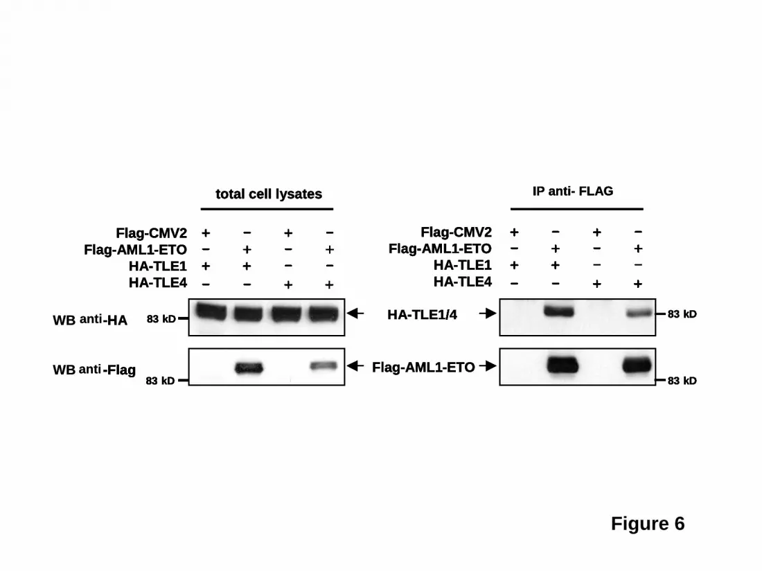
Figure 6
HA-TLE1/4
Flag-AML1-ETO
Flag-CMV2Flag-AML1-ETO
HA-TLE1HA-TLE4
total cell lysates
83 kD
83 kD
+ - + -- + - + + + - -- - + +
Flag-CMV2Flag-AML1-ETO
HA-TLE1HA-TLE4
WB -HA
WB -Flag
83 kD
83 kD
+ - + -- + - + + + - -- - + +
HA-TLE1/4
Flag-AML1-ETO
Flag-CMV2Flag-AML1-ETO
HA-TLE1HA-TLE4
total cell lysates
83 kD
83 kD
+ - + -- + - + + + - -- - + +
+ - + -- + - + + + - -- - + +
Flag-CMV2Flag-AML1-ETO
HA-TLE1HA-TLE4
-HA
-Flag
83 kD
83 kD
+ - + -- + - + + + - -- - + +
+ - + -- + - + + + - -- - + +
IP anti- FLAG
HA-TLE1/4
Flag-AML1-ETO
Flag-CMV2Flag-AML1-ETO
HA-TLE1HA-TLE4
total cell lysates
83 kD
83 kD
+ - + -- + - + + + - -- - + +
+ - + -- + - + + + - -- - + +
Flag-CMV2Flag-AML1-ETO
HA-TLE1HA-TLE4
-HA
-Flag
83 kD
83 kD
+ - + -- + - + + + - -- - + +
+ - + -- + - + + + - -- - + +
HA-TLE1/4
Flag-AML1-ETO
Flag-CMV2Flag-AML1-ETO
HA-TLE1HA-TLE4
total cell lysates
83 kD
83 kD
+ - + -- + - + + + - -- - + +
+ - + -- + - + + + - -- - + +
Flag-CMV2Flag-AML1-ETO
HA-TLE1HA-TLE4
-HA
-Flag
83 kD
83 kD
+ - + -- + - + + + - -- - + +
+ - + -- + - + + + - -- - + +
+ - + -- + - + + + - -- - + +
+ - + -- + - + + + - -- - + +
IP anti- FLAG
anti
anti
HA-TLE1/4
Flag-AML1-ETO
Flag-CMV2Flag-AML1-ETO
HA-TLE1HA-TLE4
total cell lysates
83 kD
83 kD
+ - + -- + - + + + - -- - + +
+ - + -- + - + + + - -- - + +
Flag-CMV2Flag-AML1-ETO
HA-TLE1HA-TLE4
WB -HA
WB -Flag
83 kD
83 kD
+ - + -- + - + + + - -- - + +
+ - + -- + - + + + - -- - + +
HA-TLE1/4
Flag-AML1-ETO
Flag-CMV2Flag-AML1-ETO
HA-TLE1HA-TLE4
total cell lysates
83 kD
83 kD
+ - + -- + - + + + - -- - + +
+ - + -- + - + + + - -- - + +
Flag-CMV2Flag-AML1-ETO
HA-TLE1HA-TLE4
-HA
-Flag
83 kD
83 kD
+ - + -- + - + + + - -- - + +
+ - + -- + - + + + - -- - + +
+ - + -- + - + + + - -- - + +
+ - + -- + - + + + - -- - + +
IP anti- FLAG
HA-TLE1/4
Flag-AML1-ETO
Flag-CMV2Flag-AML1-ETO
HA-TLE1HA-TLE4
total cell lysates
83 kD
83 kD
+ - + -- + - + + + - -- - + +
+ - + -- + - + + + - -- - + +
Flag-CMV2Flag-AML1-ETO
HA-TLE1HA-TLE4
-HA
-Flag
83 kD
83 kD
+ - + -- + - + + + - -- - + +
+ - + -- + - + + + - -- - + +
HA-TLE1/4
Flag-AML1-ETO
Flag-CMV2Flag-AML1-ETO
HA-TLE1HA-TLE4
total cell lysates
83 kD
83 kD
+ - + -- + - + + + - -- - + +
+ - + -- + - + + + - -- - + +
Flag-CMV2Flag-AML1-ETO
HA-TLE1HA-TLE4
-HA
-Flag
83 kD
83 kD
+ - + -- + - + + + - -- - + +
+ - + -- + - + + + - -- - + +
+ - + -- + - + + + - -- - + +
+ - + -- + - + + + - -- - + +
IP anti- FLAG
+ - + -- + - + + + - -- - + +
+ - + -- + - + + + - -- - + +
+ - + -- + - + + + - -- - + +
+ - + -- + - + + + - -- - + +
IP anti- FLAG
anti
anti
F
or personal use only. by guest on June 5, 2013.
bloodjournal.hematologylibrary.org
From
Copyright © 2022 FDOKUMEN