The leukemia-associated AML1 (Runx1)–CBFβ complex functions as a DNA-induced molecular clamp
Upload
independentCategory
view
2download
0
Cell, Vol. 87, 697–708, November 15, 1996, Copyright 1996 by Cell Press
The CBFb Subunit Is Essentialfor CBFa2 (AML1) Function In Vivo
Qing Wang,1 Terryl Stacy,1 Janelle D. Miller,1 (for review, see Orkin, 1995). A transcription factor com-plex that plays a central role in hematopoiesis is theAmy F. Lewis,1 Ting-Lei Gu,1 Xuemei Huang,2
core-binding factor (CBF, also known as PEBP2). CBFJohn H. Bushweller,2 Jean-Christophe Bories,3,7
is a heterodimeric transcription factor that consists ofFrederick W. Alt,3 Gabriella Ryan,4
a DNA-binding subunit called CBFa and a non-DNA-Pu Paul Liu,4 Anthony Wynshaw-Boris,4
binding subunit called CBFb (Ogawa et al., 1993a;Michael Binder,5 Miguel Marın-Padilla,5
Ogawa et al., 1993b; Wang et al., 1993). The CBFa sub-Arlene H. Sharpe,6 and Nancy A. Speck1
unit can bind DNA as a monomer in vitro, but its affinity1Department of Biochemistryfor DNA is 5- to 10-fold greater when it binds DNA asDartmouth Medical Schoola CBFa:b heterodimer (Ogawa et al., 1993a; Wang et al.,Hanover, New Hampshire 037551993). The only function ascribed to the CBFb subunit is2Department of Chemistrythat it increases the affinity of the CBFa subunit for DNA.Dartmouth CollegeThere is no evidence, for example, to suggest that theHanover, New Hampshire 03755CBFb subunit contains a transcriptional activation do-3Howard Hughes Medical Institutemain or modulates the intracellular localization of theThe Children’s HospitalCBFa subunit.300 Longwood Avenue
CBFa subunits are encoded by three distinct genesBoston, Massachusetts 02215in mice and humans:CBFA1, CBFA2 (AML1), and CBFA34National Center for Human Genome Research(Miyoshi et al., 1991; Bae et al., 1993, 1995; Ogawa etNational Institutes of Healthal., 1993b; Levanon et al., 1994). The CBFA1 and CBFA2Bethesda, Maryland 20892-4470genes areexpressed in a restricted setof tissues(Satake5Departments of Anatomy and Pathologyet al., 1995; Simeone et al., 1995), while the CBFA3 geneDartmouth Medical Schoolappears to be expressed ubiquitously (Bae et al., 1995).Hanover, New Hampshire 03755One of the CBFA genes, CBFA2 (AML1), is the target6Department of Pathologyof several chromosomal translocations associated withBrigham and Women’s Hospitalleukemias and myelodysplasias in humans, includingand Harvard Medical Schoolthe t(8;21)(q22;q22) associated with de novo acute my-221 Longwood Avenueeloid leukemia (M2 subtype) (Bitter et al., 1987; MiyoshiBoston, Massachusetts 02215et al., 1991), the t(12;21)(p13;q22) in de novo acutelymphocytic leukemia (pre-B cell) (Golub et al., 1995;Romana et al., 1995), and the relatively rare t(3;21)
Summary (q26;q22) associated with therapy-related leukemiasand myelodysplasias (Nucifora et al., 1993). All of these
The CBFb subunit is the non-DNA-binding subunit of translocations result in the production of novel chimericthe heterodimeric core-binding factor (CBF). CBFb as- proteins containing all or part of the CBFa2 protein,sociates with DNA-binding CBFa subunits and in- including its DNA-binding domain, fused to a proteincreases their affinity for DNA. Genes encoding the encoded on another chromosome.CBFb subunit (CBFB) and one of the CBFa subunits The CBFb subunit is encoded by one gene in mam-(CBFA2, otherwise known as AML1) are the most fre- mals, CBFB, that is ubiquitously expressed (Ogawa etquent targets of chromosomal translocations in acute al., 1993a; Wang et al., 1993). It is also disrupted inleukemias in humans. We and others previously dem- leukemias by inv(16)(p13;q22), t(16;16), and del(16)(q22)onstrated that homozygous disruption of the mouse (Liu et al., 1993). The inv(16) and t(16;16) breakpointsCbfa2 (AML1) gene results in embryonic lethality at occur primarily in the fifth intron of the CBFB gene andmidgestation due to hemorrhaging in the central ner- in six different introns of the MYH11 gene, the latter ofvous system and blocks fetal liver hematopoiesis. which encodes a smooth muscle myosin heavy chainHere we demonstrate that homozygous mutation of (SMMHC) (Liu et al., 1993; Shurtleff et al., 1995). Thethe Cbfb gene results in the same phenotype. Our result is a chimeric protein that contains amino acidsresults demonstrate that the CBFb subunit is required 1–165 of the CBFb protein, including the heterodimeriza-for CBFa2 function in vivo. tion domain for CBFa, fused to various lengths of the
C-terminal a-helical rod domain from SMMHC. A smallnumber of patients have breaks in the fourth intron ofIntroductionthe CBFB gene, which fuses amino acids 1–133 of CBFb
to SMMHC (Shurtleff et al., 1995). The CBFb(1–133)-The process by which multiple blood cell lineages differ-SMMHC protein generated from the breakpoint in intronentiate from a common pluripotential hematopoietic4 of the CBFB gene is missing four amino acids (aastem cell is controlled, in part, by transcription factors134–137) of the heterodimerization domain, which formthat regulate the expression of lineage-specific genespart of an a helix extending from aa 129–136 (unpub-lished data). CBFb lacking these four amino acidsweakly associates with CBFa on DNA in vitro, while7 Present Address: Institut D’Hematologie de L’Universite Paris VII,
Unite Inserm 93, 1 Avenue Claude Vellefaux, Paris, France. further C-terminal deletion to the end of exon 3–encoded
Cell698
sequences (aa 94) completely disrupts heterodimeriza- CBFa subunit in vitro, a partially functional CBFb proteinshould be synthesized as a result of this mutation. Usingtion with CBFa (unpublished data).
CBF binding sites are found in putative target genes the targeting vector shown in Figure 1B, we isolatedseven correctly targeted ES cell clones from 190 G418/transcribed in hematopoietic cells, including genes en-
coding cytokines (GM-CSF, IL-3), cell surface differenti- FIAU-resistant clones (Figure 1C), corresponding to atargeting frequency that was 10-fold higher than thatation markers (TCR CD3e,CSF-1 receptor), and enzymes
(myeloperoxidase, neutrophil elastase, granzyme B ser- which we obtained for the exons 4–5 deletion. TwoCbfb1/2 ES cell clones that were injectedinto blastocystsine protease) (for review, see Speck and Stacy, 1995).
The association of the CBFA2 gene with leukemias and yielded high percentage chimeric mice that transmittedthe ES cell through the germline. Mice heterozygous forthe presence of CBF binding sites in genes transcribed
in hematopoietic cells suggested that one or more of the mutation were interbred and their progeny analyzed.Analysis of mRNA and protein in Cbfb 1/1, 1/2,the CBF genes is required for hematopoiesis. We and
others recently showed that homozygous disruption of and 2/2 ES cells and embryos indicated that truncatedCBFb proteins lacking exon 5–encoded sequences werethe mouse Cbfa2 gene blocks fetal liver but not yolk sac
hematopoiesis (Okuda et al., 1996; Wang et al., 1996). synthesized at low levels from the hypomorphic Cbfballele (Figure 2). A 633 bp product was amplified byEmbryos homozygous for the Cbfa2 mutation died in
midgestation with extensive hemorrhages in the central reverse transcriptase polymerase chain reaction (RT–PCR) from Cbfb1/1 embryos, and a smaller 506 bp prod-nervous system (CNS), at nerve/CNS interfaces of cra-
nial and spinal nerves, and in somitic/intersomitic re- uct was amplified from Cbfb2/2 embryos, using primersderived from exons 1 and 6 (Figure 2A). RT–PCR prod-gions along the presumptive spinal cord (Okuda et al.,
1996; Wang et al., 1996). Hemorrhaging was preceded ucts amplified from Cbfb2/2 embryos were subclonedand sequenced (Figure 2B). Most of the subcloned in-by perivascular edema and apoptosis in these areas and
appears to have been caused by damage to developing serts were 506 bp and contained sequences derivedfrom exons 1, 2, 3, 4, and 6; the remaining inserts con-capillaries (Wang et al., 1996; unpublished data).
The association of inv(16)(p13;q22) and t(16;16) with tained sequences from exons 1, 2, 3, and 6. It appearedfrom RT–PCR from Cbfb1/2 embryos that lower amountsacute myeloid leukemias suggested that altered forms
of the CBFb subunit could interfere with CBF function, of mRNA were produced from the hypomorphic Cbfballele (Figure 2A), and this was confirmed by wholebut it did not indicate whether or not the normal CBFb
subunit is essential for CBF function in vivo. Here we mount in situ hybridization (Figure 2C). Cbfb expressionis seen in the telencephalon, cranial ganglia, and theshow that the Cbfb gene is essential for embryogenesis.
Embryos homozygous for a Cbfb mutation die in mid- dorsal root ganglia in 11.5 days postcoitum (dpc) em-bryos. The fetal liver displayed a fine, streaky pattern ofgestation with extensive hemorrhages in the CNS and
petechial hemorrhages at nerve–CNS interfaces of cra- staining, suggesting a low level of expression in thistissue. A noticeable decrease in Cbfb mRNA expressionnial and spinal nerves and in somitic/intersomitic re-
gions along the presumptive spinal cord, and they was observed in Cbfb 2/2 compared to 1/1 embryos,although the pattern of expression was identical.exhibit a severe impairment in fetal liver hematopoie-
sis—the same spectrum of abnormalities found in em- An approximately 21–22 kDa protein was detected byWestern blot in extracts prepared from Cbfb 1/1 andbryos homozygous for disruption of the Cbfa2 gene,
indicating that CBFb is required for CBFa2 function 1/2 ES cells and 11.5 dpc embryos (Figure 2D), consis-tent with the molecular weight of the CBFb(182) (21.5in vivo.kDa, lane 4) and CBFb(187) (22 kDa, data not shown)isoforms. This protein was not detected in extracts pre-
Results pared from Cbfb2/2 ES cells and embryos. Instead, acluster of faster migrating bands that could correspond
Generation of a Hypomorphic Cbfb Allele to the CBFb proteins lacking amino acids encoded inThe heterodimerization domain in the CBFb subunit that exon 5 and exons 4 and 5 were observed that are notmediates association with CBFa subunits is encoded in visible in extracts from 1/1 cells. [We will refer to theexons 1–4 (aa 1–133) plus the first four codons in exon major 155 aa protein in Cbfb2/2 cells as CBFb(155).]5 (aa 134–137) (Figure 1A) (Ogawa et al., 1993a; Golling Quantitative Western blotting (see Experimental Proce-et al., 1996). Our initial attempt to disrupt the Cbfb gene dures) indicated that the concentration of wild-typewith a targeting construct that replaced both exons 4 CBFb protein in 1/1 ES cells is approximately 4 3 1027
and 5 with the neor gene yielded only one correctly M and that the truncated CBFb proteins in Cbfb1/2 EStargeted ES cell clone, which upon injection into blasto- cells are 5- to 10-fold less abundant than CBFb pro-cysts yielded no live pups. Since the Cbfb gene is ex- duced from the wild-type allele (data not shown).pressed in ES cells, we reasoned that a 50% reduction A protein:DNA complex specific for the core site wasof the CBFb concentration might be incompatible with observed by electrophoretic mobility shift assay (EMSA)cell growth. We therefore decided to create a hypomor- in extracts prepared from Cbfb1/1 and Cbfb1/2 embryosphic Cbfb allele, replacing only the 59 half of exon 5 but not from Cbfb2/2 embryos (Figure 2E, lanes 1–9).with the neor gene. This mutation should result in the CBFb activity was also assayed by adding exogenoussynthesis of a CBFb protein lacking the last four amino CBFa2 to embryo extracts and determining whether theacids (aa 134–137) of the heterodimerization domain. extracts contained an activity that would shift theSince a CBFb protein truncated to the C-terminus of CBFa2:DNA complex to a more slowly migrating form
indicative of association with the CBFb subunit. Extractsexon 4–encoded sequences weakly associates with the
CBFb Is Required for CBFa2 (AML1) Function699
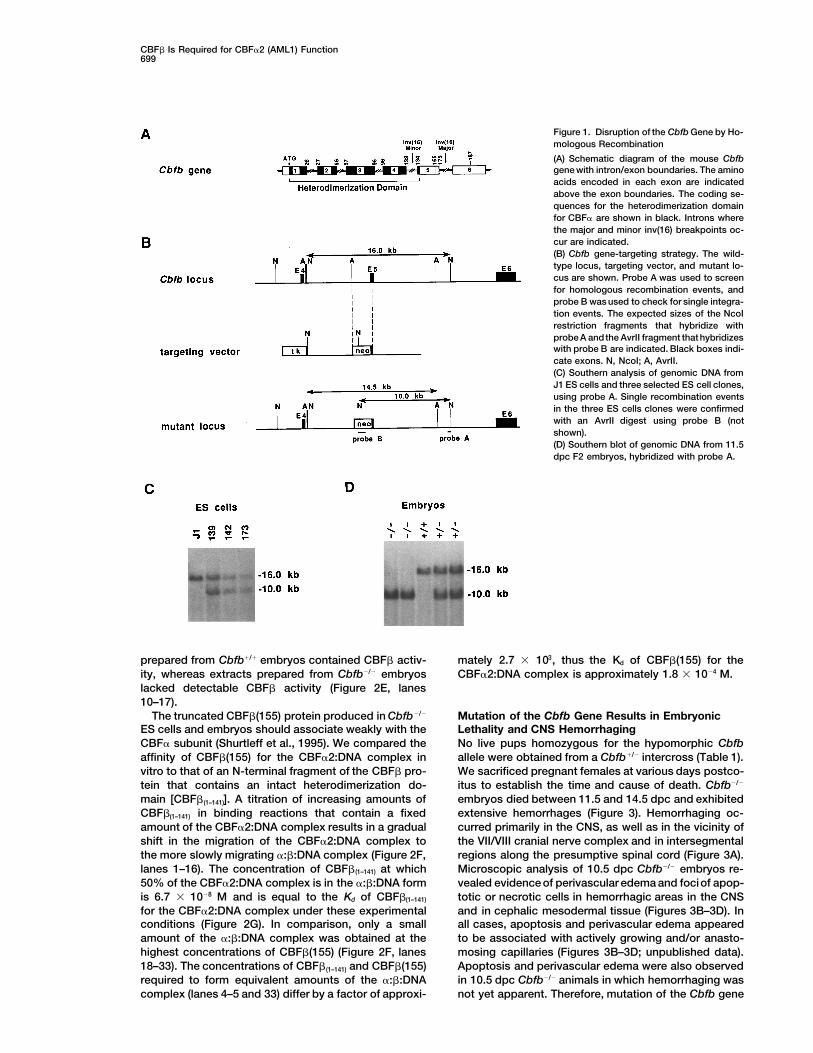
Figure 1. Disruption of the Cbfb Gene by Ho-mologous Recombination
(A) Schematic diagram of the mouse Cbfbgene with intron/exon boundaries. The aminoacids encoded in each exon are indicatedabove the exon boundaries. The coding se-quences for the heterodimerization domainfor CBFa are shown in black. Introns wherethe major and minor inv(16) breakpoints oc-cur are indicated.(B) Cbfb gene-targeting strategy. The wild-type locus, targeting vector, and mutant lo-cus are shown. Probe A was used to screenfor homologous recombination events, andprobe B was used to check for single integra-tion events. The expected sizes of the NcoIrestriction fragments that hybridize withprobe A andthe AvrII fragment that hybridizeswith probe B are indicated. Black boxes indi-cate exons. N, NcoI; A, AvrII.(C) Southern analysis of genomic DNA fromJ1 ES cells and three selected ES cell clones,using probe A. Single recombination eventsin the three ES cells clones were confirmedwith an AvrII digest using probe B (notshown).(D) Southern blot of genomic DNA from 11.5dpc F2 embryos, hybridized with probe A.
prepared from Cbfb1/1 embryos contained CBFb activ- mately 2.7 3 103, thus the Kd of CBFb(155) for theCBFa2:DNA complex is approximately 1.8 3 1024 M.ity, whereas extracts prepared from Cbfb2/2 embryos
lacked detectable CBFb activity (Figure 2E, lanes10–17).
The truncated CBFb(155) protein produced in Cbfb2/2 Mutation of the Cbfb Gene Results in EmbryonicLethality and CNS HemorrhagingES cells and embryos should associate weakly with the
CBFa subunit (Shurtleff et al., 1995). We compared the No live pups homozygous for the hypomorphic Cbfballele were obtained from a Cbfb1/2 intercross (Table 1).affinity of CBFb(155) for the CBFa2:DNA complex in
vitro to that of an N-terminal fragment of the CBFb pro- We sacrificed pregnant females at various days postco-itus to establish the time and cause of death. Cbfb2/2tein that contains an intact heterodimerization do-
main [CBFb(1–141)]. A titration of increasing amounts of embryos died between 11.5 and 14.5 dpc and exhibitedextensive hemorrhages (Figure 3). Hemorrhaging oc-CBFb(1–141) in binding reactions that contain a fixed
amount of the CBFa2:DNA complex results in a gradual curred primarily in the CNS, as well as in the vicinity ofthe VII/VIII cranial nerve complex and in intersegmentalshift in the migration of the CBFa2:DNA complex to
the more slowly migrating a:b:DNA complex (Figure 2F, regions along the presumptive spinal cord (Figure 3A).Microscopic analysis of 10.5 dpc Cbfb2/2 embryos re-lanes 1–16). The concentration of CBFb(1–141) at which
50% of the CBFa2:DNA complex is in the a:b:DNA form vealed evidence of perivascular edema and fociof apop-totic or necrotic cells in hemorrhagic areas in the CNSis 6.7 3 1028 M and is equal to the Kd of CBFb(1–141)
for the CBFa2:DNA complex under these experimental and in cephalic mesodermal tissue (Figures 3B–3D). Inall cases, apoptosis and perivascular edema appearedconditions (Figure 2G). In comparison, only a small
amount of the a:b:DNA complex was obtained at the to be associated with actively growing and/or anasto-mosing capillaries (Figures 3B–3D; unpublished data).highest concentrations of CBFb(155) (Figure 2F, lanes
18–33). The concentrations of CBFb(1–141) and CBFb(155) Apoptosis and perivascular edema were also observedin 10.5 dpc Cbfb2/2 animals in which hemorrhaging wasrequired to form equivalent amounts of the a:b:DNA
complex (lanes 4–5 and 33) differ by a factor of approxi- not yet apparent. Therefore, mutation of the Cbfb gene
Cell700
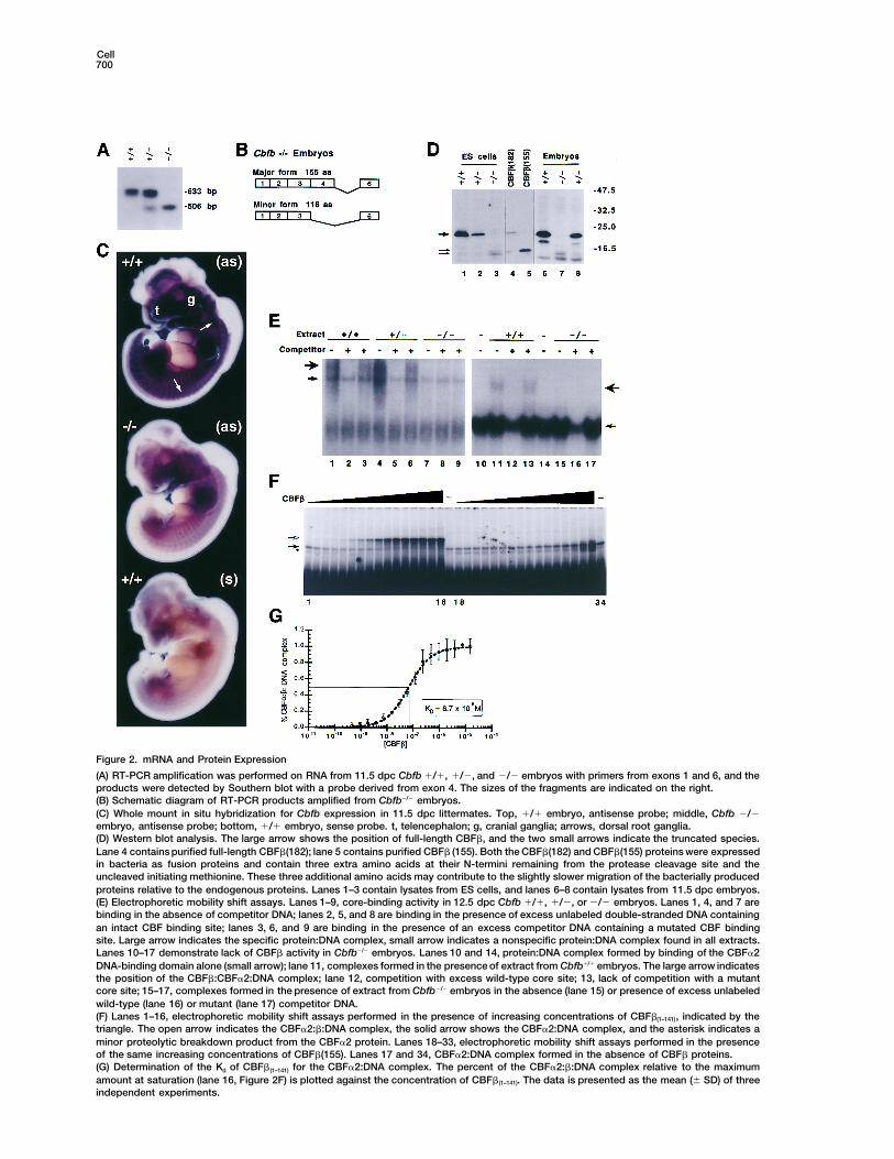
Figure 2. mRNA and Protein Expression
(A) RT-PCR amplification was performed on RNA from 11.5 dpc Cbfb 1/1, 1/2, and 2/2 embryos with primers from exons 1 and 6, and theproducts were detected by Southern blot with a probe derived from exon 4. The sizes of the fragments are indicated on the right.(B) Schematic diagram of RT-PCR products amplified from Cbfb2/2 embryos.(C) Whole mount in situ hybridization for Cbfb expression in 11.5 dpc littermates. Top, 1/1 embryo, antisense probe; middle, Cbfb 2/2embryo, antisense probe; bottom, 1/1 embryo, sense probe. t, telencephalon; g, cranial ganglia; arrows, dorsal root ganglia.(D) Western blot analysis. The large arrow shows the position of full-length CBFb, and the two small arrows indicate the truncated species.Lane 4 contains purified full-length CBFb(182); lane 5 contains purified CBFb (155). Both the CBFb(182) and CBFb(155) proteins were expressedin bacteria as fusion proteins and contain three extra amino acids at their N-termini remaining from the protease cleavage site and theuncleaved initiating methionine. These three additional amino acids may contribute to the slightly slower migration of the bacterially producedproteins relative to the endogenous proteins. Lanes 1–3 contain lysates from ES cells, and lanes 6–8 contain lysates from 11.5 dpc embryos.(E) Electrophoretic mobility shift assays. Lanes 1–9, core-binding activity in 12.5 dpc Cbfb 1/1, 1/2, or 2/2 embryos. Lanes 1, 4, and 7 arebinding in the absence of competitor DNA; lanes 2, 5, and 8 are binding in the presence of excess unlabeled double-stranded DNA containingan intact CBF binding site; lanes 3, 6, and 9 are binding in the presence of an excess competitor DNA containing a mutated CBF bindingsite. Large arrow indicates the specific protein:DNA complex, small arrow indicates a nonspecific protein:DNA complex found in all extracts.Lanes 10–17 demonstrate lack of CBFb activity in Cbfb2/2 embryos. Lanes 10 and 14, protein:DNA complex formed by binding of the CBFa2DNA-binding domain alone (small arrow); lane 11, complexes formed in the presence of extract from Cbfb1/1 embryos. The large arrow indicatesthe position of the CBFb:CBFa2:DNA complex; lane 12, competition with excess wild-type core site; 13, lack of competition with a mutantcore site; 15–17, complexes formed in the presence of extract from Cbfb2/2 embryos in the absence (lane 15) or presence of excess unlabeledwild-type (lane 16) or mutant (lane 17) competitor DNA.(F) Lanes 1–16, electrophoretic mobility shift assays performed in the presence of increasing concentrations of CBFb(1–141), indicated by thetriangle. The open arrow indicates the CBFa2:b:DNA complex, the solid arrow shows the CBFa2:DNA complex, and the asterisk indicates aminor proteolytic breakdown product from the CBFa2 protein. Lanes 18–33, electrophoretic mobility shift assays performed in the presenceof the same increasing concentrations of CBFb(155). Lanes 17 and 34, CBFa2:DNA complex formed in the absence of CBFb proteins.(G) Determination of the Kd of CBFb(1–141) for the CBFa2:DNA complex. The percent of the CBFa2:b:DNA complex relative to the maximumamount at saturation (lane 16, Figure 2F) is plotted against the concentration of CBFb(1–141). The data is presented as the mean (6 SD) of threeindependent experiments.
CBFb Is Required for CBFa2 (AML1) Function701
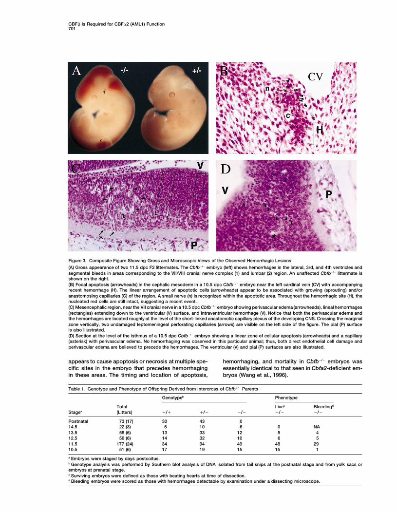
Figure 3. Composite Figure Showing Gross and Microscopic Views of the Observed Hemorrhagic Lesions
(A) Gross appearance of two 11.5 dpc F2 littermates. The Cbfb2/2 embryo (left) shows hemorrhages in the lateral, 3rd, and 4th ventricles andsegmental bleeds in areas corresponding to the VII/VIII cranial nerve complex (1) and lumbar (2) region. An unaffected Cbfb1/2 littermate isshown on the right.(B) Focal apoptosis (arrowheads) in the cephalic mesoderm in a 10.5 dpc Cbfb2/2 embryo near the left cardinal vein (CV) with accompanyingrecent hemorrhage (H). The linear arrangement of apoptotic cells (arrowheads) appear to be associated with growing (sprouting) and/oranastomosing capillaries (C) of the region. A small nerve (n) is recognized within the apoptotic area. Throughout the hemorrhagic site (H), thenucleated red cells are still intact, suggesting a recent event.(C) Mesencephalic region, near the VII cranial nerve in a 10.5 dpc Cbfb2/2 embryo showing perivascular edema (arrowheads), lineal hemorrhages(rectangles) extending down to the ventricular (V) surface, and intraventricular hemorrhage (V). Notice that both the perivascular edema andthe hemorrhages are located roughly at the level of the short-linked anastomotic capillary plexus of the developing CNS. Crossing the marginalzone vertically, two undamaged leptomeningeal perforating capillaries (arrows) are visible on the left side of the figure. The pial (P) surfaceis also illustrated.(D) Section at the level of the isthmus of a 10.5 dpc Cbfb2/2 embryo showing a linear zone of cellular apoptosis (arrowheads) and a capillary(asterisk) with perivascular edema. No hemorrhaging was observed in this particular animal; thus, both direct endothelial cell damage andperivascular edema are believed to precede the hemorrhages. The ventricular (V) and pial (P) surfaces are also illustrated.
appears to cause apoptosis or necrosis at multiple spe- hemorrhaging, and mortality in Cbfb2/2 embryos wasessentially identical to that seen in Cbfa2-deficient em-cific sites in the embryo that precedes hemorrhaging
in these areas. The timing and location of apoptosis, bryos (Wang et al., 1996).
Table 1. Genotype and Phenotype of Offspring Derived from Intercross of Cbfb1/2 Parents
Genotypeb Phenotype
Total Livec Bleedingd
Stagea (Litters) 1/1 1/2 2/2 2/2 2/2
Postnatal 73 (17) 30 43 014.5 22 (3) 6 10 6 0 NA13.5 58 (6) 13 33 12 5 412.5 56 (6) 14 32 10 6 511.5 177 (24) 34 94 49 48 2910.5 51 (6) 17 19 15 15 1
a Embryos were staged by days postcoitus.b Genotype analysis was performed by Southern blot analysis of DNA isolated from tail snips at the postnatal stage and from yolk sacs orembryos at prenatal stage.c Surviving embryos were defined as those with beating hearts at time of dissection.d Bleeding embryos were scored as those with hemorrhages detectable by examination under a dissecting microscope.
Cell702
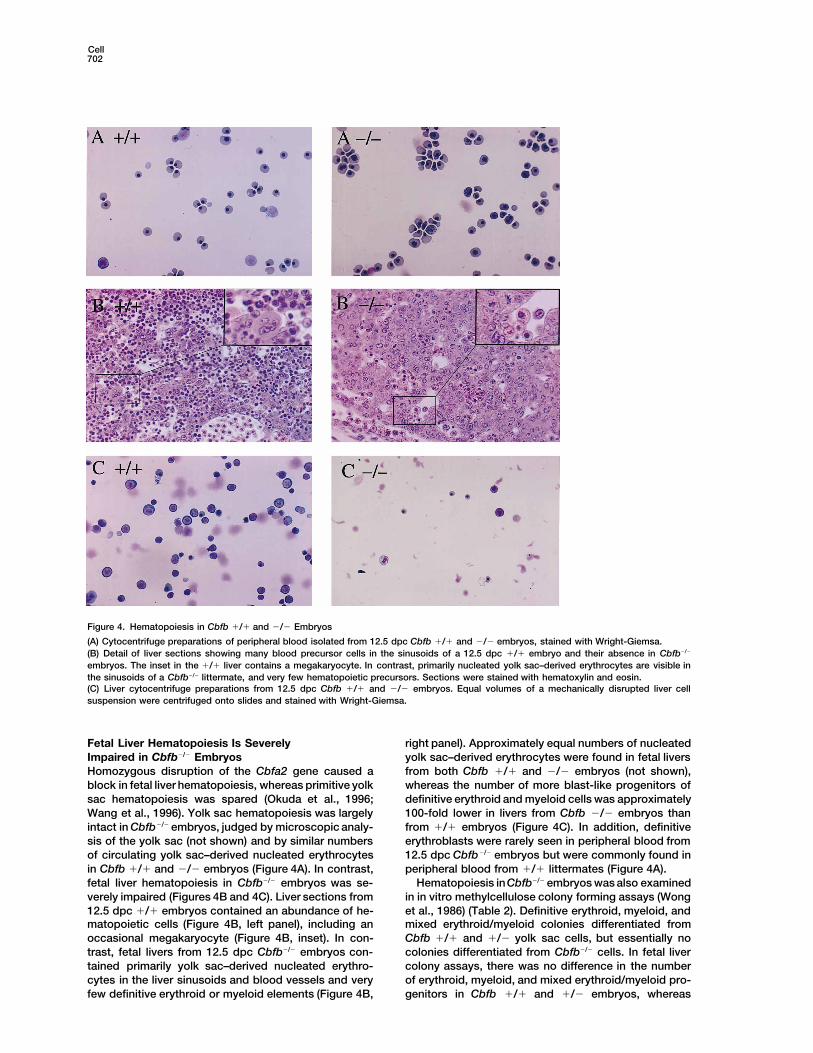
Figure 4. Hematopoiesis in Cbfb 1/1 and 2/2 Embryos
(A) Cytocentrifuge preparations of peripheral blood isolated from 12.5 dpc Cbfb 1/1 and 2/2 embryos, stained with Wright-Giemsa.(B) Detail of liver sections showing many blood precursor cells in the sinusoids of a 12.5 dpc 1/1 embryo and their absence in Cbfb2/2
embryos. The inset in the 1/1 liver contains a megakaryocyte. In contrast, primarily nucleated yolk sac–derived erythrocytes are visible inthe sinusoids of a Cbfb2/2 littermate, and very few hematopoietic precursors. Sections were stained with hematoxylin and eosin.(C) Liver cytocentrifuge preparations from 12.5 dpc Cbfb 1/1 and 2/2 embryos. Equal volumes of a mechanically disrupted liver cellsuspension were centrifuged onto slides and stained with Wright-Giemsa.
Fetal Liver Hematopoiesis Is Severely right panel). Approximately equal numbers of nucleatedyolk sac–derived erythrocytes were found in fetal liversImpaired in Cbfb2/2 Embryos
Homozygous disruption of the Cbfa2 gene caused a from both Cbfb 1/1 and 2/2 embryos (not shown),whereas the number of more blast-like progenitors ofblock in fetal liver hematopoiesis, whereas primitive yolk
sac hematopoiesis was spared (Okuda et al., 1996; definitive erythroid and myeloid cells was approximately100-fold lower in livers from Cbfb 2/2 embryos thanWang et al., 1996). Yolk sac hematopoiesis was largely
intact in Cbfb2/2 embryos, judged by microscopic analy- from 1/1 embryos (Figure 4C). In addition, definitiveerythroblasts were rarely seen in peripheral blood fromsis of the yolk sac (not shown) and by similar numbers
of circulating yolk sac–derived nucleated erythrocytes 12.5 dpc Cbfb2/2 embryos but were commonly found inperipheral blood from 1/1 littermates (Figure 4A).in Cbfb 1/1 and 2/2 embryos (Figure 4A). In contrast,
fetal liver hematopoiesis in Cbfb2/2 embryos was se- Hematopoiesis inCbfb2/2 embryos was also examinedin in vitro methylcellulose colony forming assays (Wongverely impaired (Figures 4B and 4C). Liver sections from
12.5 dpc 1/1 embryos contained an abundance of he- et al., 1986) (Table 2). Definitive erythroid, myeloid, andmixed erythroid/myeloid colonies differentiated frommatopoietic cells (Figure 4B, left panel), including an
occasional megakaryocyte (Figure 4B, inset). In con- Cbfb 1/1 and 1/2 yolk sac cells, but essentially nocolonies differentiated from Cbfb2/2 cells. In fetal livertrast, fetal livers from 12.5 dpc Cbfb2/2 embryos con-
tained primarily yolk sac–derived nucleated erythro- colony assays, there was no difference in the numberof erythroid, myeloid, and mixed erythroid/myeloid pro-cytes in the liver sinusoids and blood vessels and very
few definitive erythroid or myeloid elements (Figure 4B, genitors in Cbfb 1/1 and 1/2 embryos, whereas
CBFb Is Required for CBFa2 (AML1) Function703
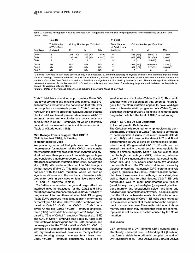
Table 2. Colonies Arising from Yolk Sac and Fetal Liver Progenitors Isolated from Offspring Derived from Intercrosses of Cbfb1/2 andCbfa21/2 Mice
10.5 dpc 11.5 dpc
Total Number Colony Number per Yolk Saca Total Number Colony Number per Fetal Liverof Yolk Sacs of Fetal Livers
Genotype Analyzed E M Mix Analyzed E M Mix
Cbfb1/1 16 263 (130) 279 (90) 38 (16) 9 486 (324) 696 (212) 212 (151)Cbfb1/2 17 237 (94) 253 (85) 42 (17) 25 652 (397) 882 (385) 236 (171)Cbfb2/2 12 0 1 1 9 1 (1) 25 (13) 3 (4)
Cbfa21/1 NDb ND ND ND 8 981 (273) 1240 (183) 331 (73)Cbfa21/2 ND ND ND ND 7 537 (167) 817 (332) 150 (107)Cbfa22/2 ND ND ND ND 6 0 0 0
a Colonies (.30 cells in size) were scored on day 7 of incubation. E, erythroid colonies; M, myeloid colonies; Mix, erythroid-myeloid mixedcolonies. Average number of colonies per yolk sac is indicated, followed by standard deviation in parentheses. The difference between thenumbers of colonies from Cbfa2 1/2 and 1/1 fetal livers is significant at P , 0.02 by Student’s t test. There is no significant differencebetween the number of colonies from Cbfb 1/1 and 1/2 yolk sacs and fetal livers. The relatively large standard deviation can be attributedprimarily to variation between litters.b Data for Cbfa2 E10.5 yolk sac progenitors is published elsewhere (Wang et al., 1996).
Cbfb2/2 fetal livers contained approximately 30- to 500- small numbers of colonies (Tables 2 and 3). This result,together with the observation that embryos heterozy-fold fewer erythroid and myeloid progenitors. These re-
sults further substantiate the conclusion that fetal liver gous for the Cbfb mutation appear to have wild-typelevels of hematopoietic progenitor cells (Table 2), sug-hematopoiesis is severely impaired in Cbfb2/2 embryos.
However, the in vitro progenitor assays suggest that the gests that the level of CBFa2 is limiting in hematopoieticprogenitor cells but the level of CBFb is saturating.block in fetal liver hematopoiesis is less severe in Cbfb2/2
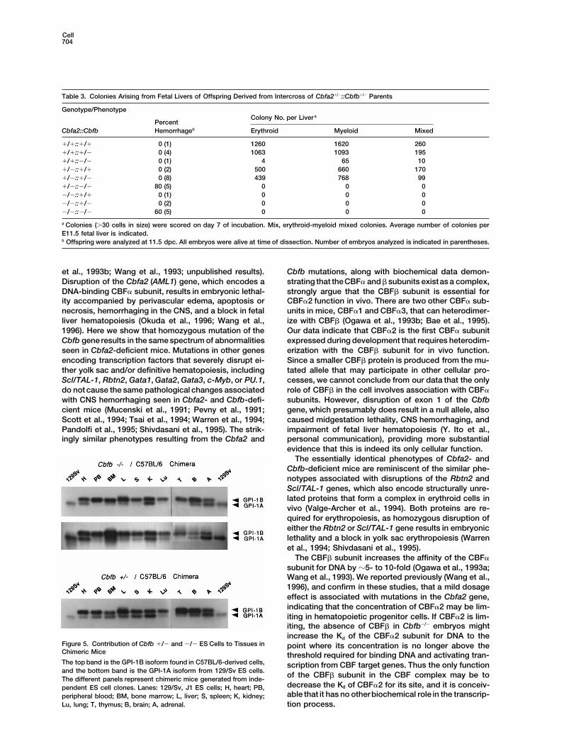
embryos, where some colonies are consistently ob-served, than in Cbfa22/2 embryos, for which essentially Cbfb2/2 ES Cells Do Not Contribute
to Hematopoietic Cells In Vivono erythroid or myeloid colonies differentiate in vitro(Table 2) (Okuda et al., 1996). The Cbfa2 gene is required for lymphopoiesis, as dem-
onstrated by the failure of Cbfa22/2 ES cells to contributeto hematopoietic tissues in chimeric animals (OkudaMild Dosage Effects Suggest That CBFa2
(AML1), but Not CBFb, Is Limiting et al., 1996) and to rescue the defect in T and B celldifferentiation in RAG-2-deficient chimeric mice (unpub-in Hematopoietic Cells
We previously reported that yolk sacs from embryos lished data). We generated Cbfb2/2 ES cells and as-sessed their ability to contribute to hematopoietic tis-heterozygous for mutation of the Cbfa2 gene consis-
tently contained fewer progenitors for erythroid and my- sues in chimeric animals. Cbfb1/2 ES cell clones gaverise to high percentage (>90%) chimeras, whereaseloid colonies than yolk sacs from wild-type embryos
and concluded that there appeared to be a mild dosage Cbfb2/2 ES cells generated chimeras that contained be-tween 30% and 70% agouti coat color. We analyzedeffect associated with mutation of the Cbfa2gene (Wang
et al., 1996). We confirmed this result in fetal liver pro- the contribution of the ES cells to different tissues byglucose phosphate isomerase (GPI) isoform analysisgenitor assays (Table 2). This mild dosage effect was
not seen with the Cbfb mutation, where we saw no (Figure 5) (Williams et al., 1994). Cbfb1/2 ES cells contrib-uted to all tissues examined, although consistently lesssignificant difference in the numbers of hematopoietic
progenitor cells in yolk sacs or fetal livers from Cbfb well to thymus than to other tissues. Cbfb2/2 ES cellscontributed well to most nonhematopoietic tissues1/1 and 1/2 embryos (Table 2).
To further characterize the gene dosage effect, we (heart, kidney, brain, adrenal gland), only weakly to liver,bone marrow, and occasionally spleen and lung, andinterbred mice heterozygous for the Cbfa2 and Cbfb
mutations toobtaintransheterozygous Cbfa21/2:: Cbfb1/2 not at all toperipheral blood orthymus. The data suggestthat the block in hematopoiesis is cell autonomous,progeny and interbred these mice to obtain F2 progeny
(Table 3).We observed no accentuation of hemorrhaging since hematopoiesis of Cbfb2/2 ES cells does not occurin the microenvironment of the hematopoietic compart-or mortality in 11.5 dpc Cbfa22/2::Cbfb2/2 embryos com-
pared to Cbfa22/2::Cbfb1/1 or Cbfa21/1:: Cbfb2/2 em- ment of a normal mouse. The small contribution to bonemarrow and spleen may indicate that the block in hema-bryos. Of the five Cbfa22/2::Cbfb2/2 embryos analyzed,
three (60%) had visible hemorrhages at 11.5 dpc, com- topoiesis is not as severe as that caused by the Cbfa2mutation.pared to 73% of Cbfa22/2 embryos (Wang et al., 1996)
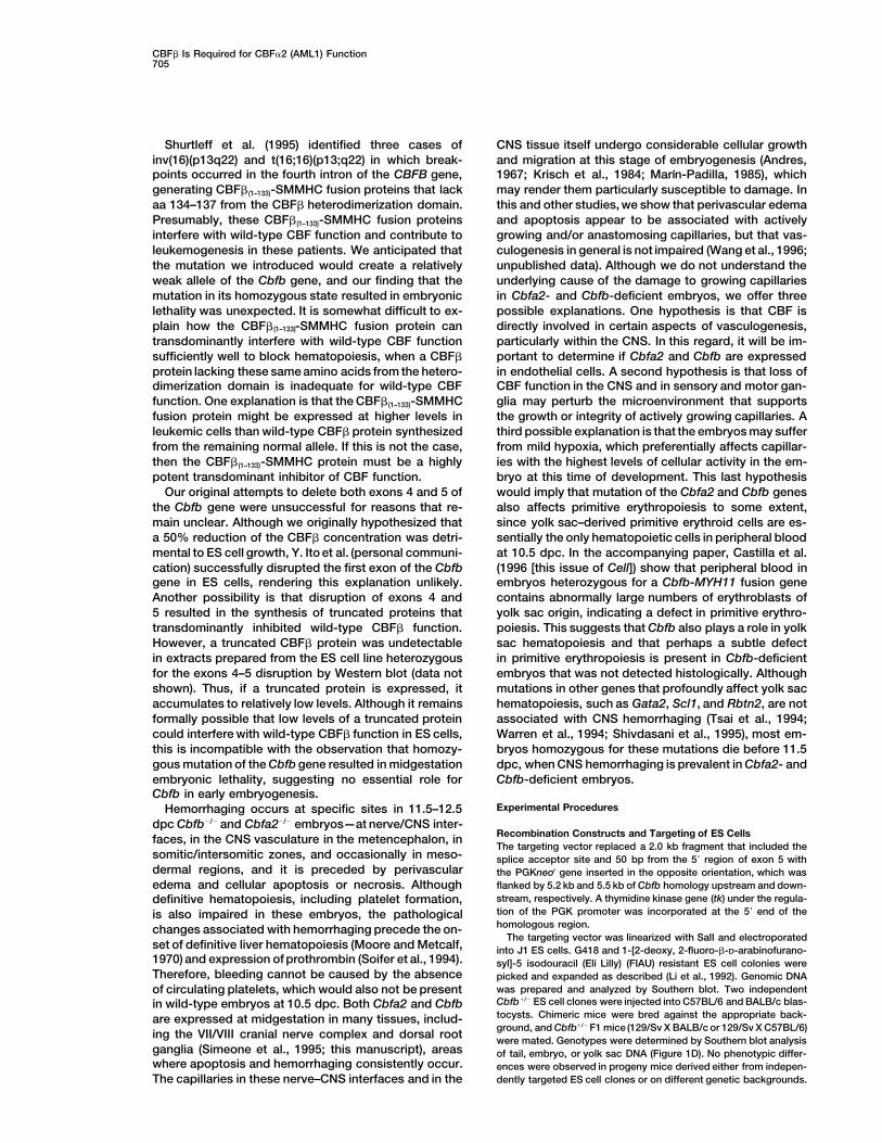
and 60% of Cbfb2/2 embryos (see Table 1). Fetal liversfrom embryos homozygous for the Cbfb mutation and Discussionheterozygous for the Cbfa2 mutation (Cbfa21/2::Cbfb2/2)contained no progenitor cells capable of differentiating CBF consists of a DNA-binding CBFa subunit and a
structurally unrelated non-DNA-binding CBFb subunitinto erythroid or myeloid colonies in methylcellulosecolony forming assays, whereas fetal livers from that form a stable heterodimeric complex on and off
DNA (Kamachi et al., 1990; Ogawa et al., 1993a; OgawaCbfa21/1::Cbfb2/2 embryos consistently gave rise to
Cell704
Table 3. Colonies Arising from Fetal Livers of Offspring Derived from Intercross of Cbfa21/2::Cbfb1/2 Parents
Genotype/PhenotypeColony No. per Liver a
PercentCbfa2::Cbfb Hemorrhageb Erythroid Myeloid Mixed
1/1::1/1 0 (1) 1260 1620 2601/1::1/2 0 (4) 1063 1093 1951/1::2/2 0 (1) 4 65 101/2::1/1 0 (2) 500 660 1701/2::1/2 0 (8) 439 768 991/2::2/2 80 (5) 0 0 02/2::1/1 0 (1) 0 0 02/2::1/2 0 (2) 0 0 02/2::2/2 60 (5) 0 0 0
a Colonies (.30 cells in size) were scored on day 7 of incubation. Mix, erythroid-myeloid mixed colonies. Average number of colonies perE11.5 fetal liver is indicated.b Offspring were analyzed at 11.5 dpc. All embryos were alive at time of dissection. Number of embryos analyzed is indicated in parentheses.
et al., 1993b; Wang et al., 1993; unpublished results). Cbfb mutations, along with biochemical data demon-strating that theCBFa and b subunits existas a complex,Disruption of the Cbfa2 (AML1) gene, which encodes a
DNA-binding CBFa subunit, results in embryonic lethal- strongly argue that the CBFb subunit is essential forCBFa2 function in vivo. There are two other CBFa sub-ity accompanied by perivascular edema, apoptosis or
necrosis, hemorrhaging in the CNS, and a block in fetal units in mice, CBFa1 and CBFa3, that can heterodimer-ize with CBFb (Ogawa et al., 1993b; Bae et al., 1995).liver hematopoiesis (Okuda et al., 1996; Wang et al.,
1996). Here we show that homozygous mutation of the Our data indicate that CBFa2 is the first CBFa subunitexpressed during development that requires heterodim-Cbfb gene results in the same spectrum of abnormalities
seen in Cbfa2-deficient mice. Mutations in other genes erization with the CBFb subunit for in vivo function.Since a smaller CBFb protein is produced from the mu-encoding transcription factors that severely disrupt ei-
ther yolk sac and/or definitive hematopoiesis, including tated allele that may participate in other cellular pro-cesses, we cannot conclude from our data that the onlyScl/TAL-1, Rbtn2, Gata1, Gata2, Gata3, c-Myb, or PU.1,
do not cause the same pathological changes associated role of CBFb in the cell involves association with CBFa
subunits. However, disruption of exon 1 of the Cbfbwith CNS hemorrhaging seen in Cbfa2- and Cbfb-defi-cient mice (Mucenski et al., 1991; Pevny et al., 1991; gene, which presumably does result in a null allele, also
caused midgestation lethality, CNS hemorrhaging, andScott et al., 1994; Tsai et al., 1994; Warren et al., 1994;Pandolfi et al., 1995; Shivdasani et al., 1995). The strik- impairment of fetal liver hematopoiesis (Y. Ito et al.,
personal communication), providing more substantialingly similar phenotypes resulting from the Cbfa2 andevidence that this is indeed its only cellular function.
The essentially identical phenotypes of Cbfa2- andCbfb-deficient mice are reminiscent of the similar phe-notypes associated with disruptions of the Rbtn2 andScl/TAL-1 genes, which also encode structurally unre-lated proteins that form a complex in erythroid cells invivo (Valge-Archer et al., 1994). Both proteins are re-quired for erythropoiesis, as homozygous disruption ofeither the Rbtn2 or Scl/TAL-1 gene results in embryoniclethality and a block in yolk sac erythropoiesis (Warrenet al., 1994; Shivdasani et al., 1995).
The CBFb subunit increases the affinity of the CBFa
subunit for DNA by z5- to 10-fold (Ogawa et al., 1993a;Wang et al., 1993). We reported previously (Wang et al.,1996), and confirm in these studies, that a mild dosageeffect is associated with mutations in the Cbfa2 gene,indicating that the concentration of CBFa2 may be lim-iting in hematopoietic progenitor cells. If CBFa2 is lim-iting, the absence of CBFb in Cbfb2/2 embryos mightincrease the Kd of the CBFa2 subunit for DNA to the
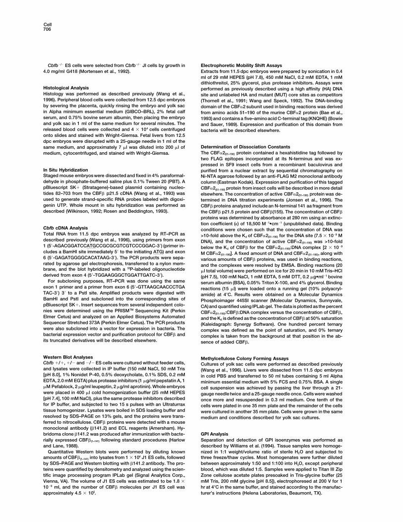
Figure 5. Contribution of Cbfb 1/2 and 2/2 ES Cells to Tissues in point where its concentration is no longer above theChimeric Mice threshold required for binding DNA and activating tran-The top band is the GPI-1B isoform found in C57BL/6-derived cells, scription from CBF target genes. Thus the only functionand the bottom band is the GPI-1A isoform from 129/Sv ES cells.
of the CBFb subunit in the CBF complex may be toThe different panels represent chimeric mice generated from inde-decrease the Kd of CBFa2 for its site, and it is conceiv-pendent ES cell clones. Lanes: 129/Sv, J1 ES cells; H, heart; PB,able that it has no otherbiochemical role in the transcrip-peripheral blood; BM, bone marrow; L, liver; S, spleen; K, kidney;
Lu, lung; T, thymus; B, brain; A, adrenal. tion process.
CBFb Is Required for CBFa2 (AML1) Function705
Shurtleff et al. (1995) identified three cases of CNS tissue itself undergo considerable cellular growthand migration at this stage of embryogenesis (Andres,inv(16)(p13q22) and t(16;16)(p13;q22) in which break-
points occurred in the fourth intron of the CBFB gene, 1967; Krisch et al., 1984; Marın-Padilla, 1985), whichmay render them particularly susceptible to damage. Ingenerating CBFb(1–133)-SMMHC fusion proteins that lack
aa 134–137 from the CBFb heterodimerization domain. this and other studies, we show that perivascular edemaand apoptosis appear to be associated with activelyPresumably, these CBFb(1–133)-SMMHC fusion proteins
interfere with wild-type CBF function and contribute to growing and/or anastomosing capillaries, but that vas-culogenesis in general is not impaired (Wang et al., 1996;leukemogenesis in these patients. We anticipated that
the mutation we introduced would create a relatively unpublished data). Although we do not understand theunderlying cause of the damage to growing capillariesweak allele of the Cbfb gene, and our finding that the
mutation in its homozygous state resulted in embryonic in Cbfa2- and Cbfb-deficient embryos, we offer threepossible explanations. One hypothesis is that CBF islethality was unexpected. It is somewhat difficult to ex-
plain how the CBFb(1–133)-SMMHC fusion protein can directly involved in certain aspects of vasculogenesis,particularly within the CNS. In this regard, it will be im-transdominantly interfere with wild-type CBF function
sufficiently well to block hematopoiesis, when a CBFb portant to determine if Cbfa2 and Cbfb are expressedin endothelial cells. A second hypothesis is that loss ofprotein lacking these same amino acids from the hetero-
dimerization domain is inadequate for wild-type CBF CBF function in the CNS and in sensory and motor gan-glia may perturb the microenvironment that supportsfunction. One explanation is that the CBFb(1–133)-SMMHC
fusion protein might be expressed at higher levels in the growth or integrity of actively growing capillaries. Athird possible explanation is that the embryos may sufferleukemic cells than wild-type CBFb protein synthesized
from the remaining normal allele. If this is not the case, from mild hypoxia, which preferentially affects capillar-ies with the highest levels of cellular activity in the em-then the CBFb(1–133)-SMMHC protein must be a highly
potent transdominant inhibitor of CBF function. bryo at this time of development. This last hypothesiswould imply that mutation of the Cbfa2 and Cbfb genesOur original attempts to delete both exons 4 and 5 of
the Cbfb gene were unsuccessful for reasons that re- also affects primitive erythropoiesis to some extent,since yolk sac–derived primitive erythroid cells are es-main unclear. Although we originally hypothesized that
a 50% reduction of the CBFb concentration was detri- sentially the only hematopoietic cells in peripheral bloodat 10.5 dpc. In the accompanying paper, Castilla et al.mental to ES cell growth, Y. Ito et al. (personal communi-
cation) successfully disrupted the first exon of the Cbfb (1996 [this issue of Cell]) show that peripheral blood inembryos heterozygous for a Cbfb-MYH11 fusion genegene in ES cells, rendering this explanation unlikely.
Another possibility is that disruption of exons 4 and contains abnormally large numbers of erythroblasts ofyolk sac origin, indicating a defect in primitive erythro-5 resulted in the synthesis of truncated proteins that
transdominantly inhibited wild-type CBFb function. poiesis. This suggests that Cbfb also plays a role in yolksac hematopoiesis and that perhaps a subtle defectHowever, a truncated CBFb protein was undetectable
in extracts prepared from the ES cell line heterozygous in primitive erythropoiesis is present in Cbfb-deficientembryos that was not detected histologically. Althoughfor the exons 4–5 disruption by Western blot (data not
shown). Thus, if a truncated protein is expressed, it mutations in other genes that profoundly affect yolk sachematopoiesis, such as Gata2, Scl1, and Rbtn2, are notaccumulates to relatively low levels. Although it remains
formally possible that low levels of a truncated protein associated with CNS hemorrhaging (Tsai et al., 1994;Warren et al., 1994; Shivdasani et al., 1995), most em-could interfere with wild-type CBFb function in ES cells,
this is incompatible with the observation that homozy- bryos homozygous for these mutations die before 11.5dpc, when CNS hemorrhaging is prevalent in Cbfa2- andgous mutation of the Cbfb gene resulted in midgestation
embryonic lethality, suggesting no essential role for Cbfb-deficient embryos.Cbfb in early embryogenesis.
Experimental ProceduresHemorrhaging occurs at specific sites in 11.5–12.5dpc Cbfb2/2 and Cbfa22/2 embryos—at nerve/CNS inter-
Recombination Constructs and Targeting of ES Cellsfaces, in the CNS vasculature in the metencephalon, inThe targeting vector replaced a 2.0 kb fragment that included the
somitic/intersomitic zones, and occasionally in meso- splice acceptor site and 50 bp from the 59 region of exon 5 withdermal regions, and it is preceded by perivascular the PGKneor gene inserted in the opposite orientation, which wasedema and cellular apoptosis or necrosis. Although flanked by 5.2 kb and 5.5 kb of Cbfb homology upstream and down-
stream, respectively. A thymidine kinase gene (tk) under the regula-definitive hematopoiesis, including platelet formation,tion of the PGK promoter was incorporated at the 59 end of theis also impaired in these embryos, the pathologicalhomologous region.changes associated with hemorrhaging precede the on-
The targeting vector was linearized with SalI and electroporatedset of definitive liver hematopoiesis (Moore and Metcalf, into J1 ES cells. G418 and 1-[2-deoxy, 2-fluoro-b-D-arabinofurano-1970) and expression of prothrombin (Soifer et al., 1994). syl]-5 isodouracil (Eli Lilly) (FIAU) resistant ES cell colonies wereTherefore, bleeding cannot be caused by the absence picked and expanded as described (Li et al., 1992). Genomic DNA
was prepared and analyzed by Southern blot. Two independentof circulating platelets, which would also not be presentCbfb1/2 ES cell clones were injected into C57BL/6 and BALB/c blas-in wild-type embryos at 10.5 dpc. Both Cbfa2 and Cbfbtocysts. Chimeric mice were bred against the appropriate back-are expressed at midgestation in many tissues, includ-ground, and Cbfb1/2 F1 mice (129/Sv X BALB/c or 129/Sv X C57BL/6)
ing the VII/VIII cranial nerve complex and dorsal root were mated. Genotypes were determined by Southern blot analysisganglia (Simeone et al., 1995; this manuscript), areas of tail, embryo, or yolk sac DNA (Figure 1D). No phenotypic differ-where apoptosis and hemorrhaging consistently occur. ences were observed in progeny mice derived either from indepen-
dently targeted ES cell clones or on different genetic backgrounds.The capillaries in these nerve–CNS interfaces and in the
Cell706
Cbfb2/2 ES cells were selected from Cbfb1/2 JI cells by growth in Electrophoretic Mobility Shift AssaysExtracts from 11.5 dpc embryos were prepared by sonication in 0.44.0 mg/ml G418 (Mortensen et al., 1992).ml of 29 mM HEPES (pH 7.8), 450 mM NaCl, 0.2 mM EDTA, 1 mMdithiothreitol, 25% glycerol, plus protease inhibitors. Assays were
Histological Analysis performed as previously described using a high affinity (HA) DNAHistology was performed as described previously (Wang et al., site and unlabeled HA and mutant (MUT) core sites as competitors1996). Peripheral blood cells were collected from 12.5 dpc embryos (Thornell et al., 1991; Wang and Speck, 1992). The DNA-bindingby severing the placenta, quickly rinsing the embryo and yolk sac domain of the CBFa2 subunit used in binding reactions was derivedin Alpha minimum essential medium (GIBCO–BRL), 2% fetal calf from amino acids 51–190 of the murine CBFa2 protein (Bae et al.,serum, and 0.75% bovine serum albumin, then placing the embryo 1993) and contains a five–aminoacid C-terminal tag (KNQHE) (Bowieand yolk sac in 1 ml of the same medium for several minutes. The and Sauer, 1989). Expression and purification of this domain fromreleased blood cells were collected and 4 3 104 cells centrifuged bacteria will be described elsewhere.onto slides and stained with Wright-Giemsa. Fetal livers from 12.5dpc embryos were disrupted with a 25-gauge needle in 1 ml of the
Determination of Dissociation Constantssame medium, and approximately 7 ml was diluted into 200 ml ofThe CBFa2(51–190) protein contained a hexahistidine tag followed bymedium, cytocentrifuged, and stained with Wright-Giemsa.two FLAG epitopes incorporated at its N-terminus and was ex-pressed in SF9 insect cells from a recombinant baculovirus and
In Situ Hybridization purified from a nuclear extract by sequential chromatography onStaged mouse embryos were dissected and fixed in 4% paraformal- Ni-NTA agarose followed by an anti-FLAG M2 monoclonal antibodydehyde in phosphate-buffered saline plus 0.1% Tween 20 (PBT). A column (Eastman Kodak). Expression and purification of this taggedpBluescript SK1 (Stratagene)-based plasmid containing nucleo- CBFa2(51–190) protein from insect cells will be described in more detailtides 82–703 from the CBFb p21.5 cDNA (Wang et al., 1993) was elsewhere. The concentration of active CBFa2(51–190) protein was de-used to generate strand-specific RNA probes labeled with digoxi- termined in DNA titration experiments (Jonsen et al., 1996). Thegenin UTP. Whole mount in situ hybridization was performed as CBFb proteins analyzed include an N-terminal 141 aa fragment fromdescribed (Wilkinson, 1992; Rosen and Beddington, 1993). the CBFb p21.5 protein and CBFb(155). The concentration of CBFb
proteins was determined by absorbance at 280 nm using an extinc-tion coefficient (e) of 18,500 M21•cm21 (unpublished data). Binding
Cbfb cDNA Analysis conditions were chosen such that the concentration of DNA wasTotal RNA from 11.5 dpc embryos was analyzed by RT–PCR as >10-fold above the Kd of CBFa2(51–190) for the DNA site (7.5 3 1029 Mdescribed previously (Wang et al., 1996), using primers from exon DNA), and the concentration of active CBFa2(51–190) was >10-fold1 (59-AGACGGATCCATGCCGCGCGTCGTCCCGGAC-39) (primer in- below the Kd of CBFb for the CBFa2(51–190):DNA complex [2 3 1029
cludes a BamHI site immediately 59 to the initiating ATG) and exon M CBFa2(51–190)]. A fixed amount of DNA and CBFa2(51–190), along with6 (59-GAGATGGGGCACATAAG-39). The PCR products were sepa- various amounts of CBFb proteins, was used in binding reactions,rated by agarose gel electrophoresis, transferred to a nylon mem- and the complexes were resolved by EMSA. Binding reactions (20brane, and the blot hybridized with a 32P-labeled oligonucleotide ml total volume) were performed on ice for 20 min in 10 mM Tris–HClderived from exon 4 (59-TGGAAGGGCTGGATTGATC-39). (pH 7.5), 100 mM NaCl, 1 mM EDTA, 5 mM DTT, 0.2 mg•ml21 bovine
For subcloning purposes, RT–PCR was done using the same serum albumin (BSA), 0.05% Triton X-100, and 4% glycerol. Bindingexon 1 primer and a primer from exon 6 (59-GTTAAGCAACCCTGA reactions (15 ml) were loaded onto a running gel (10% polyacryl-TAC-39) 39 to a PstI site. Amplified products were digested with amide) at 48C. Results were obtained on a Molecular DynamicsBamHI and PstI and subcloned into the corresponding sites of Phosphoimager 445SI scanner (Molecular Dynamics, Sunnyvale,pBluescript SK1. Insert sequences from several independent colo- CA)and quantified using IPLab gel. The data is plotted as the percentnies were determined using the PRISMTM Sequencing Kit (Perkin CBFa2(51–190):CBFb:DNA complex versus the concentration of CBFb,Elmer Cetus) and analyzed on an Applied Biosystems Automated and the Kd is defined as the concentration of CBFb at 50% saturationSequencer Stretched 373A (Perkin Elmer Cetus). The PCR products (Kaleidagraph: Synergy Software). One hundred percent ternarywere also subcloned into a vector for expression in bacteria. The complex was defined as the point of saturation, and 0% ternarybacterial expression vector and purification protocol for CBFb and complex is taken from the background at that position in the ab-its truncated derivatives will be described elsewhere. sence of added CBFb.
Western Blot Analyses Methylcellulose Colony Forming AssaysCbfb 1/1, 1/2 and 2/2 ES cells were cultured without feeder cells, Cultures of yolk sac cells were performed as described previouslyand lysates were collected in IP buffer (150 mM NaCl, 50 mM Tris (Wang et al., 1996). Livers were dissected from 11.5 dpc embryos[pH 8.0], 1% Nonidet P-40, 0.5% deoxycholate, 0.1% SDS, 0.2 mM in cold PBS and transferred to 50 ml tubes containing 5 ml AlphaEDTA, 2.0 mM EGTA) plus protease inhibitors (1 mg/ml pepstatin A, 1 minimum essential medium with 5% FCS and 0.75% BSA. A singlemM Pefablock, 2 mg/ml leupeptin, 2 mg/ml aprotinin). Whole embryos cell suspension was achieved by passing the liver through a 21-were placed in 400 ml cold homogenization buffer (25 mM HEPES gauge needle twice and a 25-gauge needle once. Cells were washed[pH 7.4], 100 mM NaCl), plus the same protease inhibitors described once more and resuspended in 0.3 ml medium. One tenth of thefor IP buffer, and subjected to two 15 s pulses with an Ultraturrax cells were plated in one 35 mm plate and the remainder of the cellstissue homogenizer. Lysates were boiled in SDS loading buffer and were cultured in another 35 mm plate. Cells were grown in the sameresolved by SDS–PAGE on 13% gels, and the proteins were trans- medium and conditions described for yolk sac cultures.ferred to nitrocellulose. CBFb proteins were detected with a mousemonoclonal antibody (b141.2) and ECL reagents (Amersham). Hy-bridoma clone b141.2 was produced after immunization with bacte- GPI Analysis
Separation and detection of GPI isoenzymes was performed asrially expressed CBFb(1–141) following standard procedures (Harlowand Lane, 1988). described by Williams et al. (1994). Tissue samples were homoge-
nized in 1:1 weight/volume ratio of sterile H2O and subjected toQuantitative Western blots were performed by diluting knownamounts of CBFb(1–141) into lysates from 1 3 104 J1 ES cells, followed three freeze/thaw cycles. Most homogenates were further diluted
between approximately 1:50 and 1:100 into H2O, except peripheralby SDS–PAGE and Western blotting with b141.2 antibody. The pro-teins were quantified by densitometry and analyzed using the scien- blood, which was diluted 1:5. Samples were applied to Titan III Zip
Zone cellulose acetate plates presoaked in Tris-glycine buffer (25tific image processing program IPLab gel (Signal Analytics Corp.,Vienna, VA). The volume of J1 ES cells was estimated to be 1.8 3 mM Tris, 200 mM glycine [pH 8.5]), electrophoresed at 200 V for 1
hr at 48C in the same buffer, and stained according to the manufac-1029 ml, and the number of CBFb molecules per J1 ES cell wasapproximately 4.5 3 105. turer’s instructions (Helena Laboratories, Beaumont, TX).

CBFb Is Required for CBFa2 (AML1) Function707
Acknowledgments the DNA methyltransferase gene results in embryonic lethality. Cell69, 915–926.
We thank Lina Du for technical assistance, Sarah Bronson for helpful Liu, P., Tarle, S.A., Hajra, A., Claxton,D.F., Marlton,P., Freedman, M.,advice, and Gustav Lienhard for critically reading this manuscript. Siciliano, M.J., and Collins, F.S. (1993). Fusion between transcriptionM. M.-P. is supported by a Jacob Javits Neuroscientist Investigator factor CBFb/PEBP2b and a myosin heavy chain in acute myeloidAward, Public Health Service grant NS22897. J. H. B. is supported leukemia. Science 261, 1041–1044.by Public Health Service grant AI39536-01. N. A. S. is supported by
Marın-Padilla, M. (1985). Early vascularizationof the embryonic cere-Public Health Service grant CA58343. N. A. S. and J. H. B. are alsobral cortex: golgi andelectron microscopic studies. J. Comp. Neurol.supported by a grant from the Human Frontier Science Program241, 237–249.Organization (J. P. Gergen, P. I.). A. F. L. is supported by a trainingMiyoshi, H., Shimizu, K., Kozu, T., Maseki, N., Kaneko, Y., and Ohki,grant from the National Institutes of Health (CA09658). This work isM. (1991). t(8;21) breakpoints on chromosome 21 in acute myeloidalso supported in part by the Lucille P. Markey Foundation, Harvardleukemia are clustered within a limited region of a single gene,Skin Disease Research Center NIH grant 1PO30AR42689, and NIHAML1. Proc. Natl. Acad. Sci. USA 88, 10431–10434.Award 1P30AR42689 (A. H. S.). P. P. L. is a Special Fellow of the
Leukemia Society of America. N. A. S. is a Leukemia Society of Moore, M.A.S., and Metcalf, D. (1970). Ontogeny of the haemopoieticAmerica Scholar. A. H. S. is a Markey Scholar. system: yolk sac origin of in vivo and in vitro colony forming cells
in the developing mouse embryo. Br. J. Haematol. 18, 279–296.Received July 29, 1996; revised September 13, 1996. Mortensen, R.M., Conner, D.A., Chao, S., Geisterfer-Lowrance,
A.A.T., and Seidman, J.G. (1992). Production of homozygous mutantReferences ES cells with a single targeting construct. Mol. Cell Biol. 12, 2391–
2395.Andres, K.H. (1967). Uber die Feinstruktur der arachnoidea und dura Mucenski, M.L., McLain, K., Kier, A.B., Swerdlow, S.H., Schreiner,mater von mammalia. Z. Zellforsch. Mikrosk. Anat. 82, 272–295. C.M., Miller, T.A., Pietryga, D.W., Scott, Jr., W.J., and Potter, S.S.Bae, S.C., Yamaguchi-Iwai, Y., Ogawa, E., Maruyama, M., Inuzuka, (1991). A functional c-myb gene is required for normal murine fetalM., Kagoshima, H., Shigesada, K., Satake, M., and Ito, Y. (1993). hepatic hematopoiesis. Cell 65, 677–689.Isolation of PEBP2aB cDNA representing the mouse homolog of Nucifora, G., Begy, C.R., Erickson, P., Drabkin, H.A., and Rowley,human acute myeloid leukemia gene, AML1. Oncogene 8, 809–814. J.D. (1993). The 3;21 translocation in myelodysplasia results in aBae, S.-C., Takahashi, E., Zhang, Y. W., Ogawa, E., Shigesada, K., fusion transcript between the AML1 gene and the gene for EAP, aNamba, Y., Satake, M., and Ito, Y. (1995). Cloning, mapping and highly conserved protein associated with the Epstein-Barr virusexpression of PEBP2aC, a third gene encoding the mammalian Runt small RNA EBER 1. Proc. Natl. Acad. Sci. USA 90, 7784–7788.domain. Gene 159, 245–248. Ogawa, E., Inuzuka, M., Maruyama, M., Satake, M., Naito-Fujimoto,Bitter, M.A., LeBeau, M.M., Rowley, J.D., Larson, R.A., Golomb, M., Ito, Y., and Shigesada, K. (1993a). Molecular cloning and charac-H.M., and Vardiman, J.W. (1987). Association between morphology, terization of PEBP2b, the heterodimeric partner of a novel Drosoph-karyotype, and clinical features in myeloid leukemias. Hum. Pathol. ila runt-related DNA binding protein PEBP2a. Virology 194, 314–331.18, 211. Ogawa, E., Maruyama, M., Kagoshima, H., Inuzuka, M., Lu, J., Sa-Bowie, J.U., and Sauer, R.T. (1989). Identification of C-terminal ex- take, M., Shigesada, K., and Ito, Y. (1993b). PEBP2/PEA2 representstensions that protect proteins from intracellular proteolysis. J. Biol. a new family of transcription factor homologous to the products ofChem. 264, 7596–7602. the Drosophila runt and the human AML1 gene. Proc. Natl. Acad.
Sci. USA 90, 6859–6863.Castilla, L.H., Wijmenga, C., Wang, Q., Stacy, T., Speck, N.A., Eck-haus, M., Marın-Padilla, M., Collins, F.S., Wynshaw-Boris, A., and Okuda, T., van Deursen, J., Hiebert, S.W., Grosveld, G., and Down-Liu, P.P. (1996). Failure of embryonic hematopoiesis and lethal hem- ing, J.R. (1996). AML1, thetarget of multiplechromosomal transloca-orrhages inmouse embryos heterozygousfor a knocked-in leukemia tions in human leukemia, is essential for normal fetal liver hemato-gene CBFB-MYH11. Cell, this issue. poiesis. Cell 84, 321–330.
Golling, G., Li, L.-H., Pepling, M., Stebbins, M., and Gergen, J.P. Orkin, S.H. (1995). Transcription factors andhematopoietic develop-(1996). Drosophila homologues of proto-oncogene product PEBP2/ ment. J. Biol. Chem. 270, 4955–4958.CBFb regulate the DNA-binding properties of Runt. Mol. Cell. Biol. Pandolfi, P.P., Roth, M.E., Karis, A., Leonard, M.W., Dzierzak, E.,16, 932–942. Grosveld, F.G., Douglas, E., and Lindenbaum, M.H. (1995). TargetedGolub, T.R., Barker, G.F., Bohlander, S.K., Hiebert, S., Ward, D.C., disruption of the GATA-3 gene causes severe abnormalities in theBray-Ward, P., Morgan, E., Raimondi, S.C., Rowley, J.D., and Gilli- nervous system and in fetal liver haematopoiesis. Nature Genet. 11,land, D.G. (1995). Fusion of the TEL gene on 12p13 to the AML1 40–44.gene on 21q22 in acute lymphoblastic leukemia. Proc. Natl. Acad. Pevny, L., Simon, M.C., Robertson, E., Klein, W.H., Tsai, S.-F., D’A-Sci. USA 92, 4917–4921. gati, V., Orkin, S.H., and Constantini, F. (1991). Erythroid differentia-Harlow, E., and Lane, D. (1988). Antibodies: A Laboratory Manual tion in chimaeric mice blocked by a targeted mutation in the gene(Cold Spring Harbor, NY: Cold Spring Harbor Laboratory). for transcription factor GATA-1. Nature 349, 257–260.
Jonsen, M.D., Petersen, J.M., Xu, Q.-P., and Graves, B.J. (1996). Romana, S.P., Mauchauffe, M., Le Coniat, M., Chumakow, I., LeCharacterization of cooperative function of inhibitory sequences in Paslier, D., Berger, R., and Bernard, O.A. (1995). The t(12;21) of acuteEts-1. Mol. Cell. Biol. 16, 2065–2073. lymphoblastic leukemia results in a tel-AML1 gene fusion. Blood 85,
3662–3670.Kamachi, Y., Ogawa, E., Asano, M., Ishida, S., Murakami, Y., Satake,M., Ito, Y., and Shigesada, K. (1990). Purification of a mouse nuclear Rosen, B., and Beddington, R.S. (1993). Whole-mount in situ hybrid-factor that binds to both the A and B cores of the polyomavirus ization in the mouse embryo: gene expression in three dimensions.enhancer. J. Virol. 64, 4808–4819. Trends Genet. 9, 162–167.
Krisch, B., Leonhardt, H., and Oksche, A. (1984). Compartment and Satake, M., Nomura, S., Yamaguchi-Iwai, Y., Takahama, Y., Hashi-perivascular arrangement of the meninges covering the cerebral moto, Y., Niki, M., Yukihiko, K., and Ito, Y. (1995). Expression ofcortex of the rat. Cell Tiss. Res. 238, 459–474. the Runt domain encoding PEBP2a genes in T cells during thymic
development. Mol. Cell. Biol. 15, 1662–1670.Levanon, D., Negreanu, V., Bernstein, Y., Bar-Am, I., Avivi, L., andGroner, Y. (1994). AML1, AML2, and AML3, the human members Scott, E.W., Simon, M.C., Anastasi, J., and Singh, H. (1994). Require-of the runt domain gene-family: cDNA structure, expression, and ment of transcription factor PU.1 in the development of multiplechromosomal localization. Genomics 23, 425–432. hematopoietic lineages. Science 265, 1573–1577.
Shivdasani, R.A., Mayer, E.L., and Orkin, S.H. (1995). Absence ofLi, E., Bestor, T.H., and Jaenisch, R. (1992). Targeted mutation of
Cell708
blood formation in mice lacking the T-cell leukaemia oncoproteintal-1/SCL. Nature 373, 432–434.
Shurtleff, S.A., Meyers, S., Hiebert, S.W., Raimondi, S.C., Head, D.R.,Willman, C.L., Wolman, S., Slovak, M.L., Carroll, A.J., Behm, F., etal. (1995). Heterogeneity in CBFB/MYH11 fusion messages encodedby the inv(16)(p13q22) and the t(16;16)(p13;q22) in acute myeloge-nous leukemia. Blood 85, 3695–3703.
Simeone, A., Daga, A., and Calabi, F. (1995). Expression of runt inthe mouse embryo. Dev. Dyn. 203, 61–70.
Soifer, S.J., Peters, K.G., O’Keefe, J., and Coughlin, S.R. (1994).Disparate temporal expression of the prothrombin and thrombinreceptor genes during mouse development. Amer. J. Pathol. 144,60–69.
Speck, N.A., and Stacy, T. (1995). A new transcription factor familyassociated with human leukemias. In Critical Reviews in EukaryoticGene Expression, G.S. Stein, J.L. Stein, and J.B. Lian, eds. (NewYork: Begell House, Inc.), pp. 337–364.
Thornell, A., Hallberg, B., and Grundstrom, T. (1991). Binding ofSL3–3 enhancer factor 1 transcriptional activators to viral and chro-mosomal enhancer sequences. J. Virol. 65, 42–50.
Tsai, J.-Y., Keller, T., Kuo, F.C., Weiss, M., Chen, J., Rosenblatt, M.,Alt, F.W., and Orkin, S.H. (1994). An early haematopoietic defect inmice lacking the transcription factor GATA-2. Nature 371, 221–226.
Valge-Archer, V.E., Osada, H., Warren, A.J., Forster, A., Li, J., Baer,R., and Rabbitts, T.H. (1994). The LIM protein RBTN2 and the basichelix-loop-helix protein TAL1 are present in a complex in erythroidcells. Proc. Natl. Acad. Sci. USA 91, 8617–8621.
Wang, S., and Speck, N.A. (1992). Purification of core-binding factor,a protein that binds the conserved core site in murine leukemia virusenhancers. Mol. Cell. Biol. 12, 89–102.
Wang, S., Wang, Q., Crute, B.E., Melnikova, I.N., Keller, S.R., andSpeck, N.A. (1993). Cloning and characterization of subunits of theT-cell receptor and murine leukemia virus enhancer core-bindingfactor. Mol. Cell. Biol. 13, 3324–3339.
Wang, Q., Stacy, T., Binder, M., Marın-Padilla, M., Sharpe, A.H., andSpeck, N.A. (1996). Disruption of the Cbfa2 gene causes necrosisand hemorrhaging in the central nervous system and blocks defini-tive hematopoiesis. Proc. Natl. Acad. Sci. USA 93, 3444–3449.
Warren, A.J., Colledge, W.H., Carlton, M.B.L., Evans, M.J., Smith,A.J.H., and Rabbitts, T.H. (1994). The oncogenic cysteine-rich LIMdomain protein Rbtn2 is essential for erythroid development. Cell78, 45–57.
Wilkinson, D.G. (1992). In Situ Hybridization: A Practical Approach(Oxford: Oxford University Press).
Williams, B.O., Schmitt, E.M., Remington, L., Bronson, R.T., Albert,D.M., Weinberg, R.A., and Jacks, T. (1994). Extensive contributionof Rb-deficient cells to adult chimeric mice with limited histopatho-logical consequences. EMBO J. 13, 4251–4259.
Wong, P.M.C., Chung, S.W., Chui, D.H.K., and Eaves, C.J. (1986).Properties of the earliest clonogenic hemapoietic precursors to ap-pear in the developing murine yolk sac. Proc. Natl. Acad. Sci. USA83, 3851–3854.
Note Added in Proof
It has come to our attention that K. Sasaki et al. have also deletedexon 5 of the Cbfb gene in mice and obtained similar results (Proc.Natl. Acad. Sci. USA, in press).
Copyright © 2022 FDOKUMEN