Loss of New Chemokine CXCL14 in Tumor Tissue Is Associated with Low Infiltration by Dendritic Cells...
14
of March 9, 2014. This information is current as Vivo Causes Attraction of DC Both In Vitro and In Human CXCL14 Expression in Tumor Cells Dendritic Cells (DC), while Restoration of Tissue Is Associated with Low Infiltration by Loss of New Chemokine CXCL14 in Tumor Gurkamal S. Chatta and Michael R. Shurin Perez, Anna Lokshin, Levent Balkir, Bobby Collins, Galina V. Shurin, Robert Ferris, Irina L. Tourkova, Lori http://www.jimmunol.org/content/174/9/5490 2005; 174:5490-5498; ; J Immunol References http://www.jimmunol.org/content/174/9/5490.full#ref-list-1 , 17 of which you can access for free at: cites 39 articles This article Subscriptions http://jimmunol.org/subscriptions is online at: The Journal of Immunology Information about subscribing to Permissions http://www.aai.org/ji/copyright.html Submit copyright permission requests at: Email Alerts http://jimmunol.org/cgi/alerts/etoc Receive free email-alerts when new articles cite this article. Sign up at: Errata http://www.jimmunol.org/content/176/6/3840.2.full.pdf or: next page An erratum has been published regarding this article. Please see Print ISSN: 0022-1767 Online ISSN: 1550-6606. Immunologists All rights reserved. Copyright © 2005 by The American Association of 9650 Rockville Pike, Bethesda, MD 20814-3994. The American Association of Immunologists, Inc., is published twice each month by The Journal of Immunology by guest on March 9, 2014 http://www.jimmunol.org/ Downloaded from by guest on March 9, 2014 http://www.jimmunol.org/ Downloaded from by guest on March 9, 2014 http://www.jimmunol.org/ Downloaded from by guest on March 9, 2014 http://www.jimmunol.org/ Downloaded from by guest on March 9, 2014 http://www.jimmunol.org/ Downloaded from by guest on March 9, 2014 http://www.jimmunol.org/ Downloaded from
-
Upload
westengland -
Category
Documents
-
view
0 -
download
0
Transcript of Loss of New Chemokine CXCL14 in Tumor Tissue Is Associated with Low Infiltration by Dendritic Cells...

of March 9, 2014.This information is current as
VivoCauses Attraction of DC Both In Vitro and InHuman CXCL14 Expression in Tumor Cells Dendritic Cells (DC), while Restoration ofTissue Is Associated with Low Infiltration by Loss of New Chemokine CXCL14 in Tumor
Gurkamal S. Chatta and Michael R. ShurinPerez, Anna Lokshin, Levent Balkir, Bobby Collins, Galina V. Shurin, Robert Ferris, Irina L. Tourkova, Lori
http://www.jimmunol.org/content/174/9/54902005; 174:5490-5498; ;J Immunol
Referenceshttp://www.jimmunol.org/content/174/9/5490.full#ref-list-1
, 17 of which you can access for free at: cites 39 articlesThis article
Subscriptionshttp://jimmunol.org/subscriptions
is online at: The Journal of ImmunologyInformation about subscribing to
Permissionshttp://www.aai.org/ji/copyright.htmlSubmit copyright permission requests at:
Email Alertshttp://jimmunol.org/cgi/alerts/etocReceive free email-alerts when new articles cite this article. Sign up at:
Errata
http://www.jimmunol.org/content/176/6/3840.2.full.pdfor:
next pageAn erratum has been published regarding this article. Please see
Print ISSN: 0022-1767 Online ISSN: 1550-6606. Immunologists All rights reserved.Copyright © 2005 by The American Association of9650 Rockville Pike, Bethesda, MD 20814-3994.The American Association of Immunologists, Inc.,
is published twice each month byThe Journal of Immunology
by guest on March 9, 2014
http://ww
w.jim
munol.org/
Dow
nloaded from
by guest on March 9, 2014
http://ww
w.jim
munol.org/
Dow
nloaded from
by guest on March 9, 2014
http://ww
w.jim
munol.org/
Dow
nloaded from
by guest on March 9, 2014
http://ww
w.jim
munol.org/
Dow
nloaded from
by guest on March 9, 2014
http://ww
w.jim
munol.org/
Dow
nloaded from
by guest on March 9, 2014
http://ww
w.jim
munol.org/
Dow
nloaded from

Loss of New Chemokine CXCL14 in Tumor Tissue Is Associatedwith Low Infiltration by Dendritic Cells (DC), while Restorationof Human CXCL14 Expression in Tumor Cells Causes Attractionof DC Both In Vitro and In Vivo1
Galina V. Shurin,* Robert Ferris,†§ Irina L. Tourkova,* Lori Perez,* Anna Lokshin,*Levent Balkir,* Bobby Collins,¶ Gurkamal S. Chatta,‡ and Michael R. Shurin2*†
Breast and kidney-expressed chemokine (BRAK) CXCL14 is a new CXC chemokine with unknown function and receptor selec-tivity. The majority of head and neck squamous cell carcinoma (HNSCC) and some cervical squamous cell carcinoma do notexpress CXCL14 mRNA, as opposed to constitutive expression by normal oral squamous epithelium. In this study, we demonstratethat the loss of CXCL14 in HNSCC cells and at HNSCC primary tumor sites was correlated with low or no attraction of dendriticcell (DC) in vitro, and decreased infiltration of HNSCC mass by DC at the tumor site in vivo. Next, we found that recombinanthuman CXCL14 and CXCL14-positive HNSCC cell lines induced DC attraction in vitro, whereas CXCL14-negative HNSCC cellsdid not chemoattract DC. Transduction of CXCL14-negative HNSCC cell lines with the human CXCL14 gene resulted in stim-ulation of DC attraction in vitro and increased tumor infiltration by DC in vivo in chimeric animal models. Furthermore,evaluating the biologic effect of CXCL14 on DC, we demonstrated that the addition of recombinant human CXCL14 to DCcultures resulted in up-regulation of the expression of DC maturation markers, as well as enhanced proliferation of allogeneic Tcells in MLR. Activation of DC with recombinant human CXCL14 was accompanied by up-regulation of NF-�B activity. Thesedata suggest that CXCL14 is a potent chemoattractant and activator of DC and might be involved in DC homing in vivo. TheJournal of Immunology, 2005, 174: 5490–5498.
T he destructive disease head and neck squamous cell car-cinoma (HNSCC)3 annually afflicts 40,000 new personsin the United States (1), and 3,000,000 new cases develop
worldwide annually (2, 3). Despite improvements in therapy anddiagnosis, the overall survival rate of generally 50% for personsdiagnosed with HNSCC has remained practically unchanged overthe last two decades (1). For this reason, new therapeutic strategiesneed to be developed to treat HNSCC and the evaluation of alter-native treatment strategies for patients with this malignancy ishighly justified. Immunotherapy has a long history, but is onlyrarely considered as the treatment of choice. However, it seemsthat increasing efficacy of immunotherapy will make it one of thepossible therapeutic options.
Specific active immunotherapy is based on the principle thatmalignant cells contain immunogenic determinants against which
an antitumor immune response can be induced. Dendritic cells(DC) that acquire Ags from tumor cells are able to induce andregulate specific antitumor immunity. Several clinical trials havebeen initiated to evaluate the efficacy of DC-based immunothera-pies in cancer, including stimulation of endogenous DC (4–6).However, it is still unclear why endogenous DC do not mediateefficient antitumor immunity in cancer patients. Whereas success-ful immunotherapy requires a functional immune system, a defectin the immune response may contribute to tumor growth. Suchdefects include active suppression of immune cells including DCby the tumor causing disturbed longevity and cell dysfunction (7,8). For instance, it has been shown that many tumor cell lines,including melanoma and colon adenocarcinoma can effectivelychemoattract DC in vitro, modulate their phenotype, and eventu-ally, severely damage DC mobility (9). From this point of view,recent reports about loss of certain chemokines in several tumors,including HNSCC, initially sound surprising (10–12). However, itis conceivable to hypothesize a new mechanism of tumor escape:loss of certain chemokines by tumor cells results in a low attractionof DC, decreased number of tumor-infiltrating DC and thus inhib-ited ability of the immune cells to recognize tumor and initiatespecific antitumor immune responses. In fact, analysis of pheno-type and distribution of immunocompetent cells in oral leukopla-kia with different levels of dysplasia revealed that the levels ofimmune effector cells varied according to the degree of dysplasia(13). Examining distribution of S100� DC in the tumor tissues andregional lymph nodes of 60 patients with HNSCC, Deng et al. (14)reported that the S100� DC density in tumor tissues was correlatedwith the tumor histologic grade, and the density of S100� DC wassignificantly higher in regional lymph nodes without tumor than inthose with metastases. A similar conclusion was reported after
Departments of *Pathology, †Immunology, ‡Medicine, and §Otolaryngology, Univer-sity of Pittsburgh Medical Center, Pittsburgh, PA 15213; and ¶Department of OralMedicine and Pathology, University of Pittsburgh School of Dental Medicine, Pitts-burgh, PA 15261
Received for publication December 3, 2004. Accepted for publication February8, 2005.
The costs of publication of this article were defrayed in part by the payment of pagecharges. This article must therefore be hereby marked advertisement in accordancewith 18 U.S.C. Section 1734 solely to indicate this fact.1 This work was supported in part by National Institutes of Health Grant 2RO1CA084270 (to M.R.S.) and University of Pittsburgh Cancer Institute Pilot Researchgrant (to G.V.S.).2 Address correspondence and reprint requests to Dr. Michael R. Shurin, ClinicalImmunopathology, Room 5725 Children’s Hospital of Pittsburgh-Medical Towers,University of Pittsburgh Medical Center, 200 Lothrop Street, Pittsburgh, PA 15213.E-mail address: [email protected] Abbreviations used in this paper: HNSCC, head and neck squamous cell carcinoma;SCC, squamous cell carcinoma; DC, dendritic cell.
The Journal of Immunology
Copyright © 2005 by The American Association of Immunologists, Inc. 0022-1767/05/$02.00
by guest on March 9, 2014
http://ww
w.jim
munol.org/
Dow
nloaded from

evaluation of 36 cases of primary HNSCC of the lip mucosa orvermillion border for the correlation between tumor-associated DCdensity and tumor grade, mitotic rate, diameter, ulceration, depthof invasion, muscle invasion, and metastasis (15). Goldman et al.(16) have determined that survival and recurrence rates for patientswith squamous cell carcinoma (SCC) of the tongue correlate withthe degree of DC infiltration of the primary tumor or adjacenttongue tissue. Patients who had greater numbers of CD1a� DCadjacent to tumor had better survival and decreased recurrencerates. These suggest that the distribution of DC subsets in HNSCCmay reflect the degree of tumor immunity induced in the host-bearing HNSCC. Altogether, these suggest a functional role of DCin the immune response to HNSCC. Localized absence of DCmight impair mucosal immunologic protection, allow microbialcolonization, and enhance carcinogenesis. However, the mecha-nisms and chemokines responsible for DC homing and accumula-tion in HNSCC are unknown.
The various members of chemokine are subdivided into fourfamilies known as either the CXC, C-C, C, and CX3C, or the �, �,�, and � subfamilies, respectively (17). Approximately 50 humanchemokines and 20 receptors are currently known. This large num-ber reflects the highly complex traffic pattern of blood leukocytes,including granulocytes, monocytes, lymphocytes, and DC. Accu-mulating evidence indicates critical regulatory roles for chemo-kines during the development of metastatic tumors by stimulatingangiogenesis and tumor growth. In addition, by regulating immu-nity, chemokines critically regulate antitumor immune responsesand chronic inflammation such as that associated with various neo-plasias (18–20).
Breast and kidney-expressed chemokine (BRAK) CXCL14 is anew CXC chemokine with unknown function and receptor selec-tivity (11, 12, 21). CXCL14 transcripts are highest in human kid-ney, small intestine, and liver tissues and expressed constitutivelyby a variety of epithelia including the basal keratinocytes and der-mal fibroblasts of skin (21). Importantly, Hromas et al. (12) re-ported that CXCL14 mRNA was expressed ubiquitously in normaltissues, but absent in a variety of in vitro established tumor celllines. Moreover, using differential display and in situ mRNA hy-bridization, Frederick et al. (11) have recently reported that squa-mous epithelium constitutively express CXCL14, whereas expres-sion in tumors was heterogeneous, with the majority of HNSCCand some cervical SCC showing loss of CXCL14 mRNA. Thisstudy demonstrates for the first time up-regulation of CXCL14mRNA in the inflammatory sites in the tumor microenvironmentand lost expression from certain cancers in vivo. The loss of ex-pression in tumors and the presence of CXCL14 in nonmalignanttissues suggest that this chemokine may play a role in host-tumorinteractions. It is also possible that down-regulation of theCXCL14 gene expression in tumor cells might be beneficial fortumor growth. However, the role of CXCL14 in the regulation ofmigration of DC in cancer and their biologic significance has notyet been investigated.
In the present work, we have established new in vitro and invivo models to address the chemotactic interaction among humanDC, CXCL14, and HNSCC tumor cells. We have demonstratedthat HNSCC tissues are low in tumor infiltrating DC although DCare present in oral dysplasia lesions. Decreased infiltration ofHNSCC by DC was correlated with no or low expression ofCXCL14 protein at the tumor site. However, intense CXCL14staining was observed in oral dysplasia (premalignant) lesions.Furthermore, we showed that CXCL14 is a potent DC chemoat-tractant in vitro and in vivo and DC are recruited to geneticallymodified CXCL14-expressing HNSCC cells. In addition to beingpotent DC chemoattractant, CXCL14 also increased functional ac-
tivity of DC, which was associated with increased activity ofNF-�B.
Materials and MethodsTumor cell lines and tissues
Human SCC-15, prostate adenocarcinoma LNCaP, and melanoma FemXcell lines were obtained from American Type Culture Collection. TheHNSCC PCI-13, PCI-16, and PCI-4B cell lines were prepared fromHNSCC tumors (22). Conditioned medium from normal human lymphnode cell suspensions served as a positive control. Tumor cells were main-tained in RPMI 1640 medium supplemented with 10% heat-inactivatedFCS, 100 U/ml penicillin, 100 �g/ml streptomycin, 0.2 mM L-glutamine,1 mM sodium pyruvate, and 0.1 mM HEPES (Invitrogen LifeTechnologies).
The immunohistochemical studies were performed on a variety of for-malin-fixed and paraffin-embedded tissue sections, including normal oraltissue (7 blocks), oral epithelial hyperplasia (8 blocks), and oral SCC (8blocks).
Mice
Male C.B-17 SCID (T�/�, B�/�) mice, 6- to 8-wk-old were obtained fromTaconic Farms. Animals were maintained in pathogen-free facility underthe controlled temperature, humidity, and a 12-h light to dark cycle.
Immunohistochemistry
Monoclonal Abs recognizing CD83, CD1a (Immunotech), CD11c(DAKO), and S-100 (Sigma-Aldrich) were used for the detection of DCinfiltration in formalin-fixed and paraffin-embedded tumor tissue sections.Five-micrometer sections were cut, mounted on positively prechargedslides (Superfrost Plus; Fisher Scientific), and allowed to dry overnight at56°C to ensure optimal adhesion. The sections were deparaffinized andrehydrated. After endogenous peroxidase quenching (0.3% H2O2 in PBSfor 30 min), Ags were retrieved by boiling the sections in 1 mM EDTA/NaOH solution, pH 8.0, in a microwave oven for three cycles (5 min each).Appropriately diluted mouse anti-human Abs against CD83 (1/50), CD1a(nondiluted), CD11c (1/100), and S-100 protein (1/1000) were applied toeach section. Immunohistochemistry was performed using an avidin-biotinperoxidase technique. Staining was developed with peroxidase and amino-9-ethylcarbazole or diaminobenzidine (Vector Laboratories).
Expression CXCL14 protein in tissue sections and tumor cell lines wasdetermined with anti-CXCL14 mAbs (1/100, overnight incubation; R&DSystems). Staining with normal murine IgG2a was performed as a negativecontrol for CXCL14 stain. Immunohistochemical and immunocytochemi-cal staining was performed using avidin-biotin peroxidase techniquedescribed.
Transduction of HNSCC cell lines with the CXCL14 encodingvector
Frederick et al. (11) have demonstrated expression of CXCL14 in PBMCstimulated with LPS. We isolated total RNA from human PBMC activatedwith Escherichia coli LPS (0.5 �g/ml; Sigma-Aldrich) for 6 h using theTriReagent (Molecular Research Center) and according to the supplier’sinstructions. Up to 2 �g of total RNA was reverse-transcribed in a finalreaction volume of 25 �l containing 2.5 �M oligonucleotides, 1� reactionbuffer (Invitrogen Life Technologies), 0.5 mM each of dNTP (InvitrogenLife Technologies), 10 mM DTT, 1 �l of RNase inhibitor (BoehringerMannheim), and 200 U of Superscript II reverse transcriptase (InvitrogenLife Technologies). For PCR, 4 �l of cDNA was amplified in a final vol-ume of 30 �l containing 1� Taq buffer, 50 �M each dNTP, and 2.5 U ofTaq polymerase enzyme (Invitrogen Life Technologies). Primers for hu-man CXCL14 were: 5�-CAG GTC GAC ATG AGG CTC CTG GCG GCCGCG and 3�-CGG GGA TCC CTA TTC TTC GTA GAC CCT GCG. PCRamplification was performed at 94°C for 10 min, followed by 35 cycles at94°C for 1 min, 64°C for 1 min, 72°C for 1 min, and a final extension at72°C for 10 min. PCR products were resolved by agarose electrophoresisand stained with ethidium bromide. To construct a eukaryotic expressionvector, the CXCL14 gene was cloned into pCR3.1 plasmid. The PCR prod-ucts were purified using QIAEX II gel extraction kit (Qiagen) and clonedinto pCR3.1 mammalian expression vector using the Eukaryotic TA Ex-pression kit (Invitrogen Life Technologies) according to the manufactur-er’s instructions.
Human HNSCC primary cell line PCI-16 was transfected with the humanCXCL14 gene using Effectene Transfection Reagent (Qiagen) according to thesupplier’s instructions. Briefly, tumor cells were counted and plated at 70%density in 10-cm petri dish 1 day before the transfection. On the next day, the
5491The Journal of Immunology
by guest on March 9, 2014
http://ww
w.jim
munol.org/
Dow
nloaded from
medium was removed, the cells were washed in HBSS (Invitrogen Life Tech-nologies), and transfection mixture containing 2 �g of DNA was added to thetumor cells. The cells were incubated with a transfection mixture at 37°C for6 h. After incubation, fresh medium (RPMI 1640, 10% FCS) was added andtumor cells were incubated at 37°C for additional 48 h. Next, transfected tumorcells were split and fresh medium containing 1 mg/ml geneticin (InvitrogenLife Technologies) was added for selection of transfected cells. Culture me-dium with geneticin was changed twice a week for 2–3 mo. Expression ofCXCL14 protein was confirmed by Western blot (recombinant humanCXCL14 served as a positive control; RDI) and immunocytochemistry.
RT-PCR
Analysis of mRNA expression of human chemokines in normal oral mu-cosa and HNSCC tissues was performed using RT-PCR technique. RNAwas extracted from five normal mucosae and five oral SCC specimens,transcribed into cDNA using 200 U of superscript reverse transcriptase,and cDNA was amplified with 2.5 U of Taq polymerase using 1.5 pM ofthe primers specific for MIP-3�, MIP-3�, CXCL8, and GAPDH. RT-PCRwas conducted as described earlier (23).
Generation of human monocyte-derived DC
CD14-derived DC were generated as described earlier (24). Briefly, PBMCwere isolated from buffy coats by Ficoll gradient centrifugation. ThePBMC were further plated at 107 cells/well in 2 ml of AIM-V medium(Invitrogen Life Technologies) in six-well plates. After 1-h incubation at37°C in a humidified 5% CO2 atmosphere, nonadherent cells were removedand adherent monocytes were gently washed with warm AIM-V medium.Adherent monocytes were cultured with recombinant human GM-CSF(1000 U/ml; PeproTech) and IL-4 (1000 U/ml; PeproTech) in completeRPMI 1640 medium for 7 days. Maturation of DC was stimulated by ad-ditional supplementation with 20 ng/ml TNF-� (PeproTech) on day 6.
Analysis of DC migration and chemotaxis in vitro and in vivo
Spontaneous and chemokine-induced migration of DC in vitro was as-sessed in the Transwell system with DC placed in the upper chamber (106
cells/ml, 100 �l) and chemokines added to the bottom chamber (600 �l) ina 4-h migration assay. Cell migration was measured in 48-well Transwellplates (5-�m pores; Corning Costar). Recombinant human MIP-1� (10–20ng/ml; PeproTech), synthetic chemotactic peptide N-fMLP (0.5–5 �g/ml;Sigma-Aldrich) and recombinant human CXCL14 (5–200 ng/ml; RDI)were diluted in RPMI 1640 medium contained 1% FBS (assay medium),and 600-�l aliquots were placed in the lower chamber of Transwell plates.Assay medium was used to measure a spontaneous migration of DC. After4-h incubation at 37°C, the Transwell inserts were removed and cells fromthe lower chamber were collected. Cells transmigrated through the 5-�mpore size membrane were acquired on FACScan (BD Biosciences) for 60 s.Data are reported as mean number of transmigrated cells from triplicatewells.
To test whether human tumor cell lines produce chemokines that attractDC, cell-free conditioned media were collected from FemX, LNCaP, anddifferent HNSCC cell lines. Tumor cells were seeded at 1 � 106 in 4 ml ofassay medium. Twenty-four hours later, cell-free supernatant was collectedand centrifuged. Conditioned medium from normal human lymph node cellsuspensions served as a positive control. Tumor-conditioned or controlmedia were placed in the lower chamber of the Transwell plate and mi-gration of DC was assessed as previously described.
Trafficking of DC in vivo was assessed in immunodeficient SCID mice(Taconic Farms) bearing human CXCL14-positive or CXCL14-negativePCI-16 HNSCC. Mice were injected s.c. with 107 CXCL14-positive orCXCL14-negative HNSCC cells and 2 � 106 human DC labeled withfluorescent dye 5-sulfofluorescein diacetate/succinimidyl ester (SFDA/SE,2.5 �M; Molecular Probes) were injected i.v. 1 wk after tumor cells ad-ministration. Tumors were harvested 48 h later and tissue sections wereanalyzed by confocal microscopy and immunohistochemistry withanti-CD1a Abs.
Flow cytometry
Expression of DC specific markers was determined as described earlier(25) by flow cytometry on a FACSCalibur (BD Biosciences) using thefollowing Abs: CD14-FITC, HLA-DR-PE (BD Biosciences), CD1a-PE,CD40-PE, CD80-PE, CD83-PE, (Immunotech/Coulter), and CD86-FITC(BD Pharmingen). The analyses were done using the CellQuest software(BD Biosciences).
MLR assay
MLR assays were performed to evaluate the effect of CXCL14 on theability of human DC to stimulate proliferation of allogeneic T cells. Con-trol and CXCL14-treated (200 ng/ml) DC were added in triplicates ingraded doses (102–106 cells per well) to T cells (1 � 105 per well) inround-bottom 96-well plates. Proliferation of T cells was measured 72 hlater by incorporation of [3H]thymidine (1 �Ci/well; DuPont-NEN) addedfor the last 16 h. Cells were harvested onto GF/C glass fiber filter paper(Whatman) and isotope incorporation was assessed by 1450 MicroBetaTRILUX liquid scintillation counter (Wallac). The counts were expressedas cpm � SEM.
NF-�B activity assay
Monocyte-derived DC were treated with CXCL14 200 ng/ml for 0–30min. TNF-� (50 ng/ml, 15 min) served as a well-known activator of NF-�Bin DC. Nuclear extract from Jurkat cells was used as an internal control.The effect of CXCL14 on NF-�B activation in DC was determined usinga method developed by Active Motif. This method was developed in anELISA format and uses binding of the active form of NF-�B to immobi-lized oligonucleotides corresponding to NF-�B nuclear consensus site 5�-GGGACTTTCC-3�. The assay was performed according to the manufac-turer’s specifications.
We additionally quantitated NF-�B in DC by an activity assay recentlydeveloped by Marligen Biosciences using a Luminex technology. The as-say is based on a specific binding of transcription factors to cognate se-quences on labeled probes. Nuclear extracts were incubated with a mixtureof PE-conjugate oligonucleotides containing appropriate cognate DNAbinding sequences. This mixture was then incubated with a digestion re-agent. In the presence of active transcription factors, label remains asso-ciated with the probes, whereas it is removed in the absence of transcriptionfactor binding. Finally, the oligonucleotides were captured onto distinctlycolored agarose microspheres that allow each of the reactions to be indi-vidually scored, and the quantitative signal generated by the label wasdetected with a Bio-Plex (Bio-Rad) reader. The amount of label remainingcorrelates with the amount of active transcription factor derived from thenuclear extract. This format allows better sensitivity and dynamic rangethan does EMSA. Furthermore, quantitative results allow comparisonsamong treatments. The assays were performed according to manufacturer’sprotocol.
Statistical analysis
For a single comparison of two groups, the Student t test was used afterevaluation for normality. If data distribution was not normal, a Mann-Whitney rank sum test was performed. For the comparison of multiplegroups, one- or two-way ANOVA was applied. For all statistical analysis,the level of significance was set at a probability of 0.05 to be consideredsignificant. All experiments were repeated at least two or three times. Dataare represented as the mean � SEM.
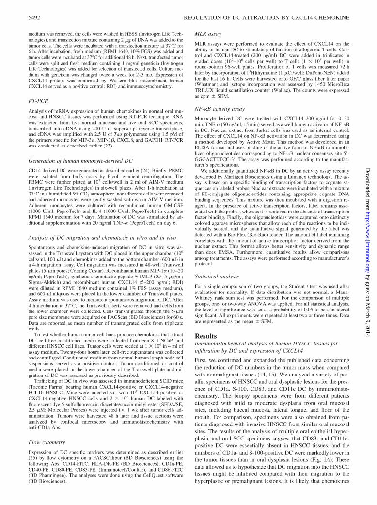
ResultsImmunohistochemical analysis of human HNSCC tissues forinfiltration by DC and expression of CXCL14
First, we confirmed and expanded the published data concerningthe reduction of DC numbers in the tumor mass when comparedwith nonmalignant tissues (14, 15). We analyzed a variety of par-affin specimens of HNSCC and oral dysplastic lesions for the pres-ence of CD1a, S-100, CD83, and CD11c DC by immunohisto-chemistry. The biopsy specimens were from different patientsdiagnosed with mild to moderate dysplasia from oral mucosalsites, including buccal mucosa, lateral tongue, and floor of themouth. For comparison, specimens were also obtained from pa-tients diagnosed with invasive HNSCC from similar oral mucosalsites. The results of the analysis of multiple oral epithelial hyper-plasia, and oral SCC specimens suggest that CD83- and CD11c-positive DC were essentially absent in HNSCC tissues, and thenumbers of CD1a- and S-100-positive DC were markedly lower inthe tumor tissues than in oral dysplasia lesions (Fig. 1A). Thesedata allowed us to hypothesize that DC migration into the HNSCCtissues might be inhibited compared with their migration to thehyperplastic or premalignant lesions. It is likely that chemokines
5492 REGULATION OF DC ATTRACTION BY CXCL14 CHEMOKINE
by guest on March 9, 2014
http://ww
w.jim
munol.org/
Dow
nloaded from

are, at least in part, responsible for differential homing of DC innormal and malignant tissues.
To test whether decreased infiltration of HNSCC by DC mightbe associated with a low expression of chemokines, we have mea-sured expression of DC attracting chemokines in HNSCC and nor-mal mucosa tissues by RT-PCR. Our data revealed similar mRNAexpression of MIP-3� (CCL20) and MIP-3� (CCL19) in HNSCCand normal mucosa (Fig. 2). These chemokines interact withCCR6 and CCR7, expressed on immature and mature DC, respec-tively. Given that expression of new chemokine CXCL14 mRNAwas reported to be lost in different tumors (11, 12), we also ex-amined CXCL14 protein in different human tissues by immuno-histochemistry. Because expression of CXCL14 in tissues has beenpreviously determined only by in situ hybridization (11, 12), we
have developed a new immunohistochemical procedure to analyzeCXCL14 protein in paraffin-embedded tissues. Fig. 1B demon-strates that both normal oral mucosa tissues (n � 7) and oral dys-plasia specimens (n � 8) were strongly positive for CXCL14,whereas HNSCC tissues (n � 8) were low or negative forCXCL14 staining. Thus, these data suggest that CXCL14 proteinis lost in human HNSCC, which led us to the hypotheses that lowinfiltration of HNSCC by DC might be associated with the lostexpression of certain chemokines (i.e., CXCL14) and whetherCXCL14 is chemoattractive for DC.
Chemoattractive properties of CXCL14 and tumor cell linestoward DC
To determine whether DC could be attracted by a CXCL14, wecompared its chemotactic activity toward DC with the known DCchemokines. Analysis of DC migration revealed that CXCL14 andtwo control DC chemokines fMLP, a prototypic bacterial chemo-tactic stimulus (26) and MIP-1� (27), all dose-dependently che-moattracted human DC (Fig. 3A). For example, in the presence of20 ng/ml MIP-1� migration of immature DC reached 6360 � 650cells/min vs 3620 � 380 cells/min spontaneously transmigrated incontrol wells ( p � 0.05). A comparable chemoattraction of DC(5960 � 568 cells/min, p � 0.05) was also detected in the presenceof 200 ng/ml (20 nM) CXCL14 (Fig. 3A). Thus, CXCL14 is apotent DC chemokine with a chemoattractive activity in the nano-grams per milliliter range.
Next question was whether CXCL14 chemoattracts both imma-ture and mature DC. Fig. 3B demonstrates that only immature, butnot mature DC, are chemoattracted by CXCL14. These data are inagreement with Shellenberger et al. (28) and the general conceptthat immature DC are attracted to nonlymphoid tissues where anumber of potent DC chemokines, including CXCL14, may beubiquitously expressed.
In the next set of experiments, we tested whether human tumorcell lines, including different HNSCC, prostate adenocarcinoma,and melanoma, might produce chemokines that could attract hu-man DC in vitro. Cell-free conditioned media were collected fromFemX melanoma, LNCaP prostate adenocarcinoma, and HNSCCcell lines SCC-15, PCI-13, PCI-16, PCI-38, and PCI-4B as de-scribed in Materials and Methods. Conditioned medium from nor-mal human lymph node cell suspensions served as a positive con-trol. Fig. 3C demonstrates that conditioned media from FemXmelanoma cells (5620 � 483 vs 3630 � 250 cells in control, p �0.005), normal lymph node cells (5880 � 602 cells, p � 0.005),and PCI-4B (6340 � 436 cells, p � 0.005), but not from LNCaP(3920 � 286 cells) and HNSCC lines PCI-13 (3720 � 405), PCI16(3320 � 241), and SCC-15 (3390 � 301) ( p � 0.1), displayedchemoattractive activity toward human DC. Selective attraction ofDC by several tumor cell lines raises the question whether it mightcorrelate with the expression of CXCL14.
The next series of experiments focused on evaluating the ex-pression of CXCL14 protein in different human tumor cell lines.Tumor cells were cultured on microscopic slides for 48–72 h, andCXCL14 protein was detected by the immunocytochemical pro-cedure. Human PBMC-derived monocytes stimulated with 0.5�g/ml LPS for 6 h served as a positive control for the expressionof CXCL14. Nonstimulated PBMC were used as a negative con-trol. We found that all tested tumor cells, with the exception ofPCI4B and FemX, were CXCL14-negative (Fig. 3D). Thus,HNSCC cell line SCC-15 and primary HNSCC cell lines PCI-13,PCI-16, and PCI-38 as well as prostate adenocarcinoma cell lineLNCaP express no CXCL14 protein. Interestingly, CXCL14-neg-ative HNSCC cell lines did not attract DC in a chemotaxis assay,
FIGURE 1. Immunohistochemical evaluation of tumor-infiltrating DC(A) and CXCL14 expression (B) in HNSCC, oral dysplasia, and normaloral tissue specimens. Five-micrometer sections were dried overnight, de-waxed, rehydrated, followed by Ag retrieval with 1 mM EDTA/NAOHsolution (see Materials and Methods). A, For evaluating tumor-infiltratingDC, the following DC-specific Abs were used: CD83, CD1a, CD11c, andS-100. Secondary Abs were biotinylated with goat anti-mouse. B, For de-tection of CXCL14 expression in HNSCC tissues anti-CXCL14 Abs orcontrol murine IgG2a were applied to the tissues overnight. Biotinylatedhorse anti-mouse secondary Abs were added for 30 min. Staining wasdeveloped with amino-9-ethylcarbazole and counterstained with hematox-ylin. Positive staining is red-brown. The representative immunohistochem-ical data from the analysis of 10–12 specimens are shown.
FIGURE 2. Analysis of chemokine mRNA expression in normal oralmucosa and HNSCC tissues by RT-PCR. mRNA was extracted from nor-mal mucosa and HNSCC specimens, transcribed into cDNA using 200 Uof superscript reverse transcriptase, and cDNA was amplified with 2.5 UTaq polymerase using 1.5 pM of the primers specific for MIP-3�, MIP-3�,CXCL8, and GAPDH, as described in Materials and Methods. The resultsof a representative experiment are shown (n � 3).
5493The Journal of Immunology
by guest on March 9, 2014
http://ww
w.jim
munol.org/
Dow
nloaded from
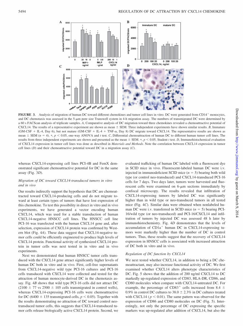
whereas CXCL14-expressing cell lines PCI-4B and FemX dem-onstrated significant chemoattractive potential for DC in the sameassay (Fig. 3D).
Migration of DC toward CXCL14-transduced tumors in vitroand in vivo
Our results indirectly support the hypothesis that DC are chemoat-tracted toward CXCL14-producing cells and do not migrate to-ward at least certain types of tumors that have lost expression ofthis chemokine. To test this possibility in direct in vitro and in vivoexperiments, we have generated a vector encoding humanCXCL14, which was used for a stable transduction of humanCXCL14-negative HNSCC cell lines. The HNSCC cell linePCI-16 was transfected with the human CXCL14 gene and, afterselection, expression of CXCL14 protein was confirmed by West-ern blot (Fig. 4A). These data suggest that CXCL14-negative tu-mor cells could be efficiently engineered to produce high levels ofCXCL14 protein. Functional activity of synthesized CXCL14 pro-tein in tumor cells was next tested in in vitro and in vivoexperiments.
Next we demonstrated that human HNSCC tumor cells trans-duced with the CXCL14 gene attract significantly higher levels ofhuman DC both in vitro and in vivo. First, cell-free supernatantsfrom CXCL14-negative wild type PCI-16 cultures and PCI-16cells transduced with CXCL14 were collected and tested for theattraction of human monocyte-derived DC in the chemotaxis as-say. Fig. 4B shows that wild type PCI-16 cells did not attract DC(2100 � 77 vs 2500 � 105 cells transmigrated in control wells),whereas CXCL14-expressing PCI-16 cells were chemoattractivefor DC (6400 � 135 transmigrated cells, p � 0.05). Together withthe results demonstrating no attraction of DC toward control neo-transduced tumor cells, this suggests that CXCL14-transduced tu-mor cells release biologically active CXCL14 protein. Second, we
evaluated trafficking of human DC labeled with a fluorescent dyein SCID mice in vivo. Fluorescent-labeled human DC were i.v.injected in immunodeficient SCID mice (n � 5) bearing both wildtype (or control neo-transduced) and CXCL14-transduced PCI-16cells for 7 days. Two days later, tumors were harvested and fluo-rescent cells were examined on 6-�m sections immediately byconfocal microscopy. The results revealed that infiltration ofCXCL14-expressing tumors by labeled DC was significantlyhigher than in wild type or neo-transduced tumors in all testedmice (Fig. 4C). Similar data were obtained when nonlabeled hu-man DC were i.v. transferred in SCID mice (n � 5) bearing PCI-16/wild type (or neo-transduced) and PCI-16/CXCL14 and infil-tration of tumors by injected DC was assessed 48 h later byimmunohistochemistry. Fig. 4D demonstrates that the levels ofaccumulation of CD1a� human DC in CXCL14-expressing tu-mors were markedly higher than the number of DC in controltumors. Thus, these results suggest that the recovery of CXCL14expression in HNSCC cells is associated with increased attractionof DC both in vitro and in vivo.
Regulation of DC function by CXCL14
We next tested whether CXCL14, in addition to being a DC che-moattractant, may also increase functional activity of DC. We firstexamined whether CXCL14 alters phenotype characteristics ofDC. Fig. 5 shows that the addition of 200 ng/ml CXCL14 to DCmarkedly up-regulated expression of CD83, HLA-DR, CD86, andCD80 molecules when compare with CXCL14-untreated DC. Forexample, the percentage of CD83� cells increased from 8.4 �0.9% in control DC cultures to 38.0 � 2.3% in DC cultures treatedwith CXCL14 ( p � 0.01). The same pattern was observed for theexpression of CD86 and CD80 molecules on DC (Fig. 5). Inter-estingly, not only the percentage of DC expressing the specificmarkers was up-regulated after addition of CXCL14, but also the
FIGURE 3. Analysis of migration of human DC toward different chemokines and tumor cell lines in vitro. DC were generated from CD14� monocytes,and DC chemotaxis was assessed in the 5-�m pore size Transwell system in 4-h migration assay. The numbers of transmigrated DC were determined bya 60 s FACScan analysis of triplicate samples. A, Comparative analysis of DC migration toward three chemokines revealed a chemoattractive potential ofCXCL14. The results of a representative experiment are shown as mean � SEM. Three independent experiments have shown similar results. B, Immature(GM-CSF � IL-4, Day 6), but not mature (GM-CSF � IL-4 � TNF-�, Day 8) DC migrate toward CXCL14. The representative results are shown asmean � SEM (n � 4). �, p � 0.05, one-way ANOVA and t test. C, Differential chemoattraction of human DC to different human tumor cell lines. Theresults from three independent experiments are shown and presented as the mean � SEM. �, p � 0.05, Student t test. D, Immunohistochemical evaluationof CXCL14 expression in tumor cell lines was done as described in Materials and Methods. Note the correlation between CXCL14 expression in tumorcell lines (D) and their chemoattractive potential toward DC in a migration assay (C).
5494 REGULATION OF DC ATTRACTION BY CXCL14 CHEMOKINE
by guest on March 9, 2014
http://ww
w.jim
munol.org/
Dow
nloaded from

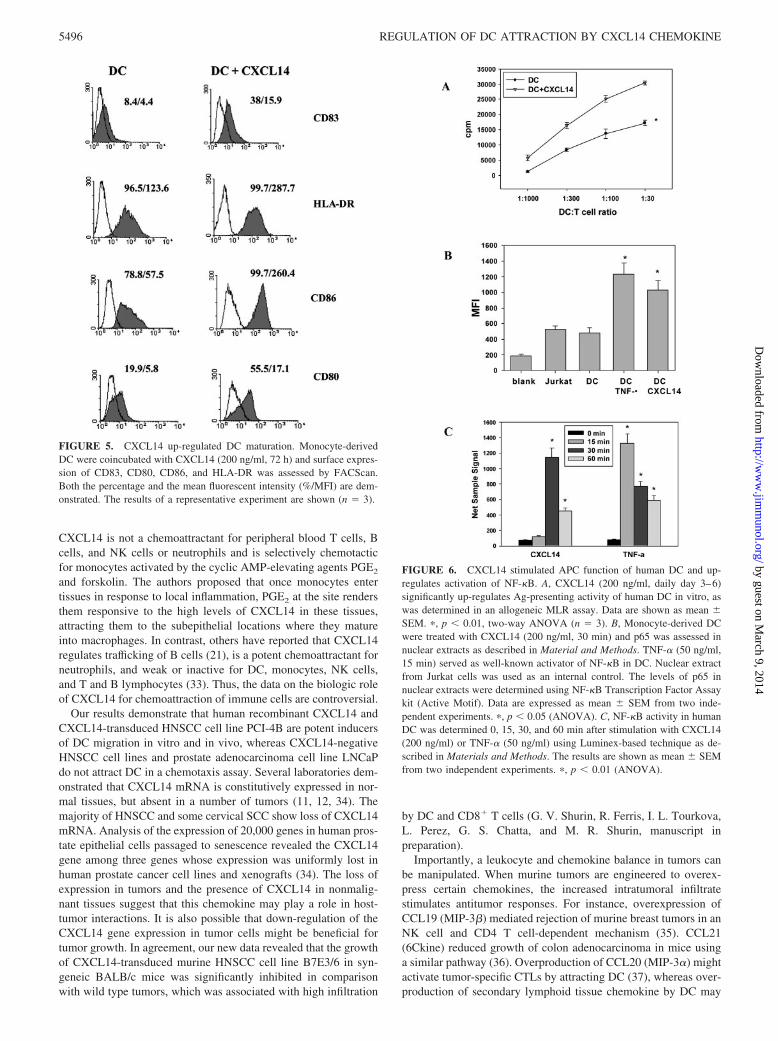
levels of CD83, CD86, CD80, and HLA-DR expression on DCwere significantly up-regulated (Fig. 5). For instance, the meanfluorescence intensity values for CD83 and HLA-DR markerswere increased from 4.4 � 0.5 in control DC to 15.9 � 1.4 ( p �0.01) on DC treated with CXCL14 and from 123.6 � 5.8 to287.7 � 9.9 ( p � 0.01), respectively (Fig. 5). Thus, it is conceiv-able that CXCL14 stimulates maturation of DC.
Further confirmation of the biologic activity of CXCL14 wasobtained in the MLR assay using DC generated from differentdonors with or without the addition of 200 ng/ml CXCL14 (Fig.6A). Significantly higher induction of allogeneic T cell prolifera-tion by CXCL14-treated DC ( p � 0.01), as compared with un-treated DC, was observed. For instance, at DC to T cell ratio 1:30,uptake of [3H]thymidine increased from 17252 � 897 cpm in con-trol to 30385 � 689 cpm ( p � 0.01) in group treated withCXCL14.
To explore the molecular mechanisms of CXCL14-mediated ac-tivation of DC, monocyte-derived DC were treated with CXCL14(200 ng/ml) and TNF-� (50 ng/ml). The levels of p65 in nuclearextracts were determined using NF-�B Transcription Factor Assaykit. TNF-� served as a well-known activator of NF-�B in DC.Nuclear extract from Jurkat cells was used as internal control. Wedemonstrated that activation of DC with CXCL14 was accompa-nied by a significant up-regulation of NF-�B activity in DC up to200% ( p � 0.01) (Fig. 6B). Next, these data were confirmed andfurther explored by using Luminex-based technique for analyzingNF-�B activation (Fig. 6C). The results also demonstrated thatCXCL14 is a strong inducer of NF-�B activation in human DC.Interestingly, the kinetic analysis of transcription factor activityrevealed that NF-�B activation induced in DC by CXCL14 wasdelayed compared with TNF-�-induced activation reaching themaximum at 30 min (Fig. 6C).
In summary, these data suggest that CXCL14, in addition tobeing DC attractant, also increases functional activity of DC.
DiscussionWe have demonstrated that expression of a new DC chemokine,CXCL14, is frequently lost in HNSCC tissues, which was accom-panied by a low infiltration of the tumor by DC. We speculate thatlow levels of HNSCC infiltration by DC may be due to a low orabsent expression of CXCL14 in tumor cells. It is well known thathoming of leukocytes to the sites of hemopoiesis, Ag priming,immune surveillance, and inflammation largely depends on thepresence of chemokines (29). The presence of DC, macrophages,and lymphocytes in solid tumors is regulated by local productionof chemokines by tumor and stromal cells. In particular, CC che-mokines are the major determinants of macrophage and lympho-cyte infiltration in carcinomas of the breast and cervix, sarcomas,and gliomas (30). CCL2 (MCP-1) has been implicated in mediat-ing macrophage infiltration into breast (19) and ovarian cancers(31), whereas CCL5 levels correlate with the extent of CD8 T cellinfiltrate in ovarian tumors (20). It is conceivable to speculate thatimmature DC might be constitutively recruited to CXCL14-ex-pressing tissues. This would allow DC and monocytes to leave thecirculation and enter these tissues in the absence of inflammation.On the contrary, the loss of CXCL14 expression in malignant tis-sues may explain a decreased rate of DC attraction and thus aug-ments efficacy of tumor escape mechanisms.
CXCL14 was initially named BRAK because it was identified inhuman breast and kidney derived cells (12). CXCL14 (KS1, Kec,BMAC, NJAC, MIP-2�) is a chemokine with an as yet unknownfunction and receptor selectivity (11, 12, 21). The mature se-quences of CXCL14 and its murine analog SK1 contain 77 aminoacids and are unique with regard to the short N-terminal end ofonly two amino acids (Ser-Lys), preceding the first of four che-mokine-typical Cys residues. The most closely related chemo-kines, MIP-2� and MIP-2�, share 30% amino acid identity withCXCL14. Kurth et al. (32) have recently provided evidence that
FIGURE 4. Transduction of CXCL14-negative human HNSCC cell line PCI-16 with the CXCL14 gene resulted in expression of high levels of CXCL14protein. Primary HNSCC cells PCI-16 were transduced with the human CXCL14 gene as described in Materials and Methods. Immunocytochemicalanalysis of CXCL14-positive cells was assessed by Western Blot (A) as described in Materials and Methods. Recombinant human CXCL14 protein servedas a positive control. B, CXCL14-transduced HNSCC cells secrete functionally active protein, which exhibits significant chemoattractive potential towardhuman DC in vitro (B) and in vivo (C and D). PCI-16 cells were transduced with the human CXCL14 gene and after selection with G148 supernatantsobtained from wild type (wt) and CXCL14-transduced PCI-16 tumor cells were tested for their ability to attract DC in Transwell-based cell migration assay(B). Medium and CXCL14 served as negative and positive controls, respectively. �, p � 0.05 vs medium (one-way ANOVA, n � 5). C and D, PCI-16wild type or PCI-16/CXCL14 tumor cells (107 cells per mouse) were injected s.c. in SCID mice on day 1. Sulfofluorescein diacetate/succinimidyl ester(SFDA/SE)-labeled or nonlabeled human DC (2 � 106 cells) were injected i.v. on day 7 and all tumors were harvested 48 h later. Tissue sections wereanalyzed by confocal microscopy (C) or immunohistochemistry with anti-CD1a Abs (red staining) (D) as described in Materials and Methods. The resultsfrom a representative experiment (n � 3) are shown.
5495The Journal of Immunology
by guest on March 9, 2014
http://ww
w.jim
munol.org/
Dow
nloaded from
CXCL14 is not a chemoattractant for peripheral blood T cells, Bcells, and NK cells or neutrophils and is selectively chemotacticfor monocytes activated by the cyclic AMP-elevating agents PGE2
and forskolin. The authors proposed that once monocytes entertissues in response to local inflammation, PGE2 at the site rendersthem responsive to the high levels of CXCL14 in these tissues,attracting them to the subepithelial locations where they matureinto macrophages. In contrast, others have reported that CXCL14regulates trafficking of B cells (21), is a potent chemoattractant forneutrophils, and weak or inactive for DC, monocytes, NK cells,and T and B lymphocytes (33). Thus, the data on the biologic roleof CXCL14 for chemoattraction of immune cells are controversial.
Our results demonstrate that human recombinant CXCL14 andCXCL14-transduced HNSCC cell line PCI-4B are potent inducersof DC migration in vitro and in vivo, whereas CXCL14-negativeHNSCC cell lines and prostate adenocarcinoma cell line LNCaPdo not attract DC in a chemotaxis assay. Several laboratories dem-onstrated that CXCL14 mRNA is constitutively expressed in nor-mal tissues, but absent in a number of tumors (11, 12, 34). Themajority of HNSCC and some cervical SCC show loss of CXCL14mRNA. Analysis of the expression of 20,000 genes in human pros-tate epithelial cells passaged to senescence revealed the CXCL14gene among three genes whose expression was uniformly lost inhuman prostate cancer cell lines and xenografts (34). The loss ofexpression in tumors and the presence of CXCL14 in nonmalig-nant tissues suggest that this chemokine may play a role in host-tumor interactions. It is also possible that down-regulation of theCXCL14 gene expression in tumor cells might be beneficial fortumor growth. In agreement, our new data revealed that the growthof CXCL14-transduced murine HNSCC cell line B7E3/6 in syn-geneic BALB/c mice was significantly inhibited in comparisonwith wild type tumors, which was associated with high infiltration
by DC and CD8� T cells (G. V. Shurin, R. Ferris, I. L. Tourkova,L. Perez, G. S. Chatta, and M. R. Shurin, manuscript inpreparation).
Importantly, a leukocyte and chemokine balance in tumors canbe manipulated. When murine tumors are engineered to overex-press certain chemokines, the increased intratumoral infiltratestimulates antitumor responses. For instance, overexpression ofCCL19 (MIP-3�) mediated rejection of murine breast tumors in anNK cell and CD4 T cell-dependent mechanism (35). CCL21(6Ckine) reduced growth of colon adenocarcinoma in mice usinga similar pathway (36). Overproduction of CCL20 (MIP-3�) mightactivate tumor-specific CTLs by attracting DC (37), whereas over-production of secondary lymphoid tissue chemokine by DC may
FIGURE 5. CXCL14 up-regulated DC maturation. Monocyte-derivedDC were coincubated with CXCL14 (200 ng/ml, 72 h) and surface expres-sion of CD83, CD80, CD86, and HLA-DR was assessed by FACScan.Both the percentage and the mean fluorescent intensity (%/MFI) are dem-onstrated. The results of a representative experiment are shown (n � 3).
FIGURE 6. CXCL14 stimulated APC function of human DC and up-regulates activation of NF-�B. A, CXCL14 (200 ng/ml, daily day 3–6)significantly up-regulates Ag-presenting activity of human DC in vitro, aswas determined in an allogeneic MLR assay. Data are shown as mean �SEM. �, p � 0.01, two-way ANOVA (n � 3). B, Monocyte-derived DCwere treated with CXCL14 (200 ng/ml, 30 min) and p65 was assessed innuclear extracts as described in Material and Methods. TNF-� (50 ng/ml,15 min) served as well-known activator of NF-�B in DC. Nuclear extractfrom Jurkat cells was used as an internal control. The levels of p65 innuclear extracts were determined using NF-�B Transcription Factor Assaykit (Active Motif). Data are expressed as mean � SEM from two inde-pendent experiments. �, p � 0.05 (ANOVA). C, NF-�B activity in humanDC was determined 0, 15, 30, and 60 min after stimulation with CXCL14(200 ng/ml) or TNF-� (50 ng/ml) using Luminex-based technique as de-scribed in Materials and Methods. The results are shown as mean � SEMfrom two independent experiments. �, p � 0.01 (ANOVA).
5496 REGULATION OF DC ATTRACTION BY CXCL14 CHEMOKINE
by guest on March 9, 2014
http://ww
w.jim
munol.org/
Dow
nloaded from
enhance T cell recruitment and immune priming to tumor-associ-ated Ags (38). In fact, injection of recombinant secondary lym-phoid tissue chemokine in the axillary lymph node region in micewith bilateral multifocal pulmonary adenocarcinomas led to amarked reduction in tumor burden with extensive lymphocytic andDC infiltration of the tumors and enhanced survival (39). Togetherwith clinical evidence demonstrating that infiltration of tumormass by DC is associated with a better patient survival, these re-sults suggest that regulated induction of DC migration into thetumor site might induce efficient antitumor immune responses.However, there are no data on whether CXC cytokines play a rolein attraction of immune cells to the tumor site and inducing anti-tumor immunity. We have shown, that genetic modification ofCXCL14-negative PCI-16 HNSCC cell line with the CXCL14gene results in stimulation of DC attraction in vitro and increasedinfiltration of the tumor by DC in vivo. In fact, we have shown ona murine HNSCC model that CXCL14-expressing tumors werehighly infiltrated by CD11c� DC suggesting their potential role indeveloping antitumor immune response at the tumor site (G. V.Shurin, R. Ferris, I. L. Tourkova, L. Perez, A. Lokshin, L. Balkir,B. Collins, G. S. Chatta, and M. R. Shurin, manuscript inpreparation).
Next, we evaluated the effect of CXCL14 on DC function. It isknown that chemokines may regulate cellular adhesion, prolifera-tion, and cell survival (10, 18, 40). Based on the current knowledgeof the life cycle of DC, it has been postulated that chemokines canplay an important role at several stages of DC development (18).Basal chemokine production and expression at the surface of en-dothelial cells can mediate DC precursor recruitment into periph-eral tissues, which is important for the maintaining DC levelswithin tissues. Once in the tissue, chemokines, such as MIP-1�,MIP-1�, MIP-3�, MIP-5, MCP-3, MCP-4, RANTES, TECK, andSDF-1 (41), may participate in differentiation of DC precursorsinto immature DC that are programmed to pick up and processAg(s). Upon initiation of an inflammatory response, chemokinesthat recruit immature DC may be up-regulated, resulting in DCaccumulation within the tissue. When DC have matured, they entertissue-draining lymphatic vessels and migrate to the T cell zones insecondary lymphoid organs under the influence of chemokinesproduced there, such as MIP-3� and 6Ckine. In the T cell zones,DC can produce chemokines that stimulate DC-T cell interaction,thereby enhancing the likelihood of clonal selection (18, 41). Ourdata show that CXCL14 chemoattracts only immature, but not ma-ture DC, which is in agreement with the concept that nonlymphoidtissue chemokines should attract immature DC. Importantly, wedemonstrated that CXCL14 also activated DC through NF-�B-mediated pathways and up-regulated expression of costimulatorymolecules on DC as well as enhanced the proliferation of alloge-neic T cells in MLR. Thus our results support the hypothesis thatCXCL14 might be a novel DC chemokine regulating their homingand activation in nonlymphoid tissues.
DisclosuresThe authors have no financial conflict of interest.
References1. Greenlee, R. T., M. B. Hill-Harmon, T. Murray, and M. Thun. 2001. Cancer
statistics. CA Cancer J. Clin. 51:15.2. Wong, D. T., R. Todd, T. Tsuji, and R. B. Donoff. 1996. Molecular biology of
human oral cancer. Crit. Rev. Oral Biol. Med. 7:319.3. Neville, B. W., and T. A. Day. 2002. Oral cancer and precancerous lesions. CA
Cancer J. Clin. 52:195.4. Becker, Y. 1992. Anticancer role of dendritic cells (DC) in human and experi-
mental cancers: a review. Anticancer Res. 12:511.5. Janjic, B. M., G. Lu, A. Pimenov, T. L. Whiteside, W. J. Storkus, and
N. L. Vujanovic. 2002. Innate direct anticancer effector function of human im-
mature dendritic cells. I. Involvement of an apoptosis-inducing pathway. J. Im-munol. 168:1823.
6. Triozzi, P. L., R. Khurram, W. A. Aldrich, M. J. Walker, J. A. Kim, andS. Jaynes. 2000. Intratumoral injection of dendritic cells derived in vitro in pa-tients with metastatic cancer. Cancer 89:2646.
7. Shurin, M. R., and D. I. Gabrilovich. 2001. Regulation of dendritic cell system bytumor. Cancer Res. Ther. Control 11:65.
8. Shurin, G. V., Z. R. Yurkovetsky, and M. R. Shurin. 2003. Tumor-induced den-dritic cell dysfunction. In Mechanisms of Tumor Escape. A. Ochoa, ed. HarwoodAcademic Publishers, p. 115.
9. Remmel, E., L. Terracciano, C. Noppen, P. Zajac, M. Heberer, G. C. Spagnoli,and E. Padovan. 2001. Modulation of dendritic cell phenotype and mobility bytumor cells in vitro. Hum. Immunol. 62:39.
10. Brault, M. S., and R. A. Kurt. 2003. Chemokines and antitumor immunity: walk-ing the tightrope. Int. Rev. Immunol. 22:199.
11. Frederick, M. J., Y. Henderson, X. Xu, M. T. Deavers, A. A. Sahin, H. Wu,D. E. Lewis, A. K. El-Naggar, and G. L. Clayman. 2000. In vivo expression ofthe novel CXC chemokine BRAK in normal and cancerous human tissue.Am. J. Pathol. 156:1937.
12. Hromas, R., H. E. Broxmeyer, C. Kim, H. Nakshatri, K. Christopherson, II,M. Azam, and Y. H. Hou. 1999. Cloning of BRAK, a novel divergent CXCchemokine preferentially expressed in normal versus malignant cells. Biochem.Biophys. Res. Commun. 255:703.
13. Bondad-Palmario, G. G. 1995. Histological and immunochemical studies of oralleukoplakia: phenotype and distribution of immunocompetent cells.J. Philippines Dental Assoc. 47:3.
14. Deng, Y., X. Yuan, and Z. Chen. 1997. Immunobiological significance of S-100protein positive dendritic cells (S-100�DC) in patients with oral squamous cellcarcinoma. Zhonghua Kou Qiang Yi Xue Za Zhi. 32:174.
15. Wei, N., and S. R. Tahan. 1998. S100� cell response to squamous cell carcinomaof the lip: inverse correlation with metastasis. J. Cutaneous Pathol. 25:463.
16. Goldman, S. A., E. Baker, R. J. Weyant, M. R. Clarke, J. N. Myers, andM. T. Lotze. 1998. Peritumoral CD1a-positive dendritic cells are associated withimproved survival in patients with tongue carcinoma. Arch. Otolaryngol. HeadNeck Surg. 124:641.
17. Rollins, B. J. 1997. Chemokines. Blood 90:909.18. McColl, S. R. 2002. Chemokines and dendritic cells: a crucial alliance. Immunol.
Cell Biol. 80:489.19. Ueno, T., M. Toi, H. Saji, M. Muta, H. Bando, K. Kuroi, M. Koike, H. Inadera,
and K. Matsushima. 2000. Significance of macrophage chemoattractant protein-1in macrophage recruitment, angiogenesis, and survival in human breast cancer.Clin. Cancer Res. 6:3282.
20. Negus, R. P., G. W. Stamp, J. Hadley, and F. R. Balkwill. 1997. Quantitativeassessment of the leukocyte infiltrate in ovarian cancer and its relationship to theexpression of C-C chemokines. Am. J. Pathol. 150:1723.
21. Sleeman, M. A., J. K. Fraser, J. G. Murison, S. L. Kelly, R. L. Prestidge,D. J. Palmer, J. D. Watson, and K. D. Kumble. 2000. B cell- and monocyte-activating chemokine (BMAC), a novel non-ELR �-chemokine. Int. Immunol.12:677.
22. Heo, D. S., C. Snyderman, S. M. Gollin, S. Pan, E. Walker, R. Deka,E. L. Barnes, J. T. Johnson, R. B. Herberman, and T. L. Whiteside. 1989. Biol-ogy, cytogenetics, and sensitivity to immunological effector cells of new head andneck squamous cell carcinoma lines. Cancer Res. 49:5167.
23. Wang, J., L. Xi, J. L. Hunt, W. Gooding, T. L. Whiteside, Z. Chen, T. E. Godfrey,and R. L. Ferris. 2004. Expression pattern of chemokine receptor 6 (CCR6) andCCR7 in squamous cell carcinoma of the head and neck identifies a novel met-astatic phenotype. Cancer Res. 64:1861.
24. Shurin, M. R. 2003. Preparation of human dendritic cells for tumor vaccination.In Methods in Molecular Biology. Cytokines and Colony Stimulating Factors:Methods and Protocols. vol. 215. D. K. a. W. Kiess, ed. Humana Press, Totowa,NJ, p. 437.
25. Shurin, G. V., M. R. Shurin, S. Bykovskaia, J. Shogan, M. T. Lotze, andE. M. Barksdale, Jr. 2001. Neuroblastoma-derived gangliosides inhibit dendriticcell generation and function. Cancer Res. 61:363.
26. Sozzani, S., F. Sallusto, W. Luini, D. Zhou, L. Piemonti, P. Allavena,J. Van Damme, S. Valitutti, A. Lanzavecchia, and A. Mantovani. 1995. Migrationof dendritic cells in response to formyl peptides, C5a, and a distinct set of che-mokines. J. Immunol. 155:3292.
27. Sozzani, S., P. Allavena, A. Vecchi, and A. Mantovani. 2000. Chemokines anddendritic cell traffic. J. Clin. Immunol. 20:151.
28. Shellenberger, T. D., M. Wang, M. Gujrati, A. Jayakumar, R. M. Strieter,M. D. Burdick, C. G. Ioannides, C. L. Efferson, A. K. El-Naggar, D. Roberts, etal. 2004. BRAK/CXCL14 is a potent inhibitor of angiogenesis and a chemotacticfactor for immature dendritic cells. Cancer Res. 64:8262.
29. Moser, B., and P. Loetscher. 2001. Lymphocyte traffic control by chemokines.Nat. Immunol. 2:123.
30. Bottazzi, B., N. Polentarutti, R. Acero, A. Balsari, D. Boraschi, P. Ghezzi,M. Salmona, and A. Mantovani. 1983. Regulation of the macrophage content ofneoplasms by chemoattractants. Science 220:210.
31. Bottazzi, B., P. Ghezzi, G. Taraboletti, M. Salmona, N. Colombo, C. Bonazzi,C. Mangioni, and A. Mantovani. 1985. Tumor-derived chemotactic factor(s) fromhuman ovarian carcinoma: evidence for a role in the regulation of macrophagecontent of neoplastic tissues. Int. J. Cancer 36:167.
32. Kurth, I., K. Willimann, P. Schaerli, T. Hunziker, I. Clark-Lewis, and B. Moser.2001. Monocyte selectivity and tissue localization suggests a role for breast andkidney-expressed chemokine (BRAK) in macrophage development. J. Exp. Med.194:855.
5497The Journal of Immunology
by guest on March 9, 2014
http://ww
w.jim
munol.org/
Dow
nloaded from
33. Cao, X., W. Zhang, T. Wan, L. He, T. Chen, Z. Yuan, S. Ma, Y. Yu, and G. Chen.2000. Molecular cloning and characterization of a novel CXC chemokine mac-rophage inflammatory protein-2� chemoattractant for human neutrophils anddendritic cells. J. Immunol. 165:2588.
34. Schwarze, S. R., S. E. DePrimo, L. M. Grabert, V. X. Fu, J. D. Brooks, andD. F. Jarrard. 2002. Novel pathways associated with bypassing cellular senes-cence in human prostate epithelial cells. J. Biol. Chem. 277:14877.
35. Braun, S. E., K. Chen, R. G. Foster, C. H. Kim, R. Hromas, M. H. Kaplan,H. E. Broxmeyer, and K. Cornetta. 2000. The CC chemokine CK�-11/MIP-3�/ELC/Exodus 3 mediates tumor rejection of murine breast cancer cells throughNK cells. J. Immunol. 164:4025.
36. Vicari, A. P., S. Ait-Yahia, K. Chemin, A. Mueller, A. Zlotnik, and C. Caux.2000. Antitumor effects of the mouse chemokine 6Ckine/SLC through angiostaticand immunological mechanisms. J. Immunol. 165:1992.
37. Fushimi, T., A. Kojima, M. A. Moore, and R. G. Crystal. 2000. Macrophageinflammatory protein 3� transgene attracts dendritic cells to established murinetumors and suppresses tumor growth. J. Clin. Invest. 105:1383.
38. Terando, A., B. Roessler, and J. J. Mule. 2004. Chemokine gene modification ofhuman dendritic cell-based tumor vaccines using a recombinant adenoviral vec-tor. Cancer Gene Ther. 11:165.
39. Sharma, S., M. Stolina, L. Zhu, Y. Lin, R. Batra, M. Huang, R. Strieter, andS. M. Dubinett. 2001. Secondary lymphoid organ chemokine reduces pulmonarytumor burden in spontaneous murine bronchoalveolar cell carcinoma. CancerRes. 61:6406.
40. Nakashima, E., A. Oya, Y. Kubota, N. Kanada, R. Matsushita, K. Takeda,F. Ichimura, K. Kuno, N. Mukaida, K. Hirose, et al. 1996. A candidate for cancergene therapy: MIP-1� gene transfer to an adenocarcinoma cell line reduced tu-morigenicity and induced protective immunity in immunocompetent mice.Pharm. Res. 13:1896.
41. Caux, C., S. Ait-Yahia, K. Chemin, O. de Bouteiller, M. C. Dieu-Nosjean,B. Homey, C. Massacrier, B. Vanbervliet, A. Zlotnik, and A. Vicari. 2000. Den-dritic cell biology and regulation of dendritic cell trafficking by chemokines.Springer Semin. Immunopathol. 22:345.
5498 REGULATION OF DC ATTRACTION BY CXCL14 CHEMOKINE
by guest on March 9, 2014
http://ww
w.jim
munol.org/
Dow
nloaded from

CORRECTIONSBrady, J., Y. Hayakawa, M. J. Smyth, and S. L. Nutt. 2004. IL-21 induces the functional maturation of murine NK cells.J. Immunol. 172: 2048–2058.
Figure 8 is incorrect. The corrected figure is shown below.
Shurin, G. V., R. Ferris, I. L. Tourkova, L. Perez, A. Lokshin, L. Balkir, B. Collins, G. S. Chatta, and M. R. Shurin. 2005.Loss of new chemokine CXCL14 in tumor tissue is associated with low infiltration by dendritic cells (DC), whilerestoration of human CXCL14 expression in tumor cells causes attraction of DC both in vitro and in vivo. J. Immunol.174: 5490–5498.
The second author’s middle initial was omitted. The correct name is Robert L. Ferris.
Santiago, H. C., C. G. Feng, A. Bafica, E. Roffe, R. M. Arantes, A. Cheever, G. Taylor, L. Q. Vierira, J. Aliberti, R. T.Gazzinelli, and A. Sher. 2005. Mice deficient in LRG-47 display enhanced susceptibility to Trypanosoma cruzi infectionassociated with defective hemopoiesis and intracellular control of parasite growth. J. Immunol. 175: 8165–8172.
The eighth author’s last name was misspelled. The correct name is Leda Q. Vieira.
The Journal of Immunology
Copyright © 2006 by The American Association of Immunologists, Inc. 0022-1767/06/$02.00

Oki, T., J. Kitaura, K. Eto, Y. Lu, M. Maeda-Yamamoto, N. Inagaki, H. Nagai, Y. Yamanishi, H. Nakajina, H. Kumagai,and T. Kitamura. 2006. Integrin �IIb�3 induces the adhesion and activation of mast cells through interaction withfibrinogen. J. Immunol. 176: 52–60.
The ninth author’s last name was misspelled. The correct name is Hideaki Nakajima.
Graham, D. B., M. P. Bell, M. M. McCausland, C. J. Huntoon, J. van Deursen, W. A. Faubion, S. Crotty, and D. J.McKean. 2006. Ly9 (CD229)-deficient mice exhibit T cell defects yet do not share several phenotypic characteristicsassociated with SLAM- and SAP-deficient mice. J. Immunol. 176: 291–300.
In Figure 2A, the three left hand dot plot panels from Ly9�/� cells were mistakenly duplicated in the three right handdot plot panels of Ly9-/- cells. The numbers in each of the quadrants are correct and the error does not change anyinterpretation in the article. The corrected figure is shown below.
Tien, M.-T., S. E. Girardin, B. Regnault, L. Le Bourhis, M.-A. Dillies, J.-Y. Coppee, R. Bourdet-Sicard, P. J. Sansonetti,and T. Pedron. 2006. Anti-inflammatory effect of Lactobacillus casei on Shigella-infected human intestinal epithelialcells. J. Immunol. 176: 1228–1237.
One of the first author’s affiliations was omitted. The corrected list of authors and affiliations is shown below.
Meng-Tsung Tien,2*†‡ Stephen E. Girardin,2* Beatrice Regnault,† Lionel Le Bourhis,§ Marie-Agnes Dillies,† Jean-Yves Coppee,† Raphaelle Bourdet-Sicard,¶ Philippe J. Sansonetti,3* and Thierry Pedron*
*Pathogenie Microbienne Moleculaire Unit, Institut National de la Sante et de la Recherche Medicale U389, ParisFrance; †DNA Chip Platform, Genopole, Evry, France; ‡Department of Biological Science and Technology, NationalChiao Tung University, Hsinchu, Taiwan, China; §Imunite Innee et Signalisation, Pasteur Institute, Paris, France; and¶Danone Vitapole, Nutrivaleur, Palaiseau, France
3841The Journal of Immunology

Atherly, L. O., M. A. Brehm, R. M. Welsh, and L. J. Berg. 2006. Tec kinases Itk and Rlk are required for CD8� T cellresponses to virus infection independent of their role in CD4� T cell help. J. Immunol. 176: 1571–1581.
In Figure 1B, the WT Ca flux data line is missing from the Ca flux graph. The corrected figure is shown below.
3842 CORRECTIONS

Stone, J. D., and L. J. Stern. 2006. CD8 T cells, like CD4 T cells, are triggered by multivalent engagement of TCRs byMHC-peptide ligands but not by monovalent engagement. J. Immunol. 176: 1498–1505.
In Discussion, the last reference in the paper is incorrect. The corrected sentence and reference are shown below.
It is known that the cytoplasmic domains of several components of the TCR complex tend to homo-oligomerize at highconcentrations (41); perhaps ligand-induced clustering of the TCR drives the cytoplasmic domains of proximal receptorsto rearrange, exposing the Nck binding epitope and propelling other signaling cascade processes.
41. Sigalov A., D. Aivazian, and L. Stern. 2004. Homooligomerization of the cytoplasmic domain of the T cell receptor zetachain and of other proteins containing the immunoreceptor tyrosine-based activation motif. Biochemistry 43: 2049–61.
Serhan, C. N., K. Gotlinger, S. Hong, Y. Lu, J. Siegelman, T. Baer, R. Yang, S. P. Colgan, and N. A. Petasis. 2006.Anti-inflammatory actions of neuroprotectin D1/protectin D1 and its natural stereoisomers: assignments of dihydroxy-containing docosatrienes. J. Immunol. 176: 1848–1859.
In Discussion, in the second sentence of paragraph six, 10S-HDNA should have been 10S-HDHA. The correctedsentence is shown below.
Recently, classic steric analysis of 10S-HDHA and the formation of 10,20-diHDHA and 17-H(p)DHA were reportedlyoptimized for the plant LOs (49).
Cante-Barrett, K., E. M. Gallo, M. M. Winslow, and G. R. Crabtree. 2006. Thymocyte negative selection is mediated byprotein kinase C- and Ca2�-dependent transcriptional induction of Bim of cell death. J. Immunol. 176: 2299–2306.
The title of the article is incorrect. The corrected title is shown below. The error has been corrected in the online version,which now differs from the print version as originally published.
Thymocyte Negative Selection Is Mediated by Protein Kinase C- and Ca2�-Dependent Transcriptional Induction ofBim
3843The Journal of Immunology