Long-Term Stability and Safety of Transgenic Cultured Epidermal Stem Cells in Gene Therapy of...
8
Stem Cell Reports Repor t Long-Term Stability and Safety of Transgenic Cultured Epidermal Stem Cells in Gene Therapy of Junctional Epidermolysis Bullosa Laura De Rosa, 1,5 Sonia Carulli, 1,5 Fabienne Cocchiarella, 1 Daniela Quaglino, 2 Elena Enzo, 1 Eleonora Franchini, 1 Alberto Giannetti, 3 Giorgio De Santis, 4 Alessandra Recchia, 1 Graziella Pellegrini, 1 and Michele De Luca 1, * 1 Center for Regenerative Medicine ‘‘Stefano Ferrari,’’ Department of Life Sciences, University of Modena and Reggio Emilia, 41125 Modena, Italy 2 Department of Life Sciences, University of Modena and Reggio Emilia, 41125 Modena, Italy 3 Emeritus of Dermatology, University of Modena and Reggio Emilia, 41125 Modena, Italy 4 Department of Medical and Surgical Sciences, University of Modena and Reggio Emilia, 41125 Modena, Italy 5 These authors contributed equally to this work *Correspondence: [email protected] http://dx.doi.org/10.1016/j.stemcr.2013.11.001 This is an open-access article distributed under the terms of the Creative Commons Attribution-NonCommercial-No Derivative Works License, which permits non-commercial use, distribution, and reproduction in any medium, provided the original author and source are credited. SUMMARY We report a long-term follow-up (6.5 years) of a phase I/II clinical trial envisaging the use of autologous genetically modified cultured epidermal stem cells for gene therapy of junctional epidermolysis bullosa, a devastating genetic skin disease. The critical goals of the trial were to evaluate the safety and long-term persistence of genetically modified epidermis. A normal epidermal-dermal junction was restored and the regenerated transgenic epidermis was found to be fully functional and virtually indistinguishable from a normal control. The epidermis was sustained by a discrete number of long-lasting, self-renewing transgenic epidermal stem cells that maintained the memory of the donor site, whereas the vast majority of transduced transit-amplifying progenitors were lost within the first few months after grafting. These data pave the way for the safe use of epidermal stem cells in combined cell and gene therapy for genetic skin diseases. INTRODUCTION The human epidermis is renewed monthly, and daily occurring wounds need timely repair. The processes involved in this regeneration and repair rely on epidermal stem cells, which generate colonies known as holoclones (Barrandon and Green, 1987; Pellegrini et al., 1999a; Rochat et al., 1994). Holoclones produce meroclones and paraclones, which have properties expected of transit- amplifying progenitors (Barrandon and Green, 1987; Pelle- grini et al., 1999a). The holoclone-forming cell is the only clonal type that possesses long-term regenerative potential, and is the stem cell of all human squamous epithelia (De Luca et al., 2006). Autologous keratinocyte cultures con- taining holoclones can permanently restore massive epithelial defects such as skin and ocular burns (Gallico et al., 1984; Pellegrini et al., 1997, 1999b, 2013; Rama et al., 2010; Ronfard et al., 2000). Inherited epidermolysis bullosa (EB) is a family of rare ge- netic disorders characterized by structural and mechanical fragility of the integuments, leading to recurrent skin and mucosal blistering and erosions that severely impair the quality of life of EB patients (Fine et al., 2008). Junctional EB (JEB) is marked by blister formation at the level of the lamina lucida of the basement membrane and absence (or severe alteration) of hemidesmosomes. JEB has been divided into three categories: Herlitz (JEB-H), non-Herlitz (JEB-nH), and JEB with pyloric atresia (JEB-PA). JEB-H is an early lethal form and is usually due to deleterious muta- tions in LAMA3, LAMB3, or LAMC2 genes causing a total absence of laminin 332 (previously known as laminin 5), a heterotrimeric protein that consists of a3, b3, and g2 chains, and links a6b4 integrins to collagen VII dermal fibrils. Mutations of the same genes cause JEB-nH, which is characterized by reduced expression of laminin 332. JEB-nH can also arise from mutations in COL17A1, the gene encoding collagen XVII, whereas JEB-PA is due to mutations in genes encoding the a6b4 integrin (Fine et al., 2008). There is no cure for EB; treatments are pallia- tive and focused on relieving the devastating clinical man- ifestations (Carulli et al., 2013). A phase I/II clinical trial showed that autologous epidermal cultures containing genetically modified holo- clones restored a normal epidermis on both upper legs of a patient (Claudio) suffering from a severe form of laminin 332-b3-dependent JEBnH (Mavilio et al., 2006; the phase I/II clinical trial was authorized by the Italian Ministry of Health and approved by the ethics review board of the University of Modena). Epidermal keratinocytes were taken from his palm skin, which, at variance with other affected body sites, contained an appropriate number of holoclones (Mavilio et al., 2006). Cells were transduced ex vivo with a murine leukemia virus (MLV)-based retroviral (RV) vector expressing long terminal repeat (LTR)-driven LAMB3 Stem Cell Reports j Vol. 2 j 1–8 j January 14, 2014 j ª2014 The Authors 1 Please cite this article in press as: De Rosa et al., Long-Term Stability and Safety of Transgenic Cultured Epidermal Stem Cells in Gene Ther- apy of Junctional Epidermolysis Bullosa, Stem Cell Reports (2014), http://dx.doi.org/10.1016/j.stemcr.2013.11.001
-
Upload
independent -
Category
Documents
-
view
6 -
download
0
Transcript of Long-Term Stability and Safety of Transgenic Cultured Epidermal Stem Cells in Gene Therapy of...
Please cite this article in press as: De Rosa et al., Long-Term Stability and Safety of Transgenic Cultured Epidermal Stem Cells in Gene Ther-apy of Junctional Epidermolysis Bullosa, Stem Cell Reports (2014), http://dx.doi.org/10.1016/j.stemcr.2013.11.001
Stem Cell Reports
ReportLong-Term Stability and Safety of Transgenic Cultured Epidermal StemCells inGene Therapy of Junctional Epidermolysis Bullosa
Laura De Rosa,1,5 Sonia Carulli,1,5 Fabienne Cocchiarella,1 Daniela Quaglino,2 Elena Enzo,1
Eleonora Franchini,1 Alberto Giannetti,3 Giorgio De Santis,4 Alessandra Recchia,1 Graziella Pellegrini,1
and Michele De Luca1,*1Center for Regenerative Medicine ‘‘Stefano Ferrari,’’ Department of Life Sciences, University of Modena and Reggio Emilia, 41125 Modena, Italy2Department of Life Sciences, University of Modena and Reggio Emilia, 41125 Modena, Italy3Emeritus of Dermatology, University of Modena and Reggio Emilia, 41125 Modena, Italy4Department of Medical and Surgical Sciences, University of Modena and Reggio Emilia, 41125 Modena, Italy5These authors contributed equally to this work
*Correspondence: [email protected]
http://dx.doi.org/10.1016/j.stemcr.2013.11.001
This is an open-access article distributed under the terms of the Creative Commons Attribution-NonCommercial-No Derivative Works License, which
permits non-commercial use, distribution, and reproduction in any medium, provided the original author and source are credited.
SUMMARY
We report a long-term follow-up (6.5 years) of a phase I/II clinical trial envisaging the use of autologous genetically modified cultured
epidermal stem cells for gene therapy of junctional epidermolysis bullosa, a devastating genetic skin disease. The critical goals of the trial
were to evaluate the safety and long-term persistence of genetically modified epidermis. A normal epidermal-dermal junction was
restored and the regenerated transgenic epidermiswas found to be fully functional and virtually indistinguishable froma normal control.
The epidermis was sustained by a discrete number of long-lasting, self-renewing transgenic epidermal stem cells that maintained
the memory of the donor site, whereas the vast majority of transduced transit-amplifying progenitors were lost within the first few
months after grafting. These data pave the way for the safe use of epidermal stem cells in combined cell and gene therapy for genetic
skin diseases.
INTRODUCTION
The human epidermis is renewed monthly, and daily
occurring wounds need timely repair. The processes
involved in this regeneration and repair rely on epidermal
stem cells, which generate colonies known as holoclones
(Barrandon and Green, 1987; Pellegrini et al., 1999a;
Rochat et al., 1994). Holoclones produce meroclones and
paraclones, which have properties expected of transit-
amplifying progenitors (Barrandon and Green, 1987; Pelle-
grini et al., 1999a). The holoclone-forming cell is the only
clonal type that possesses long-term regenerative potential,
and is the stem cell of all human squamous epithelia (De
Luca et al., 2006). Autologous keratinocyte cultures con-
taining holoclones can permanently restore massive
epithelial defects such as skin and ocular burns (Gallico
et al., 1984; Pellegrini et al., 1997, 1999b, 2013; Rama
et al., 2010; Ronfard et al., 2000).
Inherited epidermolysis bullosa (EB) is a family of rare ge-
netic disorders characterized by structural and mechanical
fragility of the integuments, leading to recurrent skin and
mucosal blistering and erosions that severely impair the
quality of life of EB patients (Fine et al., 2008). Junctional
EB (JEB) is marked by blister formation at the level of the
lamina lucida of the basement membrane and absence
(or severe alteration) of hemidesmosomes. JEB has been
divided into three categories: Herlitz (JEB-H), non-Herlitz
(JEB-nH), and JEB with pyloric atresia (JEB-PA). JEB-H is
an early lethal form and is usually due to deleterious muta-
tions in LAMA3, LAMB3, or LAMC2 genes causing a total
absence of laminin 332 (previously known as laminin 5),
a heterotrimeric protein that consists of a3, b3, and g2
chains, and links a6b4 integrins to collagen VII dermal
fibrils. Mutations of the same genes cause JEB-nH, which
is characterized by reduced expression of laminin 332.
JEB-nH can also arise from mutations in COL17A1, the
gene encoding collagen XVII, whereas JEB-PA is due to
mutations in genes encoding the a6b4 integrin (Fine
et al., 2008). There is no cure for EB; treatments are pallia-
tive and focused on relieving the devastating clinical man-
ifestations (Carulli et al., 2013).
A phase I/II clinical trial showed that autologous
epidermal cultures containing genetically modified holo-
clones restored a normal epidermis on both upper legs of
a patient (Claudio) suffering from a severe form of laminin
332-b3-dependent JEBnH (Mavilio et al., 2006; the phase
I/II clinical trial was authorized by the Italian Ministry of
Health and approved by the ethics review board of the
University ofModena). Epidermal keratinocytes were taken
from his palm skin, which, at variance with other affected
body sites, contained an appropriate number of holoclones
(Mavilio et al., 2006). Cells were transduced ex vivo with a
murine leukemia virus (MLV)-based retroviral (RV) vector
expressing long terminal repeat (LTR)-driven LAMB3
Stem Cell Reports j Vol. 2 j 1–8 j January 14, 2014 j ª2014 The Authors 1
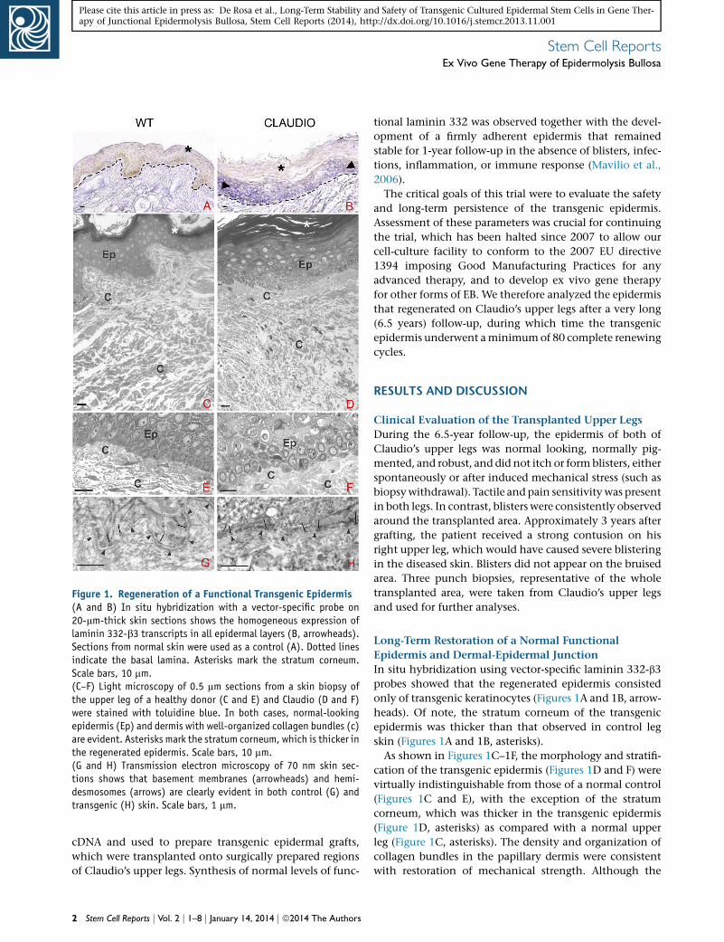
Figure 1. Regeneration of a Functional Transgenic Epidermis(A and B) In situ hybridization with a vector-specific probe on20-mm-thick skin sections shows the homogeneous expression oflaminin 332-b3 transcripts in all epidermal layers (B, arrowheads).Sections from normal skin were used as a control (A). Dotted linesindicate the basal lamina. Asterisks mark the stratum corneum.Scale bars, 10 mm.(C–F) Light microscopy of 0.5 mm sections from a skin biopsy ofthe upper leg of a healthy donor (C and E) and Claudio (D and F)were stained with toluidine blue. In both cases, normal-lookingepidermis (Ep) and dermis with well-organized collagen bundles (c)are evident. Asterisks mark the stratum corneum, which is thicker inthe regenerated epidermis. Scale bars, 10 mm.(G and H) Transmission electron microscopy of 70 nm skin sec-tions shows that basement membranes (arrowheads) and hemi-desmosomes (arrows) are clearly evident in both control (G) andtransgenic (H) skin. Scale bars, 1 mm.
Stem Cell ReportsEx Vivo Gene Therapy of Epidermolysis Bullosa
Please cite this article in press as: De Rosa et al., Long-Term Stability and Safety of Transgenic Cultured Epidermal Stem Cells in Gene Ther-apy of Junctional Epidermolysis Bullosa, Stem Cell Reports (2014), http://dx.doi.org/10.1016/j.stemcr.2013.11.001
cDNA and used to prepare transgenic epidermal grafts,
which were transplanted onto surgically prepared regions
of Claudio’s upper legs. Synthesis of normal levels of func-
2 Stem Cell Reports j Vol. 2 j 1–8 j January 14, 2014 j ª2014 The Authors
tional laminin 332 was observed together with the devel-
opment of a firmly adherent epidermis that remained
stable for 1-year follow-up in the absence of blisters, infec-
tions, inflammation, or immune response (Mavilio et al.,
2006).
The critical goals of this trial were to evaluate the safety
and long-term persistence of the transgenic epidermis.
Assessment of these parameters was crucial for continuing
the trial, which has been halted since 2007 to allow our
cell-culture facility to conform to the 2007 EU directive
1394 imposing Good Manufacturing Practices for any
advanced therapy, and to develop ex vivo gene therapy
for other forms of EB. We therefore analyzed the epidermis
that regenerated on Claudio’s upper legs after a very long
(6.5 years) follow-up, during which time the transgenic
epidermis underwent aminimumof 80 complete renewing
cycles.
RESULTS AND DISCUSSION
Clinical Evaluation of the Transplanted Upper Legs
During the 6.5-year follow-up, the epidermis of both of
Claudio’s upper legs was normal looking, normally pig-
mented, and robust, and did not itch or formblisters, either
spontaneously or after induced mechanical stress (such as
biopsywithdrawal). Tactile and pain sensitivitywas present
in both legs. In contrast, blisters were consistently observed
around the transplanted area. Approximately 3 years after
grafting, the patient received a strong contusion on his
right upper leg, which would have caused severe blistering
in the diseased skin. Blisters did not appear on the bruised
area. Three punch biopsies, representative of the whole
transplanted area, were taken from Claudio’s upper legs
and used for further analyses.
Long-Term Restoration of a Normal Functional
Epidermis and Dermal-Epidermal Junction
In situ hybridization using vector-specific laminin 332-b3
probes showed that the regenerated epidermis consisted
only of transgenic keratinocytes (Figures 1A and 1B, arrow-
heads). Of note, the stratum corneum of the transgenic
epidermis was thicker than that observed in control leg
skin (Figures 1A and 1B, asterisks).
As shown in Figures 1C–1F, the morphology and stratifi-
cation of the transgenic epidermis (Figures 1D and F) were
virtually indistinguishable from those of a normal control
(Figures 1C and E), with the exception of the stratum
corneum, which was thicker in the transgenic epidermis
(Figure 1D, asterisks) as compared with a normal upper
leg (Figure 1C, asterisks). The density and organization of
collagen bundles in the papillary dermis were consistent
with restoration of mechanical strength. Although the
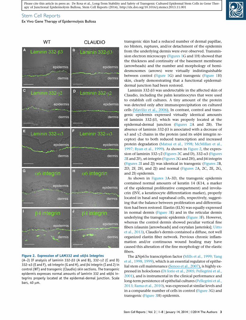
Figure 2. Expression of LAM332 and a6b4 Integrins(A–J) IF analysis of laminin 332-b3 (A and B), 332-g2 (C and D)332-a3 (E and F), a6 integrin (G and H), and b4 integrin (I and J) incontrol (WT) and transgenic (Claudio) skin sections. The transgenicepidermis expresses normal amounts of laminin 332 and a6b4 in-tegrins properly located at the epidermal-dermal junction. Scalebars, 40 mm.
Stem Cell ReportsEx Vivo Gene Therapy of Epidermolysis Bullosa
Please cite this article in press as: De Rosa et al., Long-Term Stability and Safety of Transgenic Cultured Epidermal Stem Cells in Gene Ther-apy of Junctional Epidermolysis Bullosa, Stem Cell Reports (2014), http://dx.doi.org/10.1016/j.stemcr.2013.11.001
transgenic skin had a reduced number of dermal papillae,
no blisters, ruptures, and/or detachment of the epidermis
from the underlying dermis were ever observed. Transmis-
sion electron microscopy (Figures 1G and 1H) showed that
the thickness and continuity of the basement membrane
(arrowheads) and the number and morphology of hemi-
desmosomes (arrows) were virtually indistinguishable
between control (Figure 1G) and transgenic (Figure 1H)
skin, clearly demonstrating that a functional epidermal-
dermal junction had been restored.
Laminin 332-b3 was undetectable in the affected skin of
Claudio, including the palm keratinocytes that were used
to establish cell cultures. A tiny amount of the protein
was detected only after immunoprecipitation on cultured
cells (Mavilio et al., 2006). In contrast, control and trans-
genic epidermis expressed virtually identical amounts
of laminin 332-b3, which was properly located at the
epidermal-dermal junction (Figures 2A and 2B). The
absence of laminin 332-b3 is associated with a decrease of
a3 and g2 chains in the protein (and its a6b4 integrin re-
ceptor) due to both reduced transcription and increased
protein degradation (Matsui et al., 1998; McMillan et al.,
1997; Ryan et al., 1999). As shown in Figure 2, the expres-
sion of laminin 332-g2 (Figures 2C and D), 332-a3 (Figures
2E and 2F), a6 integrin (Figures 2G and 2H), and b4 integrin
(Figures 2I and 2J) was identical in transgenic (Figures 2B,
2D, 2F, 2H, and 2J) and normal (Figures 2A, 2C, 2E, 2G,
and 2I) epidermis.
As shown in Figures 3A–3D, the transgenic epidermis
contained normal amounts of keratin 14 (K14, a marker
of the epidermal proliferative compartment) and involu-
crin (INV, a keratinocyte differentiation marker), properly
located in basal and suprabasal cells, respectively, suggest-
ing that the balance between proliferation and differentia-
tion had been restored. Elastin (ELN) was equally expressed
in normal dermis (Figure 3E) and in the reticular dermis
underlying the transgenic epidermis (Figure 3F). However,
whereas the control dermis showed peculiar vertical fine
fibers (elaunin [arrowheads] and oxytalan [asterisks]; Uitto
et al., 2013), Claudio’s dermis contained a diffuse, not well
organized elastin fiber network. Previous chronic inflam-
mation and/or continuous wound healing may have
caused this alteration of the fine morphology of the elastic
fibers.
The DNp63a transcription factor (Mills et al., 1999; Yang
et al., 1998, 1999), which is an essential regulator of epithe-
lial stem cell maintenance (Senoo et al., 2007), is highly ex-
pressed in holoclones (Di Iorio et al., 2005; Pellegrini et al.,
2001), and is instrumental in the clinical performance and
long-termpersistence of epithelial cultures (Pellegrini et al.,
2013; Rama et al., 2010), was expressed at similar levels and
in a comparable number of cells in control (Figure 3G) and
transgenic (Figure 3H) epidermis.
Stem Cell Reports j Vol. 2 j 1–8 j January 14, 2014 j ª2014 The Authors 3
Figure 3. Expression of Epidermal Markers(A–D) IF analysis of K14 (A and B) and involucrin (C and D) incontrol (WT) and transgenic (Claudio’s) epidermis.(E and F) IF analysis of elastin fibers. Network fibers (oxytalan[asterisk] and elaunin [arrowhead]) are expressed at comparablelevels, but are differently organized in WT skin (E) and Claudio’sskin (F).(G and H) IF analysis shows that the DNp63a transcription factor isexpressed at similar levels and in a comparable number of cells incontrol (G) and transgenic (H) epidermis.
4 Stem Cell Reports j Vol. 2 j 1–8 j January 14, 2014 j ª2014 The Authors
Stem Cell ReportsEx Vivo Gene Therapy of Epidermolysis Bullosa
Please cite this article in press as: De Rosa et al., Long-Term Stability and Safety of Transgenic Cultured Epidermal Stem Cells in Gene Ther-apy of Junctional Epidermolysis Bullosa, Stem Cell Reports (2014), http://dx.doi.org/10.1016/j.stemcr.2013.11.001
A Defined Number of Transduced Stem Cells Sustain
the Regenerated Epidermis
The human epidermis is renewed monthly; hence, Clau-
dio’s epidermis underwent �80 complete renewing cycles
in 6.5 years. The long-termmaintenance of the regenerated
epidermis must be determined by the engraftment of self-
renewing transduced epidermal stem cells.
A genome-wide analysis of RV integration sites was per-
formed on DNA extracted from �10 mm2 of transgenic
epidermis. Libraries of vector-genome junctions, generated
by linker-mediated (LM) nested PCR and sequenced to
saturation, retrieved six independent integrations unam-
biguously mapped on the human genome (Table 1). Provi-
ruses were classified as intergenic when they occurred at a
distance of >50 kb from any ‘‘known gene’’ (UCSC defini-
tion), perigenic when they occurred %50 kb upstream or
downstream of the transcription start site (TSS) of a known
gene, and intragenic when they occurred within the
transcribed portion of at least one known gene (Table 1).
Three out six integrations were intergenic. One of the
three intragenic integrations landed in a gene-dense re-
gion, since it was surrounded by five genes in a less
than ±40 kb window. None of these integrations belong
to a comprehensive compilation of proto-oncogenes
and genes associated with common insertion sites (CIS)
in mouse tumors (http://microb230.med.upenn.edu/
protocols/cancergenes.html). Three of the integrations,
whose TSS is more proximal to the MLV integration site,
are expressed (according to Affymetrix GeneChip analysis)
on keratinocytes cultured under the same conditions
used for transduction. The two intragenic integrations
landed in the first and second introns of expressed genes,
confirming the known integration preferences of g-RV vec-
tors in human cells (Cattoglio et al., 2010a; Cattoglio et al.,
2010b).
Considering an average of two proviral copies per
genome and an overall cloning efficiency of �30%, we
estimate the presence of approximately five to ten indepen-
dently transduced stem cells in 10mm2 of epidermis. Since
virtually all keratinocytes contain LAM332-b3 transcripts
(Figure 1B), it is clear that the entire regenerated epidermis
is sustained only by those few engrafted stem cells.
A 10 mm2 sample of cultured epidermis contains
�15,000 keratinocytes, �3,000 of which are clonogenic
and the vast majority of which (>95%) are transit-ampli-
fying progenitors. Thus, <150 stem cells are usually con-
tained in 10 mm2 of a cultured graft. Despite years of
clinical applications of epidermal cultures, we have no
(I and J) IF analysis shows that K9 is expressed in the upper layersof the transgenic epidermis (J), but is not detected in normal bodyepidermis (I).Scale bars, 40 mm. Dotted lines indicate the basal lamina.
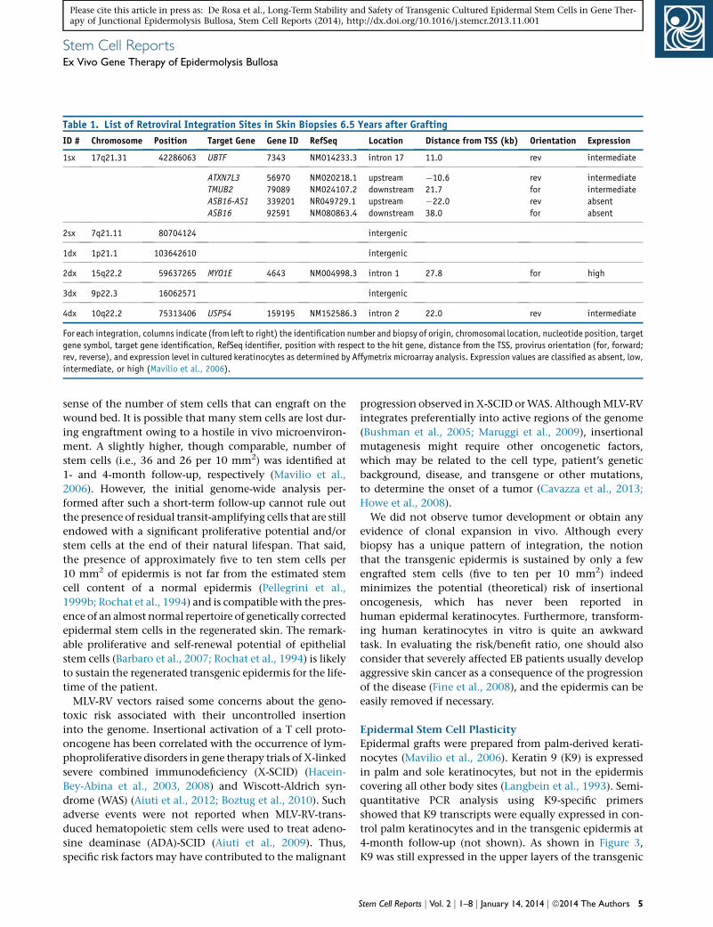
Table 1. List of Retroviral Integration Sites in Skin Biopsies 6.5 Years after Grafting
ID # Chromosome Position Target Gene Gene ID RefSeq Location Distance from TSS (kb) Orientation Expression
1sx 17q21.31 42286063 UBTF 7343 NM014233.3 intron 17 11.0 rev intermediate
ATXN7L3
TMUB2
ASB16-AS1
ASB16
56970
79089
339201
92591
NM020218.1
NM024107.2
NR049729.1
NM080863.4
upstream
downstream
upstream
downstream
�10.6
21.7
�22.0
38.0
rev
for
rev
for
intermediate
intermediate
absent
absent
2sx 7q21.11 80704124 intergenic
1dx 1p21.1 103642610 intergenic
2dx 15q22.2 59637265 MYO1E 4643 NM004998.3 intron 1 27.8 for high
3dx 9p22.3 16062571 intergenic
4dx 10q22.2 75313406 USP54 159195 NM152586.3 intron 2 22.0 rev intermediate
For each integration, columns indicate (from left to right) the identification number and biopsy of origin, chromosomal location, nucleotide position, target
gene symbol, target gene identification, RefSeq identifier, position with respect to the hit gene, distance from the TSS, provirus orientation (for, forward;
rev, reverse), and expression level in cultured keratinocytes as determined by Affymetrix microarray analysis. Expression values are classified as absent, low,
intermediate, or high (Mavilio et al., 2006).
Stem Cell ReportsEx Vivo Gene Therapy of Epidermolysis Bullosa
Please cite this article in press as: De Rosa et al., Long-Term Stability and Safety of Transgenic Cultured Epidermal Stem Cells in Gene Ther-apy of Junctional Epidermolysis Bullosa, Stem Cell Reports (2014), http://dx.doi.org/10.1016/j.stemcr.2013.11.001
sense of the number of stem cells that can engraft on the
wound bed. It is possible that many stem cells are lost dur-
ing engraftment owing to a hostile in vivo microenviron-
ment. A slightly higher, though comparable, number of
stem cells (i.e., 36 and 26 per 10 mm2) was identified at
1- and 4-month follow-up, respectively (Mavilio et al.,
2006). However, the initial genome-wide analysis per-
formed after such a short-term follow-up cannot rule out
the presence of residual transit-amplifying cells that are still
endowed with a significant proliferative potential and/or
stem cells at the end of their natural lifespan. That said,
the presence of approximately five to ten stem cells per
10 mm2 of epidermis is not far from the estimated stem
cell content of a normal epidermis (Pellegrini et al.,
1999b; Rochat et al., 1994) and is compatible with the pres-
ence of an almost normal repertoire of genetically corrected
epidermal stem cells in the regenerated skin. The remark-
able proliferative and self-renewal potential of epithelial
stem cells (Barbaro et al., 2007; Rochat et al., 1994) is likely
to sustain the regenerated transgenic epidermis for the life-
time of the patient.
MLV-RV vectors raised some concerns about the geno-
toxic risk associated with their uncontrolled insertion
into the genome. Insertional activation of a T cell proto-
oncogene has been correlated with the occurrence of lym-
phoproliferative disorders in gene therapy trials of X-linked
severe combined immunodeficiency (X-SCID) (Hacein-
Bey-Abina et al., 2003, 2008) and Wiscott-Aldrich syn-
drome (WAS) (Aiuti et al., 2012; Boztug et al., 2010). Such
adverse events were not reported when MLV-RV-trans-
duced hematopoietic stem cells were used to treat adeno-
sine deaminase (ADA)-SCID (Aiuti et al., 2009). Thus,
specific risk factors may have contributed to the malignant
progression observed inX-SCID orWAS. AlthoughMLV-RV
integrates preferentially into active regions of the genome
(Bushman et al., 2005; Maruggi et al., 2009), insertional
mutagenesis might require other oncogenetic factors,
which may be related to the cell type, patient’s genetic
background, disease, and transgene or other mutations,
to determine the onset of a tumor (Cavazza et al., 2013;
Howe et al., 2008).
We did not observe tumor development or obtain any
evidence of clonal expansion in vivo. Although every
biopsy has a unique pattern of integration, the notion
that the transgenic epidermis is sustained by only a few
engrafted stem cells (five to ten per 10 mm2) indeed
minimizes the potential (theoretical) risk of insertional
oncogenesis, which has never been reported in
human epidermal keratinocytes. Furthermore, transform-
ing human keratinocytes in vitro is quite an awkward
task. In evaluating the risk/benefit ratio, one should also
consider that severely affected EB patients usually develop
aggressive skin cancer as a consequence of the progression
of the disease (Fine et al., 2008), and the epidermis can be
easily removed if necessary.
Epidermal Stem Cell Plasticity
Epidermal grafts were prepared from palm-derived kerati-
nocytes (Mavilio et al., 2006). Keratin 9 (K9) is expressed
in palm and sole keratinocytes, but not in the epidermis
covering all other body sites (Langbein et al., 1993). Semi-
quantitative PCR analysis using K9-specific primers
showed that K9 transcripts were equally expressed in con-
trol palm keratinocytes and in the transgenic epidermis at
4-month follow-up (not shown). As shown in Figure 3,
K9 was still expressed in the upper layers of the transgenic
Stem Cell Reports j Vol. 2 j 1–8 j January 14, 2014 j ª2014 The Authors 5
Stem Cell ReportsEx Vivo Gene Therapy of Epidermolysis Bullosa
Please cite this article in press as: De Rosa et al., Long-Term Stability and Safety of Transgenic Cultured Epidermal Stem Cells in Gene Ther-apy of Junctional Epidermolysis Bullosa, Stem Cell Reports (2014), http://dx.doi.org/10.1016/j.stemcr.2013.11.001
epidermis (Figure 3J) after 6.5 years, whereas it was unde-
tectable in normal body skin (Figure 3I). These findings
are consistent with the presence of a thick stratum cor-
neum, which is another hallmark of palm and sole
epidermis, and demonstrate that epidermal stem cells
maintain the memory of their origin even after 80 com-
plete renewing cycles in vivo, even if they have been trans-
planted onto a virtually undamaged dermis.
This observation is relevant to all somatic human stem
cells. It has been suggested that some somatic stem cells
might be capable of differentiating across tissue lineage
boundaries and hence might represent versatile effectors
of therapeutic tissue regeneration. However, studies pro-
posing such ‘‘plasticity’’ remain very controversial, and ex-
isting evidence suggests that such transformations are
exceedingly rare (if they occur at all) in vivo and can be
accounted for by alternative explanations (Bianco et al.,
2013). The notion that palm-derived epidermal stem cells
do not possess sufficient plasticity to generate a body
epidermis makes one reconsider the supposed plasticity
of any somatic stem cell. It formally confirms that the
in vivo potential of a stem cell is system restricted and
cell autonomous, and strengthens the concept that a
stem cell’s function should be verified by its ability to
reconstitute a tissue in vivo (Bianco et al., 2013).
CONCLUSIONS
In summary, these data demonstrate that (1) the regener-
ated transgenic epidermis is fully functional and virtually
indistinguishable from a normal epidermis, (2) the vast
majority of transduced keratinocytes are transit-amplifying
progenitors that are lost within a fewmonths after grafting,
and (3) the regenerated epidermis is sustained by a discrete
number of engrafted, long-lasting, self-renewing trans-
genic stem cells. These data pave the way for the safe use
of epidermal stem cells in combined cell and gene therapy
for genetic skin diseases.
EXPERIMENTAL PROCEDURES
Light Microscopy, Transmission Electron Microscopy,
and ImmunofluorescenceSkin biopsies were fixed in 2.5% glutaraldehyde in Tyrode’s saline
pH 7.2 (24 hr at 4�C), postfixed in 1% osmium tetroxide (Electron
Microscopy Sciences) for 2 hr at room temperature, dehydrated
in ethanol and propylene oxide, and embedded in Spurr resin
(Polysciences) as previously described (Quaglino et al., 1991).
Semithin sections were stained with toluidine blue and observed
with a Zeiss Axiophot light microscope. Ultrathin sections were
collected on copper grids, stained with uranyl acetate and lead
citrate, and observed with a Jeol 1200 EXII (Jeol) electron
microscope.
6 Stem Cell Reports j Vol. 2 j 1–8 j January 14, 2014 j ª2014 The Authors
For immunofluorescence (IF), skin samples were embedded in
optimal cutting temperature compound, frozen, and sectioned.
IF was performed on 7 mm skin sections as previously described
(Mavilio et al., 2006) using laminin 332-b3 6F12 monoclonal anti-
body (mAb; Acris Antibodies), 332-g2 D4B5 mAb (Chemicon),
332-a3 BM165 mAb (a gift from Patricia Rousselle, IBCP), 332-a6
450-30A mAb and 332-b4 450-9D mAb (Thermo Fisher Scientific),
rabbit purified anti-p63a immunoglobulin G (IgG; PRIMM) (Di
Iorio et al., 2005), K10 and K14 guinea pig antisera (Progen), K9
sc-58743 mAb (Santa Cruz Biotechnology), elastin MAB2503
mAb (Millipore), and human involucrin mAb (Leica Biosystems).
Alexa Fluor 488 goat anti-mouse or Alexa Fluor 568 goat-anti rabbit
(Life Technologies) conjugated secondary antibodies were used for
detection. Cell nuclei were stained with DAPI.
Fluorescent signalsweremonitored under a Zeiss confocalmicro-
scope LSM510meta with a Zeiss EC Plan-Neofluar 3 40/1.3 oil
immersion objective, and analyses were done with the LSM510
Confocal Analyzer (Zeiss). Elastin staining was monitored using
an Axio Imager A1 with a Zeiss EC-Plan Neofluar x40, and analyses
were done using Axiovision Rel. 4.8 software.
In Situ HybridizationThe probe sequence was obtained by PCR reaction on Claudio’s
genomic DNA using 50-AGTAACGCCATTTTGCAAGG-30 and
50-AACAGAAGCGAGAAGCGAAC-30 primers cloned in pCRII-
topoVector (TOPO TA cloning kit; Promega). In situ hybridization
was performed as previously described (Brancaccio et al., 2004).
Digoxigenin-labeled cRNAs were synthesized using the DIG
RNA labeling kit (Roche) according to the manufacturer’s instruc-
tions. The antisense RNA probe was transcribed with T7 poly-
merase, and the control sense probe was transcribed with SP6
polymerase.
Analysis of RV Integration SitesIntegration sites were cloned by LM-PCR as previously described
(Recchia et al., 2006). Genomic DNA was digested with MseI and
PstI, and ligated to an MseI double-strand linker. LM-PCR was
performed with nested primers specific for the LTR and the linker.
PCR products were shotgun cloned by the TOPO TA cloning kit
(Invitrogen/Life Technologies) into libraries of integration junc-
tions, which were sequenced to saturation. Sequences were map-
ped onto the human genome by the BLAT genome browser
(UCSC Human Genome Project Working Draft, Feb 2009, hg19;
http://www.genome.ucsc.edu).
ACKNOWLEDGMENTS
This work was supported by the Ministero Istruzione Universita e
Ricerca, the Italian Ministry of Health and European Community
Seventh Framework Program ‘‘Optimization of Stem Cell Therapy
for Degenerative Epithelial and Muscle Diseases’’ (OptiStem,
HEALTH-F5-2009-223098), and POR-FESR 2007-13-Tecnopolo.
Received: August 13, 2013
Revised: November 1, 2013
Accepted: November 5, 2013
Published: December 26, 2013
Stem Cell ReportsEx Vivo Gene Therapy of Epidermolysis Bullosa
Please cite this article in press as: De Rosa et al., Long-Term Stability and Safety of Transgenic Cultured Epidermal Stem Cells in Gene Ther-apy of Junctional Epidermolysis Bullosa, Stem Cell Reports (2014), http://dx.doi.org/10.1016/j.stemcr.2013.11.001
REFERENCES
Aiuti, A., Cattaneo, F., Galimberti, S., Benninghoff, U., Cassani, B.,
Callegaro, L., Scaramuzza, S., Andolfi, G., Mirolo, M., Brigida, I.,
et al. (2009).Gene therapy for immunodeficiency due to adenosine
deaminase deficiency. N. Engl. J. Med. 360, 447–458.
Aiuti, A., Bacchetta, R., Seger, R., Villa, A., and Cavazzana-Calvo,
M. (2012). Gene therapy for primary immunodeficiencies: Part 2.
Curr. Opin. Immunol. 24, 585–591.
Barbaro, V., Testa, A., Di Iorio, E., Mavilio, F., Pellegrini, G., and De
Luca,M. (2007). C/EBPdelta regulates cell cycle and self-renewal of
human limbal stem cells. J. Cell Biol. 177, 1037–1049.
Barrandon, Y., and Green, H. (1987). Three clonal types of kerati-
nocyte with different capacities for multiplication. Proc. Natl.
Acad. Sci. USA 84, 2302–2306.
Bianco, P., Cao, X., Frenette, P.S., Mao, J.J., Robey, P.G., Simmons,
P.J., andWang, C.Y. (2013). Themeaning, the sense and the signif-
icance: translating the science of mesenchymal stem cells into
medicine. Nat. Med. 19, 35–42.
Boztug, K., Schmidt, M., Schwarzer, A., Banerjee, P.P., Dıez, I.A.,
Dewey, R.A., Bohm, M., Nowrouzi, A., Ball, C.R., Glimm, H.,
et al. (2010). Stem-cell gene therapy for the Wiskott-Aldrich syn-
drome. N. Engl. J. Med. 363, 1918–1927.
Brancaccio, A., Minichiello, A., Grachtchouk, M., Antonini, D.,
Sheng, H., Parlato, R., Dathan, N., Dlugosz, A.A., and Missero, C.
(2004). Requirement of the forkhead gene Foxe1, a target of sonic
hedgehog signaling, in hair follicle morphogenesis. Hum. Mol.
Genet. 13, 2595–2606.
Bushman, F., Lewinski, M., Ciuffi, A., Barr, S., Leipzig, J., Hannen-
halli, S., and Hoffmann, C. (2005). Genome-wide analysis of retro-
viral DNA integration. Nat. Rev. Microbiol. 3, 848–858.
Carulli, S., Contin, R., De Rosa, L., Pellegrini, G., and De Luca, M.
(2013). The long and winding road that leads to a cure for epider-
molysis bullosa. Regen. Med. 8, 467–481.
Cattoglio, C., Maruggi, G., Bartholomae, C., Malani, N., Pellin, D.,
Cocchiarella, F., Magnani, Z., Ciceri, F., Ambrosi, A., von Kalle, C.,
et al. (2010a). High-definition mapping of retroviral integration
sites defines the fate of allogeneic T cells after donor lymphocyte
infusion. PLoS ONE 5, e15688.
Cattoglio, C., Pellin, D., Rizzi, E., Maruggi, G., Corti, G., Miselli, F.,
Sartori, D., Guffanti, A., Di Serio, C., Ambrosi, A., et al. (2010b).
High-definition mapping of retroviral integration sites identifies
active regulatory elements in human multipotent hematopoietic
progenitors. Blood 116, 5507–5517.
Cavazza, A., Moiani, A., and Mavilio, F. (2013). Mechanisms of
retroviral integration and mutagenesis. Hum. Gene Ther. 24,
119–131.
De Luca, M., Pellegrini, G., and Green, H. (2006). Regeneration of
squamous epithelia from stem cells of cultured grafts. Regen. Med.
1, 45–57.
Di Iorio, E., Barbaro, V., Ruzza, A., Ponzin, D., Pellegrini, G., andDe
Luca, M. (2005). Isoforms of DeltaNp63 and the migration of
ocular limbal cells in human corneal regeneration. Proc. Natl.
Acad. Sci. USA 102, 9523–9528.
Fine, J.D., Eady, R.A., Bauer, E.A., Bauer, J.W., Bruckner-Tuderman,
L., Heagerty, A., Hintner, H., Hovnanian, A., Jonkman,M.F., Leigh,
I., et al. (2008). The classification of inherited epidermolysis bul-
losa (EB): Report of the Third International Consensus Meeting
on Diagnosis and Classification of EB. J. Am. Acad. Dermatol. 58,
931–950.
Gallico, G.G., 3rd, O’Connor, N.E., Compton, C.C., Kehinde, O.,
and Green, H. (1984). Permanent coverage of large burn wounds
with autologous cultured human epithelium. N. Engl. J. Med.
311, 448–451.
Hacein-Bey-Abina, S., Von Kalle, C., Schmidt, M., McCormack,
M.P., Wulffraat, N., Leboulch, P., Lim, A., Osborne, C.S., Pawliuk,
R., Morillon, E., et al. (2003). LMO2-associated clonal T cell prolif-
eration in two patients after gene therapy for SCID-X1. Science
302, 415–419.
Hacein-Bey-Abina, S., Garrigue, A., Wang, G.P., Soulier, J., Lim, A.,
Morillon, E., Clappier, E., Caccavelli, L., Delabesse, E., Beldjord, K.,
et al. (2008). Insertional oncogenesis in 4 patients after retrovirus-
mediated gene therapy of SCID-X1. J. Clin. Invest. 118, 3132–
3142.
Howe, S.J., Mansour, M.R., Schwarzwaelder, K., Bartholomae, C.,
Hubank, M., Kempski, H., Brugman, M.H., Pike-Overzet, K., Chat-
ters, S.J., de Ridder, D., et al. (2008). Insertional mutagenesis com-
bined with acquired somatic mutations causes leukemogenesis
following gene therapy of SCID-X1 patients. J. Clin. Invest. 118,
3143–3150.
Langbein, L., Heid, H.W.,Moll, I., and Franke,W.W. (1993).Molec-
ular characterization of the body site-specific human epidermal
cytokeratin 9: cDNA cloning, amino acid sequence, and tissue
specificity of gene expression. Differentiation 55, 57–71.
Maruggi, G., Porcellini, S., Facchini, G., Perna, S.K., Cattoglio, C.,
Sartori, D., Ambrosi, A., Schambach, A., Baum, C., Bonini, C.,
et al. (2009). Transcriptional enhancers induce insertional gene
deregulation independently from the vector type and design.
Mol. Ther. 17, 851–856.
Matsui, C., Pereira, P., Wang, C.K., Nelson, C.F., Kutzkey, T., Lani-
gan, C., Woodley, D., Morohashi, M., Welsh, E.A., and Hoeffler,
W.K. (1998). Extent of laminin-5 assembly and secretion effect
junctional epidermolysis bullosa phenotype. J. Exp. Med. 187,
1273–1283.
Mavilio, F., Pellegrini, G., Ferrari, S., Di Nunzio, F., Di Iorio, E.,
Recchia, A., Maruggi, G., Ferrari, G., Provasi, E., Bonini, C., et al.
(2006). Correction of junctional epidermolysis bullosa by trans-
plantation of genetically modified epidermal stem cells. Nat.
Med. 12, 1397–1402.
McMillan, J.R., McGrath, J.A., Pulkkinen, L., Kon, A., Burgeson,
R.E., Ortonne, J.P., Meneguzzi, G., Uitto, J., and Eady, R.A.
(1997). Immunohistochemical analysis of the skin in junctional
epidermolysis bullosa using laminin 5 chain specific antibodies is
of limited value in predicting the underlying gene mutation. Br.
J. Dermatol. 136, 817–822.
Mills, A.A., Zheng, B., Wang, X.J., Vogel, H., Roop, D.R., and Brad-
ley, A. (1999). p63 is a p53 homologue required for limb and
epidermal morphogenesis. Nature 398, 708–713.
Stem Cell Reports j Vol. 2 j 1–8 j January 14, 2014 j ª2014 The Authors 7
Stem Cell ReportsEx Vivo Gene Therapy of Epidermolysis Bullosa
Please cite this article in press as: De Rosa et al., Long-Term Stability and Safety of Transgenic Cultured Epidermal Stem Cells in Gene Ther-apy of Junctional Epidermolysis Bullosa, Stem Cell Reports (2014), http://dx.doi.org/10.1016/j.stemcr.2013.11.001
Pellegrini, G., Traverso, C.E., Franzi, A.T., Zingirian, M., Cancedda,
R., and De Luca, M. (1997). Long-term restoration of damaged
corneal surfaces with autologous cultivated corneal epithelium.
Lancet 349, 990–993.
Pellegrini, G., Golisano, O., Paterna, P., Lambiase, A., Bonini, S.,
Rama, P., and De Luca, M. (1999a). Location and clonal analysis
of stem cells and their differentiated progeny in the human ocular
surface. J. Cell Biol. 145, 769–782.
Pellegrini, G., Ranno, R., Stracuzzi, G., Bondanza, S., Guerra, L.,
Zambruno, G., Micali, G., and De Luca, M. (1999b). The control
of epidermal stem cells (holoclones) in the treatment of massive
full-thickness burns with autologous keratinocytes cultured on
fibrin. Transplantation 68, 868–879.
Pellegrini, G., Dellambra, E., Golisano, O., Martinelli, E., Fantozzi,
I., Bondanza, S., Ponzin, D., McKeon, F., and De Luca, M. (2001).
p63 identifies keratinocyte stem cells. Proc. Natl. Acad. Sci. USA
98, 3156–3161.
Pellegrini, G., Rama, P., Matuska, S., Lambiase, A., Bonini, S., Poco-
belli, A., Colabelli, R.G., Spadea, L., Fasciani, R., Balestrazzi, E., et al.
(2013). Biological parameters determining the clinical outcome of
autologous cultures of limbal stem cells. Regen. Med. 8, 553–567.
Quaglino, D., Fornieri, C., Botti, B., Davidson, J.M., and Pasquali-
Ronchetti, I. (1991). Opposing effects of ascorbate on collagen
and elastin deposition in the neonatal rat aorta. Eur. J. Cell Biol.
54, 18–26.
Rama, P., Matuska, S., Paganoni, G., Spinelli, A., De Luca, M., and
Pellegrini, G. (2010). Limbal stem-cell therapy and long-term
corneal regeneration. N. Engl. J. Med. 363, 147–155.
Recchia, A., Bonini, C., Magnani, Z., Urbinati, F., Sartori, D., Mur-
aro, S., Tagliafico, E., Bondanza, A., Stanghellini, M.T., Bernardi,
8 Stem Cell Reports j Vol. 2 j 1–8 j January 14, 2014 j ª2014 The Authors
M., et al. (2006). Retroviral vector integration deregulates gene
expression but has no consequence on the biology and function
of transplanted T cells. Proc. Natl. Acad. Sci. USA 103, 1457–1462.
Rochat, A., Kobayashi, K., and Barrandon, Y. (1994). Location of
stem cells of human hair follicles by clonal analysis. Cell 76,
1063–1073.
Ronfard, V., Rives, J.M., Neveux, Y., Carsin, H., and Barrandon, Y.
(2000). Long-term regeneration of human epidermis on third
degree burns transplanted with autologous cultured epithelium
grown on a fibrin matrix. Transplantation 70, 1588–1598.
Ryan, M.C., Lee, K., Miyashita, Y., and Carter, W.G. (1999). Tar-
geted disruption of the LAMA3 gene in mice reveals abnormalities
in survival and late stage differentiation of epithelial cells. J. Cell
Biol. 145, 1309–1323.
Senoo, M., Pinto, F., Crum, C.P., and McKeon, F. (2007). p63 Is
essential for the proliferative potential of stem cells in stratified
epithelia. Cell 129, 523–536.
Uitto, J., Li, Q., andUrban, Z. (2013). The complexity of elastic fibre
biogenesis in the skin—a perspective to the clinical heterogeneity
of cutis laxa. Exp. Dermatol. 22, 88–92.
Yang, A., Kaghad, M., Wang, Y., Gillett, E., Fleming, M.D., Dotsch,
V., Andrews, N.C., Caput, D., and McKeon, F. (1998). p63, a p53
homolog at 3q27-29, encodesmultiple products with transactivat-
ing, death-inducing, and dominant-negative activities.Mol. Cell 2,
305–316.
Yang, A., Schweitzer, R., Sun, D., Kaghad, M., Walker, N., Bronson,
R.T., Tabin, C., Sharpe, A., Caput, D., Crum, C., and McKeon, F.
(1999). p63 is essential for regenerative proliferation in limb,
craniofacial and epithelial development. Nature 398, 714–718.