Differential Localization of VE and N-Cadherins in Human Endothelial Cells: VECadherin Competes with...
10
The Rockefeller University Press, 0021-9525/98/03/1475/10 $2.00 The Journal of Cell Biology, Volume 140, Number 6, March 23, 1998 1475–1484 http://www.jcb.org 1475 Differential Localization of VE- and N-Cadherins in Human Endothelial Cells: VE-Cadherin Competes with N-Cadherin for Junctional Localization Pilar Navarro,* Luigi Ruco, ‡ and Elisabetta Dejana* *Laboratory of Vascular Biology, Istituto di Ricerche Farmacologiche Mario Negri, Milan, Italy; and ‡ IIº Dipartimento di Medicina Sperimentale e Patologia, Universitá di Roma La Sapienza, Rome, Italy Abstract. The two major cadherins of endothelial cells are neural (N)-cadherin and vascular endothelial (VE)- cadherin. Despite similar level of protein expression only VE-cadherin is located at cell–cell contacts, whereas N-cadherin is distributed over the whole cell membrane. Cotransfection of VE-cadherin and N-cad- herin in CHO cells resulted in the same distribution as that observed in endothelial cells indicating that the be- havior of the two cadherins was not cell specific but re- lated to their structural characteristics. Similar amounts of a- and b-catenins and plakoglobin were associated to VE- and N-cadherins, whereas p120 was higher in the VE-cadherin complex. The presence of VE-cadherin did not affect N-cadherin homotypic adhesive proper- ties or its capacity to localize at junctions when co- transfectants were cocultured with cells transfected with N-cadherin only. To define the molecular domain responsible for the VE-cadherin–dominant activity we prepared a chimeric construct formed by VE-cadherin extracellular region linked to N-cadherin intracellular domain. The chimera lost the capacity to exclude N-cad- herin from junctions indicating that the extracellular domain of VE-cadherin alone is not sufficient for the preferential localization of the molecule at the junc- tions. A truncated mutant of VE-cadherin retaining the full extracellular domain and a short cytoplasmic tail (Arg 621 –Pro 702 ) lacking the catenin-binding region was able to exclude N-cadherin from junctions. This indi- cates that the Arg 621 –Pro 702 sequence in the VE-cad- herin cytoplasmic tail is required for N-cadherin exclu- sion from junctions. Competition between cadherins for their clustering at intercellular junctions in the same cell has never been described before. We speculate that, in the endothelium, VE- and N-cadherin play dif- ferent roles; whereas VE-cadherin mostly promotes the homotypic interaction between endothelial cells, N-cad- herin may be responsible for the anchorage of the en- dothelium to other surrounding cell types expressing N-cadherin such as vascular smooth muscle cells or pericytes. C adherins are a family of transmembrane glycopro- teins that mediate calcium-dependent cell–cell ad- hesion (1, 15, 55). These adhesion receptors are expressed by virtually all cell types that form solid tissues. Cadherins are organized in junctional structures called ad- herens junctions. In these junctions, cadherins are clus- tered and connected through their cytoplasmic domain with a complex network of cytoskeletal proteins (23, 26, 28, 44). Cadherins are important in regulating morphogen- esis (15, 22, 47, 55). Through their homophilic interactions, they play a role in sorting cells of different lineages during embryogenesis, establishing cell polarity, and maintaining tissue morphology and cell differentiation. Classical cad- herins share a common basic structure consisting of an ex- tracellular domain, which contains four major repeats, a transmembrane and a cytoplasmic domain. The short cyto- plasmic region directly binds to three homologous pro- teins that belong to the “armadillo” family (b-catenin, plakoglobin, and p120). b-catenin and plakoglobin bind a-catenin, which is homologous to vinculin and mediates the binding of the cadherin–catenin complex to the actin cytoskeleton. Comparison of different cadherin sequences shows strong homology at the amino acid level (26, 55). Conservation is particularly striking in the cytoplasmic tail where the cate- nin-binding region is located. Cadherins present a certain degree of cell type specific- ity, for instance epithelial (E)-cadherin is preferentially expressed in cells of epithelial origin (53, 54), and neural (N)-cadherin 1 in cells of the nervous tissue (17, 18), vascu- Address all correspondence to Pilar Navarro, Istituto di Ricerche Farma- cologiche Mario Negri, Via Eritrea 62, 20157 Milano, Italia. Tel.: (39) 2-39014477. Fax: (39) 2-3546277. E-mail: [email protected] 1. Abbreviations used in this paper: aa, amino acids; cyt, cytoplasmic; E-, N-, P-, T-, and VE-cadherins, epithelial, neural, placental, truncated, and vas- cular endothelial cadherins; ec, extracellular; EC, endothelial cell. on January 19, 2016 jcb.rupress.org Downloaded from Published March 23, 1998
-
Upload
independent -
Category
Documents
-
view
1 -
download
0
Transcript of Differential Localization of VE and N-Cadherins in Human Endothelial Cells: VECadherin Competes with...
The Rockefeller University Press, 0021-9525/98/03/1475/10 $2.00The Journal of Cell Biology, Volume 140, Number 6, March 23, 1998 1475–1484http://www.jcb.org 1475
Differential Localization of VE- and N-Cadherinsin Human Endothelial Cells: VE-Cadherin Competeswith N-Cadherin for Junctional Localization
Pilar Navarro,* Luigi Ruco,
‡
and Elisabetta Dejana*
*Laboratory of Vascular Biology, Istituto di Ricerche Farmacologiche Mario Negri, Milan, Italy; and
‡
IIº Dipartimento di Medicina Sperimentale e Patologia, Universitá di Roma La Sapienza, Rome, Italy
Abstract.
The two major cadherins of endothelial cells are neural (N)-cadherin and vascular endothelial (VE)-cadherin. Despite similar level of protein expression only VE-cadherin is located at cell–cell contacts, whereas N-cadherin is distributed over the whole cell membrane. Cotransfection of VE-cadherin and N-cad-herin in CHO cells resulted in the same distribution as that observed in endothelial cells indicating that the be-havior of the two cadherins was not cell specific but re-lated to their structural characteristics. Similar amounts of
a
- and
b
-catenins and plakoglobin were associated to VE- and N-cadherins, whereas p120 was higher in the VE-cadherin complex. The presence of VE-cadherin did not affect N-cadherin homotypic adhesive proper-ties or its capacity to localize at junctions when co-transfectants were cocultured with cells transfected with N-cadherin only. To define the molecular domain responsible for the VE-cadherin–dominant activity we prepared a chimeric construct formed by VE-cadherin extracellular region linked to N-cadherin intracellular
domain. The chimera lost the capacity to exclude N-cad-herin from junctions indicating that the extracellular domain of VE-cadherin alone is not sufficient for the preferential localization of the molecule at the junc-tions. A truncated mutant of VE-cadherin retaining the full extracellular domain and a short cytoplasmic tail (Arg
621
–Pro
702
) lacking the catenin-binding region was able to exclude N-cadherin from junctions. This indi-cates that the Arg
621
–Pro
702
sequence in the VE-cad-herin cytoplasmic tail is required for N-cadherin exclu-sion from junctions. Competition between cadherins for their clustering at intercellular junctions in the same cell has never been described before. We speculate that, in the endothelium, VE- and N-cadherin play dif-ferent roles; whereas VE-cadherin mostly promotes the homotypic interaction between endothelial cells, N-cad-herin may be responsible for the anchorage of the en-dothelium to other surrounding cell types expressing N-cadherin such as vascular smooth muscle cells or pericytes.
C
adherins
are a family of transmembrane glycopro-teins that mediate calcium-dependent cell–cell ad-hesion (1, 15, 55). These adhesion receptors are
expressed by virtually all cell types that form solid tissues.Cadherins are organized in junctional structures called ad-herens junctions. In these junctions, cadherins are clus-tered and connected through their cytoplasmic domainwith a complex network of cytoskeletal proteins (23, 26,28, 44). Cadherins are important in regulating morphogen-esis (15, 22, 47, 55). Through their homophilic interactions,they play a role in sorting cells of different lineages duringembryogenesis, establishing cell polarity, and maintainingtissue morphology and cell differentiation. Classical cad-herins share a common basic structure consisting of an ex-tracellular domain, which contains four major repeats, a
transmembrane and a cytoplasmic domain. The short cyto-plasmic region directly binds to three homologous pro-teins that belong to the “armadillo” family (
b
-catenin,plakoglobin, and p120).
b
-catenin and plakoglobin bind
a
-catenin, which is homologous to vinculin and mediatesthe binding of the cadherin–catenin complex to the actincytoskeleton.
Comparison of different cadherin sequences shows stronghomology at the amino acid level (26, 55). Conservation isparticularly striking in the cytoplasmic tail where the cate-nin-binding region is located.
Cadherins present a certain degree of cell type specific-ity, for instance epithelial (E)-cadherin is preferentiallyexpressed in cells of epithelial origin (53, 54), and neural(N)-cadherin
1
in cells of the nervous tissue (17, 18), vascu-
Address all correspondence to Pilar Navarro, Istituto di Ricerche Farma-cologiche Mario Negri, Via Eritrea 62, 20157 Milano, Italia. Tel.: (39)2-39014477. Fax: (39) 2-3546277. E-mail:
1.
Abbreviations used in this paper
: aa, amino acids; cyt, cytoplasmic; E-, N-,P-, T-, and VE-cadherins, epithelial, neural, placental, truncated, and vas-cular endothelial cadherins; ec, extracellular; EC, endothelial cell.
on January 19, 2016jcb.rupress.org
Dow
nloaded from
Published March 23, 1998
The Journal of Cell Biology, Volume 140, 1998 1476
lar smooth muscle cells, and myocytes (8, 19, 45, 60). Morethan one cadherin may be expressed in the same cell type,but how these molecules interact with each other and howthey can contribute to the transfer of specific intracellularsignals is still unknown.
The two major cadherins in the endothelium are vascu-lar endothelial (VE)- and N-cadherin (10, 32, 49). VE-cad-herin is cell specific and is strictly located at intercellularjunctions of essentially all types of endothelium, both invitro and in vivo (10, 32). Compared with the classical cad-herins, the VE-cadherin amino acid sequence shows con-siderable differences (only 23% identity when comparedwith classical cadherins such as E-, N-, and placental
(P)-cadherins) (5, 6).
N-cadherin is present in significant amounts in the en-dothelium, but surprisingly, it is not clustered at cell–celljunctions but remains diffuse on the cell membrane (50).This observation raises several questions about the mecha-nisms that regulate cadherin clustering at adherens junc-tions, and raises the possibility that a hierarchy does existbetween cadherins, which regulates their functional be-havior and signaling properties in a cell type–specific way.
In this report, we have investigated the mechanisms re-sponsible for the differential membrane targeting of N-and VE-cadherin in the endothelium. We have found thatVE-cadherin presents structural features that are respon-sible for its ability to exclude N-cadherin from cell–cellcontacts. This dominant activity of VE-cadherin requires ashort cytoplasmic region of the molecule, which is distinctfrom the previously described catenin-binding domain.Overall these observations suggest that cadherins mightpresent different functional behaviors depending on theother cadherins expressed.
Materials and Methods
All reagents were purchased from Sigma Chemical Co. (St. Louis, MO),unless indicated otherwise.
Antibodies
Mouse mAbs against the extracellular domain of human VE-cadherinwere: clone TEA 1.31 (36), clone BV9 (Hemeris, Grenoble, France) (34,36), and clone BV6 (Bioline Diag., Torino, Italy) (34).
Anti–humanN-cadherin polyclonal antibodies against the whole molecule (1447; seereference 3) or the cytoplasmic domain (R156; see reference 13), and rab-bit pan-cadherin antiserum against the conserved cytoplasmic sequence ofall cadherins (13), were a gift from Dr. B. Geiger (Weizmann Institute,Rehovot, Israel). Mouse mAbs against the extracellular (8C11) or cyto-plasmic domain (13A9) of human N-cadherin were donated by Dr. M. J.Wheelock (University of Toledo, Toledo, OH). Mouse mAbs against
a
-catenin,
b
-catenin, plakoglobin, and p120 were purchased from Transduc-tion Laboratories (Lexington, KY). Fluorescein- and rhodamine-conju-gated secondary antibodies (reactive with either mouse or rabbit IgG)were purchased from DAKOPATTS (Copenhagen, Denmark).
Goat anti–mouse IgG peroxidase–conjugated and protein A peroxi-dase–conjugated mAbs used for immunoblotting detection were fromPierce Chemical Co. (Rockford, IL,), and anti-phosphotyrosine HRP-con-jugated mAb (RC20) was purchased from Transduction Laboratories.
Cells
Endothelial cells (ECs) were harvested from human umbilical veins andcultured as previously described (34). CHO cells were routinely main-tained on culture dishes in DME containing 10% FCS, and kept at 37
8
C in5% CO
2
. Transfection of CHO cells with the full-length cDNA of humanN-cadherin (CHO N), full-length cDNA of human VE-cadherin (CHO
VE), and cytoplasmic truncated mutant cDNA of human VE-cadherin(CHO
T
VE) was previously described (6, 41, 50).Sterile plastic ware was from Falcon (Becton Dickinson Labware, Lin-
coln Park, NJ) whereas both culture medium and serum were fromGIBCO BRL (Paisley, Scotland).
Plasmids Construction
Vectors containing the entire coding sequence of human VE-cadherin andN-cadherin have been described elsewhere (6, 50).
Cytoplasmic truncated mutant cDNA of human VE-cadherin (
T
VE)was generated by removing the coding region for the last 82 amino acids(aa) of the cytoplasmic domain as described previously (41).
VE
ec
N
cyt
plasmid contains a chimera formed by the extracellular (ec)domain of VE-cadherin and the cytoplasmic (cyt) region of N-cadherin.The chimera was constructed by substitution of the VE-cadherin cytoplas-mic coding region with the N-cadherin cytoplasmic coding region. The hu-man VE-cadherin cDNA cloned into the pBlueScript vector (6) was di-gested with BglI/BamHI to remove the coding region from the 1,519-bpposition to the end of the cDNA. To preserve the VE-cadherin coding se-quence from the 1,519-bp to the 1,884-bp position (comprising the trans-membrane domain), we generate a PCR fragment using 5
9
and 3
9
endprimers matching at the appropriate positions and containing the BglI/BamHI sites for cloning. The resultant plasmid, corresponding to the ex-tracellular and transmembrane coding region of VE-cadherin cDNA, wascalled pVE
ec
. N-cadherin cytoplasmic domain coding sequence was gener-ated by PCR using 5
9
and 3
9
end primers matching at the 2,701-bp and the3,215-bp positions of N-cadherin cDNA (provided by Dr. B. Geiger,Weizmann Institute), and containing BamHI and XbaI sites, respectively.This fragment was further cloned into pVE
ec
digested with BamHI/XbaI.The construct was subsequently checked by sequence analysis using thedideoxynucleotide chain termination method (52). This plasmid was thencut with KpnI and XbaI enzymes, and the coding sequence insert was sub-cloned into the pECE eukaryotic expression vector (12).
VE
ec
T
Ncyt chimera plasmid contains VE-cadherin extracellular andtransmembrane coding sequence, and the cytoplasmic domain of N-cad-herin lacking the last 77 aas. N-cadherin deleted cytoplasmic coding re-gion was generated by PCR using the 5
9
end primer containing the BamHIsite described above and a 3
9
end primer matching at the 2,944-bp positionof N-cadherin cDNA, and containing stop codon sequences and an XbaIsite for cloning. The fragment encodes 83 aas (Lys
746
–Pro
828
) of the cyto-plasmic domain of N-cadherin lacking the COOH terminus Gly
829
–Asp
906
(77 aas). This fragment was cloned into pVE
ec
and subcloned into pECEas described above.
Transfection Procedure
As expression control of chimeric molecules described in the previous sec-tion, CHO cells were transiently transfected with VE
ec
N
cyt
and VE
ec
T
N
cyt
plasmids using lipofectin reagent (GIBCO BRL). Cells were grown onglass coverslips, fixed 60 h after transfection, and then tested for chimeraexpression by immunofluorescence using anti–VE-cadherin mAb BV9and anti–N-cadherin mAb 13A9, or polyclonal antibody R156.
For obtaining double transfectants, CHO N cells were stably
trans-fected with VE-cadherin. Cells were plated at 3–4
3
10
6
cells per 100-mmpetri dish in DME with 10% FCS. 24 h later they were cotransfected bycalcium phosphate precipitation method (51) using 20
m
g of VE-cadherininto pECE vector and 2
m
g of pBSpac
D
p plasmid containing puromycinresistance (a gift from Dr. J. Ortin, Centro Nacional Biotecnología,Madrid, Spain; see reference 9). After 24 h, the DNA-containing mediumwas replaced by fresh DME containing 10% FCS, and then cultured foranother 48 h. Cells were then trypsinized, plated at a density of 10
6
per100-mm petri dish, and then cultured in selective medium with 7
m
g/mlpuromycin (Sigma Chemical Co.). Resistant colonies were isolated andtested for VE-cadherin expression by immunofluorescence analysis. Posi-tive cells were subcloned and used for further studies.
A similar procedure was used to generate double transfectants CHON/
T
VE (cotransfected with N and
T
VE-cadherins), CHO N/VE
ec
N
cyt
(cotransfected with N-cadherin and VE
ec
N
cyt
chimera), and CHO N/VE
ec
T-
N
cyt
(cotransfected with N-cadherin and VE
ec
T
N
cyt
-truncated chimera).
Immunohistochemistry
Human tissue specimens of different origin were embedded in optimalcryopreserving tissue compound (Ames Miles Laboratories Inc., Naper-
on January 19, 2016jcb.rupress.org
Dow
nloaded from
Published March 23, 1998
Navarro et al.
VE- and N-Cadherin Competition
1477
ville, IN), snap frozen in liquid nitrogen, and then stored at
2
80
8
C untilprocessed. Cryostat sections were fixed in acetone for 10 min at 22
8
C andimmunostained with the anti–VE-cadherin mAb TEA 1.31 or anti–N-cad-herin polyclonal antibody 1447, using avidin–biotin–peroxidase complextechnique. The specimens were then processed and analyzed as previouslydescribed (33).
Immunofluorescence Microscopy
Cells were grown on glass coverslips (for EC coated with human plasmafibronectin, 7
m
g/ml), rinsed in PBS, and fixed in methanol. The cells werethen rinsed and incubated for 45 min at 37
8
C with the relevant primary an-tibodies (against VE- or N-cadherins), washed three times with PBS, andthen incubated for 30 min with the fluorophore-conjugated secondary an-tibodies. For double staining, the coverslips were incubated with mouseanti–VE-cadherin (BV6 or BV9) in combination with rabbit anti–N-cad-herin (polyclonal antibody 1447), followed by TRITC-coupled, goat anti–mouse in combination with FITC-coupled, goat anti–rabbit. Coverslipswere then mounted in Mowiol 4-88 (Calbiochem-Novabiochem Corp., LaJolla, CA) and examined with an Axiophot microscope (Carl Zeiss Inc.,Thornwood, NY). Photographs were taken using T
max
P3200 films.For mixed culture studies (see Fig. 6), the two types of cells (CHO N
and CHO N/VE) were seeded in small aggregates and let grow until con-fluency. After methanol fixation, glass coverslips were processed for dou-ble immunofluorescence as described above.
For double transfectants CHO N/
T
VE, CHO N/VE
ec
N
cyt
, and CHON/VE
ec
T
N
cyt
, we use mAb 8C11 to recognize wild-type N-cadherin andmAbs BV6 or BV9 for truncated or chimera recognition.
Western Blot and Immunoprecipitation
Whole-cell extracts were obtained from confluent cells as previously de-scribed (33). Different cell extracts were adjusted to 1
3
Laemmli samplebuffer, and then fractionated under reducing conditions on 7.5% SDSpolyacrylamide gels (30).
Western blot analysis of the various cell extracts were carried out es-sentially as described (34). After blocking with 10% nonfat milk, the pro-teins of interest were detected by specific monoclonal or polyclonal anti-bodies at the optimal dilution in blocking buffer. This was sequentiallyfollowed by incubation with goat anti–mouse IgG, peroxidase conjugated(1
m
g/ml) for monoclonal antibodies, or protein A–peroxidase conjugated(1
m
g/ml; Pierce Chemical Co.) for polyclonal antibodies and further de-velopment of peroxidase activity using an enhanced chemiluminescencekit (Amersham International, Little Chalfont, UK) and autoradiography.
Immunoprecipitation of the cadherin–catenin complex was performedusing the nonionic, detergent-soluble fraction of cells, as previously re-ported (34) with some modifications. Briefly, cell extracts were preclearedby incubation with uncoupled protein G or protein A–Sepharose CL-4B(Pharmacia LKB Biotechnology, Uppsala, Sweden) for 2 h. After centrif-ugation, the precleared supernatants were incubated with protein G orprotein A–Sepharose coupled to mAb TEA 1.31 or polyclonal antibody1447 against N-cadherin, during 1 h. Immunocomplexes were collected bycentrifugation, washed five times in a buffer containing 0.5% TritonX-100, 0.1% BSA, 50 mM Tris-HCl, pH 7.4, 0.1 M NaCl, and 2 mM CaCl
2
,and then finally resuspended in 30
m
l of 1
3
Laemmli sample buffer andboiled for 5 min. Samples were analyzed by electrophoresis, transferred tonitrocellulose membranes, and then immunoblotted sequentially withmAb TEA 1.31 to VE-cadherin, or polyclonal antibody 1447 to N-cad-herin, and mAbs to
a
- and
b
-catenins, plakoglobin, and p120 as describedabove.
For tyrosine phosphorylation studies cells were incubated with culturemedium containing 0.5% BSA, 0.1 mM Na
3
VO
4
, and 0.2 mM H
2
O
2
for5 min before extraction. Cell extraction and immunoprecipitation was es-sentially as described above for preserving cadherin-associated proteins.Lysis and washing buffers were supplemented with 0.3 mM Na
3
VO
4
and0.6 mM H
2
O
2
. After immunoprecipitation with VE- or N-cadherin anti-bodies, tyrosine-phosphorylated proteins were identified using mAbRC20 coupled to HRP followed by enhanced chemiluminescence detec-tion system as described before.
Cell Aggregation
Calcium-dependent cell aggregation was done under conditions thatpreserve VE-cadherin expression as previously described (41). For het-erotypic aggregation assays CHON/VE cells were labeled with 2
m
M 2
9
,5
9
-
bis[2-carboxyethyl]-5[and-6] carboxyfluorescein acetoxymethylester (Mo-lecular Probes, Eugene, OR) in HBSS for 10 min at 37
8
C and processed asdescribed (41).
Cell Adhesion
EC, CHO, CHO VE, or CHO N cells were cultured in 96-well plates andgrown for 5 d to confluency. CHO N cells were labeled with [
125
I]iodode-oxyuridine (1
m
Ci/ml) overnight before the cell adhesion experiment. 12 hlater, CHO N cells were detached as described above and resuspended at3
3
10
5
cells/ml in DME with 10% FCS. 100
m
l of labeled cell suspensionwere added to different adherent cell monolayers (EC, CHO, CHO VE,and CHO N) and incubated for 1 h at 37
8
C. After three washes with PBSwith 10% FCS, the cells were solubilized with 0.5 M NaOH–0.1% SDSand counted in a
g
counter.
Results
VE- and N-Cadherin Expression in HumanEndothelial Cells
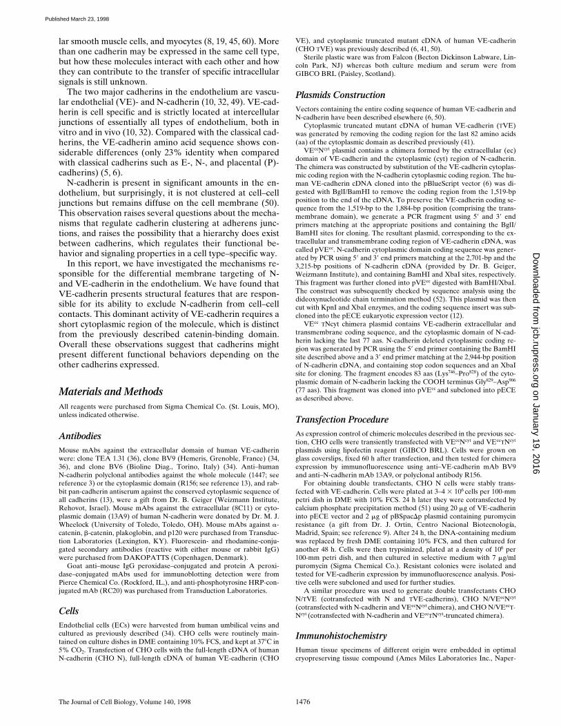
As mentioned above, the two major cadherins in ECs areN-cadherin, which is also expressed in other tissues, andVE-cadherin, which is cell specific. Western blot analysisof EC extracts showed that the level of protein expressionwas similar for both cadherins (Fig. 1
B
). These experi-ments were done using an mAb specific for VE-cadherin(BV9) (Fig. 1
B
, lane
VE
), and a polyclonal antibody spe-cific for N-cadherin (1447) (Fig. 1
B
, lane
N
). Westernblots performed with a pan-cadherin antibody able to rec-ognize both molecules (13) resulted in the identification oftwo bands of similar intensity corresponding to the appar-ent molecular weights of N- and VE-cadherins (140 and130 kD, respectively) (data not shown).
Despite otherwise comparable levels of expression, VE-
Figure 1. (A) Immunofluorescence staining of VE-cadherin (a)and N-cadherin (b) in cultured endothelium. VE-cadherin is lo-cated at cell–cell contacts whereas N-cadherin shows a diffuse lo-calization. (B) Expression of VE- and N-cadherins by Westernimmunoblot. Cell lysates from the same number of cells wereloaded in each lane, separated by SDS-PAGE, blotted to nitro-cellulose membranes, and then immunodetected with antibodiesspecific for VE- or N-cadherins. Molecular weight markers areindicated on the right. Bar, 30 mm.
on January 19, 2016jcb.rupress.org
Dow
nloaded from
Published March 23, 1998

The Journal of Cell Biology, Volume 140, 1998 1478
cadherin was found at cell–cell junctions (Fig. 1
A
,
a)
whereas N-cadherin shows a diffuse localization over thewhole cell surface (Fig.1
A
,
b)
. This immunofluorescencepattern of distribution for VE- and N-cadherin was notspecific for human umbilical vein EC, it was also observedin cultured ECs from cutaneous microcirculation (HMECline; see reference 2; data not shown).
In addition, VE- and N-cadherins distributed in vivo in away similar to cultured ECs. Fig. 2 shows a typical VE-(Fig. 2,
a
and
c
) and N-cadherin (Fig. 2,
b
and
d
) immuno-histochemical localization in vessels of different origin.Whereas VE-cadherin was found concentrated in areas ofendothelial cell–cell contacts, N-cadherin staining was al-ways diffuse in the endothelium. As expected, in arteriesN-cadherin was highly expressed in vascular smooth mus-cle cells (Fig. 2
b
).
Cotransfection of VE- and N-Cadherins in CHO Cells
We then investigated whether the capacity of VE-cadherinto inhibit N-cadherin clustering at intercellular junctionswas a specific feature of EC or was instead due to thestructural properties of the two cadherins. To this end wecotransfected VE- and N-cadherins in CHO cells.
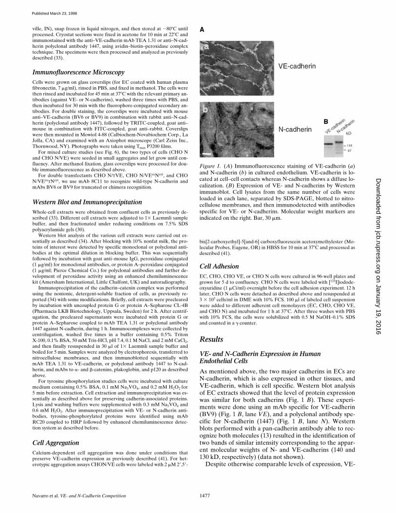
By Western blot analysis we selected the clones that hada comparable expression of N- and VE-cadherins (Fig. 3 A,lane CHO N/VE). Immunofluorescence analysis in theseclones revealed that, like in ECs, only VE-cadherin wasable to cluster at cell–cell contacts (Fig. 3 B, b) whereasN-cadherin remained diffuse on the cell surface (Fig. 3 B,c). As expected, in the absence of VE-cadherin, N-cad-herin was able to regularly concentrate at intercellular
contacts (Fig. 3 B, a). These data indicate that the domi-nant effect of VE-cadherin on N-cadherin is not endothe-lial cell specific, but is more related to the molecular prop-erties of the two cadherins.
The Presence of VE-Cadherin Does Not AffectN-Cadherin Adhesive Properties
We tested whether the inability of endothelial N-cadherinto cluster at junctions was because of the loss of its adhe-sive properties. To this purpose, we used a previously de-scribed cell adhesion assay (6). CHO cells transfected withN-cadherin (CHO N) were radioactively labeled andseeded on different cell monolayers in the presence or ab-sence of calcium. As reported in Fig. 4, N-cadherin trans-fectants adhered more efficiently to ECs or to N-cadherintransfectants than to control or VE-cadherin transfectants.This binding could be prevented by addition of EGTAduring the adhesion assay. These data show that diffuseN-cadherin on the EC surface is able to promote homo-philic binding to N-cadherin–expressing cells.
In addition, N-cadherin, in double-transfected CHOcells, retains homotypic aggregating properties. In mixedaggregation assays, CHO N/VE transfectants (fluores-cently labeled, Fig. 5 b) and N-cadherin transfectants (un-labeled, Fig. 5 a) formed heterotypic aggregates contain-ing approximately the same proportion of the two types ofcells. The presence of EGTA during the aggregation assayabolished aggregation (data not shown). Mixing CHON/VE with control transfectants only led to homotypic ag-gregates of CHO N/VE cells from which control transfec-tants were absent (data not shown).
Figure 2. Immunohistological localization of VE-(a and c) and N-cadherins (b and d) in an arteryand a vein from human lymph node tissue sections.VE-cadherin is localized at cell–cell contacts (ar-rowheads) whereas N-cadherin staining was al-ways diffuse in endothelial cells. Note the positivestaining of arterial smooth muscle cells (arrow).Bar, 50 mm.
on January 19, 2016jcb.rupress.org
Dow
nloaded from
Published March 23, 1998
Navarro et al. VE- and N-Cadherin Competition 1479
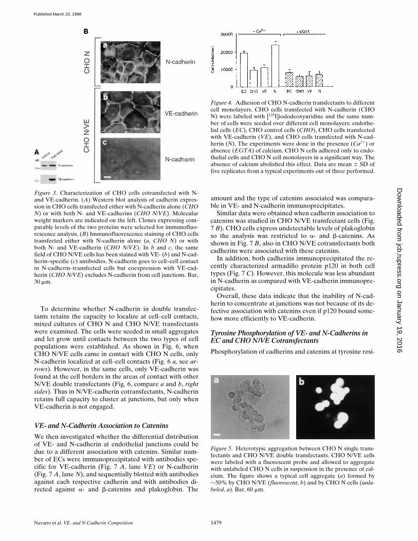
To determine whether N-cadherin in double transfec-tants retains the capacity to localize at cell–cell contacts,mixed cultures of CHO N and CHO N/VE transfectantswere examined. The cells were seeded in small aggregatesand let grow until contacts between the two types of cellpopulations were established. As shown in Fig. 6, whenCHO N/VE cells came in contact with CHO N cells, onlyN-cadherin localized at cell–cell contacts (Fig. 6 a, see ar-rows). However, in the same cells, only VE-cadherin wasfound at the cell borders in the areas of contact with otherN/VE double transfectants (Fig. 6, compare a and b, rightsides). Thus in N/VE-cadherin cotransfectants, N-cadherinretains full capacity to cluster at junctions, but only whenVE-cadherin is not engaged.
VE- and N-Cadherin Association to Catenins
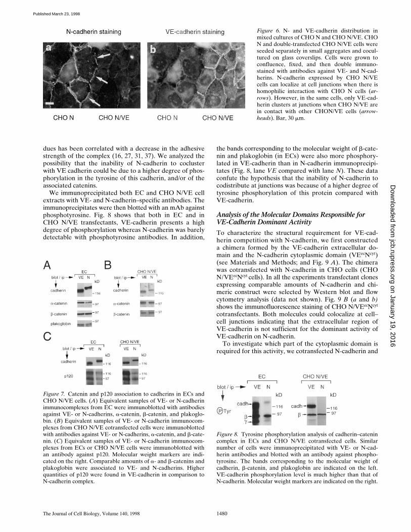
We then investigated whether the differential distributionof VE- and N-cadherin at endothelial junctions could bedue to a different association with catenins. Similar num-ber of ECs were immunoprecipitated with antibodies spe-cific for VE-cadherin (Fig. 7 A, lane VE) or N-cadherin(Fig. 7 A, lane N), and sequentially blotted with antibodiesagainst each respective cadherin and with antibodies di-rected against a- and b-catenins and plakoglobin. The
amount and the type of catenins associated was compara-ble in VE- and N-cadherin immunoprecipitates.
Similar data were obtained when cadherin association tocatenins was studied in CHO N/VE transfectant cells (Fig.7 B). CHO cells express undetectable levels of plakoglobinso the analysis was restricted to a- and b-catenins. Asshown in Fig. 7 B, also in CHO N/VE cotransfectants bothcadherins were associated with these catenins.
In addition, both cadherins immunoprecipitated the re-cently characterized armadillo protein p120 in both celltypes (Fig. 7 C). However, this molecule was less abundantin N-cadherin as compared with VE-cadherin immunopre-cipitates.
Overall, these data indicate that the inability of N-cad-herin to concentrate at junctions was not because of its de-fective association with catenins even if p120 bound some-how more efficiently to VE-cadherin.
Tyrosine Phosphorylation of VE- and N-Cadherins in EC and CHO N/VE Cotransfectants
Phosphorylation of cadherins and catenins at tyrosine resi-
Figure 3. Characterization of CHO cells cotransfected with N-and VE-cadherin. (A) Western blot analysis of cadherin expres-sion in CHO cells transfected either with N-cadherin alone (CHON) or with both N- and VE-cadherins (CHO N/VE). Molecularweight markers are indicated on the left. Clones expressing com-parable levels of the two proteins were selected for immunofluo-rescence analysis. (B) Immunofluorescence staining of CHO cellstransfected either with N-cadherin alone (a, CHO N) or withboth N- and VE-cadherin (CHO N/VE). In b and c, the samefield of CHO N/VE cells has been stained with VE- (b) and N-cad-herin–specific (c) antibodies. N-cadherin goes to cell–cell contactin N-cadherin–transfected cells but coexpression with VE-cad-herin (CHO N/VE) excludes N-cadherin from cell junctions. Bar,30 mm.
Figure 4. Adhesion of CHO N-cadherin transfectants to differentcell monolayers. CHO cells transfected with N-cadherin (CHON) were labeled with [125I]iododeoxyuridine and the same num-ber of cells were seeded over different cell monolayers: endothe-lial cells (EC), CHO control cells (CHO), CHO cells transfectedwith VE-cadherin (VE), and CHO cells transfected with N-cad-herin (N). The experiments were done in the presence (Ca21) orabsence (EGTA) of calcium. CHO N cells adhered only to endo-thelial cells and CHO N cell monolayers in a significant way. Theabsence of calcium abolished this effect. Data are mean 6 SD offive replicates from a typical experiments out of three performed.
Figure 5. Heterotypic aggregation between CHO N single trans-fectants and CHO N/VE double transfectants. CHO N/VE cellswere labeled with a fluorescent probe and allowed to aggregatewith unlabeled CHO N cells in suspension in the presence of cal-cium. The figure shows a typical cell aggregate (a) formed byz50% by CHO N/VE (fluorescent, b) and by CHO N cells (unla-beled, a). Bar, 60 mm.
on January 19, 2016jcb.rupress.org
Dow
nloaded from
Published March 23, 1998
The Journal of Cell Biology, Volume 140, 1998 1480
dues has been correlated with a decrease in the adhesivestrength of the complex (16, 27, 31, 37). We analyzed thepossibility that the inability of N-cadherin to coclusterwith VE cadherin could be due to a higher degree of phos-phorylation in the tyrosine of this cadherin, and/or of theassociated catenins.
We immunoprecipitated both EC and CHO N/VE cellextracts with VE- and N-cadherin–specific antibodies. Theimmunoprecipitates were then blotted with an mAb againstphosphotyrosine. Fig. 8 shows that both in EC and inCHO N/VE transfectants, VE-cadherin presents a highdegree of phosphorylation whereas N-cadherin was barelydetectable with phosphotyrosine antibodies. In addition,
the bands corresponding to the molecular weight of b-cate-nin and plakoglobin (in ECs) were also more phosphory-lated in VE-cadherin than in N-cadherin immunoprecipi-tates (Fig. 8, lane VE compared with lane N). These dataconfute the hypothesis that the inability of N-cadherin tocodistribute at junctions was because of a higher degree oftyrosine phosphorylation of this protein compared withVE-cadherin.
Analysis of the Molecular Domains Responsible for VE-Cadherin Dominant Activity
To characterize the structural requirement for VE-cad-herin competition with N-cadherin, we first constructeda chimera formed by the VE-cadherin extracellular do-main and the N-cadherin cytoplasmic domain (VEecNcyt)(see Materials and Methods; and Fig. 9 A). The chimerawas cotransfected with N-cadherin in CHO cells (CHON/VEecNcyt cells). In all the experiments transfectant clonesexpressing comparable amounts of N-cadherin and chi-meric construct were selected by Western blot and flowcytometry analysis (data not shown). Fig. 9 B (a and b)shows the immunofluorescence staining of CHO N/VEecNcyt
cotransfectants. Both molecules could colocalize at cell–cell junctions indicating that the extracellular region ofVE-cadherin is not sufficient for the dominant activity ofVE-cadherin on N-cadherin.
To investigate which part of the cytoplasmic domain isrequired for this activity, we cotransfected N-cadherin and
Figure 6. N- and VE-cadherin distribution inmixed cultures of CHO N and CHO N/VE. CHON and double-transfected CHO N/VE cells wereseeded separately in small aggregates and cocul-tured on glass coverslips. Cells were grown toconfluence, fixed, and then double immuno-stained with antibodies against VE- and N-cad-herins. N-cadherin expressed by CHO N/VEcells can localize at cell junctions when there ishomophilic interaction with CHO N cells (ar-rows). However, in the same cells, only VE-cad-herin clusters at junctions when CHO N/VE arein contact with other CHON/VE cells (arrow-heads). Bar, 30 mm.
Figure 7. Catenin and p120 association to cadherins in ECs andCHO N/VE cells. (A) Equivalent samples of VE- or N-cadherinimmunocomplexes from EC were immunoblotted with antibodiesagainst VE- or N-cadherins, a-catenin, b-catenin, and plakoglo-bin. (B) Equivalent samples of VE- or N-cadherin immunocom-plexes from CHO N/VE cotransfected cells were immunoblottedwith antibodies against VE- or N-cadherins, a-catenin, and b-cate-nin. (C) Equivalent samples of VE- or N-cadherin immunocom-plexes from ECs or CHO N/VE cells were immunoblotted withan antibody against p120. Molecular weight markers are indi-cated on the right. Comparable amounts of a- and b-catenins andplakoglobin were associated to VE- and N-cadherins. Higherquantities of p120 were found in VE-cadherin in comparison toN-cadherin complex.
Figure 8. Tyrosine phosphorylation analysis of cadherin–catenincomplex in ECs and CHO N/VE cotransfected cells. Similarnumber of cells were immunoprecipitated with VE- or N-cad-herin antibodies and blotted with an antibody against phospho-tyrosine. The bands corresponding to the molecular weight ofcadherin, b-catenin, and plakoglobin are indicated on the left.VE-cadherin phosphorylation level is much higher than that ofN-cadherin. Molecular weight markers are indicated on the right.
on January 19, 2016jcb.rupress.org
Dow
nloaded from
Published March 23, 1998
Navarro et al. VE- and N-Cadherin Competition 1481
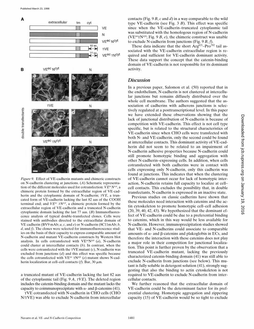
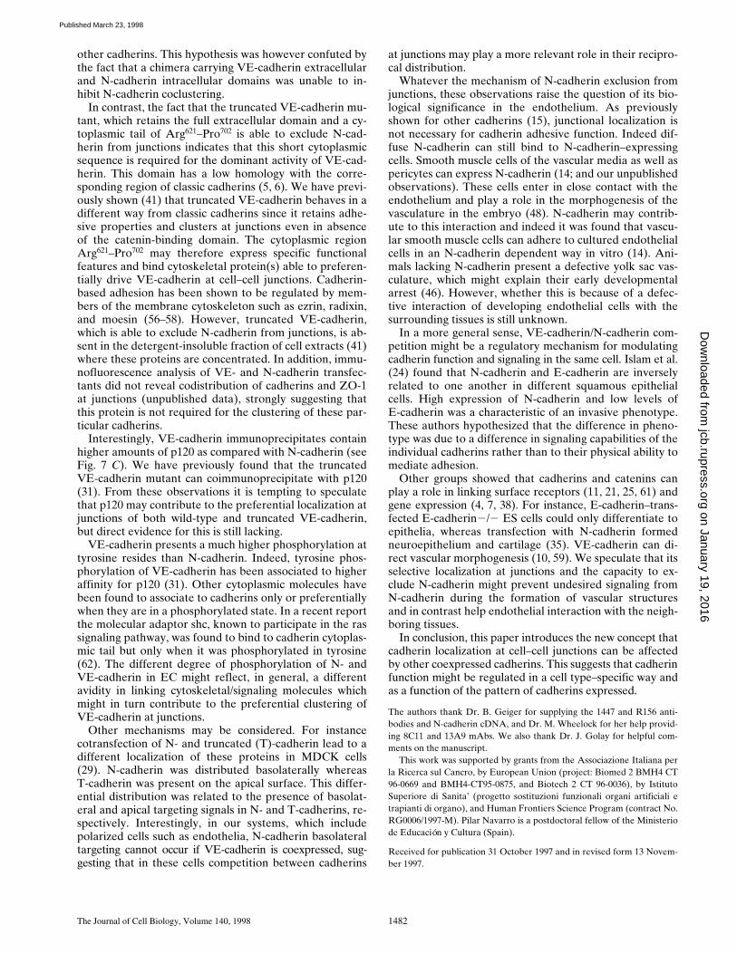
a truncated mutant of VE-cadherin lacking the last 82 aasof the cytoplasmic tail (Fig. 9 A, TVE). The deleted regionincludes the catenin-binding domain and the mutant lacks thecapacity to coimmunoprecipitate with a- and b-catenins (41).
TVE cotransfected with N-cadherin in CHO cells (CHON/TVE) was able to exclude N-cadherin from intercellular
contacts (Fig. 9 B, c and d) in a way comparable to the wildtype VE-cadherin (see Fig. 3 B). This effect was specificsince when the VE-cadherin–truncated cytoplasmic tailwas substituted with the homologous region of N-cadherin(VEecTNcyt; Fig. 9 B, e), the chimeric construct was unableto exclude N-cadherin from junctions (Fig. 9 B, f).
These data indicate that the short Arg621–Pro702 tail as-sociated with the VE-cadherin extracellular region is re-quired and sufficient for VE-cadherin dominant activity.These data support the concept that the catenin-bindingdomain of VE-cadherin is not responsible for its dominantactivity.
DiscussionIn a previous paper, Salomon et al. (50) reported that inthe endothelium, N-cadherin is not clustered at intercellu-lar junctions but remains diffusely distributed over thewhole cell membrane. The authors suggested that the as-sociation of cadherins with adherens junctions is selec-tively regulated at a posttranscriptional level. In this paperwe have extended these observations showing that thelack of junctional distribution of N-cadherin is because ofcompetition with VE-cadherin. This effect is not cell typespecific, but is related to the structural characteristics ofVE-cadherin since when CHO cells were transfected withboth N- and VE-cadherin, only the second could be foundat intercellular contacts. This dominant activity of VE-cad-herin did not seem to be related to an impairment ofN-cadherin adhesive properties because N-cadherin couldstill promote homotypic binding and aggregation withother N-cadherin–expressing cells. In addition, when cellscotransfected with both cadherins were in contact withcells expressing only N-cadherin, only this cadherin wasfound at junctions. This indicates that when the clusteringof VE-cadherin cannot occur for lack of homotypic inter-action, N-cadherin retains full capacity to localize at cell–cell contacts. This excludes the possibility that, in doubletransfectants, N-cadherin is expressed in an inactive state.
Previous studies on classic cadherins have shown thatthese molecules need interaction with catenins and the ac-tin cytoskeleton to promote homotypic cell–cell adhesion(20, 39, 40, 42, 43). We hypothesized that the dominant ef-fect of VE-cadherin could be due to a preferential bindingto catenins, which in this way would be less available forN-cadherin. However, immunoprecipitation studies showedthat VE- and N-cadherins could associate to comparableamounts of a- and b-catenins and plakoglobin in ECs, andtherefore the interaction with these catenins does not playa major role in their competition for junctional localiza-tion. This point is further proven by the observation that atruncated VE-cadherin mutant, lacking the previouslycharacterized catenin-binding domain (41) was still able toexclude N-cadherin from junctions (see below). This mu-tant is fully soluble in detergent solution (41), strongly sug-gesting that also the binding to actin cytoskeleton is notrequired to VE-cadherin to exclude N-cadherin from inter-cellular contacts.
We further reasoned that the extracellular domain ofVE-cadherin could be the determinant factor for its pref-erential clustering. Homotypic aggregation and zipperingcapacity (15) of VE-cadherin would be so tight to exclude
Figure 9. Effect of VE-cadherin mutants and chimeric constructson N-cadherin clustering at junctions. (A) Schematic representa-tion of the different molecules used for cotransfection: VEecNcyt, achimeric protein formed by the extracellular region of VE-cad-herin and the cytoplasmic domain of N-cadherin; TVE, a trun-cated form of VE-cadherin lacking the last 82 aas of the COOHterminal end; and VEec TNcyt, a chimeric protein formed by theextracellular region of VE-cadherin and a truncated N-cadherincytoplasmic domain lacking the last 77 aas. (B) Immunofluores-cence analysis of typical double-transfected clones. Cells werestained with antibodies directed to the extracellular domain ofVE cadherin (BV9 mAb; a, c, and e) or N-cadherin (8C11mAb; b,d, and f). The clones were selected for immunofluorescence stud-ies on the basis of their capacity to express comparable amount ofN-cadherin and mutant VE-cadherin constructs by Western blotanalysis. In cells cotransfected with VEecNcyt (a), N-cadherincould cluster at intercellular contacts (b). In contrast, when thecells were cotransfected with the TVE mutant (c), N-cadherin wasexcluded from junctions (d) and this effect was specific becausethe cells cotransfected with VEec TNcyt (e) mutant shows N-cad-herin localization at cell–cell contacts (f). Bar, 30 mm.
on January 19, 2016jcb.rupress.org
Dow
nloaded from
Published March 23, 1998
The Journal of Cell Biology, Volume 140, 1998 1482
other cadherins. This hypothesis was however confuted bythe fact that a chimera carrying VE-cadherin extracellularand N-cadherin intracellular domains was unable to in-hibit N-cadherin coclustering.
In contrast, the fact that the truncated VE-cadherin mu-tant, which retains the full extracellular domain and a cy-toplasmic tail of Arg621–Pro702 is able to exclude N-cad-herin from junctions indicates that this short cytoplasmicsequence is required for the dominant activity of VE-cad-herin. This domain has a low homology with the corre-sponding region of classic cadherins (5, 6). We have previ-ously shown (41) that truncated VE-cadherin behaves in adifferent way from classic cadherins since it retains adhe-sive properties and clusters at junctions even in absenceof the catenin-binding domain. The cytoplasmic regionArg621–Pro702 may therefore express specific functionalfeatures and bind cytoskeletal protein(s) able to preferen-tially drive VE-cadherin at cell–cell junctions. Cadherin-based adhesion has been shown to be regulated by mem-bers of the membrane cytoskeleton such as ezrin, radixin,and moesin (56–58). However, truncated VE-cadherin,which is able to exclude N-cadherin from junctions, is ab-sent in the detergent-insoluble fraction of cell extracts (41)where these proteins are concentrated. In addition, immu-nofluorescence analysis of VE- and N-cadherin transfec-tants did not reveal codistribution of cadherins and ZO-1at junctions (unpublished data), strongly suggesting thatthis protein is not required for the clustering of these par-ticular cadherins.
Interestingly, VE-cadherin immunoprecipitates containhigher amounts of p120 as compared with N-cadherin (seeFig. 7 C). We have previously found that the truncatedVE-cadherin mutant can coimmunoprecipitate with p120(31). From these observations it is tempting to speculatethat p120 may contribute to the preferential localization atjunctions of both wild-type and truncated VE-cadherin,but direct evidence for this is still lacking.
VE-cadherin presents a much higher phosphorylation attyrosine resides than N-cadherin. Indeed, tyrosine phos-phorylation of VE-cadherin has been associated to higheraffinity for p120 (31). Other cytoplasmic molecules havebeen found to associate to cadherins only or preferentiallywhen they are in a phosphorylated state. In a recent reportthe molecular adaptor shc, known to participate in the rassignaling pathway, was found to bind to cadherin cytoplas-mic tail but only when it was phosphorylated in tyrosine(62). The different degree of phosphorylation of N- andVE-cadherin in EC might reflect, in general, a differentavidity in linking cytoskeletal/signaling molecules whichmight in turn contribute to the preferential clustering ofVE-cadherin at junctions.
Other mechanisms may be considered. For instancecotransfection of N- and truncated (T)-cadherin lead to adifferent localization of these proteins in MDCK cells(29). N-cadherin was distributed basolaterally whereasT-cadherin was present on the apical surface. This differ-ential distribution was related to the presence of basolat-eral and apical targeting signals in N- and T-cadherins, re-spectively. Interestingly, in our systems, which includepolarized cells such as endothelia, N-cadherin basolateraltargeting cannot occur if VE-cadherin is coexpressed, sug-gesting that in these cells competition between cadherins
at junctions may play a more relevant role in their recipro-cal distribution.
Whatever the mechanism of N-cadherin exclusion fromjunctions, these observations raise the question of its bio-logical significance in the endothelium. As previouslyshown for other cadherins (15), junctional localization isnot necessary for cadherin adhesive function. Indeed dif-fuse N-cadherin can still bind to N-cadherin–expressingcells. Smooth muscle cells of the vascular media as well aspericytes can express N-cadherin (14; and our unpublishedobservations). These cells enter in close contact with theendothelium and play a role in the morphogenesis of thevasculature in the embryo (48). N-cadherin may contrib-ute to this interaction and indeed it was found that vascu-lar smooth muscle cells can adhere to cultured endothelialcells in an N-cadherin dependent way in vitro (14). Ani-mals lacking N-cadherin present a defective yolk sac vas-culature, which might explain their early developmentalarrest (46). However, whether this is because of a defec-tive interaction of developing endothelial cells with thesurrounding tissues is still unknown.
In a more general sense, VE-cadherin/N-cadherin com-petition might be a regulatory mechanism for modulatingcadherin function and signaling in the same cell. Islam et al.(24) found that N-cadherin and E-cadherin are inverselyrelated to one another in different squamous epithelialcells. High expression of N-cadherin and low levels ofE-cadherin was a characteristic of an invasive phenotype.These authors hypothesized that the difference in pheno-type was due to a difference in signaling capabilities of theindividual cadherins rather than to their physical ability tomediate adhesion.
Other groups showed that cadherins and catenins canplay a role in linking surface receptors (11, 21, 25, 61) andgene expression (4, 7, 38). For instance, E-cadherin–trans-fected E-cadherin2/2 ES cells could only differentiate toepithelia, whereas transfection with N-cadherin formedneuroepithelium and cartilage (35). VE-cadherin can di-rect vascular morphogenesis (10, 59). We speculate that itsselective localization at junctions and the capacity to ex-clude N-cadherin might prevent undesired signaling fromN-cadherin during the formation of vascular structuresand in contrast help endothelial interaction with the neigh-boring tissues.
In conclusion, this paper introduces the new concept thatcadherin localization at cell–cell junctions can be affectedby other coexpressed cadherins. This suggests that cadherinfunction might be regulated in a cell type–specific way andas a function of the pattern of cadherins expressed.
The authors thank Dr. B. Geiger for supplying the 1447 and R156 anti-bodies and N-cadherin cDNA, and Dr. M. Wheelock for her help provid-ing 8C11 and 13A9 mAbs. We also thank Dr. J. Golay for helpful com-ments on the manuscript.
This work was supported by grants from the Associazione Italiana perla Ricerca sul Cancro, by European Union (project: Biomed 2 BMH4 CT96-0669 and BMH4-CT95-0875, and Biotech 2 CT 96-0036), by IstitutoSuperiore di Sanita’ (progetto sostituzioni funzionali organi artificiali etrapianti di organo), and Human Frontiers Science Program (contract No.RG0006/1997-M). Pilar Navarro is a postdoctoral fellow of the Ministeriode Educación y Cultura (Spain).
Received for publication 31 October 1997 and in revised form 13 Novem-ber 1997.
on January 19, 2016jcb.rupress.org
Dow
nloaded from
Published March 23, 1998
Navarro et al. VE- and N-Cadherin Competition 1483
References
1. Aberle, H., H. Schwartz, and R. Kemler. 1996. Cadherin-catenin complex:protein interactions and their implications for cadherin function. J. Cell.Biochem. 61:514–523.
2. Ades, E.W., F.J. Candal, R.A. Swerlick, V.G. George, S. Summers, D.C.Bosse, and T.J. Lawley. 1992. HMEC-1: establishment of an immortal-ized human microvascular endothelial cell line. J. Invest. Dermatol. 99:683–690.
3. Ayalon, O., H. Sabanai, M.G. Lampugnani, E. Dejana, and B. Geiger.1994. Spatial and temporal relationships between cadherins and PE-CAM-1 in cell–cell junctions of human endothelial cells. J. Cell Biol. 126:247–258.
4. Behrens, J., J.P. von Kries, M. Kuhl, L. Bruhn, D. Wedlich, R. Grosschedl,and W. Birchmeier. 1996. Functional interaction of b-catenin with thetranscription factor LEF-1. Nature. 382:638–642.
5. Breier, G., F. Breviario, L. Caveda, R. Berthier, H. Schnurch, U. Gotsch,D. Vestweber, W. Risau, and E. Dejana. 1996. Molecular cloning and ex-pression of murine vascular endothelial-cadherin in early stage develop-ment of cardiovascular system. Blood. 87:630–641.
6. Breviario, F., L. Caveda, M. Corada, I. Martin Padura, P. Navarro, J. Go-lay, M. Introna, D. Gulino, M.G. Lampugnani, and E. Dejana. 1995.Functional properties of human vascular endothelial cadherin (7B4/cad-herin-5), an endothelium-specific cadherin. Arterioscler. Thromb. Vasc.Biol. 15:1229–1239.
7. Brunner, E., O. Peter, L. Schweizer, and K. Basler. 1997. Pangolin encodesa Lef-1 homologue that acts downstream of Armadillo to transduce theWingless signal in Drosophila. Nature. 385:829–833.
8. Cifuentes Diaz, C., M. Nicolet, D. Goudou, F. Rieger, and R.M. Mege.1994. N-cadherin expression in developing, adult and denervated chickenneuromuscular system: accumulations at both the neuromuscular junc-tion and the node of Ranvier. Development (Camb.). 120:1–11.
9. de la Luna, S., and J. Ortin. 1992. pac gene as efficient dominant markerand reporter gene in mammalian cells. Methods Enzymol. 216:376–385.
10. Dejana, E. 1996. Endothelial adherens junctions: implications in the con-trol of vascular permeability and angiogenesis. J. Clin. Invest. 98:1949–1953.
11. Doherty, P., E. Williams, and F.S. Walsh. 1995. A soluble chimeric form ofthe L1 glycoprotein stimulates neurite outgrowth. Neuron. 14:57–66.
12. Ellis, L., E. Clauser, D.O. Morgan, M. Edery, R.A. Roth, and W.J. Rutter.1986. Replacement of insulin receptor tyrosine residues 1162 and 1163compromises insulin-stimulated kinase activity and uptake of 2-deoxy-glucose. Cell. 45:721–732.
13. Geiger, B., T. Volberg, D. Ginsberg, S. Bitzur, I. Sabanay, and R.O. Hynes.1990. Broad spectrum pan-cadherin antibodies, reactive with the C-ter-minal 24 amino acid residues of N-cadherin. J. Cell Sci. 97:607–614.
14. Gilbertson Beadling, S.K., and C. Fisher. 1993. A potential role for N-cad-herin in mediating endothelial cell-smooth muscle cell interactions in therat vasculature. Lab. Invest. 69:203–209.
15. Gumbiner, B.M. 1996. Cell adhesion: the molecular basis of tissue architec-ture and morphogenesis. Cell. 84:345–357.
16. Hamaguchi, M., N. Matsuyoshi, Y. Ohnishi, B. Gotoh, M. Takeichi, and Y.Nagai. 1993. p60v-src causes tyrosine phosphorylation and inactivation ofthe N-cadherin-catenin cell adhesion system. EMBO (Eur. Mol. Biol. Or-gan.) J. 12:307–314.
17. Hatta, K., and M. Takeichi. 1986. Expression of N-cadherin adhesion mole-cules associated with early morphogenetic events in chick development.Nature. 320:447–449.
18. Hatta, K., T.S. Okada, and M. Takeichi. 1985. A monoclonal antibody dis-rupting calcium-dependent cell–cell adhesion of brain tissues: possiblerole of its target antigen in animal pattern formation. Proc. Natl. Acad.Sci. USA. 82:2789–2793.
19. Hatta, K., S. Takagi, H. Fujisawa, and M. Takeichi. 1987. Spatial and tem-poral expression pattern of N-cadherin cell adhesion molecules corre-lated with morphogenetic processes of chicken embryos. Dev. Biol. 120:215–227.
20. Hirano, S., N. Kimoto, Y. Shimoyama, S. Hirohashi, and M. Takeichi. 1992.Identification of a neural alpha-catenin as a key regulator of cadherinfunction and multicellular organization. Cell. 70:293–301.
21. Hoschuetzky, H., H. Aberle, and R. Kemler. 1994. Beta-catenin mediatesthe interaction of the cadherin-catenin complex with epidermal growthfactor receptor. J. Cell Biol. 127:1375–1380.
22. Huber, O., C. Bierkamp, and R. Kemler. 1996. Cadherins and catenins indevelopment. Curr. Opin. Cell Biol. 8:685–691.
23. Hulsken, J., J. Behrens, and W. Birchmeier. 1994. Tumor-suppressor geneproducts in cell contacts: the cadherin-APC armadillo connection. Curr.Opin. Cell Biol. 6:711–716.
24. Islam, S., T.E. Carey, G.T. Wolf, M.J. Wheelock, and K.R. Johnson. 1996.Expression of N-cadherin by human squamous carcinoma cells induces ascattered fibroblastic phenotype with disrupted cell–cell adhesion. J. CellBiol. 135:1643–1654.
25. Kanai, Y., A. Ochiai, T. Shibata, T. Oyama, S. Ushijima, S. Akimoto, andS. Hirohashi. 1995. c-erbB-2 gene product directly associates with beta-catenin and plakoglobin. Biochem. Biophys. Res. Commun. 208:1067–1072.
26. Kemler, R. 1993. From cadherins to catenins: cytoplasmic protein interac-tions and regulation of cell adhesion. Trends Genet. 9:317–321.
27. Kinch, M.S., G.J. Clark, C.J. Der, and K. Burridge. 1995. Tyrosine phos-phorylation regulates the adhesions of ras-transformed breast epithelia.J. Cell Biol. 130:461–471.
28. Klymkowsky, M.W., and B. Parr. 1995. The body language of cells: the inti-mate connection between cell adhesion and behavior. Cell. 83:5–8.
29. Koller, E., and B. Ranscht. 1996. Differential targeting of T- and N-cad-herin in polarized epithelial cells. J. Biol Chem. 271:30061–30067.
30. Laemmli, U.K. 1970. Cleavage of structural proteins during the assembly ofthe head of bacteriophage T4. Nature. 227:680–685.
31. Lampugnani, M., M. Corada, P. Andriopoulou, S. Esser, W. Risau, and E.Dejana. 1997. Cell confluence regulates tyrosine phosphorylation of ad-herens junction components in endothelial cells. J. Cell Sci. 110:2065–2077.
32. Lampugnani, M.G., and E. Dejana. 1997. Interendothelial junctions: struc-ture, signalling and functional roles. Curr. Opin. Cell Biol. 9:674–682.
33. Lampugnani, M.G., M. Resnati, M. Raiteri, R. Pigott, A. Pisacane, G.Houen, L.P. Ruco, and E. Dejana. 1992. A novel endothelial-specificmembrane protein is a marker of cell–cell contacts. J. Cell Biol. 118:1511–1522.
34. Lampugnani, M.G., M. Corada, L. Caveda, F. Breviario, O. Ayalon, B.Geiger, and E. Dejana. 1995. The molecular organization of endothelialcell to cell junctions: Differential association of plakoglobin, b-catenin,and a-catenin with vascular endothelial cadherin (VE-cadherin). J. CellBiol. 129:203–217.
35. Larue, L., C. Antos, S. Butz, O. Huber, V. Delmas, M. Dominis, and R.Kemler. 1996. A role for cadherins in tissue formation. Development(Camb.). 122:3185–3194.
36. Leach, L., P. Clark, M.G. Lampugnani, A.G. Arroyo, E. Dejana, and J.A.Firth. 1993. Immunoelectron characterisation of the inter-endothelialjunctions of human term placenta. J. Cell Sci. 104:1073–1081.
37. Matsuyoshi, N., M. Hamaguchi, S. Taniguchi, A. Nagafuchi, S. Tsukita, andM. Takeichi. 1992. Cadherin-mediated cell–cell adhesion is perturbed byv-src tyrosine phosphorylation in metastatic fibroblasts. J. Cell Biol. 118:703–714.
38. Molenaar, M., M. van de Wetering, M. Oosterwegel, J. Peterson Maduro,S. Godsave, V. Korinek, J. Roose, O. Destree, and H. Clevers. 1996.XTcf-3 transcription factor mediates b-catenin-induced axis formation inXenopus embryos. Cell. 86:391–399.
39. Nagafuchi, A., and M. Takeichi. 1988. Cell binding function of E-cadherinis regulated by the cytoplasmic domain. EMBO (Eur. Mol. Biol. Organ.)J. 7:3679–3684.
40. Nagafuchi, A., and M. Takeichi. 1989. Transmembrane control of cadherin-mediated cell adhesion: a 94 kDa protein functionally associated with aspecific region of the cytoplasmic domain of E-cadherin. Cell. Regul.1:37–44.
41. Navarro, P., L. Caveda, F. Breviario, I. Mandoteanu, M.G. Lampugnani,and E. Dejana. 1995. Catenin-dependent and -independent functions ofvascular endothelial cadherin. J. Biol. Chem. 270:30965–30972.
42. Ozawa, M., H. Baribault, and R. Kemler. 1989. The cytoplasmic domain ofthe cell adhesion molecule uvomorulin associates with three independentproteins structurally related in different species. EMBO (Eur. Mol. Biol.Organ.) J. 8:1711–1717.
43. Ozawa, M., M. Ringwald, and R. Kemler. 1990. Uvomorulin-catenin com-plex formation is regulated by a specific domain in the cytoplasmic regionof the cell adhesion molecule. Proc. Natl. Acad. Sci. USA. 87:4246–4250.
44. Peifer, M. 1995. Cell adhesion and signal transduction: the Armadillo con-nection. Trends Cell Biol. 5:224–229.
45. Pouliot, Y., P.C. Holland, and O.W. Blaschuk. 1990. Developmental regu-lation of a cadherin during the differentiation of skeletal myoblasts. Dev.Biol. 141:292–298.
46. Radice, G.L., H. Rayburn, H. Matsunami, K.A. Knudsen, M. Takeichi, andR.O. Hynes. 1997. Developmental defects in mouse embryos lackingN-cadherin. Dev. Biol. 181:64–78.
47. Ranscht, B. 1994. Cadherins and catenins: interactions and functions in em-bryonic development. Curr. Opin. Cell Biol. 6:740–746.
48. Risau, W. 1997. Mechanisms of angiogenesis. Nature. 386:671–674.49. Rubin, L.L. 1992. Endothelial cells: adhesion and tight junctions. Curr.
Opin. Cell Biol. 4:830–833.50. Salomon, D., O. Ayalon, R. Patel King, R.O. Hynes, and B. Geiger. 1992.
Extrajunctional distribution of N-cadherin in cultured human endothelialcells. J. Cell Sci. 102:7–17.
51. Sambrook, J., E.F. Fritsch, and T. Maniatis. 1989. Molecular Cloning: ALaboratory Manual. 2nd Ed. Cold Spring Harbor Laboratory. ColdSpring Harbor, New York. 16.33–16.36.
52. Tabor, S., and C.C. Richardson. 1987. DNA sequence analysis with a modi-fied bacteriophage T7 DNA polymerase. Proc. Natl. Acad. Sci. USA. 84:4767–4771.
53. Takeichi, M. 1987. Cadherins: a molecular family essential for selectivecell-cell adhesion and animal morphogenesis. Trends Genet. 3:213–217.
54. Takeichi, M. 1988. The cadherins: cell-cell adhesion molecules controllinganimal morphogenesis. Development (Camb.). 102:639–655.
55. Takeichi, M. 1995. Morphogenetic roles of classic cadherins. Curr. Opin.Cell Biol. 7:619–627.
on January 19, 2016jcb.rupress.org
Dow
nloaded from
Published March 23, 1998
The Journal of Cell Biology, Volume 140, 1998 1484
56. Takeuchi, K., N. Sato, H. Kasahara, N. Funayama, A. Nagafuchi, S. Yone-mura, S. Tsukita, and S. Tsukita. 1994. Perturbation of cell adhesion andmicrovilli formation by antisense oligonucleotides to ERM family mem-bers. J. Cell Biol. 125:1371–1384.
57. Tsukita, S., S. Tsukita, A. Nagafuchi, and S. Yonemura. 1992. Molecularlinkage between cadherins and actin filaments in cell–cell adherens junc-tions. Curr. Opin. Cell Biol. 4:834–839.
58. Tsukita, S., M. Itoh, A. Nagafuchi, S. Yonemura, and S. Tsukita. 1993. Sub-membranous junctional plaque proteins include potential tumor suppres-sor molecules. J. Cell Biol. 123:1049–1053.
59. Vittet, D., T. Buchou, A. Schweitzer, E. Dejana, and P. Huber. 1997. Tar-geted null-mutation in the vascular endothelial-cadherin gene impairs the
organization of vascular-like structures in embryoid bodies. Proc. Natl.Acad. Sci. USA. 94:6273–6278.
60. Volk, T., and B. Geiger. 1986. A-CAM: a 135-kD receptor of intercellularadherens junctions. I. Immunoelectron microscopic localization and bio-chemical studies. J. Cell Biol. 103:1441–1450.
61. Williams, E.J., J. Furness, F.S. Walsh, and P. Doherty. 1994. Activation ofthe FGF receptor underlies neurite outgrowth stimulated by L1, N-CAM,and N-cadherin. Neuron. 13:583–594.
62. Xu, Y., D.F. Guo, M. Davidson, T. Inagami, and G. Carpenter. 1997. Inter-action of the adaptor protein Shc and the adhesion molecule cadherin. J.Biol. Chem. 272:13463–13466.
on January 19, 2016jcb.rupress.org
Dow
nloaded from
Published March 23, 1998