ACTIBIND, a T2 RNase, Competes with Angiogenin and Inhibits Human Melanoma Growth, Angiogenesis, and...
10
2007;67:5258-5266. Cancer Res Betty Schwartz, Oded Shoseyov, Vladislava O. Melnikova, et al. Metastasis Inhibits Human Melanoma Growth, Angiogenesis, and RNase, Competes with Angiogenin and 2 ACTIBIND, a T Updated version http://cancerres.aacrjournals.org/content/67/11/5258 Access the most recent version of this article at: Material Supplementary http://cancerres.aacrjournals.org/content/suppl/2007/06/01/67.11.5258.DC1.html Access the most recent supplemental material at: Cited Articles http://cancerres.aacrjournals.org/content/67/11/5258.full.html#ref-list-1 This article cites by 37 articles, 13 of which you can access for free at: Citing articles http://cancerres.aacrjournals.org/content/67/11/5258.full.html#related-urls This article has been cited by 6 HighWire-hosted articles. Access the articles at: E-mail alerts related to this article or journal. Sign up to receive free email-alerts Subscriptions Reprints and . [email protected] Department at To order reprints of this article or to subscribe to the journal, contact the AACR Publications Permissions . [email protected] Department at To request permission to re-use all or part of this article, contact the AACR Publications Research. on December 23, 2013. © 2007 American Association for Cancer cancerres.aacrjournals.org Downloaded from Research. on December 23, 2013. © 2007 American Association for Cancer cancerres.aacrjournals.org Downloaded from
Transcript of ACTIBIND, a T2 RNase, Competes with Angiogenin and Inhibits Human Melanoma Growth, Angiogenesis, and...
2007;67:5258-5266. Cancer Res Betty Schwartz, Oded Shoseyov, Vladislava O. Melnikova, et al. MetastasisInhibits Human Melanoma Growth, Angiogenesis, and
RNase, Competes with Angiogenin and2ACTIBIND, a T
Updated version
http://cancerres.aacrjournals.org/content/67/11/5258
Access the most recent version of this article at:
Material
Supplementary
http://cancerres.aacrjournals.org/content/suppl/2007/06/01/67.11.5258.DC1.html
Access the most recent supplemental material at:
Cited Articles
http://cancerres.aacrjournals.org/content/67/11/5258.full.html#ref-list-1
This article cites by 37 articles, 13 of which you can access for free at:
Citing articles
http://cancerres.aacrjournals.org/content/67/11/5258.full.html#related-urls
This article has been cited by 6 HighWire-hosted articles. Access the articles at:
E-mail alerts related to this article or journal.Sign up to receive free email-alerts
Subscriptions
Reprints and
To order reprints of this article or to subscribe to the journal, contact the AACR Publications
Permissions
To request permission to re-use all or part of this article, contact the AACR Publications
Research. on December 23, 2013. © 2007 American Association for Cancercancerres.aacrjournals.org Downloaded from
Research. on December 23, 2013. © 2007 American Association for Cancercancerres.aacrjournals.org Downloaded from

ACTIBIND, a T2 RNase, Competes with Angiogenin and Inhibits
Human Melanoma Growth, Angiogenesis, and Metastasis
Betty Schwartz,1Oded Shoseyov,
2Vladislava O. Melnikova,
3Marya McCarty,
3Michael Leslie,
3
Levava Roiz,2Patricia Smirnoff,
1Guo-fu Hu,
4Dina Lev,
3and Menashe Bar-Eli
3
1The Institute of Biochemistry, Food Science, and Nutrition and 2The Institute of Plant Science and Genetics in Agriculture,Faculty of Agricultural, Food, and Environmental Quality Sciences, The Hebrew University of Jerusalem, Jerusalem, Israel;3Department of Cancer Biology, The University of Texas M. D. Anderson Cancer Center, Houston, Texas;and 4Department of Pathology, Harvard Medical School, Boston, Massachusetts
Abstract
Melanoma is a very aggressive and highly angiogenic tumor inwhich standard treatments have had only limited success.Patients with advanced disease have a 5-year survival rate of5%. In search for alternatives, we identified a natural productextracted from the fungus Aspergillus niger, termed ACTIBIND,that inhibits tumor growth and metastasis of melanomain vivo . ACTIBIND, a T2 RNase, exerts antitumorigenic andantiangiogenic activities by competing with the angiogenicfactor angiogenin (itself an RNase homologue). Thus, therewas decreased expression and activity of the matrix metal-loproteinase 2 in melanoma and vascular endothelial cells,decreased vascularization, and increased tumor cell apoptosisin vivo . ACTIBIND significantly inhibited angiogenesis in anin vivo angiogenesis assay with sponges containing angioge-nin. In vitro , ACTIBIND was internalized by both melanomaand human umbilical vein endothelial cells, reached the cellnuclei, and inhibited the activity of angiogenin responseelements in a dose-dependent manner. Collectively, our dataindicate that ACTIBIND should be tested for its potential as anew antiangiogenic modality for the treatment of melanoma.[Cancer Res 2007;67(11):5258–66]
Introduction
Identifying novel therapies against metastatic melanoma iscrucial in light of its aggressive nature, its position as the leadingcause of skin cancer death, and its notorious resistance to currentlyavailable cancer therapies. Overall, melanoma accounts for about3% of all malignant tumors and has dramatically increased inincidence in the last 10 years (1). When diagnosed as a thin lesion,cure with surgical resection is possible in a high percentage ofcases, with a 5-year survival rate of >80%. However, once themetastatic phase develops, it is almost always fatal, with an esti-mated median survival range of 6 to 9 months and a 5-year survivalrate of <5% (2). The gold standard for melanoma treatment is withthe chemotherapeutic drug dacarbazine (DTIC). Other modalitiesinclude the Food and Drug Administration–approved adjuvanttherapies with high-dose IFN-a2b for patients with high-risk stage
II and III melanoma and treatment with a high-dose interleukin 2for stage IV melanoma (3, 4). None of these treatments, however,have had a substantial effect on outcomes; thus, we and othershave begun to search for alternative modalities. Among the mostpromising are the RNases.Rnases, defined as 2¶ 3¶-cycling enzymes, have been the subjects
of intensive research in the past two decades. The RNases areclassified into three distinct families: RNase A, RNase T1, and RNaseT2, according to base specificity, structure, function, optimal pH,and origin (5). The specific impetus for recent scientific attention isthe possible therapeutic potential of these enzymes shown by thediscovery that a number of them exhibit antitumor and antiviralactivities both in vitro and in vivo (6). For example, onconase, anamphibian RNase, is now being tested in a phase III cancer therapytrial after it proved effective against a wide range of cancer cellsin culture and in animal studies (7). The family of T2 RNases (EC3.1.27.1), ubiquitous in nature and present in all organisms thus farexamined, are mostly located where RNA is not thought to bereadily available (e.g., outside the cell or in the vacuole; ref. 8). Thus,researchers have proposed biological functions for these enzymesother than the processing of cellular RNA. Our group was the firstto isolate and characterize a T2 RNase with antitumorigenicproperties (9, 10). We extracted a T2 RNase from the fungusAspergillus niger and established that it inhibits the elongation andalters the orientation of pollen tubes in plants by interfering withthe intracellular actin network; thus, we named it ACTIBIND (9).More recently, we reported that ACTIBIND exerted preventive andtherapeutic effects in two different models of colorectal cancer(10). Our results indicated that ACTIBIND may function as anantiangiogenic and antivascular drug, targeting tumor-associatedblood vessels (10). We postulated that ACTIBIND might exert itsantiangiogenic activities through inhibition of angiogenin.This hypothesized mechanism of action is intriguing. Angiogenin
is one of the most potent angiogenic factors in vivo. Although it isan RNase A homologue, its proangiogenic properties make it atumor-promoting factor and thus a potential cancer therapeutictarget rather than a therapeutic agent like some of the otherRNases (11). Angiogenin has been implicated in a variety of tumors;increased expression in tumor specimens and in patient sera wasobserved in breast, colorectal, gastric, pancreatic, kidney, and lungcancers, and the increases correlated with decreased time to tumorprogression and a shortened disease-free survival (12–17). Similarly,increased angiogenin levels were found in the serum of melanomapatients (18). Furthermore, Hartmann et al. showed that angioge-nin is induced by hypoxia in human melanoma cells, and thatenhanced expression correlated with tumor aggressiveness in vitroand in vivo (19). Thus, we chose angiogenin as a target for testingour hypothesized mechanism of action for ACTIBIND. In addition,
Note: Supplementary data for this article are available at Cancer Research Online(http://cancerres.aacrjournals.org/).
Requests for reprints: Menashe Bar-Eli, Department of Cancer Biology, TheUniversity of Texas M. D. Anderson Cancer Center, Box 173, 1515 Holcombe Boulevard,Houston, TX 77054. Phone: 713-794-4037; Fax: 713-794-4005; E-mail: [email protected].
I2007 American Association for Cancer Research.doi:10.1158/0008-5472.CAN-07-0129
Cancer Res 2007; 67: (11). June 1, 2007 5258 www.aacrjournals.org
Research Article
Research. on December 23, 2013. © 2007 American Association for Cancercancerres.aacrjournals.org Downloaded from
we measured the effects of ACTIBIND on human melanomagrowth and metastasis using an in vivo nude mouse model.Here, we report that ACTIBIND was effective in inhibiting
human melanoma growth and metastasis in vivo . We show thatACTIBIND exerts its antitumorigenic and antimetastatic effect bynegatively competing with angiogenin at the endothelial cell level,thus inhibiting angiogenesis. Interestingly, the data point to apossible autocrine function of angiogenin affecting melanoma cellsdirectly, which is similarly inhibited by ACTIBIND. Thus, wepropose that ACTIBIND could be used as a new modality to treatmelanoma patients.
Materials and Methods
Cell lines. The highly tumorigenic and metastatic human melanoma
A375SM cell line was established previously from a pool of lung metastasisproduced by i.v. injection of parental A375-P cells (20). Cells were
maintained in Eagle’s MEM supplemented with 10% fetal bovine serum
(FBS), sodium pyruvate, nonessential amino acids, HEPES buffer, and 1%
penicillin-streptomycin and incubated at 37jC with 5% CO2. Humanumbilical vein endothelial cells (HUVEC) were obtained from the American
Type Culture Collection (CRL-1730). HUVECs were plated in 0.5% gelatin-
coated flasks and maintained in DMEM supplemented with 15% FBS, 1%
glutamine, 1% antibiotic/anti-mycotic solution, and 10 ng/mL basicfibroblast growth factor (bFGF).
ACTIBIND. ACTIBIND was prepared from the A. niger mold as
previously described (9). Following purification, ACTIBIND was dissolved
in water, sterilized, and applied to cells or animals as described. On aWestern blot, ACTIBIND yields 32- and 40-kDa glycoprotein isoforms. All
ACTIBIND solutions used herein were tested for the presence of bacterial
endotoxin by the Limulus amebocyte lysate detection assay (Associates ofCape Cod, Inc.). The value measured in ACTIBIND solutions was below the
detection limit of the assay (0.05 EU/mL) as obtained in standard curves
using endotoxin as a control standard ( from Escherichia coli 0113:H10,
Associates of Cape Cod).Animals. Male athymic BALB/c nude mice were purchased from the
Animal Production Area of the National Cancer Institute, Frederick Cancer
Research Facility (Frederick, MD). The mice were housed in laminar flow
cabinets under specific pathogen-free conditions and used at 7 to 9 weeks ofage. Animals were maintained in facilities approved by the American
Association for Accreditation of Laboratory Animal Care in accordance with
current regulations and standards of the U.S. Department of Agriculture,Department of Health and Human Services, and NIH.
In vivo tumor growth and metastasis. Cells in exponential growth
phase were harvested by brief exposure to a 0.25% trypsin/0.02% EDTA
solution (w/v), washed, and resuspended in Ca2+/Mg2+–free HBSS. Cellviability was determined by trypan blue exclusion, and only single-cell
suspensions of >90% viability were used. S.c. tumors were produced by
injecting 105 tumor cells per 0.1 mL HBSS over the right scapular region of
the mice, and their growth was monitored weekly for 5 weeks (21).Formalin-fixed tumor sections were recovered and processed for immu-
nostaining; H&E staining was used for routine histologic examination.
For experimental lung metastasis experiments, 106 cells in 0.1 mL ofHBSS were injected into the lateral tail vein of nude mice. The mice were
killed after 60 days; the lungs were fixed with Bouin’s solution; and the
number of macroscopic tumor nodules was counted under a dissecting
microscope (22). Both the s.c. and i.v. groups were treated every other daywith either l mg (100 AL) ACTIBIND aqueous solution or with PBS alone byi.p. injection.
In vivo angiogenesis assay. Sterile Gelfoam absorbable sponges
(Pharmacia and Upjohn) were cut into 5 � 5 � 7 mm pieces and hydratedovernight at 4jC in sterile PBS. Excess PBS was removed by blotting. Sterile0.4% agarose (100 AL) containing PBS (control) or one of the proangiogenicmolecules, angiogenin or bFGF (R&D Systems, Inc.), in the presence or
absence of ACTIBIND was pipetted onto each sponge. The Gelfoam sponges
were then allowed to harden for 1 h at room temperature before beingimplanted as previously described (23).
Immunohistochemistry. For the immunofluorescent staining of CD31/
platelet/endothelial cell adhesion molecule 1 in Gelfoam specimens, frozen
sections were fixed in acetone/acetone/chloroform/acetone, washed with
PBS, blocked with fish gelatin (4% in PBS, 20 min), and incubated for 18 h at
4jC with rat anti-mouse CD31 antibody (1:800; PharMingen). Samples werethen washed with PBS and incubated with goat anti-rat antibody
conjugated to Alexa 594 for 1 h (1:200; Molecular Probes, Inc.), counter-
stained with Hoechst 33342 for visualization of nuclei, washed, and
mounted with Vectashield mounting medium (Molecular Probes). Immu-
nofluorescence microscopy was done using a Zeiss Axioplan microscope
(Carl Zeiss) equipped with a C5810 Hamamatsu color-chilled 3CCD camera.
For the quantification of microvessel density in Gelfoam specimens, ten
0.2-mm2 fields at �100 magnification were counted. Microvessel densitywas expressed as the median number and range of endothelial cells per
field.For CD31 and matrix metalloproteinase 2 (MMP-2) staining in frozen
tumor tissues, sections were washed, treated with 3% hydrogen peroxide to
block activity of the endogenous peroxidase, washed again, blocked with 5%
normal horse serum/1% normal goat serum, and incubated overnight with
mouse monoclonal anti-CD31 (1:800), or rabbit polyclonal anti-MMP-2
antibody (1:200; PharMingen). After being washed, samples were incubated
for 1 h with peroxidase-conjugated anti-mouse IgG1 or anti-rabbit IgG,
rinsed with PBS, and incubated with diaminobenzidine (Research Genetics).
The sections were then counterstained with Gill’s hematoxylin. For the
quantification of microvessel density, 10 fields of the CD31 stained samples
were counted at �100 magnification.Confocal microscopy. For ACTIBIND and CD31 co-staining in HUVECs,
cells were cultured on chamber slides, fixed with acetone, blocked with 4%fish gelatin, and incubated overnight at 4jC with rabbit polyclonal anti-
ACTIBIND antibody (1:800 dilution; prepared at Anilab) and mouse anti-
human CD31 antibody (DAKO Corp.). After washing, the slides were
incubated for 1 h with FITC-conjugated anti-rabbit antibody and Hoechst33342–conjugated anti-mouse antibody, washed, and mounted with
Vectashield mounting medium (Molecular Probes). The slides were viewed
on Zeiss LSM510 laser scanning confocal microscope. Z-sections and XZ-
sections were obtained from three-dimensional scanning by using LSM510software.
In situ terminal deoxynucleotidyl transferase–mediated nick-endlabeling assay. Thin sections (4 Am) from frozen tissues were prepared,and the terminal deoxynucleotidyl transferase–mediated nick-end labeling
(TUNEL) assay was done using a commercial kit according to the
manufacturer’s protocol (Promega). The results were determined as mean
percentage F SD of apoptotic cells from the total number of cells countedin eight fields per slide.
ELISA. A375SM cells (5 � 103) and HUVEC (5 � 103) were plated in six-
well plates and treated with ACTIBIND (1–10 Amol/L) or PBS for 4 days.Treatment for 4 days was found to produce optimal angiogenin effects andMMP-2 release (data not shown). On day 5, CMEM was replaced with
serum-free medium. After overnight incubation, the supernatants were
collected and analyzed for angiogenin or MMP-2 levels using corresponding
quantikine immunoassay kits (R&D Systems). The results were normalizedby the cell number.
Zymography. HUVECs or A375SM cells (5 � 103) were grown in six-well
plates and treated with 1, 5, or 10 Amol/L ACTIBIND or PBS for 4 days.Treatment for 4 days produced optimal effects on MMP-2 activity. On day 5,
CMEM was replaced with serum-free medium overnight. The supernatants
were collected, and the MMP-2 activity was determined on the substrate-
impregnated gels as previously described (22).Colony-formation assay. Cells were suspended in 0.36% Bactoagar
(Difco) and seeded into 24-well plates over a 0.6% agar base layer in MEM
with 10% FBS. Every other day, 250 AL of medium with different treatments
were added to each plate. Following 2 weeks, colonies were stainedovernight with 0.5 mg/mL 3-(4,5-dimethyldiazol-2-yl)-2,5-diphenyltetrazo-
lium bromide (Sigma) in PBS and counted. Each experiment was done in
triplicate and repeated at least thrice.
ACTIBIND Inhibits Melanoma Growth and Metastasis
www.aacrjournals.org 5259 Cancer Res 2007; 67: (11). June 1, 2007
Research. on December 23, 2013. © 2007 American Association for Cancercancerres.aacrjournals.org Downloaded from

Invasion assay. A375SM cells grown in six-well plates were treated with1 or 10 Amol/L ACTIBIND or with CMEM for 4 days, released from the
plates by a brief exposure to trypsin-EDTA (Life Technologies, Inc.), washed,
and resuspended in serum-free medium at 5 � 103 per mL. Cell invasion
was tested using Biocoat Matrigel invasion chambers (Becton Dickinson) asdescribed earlier (22). The data were expressed as average number of cells
from 10 fields in each of three experiments done F SD.
Luciferase reporter assay. Angiogenin-binding DNA element (ABE,
5¶-CTCTCTCTCTCTCTCTCCCTC-3¶) sequence was cloned into the pGL3-Eluciferase expression vector (24). A total of 25 � 103 cells per well in a
24-well plate were transfected with 0.5 Ag of the basic pGL3-E vector withno promoter or enhancer sequence or with 0.5 Ag of the pGL3-E/ABEconstruct using LipofectAMINE 2000 (Life Technologies). After 6 h, thetransfection medium was replaced with serum-containing growth medium.
Ten-micromolar angiogenin was added to the transfected cells for 48 h in
the presence or absence of ACTIBIND. For each transfection, 30 ng Renillaluciferase reporter pRL-BActin (Promega) was included to normalize for
differences in transfection efficiency. Luciferase activity was assayed using a
dual luciferase reporter assay system (Promega).
Angiogenin small interfering RNA transfection. To knock down theangiogenin expression, A375SM cells were transfected with 10 nmol/L
angiogenin-specific Dicer-substrate small interfering RNA (siRNA) oligonu-
cleotide duplexes (Integrated DNA Technologies) using LipofectAMINE 2000
(25). Specific RNA interference duplexes selected and probed for efficacy forknockdown angiogenin secretion were as follows: sense, 5¶-rCrGrArArCrAr-ArGrArArCrCrArCrCrUrArCrUrUrCrUrUGG; antisense, 5¶-rCrCrArArGrAr-ArGrUrArGrGrUrGrGrUrUrCrUrUrGrUrUrCrGrUrU. Cells were treated withsiRNA at a final concentration of 200 nmol/L. The control sequence was a
non-targeting sequence of the same length with no homology to any known
human gene. Control cells were A375SM cells incubated with transfection
reagent only. After 24 h of transfection, medium was replaced with freshmedium devoid of serum. This final medium was harvested after 48 h for
angiogenin or MMP secretion analysis by ELISA or MMP in-gel activity
analysis as detailed above.
Statistical analysis. The in vitro data were analyzed for significance bythe Student’s t test (two tailed), and the in vivo data were analyzed by the
Mann-Whitney U test.
Results
ACTIBIND inhibits human melanoma growth in vivo .Because angiogenin is thought to play a role in human melanoma
progression (18), we first investigated whether ACTIBIND cansuppress melanoma growth in vivo. To that end, A375SMmelanoma cells (5 � 105) were injected s.c. into nude mice(n = 5). Beginning 3 days later, the mice were treated with 1 mgACTIBIND or control PBS i.p. every other day for 30 days. Thelatency period was significantly prolonged in the ACTIBIND-treated group: all animals in the control group developed apalpable tumor within 5 days of A375SM cells injection, whereasmice in the ACTIBIND-treated group did not have any evidence oftumor (Fig. 1). Sixteen days into the study, tumors in control micehad a mean volume of 100 mm3, whereas tumor volume in treatedmice reached <10 mm3. Still later, tumors in control animals grewprogressively and produced large tumors reaching 800 mm3 meanvolume (Fig. 1), as opposed to 100 mm3 mean volume for treatedmice. Body weights measured at regular intervals were notsignificantly different in the treated group in comparison withthe control group, indicating that toxicity was low.ACTIBIND inhibits human melanoma metastasis in vivo . To
determine the effect of ACTIBIND on human melanomametastasis development, we have used an experimental melanomalung metastasis mouse model established by i.v. injection ofA375SM cells into the tail vein of nude mice. The mice weretreated with either ACTIBIND or PBS delivered i.p. every other dayfor 60 days, starting at day 5 after tumor injection. As shown inTable 1, both the incidence and number of lung metastasis ofA375SM cells were reduced in the ACTIBIND-treated mice incomparison with the control group. In control mice, A375SM cellsproduced numerous lung metastases (median, 65; range, 16–200),whereas treatment with ACTIBIND significantly reduced thenumber (median, 10; range, 0–75; P < 0.05). Collectively, thesedata show that ACTIBIND treatment suppressed melanomagrowth and metastasis. We next set out to identify the molecularmechanisms responsible for the antitumorigenic and antimeta-static effects of ACTIBIND.ACTIBIND inhibits angiogenin-induced angiogenesis in vivo .
Our previous data suggested that ACTIBIND possesses antiangio-genic properties (10). To confirm and further establish itsantiangiogenic effects, we did an in vivo Gelfoam spongeangiogenic assay as previously described (23) using eitherangiogenin or bFGF as angiogenic stimulators. Each nude mousewas implanted with Gelfoam sponges treated with agarosecontaining PBS, bFGF, or angiogenin into one flank and Gelfoamscontaining PBS + ACTIBIND, bFGF + ACTIBIND, or angiogenin +ACTIBIND implanted into the other flank. Two weeks after
Figure 1. ACTIBIND inhibits tumor growth of A375SM human melanomacells in nude mice. A375SM cells were implanted into the flanks of nude mice,and after 3 d, the mice were treated either with PBS or ACTIBIND every otherday for 30 d.
Table 1. Experimental lung metastasis of A375SM cells innude mice
Treatment Metastasis
Median (range) Incidence
Control 65 (16–200) 5/5
ACTIBIND 10 (0–75) 3/5
NOTE: A375SM cells were injected i.v. into groups of nude mice. Mice
were treated with ACTIBIND or with PBS. Experimental lung
metastasis was determined 60 d after cancer cell injection.
*P < 0.05 as determined by Mann-Whitney U test.
Cancer Research
Cancer Res 2007; 67: (11). June 1, 2007 5260 www.aacrjournals.org
Research. on December 23, 2013. © 2007 American Association for Cancercancerres.aacrjournals.org Downloaded from

implantation, the mice were sacrificed, and Gelfoams were retrievedfor further evaluation. Both angiogenin- and bFGF-induced neo-vascularization was visible within the Gelfoam, and the number ofvessels was significantly greater than in implanted Gelfoamsimpregnated with PBS alone. Immunohistochemical staining ofthe Gelfoams for CD31 showed a median number of 30 (range,3–45), 110 (range, 60–132), and 75 (range, 42–105) CD31-positivecells per�100 field in the PBS-, angiogenin-, and bFGF-impregnatedGelfoams, respectively. ACTIBIND somewhat inhibited neovascula-rization in PBS- and bFGF-treated Gelfoams and almost completelyabolished it in angiogenin-treated Gelfoams. The median numberof CD31-positive cells per �100 field were 12 (range, 0–33), 11(range, 0–25), and 21 (range, 2–33) in Gelforms impregnated withPBS + ACTIBIND, angiogenin + ACTIBIND, and bFGF + ACTIBIND,respectively. The degree of ACTIBIND inhibition of angiogenin-induced angiogenesis was statistically significant (P < 0.001). Theangiogenesis observed in the PBS-implanted sponges was likely theresult of a wound healing.Next, we implanted nude mice with Gelfoams containing only
the angiogenic molecules (angiogenin or bFGF, 1 Ag/mL; 100 ng persponge) or PBS and randomized them to receive either ACTIBIND(1 mg/100 AL starting 2 days after the implantation) or PBS i.p.every other day. After 2 weeks of treatment, the Gelfoam spongesfrom all animals were harvested, washed, and frozen for sectioning.CD31 counts after immunofluorescent staining with CD31 antibodyshow that i.p. administration of ACTIBIND significantly decreasedthe number of endothelial cells (CD31+) in sponges containingangiogenin [from a median of 90 (range, 56–123) to a median of5 (range, 5–20); P < 0.001]. The effect of ACTIBIND on bFGF-impregnated gel foams was less pronounced than for angiogeninbut was also significant [from a median of 65 (range, 43–95) to amedian of 15 (range, 3–33); P < 0.01]. In the control PBS-impregnated gel foams, ACTIBIND had no significant effect onmicrovessel density number [median of 26 (range, 2–46) in PBSgroup compared with median of 17 (range, 0–41) in ACTIBIND-treated group]. Cumulatively, these results, although they do notexclude other mechanisms of action, indicate that ACTIBIND pre-ferentially inhibits angiogenic processes mediated by angiogenin.Angiogenin directly regulates rRNA transcription and MMP-
2 expression in melanoma cells. Angiogenin has previously beensuggested to play an important role in melanoma progression (18).
Although the mechanisms of action of angiogenin are still to beelucidated, most studies focus on the effect on endothelial cellgrowth and the contribution to the development of neovasculatureas the major contributors to angiogenin-induced tumor progres-sion (26). Little is known about the direct role of angiogenin intumor cells per se. Recently, it was reported that down-regulatingangiogenin expression in HeLa cells not only reduced tumorangiogenesis but also diminished tumor cell proliferation, possiblythrough inhibition of the constitutive translocation of angiogeninto the nucleus where it plays a role in rRNA transcription (27).Inasmuch as the above results suggest that ACTIBIND acts via
inhibition of angiogenin, we first assessed whether our melanomacells and/or endothelial cells express angiogenin. ELISA showedthat A375SM cells and HUVECs released 580 and 650 pg angiogeninper million cells per day, respectively. Next, we set out to confirmthat angiogenin is functional, and that it plays a role in melanomacell growth. We focused on two possible functions of angiogenin:regulation of rRNA transcription and MMP-2 secretion and activity.Crucial for the angiogenic function of angiogenin in endothelialcells is binding to DNA in the nucleolus and stimulating rRNAtranscription (27, 28). An ABE has been previously identified fromthe non-transcribed region of the rRNA gene and characterized aspossessing angiogenin-dependent promoter activity that can drivethe expression of a luciferase reporter gene (28). We first investi-gated whether angiogenin can activate the ABE in melanoma cells:pGL3ABE-luciferase reporter construct was transfected intoA375SM cells, which were then exposed to increasing concen-trations of angiogenin; the luciferase readout was compared withthat of control A375SM cells transfected with a pGL3-luciferasevector (Fig. 2A). Basal ABE-driven luciferase activity was observed,suggesting activity of the autocrine angiogenin produced byA375SM cells. Moreover, luciferase expression increased withincreasing angiogenin dose and reached a 9-fold increase with 10Amol/L angiogenin (P < 0.001). These results suggest thatangiogenin can induce ABE-driven transcription in A375SM cellsand support a possible additional role for angiogenin in melanomaother than the induction of angiogenesis.One way that angiogenin may induce angiogenesis is through
binding to cytoplasmic actin, leading to activation of proteasecascades such as MMPs (11, 26). MMP-2 is known to play a crucialrole in melanoma angiogenesis and metastasis as it contributes to
Figure 2. A, angiogenin stimulates ABE-driven luciferase activity. A375SM cells were transfected with the pGL3E-ABE vector and pRL-BActin control vector andexposed to angiogenin at different concentrations. Luciferase activity was assayed using a dual luciferase reporter assay system. Columns, means from triplicatesamples; bars, SD. *, P < 0.01; **, P < 0.001. B, angiogenin stimulates secretion of MMP-2 by A375SM cells as determined in the cultured media by ELISA. *, P < 0.05.C, effect of angiogenin on MMP-2 activity in A375SM cells as determined by gelatin zymography.
ACTIBIND Inhibits Melanoma Growth and Metastasis
www.aacrjournals.org 5261 Cancer Res 2007; 67: (11). June 1, 2007
Research. on December 23, 2013. © 2007 American Association for Cancercancerres.aacrjournals.org Downloaded from
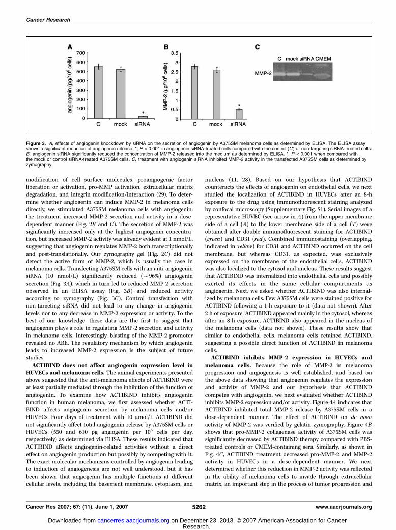
modification of cell surface molecules, proangiogenic factorliberation or activation, pro-MMP activation, extracellular matrixdegradation, and integrin modification/interaction (29). To deter-mine whether angiogenin can induce MMP-2 in melanoma cellsdirectly, we stimulated A375SM melanoma cells with angiogenin;the treatment increased MMP-2 secretion and activity in a dose-dependent manner (Fig. 2B and C). The secretion of MMP-2 wassignificantly increased only at the highest angiogenin concentra-tion, but increased MMP-2 activity was already evident at 1 nmol/L,suggesting that angiogenin regulates MMP-2 both transcriptionallyand post-translationally. Our zymography gel (Fig. 2C) did notdetect the active form of MMP-2, which is usually the case inmelanoma cells. Transfecting A375SM cells with an anti-angiogeninsiRNA (10 nmol/L) significantly reduced (f96%) angiogeninsecretion (Fig. 3A), which in turn led to reduced MMP-2 secretionobserved in an ELISA assay (Fig. 3B) and reduced activityaccording to zymography (Fig. 3C). Control transfection withnon-targeting siRNA did not lead to any change in angiogeninlevels nor to any decrease in MMP-2 expression or activity. To thebest of our knowledge, these data are the first to suggest thatangiogenin plays a role in regulating MMP-2 secretion and activityin melanoma cells. Interestingly, blasting of the MMP-2 promoterrevealed no ABE. The regulatory mechanism by which angiogeninleads to increased MMP-2 expression is the subject of futurestudies.ACTIBIND does not affect angiogenin expression level in
HUVECs and melanoma cells. The animal experiments presentedabove suggested that the anti-melanoma effects of ACTIBIND wereat least partially mediated through the inhibition of the function ofangiogenin. To examine how ACTIBIND inhibits angiogeninfunction in human melanoma, we first assessed whether ACTI-BIND affects angiogenin secretion by melanoma cells and/orHUVECs. Four days of treatment with 10 Amol/L ACTIBIND didnot significantly affect total angiogenin release by A375SM cells orHUVECs (550 and 610 pg angiogenin per 106 cells per day,respectively) as determined via ELISA. These results indicated thatACTIBIND affects angiogenin-related activities without a directeffect on angiogenin production but possibly by competing with it.The exact molecular mechanisms controlled by angiogenin leadingto induction of angiogenesis are not well understood, but it hasbeen shown that angiogenin has multiple functions at differentcellular levels, including the basement membrane, cytoplasm, and
nucleus (11, 28). Based on our hypothesis that ACTIBINDcounteracts the effects of angiogenin on endothelial cells, we nextstudied the localization of ACTIBIND in HUVECs after an 8-hexposure to the drug using immunofluorescent staining analyzedby confocal microscopy (Supplementary Fig. S1). Serial images of arepresentative HUVEC (see arrow in A) from the upper membraneside of a cell (A) to the lower membrane side of a cell (T) wereobtained after double immunofluorescent staining for ACTIBIND(green) and CD31 (red). Combined immunostaining (overlapping,indicated in yellow) for CD31 and ACTIBIND occurred on the cellmembrane, but whereas CD31, as expected, was exclusivelyexpressed on the membrane of the endothelial cells, ACTIBINDwas also localized to the cytosol and nucleus. These results suggestthat ACTIBIND was internalized into endothelial cells and possiblyexerted its effects in the same cellular compartments asangiogenin. Next, we asked whether ACTIBIND was also internal-ized by melanoma cells. Few A375SM cells were stained positive forACTIBIND following a 1-h exposure to it (data not shown). After2 h of exposure, ACTIBIND appeared mainly in the cytosol, whereasafter an 8-h exposure, ACTIBIND also appeared in the nucleus ofthe melanoma cells (data not shown). These results show thatsimilar to endothelial cells, melanoma cells retained ACTIBIND,suggesting a possible direct function of ACTIBIND in melanomacells.ACTIBIND inhibits MMP-2 expression in HUVECs and
melanoma cells. Because the role of MMP-2 in melanomaprogression and angiogenesis is well established, and based onthe above data showing that angiogenin regulates the expressionand activity of MMP-2 and our hypothesis that ACTIBINDcompetes with angiogenin, we next evaluated whether ACTIBINDinhibits MMP-2 expression and/or activity. Figure 4A indicates thatACTIBIND inhibited total MMP-2 release by A375SM cells in adose-dependent manner. The effect of ACTIBIND on de novoactivity of MMP-2 was verified by gelatin zymography. Figure 4Bshows that pro-MMP-2 collagenase activity of A375SM cells wassignificantly decreased by ACTIBIND therapy compared with PBS-treated controls or CMEM-containing sera. Similarly, as shown inFig. 4C , ACTIBIND treatment decreased pro-MMP-2 and MMP-2activity in HUVECs in a dose-dependent manner. We nextdetermined whether this reduction in MMP-2 activity was reflectedin the ability of melanoma cells to invade through extracellularmatrix, an important step in the process of tumor progression and
Figure 3. A, effects of angiogenin knockdown by siRNA on the secretion of angiogenin by A375SM melanoma cells as determined by ELISA. The ELISA assayshows a significant reduction of angiogenin release. *, P < 0.001 in angiogenin siRNA-treated cells compared with the control (C ) or non-targeting siRNA-treated cells.B, angiogenin siRNA significantly reduced the concentration of MMP-2 released into the medium as determined by ELISA. *, P < 0.001 when compared withthe mock or control siRNA-treated A375SM cells. C, treatment with angiogenin siRNA inhibited MMP-2 activity in the transfected A375SM cells as determined byzymography.
Cancer Research
Cancer Res 2007; 67: (11). June 1, 2007 5262 www.aacrjournals.org
Research. on December 23, 2013. © 2007 American Association for Cancercancerres.aacrjournals.org Downloaded from
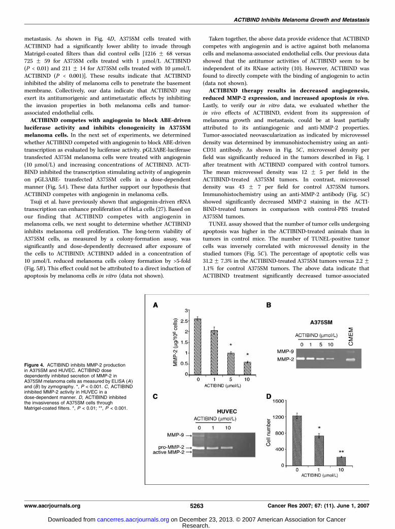
metastasis. As shown in Fig. 4D , A375SM cells treated withACTIBIND had a significantly lower ability to invade throughMatrigel-coated filters than did control cells [1216 F 68 versus725 F 59 for A375SM cells treated with 1 Amol/L ACTIBIND(P < 0.01) and 211 F 14 for A375SM cells treated with 10 Amol/LACTIBIND (P < 0.001)]. These results indicate that ACTIBINDinhibited the ability of melanoma cells to penetrate the basementmembrane. Collectively, our data indicate that ACTIBIND mayexert its antitumorigenic and antimetastatic effects by inhibitingthe invasion properties in both melanoma cells and tumor-associated endothelial cells.ACTIBIND competes with angiogenin to block ABE-driven
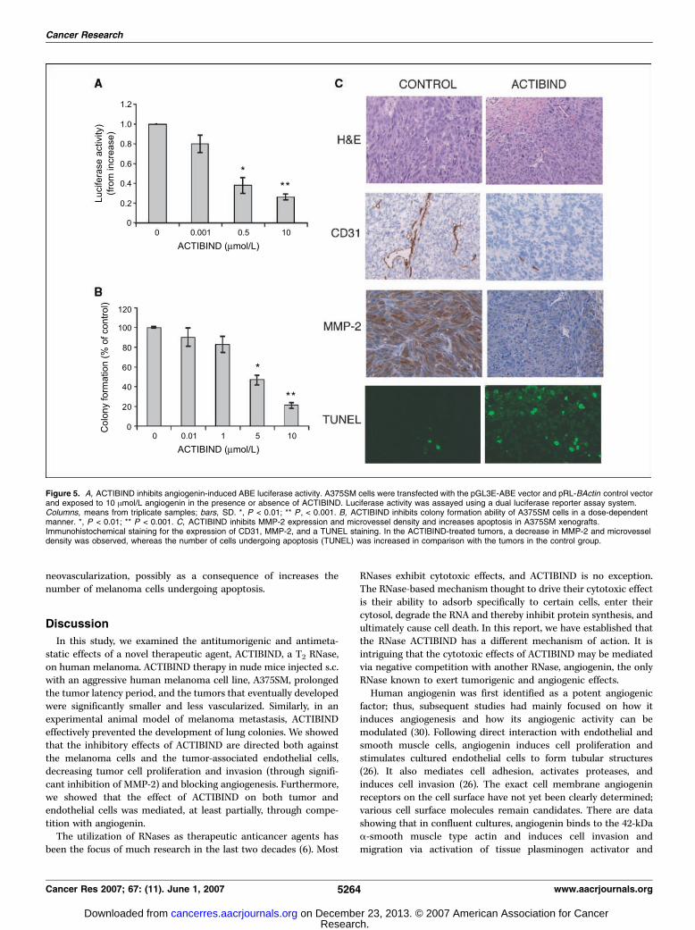
luciferase activity and inhibits clonogenicity in A375SMmelanoma cells. In the next set of experiments, we determinedwhether ACTIBIND competed with angiogenin to block ABE-driventranscription as evaluated by luciferase activity. pGL3ABE-luciferasetransfected A375M melanoma cells were treated with angiogenin(10 Amol/L) and increasing concentrations of ACTIBIND. ACTI-BIND inhibited the transcription stimulating activity of angiogeninon pGL3ABE- transfected A375SM cells in a dose-dependentmanner (Fig. 5A). These data further support our hypothesis thatACTIBIND competes with angiogenin in melanoma cells.Tsuji et al. have previously shown that angiogenin-driven rRNA
transcription can enhance proliferation of HeLa cells (27). Based onour finding that ACTIBIND competes with angiogenin inmelanoma cells, we next sought to determine whether ACTIBINDinhibits melanoma cell proliferation. The long-term viability ofA375SM cells, as measured by a colony-formation assay, wassignificantly and dose-dependently decreased after exposure ofthe cells to ACTIBIND; ACTIBIND added in a concentration of10 Amol/L reduced melanoma cells colony formation by >5-fold(Fig. 5B). This effect could not be attributed to a direct induction ofapoptosis by melanoma cells in vitro (data not shown).
Taken together, the above data provide evidence that ACTIBINDcompetes with angiogenin and is active against both melanomacells and melanoma-associated endothelial cells. Our previous datashowed that the antitumor activities of ACTIBIND seem to beindependent of its RNase activity (10). However, ACTIBIND wasfound to directly compete with the binding of angiogenin to actin(data not shown).ACTIBIND therapy results in decreased angiogenesis,
reduced MMP-2 expression, and increased apoptosis in vivo.Lastly, to verify our in vitro data, we evaluated whether thein vivo effects of ACTIBIND, evident from its suppression ofmelanoma growth and metastasis, could be at least partiallyattributed to its antiangiogenic and anti-MMP-2 properties.Tumor-associated neovascularization as indicated by microvesseldensity was determined by immunohistochemistry using an anti-CD31 antibody. As shown in Fig. 5C , microvessel density perfield was significantly reduced in the tumors described in Fig. 1after treatment with ACTIBIND compared with control tumors.The mean microvessel density was 12 F 5 per field in theACTIBIND-treated A375SM tumors. In contrast, microvesseldensity was 43 F 7 per field for control A375SM tumors.Immunohistochemistry using an anti-MMP-2 antibody (Fig. 5C)showed significantly decreased MMP-2 staining in the ACTI-BIND-treated tumors in comparison with control-PBS treatedA375SM tumors.TUNEL assay showed that the number of tumor cells undergoing
apoptosis was higher in the ACTIBIND-treated animals than intumors in control mice. The number of TUNEL-positive tumorcells was inversely correlated with microvessel density in thestudied tumors (Fig. 5C). The percentage of apoptotic cells was31.2 F 7.3% in the ACTIBIND-treated A375SM tumors versus 2.2 F1.1% for control A375SM tumors. The above data indicate thatACTIBIND treatment significantly decreased tumor-associated
Figure 4. ACTIBIND inhibits MMP-2 productionin A375SM and HUVEC. ACTIBIND dosedependently inhibited secretion of MMP-2 inA375SM melanoma cells as measured by ELISA (A)and (B) by zymography. *, P < 0.001. C, ACTIBINDinhibited MMP-2 activity in HUVEC in adose-dependent manner. D, ACTIBIND inhibitedthe invasiveness of A375SM cells throughMatrigel-coated filters. *, P < 0.01; **, P < 0.001.
ACTIBIND Inhibits Melanoma Growth and Metastasis
www.aacrjournals.org 5263 Cancer Res 2007; 67: (11). June 1, 2007
Research. on December 23, 2013. © 2007 American Association for Cancercancerres.aacrjournals.org Downloaded from
neovascularization, possibly as a consequence of increases thenumber of melanoma cells undergoing apoptosis.
Discussion
In this study, we examined the antitumorigenic and antimeta-static effects of a novel therapeutic agent, ACTIBIND, a T2 RNase,on human melanoma. ACTIBIND therapy in nude mice injected s.c.with an aggressive human melanoma cell line, A375SM, prolongedthe tumor latency period, and the tumors that eventually developedwere significantly smaller and less vascularized. Similarly, in anexperimental animal model of melanoma metastasis, ACTIBINDeffectively prevented the development of lung colonies. We showedthat the inhibitory effects of ACTIBIND are directed both againstthe melanoma cells and the tumor-associated endothelial cells,decreasing tumor cell proliferation and invasion (through signifi-cant inhibition of MMP-2) and blocking angiogenesis. Furthermore,we showed that the effect of ACTIBIND on both tumor andendothelial cells was mediated, at least partially, through compe-tition with angiogenin.The utilization of RNases as therapeutic anticancer agents has
been the focus of much research in the last two decades (6). Most
RNases exhibit cytotoxic effects, and ACTIBIND is no exception.The RNase-based mechanism thought to drive their cytotoxic effectis their ability to adsorb specifically to certain cells, enter theircytosol, degrade the RNA and thereby inhibit protein synthesis, andultimately cause cell death. In this report, we have established thatthe RNase ACTIBIND has a different mechanism of action. It isintriguing that the cytotoxic effects of ACTIBIND may be mediatedvia negative competition with another RNase, angiogenin, the onlyRNase known to exert tumorigenic and angiogenic effects.Human angiogenin was first identified as a potent angiogenic
factor; thus, subsequent studies had mainly focused on how itinduces angiogenesis and how its angiogenic activity can bemodulated (30). Following direct interaction with endothelial andsmooth muscle cells, angiogenin induces cell proliferation andstimulates cultured endothelial cells to form tubular structures(26). It also mediates cell adhesion, activates proteases, andinduces cell invasion (26). The exact cell membrane angiogeninreceptors on the cell surface have not yet been clearly determined;various cell surface molecules remain candidates. There are datashowing that in confluent cultures, angiogenin binds to the 42-kDaa-smooth muscle type actin and induces cell invasion andmigration via activation of tissue plasminogen activator and
Figure 5. A, ACTIBIND inhibits angiogenin-induced ABE luciferase activity. A375SM cells were transfected with the pGL3E-ABE vector and pRL-BActin control vectorand exposed to 10 Amol/L angiogenin in the presence or absence of ACTIBIND. Luciferase activity was assayed using a dual luciferase reporter assay system.Columns, means from triplicate samples; bars, SD. *, P < 0.01; ** P , < 0.001. B, ACTIBIND inhibits colony formation ability of A375SM cells in a dose-dependentmanner. *, P < 0.01; ** P < 0.001. C, ACTIBIND inhibits MMP-2 expression and microvessel density and increases apoptosis in A375SM xenografts.Immunohistochemical staining for the expression of CD31, MMP-2, and a TUNEL staining. In the ACTIBIND-treated tumors, a decrease in MMP-2 and microvesseldensity was observed, whereas the number of cells undergoing apoptosis (TUNEL) was increased in comparison with the tumors in the control group.
Cancer Research
Cancer Res 2007; 67: (11). June 1, 2007 5264 www.aacrjournals.org
Research. on December 23, 2013. © 2007 American Association for Cancercancerres.aacrjournals.org Downloaded from
plasmin (31). When cells are under sparse culture, angiogeninbinds to a 170-kDa putative receptor to induce phosphorylation ofmitogen-activated protein kinases (MAPK) and cell proliferation(27). Indeed, MAPK activation was reported to be an early event inmelanoma progression (32, 33). Another study found thatangiogenin might also bind and activate a member of the FGFreceptor family in endothelial cells (26). Following binding to theendothelial cell surface, angiogenin rapidly undergoes endocytosisand is translocated to the nucleus, where it stimulates the synthesisof rRNA (27, 28). This process involves receptor-mediatedendocytosis, microtubule- and lysosome-independent transportacross the cytoplasm, and nuclear localization sequence-assistednuclear import. rRNA transcription is the rate-limiting step inribosome biogenesis and is essential for protein translation and cellgrowth. Therefore, angiogenin-stimulated rRNA synthesis might bethe key process by which angiogenin induces endothelial cellproliferation and new blood vessel formation. Inhibition of nucleartranslocation of angiogenin (34) or mutagenesis at its nuclearlocalization sequence (35) abolishes its angiogenic activity.To better understand the importance of angiogenin in tumor
angiogenesis, several animal experiments have been carried out toblock the effects of human angiogenin using monoclonal anti-bodies (36). These antibodies blocked the growth of human HT-29colon cancer, lung adenocarcinoma, and fibrosarcoma transplantedinto nude mice. Unfortunately, tumor cells escaping the actionof antibodies grew into defined tumors and lost sensitivity to theantibodies. To overcome the known limitations of anticancerantibody therapy, numerous nucleoside and nucleotide compoundshave been tested as competitive inhibitors for angiogenin (37). Thusfar however, no high-affinity (subnanomolar) inhibitors have beenfound most probably because the full binding potential of thetargeted active site is not exploited. Our findings that ACTIBINDis a highly effective angiogenin competitive inhibitor are thusencouraging.Although the commonly held assumption is that angiogenin is a
tumor angiogenic protein, the results presented in our studyindicate that melanoma cell themselves can respond to angiogeninin an autocrine fashion. To the best of our knowledge, only oneother previous publication investigated a role for angiogenin incancer cells. Tsuji et al. reported that angiogenin nucleartranslocation and rRNA-induced transcription are not exclusiveto endothelial cells and also occur constitutively in HeLa cells (27).Down-regulating angiogenin in these cells resulted in inhibition ofrRNA transcription, ribosome biogenesis, cell proliferation, andtumorigenesis (27). Their results suggested that angiogeninderegulation of rRNA transcription may be an importantdeterminant in neoplastic transformation. Indeed, they found thatinhibiting angiogenin expression reduced tumorigenicity andreversed the malignant phenotype of HeLa cells. Similarly, weobserved that angiogenin induces ABE-driven transcription inhuman melanoma cells, and that inhibition of angiogenin leads toreduced melanoma cell clonogenicity. Interestingly, we also showedthat angiogenin regulates MMP-2 secretion and activity by
melanoma and endothelial cells. This finding is of specificimportance in melanoma as there is substantial evidence that ofall known MMPs, MMP-2 plays the most important role inmelanoma invasion, angiogenesis, and metastasis (29). Previously,it has been suggested that angiogenin regulates proteolytic activityin endothelial cells through a mechanism involving angiogeninbinding to cell surface actin and heparin sulfate proteoglycans,unconnected to its ribonucleolytic activity (31). The angiogenin-actin complex released from the cell surface may participate in theactivation of tissue plasminogen activator, which in turn can resultin the activation of MMPs. Our results expand these preliminaryfindings and suggest that angiogenin not only leads to activation ofMMP-2 but also induces its secretion as was shown by the increasein total MMP-2 in conditioned media from melanoma cells treatedwith angiogenin and the decrease of MMP-2 levels following siRNAknockdown of angiogenin. One possibility is that angiogenintranscriptionally regulates the expression of MMP-2; however,analysis of the MMP-2 promoter failed to identify a consensusangiogenin binding site. The mechanisms leading to MMP-2induction by angiogenin is currently investigated in our laboratory.Like other solid tumors, melanomas are not composed solely of
malignant cells but, additionally, include a diverse ensemble of hostcells that are recruited into the tumor microenvironment. It is likelythat therapies targeting both the melanoma cells and otherwisenormal tumor-associated cells would lead to improved outcomes.Therapies directed against the two different compartments of thetumor take advantage of the greater genetic stability in co-optedmicroenvironment cells than in the tumor cells, which makes themless likely to acquire resistance to the therapy. In addition to the roleangiogenin is thought to play in melanoma angiogenesis, wedetermined that angiogenin directly contributes to the malignantproperties of a melanoma cell. Our results suggest that ACTIBINDeffectively competes with angiogenin in both melanoma and endo-thelial cells. Thus, we hope that targeting angiogenin in melanomausing ACTIBIND will be superior to other therapies designed toinhibit either angiogenesis or cancer cell proliferation alone.In summary, in the present study, we show that the T2 RNase
isolated from the fungi A. niger, ACTIBIND, exerts significantantitumor and antimetastatic activities. We further show that thisT2 RNase significantly affects the clonogenicity of A375SM humanmelanoma cells and competes with angiogenin in both melanomacells and HUVECs, effects that can explain the high efficacy ofACTIBIND in vivo . ACTIBIND therapy for patients with metastaticmelanoma should be further investigated in the context of aclinical trial; if successful, it may offer new modality for melanomapatients with this devastating malignancy.
Acknowledgments
Received 1/17/2007; revised 3/6/2007; accepted 3/26/2007.Grant support: NIH grants CA76098 and P50CA093459 (M. Bar-Eli).The costs of publication of this article were defrayed in part by the payment of page
charges. This article must therefore be hereby marked advertisement in accordancewith 18 U.S.C. Section 1734 solely to indicate this fact.
References
1. Bevona C, Sober, AJ. Melanoma incidence trends.Dermatol Clin 2002;20:589–95.2. Sun W, Schuchter LM. Metastatic melanoma. CurrTreat Options Oncol 2001;3:193–202.
3. Atkins MB. Cytokine-based therapy and biochemo-therapy for advanced melanoma. Clin Cancer Res 2002;12:2353–8s.4. Kirkwood JM, Moschos S, Wang W. Strategies for thedevelopment of more effective adjuvant therapy ofmelanoma: current and future explorations of anti-
bodies, cytokines, vaccines, and combinations. ClinCancer Res 2006;12:2331–6s.5. Irie M. Structure-function relationships of acid ribo-nucleases: lysosomal, vacuolar, and periplasmicenzymes. Pharmacol Ther 1999;81:77–89.6. Schein CH. From housekeeper to microsurgeon: the
ACTIBIND Inhibits Melanoma Growth and Metastasis
www.aacrjournals.org 5265 Cancer Res 2007; 67: (11). June 1, 2007
Research. on December 23, 2013. © 2007 American Association for Cancercancerres.aacrjournals.org Downloaded from
Cancer Research
Cancer Res 2007; 67: (11). June 1, 2007 5266 www.aacrjournals.org
diagnostic and therapeutic potential of ribonucleases.Natl Biotechnol 1997;15:529–36.7. Costanzi J, Sidransky D, Navon A, Goldsweig H.Ribonucleases as a novel pro-apoptotic anticancerstrategy: review of the preclinical and clinical data forranpirnase. Cancer Invest 2005;23:643–50.8. Deshpande RA, Shankar V. Ribonucleases from T2family. Crit Rev Microbiol 2002;28:79–122.9. Roiz L, Ozeri U, Goren R, Shoseyov O. Characterizationof Aspergillus niger B-1 RNase and its inhibitory effecton pollen germination and pollen tube growth inselected tree fruit. J Am Soc Hort Sci 2000;125:9–14.10. Roiz L, Smirnoff P, Bar-Eli M, Schwartz B, ShoseyovO. Actibind: an actin-binding fungal T2-RNase withantiangiogenic and anticarcinogenic characteristics.Cancer 2005;106:2295–308.11. Adams SA, Subramanian V. The angiogenins: anemerging family of ribonuclease related proteins withdiverse cellular functions. Angiogenesis 1999;3:189–99.12. Shimoyama S, Gansauge F, Gansauge S, et al.Increased angiogenin expression in pancreatic canceris related to cancer aggressiveness. Cancer Res 1996;56:2703–6.13. Etoh T, Shibuta K, Barnard GF, Kitano S, Mori M.Angiogenin expression in human colorectal cancer: therole of focal macrophage infiltration. Clin Cancer Res2000;6:3545–51.14. Miyake H, Hara I, Yamanaka K, et al. Increasedangiogenin expression in the tumor tissue and serum ofurothelial carcinoma patients is related to diseaseprogression and recurrence. Cancer 1999;86:316–24.15. Eppenberger U, KuengW, Schlaeppi J-M, et al. Markersof tumor angiogenesis and proteolysis independentlydefine high- and low-risk subsets of node-negative breastcancer patients. J Clin Oncol 1998;16:3129–36.16. Chopra V, Dinh TV, Hannigan EV. Angiogenin,interleukins, and growth-factor levels in serum ofpatients with ovarian cancer: correlation with angio-genesis. Cancer J Sci Am 1996;2:279–85.
17. Shimoyama S, Kaminishi M. Angiogenin in sera as anindependent prognostic factor in gastric cancer. JCancer Res Clin Oncol 2003;129:239–44.18. Ugurel S, Rappl G, Tilgen W, Reinhold U. Increasedserum concentration of angiogenic factors in malignantmelanoma patients correlates with tumor progressionand survival. J Clin Oncol 2001;19:577–83.19. Hartmann A, Kunz M, Kostlin S, et al. Hypoxia-induced up-regulation of angiogenin in human malig-nant melanoma. Cancer Res 1999;59:1578–83.20. Gutman M, Singh RK, Xie K, Bucana CD, Fidler IJ.Regulation of interleukin-8 expression in human mela-noma cells by the organ environment. Cancer Res 1995;55:2470–5.21. Huang S, Jean D, Luca M, Tainsky MA, Bar-Eli M. Lossof AP-2 results in downregulation of c-KIT andenhancement of melanoma tumorigenicity and metas-tasis. EMBO J 1998;17:4358–69.22. Gershenwald JE, Sumner W, Calderone T, et al.Dominant-negative transcription factor AP-2 augmentsSB-2 melanoma tumor growth in vivo. Oncogene 2001;20:3363–75.23. McCarty MF, Baker CH, Bucana CD, Fidler IJ.Quantitative and qualitative in vivo angiogenesis assay.Int J Oncol 2002;21:5–10.24. Xu ZP, Tsuji T, Riordan JF, Hu GF. Identification andcharacterization of an angiogenin-binding DNA se-quence that stimulates luciferase reporter gene expres-sion. Biochem 2003;42:121–8.25. Kim DH, Behlke MA, Rose SD, et al. Synthetic dsRNADicer substrates enhance RNAi potency and efficacy.Natl Biotechnol 2005;23:222–6.26. Strydom DJ. The angiogenins. Cell Mol Life Sci 1998;54:811–24.27. Tsuji T, Sun Y, Kishimoto K, et al. Angiogenin istranslocated to the nucleus of HeLa cells and is involvedin ribosomal RNA transcription and cell proliferation.Cancer Res 2005;65:1352–60.28. Xu ZP, Tsuji T, Riordan JF, Hu GF. The nuclear
function of angiogenin in endothelial cells is related torRNA production. Biochem Biophys Res Commun 2002;294:287–92.29. Hofmann UB, Westphal JR, Van Muijen GN, Ruiter DJ.Matrix metalloproteinases in human melanoma. J InvestDermatol 2000;115:337–44.30. Fett JW, Strydom DJ, Lobb RR, et al. Isolation andcharacterization of angiogenin, an angiogenic proteinfrom human carcinoma cells. Biochemistry 1985;24:5480–6.31. Hu G, Riordan JF, Vallee BL. Angiogenin promotesinvasiveness of cultured endothelial cells by stimulationof cell-associated proteolytic activities. Proc Natl AcadSci U S A 1994;91:12096–100.32. Govindarajan B, Bai X, Cohen C, et al. Malignanttransformation of melanocytes to melanoma by con-stitutive activation of mitogen-activated protein kinasekinase (MAPKK) signaling. J Biol Chem 2003;278:9790–5.33. Cohen C, Zavala-Pompa A, Sequeira JH, et al.Mitogen-actived protein kinase activation is an earlyevent in melanoma progression. Clin Cancer Res 2002;8:3728–33.34. Moroianu J, Riordan JF. Nuclear translocation ofangiogenin in proliferating endothelial cells is essentialto its angiogenic activity. Proc Natl Acad Sci U S A 1994;91:1677–81.35. Holloway DE, Chavali GB, Hares MC, et al. Crystal-lographic studies on structural features that determinethe enzymatic specificity and potency of humanangiogenin: Thr44, Thr80, and residues 38-41. Biochem-istry 2004;43:1230–41.36. Olson KA, Fett JW, French TC, Key ME, Vallee BL.Angiogenin antagonists prevent tumor growth in vivo.Proc Natl Acad Sci U S A 1995;92:442–6.37. Jenkins JL, Shapiro R. Identification of small-molecule inhibitors of human angiogenin and charac-terization of their binding interactions guided bycomputational docking. Biochemistry 2003;42:6674–87.
Research. on December 23, 2013. © 2007 American Association for Cancercancerres.aacrjournals.org Downloaded from