The inversa type of recessive dystrophic epidermolysis bullosa is caused by specific arginine and...
32
Van den Akker et al. 1 The inversa type of recessive dystrophic epidermolysis bullosa is caused by specific 1 arginine and glycine substitutions in type VII collagen 2 3 Short title: COL7A1 missense mutations in RDEB-I 4 5 Peter C. van den Akker 1 , Jemima E. Mellerio 2,3 , Anna E. Martinez 3 , Lu Liu 4 , Rowdy Meijer 5 , 6 Patricia J.C. Dopping-Hepenstal 4 , Anthonie J. van Essen 1 , Hans Scheffer 5 , Robert M.W. 7 Hofstra 1 , John A. McGrath 6 , Marcel F. Jonkman 7 8 9 1 Department of Genetics, University Medical Center Groningen, University of Groningen, 10 Groningen, the Netherlands; 2 St John’s Institute of Dermatology, Guy’s and St Thomas’ 11 NHS Foundation Trust, London, UK; 3 Department of Dermatology, Great Ormond Street 12 Hospital NHS Trust, London, UK; 4 The Robin Eady National Diagnostic Epidermolysis 13 Bullosa Laboratory, GSTS Pathology, St John's Institute of Dermatology, St Thomas' 14 Hospital, London, UK; 5 Department of Human Genetics, Radboud University Nijmegen 15 Medical Centre, Nijmegen, the Netherlands; 6 St John's Institute of Dermatology, King's 16 College London (Guy's Campus) and Guy's and St Thomas' NHS Foundation Trust, London, 17 UK; 7 Department of Dermatology, University Medical Center Groningen, University of 18 Groningen, Groningen, the Netherlands 19 20 Corresponding author: Peter C. van den Akker, MD 21 Department of Genetics, University Medical Center Groningen 22 Hanzeplein 1, PO BOX 30.001, 9700 RB Groningen, the Netherlands 23 Tel. +31 50 361 7100, fax +31 50 361 7230, e-mail [email protected] 24 Word count: text 3837. 25 peer-00588661, version 1 - 26 Apr 2011 Author manuscript, published in "Journal of Medical Genetics 48, 3 (2010) 160" DOI : 10.1136/jmg.2010.082230
Transcript of The inversa type of recessive dystrophic epidermolysis bullosa is caused by specific arginine and...

Van den Akker et al.
1
The inversa type of recessive dystrophic epidermolysis bullosa is caused by specific 1
arginine and glycine substitutions in type VII collagen 2
3
Short title: COL7A1 missense mutations in RDEB-I 4
5
Peter C. van den Akker1, Jemima E. Mellerio2,3, Anna E. Martinez3, Lu Liu4, Rowdy Meijer5, 6
Patricia J.C. Dopping-Hepenstal4, Anthonie J. van Essen1, Hans Scheffer5, Robert M.W. 7
Hofstra1, John A. McGrath6, Marcel F. Jonkman7 8
9
1 Department of Genetics, University Medical Center Groningen, University of Groningen, 10
Groningen, the Netherlands; 2 St John’s Institute of Dermatology, Guy’s and St Thomas’ 11
NHS Foundation Trust, London, UK; 3 Department of Dermatology, Great Ormond Street 12
Hospital NHS Trust, London, UK; 4 The Robin Eady National Diagnostic Epidermolysis 13
Bullosa Laboratory, GSTS Pathology, St John's Institute of Dermatology, St Thomas' 14
Hospital, London, UK; 5 Department of Human Genetics, Radboud University Nijmegen 15
Medical Centre, Nijmegen, the Netherlands; 6 St John's Institute of Dermatology, King's 16
College London (Guy's Campus) and Guy's and St Thomas' NHS Foundation Trust, London, 17
UK; 7 Department of Dermatology, University Medical Center Groningen, University of 18
Groningen, Groningen, the Netherlands 19
20
Corresponding author: Peter C. van den Akker, MD 21
Department of Genetics, University Medical Center Groningen 22
Hanzeplein 1, PO BOX 30.001, 9700 RB Groningen, the Netherlands 23
Tel. +31 50 361 7100, fax +31 50 361 7230, e-mail [email protected] 24
Word count: text 3837.25
peer
-005
8866
1, v
ersi
on 1
- 26
Apr
201
1Author manuscript, published in "Journal of Medical Genetics 48, 3 (2010) 160"
DOI : 10.1136/jmg.2010.082230

Van den Akker et al.
2
ABSTRACT 1
2
Background The inversa type of recessive dystrophic epidermolysis bullosa (RDEB-I) is a 3
rare variant of dystrophic epidermolysis bullosa, characterized by blistering in the body 4
flexures, trunk, and mucosae. The cause of this specific distribution is unknown. So far, 20 5
COL7A1 genotypes have been described in RDEB-I and genotype-phenotype correlations 6
have not been studied extensively. The aim of the study was to gain more insight into the 7
pathophysiology of this intriguing RDEB-I phenotype. 8
Methods We identified 20 Dutch and British RDEB-I patients and full genotypes in 18 of 9
them. We reviewed the literature on RDEB-I genotypes and conducted an extensive 10
genotype-phenotype correlation study for RDEB-I. 11
Results All 20 patients had generalized blistering at birth and during early infancy. In most 12
patients, the age of transition from generalized to inversa distribution was before the age of 4 13
years. We noted a spectrum of disease severity ranging from the mildest ‘mucosal only’ 14
phenotype to the severest phenotype with limited acral involvement. The 29 genotypes of our 15
RDEB-I patients and those reported in the literature revealed that RDEB-I is associated with 16
specific recessive arginine and glycine substitutions in the triple-helix domain of type VII 17
collagen. 18
Discussion and conclusion Why these specific arginine and glycine substitutions cause the 19
inversa distribution remains unknown. We could not identify clear differences in location and 20
nature of substituting amino acids between these mutations and missense mutations causing 21
other RDEB phenotypes. We hypothesize that the higher skin temperature in the affected 22
areas plays an important role in the pathophysiology of RDEB-I. 23
24
peer
-005
8866
1, v
ersi
on 1
- 26
Apr
201
1

Van den Akker et al.
3
Keywords: recessive dystrophic epidermolysis bullosa, inversa type; genotype-phenotype 1
correlations; type VII collagen; COL7A1 2
3
peer
-005
8866
1, v
ersi
on 1
- 26
Apr
201
1

Van den Akker et al.
4
INTRODUCTION 1
2
The inversa type of recessive dystrophic epidermolysis bullosa (RDEB-I; OMIM #226600) is 3
one of the rare variants of dystrophic epidermolysis bullosa.[1] RDEB-I was first described by 4
Prof. T. Gedde-Dahl Jr in 1971 and is characterized by generalized blistering from birth and 5
during early infancy.[2] Later in infancy or childhood, the predilection sites for blistering 6
change and the body flexures, axis and mucous membranes become the predominant sites of 7
blistering.[2, 3] Neither the cause of the unusual distribution of blistering in RDEB-I, nor the 8
cause of the “transition” from a generalized to inversa distribution is known. The specific age 9
at which this transition in blistering predilection sites takes place is also not known, but it 10
probably varies between patients. As with the main subtypes of RDEB, there is a large 11
clinical variation between inversa patients. 12
Immunohistochemically, type VII collagen (Col7) is either slightly reduced or 13
normally present in the skin of RDEB-I patients. Anchoring fibrils, which are composed of 14
large numbers of Col7 dimers, can be either reduced or normal in number, and can have a 15
hypoplastic or normal ultrastructural appearance.[4-6] Transient retention of Col7 in basal 16
keratinocytes can be a typical immunohistochemical feature in the early stages.[7] 17
Similar to the other subtypes of DEB, RDEB-I is caused by mutations in the COL7A1 18
gene encoding type VII collagen.[1] However, while genotype-phenotype correlations for the 19
major subtypes of RDEB have been extensively studied in several populations,[1, 8-16] there 20
is little data on genotype-phenotype correlations for RDEB-I. Our study covers the largest 21
RDEB-I cohort reported to date, comprising 20 RDEB-I patients from the Netherlands and the 22
UK. Here we describe this cohort and the genotypes identified in 18 of them, and we discuss 23
the genotype-phenotype correlations identified in these patients and those previously reported 24
in the literature. 25
peer
-005
8866
1, v
ersi
on 1
- 26
Apr
201
1

Van den Akker et al.
5
1
2
PATIENTS AND METHODS 3
4
Patients 5
A diagnosis of DEB was established in a set of 20 patients on the basis of clinical findings, 6
family history, histopathology, immunofluorescence antigen mapping, electron microscopy, 7
and COL7A1 mutation analysis. The diagnosis of RDEB-I was based on the typical clinical 8
findings.[1, 3] All patients were evaluated by the same professionals in the University 9
Medical Center Groningen, the Netherlands (MFJ), Great Ormond Street Hospital for 10
Children or St. Thomas’ Hospital, London, UK (JEM, AEM, JAM). Informed consent was 11
given for all skin biopsies and venapunctures, and all studies were approved by the local 12
medical ethical committees (IRB) and conducted in accordance with the guidelines stated in 13
the Declaration of Helsinki. 14
15
Immunofluorescence and electron microscopy studies 16
For immunofluorescence studies (IF), 4 mm punch biopsies of rubbed perilesional skin and/or 17
unaffected skin from non-predilection sites (i.e. the ventral side of the upper arm) were 18
obtained from patients 1-6 and 16 and snap frozen. Four or five µm thick cryostat sections 19
were prepared and the amount of Col7 was determined using monoclonal antibody LH7:2 as 20
previously described.[14, 17, 18] For electron microscopy, 2 mm punch biopsies were taken 21
from the same locations from the same patients and prepared and analyzed as previously 22
described. 23
24
25
peer
-005
8866
1, v
ersi
on 1
- 26
Apr
201
1
Van den Akker et al.
6
COL7A1 mutation analysis 1
The COL7A1 gene was fully analyzed for pathological mutations either by complete 2
sequencing or by pre-screening using either Denaturing Gradient Gel Electrophoresis or 3
Conformation-sensitive Capillary Electrophoresis followed by targeted sequencing as 4
described previously.[14, 19] Genomic DNA was extracted from peripheral leukocytes. 5
6
7
RESULTS 8
9
Clinical characteristics 10
In total, we identified 20 patients with RDEB-I, six from the Netherlands (patients 1-6) and 11
14 from the UK (patients 7-20). Clinical characteristics are listed in supplemental Table S1 12
and Figure 1. Patients 1 and 2 are sister and brother; patient 14 reported two similarly affected 13
sibs, but they are not known in our clinics; no other patients had any siblings affected with 14
DEB. Eighteen patients (90%) presented with generalized blistering and skin erosions at birth 15
or shortly after and during early infancy; in one the onset of blistering was within the first six 16
months and in another it was unknown. Seven (35%) had congenital localized absence of skin 17
(CLAS), mostly on the lower legs/ankles/feet, which remained fragile during life. With age, 18
blistering on the extremities decreased markedly in most patients and became more restricted 19
to the body trunk and folds. The most frequently affected skin areas were the proximal body 20
flexures, i.e. the axillae and groins, and the body axis, i.e. the back, neck and buttocks 21
symmetrically. None of the patients had involvement of the acra as the dominant feature. In 22
fact, even after trauma most patients only occasionally experienced blistering of hands and 23
feet, and none of them developed ‘mitten deformities’ as seen in the ‘severe generalized’ 24
RDEB (RDEB-sev gen) subtype. However, four patients (3, 9, 19, 20) had more extensive 25
peer
-005
8866
1, v
ersi
on 1
- 26
Apr
201
1

Van den Akker et al.
7
blistering of the acra and developed mild pseudosyndactyly in one of the interdigital web 1
spaces. In contrast, in four patients (7, 13, 15, 16) skin blistering nearly ceased completely 2
after infancy and they only had mucosal involvement and nail dystrophy. 3
In 6/20 (30%) patients, lesions of the oral mucosa were already present at birth, 4
whereas in the other patients the oral mucosa became affected from infancy onwards (Table 5
S1). When the RDEB-I phenotype had established, all patients had severe mucosal 6
involvement, mostly affecting the oral cavity, oesophagus and anus. In 6/10 (60%) women the 7
genital skin and mucosa were affected, and in one this severely complicated healing after 8
childbirth. Swallowing difficulty or dysphagia was reported in 17/20 (85%) patients. Twelve 9
patients (60%) had proven oesophageal strictures and 10 (50%) needed repeated oesophageal 10
dilatations; patient 5 received more than 150 dilatations before the age of 30. Ankyloglossia 11
and microstomia due to scarring of the oral mucosa were present in 15 (75%) and 13 (65%) 12
patients, respectively, and was noted as early as 6 years of age (patient 15). Blistering of the 13
external auditory canal was seen in 7/20 (35%) patients, which was complicated by narrowing 14
of the canal in three, and subsequent conductive hearing loss after the age of 30 in two (10%). 15
None of the patients were growth retarded, had renal failure or developed squamous cell 16
carcinomas. Patients 10, 12, 15, and 20 had anaemic periods and patient 12 required 17
intermittent blood transfusions. Constipation was an almost invariable feature, although only 18
in part due to anal blistering. 19
20
Transition age 21
The age of transition of blistering from extremities to body axis and flexures was reported in 22
13/20 patients and ranged from one to 18 years. In nine patients, transition was reported 23
between birth and age four years. Patient 6 was the only one in whom the transition age could 24
be objectively determined. He was diagnosed with probable recessive bullous dermolysis of 25
peer
-005
8866
1, v
ersi
on 1
- 26
Apr
201
1

Van den Akker et al.
8
the newborn, since his generalized blisters present at birth disappeared within the first few 1
months, and the immunofluorescence biopsy revealed Col7 retention in basal keratinocytes. 2
However, towards the end of his first year, blistering activity increased on the trunk, axillae, 3
intraorally and especially in the diaper area. Hence, transition age was before the end of the 4
first year. 5
6
Immunofluorescence and electron microscopy findings 7
Immunofluorescence studies on healthy skin from non-predilection sites revealed normal 8
Col7 in patients 3, 4, 6, 16, and reduced Col7 in patient 1. Retention of Col7 was noted in 9
basal keratinocytes of patient 3 (aged 33) and patient 6, in whose skin both small and large 10
Col7 positive granules were seen at the age of 1 month. Basal keratinocytes of a peri-lesional 11
skin biopsy of patient 2, obtained a few days after birth, also revealed Col7 retention. Peri-12
lesional skin from patient 5 revealed reduced Col7. Electron microscopy studies revealed 13
slightly hypoplastic anchoring fibrils in normal (patient 4) or reduced numbers (patients 3, 6, 14
16) at non-predilection sites. Electron microscopy studies of affected skin revealed a reduced 15
number of abnormal or rudimentary anchoring fibrils in patients 1, 2, 3, 4, 5, and 16. Col7-16
containing vesicles were seen in the rough endoplasmic reticulum in patient 2. 17
18
Genotypes and genotype-phenotype correlations 19
Genotypes were available for 18 of our 20 RDEB-I patients. The genotypes of patients 1-5 20
have been reported previously.[14] Table 1 lists the 29 full genotypes associated with RDEB-I 21
identified in this study and the literature. 22
peer
-005
8866
1, v
ersi
on 1
- 26
Apr
201
1

Van den Akker et al.
9
Table 1. Genotypes of RDEB-inversa patients reported in this study and in the literature. 1 No. Mutation 11 Mutation 21 Patients Reference Nucleotide Amino acid Exon Consequence2 Domain3 Nucleotide4 Amino acid Exon5 Consequence2 Domain3 1 c.4039G>T p.Gly1347Trp 34 Missense THD c.8540insC p.Glu2848X 116 PTC NC2 Pat 16 This study 2 c.4945C>G p.Gly1649Arg 53 Missense THD NF6 Pat 13 This study 3 c.5282G>C p.Gly1761Ala 60 Missense THD c.5282G>C p.Gly1761Ala 60 Missense THD Pat 6 This study 4 c.5720G>A p.Gly1907Glu 68 Missense THD c.7805G>A p.Gly2602Glu 105 Missense THD Pat 3 [14] 5 c.5720_5721
GA>AT p.Gly1907Asp 68 Missense THD c.4018C>T p.Arg1340X 34 PTC THD 2x [26]
6 c.5720_5721GA>AT
p.Gly1907Asp 68 Missense THD c.5047C>T p.Arg1687X 54 PTC THD Pat 12, 17
This study
7 c.6022C>G p.Arg2008Gly 73 Missense THD c.6022C>G p.Arg2008Gly 73 Missense THD 2x [20, 21] 8 c.6187C>G p.Arg2063Gly 74 Missense THD c.706C>T p.Arg236X 6 PTC NC1 1x [8] 9 c.6205C>T p.Arg2069Cys 74 Missense THD c.425A>G p.Lys142Arg 3 Splice � PTC NC1 3x [20, 24] 10 c.6205C>T p.Arg2069Cys 74 Missense THD c.3551-3T>G IVS26 Splice � PTC NC1 Pat 5 [14] 11 c.6205C>T p.Arg2069Cys 74 Missense THD c.3894+1G>A IVS30 Splice � PTC THD 1x [16] 12 c.6205C>T p.Arg2069Cys 74 Missense THD c.3895-1G>A IVS30 Splice � PTC THD 1x [25] 13 c.6205C>T p.Arg2069Cys 74 Missense THD c.4018C>T p.Arg1340X 34 PTC THD 1x [16] 14 c.6205C>T p.Arg2069Cys 74 Missense THD c.4027C>T p.Arg1343X 34 PTC THD 1x [16] 15 c.6205C>T p.Arg2069Cys 74 Missense THD c.5662insG p.Pro1888AlafsX27 67 PTC THD Pat 11 This Study 16 c.6205C>T p.Arg2069Cys 74 Missense THD c.6205C>T p.Arg2069Cys 74 Missense THD 3x (pat
4, 8, 18) This study, [14]
17 c.6262G>A p.Gly2088Arg 75 Missense THD c.676C>T p.Arg226X 5 PTC NC1 1x [26] 18 c.6637G>A p.Gly2213Arg 83 Missense THD c.4918delG p.Gly1640ValfsX70 52 PTC THD Pat 9 This study 19 c.6815G>C p.Gly2272Ala 86 Missense THD c.1363G>T p.Gln455X 11 PTC NC1 Pat 15 This study 20 c.7415G>A p.Gly2472Asp 97 Missense THD c.1874delGT p.Ser625ArgfsX5 14 PTC NC1 1x [26] 21 c.7864C>T p.Arg2622Trp 105 Missense THD c.553C>T p.Arg185X 5 PTC NC1 1x [15] 22 c.7864C>T p.Arg2622Trp 105 Missense THD c.2482delCT p.Ser828CysfsX37 19 PTC NC1 1x [9] 23 c.7882C>T p.Arg2628Trp 106 Missense THD c.1332C>A p.Tyr444X 10 PTC NC1 1x [26] 24 c.8065G>A p.Gly2689Arg 109 Missense THD c.1732C>T p.Arg578X 13 PTC NC1 Pat 7 This study 25 c.8065G>C p.Gly2689Arg 109 Missense THD c.6527insC p.Gly2177TrpfsX113 80 PTC THD 1x [10] 26 c.8083G>A p.Gly2695Ser 109 Missense THD c.8083G>A p.Gly2695Ser 109 Missense THD Pat 1, 2 [12, 14, 22] 27 c.8156G>C p.Gly2719Ala 110 Missense THD c.2051-1G>C IVS15 Splice � PTC NC1 Pat 19 This study 28 c.8323G>A p.Gly2775Ser 112 Missense THD c.425A>G p.Lys142Arg 3 Splice � PTC NC1 2x (pat
14) This study, [23]
29 c.8323G>A p.Gly2775Ser 112 Missense THD c.8323G>A p.Gly2775Ser 112 Missense THD 1x [26] 1 In bold novel COL7A1 mutations. 2 2 PTC, premature termination codon; splice � PTC, splice site affecting mutation resulting in PTC. 3
peer
-005
8866
1, v
ersi
on 1
- 26
Apr
201
1

Van den Akker et al.
10
3 THD, triple-helix domain; NC1, non-collagenous 1 domain; NC2, non-collagenous 2 domain. 1 4 NF, not found; NA, not available. 2 5 IVS, intervening sequences (i.e. introns) 3 6 Only a single mutation could be identified despite complete sequencing of the other allele.4
peer
-005
8866
1, v
ersi
on 1
- 26
Apr
201
1

Van den Akker et al.
11
Six genotypes consist of homozygous or compound heterozygous missense mutations within 1
the triple-helix domain (THD) of Col7. Twenty-three genotypes are composed of THD 2
missense mutations combined with mutations generating a premature truncation codon (PTC) 3
or an unidentified second mutation (pat 13). Since PTC mutations are generally considered to 4
result in no functional protein, the functional genotype in these patients is determined by the 5
hemizygous missense mutation.[27] Hence, the functional genotype in RDEB-I is that of a 6
homozygous, compound heterozygous or hemizygous missense mutation within the THD. 7
To date, 19 missense mutations have been associated with RDEB-I, all of which are 8
either substitutions of an arginine (ASs, n = 5) or a glycine (GSs, n = 14) (Table 1, Fig. 2). 9
The 14 GSs are located at 13 different glycine codons. None of the carrier parents were 10
affected, indicating that the mutations are recessive. The question is whether these recessive 11
ASs or GSs are specific for RDEB-I or whether they can also cause other (R)DEB subtypes. 12
Of the five ASs, the p.Arg2063Gly, p.Arg2622Trp, and p.Arg2628Trp mutations have been 13
exclusively reported in RDEB-I, whereas the p.Arg2008Gly and p.Arg2069Cys mutations 14
were also found in non-inversa RDEB subtypes (Table S2). Nine of the 14 recessive GSs have 15
been exclusively reported in RDEB-I, whereas four GSs were also reported in non-inversa 16
RDEB subtypes and the p.Gly2213Arg mutation was reported as a dominant GS (Table S3). 17
Supplementary tables S2 and S3, and Fig. 2 show detailed information about the ASs and GSs. 18
To check whether ASs in general might cause the RDEB-I phenotype, we studied the 19
phenotypes of patients carrying other ASs. The THD of Col7 contains a total of 94 arginines 20
in Gly-X-Y repeats,[35] of which 12 have been found substituted in DEB (HGMD 21
professional database, www.hgmd.org, and unpublished data (HS), 26 August 2010). 22
Substitutions of 5/12 arginines (42%) are involved in RDEB-I, whereas substitutions of 9/12 23
arginines (75%) are involved in other DEB phenotypes. Hence, it seems unlikely that ASs are 24
in general the cause of RDEB-I. 25
peer
-005
8866
1, v
ersi
on 1
- 26
Apr
201
1

Van den Akker et al.
12
1
2
DISCUSSION 3
4
The cause of the typical ‘inverse’ distribution of blistering in RDEB-I is not well understood. 5
We identified 20 Dutch and UK RDEB-I patients and expanded the list of known RDEB-I 6
genotypes from the literature with nine new genotypes, which enabled us to conduct an 7
extensive genotype-phenotype correlation study in RDEB-I. Our results suggest that RDEB-I 8
is caused by a set of specific recessive arginine and glycine substitutions within the THD of 9
Col7. 10
11
Clinical characteristics 12
All 20 patients displayed the typical inversa distribution of skin blistering in the proximal 13
body flexures and the body axis. Mucosal involvement, especially of the oral mucosa, was 14
present in all patients. Nearly all patients started with generalized blistering and a third had 15
congenital localized absence of skin. The age of transition from generalized to inversa 16
distribution took place before the age of 4 years in most patients, but ranged from 1 to 18 17
years. Marked clinical variation was noted between patients and the clinical severity of 18
RDEB-I seems to cover a spectrum, as seen in other DEB phenotypes.[1] Four patients had 19
‘mucosal only’ RDEB-I in which the mucosal involvement dominated, including severe 20
oesophageal stenosis requiring multiple dilatations, but skin blistering was almost absent. A 21
similar phenotype has been described previously,[10, 23, 36] but was not considered to be 22
within the RDEB-I spectrum due to absence of typical blistering in the body flexures. We 23
think that patients with ‘mucosal only’ RDEB form the mildest end of the inversa spectrum 24
(with regard to the skin), because the severe mucosal involvement is not part of the milder 25
peer
-005
8866
1, v
ersi
on 1
- 26
Apr
201
1

Van den Akker et al.
13
forms of RDEB-O.[1, 14] On the other end of the RDEB-I spectrum, four patients had 1
significant involvement of the acra, leading to mild flexion contractures and limited 2
pseudosyndactyly. This end of the RDEB-I spectrum might be difficult to distinguish from 3
RDEB-O or even RDEB-sev gen.[5, 37] However, these RDEB-I patients were not growth 4
retarded and none had developed squamous cell carcinoma. 5
A diagnosis of RDEB-I has important implications for the patient and clinician, 6
especially when skin blistering is relatively mild. Nearly all patients have severe oral and 7
oesophageal blistering resulting in intra-oral scarring, microstomia, ankyloglossia, and 8
oesophageal strictures often requiring repeated dilatations. This can result in decreased food 9
intake, poor dental hygiene and oral infections, which warrant careful follow-up of growth 10
and weight and aggressive dental care. Peri-anal blistering commonly results in constipation 11
that should be asked about carefully and treated if present. In women, genital blistering and 12
scarring can interfere with sexual function, complicate vaginal delivery and recovery 13
afterwards, which may be a reason for elective caesarean section. Moreover, blistering of the 14
external auditory canal may lead to conductive hearing loss, requiring hearing aids after the 15
age of 30. Careful inspection and follow-up of the auditory canals is thus warranted and 16
audiologic evaluation may be indicated at later ages. 17
18
Genotype-phenotype correlations 19
The 29 functional genotypes of our 18 RDEB-I patients and those reported in the literature 20
consist of homozygous, compound heterozygous or hemizygous (i.e. in conjunction with a 21
PTC) recessive ASs or GSs within the THD of Col7. To date, five recessive ASs and 14 22
recessive GSs have been associated with RDEB-I. The collagenous THD domain is composed 23
of many consecutive Gly-X-Y triplets that form a triple-helix.[35] These Gly-X-Y repeat 24
sequences are separated into 20 collagenous subdomains (COL1-20) by 19 interruptions or 25
peer
-005
8866
1, v
ersi
on 1
- 26
Apr
201
1

Van den Akker et al.
14
imperfections (IM1-19) that provide flexibility to the protein. The largest imperfection, the 1
hinge (IM10), is composed of 39 amino acids and divides the THD into amino (COL1-10) 2
and carboxyl (COL11-20) regions. The glycine residues are located at the centre of the triple-3
helix and are essential for stable triple-helix formation, whereas the amino acids at the X and 4
Y positions are exposed on the helix surface and are less essential for triple-helix stability.[38, 5
39] Substitutions of glycine residues therefore have major disruptive effects on triple-helix 6
folding and/or stability, whereas substitutions of X and Y residues not necessarily exert 7
pathogenic effects. Therefore, pathogenicity of the GSs identified in RDEB-I was not 8
surprising. Pathogenicity of the RDEB-I ASs was also likely, as they are located at highly 9
conserved positions within Col7. 10
Two RDEB-I ASs and five GSs have also been found in patients with phenotypes 11
other than RDEB-I. This observation suggests that some arginine and glycine codons have the 12
potential to result in RDEB-I when mutated, but other as yet unidentified factors determine 13
the resultant clinical phenotype, which can be RDEB-I or sometimes other. The puzzling 14
question remains as to why some specific arginine and glycine codons possess this potential 15
to induce RDEB-I when mutated, since ASs and GSs in general cause phenotypes other than 16
RDEB-I.[28] We therefore looked more closely into the position of the arginine and glycine 17
codons in the THD and into the nature of the substituting amino acids. 18
19
The arginine substitutions 20
The five arginine codons are located in collagenous subdomains in the carboxyl region of the 21
THD, of which three in the first two subdomains after the hinge region (Fig. 2, Table S2). The 22
position of the arginine within the Gly-X-Y triplet does not differ from arginines involved in 23
other phenotypes: the five RDEB-I arginines reside in the Y position as do 7 of 9 arginines 24
involved in other DEB subtypes. 25
peer
-005
8866
1, v
ersi
on 1
- 26
Apr
201
1

Van den Akker et al.
15
Our findings suggest that for each of the five arginines there is only a specific 1
substitution that can lead to RDEB-I (Tables S2). The arginine at codon 2063 was found 2
substituted by glycine in RDEB-I, whereas substituted by tryptophan in other RDEB 3
phenotypes; the arginine at codon 2622 was found substituted by tryptophan in RDEB-I, 4
whereas substituted by glutamine in RDEB-HS. The p.Arg2008Gly mutation was found 5
homozygously in RDEB-I, whereas substitution of this arginine by cysteine or histidine was 6
reported in other RDEB subtypes. Of note, the p.Arg2008Gly mutation was also found 7
homozygously in a single RDEB-HS patient, emphasizing that the COL7A1 mutation is not 8
the only factor determining the inversa phenotype. On the other hand, the p.Arg2069Cys 9
mutation was not only found in 12 inversa patients, but also in four with RDEB-O. However, 10
it cannot be excluded that these patients will ultimately develop the RDEB-I phenotype, as it 11
can take years for the inversa phenotype to become evident. Therefore, the p.Arg2069Cys is 12
the most frequently observed RDEB-I mutation and can be considered specific for RDEB-I. 13
The specific substituting amino acids leading to RDEB-I, however, are different for 14
each arginine codon and from chemically different groups (Table S2). At codons 2008, 2063 15
and 2069, the large, hydrophilic arginine is substituted by the smaller, nonpolar residues, 16
glycine or cysteine, whereas at codons 2622 and 2628 the arginine is substituted by the large, 17
hydrophobic and aromatic tryptophan. The same substituting amino acids have been found to 18
replace other arginines in the THD in non-inversa phenotypes (Fig 2). This emphasizes that 19
there is not a specific type of amino acid that leads to RDEB-I when replacing an arginine, but 20
it is the combination of a specific substituting amino acid at a specific arginine codon position. 21
Interestingly, the arginine codons 2622 and 2628 in COL19 are located close to the cysteine 22
residue at position 2634. This is the only cysteine residue in the THD and might be involved 23
in disulfide binding with the non-collagenous domain of a pairing Col7 molecule.[35] 24
peer
-005
8866
1, v
ersi
on 1
- 26
Apr
201
1

Van den Akker et al.
16
Substituting either arginine 2622 or 2628 could therefore lead to instable Col7 dimers as a 1
result of local conformational changes. 2
3
The glycine substitutions 4
All 13 glycine codons associated with RDEB-I are located in collagenous subdomains of the 5
THD (Fig. 2, Table S3). Four of 13 (31%) glycine codons are located in subdomain COL19. 6
This number is surprisingly high, even though COL19 is the largest subdomain, containing 7
almost 16% of Gly-X-Y repeats. These four GSs, as well as p.Gly2775Ser in COL20 are 8
located close to the carboxyl end of the THD. Additionally, the p.Gly1347Trp mutation in the 9
COL2 subdomain is located near the amino end of the THD and glycine codon 1907 (COL10) 10
is located near the hinge region, the end of the amino region of the THD. It can thus be 11
speculated that GSs located at either end of the amino and carboxyl regions of the THD can 12
cause the relatively mild RDEB-I phenotype. This agrees with the finding that GSs near the 13
ends of the THD cause less severe triple-helix destabilization than central GSs and 14
concordantly lead to less severe disease.[23] Interestingly, the amino acids replacing the 15
glycines are either chemically very different and destabilizing, or chemically quite similar and 16
less destabilizing.[38] 17
Recently, Chiaverini et al. suggested that GSs located near the borders of collagenous 18
subdomains exert different, i.e. milder, pathogenic effects than GSs located in the centre of 19
collagenous subdomains and consequently result in RDEB-I.[26] However, the combined 20
genotypic data of our RDEB-I patients and those reported in the literature show that of the 13 21
glycine codons involved in RDEB-I only 6/13 (46%) are located in the first three or last three 22
Gly-X-Y triplets of their respective collagenous subdomain. The other 7/13 (54%) are located 23
in the central portion of the subdomain (Table S3). Hence, a GSs location near the border of a 24
peer
-005
8866
1, v
ersi
on 1
- 26
Apr
201
1

Van den Akker et al.
17
collagenous subdomain might predispose to RDEB-I, but it is certainly not the only factor, 1
since specific central GSs also have the potential to induce RDEB-I. 2
There were no differences in the location of the glycine codons or the nature of the 3
substituting amino acids between GSs causing the ‘mucosal only’ phenotype and the 4
phenotype with limited acral involvement (Tables 1 and S3). However, the four glycine 5
codons involved in RDEB-I with limited acral involvement have been reported in the RDEB-6
sev gen or DDEB phenotypes. In contrast, of the glycine codons involved in the RDEB-I 7
‘mucosal only’ phenotype only glycine codon 1347 was reported in other, milder RDEB 8
phenotypes.[10, 30, 32] 9
10
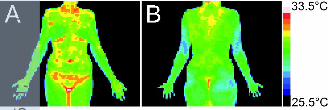
Putative temperature related pathophysiology in RDEB-I 11
Because the lesions in RDEB-I are mainly restricted to body locations where skin 12
temperatures are the highest (Fig. 3),[40, 41] we hypothesize that the pathophysiology in 13
RDEB-I is temperature-dependent. Kern et al. showed by in vitro experiments that Col7 14
homotrimers from a ‘mucosal only’ patient (genotype 18, Table 1) dissociated at lower 15
temperatures (Tm = 33-35°C) compared to normal Col7 (Tm = 39-41°C).[10] Moreover, 16
Fritsch et al. showed that Col7 trimers harbouring three different dominant GSs in the THD 17
had even more reduced thermostability (Tm 20-33°C, mono-expression).[39] Furthermore, 18
four GSs in the THD of type XVII collagen also reduced the thermostability of recombinant 19
proteins.[42] This reduced thermostability reflects folding defects of procollagen molecules 20
into Col7 trimers and/or packing defects of Col7 trimers into anchoring fibrils. The lower 21
dissociation temperatures of Col7 homotrimers harbouring the dominant GSs suggest that 22
dominant GSs have more dramatic destabilizing effects on Col7 than recessive GSs. Hence, 23
we speculate that the recessive GSs found in RDEB-I only slightly lower the temperature 24
dissociation thresholds for Col7 homotrimers. The only body regions where skin temperatures 25
peer
-005
8866
1, v
ersi
on 1
- 26
Apr
201
1

Van den Akker et al.
18
are continuously high enough to cross these temperature thresholds are the inversa regions. 1
This leads to increased Col7 destabilization with subsequent blistering only in these body 2
regions and not on the more distal body parts where skin temperatures are generally lower. 3
The invariable involvement of the oral mucosa and oesophageal involvement in most patients 4
supports this hypothesis. Temperature-dependent pathophysiology has been shown for 5
bathing suit ichthyosis (BSI), in which skin distribution is quite similar to RDEB-I.[41] 6
Moreover, also in BSI the skin features are more severe in infancy than later on. In BSI, 7
specific mutations in the transglutaminase-1 gene, TGM1, lead to decreased enzyme activity 8
at temperatures above 33°C,[43] explaining the ‘inversa-like’ skin distribution. Interestingly, 9
most BSI causing TGM1 mutations found to date are arginine substitutions.[44] The ASs 10
found in RDEB-I could thus exert the same effects as the GSs. A temperature-dependent 11
effect in skin disorders has also been noted for mutations in the TYR and CDKN2A genes.[45, 12
46] Temperature-dependent pathophysiology could also explain the transition from 13
generalized to inversa distribution, as one can assume that skin temperature in utero will be 14
higher than postnatally in all body regions. An obvious question is whether patients have 15
more severe blistering during warmer circumstances, but this was denied by all patients. 16
Hence, the temperature hypothesis is an interesting topic for future studies. 17
18
Conclusion and clinical implications 19
Our results suggest that RDEB-I is caused by specific recessive arginine and glycine 20
substitutions in the THD of Col7. The p.Arg2069Cys mutation is the most frequently 21
observed RDEB-I mutation. This study provides detailed insights into genotype-phenotype 22
correlations in RDEB-I, which is a first step towards understanding the pathophysiology of 23
the typical inverse distribution in this rare RDEB subtype. The exact mechanisms by which 24
peer
-005
8866
1, v
ersi
on 1
- 26
Apr
201
1

Van den Akker et al.
19
these ASs and GSs cause RDEB-I are not yet understood, but we hypothesize that the 1
pathophysiology of the inversa distribution is temperature-dependent. 2
These findings have important implications for the diagnosis and prognosis in DEB, 3
since the identification of one of the specific ASs or GSs as the only functional allele in a 4
newborn with DEB is predictive of the future development of RDEB-I. However, a phenotype 5
with more severe skin involvement may occasionally develop due to currently unknown 6
genetic, epigenetic or non-genetic modifiers. 7
8
9
ACKNOWLEDGEMENTS 10
11
We would like to thank the patients and their parents for their cooperation and Jackie Senior 12
for editing the manuscript. This study was supported by the Netherlands Organization for 13
Health Research and Development (ZonMw) grant 92003541, the Prof. J.P. Nater Foundation 14
and the Vlinderkind (Dutch Butterfly) Foundation. JEM and JAM acknowledge financial 15
support from the Department of Health via the National Institute for Health Research (NIHR) 16
comprehensive Biomedical Research Centre award to Guy’s and St Thomas’ NHS Foundation 17
Trust in partnership with King’s College London and King’s College Hospital NHS 18
Foundation Trust. 19
20
21
COMPETING INTERESTS 22
23
None. 24
25
peer
-005
8866
1, v
ersi
on 1
- 26
Apr
201
1

Van den Akker et al.
20
1
COPYRIGHT LICENSE STATEMENT / LICENCE FOR PUBLICATION 2
3
The Corresponding Author has the right to grant on behalf of all authors and does grant on 4
behalf of all authors, an exclusive licence on a worldwide basis to the BMJ Publishing Group 5
Ltd, and its Licensees to permit this article (if accepted) to be published in the Journal of 6
Medical Genetics and any other BMJPGL products and sublicenses such use and exploit all 7
subsidiary rights, as set out in our licence 8
(http://group.bmj.com/products/journals/instructions-for-authors/licence-forms). 9
10
11
PATIENT CONSENT 12
13
We obtained written consent for publication of all patient images.14
peer
-005
8866
1, v
ersi
on 1
- 26
Apr
201
1

Van den Akker et al.
21
REFERENCES 1
2
1 Fine JD, Eady RA, Bauer EA, Bauer JW, Bruckner-Tuderman L, Heagerty A, Hintner H, 3
Hovnanian A, Jonkman MF, Leigh I, McGrath JA, Mellerio JE, Murrell DF, Shimizu H, 4
Uitto J, Vahlquist A, Woodley D, Zambruno G. The classification of inherited 5
epidermolysis bullosa (EB): Report of the Third International Consensus Meeting on 6
Diagnosis and Classification of EB. J Am Acad Dermatol 2008;58:931-50. 7
2 Gedde-Dahl Jr T. Epidermolysis Bullosa: A clinical, genetic and epidemiological study. 8
Universitetsforlaget (Oslo), 1970. Baltimore: The Johns Hopkins Press; 1971. 9
3 Anton-Lamprecht I. Epidermolysis Bullosa. In: Rimoin DL, Connor JM, Pyeritz RE, Korf 10
BR, eds. Emery and Rimoin's Principles and Practice of Medical Genetics. Philadelphia: 11
Churchill Livingstone Elsevier 2007:3449-502. 12
4 Bruckner-Tuderman L, Niemi KM, Kero M, Schnyder UW, Reunala T. Type VII collagen 13
is expressed but anchoring fibrils are defective in dystrophic epidermolysis bullosa inversa. 14
Br J Dermatol 1990;122:383-90. 15
5 Bruckner-Tuderman L, Winberg JO, Anton-Lamprecht I, Schnyder UW, Gedde-Dahl Jr T. 16
Anchoring fibrils, collagen VII, and neutral metalloproteases in recessive dystrophic 17
epidermolysis bullosa inversa. J Invest Dermatol 1992;99:550-8. 18
6 Lin AN, Smith LT, Fine JD. Dystrophic epidermolysis bullosa inversa: report of two cases 19
with further correlation between electron microscopic and immunofluorescence studies. J 20
Am Acad Dermatol 1995;33:361-5. 21
7 Gedde-Dahl Jr T, Winberg JO. Neonatal retention of type VII collagen, transient bullous 22
dermolysis of the newborn and recessive epidermolysis bullosa dystrophica inversa. Br J 23
Dermatol 1994;130:685-6. 24
peer
-005
8866
1, v
ersi
on 1
- 26
Apr
201
1

Van den Akker et al.
22
8 Hovnanian A, Rochat A, Bodemer C, Petit E, Rivers CA, Prost C, Fraitag S, Christiano 1
AM, Uitto J, Lathrop M, Barrandon Y, de Prost Y. Characterization of 18 new mutations 2
in COL7A1 in recessive dystrophic epidermolysis bullosa provides evidence for distinct 3
molecular mechanisms underlying defective anchoring fibril formation. Am J Hum Genet 4
1997;61:599-610. 5
9 Gardella R, Castiglia D, Posteraro P, Bernardini S, Zoppi N, Paradisi M, Tadini G, Barlati 6
S, McGrath JA, Zambruno G, Colombi M. Genotype-phenotype correlation in Italian 7
patients with dystrophic epidermolysis bullosa. J Invest Dermatol 2002;119:1456-62. 8
10 Kern JS, Kohlhase J, Bruckner-Tuderman L, Has C. Expanding the COL7A1 mutation 9
database: novel and recurrent mutations and unusual genotype-phenotype constellations in 10
41 patients with dystrophic epidermolysis bullosa. J Invest Dermatol 2006;126:1006-12. 11
11 Dang N, Klingberg S, Marr P, Murrell DF. Review of collagen VII sequence variants 12
found in Australasian patients with dystrophic epidermolysis bullosa reveals nine novel 13
COL7A1 variants. J Dermatol Sci 2007;46:169-78. 14
12 Varki R, Sadowski S, Uitto J, Pfendner E. Epidermolysis bullosa. II. Type VII collagen 15
mutations and phenotype-genotype correlations in the dystrophic subtypes. J Med Genet 16
2007;44:181-92. 17
13 Kern JS, Gruninger G, Imsak R, Muller ML, Schumann H, Kiritsi D, Emmert S, Borozdin 18
W, Kohlhase J, Bruckner-Tuderman L, Has C. Forty-two novel COL7A1 mutations and 19
the role of a frequent single nucleotide polymorphism in the MMP1 promoter in 20
modulation of disease severity in a large European dystrophic epidermolysis bullosa 21
cohort. Br J Dermatol 2009;161:1089-97. 22
14 Van den Akker PC, van Essen AJ, Kraak MMJ, Meijer R, Nijenhuis M, Meijer G, Hofstra 23
RMW, Pas HH, Scheffer H, Jonkman MF. Long-term follow-up of patients with recessive 24
peer
-005
8866
1, v
ersi
on 1
- 26
Apr
201
1

Van den Akker et al.
23
dystrophic epidermolysis bullosa in the Netherlands: expansion of the mutation database 1
and unusual phenotype-genotype correlations. J Dermatol Sci 2009;56:9-18. 2
15 Escámez MJ, Garcia M, Cuadrado-Corrales N, Llames SG, Charlesworth A, De Luca N, 3
Illera N, Sanchez-Jimeno C, Holguin A, Duarte B, Trujillo-Tiebas MJ, Vicario JL, 4
Santiago JL, Hernandez-Martin A, Torrelo A, Castiglia D, Ayuso C, Larcher F, Jorcano 5
JL, Meana A, Meneguzzi G, Zambruno G, Del Rio M. The first COL7A1 mutation survey 6
in a large Spanish dystrophic epidermolysis bullosa cohort: c.6527insC disclosed as an 7
unusually recurrent mutation. Br J Dermatol 2010;163:155-61. 8
16 Jerabkova B, Kopeckova L, Buckova H, Vesely K, Valickova J, Fajkusova L. Analysis of 9
the COL7A1 gene in Czech patients with dystrophic epidermolysis bullosa reveals novel 10
and recurrent mutations. J Dermatol Sci 2010;59:136-40. 11
17 Jonkman MF, de Jong MC, Heeres K, Sonnenberg A. Expression of integrin alpha 6 beta 12
4 in junctional epidermolysis bullosa. J Invest Dermatol 1992;99:489-96. 13
18 Groves RW, Liu L, Dopping-Hepenstal PJ, Markus HS, Lovell PA, Ozoemena L, Lai-14
Cheong JE, Gawler J, Owaribe K, Hashimoto T, Mellerio JE, Mee JB, McGrath JA. A 15
homozygous nonsense mutation within the dystonin gene coding for the coiled-coil 16
domain of the epithelial isoform of BPAG1 underlies a new subtype of autosomal 17
recessive epidermolysis bullosa simplex. J Invest Dermatol 2010;130:1551-7. 18
19 Van den Akker PC, Hettema W, Meijer R, Jonkman MF, Hofstra RM, Scheffer H. Design 19
and validation of a conformation-sensitive capillary electrophoresis system for mutation 20
identification of the COL7A1 gene with automated peak comparison. Genet Test Mol 21
Biomarkers 2009;13:589-97. 22
20 Bruckner-Tuderman L. Hereditary skin diseases of anchoring fibrils. J Dermatol Sci 23
1999;20:122-33. 24
peer
-005
8866
1, v
ersi
on 1
- 26
Apr
201
1

Van den Akker et al.
24
21 Mecklenbeck S, Hammami-Hauasli N, Hopfner B, Schumann H, Kramer A, Kuster W, 1
Bruckner-Tuderman L. Clustering of COL7A1 mutations in exon 73: implications for 2
mutation analysis in dystrophic epidermolysis bullosa. J Invest Dermatol 1999;112:398-3
400. 4
22 Pulkkinen L, Uitto J. Mutation analysis and molecular genetics of epidermolysis bullosa. 5
Matrix Biol 1999;18:29-42. 6
23 Zimmer KP, Schumann H, Mecklenbeck S, Bruckner-Tuderman L. Esophageal stenosis in 7
childhood: dystrophic epidermolysis bullosa without skin blistering due to collagen VII 8
mutations. Gastroenterology 2002;122:220-5. 9
24 Kahofer P, Bruckner-Tuderman L, Metze D, Lemmink H, Scheffer H, Smolle J. 10
Dystrophic epidermolysis bullosa inversa with COL7A1 mutations and absence of GDA-11
J/F3 protein. Pediatr Dermatol 2003;20:243-8. 12
25 Wong T, Liu L, Ozoemena L, Wessagowit V, Fassihi H, Dopping-Hepenstal PJ, Jones C, 13
Mellerio JE, McGrath JA. Pathogenic nonglycine substitution missense mutations in the 14
type VII collagen triple helix: implications for genotype-phenotype correlation in 15
recessive dystrophic epidermolysis bullosa [abstract]. Br J Dermatol 2006;155 (suppl 16
1):P28. 17
26 Chiaverini C, Charlesworth AV, Youssef M, Cuny JF, Rabia SH, Lacour JP, Meneguzzi 18
G. Inversa Dystrophic Epidermolysis Bullosa Is Caused by Missense Mutations at 19
Specific Positions of the Collagenic Domain of Collagen Type VII. J Invest Dermatol. 20
Published Online First: 17 June 2010. doi:10.1038/jid.2010.159. 21
27 Christiano AM, Amano S, Eichenfield LF, Burgeson RE, Uitto J. Premature termination 22
codon mutations in the type VII collagen gene in recessive dystrophic epidermolysis 23
bullosa result in nonsense-mediated mRNA decay and absence of functional protein. J 24
Invest Dermatol 1997;109:390-4. 25
peer
-005
8866
1, v
ersi
on 1
- 26
Apr
201
1

Van den Akker et al.
25
28 Dang N, Murrell DF. Mutation analysis and characterization of COL7A1 mutations in 1
dystrophic epidermolysis bullosa. Exp Dermatol 2008;17:553-68. 2
29 Kon A, Pulkkinen L, Ishida-Yamamoto A, Hashimoto I, Uitto J. Novel COL7A1 3
mutations in dystrophic forms of epidermolysis bullosa. J Invest Dermatol 1998;111:534-4
7. 5
30 Terracina M, Posteraro P, Schubert M, Sonego G, Atzori F, Zambruno G, Bruckner-6
Tuderman L, Castiglia D. Compound heterozygosity for a recessive glycine substitution 7
and a splice site mutation in the COL7A1 gene causes an unusually mild form of localized 8
recessive dystrophic epidermolysis bullosa. J Invest Dermatol 1998;111:744-50. 9
31 Suzuki S, Shimomura Y, Yamamoto Y, Kariya N, Shibuya M, Ito M, Fujiwara H. A case 10
of recessive dystrophic epidermolysis bullosa caused by compound heterozygous 11
mutations in the COL7A1 gene. Br J Dermatol 2006;155:838-40. 12
32 Schumann H, Has C, Kohlhase J, Bruckner-Tuderman L. Dystrophic epidermolysis 13
bullosa pruriginosa is not associated with frequent FLG gene mutations. Br J Dermatol 14
2008;159:464-9. 15
33 Riedl E, Klausegger A, Bauer JW, Foedinger D, Kittler H. A novel glycine mutation in 16
the COL7A1 gene leading to dominant dystrophic epidermolysis bullosa with intra-17
familial phenotypical heterogeneity. Pediatr Dermatol 2009;26:115-7. 18
34 Almaani N, Liu L, Harrison N, Tanaka A, Lai-Cheong J, Mellerio JE, McGrath JA. New 19
glycine substitution mutations in type VII collagen underlying epidermolysis bullosa 20
pruriginosa but the phenotype is not explained by a common polymorphism in the matrix 21
metalloproteinase-1 gene promoter. Acta Derm Venereol 2009;89:6-11. 22
35 Christiano AM, Greenspan DS, Lee S, Uitto J. Cloning of human type VII collagen. 23
Complete primary sequence of the alpha 1(VII) chain and identification of intragenic 24
polymorphisms. J Biol Chem 1994;269:20256-62. 25
peer
-005
8866
1, v
ersi
on 1
- 26
Apr
201
1

Van den Akker et al.
26
36 Csikos M, Szocs HI, Laszik A, Mecklenbeck S, Horvath A, Karpati S, Bruckner-1
Tuderman L. High frequency of the 425A-->G splice-site mutation and novel mutations of 2
the COL7A1 gene in central Europe: significance for future mutation detection strategies 3
in dystrophic epidermolysis bullosa. Br J Dermatol 2005;152:879-86. 4
37 Gedde-Dahl Jr T. The childhood course of recessive epidermolysis bullosa dystrophica 5
inversa. In: Priestley GC, Tidman MJ, Weiss JB, Eady RAJ, eds. Epidermolysis bullosa: 6
A comprehensive review of classification, management and laboratory studies. 7
Crowthorne, Berkshire, UK: DEBRA, UK 1990:84-6. 8
38 Persikov AV, Pillitteri RJ, Amin P, Schwarze U, Byers PH, Brodsky B. Stability related 9
bias in residues replacing glycines within the collagen triple helix (Gly-Xaa-Yaa) in 10
inherited connective tissue disorders. Hum Mutat 2004;24:330-7. 11
39 Fritsch A, Spassov S, Elfert S, Schlosser A, Gache Y, Meneguzzi G, Bruckner-Tuderman 12
L. Dominant-negative effects of COL7A1 mutations can be rescued by controlled 13
overexpression of normal collagen VII. J Biol Chem 2009;284:30248-56. 14
40 Herry CL, Frize M. Quantitative assessment of pain-related thermal dysfunction through 15
clinical digital infrared thermal imaging. Biomed Eng Online 2004;3:19. 16
41 Oji V, Hautier JM, Ahvazi B, Hausser I, Aufenvenne K, Walker T, Seller N, Steijlen PM, 17
Kuster W, Hovnanian A, Hennies HC, Traupe H. Bathing suit ichthyosis is caused by 18
transglutaminase-1 deficiency: evidence for a temperature-sensitive phenotype. Hum Mol 19
Genet 2006;15:3083-97. 20
42 Vaisanen L, Has C, Franzke C, Hurskainen T, Tuomi ML, Bruckner-Tuderman L, 21
Tasanen K. Molecular mechanisms of junctional epidermolysis bullosa: Col 15 domain 22
mutations decrease the thermal stability of collagen XVII. J Invest Dermatol 23
2005;125:1112-8. 24
peer
-005
8866
1, v
ersi
on 1
- 26
Apr
201
1

Van den Akker et al.
27
43 Aufenvenne K, Oji V, Walker T, Becker-Pauly C, Hennies HC, Stocker W, Traupe H. 1
Transglutaminase-1 and bathing suit ichthyosis: molecular analysis of gene/environment 2
interactions. J Invest Dermatol 2009;129:2068-71. 3
44 Hackett BC, Fitzgerald D, Watson RM, Hol FA, Irvine AD. Genotype-phenotype 4
correlations with TGM1: clustering of mutations in the bathing suit ichthyosis and self-5
healing collodion baby variants of lamellar ichthyosis. Br J Dermatol 2010;162:448-51. 6
45 Berson JF, Frank DW, Calvo PA, Bieler BM, Marks MS. A common temperature-7
sensitive allelic form of human tyrosinase is retained in the endoplasmic reticulum at the 8
nonpermissive temperature. J Biol Chem 2000;275:12281-9. 9
46 Florell SR, Meyer LJ, Boucher KM, Porter-Gill PA, Hart M, Erickson J, Cannon-Albright 10
LA, Pershing LK, Harris RM, Samlowski WE, Zone JJ, Leachman SA. Longitudinal 11
assessment of the nevus phenotype in a melanoma kindred. J Invest Dermatol 12
2004;123:576-82.13
peer
-005
8866
1, v
ersi
on 1
- 26
Apr
201
1

Van den Akker et al.
28
FIGURE LEGENDS 1
2
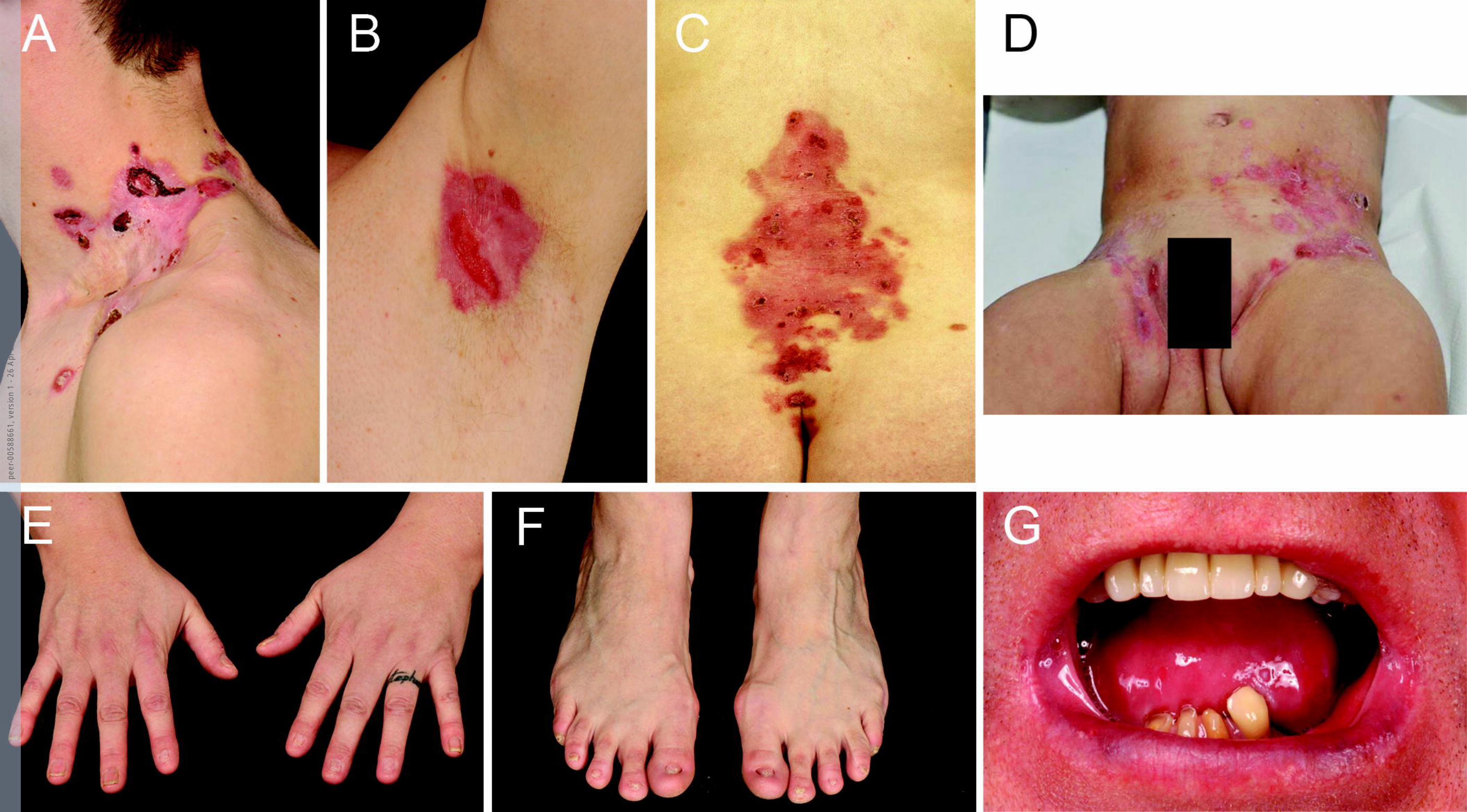
Figure 1. Clinical features of patients with RDEB-inversa. 3
Typical distribution of skin blistering in RDEB-I patients in the proximal body flexures and 4
body axis with nail dystrophy and involvement of the mucosal membranes. Skin blistering 5
and erosions in (A) the neck of patient 9, (B) the left axilla of patient 17, (C) the lower back in 6
patient 3, and (D) the groins and lower abdomen in patient 6. (E and F) The hands and feet of 7
patient 7 show absent and dystrophic nails, with no completely normal nails. There is no 8
current blistering, but small scars indicate past blistering. (G) Microstomia, ankyloglossia and 9
dental decay of the lower dentition (upper dentition has been repaired) in patient 17. Written 10
consent was obtained for publication of all images. 11
12
Figure 2. Arginine and glycine substitutions associated with RDEB-I. 13
Schematic representation of the triple-helix domain of type VII collagen. White boxes are 14
collagenous subdomains COL1-20, black bars represent collagenous imperfections IM1-19, 15
and the hinge region (IM10) is hatched. The glycine substitutions are shown above the triple-16
helix domain, and the arginine substitutions below. All known arginine and glycine 17
substitutions, as summarized by Dang and Murrell,[28] are shown. Glycine and arginine 18
substitutions associated with RDEB-I are depicted above and below the dashed lines, 19
respectively. Glycine and arginine substitutions exclusively reported in RDEB-I are shown in 20
bold. Novel glycine substitutions are shown in italics. 21
22
Figure 3. Digital thermographs of a healthy human body. 23
Photos obtained by digital infrared thermal imaging of (A) front and (B) back of a healthy 24
female human body showing higher body temperatures in the typical inversa body sites, i.e. 25
peer
-005
8866
1, v
ersi
on 1
- 26
Apr
201
1

Van den Akker et al.
29
the proximal flexures, neck, lower back and submammary folds, compared to more distal 1
body parts. The colour scales indicate the temperatures that correspond to the colours used in 2
the images. Images published with permission from Meditherm Inc, Hilversum, the 3
Netherlands. 4
peer
-005
8866
1, v
ersi
on 1
- 26
Apr
201
1
peer
-005
8866
1, v
ersi
on 1
- 26
Apr
201
1

peer
-005
8866
1, v
ersi
on 1
- 26
Apr
201
1
peer
-005
8866
1, v
ersi
on 1
- 26
Apr
201
1


![[Type the document title]](https://static.fdokumen.com/doc/165x107/6333508cb91d35198e0b9f67/type-the-document-title.jpg)