Parental Burden Associated with Borderline Personality Disorder in Female Offspring
Upload
independentCategory
view
3download
0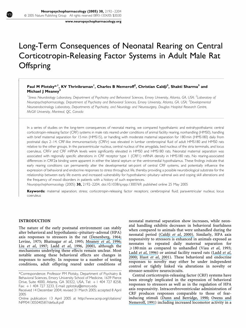
Long-Term Consequences of Neonatal Rearing on CentralCorticotropin-Releasing Factor Systems in Adult Male RatOffspring
Paul M Plotsky*,1, KV Thrivikraman1, Charles B Nemeroff2, Christian Caldji3, Shakti Sharma3 andMichael J Meaney3
1Stress Neurobiology Laboratory, Department of Psychiatry and Behavioral Sciences, Emory University, Atlanta, GA, USA; 2Laboratory of
Neuropsychopharmacology, Department of Psychiatry and Behavioral Sciences, Emory University, Atlanta, GA, USA; 3Developmental
Neuroendocrinology Laboratory, Departments of Psychiatry, and Neurology and Neurosurgery, Douglas Hospital Research Centre,
McGill University, Montreal, QC, Canada
In a series of studies on the long-term consequences of neonatal rearing, we compared hypothalamic and extrahypothalamic central
corticotropin-releasing factor (CRF) systems in male rats reared under conditions of animal facility rearing, nonhandling (HMS0), handling
with brief maternal separation for 15 min (HMS15), or handling with moderate maternal separation for 180 min (HMS180) daily from
postnatal days 2–14. CRF-like immunoreactivity (CRFir) was elevated in lumbar cerebrospinal fluid of adult HMS180 and HMS0 rats
relative to the other groups. In the paraventricular nucleus, central nucleus of the amygdala, bed nucleus of the stria terminalis, and locus
coeruleus, CRFir and CRF mRNA levels were significantly elevated in HMS0 and HMS180 rats. Neonatal maternal separation was
associated with regionally specific alterations in CRF receptor type 1 (CRF1) mRNA density in HMS180 rats. No rearing-associated
differences in CRF2a binding were apparent in either the lateral septum or the ventromedial hypothalamus. These findings indicate that
early rearing conditions can permanently alter the developmental set-point of central CRF systems, and potentially influence the
expression of behavioral and endocrine responses to stress throughout life, thereby providing a possible neurobiological substrate for the
relationship between early life events and increased vulnerability for hypothalamic–pituitary–adrenal axis and coping skill alterations and
the frequency of mood disorders in patients with a history of such experiences.
Neuropsychopharmacology (2005) 30, 2192–2204. doi:10.1038/sj.npp.1300769; published online 25 May 2005
Keywords: maternal separation; stress; corticotropin-releasing factor receptors; cerebrospinal fluid; paraventricular nucleus; locuscoeruleus
������������������������������������������������������
INTRODUCTION
The nature of the early postnatal environment can stablyalter behavioral and hypothalamic–pituitary–adrenal (HPA)axis responses to stressors in the rat (Denenberg, 1964;Levine, 1975; Bhatnagar et al, 1995; Meaney et al, 1996;Liu et al, 1997; Ladd et al, 1996, 2000), although themechanisms underlying these effects remain unclear. Mostnotable among these behavioral effects are changes inresponses to novelty. In response to a number of testingconditions, adult offspring reared under conditions of
neonatal maternal separation show increases, while neon-atal handling exhibits decreases in behavioral fearfulnesswhen compared to animals that were unhandled during theneonatal period (Caldji et al, 2000). Similarly, HPA axisresponsivity to stressors is enhanced in animals exposed asneonates to repeated daily maternal separation forX180 min as compared to unhandled (Viau et al, 1993;Ladd et al, 1996) or animal facility reared rats (Ladd et al,2000; Huot et al, 2001). These behavioral and endocrineresponses to novelty may either be under independentcontrol or tightly linked via alterations in novelty orstressor-sensitive neurocircuits.
Central corticotropin-releasing factor (CRF) systems havebeen strongly implicated in the expression of behavioralresponses to stressors as well as in the regulation of HPAaxis responsivity. Intracerebroventricular administration ofCRF produces responses comparable to those of fear-inducing stimuli (Dunn and Berridge, 1990; Owens andNemeroff, 1991) including increased locomotor activity in a
Online publication: 13 April 2005 at http://www.acnp.org/citations/NPP041305040587/default.pdf
Received 14 December 2004; revised 31 March 2005; accepted 8 April2005
*Correspondence: Professor PM Plotsky, Department of Psychiatry &Behavioral Sciences, Emory University School of Medicine, 1639 PierceDrive, Suite 4000, Atlanta, GA 30322, USA, Tel: þ 1 404 727 8258,Fax: þ 1 404 727 3233, E-mail: [email protected]
Neuropsychopharmacology (2005) 30, 2192–2204& 2005 Nature Publishing Group All rights reserved 0893-133X/05 $30.00
www.neuropsychopharmacology.org

familiar environment (reviewed in Koob et al, 1994),suppressed exploration in a novel environment (Brittonet al, 1982; Sutton et al, 1982; Berridge and Dunn, 1987;Takahashi et al, 1989), enhanced stress-induced freezing(Sherman and Kalin, 1988) and grooming (Britton et al,1984; Imaki et al, 1987), decreased sexual behavior(Sirinathsinghji et al, 1983), facilitation of acoustic startle(Swerdlow et al, 1986; Liang et al, 1992a, b), and decreasedfood intake (Krahn et al, 1986; Arase et al, 1988). Theseeffects are blocked by administration of the CRF receptorantagonist a-helical CRF (Dunn and Berridge, 1990;Menzaghi et al, 1993). In addition, CRF administrationhas aversive properties, producing both taste and placeaversions (Cador et al, 1992; Heinrichs et al, 1992a, 1993).Koob (1999) has proposed the existence of a feed-forwardsystem between norepinephrine (NE) and CRF systems tocoordinate the response to environmental challenges.Considerable pharmacologic, physiologic, and neuroanato-mical evidence supports a CRF–NE interaction in the regionof the locus coeruleus (Koob, 1999; Lejeune and Millan,2003; Page and Valentino, 1994; Plotsky et al, 1989;Valentino et al, 1998).
Further evidence for the importance of the central CRFsystem is derived from studies of a transgenic CRFoverexpression mouse model (Stenzel-Poore et al, 1994).These animals exhibit enhanced stress-induced suppressionof exploration that can be reversed with intracerebroven-tricular administration of the CRF receptor antagonist a-helical CRF9–41. These findings are consistent with the thesisthat CRF overproduction contributes to the etiology and/orpathophysiology of mood disorders (Chrousos and Gold,1992; Arborelius et al, 1999).
The effects of CRF on the expression of behavioral,endocrine, and autonomic responses to stress appear to beattributable to two major CRF systems: (a) the hypotha-lamic paraventricular nucleus (PVN) CRF neuronal system,which projects to the median eminence thus regulatingadenohypophysial adrenocorticotropin (ACTH) secretion aswell as to midbrain regions regulating sympathetic activity,and (b) CRF neurons in the central nucleus of the amygdala(CeA) and the bed nucleus of the stria terminalis (BNST),regions projecting to the PVN as well as to midbrain regionsrich in noradrenergic cell bodies (Brown, 1989; Plotsky,1991; Gray, 1993; Koob, 1999; Van Bockstaele et al, 2001).
Hofer (1994) has offered the concept that the mother orprimary caregiver acts as a hidden regulator of thedevelopment of infant physiological regulatory systems.Thus, a further impetus for this research is the growingevidence for a link between early untoward life experiences,such as neglect or abuse, and increased vulnerability to HPAaxis alterations and psychiatric diseases such as depression(Heim and Nemeroff, 2001; Heim et al, 2002, 2004).Accumulating data demonstrate increased neural CRFexpression (Raadsheer et al, 1994, 1995) and hypersecretion(Banki et al, 1987; De Bellis et al, 1993; Geracioti et al, 1992;Nemeroff et al, 1991) in some, but not all, patients withmajor depressive disorder. Many of these patients have ahistory of exposure to untoward life events occurring priorto their teenage years and often continuing into adulthood(Brown and Anderson, 1993; Mullen et al, 1996; McCauleyet al, 1997; Heim et al, 2000; Kendler et al, 2002). This raisesthe question of whether early life trauma, rather than
depression per se, is associated with dysregulation of centralCRF systems, which then results in secondary depression. Inlight of the pervasive effects of CRF on the organization ofemotion and responses to stressors (Owens and Nemeroff,1991; Koob et al, 1994), we have examined the status ofhypothalamic and extrahypothalamic CRF neurocircuits inadult rats exposed as neonates to different rearing protocolsassociated with alterations in HPA axis function andbehavioral responsivity.
MATERIALS AND METHODS
Animals
Timed-pregnant Long Evans hooded rats were delivered ongestation day 12 to Emory University from Charles River(Crl:(LE)BR; Charles River Laboratories, Portage, MI).Studies at McGill University were performed with ratsobtained from an ‘in-house’ breeding colony derived fromCharles River Laboratory Long Evans rats. All dams werehoused in transparent, polypropylene cages (20� 25 cm)with 2 cm bedding material and ad libitum access to foodand water in a room with a 12 : 12 h light : dark cycle (lightson at 0700). The day of birth was considered postnatal day(PND) 0. Litters were standardized to 8–10 male pups perdam on PND2 and were exposed to one of the followingrearing conditions from PND2 to PND14 inclusive: (a)animal facility rearing (AFR) consisted of brief handling fortwice weekly cage changes beginning on PND4 with noother handling or separation, (b) nonhandled (HMS0)animals were not exposed to any handling or experimentermanipulation, including cage bedding changes, from PND2to PND14, (c) handling with brief maternal separation for15 min (HMS15) from PND2 to PND14, and (d) handlingwith moderate maternal separation for 180 min (HMS180)from PND2 to PND14. The HMS protocol was initiated inthe morning between 0800 and 0900 by a gloved investi-gator. Each dam was removed from the home cage to anadjacent cage, then each litter was removed and placed as agroup into a plastic cage (15� 15 cm) that was transferredto an incubator in an adjacent room. These pup cages werelined with 3 cm of bedding material (wood shavings) andwere placed into an incubator set to maintain an ambienttemperature of 32þ 0.51C (PND2–5) or 30þ 0.51C (PND6–14). At the conclusion of the separation period, pups werereturned to the maternity cage, rolled in the soiled homecage bedding material, and the dam was returned. In thecourse of normal mother–pup interactions in the laboratoryrat, the mother is routinely off the litter for periods of20–25 min (Jans and Woodside, 1990). Thus, the maternalseparation protocol (HMS180), unlike handling and briefmaternal separation (HMS15), represents deprivation ofmaternal care. Unless otherwise noted, the bedding materialin all of the cages (eg maternity, transfer, pup isolation) wasnot changed from PND0 to at least PND15. According toChampagne et al (2003), gender composition of the litterdoes not significantly influence the frequency of maternallicking, grooming, or arched-back nursing and, thus, haslittle effect with respect to outcome measures of HPAdevelopment or CNS expression levels of stress-relatedgenes. This is not to deny that specific aspects of maternalbehavior toward male and female pups differ (Moore and
Early experience and central CRF systemsPM Plotsky et al
2193
Neuropsychopharmacology
Morelli, 1979; Pryce et al, 2001). Rat pups were weaned onPND21–22 and housed in groups of 2–4 same rearing groupanimals per cage until testing. Food and water was availablead libitum. All experiments were carried out in accordancewith the National Institutes of Health Guide for the Care andUse of Laboratory Animals.
Blood Sampling and Airpuff Startle
Adults were tested for HPA axis responsiveness usingairpuff startle on PND60–75. Jugular cannulae wereimplanted as previously described to allow for repeatedsampling in freely moving animals (Thrivikraman et al,2000), and a 4-day recovery period, during which time therats were handled daily, was permitted before testing.
The blood sampling protocol began between 0900 and1000 with airpuff startle initiated after obtaining a basalsample. Airpuff startle consisted of three blocks of threeairpuffs each (3 s) from a compressed air source (Dust-Off;Fisher Scientific, Atlanta, GA) that was directed toward theside of the head from a distance of approximately 10 cm,with 1 min between each block (Engelmann et al, 1996).Blood (300 ml/sample) was collected through the jugularcannula into 1.5 ml polyethylene tubes containing 10 mlEDTA (100 mg/ml) at rest and at 5, 10, 15, 30, 45, and 60 minfollowing airpuff startle. Blood samples were stored on ice,then centrifuged (12 500 g; 41C; 12 min), plasma collectedand frozen.
ACTH and Corticosterone Radioimmunoassay Analysis
Plasma ACTH and corticosterone (CORT) were assayed aspreviously described (Thrivikraman and Plotsky, 1993). ForACTH, 100 ml plasma samples were used for the AllegrotHS-ACTH kit (Nichols Institute, San Juan Capistrano, CA)with 50 ml of iodonated tracer and one avidin-coated beadper tube. The ACTH assay had a sensitivity of 2 pg/ml andintra- and intercoefficients of variation of less than 10%.CORT was determined in 5 ml plasma samples using theImmunoChemt Double Antibody kit (ICN Biomedicals,Costa Mesa, CA) with half of the trace and antibody volume.The CORT assay had a sensitivity of 1 ng/ml and intra- andinter coefficients of variation of less than 10%.
Lumbar Cerebrospinal Fluid Sampling
Intrathecal catheters were constructed of 32 ga polyurethanetubing (Micor, Allison Park, PA) of approximately 10.7 cmlength as described by Sakura et al (1996). A 5 mm longstainless-steel tubing segment (27 ga) was fitted to a heat-flared segment of the polyurethane tubing. This assemblywas cold sterilized and then a sterilized Teflon-coatedstainless-steel wire stylet (0.003 in diameter) was insertedinto the polyurethane catheter prior to placement.
The catheter was placed according to a modification ofthe technique described by Yaksh and Rudy (1976). Rats(n¼ 10 per rearing group) drawn from the same litters asanimals in the other experiments were anesthetized asdescribed above, then the back of the head and neck shaved,and disinfected with alcohol (70%). The rat was then placedin a stereotaxic headholder with the head flexed forward. Amidline incision was made on the back of the neck. The
muscle was freed from the skull and retracted to reveal thecisternal membrane. This membrane was opened with amicroknife and retracted with a dural microhook. Thesterile catheter was inserted through this defect and gentlypassed caudally approximately 9 cm into the intrathecalspace so that the tip rested in the Th11–L5 region. The duraldefect was repaired with tissue glue (Vet-Bond; HenrySchein, Port Washington, NY) and a small piece of Gel-Foam. The cannula was anchored to the skull using jeweler’sscrews and cranioplastic cement and the skin incision wasclosed with stainless-steel wound clips. A recovery period of5 days was permitted prior to sampling. Each rat washandled on a daily basis.
For sampling, the catheter was attached to a silasticextension line connected to a microsyringe pump (CMASystems, Stockholm, Sweden). Cerebrospinal fluid (CSF)was withdrawn at the rate of 500 nl/min for 4 h into areplaceable loop of Teflon tubing immersed into an ice-bath. Upon completion of each collection, the CSF wasexpelled into a cold 500 nl centrifuge tube containing 1mlEDTA (100 mg/ml) and frozen at �801C until assay.
Tissue Collection
Adult animals were killed by decapitation between 0900 and1200 under low stress conditions when they were betweenPND100 and PND120. The skull was quickly opened, thebrain removed, then rinsed and chilled in sterile saline onice. Each brain was blocked (B300 mm) in a coronal brainmatrix (Bioanalytical Systems, West Lafayette, IN). Thetissue slabs were placed on a glass plate on ice, thenindividual regions were obtained using 1 or 2 mm diameterstainless-steel punches according to the procedure ofPalkovits and Brownstein (1988). Each punch was snap-frozen on dry ice and stored in 1.5 ml polyethylenemicrocentrifuge tube at �801C until assay. Tissues fromfive to eight rats per group were collected for analysis.
CRF Radioimmunoassay
Tissue CRF-like immunoreactivity (CRFir) levels weredetermined using a modified version of the radioimmu-noassay described by Plotsky et al (1985). Tissue puncheshomogenized by sonication in a solution composed 0.1 NHCl containing 0.001% b-mercaptoethanol, 0.05% ascorbicacid, 0.01% bacitran, and 0.001% phenylmethylsulfonylfluoride. Samples were centrifuged at 12 000 g for 30 minat 41C. The supernatant was removed, lyophilized, andfrozen until assay. Samples were reconstituted inpH 7.0 phosphate buffer containing 0.1% bovine serumalbumin (BSA) fraction-V, 0.1% Triton-X, and 0.01%sodium azide.
Antiserum rC70 (provided by W Vale, The Salk Institute,La Jolla, CA) was used at a final dilution of 1 : 500 000.Synthetic rat CRF1–41 (provided by J Rivier, The SalkInstitute, La Jolla, CA) was used for assay standard and[125I]Tyr-ovine CRF (DuPont-NEN, Boston, MA) wasused for tracer. The limit of detection was 2 pg/tube withan IC50 of 22 pg. The intra-assay coefficient of variationwas 3.6%. Note that all tissue samples were run in the sameassay.
Early experience and central CRF systemsPM Plotsky et al
2194
Neuropsychopharmacology
In Situ Hybridization
Rats (PND100–120) were deeply anesthetized (0800–1200),decapitated, and brains removed and stored at �801C.Serial coronal brain sections (15 mm) were cut on a cryostatat �181C and thaw mounted onto SuperFrost Plus slides(Fisher Scientific, Atlanta, GA) under RNase-free conditionsand stored with Humi-Cap desiccant capsules (Gibco BRLProducts, Grand Island, NY) at �801C until assay. In situhybridization was performed according to the proceduresdescribed by Simmons et al (1989). Briefly, slides werewarmed in a stepwise manner to room temperature,postfixed in 4% paraformaldehyde (pH 7.4) for 20 min,and rinsed twice in 10 mM phosphate-buffered saline (PBS,pH 7.4) for 2 min. Next, the slides were treated for 15 minwith proteinase K (Promega, Madison, WI) 10 mg/ml in0.1 M Tris with 50 mM EDTA followed by acetylation(10 min, 0.5% acetic anhydride in 0.1 M triethanolamine,pH 8.0), two rinses in 2� SSC, and dehydrations through agraded ethanol series. The sections were then air-dried forat least 1 h prior to hybridization.
The CRF riboprobe was constructed from a 1.2 kb EcoRIfragment of a full-length rat CRF cDNA (provided by KMayo, Northwestern University, Evanston, IL) subclonedinto a pBluescript II-SKþ plasmid. The CRF1 riboprobewas constructed from a 1.3 kb PstI–PstI fragment containingthe full-length coding region for the rat CRF1 receptor(provided by W Vale, The Salk Institute, La Jolla, CA),ligated into a pBluescript II-SKþ plasmid. The CRF2ariboprobe was constructed from a 275 bp fragment, encod-ing the N-terminal region of the CRF2a receptor, ligatedinto a pBluescript II-SKþ plasmid (provided by W Vale,The Salk Institute, La Jolla, CA). Radiolabeled antisensecRNAs were synthesized by incorporating either [35S]UTP(DuPont-NEN) into the CRF probe or [35S]CTP plus[35S]UTP into the CRF1 and CRF2 receptor probes. Thetranscription reactions were performed utilizing the Am-bion MAXIscript kit (Austin, TX) with SP6 (CRF), T7(CRF1), or T3 (CRF2) RNA polymerases according to theinstructions provided. Following transcription and removalof the cDNA template with 2 U DNase (Ambion), the cRNAprobes were recovered through ethanol precipitation and/orgel filtration using a G-50 Sephadex Quick Spin column(Boehringer-Mannheim, Indianapolis, IN).
The brain sections were hybridized overnight at 58–601Cwith 2� 106 cpm of 35S-labeled cRNA probe diluted intohybridization buffer (50% formamide, 10% dextran sulfate,0.3 M NaCl, 1� Denhardt’s solution, 10 mM Tris, 1 mMEDTA, 2 mg/ml yeast tRNA, 10 mM DTT) in humidifiedNunc trays (Nalge Nunc Inc., Naperville, IL). The next day,slides were allowed to cool to room temperature beforebeing washed four times in 4� SSC for 5 min on a rotatingplatform at 60 rpm. The sections were then treated with250 mg/ml RNase A (Promega, Madison, WI) for 30 min at371C. Subsequently, the slides underwent a series of SSCwashes (supplemented with 1 mM DTT) with salt concen-trations decreasing from 2� to 0.5� , followed by a 60 minhigh-stringency wash with 0.1� SSCþ 1 mM DTT at 601C,then dehydration through a graded ethanol series. Theslides were air-dried for at least 1 h and then apposed toKodak Biomax MR film (Rochester, NY) for 12 h to 12 daysdepending on the probe and brain region examined with 14C
standards (Amersham, Piscataway, NJ). Controls wereperformed to establish specificity of the signal includedhybridization with sense strand probes, as well as prediges-tion with RNase A.
CRF Receptor Autoradiography
Following a modification of the techniques of De Souza et al(1985), De Souza (1987), and Primus et al (1997), in vitroCRF receptor autoradiography was performed on 15 mm ratbrain sections mounted on SuperFrost Plus slides. Sectionswere fixed for 2 min in 0.1% paraformaldehyde followed by15 min incubation in assay buffer (50 mM Tris, 10 mMMgCl2, 2 mM EGTA, 0.1% BSA, 0.1 mM bacitracin, and 0.1%aprotinin; pH 7.5) to remove endogenous CRF. Triplicateslides containing adjacent brain sections were incubated for2 h at room temperature in one of three conditions: (a)0.1 nM radiolabeled [125I]sauvagine (DuPont-NEN) todetermine total binding at both the CRF1 and CRF2receptor subtypes, (b) 0.1 nM radiolabeled [125I]sauvagineþ 1 mM CP-154,526 (CRF1 antagonist provided by PfizerInc., Groton, CT) to determine CRF2a receptor-specificbinding, or (c) 0.1 nM radiolabeled [125I]sauvagineþ 1 mMunlabeled sauvagine (American Peptide Company, Sunny-vale, CA) to determine nonspecific binding. Following theincubation, unbound radioligand was removed by two5 min rinses in ice-cold (41C) PBSþ 1% BSA on a rotatingplatform at 60 rpm, followed by two brief dips in ice-coldddH2O. Slides were then rapidly dried and apposed toKodak Biomax MR film (Rochester, NY) with 125I-micro-scale standards (Amersham, Piscataway, NJ) for 80–90 h.
Image Analysis
Images from the in situ hybridization and receptorautoradiography films were digitized using a Dage-MTICCD72 camera (Sony Corp., Tokyo, Japan) and images werecaptured using a Frame Grabber card (Data Translation, SNatick, MA) equipped Quadra 950 computer. Semiquanti-tative analysis was performed using NIH Image (WayneRasband, NIH, Bethesda, MD). Optical densities werecalibrated against 14C standards (in situ hybridizationfilms) or 125I-microscale standards (receptor autoradiogra-phy films) and expressed in terms of nCi/g tissueequivalent. For the purpose of quantifying mRNA levels,specific signal was determined as the optical density of theregion examined minus the optical density of neutralbackground present in the same brain section. For thepurpose of quantifying CRF receptor level, CRF1 receptor-specific binding was calculated as total binding minus CRF2receptor-specific binding, while CRF2 receptor-specificbinding was calculated as CRF2 receptor-specific bindingminus nonspecific binding. In all cases, 3–5 sections perregion were matched for rostrocaudal level according to theatlas of Paxinos and Watson (1986) and used to produce asingle value for each animal. Sections from five to eight ratsper group were used for statistical analysis.
Statistical Analysis
Statistical comparisons were performed using the ANOVAwith Tukey–Kramer multicomparison post hoc tests using
Early experience and central CRF systemsPM Plotsky et al
2195
Neuropsychopharmacology
InStat 3 and Prism 4 (GraphPad Software, San Diego, CA).Data are presented as meanþ SEM.
RESULTS
Basal and Stress-Induced ACTH and CORT
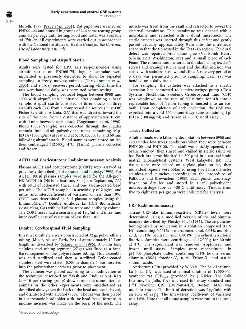
Basal plasma ACTH levels were similar among the rearinggroups (AFR 19.9471.27 pg/ml; HMS0 19.3371.72; HMS1518.7471.21; HMS180 19.1871.93). As shown in Figure 1a,presentation of an acute APS stressor was associated with asignificant rearing-associated (F3, 216¼ 69.51, po0.0001)elevation in ACTH over time (F6, 216 ¼ 586.60, po0.0001)as determined by ANOVA corrected for repeated measures.In all four groups, acute APS caused a marked increase inmean plasma ACTH concentration, peaking at 10 min andreturning toward baseline by 60 min poststress. The plasmaACTH peak was approximately 1.6-fold greater (po0.001)in HMS180 rats (263719 pg/ml) as compared to eitherHMS15 or AFR rats (148715 vs 165715 pg/ml, respec-
tively), which were not significantly different from oneanother. Peak ACTH levels in the HMS0 rats (219716 pg/ml) were significantly less (po0.001) than that observed inHMS180 rats, but significantly different from either HMS15or AFR rats (po0.001).
Two-way ANOVA with repeated measures on timerevealed significant effects for rearing (F3, 216 ¼ 49.85,po0.0001) and time (F6, 216 ¼ 127.11, po0.0001) on plasmaCORT levels following an acute APS stressor (Figure 1b).However, no rearing-associated differences in basal troughCORT levels were observed. Plasma CORT levels peaked15 min after initiation of APS in all of the groups. Peaklevels at 15 min were not significantly different between AFR(143716 ng/ml) and HMS15 (137714 ng/ml) rats. Peaklevels for HMS0 (203720 ng/ml) and HMS180 (300726 ng/ml) rats were significantly different (po0.01) from oneanother and from AFR and HMS15 rats (po0.001). WhileCORT returned toward initial levels in the AFR and HMS15groups, it remained significantly elevated in the HMS0 andHMS180 groups.
Lumbar CSF CRFir
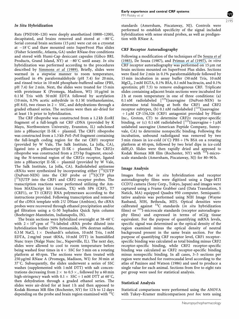
The distribution of morning CSF CRFir levels is shown inFigure 2. One-way ANOVA revealed a significant effect ofrearing (F3, 36¼ 15.19, po0.001) on CSF CRF. Levels(mean7SEM) of the AFR (3773 pg/ml) and HMS15(3672 pg/ml) groups were not significantly different fromone another, but were greater than that of either the HMS0(5374 pg/ml) or HMS180 (6876 pg/ml) groups.
CRF mRNA Expression
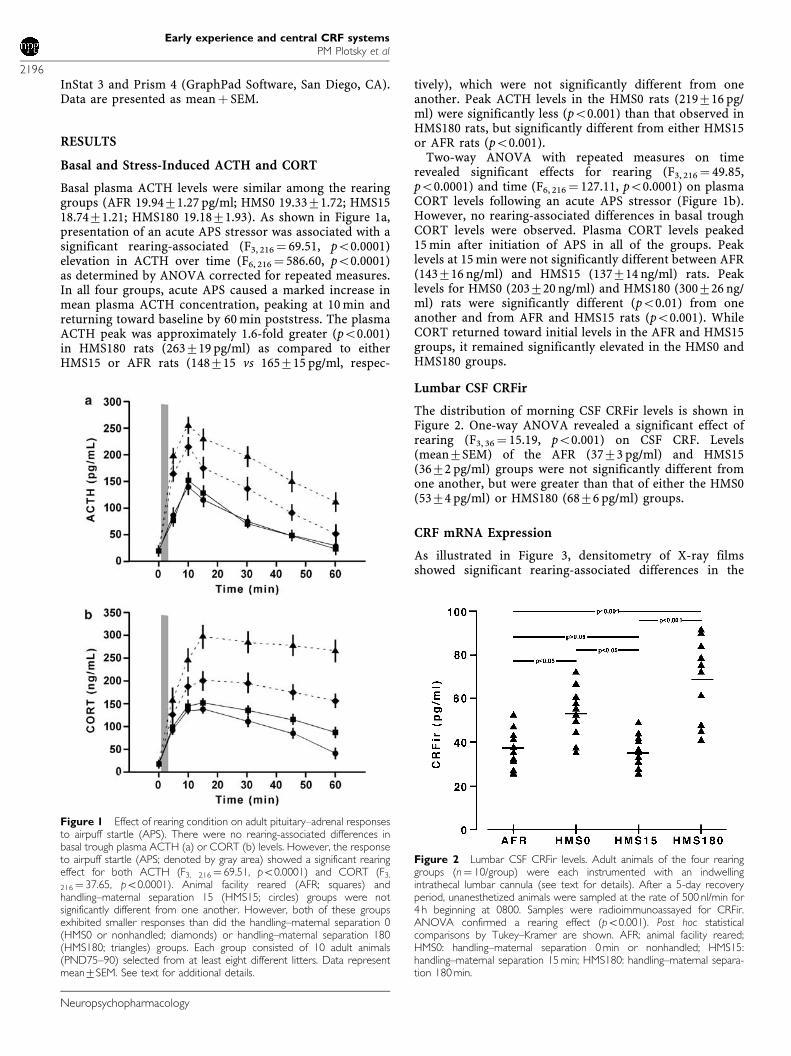
As illustrated in Figure 3, densitometry of X-ray filmsshowed significant rearing-associated differences in the
Figure 1 Effect of rearing condition on adult pituitary–adrenal responsesto airpuff startle (APS). There were no rearing-associated differences inbasal trough plasma ACTH (a) or CORT (b) levels. However, the responseto airpuff startle (APS; denoted by gray area) showed a significant rearingeffect for both ACTH (F3, 216¼ 69.51, po0.0001) and CORT (F3,
216¼ 37.65, po0.0001). Animal facility reared (AFR; squares) andhandling–maternal separation 15 (HMS15; circles) groups were notsignificantly different from one another. However, both of these groupsexhibited smaller responses than did the handling–maternal separation 0(HMS0 or nonhandled; diamonds) or handling–maternal separation 180(HMS180; triangles) groups. Each group consisted of 10 adult animals(PND75–90) selected from at least eight different litters. Data representmean7SEM. See text for additional details.
Figure 2 Lumbar CSF CRFir levels. Adult animals of the four rearinggroups (n¼ 10/group) were each instrumented with an indwellingintrathecal lumbar cannula (see text for details). After a 5-day recoveryperiod, unanesthetized animals were sampled at the rate of 500 nl/min for4 h beginning at 0800. Samples were radioimmunoassayed for CRFir.ANOVA confirmed a rearing effect (po0.001). Post hoc statisticalcomparisons by Tukey–Kramer are shown. AFR: animal facility reared;HMS0: handling–maternal separation 0 min or nonhandled; HMS15:handling–maternal separation 15 min; HMS180: handling–maternal separa-tion 180 min.
Early experience and central CRF systemsPM Plotsky et al
2196
Neuropsychopharmacology
levels of CRF mRNA in the PVN (F3, 20 ¼ 11.35, po0.01),CeA (F3, 15 ¼ 6.79, po0.05), BNST (F3, 15 ¼ 5.82, po0.05),and locus coeruleus/parabrachial nucleus (LC/PBN; F3, 15¼4.45, po0.05). In each brain region, the HMS180 ratsexhibited significantly greater levels of CRF mRNA than didthe AFR animals. There were no significant differences inCRF mRNA density between AFR and HMS15 rats in anyregion examined. Adult CRF mRNA levels in the HMS180rats were 2.1-fold (PVN, po0.001), 1.8-fold (CeA, po0.001),2.2-fold (BNST, po0.001), and 1.5-fold (LC/PBN, po0.001)higher than in the AFR groups. Interestingly, CRF mRNA inthe PVN of HMS0 rats was significantly greater than that ofeither AFR or HMS15 animals (po0.05), but was signifi-cantly less than that of the HMS180 group.
CRFir Content
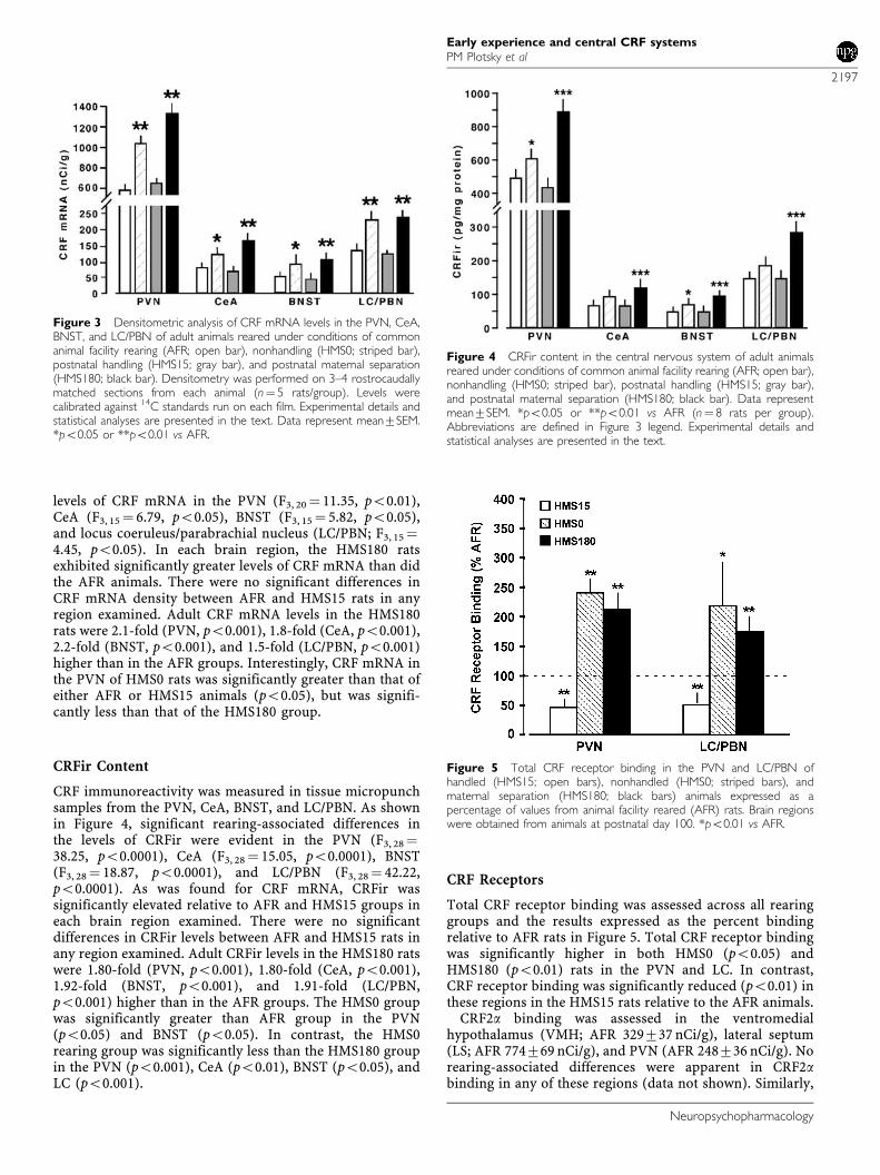
CRF immunoreactivity was measured in tissue micropunchsamples from the PVN, CeA, BNST, and LC/PBN. As shownin Figure 4, significant rearing-associated differences inthe levels of CRFir were evident in the PVN (F3, 28¼38.25, po0.0001), CeA (F3, 28 ¼ 15.05, po0.0001), BNST(F3, 28 ¼ 18.87, po0.0001), and LC/PBN (F3, 28¼ 42.22,po0.0001). As was found for CRF mRNA, CRFir wassignificantly elevated relative to AFR and HMS15 groups ineach brain region examined. There were no significantdifferences in CRFir levels between AFR and HMS15 rats inany region examined. Adult CRFir levels in the HMS180 ratswere 1.80-fold (PVN, po0.001), 1.80-fold (CeA, po0.001),1.92-fold (BNST, po0.001), and 1.91-fold (LC/PBN,po0.001) higher than in the AFR groups. The HMS0 groupwas significantly greater than AFR group in the PVN(po0.05) and BNST (po0.05). In contrast, the HMS0rearing group was significantly less than the HMS180 groupin the PVN (po0.001), CeA (po0.01), BNST (po0.05), andLC (po0.001).
CRF Receptors
Total CRF receptor binding was assessed across all rearinggroups and the results expressed as the percent bindingrelative to AFR rats in Figure 5. Total CRF receptor bindingwas significantly higher in both HMS0 (po0.05) andHMS180 (po0.01) rats in the PVN and LC. In contrast,CRF receptor binding was significantly reduced (po0.01) inthese regions in the HMS15 rats relative to the AFR animals.
CRF2a binding was assessed in the ventromedialhypothalamus (VMH; AFR 329737 nCi/g), lateral septum(LS; AFR 774769 nCi/g), and PVN (AFR 248736 nCi/g). Norearing-associated differences were apparent in CRF2abinding in any of these regions (data not shown). Similarly,
Figure 3 Densitometric analysis of CRF mRNA levels in the PVN, CeA,BNST, and LC/PBN of adult animals reared under conditions of commonanimal facility rearing (AFR; open bar), nonhandling (HMS0; striped bar),postnatal handling (HMS15; gray bar), and postnatal maternal separation(HMS180; black bar). Densitometry was performed on 3–4 rostrocaudallymatched sections from each animal (n¼ 5 rats/group). Levels werecalibrated against 14C standards run on each film. Experimental details andstatistical analyses are presented in the text. Data represent mean7SEM.*po0.05 or **po0.01 vs AFR.
Figure 4 CRFir content in the central nervous system of adult animalsreared under conditions of common animal facility rearing (AFR; open bar),nonhandling (HMS0; striped bar), postnatal handling (HMS15; gray bar),and postnatal maternal separation (HMS180; black bar). Data representmean7SEM. *po0.05 or **po0.01 vs AFR (n¼ 8 rats per group).Abbreviations are defined in Figure 3 legend. Experimental details andstatistical analyses are presented in the text.
Figure 5 Total CRF receptor binding in the PVN and LC/PBN ofhandled (HMS15; open bars), nonhandled (HMS0; striped bars), andmaternal separation (HMS180; black bars) animals expressed as apercentage of values from animal facility reared (AFR) rats. Brain regionswere obtained from animals at postnatal day 100. *po0.01 vs AFR.
Early experience and central CRF systemsPM Plotsky et al
2197
Neuropsychopharmacology
no rearing-associated changes in CRF2a mRNA wereevident in the VMH (AFR 1972.1 nCi/g), LS (AFR1371.6 nCi/g), or PVN (AFR 1071.2 nCi/g).
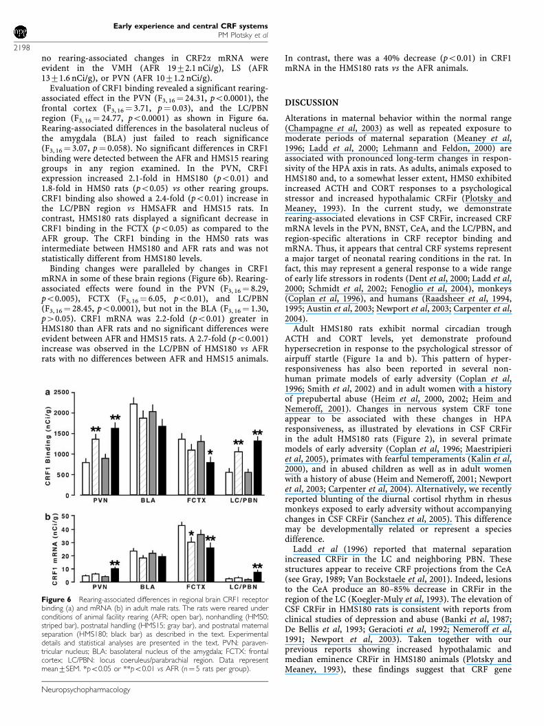
Evaluation of CRF1 binding revealed a significant rearing-associated effect in the PVN (F3, 16¼ 24.31, po0.0001), thefrontal cortex (F3, 16 ¼ 3.71, p¼ 0.03), and the LC/PBNregion (F3, 16¼ 24.77, po0.0001) as shown in Figure 6a.Rearing-associated differences in the basolateral nucleus ofthe amygdala (BLA) just failed to reach significance(F3, 16 ¼ 3.07, p¼ 0.058). No significant differences in CRF1binding were detected between the AFR and HMS15 rearinggroups in any region examined. In the PVN, CRF1expression increased 2.1-fold in HMS180 (po0.01) and1.8-fold in HMS0 rats (po0.05) vs other rearing groups.CRF1 binding also showed a 2.4-fold (po0.01) increase inthe LC/PBN region vs HMSAFR and HMS15 rats. Incontrast, HMS180 rats displayed a significant decrease inCRF1 binding in the FCTX (po0.05) as compared to theAFR group. The CRF1 binding in the HMS0 rats wasintermediate between HMS180 and AFR rats and was notstatistically different from HMS180 levels.
Binding changes were paralleled by changes in CRF1mRNA in some of these brain regions (Figure 6b). Rearing-associated effects were found in the PVN (F3, 16 ¼ 8.29,po0.005), FCTX (F3, 16¼ 6.05, po0.01), and LC/PBN(F3, 16 ¼ 28.45, po0.0001), but not in the BLA (F3, 16 ¼ 1.30,p40.05). CRF1 mRNA was 2.2-fold (po0.01) greater inHMS180 than AFR rats and no significant differences wereevident between AFR and HMS15 rats. A 2.7-fold (po0.001)increase was observed in the LC/PBN of HMS180 vs AFRrats with no differences between AFR and HMS15 animals.
In contrast, there was a 40% decrease (po0.01) in CRF1mRNA in the HMS180 rats vs the AFR animals.
DISCUSSION
Alterations in maternal behavior within the normal range(Champagne et al, 2003) as well as repeated exposure tomoderate periods of maternal separation (Meaney et al,1996; Ladd et al, 2000; Lehmann and Feldon, 2000) areassociated with pronounced long-term changes in respon-sivity of the HPA axis in rats. As adults, animals exposed toHMS180 and, to a somewhat lesser extent, HMS0 exhibitedincreased ACTH and CORT responses to a psychologicalstressor and increased hypothalamic CRFir (Plotsky andMeaney, 1993). In the current study, we demonstraterearing-associated elevations in CSF CRFir, increased CRFmRNA levels in the PVN, BNST, CeA, and the LC/PBN, andregion-specific alterations in CRF receptor binding andmRNA. Thus, it appears that central CRF systems representa major target of neonatal rearing conditions in the rat. Infact, this may represent a general response to a wide rangeof early life stressors in rodents (Dent et al, 2000; Ladd et al,2000; Schmidt et al, 2002; Fenoglio et al, 2004), monkeys(Coplan et al, 1996), and humans (Raadsheer et al, 1994,1995; Austin et al, 2003; Newport et al, 2003; Carpenter et al,2004).
Adult HMS180 rats exhibit normal circadian troughACTH and CORT levels, yet demonstrate profoundhypersecretion in response to the psychological stressor ofairpuff startle (Figure 1a and b). This pattern of hyper-responsiveness has also been reported in several non-human primate models of early adversity (Coplan et al,1996; Smith et al, 2002) and in adult women with a historyof prepubertal abuse (Heim et al, 2000, 2002; Heim andNemeroff, 2001). Changes in nervous system CRF toneappear to be associated with these changes in HPAresponsiveness, as illustrated by elevations in CSF CRFirin the adult HMS180 rats (Figure 2), in several primatemodels of early adversity (Coplan et al, 1996; Maestripieriet al, 2005), primates with fearful temperaments (Kalin et al,2000), and in abused children as well as in adult womenwith a history of abuse (Heim and Nemeroff, 2001; Newportet al, 2003; Carpenter et al, 2004). Alternatively, we recentlyreported blunting of the diurnal cortisol rhythm in rhesusmonkeys exposed to early adversity without accompanyingchanges in CSF CRFir (Sanchez et al, 2005). This differencemay be developmentally related or represent a speciesdifference.
Ladd et al (1996) reported that maternal separationincreased CRFir in the LC and neighboring PBN. Thesestructures appear to receive CRF projections from the CeA(see Gray, 1989; Van Bockstaele et al, 2001). Indeed, lesionsto the CeA produce an 80–85% decrease in CRFir in theregion of the LC (Koegler-Muly et al, 1993). The elevation ofCSF CRFir in HMS180 rats is consistent with reports fromclinical studies of depression and abuse (Banki et al, 1987;De Bellis et al, 1993; Geracioti et al, 1992; Nemeroff et al,1991; Newport et al, 2003). Taken together with ourprevious reports showing increased hypothalamic andmedian eminence CRFir in HMS180 animals (Plotsky andMeaney, 1993), these findings suggest that CRF gene
Figure 6 Rearing-associated differences in regional brain CRF1 receptorbinding (a) and mRNA (b) in adult male rats. The rats were reared underconditions of animal facility rearing (AFR; open bar), nonhandling (HMS0;striped bar), postnatal handling (HMS15; gray bar), and postnatal maternalseparation (HMS180; black bar) as described in the text. Experimentaldetails and statistical analyses are presented in the text. PVN: paraven-tricular nucleus; BLA: basolateral nucleus of the amygdala; FCTX: frontalcortex; LC/PBN: locus coeruleus/parabrachial region. Data representmean7SEM. *po0.05 or **po0.01 vs AFR (n¼ 5 rats per group).
Early experience and central CRF systemsPM Plotsky et al
2198
Neuropsychopharmacology
expression and peptide synthesis within the PVN and CeA–LC systems may represent a common target in animalsexposed to what may be considered abnormal neonatalrearing conditions regardless of which end of the rearingspectrum they fall intoFmonotonous housing and care (ieHMS0) vs extended periods of neonatal maternal separation(ie HMS180). The observation that both the HMS180 andHMS0 rearing conditions result in a similar directionality ofchanges in these CRF systems is a conundrum that isdifficult to reconcile (see below).
In contrast, animals exposed to either HMS15 or AFRrearing conditions exhibit comparable levels of CRF mRNAin the PVN and CeA as well as similar concentrations ofCRFir in the median eminence (also see Plotsky andMeaney, 1993; Viau et al, 1993) and LC. This is notunexpected as care protocols in most breeding coloniesexpose animals to repeated cage cleaning during the first 2weeks of life that involve transferring the mother and thepups from the maternity cage to a clean, novel cage, andmay therefore be considered as variant of the handlingprocedure (Greenberg and Bursdal, 1982). As we show herewith respect to CRF and as we and others have previouslyreported, both routine animal husbandry protocols (egAFR) and the HMS15 protocol produce similar neurobeha-vioral outcomes (Caldji et al, 2000; Pryce et al, 2001; Pryceand Feldon, 2003). However, Fenoglio et al (2004) recentlyreported that handling during PND2–9 was associated witha transient increase of CRF mRNA in the CeA by PND6, atransient increase of peptide levels in BNST, and a long-lasting decrease of CRF levels in PVN beginning by PND9.Importantly, these changes in CRF preceded changes inhippocampal GR expression by many weeks. It is note-worthy that a majority of the rodent neuroscience andbehavioral literature is derived from studies on what areessentially animal facility reared rats; therefore, we proposethat the AFR group is an appropriate comparison or colonycontrol group for these types of experiments.
The HMS0 rats represent a problematical group withrespect to their tendency to approximate the HMS180 groupon many outcome variables such as enhanced behavioralfearfulness (Francis et al, 1999; Caldji et al, 2000), reducedhippocampal GR mRNA (Meaney et al, 1996; Avishai-Elineret al, 2001), and enhanced PVN CRF mRNA (see Figure 3).The question of why the neonatal experiences of beingeither undisturbed (eg HMS0) or maternally separated(HMS180) exert a similar effect on the phenotype of theoffspring awaits a definitive answer. This observationappears to contradict the ‘maternal mediation hypothesis’(Denenberg et al, 1962; Denenberg, 1999), according towhich differences in maternal care upon reunion of the damwith her pups may account for a significant portion of theneurobehavioral differences in the adult offspring (Sucheckiet al, 1993; Meaney, 2001).
The environmental impoverishment inherent in standardlaboratory rodent housing may render the HMS0 rats morefearful and reactive than HMS15 animals (Bors and Forrin,1996; Rosenzweig and Bennett, 1996; Larsson et al, 2002).According to Macri et al (2004), the difference inneurobehavioral phenotype between HMS0 and handledoffspring reflects adaptive plasticity in the HPA axis andbehavioral function with respect to energy utilization andoverall health. In this view, HMS0 rats live in a relatively
stress-free environment and, therefore, retain a highlyreactive stress response while HMS15 rats that have beenexposed to repetitive stressors adapt by downregulatingHPA axis responsiveness. The enhanced responsiveness ofthe HMS180 rats, considered to be maladaptive, reflectsdisruption of normal pup homeostasis and in the intervalsbetween nursing bouts as a result of the repeated, prolongedneonatal separations.
Disruption of the perceived safety of the nest area by thematernal separation protocol, as well as the actual physicalseparation of the dam from her pups, may elicit changes inbehavior of the dam (Kalinichev et al, 2000; Fleming et al,2002). In fact, we (Ladd et al, 2000; Huot et al, 2004) haveproposed that in response to maternal separation, thequantity and/or quality of dam–pup interaction differs fromthat observed in AFR, HMS15, and HMS0 groups. Althougheach individual measure may exhibit only a small change,appropriate coordination of the sequence of maternalbehavior may be altered. It should be noted that in amajority of the studies in which maternal behavior wasmeasured, it was either the frequency and/or duration of theoccurrence of particular maternal behaviors that wasrecorded (Champagne et al, 2003; Pryce et al, 2001; Stern,1997). However, our own ongoing observations usingMarkov chain analysis indicate that the normally coordi-nated pattern of maternal behaviors may be disrupted in theHMS180 condition and that this disorganization of thesequencing of maternal behaviors may be equally or evenmore important than the frequency or duration ofoccurrence of isolated behaviors.
Differences in amygdaloid CRF expression are of interestconsidering the effects of early life events on behavioralresponses to stress. The CeA has long been implicated in theexpression of autonomic and behavioral responses to stress(reviewed in Davis, 1992; LeDoux, 1992, 1994; Gray, 1993).Lesions of the CeA have been found to block many of thebehavioral effects associated with stress and the literatureon the role of the amygdala in conditioned emotionalresponses is especially rich. Lesions of the CeA block theexcitatory effects of both conditioned and unconditionedfear on startle responses (Hitchcock and Davis, 1986, 1989)and the development of conditioned emotional responses(van der Kar et al, 1991; LeDoux, 1994). Interestingly, boththe central and the lateral nucleus of the amygdala havebeen found to be important substrates for the anxiolyticeffects of benzodiazepines on stress-related behaviors.
These amygdaloid effects are at least partially mediated byCRF. Pich et al (1995), using in vivo microdialysis, reportedan increase in extracellular CRFir levels in the amygdala inresponse to stress and ethanol withdrawal. Microinjectionsof the CRF antagonist a-helical CRF9–41 into the CeA blockthe anxiogenic effects of ethanol withdrawal (Rassnick et al,1993), fear-induced freezing (Swiergiel et al, 1993), andincreased emotionality following social defeat (Heinrichset al, 1992b). In contrast, CRF microinjections into theamygdala decrease exploratory behavior in an open-fieldtest (Liang and Lee, 1988; Elkabir et al, 1990) and increasedefensive aggression (Elkabir et al, 1990). Finally, lesions ofthe CeA block the excitatory effects of CRF on the acousticstartle reflex (Liang et al, 1992a). However, it must be notedthat in attempting to extrapolate these observations tohuman psychopathology, one must take into account
Early experience and central CRF systemsPM Plotsky et al
2199
Neuropsychopharmacology
species differences in the distribution of both CRF and itsreceptor subtypes (Sanchez et al, 1999, 2001). Thus, CRFacts at the level of the amygdala to mediate the expressionof behavioral responses to fear and, predictably, theobserved changes in this system in response to rearingcondition altered anxiety-like behavior.
CRF also serves as a neurotransmitter/neuromodulator inthe projection from the amygdala to the LC and neighboringPBN (Moga and Gray, 1989). The LC has long beenconsidered as a major site for mediation of anxiety-likestates in primates and rodents (see Schulkin et al, 1994 forreview). This structure sends noradrenergic projectionsextending throughout multiple cortical and subcorticalregions, including the prefrontal cortex, the hippocampus,the amygdala, and the hypothalamus (Foote et al, 1983).Stress increases CRF concentrations in the LC (Chappellet al, 1986) and LC injection of CRF is associated withincreased catecholamine release in the hypothalamus andprefrontal cortex of freely moving rats (Lavicky and Dunn,1993). Both stress and CRF infusion have been shown toincrease the firing rate of LC neurons and to stimulate theautonomic nervous system (Brown, 1989). The effects on LCcan be blocked by local injections of the CRF receptorantagonist a-helical CRF9–41 (Valentino and Foote, 1988;Valentino et al, 1998). Microinjections of a CRF receptorantagonist into the LC also attenuate novelty-inducedsuppression of exploratory behavior in the defensivewithdrawal paradigm (Butler et al, 1990), as well as stress-induced behavioral freezing (Swiergiel et al, 1993). Inaddition to changes in CRF expression, we found increasedCRF receptor binding capacity in the pericoerulear region ofboth HMS180 and HMS0 rats (Figure 5). Both specific CRF1receptor binding and mRNA were increased in HMS180 ratsvs AFR and HMS15 rats (Figure 6). The CRF1 receptorsubtype is associated with enhanced anxiogeneic effects(Streckler and Holsboer, 1999). Taken together, these datasuggest that changes in CRF tone to the LC and related areasmay change the sensitivity (ie alter the set-point) of thesepathways to stimuli that normally activate them.
These observations imply that there would be increasedCRF-induced activation of the LC during stress in the HMS0and HMS180 animals. At least two findings are consistentwith this idea. In comparison to HMS15 rats, acute restraintstress in both HMS0 and HMS180 rats is associated with agreater increase in Fos-ir in the LC (Pearson et al, 1997) andlarger increases in extracellular NE levels in the PVN (Liuet al, 2000). In a recent post-mortem study of depressedsuicide victims, Austin et al (2003) observed increasedCRFir in the LC as well as median and dorsal raphe neurons.Thus, we propose that the neonatal environment canregulate the expression of behavioral responses to stress,at least in part, by altering the set-point of the CeA/BNST–LC CRF–NE system during a developmentally sensitivewindow occurring during the 10–14 days immediately afterbirth.
The behavioral differences in stress responsiveness notedbetween AFR and HMS15 vs HMS0 and HMS180 animalsparallel those found in HPA axis responsiveness. Maternalseparation increases plasma ACTH and CORT responses toacute psychological stressors (ie stressors involving condi-tions that signal threat) as compared to HMS15 or AFR rats(see Meaney et al, 1996 for review). These differences in
HPA axis responsivity to stress are, in part, associated withdifferential expression of CRF, as well as arginine vaso-pressin (AVP) in parvocellular neurons of the PVN (Plotskyand Meaney, 1993; Viau et al, 1993; Bhatnagar et al, 1995).These neurons project to the external zone of the medianeminence and provide the neurohumoral signal for therelease of ACTH from pituitary corticotrophs (see Plotsky,1991 for review). Thus, our current findings of increasedPVN CRF mRNA expression as well as increased CRFpeptide content in the median eminence terminals ofHMS180 and HMS0 rats (Plotsky and Meaney, 1993)provide strong support for the thesis that the neuroendo-crine CRF system and, thus, HPA axis responsivity, issensitive to the early rearing environment. The upregulationof CRF1 receptors in the PVN of HMS180 rats without achange in CRF2 binding or mRNA suggests functionaladaptation in this region. Previous reports of CRF- andstress-induced upregulation have appeared (Rivest et al,1995; Imaki et al, 1996; Mansi et al, 1996). Remarkablysimilar endocrine findings have been reported in depressedwomen with a history of childhood sexual or physical abuse(Heim and Nemeroff, 2001; Heim et al, 2002).
The effects of neonatal rearing on the PVN medianeminence and the CeA/BNST–LC neurocircuits are likely toramify throughout the brain to modify additional neuro-transmitter circuits, which together may contribute todifferences in behavioral and neuroendocrine responsive-ness to stressors. In addition to the LC, Ladd et al (1996)found that maternal separation increased CRF receptorbinding in the raphe nuclei. This region also showsincreased CRFir in depressed suicide victims (Austin et al,2003). Due to the anatomical and functional relationshipsamong these structures, these findings suggest that earlyexperience-induced changes in CRF systems may lead to acascade of adaptations including both the noradrenergicand serotonergic systems, thus rendering the individualmore responsive to subsequent stressors.
It is likely that similar changes in these systems alsocontribute to human psychopathology. Indeed, in humans,abuse or neglect in early life has been repeatedly shown tobe a major risk factor for depression and/or anxietydisorders in adulthood (Bifulco et al, 1991; Brown andAnderson, 1993; Mullen et al, 1996; McCauley et al, 1997;Weiss et al, 1999; Bulik et al, 2001; Molnar et al, 2001;Kendler et al, 2002). Hyperactivity of central CRF systemshas been implicated in the expression of these affectivedisorders (Stout and Nemeroff, 1994; Heim and Nemeroff,2001; Heim et al, 2002, 2004), and both noradrenergic andserotonergic systems appear to be involved in the develop-ment of affective disorders.
Overall, these findings provide further validation ofneonatal maternal separation in rats as a model ofvulnerability to an anxiety- and depression-like syndrome.Altered rearing conditions are strongly associated withpersistent alterations in stress-responsive neurocircuits. Thisprocess, which may occur via altered methylation of targetgenes in a region-specific pattern (Weaver et al, 2004a, b), orother mechanisms serve to ‘tune’ stress-sensitive neurocir-cuits in an adaptive or maladaptive manner. Specifically, thisstudy strengthens the linkage between early life environmentand long-term alterations in central CRF systems. Thesefindings may provide a mechanism whereby life events,
Early experience and central CRF systemsPM Plotsky et al
2200
Neuropsychopharmacology
interacting with genetic predisposition, may increasevulnerability to pathology in later life.
ACKNOWLEDGEMENTS
This research was supported by the NIMH-funded Silvio OConte Center for the Neuroscience of Mental Disease(MH58922; CBN, PMP) and by Grants MH50113 (PMP,MJM) and MH42088 (CBN). PMP has received speaking feesfrom Janssen Pharmaceutica, Johnson & Johnson Pharma-ceutical Research & Development, LLC, and Roche PaloAlto, LLC, as well as research support from Johnson &Johnson Pharmaceutical Research & Development, LLC,Pharmacia Upjohn, Synaptic/Lunbeck, and NV Organon.PMP is the recipient of a GlaxoSmithKline endowed chair inPsychiatry and Behavioral Science. CBN has receivedconsulting fees, speaking fees, research support, or otherfinancial support from Abbott Laboratories, Acadia Phar-maceuticals, AstraZeneca, Bristol-Myers Squibb, Corcept,Cypress Biosciences, Cyberonics, Eli Lilly, Forest Labora-tories, GlaxoSmithKline, Janssen Pharmaceutica, Merck,Neurocrine Biosciences, Novartis, Organon, Otsuka, Sanofi-Synthelabo, Somerset, Pfizer Pharmaceuticals, and Wyeth-Ayerst. MJM was also supported by a grant from theCanadian Institutes of Health Research and a SeniorScientist award. CC was supported by a graduate fellowshipfrom the Canadian Institutes of Health Research.
REFERENCES
Arase K, York DA, Shimizu H, Shargill N, Bray GA (1988). Effectsof corticotropin-releasing factor on food intake and brownadipose tissue thermogenesis in rats. Am J Physiol 255: E255–E259.
Arborelius L, Owens MJ, Plotsky PM, Nemeroff CB (1999). The roleof corticotropin-releasing factor (CRF) in depression andanxiety disorders. J Endocrinol 160: 1–12.
Austin MC, Janosky JE, Murphy HA (2003). Increased cortico-tropin-releasing hormone immunoreactivity in monoamine-containing pontine nuclei of depressed suicide men. MolPsychiatry 8: 324–332.
Avishai-Eliner S, Eghbal-Ahmadi M, Tabachnik E, Brunson KL,Baram TZ (2001). Down-regulation of corticotropin-releasinghormone messenger ribonucleic acid (mRNA) precedes early-lifeexperience-induced changes in hippocampal glucocorticoidreceptor mRNA. Endocrinology 142: 89–97.
Banki CM, Bissette G, Arato M, O’Connor L, Nemeroff CB (1987).Cerebrospinal fluid corticotropin-releasing factor-like immu-noreactivity in depression and schizophrenia. Am J Psychiatry144: 873–877.
Berridge CW, Dunn AJ (1987). A corticotropin-releasing factorantagonist reverses the stress induced changes of exploratorybehavior in mice. Horm Behav 21: 393–401.
Bhatnagar S, Shanks N, Meaney MJ (1995). Hypothalamic–pituitary–adrenal function in handled and nonhandled rats inresponse to chronic stress. J Neuroendocrinol 7: 107–119.
Bifulco A, Brown GW, Adler Z (1991). Early sexual abuse andclinical depression in aduult life. Br J Psychiat 159: 115–122.
Bors DA, Forrin B (1996). The effects of post-weaning environ-ment, biological dam, and nursing dam on feeding neophobia,open field activity, and learning. Can J Exp Psychol 50: 197–204.
Britton DR, Hoffman DK, Lederis K, Rivier JA (1984). Comparisonof the behavioral effects of GRF, sauvagine and urotensin I.Brain Res 304: 201–205.
Britton DR, Koob GF, Rivier J, Vale W (1982). Intraventricularcorticotropin-releasing factor enhances behavioral effects ofnovelty. Life Sci 31: 363–367.
Brown GR, Anderson B (1993). Psychiatric morbidity in adultinpatients with childhood histories of sexual and physical abuse.Am J Psychiat 148: 55–61.
Brown MR (1989). Neuropeptide regulation of the autonomicnervous system. In: Tache Y, Morley JE, Brown MR (eds).Neuropeptides and Stress. Springer-Verlag: New York. pp 107–120.
Bulik CM, Prescott CA, Kendler KS (2001). Features of childhoodsexual abuse and the development of psychiatric and substanceuse disorders. Br J Psychiatry 179: 444–449.
Butler PD, Weiss JM, Stout JC, Nemeroff CB (1990). Corticotropin-releasing factor produces fear-enhancing and behaviouralactivating effects following infusion into the locus coeruleus.J Neurosci 10: 176–183.
Cador M, Ahmed SH, Koob GF, Le Moal M, Stinus L (1992).Corticotropin-releasing factor induces a place aversion inde-pendent of its neuroendocrine role. Brain Res 41: 61–69.
Caldji C, Francis D, Sharma S, Plotsky PM, Meaney MJ (2000). Theeffects of early rearing environment on the development ofGABAA and central benzodiazepine receptor levels and novelty-induced fearfulness in the rat. Neuropsychopharmacology 22:219–229.
Carpenter LL, Tyrka AR, McDougle CJ, Malison RT, Owens MJ,Nemeroff CB et al (2004). Cerebrospinal fluid corticotropin-releasing factor and perceived early-life stress in depressedpatients and healthy controls. Neuropsychopharmacology 29:777–784.
Champagne FA, Francis DD, Mar A, Meaney MJ (2003). Variationsin maternal care in the rat as a mediating influence foreffects of environment on development. Physiol Behav 79:359–371.
Chappell PB, Smith MA, Kitts CD, Bisette G, Ritchie J, Anderson Cet al (1986). Alterations in corticotropin-releasing factor-likeimmunoreactivity in discrete rat brain regions after acute andchronic stress. J Neurosci 6: 2908–2914.
Chrousos GP, Gold PW (1992). The concepts of stress and stresssystem disorders. JAMA 267: 1244–1252.
Coplan JD, Andrews MW, Rosenblum LA, Owens MJ, Friedman S,Gorman JM et al (1996). Persistent elevations of cerebrospinalfluid concentrations of corticotropin-releasing factor in adultnonhuman primates exposed to early-life stressors: implicationsfor the pathophysiology of mood and anxiety disorders. ProcNatl Acad Sci USA 93: 1619–1623.
Davis M (1992). The role of the amygdala in fear and anxiety. AnnuRev Neurosci 15: 353–375.
De Bellis MD, Gold PW, Geracioti TD, Listwak SJ, Kling MA(1993). Fluoxetine significantly reduces CSF CRH and AVPconcentrations in patients with major depression. Am JPsychiatry 150: 656–657.
De Souza EB (l987). Corticotropin releasing factor receptors in therat central nervous system: characterization and regionaldistribution. J Neurosci 7: 88–100.
De Souza EB, Insel TR, Perrin MH, Rivier J, Vale WW, Kuhar MJ(1985). Corticotropin-releasing factor receptors are widelydistributed with the rat central nervous system, an autoradio-graphic study. J Neurosci 5: 3189–3202.
Denenberg VH (1964). Critical periods, stimulus input, andemotional reactivity: a theory of infantile stimulation. PsycholRev 71: 335–351.
Denenberg VH (1999). Commentary: is maternal stimulationthe mediator of the handling effect in infancy. Dev Psychobiol34: 1–3.
Denenberg VH, Ottinger DR, Stephens MW (1962). Effects ofmaternal factors upon growth and behavior of the rat. Child Dev33: 65–71.
Early experience and central CRF systemsPM Plotsky et al
2201
Neuropsychopharmacology
Dent GW, Okimoto DK, Smith MA, Levine S (2000). Stress-inducedalterations in corticotropin-releasing hormone and vasopressingene expression in the paraventricular nucleus during ontogeny.Neuroendocrinology 71: 333–342.
Dunn AJ, Berridge GW (1990). Physiological and behavioralresponses to corticotropin releasing factor administration: isCRF a mediator of anxiety or stress responses? Brain Res Rev 15:71–100.
Elkabir DR, Wyatt ME, Vellucci SV, Herbert J (1990). The effects ofseparate or combined infusions of corticotropin-releasing factorand vasopressin either intraventricularly or into the amygdalaon aggressive and investigative behavior in the rat. Regul Pept28: 199–214.
Engelmann M, Thrivikraman KV, Su Y, Nemeroff CB, MontkowskiA, Landgraf R et al (1996). Endocrine and behavioral effects ofairpuff-startle in rats. Psychoneuroendocrinology 21: 391–400.
Fenoglio KA, Brunson KL, Avishai-Eliner S, Chen Y, Baram TZ(2004). Region-specific handling-induced changes in corticotro-pin-releasing factor and glucocorticoid receptor expression.Endocrinology 145: 2702–2706.
Fleming AS, Kraemer GW, Gonzalez A, Lovic V, Rees S, Melo A(2002). Mothering begets mothering: the transmission ofbehavior and its neurobiology across generations. PharmacolBiochem Behav 73: 61–75.
Foote SL, Bloom FE, Aston-Jones G (1983). Nucleus locus ceruleus:new evidence of anatomical and physiological specificity. PhysiolRev 63: 844–914.
Francis DD, Caldji C, Champagne F, Plotsky PM, Meaney MJ(1999). The role of corticotropin-releasing factor–norepinephr-ine systems in mediating the effects of early experience on thedevelopment of behavioral and endocrine responses to stress.Biol Psychiatry 46: 1153–1166.
Geracioti TD, Orth DN, Ekhator NN, Blumenkopf B, Loosen PT(1992). Serial cerebrospinal fluid corticotropin-releasing hor-mone concentrations in healthy and depressed humans. J ClinEndocrinol Metab 74: 1325–1330.
Gray TS (1989). The organization and possible function ofamygdaloid corticotropin-releasing factor pathways. In: DeSouza EB, Nemeroff CB (eds). Corticotropin-Releasing Factor:Basic and Clinical Studies of a Neuropeptide. CRC Press: BocaRaton. pp 53–68.
Gray TS (1993). Amygdaloid CRF pathways. Role in autonomic,neuroendocrine, and behavioral responses to stress. Ann NYAcad Sci 697: 53–60.
Greenberg G, Bursdal C (1982). Animal colony practices in NorthAmerican academic institutions: a survey. J Gen Psychol 106:165–173.
Heim C, Nemeroff CB (2001). The role of childhood trauma in theneurobiology of mood and anxiety disorders: preclinical andclinical studies. Biol Psychiatry 49: 1023–1039.
Heim C, Newport DJ, Heit S, Graham YP, Wilcox M, Bonsall R et al(2000). Pituitary–adrenal and autonomic responses to stress inwomen after sexual and physical abuse in childhood. JAMA 284:592–597.
Heim C, Newport DJ, Wagner D, Wilcox MM, Miller AH, NemeroffCB (2002). The role of early adverse experience and adulthoodstress in the prediction of neuroendocrine stress reactivity inwomen: a multiple regression analysis. Depress Anxiety 15:117–125.
Heim C, Plotsky PM, Nemeroff CB (2004). Importance of studyingthe contributions of early adverse experience to neurobiologicalfindings in depression. Neuropsychopharmacology 29: 641–648.
Heinrichs SC, Cole BJ, Pich EM, Menzaghi F, Koob GF, Hauger RL(1992a). Endogenous corticotropin-releasing factor modulatesfeeding induced by neuropeptide Y or a tail pinch stressor.Peptides 13: 879–884.
Heinrichs SC, Menzaghi F, Pich EM, Hauger RL, Koob GF (1993).Corticotropin-releasing factor in the paraventricular nucleus
modulates feeding induced by neuropeptide Y. Brain Res 611:18–24.
Heinrichs SC, Pich EM, Miczek K, Britton KT, Koob GF (1992b).Corticotropin-releasing factor antagonist reduces emotionalityin socially defeated rats via direct neurotropic action. Brain Res581: 190–197.
Hitchcock JM, Davis M (1986). Lesions of the amygdala, but not ofthe cerebellum or red nucleus, block conditioned fear asmeasured with the potentiated startle paradigm. Behav Neurosci100: 11–22.
Hitchcock JM, Sananes CB, Davis M (1989). Sensitization of thestartle reflex by footshock: blockade by lesions of the centralnucleus of the amygdala or its efferent pathway to the brainstem.Behav Neurosci 103: 509–518.
Hofer MA (1994). Early relationships as regulators of infantphysiology and behavior. Acta Paediatr Suppl 397: 9–18.
Huot RL, Gonzalez ME, Ladd CO, Thrivikraman KV, Plotsky PM(2004). Foster litters prevent hypothalamic–pituitary–adrenalaxis sensitization mediated by neonatal maternal separation.Psychoneuroendocrinology 29: 279–289.
Huot RL, Thrivikraman KV, Meaney MJ, Plotsky PM (2001).Development of adult ethanol preference and anxiety as aconsequence of neonatal maternal separation in Long Evans ratsand reversal with antidepressant treatment. Psychopharmacology158: 366–373.
Imaki T, Naruse M, Harada S, Chikada N, Imaki J, Onodera H et al(1996). Corticotropin-releasing factor up-regulates its ownreceptor mRNA in the paraventricular nucleus of the hypotha-lamus. Mol Brain Res 38: 166–170.
Imaki T, Shibasaki T, Masuda A, Demura H, Shizume K, Ling N(1987). Effects of adrenergic blockers on corticotropin-releasingfactor-induced behavioral changes in rats. Regul Pept 19: 243–252.
Jans J, Woodside BC (1990). Nest temperature: effects on maternalbehavior, pup development, and interactions with handling.Dev Psychobiol 23: 519–534.
Kalin NH, Shelton SE, Davidson RJ (2000). Cerebrospinal fluidcorticotropin-releasing hormone levels are elevated in monkeyswith patterns of brain activity associated with fearful tempera-ment. Biol Psychiatry 47: 579–585.
Kalinichev M, Easterling KW, Holtzman SG (2000). Periodicpostpartum separation from the offspring results in long-lasting changes in anxiety-related behaviors and sensitivity tomorphine in Long–Evans mother rats. Psychopharmacology 152:431–439.
Kendler KS, Gardner CO, Prescott CA (2002). Toward acomprehensive developmental model for major depression inwomen. Am J Psychiatry 159: 1133–1145.
Koegler-Muly SM, Owens MJ, Ervin GN, Kilts CD, Nemeroff CB(1993). Potential corticotropin-releasing factor pathways in therat brain as determined by bilateral electrolytic lesions of thecentral amygdaloid nucleus and paraventricular nucleus of thehypothalamus. J Neuroendocrinol 5: 95–98.
Koob GF (1999). Corticotropin-releasing factor, norepinephrine,and stress. Biol Psychiatry 46: 1167–1180.
Koob GF, Heinrichs SC, Menzaghi F, Pich EM, Britton KT (1994).Corticotropin releasing factor, stress and behavior. SeminNeurosci 6: 221–229.
Krahn DD, Gosnell BA, Grace M, Levine AS (1986). CRF antagonistpartially reverses CRF- and stress-induced effects on feeding.Brain Res Bull 17: 285–289.
Ladd CO, Huot RL, Thrivikraman KV, Nemeroff CB, Meaney MJ,Plotsky PM (2000). Long-term behavioral and neuroendocrineadaptations to adverse early experience. Prog Brain Res 122:81–103.
Ladd CO, Owens MJ, Nemeroff CB (1996). Persistent changes incorticotropin-releasing factor neuronal systems induced bymaternal deprivation. Endocrinology 137: 1212–1318.
Early experience and central CRF systemsPM Plotsky et al
2202
Neuropsychopharmacology
Larsson F, Winblad B, Mohammed AH (2002). Psychological stressand environmental adaptation in enriched vs. impoverishedhoused rats. Pharmacol Biochem Behav 73: 193–207.
Lavicky J, Dunn AJ (1993). Corticotropin-releasing factor stimu-lates catecholamine release in hypothalamus and prefrontalcortex in freely moving rats as assessed by microdialysis. JNeurochem 60: 602–612.
LeDoux JE (1992). Emotion and the amygdala. In: Aggleton JP (ed).The Amygdala: Neurobiological Aspects of Emotion, Memory,and Mental Dysfunction. Wiley-Liss: New York. pp 339–351.
LeDoux JE (1994). The amygdala: contributions to fear and stress.Semin Neurosci 6: 231–237.
Lehmann J, Feldon J (2000). Long-term biobehavioral effects ofmaternal separation in the rat: consistent or confusing? RevNeurosci 11: 383–408.
Lejeune F, Millan MJ (2003). The CRF1 receptor antagonist,DMP695, abolishes activation of locus coeruleus noradrenergicneurones by CRF in anesthetized rats. Eur J Pharmacol 464:127–133.
Levine S (1975). Psychosocial factors in growth and development.In: Levi L (ed). Society, Stress and Disease. Oxford UniversityPress: London. pp 43–50.
Liang KC, Lee EHY (1988). Intra-amygdala injections of cortico-tropin-releasing factor facilitate inhibitory avoidance learningand reduce exploratory behavior in rats. Psychopharmacology96: 232–236.
Liang KC, Melia KR, Miserendino MJ, Fails WA, Campeau S, DavisM (1992b). Corticotropin releasing factor: long-lasting facilita-tion of the acoustic startle reflex. J Neurosci 12: 2303–2312.
Liang KC, Melia KR, Campeau S, Falls WA, Miserendino MJ, DavidM (1992a). Lesions of the central nucleus of the amvgdala, butnot the paranventricular nucleus of the hypothalamus, block theexcitatory effects of corticotropin releasing factor on the acousticstartle reflex. J Neurosci 12: 2313–2320.
Liu D, Caldji C, Sharma S, Plotsky PM, Meaney MJ (2000).Influence of neonatal rearing condition on stress-inducedadrenocorticotropin responses and norepinephrine release inthe hypothalamic paraventricular nucleus. J Neuroendocrinol 12:5–12.
Liu D, Tannenbaum B, Caldji C, Francis D, Freedman A, Sharma Set al (1997). Maternal care, hippocampal glucocorticoid receptorgene expression and hypothalamic–pituitary–adrenal responsesto stress. Science 277: 1659–1662.
Macri S, Mason GJ, Wurbel H (2004). Dissociation in the effects ofneonatal maternal separations on maternal care and theoffspring’s HPA and fear responses in rats. Eur J Neurosci 20:1017–1024.
Maestripieri D, Lindell SG, Ayala A, Gold P, Higley JD (2005).Neurobiological characteristics of rhesus macaque abusivemothers and their relation to social and maternal behavior.Neurosci Neurobehav Rev 29: 51–57.
Mansi JA, Rivest S, Drolet G (1996). Regulation of corticotropin-releasing factor type 1 (CRF1) receptor messenger ribonucleicacid in the paraventricular nucleus of rat hypothalamus byexogenous CRF. Endocrinology 137: 4619–4629.
McCauley J, Kern D, Kolodner K (1997). Clinical characteristics ofwomen with a history of childhood abuse. JAMA 277: 1362–1368.
Meaney MJ (2001). Maternal care, gene expression, and thetransmission of individual differences in stress reactivity acrossgenerations. Annu Rev Neurosci 24: 1161–1192.
Meaney MJ, Diorio J, Widdowson J, LaPlante P, Caldji C, Seckl JRet al (1996). Early environmental regulation of forebrainglucocorticoid receptor gene expression: implications foradrenocortical responses to stress. Dev Neurosci 18: 49–72.
Menzaghi F, Heinrichs SC, Pich EM, Tilders FJH, Koob GF (1993).Functional impairment of hypothalamic corticotropin-releasingfactor neurons with immunotargeted toxins enhances foodintake induced by neuropeptide Y. Brain Res 618: 76–82.
Moga MM, Gray TS (1989). Evidence for corticotropin-releasingfactor, neurotensin, and somatostatin in the neural pathwayfrom the central nucleus of the amygdala to the parabrachialnucleus. J Comp Neurol 241: 275–284.
Molnar BE, Buka SL, Kessler RC (2001). Child sexual abuse andsubsequent psychopathology: results from the National Comor-bidity Survey. Am J Public Health 91: 753–760.
Moore CL, Morelli GA (1979). Mother rats interact differently withmale and female offspring. J Comp Physiol Psychol 93: 677–684.
Mullen PE, Martin JL, Anderson JC, Romans SE, Herbison GP(1996). The long-term impact of the physical, emotional, andsexual abuse of children: a community study. Child Abuse Negl20: 7–21.
Nemeroff CB, Bissette G, Akil H, Fink M (1991). Neuropeptideconcentrations in the cerebrospinal fluid of depressed patientstreated with electroconvulsive therapy: corticotropin-releasingfactor, b-endorphin, and somatostatin. Br J Psychiatry 158:59–63.
Newport DJ, Heim C, Owens MJ, Ritchie JC, Ramsey CH, Bonsall Ret al (2003). Cerebrospinal fluid corticotropin-releasing factor(CRF) and vasopressin concentrations predict pituitary responsein the CRF stimulation test: a multiple regression analysis.Neuropsychopharmacology 28: 569–576.
Owens MJ, Nemeroff CB (1991). The physiology and pharmacologyof corticotropin-releasing factor. Pharmacol Rev 43: 425–473.
Page ME, Valentino RJ (1994). Locus coeruleus activation byphysiological challenges. Brain Res Bull 35: 557–560.
Palkovits M, Brownstein MJ (1988). Maps and Guide to Micro-dissection of the Rat Brain. Elsevier: New York.
Paxinos G, Watson C (1986). The Rat Brain in StereotaxicCoordinates. Academic Press: San Diego.
Pearson D, Sharma S, Plotsky PM, Pfaus JG, Meaney MJ (1997).The effect of postnatal environment on stress-induced changesin hippocampal FOS-like immunoreactivity in adult rats. SocNeursci Abstr 23: 1849.
Pich EM, Lorang M, Yeganeh M, Rodriguez de Fonseca F, Raber J,Koob GF et al (1995). Increase of extracellular corticotropin-releasing factor-like immunoreactivity levels in the amygdala ofawake rats during restraint stress and ethanol withdrawal asmeasured by microdialysis. J Neurosci 15: 5439–5447.
Plotsky PM (1991). Pathways to the secretion of adrenocortico-tropin: a view from the portal. J Neuroendocrinol 3: 1–9.
Plotsky PM, Bruhn TO, Vale WW (1985). Evidence for multifactorregulation of the adrenocortical secretory response to hemody-namic stimuli. Endocrinology 116: 633–639.
Plotsky PM, Cunningham ET, Widmaier EP (1989). Catecholami-nergic modulation of corticotropin-releasing factor and adreno-corticotropin secretion. Endocr Rev 10: 437–458.
Plotsky PM, Meaney MJ (1993). Early, postnatal experience altershypothalamic corticotropin-releasing factor (CRF) mRNA,median eminence CRF content and stress-induced release inadult rats. Mol Brain Res 18: 195–200.
Primus RJ, Yevich E, Baltazar C, Gallager DW (1997). Autoradio-graphic localization of CRF1 and CRF2 binding sites in adult ratbrain. Neuropsychopharmacology 17: 308–316.
Pryce CR, Bettschen D, Feldon J (2001). Comparison of the effectsof early handling and early deprivation on maternal care in therat. Dev Psychobiol 38: 239–251.
Pryce CR, Feldon J (2003). Long-term neurobehavioural impact ofthe postnatal environment in rats: manipulations, effects andmediating mechanisms. Neurosci Biobehav Rev 27: 57–71.
Raadsheer FC, Hoogendijk WJ, Stam FC, Tilders FJ, Swaab DF(1994). Increased numbers of corticotropin-releasing hormoneexpressing neurons in the hypothalamic paraventricular nucleusof depressed patients. Neuroendocrinology 60: 436–444.
Raadsheer FC, van Heerikhuize JJ, Lucassen PJ, Hoogendijk WJ,Tilders FJ, Swaab DF (1995). Corticotropin-releasing hormonemRNA levels in the paraventricular nucleus of patients with
Early experience and central CRF systemsPM Plotsky et al
2203
Neuropsychopharmacology
Alzheimer’s disease and depression. Am J Psychiatry 152:1372–1376.
Rassnick S, Heinrichs SC, Britton KT, Koob GF (1993). Micro-injection of a corticotropin releasing factor antagonist into thecentral nucleus of the amvgdala reverses anxiogenic-like effectsof ethanol withdrawal. Brain Res 605: 25–32.
Rivest S, Leflamme N, Nappi RE (1995). Immune challenge andimmobilization stress induce transcription of the gene encodingthe CRF receptor in selective nuclei of the rat hypothalamus.J Neurosci 15: 2680–2695.
Rosenzweig MR, Bennett EL (1996). Psychobiology of plasticity:effects of training and experience on brain and behavior. BehavBrain Res 78: 57–65.
Sakura S, Hashimoto K, Bollen AW, Ciriales R, Drasner K (1996).Intrathecal catheterization in the rat: improved technique formorphologic analysis of drug-induced injury. Anesthesiology 85:1184–1189.
Sanchez MM, Ladd CO, Plotsky PM (2001). Early adverseexperience as a developmental risk factor for later psychopatho-logy: evidence from rodent and primate models. Dev Psycho-pathol 13: 419–449.
Sanchez MM, Noble PM, Lyon CK, Plotsky PM, Davis M, NemeroffCB et al (2005). Alterations in diurnal cortisol rhythm andacoustic startle response in nonhuman primates with adverserearing. Biol Psychiatry 57: 373–381.
Sanchez MM, Young LJ, Plotsky PM, Insel TR (1999). Autoradio-graphic and in situ hybridization localization of CRF1 and CRF2receptors in non-human primate brain. J Comp Neurol 408:365–377.
Schmidt M, Oitzl MS, Levine S, de Kloet ER (2002). The HPAsystem during the postnatal development of CD1 mice and theeffects of maternal deprivation. Brain Res Dev 139: 39–49.
Schulkin J, McEwen BS, Gold PW (1994). Allostasis, the amygdalaand anticipatory angst. Neurosci Biobehav Rev 18: 1–12.
Sherman JE, Kalin NH (1988). ICV-CRH alters stress-inducedfreezing behavior without affecting pain sensitivity. PharmacolBiochem Behav 30: 801–807.
Simmons DM, Arriza JL, Swanson LW (1989). A complete protocolfor in situ hybridization of messenger RNAs in brain and othertissues with radiolabeled single-stranded RNA probes. JHistotechnol 12: 169–181.
Sirinathsinghji DJS, Rees LH, Rivier J, Vale W (1983). Cortico-tropin-releasing factor is a potent inhibitor of sexual receptivityin the female rat. Nature 305: 230–235.
Smith EL, Batuman OA, Trost RC, Coplan JD, Rosenblum LA(2002). Transforming growth factor-beta 1 and cortisol indifferentially reared primates. Brain Behav Immun 16: 140–149.
Stenzel-Poore MP, Heinrichs SC, Rivest S, Koob GF, Vale WW(1994). Overproduction of corticotropin-releasing factor intransgenie mice: a genetic model of anxiogenic behavior. JNeurosci 14: 2579–2584.
Stern J (1997). Offspring-induced nurturance: animal–humanparallels. Dev Psychobiol 31: 19–37.
Stout SC, Nemeroff CB (1994). Stress and psychiatric disorders.Semin Neurosci 6: 271–280.
Streckler T, Holsboer F (1999). Corticotropin-releasing hormonereceptor subtypes and emotion. Biol Psychiatry 46: 1480–1508.
Suchecki D, Rosenfeld P, Levine S (1993). Maternal regulation ofthe hypothalamic–pituitary–adrenal axis in the infant rat: therole of feeding and stroking. Dev Brain Res 75: 192–195.
Sutton RE, Koob GF, Le Moal M, RivierJ, Vale W (1982).Corticotropin-releasing factor (CRF) produces behavioral acti-vation in rats. Nature 297: 331–333.
Swerdlow NR, Gever MA, Vale WW, Koob GF (1986). Cortico-tropin releasing factor potentiates acoustic startle in rats:blockade by chlordiazepoxide. Psychopharmacology 88: 142–152.
Swiergiel AH, Takahashi LK, Kahn NH (1993). Attenuation ofstress-induced by antagonism of corticotropin-releasing factorreceptors in the central amygdala in the rat. Brain Res 623:229–234.
Takahashi LK, Kalin NH, Vandenburgt JA, Sherman JE (1989).Corticotropin-releasing factor modulates defensive withdrawaland exploratory behavior in rats. Behav Neurosci 103: 648–654.
Thrivikraman KV, Nemeroff CB, Plotsky PM (2000). Sensitivity toglucocorticoid-mediated fast-feedback regulation of the hy-pothalamic–pituitary–adrenal axis is dependent upon stressorspecific neurocircuitry. Brain Res 870: 87–101.
Thrivikraman KV, Plotsky PM (1993). Absence of glucocorticoidnegative feedback to moderate hemorrhage in conscious rats.Am J Physiol 264: E497–E503.
Valentino RJ, Curtis AL, Page ME, Pavcovich LA, Florin-LechnerSM (1998). Activation of the locus cereulus brain noradrenergicsystem during stress: circuitry, consequences, and regulation.Adv Pharmacol 42: 781–784.
Valentino RJ, Foote SL (1988). Corticotropin-releasing hormoneincreases tonic but not sensory-evoked activity of noradrenergiclocus coeruleus neurons in unanesthetized rats. J Neurosci 8:1016–1025.
Van Bockstaele EJ, Bajic D, Proudfit H, Valentino RJ (2001).Topographic architecture of stress-related pathways tar-geting the noradrenergic locus coeruleus. Physiol Behav 73:273–283.
van der Kar LD, Piechowski RA, Rittenhouse PA, Gray TS (1991).Amygdaloid lesions: differential effect on conditioned stress andimmobilization-induced increases in corticosterone and reninsecretion. Neuroendocrinology 54: 89–95.
Viau V, Sharma S, Plotsky PM, Meaney MJ (1993). Thehypothalamic–pituitary–adrenal response to stress in handledand nonhandled rats: differences in stress-induced plasmaACTH secretion are not dependent upon increased corticoster-one levels. J Neurosci 13: 1097–1105.
Weaver ICG, Cervoni N, Champagne FA, D’Alessio AC, Sharma S,Seckl JR et al (2004b). Epigenetic programming by maternalbehavior. Nat Neurosci 8: 847–854.
Weaver ICG, Diorio D, Seckl JR, Dymov S, Meaney MJ (2004a).Early environmental regulation of hippocampal glucocorticoidreceptor gene expression: characterization of intracellularmediators and potential genomic targets. Ann NY Acad Sci1024: 182–212.
Weiss EL, Longhurst JG, Mazure CM (1999). Childhood sexualabuse as a risk factor for depression in women: psychosocial andneurobiological correlates. Am J Psychiatry 156: 816–828.
Yaksh TL, Rudy T (1976). Chronic catheterization of the spinalsubarachnoid space. Physiol Behav 17: 1031–1036.
Early experience and central CRF systemsPM Plotsky et al
2204
Neuropsychopharmacology
Copyright © 2022 FDOKUMEN


![Rearing Healthy Calves Manual 2nd ed (1)[2] copy](https://static.fdokumen.com/doc/165x107/6326a762051fac18490ddddd/rearing-healthy-calves-manual-2nd-ed-12-copy.jpg)